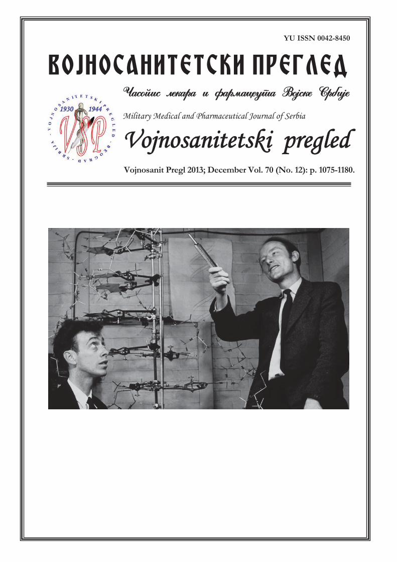
60th Anniversary of the DNA Secondary Structure Discovery
106
YU ISSN 0042-8450 VOJNOSANITETSKI PREGLED ^asopis lekara i farmaceuta Vojske Srbije Military Medical and Pharmaceutical Journal of Serbia Vojnosanitetski pregled Vojnosanit Pregl 2013; December Vol. 70 (No. 12): p. 1075-1180.
Transcript of 60th Anniversary of the DNA Secondary Structure Discovery
YU ISSN 0042-8450
VOJNOSANITETSKI PREGLED^asopis lekara i farmaceuta Vojske Srbije
Military Medical and Pharmaceutical Journal of Serbia
Vojnosanitetski pregledVojnosanit Pregl 2013; December Vol. 70 (No. 12): p. 1075-1180.

YU ISSN 0042-8450 vol. 70, br. 12, 2013.
Štampa Vojna štamparija, Beograd, Resavska 40 b.
VOJNOSANITETSKI PREGLEDPrvi broj Vojnosanitetskog pregleda izašao je septembra meseca 1944. godine
asopis nastavlja tradiciju Vojno-sanitetskog glasnika, koji je izlazio od 1930. do 1941. godine
IZDAVAUprava za vojno zdravstvo MO Srbije
IZDAVA KI SAVET
prof. dr sc. med. Boris Ajdinoviprof. dr sc. pharm. Mirjana Antunovi
prof. dr sc. med. Dragan Din i , puk.prof. dr sc. med. Zoran Hajdukovi , puk.
prof. dr sc. med. Nebojša Jovi , puk.prof. dr sc. med. Marijan Novakovi , brigadni general
prof. dr sc. med. Zoran Popovi , brigadni general (predsednik)prof. dr Sonja Radakovi
prof. dr sc. med. Predrag Romi , puk.prim. dr Stevan Sikimi , puk.
ME UNARODNI URE IVA KI ODBOR
Prof. Andrej Aleksandrov (Russia)Assoc. Prof. Kiyoshi Ameno (Japan)
Prof. Rocco Bellantone (Italy)Prof. Hanoch Hod (Israel)
Prof. Abu-Elmagd Kareem (USA)Prof. Hiroshi Kinoshita (Japan)
Prof. Celestino Pio Lombardi (Italy)Prof. Philippe Morel (Switzerland)
Prof. Kiyotaka Okuno (Japan)Prof. Stane Repše (Slovenia)
Prof. Mitchell B. Sheinkop (USA)Prof. Hitoshi Shiozaki (Japan)
Prof. H. Ralph Schumacher (USA)Prof. Miodrag Stojkovi (UK)
Assist. Prof. Tibor Tot (Sweden)
URE IVA KI ODBOR
Glavni i odgovorni urednikprof. dr sc. pharm. Silva Dobri
Urednici:
prof. dr sc. med. Bela Balintprof. dr sc. stom. Zlata Brkiprof. dr sc. med. Snežana Ceroviakademik Miodrag oli , brigadni generalakademik Radoje oloviprof. dr sc. med. Aleksandar urovi , puk.prof. dr sc. med. Branka uroviprof. dr sc. med. Borisav Jankoviprof. dr sc. med. Lidija Kandolf-Sekuloviakademik Vladimir Kanjuhakademik Vladimir Kostiprof. dr sc. med. Zvonko Magiprof. dr sc. med. oko Maksi , puk.prof. dr sc. med. Gordana Mandi -Gajiprof. dr sc. med. Dragan Miki , puk.prof. dr sc. med. Darko Mirkoviprof. dr sc. med. Slobodan Obradovi , potpukovnikakademik Miodrag Ostojiakademik Predrag Peško, FACSakademik or e Radakprof. dr sc. med. Ranko Rai evi , puk.prof. dr sc. med. Predrag Romi , puk.prof. dr sc. med. Vojkan Stani , puk.prof. dr sc. med. Dara Stefanoviprof. dr sc. med. Dušan Stefanovi , puk.prof. dr sc. med. Vesna Šuljagiprof. dr sc. stom. Ljubomir Todoroviprof. dr sc. med. Milan Višnjiprof. dr sc. med. Slavica Vu ini
Tehni ki sekretari ure iva kog odbora:dr sc. Aleksandra Gogi , dr Snežana JankoviREDAKCIJAGlavni menadžer asopisa:dr sc. Aleksandra Gogi
Stru ni redaktori:mr sc. med. dr Sonja Andri -Krivoku a, dr Maja Markovi ,dr Snežana Jankovi
Tehni ki urednik: Milan PerovanoviRedaktor za srpski i engleski jezik:Dragana Mu ibabi , prof.Korektori: Ljiljana Milenovi , Brana SaviKompjutersko-grafi ka obrada:Vesna Toti , Jelena Vasilj, Snežana uji
Adresa redakcije: Vojnomedicinska akademija, Institut za nau ne informacije, Crnotravska 17, poštanski fah 33–55, 11040 Beograd, Srbija. Telefoni:glavni i odgovorni urednik 3609 311, glavni menadžer asopisa 3609 479, pretplata 3608 997. Faks 2669 689. E-mail (redakcija): [email protected] objavljene u „Vojnosanitetskom pregledu“ indeksiraju: Science Citation Index Expanded (SCIE), Journal CitationReports/Science Edition, Index Medicus (Medline), Excerpta Medica (EMBASE), EBSCO, Biomedicina Serbica. Sadržajeobjavljuju Giornale di Medicine Militare i Revista de Medicina Militara. Prikaze originalnih radova i izvoda iz sadržajaobjavljuje International Review of the Armed Forces Medical Services.
asopis izlazi dvanaest puta godišnje. Pretplate: Žiro ra un br. 840-314849-70 MO – Sredstva objedinjene naplate – VMA (za Vojnosanitetskipregled), poziv na broj 12274231295521415. Za pretplatu iz inostranstva obratiti se službi pretplate na tel. 3608 997. Godišnja pretplata: 5 000dinara za gra ane Srbije, 10 000 dinara za ustanove iz Srbije i 150 € (u dinarskoj protivvrednosti na dan uplate) za pretplatnike iz inostranstva.Kopiju uplatnice dostaviti na gornju adresu.
YU ISSN 0042-8450 vol. 70 No. 12, 2013
Printed by: Vojna štamparija, Beograd, Resavska 40 b.
VOJNOSANITETSKI PREGLEDThe first issue of Vojnosanitetski pregled was published in September 1944
The Journal continues the tradition of Vojno-sanitetski glasnik which was published between 1930 and 1941
PUBLISHERMilitary Health Department, Ministry of Defence, Serbia
PUBLISHER’S ADVISORY BOARD
Prof. Boris Ajdinovi , MD, PhDAssoc. Prof. Mirjana Antunovi , BPharm, PhD
Col. Assoc. Prof. Dragan Din i , MD, PhDCol. Assoc. Prof. Zoran Hajdukovi , MD, PhD
Col. Prof. Nebojša Jovi , MD, PhDBrigadier General Prof. Marijan Novakovi , MD, PhD
Brigadier General Prof. Zoran Popovi , MD, PhD (Chairman)Prof. Sonja Radakovi , MD, PhD
Col. Prof. Predrag Romi , MD, PhDCol. Stevan Sikimi , MD
INTERNATIONAL EDITORIAL BOARD
Prof. Andrej Aleksandrov (Russia)Assoc. Prof. Kiyoshi Ameno (Japan)
Prof. Rocco Bellantone (Italy)Prof. Hanoch Hod (Israel)
Prof. Abu-Elmagd Kareem (USA)Prof. Hiroshi Kinoshita (Japan)
Prof. Celestino Pio Lombardi (Italy)Prof. Philippe Morel (Switzerland)
Prof. Kiyotaka Okuno (Japan)Prof. Stane Repše (Slovenia)
Prof. Mitchell B. Sheinkop (USA)Prof. Hitoshi Shiozaki (Japan)
Prof. H. Ralph Schumacher (USA)Prof. Miodrag Stojkovi (UK)
Assist. Prof. Tibor Tot (Sweden)
EDITORIAL BOARDEditor-in-chiefProf. Silva Dobri , BPharm, PhD
Co-editors:Prof. Bela Balint, MD, PhDAssoc. Prof. Zlata Brki , DDM, PhDAssoc. Prof. Snežana Cerovi , MD, PhDBrigadier General Prof. Miodrag oli , MD, PhD, MSAASProf. Radoje olovi , MD, PhD, MSAASCol. Assoc. Prof. Aleksandar urovi , MD, PhDAssoc. Prof. Branka urovi , MD, PhDProf. Borisav Jankovi , MD, PhDAssoc. Prof. Lidija Kandolf-Sekulovi , MD, PhDProf. Vladimir Kanjuh, MD, PhD, MSAASProf. Vladimir Kosti , MD, PhD, MSAASProf. Zvonko Magi , MD, PhDCol. Prof. oko Maksi , MD, PhDAssoc. Prof. Gordana Mandi -Gaji , MD, PhDCol. Assoc. Prof. Dragan Miki , MD, PhDProf. Darko Mirkovi , MD, PhDAssoc. Prof. Slobodan Obradovi , MD, PhDProf. Miodrag Ostoji , MD, PhD, MSAASProf. Predrag Peško, MD, PhD, MSAAS, FACSProf. or e Radak, MD, PhD, MSAASCol. Prof. Ranko Rai evi , MD, PhDCol. Prof. Predrag Romi , MD, PhDCol. Prof. Vojkan Stani , MD, PhDAssoc. Prof. Dara Stefanovi , MD, PhDCol. Prof. Dušan Stefanovi , MD, PhDProf. Milan Višnji , MD, PhDAssoc. Prof. Slavica Vu ini , MD, PhDAssoc. Prof. Vesna Šuljagi , MD, PhD.Prof. Ljubomir Todorovi , DDM, PhD
Technical secretaryAleksandra Gogi , PhD, Snežana Jankovi , MD
EDITORIAL OFFICEMain Journal ManagerAleksandra Gogi , PhDEditorial staffSonja Andri -Krivoku a, MD, MSc; Snežana Jankovi , MD;Maja Markovi , MD; Dragana Mu ibabi , BATechnical editorMilan PerovanoviProofreadingLjiljana Milenovi , Brana SaviTechnical editingVesna Toti , Jelena Vasilj, Snežana uji
Editorial Office: Military Medical Academy, INI; Crnotravska 17, PO Box 33–55, 11040 Belgrade, Serbia. Phone:Editor-in-chief +381 11 3609 311; Main Journal Manager +381 11 3609 479; Fax: +381 11 2669 689; E-mail: [email protected] published in the Vojnosanitetski pregled are indexed in: Science Citation Index Expanded (SCIE), Journal CitationReports/Science Edition, Index Medicus (Medline), Excerpta Medica (EMBASE), EBSCO, Biomedicina Serbica. Contents arepublished in Giornale di Medicine Militare and Revista de Medicina Militara. Reviews of original papers and abstracts ofcontents are published in International Review of the Armed Forces Medical Services.The Journal is published monthly. Subscription: Giro Account No. 840-314849-70 Ministry of Defence – Total means ofpayment – VMA (for the Vojnosanitetski pregled), refer to number 12274231295521415. To subscribe from abroad phone to+381 11 3608 997. Subscription prices per year: individuals 5,000.00 RSD, institutions 10,000.00 RSD, and foreign subscribers150 €.

Poštovani autori, urednici, recenzenti i saradnici Vojnosanitetskog pregleda,
Opraštaju i se od stare godine, zahvaljujem vam na izuzetnoj saradnji i podršci uz želje danam nastupaju a 2014. godina donese još više uspeha i radosti!
SRE NA NOVA GODINA I BOŽI NI PRAZNICI!Srda no,prof. dr Silva Dobri ,glavni i odgovorni urednik
Dear Authors, Editors, Reviewers, and Collaborators of the Vojnosanitetski Pregled,
Saying farewell to 2013, I express my deep gratitute to your extraordinary cooperationand support along with my best whishes that the coming New Year 2014 bring us moresuccess and happiness!
MARRY CHRISTMAS AND A HAPPY NEW YEAR!
Cordially,Prof. Dr. Silva Dobri
Editor-in-Chief
VOJNOSANITETSKI PREGLED
VOJNOMEDICINSKA AKADEMIJA
Crnotravska 17, 11040 Beograd, SrbijaTel/faks: +381 11 [email protected]@hotmail.com
Poziv za reklamiranje u 2014. godini
U prilici smo da vam ponudimo mogu nost oglašavanja i reklamiranja proizvoda i usluga u asopisu
„Vojnosanitetski pregled“ (VSP). To je sigurno najbolji vid i najzastupljeniji na in upoznavanja
eventualnih korisnika sa vašim uslugama i proizvodima.
asopis „Vojnosanitetski pregled“, zvani ni organ lekara i farmaceuta Vojske Srbije, nau no-
stru nog je karaktera i objavljuje radove iz svih oblasti medicine, stomatologije i farmacije. Radove
ravnopravno objavljuju stru njaci iz vojnih i civilnih ustanova i iz inostranstva. Štampa se na srpskom i
engleskom jeziku. asopis izlazi neprekidno od 1944. godine do sada. Jedini je asopis u zemlji koji izlazi
mese no (12 brojeva), na oko 100 strana A4 formata, a povremeno se objavljuju i tematski dodaci
(suplementi). Putem razmene ili pretplate VSP se šalje u 23 zemlje sveta. Radove objavljene u VSP-u
indeksiraju: Science Citation Index Expanded (SCIE), Journal Citation Reports/Science Edition, Index Medicus
(Medline), Excerpta Medica (EMBASE), EBSCO (preko ove baze VSP je dostupan on line od 2002. godine u
pdf formatu) i Biomedicina Serbica.
Cene reklama i oglasa u asopisu „Vojnosanitetski pregled“ u 2014. godini su:
1. Oglas u crno-beloj tehnici A4 formata za jedan broj 20 000,00 dinara2. Oglas u c/b tehnici A4 formata za celu godinu (11-12 brojeva) 200 000,00 dinara3. Oglas u boji A4 formata za jedan broj 35 000,00 dinara4. Oglas u boji A4 formata za celu godinu (11-12 brojeva) 330 000,00 dinara5. Oglas u boji na koricama K3 za jedan broj 50 000,00 dinara6. Oglas u boji na koricama K3 za celu godinu (11-12 brojeva) 455 000,00 dinara7. Oglas u boji na koricama K2 i K4 za jedan broj 55 000,00 dinara8. Oglas u boji na koricama K2 i K4 za celu godinu (11-12 brojeva) 530 000,00 dinara
Za sva obaveštenja, uputstva i ponude obratiti se redakciji asopisa „Vojnosanitetski pregled“.
Sredstva se upla uju na žiro ra un kod Uprave javnih pla anja u Beogradu broj: 840-941621-02 VMA (za
Vojnosanitetski pregled ili za VSP), PIB 102116082. Uplatnicu (dokaz o uplati) dostaviti li no ili poštom
(pismom, faksom, e-mail-om) na adresu: Vojnosanitetski pregled, Crnotravska 17, 11000 Beograd; tel/faks:
011 2669 689, e-mail: [email protected] ili [email protected]
Volumen 70, Broj 12 VOJNOSANITETSKI PREGLED Strana MLXXIX
CONTENTS / SADRŽAJ
ORIGINAL ARTICLES / ORIGINALNI LANCI
Sonja Pr i , Zorica Gajinov, Bogdan Zrni , Anica Radulovi , Milan Mati , Verica DjuranEpidemiological and clinical features of erythema infectiosum in children in Novi Sad from 2000to 2009Epidemiološke i klini ke karakteristike infektivnog eritema kod dece u Novom Saduu periodu 2000–2009................................................................................................................................... 1081Nikola Stankovi , Predrag Vlahovi , Vojin SaviHistomorphological and clinical study of primary and secondary glomerulopathies in SoutheastSerbia (20-year period of analysis)Histomorfološko i klini ko ispitivanje primarnih i sekundarnih glomerulopatija u jugoisto noj Srbiji(analiza u periodu od 20 godina) ................................................................................................................. 1085Aleksandar Argirovi , Djordje ArgiroviDoes the addition of Serenoa repens to tamsulosin improve its therapeutical efficacy in benignprostatic hyperplasia?Da li dodavanje Serenoa repens tamsulosinu poboljšava njegovu terapeutsku efikasnost kod benignehiperplazije prostate?................................................................................................................................... 1091Tatjana Gazibara, Aleksandra Filipovi , Vesna Kesi , Darija Kisi -Tepav evi , Tatjana PekmezoviRisk factors for epithelial ovarian cancer in the female population of Belgrade, Serbia: A case-control studyFaktori rizika od epitelijalnog karcinoma ovarijuma u ženskoj populaciji Beograda ................................. 1097Marko Banovi , Bosiljka Vujisi -Teši , Vuk Kuja i , Slobodan Obradovi ,Zdenko Crkvenac, Miodrag OstojiAssociation between aortic stenosis severity and contractile reserve measured by two-dimensional strain under low-dose dobutamine testingUticaj težine aortne stenoze na procenu kontraktilne rezerve procenjene pomo u dvodimenzionalnognaprezanja tokom niskodoznog dobutaminskog testa ................................................................................. 1103Vesna Mioljevi , Ljiljana Markovi -Deni , Ana Vidovi , Milica Jovanovi ,Tanja Toši , Dragica TominRisk factors for vancomycin-resistant Enterococcus colonization in hematologic patientsFaktori rizika od kolonizacije vankomicin-rezistentnog Enterococcus-a kod hematoloških bolesnika...... 1109Igor M. Nikoli , Goran M. Tasi , Vladimir T. Jovanovi , Nikola R. Repac, Aleksandar M. Jani ijevi ,Vuk D. Š epanovi , Branislav D. NestoroviAssessing the quality of angiographic display of brain blood vessels aneurysms compared tointraoperative stateProcena kvaliteta angiografskog prikaza aneurizmi krvnih sudova mozga u odnosu na intraoperativninalaz............................................................................................................................................................. 1117Boban Djordjevi , Marijan Novakovi , Milan Milisavljevi , Saša Mili evi , Aleksandar MalikoviSurgical anatomy and histology of the levator palpebrae superioris muscle for blepharoptosiscorrectionHirurška anatomija i histologija miši a podiza a gornjeg kapka u korekciji blefaroptoze ......................... 1124Vojislava Neškovi , Predrag Milojevi , Dragana Uni -Stojanovi , Ivan Ili , Zoran SlavkoviHigh thoracic epidural anesthesia in patients with synchronous carotid endarterectomy and off-pump coronary artery revascularizationVisoka torakalna epiduralna anestezija kod bolesnika sa istovremenom karotidnom endarterektomijomi off-pump revaskularizacijom koronarne arterije........................................................................................ 1132
Strana MLXXX VOJNOSANITETSKI PREGLED Volumen 70, Broj 12
GENERAL REWIEV / OPŠTI PREGLEDŽana Stankovi , Miroslava Jašovi -Gaši , Dušica Le i -ToševskiPsychological problems in patients with type 2 diabetes – Clinical considerationsPsihološki problemi bolesnika sa dijabetesom tipa 2 – klini ka razmatranja.............................................. 1138
CURRENT TOPIC / AKTUELNA TEMALjiljana Gojkovi Bukarica, Dragana Proti , Vladimir Kanjuh, Helmut Heinle, Radmila Novakovi ,Radisav Š epanoviCardiovascular effects of resveratrolKardiovaskularni efekti rezveratrola ........................................................................................................... 1145
CASE REPORTS / KAZUISTIKASvetlana Jovanovi , Zorica Jovanovi , Jelena Paovi , Vesna Stankovi eperkovi , Snežana Peši ,Vujica MarkoviTwo cases of uveitis masquerade syndrome caused by bilateral intraocular large B-celllymphomaDva bolesnika sa maskiranim sindromom uveitisa nastalim kao posledica obostranog intraokularnoglimfoma velikih B- elija.............................................................................................................................. 1151Ljubinka Jankovi Veli kovi , Irena Dimov, Dragan Petrovi , Slavica Stojnev, Stefan Da i ,Stefan Veli kovi , Vladisav StefanoviStromal reaction and prognosis in acinic cell carcinoma of the salivary glandStromalna reakcija i prognoza acino elijskog karcinoma pljuva ne žlezde................................................ 1155Aleksandra Lovrenski, Mirna Djuri , Ištvan Klem, Živka Eri, Milana Panjkovi , Dragana Tegeltija,Djordje PovažanMultisystem Langerhans cell histiocytosis coexisting with metastasizing adenocarcinoma of thelung: A case reportMultisistemska histiocitoza Langerhansovih elija udružena sa metastaziraju im adenokarcinomomplu a ............................................................................................................................................................ 1159Mihailo Vukmirovi , Lazar Angelkov, Filip Vukmirovi , Irena Tomaševi VukmiroviSuccessful implantation of a biventricular pacing and defibrillator device via a persistent leftsuperior vena cavaUspešna ugradnja resinhronizaciono-defibrilatorskog aparata preko perzistentne leve gornje šuplje vene ... 1162
HISTORY OF MEDICINE / ISTORIJA MEDICINEGoran Brajuškovi , Dušanka Savi Pavi evi , Stanka RomacThe 60th anniversary of the discovery of DNA secondary structureOtkri e sekundarne strukture molekula DNK – 60-godišnjica.................................................................... 1165
PISMO UREDNIKU / LETTER TO THE EDITOR .................................................................................. 1171
BOOK REVIEW / PRIKAZ KNJIGE......................................................................................................... 1175
UPUTSTVO AUTORIMA / INSTRUCTIONS TO THE AUTHORS....................................................... 1177
Watson & Crick`s deoxyribonucleic acid (DNA) model in 1953The discovery in 1953 of the double helix, the twisted-ladder structure of DNA, byJames Watson and Francis Crick marked a milestone in the history of science andgave rise in modern molecular biology, wich is largely concerned withunderstanding how genes control biochemical processes within cells. This year 60thanniversary of this discovery has been marked around the world (see pages 1165–70).
Watson-ov i Crick-ov model dezoksiribonukleinske kiseline (DNK) iz 1953. godineOtkri e Watson-a i Crick-a iz 1953. godine da DNK ima strukturu dvostruke spi-rale u obliku uvijenih stepenica, ozna ilo je prekretnicu u istoriji nauke i dovelo dobrzog razvoja moderne molekularne biologije i razumevanja na ina na koji genikontrolišu biohemijske procese unutar elije. Ove godine, širom sveta obeležena je60. godišnjica tog otkri a (vidi str. 1165–70).
Vojnosanit Pregl 2013; 70(12): 1081–1084. VOJNOSANITETSKI PREGLED Strana 1081
Correspondence to: Sonja Pr i , Department Of Dermatovener ology, Institute for Child and Youth Health Care of Vojvodina, HajdukVeljkova 10, Novi Sad, Serbia. Phone: +381 21 488 0444. Fax: +381 21 520 436. E-mail: [email protected]
O R I G I N A L A R T I C L E S UDC: 616.9-053.2-02:578.822]:616.511DOI: 10.2298/VSP110607026P
Epidemiological and clinical features of erythema infectiosum inchildren in Novi Sad from 2000 to 2009Epidemiološke i klini ke karakteristike infektivnog eritema kod dece u Novom
Sadu u periodu 2000–2009.
Sonja Pr i *, Zorica Gajinov†, Bogdan Zrni ‡, Anica Radulovi *, Milan Mati †,Verica Djuran†
*Department of Dermatovenerology, Institute for Child and Youth Health Care ofVojvodina, Novi Sad, Serbia; †Clinic of Dermatovenerology Diseases, Clinical Center of
Vojvodina, Novi Sad, Serbia; ‡Faculty of Medicine, University of Banja Luka, BanjaLuka, Republic of Srpska, Bosnia and Herzegovina
Abstract
Background/Aim. Erythema infectiosum (EI) is a com-mon childhood illness, caused by human parvovirus B19.It occurs sporadically or in epidemics and is characterizedby mild constitutional symptoms and a blotchy or maculo-papular lacy rash on the cheeks (slapped-cheek) spreadingprimarily to the extremities and trunk. The aim of ourstudy was to analyse the epidemiological and clinical char-acteristics of erythema infectiosum in children. Methods.This study included 88 children observed in the Depart-ment of Dermatology of the Institute for Child and YouthHealth Care of Vojvodina, in Novi Sad, during the periodJanuary 2000–December 2009. We compared the dataabout the clinical characteristics during and after the out-break of EI observed from December 2001 to September2002. The data were retrieved from the hospital database.Results. During the study period, EI was detected in 88children (44 females and 44 males), 0.213% of the totalnumber of 41,345 children observed in the Department ofDermatology. An outbreak of erythema infectiosum wasobserved from December 2001 to September 2002, with
the peak frequency in April and May 2002 and 39 diag-nosed cases, and stable number of cases from 2005 to2009 (a total of 49 diagnosed cases). The average age ofinfected children was 7.59 ± 3.339. Eleven (12.5%) chil-dren were referred from primary care pediatricians withthe diagnosis of urticaria or rash of allergic origin. Themost constant clinical sign was reticular exanthema on thelimbs, present in 100% of the cases, followed by 89.77%of cheek erythema. Pruritus was present in 9.09% of thechildren, mild constitutional symptoms in 5.68% and pal-pable lymph glands in 3.41% of the children. In all thecases the course of the disease was without complications.Conclusion. The results of this study confirm the pres-ence of EI (the fifth disease) in our area with a mildcourse in the majority of patients. Since the diagnosis ofEI is usually based on clinical findings, continuing medicaleducation of primary health care pediatricians is essentialfor reducing the number of misdiagnosed cases.
Key words:erythema infectiosum; child; serbia; disease outbreaks;epidemiology; drug therapy; prognosis.
Apstrakt
Uvod/Cilj. Infektivni eritem je de ja osipna bolest, koja sekarakteriše homogenim, veoma izraženim eritemom obrazai mrežastim osipom, lokalizovanim na ekstremitetima i tru-pu, bez zna ajnijeg poreme aja opšteg stanja. Izaziva jeparvovirus B19. Javlja se sporadi no ili periodi no, u vidumanjih epidemija. Cilj našeg rada bio je da se utvrde epide-miološke i klini ke karakteristike infektivnog eritema. Me-tode. Studijom je bilo obuhva eno 88 obolele dece 44 de-voj ice i 44 de aka koja su se javila na pregled u Odseku zadermatologiju, Instituta za zdravstvenu zaštitu dece i omla-dine u Novom Sadu, u periodu od januara 2000. do decem-
bra 2009. godine. Podaci o klini kim karakteristikama bole-snika dijagnostikovanih tokom epidemije 2001/2002. godi-ne (39 obolelih) pore eni su sa periodom od 2005 do 2009.godine kada je broj dijagnostikovanih slu ajeva bio ujedna-en (ukupno 49). Rezultati. Infektivni eritem dijagnostiko-
van je kod 0,213% dece od ukupno 41 345 pregledane deceu Odseku za dermatologiju. Od decembra 2001. do septem-bra 2002. godine došlo je do naglog porasta broja obolelihod infektivnog eritema, dok je najve i broj obolelih bio umaju i aprilu 2002. godine. Prose an uzrast obolele dece iz-nosio je 7,59 ± 3,339 godine. Ukupno 12,5% bolesnikaupu eno je pod sumnjom na urtikariju ili osip alergijskogporekla. Mrežast osip na ekstremitetima bio je prisutan kod

Strana 1082 VOJNOSANITETSKI PREGLED Volumen 70, Broj 12
Pr i S, et al. Vojnosanit Pregl 2013; 70(12): 1081–1084.
svih obolelih, eritem obraza kod 89,77%, svrab je bio pri-sutan kod 9,09% dece, blagi opšti simptomi infekcije kod5,68%, a limfadenopatija kod 3,41% dece. Nisu zabeleženekomplikacije oboljenja. Zaklju ak. Sva obolela deca imalasu karakteristi nu klini ku sliku i dobro udan tok bolesti.Pošto se dijagnoza infektivnog eritema u de jem uzrastu po-stavlja pretežno na osnovu klini ke slike, naglašavamo važ-
nost kontinuiranog obrazovanja pedijatara koji pružaju pri-marnu zdravstvenu zaštitu sa klini kim tokom i diferencijal-nom dijagnozom ovog oboljenja.
Klju ne re i:eritem, infektivni; deca; srbija; epidemije;epidemiologija; le enje lekovima; prognoza.
Introduction
Erythema infectiosum (EI) is an acute childhood illness,characterized by blotchy or maculopapular rash starting oncheeks, spreading to extremities and the trunk, giving a typicalslapped cheek appearance and lacy configuration on limbs.Constitutional symptoms are mild 1, 2. The disease is caused byhuman parvovirus B19 (Pv B19), the route of natural transmis-sion is presumably the respiratory route, and parenteral andtransplacental transmission have been proved, as well. A re-ceptor molecule for B19 is glycolipid antigen on the erythro-cyte surface, and hosts with the decreased production or in-creased destruction of red blood cells (such as hemolytic ane-mia or pure red cell aplasia due to various causes) are prone toaplastic crises and protracted severe anemia upon infectionwith human Pv B19 2–4. In immunocompromised hosts thecourse of Pv B19 infection can be severe, even caused by he-mophagocytic syndrome 4. Acute infection during pregnancycan result in hydrops fetalis (risk estimated in 10% of cases) 5.The incubation period is 4–14 days, during which patients areinfectious, only prior to the onset of the rash 6, 7.
EI usually develops suddenly, prodromal symptoms aremild or may be absent. The course of the disease is three-phasic: facial “slapped-cheeks“ rash, followed by lacy or re-ticular rash of the upper extremities and an evanes-cence/recrudescence stage. Eruption is pruritic in about 15%of children. EI usually lasts for 2 weeks, but can recur withmechanical, physical or emotional triggers. The diagnosis ofEI is usually made on the basis of the characteristic clinicalfeatures. The differential diagnosis includes the other viralrashes (rubella, measles, enteroviral infection), scarlet fever,cheek erysipelas, hypersensitivity reaction (urticaria, drugreaction or allergic exanthemata) and collagen vascular dis-eases (systemic lupus erythematosus). Due to the mild courseonly symptomatic treatment is necessary (antipyretics, anti-histamines) 1, 7, 8.
Methods
This retrospective study was conducted in the Depart-ment of Dermatology, Institute for Child and Youth HealthCare of Vojvodina in Novi Sad, as tertiary referral center forthe South Ba ka region in the Province Vojvodina. Age, sexdistribution, the presence of constitutional symptoms andclinical characteristics of the disease were retrieved from themedical records of all EI patients diagnosed between January2000 and December 2009. The data from the period 2005–2009, and those previously published for the period 2000–2004 were compared 9.
All the patients were diagnosed by one of the derma-tologists (authors), based on the clinical findings in the ma-jority of cases. A total of 12 patients (2 during the period of2005–2009) serum samples were additionally analyzed usingSERION ELISA (enzyme-linked immunosorbent assay)classic parvovirus B19 IgG/IgM quantitative and qualitativetests for identification of specific antibodies against humanparvovirus B19. Complement-fixing reactions (CFR) to vi-ruses (Rubella, Adenovirus, Coxsackie virus) were per-formed in 5 children, with constitutional symptoms and pal-pable lymph nodes.
Results
During the study period (from the beginning of 2000 tothe end of 2009), at the Department of Dermatology, the totalnumber of first visits was 41,345 and erythema infectiosumwas diagnosed in 88 children (0.213%). Figure 1 shows the
number of patients during a 10-year period. Patients with EIwere not observed within 2000, and sporadic cases emergedby the end of 2001. A sudden outbreak was noted from De-cember 2001 to September 2002 with the highest number ofcases recorded in April and May, 2002 9. In a subsequent 5year period (2005–2009) EI was diagnosed in 49 children, 22girls and 27 boys, average age being 7.98 ± 3.554 years.During a total period of 10 years, the average age of EI pa-tients was 7.59 ± 3.339 years, with the majority of patients inthe 5–10 years group, and equal sex ratio (44 girls and 44boys affected).
During the whole study period 11 children (12.5%)were referred from primary care pediatricians with the diag-nosis of urticaria or rash of allergic origin, 7 during the pe-
0
5
10
15
20
25
30
35
40
2000 2001 2002 2003 2004 2005 2006 2007 2008 2009
the
No o
f pat
ient
s
Fig. 1 – The number of patients with erythema infectiosumin a 10-year research period.
Volumen 70, Broj 12 VOJNOSANITETSKI PREGLED Strana 1083
Pr i S, et al. Vojnosanit Pregl 2013; 70(12): 1081–1084.
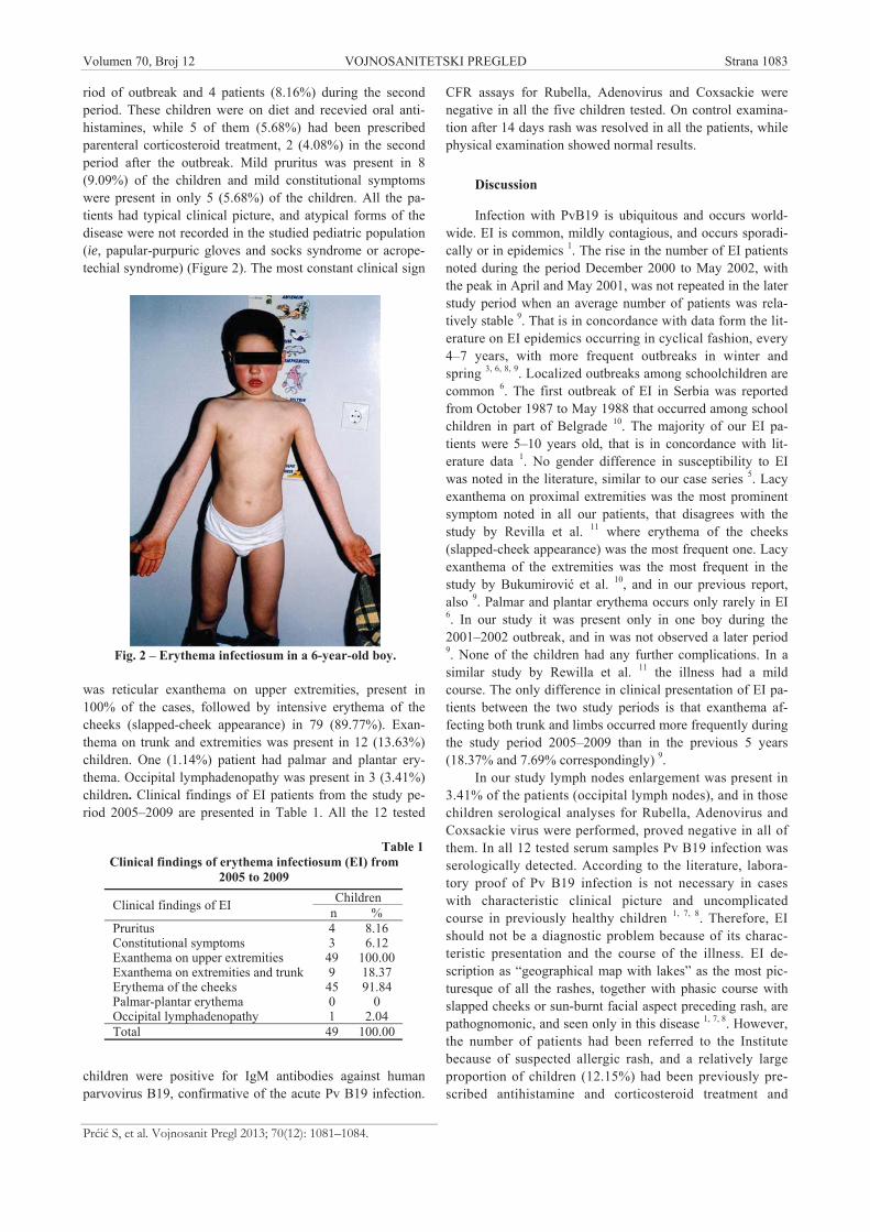
riod of outbreak and 4 patients (8.16%) during the secondperiod. These children were on diet and recevied oral anti-histamines, while 5 of them (5.68%) had been prescribedparenteral corticosteroid treatment, 2 (4.08%) in the secondperiod after the outbreak. Mild pruritus was present in 8(9.09%) of the children and mild constitutional symptomswere present in only 5 (5.68%) of the children. All the pa-tients had typical clinical picture, and atypical forms of thedisease were not recorded in the studied pediatric population(ie, papular-purpuric gloves and socks syndrome or acrope-techial syndrome) (Figure 2). The most constant clinical sign
was reticular exanthema on upper extremities, present in100% of the cases, followed by intensive erythema of thecheeks (slapped-cheek appearance) in 79 (89.77%). Exan-thema on trunk and extremities was present in 12 (13.63%)children. One (1.14%) patient had palmar and plantar ery-thema. Occipital lymphadenopathy was present in 3 (3.41%)children. Clinical findings of EI patients from the study pe-riod 2005–2009 are presented in Table 1. All the 12 tested
children were positive for IgM antibodies against humanparvovirus B19, confirmative of the acute Pv B19 infection.
CFR assays for Rubella, Adenovirus and Coxsackie werenegative in all the five children tested. On control examina-tion after 14 days rash was resolved in all the patients, whilephysical examination showed normal results.
Discussion
Infection with PvB19 is ubiquitous and occurs world-wide. EI is common, mildly contagious, and occurs sporadi-cally or in epidemics 1. The rise in the number of EI patientsnoted during the period December 2000 to May 2002, withthe peak in April and May 2001, was not repeated in the laterstudy period when an average number of patients was rela-tively stable 9. That is in concordance with data form the lit-erature on EI epidemics occurring in cyclical fashion, every4–7 years, with more frequent outbreaks in winter andspring 3, 6, 8, 9. Localized outbreaks among schoolchildren arecommon 6. The first outbreak of EI in Serbia was reportedfrom October 1987 to May 1988 that occurred among schoolchildren in part of Belgrade 10. The majority of our EI pa-tients were 5–10 years old, that is in concordance with lit-erature data 1. No gender difference in susceptibility to EIwas noted in the literature, similar to our case series 5. Lacyexanthema on proximal extremities was the most prominentsymptom noted in all our patients, that disagrees with thestudy by Revilla et al. 11 where erythema of the cheeks(slapped-cheek appearance) was the most frequent one. Lacyexanthema of the extremities was the most frequent in thestudy by Bukumirovi et al. 10, and in our previous report,also 9. Palmar and plantar erythema occurs only rarely in EI6. In our study it was present only in one boy during the2001–2002 outbreak, and in was not observed a later period9. None of the children had any further complications. In asimilar study by Rewilla et al. 11 the illness had a mildcourse. The only difference in clinical presentation of EI pa-tients between the two study periods is that exanthema af-fecting both trunk and limbs occurred more frequently duringthe study period 2005–2009 than in the previous 5 years(18.37% and 7.69% correspondingly) 9.
In our study lymph nodes enlargement was present in3.41% of the patients (occipital lymph nodes), and in thosechildren serological analyses for Rubella, Adenovirus andCoxsackie virus were performed, proved negative in all ofthem. In all 12 tested serum samples Pv B19 infection wasserologically detected. According to the literature, labora-tory proof of Pv B19 infection is not necessary in caseswith characteristic clinical picture and uncomplicatedcourse in previously healthy children 1, 7, 8. Therefore, EIshould not be a diagnostic problem because of its charac-teristic presentation and the course of the illness. EI de-scription as “geographical map with lakes” as the most pic-turesque of all the rashes, together with phasic course withslapped cheeks or sun-burnt facial aspect preceding rash, arepathognomonic, and seen only in this disease 1, 7, 8. However,the number of patients had been referred to the Institutebecause of suspected allergic rash, and a relatively largeproportion of children (12.15%) had been previously pre-scribed antihistamine and corticosteroid treatment and
Fig. 2 – Erythema infectiosum in a 6-year-old boy.
Table 1Clinical findings of erythema infectiosum (EI) from
2005 to 2009ChildrenClinical findings of EI n %
Pruritus 4 8.16Constitutional symptoms 3 6.12Exanthema on upper extremities 49 100.00Exanthema on extremities and trunk 9 18.37Erythema of the cheeks 45 91.84Palmar-plantar erythema 0 0Occipital lymphadenopathy 1 2.04Total 49 100.00
Strana 1084 VOJNOSANITETSKI PREGLED Volumen 70, Broj 12
Pr i S, et al. Vojnosanit Pregl 2013; 70(12): 1081–1084.
elimination diet. Even though EI is not a rare disease, itappears periodically every 4–7 years in the form of spo-radic epidemics lasting for several months, that could bethe reason not to be easily recognized among primaryhealth care physicians. Albeit, during 2001/2002 outbreakwhen 18% of children were referred from primary care assuspect allergic reaction, in the period 2005–2009 propor-tion of unrecognized cases decreased to 8%. That is an en-couraging result pointing that previous epidemics in-creased awareness and enhanced recognition of EI amongnon-dermatologists 9.
Conclusion
The results of this study confirm the presence of EI (thefifth disease) in our area with a mild course in the majority ofpatients. Since the diagnosis of EI is usually based on clini-cal findings, the continuing medical education of primaryhealth care pediatricians is essential for reducing the numberof misdiagnosed cases. Further studies within longer obser-vation periods and larger groups of patients are necessary todetermine the epidemiological and clinical characteristics oferythema infectiosum in Serbia.
R E F E R E N C E S
1. Vafaie J, Schwartz RA. Erythema infectiosum. J Cutan MedSurg 2005; 9(4): 159 61.
2. Drew WL. Parvoviruses. In: Wilson WR, editor. Current diag-nosis and treatment in infectiosus diseases. New York: McGraw-Hill Co; 2001. p. 451 3.
3. American Academy of Pediatrics Committee on Infectious Diseases. Par-vovirus B19. In: Pickering LK, Baker CJ, Kimberlin DW, Long SS,editorss. 2009 Red Book: Report of the Committee on Infec-tious Diseases. 28th ed. Elk Grove Village, IL: American Acad-emy of Pediatrics; 2009. p. 491 3.
4. Heegaard ED, Brown KE. Human parvovirus B19. Clin Micro-biol Rev 2002; 15(3): 485–505.
5. Anderson LJ, Hurwitz ES. Human parvovirus B19 and preg-nancy. Clin Perinatol 1988; 15(2): 273 86.
6. Mancinci A. Exanthems in childhood: an update. Pediatr Ann1998; 27(3):163 70.
7. Wiss K. Erythema infectiosum and Parvovirus B19 infection.In: Fitzpatrick TB, Eisen AZ, Wolff K, Freedberg IM, Austen KF,
editors. Dermatology in general medicine. 5th ed. New York:Mc Graw-Hill; 2003. p. 2409 13.
8. Zellman G. Erythema Infectiosum (Fifth Disease). EmedicineDermatology (journal series online). 2002. Available at:http://author.emedicine.com/derm/topic 136.htm.
9. Prcic S, Jakovljevi A, Djuran V, Gajinov Z. Erythema infectio-sum in children: a clinical study. Med Pregl 2006; 59(1 2):5 10. (Serbian, English)
10. Bukumirovi K, Radosavljevi M, Milki V, Antonijevi B, Polak D, DodiM, et al. Epidemiologic characteristics of human parvovirus B19.Report of an epidemic of erythema infectiosum in the area of Bel-grade. Srp Arh Celok Lek 1992; 120(5 6): 171 4. (Serbian)
11. Revilla G, Carro G, Sanchez DM, Galan C, Nebreda M. Outbreakof infectious erythema at a urban health center. Aten Primaria2000; 26(3): 172 5. (Spanish)
Received on Jun 7, 2011.Revised on Avgust 18, 2011.
Accepted on September 5, 2011.OnLine-First May, 2013.

Vojnosanit Pregl 2013; 70(12): 1085–1090. VOJNOSANITETSKI PREGLED Strana 1085
Correspondence to: Nikola Stankovi , Mother and Child Health Care Institute, Cetinjska 32/7, 11 000 Belgrade, Serbia.Phone: +381 60 013 57 13. E-mail: [email protected]
O R I G I N A L A R T I C L E UDC: 616-036.22::616.61-076DOI: 10.2298/VSP110614027S
Histomorphological and clinical study of primary and secondaryglomerulopathies in Southeast Serbia (20-year period of analysis)Histomorfološko i klini ko ispitivanje primarnih i sekundarnih glomerulopatija
u jugoisto noj Srbiji (analiza u periodu od 20 godina)
Nikola Stankovi *, Predrag Vlahovi †, Vojin Savi ‡
*Mother and Child Health Care Institute, Belgrade, Serbia; †Center for MedicalBichemistry, ‡Institute of Nephrology and Hemodialysis, Clinical Center Niš, Niš, Serbia
Abstract
Background/Aim. Epidemiological studies of renal biop-sies have been performed to follow up the incidence ofglomerular diseases on a specified territory and to comparethe obtained results with results from other regions. Theaim of this study was to analyze the frequency of certainhistopathophysiological types of glomerular diseases on theterritory of Southeast Serbia. Methods. In a 20-year period(1986–2006), 316 kidney biopsies were performed in pa-tients with clinicall signs of impaired renal function, inSoutheast Serbia. On average 1.6 biopsies were made peryear per 100 000 inhabitants. Results. Biopsies of adult pa-tients represented 88% of all biopsies, biopsies in children(aged under 18 years) represented 8%, while biopsies of eld-erly patients (more than 60 years) represented 4% of all bi-opsies. The predominance of male patients was describedwith male/female ratio of 1.4. The most frequent clinicalmanifestation in patients at the time of biopsy were ne-phrotic syndrome (42.5%), and asymptomatic proteinuriaand/or hematuria (31.3%) and nephritic syndrome (14.9%).The most common glomerular disease was IgA nephropa-thy with an incidence of 21.5% of total biopsy diagnosedglomerulopathies, followed by: membranous glomerulone-phritis (12.6%), focal segmental proliferative and sclerosingglomerulonephritis (10.7%), lupus nephritis (8.4%), nephro-angiosclerosis (7.0%), mesangio-proliferative glomerulone-phritis (6.1%), minimal change disease (2.8%), mesangio-capillary glomerulonephritis (2.3%). Conclusion. The fre-quency of certain histopathologic findings significantly cor-related with data from studies that we used for comparison,with the exception of minimal change disease whose inci-dence in our study was smaller.
Key words:kidney diseases; glomerulonephritis; biopsy;histocytochemistry; epidemiology; serbia.
Apstrakt
Uvod/Cilj. Epidemiološke analize biopsija bubrega imajuza cilj pra enje u estalosti pojedinih glomerulskih bolesti naodre enom podru ju i upore ivanje dobijenih rezultata sarezultatima iz drugih regiona. Cilj ovog istraživanja bio je dase analizira u estalost pojedinih histopatofizioloških tipovaglomerulskih bolesti na teritoriji jugoisto ne Srbije. Meto-de. U periodu od 20 godina (1986–2006), na teritoriji jugoi-sto ne Srbije, ura ena je biopsija bubrega kod 316 bolesnikakod kojih su klini ki bili prisutni znaci poreme ene bubrež-ne funkcije. Prose no je ura eno 1,6 biopsija godišnje na100 000 stanovnika. Rezultati. Od ura enih biopsija 88%su bile biopsije kod odraslih osoba, 8% kod dece, dok subiopsije kod starijih osoba inile 4%. Predominacija boles-nika muškog pola, iskazana muško/ženskim odnosom biop-siranih bolesnika iznosila je 1,4. Najzastupljenije klini keprezentacije kod bolesnika u vreme biopsije bile su nefrotskisindrom (42,5%), zatim asimptomatska proteinurija i/ilihematurija (31,3%) i nefritski sindrom (14,9%). Naju estalijaglomerulska bolest bila je IgA nefropatija sa u estaloš u od21,5% od ukupnog broja biopsijom dijagnostifikovanihglomerulopatija, a sledili su: membranski glomerulonefritis(12,6%), fokalnosegmentalni proliferativni i skleroziraju iglomerulonefritis (10,7%), lupus nefritis (8,4%), nefroangio-skleroza (7,0%), mezangioproliferativni glomerulonefritis(6,1%), bolest minimalnih promena (2,8%), mezangio-kapilarni glomerulonefritis (2,3%). Zaklju ak. U estalostpojedinih patohistoloških nalaza u zna ajnoj meri koreliše sapodacima iz studija koje su nam služile za komparaciju, saizuzetkom bolesti minimalnih promena ija je incidencija ni-ža u našoj studiji.
Klju ne re i:bubreg, bolesti; glomerulonefritis; biopsija;histocitohemija; epidemiologija; srbija.
Strana 1086 VOJNOSANITETSKI PREGLED Volumen 70, Broj 12
Stankovi N, et al. Vojnosanit Pregl 2013; 70(12): 1085–1090.
Introduction
Glomerulonephritis (GN) is a bilateral, non-bacterial,non-suppurative inflammation of kidney which primarily af-fects glomerulus and then the other kidney structures.
Histological features are the most important criterionfor the nomenclature and classification of glomerular dis-ease. In addition to localization, the histological characteris-tics define the nature of glomerular lesion as well as cellproliferation, deposit of immune complexes or extracellularcomponents lesions.
Immune processes underlie most cases of primary GN,but they are also present in many secondary events in theglomerulus. Therefore, the division of etiopathologic mecha-nisms of glomerulopathy is reduced to immunological andnon-immunological 1.
Glomerular lesion is clinically expressed with a rela-tively small number of signs and symptoms. There are fivemain clinical syndromes which may be manifested in glo-merular disease: acute GN syndrome (acute nephritic syn-drome), syndrome of rapidly progressive GN, nephrotic syn-drome, asymptomatic urinary abnormalities (asymptomaticproteinuria and hematuria) and the syndrome of chronic GN.
The diagnosis of glomerular disease involves a com-plete understanding of the clinical condition of the patient,laboratory diagnostics, immunological and morphologicalstudies. Diagnosis based only on the clinical manifestationsmay be unsuccessful because different glomerular diseasesare characterized by similar or the same clinical presentation.On the other hand, the same glomerular disease may be pre-sented differently in different patients 2.
Therapeutic approach, response to therapy and progno-sis, depend on the type of GN. The frequency of certainforms of GN is different in different geographical areas andassociated with genetic and environmental factors.
This retrospective study was conducted with the aim todetermine the frequency of certain types of glomerular dis-ease in Southeast Serbia on the basis of which may be possi-ble to predict the response to therapy and prognosis.
Methods
During a 20-year period (1986–2006), 316 kidney biop-sies were performed in patients with clinically and laboratoryshown signs of some primary or secondary glomerulopathy(nephritic or nephrotic syndrome, syndrome of rapide pro-gressive GN and asymptomatic hematuria or proteinuria).All biopsies were done at the Institute of Nephrology andHemodialysis in Clinical Center Niš and the whole tissue bi-opsy material was processed in the histopathological labora-tory of this institute. The histopathologist has always takenbiopsy material from a patient and prepared it for light mi-croscopic analysis, immunofluorescence and if necessary forelectron microscopy.
The same method for taking biopsy material was al-ways used – blind percutaneous renal biopsy with previousintravenous pyelography or ultrasound examination. In par-allel to light microscopy, immunofluorescence with IgG,
IgA, IgM, C1q, C3, fibrinogen, albumin, kappa and lambdachains was always done. Electron microscopy was performedin 3–4% of cases when it was possible (during the difficulteconomic crisis) and when indications were present (othermorphological examination aroused suspicion of a change inthe GBM structure; existence of deposit which could notbeen precisely localize in immunofluorescence findings).
Of the total number of samples, 214 biopsies had posi-tive histopathological findings in the glomeruli, while 102findings were not included in this study. Only those caseswho clearly indicated the existence of glomerulopathy wereincluded. The patients whose clinical presentation and labo-ratory findings did not fit into diagnosis of glomerulopathyor tubulointerstitial changes were clinically and histopa-thologicaly confirmed, were not included in this study.Among samples which were not included in the study, 26had negative glomerular findings, 50 of them did not haveenough material for analysis, and in 26 of them findings intubulointerstitial system were dominant (chronic pyelone-phritis, acute tubular necrosis, balkan endemic nephropathy,etc).
Immunological tests included measurements of serumimmunoglobulins concentration, complement fractions in se-rum, immune complexes while the presence of certain auto-antibodies were detected by direct immunofluorescence.Immunoglobulins and complement fractions were measuredby the nephelometric method (II BEN nefelometar – Dade-Behring, Magdeburg, Germany). Immune complexes weremeasured by the deposition of immunoglobulins with poly-ethylene glycol (PEG-6000). The presence of autoantibodiesin biopsy specimens was performed by direct immunofluo-rescence with autoantibodies labeled with fluorescein(FITC).
Other analysis have included detailed medical history,routine biochemical tests as well as other relevant testing.The following findings have been specifically analyzed: du-ration of clinical manifestations of impaired kidney function,previous diseases, edema, the appearance of micro- ormacrohematuria, blood pressure, finding of proteinuria, hy-poproteinemia and hipoalbuminemia findings, and elevatedserum cholesterol and triglycerides.
The data were analyzed by descriptive statistics usingthe program Sigma Stat.
Results
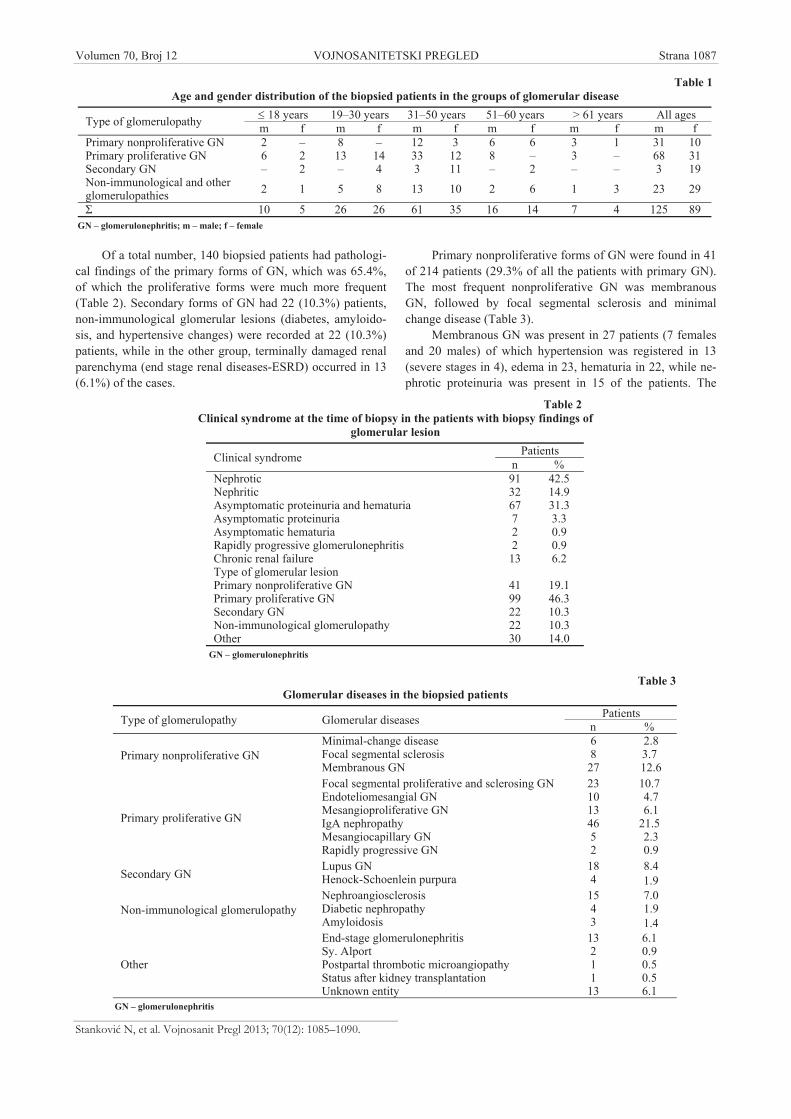
Of 214 patients with biopsy confirmed diagnosis of aglomerulopathy, 125 (58.4%) were males and 89 (41.6%)females.
Age and gender distributions are shown in Table 1. Agedata were available for 204 patients. Most of the patientswere aged 31–50 years, a total of 98 patients (61 males and37 females), that was 45.8% of the total number of biopsieswith positive findings. Within this age group, most weremale patients with primary proliferative GN (33% or 33.7%).
Clinical presentation of the patients with GN is shownin Table 2. Of the 7 described syndromes, the most commonwas nephrotic syndrome in 91 (42.5%) patients.
Volumen 70, Broj 12 VOJNOSANITETSKI PREGLED Strana 1087
Stankovi N, et al. Vojnosanit Pregl 2013; 70(12): 1085–1090.
Of a total number, 140 biopsied patients had pathologi-cal findings of the primary forms of GN, which was 65.4%,of which the proliferative forms were much more frequent(Table 2). Secondary forms of GN had 22 (10.3%) patients,non-immunological glomerular lesions (diabetes, amyloido-sis, and hypertensive changes) were recorded at 22 (10.3%)patients, while in the other group, terminally damaged renalparenchyma (end stage renal diseases-ESRD) occurred in 13(6.1%) of the cases.
Primary nonproliferative forms of GN were found in 41of 214 patients (29.3% of all the patients with primary GN).The most frequent nonproliferative GN was membranousGN, followed by focal segmental sclerosis and minimalchange disease (Table 3).
Membranous GN was present in 27 patients (7 femalesand 20 males) of which hypertension was registered in 13(severe stages in 4), edema in 23, hematuria in 22, while ne-phrotic proteinuria was present in 15 of the patients. The
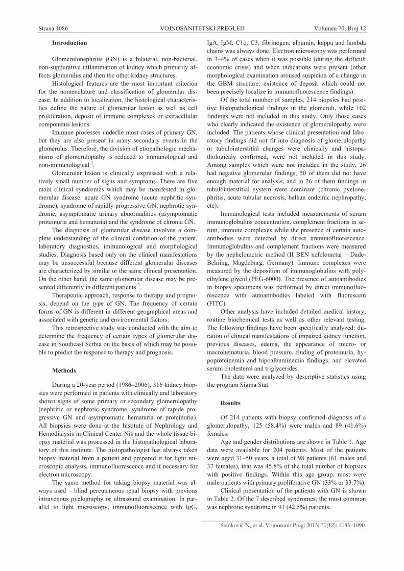
Table 1Age and gender distribution of the biopsied patients in the groups of glomerular disease
18 years 19–30 years 31–50 years 51–60 years > 61 years All agesType of glomerulopathy m f m f m f m f m f m fPrimary nonproliferative GN 2 – 8 – 12 3 6 6 3 1 31 10Primary proliferative GN 6 2 13 14 33 12 8 – 3 – 68 31Secondary GN – 2 – 4 3 11 – 2 – – 3 19Non-immunological and otherglomerulopathies 2 1 5 8 13 10 2 6 1 3 23 29
10 5 26 26 61 35 16 14 7 4 125 89GN – glomerulonephritis; m – male; f – female
Table 2Clinical syndrome at the time of biopsy in the patients with biopsy findings of
glomerular lesionPatientsClinical syndrome n %
Nephrotic 91 42.5Nephritic 32 14.9Asymptomatic proteinuria and hematuria 67 31.3Asymptomatic proteinuria 7 3.3Asymptomatic hematuria 2 0.9Rapidly progressive glomerulonephritis 2 0.9Chronic renal failure 13 6.2Type of glomerular lesionPrimary nonproliferative GN 41 19.1Primary proliferative GN 99 46.3Secondary GN 22 10.3Non-immunological glomerulopathy 22 10.3Other 30 14.0
GN – glomerulonephritis
Table 3Glomerular diseases in the biopsied patients
PatientsType of glomerulopathy Glomerular diseases n %Minimal-change disease 6 2.8Focal segmental sclerosis 8 3.7Primary nonproliferative GNMembranous GN 27 12.6Focal segmental proliferative and sclerosing GN 23 10.7Endoteliomesangial GN 10 4.7Mesangioproliferative GN 13 6.1IgA nephropathy 46 21.5Mesangiocapillary GN 5 2.3
Primary proliferative GN
Rapidly progressive GN 2 0.9Lupus GN 18 8.4
Secondary GN Henock-Schoenlein purpura 4 1.9Nephroangiosclerosis 15 7.0Diabetic nephropathy 4 1.9Non-immunological glomerulopathyAmyloidosis 3 1.4
Other
End-stage glomerulonephritisSy. AlportPostpartal thrombotic microangiopathyStatus after kidney transplantationUnknown entity
13211
13
6.10.90.50.56.1
GN – glomerulonephritis
Strana 1088 VOJNOSANITETSKI PREGLED Volumen 70, Broj 12
Stankovi N, et al. Vojnosanit Pregl 2013; 70(12): 1085–1090.
findings of immunofluorescence of IgG were positive in 25of the patients.
Detection of primary proliferative GN was present in 99patients of 214 which is 70.7% of the biopsied patients withfindings of primary glomerulonephritis. The most commonform of primary proliferative GN was IgA nephropathy, thenfocal segmantal praliferative and sclerosing GN, mesangio-proliferative GN, endoteliomezangial GN, mesangiocapillaryGN and rapidly progressive GN (Table 3).
IgA nephropathy was diagnosed in 46 patients (14 fe-males and 32 males). The youngest patients belonged to theage group of 11–20 years while the oldest were between 51and 60 years. Hypertension was present in 21, edema in 10,the finding of hematuria was present in all the patients, whileproteinuria greater than 3.5 g/L was present in 8 patientswith this finding. The largest number of light microscopyfindings belonged to the group with focal segmental-proliferative and sclerosing GN. Second most frequent find-ings matched to focal segmental proliferative GN with mes-angial area lesions. Diffuse changes in the glomeruli, such asdiffuse proliferative and diffuse proliferative and sclerosingGN were registrated in a much lower number of cases. Ex-tracapillary proliferation with less than 50% of affected glo-meruli was observed in only one biopsy. Considering immu-nofluorescence findings all the patients of this group werepositive for IgA.
Secondary forms of GN were registered in 22 out of214 biopsied patients, which is 10.3%. The definitive diag-nosis in these cases was confirmed by other immunologicaltests (primarily by determining the presence of serum auto-antibodies). Out of 22 patients 18 had systemic lupus ery-thematosus, and 4 showed light microscopic signs of focalproliferative and sclerosing GN with changes in the skin andpositive immunofluorescence finding, which is definitelypointing on Henoch-Schonlein purpura (Table 3).
Lupus nephritis was present in 18 cases. All the patientswere females, aged 17 to 48 years. Hypertension was regis-trated in 7 of the patients, edema in 13, proteinuria andhematuria in all the patients. Serum levels of C3 and C4 werereduced in most patients, while immune complexes and anti-nuclear antibodies were present in 9 of the patients to whomthese measurements were made. Non-immunological andother glomerulopathies were diagnosed in 52 patients, whichis 24.3%. Nephroangiosclerosis was diagnosed in 15 of thepatients, glomerular lesion in diabetic nephropathy in 4 pa-tients and glomerular lesions in amyloidosis in 3 patients(Table 3). Other biopsy material presented 14% of the biop-sied patients with positive findings and spoke in support ofend-stage GN (13 patients), Alport syndrome (2 patients),postpartal thrombotic microangiopathy (1 patient), status af-ter kidney transplantation (1 patient) or it was not possible tocome out of any known entity.
Discussion
Epidemiology of glomerular disease often shows somepeculiarities related to the geographic area, requiring a re-gional monitoring and mutual comparison of morbidity in
different territories. This study presents the frequency ofsome glomerular diseases diagnosed by biopsy of renal tissuein the period of 20 years in Southeast Serbia (population over1 million).
A large number of biopsy samples with insufficientmaterial is probably due to technical reasons of biopsy pro-cedure (blind percutaneous renal biopsy) and in some degreedisparage the preciseness of results. According to our results,1.6 biopsies were performed per 100 000 inhabitants peryear. This number is several times lower than the rates pres-ent in Western European countries like Spain 3, Italy 4,France 5, and the Czech Republic 6, or Australia 7, but issimilar to the rate of biopsies in Romania 8, whose registry isone of the few renal biopsy registers of Eastern Europe, andto the rate of biopsies obtained in another single centre studyconducted in Serbia, for the same period in a much largersample 9.
Biopsies in adult patients represented 88% of all biop-sies, biopsies in children (aged under 18 years) represented8%, while the biopsies in elderly patients (more than 60years) represented 4% of all biopsies (Table 1). This rela-tionship between the number of biopsies of children and theelderly is quite different from the relationship presented inthe Spanish registry of renal biopsies, where renal biopsy inchildren represented 7% and 22% in the elderly 3. These re-sults show predominance of adults in the biopsied patientsand that a large number of elderly patients and children weretreated on the basis of clinical manifestations.
The predominance of male patients is reported withmale/female ratio of 1.4, which is consistent with data fromother international studies 3,7,10. From this, we can excludesecondary forms of glomerulonephritis, in which the pre-dominance of females was present, what can be explained bya much higher incidence of systemic lupus in women (Table1). However, male/female ratio is significantly different fromthe results obtained in another single centre study conductedin Serbia, for the same period in a much larger sample 9.
The most frequent clinical presentations in patients atthe time of biopsy were nephrotic syndrome (42.5%),asymptomatic proteinuria and/or hematuria (31.3%) andnephritic syndrome (14.9%). Other syndromes were signifi-cantly less frequent (Table 2). These data are consistent withthe results from the Spanish 3 and Czech register 6, as well aswith data from a Japanese study with 1850 biopsied patients.Also, data from another single centre study conducted inSerbia were similar, except that the incidence of nephroticsyndrome showed more pronounced dominance 9. However,the obtained data differ the results from the Italian registrywhere asymptomatic urinary abnormalities were more fre-quent than nephrotic syndrome 4 and the results from theRomanian registry where asymptomatic urinary abnormali-ties were less frequent than all other syndromes 8 (Table 4).
According to the literature and larger registers 4, 11–13, adifference between glomerular pathology in children, adultand elderly population is clearly visible. With the increase ofthe average number of years of biopsied patients reduces theincidence of primary GN and increases the incidence of sec-ondary forms of glomerular disease 3. The results of this

Volumen 70, Broj 12 VOJNOSANITETSKI PREGLED Strana 1089
Stankovi N, et al. Vojnosanit Pregl 2013; 70(12): 1085–1090.
study confirm the distribution of certain glomerular patologicentities by the age of the group, although with less certainty,possibly due to the relatively small number of biopsied pa-tients (Table 2).
Similar to other registers, primary GN was proven inabout two thirds of biopsied patients 5, 6, while the secondaryform and non-immunological GN were equally presentedwith one tenth of biopsied patients.
The most common was IgA nephropathy with an inci-dence of 21.5% of total diagnosed glomerulopathies. Thisfrequency is most similar to the frequency of this entity inthe Netherlands 14 and Spain 3, and significantly lower thanreported in other European studies 4, 6, 8, 12. The most frequentclinical presentation among this patients was asymptomaticproteinuria and/or hematuria. All the patients with this histo-pathological diagnosis in our study were younger than 50years and only 4 of them were younger than 19 years.
Second most frequent glomerular disease, with an inci-dence of 12.6% was membranous glomerulopathy. The pa-tients in this group had a higher incidence of developed ne-phrotic syndrome than all other histopathological groups. Inthis group, histopathological finding of focal segmental pro-liferative and sclerosing GN with an incidence of 10.7%, wasthree times as frequent as isolated sclerosing form of thisfinding.
Despite the fact that a small number of biopsies wasperformed in children, the incidence of minimal change dis-ease was significantly lower (2.8%) than in other Europeanand world studies 3, 4, 7, 15. Conservative treatment untilsymptoms withdrawal and lack of technical conditions forelectron microscopy during the bombing of Serbia could bejust some of the reasons for these results.
In the group of secondary GN, lupus nephritis as adominant finding with the frequency of 8.4% is consistentwith all known registries. At the time of biopsy these patientspresented predominantlly nephrotic syndrome and asympto-
matic proteinuria and/or hematuria. The most frequent his-tological form of lupus nephritis was diffuse proliferative lu-pus nephritis (class IV) registered in 12 out of 18 patients,followed by focal segmental lupus nephritis (class III) in 4patients, mesangial lupus nephritis (Class II) in 1 patient, lu-pus nephritis without histological changes (Class I) in 1 pa-tient, while patients with membranousand (Class V) and end-stage lupus nephritis (Class VI) was not registrated. Al-though in a small sample and with lower frequency of mes-angial lupus nephritis, these those are similar to results ob-tained by Dimitrijevic et al. 16 on a sample of 311 patientswith lupus nephritis. In all the patients immunofluorescencefindings were positive primarily for IgG and C3 and in thelower percentage on IgA, IgM and C4. All of 18 biopsyspecimens with this findings belong to women aged between19 and 50 years, except in two cases of 17 and 14 years ofage (Table 3).
Conclusion
The obtained results lead to a conclusion that in the re-gion of Southeast Serbia the rate of biopsied patients in orderto obtain accurate histological diagnosis of clinically mani-fested glomerular disorders is significantly lower than inEuropean countries. Also, the age distribution of the biopsiedpatients is significantly different compared to western Euro-pean countries due to a small number of renal biopsies inchildren and in elderly. Primary proliferative forms of glo-merular disease is a dominant finding in the study group. Thefrequency of respective histopathologic findings significantlycorrelated with data from studies that we used for compari-son, with the exception of minimal change disease whose in-cidence in our study is lower. These data are a significantcontribution to the epidemiological analisys of glomerulardisease in Serbia and represent a database for comparisonand improvement of renal diseases treatment.
R E F E R E N C E S
1. Churg J, Bernstein J, Glassock JR. Renal disease, classi cation andatlas of glomerular disease. New York: Igaku-Shoin; 1995.
2. Marcussen N, Olsen S, Larsen S, Starklint H, Thomsen OF. Repro-ducability of the WHO classi cation of glomerulonephritis.Clin Nephrol 1995; 44(4): 220 4.
3. Rivera F, Lopez-Gomez JM, Perez-Garc a R. Frequency of renalpathology in Spain 1994–1999. Nephrol Dial Transplant2002; 17(9): 1594–602.
4. Schena FP. Survey of the Italian Registry of Renal Biop-sies.Frequency of the enal diseases for 7 consecutive years.The Italian Group of Renal Immunopathology. Nephrol DialTransplant 1997; 12(3): 418–26.
5. Simon P, Ramée MP, Autuly V, Laruelle E, Charasse C, Cam G,Ang KS. Epidemiology of primary glomerular diseases in aFrench region. Variations according to period and age.Kidney Int 1994; 46(4): 1192 8.
6. Rychlík I, Jancová E, Tesar V, Kolsky A, Lácha J, Stejskal J, et al.The Czech registry of renal biopsies. Occurrence of renal dis-eases in the years 1994–2000. Nephrol Dial Transplant 2004;19(12): 3040–9.
7. Briganti EM, Dowling J, Finlay M, Hill PA, Jones CL, Kincaid-Smith PS, et al. The incidence of biopsy-proven glomerulone-phritis in Australia. Nephrol Dial Transplant 2001; 16(7):1364–7.
Table 4Most common clinical syndromes in the biopsied patients in 4 regions
Clinical syndrome Southeast Serbia (%) Romania (%)6 Italia (%)2 Another single centre in Serbia (%)9
Nephrotic syndrome 42.5 52.3 27.1 63.8Asymptomatic proteinuriaand/or hematuria 31.3 3.3 39.5 15.2
Nephritic syndrome 14.9 21.9 5.4 7.9Other syndroms 11.3 22.5 28.0 13.1

Strana 1090 VOJNOSANITETSKI PREGLED Volumen 70, Broj 12
Stankovi N, et al. Vojnosanit Pregl 2013; 70(12): 1085–1090.
8. Covic A, Schiller A, Volovat C, Gluhovschi G, Gusbeth-Tatomir P,Petrica L, et al. Epidemiology of renal disease in Romania: a10 year review of two regional renal biopsy databases. Neph-rol Dial Transplant 2006; 21(2): 419 24.
9. Naumovic R, Pavlovic S, Stojkovic D, Basta-Jovanovic G, Nesic V.Renal biopsy registry from a single centre in Serbia: 20 yearsof experience. Nephrol Dial Transplant 2009; 24(3): 877–85.
10. Malafronte P, Mastroianni-Kirsztajn G, Betônico GN, Romão JE Jr,Alves MA, Carvalho MF, et al. Paulista registry of glome-rulonephritis: 5-year data report. Nephrol Dial Transplant2006; 21(11): 3098–105.
11. Research Group on Progressive Chronic Renal Disease. Nationwide andlong-term survey of primary glomerulonephritis in Japan as ob-served in 1,850 biopsied cases. Nephron 1999; 82(3): 205–13.
12. Heaf J, Okkegaard H, Larsen S. The epidemiology and prognosisof glomerulonephritis in Denmark 1985–1997. Nephrol DialTransplant 1999; 14(8): 1889–97.
13. Werner T, Brodersen, HP, Janssen U. Analysis of the spectrum ofnephropathies over 24 years in a West German center based
on native kidney biopsies. Med Klin (Munich) 2009; 104(10):753 9.
14. van Paassen P, van Breda Vriesman PJ, van Rie H,Tervaert JW.Signs and symptoms of thin basement membrane nephropa-thy: A prospective regional study on primary glomerular dis-ease – The Limburg Renal Registry. Kidney Int 2004; 66(3):909–13.
15. Choi IJ, Jeong HJ, Han DS, Lee JS, Choi KH, Kang SW, et al. Ananalysis of 4,514 cases of renal biopsy in Korea. Yonsei Med J2001; 42(2): 247–54.
16. Dimitrijevi J, Dukanovi L, Kovacevi Z, Bogdanovi R, Maksi D,Hrvacevi R, et al. Lupus nephritis: histopathologic features,classification and histologic scoring in renal biopsy. Vojno-sanit Pregl 2002; 59(6 Suppl): 21 31.
Received on June 14, 2011.Revised on October 11, 2011.
Accepted on January 25, 2012.OnLine-First May, 2013.
Vojnosanit Pregl 2013; 70(12): 1091–1096. VOJNOSANITETSKI PREGLED Strana 1091
Correspondence to: Aleksandar Argirovi , Department of Urology, Clinical Hospital Center Zemun, Vukova 9, 11 080 Belgrade, Serbia.Phone: + 38162 23 66 59. E-mail: [email protected]
O R I G I N A L A R T I C L E UDC: 616.65-08DOI: 10.2298/VSP110620029A
Does the addition of Serenoa repens to tamsulosin improve itstherapeutical efficacy in benign prostatic hyperplasia?
Da li dodavanje Serenoa repens tamsulosinu poboljšava njegovu terapeutskuefikasnost kod benigne hiperplazije prostate?
Aleksandar Argirovi *, Djordje Argirovi †
*Department of Urology, Clinical Hospital Center Zemun, Belgrade, Serbia;†Outpatient Urology Clinic “Argirovi ”, Belgrade, Serbia
Abstract
Background/Aim. It has been observed that a large num-ber of patients with low urinary tract symptoms due to be-nign prostatic hyperplasia (LUTS/BPH)) has been treatedwith a combination of tamsulosin (TAM) + Serenoa repens(SR) (TAM + SR). The aim of this study was to compare acombination TAM + SR with TAM and SR alone, to see ifthere was any difference in efficacy and tolerance of each inpatients with LUTS/BPH. Methods. In this prospectivestudy patients had to have prostate volume (PV) < 50 mL,International Prostate Symptom Score (IPSS) of 7–18,Quality of Life score (QoLs) > 3, a maximal flow rate(Qmax) of 5–15 mL/s, with post voiding residual volume(PVR) < 150 mL and serum prostatic antigen (PSA) < 4ng/mL. TAM (0.4 mg) was administered once a day, SR(320 mg) daily or SR (320 mg) + TAM (0.4 mg) daily for amedian period of 6 months. Results. A total of 297 patientswere recruited, whereas 265 patients were fully available: 87into the group TAM, 97 into the group SR and 81 into thegroup TAM + SR. There was no statistically significant dif-ference between the treatment groups in the sense ofdemographic and other baseline parameters. No differencewas found among the 3 treatment groups, neither in themajor endpoint of the study in the sense of a change be-tween baseline and final evaluation in total IPSS, obstructiveand irritative subscores, improvement of QoLs, increase inQmax, nor for the second endpoint including diminution ofPV, PSA and PVR. During the treatment period 20 (23%)of the patients managed with TAM and 17 (21%) with TAM+ SR had drug- treated with related adverse reactions. Noadverse effect was detected in the group SR. Conclusion.Treatment of BPH by both SR and TAM seems to be effi-cacious alone. None of them had superiority over anotherand, additionally, a combined therapy (TAM + SR) does notprovide extra benefits. Furthermore, SR is a well-toleratedagent that can be used alternatively in the treatment ofLUTS/BPH.
Key words:prosthatic hyperplasia; adrenergic alpha-antagonists;phytotherapy; treatment outcome.
Apstrakt
Uvod/Cilj. Uo eno je da veliki broj bolesnika sa simpto-mima od strane donjih partija urotrakta izazvanih benignomhiperplazijom prostate (SDPU/BHP) ima terapiju sa kom-binacijom tamsulosina (TAM) + Serenoa repens (SR) (TAM +SR). Ova studija imala je za cilj da uporedi kombinacijuTAM + SR sa samo TAM i SR, da bi se videlo da li postojirazlika izme u njih u pogledu efikasnosti i podnošljivostikod bolesnika sa SDPU/BHP. Metode. U ovoj prospektiv-noj studiji bolesnici su imali volumen prostate (VP) < 50mL, internacionalni prostata simptom skor (IPSS) 7–18,ocenu kvaliteta života (KŽ) > 3, maksimalni protok urina(MPU) 5–15 mL/s, sa volumenom rezidualnog urina (RU)< 150 mL i prostata specifi nim antigenom (PSA) < 4ng/mL. TAM (0,4 mg) je bio primenjivan jedan put dnevno,SR u dozi od 320 mg dnevno, a kombinacija SR (320 mg) +TAM (0,4 mg) dnevno za prose ni period od 6 meseci. Re-zultati. Ukupno 297 bolesnika bilo je uklju eno u studiju, stim da je 265 bolesnika bilo dostupno potpunoj proceni: 87u TAM grupi, 97 u SR grupi i 81 u TAM + SR grupi. Nijebilo statisti ki zna ajne razlike izme u grupa u pogledu de-mografskih i drugih parametara pri po etnoj proceni. Tako-
e, nije bilo razlike izme u tri grupe, kako u pogledu pri-marnog cilja studije, promene izme u po etne i završneocene ukupnog IPSS, opstruktivnom i nadražuju em sup-skoru, poboljšanju KŽ, pove anju MPU, kao ni u pogledusekundarnog cilja studije uklju uju i smanjenje VP, PSA iRU. Tokom le enja, 20 (23%) bolesnika le enih TAM i 17(21%) bolesnika na kombinaicji TAM + SR imali su neže-ljene reakcije. Nije bilo neželjenih sporednih efekata u grupiSR. Zaklju ak. Le enje BPH sa SR i TAM je podjednakoefikasno. Ni jedan od tretmana nema superiornost u odnosuna drugi i dodatno, kombinovana terapija (TAM + SR) nedoprinosi dodatnom poboljšanju efikasnosti. Štaviše, izgledada je SR dobro podnošljiv fitopreparat koji se može alterna-tivno primeniti u le enju SDPU/BHP.
Klju ne re i:prostata, hipertrofija; alfa blokatori; fitoterapija;le enje, ishod.

Strana 1092 VOJNOSANITETSKI PREGLED Volumen 70, Broj 12
Argirovi A, Argirovi Dj. Vojnosanit Pregl 2013; 70(12): 1091–1096.
Introduction
Low urinary tract symptoms (LUTS) are frequently as-sociated with benign prostatic hyperplasia (BPH) caused bycellular hyperplasia of both glandular and stromal elements.With an aging population the number of men affected byBPH is likely to increase 1, 2. Symptoms severity appears tobe dependent, at least in part, on smooth muscle tone in theprostate and bladder neck ³. Since medical inhibitors, in-cluding alpha-blockers (ABs) 4, alpha-reductase inhibitors(5-ARIs) and phytotherapeutic agents offer an attractive al-ternative to surgery, the number of transurethral resections ofthe prostate has declined in recent years 5, 6. However, thetolerability of these agents varies. Some ABs are associatedwith cardiovascular adverse events (AEs) (postural hypoten-sion, dizziness, and headache) and 5-ARIs can lead to sexualdysfunction 7. Conversely, a drug with high affinity for al-pha 1A-adrenoreceptors (tamsulosin) (TAM) may be moreprostate specific and may maintain the therapeutic responsein the treatment of symptomatic BPH with less effect onblood pressure and fewer cardiovascular AEs 8. In selectedpatients, a combination of AB and 5-ARI is the most effec-tive form of BPH medical therapy to reduce the risk of clini-cal progression, i.e. acute urinary retention (AUR) and BPH-related surgery 9. On the other hand, increasing attention hasbeen focused on the use of phytotherapeutic agents to allevi-ate the symptoms of BPH. The most described and studiedphytotherapeutic agent for the medical treatment of BPH isetahnolic extract of Serenoa repens (SR) (Sabal serrulata)derived from the berry of the American dwarf palm tree 10–12.The antiandrogenic, antiproliferative and anti-inflammatorycomplementary activities of SR extracts could constitute anadvantage over ABs to treated symptomatic BPH where both“obstruction” and “irritation” are involved.
The aim of this prospective pilot study was to test thehypothesis that the efficacy of combination TAM + SR is su-perior to TAM and SR alone for the relief of LUTS/BPH.The main endpoints of the study were changes in the totalInternational Prostate Symptom Score (IPSS), Quality of alife score (QoLs), maximal flow rate (Qmax) and post void-ing residual volume (PVR) from baseline to the last observa-tion carried forward. This was applied only in naive patientssuffering from LUTS/BPH without previous treatment withABs, 5-ARIs or phytotherapy.
Methods
Between June 2008 and September 2010, 297 men aged50–87 years, with symptomatic BPH were included in thestudy containing 3 regimens: TAM (Tamsol®) 0.4 mg daily(n = 98), SR (Prostamol uno®) and TAM 0.4 mg + SR 320mg daily (n = 92), to compare the efficacy of each of thesetreatment regimens. All the patients signed informed consentform before any treatment. Pre-treatment procedures con-sisted of collection of the medical history (including urologichistory), check of concomitant medications, physical exami-nation [including digital rectal examination (DRE)], routinelaboratory tests [urine analysis, urine culture, creatinine,
prostate specific antigen (PSA)], total IPSS, irritative and ob-structive subscores, QoLs, prostate volumen (PV), Qmaxand PVR. The study was specially designed for medicaltreatment of patients suffering from low risk of AUR andBPH-related surgery. Inclusion criteria were men > 50 yearsof age, a total IPSS of 7–18, QoLs >3, Qmax of 5–15 mL/s,with PVR < 150 mL, PV < 50 mL, measured by transrectalultrasound (TRUS) and serum PSA 1.5–4 ng/mL. TRUS-guided biopsies of the prostate were performed in patientswith PSA > 4 ng/mL, abnormal DRE, and/or suspiciousechogenicity on TRUS. The subjects with a significant blad-der outlet obstruction (BOO) were excluded a priori fromthe study (PVR > 200 mL, Qmax < 5 mL/s). Patients wereexcluded from the study if they had the history of bladderdisease likely to affect micturition, urethral stenosis, prostateand/or bladder cancer, bladder stone, previous pelvic radio-therapy, recurrent urinary retention, neurogenic lower uri-nary tract dysfunction, repeated infection of the urinary tract,chronic bacterial prostatitis, or any other disease that cancause urinary problems. Assessment visits were performedhead to head and were scheduled at randomization (day 0),and latter at months 3 and 6. PV and PSA were measured atselection and at endpoint, whereas the total IPSS, obstructiveand irritative subscore, Qols, Qmax and PVR were evaluatedat baseline and later every 3 months. Responders were de-fined on the basis of IPSS and Qmax by decrease of > 25%and increase of > 30% from baseline, respectively. The pa-tients without subjective and objective improvement wererejected from the study within 3 months from the initiation ofthe treatment, after both patients and physicians had agreedabout that.
The Kruskal-Wallis test was used for comparison of thegroups and the Wilcoxon signed-rank test for analysis of thebaseline and a 6-month treatment parameters. A p value <0.05 was considered statistically significant. Data processingwas done by SPSS package for Windows version 11.0.
Results
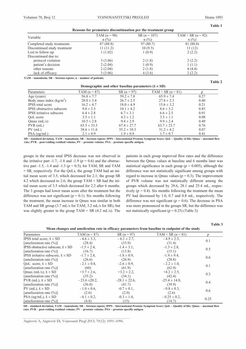
A total of 87, 97 and 81 patients were fully availableregarding the treatment regimen, according to TAM, SRand TAM + SR, respectively. The main reason for studydiscontinuation was voluntary withdrawal (1.3%), protocolviolation (2.4%), lack of efficacy (3.03%) and other rea-sons (2.7%). Four (1.3%) patients were lost to follow-up(Table 1).
The treatment groups had comparable distribution interms of age, body mass index, a total IPSS, irritative andobstructive subscores, QoLs, Qmax, PVR, PV and PSA (Ta-ble 2). The mean age was 64.9 ± 7.6 years.
After 6 months of the treatment, the mean decrease inIPSS was -4.6, -6.1 and -4.9 in the TAM, SR and TAM + SRgroups respectively. The difference between IPSS values atbaseline and 6 months later were significant in each group (p< 0.05). The patients in the group SR had a greater reductionin symptoms than the other group. However, statisticalanalysis did not reveal this expected difference between thetreatment regimens (p = 0.1). This difference between the
Volumen 70, Broj 12 VOJNOSANITETSKI PREGLED Strana 1093
Argirovi A, Argirovi Dj. Vojnosanit Pregl 2013; 70(12): 1091–1096.
groups in the mean total IPSS decrease was not observed inthe irritative part -1.7, -1.8 and -1.9 (p = 0.6) and the obstruc-tive part -1.5, -1.4 and -1.3 (p = 0.5), for TAM, SR and TAM+ SR, respectively. For the QoLs, the group TAM had an ini-tial mean score of 3.5, which decreased for 2.1; the group SR4.2 which decreased to 2.6; the group TAM + SR had the ini-tial mean score of 3.5 which decreased for 2.2 after 6 months.The 3 groups had lower mean score after the treatment but thedifference was not significant (p = 0.1). Six months followingthe treatment, the mean increase in Qmax was similar in bothTAM and SR group (3.7 mL/s for TAM, 3.2 mL/s for SR), butwas slightly greater in the group TAM + SR (4.2 mL/s). The
patients in each group improved flow rates and the differencebetween the Qmax values at baseline and 6 months later wasstatistical significance in each group (p < 0.005), although thedifference was not statistically significant among groups withregard to increase in Qmax values (p = 0.3). The improvementof PVR volume was not statistically different among thegroups which decreased by 29.6, 28.1 and 25.4 mL, respec-tively (p = 0.4). Six months following the treatment the meanPV had decreased by 1.0, 0.7 and 0.8 mL, respectively. Thedifference was not significant (p = 0.6). The decrease in PSAwas more pronounced in the groups SR, but the difference wasnot statistically significant (p = 0.25) (Table 3).
Table 3Mean changes and ameliration rate in efficacy parameters from baseline to endpoint of the study
Parameters TAM (n = 87) SR (n = 97) TAM + SR (n = 81) pIPSS total score, ± SD[amelioration rate (%)]
- 4.6 ± 3.3,(28.4)
- 6.1 ± 2.7,(33.9)
- 4.9 ± 2.3,(31.4) 0.1
IPSS obstructive subscore, ± SD[amelioration rate (%)]
-1.5 ± 2.4,(16.7)
-1.4 ± 3.1,(13.8)
-1.3 ± 2.8,(15.1) 0.5
IPSS irritative subscore, ± SD[amelioration rate (%)]
-1.7 ± 2.8,(26.6)
-1.8 ± 0.9,(26.9)
-1.9 ± 9.4,(28.8) 0.6
QoL score, ± SD[amelioration rate (%)]
- 2.1 ± 0.8,(60)
-2.6 ± 0.9,(61.9)
- 2.2 ± 1.0,(62.9) 0.1
Qmax (mL/s), ± SD[amelioration rate (%)]
+3.7 ± 2.6,(35.2)
+3.2 ± 2.2,(34.1)
+4.2 ± 2.5,(42.4) 0.3
PVR (mL), ± SD[amelioration rate (%)]
- 23.6 ±20.2,(36.0)
-28.1 ± 22.6,(41.7)
-25.4 ± 14.8,(39.9) 0.4
PV (mL), ± SD[amelioration rate (%)]
- 1.0 ± 0.6,(2.6)
-0.7 ± 0.1,(2.0)
- 0.8 ± 0.3,(2.6) 0.6
PSA (ng/mL), ± SD[amelioration rate (%)]
- 0.1 ± 0.2,(4.8)
-0.3 ± 1.4,(15)
- 0.25 ± 0.2,(14.7) 0.25
SD – standard deviation, TAM – tamsulosin; SR – Serenoa repens; IPPS – International Prostate Symptom Score; QoL – Quality of life; Qmax – maximal flowrate; PVR – post-voiding residual volume; PV – prostate volume; PSA – prostate specific antigen.
Table 1Reasons for premature discontinuation per the treatment group
Variable TAM (n = 98)n (%)
SR (n = 107)n (%)
TAM + SR (n = 92)n (%)
Completed study treatments 87 (88.8) 97 (80.7) 81 (88.0)Discontinued study treatment 11 (11.2) 10 (9.3) 11 (12)Lost to follow-up 1 (1.02) 1 (0.9) 2 (2.2)Discontinued due to:
protocol violation 3 (3.06) 2 (1.8) 2 (2.2)patient’s decision 2 (2.04) 1 (0.9) 1 (1.1)other reasons 2 (2.04) 2 (1.8) 4 (4.4)lack of efficacy 3 (3.06) 4 (3.6) 2 (2.2)
TAM – tamsulosin; SR – Serenoa repens; n – number of patients.Table 2
Demographic and other baseline parameters ( ± SD)Parameters TAM (n = 87) SR (n = 97) TAM + SR (n = 81) pAge (years) 56.8 ± 7.7 59.2 ± 7.8 65.9 ± 7.4 0.27Body mass index (kg/m2) 28.0 ± 3.4 26.7 ± 2.5 27.8 ± 2.3 0.40IPSS total score 16.2 ± 4.7 18.0 ± 4.9 15.6 ± 3.2 0.21IPSS obstructive subscore 9.0 ± 3.5 10.1 ± 4.2 8.6 ± 3.2 0.85IPSS irritative subscore 6.4 ± 2.8 6.7 ± 3.1 6.6 ± 2.5 0.91QoL score 3.5 ± 1.1 4.2 ± 1.2 3.5 ± 1.1 0.08Qmax (mL/s) 10.5 ± 2.8 9.4 ± 2.9 9.9 ± 2.4 0.49PVR (mL) 65.5 ± 33.3 67.4 ± 27.7 63.7 ± 23.7 0.76PV (mL) 38.6 ± 11.6 35.2 ± 10.3 31.2 ± 4.2 0.07PSA (ng/mL) 2.1 ± 0.9 1.9 ± 0.9 1.7 ± 0.7 0.41
SD – standard deviation, TAM – tamsulosin; SR – Serenoa repens; IPPS – International Prostate Symptom Score; QoL – Quality of life; Qmax – maximal flowrate; PVR – post-voiding residual volume; PV – prostate volume; PSA – prostate specific antigen.
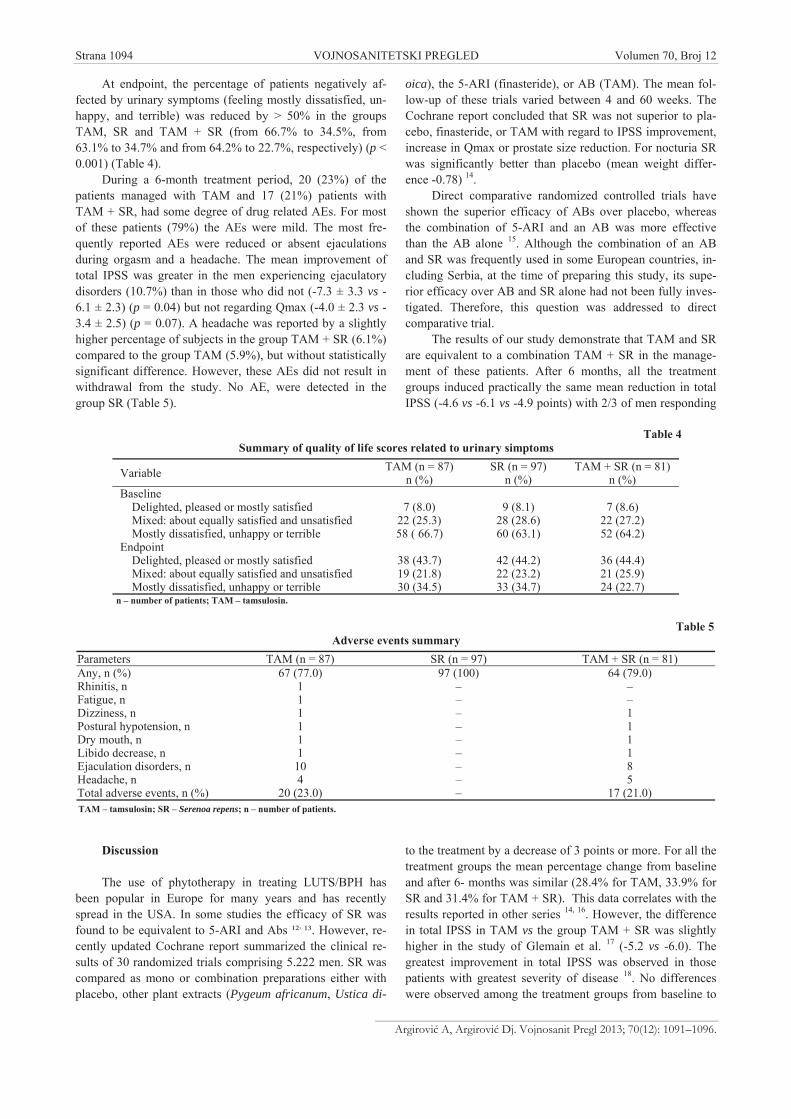
Strana 1094 VOJNOSANITETSKI PREGLED Volumen 70, Broj 12
Argirovi A, Argirovi Dj. Vojnosanit Pregl 2013; 70(12): 1091–1096.
At endpoint, the percentage of patients negatively af-fected by urinary symptoms (feeling mostly dissatisfied, un-happy, and terrible) was reduced by > 50% in the groupsTAM, SR and TAM + SR (from 66.7% to 34.5%, from63.1% to 34.7% and from 64.2% to 22.7%, respectively) (p <0.001) (Table 4).
During a 6-month treatment period, 20 (23%) of thepatients managed with TAM and 17 (21%) patients withTAM + SR, had some degree of drug related AEs. For mostof these patients (79%) the AEs were mild. The most fre-quently reported AEs were reduced or absent ejaculationsduring orgasm and a headache. The mean improvement oftotal IPSS was greater in the men experiencing ejaculatorydisorders (10.7%) than in those who did not (-7.3 ± 3.3 vs -6.1 ± 2.3) (p = 0.04) but not regarding Qmax (-4.0 ± 2.3 vs -3.4 ± 2.5) (p = 0.07). A headache was reported by a slightlyhigher percentage of subjects in the group TAM + SR (6.1%)compared to the group TAM (5.9%), but without statisticallysignificant difference. However, these AEs did not result inwithdrawal from the study. No AE, were detected in thegroup SR (Table 5).
Discussion
The use of phytotherapy in treating LUTS/BPH hasbeen popular in Europe for many years and has recentlyspread in the USA. In some studies the efficacy of SR wasfound to be equivalent to 5-ARI and Abs ¹², ¹³. However, re-cently updated Cochrane report summarized the clinical re-sults of 30 randomized trials comprising 5.222 men. SR wascompared as mono or combination preparations either withplacebo, other plant extracts (Pygeum africanum, Ustica di-
oica), the 5-ARI (finasteride), or AB (TAM). The mean fol-low-up of these trials varied between 4 and 60 weeks. TheCochrane report concluded that SR was not superior to pla-cebo, finasteride, or TAM with regard to IPSS improvement,increase in Qmax or prostate size reduction. For nocturia SRwas significantly better than placebo (mean weight differ-ence -0.78) 14.
Direct comparative randomized controlled trials haveshown the superior efficacy of ABs over placebo, whereasthe combination of 5-ARI and an AB was more effectivethan the AB alone 15. Although the combination of an ABand SR was frequently used in some European countries, in-cluding Serbia, at the time of preparing this study, its supe-rior efficacy over AB and SR alone had not been fully inves-tigated. Therefore, this question was addressed to directcomparative trial.
The results of our study demonstrate that TAM and SRare equivalent to a combination TAM + SR in the manage-ment of these patients. After 6 months, all the treatmentgroups induced practically the same mean reduction in totalIPSS (-4.6 vs -6.1 vs -4.9 points) with 2/3 of men responding
to the treatment by a decrease of 3 points or more. For all thetreatment groups the mean percentage change from baselineand after 6- months was similar (28.4% for TAM, 33.9% forSR and 31.4% for TAM + SR). This data correlates with theresults reported in other series 14, 16. However, the differencein total IPSS in TAM vs the group TAM + SR was slightlyhigher in the study of Glemain et al. 17 (-5.2 vs -6.0). Thegreatest improvement in total IPSS was observed in thosepatients with greatest severity of disease 18. No differenceswere observed among the treatment groups from baseline to
Table 4Summary of quality of life scores related to urinary simptoms
Variable TAM (n = 87)n (%)
SR (n = 97)n (%)
TAM + SR (n = 81)n (%)
BaselineDelighted, pleased or mostly satisfied 7 (8.0) 9 (8.1) 7 (8.6)Mixed: about equally satisfied and unsatisfied 22 (25.3) 28 (28.6) 22 (27.2)Mostly dissatisfied, unhappy or terrible 58 ( 66.7) 60 (63.1) 52 (64.2)
EndpointDelighted, pleased or mostly satisfied 38 (43.7) 42 (44.2) 36 (44.4)Mixed: about equally satisfied and unsatisfied 19 (21.8) 22 (23.2) 21 (25.9)Mostly dissatisfied, unhappy or terrible 30 (34.5) 33 (34.7) 24 (22.7)
n – number of patients; TAM – tamsulosin.
Table 5Adverse events summary
Parameters TAM (n = 87) SR (n = 97) TAM + SR (n = 81)Any, n (%) 67 (77.0) 97 (100) 64 (79.0)Rhinitis, n 1 – –Fatigue, n 1 – –Dizziness, n 1 – 1Postural hypotension, n 1 – 1Dry mouth, n 1 – 1Libido decrease, n 1 – 1Ejaculation disorders, n 10 – 8Headache, n 4 – 5Total adverse events, n (%) 20 (23.0) – 17 (21.0)TAM – tamsulosin; SR – Serenoa repens; n – number of patients.
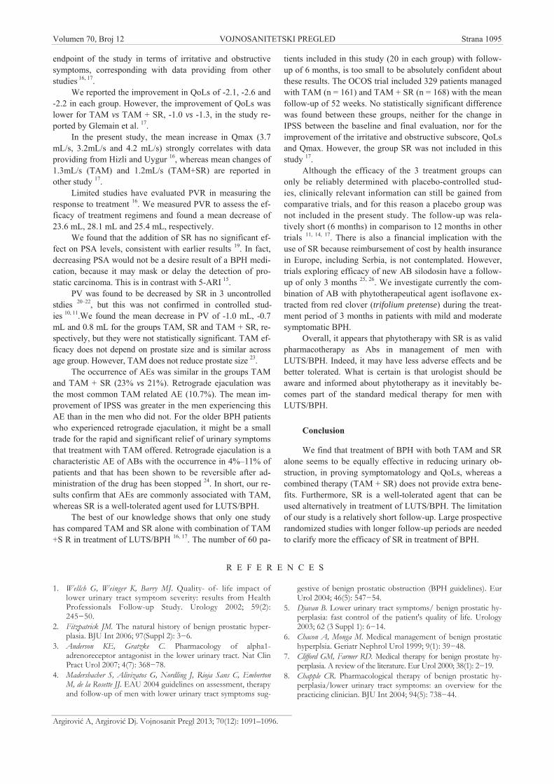
Volumen 70, Broj 12 VOJNOSANITETSKI PREGLED Strana 1095
Argirovi A, Argirovi Dj. Vojnosanit Pregl 2013; 70(12): 1091–1096.
endpoint of the study in terms of irritative and obstructivesymptoms, corresponding with data providing from otherstudies 16, 17.
We reported the improvement in QoLs of -2.1, -2.6 and-2.2 in each group. However, the improvement of QoLs waslower for TAM vs TAM + SR, -1.0 vs -1.3, in the study re-ported by Glemain et al. 17.
In the present study, the mean increase in Qmax (3.7mL/s, 3.2mL/s and 4.2 mL/s) strongly correlates with dataproviding from Hizli and Uygur 16, whereas mean changes of1.3mL/s (TAM) and 1.2mL/s (TAM+SR) are reported inother study 17.
Limited studies have evaluated PVR in measuring theresponse to treatment 16. We measured PVR to assess the ef-ficacy of treatment regimens and found a mean decrease of23.6 mL, 28.1 mL and 25.4 mL, respectively.
We found that the addition of SR has no significant ef-fect on PSA levels, consistent with earlier results 19. In fact,decreasing PSA would not be a desire result of a BPH medi-cation, because it may mask or delay the detection of pro-static carcinoma. This is in contrast with 5-ARI 15.
PV was found to be decreased by SR in 3 uncontrolledstdies 20–22, but this was not confirmed in controlled stud-ies 10, 11.We found the mean decrease in PV of -1.0 mL, -0.7mL and 0.8 mL for the groups TAM, SR and TAM + SR, re-spectively, but they were not statistically significant. TAM ef-ficacy does not depend on prostate size and is similar acrossage group. However, TAM does not reduce prostate size 23.
The occurrence of AEs was similar in the groups TAMand TAM + SR (23% vs 21%). Retrograde ejaculation wasthe most common TAM related AE (10.7%). The mean im-provement of IPSS was greater in the men experiencing thisAE than in the men who did not. For the older BPH patientswho experienced retrograde ejaculation, it might be a smalltrade for the rapid and significant relief of urinary symptomsthat treatment with TAM offered. Retrograde ejaculation is acharacteristic AE of ABs with the occurrence in 4%–11% ofpatients and that has been shown to be reversible after ad-ministration of the drug has been stopped 24. In short, our re-sults confirm that AEs are commonly associated with TAM,whereas SR is a well-tolerated agent used for LUTS/BPH.
The best of our knowledge shows that only one studyhas compared TAM and SR alone with combination of TAM+S R in treatment of LUTS/BPH 16, 17. The number of 60 pa-
tients included in this study (20 in each group) with follow-up of 6 months, is too small to be absolutely confident aboutthese results. The OCOS trial included 329 patients managedwith TAM (n = 161) and TAM + SR (n = 168) with the meanfollow-up of 52 weeks. No statistically significant differencewas found between these groups, neither for the change inIPSS between the baseline and final evaluation, nor for theimprovement of the irritative and obstructive subscore, QoLsand Qmax. However, the group SR was not included in thisstudy 17.
Although the efficacy of the 3 treatment groups canonly be reliably determined with placebo-controlled stud-ies, clinically relevant information can still be gained fromcomparative trials, and for this reason a placebo group wasnot included in the present study. The follow-up was rela-tively short (6 months) in comparison to 12 months in othertrials 11, 14, 17. There is also a financial implication with theuse of SR because reimbursement of cost by health insurancein Europe, including Serbia, is not contemplated. However,trials exploring efficacy of new AB silodosin have a follow-up of only 3 months 25, 26. We investigate currently the com-bination of AB with phytotherapeutical agent isoflavone ex-tracted from red clover (trifolium pretense) during the treat-ment period of 3 months in patients with mild and moderatesymptomatic BPH.
Overall, it appears that phytotherapy with SR is as validpharmacotherapy as Abs in management of men withLUTS/BPH. Indeed, it may have less adverse effects and bebetter tolerated. What is certain is that urologist should beaware and informed about phytotherapy as it inevitably be-comes part of the standard medical therapy for men withLUTS/BPH.
Conclusion
We find that treatment of BPH with both TAM and SRalone seems to be equally effective in reducing urinary ob-struction, in proving symptomatology and QoLs, whereas acombined therapy (TAM + SR) does not provide extra bene-fits. Furthermore, SR is a well-tolerated agent that can beused alternatively in treatment of LUTS/BPH. The limitationof our study is a relatively short follow-up. Large prospectiverandomized studies with longer follow-up periods are neededto clarify more the efficacy of SR in treatment of BPH.
R E F E R E N C E S
1. Wellch G, Weinger K, Barry MJ. Quality- of- life impact oflower urinary tract symptom severity: results from HealthProfessionals Follow-up Study. Urology 2002; 59(2):245 50.
2. Fitzpatrick JM. The natural history of benign prostatic hyper-plasia. BJU Int 2006; 97(Suppl 2): 3 6.
3. Anderson KE, Gratzke C. Pharmacology of alpha1-adrenoreceptor antagonist in the lower urinary tract. Nat ClinPract Urol 2007; 4(7): 368 78.
4. Madersbacher S, Alivizatos G, Nordling J, Rioja Sans C, EmbertonM, de la Rosette JJ. EAU 2004 guidelines on assessment, therapyand follow-up of men with lower urinary tract symptoms sug-
gestive of benign prostatic obstruction (BPH guidelines). EurUrol 2004; 46(5): 547 54.
5. Djavan B. Lower urinary tract symptoms/ benign prostatic hy-perplasia: fast control of the patient's quality of life. Urology2003; 62 (3 Suppl 1): 6 14.
6. Chacon A, Monga M. Medical management of benign prostatichyperplsia. Geriatr Nephrol Urol 1999; 9(1): 39 48.
7. Clifford GM, Farmer RD. Medical therapy for benign prostate hy-perplasia. A review of the literature. Eur Urol 2000; 38(1): 2 19.
8. Chapple CR. Pharmacological therapy of benign prostatic hy-perplasia/lower urinary tract symptoms: an overview for thepracticing clinician. BJU Int 2004; 94(5): 738 44.
Strana 1096 VOJNOSANITETSKI PREGLED Volumen 70, Broj 12
Argirovi A, Argirovi Dj. Vojnosanit Pregl 2013; 70(12): 1091–1096.
9. Roehborn CG. BPH progression: concept and key learning fromMTOPS, ALTESS, COMBAT and ALF-ONE. BJU Int 2008;101( Suppl 3): 17 21.
10. Boyle P, Robertson C, Lowe F, Roehborn C. Update meta-analysisof clinical trials of Serenoa repens extract in the treatment ofasymptomatic benign prostatic hyperplasia. BJU Int 2004;93(6): 751 6.
11. Willets KE, Clements MS, Champion S, Eschman S, Eden JA. Sere-noa repens extracts for benign prostate hyperplasia: a ran-domized controlled trial. BJU Int 2003; 92(3): 267 70.
12. Lowe FC. Phytotherapy in the management of benign prostatichyperplasia. Urology 2001; 58(6 Suppl 1): 71 6; discussion 76 7.
13. Debruyne F, Koch G, Boyle P, Da Silva FG, Gillenwater JG, HamdyFC, et al. Comparison of a phytotherapeutic agent (Permixon)with an alpha blocker (Tamsulosin) in the treatment of benignprostatic hyperplasia. A 1-year randomized international study.Eur Urol 2002; 41(5): 497 506; discussion 506 7.
14. Wilt T, Ishani A, Mac Donald R. Serenoa repens for benign pro-static hyperplasia. Cochrane Database Syst Rev 2002; (3):CD001423.
15. Roehrborn CG, Siami P, Barkin J, Damião R, Becher E, Miñana B,et al. The influence of baseline parameters on changes in in-ternational prostate symptom score with dutasteride, tamsu-losin, and combination therapy among men with symptomaticbenign prostatic hyperplasia and an enlarged prostate: 2-yeardata from the CombAT study. Eur Urol 2009; 55(2): 461 71.
16. Hizli F, Uygur CM. A prospective study of the efficacy of Sere-noa repens, tamsulosin and Serenoa repens plus tamsulosintreatment for patients with benign prostate hyperplasia. IntUrol Nephrol 2007; 39(3): 879 86.
17. Glemain P, Coulange C, Billebaud T, Gattegno B, Muczynski R, LoebG. Tamsulosin with or without Serenoa repens in benign pro-static hyperplasia: the OCOS trial. Prog Urol 2002; 12(3):395 403; discussion 404. (French)
18. Debruyne F, Boyle P, Calais Da Silva F, Gillenwater JG, Hamdy FC,Perrin P, et al. Evaluation of the clinical benefit of Permixon
and Tamsulosin in severe BPH patients-PERMAL study sub-set analysis. Eur Urol 2004; 14(3): 326 31. (French)
19. Gerber GS, Zgaja GP, Bales GT, Chodak GW, Contreras BA. Sawpalmetto (Serenoa repens) in men with lower urinary tractsymptoms: effects on urodynamic parameters and voidingsymptoms. Urology 1998; 51(6): 1003–7.
20. Braeckman J. The extract of serenoa repens in the treatment ofbenign prostatic hyperplasia: a multicenter study. Curr TherRes 1994; 55: 776 85.
21. Romicz I, Schmitz H, Frang D. Experience in treating benignprostatic hypertrophy with Sabal serrulata for one year. IntUrol Nephrol 1993; 25(6): 565 9.
22. Kondás J, Philipp V, Diószeghy G.. Sabal serulata (Strogen forte)in the treatment of symptomatic benign prostatic hyperplasia.Int Urol Nephrol 1996; 28(6): 767 72.
23. Wilt TJ, Mac Donald R, Rutks J. Tamsulosin for benign prostatichyperplasia. Cochrane Data Base Syst Rev 2003; (1)CD002081.
24. van Dijk MM, de la Rosette JJ, Michel MC. Effects of alpha1-adrenoeceptor antagonist in male sexual function. Drugs 2006;66(3): 287 301.
25. Chapple CR, Montorsi F, Tammella TL, Wirth M, Koldewiju E, Fer-nández Fernández E. Silodosin therapy for lower urinary tractsymptoms in men with suspected benign prostatic hyperplasia:Results of an international, randomized, double-blind, pla-cebo- and active-controlled clinical trial performed in Europe.Eur Urol 2011; 59(3): 342 52.
26. Marks LS, Gittelman MC, Hill LA, Volinn W, Hoel G. Rapid ef-ficacy of the highly selective alpha1A-adrenoceptor antagonistsilodosin in men with signs and symptoms of benign prostatichyperplasia: pooled results of 2 phase 3 studies. J Urol 2009;181(6): 2634 40.
Received on June 20, 2011.Revised on November 24, 2011.
Accepted on November 30, 2011.OnLine-First June, 2013.

Vojnosanit Pregl 2013; 70(12): 1097–1102. VOJNOSANITETSKI PREGLED Strana 1097
Correspondence to: Tatjana Pekmezovi , Institute of Epidemiology, Faculty of Medicine, University of Belgrade, Visegradska 26A, Bel-grade 11000, Serbia. Phone: +381 11 3607 062. E-mail: [email protected]
O R I G I N A L A R T I C L E UDC: 613.99::618.11-006DOI: 10.2298/VSP110629030G
Risk factors for epithelial ovarian cancer in the female population ofBelgrade, Serbia: A case-control study
Faktori rizika od epitelijalnog karcinoma ovarijuma u ženskoj populacijiBeograda
Tatjana Gazibara*, Aleksandra Filipovi †, Vesna Kesi ‡,Darija Kisi -Tepav evi *, Tatjana Pekmezovi *
*Institute of Epidemiology, Faculty of Medicine, University of Belgrade, Belgrade,Serbia; †Public Health Center “Dr Simo Miloševi ”, Belgrade, Serbia; ‡Department of
Gynecology and Obstretics, Clinical Center of Serbia, Belgrade, Serbia
Abstract
Background/Aim. Ovarian cancer (OC) comprises 3% ofall cancers, but it is the fifth most common cause of cancerdeath in women. The aim of this case-control study was todetermine the risk factors for OC in the female population ofBelgrade, Serbia. Methods. A total of 80 consecutive patientswere enrolled in the study between 2006 and 2008 in two na-tional referral centers for OC in Serbia. The control subjectswere recruited during the regular gynecological check-ups inthe Public Health Center of the corresponding municipalities.All the study participants were interviewed during their visitsto the above mentioned institutions by two physicians usingthe same questionnaire. In order to analyze the influence ofspecific exposure to the risk of the disease, we categorizedvariables according to the cut-off values. Odds ratios (OR)and 95% confidence intervals (95% CI) were calculated sepa-rately for each variable using univariate conditional logisticregression analysis. Results. There were no statistically sig-nificant differences in educational level, years of schooling,occupational and employment status between patients withOC and women in the control group. Oral contraceptives useand other contraceptive methods (condoms, mechanicalcontraceptive devices) were highly statistically significantlymore frequent among women in the control group (OR =0.2, 95% CI 0.1–0.7, p = 0.005; OR = 0.1, 95% CI 0.01–0.5, p= 0.001, respectively). The patients with OC practiced sportsfor 6.3 ± 2.1 years, and controls for 11.8 ± 9.9 years. Sportand recreation activities were statistically significantly protec-tive (OR = 0.2, p = 0.011; OR = 0.4, p = 0.019). Tea con-sumption on daily basis had a highly statistically significatprotective effect (OR = 0.3, p = 0.001). Conclusions. Oralcontraceptives use and physical activity were independentprotective factors for OC in this study.
Key words:ovarian neoplasms; risk factors; motor activity;contraceptive agents.
Apstrakt
Uvod/Cilj. Karcinom ovarijuma ini 3% od svih karicnoma, izauzima 5. mesto me u naj eš im uzrocima smrti od karcino-ma kod žena. Cilj ove studije bio je da se utvrde faktori rizikaod nastanka epitelijalnog karcinoma ovarijuma u ženskoj po-pulaciji Beograda. Metode. Studija je izvedena u periodu od2006. do 2008. godine. Ukupno 80 bolesnica iz dva tercijarnacentra u Beogradu sa dijagnozom epitelijalnog karcinoma ova-rijuma uklju eno je u studijsku grupu. Kontrolnu grupu inilesu žene koje su dolazile na redovne godišnje ginekološke pre-glede u domove zdravlja opština u kojima su živele. Podatke suprikupljala dva lekara putem upitnika. U cilju procene uticajaspecifi ne izloženosti riziku od pojave bolesti, varijable su kate-gorisane prema grani nim (cut-off) vrednostima. Unakrsni odnos(odds ratio – OR) i 95% interval poverenja (95% IP) izra unava-ni su za svaku varijablu pojedina no koriš enjem univarijantnelogisti ke regresije. Rezultati. Izme u grupe bolesnica i kon-trole grupe nije postojala statisti ki zna ajna razlika u nivou ob-razovanja, dužini školovanja, zanimanju i zaposlenosti. Oralnikontraceptivi i druge metode kontracepcije (prezervativi, me-hani ka kontraceptivna sredstva) bili su statisti ki visokozna-ajno eš e koriš eni me u ženama u kontrolnoj grupi nego
kod bolesnica sa ovarijalnim karcinomom (OR = 0,2; 95% IP0,1–0,7; p = 0,005 za oralne kontraceptive; OR = 0,1, 95% IP0,01–0,5 p = 0,001 za druge metode kontracepcije). Ispitanicesa ovarijalnim karcinomom bavile su se sportom u proseku 6,3± 2,1 godine, dok je za one iz kontrolne grupe taj period u pro-seku iznosio 11,8 ± 9,9 godina. Sport i rekreacija imali su statis-ti ki zna ajan zaštitni efekat na nastanak ovog tumora (OR =0,2, p = 0.011; OR = 0,4, p = 0,019, redom). Statisti ki visoko-zna ajan zaštitni efekat imala je dnevna konzumacija aja (OR= 0,3, p = 0,001). Zaklju ak. Upotreba oralnih kontraceptiva ifizi ka aktivnost bili su nezavisni zaštitni faktori od nastankaepitelijalnog karcinoma ovarijuma u našoj studiji.
Klju ne re i:jajnik, neoplazme; faktori rizika; motorna aktivnost;kontrola ra anja, sredstva.
Strana 1098 VOJNOSANITETSKI PREGLED Volumen 70, Broj 12
Gazibara T, et al. Vojnosanit Pregl 2013; 70(12): 1097–1102.
Introduction
Ovarian cancer (OC) comprises 3% of all cancers, but itis the fifth most common cause of cancer death in women 1.The 5-year relative survival rate ranges from 30% to 45%,without significant improvement in the past years eventhough the new methods in therapy have been used 2–4. Thehighest incidence rates of OC have been registered in Scan-dinavia, Eastern Europe and Canada, where it varies between10 and 15 per 100,000 women 5. The lowest ones were foundin Asia (excluding Japan) and Africa, with less than 5 per100,000 women 6. In Serbia, the standardized incidence ratein 1999 was 9.7 per 100.000, while in 2005 it rose up to 11.5per 100.000 7.
Risk factors for OC are still not well established. Age,family history of OC, infertility treatment and assisted fer-tilization, hormonal substitution in menopause, and obesityare potential factors in favor of developing the OC 8, 9. Also,it has been noted that nulliparous women have an increasedrisk for OC 8, 10. On the other hand, it has been well definedand quantified that the use of oral contraceptives decreasesthe risk 6 as well as multiple pregnancies. In addition, tuballigation, hysterectomy and lactation are found to be protec-tive factors, too 11.
Genetic factors also play an important role in the etiol-ogy of this tumor. The mutation of genes BRCA-1 andBRCA-2 in 17q are identified in two separate types of he-reditary carcinomas. Another hereditary type of OC is foundin cancer family syndrome Lynch type 2 12. As for environ-mental factors, it has been suspected that talc and asbestosmay influence the onset of the disease, due to the fact thatthe highest incidence rates occur in highly industrializedcountries. Some migrant studies have shown that whenwomen from an undeveloped or developing country move toindustrialized country develop OC 10, while certain case-control studies pointed out that high intake of animal fat, al-cohol and smoking may increase the risk 13.
Regarding burden of OC in our country, official datarevealed it as the 7th most frequent cause of cancer-relateddeath as well as the 2nd most common cause in gynecologi-cal cancer deaths in the Serbian female population 7. Fur-thermore, recent investigation showed statistically significantincrease in OC mortality trend during the period 1976–2007 14. Thus, the aim of this study was to determine the riskfactors for OC in the population of Belgrade (Serbia).
Methods
This case-control study included 80 consecutive pa-tients treated and followed in the Department of Gynecologyand Obstetrics of the Clinical Center of Serbia and the Clinicof Gynecology and Obstetrics "Narodni Front" in Belgrade,between 2006 and 2008. Both of these hospitals are the na-tional referral centers for OC in Serbia. All cases resided onthe territory of Belgrade and had histologically verified diag-nose of epithelial OC. The control group consisted of 160women, double matched according to age (± 2 years) andmunicipality of residence. For the study period, the control
subjects were recruited during regular gynecological check-ups in the Public Health Center in corresponding municipali-ties. These women had no malignant tumors and/or hor-mone-dependent problems. All women, in both groups,signed the informed consent for the participation in thestudy. The research was approved by the Institutional Re-view Committee. Written informed consents were obtainedfrom the study participants.
All the study participants were interviewed during theirvisits to the above mentioned institutions by two physiciansusing the same questionnaire to collect demographic infor-mation, as well as information regarding personal and familyhistory, lifetime residence, particular lifestyle (smoking, al-cohol and coffee intake), occupational exposures to radiationand chemicals, as well as reproductive history. Smokers weredefined as persons who reported everyday smoking during a60-day period prior to completing the questionnaire. To as-sess sport and recreation activities participants were asked ifthey do moderate activities for at least 10 minutes at a time,such as brisk walking, cycling, swimming, or any other ac-tivity that causes some increase in breathing or heart rate. Inorder to analyze the influence of specific exposure to the riskof the disease, we categorized variables according to the cut-off values. These values were determined based on the mean(± SD) level of variables investigated in the control group.All questions were referring to the 5-year period prior to thediagnosis or the corresponding period for the controls. Thiswas supplemented and validated by an examination of themedical records.
Odds ratios (OR) and 95% confidence intervals (95%CI) were calculated separately for each variable using uni-variate conditional logistic regression analysis. Variables thatwere related to OC at a significant level of p < 0.05, enteredthe final model of multivariate conditional logistic regressionanalysis to evaluate their independent contribution to theoverall risk of OC.
Results
The average age of women in the study and the controlgroups was 56.1 ± 10.8 years and 56.7 ± 10.6 years, respec-tively.
Average income in patients was 245.52 ± 10.2 euros asopposed to the control group, where it was around 534 ±146.5 euros. Lower family income ( 250 euros a month)was statistically significant risk factor for OC, with OR = 2.5(95% CI 1.4–4.3), p = 0.001. Also, the living space in thestudy group it was in average 56.5 ± 23.0 m² while in thecontrol group was 64.3 ± 67.3 m², thus the calculated ORwas 2.0 (95% CI 1.1–3.6), p = 0.018, which was statisticallysignificant.
The characteristics of OC cases and controls regardingeducation level and occupational history are presented in Ta-ble 1. There were no statistically significant differences ineducational level, years of schooling, occupational and em-ployment status between patients and the controls.
Considering the existing major chronic diseases in bothstudy groups, occurrence of cardiovascular (OR = 2.0, 95%
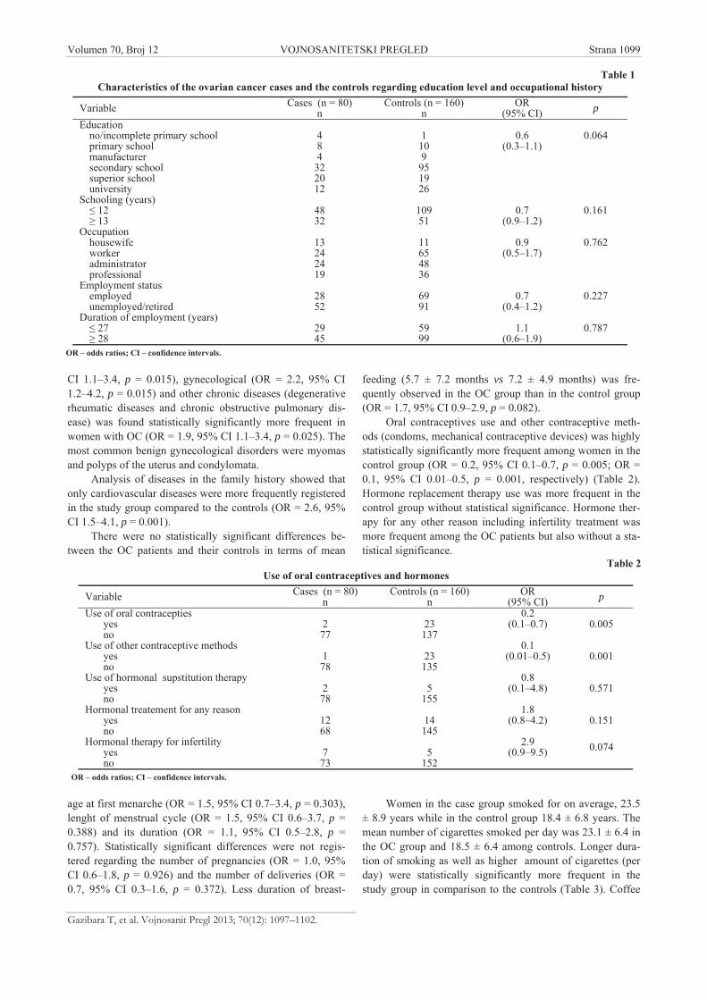
Volumen 70, Broj 12 VOJNOSANITETSKI PREGLED Strana 1099
Gazibara T, et al. Vojnosanit Pregl 2013; 70(12): 1097–1102.
CI 1.1–3.4, p = 0.015), gynecological (OR = 2.2, 95% CI1.2–4.2, p = 0.015) and other chronic diseases (degenerativerheumatic diseases and chronic obstructive pulmonary dis-ease) was found statistically significantly more frequent inwomen with OC (OR = 1.9, 95% CI 1.1–3.4, p = 0.025). Themost common benign gynecological disorders were myomasand polyps of the uterus and condylomata.
Analysis of diseases in the family history showed thatonly cardiovascular diseases were more frequently registeredin the study group compared to the controls (OR = 2.6, 95%CI 1.5–4.1, p = 0.001).
There were no statistically significant differences be-tween the OC patients and their controls in terms of mean
age at first menarche (OR = 1.5, 95% CI 0.7–3.4, p = 0.303),lenght of menstrual cycle (OR = 1.5, 95% CI 0.6–3.7, p =0.388) and its duration (OR = 1.1, 95% CI 0.5–2.8, p =0.757). Statistically significant differences were not regis-tered regarding the number of pregnancies (OR = 1.0, 95%CI 0.6–1.8, p = 0.926) and the number of deliveries (OR =0.7, 95% CI 0.3–1.6, p = 0.372). Less duration of breast-
feeding (5.7 ± 7.2 months vs 7.2 ± 4.9 months) was fre-quently observed in the OC group than in the control group(OR = 1.7, 95% CI 0.9–2.9, p = 0.082).
Oral contraceptives use and other contraceptive meth-ods (condoms, mechanical contraceptive devices) was highlystatistically significantly more frequent among women in thecontrol group (OR = 0.2, 95% CI 0.1–0.7, p = 0.005; OR =0.1, 95% CI 0.01–0.5, p = 0.001, respectively) (Table 2).Hormone replacement therapy use was more frequent in thecontrol group without statistical significance. Hormone ther-apy for any other reason including infertility treatment wasmore frequent among the OC patients but also without a sta-tistical significance.
Women in the case group smoked for on average, 23.5± 8.9 years while in the control group 18.4 ± 6.8 years. Themean number of cigarettes smoked per day was 23.1 ± 6.4 inthe OC group and 18.5 ± 6.4 among controls. Longer dura-tion of smoking as well as higher amount of cigarettes (perday) were statistically significantly more frequent in thestudy group in comparison to the controls (Table 3). Coffee
Table 1Characteristics of the ovarian cancer cases and the controls regarding education level and occupational history
Variable Cases (n = 80)n
Controls (n = 160)n
OR(95% CI) p
Educationno/incomplete primary schoolprimary schoolmanufacturersecondary schoolsuperior schooluniversity
484322012
1109
951926
0.6(0.3–1.1)
0.064
Schooling (years) 12 13
4832
10951
0.7(0.9–1.2)
0.161
Occupationhousewifeworkeradministratorprofessional
13242419
11654836
0.9(0.5–1.7)
0.762
Employment statusemployedunemployed/retired
2852
6991
0.7(0.4–1.2)
0.227
Duration of employment (years) 27 28
2945
5999
1.1(0.6–1.9)
0.787
OR – odds ratios; CI – confidence intervals.
Table 2Use of oral contraceptives and hormones
Variable Cases (n = 80)n
Controls (n = 160)n
OR(95% CI) p
Use of oral contraceptiesyesno
277
23137
0.2(0.1–0.7) 0.005
Use of other contraceptive methodsyesno
178
23135
0.1(0.01–0.5) 0.001
Use of hormonal supstitution therapyyesno
278
5155
0.8(0.1–4.8) 0.571
Hormonal treatement for any reasonyesno
1268
14145
1.8(0.8–4.2) 0.151
Hormonal therapy for infertilityyesno
773
5152
2.9(0.9–9.5) 0.074
OR – odds ratios; CI – confidence intervals.
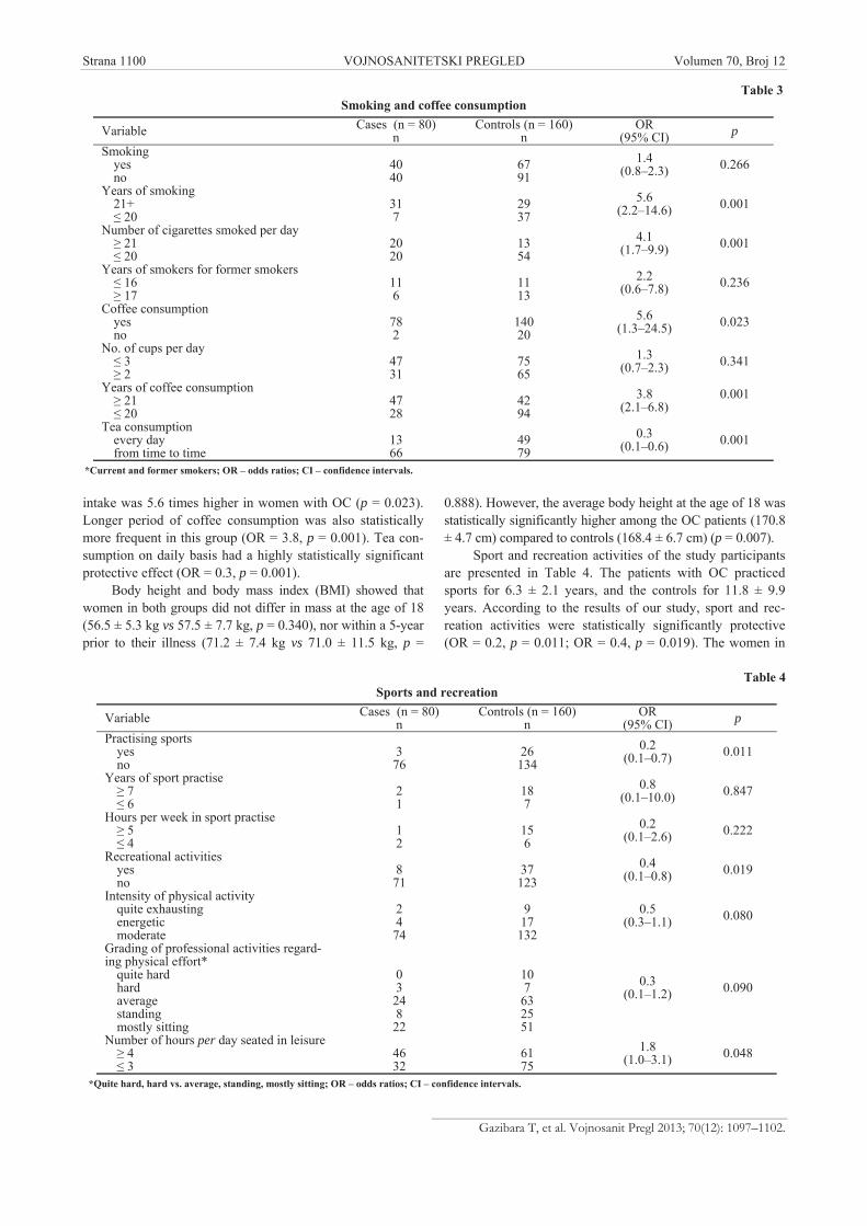
Strana 1100 VOJNOSANITETSKI PREGLED Volumen 70, Broj 12
Gazibara T, et al. Vojnosanit Pregl 2013; 70(12): 1097–1102.
intake was 5.6 times higher in women with OC (p = 0.023).Longer period of coffee consumption was also statisticallymore frequent in this group (OR = 3.8, p = 0.001). Tea con-sumption on daily basis had a highly statistically significantprotective effect (OR = 0.3, p = 0.001).
Body height and body mass index (BMI) showed thatwomen in both groups did not differ in mass at the age of 18(56.5 ± 5.3 kg vs 57.5 ± 7.7 kg, p = 0.340), nor within a 5-yearprior to their illness (71.2 ± 7.4 kg vs 71.0 ± 11.5 kg, p =
0.888). However, the average body height at the age of 18 wasstatistically significantly higher among the OC patients (170.8± 4.7 cm) compared to controls (168.4 ± 6.7 cm) (p = 0.007).
Sport and recreation activities of the study participantsare presented in Table 4. The patients with OC practicedsports for 6.3 ± 2.1 years, and the controls for 11.8 ± 9.9years. According to the results of our study, sport and rec-reation activities were statistically significantly protective(OR = 0.2, p = 0.011; OR = 0.4, p = 0.019). The women in
Table 3Smoking and coffee consumption
Variable Cases (n = 80)n
Controls (n = 160)n
OR(95% CI) p
Smokingyesno
4040
6791
1.4(0.8–2.3) 0.266
Years of smoking21+
20317
2937
5.6(2.2–14.6) 0.001
Number of cigarettes smoked per day 21 20
2020
1354
4.1(1.7–9.9) 0.001
Years of smokers for former smokers 16 17
116
1113
2.2(0.6–7.8) 0.236
Coffee consumptionyesno
782
14020
5.6(1.3–24.5) 0.023
No. of cups per day 3 2
4731
7565
1.3(0.7–2.3) 0.341
Years of coffee consumption 21 20
4728
4294
3.8(2.1–6.8)
0.001
Tea consumptionevery dayfrom time to time
1366
4979
0.3(0.1–0.6) 0.001
*Current and former smokers; OR – odds ratios; CI – confidence intervals.
Table 4Sports and recreation
Variable Cases (n = 80)n
Controls (n = 160)n
OR(95% CI) p
Practising sportsyesno
376
26134
0.2(0.1–0.7) 0.011
Years of sport practise 7 6
21
187
0.8(0.1–10.0) 0.847
Hours per week in sport practise 5 4
12
156
0.2(0.1–2.6) 0.222
Recreational activitiesyesno
871
37123
0.4(0.1–0.8) 0.019
Intensity of physical activityquite exhaustingenergeticmoderate
2474
917132
0.5(0.3–1.1) 0.080
Grading of professional activities regard-ing physical effort*
quite hardhardaveragestandingmostly sitting
0324822
107
632551
0.3(0.1–1.2) 0.090
Number of hours per day seated in leisure 4 3
4632
6175
1.8(1.0–3.1) 0.048
*Quite hard, hard vs. average, standing, mostly sitting; OR – odds ratios; CI – confidence intervals.

Volumen 70, Broj 12 VOJNOSANITETSKI PREGLED Strana 1101
Gazibara T, et al. Vojnosanit Pregl 2013; 70(12): 1097–1102.
the control group were practicing sports for more years andfor more hours weekly than the cases, without a statisticalsignificance.
Additionally, women in the study group were seated for4.1 ± 3.1 h per day during their working time while for thewomen in the control group it was 4.6 ± 2.6 h (p = 0.366).The average time in sitting position in their leisure was 3.4 ±1.4 h in the study group as opposed to the control group,where it was 4.0 ± 2.3 h (p = 0.033). A greater number ofhours in sedentary position was statistically significantlymore frequent in the study group than in the control one (OR= 1.8, 95% CI 1.0–3.1, p = 0.048).
All variables related to OC at a significant level of p <0.05 using univariate logistic regression analysis were in-cluded in the model of multivariate logistic regression analy-sis. According to multivariate analysis the following factorswere significantly negatively related to OC: oral contracep-tive use (OR = 0.1, 95% CI 0.1–0.5, p = 0.009) and recrea-tion activities (OR = 0.3, 95% CI 0.1–0.7, p = 0.007).
Discussion
In our case-control study conducted between 2006 and2008 in Belgrade, Serbia, we included 80 OC patients and 160controls. The results we found in this study indicate that theuse of oral contraceptives, as well as sports and recreation ac-tivities statistically significantly decrease the risk of OC.
A strong evidence of negative association between oralcontraceptives use and the occurrence of OC has been re-corded in a number of studies 15–19. Oral contraceptives havea long-term favorable effect on the OC risk 20. Specifically,this phenomenon occurs due to the reduction in estrogen lev-els in the ovaries and prevention of the ovulation 15, 21. Evena short-term oral contraceptive use has been reported to re-duce the risk 22. Our results confirm this well-documentedand defined association.
Even though the relationship between physical activityand the risk of hormone-dependent tumors (such as breastand endometrium cancer) have been known 23–26, there isonly a small number of studies regarding the effect of exer-cising on OC. The two studies in the US and China reporteda decreasing risk of OC among women who exercise somekind of physical activity 27, 28. However, a Swedish prospec-tive cohort study found no such evidence 29. Being a hor-mone-dependent tumor, OC largely depends on oscilations ofestrogen. Physical activity affects adipose tissue by reducingit, thus mobilizing hormone depots. Also, certain theoriessuggest that very strenuous physical activity induces latemenarche, ammenorrhea and anovulatory cycles 30–32. In ad-dition, some authors 19, 33 registered that older age at men-arche has a protective effect upon OC, but our study did notshow any link between these two variables.
In a pooled analysis of case-control studies Ness et al. 34
found no association between the use of fertility drugs andoverall risk of OC which is in accordance with the results
obtained in our study. On the other hand, systematic reviewand meta-analysis performed by Greise et al. 35 showed thatboth menopausal estrogene and progestin therapies are therisk factors for OC.
Many authors 15–19 have reported nulliparity to bestrongly associated with the ccurrence of OC, but in oursample of patients no significant difference in parity betweenthe cases and the controls was found. Our patients also brest-fed for shorter period. Moorman et al. 15 reported breast-feeding to be protective factor for the occurence of OC. Theyhad lower family income and were of less educational levelwhich is in concordance with the findings of El-Khwsky etal. 16 in Egypt and Song et al. 18 in China, whereas Zhang etal. 36 from the USA reported opposite results.
The women with OC were taller at the age of 18, but asfor BMI no relationship was established. Moorman et al. 15
registered body height and BMI of 35 and over to be the riskfactors for this tumor. Greer et al. 22 reported that womenwho had greater both recent weight and weight at the age of18 were at higher risk of OC. Beehler et al. 37 showed thatobese, premenopausal women have 2 times more chance ofdeveloping OC. However, they have not recorded any asso-ciation between BMI in postmenopausal women and OC,which corresponds to our findings, that BMI does not carryany risk of this disease.
Smoking is often associated with many types of can-cers 33 and our research confirmed that the cases smoke morethan their controls. However, controls drank more tea andcoffee on daily basis. Data on this issue in literature is con-troversial. Some studies 18, 36 have shown protective effect ofthe green tea, as opposed to black tea. Our study did not di-vide teas into categories. And that neither caffeinated or de-caiffenated coffees were associated with the risk of OC,which opposes to our findings 18.
One can criticize that the recall bias may have influ-enced the results in this case-control study and the size of ourstudy groups. In addition, population controls may have beenmore appropriate and therefore our study is certainly subjectto selection bias. In the present research we did not look forspecific dietary patterns of our patients, even though certainarticles suggest micronutrients such as calcium, vitamin Eand beta-carotene have protective effect upon occurrence ofOC 38, 39 and that meat and fat are associated with an in-creased risk of OC 40.
Conclusion
Based on data obtained in our study, oral contraceptivesuse and physical activity are independent protective factorsfor OC. Overall, this is the first epidemiological study on riskfactors for this cancer in our country. In future, attentionshould be paid on finding a larger sample, which would se-lect participants throughout the country, not only from onemajor center (Belgrade) and broaden the scope of the spe-cific questionnaire.
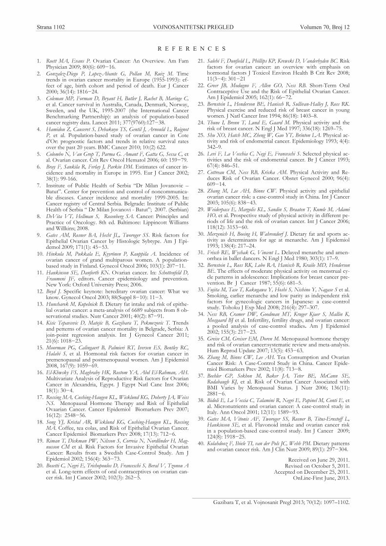
Strana 1102 VOJNOSANITETSKI PREGLED Volumen 70, Broj 12
Gazibara T, et al. Vojnosanit Pregl 2013; 70(12): 1097–1102.
R E F E R E N C E S
1. Roett MA, Evans P. Ovarian Cancer: An Overview. Am FamPhysician 2009; 80(6): 609 16.
2. Gonzalez-Diego P, Lopez-Abante G, Pollan M, Ruiz M. Timetrends in ovarian cancer mortality in Europe (1955-1993): ef-fect of age, birth cohort and period of death. Eur J Cancer2000; 36(14): 1816 24.
3. Coleman MP, Forman D, Bryant H, Butler J, Rachet B, Maringe C,et al. Cancer survival in Australia, Canada, Denmark, Norway,Sweden, and the UK, 1995-2007 (the International CancerBenchmarking Partnership): an analysis of population-basedcancer registry data. Lancet 2011; 377(9760):127 38.
4. Hamidou Z, Causeret S, Debakuyo TS, Gentil J, Arnould L, RoignotP, et al. Population-based study of ovarian cancer in Coted’Or: prognostic factors and trends in relative survival ratesover the past 20 years. BMC Cancer 2010; 10:(2) 622.
5. Colombo N, Van Grop T, Parma G. Amant F, Gatta G, Sessa C, etal. Ovarian cancer. Crit Rev Oncol Hematol 2006; 60: 159 79.
6. Bray F, Sankila R, Ferlay J, Parkin DM. Estimates of cancer in-cidence and mortality in Europe in 1995. Eur J Cancer 2002;38(1): 99-166.
7. Institute of Public Health of Serbia “Dr Milan Jovanovic –Batut”. Center for prevention and control of noncommunica-ble diseases. Cancer incidence and mortality 1999-2005. In:Cancer registry of Central Serbia. Belgrade: Institute of PublicHealth of Serbia “ Dr Milan Jovanovi - Batut”; 2007. (Serbian)
8. DeVita VT, Hellman S, Rosenberg SA. Cancer: Principles andPractice of Oncology. 8th ed. Baltimore: Lippincott Williamsand Willkins; 2008.
9. Gates AM, Rosner BA, Hecht JL, Tworoger SS. Risk factors forEpithelial Ovarian Cancer by Histologic Sybtype. Am J Epi-demol 2009; 171(1): 45 53.
10. Hinkula M, Pukkala E, Kyyrönen P, Kauppila A. Incidence ofovarian cancer of grand multiparous women. A population-based study in Finland. Gynecol Oncol 2006; 103(1): 207 11.
11. Hankinson SE, Danforth KN. Ovarian cancer. In: Schottenfeld D,Fraumeni JF, editors. Cancer epidemiology and prevention.New York: Oxford University Press; 2006.
12. Boyd J. Specific keynote: hereditary ovarian cancer: What weknow. Gynecol Oncol 2003; 88(Suppl 8 10): 11 3.
13. Huncharek M, Kupelnick B. Dietary fat intake and risk of epithe-lial ovarian cancer: a meta-analysis of 6689 subjects from 8 ob-servational studies. Nutr Cancer 2001; 40(2): 87 91.
14. Kisic Tepavcevic D, Matejic B, Gazibara T, Pekmezovic T. Trendsand petterns of ovarian cancer mortality in Belgrade, Serbia: Ajoin-point regression analysis. Int J Gynecol Cancer 2011;21(6): 1018 23.
15. Moorman PG, Calingaert B, Palmieri RT, Iversen ES, Bentley RC,Halabi S, et al. Hormonal risk factors for ovarian cancer inpremenopausal and postmenopausal women. Am J Epidemiol2008, 167(9): 1059 69.
16. El-Khwsky FS, Maghraby HK, Rostom YA, Abd El-Rahman, AH.Multivariate Analysis of Reproductive Risk factors for OvarianCancer in Alexandria, Egypt. J Egypt Natl Canc Inst 2006;18(1): 30 4.
17. Rossing MA, Cushing-Haugen KL, Wicklund KG, Doherty JA, WeissNS. Menopausal Hormone Therapy and Risk of EpithelialOvaarian Cancer. Cancer Epidemiol Biomarkers Prev 2007;16(12): 2548 56.
18. Song YJ, Kristal AR, Wicklund KG, Cushing-Haugen KL, RossingMA. Coffee, tea colas, and Risk of Epithelial Ovarian Cancer.Cancer Epidemiol Biomarkers Prev 2008; 17(13): 712 6.
19. Riman T, Dickman PW, Nilsson S, Correia N, Nordlinder H, Mag-nusson CM et al. Risk Factors for Invasive Epithelial OvarianCancer: Results from a Swedish Case-Control Study. Am JEpidemiol 2002; 156(4): 363 73.
20. Bosetti C, Negri E, Trichopoulos D, Franceschi S, Beral V, Tzonou Aet al. Long-term effects of oral contraceptives on ovarian can-cer risk. Int J Cancer 2002; 102(3): 262 5.
21. Salehi F, Dunfield L, Phillips KP, Krewski D, Vanderhyden BC. Riskfactors for ovarian cancer: an overview with emphasis onhormonal factors J Toxicol Environ Health B Crit Rev 2008;11(3 4): 301 21
22. Greer JB, Modugno F, Allen GO, Ness RB. Short-Term OralContraceptive Use and the Risk of Epithelial Ovarian Cancer.Am J Epidemiol 2005; 162(1): 66 72.
23. Bernstein L, Henderson BE, Hanisch R, Sullivan-Halley J, Ross RK.Physical exercise and reduced risk of breast cancer in youngwomen. J Natl Cancer Inst 1994; 86(18): 1403–8.
24. Thune I, Brenn T, Lund E, Gaard M. Physical activity and therisk of breast cancer. N Engl J Med 1997; 336(18): 1269–75.
25. Shu XO, Hatch MC, Zheng W, Gao YT, Brinton LA. Physical ac-tivity and risk of endometrial cancer. Epidemiology 1993; 4(4):342–9.
26. Levi F, La Vecchia C, Negi E, Franceschi S. Selected physical ac-tivities and the risk of endometrial cancer. Br J Cancer 1993;67(4): 846–51.
27. Cottreau CM, Ness RB, Kriska AM. Physical Activiry and Re-duces Risk of Ovarian Cancer. Obstet Gynecol 2000; 96(4):609 14.
28. Zhang M, Lee AH, Binns CW. Physical activity and epithelialovarian cancer risk: a case-control study in China. Int J Cancer2003; 105(6): 838 43.
29. Weiderpass E, Margolis KL, Sandin S, Braaten T, Kumle M, AdamiHO, et al. Prospective study of physical activity in different pe-riods of life and the risk of ovarian cancer. Int J Cancer 2006;118(12): 3153 60.
30. Merzenich H, Boeing H, Wahrendorf J. Dietary fat and sports ac-tivity as determinants for age at menarche. Am J Epidemiol1993; 138(4): 217–24.
31. Frisch RE, Wyshak G, Vincent L. Delayed menarche and amen-orrhea in ballet dancers. N Engl J Med 1980; 303(1): 17–9.
32. Bernstein L, Ross RK, Lobo RA, Hanisch R, Krailo MD, HendersonBE. The effects of moderate physical activity on menstrual cy-cle patterns in adolescence: Implications for breast cancer pre-vention. Br J Cancer 1987; 55(6): 681–5.
33. Fujita M, Tase T, Kakugawa Y, Hoshi S, Nishino Y, Nagase S et al.Smoking, earlier menarche and low parity as independent riskfactors for gynecologic cancers in Japanese: a case-controlstudy. Tohoku J Exp Med 2008; 216(4): 297–307.
34. Ness RB, Cramer DW, Goodman MT, Kruger Kjaer S, Mallin K,Mosgaard BJ et al. Infertility, fertility drugs, and ovarian cancer:a pooled analysis of case-control studies. Am J Epidemiol2002; 155(3): 217 23.
35. Greise CM, Greiser EM, Doren M. Menopausal hormone therapyand risk of ovarian cancer:systematic review and meta-analysis.Hum Reprod Update 2007; 13(5): 453 63.
36. Zhang M, Binns CW, Lee AH. Tea Consumption and OvarianCancer Risk: A Case-Control Study in China. Cancer Epide-miol Biomarkers Prev 2002; 11(8): 713 8.
37. Beehler GP, Sekhon M, Baker JA, Teter BE, McCann SE,Rodabaugh KJ, et al. Risk of Ovarian Cancer Associated withBMI Varies by Menopausal Status. J Nutr 2006; 136(11):2881 6.
38. Bidoli E, La Veccia C, Talamini R, Negri E, Papinel M, Conti E, etal. Micronutrients and ovarian cancer: A case-control study inItaly. Ann Oncol 2001; 12(11): 1589 93.
39. Gates MA, Vitonis AF, Tworoger SS, Rosner B, Titus-Ernstoff L,Hankinson SE, et al. Flavonoid intake and ovarian cancer riskin a population-based case-control study. Int J Cancer 2009;124(8): 1918 25.
40. Kolahdooz F, Ibiele TI, van der Pols JC, Webb PM. Dietary patternsand ovarian cancer risk. Am J Clin Nutr 2009; 89(1): 297 304.
Received on June 29, 2011.Revised on October 5, 2011.
Accepted on December 25, 2011.OnLine-First June, 2013.
Vojnosanit Pregl 2013; 70(12): 1103–1108. VOJNOSANITETSKI PREGLED Strana 1103
Correspondence to: Marko Banovi , Department of Cardiology, University Clinical Centre of Serbia, 8 Koste Todorovi a,11000 Belgrade, Serbia. Phone: +381 63 80 50 258; E-mail: [email protected], [email protected]
O R I G I N A L A R T I C L E UDC: 616.126.32-036-08:616.12-07DOI: 10.2298/VSP110630031B
Association between aortic stenosis severity and contractile reservemeasured by two-dimensional strain under low-dose dobutaminetestingUticaj težine aortne stenoze na procenu kontraktilne rezerve procenjene pomo u
dvodimenzionalnog naprezanja tokom niskodoznog dobutaminskog testa
Marko Banovi *, Bosiljka Vujisi -Teši *, Vuk Kuja i †, Slobodan Obradovi ‡§,Zdenko Crkvenac†, Miodrag Ostoji *
*Department of Cardiology, University Clinical Centre of Serbia, Belgrade,Serbia;†Sahlgrenska University Hospital/Östra, Gothenburg, Sweden; ‡Clinic of UrgentInternal Medicine, Military Medical Academy, Belgrade, Serbia; §Faculty of Medicine
of the Military Medical Academy, University of Defence, Belgrade, Serbia
Abstract
Background/Aim. Early detection of left ventricle (LV)systolic dysfunction could be a clue for surgical treatmentin patients with significant aortic stenosis (AS). Therefore,we evaluated LV peak of global longitudinal strain (PGLS)using speckle tracking imaging at rest and during low-dosedobutamine infusion in asymptomatic patients with mod-erate and severe AS and preserved LV ejection fraction(EF). Methods. All the patients underwent coronary angi-ography and had no obstructive coronary disease (definedas having no stenosis greater than 50% in diameter). Thepatients were divided into two groups: above and belowmedian of 0.785 cm2 aortic valve area (AVA). PGLS wasmeasured from acquired apical 4-chamber and 2-chambercine loops using a EchoPac PC-workstation at rest andduring 5 μg/kg/min, 10 μg/kg/min, and 20 μg/kg/mindobutamine infusion, respectively. The global strain wasthe average of segment strains from the apical views. Re-sults: A total of 62 patients with moderate and severe AS(AVA < = 1.5 cm2), the mean age 66.12 ± 9.91, (57.14%males), were enrolled in this prospective study. At rest,mean gradient was 43.57 ± 0.29 mmHg and mean EF was
72.24 ± 0.45%. When divided according to median AVA,both groups had decreased average PGLS at rest (-9.33 ±4.46% vs -8.95 ± 3.08%; p = ns). During dobutamine bothgroups increased their average PGLS, but only the groupwith AVA > median reached the statistical significance (-8.71 ± 2.68% vs -11.93 ± 3.74%, p = 0.002). In addition,PGLS increase was also significant in 4-chamber view inthe patients with AVA above median, but only whencomparing baseline to peak 20 μg/kg/min (-10.72 ±3.07% vs -13.14 ± 4.79%; p = 0.034). Conversely, in bothgroups the increase of PGLS in 2-chamber view did notreach significance. Conclusion. Two-dimensional strainspeckle tracking analysis of myocardial deformation withmeasurement of peak systolic strain during dobutamine in-fusion is a feasible and accurate method to determinemyocardial longitudinal systolic function and contractilereserve and may contribute to clinical decision making inpatients with significant AS.
Key words:ventricular function, left; myocardial contraction; aorticvalue stenosis; dobutamine; heart function tests;ultrasonography.
Apstrakt
Uvod/Cilj. Rano otkrivanje sistolne disfunkcije leve ko-more kod bolesnika sa znatnom aortnom stenozom (AS)je važno, jer nam može na vreme ukazati na potrebu da sebolesnik uputi na hirurško le enje. Iz tog razloga, koriste idvodimenzionalnu speckle tracking tehniku, ispitali smo ko-lika je vrednost maksimalnog globalnog longitudinalnognaprezanja (maximal global longitudinal strain – MGLS) u mi-ru i kako se menja tokom niskodoznog dobutaminskog te-sta kod bolesnika sa umerenom i tesnom AS i o uvanom
ejekcionom frakcijom (EF) u miru. Metode. Svim boles-nicima je ura en koronarni angiogram i nijedan bolesniknije imao suženje ve e od 50% pre nika epikardnog koro-narnog krvnog suda. Bolesnici su na osnovu medijane po-vršine aortnog uš a (PAŠ) koja je iznosila 0,785 cm2 pode-ljeni u dve grupe: iznad i ispod medijane. MGLS je mereniz apikalnog etvoro i dvošupljinskog preseka, pomo uEchoPac PC-radne stanice u miru i tokom niskodoznogdobutaminskog testa koji je obuhvatao tri nivoa: 5, 10, i 20μg/kg/min. Ukupno globalno naprezanje izra unato jekao srednja vrednost naprezanja izra unatog iz etiri i dve
Strana 1104 VOJNOSANITETSKI PREGLED Volumen 70, Broj 12
Banovi M, et al. Vojnosanit Pregl 2013; 70(12): 1103–1108.
šupljine. Rezultati. Ukupno 62 bolesnika sa umerenom itesnom AS (PAŠ 1,5 cm2), prose ne starosti 66.12 ±9.91 godine, (57.14% muškarci), bile su uklju ena u ovuprospektivnu studiju. U miru, srednji gradijent preko aort-nog uš a iznosio je 43.57 ± 0.29 mmHg, a srednja vred-nost EF bila je 72.24 ± 0.45%. Obe grupe bolesnika imalesu sniženu prose nu vrednost MGLS u miru (-9.33 ±4.46% vs -8.95 ± 3.08%, p = ns). Tokom dobutaminskogtesta obe grupe bolesnika pove ale su prose nu vrednostMGLS, ali je samo u grupi bolesnika ija je PAŠ bila iznadmedijane taj porast bio statisti ki zna ajan (-8,71 ± 2,68%vs -11,93 ± 3,74%, p = 0,002). Tako e, u ovoj grupi boles-nika statisti ki zna ajan bio je i porast MGLS u apikalnompreseku etiri šupljine (-10,72 ± 3,07% vs -13,14 ± 4,79%,
p = 0,034). Sa druge strane, nijedna grupa bolesnika nijedostigla statisti ki zna ajan porast MGLS u apikalnompreseku dve šupljine. Zaklju ak. Dvodimenzionalna spec-kle tracking analiza miokardne deformacije sa merenjemMGLS u miru i tokom niskodoznog dobutaminskog testasigurna je, izvodljiva i precizna metoda za odre ivanje lon-gitudinalne sistolne funkcije leve komore u miru i njenekontraktilne rezerve i može doprineti boljem klini kom ra-su ivanju kod bolesnika sa hemodinamski zna ajnom AS.
Klju ne re i:srce, funkcija leve komore; miokard, kontrakcija;zalistak aorte, stenoza; dobutamin; srce, funkcijskitestovi; ultrasonografija.
Introduction
Speckles are natural acoustic markers due to interfer-ence patterns caused by backscattered signals from smallstructures in myocardium 1. Long axis systolic left ventricu-lar (LV) function is governed by the subendocardial myocar-dial fibres that can be reliably quantified by the measurementof longitudinal myocardial deformation using the two dimen-sional, 2D speckle tracking imaging 2. Dobutamine is a po-tent beta-agonist which increases heart rate and contractilityof the heart, but, in low dose, the effect is more pronouncedon increasing myocardial contractility than heart rate 3.
LV response to chronic pressure overload and increasedwall stress in aortic stenosis (AS) is a concentric hypertrophy –an increase in mass due to increased wall thickness withoutchamber dilatation 4. This mechanism enables left ventricleejectin fraction (LVEF) to remain preserved until late in thedisease course. However, revealing the progression from com-pensatory hypertrophy to heart failure, in timely manner, maybe important because once symptoms start to occur and LVEFto decrease, outcome becomes significantly worse 5. Thus,early detection of diminished or absent LV long-axis myocar-dial deformation, as a marker of LV systolic dysfunction in AS,could be helpful for better decision-making in these patients 6.
The aim of this study was to evaluate the impact of ASon LV longitudinal systolic function by using 2D-speckletracking of myocardial deformation at rest and during low-dose dobutamine infusion, in asymptomatic patients withmoderate and severe AS and preserved LVEF.
Methods
A cohort of 70 patients with AS (effective orifice areaof 1.5 cm2 or less) and preserved EF at rest (EF > 50%), ascalculated by Doppler and 2D echocardiography, used to beenrolled consecutively from May 2009 to September 2010 inthe clinical echocardiography laboratory of the UniversityClinical Center, Belgrade, Serbia. All the patients underwentcoronary angiography and had no obstructive coronary dis-ease (defined as having no stenosis greater than 50% in di-ameter). Due to insufficient image quality at rest and duringdobutamine testing (DBT) (of the 2D-strain especially), 8
patients were excluded forming the final group of 62 pa-tients. According to the median aortic valve area (AVA) of0.785 cm2 the patients were divided into two groups: belowand above the median level. Exclusion criteria were atrio-ventricular block or bradycardia with heart rate (HR) below50 beats per minute, other significant valvular disease anduncontrolled hypertension (> 180/100 mmHg). The EthicsCommittee of the University Clinical Center approved thestudy, and all the patients gave written informed consent.
Echocardiography
Transthoracic echocardiography exam was performedwith an General Electric, Vivid 4 cardiac ultrasound system(BTO6, 1.5–3.6 MHz; GE Healthcare Technologies,Waukesha, WI, USA). The subjects were studied in the leftlateral decubitus. Left ventricular internal dimension, posteriorwall thickness (PWT) and interventricular septum thickness(IVST) were measured at end-diastole, at a level immediatelyapical to the mitral valve leaflet tips, in two-dimensional para-sternal long-axis view 7. The LV mass was calculated usingthe corrected formula of the American Society of Echocardi-ography and was indexed for body surface area (BSA) 8. Rela-tive wall thickness (RWT) was calculated with the formula:RWT = (PWT + IVST)/LVEDD. Significant LV hypertrophywas defined as LV mass index > 134 g/m2 for men and > 110g/m2 for women and as RWT > 0.5 9. AS was graded using thecontinuity equation10 calculated as moderate (AVA from 1.0cm2 to 1.5 cm2) or severe (AVA 1.0 cm2 or less). The subaorticdiameter was measured from inner edge to inner edge at thelevel of the base of the aortic cusps in a parasternal long axisframe frozen in mid-systole. Pulsed Doppler recordings weremade in apical 5-chamber view with the sample volumemoved axially from the aortic annulus, usually 0.5 cm to 1 cmbelow the valve, recording maximal velocity and velocity-timeintegral. Continuous wave recordings were made from theapex and right intercostal positions and the optimal signal wastraced to obtain peak velocity, velocity-time integral, systolicejection time and peak and mean pressure difference, using theon-line software.
After echocardiography exam at rest, patients under-went low dose DBT with three levels, starting from 5μg/kg/min, than 10 μg/kg/min and peak 20 μg/kg/min, re-
Volumen 70, Broj 12 VOJNOSANITETSKI PREGLED Strana 1105
Banovi M, et al. Vojnosanit Pregl 2013; 70(12): 1103–1108.
spectively. Each level was lasting for 3 minutes. All standardechocardiographic measures were recorded during the lastminute of each level and analysed off-line.
Strain measurement
Strain measurement was based on the speckle trackingapproach: the global longitudinal myocardial deformation wasevaluated from the standard 2D images. The image acquisitionframe rate was 60–90 Hz (mean value 75 Hz). Peak strain wasmeasured from acquired apical 4-chamber and 2-chamber cineloops using an EchoPac PC-workstation at rest and during 5μg/kg/min, 10 μg/kg/min, and 20 μg/kg/min DBT. In brief, bytracing the endocardial borders on an end-systolic frame, thesoftware automatically tracked the contour on the subsequentframes. Adequate tracking was verified in real-time and wasmanually corrected, if necessary. The peak global longitudinaldeformation was the average of segment strains from apical 4-and 2- chamber view. The inter-observer reproducibility ofmeasurements was tested by random selection of 10 patients.Inter-observer agreement was 90%.
Statistical analysis
The data were expressed as mean values and standarddeviations or percentages, and analyzed with the paired sam-ples t-test. The median split method was used to divide pa-tients into two equal cohorts: the cohort of patients below
median level representing patients with severe AS and thecohort of patients above median level, representing patientswith moderate AS. A p value < 0.05 was considered to bestatistically significant. Statistical analysis was performedusing SPSS statistical software (SPSS for Windows, release17.0, SPSS, Chicago, IL).
Results
A total of 537 cine loops were analyzed in the cohort of62 asymptomatic AS patients, mean age 66.12 ± 9.91 years;range from 33 to 83 years; 54.8% were males. Dobutamineinfusion was generally well-tolerated, no adverse event wasregistered during or after the testing. Table 1 presents theechocardiographic data of our patient cohort. All the patientshad normal end-systolic and end-diastolic LV measures, and,by definition, AVA was reduced and mean and peak gradi-ents increased. However, the signs of LV hypertrophy anddiastolic dysfunction were present, with relatively high E/E’relationship indicating increased LV end-diastolic pressure.
Heart rate was increasing under DBT (p < 0.05) parallelwith dobutamine dose increase. All parameters describingthe severity of AS and systolic LV function, and when ana-lyzing according to AVA median level, significantly changedin all patients (p < 0.05) during DBT (Table 2).
Table 1Echocardiographic parameters describing diastolic function and left ventricle (LV) hypertrophy
in all the patientsParameters ± SDLV mass index (g/m2) 141.62 ± 33.52Relative wall thickness 0.51 ± 0.08Septum (cm) 1.31 ± 0.13Posterior wall (cm) 1.24 ± 0.13Isovolumetic relaxation time (ms) 94.00 ± 39.06Deceleration time (ms) 243.88 ± 73.17E/E’ (index of left ventricular filling pressure) (cm/s) 14.10 ± 7.21LV end-diastolic volume (mL) 88.07 ± 22.35LV end-systolic volume (mL) 25.04 ± 10.14
Table 2Clinical and echocardiographic parameters at rest and during peak dobutamine infusion for all the patients and according to
aortic value area (AVA) median levelAll patients AVA < median AVA > median
Parameters rest( ± SD)
peak dobuta-mine level( ± SD)
p rest( ± SD)
peak dobuta-mine level( ± SD)
p rest( ± SD)
peak dobuta-mine level( ± SD)
p
Heart rate (bpm) 69.91± 11.51 100.98 ± 18.13 0.000 73.38 ± 11.11 107.58 ± 18.43 0.000 66.45 ± 11.02 94.38 ± 15.02 0.000Systolic arterialpressure (mmHg) 147.58 ± 20.40 140.72 ± 20.18 0.001 146.12 ± 21.04 139.83 ± 21.19 0.016 149.03 ± 19.97 141.61 ± 19.42 0.002Diastolic arterialpressure (mmHg) 88.79 ± 10.14 85.32 ± 11.76 0.014 88.70 ± 9.65 83.87 ± 10.93 0.026 88.87 ± 10.77 86.77 ± 12.55 nsAortic valve area(cm2) 0.83 ± 0.23 1.01 ± 0.30 0.000 0.65 ± 0.10 0.81 ± 0.16 0.000 1.01 ± 0.19 1.20 ± 0.27 0.000Maximal velocity(m/s) 4.28 ± 0.45 4.96 ± 0.55 0.000 4.40 ± 0.43 5.12 ± 0.53 0.000 4.16 ± 0.43 4.81 ± 0.52 0.000Mean gradient(mmHg) 43.57 ± 10.92 57.42 ± 14.93 0.000 46.43 ± 10.49 61.84 ± 15.43 0.000 40.70 ± 9.40 52.99 ± 13.21 0.000Indexed stroke vol-ume (mL/m2) 39.81 ± 10.98 45.78 ± 10.92 0.000 34.32 ± 9.62 41.31 ± 7.50 0.001 45.31 ± 9.46 50.24 ± 12.04 0.001Ejection fraction(%) 72.24 ± 6.31 78.23 ± 8.52 0.000 71.19 ± 5.05 79.00 ± 7.68 0.000 73.29 ± 7.29 77.46 ± 9.36 0.006S’ (systolic mitralannulus tissue Dop-pler) (cm/s)
7.01 ± 1.49 9.63 ± 2.60 0.000 6.75 ± 1.34 8.87 ± 1.93 0.000 7.27 ± 1.61 10.40 ± 2.75 0.000
E/E’ (index of leftventricular fillingpressure) (cm/s)
14.10 ± 7.21 9.95 ± 6.14 p = ns 19.20 ± 5.43 14.20 ± 4.93 0.000 9.00 ± 3.22 5.60 ± 2.84 0.000

Strana 1106 VOJNOSANITETSKI PREGLED Volumen 70, Broj 12
Banovi M, et al. Vojnosanit Pregl 2013; 70(12): 1103–1108.
When divided according to median AVA, both groupsof patients had a decreased average peak of global longitudi-nal strain (PGLS) at rest. No significant difference was foundbetween them (p = ns), although patients with moderate AShad somewhat lower baseline values. However, during DBTboth groups increased their average PGLS, but only thegroup with AVA > median level reached the statistical sig-nificance, during both 10 μg/kg/min and 20 μg/kg/min infu-
sion (p = 0.012 and p = 0.020), while the increase during 5μg/kg/min infusion was very close to statistical significance(p = 0.053) (Figure 1). In addition, PGLS increase was alsosignificant in 4-chamber view in the patients with AVAabove median level, but only when comparing baseline topeak 20 μg/kg/min DBT. In contrast, the patients with AVAbelow the median did not reach a significant increase inPGLS during DBT (Table 3). Conversely, in both groups the
AVA < median value (0.785 cm2) AVA > median value (0.785 cm2)PGSrest - PGS5; p = ns p = 0.053PGSrest - PGS10; p = ns p = 0.012PGSrest - PGS20; p = ns p = 0.020
Fig. 1 – Peak average global longitudinal strain (PGS) at rest and during dobutamine infusion according to median aorticvalue area (AVA) level.
Table 3Peak global longitudinal strain (PGLS) at rest and during dobutamine infusion from apical 4- and 2- chamber view
according to median aortic value area (AVA) levelAVA ± SD (%) p
< median value (0.785 cm2)PGLS 4-chamber view at restPGLS 4-chamber view at DBT 5 μg/kg/min
-10.65 ± 3.96-10.33 ± 4.04
ns
PGLS 4-chamber view at restPGLS 4-chamber view at DBT 10 μg/kg/min
-10.94 ± 3.91-10.77 ± 3.42
ns
PGLS 4-chamber view at restPGLS 4-chamber view at DBT 20 μg/kg/min
-11.16 ± 4.09-11.40 ± 3.36
ns
> median value (0.785 cm2)PGLS 4-chamber view at restPGLS 4-chamber view at DBT 5 μg/kg/min
-10.00 ± 3.05-9.89 ± 3.67
ns
PGLS 4-chamber view at restPGLS 4-chamber view at DBT 10 μg/kg/min
-9.98 ± 3.10-10.96 ± 4.52
ns
PGLS 4-chamber view at restPGLS 4-chamber view at DBT 20 μg/kg/min
-10.72 ± 3.07-13.14 ± 4.79
0.034
< median value (0.785 cm2)PGLS 2-chamber view at restPGLS 2-chamber view at DBT 5 μg/kg/min
-9.55 ± 3.45-8.86 ± 3.57
ns
PGLS 2-chamber view at restPGLS 2-chamber view at DBT 10 μg/kg/min
-9.46 ± 3.63-10.08 ± 3.99
ns
PGLS 2-chamber view at restPGLS 2-chamber view at DBT 20 μg/kg/min
-9.52 ± 3.69-10.59 ± 3.07
ns
> median value (0.785 cm2)PGLS 2-chamber view at restPGLS 2-chamber view at DBT 5 μg/kg/min
-9.17 ± 2.88-10.16 ± 3.62
ns
PGLS 2-chamber view at restPGLS 2-chamber view at DBT 10 μg/kg/min
-9.02 ± 2.77-10.16 ± 3.62
ns
PGLS 2-chamber view at restPGLS 2-chamber view at DBT 20 μg/kg/min
-9.48 ± 2.77-10.40 ± 3.56
ns
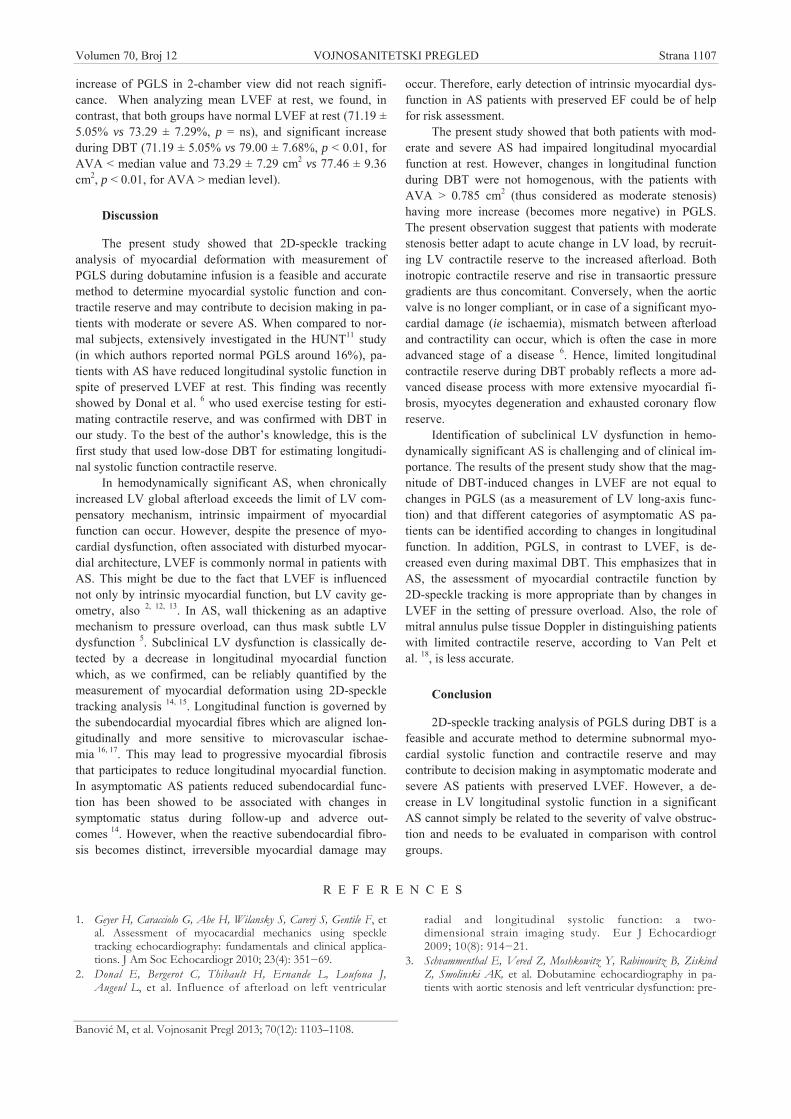
Volumen 70, Broj 12 VOJNOSANITETSKI PREGLED Strana 1107
Banovi M, et al. Vojnosanit Pregl 2013; 70(12): 1103–1108.
increase of PGLS in 2-chamber view did not reach signifi-cance. When analyzing mean LVEF at rest, we found, incontrast, that both groups have normal LVEF at rest (71.19 ±5.05% vs 73.29 ± 7.29%, p = ns), and significant increaseduring DBT (71.19 ± 5.05% vs 79.00 ± 7.68%, p < 0.01, forAVA < median value and 73.29 ± 7.29 cm2 vs 77.46 ± 9.36cm2, p < 0.01, for AVA > median level).
Discussion
The present study showed that 2D-speckle trackinganalysis of myocardial deformation with measurement ofPGLS during dobutamine infusion is a feasible and accuratemethod to determine myocardial systolic function and con-tractile reserve and may contribute to decision making in pa-tients with moderate or severe AS. When compared to nor-mal subjects, extensively investigated in the HUNT11 study(in which authors reported normal PGLS around 16%), pa-tients with AS have reduced longitudinal systolic function inspite of preserved LVEF at rest. This finding was recentlyshowed by Donal et al. 6 who used exercise testing for esti-mating contractile reserve, and was confirmed with DBT inour study. To the best of the author’s knowledge, this is thefirst study that used low-dose DBT for estimating longitudi-nal systolic function contractile reserve.
In hemodynamically significant AS, when chronicallyincreased LV global afterload exceeds the limit of LV com-pensatory mechanism, intrinsic impairment of myocardialfunction can occur. However, despite the presence of myo-cardial dysfunction, often associated with disturbed myocar-dial architecture, LVEF is commonly normal in patients withAS. This might be due to the fact that LVEF is influencednot only by intrinsic myocardial function, but LV cavity ge-ometry, also 2, 12, 13. In AS, wall thickening as an adaptivemechanism to pressure overload, can thus mask subtle LVdysfunction 5. Subclinical LV dysfunction is classically de-tected by a decrease in longitudinal myocardial functionwhich, as we confirmed, can be reliably quantified by themeasurement of myocardial deformation using 2D-speckletracking analysis 14, 15. Longitudinal function is governed bythe subendocardial myocardial fibres which are aligned lon-gitudinally and more sensitive to microvascular ischae-mia 16, 17. This may lead to progressive myocardial fibrosisthat participates to reduce longitudinal myocardial function.In asymptomatic AS patients reduced subendocardial func-tion has been showed to be associated with changes insymptomatic status during follow-up and adverce out-comes 14. However, when the reactive subendocardial fibro-sis becomes distinct, irreversible myocardial damage may
occur. Therefore, early detection of intrinsic myocardial dys-function in AS patients with preserved EF could be of helpfor risk assessment.
The present study showed that both patients with mod-erate and severe AS had impaired longitudinal myocardialfunction at rest. However, changes in longitudinal functionduring DBT were not homogenous, with the patients withAVA > 0.785 cm2 (thus considered as moderate stenosis)having more increase (becomes more negative) in PGLS.The present observation suggest that patients with moderatestenosis better adapt to acute change in LV load, by recruit-ing LV contractile reserve to the increased afterload. Bothinotropic contractile reserve and rise in transaortic pressuregradients are thus concomitant. Conversely, when the aorticvalve is no longer compliant, or in case of a significant myo-cardial damage (ie ischaemia), mismatch between afterloadand contractility can occur, which is often the case in moreadvanced stage of a disease 6. Hence, limited longitudinalcontractile reserve during DBT probably reflects a more ad-vanced disease process with more extensive myocardial fi-brosis, myocytes degeneration and exhausted coronary flowreserve.
Identification of subclinical LV dysfunction in hemo-dynamically significant AS is challenging and of clinical im-portance. The results of the present study show that the mag-nitude of DBT-induced changes in LVEF are not equal tochanges in PGLS (as a measurement of LV long-axis func-tion) and that different categories of asymptomatic AS pa-tients can be identified according to changes in longitudinalfunction. In addition, PGLS, in contrast to LVEF, is de-creased even during maximal DBT. This emphasizes that inAS, the assessment of myocardial contractile function by2D-speckle tracking is more appropriate than by changes inLVEF in the setting of pressure overload. Also, the role ofmitral annulus pulse tissue Doppler in distinguishing patientswith limited contractile reserve, according to Van Pelt etal. 18, is less accurate.
Conclusion
2D-speckle tracking analysis of PGLS during DBT is afeasible and accurate method to determine subnormal myo-cardial systolic function and contractile reserve and maycontribute to decision making in asymptomatic moderate andsevere AS patients with preserved LVEF. However, a de-crease in LV longitudinal systolic function in a significantAS cannot simply be related to the severity of valve obstruc-tion and needs to be evaluated in comparison with controlgroups.
R E F E R E N C E S
1. Geyer H, Caracciolo G, Abe H, Wilansky S, Carerj S, Gentile F, etal. Assessment of myocacardial mechanics using speckletracking echocardiography: fundamentals and clinical applica-tions. J Am Soc Echocardiogr 2010; 23(4): 351 69.
2. Donal E, Bergerot C, Thibault H, Ernande L, Loufoua J,Augeul L, et al. Influence of afterload on left ventricular
radial and longitudinal systolic function: a two-dimensional strain imaging study. Eur J Echocardiogr2009; 10(8): 914 21.
3. Schvammenthal E, Vered Z, Moshkowitz Y, Rabinowitz B, ZiskindZ, Smolinski AK, et al. Dobutamine echocardiography in pa-tients with aortic stenosis and left ventricular dysfunction: pre-
Strana 1108 VOJNOSANITETSKI PREGLED Volumen 70, Broj 12
Banovi M, et al. Vojnosanit Pregl 2013; 70(12): 1103–1108.
dicting outcome as a function of management strategy. Chest2001; 119(6): 1766 77.
4. Faggiano P, Rusconi C, Ghizzoni G. Left ventricular remodelingin valvular aortic stenosis. Echocardiographic and clinicalchanges in two patients during follow up. Chest 1994; 105(4):1273 5.
5. Serri K, Reant P, Lafitte M, Berhouet M, Le Bouffos V, RoudautR, et al. Global and regional myocardial function quantificationby two-dimensional strain: Application in hypertrophic car-diomyopathy. J Am Coll Cardiol 2006; 47(6): 1175 81.
6. Donal E, Thebault C, O’Connor K, Veillard D, Rosca M, Pierard L,et al. Impact of aortic stenosis on longitudinal myocardial de-formation during exercise. Eur J Echocardiogr 2011; 12(3):235 41.
7. Lang RM, Bierig M, Devereux RB, Flachskampf FA, Foster E, Pe-likka PA, et al. Recommendations for chamber quantification.Eur J Echocardiogr 2006; 7(2): 79 108.
8. Sahn DJ, DeMaria A, Kisslo J, Weyman A. The committee on M-Mode Standardization of the American Society of Echocardi-ography: Recommendations regarding quantificationin M-mode echocardiography: Results of a survey of echocardio-graphic measurements. Circulation 1978; 58(6): 1072 83.
9. Hammond IW, Devereux RB, Alderman MH, Lutas EM, SpitzerMC, Crowley JS, et al. The prevalence and correlates of echo-cardiographic left ventricular hypertrophy among patients withuncomplicated hypertension. J Am Coll Cardiol 1986; 7(3):639 50.
10. Baumgartner H, Hung J, Bermejo J, Chambers JB, Evangelista A,Griffin BP, et al. Echocardiographic assessment of valve steno-sis: EAE/ASE recommendations for clinical practice. Eur JEchocardiogr 2009; 10(1): 1 25.
11. Dalen H, Thorstensen A, Aase A, Ingul CB, Torp H, Vatten LJ, etal. Segmental and global longitudinal strain and strain rate
based on echocardiography of 1266 healthy individuals: theHUNT study in Norway. Eur J Echocardiogr 2010; 11(2):176 83.
12. Buermans HP, Paulus WJ. Iconoclasts topple adaptive myocar-dial hypertrophy in aortic stenosis. Eur Heart J 2005; 26(17):1697 9.
13. Carabello B. Evolution of the study of left ventricular function:everything old is new again. Circulation 2002; 105(23):2701 3.
14. Lafitte S, Perlant M, Reant P, Serri K, Douard H, DeMaria A, et al.Impact of impaired myocardial deformations on exercise toler-ance and prognosis in patients with asymptomatic aortic ste-nosis. Eur J Echocardiogr 2009; 10(3): 414 9.
15. Vinereanu D, Lim PO, Frenneaux MP, Fraser AG. Reducedmyocardial velocities of left ventricular long-axis contractionidentify both systolic and diastolic heart failure-a comparisonwith brain natriuretic peptide. Eur J Heart Fail 2005; 7(4):512 9.
16. Garcia D, Camici PG, Durand LG, Rajappan K, Gaillard E, Ri-moldi OE, et al. Impairment of coronary flow reserve in aorticstenosis. J Appl Physiol 2009; 106(1): 113 21.
17. Delgado V, Tops LF, van Brommel RJ, van der Kley F, Marsan NA,Klautz RJ, et al. Strain analysis in patients with severe aorticstenosis and preserved left ventricular ejection fraction under-going surgical valve replacement. Eur Heart J 2009; 30(24):3037 47.
18. Van Pelt NC, Stewart RA, Legget ME, Whalley GA, Wong SP,Zeng I, et al. Longitudinal left ventricular contractile dysfunc-tion after exercise in aortic stenosis. Heart 2007; 93(6): 732 8.
Received on June 30, 2011.Accepted on November 28, 2011.
OnLine-First June, 2013.
Vojnosanit Pregl 2013; 70(12): 1109–1116. VOJNOSANITETSKI PREGLED Strana 1109
Correspondence to: Vesna Mioljevi , Clinical Center of Serbia, Department of hospital epidemiology and hygiene, Belgrade, Serbia.E-mail: [email protected]
O R I G I N A L A R T I C L E UDC: 614.2:616-036.22]:616.98DOI: 10.2298/VSP1312109M
Risk factors for vancomycin-resistant Enterococcus colonization inhematologic patients
Faktori rizika od kolonizacije vankomicin-rezistentnog Enterococcus-a kodhematoloških bolesnika
Vesna Mioljevi *, Ljiljana Markovi -Deni †, Ana Vidovi ‡§, Milica Jovanovi ¶,Tanja Toši ¶, Dragica Tomin‡§
*Department of Hospital Epidemiology and Hygiene, ‡Clinic of Hematology,¶Microbiology Department, Clinical Center of Serbia, Belgrade, Serbia; †Institute of
Epidemiology, Faculty of Medicine, University of Belgrade, Belgrade, Serbia; §Facultyof Medicine, University of Belgrade, Belgrade, Serbia
Abstract
Background/Aim. Vancomycin-resistant Enterococci (VRE) isone of the most important hospital pathogens. The aim of thestudy was to evaluate VRE colonization in patients hospitalized atthe Hematology Intensive Care Unit, as well as the associated riskfactors. Methods. A prospective cohort study involved 70 pa-tients hospitalized at the Intensive Care Unit (ICU), Clinic forHematology, Clinical Center of Serbia, Belgrade, during 3 months.Baseline demographic data, data about antibiotic usage and otherrisk factors for VRE colonization during the present and previoushospitalizations (within 6 months) were recorded for each patientusing the questionnaire. Feces or rectal swab was collected forculture from patients on admission and at discharge in case whenVRE was not isolated on admission. Enterococci were isolated bystandard microbiological methods. Isolate sensitivity was tested bydisk-diffusion test using 30 g/mL (BBL) Vancomycin plates ac-cording to the Clinical and Laboratory Standards Institute (CLSI)standard. Results. Analysing results showed that 7% of the pa-tients had been already colonized with VRE upon ICU admis-sion. The rate of VRE colonization during present hospitalizationwas 41.5%. Univariate logistic regression demonstrated the statis-tically significant differences in diagnosis, length of present stay,use of aminoglycosides and piperacillin/tazobactam in presenthospitalization, duration of use of carbapenem and piperacil-lin/tazobactam in present hospitalization between the VRE-colonized and non-colonized patients. Acute myeloid leukemia(AML), use of carbapenem in previous hospitalization and dura-tion of use of piperacillin/tazobactam in present hospitalizationwere independent risk factors for VRE-colonized patients ac-cording to multivariate logistic regression. Conclusion. VREcolonization rate was high among the patients admitted to hema-tology ICU. Rational use of antibiotics and active surveillancemay be helpful preventive measures against the development ofbacterial resistance to antimicrobial agents.
Key words:enterococcus faecium; vancomycin; drug resistance,bacterial; hematologic diseases; risk factors.
Apstrakt
Uvod/Cilj. Enterococcus spp. rezistentan na vankomicin(VRE) jedan je od najzna ajnih bolni kih patogena. Cilj radabio je da se utvrde stope rektalne kolonizacije VRE kod bole-snika le enih u Odeljenju za hematološku intenzivnu negu, ida se sagledaju faktori rizika od kolonizacije. Metode. Pros-pektivnom kohortnom studijom obuhva eno je 70 bolesnikale enih u periodu od tri meseca u Klinici za hematologijuKlini kog centra Srbije u Beogradu. Podaci o demografskimkaratkteristikama bolesnika, upotrebi antibiotika i drugim fa-ktorima rizika od VRE kolonizacije tokom sadašnje i pretho-dnih hospitalizacija (tokom 6 meseci) prikupljani su za svakogbolesnika uz pomo upitnika. Bolesnicima je uzmana kopro-kultura ili rektalni bris na prijemu, a prilikom otpusta onimbolesnicima kod kojih na prijemu nije izolovan VRE. Osetlji-vost izolata proverena je disk-difuzionim testom sa diskovimavankomicina 30 g/mL (BBL) u skladu sa Clinical andLaboratory Standards Institute (CLSI) standardima. Rezultati.Na prijemu je bilo 7% VRE kolonizovnaih bolesnika. StopaVRE kolonizacije tokom teku e hospitalizacije iznosila je41,5%. Univarijantna logisti ka regresija pokazala je statisti kizna ajne razlike u pogledu dijagnoze, dužine sadašnje hospi-talizacije, primeni aminoglikozida i piperacilin/tazobaktama usadašnjoj hospitalizaciji, dužini primene karbapenema i pipe-racilin /tazobaktama u sadašnjoj hospitalizaciji izme u boles-nika kolonizovanih VRE i nekolonizovanih bolesnika. Multi-varijantnom logisti kom regresijom ustanovljeno je da suakutna mijeloidna leukemija (AML), primena karbapenema uprethodnoj hospitalizaciji i dužina primene piperaci-lin/tazobactama u sadašnjoj hospitalizaciji bili nezavisni fak-tori rizika od kolonizacije bolesnika VRE. Zaklju ak. Zabe-ležena je visoka stopa kolonizacije pacijenata VRE. Racional-na upotreba antibiotika i aktivni nadzor mogu biti korisnemere prevencije nastanka rezistencije bakterija na antibiotike.
Klju ne re i:enterococcus faecium; vankomicin; lekovi, rezistencijamikroorganizama; hematološke bolesti; faktori rizika.
Strana 1110 VOJNOSANITETSKI PREGLED Volumen 70, Broj 12
Mioljevi V, et al. Vojnosanit Pregl 2013; 70(12): 1109–1116.
Introduction
Bacteria of Enterococcus genus are a significant causeof hospital-acquired infections (HAI), second among urinarytract infections (UTI) and third among bacteriemias. Thereare many different species of Enterococci. The most preva-lent species culturated from humans are E. faecalis (the mostcommon) and E. faecium. Enterococci have both an intrinsic(nature) and acquired resistance to antibiotics, making themimportant nosocomial pathogens. They are intrinsically re-sistant to penicillin (low level), all cephalosporins, aztreo-nam, macrolides, and low levels of clindamycin. This naturalresistance is present in all members of species and is chro-mosomally mediated. Acquired resistance to antibiotics in-cludes resistance to glycopeptides, lactamases fluoro-quinolones, tetracycline, high aminoglycoside doses and gly-copeptides (vancomycin), as a result of mutations in DNA orthe acquisition of new gene(s). Glycopeptides (vancomycin)resistance has been seen in around 70–78% of the nosoco-mial E. faecium population 1–3. The mechanism of vancomy-cin resistence is due to preventing the synthesis of peptido-glycan precursors of the bacterial cell wall by blocking twosteps: the transglycosylation and the transpeptidation 3. It ismediated by 5 genes referred to as vanA, which can inducehigh level resistance to both vancomycin and teicoplanin;vanB which is found intrinsically in non-pathogenic entero-coccal species; vanC; vanD, and vanE. VanA is more widelydistributed 4.
Vancomycin resistant Enterococcus spp. (VRE) wasfirst isolated in England and France in 1986 and later on inother European and countries worldwide 5. The proportion ofenterococcal bacteremia attributable to VRE in the UK in2007 was 8.5–12.5% for all enterococci 6. From 2005 to2008, a significant decrease in vancomycin resistance wasobserved in France (from 2 to 0.6%), Greece (from 37 to28%), Israel (from 46 to 20%) and Italy (from 19 to 6%).Ireland, Luxembourg and Greece in 2009 reported resistanceproportions above 25%, while the majority of countries (18of 26 countries) reported resistant proportions below 7%.Several countries reported it even below 1% (Bulgaria, Esto-nia, Finland, France, Norway and Sweden) 3. During the pastfour years, a significant increase was observed only in Aus-tria. In 2010, 28 EU countries reported 5,577 isolates of E.faecium, of which 7.4% were resistant to vancomycin. Dur-ing the four past years, only Latvia reported an increasedtrend of these enterococci 7.
According to the USA National Nosocomial InfectionsSurveillance (NNIS), the percentage of enterococcal isolatesresistant to vancomycin increased from 12% in the period1998–2002 to 28.5% of all isolates in 2003 8.
VRE can cause different types of HAI, like urinarytract, surgical site, bacteriemia, meningitis, endocarditis,most commonly in immunocompromised patients. Entero-cocci are responsible for high morbidity and mortality ratesin these patients 9.
Risk factors for VRE colonization and infections arethe following: age, hepatic and renal dysfunction, hemato-logical diseases, chronic diseases, application of the invasive
diagnostic and therapeutic procedures, stay in intensive careunit (ICU), abdominal surgery, transplantation, prolongedhospitalization, and broad-spectrum antibiotic use 10, 11.
VRE may survive on dry surfaces several weeks (from7 days to 4 months). Consequently, VRE is most commonlytransmitted in hospitals from person to person by direct con-tact with personnel and patient’s hands, either from feces,urine, or blood of a person carrying the organism. It can alsobe spread indirectly via hand contact with open wounds, orwith contaminated environments (some parts of medicalequipment and working surfaces contaminated by VRE) 12.VRE colonization could persist for years. The colonized pa-tients are significant reservoirs and sources of contaminationof the environment. VRE is not transmitted through the air 13.
Methods
This prospective cohort study involved 70 patients hos-pitalized at the Intensive Care Unit (ICU), Clinic for Hema-tology, Clinical Center of Serbia, Belgrade, in the periodfrom September to December 2011. The coproculture orrectal swab was collected from all the patients on admissionand before discharge.
All the patients were daily observed during their hospi-talization by an epidemiologist. Clinical charts were system-atically reviewed and, when necessary, the medical staff wasinterviewed. Baseline demographic data, data about antibi-otic usage and other risk factors for VRE colonization duringpresent and previous hospitalization were recorded for eachpatient using the questionnaire. The following characteristicsrelated to the patients, and applied diagnostic and therapeuti-cal procedures were recorded: age, sex, underlying disease,recent prior hospitalization (within 6 months) and recent an-timicrobial use, operation, inserted central venous and uri-nary catheter, mechanical ventilation, antibiotic prophylaxisand therapy (type of antibiotics), and VRE isolated fromcoproculture on admission and the discharge. Surveillancefor VRE infections was carried out during the course of thisstudy.
VRE isolation and identification
Enterococci were isolated by standard microbiologicalmethods. Enterococcal identification was based on culturalcharacteristics, Gram-stained specimens, mobility in 0.5%agar, capacity of pigment production and biochemical char-acteristics. Isolate sensitivity was tested by the disk-diffusiontest using 30 g/mL (BBL) Vancomycin plates according tothe Clinical and Laboratory Standards Institute (CLSI) stan-dard. The plates were incubated 24 hours at 35°C. The in-crease in more than one colony or a part of colony was inter-preted as resistance to vancomycin 14, 15. E-test was not usedin methodology given than only enterococcal isolates wererecovered and identified from rectal swab or feces for thepurpose of determination of the carrier state.
The descriptive and analytical methods were used fordata processing: 2 test (for categorical data) or t-test (forcontinuous variables). The results were expressed as percent-ages or as mean ± standard deviations. To identify the risk
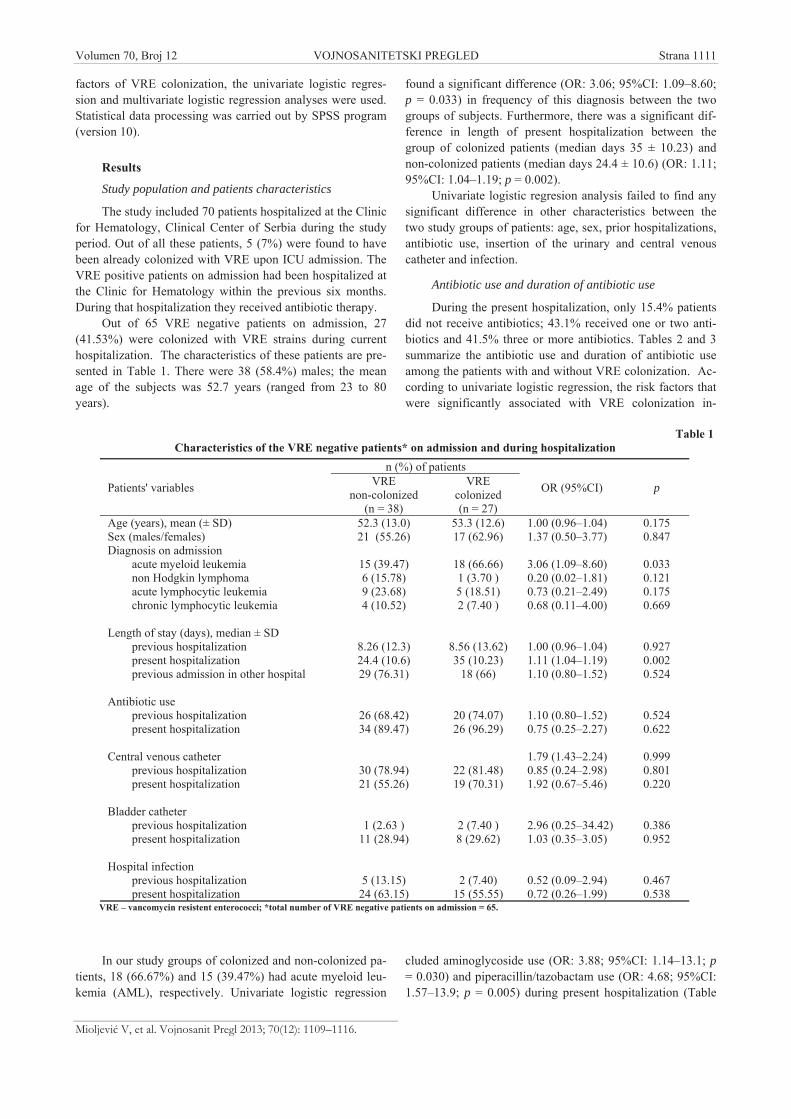
Volumen 70, Broj 12 VOJNOSANITETSKI PREGLED Strana 1111
Mioljevi V, et al. Vojnosanit Pregl 2013; 70(12): 1109–1116.
factors of VRE colonization, the univariate logistic regres-sion and multivariate logistic regression analyses were used.Statistical data processing was carried out by SPSS program(version 10).
Results
Study population and patients characteristics
The study included 70 patients hospitalized at the Clinicfor Hematology, Clinical Center of Serbia during the studyperiod. Out of all these patients, 5 (7%) were found to havebeen already colonized with VRE upon ICU admission. TheVRE positive patients on admission had been hospitalized atthe Clinic for Hematology within the previous six months.During that hospitalization they received antibiotic therapy.
Out of 65 VRE negative patients on admission, 27(41.53%) were colonized with VRE strains during currenthospitalization. The characteristics of these patients are pre-sented in Table 1. There were 38 (58.4%) males; the meanage of the subjects was 52.7 years (ranged from 23 to 80years).
In our study groups of colonized and non-colonized pa-tients, 18 (66.67%) and 15 (39.47%) had acute myeloid leu-kemia (AML), respectively. Univariate logistic regression
found a significant difference (OR: 3.06; 95%CI: 1.09–8.60;p = 0.033) in frequency of this diagnosis between the twogroups of subjects. Furthermore, there was a significant dif-ference in length of present hospitalization between thegroup of colonized patients (median days 35 ± 10.23) andnon-colonized patients (median days 24.4 ± 10.6) (OR: 1.11;95%CI: 1.04–1.19; p = 0.002).
Univariate logistic regresion analysis failed to find anysignificant difference in other characteristics between thetwo study groups of patients: age, sex, prior hospitalizations,antibiotic use, insertion of the urinary and central venouscatheter and infection.
Antibiotic use and duration of antibiotic use
During the present hospitalization, only 15.4% patientsdid not receive antibiotics; 43.1% received one or two anti-biotics and 41.5% three or more antibiotics. Tables 2 and 3summarize the antibiotic use and duration of antibiotic useamong the patients with and without VRE colonization. Ac-cording to univariate logistic regression, the risk factors thatwere significantly associated with VRE colonization in-
cluded aminoglycoside use (OR: 3.88; 95%CI: 1.14–13.1; p= 0.030) and piperacillin/tazobactam use (OR: 4.68; 95%CI:1.57–13.9; p = 0.005) during present hospitalization (Table
Table 1Characteristics of the VRE negative patients* on admission and during hospitalization
n (%) of patientsVRE VRE
non-colonized colonizedPatients' variables
(n = 38) (n = 27)
OR (95%CI) p
Age (years), mean (± SD) 52.3 (13.0) 53.3 (12.6) 1.00 (0.96–1.04) 0.175Sex (males/females) 21 (55.26) 17 (62.96) 1.37 (0.50–3.77) 0.847Diagnosis on admission
acute myeloid leukemia 15 (39.47) 18 (66.66) 3.06 (1.09–8.60) 0.033non Hodgkin lymphoma 6 (15.78) 1 (3.70 ) 0.20 (0.02–1.81) 0.121acute lymphocytic leukemia 9 (23.68) 5 (18.51) 0.73 (0.21–2.49) 0.175chronic lymphocytic leukemia 4 (10.52) 2 (7.40 ) 0.68 (0.11–4.00) 0.669
Length of stay (days), median ± SDprevious hospitalization 8.26 (12.3) 8.56 (13.62) 1.00 (0.96–1.04) 0.927present hospitalization 24.4 (10.6) 35 (10.23) 1.11 (1.04–1.19) 0.002previous admission in other hospital 29 (76.31) 18 (66) 1.10 (0.80–1.52) 0.524
Antibiotic useprevious hospitalization 26 (68.42) 20 (74.07) 1.10 (0.80–1.52) 0.524present hospitalization 34 (89.47) 26 (96.29) 0.75 (0.25–2.27) 0.622
Central venous catheter 1.79 (1.43–2.24) 0.999previous hospitalization 30 (78.94) 22 (81.48) 0.85 (0.24–2.98) 0.801present hospitalization 21 (55.26) 19 (70.31) 1.92 (0.67–5.46) 0.220
Bladder catheterprevious hospitalization 1 (2.63 ) 2 (7.40 ) 2.96 (0.25–34.42) 0.386present hospitalization 11 (28.94) 8 (29.62) 1.03 (0.35–3.05) 0.952
Hospital infectionprevious hospitalization 5 (13.15) 2 (7.40) 0.52 (0.09–2.94) 0.467present hospitalization 24 (63.15) 15 (55.55) 0.72 (0.26–1.99) 0.538
VRE – vancomycin resistent enterococci; *total number of VRE negative patients on admission = 65.
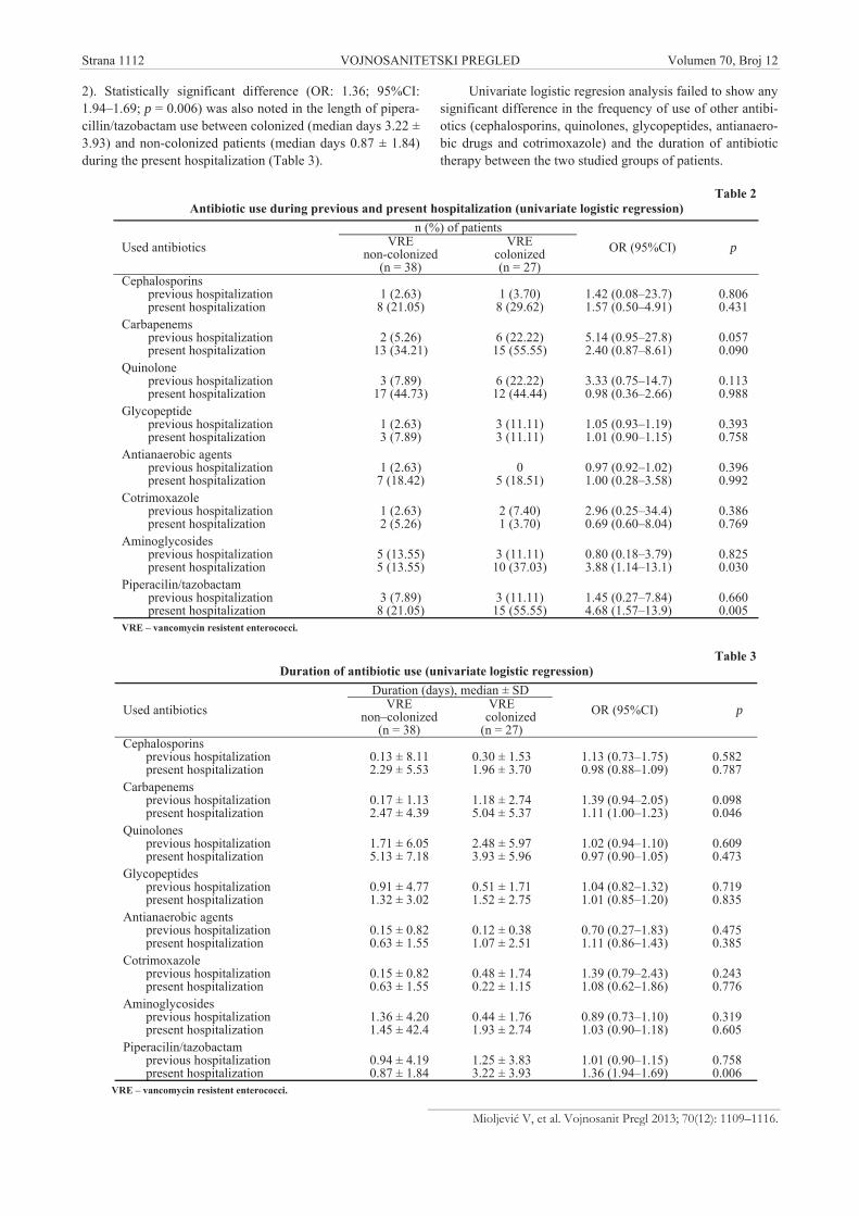
Strana 1112 VOJNOSANITETSKI PREGLED Volumen 70, Broj 12
Mioljevi V, et al. Vojnosanit Pregl 2013; 70(12): 1109–1116.
2). Statistically significant difference (OR: 1.36; 95%CI:1.94–1.69; p = 0.006) was also noted in the length of pipera-cillin/tazobactam use between colonized (median days 3.22 ±3.93) and non-colonized patients (median days 0.87 ± 1.84)during the present hospitalization (Table 3).
Univariate logistic regresion analysis failed to show anysignificant difference in the frequency of use of other antibi-otics (cephalosporins, quinolones, glycopeptides, antianaero-bic drugs and cotrimoxazole) and the duration of antibiotictherapy between the two studied groups of patients.
Table 2Antibiotic use during previous and present hospitalization (univariate logistic regression)
n (%) of patientsVRE VRE
non-colonized colonizedUsed antibiotics(n = 38) (n = 27)
OR (95%CI) p
Cephalosporinsprevious hospitalization 1 (2.63) 1 (3.70) 1.42 (0.08–23.7) 0.806present hospitalization 8 (21.05) 8 (29.62) 1.57 (0.50–4.91) 0.431
Carbapenemsprevious hospitalization 2 (5.26) 6 (22.22) 5.14 (0.95–27.8) 0.057present hospitalization 13 (34.21) 15 (55.55) 2.40 (0.87–8.61) 0.090
Quinoloneprevious hospitalization 3 (7.89) 6 (22.22) 3.33 (0.75–14.7) 0.113present hospitalization 17 (44.73) 12 (44.44) 0.98 (0.36–2.66) 0.988
Glycopeptideprevious hospitalization 1 (2.63) 3 (11.11) 1.05 (0.93–1.19) 0.393present hospitalization 3 (7.89) 3 (11.11) 1.01 (0.90–1.15) 0.758
Antianaerobic agentsprevious hospitalization 1 (2.63) 0 0.97 (0.92–1.02) 0.396present hospitalization 7 (18.42) 5 (18.51) 1.00 (0.28–3.58) 0.992
Cotrimoxazoleprevious hospitalization 1 (2.63) 2 (7.40) 2.96 (0.25–34.4) 0.386present hospitalization 2 (5.26) 1 (3.70) 0.69 (0.60–8.04) 0.769
Aminoglycosidesprevious hospitalization 5 (13.55) 3 (11.11) 0.80 (0.18–3.79) 0.825present hospitalization 5 (13.55) 10 (37.03) 3.88 (1.14–13.1) 0.030
Piperacilin/tazobactamprevious hospitalization 3 (7.89) 3 (11.11) 1.45 (0.27–7.84) 0.660present hospitalization 8 (21.05) 15 (55.55) 4.68 (1.57–13.9) 0.005
VRE – vancomycin resistent enterococci.
Table 3Duration of antibiotic use (univariate logistic regression)
Duration (days), median ± SDVRE VRE
non–colonized colonizedUsed antibiotics(n = 38) (n = 27)
OR (95%CI) p
Cephalosporinsprevious hospitalization 0.13 ± 8.11 0.30 ± 1.53 1.13 (0.73–1.75) 0.582present hospitalization 2.29 ± 5.53 1.96 ± 3.70 0.98 (0.88–1.09) 0.787
Carbapenemsprevious hospitalization 0.17 ± 1.13 1.18 ± 2.74 1.39 (0.94–2.05) 0.098present hospitalization 2.47 ± 4.39 5.04 ± 5.37 1.11 (1.00–1.23) 0.046
Quinolonesprevious hospitalization 1.71 ± 6.05 2.48 ± 5.97 1.02 (0.94–1.10) 0.609present hospitalization 5.13 ± 7.18 3.93 ± 5.96 0.97 (0.90–1.05) 0.473
Glycopeptidesprevious hospitalization 0.91 ± 4.77 0.51 ± 1.71 1.04 (0.82–1.32) 0.719present hospitalization 1.32 ± 3.02 1.52 ± 2.75 1.01 (0.85–1.20) 0.835
Antianaerobic agentsprevious hospitalization 0.15 ± 0.82 0.12 ± 0.38 0.70 (0.27–1.83) 0.475present hospitalization 0.63 ± 1.55 1.07 ± 2.51 1.11 (0.86–1.43) 0.385
Cotrimoxazoleprevious hospitalization 0.15 ± 0.82 0.48 ± 1.74 1.39 (0.79–2.43) 0.243present hospitalization 0.63 ± 1.55 0.22 ± 1.15 1.08 (0.62–1.86) 0.776
Aminoglycosidesprevious hospitalization 1.36 ± 4.20 0.44 ± 1.76 0.89 (0.73–1.10) 0.319present hospitalization 1.45 ± 42.4 1.93 ± 2.74 1.03 (0.90–1.18) 0.605
Piperacilin/tazobactamprevious hospitalization 0.94 ± 4.19 1.25 ± 3.83 1.01 (0.90–1.15) 0.758present hospitalization 0.87 ± 1.84 3.22 ± 3.93 1.36 (1.94–1.69) 0.006
VRE – vancomycin resistent enterococci.

Volumen 70, Broj 12 VOJNOSANITETSKI PREGLED Strana 1113
Mioljevi V, et al. Vojnosanit Pregl 2013; 70(12): 1109–1116.
Multivariate logistic regresion analysis included all thevalues of p < 0.1 (diagnosis, number of hospital days in presenthospitalization, carbapenem use in earlier and present hospitali-zation, use of aminoglycosides and piperacillin/tazobactam usein present hospitalization, duration of carbapenem use in previ-ous and present hospitalization as well as length of piperacil-lin/tazobactam use in present hospitalization).
The results of multivariate analysis demonstrated thatthe diagnosis of the disease (AML), carbapenem use in ear-lier hospitalization and length of piperacillin/tazobactam usein present hospitalization were independent risk factors ofcolonization of patients with VRE (Table 4).
Infections and pathogens
Data processing did not reveal any significant differ-ence in the incidence of all HAI (p > 0.05), as well as certainHAI (urinary tract infections, bloodstream infections and
pneumonia) between the two study groups of patients (Table5). In the VRE-colonized patients, vancomycin resistant En-teroccus spp was a cause of urinary tract infections in 4 pa-tients, while VRE was not isolated as the cause of infectionin the non-colonized patients (Table 6).
Discussion
The patients affected by malignant hemopathies are of-ten rehospitalized and, therefore possibilly colonized by hos-pital pathogens including VRE. Immunosuppressed patientsappear to be at special risk for VRE colonization and severe
VRE infections. VRE are a particular problem in the inten-sive care units of large hospitals where they usually occur.Our study was designed to evaluate the colonization rateduring hospitalization at the Hematology ICU, colonizationrate on admission, and risk factors of colonization. The re-
Table 4Risk factors for vancomycin-resistant Enterococci (VRE) colonization
according to multivariate logistic regression analysisRisk factors B SE OR (95%CI) pDiagnosis (AML) 0.001 0.001 0.92 (0.99–1.0) 0.048Antibiotic useCarbapenems – previous hospitalization 1.651 2.487 5.21 (0.04–6.81) 0.040Duration of antibiotic usePiperacillin/tazobactam – present hopsitalization 1.918 0.884 6.80 (1.20–3.85) 0.030
AML – acute myeloid leukemia.
Table 5Infection in vancomycin-resistant Enterococci (VRE) – colonized patients in present hospitalization
n (%) of patientsHospital–acquired VRE non–colonized VRE colonizedinfections (HAI) (n = 38) (n = 27)
Total patientsn (%)
Urinary tract infections 12 (31.6) 8 (29.6) 20 (30.8)Bloodstream infections 8 (21.1) 3 (11.1) 11 (16.9)Pneumonia 0 (0.0) 2 (7.40) 2 (3.1)Without HAI 18 (47.3) 14 (51.9) 32 (49.2)Total n (%) 38 (100.0) 27 (100.0) 65 (100.0)
Table 6Cases of infection in the vancomycin-resistant Enterococci (VRE) – colonized and non–colonized patients in present
hospitalizationn (%) of patients
VRE VREnon–colonized colonized
Hospital acquiredinfection ( HAI)
(n = 38) (n = 27)
Total patientsn (%)
Urinary tract infectionEsherichia coli 5 (41.6) 0 (0.0) 5 (25.0)Klebsiella spp. 5 (41.6) 1 (12.5) 6 (30.0)Enterococcus spp.(vancomycin sensitive) 1 (8.3) 3 (37.5) 4 (20.0)Enterococcus spp.(vancomycin resistent) 0 (0.0) 4 (50.0) 4 (20.0)Providentia retgery 1 (8.3 ) 0 (0.0) 1 (5.0)
Total patients, n (%) 12 (60) 8 (40) 20 (100.0)Bloodstream infection CNS* 4 (50.0) 2 (66.6) 6 (54.5)
Klebsiella spp 1 (12.5) 0 (0.0) 1 (9.09)Pseudomonas spp. 1 (12.5) 0 (0.0) 1 (9.09)Stenotrophomonas maltophilia 1 (12.5) 0 (0.0) 1 (9.09)Acinetobacter spp. 1 (12.5) 0 (0.0) 1 (9.09)Esherichia coli 0 1 (33.3) 1 (9.09)
Total patients, n (%) 8 (72.72) 3 (27.28) 11 (100.0)*CNS – coagulasa negative staphylococci.
Strana 1114 VOJNOSANITETSKI PREGLED Volumen 70, Broj 12
Mioljevi V, et al. Vojnosanit Pregl 2013; 70(12): 1109–1116.
sults of this study showed that 7% of the patients were VREpositive on admission (VRE isolated from feces culture orrectal swab). The colonization rate during ICU hospitaliza-tion was 41.5%.
A relatively small number of articles described VREcolonization in hematological patients 10, 16–19. However, En-teroccoci have recently emerged as nosocomial agents, espe-cially in patients with hematooncological diseases. Thestudies from France and the Netherlands showed that 37%,and 49% of hematological patients were colonized by VRE,respectively 16, 17. Contrary to these results, VRE colonizationin the USA immunocompromised patients was reported inconsiderably lower percentage. It was noted that out of 2,115hematological patients, 4.7% patients had verified rectalVRE colonization. Among all colonized patients, 5.4% werepatients with leukemia, 4.9% with hematopoietic stem celltransplantation recipients, and 2.2% with lymphoma 20. Inother study which was carried out on the hematologyoncol-ogy unit, 7.7% of patients, predominantly with hematologicmalignancies, were colonized or infected with VRE duringthe study period 21. A much higher rate of VRE colonizationin our study (41.5%) is probably the result of the lack ofcontact isolation measures and the increased use of antibiot-ics. This is supported by the fact that only 15% of patientsdid not receive antibiotics during present hospitalization.
Many years ago, it was demonstrated that 5–50% of allantibiotic prescriptions are considered inappropriate whichcan cause the emergence and dissemination of resistant or-ganisms. However, there is not standard treatment protocolfor antibiotic prescription in our country. Beside that, the an-tibiotics were until recently available over-the-counter in thepharmacies. Antibiotic prescription is frequently done with-out antibiograms or even without bacteriology isolation ofpathogens. All of the above mentioned can lead to high ratesof bacterial resistance to antibiotics. It is important to em-phasize that there is an increasing trend of vancomycin re-sistance in our country 22.
Our study failed to find any association of sex and theage and development of VRE fecal colonization at discharge.Our findings are consistent with the results of similar studiesconducted in Korea 23. On the contrary, a study carried out atthe Thessaloniki University Clinic confirmed that VRE colo-nization was significantly more frequent in patients olderthan 60 years of age 24.
Analysis of our results showed that VRE colonizationwas significantly more frequent in patients with AML. Mul-tivariate regression analysis demonstrated that AML was anindependent risk factor for VRE colonization. Similar resultswere found in other studies as well 21. It can be explained bythe long length of hospitalization. Namely, duration of hos-pital stay of our patients with AML ranged from 28 to 40days, in distinction from the patients with acute lymphocyticleukemia (ALL), non-Hodgkin lymphoma (NHL) andchronic lymphocytic leukemia (HLL) who were hospitalizedduring significantly shorter period. From the aspect of a cli-nician this may be explained by the length of therapy proto-col application. Besides other risk factors, the length of hos-pital stay is considered as one of most important risk fac-
tor 10, 11, 24. Accordingly, AML patients are at higher risk ofcolonization than patients with shorter hospitalization. Theresults of this study are only the introduction to more com-prehensive and detailed analysis of the problem not only inhematological, but also in other immunocompromised pa-tients.
Our study failed to find any significant difference inprevious hospitalization between the two studied groups ofpatients, contrary to the results of other studies 20, 25. How-ever, mean length of present hospitalization in VRE-colonized patients was significantly longer in relation to non-colonized patients, ie 35 vs 24 days.
The results of several studies showed that the use ofglycopeptides, second and third generation cephalosporinsand antianaerobic antibiotics are associated with the pa-tients colonized with VRE 25, 26. Only few studies analyzedthe association of quinolone use and VRE colonization.Our study failed to establish any significant difference inthe use and length of use of antibiotics from the group ofquinolones (ciprofloxacin) between the two groups of sub-jects what is compatible with the findings obtained in otherstudies 25.
Several studies on the effect of carbapenem as the riskfactor for VRE colonization did not show any significant dif-ference in the use of these antibiotics between VRE-colonized and non-colonized patients 20, 26. However, in otherstudies prior carbapenem use was a significant risk factor forVRE colonization 24, 25. Our study showed that there was adifference in the duration of the use of antibiotics from car-bapenem group (imipenem and meropenem) between twogroups of subjects in repeated hospitalization. Mean durationof carbapenem use in the VRE-colonized and in non-colonized patients in the repeated hospitalization was 5 and2.47 days, respectively. Moreover, carbapenem use in previ-ous hospitalization was an independent risk factor of VREcolonization.
Vancomycin is a glycopeptide antibiotic that is used totreat infections caused by Gram-positive bacteria. For manyyears, it has traditionally been reserved as a drug of “last re-sort”, used to treat severe infections for which other antibi-otics had failed. Vancomycin use has increased linearly inthe last decades, especially for infections related to the pres-ence of indwelling vascular catheter which is the case withhematology-oncology patients. Vancomycin is not recom-mended for regular antibioprophylaxis in surgery. However,a growing number of infections caused by Staphylococcusaureus resistant to meticillin has led to the widespread use ofvancomycin in hospitals. The results of studies on associa-tion of vancomycin use and VRE colonization and infectionhave been controversial. A meta-analysis of 20 studiesshowed that the use of vancomycin increased the risk ofVRE colonization by 4.5 times 27. Using the multivariateanalysis, Ostrowsky et al. 28, in contrast, demonstrated thatthere was no association between the vancomycin use andVRE colonization. In addition, the results of recent system-atic review did not determine a potential role for vancomycinusage reduction in controlling VRE colonization 29. Ourstudy did not find any significant difference in the frequency
Volumen 70, Broj 12 VOJNOSANITETSKI PREGLED Strana 1115
Mioljevi V, et al. Vojnosanit Pregl 2013; 70(12): 1109–1116.
of vancomycin use and length of its use between the twogroups of patients.
Analysing a study on the effect of use of antibioticsfrom the group of aminoglycosides failed to find any signifi-cant difference in the frequency of use of these antibioticsbetween VRE-colonized and non-colonized patients 17. Onthe contrary, our results showed a significant difference inthe use of aminoglycoside antibiotics between the twogroups of subjects during repeated hospitalization. In theVRE-colonized group, 37% of the patients were adminis-tered aminoglycoside antibiotics while in the non-colonizedgroup 13% of the patients received these antibiotics.
Analysing of our data showed that the VRE-colonizedpatients received piperacillin/tazobactam in a significantlyhigher percentage (55%) then non-colonized patients (21%).An average length of piperacillin/tazobactam use in the VRE-colonized and non-colonized patients during the present hos-pitalization was 3.22 and 0.87 days, respectively. Moreover,the use of this antibiotic in present hospitalization was an in-dependent risk factor of VRE colonization. The incidence ofVRE was positively correlated with the use of piperacil-lin/tazobactam or beta-lactam agents in other studies as well 30.
As the part of normal fecal flora, Enteroccoci were nottraditionally considered as important nosocomial pathogens.But, they have emerged as increasingly important pathogenswith increased resistance to antibiotics. VRE become one ofthe leading causes of HAI, especially of urinary tract andbloodstream infections. VRE commonly colonise, but less
frequently cause the infections. However, colonization pre-cedes most infections. In our study, VRE was a cause of UTIin the colonized patients but not in the non-colonized pa-tients. VRE infections are not more virulent than other en-teroccocal infections. But, VRE infections are very problem-atic for treatment. Enterococci are the most frequent cause ofUTI. During one year of surveillance, organized by the Na-tional Healthcare Safety Network in the USA there wasfound that Enteroccocus spp was third the most frequentpathogen of HAI participated with 12% in the overall num-ber of pathogenic isolates. Regarding rank-order distribution,it was at second position for bloodstream infections and atthird position for UTI. E. faecium and E. faecalis showed ahigh proportion of vancomycin resitance: 98.4–99.5% and91.9–98.4%, respectively 31.
Conclusion
VRE colonization rate was high among patients admit-ted to hematology ICU. Rational use of antibiotics and ac-tive surveillance may be helpful preventive measures againstthe development of bacterial resistance to antimicrobialagents.
Acknowledgements
This work was supported by the Ministry of Education,Science and Technological Development of the Republic ofSerbia (Contract No. 175046; 2011–2014).
R E F E R E N C E S
1. Willems RJ, Bonten MJ. Glycopeptide-resistant enterococci: de-ciphering virulence, resistance and epidemicity. Curr Opin In-fect Dis 2007; 20(4): 384 90.
2. Sujatha S, Praharaj I. Glycopeptide resistance in gram-positivecocci: a review. Interdiscip Perspect Infect Dis 2012; 2012:781679.
3. Surveillance report. Antimicrobial resistance surveillance inEurope, 2009. Available from:www.ecdc.europa.eu/en/.../1011_SUR_annual_EARS_Net_2009
4. Arias C, Murray B. The rise of the Enterococcus: beyond van-comycin resistance. Nat Rev Microbiol 2012; 10(4): 266 78.
5. Low DE, Keller N, Barth A, Jones RN. Clinical prevalence, anti-microbial susceptibility, and geographic resistance patterns ofenterococci: results from the SENTRY Antimicrobial Surveil-lance Program, 1997-1999. Clin Infect Dis 2001; 32 Suppl 2:S133 45.
6. Werner G, Coque TM, Hammerum AM, Hope R, Hryniewicz W,Johnson A, et al. Emergence and spread of vancomycin resis-tance among enterococci in Europe. Euro Surveill 2008;13(47). pii: 19046.
7. ECDC. Antimicrobial resistance surveillance in Europe 2010.Annual report of the European Antimicrobial Resistance Sur-veillance Network (EARS-Net). Available from:http://repositorio.insa.pt/handle/10400.18/702
8. National Nosocomial Infections Surveillance (NNIS) SystemReport data summary from January 1992 through June 2004,issued October 2004. Am J Infect Control 2004; 32(8):470 85.
9. Achenbach C, Flores E, Farerrell P, Pitrak D,Weber G. Prevalenceof risk factors for colonization with vancomicyn-resistant en-terococcus among human immunodeficiency virus positiveoutpatients. Infect Control Hosp Epidemiol 2006; 27(1):102 4.
10. Sakka V, Tsiodras S, Galani L, Antoniadou A, Souli M, Galani I,et al. Risk-factors and predictors of mortality in patients colo-nised with vancomycin-resistant enterococci. Clin MicrobiolInfect 2008; 14(1): 14 21.
11. Tacconelli E, Cataldo MA. Vancomycin-resistant enterococci(VRE): transmission and control. Int J Antimicrob Agents2008; 31: 99 106.
12. Muto CA, Jernigan JA, Ostrowsky BE, Richet HM, Jarvis WR, BoyceJM, et al. SHEA Guideline for Preventing Nosocomial Trans-mission of Multidrug-Resistant Strains of Staphylococcusaureus and Enterococcus spp. Infect Control Hosp Epidemiol2003; 24(5); 362 86.
13. Yoon YK, Lee SE, Lee J, Kim HJ, Kim JY, Park DW, et al. Riskfactors for prolonged carriage of vancomycin-resistant Ente-rococcus faecium among patients in intensive care units: acase-control study. J Antimicrob Chemother 2011; 66(8):1831 8.
14. CLSI. Performance Standards for Antimicrobial Disk Suscep-tibility Testing; Seventeenth Informational Supplement M100-S17. Wayne, PA: Clinical and Laboratory Standards Institute;2007.
15. CLSI. Performance Standards for Antimicrobial Disk Suscep-tibility Tests; M02-A10. Approved Standard Tenthth edi-tion. Wayne, PA: Clinical and Laboratory Standards Institute;2009.
Strana 1116 VOJNOSANITETSKI PREGLED Volumen 70, Broj 12
Mioljevi V, et al. Vojnosanit Pregl 2013; 70(12): 1109–1116.
16. Gambarotto K, Ploy MC, Turlure P, Grélaud C, Martin C, Bordes-soule D, et al. Prevalence of vancomycin-resistant enterococciin fecal samples from hospitalized patients and nonhospital-ized controls in a cattle-rearing area of France. J Clin Micro-biol 2000; 38(2): 620 4.
17. van den Braak N, Ott A, van Belkum A, Kluytmans JA, KoelemanJG, Spanjaard L, et al. Prevalence and determinants of fecalcolonization with vancomycin-resistant Enterococcus in hos-pitalized patients in The Netherlands. Infect Control HospEpidemiol 2000; 21(8): 520 4.
18. Almyroudis NG, Lesse AJ, Hahn T, Samonis G, Hazamy PA,Wongkittiroch K, et al. Molecular epidemiology and risk factorsfor colonization by vancomycin-resistant Enterococcus in pa-tients with hematologic malignancies. Infect Control HospEpidemiol 2011; 32(5): 490 6.
19. Liss BJ, Vehreschild JJ, Cornely OA, Hallek M, FätkenheuerG, Wisplinghoff H, et al. Intestinal colonisation and bloodstream infections due to vancomycin-resistantentero-cocci (VRE) and extended-spectrum beta-lactamase-producingEnterobacteriaceae (ESBLE) in patients with haematologicaland oncological malignancies. Infection 2012; 40(6): 613 9.
20. Matar MJ, Tarrand J, Raad I, Rolston KV. Colonization and in-fection with vancomycin-resistant Enterococcus among pa-tients with cancer. Am J Infect Control 2006; 34(8): 534 6.
21. Suntharam N, Lankford MG, Trick WE, Peterson LR, Noskin GA.Risk factors for acquisition of vancomycin-resistant entero-cocci among hematology-oncology patients. Diagn MicrobiolInfect Dis 2002; 43(3): 183 8.
22. Mirovi V, Tomanovi B, Konstantinovi S. The frequency of resis-tance to antibiotics of most frequently isolated bacteria fromblood cultures during the period 1997-2002. Vojnosanit Pregl2004; 61(4): 391 7. (Serbian)
23. Cheong HJ, Song JY, Eom JS, Kim WJ, Choi SJ, Choi JH, et al.Colonization rate, risk factor for acquisition, and genetic sen-sity of vancomicyn-resistant enterococci (VRE) isolated fromculture of patients in intensive care unit from Korea. Korean JInfect Dis 2002; 34: 276 84.
24. Metallidis S, Chatzidimitriou M, Tsona A, Bisiklis A, Lazaraki G,Koumentaki E, et al. Vancomicyn-Resistant Enterococci, Colo-
nizing the Intenstinal Tract of Patients in a University Hospitalin Greece. Braz J Infect Dis 2006; 10(3): 179 84.
25. Song JY, Cheong HJ, Jo YM, Choi WS, Noh JY, Heo JY, et al.Vancomicyn-Resistant Enterococci colonization before ad-mision to the intensive care unit: A clinico-epidemiologicalanalysis. Am J Infect Control 2009; 37(9): 734 40.
26. Padiglione AA, Wolfe R, Grabsch EA, Olden D, Pearson S, FranklinC, et al. Risk Factors for New Detection of Vancomycin-Resistant Enterococci in Acute-Care Hospitals That EmployStrict Infection Control Procedures. Antimocrob AgentsChemother 2003; 47(8): 2492–8.
27. Carmely Y, Samore MH, Huskins C. The association betweenantecedent vancomicyn teatment and hospital-acquired van-comicyn-resistent eneterococci; a meta analysis. Arch InternMed 1999; 159(20): 2461 8.
28. Ostrovsky BE, Venkataraman L, D'Agata EM, Gold HS, De Gi-rolami, Samore MH. Vancomicyn-resistant enterococci intensivecare units: high freguency of stool carrige during a non out-break period. Arch Intern Med 1999; 159(13): 1467 72.
29. de Bruin MA, Riley LW. Does vancomycin prescribing inter-vention affect vancomycin-resistant enterococcus infectionand colonization in hospitals? A systematic review. BMC In-fect Dis 2007; 7: 24 35.
30. Stiefel U, Paterson DL, Pultz NJ, Gordon SM, Aron DC, DonskeyCJ. Effect of the increasing use of piperacillin/tazobactam onthe incidence of vancomycin-resistant enterococci in four aca-demic medical centers. Infect Control Hosp Epidemiol 2004;25(5): 380 3.
31. Hidron AI, Edwards JR, Patel J, Horan TC, Sievert DM, PollockDA, at al. NHSN annual update: antimicrobial-resistantpathogens associated with healthcare-associated infections:annual summary of data reported to the National HealthcareSafety Network at the Centers for Disease Control and Pre-vention, 2006-2007. Infect Control Hosp Epidemiol 2008;29(11): 996 1011.
Received on April 13, 2012.Revised on September 6, 2012.Accepted on October 23, 2012.

Vojnosanit Pregl 2013; 70(12): 1117–1123. VOJNOSANITETSKI PREGLED Strana 1117
Correspondence to: Igor M. Nikoli , Clinic of Neurosurgery, Clinical Center of Serbia, Koste Todorovi a 4, 11 000, Belgrade, Serbia.E-mail: [email protected]
O R I G I N A L A R T I C L E UDC: 616.13-007.64-07DOI: 10.2298/VSP1312117N
Assessing the quality of angiographic display of brain blood vesselsaneurysms compared to intraoperative state
Procena kvaliteta angiografskog prikaza aneurizmi krvnih sudova mozga uodnosu na intraoperativni nalaz
Igor M. Nikoli *†, Goran M. Tasi *†, Vladimir T. Jovanovi *†, Nikola R. Repac*,Aleksandar M. Jani ijevi *, Vuk D. Š epanovi *, Branislav D. Nestorovi *†
*Clinic of Neurosurgery, Clinical Center of Serbia, Belgrade, Serbia; †Faculty ofMedicine, University of Belgrade, Belgrade, Serbia
Abstract
Background/Aim. Aneurysms in brain blood vessels areexpanding bags composed of a neck, body and fundus.Clear visibility of the neck, the position of the aneurysm andsurrounding structures are necessary for a proper choice ofmethods for excluding the aneurysm from the circulation.The aim of this study was to evaluate the reliability of spatialreconstruction of blood vessels of the brain based on theoriginal software for 3D reconstruction of the equipmentmanufacturer and a personal computer model developedearlier in the Clinic for Neurosurgery, Clinical Center ofSerbia, Belgrade, compared to intraoperative identificationof these aneurysms. Methods. This study included 137 pa-tients of both sexes. The presence of an aneurysm was veri-fied by angiographic methods [computed tomographic an-giography (CTA), multislice computed tomography angiog-raphy (MSCTA), magnetic resonance imaging angiography(MRA), or digital subtraction angiography (DSA)]. Results.The quality score (0 to 5) for CTA was 3.180 ± 0.961,MSCTA 4.062 ± 0.928, and for DSA 4.588 ± 0.758 (p <0.01). The results of this study favorite conventional angiog-raphy as the gold standard for diagnostic of intracranial an-eurysms. Conclusion. The results of this study are consis-tent with current publications review and clearly recognizethe advantages and disadvantages of diagnostic neuroradi-ological procedures, with DSA of brain blood vessels as abinding preoperative diagnostic procedure in cases in who itis not possible to clearly visualize the supporting blood ves-sel and neck of the aneurysm by using the findings of CTA,MRA and MSCTA.
Key words:intracranial aneurysm; diagnosis; angiography;tomography, x-ray computed; magnetic resonanceangiography; angiography, digital subtraction.
Apstrakt
Uvod/Cilj. Aneurizme na krvnim sudovima mozga predsta-vljaju vre asta proširenja kod kojih se razlikuju vrat, telo ifundus. Jasna vizualizacija vrata, položaj aneurizme i odnos saokolnim strukturama su uslov za adekvatnu odluku o izborumetode za isklju enje aneurizme iz cirkulacije. Cilj ovog radabio je da se proceni pouzdanost prostorne rekonstrukcije kr-vnih sudova mozga zasnovane na originalnom softveru za 3Drekonstrukciju proizvo a a opreme i personalnom kompju-terskom modelu ranije razvijenom u Neurohirurškoj kliniciKlini kog Centra Srbije u odnosu na intraoperativnu identifi-kaciju ovih aneurizmi. Metode. Ova studija obuhvatila je 137bolesnika oba pola (90 žena i 47 muškaraca). Prisustvo aneu-rizmi verifikovano je jednom od angiografskih metoda: kom-pjuterizovanom tomografskom angiografijom (CTA), multis-lajsnom kompjuterizovanom tomografskom angiografijom(MSCTA), angiografijom magnetnom rezonancom (MRA) ilidigitalnom suptrakcionom angiografijom (DSA). Rezultati.Ocena kvaliteta (skor od 0 do 5) za CTA bila je 3,180 ± 0,961za, MSCTA 4,062 ± 0,928, i za DSA 4,588 ± 0,758 (p < 0.01).Rezultati ove studije favorizuju konvencionalnu angiografijukao zlatni standard u dijagnostici aneurizmi. Zaklju ak. Re-zultati ove studije u skladu su sa pregledom aktuelnih publi-kacija i jasno prepoznaju dijagnosti ke prednosti i nedostatkeneuroradioloških procedura, pri emu se DSA krvnih sudovamozga izdvaja kao obavezuju a preoperativna dijagnosti kaprocedura kod bolesnika kod kojih nije mogu a jasna vizuali-zacija nose eg krvnog suda i vrata aneurizme na osnovu nala-za CTA, MSCTA i MRA.
Klju ne re i:aneurizma, intrakranijalna; dijagnoza; angiografija;tomografija, komjuterizovana, rendgenska; magnetnarezonanca, angiografija; angiografija, digitalnasuptrakcijska.
Strana 1118 VOJNOSANITETSKI PREGLED Volumen 70, Broj 12
Nikoli MI, et al. Vojnosanit Pregl 2013; 70(12): 1117–1123.
Introduction
Aneurysms in brain blood vessels are expanding bagscomposed of a neck, body and fundus. A connection, an in-teraction between the neck and the body is not constant anddetermined, thus, there are small wide neck aneurysms, butalso giant aneurysms with small necks. Clear visibility of theneck, the position of the aneurysm and surrounding struc-tures are necessary for making a proper decision on methodsfor excluding an aneurysm from the circulation. In additionto conventional digital subtraction panangiography (DSA) ofbrain blood vessels 1, the development of sophisticated diag-nostic procedures – computed tomography (CT) and mag-netic resonance imaging (MRI) has enabled noninvasive im-aging technology of blood vessels of the brain: CT angiogra-phy (CTA), 3D multislice (MSCT) angiography (MSCTA)and magnetic resonance angiography (MRA) 2.
Today’s CTA technology allows visualization of aneu-rysm diameters greater than 0.7 mm, the console stand-outpoint of 100–120 HU, and shows only lumens of blood ves-sels with contrast and bone 3. But despite this, the specifieddiagnosis is commonly used in many institutions as ascreening method of choice, and sometimes even 3D-CTA isused as the sole diagnostic method 4–8.
Digital subtraction angiography (DSA) is based ondigital elimination of bony structures so that the imageshows an isolated artery in which we inject an iodine contrastagent. This is achieved by increasing the resolution until itreaches the value of 0.05 mm or by increasing the number ofshoots until it reaches the number of 4–6 frames per second.3D DSA is a software model based on rotational angiogra-phy that can show the fine structure of brain blood vessels,and this method is more accurate than standard DSA 9–11.
MRA is a noninvasive technique based on time-of-flight (TOF) sequences and contrast-enchanced MR(CEMR). There is a problem of spatial resolution, and theminimum detection volume is 3 mm 12. It is possible to visu-alize aneurysm diameters less than 3 mm and is usually todetect those larger than 5 mm 13, 14. Despite the developedsoftware, sensitivity is less than that of DSA and is about 86–95% with a resolution of 0.2 mm2. 8, 15–17, making it ideal as anoninvasive screening procedure 12, 14, 18.
Despite technological advances, digital DSA remains thegold standard for diagnosis. Newer methods, unfortunately,have much lower accuracy in the diagnosis 19, 20. Microaneu-rysms can be visualized almost by the use of the DSA 8.
The aim of this study was to evaluate the reliability ofspatial reconstruction of blood vessels of the brain based onthe original software for 3D reconstruction of the equipmentmanufacturer and a personal computer model developed ear-lier in the Clinic of Neurosurgery at the Clinical Center ofSerbia 21, 22 compared to the intraoperative identification ofthese aneurysms.
Methods
This study included 137 patients of both sexes. Thepresence of an aneurysm was verified by the angiographic
methods (CTA, MSCT, MRA, or DSA). The analysis in-cluded patients who fulfiled the following requirements:clinically, lumbar puncture (LP) and endocranial CT verifiedattack of spontaneous subarachnoid hemorrhage (SAH); oneor more aneurysms of the anterior cerebral artery stream ofthe base of the brain were verified using one or more angio-graphic procedures, made surgery or embolization; aneu-rysms of the carotid artery trunk (according to the smallnumber of surgically treated aneurysms of the vertebrobasi-lar trunk).
In the analysis were used angiographic findings, spatialreconstruction of blood vessels of the brain based on theoriginal software for 3D reconstruction of the equipmentmanufacturer, and based on computer models previously de-veloped in our Clinic 21, 22.
A score of 0 to 5 was given to each angiographic find-ing according to the following criteria: aneurysm verification(negative findings exclude other criteria), aneurysm shape(0/1), aneurysm size (0/1), aneurysm orientation (0/1), aneu-rysm relationship to the carrying blood vessel (0/1), relation-ship of the aneurysm with perforators (0/1).
Aneurysm morphometric analysis, a comparative analy-sis of angiographic findings, and comparison of the qualityof angiography in relation to the intraoperative findings wereentered into questionnaire, followed by descriptive (meas-ures of central tendency and dispersion measures), and ana-lytical statistics. We used parametric (t-test) and non-parametric tests ( 2 test, and median), as well as correlationtests (linear correlation and regression).
Data analysis was performed on a personal computerwith Intel processor (generation of Intel Pentium III at 950MHz, Intel QuadCore 6600 in the 2GHz Intel T6500CoreTM2 Duo at 2.1 GHz) with a graphics card from NvidiaTNT 2 Pro with 32 MB, and Gforce Gforce G105M 8800with 512 MB VRAM. Digitalisation of images when it wasnot in DICOM format was done using a scanner A4 HPScanJet 5P (300 to 1200 dpi) and PoweShot camera CanonA710 IS (7.1 Mpixel).
Results
A total of 137 patients of both sexes (90 women and 47men), the mean age 50.39 ± 8.25 years, were included in thestudy. The mean ages of the female and male patients were52.15 ± 6.64 years, and 46.84 ± 9.96 years, respectively. Theyoungest patient was a 21-year-old and the oldest one a 72-year-old. There were 185 aneurysms in observed group: 164(88.65%) were located in the carotid stream and 21 (11.35%)in the vertebrobasilar stream. The distribution of aneurysmsby the carrying artery is shown in Table 1.
In 52 (37.96%) patients angiography was performed byusing CTA. In 33% cases it was the only method of preop-erative angiography. There were 17 diagnosis that were sup-plemented by DSA, and in 2 patients with MSCTA. In 9(17.31%) patients, CTA was initially falsely negative, whichwas later confirmed by the subsequent diagnosis using DSAor MSCT. The largest number of false negative results wasrelated to the internal carotid artery (ICA) (5).

Volumen 70, Broj 12 VOJNOSANITETSKI PREGLED Strana 1119
Nikoli MI, et al. Vojnosanit Pregl 2013; 70(12): 1117–1123.
Table 1Carrying artery aneurysms distribution
Artery NumberInternal carotid artery (ICA) 50Anterior cerebral artery (ACA-A1) 2Anterior communication artery (AcoA) 41Pericallosal artery (PA) 4Medial cerebral artery (MCA) 67Posterior cerebral artery (PCA) 3Basilar artery (BA) 8Superior cerebral artery (SCA) 4Inferior anterior cerebral artery (IACA) 1Posterior inferior cerebral artery (PICA) 3Vertebral artery (VA) 2
A quality score was determined by CTA in 50 patientsbecause ICA occlusion in the neck was found in 2 patients,and aneurysm was not visualized intraoperatively. The reli-ability score was 3.18 ± 0.96 with a median (Med) = 3.MSCTA was performed in 18 patients. Only in 5 patients itwas the only method that was performed. A false-negativefinding was observed in one patient with aneurysm on theanterior communication artery (AcoA) complex. Here, theaneurysm was confirmed with DSA and intraoperatively. In13 patients, MSCTA was supplemented with DSA.
A quality score was determined by MSCTA in 16 pa-tients, because in 2 patients embolization was performed, andaneurysm was no interoperatively visualized. The score was4.06 ± 0.93 with a Med = 5
MRA was performed as initial diagnostic method in 12patients during the acute phase of illness when they hospi-talized in other centers. After diagnosing spontaneous sub-arachnoid hemorrhage caused by ruptured aneurysm theywere referred for further treatment in the Clinic of Neurosur-gery, Clinical Center of Serbia. All the patients underwentadditional angiographic diagnosis by using DSA (11 pa-tients) or CTA (4 patients).
It was shown that by using CTA significantly lowerscore for ICA aneurysm angiographic finding quality wasobtained in comparison with the score obtained by usingDSA. Statistically significant difference was not found be-tween CTA and DSA in scores for medial cerebral artery(MCA) and AcoA aneurysm angiographic finding quality.
Due to the small number of patients submitted to MRA,which was not the only diagnostic procedure in neither case,statistical analysis of this method was not performed. Also,MRA was not performed on the same equipment in neithercase, but at different magnetic fields (from 1.0 to 3.0 T).
DSA was performed in 97 patients. In 66 of the patientsDSA was the only angiographic method for verification ofintracranial aneurysms. In the remaining cases (n = 31), con-ventional angiography was the additional diagnostic proce-dure in preparing patients for surgery.
In all the patients angiography was performed for 3Dreconstruction. In 46 patients 3D rotational angiography wasperformed as a part of diagnostic procedure, while the 51stpatient subsequently made a spatial reconstruction based on2D images and protocol for reconstruction in MatLab.
The quality score of DSA was determined in 71 patientssubmitted to direct intracranial aneurysm surgery. For otherpatients, occlusion was performed by endovascular proce-dure. The aneurysm was not visualized interoperatively. Thequality score was 4.59 ± 0.76 with a Med = 5. If we com-pared DSA scores for MCA, ACA and ACI aneurysm visu-alization finding no statistically significant differences werefound.
After evaluating reliability analysis for each of the di-agnostic procedures used, comparative analysis among thediffernet diagnostic procedures was done, with the exceptionof MRA. Table 3 shows the results of testing comparativereliability quality scores of CTA, MSCTA and DSA.
Table 2Mean quality score of computed tomography angiography
(CTA) and digital subtraction angiography (DSA) forcarotid aneurysms
CTA DSACarrying arterymean SD mean SD
MCA 3.48 1.40 4.65 0.80AcoA 3.41 1.003 4.63 0.83ICA 2.33 1.37 4.41 0.71
MCA – medial cerebral artery; ICA – internal carotid artery;AcoA – anterior communication artery.
Table 3The results of the t-test for comparison of angiographic
methodsAngiographic method t DF pCTA/MSCTA 3.222 64 < 0.01CTA/DSA 8.310 119 < 0.01MSCTA/DSA 2.080 85 < 0.05
CTA – computed tomography angiography;MSCTA – multislice computed tomography angiography;DSA – digital subtraction angiography.
Discussion
In the observed group of patients standardized CTA wasperformed in 52 patients, while MSCTA was performed in 18patients. MSCTA was performed in a much smaller number ofcases because the equipment was purchased later (it entered inthe second half of the study). False negative findings on CTAwere found by the subsequent additional diagnostics (DSA orMSCTA) in 9 (17.31%) patients, while 5 of them had the ICAaneurysms. When evaluating the reliability of methods for in-tracranial aneurysms detection and its relationship with thesupporting blood vessel and perforators, CTA assessment re-ceived score of 3.18 ± 0.96, and MSCTA 4.06 ± 0.93 (foundonly one false-negative finding). Despite of almost equalquality of CTA and DSA for detection of middle cerebral ar-tery aneurysms, the grades observed sample was considerablydifferent and there was a statistically significant difference inKorst DSA as a better method 23. On the other hand, Dehdastiet al. 24 in comparative analysis indicate that CTA is still insuf-ficient compared to DSA.
Because of the fact that the accuracy in the diagnosis ofintracranial aneurysms according to literature data is about84.6%, we emphasize that our findings are consistent with
Strana 1120 VOJNOSANITETSKI PREGLED Volumen 70, Broj 12
Nikoli MI, et al. Vojnosanit Pregl 2013; 70(12): 1117–1123.
the literature 25. This lack of precision primarily relates to thedetection of small aneurysms, subtraction bone in infracli-noid aneurysms, the lack of the knowledgable of radiologists,and artifacts that give previously placed clips or coil 3, 26–31.The problem with small aneurysms detection, and increasedaccuracy compared to standard CTA that works on a four- or16-slice aparatus 64MSCTA somewhat beyond, and it can bealmost comparable with the standard 2D DSA 26, 32. But stillfor aneurysms less than 4 mm combination of DSA with3D rotational angiography (3DR ) is the method ofchoice 33. Also, the development of “dual-energy direct boneremoval” CTA technique (DE-BR-CTA) was started in thelate 70s, has enabled great progress in subtraction bonestructures in MSCT, thus the image quality is much closer toconventional angiography 32, 34. 64MSCTA can be used as aninitial diagnostic procedure and to assess whether it is suit-able for direct aneurysm surgery or embolization, given thepossibility of 3D reconstruction in subtraction 32. A signifi-cant progress in subtraction resulted in orbital synchronizedhelical scan (OSHST) technique that allows a piston or asubtraction coil on postoperative recordings 35. However, de-spite the development of all CT, angiography is limited indiagnosing aneurysms of the posterior stream 36.
Despite the shortcomings CTA and MSCT are not onlyused as initial diagnostic procedure and the screening methodof choice, but in some establishments 3D-CTA is used as thesole diagnostic method for preoperative treatment of patientswith spontaneous subarachnoid hemorrhage 4–8, 37, 38. Weshould not ignore the cost of diagnostic procedures, becausethe cost of 3D CTA is significantly lower than the cost ofDSA, and can be considered as the method of choice inscreening 39. Due to lower prices and a small number ofcomplications, this method is very suitable for postoperativemonitoring and evaluation, as well as the only diagnosis inelderly patients with degenrative altered blood vessels, wherecatheter placement is difficult 40, 41.
Conventional angiography is an invasive method for ra-diological detection of blood vessels and their pathologicalchanges. The biggest positive step forward is made by theintroduction of digital substraction and computer modelswith which we eliminate the bone structure. DSA with spa-tial reconstruction is the gold standard in diagnosing intra-cranial aneurysms 42–44. Also, van Rooij et al. 45, 46, in theirstudies emphasize the advantage of rotational angiographyover conventional angiography for detection of small aneu-rysms. They believe that negative DSA angiographic find-ings should be done by the 3D-DSA, because in a greatnumber of cases small aneurysms can be detected with thismethod 45. According to Hai et al. 47 it is of great benefit inplanning 3DRA embolization of small aneurysms, the proce-dure itself reduces the radiation dose in comparison to con-ventional DSA.
However, Hirai et al. 48 stress that the lack of methodsis false pseudostenosis of the intracranial blood vessels. Thisphenomenon is related to the angle at which it is rotationalangiography and length of blood vessel 48. A reduction in ar-tifacts of pseudostenosis solved by the introduction of flatpanel detector (FPD) system 49.
In more than 2/3 (70.8%) of the patients in the observedgroup DSA was performed. Study on comparative analysisof biplanar DSA and 3D angiography demonstrated the ad-vantages of 3-dimensional images, suggesting that all pa-tients should be subjected to it for spatial reconstruction ofbrain blood vessels and intracranial aneurysms. It is doneeither in the form of 3DRA as a part of standard diagnosticapparatus using the Siemens Axiom Artis, or in the form ofcomputer reconstructions based on MatLab biplanarnih shots(PA and lateral projections) 21, 22, 50–52.
False negative findings were not found in any case, re-liability score was performed in 71 patients with direct aneu-rysm surgery. DSA with spatial reconstruction in the ob-served group received a high score of 4.59 ± 0.76. This rela-tively higher score of reliability is consistent with literature.DSA with 3D angiography is still considered to be a goldstandard, because of the fact that microaneurysms can bevisualized almost by the use of the DSA 8, 42, 45, 53, 54. Imper-fections in the diagnosis among other things can be ex-plained by Jou et al. 55. Artifacts can be created by the effectof pulsations, gravity and unequal density and contrast lev-els.
Aneurysms of various localization were slightly lessnoticeable than aneurysms in ICA, but if we compare thegrades among themselves there were no statistically signifi-cant differences (p > 0.05). It is not a small problem in theconversion of pixels as the relative size in millimeters, ie.real size. Fox et al. 56 conducted a study on comparativeanalysis of digital angiography in different picture archivingand communication systems (PACS). Kawashima et al. 57
emphasize that there are differences in morphometric meas-urements between biplanar DSA and 3D subtraction angiog-raphy. In this study comparative analysis of the intraopera-tive findings was not performed, thus the conclusion about amore realistic value could not be drawn. Beck et al. 58 findthat the dimensions of aneurysms by 3DRA are not less thanby biplanar DSA and are close to their actual sizes.
Several studies demonstrated the application of 3DRAwithin the system neuronavigation. Raabe et al. 59 carried outa number of operations of the intracranial aneurysms usingBrainLab’s apparatus for neuronavigation, Vector Vision 2and the Philips Integris Allura System angiographic. Previ-ously it was used during diagnostic frame for registration.During the work it was shown that the maximum error is inthe angulation of 90° and 90° in the rotation. According tothem, and despite the fact that such a system requires furtherimprovement it provides a useful topographical informationabout the vascular anatomy 59. Willems et al. 60 developed theintegrated 3DRA neuronavigation system and the firstphantom, and then applied it in the course of operations.They used a Philips Integris BV5000 angiographic apparatusand Medtronic’s neuronavigation StealthStation system. Im-possibility of direct transfer of data from the angiographicsystem console to the console of the neuronavigation system,they overcame by using a dynamic reference frame (DRF).By this way they were enabled to enhance visualization ofthe operation of complex giant aneurysms and arteriovenousmalformations 60.

Volumen 70, Broj 12 VOJNOSANITETSKI PREGLED Strana 1121
Nikoli MI, et al. Vojnosanit Pregl 2013; 70(12): 1117–1123.
MRI, or nuclear magnetic resonance (NMR) imaging, isa noninvasive diagnostic procedure without the use of ioniz-ing radiation. Because of that it is ideal as a noninvasivescreening procedure 12, 14, 18. Also it is grateful for its nonin-vasive and long-term monitoring of patients after treat-ment 61–63. With magnetic field intensification and matrixsize recording, resolution and sensitivity increase as indi-cated by phantom studies 64, 65. Clinical study on diagnosingunruptured aneurysms using DSA of 1.5T and 7.0T as thereference standard by Mönninghoff et al. 66 showed the ad-vantage of a stronger magnetic field in the detection of intra-cranial aneurysms. Gibbs et al. 67 obtained similar results.They also reduced magnetic field amplification and record-ing time.
Sensitivity, despite the developed software, is alwayslower than in DSA and is about 86–95% with a resolutionof 0.2 mm2 20. Schwab et al. 53 did a comparative analysisof MRA and DSA findings in 133 patients with aneurysmsand found deviation of MRA findings with regard to DSAin as much as 59%. These differences relate not only to thelocation and number of aneurysms, but also to the type of
aneurysm. Therefore, they recommend screening beforeusing CTA as a less invasive method of DSA 53. In order toassess the quality of MRA as a screening method, Hi-ratsuka et al. 68 worked comparative analysis of 3T MRA64MDCTA with 3D-DA as a reference standard. Theirstudy of 38 patients with nonruptured aneurysms showedno statistically significant difference between MRA andMDCT. In both methods we used volume rendering, andthe advantage of it is contrary to Schwab et al. 53 becauseMRA is completely noninvasive and does not use contrastagents 68.
Conclusion
The results of this study, consistent with the review ofcurrent publications, clearly recognize the advantages anddisadvantages of diagnostic neuroradiological procedures,with DSA of brain vessels as a binding preoperative diag-nostic procedure in case that findings on CT, MRA andMSCT are not enough for clear visualization of the support-ing blood vessel and the neck of aneurysm.
R E F E R E N C E S
1. Kato Y, Nair S, Sano H, Sanjaykumar MS, Katada K, HayakawaM, et al. Multi-slice 3D-CTA - an improvement over singleslice helical CTA for cerebral aneurysms. Acta Neruochir(Wien) 2002; 144(7): 715 22.
2. Morhard D, Ertl-Wagner B. MDCT in Neuro-Vascular Imaging.In: Reiser MF, Becker, Nikolaou K, Glazer G, editors. Multi-SliceCT. Berlin: Springer-Verlag; 2009. p. 123 36.
3. Wolf RL. CT Imaging and Physiologic Techniques in Inter-ventional Neuroradiology. In: Hurst RW, Rosenwasser RH, edi-tors. Interventional neuroradiology. New York: InformaHealthcare; 2008. p. 87 111.
4. Kato Y, Sano H, Katada K, Ogura Y, Hayakawa M, Kanaoka N, etal. Application of three-dimensional CT angiography (3D-CTA) to cerebral aneurysms. Surg Neurol 1999; 52(2): 113 21.
5. Kato Y, Hayakawa M, Katada K. Three-dimensional multislicehelical CT angiography of cerebral aneurysms. Nippon Rinsho2004. 62(4): 715 21. (Japanese)
6. Matsumoto M, Sato M, Nakano M, Endo Y, Watanabe Y, Sasaki T,et al. Three-dimensional computerized tomography angiogra-phy-guided surgery of acutely ruptured cerebral aneurysms. JNeurosurg, 2001; 94(5): 718 27.
7. Matsumoto M, Endo Y, Sato M, Sato S, Sakuma J, Konno Y, et al.Acute aneurysm surgery using three-dimensional CT angiogr-phy without conventional catheter angiography. Fukushima JMed Sci 2002. 48(2): 63 73.
8. Kornienko VN, Pronin IN. Diagnostic Neuroradiology. Berlin:Springer-Verlag; 2009:
9. Sugahara T, Korogi Y, Nakashima K, Hamatake S, Honda S, Taka-hashi M. Comparison of 2D and 3D digital subtraction angiog-raphy in evaluation of intracranial aneurysms. AJNR Am JNeuroradiol 2002; 23(9): 1545 52.
10. Manabe H, Takemura A, Hasegawa S, Nagahata M, Iko Y. Extra-vasation from Rupturing Aneurysm Demonstrated by 3DDigital Subtraction Angiography. AJNR Am J Neuroradiol,2005; 26(6): 1370 1.
11. Asir A, Guven G, Tekin T, Kutlay M, Colak A, Simsek H, et al.Three-dimensional versus conventional digital subtraction an-giography in the diagnosis of intracranial aneurysms: case re-port. Balkan Military Medical Review 2007; 10: 147 9.
12. Huston J 3rd. Magnetic Resonance Angiography. In: Winn RH,editor. Youman`s Neurological Surgery. Philadelphia: Saun-ders; 2004. p. 1575 99.
13. Huston J 3rd, Torres VE, Sulivan PP, Offord KP, Wiebers DO.Value of magnetic resonance angiography for the detection ofintracranial aneurysms in autosomal dominant polycystic kid-ney disease. J Am Soc Nephrol 1993. 3(12): 1871 7.
14. Hughes DG. Neuroradiology and Ultrasound. In: Moore AJ,Newell DV, editor. Neurosurgery: Principles and Practice.London: Springer; 2005. p. 23 8.
15. Anzalone N, Scomazzoni F, Cirillo M, Righi C, Simionato F, CadioliM, et al. Follow-up of coiled cerebral aneurysms at 3T: com-parison of 3D time-of-flight MR angiography and contrast-enhanced MR angiography. AJNR Am J Neuroradiol 2008;29(8): 1530 6.
16. Huston J 3rd, Lewis BD, Wiebers DO, Meyer FB, Riederer SJ,Weaver AL. Carotid artery: prospective blinded comparison oftwo-dimensional time-of-flight MR angiography with conven-tional angiography and duplex US. Radiology 1993; 186(2):339 44.
17. Huston J 3rd, Ehman RL. Comparison of time-of-flight andphase-contrast MR neuroangiographic techniques. Ra-diographics 1993; 13(1): 5 15.
18. Berisavac I, Bojovi V, Bizjak B, Ateljevi M, Bi anin G. Intracra-nial aneurysmal bleeding. Subotica: Biografika; 1998. (Serbian)
19. Satoh T. Delineation of cerebral aneurysms with fly-throughimaging of 3D-MRA using perspective volume rendering. NoShinkei Geka 2001; 29(2): 181 6. (Japanese)
20. Watanabe Z, Kikuchi Y, Izaki K, Hanzu N, Gotou H, Koizumi J,et al. The usfulness of 3D MR angiography in surgery forruptured cerebral aneurysms. Surg Neurol 2001; 55(6):359 64.
21. Nikoli IM. Computer-assisted spatial reconstruction of cere-bral blood vessels and intra cranial aneurysms. Med Pregl2006; 59(1 2): 24 7. (Serbian)
22. Nikoli IM. The application of mathematical modeling in thepreoperative preparation of patients with cerebrovascular mal-formations - aneurysms. Belgrade: Faculty of Medicine, Uni-versity of Belgrade; 2003. (Serbian)
Strana 1122 VOJNOSANITETSKI PREGLED Volumen 70, Broj 12
Nikoli MI, et al. Vojnosanit Pregl 2013; 70(12): 1117–1123.
23. Villablanca JP, Hooshi P, Martin N, Jahan R, Duckwiler G, Lim S,et al. Three-dimensional helical computerized tomography an-giography in the diagnosis, characterization, and managementof middle cerebral artery aneurysms: comparison with conven-tional angiography and intraoperative findings. J Neurosurg2002; 97(6): 1322 32.
24. Dehdashti AR, Rufenacht DA, Delavelle J, Reverdin A, de Tribolet N.Therapeutic decision and management of aneurysmal sub-arachnoid haemorrhage based on computed tomographic an-giography. Br J Neurosurg 2003; 17(1): 46 53.
25. Schuknecht B. High-concentration contrast media (HCCM) inCT angiography of the carotid system: impact on therapeuticdecision making. Neuroradiology 2007; 49(Suppl 1): S15 26.
26. Li Q, Lv F, Li Y, Li K, Luo T, Xie P. Subtraction CT angiogra-phy for evaluation of intracranial aneurysms: comparison withconventional CT angiography. Eur Radiol 2009; 19(9):2261 7.
27. González-Darder JM, Pesudo-Martínez JV, Feliu-Tatay RA. Micro-surgical management of cerebral aneurysms based in CT angi-ography with three-dimensional reconstruction (3D-CTA) andwithout preoperative cerebral angiography. Acta Neruochir(Wien) 2001; 143(7): 673 9.
28. Meyer RE, Nickerson JP, Burbank HN, Alsofrom GF, Linnell GJ,Filippi CG. Discrepancy rates of on-call radiology residents'interpretations of CT angiography studies of the neck and cir-cle of Willis. AJR Am J Roentgenol 2009; 193(2): 527 32.
29. Livingston RR. Regarding the Risk of Death from CT Angiog-raphy in patients with Subarachnoid Hemorrhage. AJNR Am JNeuroradiol 2008; 29(6): e44; author reply e46 7.
30. Karamessini MT, Kagadis GC, Petsas T, Karnabatidis D, Konstanti-nou D, Sakellaropoulos GC, et al. CT angiography with three-dimensional techniques for the early diagnosis of intracranialaneurysms. Comparison with intra-arterial DSA and the surgi-cal findings. Eur J Radiol 2004; 49(3): 212 23.
31. Romijn M, Gratama van Andel HA, van Walderveen MA, SprengersME, van Rijn JC, van Rooij WJ, et al. Diagnostic accuracy of CTangiography with matched mask bone elimination for detec-tion of intracranial aneurysms: comparison with digital sub-traction angiography and 3D rotational angiography. AJNRAm J Neuroradiol 2008; 29(1): 134 9.
32. Li Q, Lv F, Li Y, Luo T, Li K, Xie P. Evaluation of 64-sectionCT angiography for detection and treatment planning of intra-cranial aneurysms by using DSA and surgical findings. Radiol-ogy 2009; 252(3): 808 15.
33. McKinney AM, Palmer CS, Truwit CL, Karagulle A, Teksam M.Detection of aneurysms by 64-section multidetector CT angi-ography in patients acutely suspected of having an intracranialaneurysm and comparison with digital subtraction and 3D ro-tational angiography. AJNR Am J Neuroradiol 2008; 29(3):594 602.
34. Watanabe Y, Uotani K, Nakazawa T, Higashi M, Yamada N, HoriY, et al. Dual-energy direct bone removal CT angiography forevaluation of intracranial aneurysm or stenosis: comparisonwith conventional digital subtraction angiography. Eur Radiol2009; 19(4): 1019 24.
35. Watanabe Y, Kashiwagi N, Yamada N, Higashi M, Fukuda T,Morikawa S, et al. Subtraction 3D CT angiography with the or-bital synchronized helical scan technique for the evaluation ofpostoperative cerebral aneurysms treated with cobalt-alloyclips. AJNR Am J Neuroradiol 2008; 29(6): 1071 5.
36. Amagasaki K, Takeuchi N, Sato T, Kakizawa T, Shimizu T. Cur-rent usage of three-dimensional computed tomography angi-ography for the diagnosis and treatment of ruptured cerebralaneurysms. J Clin Neurosci 2004; 11(5): 481 5.
37. Agid R, Willinsky RA, Farb RI, Terbrugge KG. Life at the end ofthe tunnel: why emergent CT angiography should be done forpatients with acute subarachnoid hemorrhage. AJNR Am JNeuroradiol 2008. 29(6): e45; author reply e46 7.
38. Fox AJ, Symons SP, Aviv RI. CT angiography is state-of-the-artfirst vascular imaging for subarachnoid hemorrhage. AJNRAm J Neuroradiol 2008; 29(6): e41 2.
39. Erdem Y, Yilmaz A, Ergün E, Ko ar U, Karatay M, Bayar MA.Bilateral internal carotid artery hypoplasia and multiple poste-rior circulation aneurysms. Importance of 3DCTA for the di-agnosis. Turk Neurosurg 2009; 19(2): 168 71.
40. Pechlivanis I, Harders A, Tuttenberg J, Barth M, Schulte-Altedorneburg G, Schmieder K. Computed tomographic angiogra-phy: diagnostic procedure of choice in the management ofsubarachnoid hemorrhage in the elderly patient? CerebrovascDis 2009; 28(5): 481 9.
41. Chen W, Yang Y, Qiu J, Peng Y, Xing W. Clinical application of16-row multislice computed tomographic angiography in thepreoperative and postoperative evaluation of intracranial aneu-rysms for surgical clipping. Surg Neurol 2009; 71(5): 559 65.
42. Weyerbrock A, Woznica M, Rosahl SK, Berlis A. Aneurysmal andNon-Aneurysmal SAH - Is Initial Computed TomographyPredictive? Rofo 2009; 181(9): 881 7.
43. van Rooij SB, van Rooij WJ, Sluzewski M, Sprengers ME.Fenestrations of intracranial arteries detected with 3D rota-tional angiography. AJNR Am J Neuroradiol 2009; 30(7):1347 50.
44. de Gast AN, van Rooij WJ, Sluzewski M. Fenestrations of theanterior communicating artery: incidence on 3D angiographyand relationship to aneurysms. AJNR Am J Neuroradiol 2008;29(2): 296 8.
45. van Rooij WJ, Peluso JP, Sluzewski M, Beute GN. Additional Valueof 3D Rotational Angiography in Angiographically NegativeAneurysmal Subarachnoid Hemorrhage: How Negative isNegative? AJNR Am J Neuroradiol 2008; 29(5): 962 6.
46. van Rooij WJ, Sprengers ME, de Gast AN, Peluso JP, Sluzewski M.3D rotational angiography: the new gold standard in the de-tection of additional intracranial aneurysms. AJNR Am J Neu-roradiol 2008; 29(5): 976 9.
47. Hai J, Deng DF, Chen ZQ, Pan QG. Endovascular embolizationof small ruptured intracranial aneurysms using a biplane angio-graphic system with three-dimensional rotational digital sub-traction angiography. J Clin Neurosci 2009; 16(8): 1028 33.
48. Hirai T, Korogi Y, Ono K, Yamura M, Uemura S, Yamashita Y.Pseudostenosis phenomenon at volume-rendered three-dimensional digital angiography of intracranial arteries: fre-quency, location, and effect on image evaluation. Radiology2004; 232(3): 882 7.
49. Kakeda S, Korogi Y, Ohnari N, Hatakeyama Y, Moriya J, Oda N, etal. 3D digital subtraction angiography of intracranial aneu-rysms: comparison of flat panel detector with conventionalimage intensifier TV system using a vascular phantom. AJNRAm J Neuroradiol 2007; 28(5): 839 43.
50. Nikoli I, Milovanovi D, Antunovi V, Stankovi S. The Evalua-tion of Computer Based 3-D Reconstruction of MCA Aneu-rysms. J Automatic Control 2005; 15(Suppl): 35 7.
51. Nikoli IM, Naguli M, Antunovi V. Significance of the spatialreconstruction based on mathematical modeling in the surgicaltreatment of giant intracranial aneurysms. Vojnosanit Pregl2006; 63(1): 65 8. (Serbian)
52. Nikoli IM, Raki MLj, Slavik EE, Tasi GM, urovi BM, Jova-novi VT, et al. Space reconstruction of the aneurysms of thevertebrobasilary confluence based on conventional angiogra-phy-our experience. Acta Chir Iugosl 2008; 55(2): 75 8. (Ser-bian)
53. Schwab KE, Gailloud P, Wyse G, Tamargo RJ. Limitations ofmagnetic resonance imaging and magnetic resonance angiog-raphy in the diagnosis of intracranial aneurysms. Neurosurgery2008; 63(1): 29 35.
54. Piotin M, Gailloud P, Bidaut L, Mandai S, Muster M, Moret J, et al.CT angiography, MR angiography and rotational digital sub-traction angiography for volumetric assessment of intracranial
Volumen 70, Broj 12 VOJNOSANITETSKI PREGLED Strana 1123
Nikoli MI, et al. Vojnosanit Pregl 2013; 70(12): 1117–1123.
aneurysms. An experimental study. Neuroradiology 2003;45(6): 404 9.
55. Jou LD, Mohamed A, Lee DH, Mawad ME. 3D rotational digitalsubtraction angiography may underestimate intracranial aneu-rysms: findings from two basilar aneurysms. AJNR Am J Neu-roradiol 2007; 28(9): 1690 2.
56. Fox AJ, Millar J, Raymond J, Pryor JC, Roy D, Tomlinson GA, etal. Dangerous Advances in Measurements from Digital Sub-traction angiography: When Is a Milimeter Not a Milimeter?AJNR Am J Neuroradiol 2009; 30(3): 459 61.
57. Kawashima M, Kitahara T, Soma K, Fujii K. Three-dimensionaldigital subtraction angiography vs two-dimensional digitalsubtraction angiography for detection of ruptured intracranialaneurysms: A study of 86 aneurysms. Neurol India 2005; 53(3):287 9; discussion 290.
58. Beck J, Rohde S, Berkefeld J, Seifert V, Raabe A. Size and locationof ruptured and unruptured intracranial aneurysms measuredby 3-dimensional rotational anegiography. Surg Neurol 2006;65(1): 18 25.
59. Raabe A, Beck J, Rohde S, Berkefeld J, Seifert V. Three-dimensional rotational angiography guidance for aneurysmsurgery. J Neurosurg 2006; 105(3): 406 11.
60. Willems PW, van Walsum T, Woerdeman PA, van de Kraats EB, deKort GA, Niessen WJ, et al. Image-guided vascular neurosurgerybased on three-dimensional rotational angiography. Technicalnote. J Neurosurg 2007; 106(3): 501 6.
61. Pierot L, Delcourt C, Bouquigny F, Breidt D, Feuillet B, Lanoix O, etal. Follow-up of intracranial aneurysms selectively treated withcoils: Prospective evaluation of contrast-enhanced MR angiog-raphy. AJNR Am J Neuroradiol 2006; 27(4): 744 9.
62. Majoie CB, Sprengers ME, van Rooij WJ, Lavini C, Sluzewski M,van Rijn JC, et al. MR angiography at 3T versus digital subtrac-
tion angiography in the follow-up of intracranial aneurysmstreated with detachable coils. AJNR Am J Neuroradiol 2005;26(6): 1349 56.
63. Thornton J, Debrun GM, Aletich VA, Bashir Q, Charbel FT, Aus-man J. Follow-up angiography of intracranial aneurysms treatedwith endovascular placement of Guglielmi detachable coils.Neurosurgery 2002; 50(2): 239 50.
64. Kakeda S, Korogi Y, Hiai Y, Sato T, Ohnari N, Moriya J, et al.MRA of intracranial aneurysms embolized with platinum coils:a vascular phantom study at 1.5T and 3T. J Magn Reson Im-aging 2008; 28(1): 13 20.
65. Hiai Y, Kakeda S, Sato T, Ohnari N, Moriya J, Kitajima M, et al.3D TOF MRA of intracranial aneurysms at 1.5 T and 3 T: in-fluence of matrix, parallel imaging, and acquisition time on im-age quality - a vascular phantom study. Acad Radiol 2008;15(5): 635 40.
66. Mönninghoff C, Maderwald S, Theysohn JM, Kraff O, Ladd SC, LaddME, et al. Evaluation of intracranial aneurysms with 7 T ver-sus 1.5 T time-of-flight MR angiography - initial experience.Rofo 2009; 181(1): 16 23.
67. Gibbs GF, Huston J 3rd, Bernstein MA, Riederer SJ, Brown RD Jr.Improved image quality of intracranial aneurysms: 3.0-T versus1.5-T time-of-flight MR angiography. AJNR Am J Neuroradiol2004; 25(1): 84 7.
68. Hiratsuka Y, Miki H, Kiriyama I, Kikuchi K, Takahashi S, Matsu-bara I, et al. Diagnosis of unruptured intracranial aneurysms:3T MR angiography versus 64-channel multi-detector row CTangiography. Magn Reson Med Sci 2008; 7(4): 169 78.
Received on April 25, 2012.Revised on July 13, 2012.
Accepted on September 27, 2012.
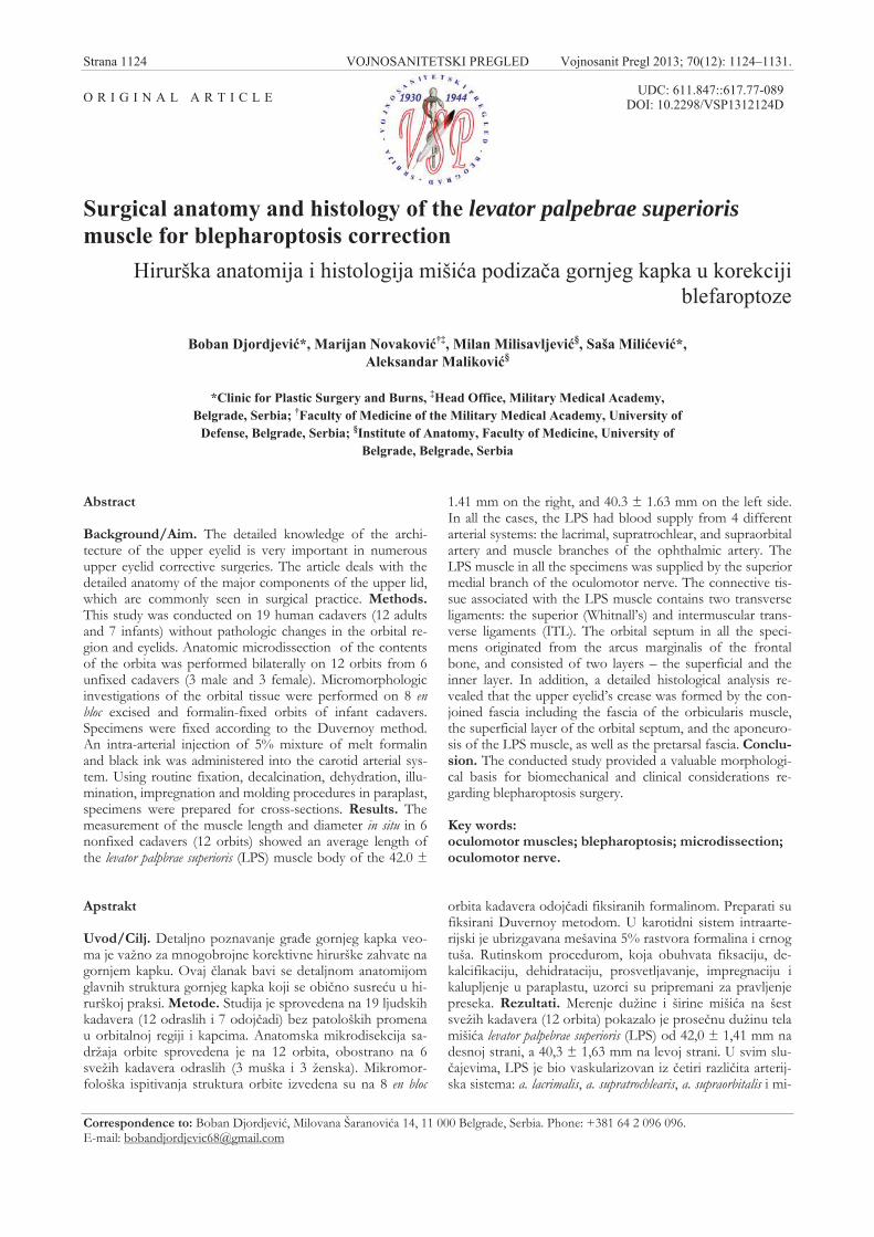
Strana 1124 VOJNOSANITETSKI PREGLED Vojnosanit Pregl 2013; 70(12): 1124–1131.
Correspondence to: Boban Djordjevi , Milovana Šaranovi a 14, 11 000 Belgrade, Serbia. Phone: +381 64 2 096 096.E-mail: [email protected]
O R I G I N A L A R T I C L E UDC: 611.847::617.77-089DOI: 10.2298/VSP1312124D
Surgical anatomy and histology of the levator palpebrae superiorismuscle for blepharoptosis correction
Hirurška anatomija i histologija miši a podiza a gornjeg kapka u korekcijiblefaroptoze
Boban Djordjevi *, Marijan Novakovi †‡, Milan Milisavljevi §, Saša Mili evi *,Aleksandar Malikovi §
*Clinic for Plastic Surgery and Burns, ‡Head Office, Military Medical Academy,Belgrade, Serbia; †Faculty of Medicine of the Military Medical Academy, University of
Defense, Belgrade, Serbia; §Institute of Anatomy, Faculty of Medicine, University ofBelgrade, Belgrade, Serbia
Abstract
Background/Aim. The detailed knowledge of the archi-tecture of the upper eyelid is very important in numerousupper eyelid corrective surgeries. The article deals with thedetailed anatomy of the major components of the upper lid,which are commonly seen in surgical practice. Methods.This study was conducted on 19 human cadavers (12 adultsand 7 infants) without pathologic changes in the orbital re-gion and eyelids. Anatomic microdissection of the contentsof the orbita was performed bilaterally on 12 orbits from 6unfixed cadavers (3 male and 3 female). Micromorphologicinvestigations of the orbital tissue were performed on 8 enbloc excised and formalin-fixed orbits of infant cadavers.Specimens were fixed according to the Duvernoy method.An intra-arterial injection of 5% mixture of melt formalinand black ink was administered into the carotid arterial sys-tem. Using routine fixation, decalcination, dehydration, illu-mination, impregnation and molding procedures in paraplast,specimens were prepared for cross-sections. Results. Themeasurement of the muscle length and diameter in situ in 6nonfixed cadavers (12 orbits) showed an average length ofthe levator palpbrae superioris (LPS) muscle body of the 42.0 ±
1.41 mm on the right, and 40.3 ± 1.63 mm on the left side.In all the cases, the LPS had blood supply from 4 differentarterial systems: the lacrimal, supratrochlear, and supraorbitalartery and muscle branches of the ophthalmic artery. TheLPS muscle in all the specimens was supplied by the superiormedial branch of the oculomotor nerve. The connective tis-sue associated with the LPS muscle contains two transverseligaments: the superior (Whitnall’s) and intermuscular trans-verse ligaments (ITL). The orbital septum in all the speci-mens originated from the arcus marginalis of the frontalbone, and consisted of two layers – the superficial and theinner layer. In addition, a detailed histological analysis re-vealed that the upper eyelid’s crease was formed by the con-joined fascia including the fascia of the orbicularis muscle,the superficial layer of the orbital septum, and the aponeuro-sis of the LPS muscle, as well as the pretarsal fascia. Conclu-sion. The conducted study provided a valuable morphologi-cal basis for biomechanical and clinical considerations re-garding blepharoptosis surgery.
Key words:oculomotor muscles; blepharoptosis; microdissection;oculomotor nerve.
Apstrakt
Uvod/Cilj. Detaljno poznavanje gra e gornjeg kapka veo-ma je važno za mnogobrojne korektivne hirurške zahvate nagornjem kapku. Ovaj lanak bavi se detaljnom anatomijomglavnih struktura gornjeg kapka koji se obi no susre u u hi-rurškoj praksi. Metode. Studija je sprovedena na 19 ljudskihkadavera (12 odraslih i 7 odoj adi) bez patoloških promenau orbitalnoj regiji i kapcima. Anatomska mikrodisekcija sa-držaja orbite sprovedena je na 12 orbita, obostrano na 6svežih kadavera odraslih (3 muška i 3 ženska). Mikromor-fološka ispitivanja struktura orbite izvedena su na 8 en bloc
orbita kadavera odoj adi fiksiranih formalinom. Preparati sufiksirani Duvernoy metodom. U karotidni sistem intraarte-rijski je ubrizgavana mešavina 5% rastvora formalina i crnogtuša. Rutinskom procedurom, koja obuhvata fiksaciju, de-kalcifikaciju, dehidrataciju, prosvetljavanje, impregnaciju ikalupljenje u paraplastu, uzorci su pripremani za pravljenjepreseka. Rezultati. Merenje dužine i širine miši a na šestsvežih kadavera (12 orbita) pokazalo je prose nu dužinu telamiši a levator palpebrae superioris (LPS) od 42,0 ± 1,41 mm nadesnoj strani, a 40,3 ± 1,63 mm na levoj strani. U svim slu-ajevima, LPS je bio vaskularizovan iz etiri razli ita arterij-
ska sistema: a. lacrimalis, a. supratrochlearis, a. supraorbitalis i mi-
Volumen 70, Broj 12 VOJNOSANITETSKI PREGLED Strana 1125
Djordjevi B, et al. Vojnosanit Pregl 2013; 70(12): 1124–1131.
ši nih grana a. ophthalmicae. Miši LPS u svim slu ajevimainervisala je gornja medijalna grana n. oculomotoriusa. Usta-novljeno je da je vezivno tkivo povezano sa LPS miši em,sastavljeno od dva popre na ligamenta: gornjeg (Whit-nallovog) i intermuskularnog popre nog ligamenta (ITL).Orbitalni septum je u svim slu ajevima polazio od supra-orbitalne ivice eone kosti i sastojao se od dva sloja – po-vršnog i dubokog. Osim toga, detaljna histološka analizapokazala je da gornji kapa ni žleb formira fascija miši a or-
bicularis oculi, površni sloj orbitalnog septuma, aponeurozamiši a levator palpebrae superioris i pretarzalna fascija. Zak-lju ak. Istraživanjem je obezbe ena važna morfološka os-nova za biomehani ka i klini ka ispitivanja u hirurgiji ble-faroptoza.
Klju ne re i:miši i, okulomotorni; blefaroptoza; mikrodisekcija;n. oculomotorius.
Introduction
A wider knowledge of the levator palpebrae superioris(LPS) muscle and the suspensory fibrous tissue related to theLPS muscle is essential for the eyelid surgery, especially forthe blepharoptosis correction. Even though, there are severalpapers providing the description of the eyelid anatomy, anat-omic features of the orbicularis oculi and the LPS muscle,there are various anatomical details that are still controver-sial. In particular, the importance of the orbital connectivetissue and the role they play in pathogenesis or the blepha-roptosis treatment are still unclear.
In most patients with congenital and acquired blepha-roptosis, palpebral creases are not so distinctive. In addition,Anderson et al. 1 described an atrophic and dehiscent supe-rior transverse ligament (Whitnall’s ligament-WL 2) in thosepatients. Fink et al. 3 first reported the presence of the con-nective tissue which underlines the LPS muscle, the tissuethat was called the intermuscular transverse ligament (ITL)by Lukas et al. 4. However, the functional role of these liga-ments in the LPS muscle is still unclear.
The aim of this study was to reinvestigate the detailedanatomy of the connective tissues related to the LPS muscle,and to establish their role in the suspension and creation ofnormal upper eyelid contours in the occidental race.
Methods
The anatomic microdissection study and histologicalanalysis were conducted on 19 human cadavers (12 adultsand 7 infants). We used microdissection techniques for theorbits from formalin-unfixed/fixed cadavers, as well as mi-cromorphologic investigation methods.
The anatomic microdissection of the contents of the or-bita through the anterior cranial fossa (transcranial approach)was performed bilaterally on 12 orbits from six unfixed ca-davers (3 male and 3 female). The cadavers, aged 42–75years (the mean age was 60 years) had no pathologic changesin the orbital region and eyelids. We used the standard tech-nique for the brain extraction, and the customary abductiontechnique to remove the orbital bony roof. The periorbitawas then incised, and turned laterally so that the levatormuscle of the upper eyelid could be seen. Anteriorly, wefound the transverse fibrous strands of Whitnall’s ligament,and, after cutting the LPS muscle proximally, an intramus-cular transverse ligament with connections to the bone of theorbital roof. The space under the aponeurosis of the LPS
muscle was dissected to expose the tarsal plate. Microdis-section of the orbital subject was performed using a stereomagnifying glass (4 ).
The method of standard dissection of 12 orbits from 6formalin fixed cadavers (3 male and 3 female) was per-formed by the combined transcutaneous and transcranial ap-proach. Incisions were made on the skin, the orbicularis oculimuscle was exposed, and a strip of orbicularis oculi musclewas turned aside. After the junction of the orbital septum andlevator aponeurosis was observed, a blunt dissection was car-ried out through the outer layer of the orbital septum, andcontinued to the Whitnall’s ligament between the levatoraponeurosis and the inner layer of the orbital septum, whichcovers the posterior surface of the orbital fat.
All the specimens were photographed by a Sony CyberShot DCS P8 digital camera.
Micromorphologic investigations of the orbital tissuewere performed on 8 en bloc excised and formalin-fixed orbitsof infant cadavers with no pathologic changes in the orbital re-gion and eyelids. An intra-arterial injection of 5% mixture ofmelt formalin and black ink was administered in the carotidarterial system, and rinsed out using a saline solution and 4%neutral buffered solution of formaldehyde. The specimenswere fixed according to the method of Duvernoy. We used thestandard technique for the brain removal, and the specific tran-scranial approach. Microdissections (using micro instruments)of injected orbital blood vessels, nerves and muscles wereanalyzed under the Leica MZ6 stereomicroscope.
All the specimens were photographed by Sony CyberShot DCS P8 digital camera. To get more easily visible de-tails, we used the Leica DC 300 digital camera (magnifica-tions 6.3 , 10 and 20 ).
Histological analysis was carried out on 6 en bloc ex-cised and formalin-fixed infant orbits. We used the materialfrom the Anatomic Institute of the Faculty of Medicine, Bel-grade. The aim was to establish the exact micromorphologicarchitecture of the upper eyelid. Orbital and frontal regionspecimens were perfunded with the saline solution through acannula placed in one of the carotid arteries, and the 4%neutral buffered formaldehyde solution was used afterwardsfor the same purpose. All the specimens were fixed in the 4%neutral buffered formaldehyde solution in volume 20 timeslarger than the tissue volume prepared for fixation. Usingroutine fixation, decalcination, dehydration, illumination,impregnation and molding procedures (Bio-Plas plus, Bio-Optica, Italy), paraplast specimens were prepared for sec-tions At the distance of 15 m, we cut off a series of tissue
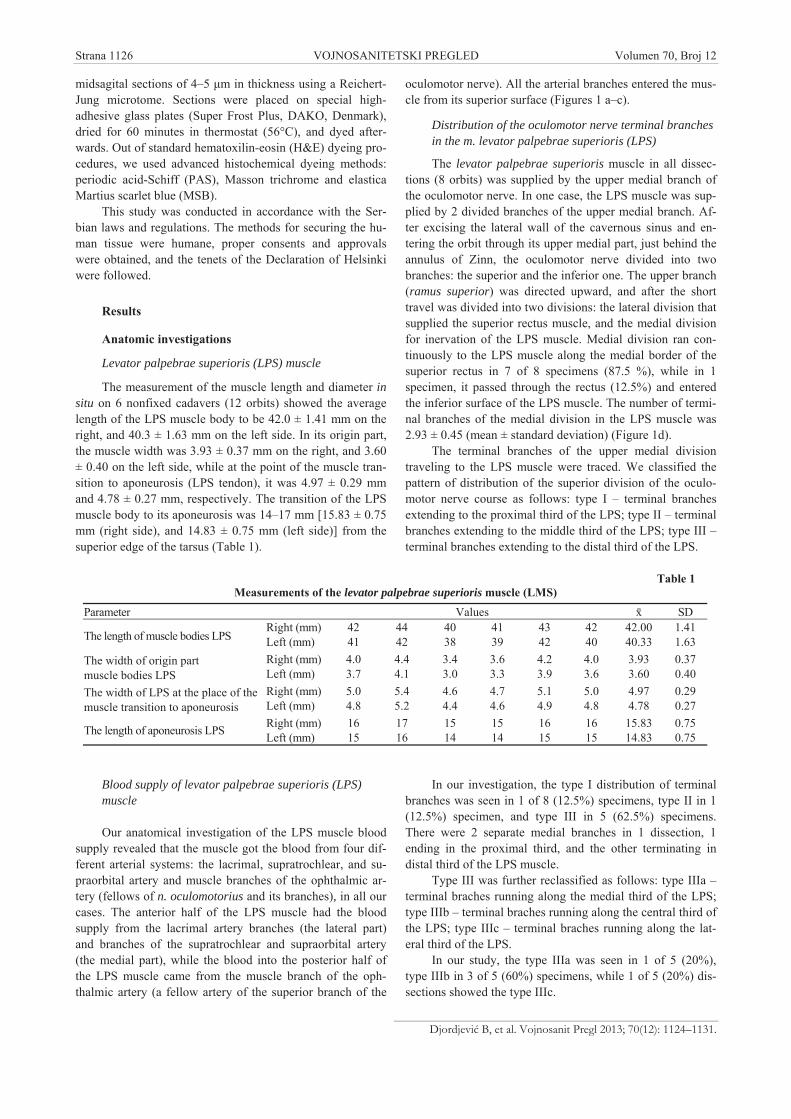
Strana 1126 VOJNOSANITETSKI PREGLED Volumen 70, Broj 12
Djordjevi B, et al. Vojnosanit Pregl 2013; 70(12): 1124–1131.
midsagital sections of 4–5 m in thickness using a Reichert-Jung microtome. Sections were placed on special high-adhesive glass plates (Super Frost Plus, DAKO, Denmark),dried for 60 minutes in thermostat (56°C), and dyed after-wards. Out of standard hematoxilin-eosin (H&E) dyeing pro-cedures, we used advanced histochemical dyeing methods:periodic acid-Schiff (PAS), Masson trichrome and elasticaMartius scarlet blue (MSB).
This study was conducted in accordance with the Ser-bian laws and regulations. The methods for securing the hu-man tissue were humane, proper consents and approvalswere obtained, and the tenets of the Declaration of Helsinkiwere followed.
Results
Anatomic investigations
Levator palpebrae superioris (LPS) muscle
The measurement of the muscle length and diameter insitu on 6 nonfixed cadavers (12 orbits) showed the averagelength of the LPS muscle body to be 42.0 ± 1.41 mm on theright, and 40.3 ± 1.63 mm on the left side. In its origin part,the muscle width was 3.93 ± 0.37 mm on the right, and 3.60± 0.40 on the left side, while at the point of the muscle tran-sition to aponeurosis (LPS tendon), it was 4.97 ± 0.29 mmand 4.78 ± 0.27 mm, respectively. The transition of the LPSmuscle body to its aponeurosis was 14–17 mm [15.83 ± 0.75mm (right side), and 14.83 ± 0.75 mm (left side)] from thesuperior edge of the tarsus (Table 1).
Blood supply of levator palpebrae superioris (LPS)muscle
Our anatomical investigation of the LPS muscle bloodsupply revealed that the muscle got the blood from four dif-ferent arterial systems: the lacrimal, supratrochlear, and su-praorbital artery and muscle branches of the ophthalmic ar-tery (fellows of n. oculomotorius and its branches), in all ourcases. The anterior half of the LPS muscle had the bloodsupply from the lacrimal artery branches (the lateral part)and branches of the supratrochlear and supraorbital artery(the medial part), while the blood into the posterior half ofthe LPS muscle came from the muscle branch of the oph-thalmic artery (a fellow artery of the superior branch of the
oculomotor nerve). All the arterial branches entered the mus-cle from its superior surface (Figures 1 a–c).
Distribution of the oculomotor nerve terminal branchesin the m. levator palpebrae superioris (LPS)
The levator palpebrae superioris muscle in all dissec-tions (8 orbits) was supplied by the upper medial branch ofthe oculomotor nerve. In one case, the LPS muscle was sup-plied by 2 divided branches of the upper medial branch. Af-ter excising the lateral wall of the cavernous sinus and en-tering the orbit through its upper medial part, just behind theannulus of Zinn, the oculomotor nerve divided into twobranches: the superior and the inferior one. The upper branch(ramus superior) was directed upward, and after the shorttravel was divided into two divisions: the lateral division thatsupplied the superior rectus muscle, and the medial divisionfor inervation of the LPS muscle. Medial division ran con-tinuously to the LPS muscle along the medial border of thesuperior rectus in 7 of 8 specimens (87.5 %), while in 1specimen, it passed through the rectus (12.5%) and enteredthe inferior surface of the LPS muscle. The number of termi-nal branches of the medial division in the LPS muscle was2.93 ± 0.45 (mean ± standard deviation) (Figure 1d).
The terminal branches of the upper medial divisiontraveling to the LPS muscle were traced. We classified thepattern of distribution of the superior division of the oculo-motor nerve course as follows: type I – terminal branchesextending to the proximal third of the LPS; type II – terminalbranches extending to the middle third of the LPS; type III –terminal branches extending to the distal third of the LPS.
In our investigation, the type I distribution of terminalbranches was seen in 1 of 8 (12.5%) specimens, type II in 1(12.5%) specimen, and type III in 5 (62.5%) specimens.There were 2 separate medial branches in 1 dissection, 1ending in the proximal third, and the other terminating indistal third of the LPS muscle.
Type III was further reclassified as follows: type IIIa –terminal braches running along the medial third of the LPS;type IIIb – terminal braches running along the central third ofthe LPS; type IIIc – terminal braches running along the lat-eral third of the LPS.
In our study, the type IIIa was seen in 1 of 5 (20%),type IIIb in 3 of 5 (60%) specimens, while 1 of 5 (20%) dis-sections showed the type IIIc.
Table 1Measurements of the levator palpebrae superioris muscle (LMS)
Parameter Values SDRight (mm) 42 44 40 41 43 42 42.00 1.41
The length of muscle bodies LPS Left (mm) 41 42 38 39 42 40 40.33 1.63Right (mm) 4.0 4.4 3.4 3.6 4.2 4.0 3.93 0.37The width of origin part
muscle bodies LPS Left (mm) 3.7 4.1 3.0 3.3 3.9 3.6 3.60 0.40Right (mm) 5.0 5.4 4.6 4.7 5.1 5.0 4.97 0.29The width of LPS at the place of the
muscle transition to aponeurosis Left (mm) 4.8 5.2 4.4 4.6 4.9 4.8 4.78 0.27Right (mm) 16 17 15 15 16 16 15.83 0.75The length of aponeurosis LPS Left (mm) 15 16 14 14 15 15 14.83 0.75
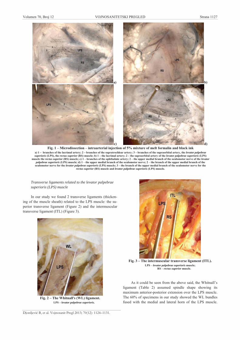
Volumen 70, Broj 12 VOJNOSANITETSKI PREGLED Strana 1127
Djordjevi B, et al. Vojnosanit Pregl 2013; 70(12): 1124–1131.
Transverse ligaments related to the levator palpebraesuperioris (LPS) muscle
In our study we found 2 transverse ligaments (thicken-ing of the muscle sheath) related to the LPS muscle: the su-perior transverse ligament (Figure 2) and the intermusculartransverse ligament (ITL) (Figure 3).
Fig. 2 – The Whitnall's (WL) ligament.LPS – levator palpebrae superioris.
Fig. 3 – The intermuscular transverse ligament (ITL).LPS – levator palpebrae superioris muscle;
RS – rectus superior muscle.
As it could be seen from the above said, the Whitnall’sligament (Table 2) assumed spindle shape showing itsmaximum anterior-posterior extension over the LPS muscle.The 60% of specimens in our study showed the WL bundlesfused with the medial and lateral horn of the LPS muscle.
a) b)
c) d)Fig. 1 – Microdissection – intraarterial injection of 5% mixture of melt formalin and black ink
a) 1 – branches of the lacrimal artery; 2 – branches of the supratrochlear artery; 3 – branches of the supraorbital artery, the levator palpebraesuperioris (LPS), the rectus superior (RS) muscle; b) 1 – the lacrimal artery; 2 – the supraorbital artery of the levator palpebrae superioris (LPS)
muscle the rectus superior (RS) muscle; c) 1 – branches of the ophthalmic artery; 2 – the upper medial branch of the oculomotor nerve of the levatorpalpebrae superioris (LPS) muscle; d) 1 – the upper medial branch of the oculomotor nerve; 2 – the branch of the upper medial branch of the
oculomotor nerve for the levator palpebrae superioris (LPS) muscle; 3 – the branch of the upper medial branch of the oculomotor nerve for therectus superior (RS) muscle and levator palpebrae superioris (LPS) muscle.
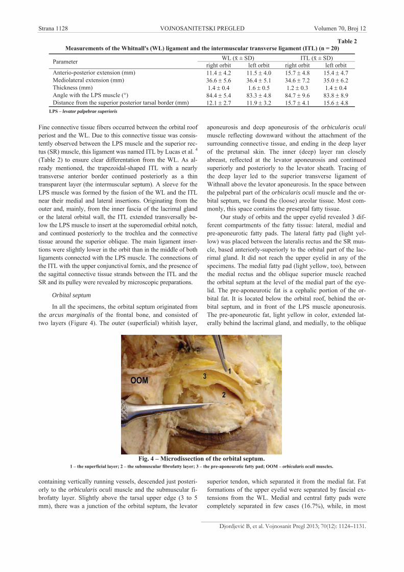
Strana 1128 VOJNOSANITETSKI PREGLED Volumen 70, Broj 12
Djordjevi B, et al. Vojnosanit Pregl 2013; 70(12): 1124–1131.
Fine connective tissue fibers occurred between the orbital roofperiost and the WL. Due to this connective tissue was consis-tently observed between the LPS muscle and the superior rec-tus (SR) muscle, this ligament was named ITL by Lucas et al. 4
(Table 2) to ensure clear differentation from the WL. As al-ready mentioned, the trapezoidal-shaped ITL with a nearlytransverse anterior border continued posteriorly as a thintransparent layer (the intermuscular septum). A sleeve for theLPS muscle was formed by the fusion of the WL and the ITLnear their medial and lateral insertions. Originating from theouter and, mainly, from the inner fascia of the lacrimal glandor the lateral orbital wall, the ITL extended transversally be-low the LPS muscle to insert at the superomedial orbital notch,and continued posteriorly to the trochlea and the connectivetissue around the superior oblique. The main ligament inser-tions were slightly lower in the orbit than in the middle of bothligaments connected with the LPS muscle. The connections ofthe ITL with the upper conjunctival fornix, and the presence ofthe sagittal connective tissue strands between the ITL and theSR and its pulley were revealed by microscopic preparations.
Orbital septum
In all the specimens, the orbital septum originated fromthe arcus marginalis of the frontal bone, and consisted oftwo layers (Figure 4). The outer (superficial) whitish layer,
containing vertically running vessels, descended just posteri-orly to the orbicularis oculi muscle and the submuscular fi-brofatty layer. Slightly above the tarsal upper edge (3 to 5mm), there was a junction of the orbital septum, the levator
aponeurosis and deep aponeurosis of the orbicularis oculimuscle reflecting downward without the attachment of thesurrounding connective tissue, and ending in the deep layerof the pretarsal skin. The inner (deep) layer ran closelyabreast, reflected at the levator aponeurosis and continuedsuperiorly and posteriorly to the levator sheath. Tracing ofthe deep layer led to the superior transverse ligament ofWithnall above the levator aponeurosis. In the space betweenthe palpebral part of the orbicularis oculi muscle and the or-bital septum, we found the (loose) areolar tissue. Most com-monly, this space contains the preseptal fatty tissue.
Our study of orbits and the upper eyelid revealed 3 dif-ferent compartments of the fatty tissue: lateral, medial andpre-aponeurotic fatty pads. The lateral fatty pad (light yel-low) was placed between the lateralis rectus and the SR mus-cle, based anteriorly-superiorly to the orbital part of the lac-rimal gland. It did not reach the upper eyelid in any of thespecimens. The medial fatty pad (light yellow, too), betweenthe medial rectus and the oblique superior muscle reachedthe orbital septum at the level of the medial part of the eye-lid. The pre-aponeurotic fat is a cephalic portion of the or-bital fat. It is located below the orbital roof, behind the or-bital septum, and in front of the LPS muscle aponeurosis.The pre-aponeurotic fat, light yellow in color, extended lat-erally behind the lacrimal gland, and medially, to the oblique
superior tendon, which separated it from the medial fat. Fatformations of the upper eyelid were separated by fascial ex-tensions from the WL. Medial and central fatty pads werecompletely separated in few cases (16.7%), while, in most
Fig. 4 – Microdissection of the orbital septum.1 – the superficial layer; 2 – the submuscular fibrofatty layer; 3 – the pre-aponeurotic fatty pad; OOM – orbicularis oculi muscles.
Table 2Measurements of the Whitnall's (WL) ligament and the intermuscular transverse ligament (ITL) (n = 20)
WL ( ± SD) ITL ( ± SD)Parameter right orbit left orbit right orbit left orbitAnterio-posterior extension (mm) 11.4 4.2 11.5 4.0 15.7 4.8 15.4 4.7Mediolateral extension (mm) 36.6 5.6 36.4 5.1 34.6 7.2 35.0 6.2Thickness (mm) 1.4 0.4 1.6 0.5 1.2 0.3 1.4 0.4Angle with the LPS muscle (°) 84.4 5.4 83.3 4.8 84.7 9.6 83.8 8.9Distance from the superior posterior tarsal border (mm) 12.1 2.7 11.9 3.2 15.7 4.1 15.6 4.8
LPS – levator palpebrae superioris
Volumen 70, Broj 12 VOJNOSANITETSKI PREGLED Strana 1129
Djordjevi B, et al. Vojnosanit Pregl 2013; 70(12): 1124–1131.
cases, they were connected partially. The blood supply andthe innervation of fatty pads came from terminal branches ofthe supraorbital artery and nerve.
Histological analysis
In our upper eyelid investigation, we identified a forma-tion that creates the palpebral crease called the conjoined fasciaby Siegel. In all the cases, it was placed in front of the tarsus atthe level of the upper palpebral crease, and consisted of or-bicularis oculi fascia, the superficial layer of the orbital septum,the LPS muscle aponeurosis and the pretarsal fascia. At thelevel of the upper palpebral crease, the LPS muscle aponeurosisshowed a junction to the orbital septum and the orbicularis oc-uli fascia. These fibers proceeded downward, and met the pre-tarsal fascia. The conjoined fascia was firmly attached to thetarsus and the pretarsalis part of the orbicularis oculi muscle.Some fibers of the LPS muscle aponeurosis continued forwardand was inserted in the deep surface of the skin. Behind the or-bicularis oculi muscle, as well as in the front of the orbitalseptum, we found the lose fibrofat layer, mostly avascular, andcalled it the suborbicular fascial plan (Figure 5a).
The dense, thick collagen fibers of both layers of theorbital septum enclosed the pre-aponeurotic fat in the mid-sagital line. Collagen fibers of the deep layer proceeded tothe WL and sheath of the LPS muscle. At the level of the up-per palpebral crease, the deep layer of the orbital septum wasintimate with and directly adjacent to the LPS muscle apo-neurosis. The deep layer of the orbital septum near the su-praorbital edge terminated in the periorbital periostium,without a connection to the superficial layer (Figure 5b).
In addition, we found that the superficial layer of the or-bital septum ran cephalad as a galeal layer, without a direct orindirect connection to the frontal muscle. We identified fibrousconnections between the orbital septum and the suborbicularfascial layer near the eyebrow. These connections indirectlylimited the frontal muscle in the eyebrow elevation (Figure 5c).
Discussion
Our analysis of anatomical and topographic features ofthe LPS muscle has not shown any statistically significantdifferences compared to previous studies 5–8.
In all the cases, the anterior half of the LPS muscle wassupplied by the branches of the lacrimal, the supratrochlear,and the supraorbital arteries, while the posterior half wassupplied by the muscular branch of the ophthalmic artery(fellow artery of the superior division of the oculomotornerve). George 9 found that the posterior half of the LPSmuscle had the blood supply by the posterior ethmoid arterythat we did not find in any case.
We consider that injuries to the blood vessels supplyingthe anterior part of the muscle might be indirectly involvedin muscle activity, that is the inadequate hemostasis of thesevessels, especially in surgical procedures involving the LPSmuscle, might result in hematoma, and interfere with postop-erative results.
The LPS muscle in all the specimens was supplied bythe superior medial branch of the oculomotor nerve. The up-per medial branch of the oculomotor nerve extended to themiddle third of the LPS muscle (Type III distribution ac-cording to the study of Hwang et al. 10).
These nerve endings were exposed and vulnerable, sotheir injuries could cause permanent postoperative blepha-roptosis. They were also sensitive to local anesthetics, andcould be disturbed during the eyelid surgery under local an-esthesia. Due to that, exceptional care should be taken duringthe infiltration of anesthetic near the LPS muscle in order toavoid its transient paralysis postoperatively.
The microarchitecture of the orbital connective tissuesystem has a significant role in extraorbicular muscles func-tioning 11–15. The connective tissue associated to the LPSmuscle contains two transverse ligaments: the superior trans-verse (Whitnall’s) ligament and the intermuscular transverseligament. The Whitnall’s ligament has been described as acondensed sheet of fascia underlying the LPS on the superiorsurface of the muscle, localized in the transitional zone be-tween the muscle body and the aponeurosis. In spite of sev-eral descriptions, the function of the WL is still controver-
sial. Whitnall has considered its function as a check liga-ment. Lemke et al. 16 have mentioned that the WL shows notension with the open eyelid, while Dutton 17 believes thatthe WL plays no role in the suspensory activity of the LPS.Anderson and Dixon 18 think that the WL has the role in
a) b) c)Fig. 5 – Phistological analysis of the orbital septum.
a) Van Gieson’s methods ( 15); b) Masson trichrome methods ( 15); c) Martius scarlet blue (MSB) methods ( 15).
Strana 1130 VOJNOSANITETSKI PREGLED Volumen 70, Broj 12
Djordjevi B, et al. Vojnosanit Pregl 2013; 70(12): 1124–1131.
changing the anterior-posterior traction power of the LPSinto vertical one, relieving the eyebrow elevation. In addi-tion, they have found the WL dehiscence and atrophy insome patients with congenital blepharoptosis.
Our study revealed that the superior transverse ligament(WL) and the intermuscular transverse ligament surroundingthe LPS muscle in all cases were located at the junction ofthe muscle body and its aponeurosis. Both transverse liga-ments had their origin from the medial and lateral orbitalwall. The Withnall’s ligament had firm attachments to theLPS on the outer parts of the muscle. The main and dominantorigin of the WL was the lateral one, under the LPS muscle,thus, it could be of a great importance for the suspensory ac-tivity of the LPS. Under the upper surface of the WL, therewere brands of the loose connective tissue reaching perios-tium of the orbital roof. The ITL is the condensed fascia ofthe LPS muscle, between the LPS and the rectus superiormuscle, connected by brands to the superior conjunctivalfornix. This ligament showed a greater anterior-posteriorextension than the WL. The main insertion was placed underthe middle of both transverse ligaments. We assume that theLPS muscle runs freely over the ITL thanks to condensing ofthe connective tissue in the anterior parts of the ligament.
According to the conclusions of a separate examination,we think that these two ligaments could be inadequate. Ana-tomically, both ligaments create an annular formation con-taining the fibroelastic and fibromuscular tissue surroundingthe LPS muscle near its musculoaponeurotic junction. Weconsider that, despite the fact that the dominant origin ofboth ligaments is lower, it could be the morphological sub-stratum of suspension action of the LPS muscle during thepassive lowering of the eyelid with the upward view.
In the Gray’s anatomy, the upper eyelid elevation is tobe checked by the orbital septum 19. In 1910 Whitnall de-scribed how the superficial part of the levator sheath forms aconspicuous band above the LPS muscle in a vertical sectionthrough the orbit, just behind the aponeurosis. Moreover, he
recognized a delicate layer of the connective tissue runningover the aponeurosis and posteriorly to the orbital septum.Fink 3 reported that the fascial sheath of the LPS musclecontinued anteriorly from the WL as a continuous membranethat could be traced up to the supraorbital rim, where itblends with the orbital septum.
Conclusion
Our study showed that the superficial layer of the or-bital septum, 3 to 5 mm above the superior tarsal edge, hadits origin from the LPS fascia, and continued upward as adeep galeal layer in the frontal region, without connections tothe frontal muscle, and exhibited no blending with the super-ficial layer of the orbital septum. Behind and above the ori-gin of the superficial layer of the orbital septum, the aponeu-rosis of the LPS muscle deep layer of the orbital septum ex-hibited, the junction to aponeurosis by the connection to anundefined integrity. We consider that the junction of the or-bital septum, fascia of the LPS muscle, and the deep fascia ofthe orbicularis oculi muscle plays a supporting role for theperiorbital fat, and is placed more medially than laterally inthe upper eyelid 16, 20–22.
In addition, this detailed histological analysis revealedthat the upper eyelid’s crease was formed by the conjoinedfascia containing the fascia of the orbicularis muscle, the su-perficial layer of the orbital septum, and the aponeurosis ofthe LPS muscle, as well as the pretarsal fascia. These find-ings suggest that in creating the upper eyelid’s crease, theskin suture should be placed through the aponeurosis and thesuperficial layer of the orbital septum, as to protect the integ-rity of the crease (levator-dermal fixation).
Acknowledgments
This work was supported by the grant No. 175030 fromthe Ministry of Education, Science and Technological De-volopment of the Republic of Serbia.
R E F E R E N C E S
1. Anderson RL, Jordan DR, Dutton JJ. Whitnall's sling forpoor function ptosis. Arch Ophthalmol 1990; 108(11):1628 32.
2. Whitnall SE. On a ligament acting as a check to the action ofthee levator palpebrae superioris muscle. J Anat Physiol 1910;45: 131 9.
3. Fink WH. An anatomic study of the check mechanism of thevertical muscles of the eyes. Am J Ophthalmol 1957; 44(6):800 9.
4. Lukas JR, Priglinger S, Denk M, Mayr R. Two fibromusculartransverse ligaments related to the levator palpebrae superioris:Whitnall's ligament and an intermuscular transverse ligament.Anat Rec 1996; 246(3): 415 22.
5. Plock J, Contaldo C, von Ludinghausen M. Levator palpebrae supe-rioris muscle in human fetuses: anatomical findings and theirclinical relevance. Clin Anat 2005; 18(7): 473 80.
6. Kakizaki H, Zako M, Nakano T, Asamoto K, Miyaishi O, IwakiM. The Levator Aponeurosis Consists of Two Layers That In-clude Smooth Muscle. Ophthal Plast Reconstr Surg 2005;21(5): 379 82.
7. Ettl A, Daxer A, Priglinger S, Kramer J, Koornneef L. Dynamicmagnetic resonance imaging of the levator palpebrae superiorismuscle. Ophthalmic Res 1998; 30(1): 54 8.
8. Tian J, Yu C, Sun T. Transcranial approach to the orbit: a mi-croanatomical study. Zhonghua Yi Xue Za Zhi 1999; 79(6):435 8. (Chinese)
9. George JL. Arterial vascularization of the levator muscle of theupper eyelid. J Fr Ophtalmol 1984; 7(2): 143 9. (French)
10. Hwang K, Lee DK, Chung IH, Lee SI. Patterns of oculomotornerve distribution to the levator palpebrae superioris muscle,and correlation to temporary ptosis after blepharoplasty. AnnPlast Surg 2001; 47(4): 381 4.
11. Ettl A, Priglinger S, Kramer J, Koornneef L. Functional anatomy ofthe levator palpebrae superioris muscle and its connective tis-sue system. Br J Ophthalmol 1996; 80(8): 702 7.
12. Moreno JJ, González CH. La ptosis palpebral: fundamentos ytécnica quirúrgica. Madrid: Allergan; 1995.
13. Ettl A, Zonneveld F, Daxer A, Koornneef L. Is Whitnall's ligamentresponsible for the curved course of the levator palpebrae su-perioris muscle. Ophthalmic Res 1998; 30(5): 321 6.

Volumen 70, Broj 12 VOJNOSANITETSKI PREGLED Strana 1131
Djordjevi B, et al. Vojnosanit Pregl 2013; 70(12): 1124–1131.
14. Goldberg RA, Wu JC, Jesmanowicz A, Hyde JS. Eyelid anatomyrevisited: dynamic high-resolution magnetic resonance imagesof Whitnall's ligament and upper eyelid structures with the useof surface coil. Arch Ophthalmol 1992; 110(11): 1598 600.
15. Shin HH, Kim JH, Kwon IT. Anatomic charcteristics of uppereyelid structures important in ptosis surgery. Korean J Oph-thalmol 1993; 34: 599 605.
16. Lemke BN, Stasior OG, Rosenberg PN. The surgical relations ofthe levator palpebrae superioris muscle. Ophthal Plast Re-constr Surg 1988; 4(1): 25 30.
17. Dutton JJ. Clinical and Surgical Orbital Anatomy. 2nd ed.Philadelphia: Saunders; 1994. p. 96 7.
18. Anderson RL, Dixon RS. Aponeurotic ptosis surgery. ArchOphthalmol 1979; 97(6): 1123 8.
19. Standring S. Gray's Anatomy. The Anatomical Basis of ClinicalPractice. 40th ed. Philadelphia: Churchill Livingstone Elsevier;2008.
20. Hwang K, Kim DJ, Chung RS, Lee SI, Hiraga Y. An anatomicalstudy of the junction of the orbital septum and the levatoraponeurosis in Orientals. Br J Plast Surg 1998; 51(8): 594 8.
21. Hoffmann KT, Hosten N, Lemke AJ, Sander B, Zwicker C, Felix R.Septum orbitale: high-resolution MR in orbital anatomy. AJNRAm J Neuroradiol 1998; 19(1): 91 4.
22. Meyer DR, Linberg JV, Wobig JL, McCormick SA. Anatomy ofthe orbital septum and associated eyelid connective tissues:implications for ptosis suregery. Ophthal Plast Reconstr Surg1991; 7(2):104 13.
Received on April 28, 2012.Revised on July 3, 2012.
Accepted on July 4, 2012.
Strana 1132 VOJNOSANITETSKI PREGLED Vojnosanit Pregl 2013; 70(12): 1132–1137.
Correspondence to: Vojislava Neškovi , Clinic for Anesthesiology and Intensive Care, Military Medical Academy, Crnotravska 17,11 000 Belgrade, Serbia, Phone: +381 11 1775 320. E-mail: [email protected]
O R I G I N A L A R T I C L E UDC: 617-089.5::616.1-089DOI: 10.2298/VSP1312132N
High thoracic epidural anesthesia in patients with synchronouscarotid endarterectomy and off-pump coronary arteryrevascularization
Visoka torakalna epiduralna anestezija kod bolesnika sa istovremenomkarotidnom endarterektomijom i off-pump revaskularizacijom miokarda
Vojislava Neškovi *, Predrag Milojevi †, Dragana Uni -Stojanovi †, Ivan Ili ‡,Zoran Slavkovi *§
*Clinic for Anesthesiology and Intensive Care, Military Medical Academy, Belgrade,Serbia; †Dedinje Cardiovascular Institute, Belgrade, Serbia; ‡Clinical Hospital Center
Zemun, Belgrade, Serbia; §Faculty of Medicine of the Military Medical Academy,University of Defence, Belgrade, Serbia
Abstract
Background/Aim. In order to reduce the risk of cerebro-vascular insults (CVI), the latest recommendations suggestthat carotid endarterectomy (CEA) is strongly indicated inpatients scheduled for coronary surgery when significant ca-rotid artery stenosis is symptomatic and/or bilateral. The bestresults are obtained in small studies with CEA performedimmediately prior to off-pump coronary bypass (OPCAB).We present 16 consecutive patients who underwent synchro-nous CEA and OPCAB under general anesthesia combinedwith high thoracic epidural anesthesia (TEA) in order toevaluate the safety and potential benefits of such anestheticmanagement. Methods. A total of 16 consecutive patientsscheduled for simultaneous CEA and OPCAB with no con-traindication for TEA were enrolled in the study. All the pa-tients were anesthetized with TEA combined with generalanesthesia. Early extubation was planed in all the patients forearly assessment of neurological outcome. Demographics,comorbidity, quality of postoperative recovery, duration ofmechanical ventilation, successful early extubation, outcome,length of Intensive Care Unit (ICU) and hospital stay were
recorded. Results. Only two patients did not fulfill the crite-ria for early extubation. The average duration of mechanicalventilation for patients who fulfilled criteria for early extuba-tion was 87.9 ± 85.0 (0–255) min. Five (31.25%) patientswere extubated in the operating theater at the end of surgery.There were no deaths, nor neurological complications ofTEA. Seven (43.7%) patients had at least one of the postop-erative complications considered significant. None of themhad CVI. None of the early extubated patients was reintu-bated or had postoperative respiratory failure. Conclusion.Our study revealed that a combination of general anesthesiawith TEA appears to be good choice in synchronous CEAand OPCAB due to advantages of early extubation and earlyneurological assessment. Larger studies are necessary to de-termine real benefits on both short and long-term outcomesof such anesthetic management in synchronous CEA andOPCAB.
Key words:endarterectomy, carotid; coronary artery bypass;comorbidity; anesthesia, epidural; postoperativecomplications.
Apstrakt
Uvod/Cilj. Najnovije preporuke ukazuju da je kod boles-nika planiranih za hiruršku revaskularizaciju miokarda kojiimaju zna ajnu bilateralnu i/ili simptomatsku stenozu ka-rotidnih arterija potrebno uraditi karotidnu endarterekto-miju (CEA) pre revaskularizacije miokarda. Ovim bi semoglo posti i sniženje rizika od nastanka perioperativnogcerebrovaskularnog inzulta (CVI). Najbolji rezultati su po-stignuti u studijama sa malim brojem bolesnika, kod kojihje ura ena sinhrona operacija CEA i revaskularizacija mio-karda bez ekstrakorporalne, cirkulacije, off-pump (OPCAB).
Ovde je prikazana grupa od 16 uzastopnih bolesnika kodkojih je ura ena sinhrona operacija CEA i OPCAB koji subili anestezirani visokom torakalnom epiduralnom aneste-zijom (TEA), kombinovanom sa opštom anestezijom, saciljem da se ocene efekti ovakvog anesteziološkog pristu-pa. Metode. Ukupno 16 uzastopnih bolesnika kod kojih jebila indikovana sinhrona operacija CEA i OPCAB, bezkontraindikacije za TEA, bili su uklju eni u studiju. Svibolesnici anestezirani su kombinovanom TEA i opštomanestezijom. Rana ekstubacija planirana je kod svih boles-nika sa ciljem da se rano proceni postoperativna neurološ-ka funkcija. Kod ispitivanih bolesnika pra eni su slede i
Volumen 70, Broj 12 VOJNOSANITETSKI PREGLED Strana 1133
Neškovi V, et al. Vojnosanit Pregl 2013; 70(12): 1132–1137.
parametri: demografske karakteristike, komorbiditet, kva-litet postoperativnog oporavka, dužina mehani ke ventila-cije, uspešnost rane ekstubacije, ishod, kao i dužina borav-ka u jedinici intenzivnog le enja i bolnici. Rezultati. Samodva bolesnika nisu ispunila kriterijume za ranu ekstubaciju.Prose no trajanje mehani ke ventilacije rano ekstubiranihbolesnika bilo je 87,9 ± 85,0 min (0–255 min). Pet(31,25%) bolesnika ekstubirano je na kraju operacije uoperacionoj sali. Nije bilo smrtnih ishoda. Nije bilo neu-roloških komplikacija vezanih za TEA. Sedam (43,7%)bolesnika imalo je bar jednu od postoperativnih komplika-cija koje su pra ene. Nijedan bolesnik nije imao CVI. Ni-jedan rano ekstubiran bolesnik nije bio reintubiran, niti
imao respiratornu insuficijenciju. Zaklju ak. Rezultati na-še studije ukazuju da je kombinacija TEA i opšte anestezijedobar izbor anestezije za bolesnike sa sinhronom operaci-jom CEA i OPCAB. Osnovne prednosti su mogu nost ra-ne ekstubacije i rane procene neurološke funkcije. Potreb-ne su studije sa ve im brojem bolesnika da bi se pokazalesve prednosti i uticaj na kratkoro ne i dugoro ne rezultatesinhrone operacije CEA i OPCAB.
Klju ne re i:endarterektomija; aa. carotis; aa. coronariae,premoš avanje; komorbiditet; anestezija, epiduralna;postoperativne komplikacije.
Introduction
Cerebrovascular insult (CVI) is among grim compli-cations following cardiac surgery with the overall incidenceof 2% 1. Significant stenosis of the carotid artery has beenrecognized as the most powerful predictor of perioperativestroke after cardiac surgery 2, 3. There is a 14% risk of peri-operative stroke in patients with severe carotid artery dis-ease undergoing coronary artery bypass grafting (CABG),and the stroke rate remains 4% per year for the first 4 yearsafter coronary revascularization 4, 5. Significant carotid ar-tery stenosis is present in near 20% of patients scheduledfor CABG 6. The incidence can be even higher in thosehaving left main coronary artery disease 7. Despite beingsignificant risk factor for developing perioperative CVI, ca-rotid artery disease is estimated to be direct cause in only40% of the cases 1. Also, CEA performed in nonrevascu-larized patients with severe coronary artery disease hasbeen shown to be associated with mortality rate as high as20%; myocardial infarction has been found to be responsi-ble for 50% to 75% of all late deaths in this patient popula-tion 3.
Many studies have identified risk factors for strokeduring coronary atery operation, including hypertension, age,diabetes, carotid bruit, previous transient ischemic attack,and prior stroke 8, 9. In addition, prolonged cardiophylmonarybypass (CPB) time also has been reported to be associatedwith a higher risk of stroke 8, 10. In addition to CPB time,also, surgery technique brings some additional risk factors,and most of them are related to the use of extracorporeal cir-culation. Of note, CVI may be a consequence of the emboli-zation due to cardiac or aortic manipulation during the pro-cedure 11.
Current recommendations for carotid revascularizationin patients undergoing CABG surgery are based on expertopinion, since there is a lack of strong evidence to guide de-cision-making 12, 13. The latest recommendations suggest thatCEA should be strongly considered in patients scheduled forCABG surgery with concomitant significant symptomaticand/or bilateral carotid artery stenosis 14–16.
Up to now, encouraging results have been shown insmall series of patients, where CEA had been performedimmediately prior to off-pump coronary bypass (OPCAB),
when compared to synchronous CEA with standard CABGsurgery 17. This may be related to the fact that OPCAB bydefinition avoids CPB, as one of the most important contrib-uting factors for stroke 3. However, available data are scarceon which surgical or anesthetic technique is superior for thisspecific patient population at high risk and individual ap-proach has been strongly suggested.
We presented a series of 16 consecutive patients withsimultaneous CEA and OPCAB, operated on under generalanesthesia combined with high thoracic epidural anesthesia(TEA). We sought to determine the safety and potentialbenefits of such anesthetic management.
Methods
From February 2002 until October 2005, after localEthical Committy approval, a total of 16 consecutive patientsscheduled for simultaneous carotid endarterectomy and off-pump coronary bypass surgery (OPCAB) were included inthe observational study at the Dedinje Cardiovascular Insti-tute.
All the patients indicated for simultaneous carotid end-arterectomy and CABG, with no contraindication for TEAwere considered eligible for the study.
All the patients had significant carotid stenosis of morethan, or equal to 70% on preoperative carotid duplex scan-ning. They were either symptomatic, had bilateral significantcarotid stenosis, or were classified as having an unstable ath-erosclerotic plaque.
All the patients were anesthetized with high TEA com-bined with general anesthesia. Early extubation was planedin all the patients for early assessment of neurological out-come.
Exclusion criteria for the study were: acute infections,myocardial infarction within one month before surgery, co-agulation disorders, and emergency surgery.
Preoperative assessment and medication were standardfor all the patients. Induction of anesthesia was done intrave-nously, with midazolam (up to 5 mg), bolus doses of propo-fol, fentanyl and pancuronium. For the maintenance of ade-quate depth of general anesthesia sevoflurane was used, to-gether with intravenous bolus doses of fentanyl and pancuro-nium.
Strana 1134 VOJNOSANITETSKI PREGLED Volumen 70, Broj 12
Neškovi V, et al. Vojnosanit Pregl 2013; 70(12): 1132–1137.
Epidural catheter was placed at Th2-Th3 or Th3-Th4level, 30 min before surgery, or at least 2 h before the firstdose of heparin was used. After the test dose, bolus of bu-pivacaine with fentanyl in different concentrations (0.125%,0.25%, or 0.5%) was used followed by continuous infusionof the same local anesthetic with fentanyl, usually at the0.125% or 0.25% concentration. The rate of continuous infu-sion was adjusted accordingly; usually, it was 5–10 mL perhour during the operation.
Postoperative analgesia was mainly based on continu-ous epidural infusion of local anesthetic (0.125% bupiva-caine with fentanyl). According to the study protocol, it wasplanned to take out epidural catheter at the end of the secondpostoperative day, two hours before the prescribed dose ofthe low-molecular weight heparin.
Antithrombotic therapy was stopped before electivesurgery (ticlopidine and clopidogrel 10 days before the op-eration). Aspirin was not stopped in all patients, and was notconsidered as the contraindication for TEA.
Coagulation studies – prothrombin time (PT), partialthromboplastin time (PTT), international normalized (INR)and platelet count, were performed in all the patients pre-operatively. Any detected abnormality was considered ascontraindication for TEA and exclusion criteria for thestudy.
Heparin was used in standard intraoperative doses, in-cluding 50 mg i.v. bolus dose just before carotid artery wasclamped, and was neutralized with protamine after finishingthe last proximal anastomosis.
Demographics, comorbidity, pre- and intraoperativetherapy, operation time, ischemic time and number of graftswere noted.
Quality of postoperative recovery, duration of mechani-cal ventilation, as well as successful early extubation, out-come, length of Intensive Care Unit (ICU) and hospital stay,were also noted. Any postoperative complication, includingcardiovascular, respiratory, neurological, gastrointestinal, aswell as possible infections, were followed.
Surgery technique
Vascular surgeon performed CEA before sternotomy.No shunt was used. The neck wound was left open untilheparin was reversed with protamine after CABG. Thewound was closed after CABG and after reversing heparin,with or without drainage.
Cardiac team performed coronary artery surgery. OP-CAB was performed through median sternotomy. Different
mechanical stabilizers were used (The Octopus or Starfish).No intracoronary shunts were used during the procedure.
Results
All except one patient included in this study were man,61.9 ± 7.3 years of age (range from 50–70 years).
Demographic characteristics of the patients are shownin Table 1.
Preoperative risk factors for all the patients are shownin Table 2.
Table 2Preoperative risk factors of the patients
Risk factors Number of patients (%)Hypertension 12 (75)Angina 15 (93.8)MI 7 (43.8)Diabetes mellitus 5 (31.3)Hyperlipidemia 6 (40.0)Smoking 8 (53.3)COPD 2 (13.3)CVI 3 (18.8)CEA (previous) 4 (24.8)Preoperatively symptomatic 8 (50.0)
MI – Myocardial infarction; CEA – Carotid endarterectomy; COPD –chronic obstructive pulmonary disease; CVI – cerbrovascular incident.
Preoperative -blocker therapy was present in 9/16(56.3%) of patients, while 8/16 (50%) were on ACE-inhibitors.
Aspirin was not stopped preoperatively in 10/16(62.5%) of the patients.
Epidural anesthesia was adequate in all the patients.Local anesthetic in low concentration (0.25% or 0.125% bu-pivacaine) mixed with fentanyl, either as i.v. bolus and con-tinuous i.v. infusion was given in 9/16 (56.3%) of the pa-tients. The rest of the patients, 7/16 (43.7%), were given bu-pivacaine in higher concentration (0.5% bupivacaine as i.v.bolus, followed by 0.25% bupivacaine in i.v. continuous in-fusion). General anesthesia was maintained with sevofluranein 15/16 (93.8%) of the patients.
Intraoperative hypotension treated with inotropes or va-sopressors was present in 4/16 (25%) of the patients. Symp-tomatic bradycardia was present in 6/16 (37%) of the pa-tients and was treated either with atropine or ephedrine.
Eversion CEA, which employs a transverse arteriotomyand reimplantation of the carotid artery, was done in 13/16(81.3%) of the patients. Left CEA was performed in 12/16
Table 1Demographic characteristics of the patients
Characteristics of the patients Mean value ± SD RangeWeight (kg) 81.3 ± 14.7 60–106Height (cm) 171.0 ± 8.3 150–187BMI (kg/m2) 27.7 ± 4.1 20.8–32.7EF (%) 43.1 ± 12.0 15–60Euroscore 5.3 ± 1.8 3–9NYHA class 2.8 ± 0.6 2–4
BMI – body mass index; EF – ejection fraction; NYHA – New York Heart Association.
Volumen 70, Broj 12 VOJNOSANITETSKI PREGLED Strana 1135
Neškovi V, et al. Vojnosanit Pregl 2013; 70(12): 1132–1137.
(75%), and right CEA in 4/16 (25%) of the patients. Myo-cardial revascularization was done simultaneously in all thepatients. Triple bypass grafts were done most frequently, in8/16 (50%) patients. Four (25%) patients had two grafts,while double or single bypass was performed in two (12.5%each) patients each.
The average duration of the surgery was 204.1 ± 41.4min (range from 125–300 min).
Only two patients did not fulfill the criteria for earlyextubation (within 6 hours from the end of the surgery). Oneof them had slow recovery from anesthesia and was extu-bated 450 min after the end of surgery. The other patient de-veloped neck hematoma that required reexploration forbleeding in the first 2 postoperative hours. Although aspirinwas not stopped preoperatively and that could contribute tohematoma development, surgical bleeding was found andhemostasis performed. After that he also developed two ma-jor postoperative complications: myocardial infarction andlow cardiac output. He was mechanically ventilated for 1170min. In the same patient neurological deficit was noticed af-ter the extubation: speech deficit and dysphagia as well assuspected lesion of n. hypoglosus. The patient received anti-inflammatory and antiedematous therapy and recovered sig-nificantly in the next few days.
The average duration of mechanical ventilation was178.1 ± 290.5 min. For the patients that fulfilled criteria forearly extubation, average duration of mechanical ventilationwas 87.9 ± 85.0 minutes (range from 0–255 min). Five(31.25%) patients were extubated in the operating theater, atthe end of surgery.
There were no deaths. There were no neurologicalcomplications of TEA. Seven (43.7%) of the patients had atleast one of the postoperative complications considered sig-nificant (Table 3).
Table 3Incidence of the postoperative complications in the study
groupComplications Number of patients (%)Agitation 1 (6.3)Infection* 3 (18.8)Myocardial infarction 2 (12.5)Low CO 2 (12.5)Respiratory failure 1 (6.3)Neurological deficit 3 (18.8)Arrhythmias 1 (6.3)Pneumothorax 1 (6.3)
*Presence of signs of systemic, respiratory and urinary infections
The patient with respiratory failure was reintubated andmechanically ventilated between the third and sixth postop-erative day. He had preoperative chronic renal failure anddiabetes mellitus poorly regulated postoperatively, which ledto severe metabolic acidosis and respiratory insufficiency.This was the patient that had slow postoperative recoveryfrom anesthesia and late postoperative extubation (after 450minutes from the end of surgery).
None of the early extubated patients was reintubated orhad postoperative respiratory failure.
The average stay in the ICU was 61.6 ± 43.9 hours(range 18–168 hours) and length of hospital stay was 10.5 ±6.3 days (range 7–29 days).
Discussion
Management of patients with severe combined carotidand coronary artery disease is controversial, thus a balancedand individualized approach is needed 13.
CEA performed in conjunction with CABG surgery car-ries increased perioperative risk 18. Although the latest rec-ommendations suggest that CEA should be strongly consid-ered in patients scheduled for CABG surgery with concomi-tant significant symptomatic and/or bilateral carotid arterystenosis 14–16, these cases are not the most commonly seen ineveryday practice. The most frequent, severe carotid arterystenosis in this patient population is asymptomatic and uni-lateral 15, 16. The real clinical challenge is to manage thiscombined arterial disease with the lowest possible adverseoutcome rate.
Although carotid stenosis is an important risk factor fordevelopment of perioperative stroke, increasing evidencesuggests that the most common single cause of post-CABGstroke is embolisation of atherosclerotic material from theatheromatous aortic arch during surgical manipulation 19. Asa consequence, a number of surgical strategies have been de-veloped to reduce the risk of macroembolisation during aor-tic cannulation, cross clamping and finishing proximal anas-tomoses. OPCAB may avoid many of these perioperativerisks, but these potential benefits have to be balanced withthe possible drawbacks 3, 20, 21.
Available data confirm that the 30-day reporteddeath/stroke rate is significantly lower when CEA is per-formed immediately prior to OPCAB surgery (1.0%) 22, 23, ascompared to reported risks for patients undergoing synchro-nous CEA and CABG (30-day death/stroke 8.7% ). One pos-sible interpretation may be that the lower stroke risk in pa-tients undergoing CEA and OPCAB may be attributable to aminimal manipulation with aorta without cannulation (lesspotential for embolisation), rather than to effects of the pro-phylactic CEA. However, very low procedural risk observedfollowing synchronous CEA and OPCAB might simply re-flect small numbers and selective reporting.
Importantly, little data exist on anesthetic techniquesthat would be the most beneficial for this high risk and par-ticular patient population.
The landmark general anesthesia versus local anesthesiafor carotid surgery, (GALA) trial (3,526 patients, enrolled in95 centers in 24 countries), randomized patients with CEA,to general anesthesia or local anesthesia 24. This study re-vealed that the applied anesthetic technique itself was not as-sociated with a significant difference in the trial endpoint,which was a composite of perioperative death, myocardialinfarction, and stroke (4.8% for general and 4.5% for localanesthesia, respectively).
Additionally, a recent large single-center “real-worldexperience” trial failed to demonstrate any outcome advan-tage of local anesthesia 25.
Strana 1136 VOJNOSANITETSKI PREGLED Volumen 70, Broj 12
Neškovi V, et al. Vojnosanit Pregl 2013; 70(12): 1132–1137.
To our knowledge, there are no studies examining ef-fects of different anesthetic techniques in simultaneous CEAand coronary artery surgery, including OPCAB.
Chakravarthy et al. 26, published two case reports of CEAand OPCAB, performed in regional anesthesia for both CEAand OPCAB, during the simultaneous procedure. Authors be-lieve in the advantage of having conscious patients during ca-rotid surgery, that offers possibility of making rapid diagnosis oftransient cerebral ischemia and avoiding any further damage byprompt intervention. At the same time, TEA is an alternatetechnique in which the benefits of epidural anesthesia are re-tained without the adverse effects of general anesthesia 27. Al-though performed without problems in our series of patients,this the anesthetic choice may be demanding and not withoutlimitations. According to available published literature, 16 con-secutive patients presented in our study are the only group ofpatients with simultaneous CEA and OPCAB surgery operatedon under combined general and TEA, in order to achieve earlyextubation and early neurological assessment. Almost 90% ofour patients were, due to anesthetic choice, extubated early,within two hours from the end of the surgery. Five of them werefully awake and extubated in the operating theater. None of theearly extubated patients experienced respiratory failure and re-intubation.
Our group of patients was assessed preoperatively asthe high-risk group (Euroscore for the group was over 5),with expected mortality rate of more than 10% 28. Also, allthe patients scheduled for simultaneous procedure had sig-nificant carotid stenosis of more than or equal to 70%. Halfof them had symptoms and the rest bilateral significant ca-rotid stenosis, or were classified as having an unstable ath-erosclerotic plaque. It is well known that these patients’characteristics correlate with bad outcome 1. In spite of theserisks, there were neither deaths nor cerebrovascular insults inour study. Only two patients were diagnosed myocardial in-farction, and only one of them was experiencing prolongedrecovery. We believe that these results can be considered asmuch better than it was anticipated.
TEA proved to be good choice of anesthesia for thispatient population. The patients were hemodynamically sta-ble; anesthesia was adequate, early recovery was achieved inthe majority despite of high perioperative risk. There were nocomplications related to the anesthesia itself.
Although there was no mortality, seven patients had atleast one of the postoperative complications considered assignificant. This explains rather long ICU and hospital stay(62 h and nearly 10 days, respectively).
The incidence of postoperative atrial fibrillation inpatients with simultaneous CEA and OPCAB was as high
as 46% in one study 21, while the expected incidence ofthis complication after CABG was around 20%. In ourstudy, only 3 (18.8%) of the patients had postoperativeatrial fibrillation, which is the expected incidence in gen-eral cardiac surgery population. Nevertheless, this inci-dence is still higher in comparison with data published instudies referring to positive effects of TEA in cardiac sur-gery 1. However, the number of patients in our study istoo small for drawing out any firm and general conclu-sions regarding the occurrence of postoperative atrial fib-rillation.
There are several limitations of our study that shouldbe discussed. This study was observational, with a smallnumber of included patients. However, these patients wereconsecutive and enrollment lasted for 4 years, because theindication for simultaneous carotid and OPCAB was notmade often. Also, these are the results of only one cardiacsurgery team, which was involved in the OPCAB program.Secondly, neuroprotective effects of our approach cannotbe assumed, due to a small number of patients and incom-plete postoperative neurologic evaluation. According to ourstudy protocol, only obvious, significant neurologic deficitscould be noted. Again, a limited number of patients pre-vents drawing out more conclusions on the effects of thesurgical technique, as well as the choice of anesthesia onpatient outcome. However, we presumed that combiningtwo techniques, both in surgery and anesthesia, that haveknown advantages in this patient population, may lead tobetter outcome for a high-risk group of patients involved inthis study. Lastly, since the patients were followed-up untilthe end of their hospital stay, long-term complications andoutcome related to the OPCAB procedure (graft closure,cerebrovascular incidents, long term mortality) could notbe determined.
Conclusion
Our study revealed that a combination of general an-esthesia with TEA appears to be good choice for simulta-neous CEA and OPCAB. This anesthetic approach offersseveral advantages, including early extubation and earlyneurological assessment, the possibility of early re-exploration if necessary, but also, hemodynamic stability,comfort for the patients, and possibility for the implemen-tation of the fast track protocol. Even in this high-riskgroup of patients, the results were encouraging. Largerstudies are necessary to determine real benefits on bothshort and long-term outcome of such anesthetic manage-ment in simultaneous CEA and OPCAB.
R E F E R E N C E S
1. Naylor AR. A critical review of the role of carotid disease andthe outcomes of staged and synchronous carotid surgery. SemCardiothoracic Vasc Anesth 2004; 8(1): 37 42.
2. Ricotta JJ, Faggioli GL, Castilone A, Hasset JM. Risk factors forstroke after cardiac surgery. Buffalo Cardiac Cerebral Group. JVasc Surg 1995; 21(2): 359 63.
3. Meharwal ZS, Mishra A, Trehan N. Safety and efficacy of onestage off-pump coronary artery operation and carotid endar-terectomy. Ann Thorac Surg 2002; 73(3): 793 7.
4. Lees CD, Hertzer NR. Postoperative stroke and late neurologiccomplications after carotid endarterectomy. Arch Surg 1981;116(2): 1561 8.
Volumen 70, Broj 12 VOJNOSANITETSKI PREGLED Strana 1137
Neškovi V, et al. Vojnosanit Pregl 2013; 70(12): 1132–1137.
5. Hertzer NR, Loop FD, Beven EG, O'Hara PJ, Krajewski LP. Sur-gical staging for simultaneous coronary and carotid disease: astudy including prospective randomization. J Vasc Surg 1989;9(3): 455 63.
6. Bridgman AH, Kieffer RW, Wilcox RA, McCann RL, Tawil MP, etal. Asymptomatic carotid stenosis and stroke in patients un-dergoing cardiopulmonary bypass. J Vasc Surg 1995; 21(1):146 53.
7. Homick P, Smith PL, Taylor KM. Cerebral complications aftercoronary artery bypass grafting. Curr Opin Cardiol 1994; 9(6):670 9.
8. McKhann GM, Goldsborough MA, Borowicz LM Jr, Mellits ED,Brookmeyer R, Quaskey SA, et al. Predictors of stroke risk incoronary artery bypass patients. Ann Thorac Surg 1997; 63(2):516 21.
9. Mickelborough L, Walker P, Takagi Y, Ohashi M, Ivanov J, TamarizM. Risk factors for stroke in patients undergoing coronary ar-tery bypass grafting. J Thorac Cardiovasc Surg 1996; 112(5):1250 9.
10. Craver JM, Bufkin BL, Weintraub WS, Guyton RA. Neurologicevents after coronary artery bypass grafting: further observa-tions with warm cardioplegia. Ann Thorac Surg 1995; 59(6):1429 34.
11. Wolman RL, Nussmeier NA, Aggarwal A, Kanchuger MS, RoachGW, Newman MF,et al. Cerebral injury after cardiac surgery:identification of a group at extraordinary risk. Stroke 1999;30(3): 514 22.
12. Mortaz HS, Mostafazadeh DB, Sahraian MA. Carotid endarter-ectomy for carotid stenosis in patients selected for coronaryartery bypass surgery. Cochrane Database Syst Rev 2009; 4:CD006074.
13. Augoustides JG. Advances in the management of carotid arterydisease: focus on recent evidence and guidelines. J Cardiotho-rac Vasc Anesth 2012; 26(1): 166 71.
14. Naylor RA. Managing patients with symptomatic coronary andcarotid artery disease. Perspect Vasc Surg Endovasc Ther2010; 22(2): 70 6.
15. Venkatachalam S, Gray BH, Mukherjee D, Shishehbor MH. Con-temporary management of concomitant carotid and coronaryartery disease. Heart 2011; 97(3): 175 80.
16. Venkatachalam S, Shishehbor MH. Management of carotid dis-ease in patients undergoing coronary artery bypass surgery: Isit time to change our approach. Curr Opin Cardiol 2011; 26(6):480 7.
17. Fareed KR, Rothwell PM, Mehta Z, Naylor AR. Synchronous ca-rotid endarterectomy and off-pump coronary bypass: an up-dated, systematic review of early outcomes. Eur J Vasc Endo-vasc Surg 2009; 37(4): 375 8.
18. Cywinski JB, Koch CG, Krajewski LP, Smedira N, Li L, Starr NJ.Increased risk associated with combined carotid endarterec-tomy and coronary artery bypass graft surgery: a propensity-matched comparison with isolated coronary artery bypass graftsurgery. J Cardiothorac Vasc Anesth 2006; 20(6): 796 802.
19. Djaiani GN. Aortic Arch Atheroma: Stroke Reduction in Car-diac Surgical Patients. Sem Cardiothoracic Vasc Anesth 2006;10(2): 143 57.
20. Lytle BW. On-pump and off-pump coronary bypass surgery.Circulation 2007; 116(10): 1108 9.
21. Youssuf AM, Karanam R, Prendergast T, Brener B, Hertz S, SaundersCR, et al. Combined off-pump myocardial revascularizationand carotid endarterectomy: early experience. Ann ThoracSurg 2001; 72(5): 1542 5.
22. Naylor AR, Cuffe RL, Rothwell PM, Bell PR. A systematic reviewof outcomes following staged and synchronous carotid endar-terectomy and coronary artery bypass. Eur J Vasc EndovascSurg 2003; 25(5): 380 9.
23. Naylor R, Cuffe RL, Rothwell PM, Loftus IM, Bell PR. A system-atic review of outcome following synchronous carotid endar-terectomy and coronary artery bypass: Influence of surgicaland patient variables. Eur J Vasc Endovasc Surg 2003; 26(3):230 41.
24. Lewis SC, Warlow CP, Bodenham AR, Colam B, Rothwell PM,Torgerson D, et al. General anaesthesia versus local anaesthesiafor carotid surgery (GALA): a multicentre, randomised con-trolled trial. Lancet 2008; 372(9656): 2132 42.
25. Sideso E, Walton J, Handa A. General or local anesthesia for ca-rotid endarterectomy: the "real-world" experience. Angiology2011; 62(8): 609 13.
26. Chakravarthy M, Jawali V, Manohar MV, Patil T, Jayaprakash K,Kalligudd P, et al. Combined carotid endarterectomy and off-pump coronary artery bypass surgery under thoracic epiduralanesthesia without endotracheal general anesthesia. J Cardio-thorac Vasc Anesth 2006; 20(6): 850 2.
27. Chakravarthy M, Jawali V, Patil TA, Jayaprakash K, ShivanandaNV. High thoracic epidural anesthesia as the sole anestheticfor performing multiple grafts in off-pump coronary arterybypass surgery. J Cardiothorac Vasc Anesth 2003; 17(2):160 4.
28. Nashef SA, Roques F, Michael P, Gauduchaeau E, Lemeshow S,Salamon R. The EuroSCORE study group. European systemfor cardiac operative risk evaluation (EuroSCORE). Eur JCardiothorac Surg 1999; 16(1): 9 13.
Received on September 3, 2012.Revised on October 21, 2012.
Accepted on November 7, 2012.
Strana 1138 VOJNOSANITETSKI PREGLED Vojnosanit Pregl 2013; 70(12): 1138–1144.
Correspondence to: Žana Stankovi Clinic for Psychiatry, Clinical Centre of Serbia, 11000 Belgrade, Serbia. Phone: +381 11 2683 093.E-mail: [email protected]
G E N E R A L R E V I E W UDC: 613.86:616.379-008.64DOI: 10.2298/VSP1312138S
Psychological problems in patients with type 2 diabetes – Clinicalconsiderations
Psihološki problemi bolesnika sa dijabetesom tipa 2 – klini ka razmatranja
Žana Stankovi *, Miroslava Jašovi -Gaši †, Dušica Le i -Toševski†‡
*Clinic for Psychiatry, Clinical Centre of Serbia, Belgrade, Serbia; †Faculty of Medicine,University of Belgrade, Belgrade, Serbia; ‡Institute of Mental Health, Belgrade, Serbia
Key words:diabetes mellitus, type 2; comorbitidy; mental health;mental disorders; quality of life; therapeutics.
Klju ne re i:dijabetes melitus, insulin-nezavisni; komorbiditet;mentalno zdravlje; psihi ki poreme aji; kvalitet života;le enje.
Introduction
Diabetes mellitus is a major health and social problem.The prevalence of diabetes for all age groups worldwide wasestimated to be 2.8% in 2000 and 4.4% in 2030 1. Type 2diabetes mellitus (DMT2) (adult-onset diabetes) is the mostcommon form of this disease, which is present in 90–95% ofpatients 2. In Serbia, the prevalence of DMT2 in the year2000 was 4.5%, both in men and women 3. Diabetes hasnegative impact on physical, psychological and social func-tioning 4. DMT2 is a disease related to stress and stressfullife events which may have an important role in its patho-genesis, course and outcome.
The interactions between behaviour, central nervousand endocrine systems that might cause immunosuppressionis the most fascinating finding in modern medicine, and itsimplications are important for the prevention and treatmentof somatic illnesses 5. The patophysiological mechanismsrelated to the role of stressful life events on the emergence ofDMT2 include hypothalamic arousal syndrome, with parallelactivation of the hypothalamic-pituitary-adrenal axis (HPA)and the central sympathetic nervous system, leading to de-velopment of endocrine abnormalities, insulin resistance, andDMT2 6.
DMT2 is a chronic, self-managed disease that signifi-cantly affects the lives of patients and their families. Ac-cording to the biopsychosocial model originally proposed byEngel 7, the mind and body are two important systems thatare interconnected. The model offers additional insights intohow chronic illnesses such as diabetes can affect daily life.This model makes a distinction between pathological proc-esses that cause disease, which includes the individual’s per-
ception of his or her health and its subsequent impact onhealth. Stress and coping with diabetes can affect the severityof disease directly, through pathophysiological processes orindirectly, through patients’ own perception of illness bydeteriorating adherence to therapy and daily functioning.
The results of numerous studies indicated the presenceof difficulties of many patients with diabetes living with theirdisease 8–10, such as the threat of serious complications (renaldisease, amputation, blindness) and the potential for reducedlife expectancy 11. The constant stress of maintaining tightglycemic control can result in two types of psychologicaldistress: subclinical emotional distress and diagnosable psy-chological disorders 10.
Comorbid mental disorders in patients with type2 diabetes
According to the results of a number of studies, anxiousdisorders and depression are the most frequent psychiatriccomorbid conditions in DMT2 patients 12, 13. People withdiabetes of any type have a 20% higher prevalence of life-time diagnosis of anxiety than those without it 14. General-ized anxiety disorder (GAD) appears to be the most commonanxiety disorder in this patients' population, with prevalencerate ranging from 13% to 14% 15. Panic disorder (PD), post-traumatic stress disorder (PTSD) and social phobia are alsomore frequent in patients with DMT2 than in general popu-lation.
Depression occurs two to three times more frequently inDMT2 patients in relation to the general population 16. De-pression is also more common in patients with undiag-nosed 17 and newly diagnosed DMT2 18. The relationship
Volumen 70, Broj 12 VOJNOSANITETSKI PREGLED Strana 1139
Stankovi Ž, et al. Vojnosanit Pregl 2013; 70(12): 1138–1144.
between DMT2 and depression appears to be bidirectional interms of causes, influences and treatment outcomes 19.Comorbidity of these two conditions is associated with sub-optimal adherence to pharmacological therapy and dietaryregimen, resulting in unfavorable clinical outcome 20, 21. An-tidepressant treatment of comorbid depression in DMT2 pa-tients is associated with reduction of depressive symptomsand improvement of quality of life. However, results refer-ring to the effect of antidepressant treatment on improvementof glycemic control are inconsistent 22–24.
Diabetes is clearly associated with impaired health-related quality of life, in comparison with the populationwithout diabetes, independently of psychological factors andpresence of somatic comorbid diseases 25, which was shownin many studies 13, 26– 28. In one of our studies, the severity ofself-rated depressive symptoms in patients with DMT2 sig-nificantly inversely correlated with both social adjustmentand quality of life 29.
Along with anxious disorders and depression, variousother psychiatric disorders are present among DMT2 pa-tients: adjustment disorder 30, substance and/or alcohol usedisorders 31, 32, bipolar I disorder 33 and schizophrenia 34.DMT2 also increases the risk of developing dementia,mostly Alzheimer's disease and vascular dementia 35.
Diabetes-related distress
Diabetes-related distress (DD), a measure of health-related quality of life, includes negative emotional reactionsto the diagnosis, threat of complications, self-managementdemands (testing and monitoring blood glucose level, com-pliance with dietary regimen and engaging in regular physi-cal activity), treatment and social support. The results of alarge cross-national DAWN study (The Diabetes Attitudes,Wishes, and Needs Study), examining the experiences of pa-tients and health professionals in dealing with diabetes, haveshown that DD was frequently present and interfered withtheir self-management efforts 36, 37. The majority of patients(85.2%) reported feelings of shock, guilt, anger, anxiety, de-pression and helplessness at the time of diagnosis. The dis-tress remained common in the long period of time after diag-nosis (15 years in average) 36. Of particular clinical impor-tance are adaptation problems associated with repeated hy-poglycaemic episodes and the implementation of insulintherapy.
Although emotional problems related to diabetes inmajority of patients are not severe enough to fulfil criteria formental disorders, they are associated with worsening ofquality of life and self-management of disease 38. DD maydiffer by country and ethnicity due to cultural influences andhealth care system factors.
Clinical, behavioural and biological correlates ofdiabetes–related distress
DD was found to be an important contributor both topoor metabolic control and suboptimal adherence to treat-ment. One of important barrier to optimal adherence in
DMT2 patients is the presence of multiple somatic comorbidillnesses, each having its own set of guidelines. That couldfurther complicate adherence to treatment and self-management of the disease 39, 40.
Several cross-sectional studies have shown that DD isassociated with glycemic control 41–45, obesity, depressivesymptoms, and quality of life 43. The psychosocial factors di-rectly influenced diabetes self-care habits 42 and women re-ported significantly higher DD 44. According to the results ofprospective studies, DD predicted glycemic control 46, 47, in-directly through self-efficacy 47. Reduction of the intensity ofDD, not a change in depressive symptoms, was associatedwith improvement of glycemic control 48–50. A diabetes self-management education intervention has proven successful inpatients of different ethnic origin 49. Fisher et al. 51 haveshown that DD was significantly associated with glycemiccontrol and physical activity and that both DD and severityof depression were significantly and independently associ-ated with diet and adherence to medication.
There are several studies that clearly showed an asso-ciation between DD and type of antidiabetic therapy. Thepatients treated with insulin experienced higher DD in com-parison with those who were treated with oral hypoglicemictherapy or diet 52. A negative appraisal of insulin therapy wassignificantly associated with higher level of DD, higher se-verity of depression and low education 53, 54.
Hypoglycemia is often associated with unpleasantsymptoms, such as tremors, profuse sweating, cognitive dys-function, and irritability. Risk factors for the emergence ofhypoglycemia would be the following: a history of hypogly-cemia length of time since the first insulin treatment and ahigher level of variability in blood glucose level. Severe hy-poglicemic reactions can lead to unconsciousness, coma anddeath. Patients may manifest subclinical symptoms of anxi-ety related to hypoglycemia that may also adversely affectdiabetes self-management. A fear of hypoglicemia is linkedto both state and trait anxiety, although this relationship iscomplex 55.
“Psychological insulin resistance” (PIR) (patients' re-luctance to both initiate and intensify insulin therapy) repre-sents a complex of beliefs about the meaning of insulin ther-apy, poor self-efficacy concerning the skills needed for this,a lack of accurate information, the fear of unwanted effectsand complications from insulin use, as well as lifestyle ad-aptations, restrictions required by insulin use and socialstigma 56, 57. Worry about efficacy was the factor moststrongly correlated with delay of insulin therapy 54, and self-blame has been identified as the attitude most predictive ofpatients' unwillingness to begin insulin therapy 58. PIR is themost frequent among females and ethnic minorities 59. Ap-proximately 9% of diabetic patients on insulin treatment re-ported anxiety symptoms related to self-injecting 60.
Psychological factors and psychiatric disordersassociated with diabetes–related distress
The results of 18-months study showed that risk factorsfor subsequent DD over time were female gender, previously

Strana 1140 VOJNOSANITETSKI PREGLED Volumen 70, Broj 12
Stankovi Ž, et al. Vojnosanit Pregl 2013; 70(12): 1138–1144.
having major depression, experiencing more negative eventsor more chronic stress, having more complications, poor dietand low exercise. Negative life events increased the negativeeffects of both poor glycemic control and complications onthe emergence of distress over time 61.
Personal characteristics of patients with DMT2, such ascoping style and temperament could also significantly impactthe intensity of DD. Poor coping with anger may causepoorer glycemic control by provoking greater DD 62. A clearmajority of patients with diabetes of different nationalitiesand cultural characteristics reported to accept their dis-ease 63, 64. Denial and/or mental disengagement and resigna-tion were present only in a small minority of patients. In aNorwegian survey 40% of the respondents reported that theyoften blamed themselves. Self-blame correlated significantlywith both active and passive coping styles 63. The most fre-quently used coping strategies in Turkish patients were ac-ceptance, religion, planning, positive reframing, instrumentalsupport, emotional support, self-distraction and venting. Theeffect of certain coping strategies on patient's level of anxietymay be indicative of cultural differences in how patientsfrom various cultures distract or vent their DD 64. A recentstudy 65 showed a greater variance in emotional distress ac-counted for by coping styles, and perceived support than byclinical factors.
There are few studies of temperament and metaboliccontrol in patients with DMT2. Patients with excessive de-pressive and anxious temperaments had worse psychologicaladjustment to diabetes, more depressive symptoms andworse metabolic control. Only depressive temperament wasindependently associated with metabolic control 66. Thesefindings may indicate that healthcare providers should paymore attention to non-clinical factors such as personalitytraits, coping styles and social support, when addressing DD.
A number of studies have shown an association be-tween DD and depression 67. Diabetes-specific emotionalproblems were most common in patients with a comorbiddepressive disorder 68. DD was shown to be significantly re-lated to the severity of depressive symptoms, independent ofphysical complications and glycemic control 69. DD, severityof baseline depression and a degree to which depression dis-rupted patients' quality of life were shown to be independentpredictors of 1-year depression outcomes 70. DD was associ-ated with higher levels of depression and poor emotionalwell-being 71 and mediated the relation between depressionand glycemic control 72, 73. Our recent cross-sectional study 74
indicated that the level of DD was significantly higher in pa-tients with a comorbid major depression in comparison tothose without, as well as that DD, extra-disease stressful lifeevents and polineuropathy were significant predictors of de-pression.
Incomplete therapeutic adherence, reduced self-efficacy, negative attitudes toward treatment (introduction ofinsulin treatment) are the components of DD that can resultin poor glycemic control. The influence of sociodemographicfactors (gender, education level, socio-economic status, cul-tural environment) as well as psychological characteristics ofindividuals and the presence of symptoms of depression were
significantly associated with DD and glycemic control. Bet-ter glycemic control can be achieved by appropriate inter-ventions (person-centred, i.e. adjusted to the personal andsocial characteristics of the patients) to overcome DD. It isobvious that the course and outcome of the illness dependnot only on the application of optimal hypoglycemic therapy,but also on the timely detection of DD, and the implementa-tion of interventions aimed at changing attitudes and behav-ior of patients and alleviating fears associated with the illnessand its treatment.
Recognition of diabetes-related distress
Peyrot et al. 37 have shown that most health providers ofthe countries studied are aware of the level of patient's dis-tress secondary to diabetes. In spite of this general aware-ness, many providers had a lack of confidence in their abilityto identify and evaluate psychological problems and to pro-vide support for their patients. Thus, these factors remainconsiderable barriers to managing negative emotions moreeffectively and improving quality of life in this population ofpatients. Recognition of DD and application of effectivestrategies to reduce its intensity, even if diabetes self-care isadequate might be a key health care intervention in patientswith diabetes.
Discussion about distress may be the most effectiveclinical approach and the first step in detection of psycho-logical problems related to diabetes. Many diabetic patientshesitate to talk to their physician about emotional distressand prefer to report medical symptoms and complaints.Some patients spontaneously express their DD, often interms of demoralization about their ability to manage theirdiabetes and an unwillingness or inability to engage in activeself-management despite recognition of the need forchange 75. The next step in identifying patients with DDwould be asking questions about specific sources and inten-sity of the distress (having trouble accepting diabetes, feelingoverwhelmed or burned out by the demands of diabetesmanagement, getting support from family and worry aboutgetting complications).
The ability of depression screening measures to identifyDD is modest 76. The Problem Areas in Diabetes (PAID)questionnaire 39, that takes less than 5 minutes, could be use-ful. Shorter versions of the PAID, PAID-5 and PAID-1 (onlyone question referring to worrying about the future and seri-ous complications), appear to be psychometrically robustmeasures of DD 77.
Recognition of DD and mental disorders associatedwith diabetes is very important for primary care physicians,who participate in the implementation of prevention andtreatment of DMT2 as well as other chronic somatic dis-eases 36. Since detection of patients with severe mental dis-orders by primary care physicians is better than detection ofsubthreshold symptoms, mild form of anxiety and other fac-tors making distress related to disease 78, it is necessary toprovide special training for primary care physicians by men-tal health care specialists, which would significantly advancethis important area of clinical practice. Introduction of clini-
Volumen 70, Broj 12 VOJNOSANITETSKI PREGLED Strana 1141
Stankovi Ž, et al. Vojnosanit Pregl 2013; 70(12): 1138–1144.
cal guidelines for screening and application of necessary in-terventions for alleviation of emotional reactions and im-provement of health behavior and compliance with treatmentwould be very useful. Education should be held continu-ously, in other to provide the newest methods and results inthat field of clinical practice.
The symptoms of several psychiatric conditions such asadjustment disorder, depression and anxiety disorders couldoverlap with emotional and behavioural problems that makeDD. Distinguishing DD from these mental disorders is im-portant due to necessity to implement appropriate interven-tions that would be more efficient than treatment specificallydirected at clinical depression and other mental disorders 79.
Depression is related to, but distinct from, diabetes dis-tress. There has been considerable confusion among majordepressive disorder (MDD), diabetes distress, and depressivesymptoms. The physical symptoms associated with diabetescould also complicate distress assessment because they maybe mistaken for symptoms of MDD 80, 81. It is important toknow that the connection between chronic illness and de-pressive symptoms diminishes with age, as does the associa-tion between functional disability and depressive symptoms.Expectations of functioning in important roles appear crucialfor explaining the link between disease and significant emo-tional distress.
Interventions to reduce the intensity ofdiabetes-related distress
The intensity of DD can vary considerably over time,depending on diabetes status, and should be regularly evalu-ated as part of a comprehensive diabetes care. Because of thebidirectional relationship between distress and diabetes man-agement, interventions that focus on addressing both DD anddiabetes management are likely to have maximal effects.
Initiating discussion about DD by health provider couldact positively on patients. Even brief conversations that ad-dress feelings and link them to difficulties with self-management could normalize emotional reactions related todisease. The patient's verbalization and expression of emo-tional experiences of having diabetes can be therapeutic. Be-cause of the reciprocal influences between emotional distressand diabetes self management, integrative approach (psy-chological treatment and changes in health behaviour) thattarget both of these problems are likely to have stronger ef-fects on diabetes health outcome than those that focus on ei-ther in isolation 81. It includes participation of nurses, nutri-tionists, health psychologists and psychiatrists. Taking intoaccount that patients on insulin therapy represent group withmore severe form of DMT2 requiring more demanding self-management, interventions leading to both reduction of dis-tress and better health behaviour would be of particular im-portance.
A recent randomized study of the new developed pro-gram for the initiation of intensive insulin therapy in DMT2patients (MEDIAS 2 ICT: More Diabetes Self-managementfor Type 2 Diabetes – Intensive Conventional Insulin Ther-apy) conducted as group sessions in comparison with an es-
tablished education program (a combination of two oldereducation programs regarding initiating mealtime insulin andtreating hypertension) as an active comparator condition,has shown that this program was as effective in loweringHbA1c as the control education program, but superior in re-ducing DD. A key element of that program is shared deci-sion-making between patients and diabetes educators refer-ring to realistic treatment goals. The patients discuss individ-ual problems and barriers to achieving the treatment goalsand methods to overcome the barriers as well as attitudes andpersonal perceptions about certain aspects of diabetes treat-ment. An important issue of that program is social supportwith an active participation of family members, partners orfriends of patients with diabetes 82.
Ethnic minorities with low socio-economic status arethe group of patients with risk for unfavourable diabetes out-come. The treatment adapted to educational level and socialcharacteristics of the patients appear to be successful. A pilotstudy 83 demonstrated that literacy-adapted, intensive, prob-lem-solving-based diabetes self-management training was ef-fective for key clinical and behavioural outcomes in a sampleof patients with lower income.
In a review by Plack et al. 84 summarizing the effects ofinterventions on metabolic control and other medical vari-ables, as well as diabetes self-management and psychologicaloutcomes it was concluded that behavioral interventions areeffective in diabetes treatment, especially in patients with ahigh level of DD, difficulty in coping, or insufficient bloodglucose awareness.
Novel approaches to the emotional and self-management problems related to diabetes are clearly neededfor the far larger population of patients struggling with dis-ease-related distress.
Patients who experience long-standing and profounddiabetes distress and those who have any mental disordermay require a referral for specialized care. Efficient consul-tation-liaison services (with psychiatrists and psychologistswho are integrated into the multidisciplinary diabetes careteam) and education of endocrinologists in recognition ofdiabetes distress as well as psychiatric disorders associatedwith diabetes would be necessary for the implementation ofintegrative treatment of these patients.
To our mind, the main limitations of previous studieson disease related distress in DMT2 patients would be meth-odological problems related to the proper diagnosis ofcomorbid depression and its differentiation from the diseaserelated distress. It is therefore necessary to use structured di-agnostic interviews for mental disorders, because it has beenshown 38 that a considerable number of DMT2 patients hadhigh levels of depressive symptoms on self-report measuresand were not clinically depressed when structured interviewswere used. In addition, more prospective studies on the rela-tionship between personal characteristics, distress and de-pression, as well as the impact of distress on clinical andmetabolic parameters of the disease would be of specialclinical significance. Taking into account the increasingnumber of DMT2 patients, further studies on the impact ofsociodemographic and clinical factors on the effectiveness of
Strana 1142 VOJNOSANITETSKI PREGLED Volumen 70, Broj 12
Stankovi Ž, et al. Vojnosanit Pregl 2013; 70(12): 1138–1144.
different interventions to reduce DD are needed, as well asthe application of new developed programs and integrated,multidisciplinary care.
Conclusion
Distress related to disease, a non-psychiatric, subclini-cal emotional distress is present in many patients withDMT2. Diabetes-specific distress corresponds to a complexset of repetitive thoughts regarding feeling of being over-whelmed by diabetes, worries about access to care, concernsabout diet, physical activity, medications, and not receivingunderstanding and appropriate support from others. Distresssecondary to diabetes is a significant contributor to unfa-vourable disease course and outcome due to its relationshipto both poor metabolic control, suboptimal adherence totreatment and impairment of quality of life. High level ofdiabetes distress is related to a negative appraisal of insulintherapy and patients' reluctance to both initiate and intensifytreatment with insulin (“psychological insulin resistance“).Personal characteristics of patients with diabetes, such ascoping style and temperament could also contribute to theintensity of disease distress.
Recognition of distress related to disease and applica-tion of effective strategies to reduce its intensity is a keyhealth care intervention in patients with diabetes. Specialtraining is needed for primary care physicians in mentalhealth issues in order to acquire necessary knowledge andskills in identifying diabetes distress and implement inter-ventions for its alleviation.
Interventions that aim to alleviate psychological prob-lems in patients with diabetes, even those that may not meetdiagnostic thresholds, have the potential to not only improvemental health and quality of life of patients, but may alsohave important impact on treatment outcome. In order toachieve maximal efficacy of these interventions, a compre-hensive approach is necessary, that integrates the treatmentaimed at reducing fears related to the illness, decreasing so-cial stigma, improving health behavior and compliance withdietary regimen. The development of efficient consultation-liaison services and person-centred medicine in general hos-pitals with providing education about the psychological as-pects of diabetes would allow an effective collaboration be-tween endocrinologists and mental health care professionalswhich would lead to the improvement of both the patient'spsychological functioning and disease outcome.
R E F E R E N C E S
1. Wild D, von Maltzahn R, Brohan E, Christensen T, Clauson P, Gonder-Frederick L. A critical review of the literature on fear of hypo-glycemia in diabetes: Implications for diabetes managementand patient education. Patient Educ Couns 2007; 68(1): 10 5.
2. American Diabetes Association: American Diabetes Associa-tion Complete Guide to Diabetes. Alexandria, Va: AmericanDiabetes Association; 1999.
3. Atanackovi -Markovi Z, Bjegovi J, Jankovi S, Kocev N, Laaser U,Marinkovi J. The Burden of Disease and Injury in Serbia. Ser-bian Burden of Disease Study. Belgrade: Ministry of Health ofthe Republic Serbia; 2003.
4. Koopmanschap M. CODE-2 Advisory Board. Coping with TypeII diabetes: the patient’s perspective. Diabetologia 2002; 45(7):S18–22.
5. Lecic Tosevski D, Pejovic Milovancevic M. Stressful life events andphysical health. Curr Opin Psychiatry 2006; 19(2): 184–9.
6. Björntorp P, Holm G, Rosmond R. Hypothalamic arousal, insulinresistance and Type 2 diabetes mellitus. Diabet Med 1999;16(5): 373–83.
7. Engel GL. The need for a new medical model: a challenge forbiomedicine. Science 1977; 196(4286): 129–36.
8. Polonsky WH, Welch G. Listening to our patients’ concerns: un-derstanding and addressing diabetes-specific emotional dis-tress. Diabetes Spectrum 1996; 9(1): 8–11.
9. Black SA, Ray LA, Markides KS. The prevalence and healthburden of self-reported diabetes in older Mexican Americans:findings from the Hispanic established populations for epide-miologic studies of the elderly. Am J Public Health 1999;89(4): 546–52.
10. Rubin RR, Payrot M. Psychological Issues and Treatments forPeople with Diabetes. J Clin Psychol 2001; 57(4): 457 78.
11. American Diabetes Association. Diabetes 1996 vital statistics. Al-exandria, VA: American Diabetes Association, 1996.
12. Almawi W, Tamim H, Al-Sayed N, Arekat MR, Al-Khateeb GM,Baqer A, et al. Association of comorbid depression, anxiety,and stress disorders with Type 2 diabetes in Bahrain, a country
with a very high prevalence of Type 2 diabetes. J EndocrinolInvest 2008; 31(11): 1020–4.
13. Das-Munshi J, Stewart R, Ismail K, Bebbington PE, Jenkins R, PrinceMJ. Diabetes, common mental disorders, and disability: find-ings from the UK National Psychiatric Morbidity Survey.Psychosom Med 2007; 69(6): 543–50.
14. Li C, Barker L, Ford ES, Zhang X, Strine TW, Mokdad AH.Diabetes and anxiety in US adults: Findings from the 2006 be-havioral risk factor surveillance system. Diabet Med 2008;25(7): 878–81.
15. Grigsby AB, Anderson RJ, Freedland KE, Clouse RE, Lustman PJ.Prevalence of anxiety in adults with diabetes: A systematic re-view. J Psychosom Res 2002; 53(6): 1053–60.
16. Anderson RJ, Freedland KE, Clouse RE, Lustman PJ. The prevalenceof comorbid depression in adults with diabetes: a meta-analysis.Diabetes Care 2001; 24(6): 1069–78.
17. Gale CR, Kivimaki M, Lawlor DA, Carroll D, Phillips AC, BattyGD. Fasting glucose, diagnosis of type 2 diabetes, and depression:the Vietnam experience study. Biol Psychiatry 2010; 67(2): 189–92.
18. Perveen S, Otho MS, Siddiqi MN, Hatcher J, Rafique G. Associationof depression with newly diagnosed type 2 diabetes among adultsaged between 25 to 60 years in Karachi, Pakistan. Diabetol MetabSyndr 2010; 2(1): 17.
19. Mezuk B, Eaton WW, Albrecht S, Golden SH. Depression andtype 2 diabetes over the lifespan: a meta-analysis. DiabetesCare 2008; 31(12): 2383–90.
20. Ciechanowski PS, Katon WJ, Russo JE. Depression and diabetes:impact of depressive symptoms on adherence, function, andcosts. Arch Intern Med 2000; 160(21): 3278–85.
21. Katon WJ. The comorbidity of diabetes mellitus and depres-sion. Am J Med 2008; 121(11): 8–15.
22. Lustman PJ, Williams MM, Sayuk GS, Nix BD, Clouse RE. Fac-tors influencing glycemic control in type 2 diabetes duringacute- and maintenance-phase treatment of major depressivedisorder with bupropion. Diabetes Care 2007; 30(3): 459–66.

Volumen 70, Broj 12 VOJNOSANITETSKI PREGLED Strana 1143
Stankovi Ž, et al. Vojnosanit Pregl 2013; 70(12): 1138–1144.
23. Abrahamian H, Hofmann P, Prager R, Toplak H. Diabetes melli-tus and co-morbid depression: treatment with milnacipran re-sults in significant improvement of both diseases (results fromthe Austrian MDDM study group). Neuropsychiatr Dis Treat2009; 5: 261–6.
24. Georgiades A, Zucker N, Friedman KE, Mosunic CJ, Applegate K,Lane JD, et al. Changes in depressive symptoms and glycemiccontrol in diabetes mellitus. Psychosom Med 2007; 69(3): 235–41.
25. Choi YJ, Lee MS, An SY, Kim TH, Han SJ, Kim HJ, et al. TheRelationship between Diabetes Mellitus and Health-RelatedQuality of Life in Korean Adults: The Fourth Korea NationalHealth and Nutrition Examination Survey (2007-2009). Dia-betes Metab J 2011; 35(6): 587–94.
26. Aikens JE, Perkins DW, Piette JD, Lipton B. Association betweendepression and concurrent Type 2 diabetes outcomes varies bydiabetes regimen. Diabet Med 2008; 25(11): 1324–9.
27. Egede LE, Ellis C. The effects of depression on metabolic controland quality of life in indigent patients with type 2 diabetes.Diabetes Technol Ther 2010; 12(4): 257–62.
28. Stankovi Ž. Psycho-social, clinical and neurobiological corre-lates of depresion in patients with type 2 diabetes [disserta-tion]. Belgrade: Faculty of Medicine, University of Belgrade;2011. (Serbian).
29. Stankovi Ž, Nikoli -Balkoski G, Leposavi Lj, Popovi Lj. Percep-tion of quality of life and social adjustment of patients with re-current depression. Srp Arh Celok Lek 2006; 134 (9 10): 369–74. (Serbian)
30. Dalvi M, Feher M, Caglar E, Catalan J. Liaison psychiatrist in aspecialist diabetes centre. The Psychiatrist 2008; 32: 461–3.
31. Banerjea R, Sambamoorthi U, Smelson D, Pogach, L. Expendituresin mental illness and substance use disorders among veteranclinic users with diabetes. J Behav Health Serv Res 2008; 35(3):290–303.
32. Prisciandaro JJ, Gebregziabher M, Grubaugh AL, Gilbert GE,Echols C, Egede LE. Impact of psychiatric comorbidity onmortality in veterans with type 2 diabetes. Diabetes TechnolTher 2011; 13(1): 73–8.
33. Regenold WT, Thapar RK, Marano C, Gavirneni S, KondapavuluruPV. Increased prevalence of type 2 diabetes mellitus amongpsychiatric inpatients with bipolar I affective and schizoaffec-tive disorders independent of psychotropic drug use. J AffectDisord 2002; 70(1):19–26.
34. Lin PI, Shuldiner AR. Rethinking the genetic basis for comor-bidity of schizophrenia and type 2 diabetes. Schizophr Res2010; 123(2 3): 234–43.
35. Luchsinger JA. Type 2 diabetes, related conditions, in relationand dementia: an opportunity for prevention? J AlzheimersDis 2010; 20(3): 723–36.
36. Skovlund SE, Peyrot M.. The Diabetes Attitudes, Wishes, andNeeds (DAWN) program: a new approach to improving out-comes of diabetes care. Diabetes Spectrum 2005; 18(3): 136–42.
37. Peyrot M, Rubin RR, Lauritzen T, Snoek FJ, Matthews DR, Skov-lund SE. Psychosocial problems and barriers to improved dia-betes management: results of the cross-national Diabetes At-titudes, Wishes, and Needs study. Diabet Med 2005; 22(10):1379–85.
38. Fisher L, Skaff MM, Mullan JT, Arean P, Mohr D, Masharani U,et al. Clinical Depression Versus Distress Among PatientsWith Type 2 Diabetes: Not just a question of semantics. Dia-betes Care 2007; 30(3): 542–8.
39. Polonsky WH, Anderson BJ, Lohler PA, Welch G, Jacobson AM,Aponte JE, et al. Assessment of diabetes related distress. Dia-betes Care 1995; 18(6):754–60.
40. Gary TL, Crum RM, Cooper-Patrick L, Ford D, Brancati FL. De-pressive symptoms and metabolic control in African Ameri-cans with type 2 diabetes. Diabetes Care 2000; 23(1): 23–9.
41. Polonsky WH, Anderson BJ, Lohrer PA, Aponte JE, Jacobson AM,Cole CF. Insulin omission in women with IDDM. DiabetesCare 1994; 17(10): 1178–85.
42. Ogbera A, Adeyemi-Doro A. Emotional distress is associatedwith poor self care in type 2 diabetes mellitus. J Diabetes 2011;3(4): 348–52.
43. Ting RZ, Nan H, Yu MV, Kong AP, Ma RC, Wong RY, et al.Diabetes-Related Distress and Physical and PsychologicalHealth in Chinese Type 2 Diabetic Patients. Diabetes Care2011; 34(5):1094–6.
44. Graue M, Haugstvedt A, Wentzel-Larsen T, Iversen MM, Karlsen B,Rokne B. Diabetes-related emotional distress in adults: Reli-ability and validity of the Norwegian versions of the ProblemAreas in Diabetes Scale (PAID) and the Diabetes DistressScale (DDS). Int J Nurs Stud 2012; 49(2):174–8.
45. Shakibazadeh E, Rashidian A, Larijani B, Shojaeezadeh D.Psychometric Properties of the Iranian Version of Resourcesand Support for Chronic Illness Self-management Scale in Pa-tients with Type 2 Diabetes. Int J Prev Med 2012; 3(2): 84–90.
46. Nozaki T, Morita C, Matsubayashi S, Ishido K, Yokoyama H, KawaiK, et al. Relation between psychosocial variables and the gly-cemic control of patients with type 2 diabetes: a cross-sectionaland prospective study. Biopsychosoc Med 2009; 3: 4.
47. Nakahara R, Yoshiuchi K, Kumano H, Hara Y, Suematsu H, KubokiT. Prospective study on influence of psychosocial factor onglycemic control in Japanese patients with type 2 diabetes.Psychosomatics 2006; 47(3): 240–6.
48. Zagarins SE, Allen NA, Garb JL, Welch G. Improvement in gly-cemic control following a diabetes education intervention isassociated with change in diabetes distress but not change indepressive symptoms. J Behav Med 2012; 35(3):299–304.
49. Leyva B, Zagarins SE, Allen NA, Welch G. The relative impactof diabetes distress vs. depression on glycemic control in His-panic patients following a diabetes self-management educationintervention. Ethn Dis 2011; 21(3): 322–7.
50. Fisher L, Mullan JT, Arean P, Glasgow RE, Hessler D, MasharaniU. Diabetes distress but not clinical depression or depressivesymptoms is associated with glycemic control in both cross-sectional and longitudinal analyses. Diabetes Care 2010; 33(1):23–8.
51. Fisher L, Glasgow RE, Strycker LA. The relationship betweendiabetes distress and clinical depression with glycemic controlamong patients with type 2 diabetes. Diabetes Care 2010;33(5):1034–6.
52. Delahanty LM, Grant RW, Wittenberg E, Bosch JL, Wexler DJ, Ca-gliero E, et al. Association of diabetes-related emotional distresswith diabetes treatment in primary care patients with Type 2diabetes. Diabet Med 2007; 24(1): 48–54.
53. Makine C, Kar ida C, Kadio lu P, Ilkova H, Kar ida K, SkovlundS, et al. Symptoms of depression and diabetes-specific emo-tional distress are associated with a negative appraisal of insu-lin therapy in insulin-naïve patients with Type 2 diabetes mel-litus. A study from the European Depression in Diabetes[EDID] Research Consortium. Diabet Med 2009; 26(1): 28–33.
54. Peyrot M, Rubin RR, Lauritzen T, Skovlund SE, Snoek FJ, Mat-thews DR, et al. Resistance to insulin therapy among patientsand providers: results of the cross-national Diabetes Attitudes,Wishes, and Needs (DAWN) study. Diabetes Care 2005;28(11): 2673–9.
55. Wild S, Roglic G, Green A, Sicree R, King H. Global prevalence ofdiabetes: estimates for the year 2000 and projections for 2030.Diabetes Care 2004; 27(5): 1047–53.
56. Polonsky WH, Fisher L, Guzman S, Villa-Caballero L, EdelmanSV. Psychological insulin resistance in patients with type 2diabetes: the scope of the problem. Diabetes Care 2005;28(10): 2543–5.
Strana 1144 VOJNOSANITETSKI PREGLED Volumen 70, Broj 12
Stankovi Ž, et al. Vojnosanit Pregl 2013; 70(12): 1138–1144.
57. Brod M, Kongsø JH, Lessard S, Christensen TL. Psychological in-sulin resistance: patient beliefs and implications for diabetesmanagement. Qual Life Res 2009;18(1): 23–32.
58. Peyrot M, Rubin RR, Siminerio LM. Physician and nurse use ofpsychosocial strategies in diabetes care: results of the cross-national Diabetes Attitudes, Wishes and Needs (DAWN)study. Diabetes Care 2006; 29(6):1256–62.
59. Nam S, Chesla C, Stotts NA, Kroon L, Janson SL. Factors associ-ated with psychological insulin resistance in individual withtype 2 diabetes. Diabetes Care 2010; 33(8):1747–9.
60. Mollema ED, Snoek FJ, Ader HJ, Heine RJ, van der Ploeg HM. Insulin-treated diabetes patients with fear of self-injecting or fear ofself-testing: Psychological comorbidity and general well-being.J Psychosom Res 2001; 51(5): 665–72.
61. Fisher L, Mullan JT, Skaff MM, Glasgow RE, Arean P, Hessler D.Predicting diabetes distress in patients with Type 2 diabetes: alongitudinal study. Diabet Med 2009; 26(6): 622–7.
62. Yi JP, Yi JC, Vitaliano PP, Weinge K. How Does Anger CopingStyle Affect Glycemic Control in Diabetes Patients? Int J BehavMed 2008; 15(3): 167–72.
63. Karlsen B, Bru E. Coping styles among adults with type 1 and type2 diabetes. Psychol Health Med 2002; 7(3): 245–59.
64. Tuncay T, Musabak I, Gok DE, Kutlu M. The relationship be-tween anxiety, coping strategies and characteristics of patientswith diabetes. Health Qual Life Outcomes 2008; 6: 79.
65. Karlsen B, Oftedal B, Bru E. The relationship between clinicalindicators, coping styles, perceived support and diabetes-related distress among adults with type 2 diabetes J Adv Nurs2012; 68(2): 391 401.
66. Gois C, Barbosa A, Ferro A, Santos AL, Sousa F, Akiskal H, et al.The role of affective temperaments in metabolic control in pa-tients with type 2 diabetes. J Affect Disord 2011; 134(1 3):52–8.
67. Stankovi Ž, Jašovi -Gaši M. Comorbidity of depression andtype 2 diabetes–risk factors and clinical significance. Vojno-sanit Pregl 2010; 67(6): 493–500. (Serbian)
68. Kokoszka A, Pouwer F, Jodko A, Radzio R, Mu ko P, Bie kowskaJ, et al. Serious diabetes-specific emotional problems in pa-tients with type 2 diabetes who have different levels of comor-bid depression: a Polish study from the European Depressionin Diabetes (EDID) Research Consortium. Eur Psychiatry2009; 24(7): 425–30.
69. Hosoya T, Matsushima M, Nukariya K, Utsunomiya K. The rela-tionship between the severity of depressive symptoms anddiabetes-related emotional distress in patients with type 2 dia-betes. Intern Med 2012; 51(3): 263–9.
70. Pibernik-Okanovic M, Begic D, Peros K, Szabo S, Metelko Z. Psy-chosocial factors contributing to persistent depressive symp-toms in type 2 diabetic patients: a Croatian survey from theEuropean Depression in Diabetes Research Consortium. JDiabetes Complications 2008; 22(4): 246–53.
71. Huis In 't Veld EM, Makine C, Nouwen A, Kar da C, Kad o lu P,Kar da K, et al. Validation of the Turkish version of the
problem areas in diabetes scale. Cardiovasc Psychiatry Neurol2011; 2011: 315068.
72. van Bastelaar KM, Pouwer F, Geelhoed-Duijvestijn PH, Tack CJ,Bazelmans E, Beekman AT, et al. Diabetes-specific emotionaldistress mediates the association between depressive symp-toms and glycaemic control in Type 1 and Type 2 diabetes.Diabet Med 2010; 27(7): 798–803.
73. Welch G, Zagarins SE, Feinberg RG, Garb JL. Motivational inter-viewing delivered by diabetes educators: Does it improveblood glucose control among poorly controlled type 2 diabetespatients. Diabetes Res Clin Pract 2011; 91(1): 54–60.
74. Stankovi Ž, Jašovi -Gaši M, Zamaklar M. Psycho-social andclinical variables associated with depression in patients withtype 2 diabetes. Psychiatr Danub 2011; 23(1): 34–44.
75. Peyrot M, Rubin RR. Behavioral and Psychosocial Interventionsin Diabetes: A conceptual review. Diabetes Care 2007; 30(10):2433–40.
76. Hermanns N, Kulzer B, Krichbaum M, Kubiak T, Haak T. How toscreen for depression and emotional problems in patients withdiabetes: comparison of screening characteristics of depressionquestionnaires, measurement of diabetes-specific emotionalproblems and standard clinical assessment. Diabetologia 2006;49(3): 469–77.
77. McGuire BE, Morrison TG, Hermanns N, Skovlund S, Eldrup E,Gagliardino J, et al. Short-form measures of diabetes-relatedemotional distress: the Problem Areas in Diabetes Scale(PAID)-5 and PAID-1. Diabetologia 2010; 53(1): 66–9.
78. Borowsky SJ, Rubenstein LV, Meredith LS, Camp P, Jackson- TricheM, Wells K. Who is at Risk of Nondetection of Mental HealthProblems in Primary Care? J Gen Intern Med 2000; 15(6):381–8.
79. Casey P, Bailey S. Adjustment disorders: the start of the art.World Psychiatry 2011; 10(1): 11–8.
80. Fisher L, Glasgow RE, Mullan JT, Skaff MM, Polonsky WH. De-velopment of a Brief Diabetes Distress Screening Instrument.Ann Fam Med 2008; 6(3): 246–52.
81. Gonzalez JS, Fisher L, Polonsky WH. Depression in diabetes:have we been missing something important? Diabetes Care2011; 34(1): 236–9.
82. Hermanns N, Kulzer B, Maier B, Mahr M, Haak T. The effect ofan education programme (MEDIAS 2 ICT) involving intensiveinsulin treatment for people with type 2 diabetes. Patient EducCouns 2012; 86(2): 226–32.
83. Hill-Briggs F, Lazo M, Peyrot M, Doswell A, Chang YT, Hill MN,et al. Effect of problem-solving-based diabetes self-management training on diabetes control in a low income pa-tient sample. J Gen Intern Med 2011; 26(9): 972–8.
84. Plack K, Herpertz S, Petrak F. Behavioral medicine interventionsin diabetes. Curr Opin Psychiatry 2010; 23(2): 131–8.
Received on April 18, 2012.Revised on August 2, 2012.
Accepted on August 6, 2012.
Vojnosanit Pregl 2013; 70(12): 1145–1150. VOJNOSANITETSKI PREGLED Strana 1145
Correspondence to: Ljiljana Gojkovi Bukarica, Institute of Pharmacology, Clinical Pharmacology and Toxicology, Faculty of Medicine,P.O. Box 38, University of Belgrade, 11 129 Belgrade, Serbia. Phone: +381 11 3643 395; Fax: +381 11 3643 397.E-mail: [email protected]
C U R R E N T T O P I C UDC: 615.322: 615.035:615.22DOI: 10.2298/VSP120613012G
Cardiovascular effects of resveratrolKardiovaskularni efekti rezveratrola
Ljiljana Gojkovi Bukarica*, Dragana Proti *, Vladimir Kanjuh†, HelmutHeinle‡, Radmila Novakovi *, Radisav Š epanovi §
*Institute of Pharmacology, Clinical Pharmacology and Toxicology, Faculty ofMedicine, University of Belgrade, Belgrade, Serbia; †Serbian Academy of Sciences and
Arts, Belgrade, Serbia; ‡Institute of Physiology, University of Tuebingen, Germany;§Clinical Hospital Center “Dr Dragiša Mišovi ”, Belgrade, Serbia
Key words:phytotherapy; stilbenes; risk assesment; cardiovasculardiseases; diet.
Klju ne re i:fitoterapija; stilbeni; rizik, procena; kardiovaskularnebolesti; ishrana.
Introduction
Resveratrol, trans-3, 5, 4’-trihydroxy stilbene is a natu-rally occurring phytoalexin present in many different typesof nutrients which we consume on daily basis. Resveratrolwas first isolated from dried roots of Polygonum cuspidatum,as the principal active ingredient. Polygonum cuspidatumand its extract have been used in Japanese and Chinese tradi-tional medicine for treatment of various skin inflammations,cardiovascular and liver diseases, and fungal infections 1, 2.
There are two isoforms of resveratrol: cis- and trans–resveratrol. Trans–resveratrol is biologically active isoform.The main source of resveratrol is grape skin. Also, resvera-trol is present in fruits such as cranberry, lingonberry, bil-berry, mulberry, deer berry, blueberry, sparkleberry, par-tridgeberry, jackfruit, and in a peanut orchid tree, scots pine,corn lily, white hellebore, eucalyptus, spruce etc. (Table1) 3, 4. Resveratrol has anti-cancer and anti-inflammatory ef-fects and beneficial cardiovascular effects 4. There werearound 800 published articles about biological properties ofresveratrol and its health benefits, from 1940 until 2005.From 2005 until nowadays, there are more than 4,000 newstudies with resveratrol on cells, isolated animal organs,animals and humans.
It is well known that resveratrol has beneficial effectson the cardiovascular system. It plays the most importantrole in the epidemiological phenomenon called “Frenchparadox” (existence of cardiovascular risk factors with lowincidence/mortality rates which may attribute to moderateconsumption of red wine) 12, 13. The following will describe
the most important effects of resveratrol on the cardiovascu-lar system.
Table 1The amount of resveratrol found in natural food
Source Amount ofresveratrol
Bilberries ~0.65 μg/g 5
Blueberries ~0.32 ng/g 6Dry grape skin ~24.06 μg/g 7Grapes 0.16–3.54 μg/g 8Peanuts 0.02–1.92 μg/g 9Pistachios 0.09–1.67 μg/g 10
Red wines 01–14.3 mg/L 11
Bioavailability of resveratrol
After oral administration, resveratrol absorbs rapidly(75%) by transepithelial diffusion. It is detected in a 15-minpost-administration and reaches peak concentrations after 30min. Values returned to baseline within 4 h 14. Previously, weshowed that different bile acids micellar solutions improvedresveratrol solubilization 15. The metabolism of resveratrol isextensive in the intestine and liver. Because of intense me-tabolism, an oral bioavailability of resveratrol is less than1%. The major active metabolites of resveratrol are glucuro-nides (trans-resveratrol-3-O-glucuronide) and sulfates (trans-resveratrol-3-sulfate). Also, colonic bacterial metabolismplays an important role in resveratrol metabolism 16. Me-tabolites of resveratrol are eliminated by kidneys.
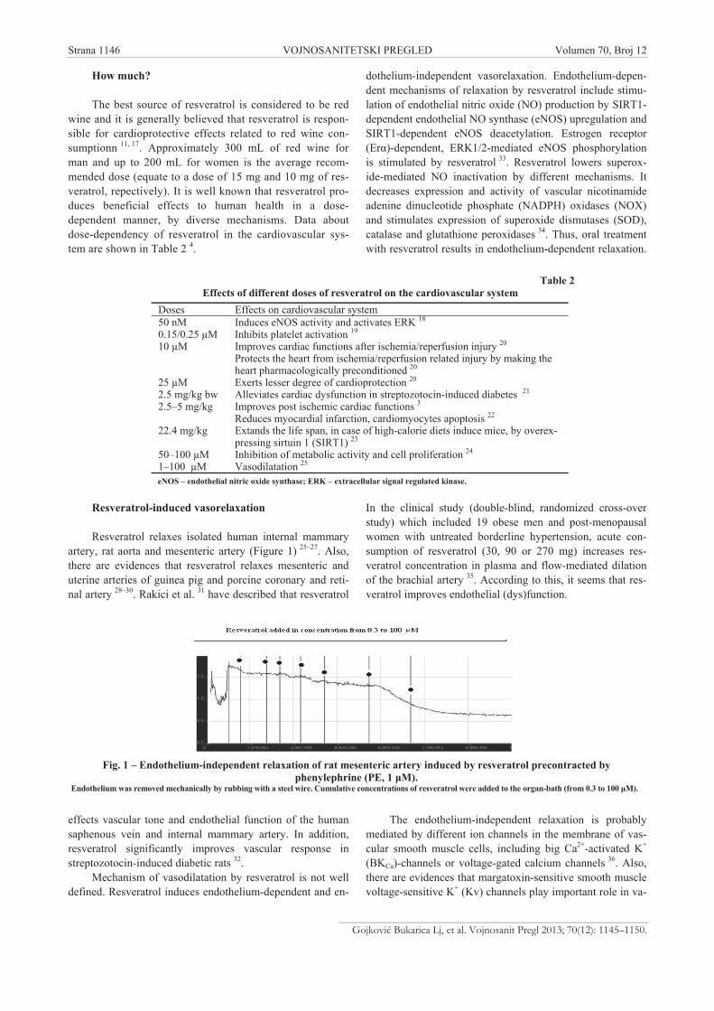
Strana 1146 VOJNOSANITETSKI PREGLED Volumen 70, Broj 12
Gojkovi Bukarica Lj, et al. Vojnosanit Pregl 2013; 70(12): 1145–1150.
How much?
The best source of resveratrol is considered to be redwine and it is generally believed that resveratrol is respon-sible for cardioprotective effects related to red wine con-sumptionn 11, 17. Approximately 300 mL of red wine forman and up to 200 mL for women is the average recom-mended dose (equate to a dose of 15 mg and 10 mg of res-veratrol, repectively). It is well known that resveratrol pro-duces beneficial effects to human health in a dose-dependent manner, by diverse mechanisms. Data aboutdose-dependency of resveratrol in the cardiovascular sys-tem are shown in Table 2 4.
Resveratrol-induced vasorelaxation
Resveratrol relaxes isolated human internal mammaryartery, rat aorta and mesenteric artery (Figure 1) 25–27. Also,there are evidences that resveratrol relaxes mesenteric anduterine arteries of guinea pig and porcine coronary and reti-nal artery 28–30. Rakici et al. 31 have described that resveratrol
effects vascular tone and endothelial function of the humansaphenous vein and internal mammary artery. In addition,resveratrol significantly improves vascular response instreptozotocin-induced diabetic rats 32.
Mechanism of vasodilatation by resveratrol is not welldefined. Resveratrol induces endothelium-dependent and en-
dothelium-independent vasorelaxation. Endothelium-depen-dent mechanisms of relaxation by resveratrol include stimu-lation of endothelial nitric oxide (NO) production by SIRT1-dependent endothelial NO synthase (eNOS) upregulation andSIRT1-dependent eNOS deacetylation. Estrogen receptor(Er )-dependent, ERK1/2-mediated eNOS phosphorylationis stimulated by resveratrol 33. Resveratrol lowers superox-ide-mediated NO inactivation by different mechanisms. Itdecreases expression and activity of vascular nicotinamideadenine dinucleotide phosphate (NADPH) oxidases (NOX)and stimulates expression of superoxide dismutases (SOD),catalase and glutathione peroxidases 34. Thus, oral treatmentwith resveratrol results in endothelium-dependent relaxation.
In the clinical study (double-blind, randomized cross-overstudy) which included 19 obese men and post-menopausalwomen with untreated borderline hypertension, acute con-sumption of resveratrol (30, 90 or 270 mg) increases res-veratrol concentration in plasma and flow-mediated dilationof the brachial artery 35. According to this, it seems that res-veratrol improves endothelial (dys)function.
The endothelium-independent relaxation is probablymediated by different ion channels in the membrane of vas-cular smooth muscle cells, including big Ca2+-activated K+
(BKCa)-channels or voltage-gated calcium channels 36. Also,there are evidences that margatoxin-sensitive smooth musclevoltage-sensitive K+ (Kv) channels play important role in va-
Fig. 1 – Endothelium-independent relaxation of rat mesenteric artery induced by resveratrol precontracted byphenylephrine (PE, 1 M).
Endothelium was removed mechanically by rubbing with a steel wire. Cumulative concentrations of resveratrol were added to the organ-bath (from 0.3 to 100 M).
Table 2Effects of different doses of resveratrol on the cardiovascular system
Doses Effects on cardiovascular system50 nM Induces eNOS activity and activates ERK 18
0.15/0.25 μM Inhibits platelet activation 19
10 μM Improves cardiac functions after ischemia/reperfusion injury 20
Protects the heart from ischemia/reperfusion related injury by making theheart pharmacologically preconditioned 20
25 μM Exerts lesser degree of cardioprotection 20
2.5 mg/kg bw Alleviates cardiac dysfunction in streptozotocin-induced diabetes 21
2.5–5 mg/kg Improves post ischemic cardiac functions 3Reduces myocardial infarction, cardiomyocytes apoptosis 22
22.4 mg/kg Extands the life span, in case of high-calorie diets induce mice, by overex-pressing sirtuin 1 (SIRT1) 23
50–100 μM Inhibition of metabolic activity and cell proliferation 24
1–100 μM Vasodilatation 25
eNOS – endothelial nitric oxide synthase; ERK – extracellular signal regulated kinase.
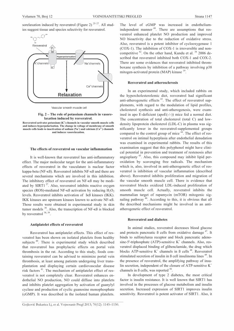
Volumen 70, Broj 12 VOJNOSANITETSKI PREGLED Strana 1147
Gojkovi Bukarica Lj, et al. Vojnosanit Pregl 2013; 70(12): 1145–1150.
sorelaxation induced by resveratrol (Figure 2) 25–27. All stud-ies suggest tissue and species selectivity for resveratrol.
Fig. 2 – The role of potassium channels in vasore-laxation induced by resveratrol.
Resveratrol activates potassium (K+) channels in vascular smooth muscle cellsand induces hyperpolarisation. The change in voltage of membrane of smoothmuscle cells leads to inactivation of sodium (Na+) and calcium (Ca2+) channels
and induces vasorelaxation.
The effects of resveratrol on vascular inflammation
It is well-known that resveratrol has anti-inflammatoryeffect. The major molecular target for the anti-inflammatoryeffects of resveratrol in the vasculature is nuclear factorkappa-beta (NF- ß). Resveratrol inhibits NF- ß and there areseveral mechanisms which are involved in this inhibition.The inhibitory effect of resveratrol on NF- ß may be medi-ated by SIRT1 37. Also, resveratrol inhibits reactive oxygenspecies (ROS)-mediated NF- ß activation by reducing H2O2
levels. Resveratrol inhibits activation of I ß kinases (IKK).IKK kinases are upstream kinases known to activate NF- ß.Those results were obtained in experimental study in skintumor models 38. Also, the transcription of NF- ß is blockedby resveratrol 38, 39.
Antiplatelet effects of resveratrol
Resveratrol has antiplatelet effects. This effect of res-veratrol has been shown on isolated platelets from healthysubjects 40. There is experimental study which describedthat resveratrol has prophylactic effects on portal veinthrombosis in the rat. According to this study, foods con-taining resveratrol can be advised to minimize portal veinthrombosis, at least among patients undergoing liver trans-plantation and displaying certain cardiovascular diseaserisk factors 41. The mechanism of antiplatelet effect of res-veratrol is not completely clear. Resveratrol enhances en-dothelial NO production, NO could diffuse into plateletsand inhibits platelet aggregation by activation of guanylylcyclase and production of cyclic guanosine monophosphate(cGMP). It was described in the isolated human platelets.
The level of cGMP was increased in endothelium-independent manner 40. There are assumptions that res-veratrol enhanced platelet NO production and improvedNO bioactivity due to the reduction of oxidative stress.Also, resveratrol is a potent inhibitor of cyclooxygenase 1(COX-1). The inhibition of COX-1 is irreversible and non-competitive 42. On the other hand, Kundu et al. 38 2006 de-scribed that resveratrol inhibited both COX-1 and COX-2.There are some evidences that resveratrol inhibited throm-boxane synthesis by inhibition of a pathway involving p38mitogen-activated protein (MAP) kinase 19.
Resveratrol and atherosclerosis
In an experimental study, which included rabbits onthe hypercholesterolemic diet, resveratrol had significantanti-atherogenetic effects 43. The effect of resveratrol sup-plements, with regard to the modulation of lipid profiles,cholesterol synthesis and anti-atherogenesis, were exam-ined in apo E-deficient (apoE(-/-)) mice fed a normal diet.The concentration of total cholesterol (total C) and low-density lipoprotein cholesterol (LDL-C) in plasma was sig-nificantly lower in the resveratrol-supplemented groupscompared to the control group of mice 44. The effect of res-veratrol on intimal hyperplasia after endothelial denudationwas examined in experimental rabbits. The results of thisexamination suggest that this polyphenol might have clini-cal potential in prevention and treatment of restenosis afterangioplasty 45. Also, this compound may inhibit lipid per-oxidation by scavenging free radicals. The mechanismwhich is, also, involved in anti-atherogenetic effect of res-veratrol is inhibition of vascular inflammation (describedabove). Resveratrol inhibits proliferation and migration ofthe vascular smooth muscle cell. There is evidence thatresveratrol blocks oxidized LDL-induced proliferation ofsmooth muscle cell. Actually, resveratrol inhibits themammalian target of rapamycin (mTOR) mitogenic sig-naling pathway 46. According to this, it is obvious that allthe described mechanisms might be involved in an anti-atherogenetic effect of resveratrol.
Resveratrol and diabetes
In animal studies, resveratrol decreases blood glucoseand protects pancreatic ß cells from oxidative damage 47. Itbinds to sulfonylurea receptor and block pancreatic adeno-sine-5'-triphosphate (ATP)-sensitive K+ channels. Also, res-veratrol displaced binding of glibenclamide, the drug whichblocks ATP-sensitive K+ channels in ß cells 48. Resveratrolstimulated secretion of insulin in ß cell insulinoma lines 49. Inthe presence of resveratrol, the amplifying pathway of insu-lin secretion, independent of the closure of ATP-sensitive K+
channels in ß cells, was reported 50.In development of type 2 diabetes, the most critical
factor is insulin resistance. It is well known that SIRT1 hasinvolved in the processes of glucose metabolism and insulinsecretion. Increased expression of SIRT1 improves insulinsensitivity. Resveratrol is potent activator of SIRT1. Also, it
Strana 1148 VOJNOSANITETSKI PREGLED Volumen 70, Broj 12
Gojkovi Bukarica Lj, et al. Vojnosanit Pregl 2013; 70(12): 1145–1150.
attenuates high fat diet-induced insulin resistance in vivo, indose of 2.5 mg/kg/day 51.
The effect of resveratrol on energy metabolism andmetabolic profile was investigated in randomized double-blind, crossover study which included 11 healthy, obese mentreated with placebo or with resveratrol 150 mg/day for 30days. The conclusion of this study was that resveratrol in-duced metabolic changes in the obese humans, mimickingthe effects of calorie restriction. Resveratrol decreases intra-hepatic lipid content, circulating glucose, triglycerides andinflammation markers 52.
Resveratrol and oxidative stress
The direct antioxidant effect of resveratrol is notprominent. Well established antioxidants, such as ascorbateand cysteine are more potent than resveratrol 53. Resveratrolhas been shown to be a scavenger of hydroxyl, superoxide,metal-induced radicals and H2O2. In cardiovascular tissues,resveratrol induces antioxidant enzymes. The molecularmechanisms of induction of antioxidant enzymes by res-veratrol are not completely understood. Studies have demon-strated that SIRT1 and the nuclear factor E2-related factor-2(Nrf2) play crucial roles in this process. Resveratrol inducedSOD2 upregulation in cultured human coronary arterial en-dothelial cells. Such upregulation can be blocked by smallinterfering RNA (siRNA)-mediated knockdown of SIRT1.An overexpression of SIRT1 leads to SOD2 upregulation.Nrf2 is transcription factor involved in the regulation of anumber of ROS detoxifying enzymes. In Nrf2-dependentmanner, in cultured endothelial cell, resveratrol inducedNAD(P)H: quinine oxidoreductases (NQO1), heme oxygen-ase-1 (HO-1) and c-glutamylcysteine synthetase (GCLC).The listed enzymes are rate-limited for glutathione synthe-sis. Also, resveratrol inhibits ROS production 54. Treatmentwith resveratrol reduces the expression of NOS in the heartof hypercholesteroemic mouse, as well as mono-nitrogenoxides in the aorta of trauma hemorrhagic rats 55, 56. The ac-tivity of the NADPH oxidase enzyme complex is reduced byresveratrol 57.
Resveratrol and heart
According to numerous animal studies, proposed anti-ischemic mechanisms of resveratrol include: coronary vaso-dilatation, inhibition of atheroma formation, metabolic pro-tection and less ischemic-reperfusion injury. In addition, res-veratrol (5 μM/L) inhibited growth of cardiac fibroblastsstimulated by angiotensin II, epidermal growth factor andtransforming growth factor , which are essential in cardiacfibrosis and heart failure 12. Zheng et al. 13 demonstrated an-tiarrhythmic effect of resveratrol. In the papillary muscles ofguinea pig heart, resveratrol shortened duration of actionpotential and decreased velocity of phase 0 depolarization.Also, it inhibited delayed after depolarization and triggeredactivity. These effects were due to a decrease of calcium in-flux and intracellular calcium concentration.
Conclusion
Cardioprotection includes all the described effects ofresveratrol. The most important evidence for cardioprotec-tion rendered by resveratrol comes from in vivo studies car-ried out on animal models of chronic heart disease that re-sulted in heart dysfunction. Those include hypertension, obe-sity, metabolic syndrome, type I diabetes, type II diabetes,viral cardiomiopathy, toxin cardiomiopathy and aging.
According to the presented results, the cardioprotectiveeffect of resveratrol is obvious. It is complex and not welldefined. It is necessary to emphasize that all positive effectsof resveratrol were demonstrated mainly in in vitro studiesand in studies with experimental animals, while quality clini-cal studies are lacking.
Further clinical studies are necessary in order to definepharmacokinetics, efficacy, adequate therapeutic doses, toler-ability and possible interactions of resveratrol with other drugs.
Acknowledgments
This work was supported by the Ministry of Education,Science and Technological Development of the Republic ofSerbia (Project No TR 31020).
R E F E R E N C E S
1. Arichi H, Kimura Y, Okuda H, Baba K, Kozawa M, Arichi S. Ef-fects of stilbene components of the roots of Polygonum cus-pidatum Sieb. et Zucc. on lipid metabolism. Chem Pharm Bull(Tokyo) 1982; 30(5): 1766 70.
2. Vastano BC, Chen Y, Zhu N, Ho CT, Zhou Z, Rosen RT. Isola-tion and identification of stilbenes in two varieties of Polygo-num cuspidatum. J Agric Food Chem 2000; 48(2): 253 6.
3. Dudley J, Das S, Mukherjee S, Das DK. Resveratrol, a uniquephytoalexin present in red wine, delivers either survival signalor death signal to the ischemic myocardium depending ondose. J Nutr Biochem 2009; 20(6): 443-452.
4. Mukherjee S, Dudley JI, Das DK. Dose-dependency of resvera-trol in providing health benefits. Dose Response 2010; 8(4):478–500.
5. Ibern-Gómez M, Roig-Pérez S, Lamuela-Raventós RM, de la Torre-Boronat MC. Resveratrol and piceid levels in natural and
blended peanut butters. J Agric Food Chem 2000; 48(12):6352 4.
6. Sobolev VS, Cole RJ. Trans-resveratrol content in commercial peanutsand peanut products. J Agric Food Chem 1999; 47(4): 1435 9.
7. Romero-Pérez AI, Lamuela-Raventós RM, Andrés-Lacueva C, de LaTorre-Boronat MC. Method for the quantitative extraction ofresveratrol and piceid isomers in grape berry skins. Effect ofpowdery mildew on the stilbene content. J Agric Food Chem2001; 49(1): 210 5.
8. Soleas GJ, Goldberg DM, Diamandis EP, Karumanchiri A, Yan J,Ng E. A derivatized as chromatographic mass spectrometricmethod for the analysis of both isomers of resveratrol in juiceand wine. Am J Enol Vitic 1995; 46(3): 346–52.
9. Sanders TH, McMichael RW Jr, Hendrix KW. Occurrence of res-veratrol in edible peanuts. J Agric Food Chem 2000; 48(4):1243 6.
Volumen 70, Broj 12 VOJNOSANITETSKI PREGLED Strana 1149
Gojkovi Bukarica Lj, et al. Vojnosanit Pregl 2013; 70(12): 1145–1150.
10. Toku oglu O, Unal MK, Yemi F. Determination of the phytoa-lexin resveratrol (3,5,4'-trihydroxystilbene) in peanuts and pis-tachios by high-performance liquid chromatographic diode ar-ray (HPLC-DAD) and gas chromatography-mass spectrometry(GC-MS). J Agric Food Chem 2005; 53(12): 5003 9.
11. Cvejic JM, Djekic SV, Petrovic AV, Atanackovic MT, Jovic SM,Brceski ID, et al. Determination of trans- and cis-resveratrol inSerbian commercial wines. J Chromatogr Sci 2010; 48(3):229 34.
12. Opie LH, Leceur S. The red wine hypothesis: from concept toprotective signalling molecules. Eur Heart J 2007; 28(14):1683 93.
13. Zheng L, Zhang LP, Ma HJ, Wang C, Ming L, Wang QS. Res-veraatrol reduces free calcium concentration in rat ventricularmyocytes. A Physiol Sin 2005; 57(5): 599 604.
14. Soleas GJ, Angelini M, Grass L, Diamandis EP, Goldberg DM. Ab-sorption of trans-resveratrol in rats. Methods Enzymol 2001;335: 145 54.
15. Atanackovi M, Posa M, Heinle H, Gojkovi -Bukarica L, Cveji J.Solubilization of resveratrol in micellar solutions of differentbile acids. Colloids Surf B Biointerfaces 2009; 72(1): 148 54.
16. Walle T. Bioavailability of resveratrol. Ann N Y Acad Sci 2011;1215: 9 15.
17. Renaud S, De Lorgeril M. Wine, alcohol, platelets, and theFrench paradox for coronary heart disease. Lancet 1992;339(8808): 1523 6.
18. Klinge CM, Blankenship KA, Risinger KE, Bhatnagar S, Noisin EL,Sumanasekera WK, et al. Resveratrol and estradiol rapidly acti-vate MAPK signaling through estrogen receptors alpha andbeta in endothelial cells. J Biol Chem 2005; 280(9): 7460 8.
19. Shen MY, Hsiao G, Liu CL, Fong TH, Lin KH, Chou DS, et al.Inhibitory mechanisms of resveratrol in platelet activation:pivotal roles of p38 MAPK and NO/cyclic GMP. Br J Hae-matol 2007; 139(3): 475 85.
20. Baur JA, Sinclair DA. Therapeutic potential of resveratrol: thein vivo evidence. Nat Rev Drug Discov 2006; 5(6): 493 506.
21. Thirunavukkarasu M, Penumathsa SV, Koneru S, Juhasz B, Zhan L,Otani H, et al. Resveratrol alleviates cardiac dysfunction in strepto-zotocin-induced diabetes: Role of nitric oxide, thioredoxin, andheme oxygenase. Free Radic Biol Med 2007; 43(5): 720 9.
22. Xanthoudakis S, Curran T. Identification and characterizationof Ref-1, a nuclear protein that facilitates AP-1 DNA-bindingactivity. Embo J 1992; 11(2): 653 65.
23. Nisoli E, Tonello C, Cardile A, Cozzi V, Bracale R, Tedesco L, et al.Calorie restriction promotes mitochondrial biogenesis by in-ducing the expression of eNOS. Science 2005; 310(5746):314 7.
24. Caddeo C, Teskac K, Sinico C, Kristl J. Effect of resveratrol in-corporated in liposomes on proliferation and UV-B protectionof cells. Int J Pharm 2008; 363(1 2): 183 91.
25. Gojkovic-Bukarica L, Novakovic A, Kanjuh V, Bumbasirevic M, Le-sic A, Heinle H. A role of ion channels in the endothelium-independent relaxation of rat mesenteric artery induced by res-veratrol. J Pharmacol Sci 2008; 108(1): 124 30.
26. Novakovic A, Gojkovic-Bukarica L, Peric M, Nezic D, Djukanovic B,et. The mechanism of endothelium-independent relaxation in-duced by the wine polyphenol resveratrol in human internalmammary artery. J Pharmacol Sci 2006; 101(1): 85 90.
27. Novakovic A, Gojkovic-Bukarica L, Kanjuh V, Heinle H. Potas-sium channels-mediated vasorelaxation of rat aorta induced byresveratrol. Basic Clin Pharmacol Toxicol 2006; 99(5): 360 4.
28. Naderali EK, Doyle PJ, Williams G. Resveratrol induces vasore-laxation of mesenteric and uterine arteries from female guineapigs. Clin Sci (Lond) 2000; 98(5): 537–43.
29. Jager U, Nguyen-Duong H. Relaxant effect of trans-resveratrol onisolated porcine coronary arteries. Arzneimittelforschung 1999;49(3): 207–11.
30. Nagaoka T, Hein TW, Yoshida A, Kuo L. Resveratrol, a compo-nent of red wine, elicits dilation of isolated porcine retinal arte-rioles: role of nitric oxide and potassium channels. InvestOphthalmol Vis Sci 2007; 48(9): 4232 9.
31. Rakici O, Kiziltepe U, Coskun B, Aslamaci S, Akar F. Effects ofresveratrol on vascular tone and endothelial function of hu-man saphenous vein and internal mammary artery. Int J Car-diol 2005; 105(2): 209 15.
32. Silan C. The effects of chronic resveratrol treatment on vascu-lar responsiveness of streptozotocin-induced diabetic rats. BiolPharm Bull 2008; 31(5): 897–902.
33. Li H, Forstermann U. Resveratrol: a multifunctional compoundimproving endothelial function. Editorial to: «Resveratrol sup-plementation gender independently improves endothelial reac-tivity and suppresses superoxide production in healthy rats» byS. Soylemez et al. Cardiovasc Drugs Ther 2009; 23(6): 425 9.
34. Spanier G, Xu H, Xia N, Tobias S, Deng S, Wojnowski L, et al.Resveratrol reduces endothelial oxidative stress by modulatingthe gene expression of superoxide dismutase 1 (SOD1), glu-tathione peroxidase 1 (GPx1) and NADPH oxidase subunit(Nox4). J Physiol Pharmacol 2009; 60(Suppl 4): 111 6.
35. Wong RH, Howe PR, Buckley JD, Coates AM, Kunz I, Berry NM.Acute resveratrol supplementation improves flow-mediateddilatation in overweight/obese individuals with mildly elevatedblood pressure. Nutr Metab Cardiovasc Dis 2010; 21(11):851 6.
36. Campos-Toimil M, Elíes J, Alvarez E, Verde I, Orallo F. Effects oftrans- and cis-resveratrol on Ca2+ handling in A7r5 vascularmyocytes. Eur J Pharmacol 2007; 577(1 3): 91 9.
37. Yeung F, Hoberg JE, Ramsey CS, Keller MD, Jones DR, Frye RA, etal. Modulation of NF-kappaB-dependent transcription and cellsurvival by the SIRT1 deacetylase. EMBO J 2004; 23(12):2369–80.
38. Kundu JK, Shin YK, Kim SH, Surh YJ. Resveratrol inhibits phor-bol esterinduced expression of COX-2 and activation of NF-kappaB in mouse skin by blocking IkappaB kinase activity.Carcinogenesis 2006; 27(7): 1465–74.
39. Manna SK, Mukhopadhyay A, Aggarwal BB. Resveratrol sup-presses TNFinduced activation of nuclear transcription factorsNF-kappa B, activator protein-1, and apoptosis: potential roleof reactive oxygen intermediates and lipid peroxidation. J Im-munol 2000; 164(12): 6509–19.
40. Wang Z, Huang Y, Zou J, Cao K, Xu Y, Wu JM. Effects of redwine and wine polyphenol resveratrol on platelet aggregationin vivo and in vitro. Int J Mol Med 2002; 9(1): 77–9.
41. Kirimlioglu V, Sozen H, Turkoglu S, Haberal M. Protective effectof resveratrol, a red wine constituent polyphenol, on rats sub-jected to portal vein thrombosis. Transplant Proc 2008; 40(1):290 2.
42. Szewczuk LM, Forti L, Stivala LA, Penning TM. Resveratrol is aperoxidasemediated inactivator of COX-1 but not COX-2: amechanistic approach to the design of COX-1 selective agents.J Biol Chem 2004; 279(21): 22727 37.
43. Wang Z, Zou J, Cao K, Hsieh TC, Huang Y, Wu JM. Dealcohol-ized red wine containing known amounts of resveratrol sup-presses atherosclerosis in hypercholesterolemic rabbits withoutaffecting plasma lipid levels. Int J Mol Med 2005; 16(4): 533–40.
44. Do GM, Kwon EY, Kim HJ, Jeon SM, Ha TY, Park T, et al.Long-term effects of resveratrol supplementation on suppres-sion of atherogenic lesion formation and cholesterol synthesisin apo E-deficient mice. Biochem Biophys Res Commun 2008;374(1): 55 9.
45. Zou J, Huang Y, Cao K, Yang G, Yin H, Len J, et al. Effect ofresveratrol on intimal hyperplasia after endothelial denudationin an experimental rabbit model. Life Sci 2000; 68(2): 153 63.
46. Brito PM, Devillard R, Nègre-Salvayre A, Almeida LM, Dinis TC,Salvayre R, et al. Resveratrol inhibits the mTOR mitogenic sig-
Strana 1150 VOJNOSANITETSKI PREGLED Volumen 70, Broj 12
Gojkovi Bukarica Lj, et al. Vojnosanit Pregl 2013; 70(12): 1145–1150.
naling evoked by oxidized LDL in smooth muscle cells. Ath-erosclerosis 2009; 205(1): 126 34.
47. Zhang Y, Jayaprakasam B, Seeram NP, Olson LK, DeWitt D, NairMG. Insulin secretion and cyclooxygenase enzyme inhibitionby cabernet sauvignon grape skin compounds. J Agric FoodChem 2004; 52(2): 228–33.
48. Hambrock A, de Oliveira Franz CB, Hiller S, Grenz A, AckermannS, Schulze DU, et al. Resveratrol binds to the sulfonylurea re-ceptor (SUR) and induces apoptosis in a SUR subtype-specificmanner. J Biol Chem 2007; 282: 3347–56.
49. Chen WP, Chi TC, Chuang LM, Su MJ. Resveratrol enhances in-sulin secretion by blocking KATP and KV channels of betacells. Eur J Pharmacol 2007; 568(1 3): 269–77.
50. Szkudelski T. Resveratrol-induced inhibition of insulin secre-tion from rat pancreatic islets: evidence for pivotal role ofmetabolic disturbances. Am J Physiol Endocrinol Metab 2007;293(4): E901–7.
51. Sun C, Zhang F, Ge X, Yan T, Chen X, Shi X, Zhai Q. SIRT1improves insulin sensitivity under insulin-resistant conditionsby repressing PTP1B. Cell Metab 2007; 6(4): 307 19.
52. Timmers S, Konings E, Bilet L, Houtkooper RH, van de Weijer T,Goossens GH, et al. Calorie restriction-like effects of 30 days ofresveratrol supplementation on energy metabolism and meta-
bolic profile in obese humans. Cell Metab 2011; 14(5):612 22.
53. Bradamante S, Barenghi L, Villa A. Cardiovascular protective ef-fects of resveratrol. Cardiovasc Drug Rev 2004; 22(5): 169–88.
54. Li H, Xia N, Förstermann U. Cardiovascular effects and mo-lecular targets of resveratrol. Nitric Oxide 2012; 26(2): 102–10.
55. Yu HP, Hwang TL, Hwang TL, Yen CH, Lau YT. Resveratrolprevents endothelial dysfunction and aortic superoxide pro-duction after trauma hemorrhage through estrogen receptor-dependent hemeoxygenase-1 pathway. Crit Care Med 2010;38(4): 1147–54.
56. Xia N, Daiber A, Habermeier A, Closs EI, Thum T, Spanier G, etal. Resveratrol reverses endothelial nitric-oxide synthase un-coupling in apolipoprotein E knockout mice. J Pharmacol ExpTher 2010; 335(1): 149 54.
57. Hshu YC, Wang JS, Chen JK. Resveratrol attenuates oxLDL-stimulated NADPH oxidase activity and protects endothelialcells from oxidative functional damages. J Appl Physiol 2007;102(4): 1520 7.
Received on June 13, 2012.Revised on July 15, 2012.
Accepted on August 2, 2012.
Vojnosanit Pregl 2013; 70(12): 1151–1154. VOJNOSANITETSKI PREGLED Strana 1151
Correspondence to: Svetlana Jovanovi , Department of Ophthalmology, Clinical Center "Kragujevac", Grada Sirena 30,34 000 Kragujevac, Serbia. Phone: +381 63 683 054. E-mail: [email protected] , [email protected]
C A S E R E P O R T S UDC: 617.7/006.44DOI: 10.2298/VSP1312151J
Two cases of uveitis masquerade syndrome caused by bilateralintraocular large B-cell lymphoma
Dva bolesnika sa maskiranim sindromom uveitisa nastalim kao posledicaobostranog intraokularnog limfoma velikih B- elija
Svetlana Jovanovi *†, Zorica Jovanovi †, Jelena Paovi ‡§,Vesna Stankovi eperkovi †, Snežana Peši †, Vujica Markovi ‡§
*Clinic of Ophthalmology, Clinical Center “Kragujevac”, Kragujevac, Serbia; †Facultyof Medicine, University of Kragujevac, Kragujevac, Serbia; ‡Clinic of Ophthalmology,
Clinical Center of Serbia, Belgrade, Serbia; §Faculty of Medicine, University ofBelgrade, Serbia
Abstract
Introduction. Sometimes it is not easy to clinically recognizesubtle differences between intraocular lymphoma and non-infectious uveitis. The most common lymphoma subtype in-volving the eye is B-cell lymphoma. Case report. We pre-sented two patients aged 59 and 58 years with infiltration ofthe subretinal space with a large B-cell non-Hodgkin intra-ocular lymphoma. The patients originally had clinicallymasked syndrome in the form of intermediate uveitis. As itwas a corticosteroid-resistant uveitis, we focused on the pos-sible diagnosis of neoplastic causes of this syndrome. Duringhospitalization, the neurological symptoms emerged andmultiple subretinal changes accompanied by yellowish whitepatches of retinal pigment epithelium with signs of vitritis,which made us suspect the intraocular lymphoma. Endocra-nial magnetic resonance imaging established tumorous infil-tration in the region of the left hemisphere of the cerebellum.The histopathological finding confirmed the diagnosis oflarge B-cell non-Hodgkin lymphoma of risk moderate degree,immunoblast – centroblast cytological type. The other patienthad clinical chronic uveitis accompanied by yellowish shapedwhite echographic changes of the retina and localized changesin the level of the subretina. The diagnosis of lymphoma wasmade by brain biopsy. Conclusion. Uveitis masquerade syn-drome should be considered in all patients over 40 years withidiopathic steroid-resistant uveitis. Treatment begun on timecan affect the course and improve the prognosis of uveitismasquerade syndrome (UMS) and systemic disease.
Key words:eye neoplasms; lymphoma, non-hodgkin; uveitis;diagnosis, differential.
Apstrakt
Uvod. Ponekad je teško ustanoviti suptilnu razlika izme uintraokularnog limfoma i neinfektivnog uveitisa. Naj eš ipodtip intraokularnog limfoma je B- elijski limfom. Prikazbolesnika. Prikazali smo dva bolesnika, starosti 59 i 58 go-dina, sa infiltracijom subretinalnog prostora velikim B-elijama non-Hodgkin itraokularnog limfoma. Prvi bolesnik
prvobitno je imao klini ku sliku maskiranog sindoma u viduintermedijalnog uveitisa. Kako se radilo o kortikosteroid-rezistentnom uveitisu, usredsredili smo se na mogu u dijag-nozu neoplasti nog uzroka ovog maskiranog sindroma. Utoku hospitalizacije na neurologiji pojavili su se subretinalnieksudati pra eni žu kastobeli astim promenama retinalnogpigmentnog epitela i znacima vitritisa koji su nas naveli nasumnju na intraokularni limfom. Magnetna rezonanca (MR)endokranijuma potvdila je infiltraciju leve hemisfere cere-beluma. Patohistološki nalaz operisanog tumora cerebelumapotvrdio je dijagnozu non-Hodgkin limfoma velikih B- elija,umerenog stepena rizika, imunoblast-centroblastom citološ-kog tipa. Drugi bolesnika imao je klini ku sliku hroni nogzadnjeg uveitisa sa žu kasto beli astim promenama retine iehografski lokalizovanim promenama u nivou subretine.Dijagnoza limfoma postavljena je biopsijom mozga. Zak-lju ak. Uveitis maskirani sindom (UMS) treba razmotritikod svih bolesnika starijih od 40 godina sa idiopatskim kor-tikosteroid-rezistentnim uveitisom. Le enje zapo eto navreme može uticati na tok i poboljšati prognozu UMS i sis-temske bolesti.
Klju ne re i:oko, neoplazme; limfom, nehodžkinov; uveitis;dijagnoza, diferencijalna.
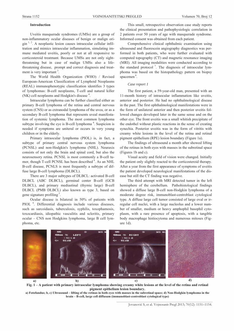
Strana 1152 VOJNOSANITETSKI PREGLED Volumen 70, Broj 12
Jovanovi S, et al. Vojnosanit Pregl 2013; 70(12): 1151–1154.
Introduction
Uveitis masquerade syndromes (UMSs) are a group ofnon-inflammatory ocular diseases of benign or malign ori-gin 1, 2. A neoplastic lesion causes intraocular cellular infil-tration and mimics intraocular inflammation, simulating im-mune mediated uveitis, poorly or not at all responsive tocorticosteroid treatmant. Because UMSs are not only sight-threatening but in case of malign UMSs also a life-threatening disease, prompt and correct diagnosis and treat-ment is very important 3.
The World Health Organization (WHO) / RevisedEuropean-American Classification of Lymphoid Neoplasms(REAL) immunophenotypic classification identifies 3 typesof lymphomas: B-cell neoplasms, T-cell and natural killer(NK) cell neoplasms and Hodgkin's disease 4.
Intraocular lymphoma can be further classified either asprimary B-cell lymphoma of the retina and central nervoussystem (CNS) or as extranodal lymphoma of the uvea, or as asecondary B-cell lymphoma that represents uveal manifesta-tion of systemic lymphoma. The most common lymphomasubtype involving the eye is B-cell lymphoma 4. Suspicion isneeded if symptoms are uniteral or occurs in very youngchildren or in the elderly.
Primary intraocular lymphoma (PIOL) is, in fact, asubtype of primary central nervous system lymphoma(PCNSL) and non-Hodgkin's lymphoma (NHL). Neuraxisconsists of not only the brain and spinal cord, but also theneurosensory retina. PCNSL is most commonly a B-cell tu-mor, though T-cell PCNSL has been described 4. As an NHLB-cell disease, PCNLS is most frequently a subtype of dif-fuse large B-cell lymphoma (DLBCL).
There are 3 major subtypes of DLBCL: activated B-cellDLBCL (ABC DLBCL), germinal center B-cell (GCBDLBCL), and primary mediastinal (thymic large) B-cellDLBCL (PMB DLBCL) also known as type 3, based ongene signature profiling 5.
Ocular disease is bilateral in 50% of patients withPIOL 6. Differential diagnosis include various diseases,such as sarcoidasis, tuberculosis, syphilis, toxophamosis,toxocardiasis, idiopathic vasculitis and scleritis, primaryocular – CNS non Hodgkins lymphoma, large B cell lym-phoma, etc.
This small, retrospective observation case study reportsthe clinical presentation and pathophysiologic correlation in2 patients over 50 years of age with masquerade syndrome.Informed consent was obtained from each patient.
Comprehensive clinical ophthalmic examination usingultrasound and fluorescein angiography diagnostics was per-formed in both patients, who were further evaluated withcomputed topography (CT) and magnetic resonance imaging(MRI). All imaging modalities were conducted according tothe standard protocol 7. The diagnosis of intraocular lym-phoma was based on the histopathology pattern on biopsyspacemen 8.
Case report 1
The first patient, a 59-year-old man, presented with an11-month history of intraocular inflammation like uveitis,anterior and posterior. He had no ophthalmological diseasein the past. The first ophthalmological manifestations were inthe form of unilateral anterior and then posterior uveitis fol-lowed changes developed later in the same sense and on theother eye. The front uveitis was a small whitish precipitate ofthe endothel without plastic reactions in the sense of creatingsynechia. Posterior uveitis was in the form of vitritis withcreamy white lesions in the level of the retina and retinalpigment epithelium (RPE) lesion boundary (Figure 1a).
The findings of ultrasound a month after showed liftingof the retinas in both eyes with masses in the subretinal space(Figures 1b and c).
Visual acuity and field of vision were changed. Initiallythe patient only slightly reacted to the corticosteroid therapy.After a year from the first appearance of symptoms of uveitisthe patient developed neurological manifestations of the dis-ease but still the CT finding was negative.
The third attempt with MRI detected tumor in the lefthemisphere of the cerebellum. Pathohistological findingsshowed a diffuse large B-cell non-Hodgkin lymphoma of amoderate degree risk, immunoblast-centroblast cytologicaltype. A diffuse large cell tumor consisted of large oval or ir-regular cell nuclei, with a large nucleolus and a lower num-ber of smaller, medium or heavy amphophil basophil cyto-plasm, with a rare presence of apoptosis, with a tangiblebody macrophage histiocytoma and numerous mitoses (Fig-ure 1d).
a) b) c) d)Fig. 1 – A patient with primary intraocular lymphoma showing creamy white lesions at the level of the retina and retinal
pigment epithelium lesion boundary.a) Fotofundus; b, c) Ultrasound – lifting of the retinas in both eyes with masses in the subretinal space; d) Non-Hodgkin lymphoma in the
brain – B-cell, large cell diffusum (immunoblast-centroblast cytological type)

Volumen 70, Broj 12 VOJNOSANITETSKI PREGLED Strana 1153
Jovanovi S, et al. Vojnosanit Pregl 2013; 70(12): 1151–1154.
He was afebrile and his routine blood test was normalexcept low total red blood cell count (RBC), hemoglobin(Hgb), mean platelet volume (MPV) and relative volume ofthrombocytes (Pct) were on little level. The patient did notallow biopsy of the retina.
Localization of the tumor was in the brain and subreti-nal space, actually in the immune privileged sites.
Case report 2
The second patient was a 58-year-old woman with ante-rior and posterior uveitis for several months. She showed nosatisfactory respond to the treatment with corticosteroids. Infact, the therapy with corticosteroids had led to an incom-plete reabsorption flare and precipitate in the front segment,with signs of vitritis in the back. The last segment was visi-ble and the ultrasound findings showed an improvement. Theposterior uveitis was manifested in the form of creamy, yel-low white to orange subretinal pigment epithel (RPE) infil-trates, which also disappeared as a result of the treatmentwith corticosteroids, but the last segment as a whole waschanged (Figure 2a).
Ultrasound suggested that intraocular lymphoma stillresponded to corticosteroid therapy. The fluorescein angiog-raphy in the intraocular lymphoma had a characteristic ap-pearance. RPE disturbances included granularity, mottling,and late staining patterns. Fluorescence blockage at the levelof the RPE, due to tumor infiltration could correspond to thedeep retinal or subretinal creamy colored lesions noted onfundus photography. Visual acuity and visual field testing,were both reduced significantly. Intraocular (IOP) was 14mm Hg. A routine blood test was normal except for Hgb.MR images demonstrated enhancement in the left temporallobe. The diagnosis of lymphoma was made by brain biopsy.Histopathological finding was diffuse large B-cell non-Hodgkin lymphoma (Figures 2 b, c and d).
Discussion
Primary intraocular lymphoma is the most commonmanifestation of masked uveitis syndromes 9. It is typicallypresented as posterior uveitis with cellular exudation in thevitreous fluid 10. Imaging of the eye is the first step in evalu-ating the diagnosis with suspicion of PIOL. The role of an
ophthalmologist is particularly important. Imaging of thecentral nervous system should be included. Imaging modali-ties are: full field fundus photography, ultrasound, fluores-cein angiography, indocyanine green angiography, ocularcoherence tomography, brain imaging. Systemic organs im-aging is generally not necessary in the cases of suspectedPIOL or PCNSL, and this practice is not highly recom-mended 11. When there is a reason to suspect systemic lym-phoma added to the standard tests are a complete blood cellcount, erythrocyte sedimentation rate, and bone marrowevaluation 12. Vitreous biopsy is a useful tool to diagnosePIOL. If it is implemented prior to steroid therapy, it mightsuppress the number of vitreous cells, including lymphomacells, which may result in a negative vitreous cytology 13. Thelymphoma cells of POIL and PCNSL are very fragile, and ifsystemic corticosteroids are used to treat a presumed "uveitis",the lymphoma cells may be even more fragile 14. Conventionalocular and brain examination includes cytological and his-tological examination (macroscopic and microscopic). Mo-lecular pathology involves gene rearrangements, transloca-tions, molecular signals, infectious deoxyribonucleic acid(DNA) 15. Immunohistochemistry shows the same picture asthat for PIOL. As the majority of PCNSL cells are B-cells,they stain for CD19, CD20, and surface immunoglobulin 16, 17.
Conclusion
Sometimes, uveitis is the only initial manifestation ofan occult systemic problem in patients older than 40 years.UMS should be considered in all patients with idiopathiccorticosteroid resistant chronic uveitis. Primary intraocularlymphoma should be considered in all patients aged 40 andolder with ultrasonographic findings of subretinal lesions andvitreous cells. Malignancies and other diseases should beconsidered, with implementation of diagnosis biopsies of vi-
trous fluid and brain. Timely treatment may improve theprognosis of UMS. Direct treatment of malignancy or un-derlying condition may be required to control uveitis.
Acknowledgements
This work was supported by the Grant No. 500-01-00035, the Ministry of Health of the Republic of Serbia.
a) b) c) d)Fig. 2 – A patient with primary intraocular lymphoma showing creamy, yellow-white to orange sub-retinal pigment
epithelium infiltratesa) Fotofundus; b) Ultrasound – lifting of the retinas in both eyes with masses in the subretinal space before the therapy c) after the therapy;
d) Non-Hodgkin lymphoma in the brain (B-cell, large cell diffusum).
Strana 1154 VOJNOSANITETSKI PREGLED Volumen 70, Broj 12
Jovanovi S, et al. Vojnosanit Pregl 2013; 70(12): 1151–1154.
R E F E R E N C E S
1. Chan CC, Wallace DJ. Intraocular Lymphoma: Update on Di-agnosis and Management. Cancer Control 2004; 11(5):285 95.
2. Kubicka-Trzaska A, Romanowska-Dixon B. Malignant uveitismasquerade syndromes. Klin Oczna 2008; 110(4 6):199 202.
3. Choi JY, Kafkala C, Foster CS. Primary intraocular lymphoma: Areview. Semin Ophthalmol 2006; 21(3): 125 33.
4. Parikh AH, Khan SH, Wright JD Jr, Oh KT. Systemic non-Hodgkin's lymphoma simulating primary intraocular lym-phoma. Am J Ophtalmol 2005; 139(3): 573 4.
5. Choi JS, Nam DH, Ko YH. Primary central nervous systemlymphoma in Korea: comparison of B- and T- cell lymphomas.Am J Surg Pathol 2003; 27(7): 919 28.
6. Wright G, Tan B, Rosenwald A, Hurt EH. A gene expression-based method to diagnose clinically distinct subgroups of dif-fuse large B-cell lymphoma. Proc Natl Acad Sci USA 2003;100(17): 9991 6.
7. Hoffman PM, McKelvie P, Hall AJ, Stawell RJ, Santamaria JD. In-traocular lymphoma: a series of 14 patients with clinicopa-thological features and treatment outcomes. Eye (Lond) 2003;17(4): 513 21.
8. Pileri SA, Agostinelli C, Sabattini E, Bacci F, Sagramoso C, Pileri AJr, et al. Lymphoma classification: the quiet after the storm.Semin Diagn Pathol 2011; 28(2): 113 23.
9. Cogliatti SB, Schimid U. Who is Who and what is REAL? SwissMed Wkly 2002; 132(43 44): 607 17.
10. Rothova A, Ooijman F, Kerkhoff F, Van Der Lelij A, Lokhorst HM.Uveitis masquerade syndromes. Ophthalmology 2001; 108(2):386 99.
11. Nussenblatt RB, Chan CC, Wilson WH, Hochman J, Gottesman M;CNS and Ocular Lymphoma Workshop Group. InternationalCentral Nervous System and Ocular Lymphoma Workshop:recommendations for the future. Ocul Immunol Inflamm2006; 14(3): 139 44.
12. Leavy-Clarke GA, Chan CC, Nussenblatt RB. Diagnosis andmanagement of primary intraocular lymphoma. Hematol On-col Clin North Am 2005; 19(4):739 49.
13. Buggage RR, Chan CC, Nussenblatt RB.Ocular manifestation ofcentralnervous system lymphoma. Curr Opin Oncol 2001;13(3): 137 42.
14. Kim E, Kim C, Lee J, Cho Y. A case of primary intraocular lym-phoma treated by intravitreal methotrexate. Korean J Oph-thalmol 2009; 23(3): 210 4.
15. Wallace DJ, Shen D, Read GF, Miyanaga M, Mochizuki M, SenHN, et al. Detection of the bcl-2 t(14;18) translocation andproto-oncogene expression in primary intraocular lymphoma.Invest Ophthalmol Vis Sci 2006; 47(7): 2750 6.
16. Bashir R, McManus B, Cuningham C, Weisburger D, Hoshberg F.Detection of Eber-1 RNA in primary brain lymphomas inimmunocompetent and immunocompromised patients. J Neu-rooncol 1994; 20(1): 47 53.
17. Schlegel U. Primary CNS Lymphoma Ther Adv Neurol Disord2009; 2(2): 93 104.
Received on September 12, 2011.Accepted on June 11, 2012.

Vojnosanit Pregl 2013; 70(12): 1155–1158. VOJNOSANITETSKI PREGLED Strana 1155
Correspondence to: Vladisav Stefanovi , Faculty of Medicine, Bul. Zorana Djindji a 81, 18 000 Niš, Serbia. E-mail: [email protected]
C A S E R E P O R T UDC: 616.36-006DOI: 10.2298/VSP1312155J
Stromal reaction and prognosis in acinic cell carcinoma of the salivarygland
Stromalna reakcija i prognoza acino elijskog karcinoma pljuva ne žlezde
Ljubinka Jankovi Veli kovi *, Irena Dimov†, Dragan Petrovi ‡,Slavica Stojnev*, Stefan Da i ‡, Stefan Veli kovi ‡, Vladisav Stefanovi §
*Institute of Pathology, †Department of Immunology, ‡Clinic of Stomatology, §Instituteof Nephrology, Faculty of Medicine, University of Niš, Niš, Serbia
Abstract
Introduction. Primary acinic cell carcinoma (ACC) is an un-common malignant neoplasm of the salivary gland (SG), whichusually presents as slow growing tumor. Case report. We re-ported a 69-year-old woman with tumor in the right parotidgland with a 5-year progress. Biopsy sections revealed a hybridform of ACC with a low- and high-grade component andprominent lymphoid tissue in tumor stroma. Immunohisto-chemistry was performed to define the molecular profile of thisunusual ACC, with special interest for stromal influence on tothe proliferative activity of ACC with dedifferentiation. Wedetected that the level and the type of stromal lymphoid reac-tion (particularly CD8+/CD4+ ratio) had a significant influenceon to Ki-67 index in the high-grade component of ACC, aswell as the involvement of the CXCR4 signaling axis in thestromal reaction influence. Conclusion. We suggest that tu-mor stroma may be a source of potential new tumor biomark-ers which can determine the aggressivity of this tumor.
Key words:parotid neoplasms; carcinoma, acinar cell; ki-67antigen; prognosis.
Apstrakt
Uvod. Primarni acino elijski karcinom (ACC) je redak mali-gni tumor pljuva nih žlezda koji obi no ima sporu progre-siju. Prikaz bolesnika. U radu je prikazana bolesnica, stara69 godina, sa tumorom u desnoj parotidnoj žlezdi. Na pre-secima tkiva otkrivena je hibridna forma ACC, sa kompo-nentom niskog i visokog gradusa, i izraženim limfnim tki-vom u stromi tumora. Tumor je pokazivao 5-godišnju pro-gresiju. Imunohistohemijska metoda koriš ena je kako bi sedefinisao molekularni profil ove retke forme ACC, kao i uti-caj strome na proliferativnu aktivnost ACC sa njegovomdediferencijacijom. Pokazali smo da nivo i tip stromalnelimfoidne reakcije (pogotovo odnos CD8+/CD4+) ima zna-ajan uticaj na Ki-67 indeks u komponenti visokog gradusa
ACC, kao i uticaj CXCR4 signalnog puta. Zaklju ak. Tu-morska stroma može biti izvor potencijalno novih tumor-skih biomarkera, kada imuni odgovor može uticati na agre-sivnost ovog tumora.
Klju ne re i:parotidne žlezde, neoplazme; karcinom acinusnigelija; antigen, ki-67; prognoza.
Introduction
Although acinic cell carcinomas (ACCs) represent only2–6% of salivary gland tumors, they are the third most com-mon epithelial malignancy after mucoepidermoid carcinoma,thus being about 10% of all malignant salivary gland tu-mors 1. Well-differentiated ACCs present as well circum-scribed encapsulated tumors with a solid or microcystic pat-tern in which tumor cells are surrounded and intermingledwith prominent lymphoid response. Dedifferentiated ACCpresents itself with areas of low-grade ACC and areas of de-differentiated high grade ACC, or undifferentiated carcinomawithin the same tumor 2, 3. Investigation of biological mark-ers is very important and necessary for predicting prognosis
of salivary malignancies and better understanding the patho-genesis of salivary cancer. Ki-67 was already proven as use-ful prognostic markers for survival in a few studies of pa-tients with ACC and other salivary glands tumors 4, 5. Tumorstroma may be a source of potential new tumor biomarkers,in which the immune response is of major importance. Herewe investigated stromal influence onto the proliferative ac-tivity of ACC with dedifferentiation and evolution of this pa-rotid gland tumor.
Case report
A 69-year-old woman presented with a superficiallobulated swelling in the lower half of the right parotid
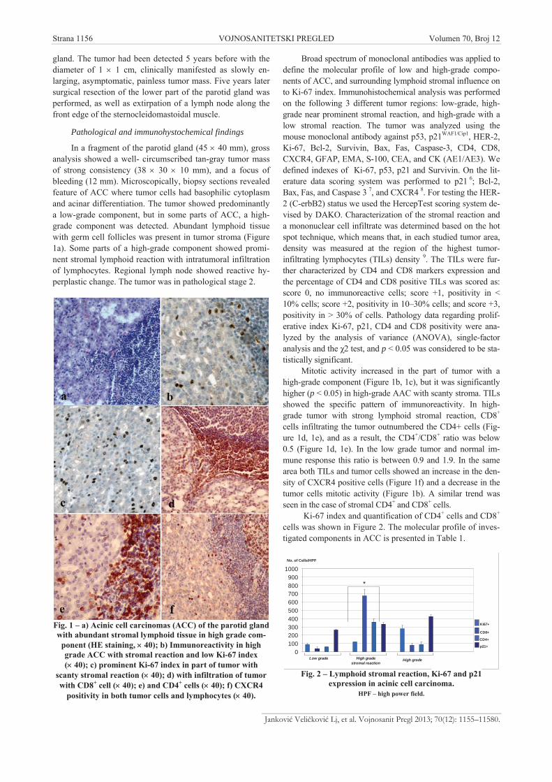
Strana 1156 VOJNOSANITETSKI PREGLED Volumen 70, Broj 12
Jankovi Veli kovi Lj, et al. Vojnosanit Pregl 2013; 70(12): 1155–11580.
gland. The tumor had been detected 5 years before with thediameter of 1 1 cm, clinically manifested as slowly en-larging, asymptomatic, painless tumor mass. Five years latersurgical resection of the lower part of the parotid gland wasperformed, as well as extirpation of a lymph node along thefront edge of the sternocleidomastoidal muscle.
Pathological and immunohystochemical findings
In a fragment of the parotid gland (45 40 mm), grossanalysis showed a well- circumscribed tan-gray tumor massof strong consistency (38 30 10 mm), and a focus ofbleeding (12 mm). Microscopically, biopsy sections revealedfeature of ACC where tumor cells had basophilic cytoplasmand acinar differentiation. The tumor showed predominantlya low-grade component, but in some parts of ACC, a high-grade component was detected. Abundant lymphoid tissuewith germ cell follicles was present in tumor stroma (Figure1a). Some parts of a high-grade component showed promi-nent stromal lymphoid reaction with intratumoral infiltrationof lymphocytes. Regional lymph node showed reactive hy-perplastic change. The tumor was in pathological stage 2.
Fig. 1 – a) Acinic cell carcinomas (ACC) of the parotid glandwith abundant stromal lymphoid tissue in high grade com-
ponent (HE staining, 40); b) Immunoreactivity in highgrade ACC with stromal reaction and low Ki-67 index( 40); c) prominent Ki-67 index in part of tumor with
scanty stromal reaction ( 40); d) with infiltration of tumorwith CD8+ cell ( 40); e) and CD4+ cells ( 40); f) CXCR4
positivity in both tumor cells and lymphocytes ( 40).
Broad spectrum of monoclonal antibodies was applied todefine the molecular profile of low and high-grade compo-nents of ACC, and surrounding lymphoid stromal influence onto Ki-67 index. Immunohistochemical analysis was performedon the following 3 different tumor regions: low-grade, high-grade near prominent stromal reaction, and high-grade with alow stromal reaction. The tumor was analyzed using themouse monoclonal antibody against p53, p21WAF1/Cip1, HER-2,Ki-67, Bcl-2, Survivin, Bax, Fas, Caspase-3, CD4, CD8,CXCR4, GFAP, EMA, S-100, CEA, and CK (AE1/AE3). Wedefined indexes of Ki-67, p53, p21 and Survivin. On the lit-erature data scoring system was performed to p21 6; Bcl-2,Bax, Fas, and Caspase 3 7, and CXCR4 8. For testing the HER-2 (C-erbB2) status we used the HercepTest scoring system de-vised by DAKO. Characterization of the stromal reaction anda mononuclear cell infiltrate was determined based on the hotspot technique, which means that, in each studied tumor area,density was measured at the region of the highest tumor-infiltrating lymphocytes (TILs) density 9. The TILs were fur-ther characterized by CD4 and CD8 markers expression andthe percentage of CD4 and CD8 positive TILs was scored as:score 0, no immunoreactive cells; score +1, positivity in <10% cells; score +2, positivity in 10–30% cells; and score +3,positivity in > 30% of cells. Pathology data regarding prolif-erative index Ki-67, p21, CD4 and CD8 positivity were ana-lyzed by the analysis of variance (ANOVA), single-factoranalysis and the 2 test, and p < 0.05 was considered to be sta-tistically significant.
Mitotic activity increased in the part of tumor with ahigh-grade component (Figure 1b, 1c), but it was significantlyhigher (p < 0.05) in high-grade AAC with scanty stroma. TILsshowed the specific pattern of immunoreactivity. In high-grade tumor with strong lymphoid stromal reaction, CD8+
cells infiltrating the tumor outnumbered the CD4+ cells (Fig-ure 1d, 1e), and as a result, the CD4+/CD8+ ratio was below0.5 (Figure 1d, 1e). In the low grade tumor and normal im-mune response this ratio is between 0.9 and 1.9. In the samearea both TILs and tumor cells showed an increase in the den-sity of CXCR4 positive cells (Figure 1f) and a decrease in thetumor cells mitotic activity (Figure 1b). A similar trend wasseen in the case of stromal CD4+ and CD8+ cells.
Ki-67 index and quantification of CD4+ cells and CD8+
cells was shown in Figure 2. The molecular profile of inves-tigated components in ACC is presented in Table 1.
0100200300400500600700800900
1000
Ki67+
CD4+
CD8+
*
High gradestromal reaction
Low grade
No. of Cells/HPF
High grade
p21+
Fig. 2 – Lymphoid stromal reaction, Ki-67 and p21expression in acinic cell carcinoma.
HPF – high power field.
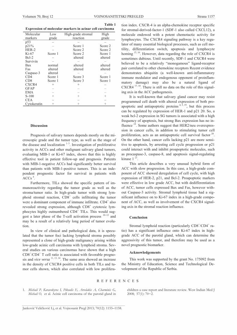
Volumen 70, Broj 12 VOJNOSANITETSKI PREGLED Strana 1157
Jankovi Veli kovi Lj, et al. Vojnosanit Pregl 2013; 70(12): 1155–1158.
Table 1Expression of molecular markers in acinar cell carcinomaMolecularmarkers
Lowgrade
High-grade stromalreaction
Highgrade
p53 – – –p21% – Score 1 Score 2HER-2 – Score 2 Score 2Ki-67 Score 1 Score 2 Score 1Bcl-2 – altered alteredSurvivin – – –Bax normal altered alteredFas altered altered alteredCaspase-3 altered – –CD4 Score 1 Score 3 Score 1CD8 Score 1 Score 3 Score 1CXCR4 + + –GFAP – – –EMA – – –S-100 + + +CEA – – –Cytokeratin + + +
Discussion
Prognosis of salivary tumors depends mostly on the mi-croscopic grade and the tumor type, as well as the stage ofthe disease and localization 1–3. Investigation of proliferativeactivity in ACCs and other malignant salivary gland tumors,evaluating MIB-1 or Ki-67 index, shows that this is highlyeffective tool in patient follow-up and prognosis. Patientswith MIB-1-negative ACCs had significantly better survivalthan patients with MIB-1-positive tumors. This is an inde-pendent prognostic factor for survival in patients withACCs 4.
Furthermore, TILs showed the specific pattern of im-munoreactivity regarding the tumor grade as well as thestroma/tumor ratio. In high-grade tumor with strong lym-phoid stromal reaction, CD8+ cells infiltrating the tumorwere a dominant component of immune infiltrate. CD4+ alsorevealed strong expression, although CD8+ cytotoxic lym-phocytes highly outnumbered CD4+ TILs. This would sug-gest a later phase of the T-cell activation process 10–12 andmay be a result of a relatively long period of tumor evolu-tion.
In view of clinical and pathological data, it is specu-lated that the tumor foci lacking lymphoid stroma possiblyrepresented a clone of high-grade malignancy arising withinlow-grade acinic cell carcinoma with lymphoid stroma. Sev-eral studies on various carcinomas have shown that a highCD8+/CD4+ T cell ratio is associated with favorable progno-sis and vice versa 11, 13, 14. The same area showed an increasein the density of CXCR4 positive cells in both TILs and tu-mor cells shown, which also correlated with low prolifera-
tion index. CXCR-4 is an alpha-chemokine receptor specificfor stromal-derived-factor-1 (SDF-1 also called CXCL12), amolecule endowed with a potent chemotactic activity forlymphocytes. The CXCR4 signaling pathway is a key regu-lator of many essential biological processes, such as cell mo-tility, differentiation switch, apoptosis and lymphocytehoming 15, 16. However, data regarding the role of CXCR4 issometimes dubious. Until recently, SDF-1 and CXCR4 werebelieved to be a relatively “monogamous” ligand-receptorpair correlated to other chemokine receptors Recent evidencedemonstrates ubiquitin (a well-known anti-inflammatoryimmune modulator and endogenous opponent of proinflam-matory damage) may also be a natural ligand ofCXCR4 17, 18. There is still no data on the role of this signal-ing axis in the ACC pathogenesis.
It is well-known that salivary gland cancer may resistprogrammed cell death with altered expression of both pro-apoptotic and antiapoptotic proteins 4, 5, 19, but this processcan be regulated by expression of HER-1 and p21 20. So, aweak bcl-2 expression in SG tumors is associated with a highfrequency of apoptosis, but strong Bax expression has no in-fluence 19. Some authors suggest that HER2/neu overexpres-sion in cancer cells, in addition to stimulating tumor cellproliferation, acts as an antiapoptotic cell survival factor 20.On the other hand, cancer cells lacking p21 are more sensi-tive to apoptosis, by arresting cell cycle progression or p21could interact with and inhibit proapoptotic molecules, suchas procaspase-3, caspase-8, and apoptosis signal-regulatingkinase 1 21.
This article describes a very unusual hybrid form ofACC with slow progression. In this case, a high-grade com-ponent of ACC showed deregulation of cell cycle, with highexpression of HER-2, p21, and Bcl-2. Proapoptotic markerswere effective in low grade ACC, but with dedifferentiationof ACC, tumor cells expressed Bax and Fas, however with-out Caspase-3 activity. Stromal lymphoid tissue had a sig-nificant influence on to Ki-67 index in a high-grade compo-nent of ACC, as well as involvement of the CXCR4 signal-ing axis in the stromal reaction influence.
Conclusion
Stromal lymphoid reaction (particularly CD8+/CD4+ ra-tio) has a significant influence onto Ki-67 index in high-grade ACC of the parotid gland, which can determine theaggressivity of this tumor, and therefore may be used as anovel prognostic biomarker.
Acknowledgements
This work was supported by the grant No. 175092 fromthe Ministry of Education, Science and Technological De-velopment of the Republic of Serbia.
R E F E R E N C E S
1. Michail P, Karavokyros I, Pikoulis E, Arvelakis A, Charminis G,Michail O, et al. Acinic cell carcinoma of the parotid gland in
children a case report and literature review. West Indian Med J2008; 57(1): 70 2.

Strana 1158 VOJNOSANITETSKI PREGLED Volumen 70, Broj 12
Jankovi Veli kovi Lj, et al. Vojnosanit Pregl 2013; 70(12): 1155–1158.
2. Skálová A, Sima R, Vanecek T, Muller S, Korabecna M, Nemcova J,et al. Acinic cell carcinoma with high-grade transformation: areport of 9 cases with immunohistochemical study and analysisof TP53 and HER-2/neu genes. Am J Surg Pathol 2009; 33(8):1137 45.
3. Toshitaka N, Isamu S, Yasuo I, Akira A, Shigeru M., Kazuto Y,et al. Hybrid carcinomas of the salivary glands: report ofnine cases with a clinicopathologic, immunohistochemical,and p53 gene alteration analysis. Mod Pathol 2002; 15(7):724–33.
4. Hellquis HB, Sundelin K, Di Bacco A, Tytor M, Manzotti M, VialeG. Tumour growth fraction and apoptosis in salivary glandacinic cell carcinomas. Prognostic implications of Ki-67 andbcl-2 expression and of in situ end labelling (TUNEL). JPathol 1997; 181(3): 323 9.
5. Yin HF, Okada N, Takagi M. Apoptosis and apoptotic-relatedfactors in mucoepidermoid carcinoma of the oral minor sali-vary glands. Pathol Int 2000; 50(8): 603 9.
6. Tarakji B, Mohammad NZ. Immunohistochemical expression ofp21 in normal tissues of salivary gland, pleomorphic adenomaand carcinoma ex pleomorphic adenoma-(undifferentiated andadenocarcinoma types). Med Oral Patol Oral Cir Bucal 2010;15(5): e697 703.
7. Giannopoulou I, Nakopoulou L, Zervas A, Lazaris AC, StravodimosC, Giannopoulos A, et al. Immunohistochemical study of pro-apoptotic factors Bax, Fas and CPP32 in urinary bladder can-cer: prognostic implications. Urol Res 2002; 30(5): 342 5.
8. Burger M, Glodek A, Hartmann T, Schmitt-Gräff A, Silberstein LE,Fujii N, et al. Functional expression of CXCR4 (CD184) onsmall-cell lung cancer cells mediates migration, integrin activa-tion, and adhesion to stromal cells. Oncogene 2003; 22(50):8093–101.
9. Tímár J, Forster-Horváth C, Lukits J, Döme B, Ladányi A, RemenárE, et al. The effect of leukocyte interleukin injection (Mul-tikine) treatment on the peritumoral and intratumoral sub-population of mononuclear cells and on tumor epithelia: Apossible new approach to augmenting sensitivity to radiationtherapy and chemotherapy in oral cancer—A multicenterphase I/II clinical trial. Laryngoscope 2003; 113(12): 2206 17.
10. Kumamoto Y, Mattei LM, Sellers S, Payne GW, Iwasaki A. CD4+T cells support cytotoxic T lymphocyte priming by controllinglymph node input. Proc Natl Acad Sci USA 2011; 108(21):8749 54.
11. Shedlock DJ, Shen H. Requirement for CD4 T cell help in gen-erating functional CD8 T cell memory. Science 2003;300(5617): 337 9.
12. Sun JC, Bevan MJ. Defective CD8 T cell memory followingacute infection without CD4 T cell help. Science 2003;300(5617): 339 42.
13. Piersma SJ, Jordanova ES, van Poelgeest MI, Kwappenberg KM, vander Hulst JM, Drijfhout JW, et al. High number of intraepithelialCD8+ tumor-infiltrating lymphocytes is associated with theabsence of lymph node metastases in patients with large early-stage cervical cancer. Cancer Res 2007; 67(1): 354 61.
14. Sato E, Olson SH, Ahn J, Bundy B, Nishikawa H, Qian F, et al.Intraepithelial CD8+ tumor-infiltrating lymphocytes and ahigh CD8+/regulatory T cell ratio are associated with favor-able prognosis in ovarian cancer. Proc Natl Acad Sci USA2005; 102: 18538 43.
15. Khan MZ, Brandimarti R, Musser BJ, Danielle Marie Resue BJ, Fa-tatis DM, Meucci O. The chemokine receptor CXCR4 regulatescell-cycle proteins in neurons. J Neurovirol 2003; 9(3): 300–14.
16. Chen AM, Granchi PJ, Garcia J, Bucci MK, Fu KK, Eisele DW. Lo-cal-regional recurrence after surgery without postoperative ir-radiation for carcinomas of the major salivary glands: implica-tions for adjuvant therapy. Int J Radiat Oncol Biol Phys 2007;67(4): 982 7.
17. Majetschak M. Extracellular ubiquitin: immune modulator andendogenous opponent of damage-associated molecular patternmolecules. J Leukoc Biol 2010; 89(2): 205–219.
18. Winsauer P, Walker E, Vande Stouwe C, Porretta C, Molina P.Chronic -9-tetrahydrocannabinol administration increaseslymphocyte CXCR4 expression in rhesus macaques. J Neu-roimmune Pharmacol 2011; 6(4): 540 5.
19. Soini Y, Törmänen U, Pääkkö P. Apoptosis is inversely related tobcl-2 but not to bax expression in salivary gland tumours.Histopathology 1998; 32(1): 28 34.
20. Roh H, Pippin J, Drebin JA. Down-regulation of HER2/neu ex-pression induces apoptosis in human cancer cells that overex-press HER2/neu. Cancer Res 2000; 60(3): 560 5.
21. Chiang CT, Way TD, Lin JK. Sensitizing HER2-overexpressingcancer cells to luteolin-induced apoptosis through suppressingp21WAF1/CIP1 expression with rapamycin. Mol Cancer Ther2007; 6(7): 2127–38.
Received on December, 20 2011.Revised on October 8, 2012.
Accepted on October 24, 2012.
Vojnosanit Pregl 2013; 70(12): 1159–1161. VOJNOSANITETSKI PREGLED Strana 1159
Correspondence to: Aleksandra Lovrenski, Center for Pathology, Institute for Pulmonary Diseases of Vojvodina, Sremska Kamenica,Serbia. E-mail: [email protected]
C A S E R E P O R T UDC: 616.24-006-033.2 + 616.379-091.8-031DOI: 10.2298/VSP1312159L
Multisystem Langerhans cell histiocytosis coexisting withmetastasizing adenocarcinoma of the lung: A case report
Multisistemska histiocitoza Langerhansovih elija udružena sametastaziraju im adenokarcinomom plu a
Aleksandra Lovrenski*†, Mirna Djuri †‡, Ištvan Klem*†, Živka Eri*†,Milana Panjkovi *†, Dragana Tegeltija*†, Djordje Považan†‡
*Center for Pathology, ‡Clinic for General Pulmology, Institute for Pulmonary Diseasesof Vojvodina, Sremska Kamenica, Serbia; †Faculty of Medicine, University of Novi Sad,
Novi Sad, Serbia
Abstract
Introduction. Langerhans cell histiocytosis (LCH) is an un-common disease of unknown etiology characterized by un-controlled proliferation and infiltration of various organs byLangerhans cells. Case report. We presented a 54-year-oldman, heavy smoker, with dyspnea, cough, hemoptysis,headache and ataxia, who died shortly after admission toour hospital. On the autopsy, tumor was found in the poste-rior segment of the right upper pulmonary lobe as well as aright-sided occipitoparietal lesion which penetrated into theright ventricle resulting in internal and external hemato-cephalus. Histologically and immunohistohemically, the di-agnosis of primary lung adenocarcinoma with brain metas-tasis was made (tumor cells showed positivity for CK7 andTTF-1 which confirmed the diagnosis). In the lung paren-chyma around the tumor, as well as in brain tissue aroundthe metastatic adenocarcinoma histiocytic lesions werefound. Light microscopic examination of the other organsalso showed histiocytic lesions involving the pituitary gland,hypothalamus, spleen and mediastinal lymph nodes. Immu-nohistochemical studies revealed CD68, S-100 and CD1aimmunoreactivity within the histiocytes upon which the di-agnosis of Langerhans' cells histiocytosis was made. Con-clusion. The multisystem form of LCH with extensive or-gan involvement was an incidental finding, while metastaticlung adenocarcinoma to the brain that led to hematocepha-lus was the cause of death.
Key words:histiocytosis, langerhans-cell; diagnosis; lungneoplasms; adenocarcinoma; immunohistochemistry.
Apstrakt
Uvod. Histiocitoza Langerhansovih elija je retko obolje-nje nepoznate etiologije koje se karakteriše nekontrolisa-nom proliferacijom i infiltracijom razli itih organa Lan-gerhansovim elijama. Prikaz bolesnika. Prikazan je bo-lesnik, star 54 godine, teški puša , sa simptomima dispne-je, kašlja, hemoptizija, glavobolje i ataksije koji je kratkonakon prijema u našu ustanovu egzitirao. Na obdukciji,na en je tumor u posteriornom segmentu desnog gornjegrežnja, kao i tumorska masa lokalizovana desno okcipito-parijetalno koja je penetrirala u desnu komoru dovode ido unutrašnjeg i spoljašnjeg hematocefalusa. Histološki iimunohistohemijski, postavljena je dijagnoza primarnogplu nog adenokarcinoma sa metastazom u mozak (tumor-ske elije su pokazale pozitivnost za CK7 i TTF-1, što jepotvrdilo dijagnozu). U plu nom parenhimu oko tumora,kao i u moždanom parenhimu oko tumora na ene su his-tiocitne lezije. Histološka analiza ise aka uzetih iz hipofize,hipotalamusa, slezine i medijastinalnih limfnih vorova ot-krila je, tako e, prisustvo ovih lezija. Imunohistohemijski,ove lezije pokazale su pozitivnost na CD68, S-100 i CD1a,na osnovu ega je i postavljena dijagnoza histiocitoze Lan-gerhansovih elija. Zaklju ak. Multisistemska forma his-tiocitoze Langerhansovih elija sa ekstenzivnim zahvata-njem organa je slu ajan nalaz, dok je hematocefalus izaz-van metastatskim adenokarcinomom bio uzrok smrti kodovog bolesnika.
Klju ne re i:histiocitoza x; dijagnoza; plu a, neoplazme;adenokarcinom; imunohistohemija.

Strana 1160 VOJNOSANITETSKI PREGLED Volumen 70, Broj 12
Lovrenski A, et al. Vojnosanit Pregl 2013; 70(12): 1159–1161.
Introduction
Langerhans cell histiocytosis (LCH) includes diseasespreviously designated as histiocytosis X, eosinophilic granu-loma, Letterer-Siwe disease, Hand-Schuller-Christian syn-drome and Langerhans cell granulomatosis. This is an un-common disease of unknown etiology characterized by un-controlled proliferation and infiltration of various organs byLangerhans cells, often organized into granulomas 1. It occurspredominantly in children and young adults, but no age is anexemption. Practically any organ can be involved, but boneand skin are the sites of predilection. Patients are categorizedas having single-system disease at a single or multiple sites, oras having multisystem disease. Single-system disease of LCHinvolves mainly bones or lungs and usually follows a benigncourse and can regress spontaneously. The clinical presenta-tion of multisystem LCH, which carries a poor prognosis in anumber of cases, is highly variable depending on the organsinvolved, mainly bones, skin, lungs, pituitary glands and lesscommonly liver, spleen, hematopoietic and central nervoussystem 1–3. The association of single-system disease with ma-lignant neoplasm is not so rare (particularly in association withmalignant lymphoma), but association of multisystem LCHwith a malignant neoplasm is rare and, generally, has been thesubject of isolated case reports 4. To our knowledge, this is thefirst case reporting on the association of multisystem LCHwith metastasizing adenocarcinoma of the lung.
Case report
A 54-year-old man, heavy smoker (30 years/3 packs aday), was admitted to our hospital for further diagnostic ap-proach to the radiologically detected change in the right lung.
Four months before admission shortness of breath and coughwith hemoptysis occured, and a month before admission thepatient was refered to the neurologist because of walking in-stability, loss of strength in the left half of the body, andmorning headaches. Computerized tomography (CT) scan ofthe endocranium showed right-sided occipitoparietal lesion,which primarily exhibited characteristics of secondary infil-tration, and CT scan of the thorax showed inhomogeneous,extensive infiltration predominantly localized in the upperlobe. On the fourth day after admission hemoptysis occured,and bronchoscopic examination was performed, but histo-pathological findings did not clarify the etiology of thechange. After two days a deterioration in general conditionwith intense headache developed, and despite all the appliedtherapeutic measures the patient passed away.
At autopsy, macroscopic examination of the lungs re-vealed an excavated tumor mass 1.8 cm in largest dimensionin the posterior segment of the right upper lobe, which histo-pathologically corresponded to adenocarcinoma. In brain pa-renchyma, right-sided occipitoparietal necrotic, and hemor-rhagic lesion 5.7 3,8 cm in largest diameter, which pene-trated into the right ventricle resulting in internal and exter-nal hematocephalus, was observed. Histologically and im-munohistohemically, the diagnosis of primary lung adeno-carcinoma with brain metastasis was made (tumor cellsshowed positivity for CK7 and TTF-1 which confirmed thediagnosis) (Figures 1a and 1b).
Within the tumor, in the lung parenchyma around the tu-mor, as well as in the brain tissue around metastatic adenocar-cinoma, histiocytic lesions were found (Figure 2a). Light mi-croscopic examination of the other organs also showed histio-cytic lesions involving the pituitary gland, spleen, hypothala-mus, and mediastinal lymph nodes (Figures 2b and 2c).
a) b) Fig. 1 – a) Adenocarcinoma of the lung (HE, 10); b) Metastatic adenocarcinoma in the brain parenchyma (HE, 5).
a) b) c)
Fig. 2 – a) Histiocytic lesion within the lung parenchyma around the tumor (HE, 10); b) Histiocytic lesion in the mediasti-nal lymph node (HE, 10); c) Histiocytic lesion in the hypothalamus (HE, 20).
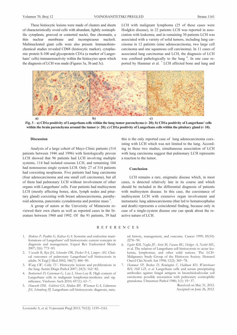
Volumen 70, Broj 12 VOJNOSANITETSKI PREGLED Strana 1161
Lovrenski A, et al. Vojnosanit Pregl 2013; 70(12): 1159–1161.
These histiocytic lesions were made of clusters and sheetsof characteristically ovoid cells with abundant, lightly eosinoph-ilic cytoplasm, grooved or contorted nuclei, fine chromatin, athin nuclear membrane and inconspicuous nucleoli.Multinucleated giant cells were also present. Immunohisto-chemical studies revealed CD68 (histiocytic marker), cytoplas-mic protein S-100 and glycoprotein CD1a (a marker of Langer-hans’ cells) immunoreactivity within the histiocytes upon whichthe diagnosis of LCH was made (Figures 3a, 3b and 3c).
Discussion
Analysis of a large cohort of Mayo Clinic patients (314patients between 1946 and 1996) with histologically provenLCH showed that 96 patients had LCH involving multiplesystems, 114 had isolated osseous LCH, and remaining 104had nonosseous single system LCH. Only 27 of 314 patientshad coexisting neoplasms. Five patients had lung carcinoma(four adenocarcinoma and one small cell carcinoma), but allof them had pulmonary LCH without involvement of otherorgans with Langerhans' cells. Four patients had multisystemLCH (mostly affecting bones, skin, lymph nodes and pitui-tary gland) coexisting with breast adenocarcinoma, parathy-roid adenoma, pancreatic cystadenoma and pontine mass 5.
A group of autors at the University of Minnesota re-viewed their own charts as well as reported cases in the lit-erature between 1960 and 1992. Of the 91 patients, 39 had
LCH with malignant lymphoma (25 of these cases wereHodgkin disease), in 22 patients LCH was reported in asso-ciation with leukemia, and in remaining 30 patients LCH wasassociated with a variety of solid tumors, including lung car-cinoma in 12 patients (nine adenocarcinoma, two large cellcarcinoma and one squamous cell carcinoma). In 11 cases ofassociated lung carcinomas and LCH, the diagnosis of LCHwas confined pathologically to the lung 6. In one case re-ported by Hammar et al. 7 LCH affected bone and lung and
this is the only reported case of lung adenocarcinoma coex-isting with LCH which was not limited to the lung. Accord-ing to these two studies, simultaneous association of LCHwith lung carcinoma suggest that pulmonary LCH representsa reaction to the tumor.
Conclusion
LCH remains a rare, enigmatic disease which, in mostcases, is detected relatively late in its course and whichshould be included in the differential diagnosis of patientswith multisystem disease. In this case, the coexistance ofmultisystem LCH with extensive organ involvement andmetastastic lung adenocarcinoma (that led to hematocephalusand death) represents a coincidental finding, because only incase of a single-system disease one can speak about the re-active nature of LCH.
R E F E R E N C E S
1. Makras P, Piaditis G, Kaltsas GA. Systemic and endocrine mani-festations of Langerhans’ cell histiocytosis: current concepts indiagnosis and management. Expert Rev Endocrinol Metab2007; 2(6): 773 83.
2. Vassallo R, Ryu JH, Schroeder DR, Decker PA, Limper AH. Clini-cal outcomes of pulmonary Langerhans'-cell histiocytosis inadults. N Engl J Med 2002; 346(7): 484 90.
3. Wang CW, Colby TV. Histiocytic lesions and proliferations inthe lung. Semin Diagn Pathol 2007; 24(3): 162 82.
4. Benharroch D, Guterman G, Levy I, Shaco-Levy R. High content ofLangerhans cells in malignant lymphoma-incidence and sig-nificance. Virchows Arch 2010; 457(1): 63 7.
5. Howarth DM, Gilchrist GS, Mullan BP, Wiseman GA, EdmonsonJH, Schomberg PJ. Langerhans cell histiocytosis: diagnosis, natu-
ral history, management, and outcome. Cancer 1999; 85(10):2278 90.
6. Egeler RM, Neglia JP, Aricò M, Favara BE, Heitger A, Nesbit ME,et al. The relation of Langerhans cell histiocytosis to acute leu-kemia, lymphomas, and other solid tumors. The LCH-Malignancy Study Group of the Histiocyte Society. HematolOncol Clin North Am 1998; 12(2): 369 78.
7. Hammar SP, Bockus D, Remington F, Hallman KO, WinterbauerRH, Hill LD, et al. Langerhans cells and serum precipitatingantibodies against fungal antigens in bronchioloalveolar cellcarcinoma: possible association with pulmonary eosinophilicgranuloma. Ultrastruct Pathol 1980; 1(1): 19 37.
Received on May 31, 2012.Accepted on June 26, 2012.
a) b) c)Fig. 3 – a) CD1a positivity of Langerhans cells within the lung tumor parenchyma ( 20); b) CD1a positivity of Langerhans’ cellswithin the brain parenchyma around the tumor ( 20); c) CD1a positivity of Langerhans cells within the pituitary gland ( 10).
Strana 1162 VOJNOSANITETSKI PREGLED Vojnosanit Pregl 2013; 70(12): 1162–1164.
Correspondence to: Mihailo Vukmirovi , Clinical Center of Montenegro, Center of Cardiology, Ljubljanska bb, 81000 Podgorica, Mon-tenegro. Phone: +382 20 412 258. Mob. +382 69 304 400. E-mail: [email protected]
C A S E R E P O R T UDC: 616.14-056.7::611.94]:615.817DOI: 10.2298/VSP1312162V
Successful implantation of a biventricular pacing and defibrillatordevice via a persistent left superior vena cavaUspešna ugradnja resinhronizaciono-defibrilatorskog aparata preko perzistentne
leve gornje šuplje vene
Mihailo Vukmirovi *, Lazar Angelkov†, Filip Vukmirovi ‡,Irena Tomaševi Vukmirovi §
*Center of Cardiology, ‡Center of Patology, §Center of Radiology, Clinical Center ofMontenegro, Podgorica, Montenegro; †Institute of Cardiovascular Disease “Dedinje”,
Belgrade, Serbia
Abstract
Introduction. Persistent left superior vena cava is the mostcommon thoracic venous abnormality which is usuallyasymptomatic, found incidentally during pacemaker im-plantation. The main problem is related to reaching the ap-propriate pacing site and ensuring stable lead placement.Case report. We reported a successful implantation of a bi-ventricular pacing and defibrillator device (CRT-D) via apersistent left superior vena cava in a 55-year-old man withdilated cardiomyopathy and severe heart failure. A persis-tent left superior vena cava was detected during CRT-D im-plantation. We managed to position electrodes in the rightventricular outflow tract, a posterior branch of the coronarysinus and in the right atrium. Conclusion. Congenitalanomalies of thoracic veins may complicate lead placementon the appropriate and stable position. The presented casedemonstrates a successful biventricular pacing and defibril-lator therapy device implantation in a patient with dilatedcardiomyopathy and severe heart failure.
Key words:vena cava superior; vascular malformations; cardiacpacing artificial; defibrilator implantable; treatmentoutcome.
Apstrakt
Uvod. Leva gornja šuplja vena je naj eš a anomalija vena to-raksa koja je uglavnom asimptomatska. Obi no se verifikujeprilikom ugradnje pejsmejkera kada može jako otežati, a ne-kada i potpuno onemogu iti postavljanje elektroda. Problemkoji nastaje zbog anomalije venskog sistema je u odgovaraju-em pozicioniranju elektroda, odnosno u njihovoj stabilnosti.
Prikaz bolesnika. Prikazali smo uspešnu implantaciju resin-hronizacionog defibrilatorskog aparata (CRT-D) kod bolesni-ka starog 55 godina, sa dilatativnom kardiomiopatijom, odno-sno teškom sr anom slaboš u, kojem je u toku intervencijedetektovana perzistentna leva gornja šuplja vena. Uprkosanomaliji venskog sistema toraksa, uspeli smo da pozicioni-ramo elektrodu u izlazni trakt desne komore, posteriornugranu koronarnog sinusa, odnosno u desnu pretkomoru. Za-klju ak. Uro ene anomalije vena toraksa mogu jako da ote-žaju postavljanje elektroda na adekvatnu, odnosno stabilnupoziciju. Ipak, pokazali smo da se i kod ovakvih bolesnikamože uspešno ugraditi resinhronizacioni defibrilator.
Klju ne re i:v. cave superior; krvni sudovi, malformacije; srce,vešta ko uskla ivanje ritma; defibrilator, implantabilni;le enje, ishod.
Introduction
Persistent left superior vena cava (PLSVC) is the mostcommon variation in the thoracic venous system. PLSVC isfound in 0.3% to 0.5% of the population and in 5% to 10%of patients with other congenital heart defects (atrial septaldefect, bicuspid aortic valve, coarctation of aorta, coronarysinus ostial atresia and cor triatriatum) 1–6. Several subtypesof PLSVC can be distinguished. In 68% of cases an in-nominate vein bridges the two superior venae cavae 7. In
about 20% of patients, as well as in the presented case, theright superior vena cava (RSVC) is absent resulting indrainage of venous blood from the head and both armsthrough the left brachiocephalic vein, PLSVC and thecoronary sinus into the right atrium 8. This condition istypically asymptomatic, usually incidentally discoveredduring pacemaker implantation can complicate lead place-ment through the subclavian approach. We reported a caseof a successful biventricular pacing and defibrillator deviceimplantation (CRT-D) via a persistent left superior vena in
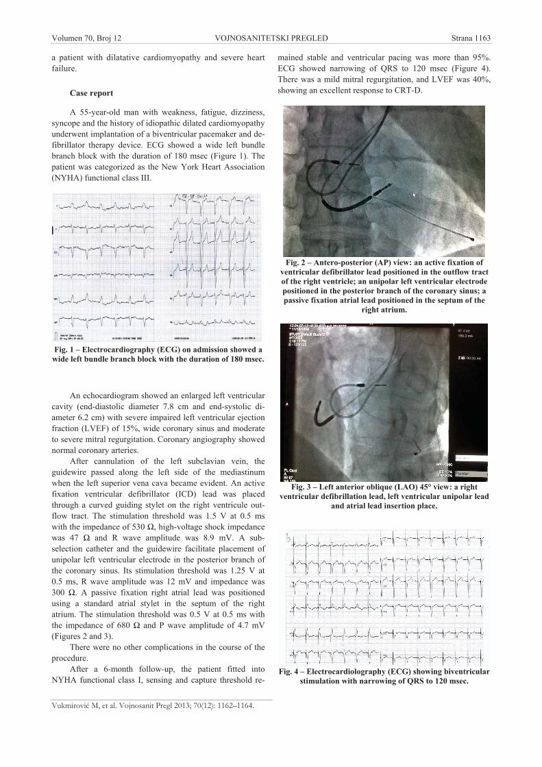
Volumen 70, Broj 12 VOJNOSANITETSKI PREGLED Strana 1163
Vukmirovi M, et al. Vojnosanit Pregl 2013; 70(12): 1162–1164.
a patient with dilatative cardiomyopathy and severe heartfailure.
Case report
A 55-year-old man with weakness, fatigue, dizziness,syncope and the history of idiopathic dilated cardiomyopathyunderwent implantation of a biventricular pacemaker and de-fibrillator therapy device. ECG showed a wide left bundlebranch block with the duration of 180 msec (Figure 1). Thepatient was categorized as the New York Heart Association(NYHA) functional class III.
Fig. 1 – Electrocardiography (ECG) on admission showed awide left bundle branch block with the duration of 180 msec.
An echocardiogram showed an enlarged left ventricularcavity (end-diastolic diameter 7.8 cm and end-systolic di-ameter 6.2 cm) with severe impaired left ventricular ejectionfraction (LVEF) of 15%, wide coronary sinus and moderateto severe mitral regurgitation. Coronary angiography showednormal coronary arteries.
After cannulation of the left subclavian vein, theguidewire passed along the left side of the mediastinumwhen the left superior vena cava became evident. An activefixation ventricular defibrillator (ICD) lead was placedthrough a curved guiding stylet on the right ventricule out-flow tract. The stimulation threshold was 1.5 V at 0.5 mswith the impedance of 530 , high-voltage shock impedancewas 47 and R wave amplitude was 8.9 mV. A sub-selection catheter and the guidewire facilitate placement ofunipolar left ventricular electrode in the posterior branch ofthe coronary sinus. Its stimulation threshold was 1.25 V at0.5 ms, R wave amplitude was 12 mV and impedance was300 . A passive fixation right atrial lead was positionedusing a standard atrial stylet in the septum of the rightatrium. The stimulation threshold was 0.5 V at 0.5 ms withthe impedance of 680 and P wave amplitude of 4.7 mV(Figures 2 and 3).
There were no other complications in the course of theprocedure.
After a 6-month follow-up, the patient fitted intoNYHA functional class I, sensing and capture threshold re-
mained stable and ventricular pacing was more than 95%.ECG showed narrowing of QRS to 120 msec (Figure 4).There was a mild mitral regurgitation, and LVEF was 40%,showing an excellent response to CRT-D.
Fig. 2 – Antero-posterior (AP) view: an active fixation ofventricular defibrillator lead positioned in the outflow tractof the right ventricle; an unipolar left ventricular electrodepositioned in the posterior branch of the coronary sinus; apassive fixation atrial lead positioned in the septum of the
right atrium.
Fig. 3 – Left anterior oblique (LAO) 45° view: a rightventricular defibrillation lead, left ventricular unipolar lead
and atrial lead insertion place.
Fig. 4 – Electrocardiolography (ECG) showing biventricularstimulation with narrowing of QRS to 120 msec.

Strana 1164 VOJNOSANITETSKI PREGLED Volumen 70, Broj 12
Vukmirovi M, et al. Vojnosanit Pregl 2013; 70(12): 1162–1164.
Discussion
Although the left superior vena cava may complicate orcompletely disable biventricular pacemaker and defibrillatordevices implantation, several cases of successful implantationhave been described in the literature 7, 9, 10. However, some dif-ficulties may occur during this procedure. Right ventricularlead placement is the major problem. In patients with the in-nominate vein which connects the right and left superior venacava, right ventricular lead implantation is usually achievablethrough this one using a conventional method 7. Typically, thePLSVC drains directly into the right atrium through the greatlyenlarged coronary sinus because of a significant increase inblood flow 7. When an electrode is introduced into the rightatrium, the tip of the right ventricular lead usually tends to de-flect away from the tricuspid annulus. There are several meth-ods to overcome this difficulty. Biffi et al. 11 used a manuallyformed U-shaped stylet, requiring considerable manoeuvring,forming a loop in the right atrium, using the right atrial freewall for support. Srimannarayana et al. 12 reported the use ofatrial J-shaped stylet for ventricular lead placement 12. Kon-stantino et al. 13 demonstrated ventricular lead placement intothe right ventricular outflow tract using a coronary sinus deliv-ery system. These techniques allow acute angulation of ven-tricular electrode to reach right ventricul through the tricus-pidal valve, which is the critical juncture mostly requiring ad-ditional complex maneuvers. These maneuvers have not beencompletely described in the published literature so far 14, 15.
In the presented case of ICD lead implantation we useda manually formed U-shaped stylet with the rotation of elec-trode in clockwise direction toward the tricuspid valve an-
nulus and after a sudden withdrawal of the stylet, the ven-tricular lead got into the right ventricul across the tricuspidalvalve. The atrium lead was implanted using J-shaped stylet,but with contraclockwise rotation in the right atrium septum.It is advisable to apply both maneuvers in the direction ofrotation opposite to the one done during the conventional bi-ventricular pacemaker device lead implantation.
The left ventricular lead, if possible, is introducedthrough the PLSVC into the coronary sinus and implanted inthe appropriate coronary vein. The difficulty in coronary si-nus cannulation is still one of the reasons for failing biven-tricular pacing system in implantation. Balloon-occlusionretrograde angiography is not possible because of coronarysinus dilation. Coronary cannulation vein requires a lead tobe manipulated through sharp angles. Sometimes, left coro-nary angiography is performed in order to evaluate possibleposition for left ventricular lead placement. However, thiscan lead to large contrast volume and prolonged angio-graphic time. In the case, we used the sub-selection catheterwith a curve of 90° and a guidewire which facilitates unipo-lar left ventricular electrode placement in the posteriorbranch of the coronary sinus.
Conclusion
Despite venous abnormalities, biventricular pacing anddefibrillator device implantation via a persistent left superiorvena cava is feasible. According to our findings, these ma-neuvers have not been completely described in the publishedliterature so far. We hope that these techniques may facilitatelead implantation via the left superior vena cava.
R E F E R E N C E S
1. Fry AC, Warwicker P. Bilateral superior vena cava. N Engl J Med2007; 356: 18.
2. Peltier J, Destrieux C, Desme J, Renard C, Remond A, Velut S. The per-sistent left superior vena cava: anatomical study, pathogenesis andclinical considerations. Surg Radiol Anat 2006; 28(2): 206 10.
3. Schreve-Steensma AM, Van der Valk PH, Ten Kate JB, Kofflard MJ.Discovery of a persistent left superior vena cava during pacemakerimplantation. Neth Heart J 2008; 16(7 8): 272–4.
4. Tak T, Crouch E, Drake GB. Persistent left superior vena cava: in-cidence, significance and clinical correlates. Int J Card 2002; 82(1):91 3.
5. Jovi Z, Mijailovi Z, Obradovi S, Tav iovski D, Matunovi R, Rusovi S.Successful implantation of a permanent pacemaker through a per-sistent left superior vena cava by using a right subclavian ap-proach. Vojnosanitetski pregled 2011; 68(9): 792 4.
6. Reinhardt D, Surber R, Kuehnert H, Heinke M, Figulla HR. Implantationof a re- synchronization device in a patient with persistent left supe-rior vena cava-a case report. Indian Heart J 2010; 62(4): 344 5.
7. Antonelli D, Freedberg M, Feldman A. Implantation of a Resynchro-nization Implantable Cardioverter Defibrillator in a Patient withPersistent Left Superior Vena Cava. Indian Pacing Electrophysiol J2007; 7(4): 246 8.
8. Lappegard KT, Prytz JF, Haug B. Pacemaker implantation in patientswith persistent left superior vena cava. Heart Vessels 2004; 19(3):153 4.
9. Smyth YM, Barrett CD, Fahy GJ. Biventricular pacemaker implant ina patient with persistent left sided superior vena cava. Heart 2005;91(11): 1427.
10. Gasparini M, Mantica M, Galimberti P, Coltorti F, Simonini S, Ceriotti C,et al. Biventricular pacing via a persistent left superior vena cava:report of four cases. Pacing Clin Electrophysiol 2003; 26(1):192 6.
11. Biffi M, Boriani G, Frabetti L, Bronzetti G, Branzi A. Left superiorvena cava persistence in patients undergoing pacemaker or cardio-verter-defibrillator implantation: a 10-year experience. Chest 2001;120(1): 139 44.
12. Srimannarayana J, Sekhlar Varma R, Satheesh S, Anilkumar R,Balachander J. Transvenous permanent pacemaker implantationthrough persistent left superior vena cava. Indian Heart J 2004;56(4): 346 8.
13. Konstantino Y, Kusniec J, Shohat-Zabarski R, Battler A, Strasberg B.Cardiac defibrillator implantation via persistent left superior venacava facilitated by a coronary sinus delivery system. Europace2009; 11(1): 119 20.
14. Rogers D, Walker F, Chow F, Lambiase PD. Biventricular DeviceImplantation in a Patient with Congenitally Corrected Transposi-tion and Left-Sided Superior Vena Cava. Pacing Clin Electro-physiol 2008; 31(4): 499 502.
15. Kapetanopoulos A, Peckham G, Kiernan F, Clyne C, Kluger J, MigeedMA. Implantation of a biventricular pacing and defibrillator devicevia a persistent left superior vena cava. J Cardiovasc Med(Hagerstown) 2006; 7(6): 430 3.
Received on June 10, 2012.Revised on August 14, 2012.
Accepted on September 28, 2012.
Vojnosanit Pregl 2013; 70(12): 1165–1170. VOJNOSANITETSKI PREGLED Strana 1165
Correspondence to: Goran Brajuškovi , Faculty of Biology, University of Belgrade, 11 000 Belgrade, Serbia. Phone: + 381 11 263 9 100.E-mail: [email protected]
H I S T O R Y O F M E D I C I N E UDC: 575:61]:577.2(091)
The 60th anniversary of the discovery of DNA secondary structureOtkri e sekundarne strukture molekula DNK – 60-godišnjica
Goran Brajuškovi , Dušanka Savi Pavi evi , Stanka Romac
Faculty of Biology, University of Belgrade, Belgrade, Serbia
Key words:DNA; molecular biology; genomics; history ofmedicine; nobel prize.
Klju ne re i:DNK; biologija, molekulska; genomika; istorijamedicine; nobelova nagrada.
Introduction
The year 2013 is the year of great anniversaries in mo-lecular biology. In 1953 Watson and Crick published themodel of DNA structure in the scientific journal “Nature”,indicating the model of its self-replication as well 1, 2. Thiswas the final proof that DNA molecule contains genetic in-formation. With this discovery, exactly 60 years ago, mo-lecular biology became distinct scientific discipline and to-day it is one of the most dynamic fields of science. There isno doubt that completion of one of the largest and the mostexpensive scientific project of all times, deciphering the se-quence of the human genome, is one of the milestones thatmarked the beginning of the 21st century. The InternationalHuman Genome Sequencing Consortium published the re-sults of their project in “Nature” in 2001 3. The very next daythe results of the work done by a group of scientists em-ployed by the company “Celera” appeared in “Science” 4.The complete sequence of the human genome with the as-sessment of the gene number was published in 2003 5. At thetime, the estimation of the number of the protein-codinggenes was 30,000. Since then every year we witness a newrevision of the number of human genes. According to the lastdata, the number of protein-coding genes is slightly morethan 20,000 6. Starting in 2003, the Encyclopedia of DNAElements (ENCODE) project set out to map which parts ofhuman chromosomes are transcribed, how transcription isregulated and how the process is affected by the way DNA ispackaged in the cell nucleus. The results of this project, pub-lished in 2012, showed that 80.4% of the human genomedisplays some functionality in at least one of 147 differentcell types analysed 7.
While deciphering the human genome and other spe-cies’ genomes, the new scientific branch, genomics, was es-
tablished. The object of study in genomics is the whole ge-nomes of species. It is the opinion of the scientific commu-nity that genomics of all species will bring about the mostprogress in the everlasting human struggle against incurablediseases and ageing 8. The most important discoveries thatenabled the advances in molecular biology and the beginningof the genomic era will be presented in this article.
Discovery of the secondary structure of DNAmolecule
In spring 1951, during a zoology congress, the Americanzoologist James Dewey Watson (born 1928) met the NewZealander Maurice Hugh Frederick Wilkins (1916–2004) whoshowed him diffraction images of DNA molecule. For youngWatson, this experience was initiation into the research of thechemical structure of nucleic acids and proteins. By the end of1952 Watson also met a British physicist and biologist FrancisHarry Compton Crick (1916–2004). The two started workingtogether on the 3-dimensional model of a DNA molecule. Inno more than couple of months, making molecular modelsbased on characteristics of diffraction images of DNA, Watsonand Crick established that a DNA molecule consisted of twoantiparallel polynucleotide strands joined together by hydro-gen bonds 9.
On April 25, 1953 in its 171st volume, the journal “Na-ture” published the article “Molecular structure of nucleic ac-ids – A Structure for Deoxyribose Nucleic Acid” 1. It wasknown at that time that DNA is the carrier of hereditary infor-mation. This was mainly due to the work of Avery et al. 10 whoinvestigated the transformation of Pneumococcus type IIIbacteria and the horizontal gene transfer in this species. There-fore, it is not surprising that this publication was met by theutmost interest of the scientific community as soon as it ap-
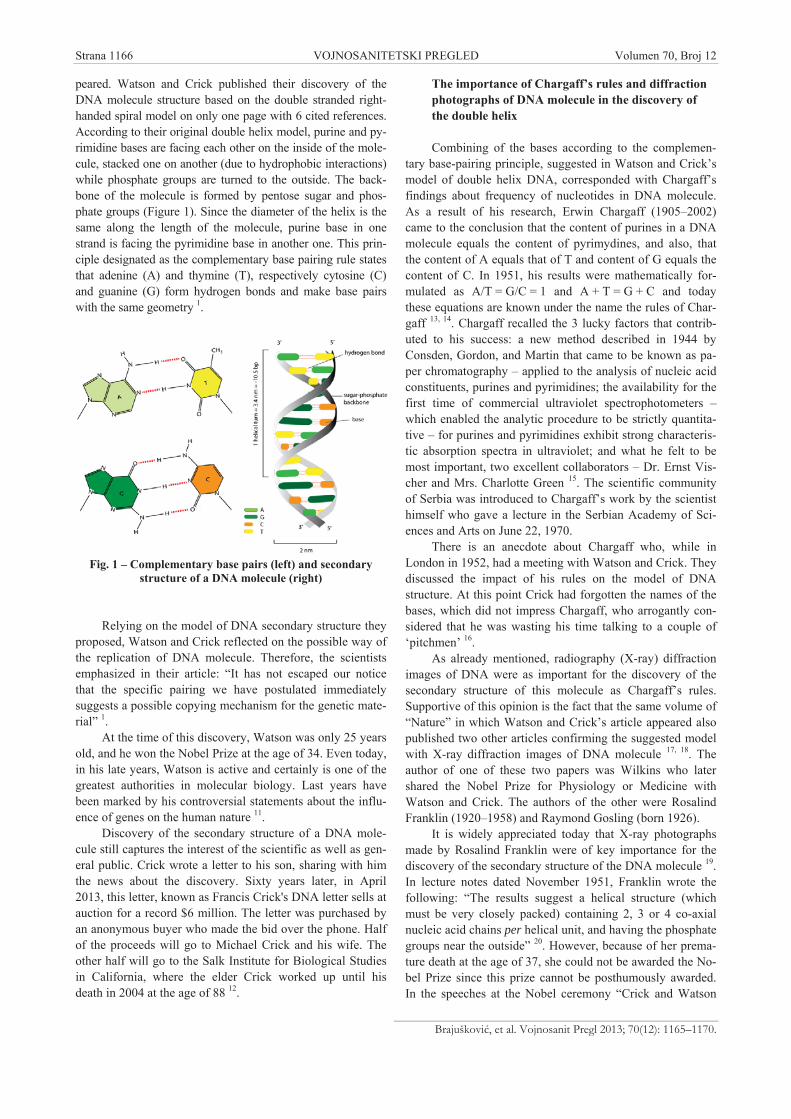
Strana 1166 VOJNOSANITETSKI PREGLED Volumen 70, Broj 12
Brajuškovi , et al. Vojnosanit Pregl 2013; 70(12): 1165–1170.
peared. Watson and Crick published their discovery of theDNA molecule structure based on the double stranded right-handed spiral model on only one page with 6 cited references.According to their original double helix model, purine and py-rimidine bases are facing each other on the inside of the mole-cule, stacked one on another (due to hydrophobic interactions)while phosphate groups are turned to the outside. The back-bone of the molecule is formed by pentose sugar and phos-phate groups (Figure 1). Since the diameter of the helix is thesame along the length of the molecule, purine base in onestrand is facing the pyrimidine base in another one. This prin-ciple designated as the complementary base pairing rule statesthat adenine (A) and thymine (T), respectively cytosine (C)and guanine (G) form hydrogen bonds and make base pairswith the same geometry 1.
Fig. 1 – Complementary base pairs (left) and secondarystructure of a DNA molecule (right)
Relying on the model of DNA secondary structure theyproposed, Watson and Crick reflected on the possible way ofthe replication of DNA molecule. Therefore, the scientistsemphasized in their article: “It has not escaped our noticethat the specific pairing we have postulated immediatelysuggests a possible copying mechanism for the genetic mate-rial” 1.
At the time of this discovery, Watson was only 25 yearsold, and he won the Nobel Prize at the age of 34. Even today,in his late years, Watson is active and certainly is one of thegreatest authorities in molecular biology. Last years havebeen marked by his controversial statements about the influ-ence of genes on the human nature 11.
Discovery of the secondary structure of a DNA mole-cule still captures the interest of the scientific as well as gen-eral public. Crick wrote a letter to his son, sharing with himthe news about the discovery. Sixty years later, in April2013, this letter, known as Francis Crick's DNA letter sells atauction for a record $6 million. The letter was purchased byan anonymous buyer who made the bid over the phone. Halfof the proceeds will go to Michael Crick and his wife. Theother half will go to the Salk Institute for Biological Studiesin California, where the elder Crick worked up until hisdeath in 2004 at the age of 88 12.
The importance of Chargaff’s rules and diffractionphotographs of DNA molecule in the discovery ofthe double helix
Combining of the bases according to the complemen-tary base-pairing principle, suggested in Watson and Crick’smodel of double helix DNA, corresponded with Chargaff’sfindings about frequency of nucleotides in DNA molecule.As a result of his research, Erwin Chargaff (1905–2002)came to the conclusion that the content of purines in a DNAmolecule equals the content of pyrimydines, and also, thatthe content of A equals that of T and content of G equals thecontent of C. In 1951, his results were mathematically for-mulated as A/T = G/C = 1 and A + T = G + C and todaythese equations are known under the name the rules of Char-gaff 13, 14. Chargaff recalled the 3 lucky factors that contrib-uted to his success: a new method described in 1944 byConsden, Gordon, and Martin that came to be known as pa-per chromatography – applied to the analysis of nucleic acidconstituents, purines and pyrimidines; the availability for thefirst time of commercial ultraviolet spectrophotometers –which enabled the analytic procedure to be strictly quantita-tive – for purines and pyrimidines exhibit strong characteris-tic absorption spectra in ultraviolet; and what he felt to bemost important, two excellent collaborators – Dr. Ernst Vis-cher and Mrs. Charlotte Green 15. The scientific communityof Serbia was introduced to Chargaff’s work by the scientisthimself who gave a lecture in the Serbian Academy of Sci-ences and Arts on June 22, 1970.
There is an anecdote about Chargaff who, while inLondon in 1952, had a meeting with Watson and Crick. Theydiscussed the impact of his rules on the model of DNAstructure. At this point Crick had forgotten the names of thebases, which did not impress Chargaff, who arrogantly con-sidered that he was wasting his time talking to a couple of‘pitchmen’ 16.
As already mentioned, radiography (X-ray) diffractionimages of DNA were as important for the discovery of thesecondary structure of this molecule as Chargaff’s rules.Supportive of this opinion is the fact that the same volume of“Nature” in which Watson and Crick’s article appeared alsopublished two other articles confirming the suggested modelwith X-ray diffraction images of DNA molecule 17, 18. Theauthor of one of these two papers was Wilkins who latershared the Nobel Prize for Physiology or Medicine withWatson and Crick. The authors of the other were RosalindFranklin (1920–1958) and Raymond Gosling (born 1926).
It is widely appreciated today that X-ray photographsmade by Rosalind Franklin were of key importance for thediscovery of the secondary structure of the DNA molecule 19.In lecture notes dated November 1951, Franklin wrote thefollowing: “The results suggest a helical structure (whichmust be very closely packed) containing 2, 3 or 4 co-axialnucleic acid chains per helical unit, and having the phosphategroups near the outside” 20. However, because of her prema-ture death at the age of 37, she could not be awarded the No-bel Prize since this prize cannot be posthumously awarded.In the speeches at the Nobel ceremony “Crick and Watson
Volumen 70, Broj 12 VOJNOSANITETSKI PREGLED Strana 1167
Brajuškovi G, et al. Vojnosanit Pregl 2013; 70(12): 1165–1170.
notoriously and shamefully did not mention Rosalind, andWilkins's tribute was slight, but who can say what mighthave happened if she had lived” 19. Moreover, it is no secretpresently that she did not know that they had seen either herX-ray photograph, showing unmistakable evidence of a heli-cal structure, or her precise measurements of the unit cell(the smallest repeating unit) of the DNA crystal 21. After shehad done the work on diffraction images of DNA moleculeand proteins, she left King’s College and turned to the inves-tigation of the tobacco mosaic virus structure. RosalindFranklin published nearly 50 scientific papers. In the yearshe died her model of the tobacco mosaic virus was dis-played at the international exhibition in Brussels, to bemoved later to the new Laboratory of Molecular Biology inCambridge 19. Truth be told, during the last year of her life, shebecame a close friend of Crick and his wife Odile who shestayed with while receiving her treatments for ovarian cancer.After her death, Franklin has become a feminist icon – theSylvia Plath of molecular biology – seen as a genius whosegifts were sacrificed to the greater glory of the male 21.
This is by no means the end of controversy regardingdiffraction photographs of a DNA molecule. There can be nodoubt Franklin's role was crucial: it was her skill in the tech-nique known as X-ray crystallography that resulted in thefamous Photograph 51. But it was Franklin's student, 22year-old Raymond Gosling (Figure 2), who actually took thephoto 17.
Fig. 2 – Raymond Gosling.
The anniversary itself was an opportunity for Gosling torecall the days he had spent in Rosalind Franklin’s laboratoryand of the Watson and Crick’s discovery he designated as“Eureka moment” 22. “Standing in the dark room outside thislead-lined room, and looking at the developer, and up
through the developer tank swam this beautiful spotted pho-tograph, you are familiar with them now I’m sure. It took 90-something hours to take the photograph, again, pot luck. Butit really was the most wonderful thing. And I knew at thetime that what I’d just done was to produce a crystalline statein these fibres, and if then the DNA was the gene material, Imust be the first person ever to make genes crystallize” 23.According to Gosling himself, the importance of RudolfSigner (1903–1990), a Swiss biochemist should not be for-gotten 23. Rudolf Signer delivered a lecture in the Royal So-ciety on his method for producing DNA of a superior quality.In Gosling’s words: “Signer asked at the end of the lecture ifanybody would like some of this material and he had aspecimen tube full of this freeze dried material. Only twopeople put their hand up. I’m glad to say that Maurice (Wil-kins) was awake enough to put his hand up!” 24. Anyway,this scientific discovery, as any other, is a merit of many sci-entists whose work has been directed towards common goal,elucidation of the DNA secondary structure in this case.
The second half of the 20th century, a golden age ofmolecular biology
Shortly after the discovery of the secondary structure ofDNA, the central dogma of molecular biology was postu-lated. It states that in the cell, the hereditary information isbeing transmitted in one direction from nucleic acids to pro-teins and never the other way around, and from DNA toDNA between generations. Quite a few scientists were in-volved in defining central dogma, and Crick was the most in-fluential among them 25. It is the opinion of many that de-fining this rule (in the year 1958) actually represents the be-ginning of the golden age of molecular biology.
Five years after the discovery of the DNA secondarystructure, the American Matthew Meselson (born 1930) gavean experimental proof for the model of replication suggestedby Watson and Crick 26. During the mid 50s of the last cen-tury, American biochemist Arthur Kornberg (1918–2007)isolated the first enzyme involved in replication of DNAmolecule. It was DNA polymerase I from E. coli. Using thisnewly discovered enzyme, Kornberg successfully synthe-sized DNA in vitro in the presence of deoxyribonucleosidetriphosphate, molecules of DNA, magnesium ions andadenosine triphosphate. In addition, he discovered that thisprocess always takes place in 5’ 3’ direction 27. On the dayArthur Kornberg received the Nobel Prize, his wife, bio-chemist in the same scientific group, Sylvy Ruth Levy (birth:unknown-1986), gave a brief statement to the press: “I wasrobbed”. The science is “family business” of Kornbergs.Apart from Kornberg and his wife, their two sons are alsodistinguished scientists. Roger David Kornberg (born 1947)is a professor of structural biology and one of the pioneers inthe field of chromatin structure and function research. In2006 he was awarded the Nobel Prize for his work on tran-scription in eukaryotic cells. His brother, Thomas Bill Korn-berg (born 1948) was a member of research group which dis-covered enzymes DNA polymerase II and DNA polymeraseIII in 1970 28, 29.

Strana 1168 VOJNOSANITETSKI PREGLED Volumen 70, Broj 12
Brajuškovi , et al. Vojnosanit Pregl 2013; 70(12): 1165–1170.
In the field of molecular biology, the beginning of the60s of the last century was marked by the discovery of therole of messenger RNA (mRNA) in the flow of genetic in-formation in cells and by introducing the notion of codon. Inrelation to this, Crick’s adaptor hypothesis should be men-tioned. In the year 1961, Crick and colleagues discoveredthat DNA “communicates in specific language” in which thewords (codons) are always composed of three letters (eachletter corresponds to one nucleobase of the DNA sequence).During these years, a great number of scientists were in-volved in the work on transcription and translation processes,but the discovery of mRNA is considered mostly theachievement of three of them: Matthew Meselson, BritonSydney Brenner (born 1927) and French François Jacob(1920–2013) 30.
In 1963 the American Marshall Nirenberg (1927–2010)and Indian Har Gobind Khorana (1922–2011), based on theresults of their independent research, explained the way inwhich nucleic acids (with 4 bases, that is 4 letters) determinethe sequence of 20 amino acids in polypeptide chains usingcodons (three letter words). Nirenberg gave a correct esti-mate of the total number of codons – 64 (4 4 4). Throughexperiments started in 1961 in collaboration with the Ameri-can Philip Leder (born 1934) he confirmed the hypothesisthat nucleic acids determine the sequence of 20 amino acidsusing 64 codons 31, 32.
The year 1974 was marked by one of the most unex-pected discoveries in the molecular biology which had a tre-mendous influence on its further development. The Ameri-cans Richard John Roberts (born 1943) and Phillip AllenSharp (born 1944) discovered that eukaryotic genes consistof coding (exons) and non-coding (introns) sequences. Aftertranscription, the processing of the primary transcript takesplace, which includes splicing out introns and joining to-gether of exons to form the strand of mRNA 33.
The first recombinant DNA molecule was created inthe begging of 70s of the 20th century, which initiated thenew era of recombinant DNA technology. American bio-chemist Paul Berg (born 1926) created in vitro the firsthybrid circular DNA molecule using the sequences of viral(SV40) and bacterial (E. coli) DNA 34. This was possibleonly due to the discovery of restriction endonucleases andDNA ligase. The first isolated restriction enzyme wasHind III from the bacteria Haemophilus influenzae. It wasisolated in 1970 by three microbiologists – the AmericanHamilton Smith (born 1931), the Swiss Werner Arber(born 1929) and another American Daniel Nathans (1928–1999) 30.
During the mid-seventies of the last century first tech-niques for DNA sequencing were developed. The BritishFrederick Sanger (born 1918) and the American WalterGilbert (born 1932) independently created original methodsto determine the sequence of nucleotides in DNA mole-cule 35, 36. By applying his own method, Sanger managed tosequence a stretch of the DNA from E. coli bacteriophageØX174 whose genome is 5.375 base pairs (bp) long 37. Fre-derick Sanger, double laureate of the Nobel Prize, is one ofthe most famous living scientists.
Entry into the genomic era
With the development and automation of methods forDNA sequencing it was possible to consider sequencing ofthe human genome and genomes of other species. The ideaabout deciphering the human genome originated in the USDepartment for Energy in 1985. This idea was met with theapproval of the scientific community and as a result, in1988, the National Center for Human Genome Research(NCHGR) was founded. The estimate of the number ofgenes in human genome revolved around 100,000 at theend of the 80s of the last century. Based on this estimatethe plan was devised for scientists from NCHGR to se-quence the entire DNA in human cells over period of 15years spending the budget of 3 billion dollars. October1990 is usually considered as the official launch date of theproject. At the very start it was clear that it surpasses thescope of a national project. It became an international proj-ect as the scientific institutes from the European Union,China, Japan, Australia and other countries (18 in total)joined. The Human Genome Organization (HUGO) wasfounded first with the task to coordinate the work of thislarge number of institutions and later the International Hu-man Genome Sequencing Consortium with the same pur-pose. Watson was the director of the consortium for a pe-riod. One of the founders and directors of the NCHRG wasCraig Venter (born 1946). Due to the misunderstanding re-garding the methodology of sequencing, Venter leavesNCHGR and founds his private institute – TIGR (the In-stitute for Genomic Research) 38. This institute succeeds tosequence the first whole genome of an organism in lessthan six months. It was the genome of the bacterium Hae-mophilus influenzae 1.830.137 bp long and consisting of1,749 genes 39. After deciphering the first genome of a liv-ing organism, interest in deciphering the human genomewas growing over years. The number of pharmaceuticalcompanies and companies of other type that would readilymake considerable investments in this project was alsogrowing. In 1998 Venter starts the private company “CeleraGenomics”. After the sequence of the first prokaryotic ge-nome had been published, one of the most exciting races inthe history of science begun. Its participants were scientistsfrom NCHGR and from “Celera”, while its goal was to readout the complete sequence of the human genome 38.
As the work progressed towards the end, the rivalry wasbecoming more open. Arguments in favour of one or theother sequencing strategy were the topic of many scientific,but also social debates. The confrontations of the two teamswith two different approaches and mutual challenges threat-ened the whole project. Under the circumstances the USpresident Bill Clinton was bound to intervene. He organizedmeeting of the leaders of both teams in the East Room of theWhite House on June 26, 2000. Presidential mediation ledthe scientists to bury the hatchet and shake hands 40. Thewish to successfully complete the project of deciphering thehuman genome overtook. The exchange of the results ofreadout of the human genome obtained by that moment wasarranged between the teams. The result of described efforts

Volumen 70, Broj 12 VOJNOSANITETSKI PREGLED Strana 1169
Brajuškovi G, et al. Vojnosanit Pregl 2013; 70(12): 1165–1170.
was a successful completion of one of the most expensivescientific projects of all times – the deciphering of the humangenome.
Ending of a project of this sort is the culmination ofthe investigations that started with the discovery of thesecondary structure of the DNA molecule. At the sametime it is the starting point for the new scientific branchnamed genomics. The research in genomics extends overmany disciplines mostly including: sequencing of the ge-nomes, determining the number and the structure of thegenes, investigations of the gene expression profiles anddetermining the structure and function of proteins. Com-parative genomics has as its main topic investigationsinto similarities and differences between the genomes ofdifferent species. In parallel with genomics another novelscientific discipline develops – bioinformatics. Bioin-formatics is defined as application of information tech-nologies in biology. It refers primarily to the collection,
processing and analysis of experimental results. Deci-phering of the billions of base pairs of the hundreds ofgenomes of different species is currently underway. Thework of such a large scale requires the creation of spe-cific data bases. The final goal of this approach is tomake results of the work of scientists investigating ge-nomes of many organisms accessible via Internet to allinterested researchers around the world. The future ofgenomics can be seen in the application of its findings innumerous sciences such as agronomy (agrogenomics),pharmacology (pharmacogenomics), and especiallymedicine (genomic medicine) 41.
Acknowledgments
The research was supported by the Ministry of Educa-tion, Science and Technological Development of the Repub-lic of Serbia (Project no. 173016).
R E F E R E N C E S
1. Watson JD, Crick FH. Molecular structure of nucleic acids: Astructure for deoxyribose nucleic acid. Nature 1953; 171(4356):737 8.
2. Watson JD, Crick FH. Genetical implications of the structure ofdeoxyribonucleic acid. Nature 1953; 171(4361): 964 7.
3. Cheung VG, Nowak N, Jang W, Kirsch IR, Zhao S, Chen XN, etal. Integration of cytogenetic landmarks into the draft se-quence of the human genome. Nature 2001; 409(6822):953 8.
4. Venter JC, Adams MD, Myers EW, Li PW, Mural RJ. The se-quence of the human genome. Science 2001; 291(5507):1304 51.
5. International Human Genome Sequencing Consortium. Finishing theeuchromatic sequence of the human genome. Nature 2004;431(7011): 931 45.
6. Harrow J, Frankish A, Gonzalez JM, Tapanari E, Diekhans M,Kokocinski F, et al. GENCODE: the reference human genomeannotation for The ENCODE Project. Genome Res 2012;22(9): 1760 74.
7. Qu H, Fang X. A Brief Review on the Human Encyclopedia ofDNA Elements (ENCODE) Project. Genomics ProteomicsBioinformatics 2013; 11(3): 135 41.
8. Brajuškovi G. Genomics. Vojnosanit Pregl 2006; 63(6):604 10. (Serbian)
9. Watson JD. The Double Helix. A Personal Account of the Dis-covery of the Structure of DNA. New York: W. W. Nortonand Company; 1980
10. Avery OT, Macleod CM, McCarty M. Studies of the chemicalnature of the substance inducing transformation of pneumo-coccal types. Induction of transformation by desoxyribonu-cleic acid fraction isolated from pneumococcus type III. J ExpMed 1944; 79(2): 137 58.
11. Khamsi R. James Watson retires amidst race controversy. Sci-ence in Society 2007. Available from:http://www.newscientist.com/article/dn12835-james-watson-retires-amidst-race-controversy.html [cited 2007 October 25].
12. Boyle A. Francis Crick's DNA letter to his son sells at auctionfor a record $6 million. Available from: muckrack.com/.../francis-cricks-dna-letter-to-his [cited 2013April 10].
13. Chargaff E, Vischer E. Nucleoproteins, nucleic acids, and relatedsubstances. Annu Rev Biochem 1948; 17: 201 26.
14. Magasanik B, Chargaff E. Studies on the structure of ribonucleicacids. Biochim Biophys Acta 1951; 7(3): 396 412.
15. Weintraub B. Erwin Chargaff (1905–2002) and the Chargaff’sRules. Available from:http://www.chemistry.org.il/booklet/22/pdf/bob_weintraub.pdf
16. Manchester KL. Historical opinion: Erwin Chargaff and his'rules' for the base composition of DNA: why did he fail to seethe possibility of complementarity. Trends Biochem Sci 2008;33(2): 65 70.
17. Franklin RE, Gosling RG. Molecular configuration in sodiumthymonucleate. Nature 1953; 171(4356): 740 1.
18. Wilkins MH, Stokes AR, Wilson HR. Molecular structure of de-oxypentose nucleic acids. Nature 1953; 171(4356): 738 40.
19. Glynn J. Rosalind Franklin: 50 years on. Notes Rec R Soc 2008;62: 253 5.
20. Braun G, Tierney D, Schmitzer H. How Rosalind Franklin Dis-covered the Helical Structure of DNA: Experiments in Dif-fraction. The Physics Teacher 2011; 49(3): 140 3.
21. Maddox B. The double helix and the 'wronged heroine'. Nature2003; 421(6921): 407 8.
22. Doolittle FW, Fraser P, Gerstein MB, Graveley BR, Henikoff S,Huttenhower C, et al. Sixty years of genome biology. GenomeBiol 2013; 14(4): 113.
23. Attar N. Raymond Gosling: the man who crystallized genes.Genome Biol 2013; 14(1): 402.
24. Balaram P. The Double Helix: Sixty Years On. Curr Sci 2013;104(9): 1127 8.
25. Crick FH. On protein synthesis. Symp Soc Exp Biol 1958; 12:138 63.
26. Meselson M, Stahl FW. The replication of DNA in Escherichiacoli. Proc Natl Acad Sci U S A 1958; 44(7): 671 82.
27. Lehman IR. Historical perspective: Arthur Kornberg, a giant of20th century biochemistry. Trends Biochem Sci 2008; 3(6):291 6.
28. Kornberg T, Gefter ML. Purification and DNA synthesis in cell-free extracts: properties of DNA polymerase II. Proc NatlAcad Sci U S A 1971; 68(4): 761 4.
29. Kornberg T, Gefter ML. Deoxyribonucleic acid synthesis in cell-free extracts. IV. Purification and catalytic properties of deoxy-ribonucleic acid polymerase III. J Biol Chem 1972; 247(17):5369 75.

Strana 1170 VOJNOSANITETSKI PREGLED Volumen 70, Broj 12
Brajuškovi G, et al. Vojnosanit Pregl 2013; 70(12): 1165–1170.
30. Brajuškovi G. History of Molecular Biology. In: Brajuškovi G,editor. Molecular Biology 2. Belgrade: Savremena adminis-tracija; 2012. p. 237 60. (Serbian)
31. Leder P, Clark BF, Sly WS, Pestka S, Nirenberg MW. Cell-Free Peptide Synthesis Dependent upon Synthetic Oligo-deoxynucleotides. Proc Natl Acad Sci U S A 1963; 50(6):1135 43.
32. Falaschi A, Adler J, Khorana HG. Chemically synthesized de-oxypolynucleotides as templates for ribonucleic acid polymer-ase. J Biol Chem 1963; 238: 3080 5.
33. Sharp PA. The discovery of split genes and RNA splicing.Trends Biochem Sci 2005; 30(6): 279 81.
34. Singer M, Berg P. Recombinant DNA: NIH Guidelines. Science1976; 193(4249): 186 8.
35. Sanger F, Coulson AR. A rapid method for determining se-quences in DNA by primed synthesis with DNA polymerase. JMol Biol 1975; 94(3): 441 8.
36. Maxam AM, Gilbert W. A new method for sequencing DNA.Proc Natl Acad Sci U S A 1977; 74(2): 560 4.
37. Sanger F, Nicklen S, Coulson AR. DNA sequencing with chain-terminating inhibitors. Proc Natl Acad Sci USA 1977; 74(12):5463 7.
38. Genome timeline. What a long, strange trip it's been. Nature2001; 409(6822): 756 7.
39. Fleischmann RD, Adams MD, White O, Clayton RA, Kirkness EF,Kerlavage AR, et al. Whole-genome random sequencing and as-sembly of Haemophilus influenzae Rd. Science 1995;269(5223): 496 512.
40. Weaver RF. Molecular biology. 2nd ed. Boston: McGraw-Hill;2002.
41. Brajuškovi G. Genomics. In: Brajuškovi G, editor. Molecularbiology 2. Belgrade: Savremena administracija; 2012. p.203 36. (Serbian)
Received on August 27, 2013.Accepted on September 2, 2013.
Vojnosanit Pregl 2013; 70(12): 1171–1174. VOJNOSANITETSKI PREGLED Strana 1171
Correspondence to: Slavko Jankovi , Clinic for Neurology, Clinical Center of Serbia, Dr Subotica Street 6, 11 000 Belgrade, Serbia.Phone: +381 63 827 5748; +381 11 7471 661. E-mail: [email protected]
LETTERS TO THE EDITOR / PISMO UREDNIKU UDC: 616.8-009.836(045)(048.8)
Two original articles in the field of sleep medicine published in 1932and 1934 by the authors from the Faculty of Medicine in Belgrade(on the occasion of the 90th anniversary of neuropsychiatric service inSerbia)Prikaz dva originalna rada iz oblasti spavanja nastala na Medicinskom fakultetu
u Beogradu 1932. i 1934. godine (povodom proslave 90. godišnjice osnivanjaneuropshijatrijske službe u Srbiji)
To the Editor:
Articles originating from the first part of the 20th centryin the field of neurology and psychiatry by Serbian authorspreserved in original are rare. We present two articles writtenby two Professors from the Faculty of Medicine in Belgrade,Serbia, (Prof. Dr. Vladimir F. Vuji , 1932, and Prof. Dr.Dimitrije T. Dimitrijevi , 1934) dealing with the problems insleep, a topic very popular in the first half of the past century.
Although their results are now a part of history, the ideaand methodology are still attractive. The article by Prof. Vu-ji presents original research dealing with the measurementof pressure cerebrospinal liquor during sleep in patients withdifferent diagnoses with the idea that a single disease mayhave a characteristic graph of a change in liquor pressure.The paper by Prof. Dimitrijevi reports a patient with narco-lepsy indicating medical attitudes on narcolepsy in Serbiaprior to the Second World War.
There is only a small number of preserved original arti-cles in the field of neurology and psychiatry, printed beforethe Second World War by the Serbian medical doctors. Wepresent two of the articles. The first was written in 1932 byProf. Dr. Vladimir Vuji from the University Clinic forMental and Nervous Diseases in Belgrade, titled “Sleep andthe liquor pressure. A contribution to the physiology andpathology of sleep“ 1. The second article was written in 1934by Dr. Dimitrije T. Dimitrijevi from Belgrade, titled “Con-tribution to the knowledge about traumatic narcolepsy” 2.Both articles may be read or reprints taken from the HistorySection of the Serbian Somnologic Society web site 3.
The author of the first article, Prof. Dr. Vladimir F. Vu-ji , studied medicine in Paris and Prague. He finished thespecialization in neuropsychiatry in 1925 in Vienna as a stu-dent of Prof. Dr. Julius Wagner Ritter von Jauregg, a NobelPrize winner in Physiology or Medicine in 1927. At the endof the Second World War in 1945 as the first director of theNeuropsychiatric Clinic, Prof. Dr. Laza Stanojevi retired, hebecame the Director and was immediately elected Vice Deanof the Medical Faculty of Medicine. He also became a FullProfessor and head of the cathedras for neurology, psychiatryand medical psychology (from 1945 to 1953).
Professor Dr. Vladimir F. Vuji published in 1932 inVienna the article in German Language titled “Schlaf undLiquordruck, Beitrag zur Physiologie und Pathologie desSchlafes” (Figure 1) (the publisher was famous 'Verlag vonJulius Springer aus Wien'). The study was performed at theUniversity Clinic for Mental and Nervous Diseases, the di-rector at that time being Prof. Dr. Laza Stanojevi ('Univer-sitätsklinik für Geistes – und Nervenkrankheiten; vorstandProfessor Dr. L. Stanojevi , Belgrade, Serbien'). It wasprinted as a special issue on 16 pages, as a separate from thefirst of the three volumes of the 49. of the Yearbook for Psy-chiatry and Neurology (Sonderabdruck aus Heft 1/3, Band49 der Jahrbücher für Psychiatrie und Neurologie) 1. Thetext contains 20 tables with comments. Unfortunately, thepages with the literature used by Prof. Vuji in this article arenot preserved. Prof. Vuji delivered oral presentation of thesame title in Vienna, on November 8, 1932.
Fig. 1 – Title page of Prof. Vladimir Vuji 's article publishedin 1932.

Strana 1172 VOJNOSANITETSKI PREGLED Volumen 70, Broj 12
Jankovi S, et al. Vojnosanit Pregl 2013; 70(12): 1171–1174.
The introduction of his work on the pressure of cerebro-spinal liquor Prof. Vuji dedicated to at that time contempo-rary meetings (reports at the International Neurologic Con-gress in Paris in 1927) and the literature on the problem ofsleep (monograph “Sleep” of the Viennese authors from1929; report of Hans Zweig in the “Zentralblatt für Neurolo-gie und Psychiatrie” from 1931; report of Hess on the YearlyMeeting of the Society for Biology in Paris in 1931). As themost competent contemporaries in the field of sleep hequoted von Ekonomo, Plötzl, Piéron and Legendre, Pavlovand Hess “whose experience highly widened the knowledgeon sleep”. Starting from these points as the basis for his workProf. Vuji quoted the oldest theories of sleep as 'circulatory'and 'vasomotor'; they deal with oligemia and hyperemia, re-spectively, of the brain during sleep. However, he cautiouslynoted that “none of these theories are proved”.
Very cautiously Prof. Vuji also states that “with thisexploration he does not expect to come closer to the centralproblem of sleep“, however he wants to eventually clarifycirculatory nature and some other questions on sleep. Inmethodology Prof. Vuji states that the measurement of thepressure of liquor was performed during wakefulness, fallingasleep, sleep and again in wakefulness, with patients in lyingposition and administration of hypnotics. Professor Vuji ,only four years after Hans Berger’s formal description ofhuman electroencephalography (EEG) 4 clearly states that hedid not want to disturb the “process of falling asleep and thesleep depth”, implying that he considers sleep to be an activeprocess with a certain profile of development throughout thenight; it should be stressed that at that time the dual naturenon-repid eye movement (NREM) and rapid eye movement(REM) as well as the depth (i.e., sleep stages) of sleep werenot known and were described 21 (by Aserinsky and Kleit-man in 1953) 5 and 36 years (by Rechtschaffen and Kales in1968) 6 later. Due to care for patients, attempted determina-tion of the minimal liquor pressure with the Barany methodvia hyperventilation was abandoned for three reasons. First,the possibility that patients with epilepsy may provoke anepileptic attack. Second, the pressure of the liquor at the endof hyperventilation increases steeply. Third, demented pa-tients could not perform that task. For induction of sleep heused paraldehide and ampoules of Somnifen-Roche, while“unfavorable” scopolamine in the form of Pantopon-Scopolamon Roche was rarely used. To determine liquorpressure he used a Claude’s manometer, including 105 pa-tients, all males except for 3 females. The diagnoses were:paralysis progressiva (33 patients), schizophrenia (13 pa-tients), (erratic) imbecility (with epileptic attacks) (4 pa-tients), encephalitis chronica (16 patients), catatonia chron-ica, hydrocephalus (1 patient), epilepsy (29 patients), chorea(1 patient), mild alcoholism, morbus Parkinsoni (1 patient),catatonia, hysteria (3 patients). All measurements were per-formed in the afternoons from 19 to 24 hours, with the helpof Dr. Ilija Gruji i , Assis. Prof. at the Physiology Unit. Apart of the article of Prof. Vuji is missing and is preservedonly for the patients with progressive paralysis in whom, af-ter falling asleep, an increase of 5 cm in liquor pressure wasobserved.
The idea to measure the pressure of liquor was originalfor that time. As Prof. Vuji states, “if we throw away the re-search with trephination”, there is only one article dealingwith the same problem, published in 1932 in the July issue ofthe Archiv für Psychiatrie und Neurologie. Professor Vujisays that “he tried to find a specific curve of the liquor pres-sure change in wakefulness, falling asleep and sleep thatcould be characteristic for each particular disease”.
It is impressive that Prof. Vuji performed a scientificwork at the time when the field of sleep was very up-to-date.This work was certainly pioneering since the book of Kleit-man 7 and his seminal work with Aserinsky and Kleitman 5
were to appear only after a few decades. The idea to deter-mine a specific graph of liquor pressure in a particular dis-ease during wakefulness and sleep is original. Although to-day the idea may look simple, it is original and was certainlygood to pursue. In respect to our short review the original ar-ticle of Prof. Vuji appeared 81 years ago.
A close collaborator of Prof. Dr. Laza Stanojevi , laterProf. of Neuropsychiatry at the Faculty of Medicine in Sara-jevo, Dr. Dimitrije T. Dimitrijevi published a number ofstudies. His “Neuroses with thalamopathic events“ 8 ap-peared in 1953 on 98 pages; “Schisasthenia: psychopatho-logic-clinical study“ 9 was published in 1954 on 52 pages;“Hysteria as a neurodynamic problem” 10 was published in1956 on 94 pages.
The article on a case with traumatic narcolepsy, fullypreserved to our days, was published in Serbian Language in1934, titled “Contribution to the knowledge about traumaticnarcolepsy” 2. It appeared as a special print in Belgrade, inthe third volume of “Medical Review”, in March 1934 (Fig-ure 2) (in Serbian and German, printed at that time by the
publishers from Belgrade-Zagreb-Ljubljana). The originalarticle was published in the printing shop “Economic Re-view“ (Privredni pregled) located in 49 Dubljanska Street,which was close to the then “General State Hospital”. The
Fig. 2 – Title page of Prof. Dimitrije T. Davidovi 's articlepublished in 1934.
Volumen 70, Broj 12 VOJNOSANITETSKI PREGLED Strana 1173
Jankovi S, et al. Vojnosanit Pregl 2013; 70(12): 1171–1174.
article bears a dedication to Dr. Savi “as a token of care”.The article was printed on 18 pages with an abstract in Ger-man Language with 20 references most of which refer to theworks of great contemporaries as were Lhermitte in 1927,Emil Redlich, C. Rosenthal, William Richard Gowers inGerman from 1921 and IA Barre from 1928.
From this point of time, the article on narcolepsy byProf. Dimitrijevi was published 79 years ago.
Professor Dimitrijevi starts the article by listing theprincipal facts unknown about narcolepsy: is narcolepsy onlya sleep disturbance or it encompass the loss of muscle tone(cataplectic attacks), as initialy described by Jean BaptisteEdouard Gélineau 11. The etiology of narcolepsy at the timewas unclear, ranging from the view that it is a neurosis to theview that it is a disease sui generis. Traumatic narcolepsy isdescribed as immediate, born immediately after the injury, oras tardive “at a time distance from traumatic causes whoseetiological relationship with the disease is sometimes diffi-cult to prove”. He describes a case of a 51-year-old man whowas in a military exercise in Carinthia (Koruška) in 1919who as a consequence of explosion of shells lost conscious-ness for several minutes. Six years after the injury excessivedaytime sleepiness started at least four times a day (with noloss of muscle tone), which he tried to cure with ephedrine(which the patient, actually, did not take). Sleep attacksspontaneously thinned and disappeared. The author rejectsthe possibility of “idiopathic narcolepsy ... as it occurs inother circumstances and in much younger people. Commo-tion which the patient survived remains the only option foran explanation of narcolepsy”. He also adds that “if epilep-sies are given the opportunity to occur several years after theinjury, why it would not be the same case with narcolepsy, asone part of narcolepsy undoubtedly belongs to the field ofepilepsy”. Professor Dimitrijevi came to a conclusion thatvasospasm mechanism responsible for epilepsy must be, aswell, responsible for the development of narcolepsy. As a“hypnic center” he names the subcortical regions of the IIIventricle in which violations of sympathetic centers lead to a“vagal hypertonia” that dominates during sleep. The paperconcludes with the abstracts in German (“Beitrag zurKenntniss der traumatischen Narkolepsie”) and the refer-ences of 20 contemporary units.
The paper by Prof. Dimitrijevic on posttraumatic nar-colepsy has two characteristics. It shows a good knowledgeof the disease, as it was written at the time when a definitivestand on the disease only emerged: Löewenfeld 12 only in1902 introduced the term “cataplexy” for a sudden muscleweakness caused by emotions, and in 1930, only 4 years be-fore the work of Prof. Dimitrijevic, ephedrine (without anyclinical effect) and amphetamine were introduced in therapy.Professor Dimitrijevic was familiar with the recent work ofvon Economo's 13 (which was already published in English),as quoted by, and accepts a new position on the localizationof injury in narcolepsy. On the other hand, the article sug-gests a high competence of the author, but indirectly, also ofthe entire generation of his colleagues.
After the Second World War the registering of sleep onexperimental animals was performed by academic Prof. Ve-selinka Šusi who in 1970 had her Ph.D. thesis in the field ofsleep 14, and in 1977 published the first book in Serbia onsleep, entitled "Vigilance, sleep and dreaming" 15. First reg-istration of polysomnography (PSG) was performed by Prof.Dragoslav Ercegovac and Prof. Žarko Martinovi who reg-istered sleep in the afternoons from 1972 to 1974 16, and afirst whole-night polysomnography in 1978 and 1979 17.
Papers by Professors Vladimir F. Vuji and Dimitrije T.Dimitrijevi provide a clear picture of the achievements ofSerbian neurology in the period between the two wars. Thefirst paper is original in the study of physiological changes inthe cerebrospinal fluid during wakefulness and sleep and theother provides an excellent insight into the understanding ofthe pathology and etiology of narcolepsy at the time that thedisease has taken on, which became the basis for further re-search and development of the modern theory of narcolepsy.Both studies were performed at the time when Europe andAmerica just released the first series of patients with narco-lepsy (Addie 1926) 18, (Wilson, 1928) 19, (Daniels, 1934) 20,i.e., at the time when the first textbook on sleep appeared(Kleitman, 1939) 7.
Slavko Jankovi *, Dragoslav Soki *, Nikola Vojvodi *,Aleksandar Risti *, Maša Kova evi *, Vladimir Kosti *†
*Clinic for Neurology, Clinical Center of Serbia, Belgrade,Serbia; †Faculty of Medicine, University of Belgrade, Belgrade,
Serbia
R E F E R E N C E S
1. Vujic V. „Schlaf und Liquordruck. Beitrag zur Physiologieund Pathologie des Schlafes“. (Universitätsklinik fürGeistes- und Nervenkrankheiten; vorstand Professor DrL. Stanojevi , Belgrad, Serbien). Sonderabdruck aus Heft1/3, Band 49 der Jahrbücher für Psychiatrie und Neu-rologie, Verlag von Julius Springer aus Wien, 1932, p:113-128.
2. Dimitrijevi DT. Contribution to the knowledge about traumaticnarcolepsy. Belgrade: Medicinski pregled; 1934. (Serbian)
3. Serbian Sleep Society. Available from: http://www.sss.rs (Ser-bian)
4. Berger H. Über das Elektrenkephalogramm des Menschen. Ar-chiv für Psychiatrie 1929; 87(1): 527–70.
5. Aserinsky E, Kleitman N. Regularly occurring periods of eyemotility, and concomitant phenomena, during sleep. Science1953; 118(3062): 273 4.
6. Rechtschaffen A, Kales A. A manual of standardized terminology,techniques and scoring system for sleep stages of human subjects.Bethesda, Md., U. S: National Institute of Neurological Diseasesand Blindness, Neurological Information Network; 1968.
7. Kleitman N. Sleep and wakefulness. Chicago: Chicago Univer-sity Press; 1939.
8. Dimitrijevi DT. Neuroses with thalamopathic events. Sarajevo:Državna štamparija; 1953. (Serbian)
9. Dimitrijevi DT. Schisasthenia: psychopathologic-clinical study.Sarajevo: Državna štamparija; 1954. (Serbian)

Strana 1174 VOJNOSANITETSKI PREGLED Volumen 70, Broj 12
Jankovi S, et al. Vojnosanit Pregl 2013; 70(12): 1171–1174.
10. Dimitrijevi DT. Hysteria as a neurodynamic problem. Sarajevo:Narodna štamparija; 1956. (Serbian)
11. Gélineau JBE. De la narcolepsie. Gaz Hôp (Paris) 1880; 55:635–7.
12. Loëwenfeld L. Über narkolepsie. Med Wochenschr (Münch)1902; 49: 1041 5.
13. von Economo C. Sleep as a problem of localization. J Nerv MentDis 1930; 71(3): 249 59.
14. Šuši V. Physical and changes in muscle tone phenomenons ofthe paradoxical sleep in cats [dissertation]. Belgrade: Faculty ofMedicine; 1970. (Serbian)
15. Šuši V. Vigilance, sleep and dreaming. Belgrade: Institute fortraining and specialization of health workers; 1977. (Serbian)
16. Martinovi Ž. Electrosleep and EEG forms of sleep in chronicalcoholics during abstinence. Alkoholizam 1977; 14(1 2):38 99. (Serbian)
17. Martinovi Ž. Dream analysis of chronic alcoholics during elec-trosleep procedures. In: Wageneder F, German RH, editors.Electrotherapeutic Sleep and Electroanesthesia. Graz: Tech-nische Universitaet; 1978. p. 149 53.
18. Adie WJ. Idiopathic narcolepsy: a disease sui generis; withremarks on the mechanism of sleep. Brain 1926; 49(3):257 306.
19. Wilson SA. The narcolepsies. Brain 1928; 51(1): 63 10920. Daniels LE. Narcolepsy. Medicine 1934; 13(1): 1 122.
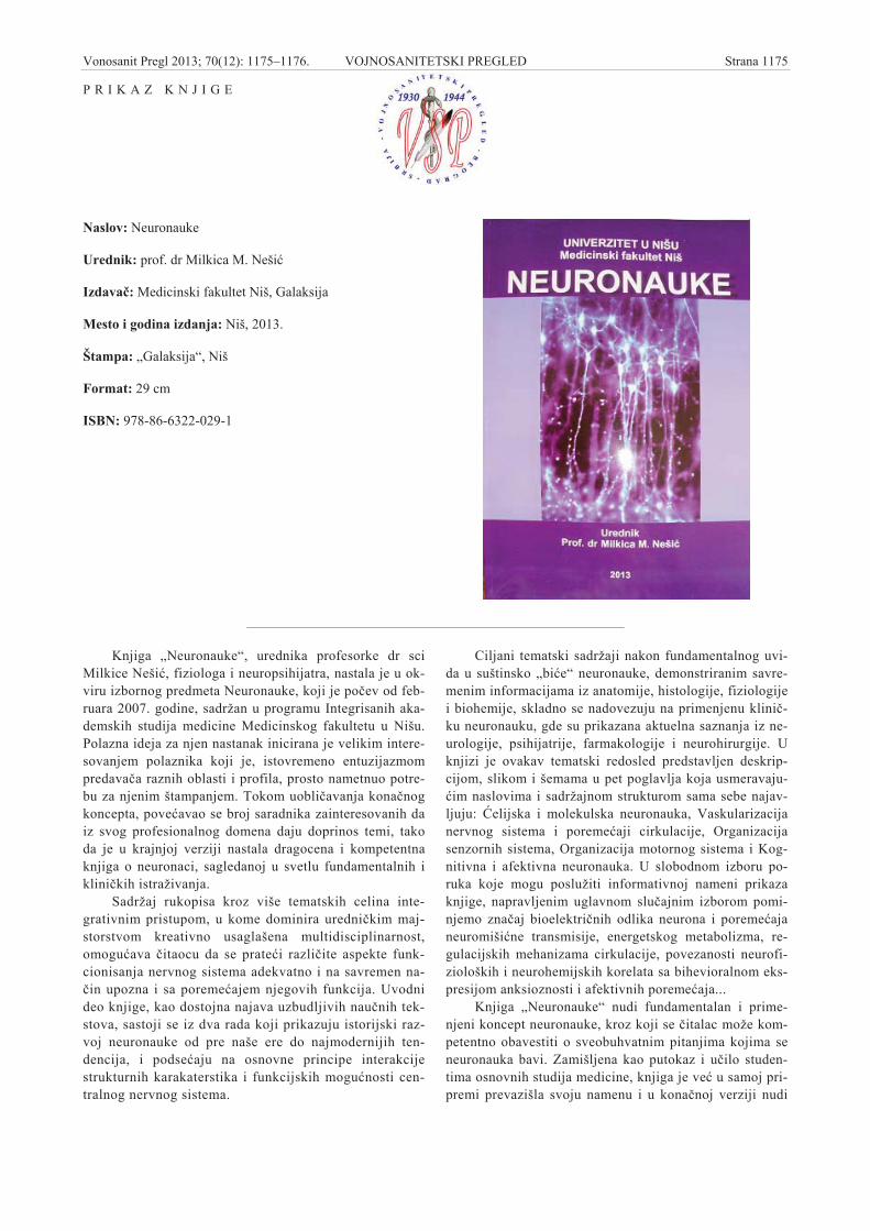
Vonosanit Pregl 2013; 70(12): 1175–1176. VOJNOSANITETSKI PREGLED Strana 1175
P R I K A Z K N J I G E
Naslov: Neuronauke
Urednik: prof. dr Milkica M. Neši
Izdava : Medicinski fakultet Niš, Galaksija
Mesto i godina izdanja: Niš, 2013.
Štampa: „Galaksija“, Niš
Format: 29 cm
ISBN: 978-86-6322-029-1
Knjiga „Neuronauke“, urednika profesorke dr sciMilkice Neši , fiziologa i neuropsihijatra, nastala je u ok-viru izbornog predmeta Neuronauke, koji je po ev od feb-ruara 2007. godine, sadržan u programu Integrisanih aka-demskih studija medicine Medicinskog fakultetu u Nišu.Polazna ideja za njen nastanak inicirana je velikim intere-sovanjem polaznika koji je, istovremeno entuzijazmompredava a raznih oblasti i profila, prosto nametnuo potre-bu za njenim štampanjem. Tokom uobli avanja kona nogkoncepta, pove avao se broj saradnika zainteresovanih daiz svog profesionalnog domena daju doprinos temi, takoda je u krajnjoj verziji nastala dragocena i kompetentnaknjiga o neuronaci, sagledanoj u svetlu fundamentalnih iklini kih istraživanja.
Sadržaj rukopisa kroz više tematskih celina inte-grativnim pristupom, u kome dominira uredni kim maj-storstvom kreativno usaglašena multidisciplinarnost,omogu ava itaocu da se prate i razli ite aspekte funk-cionisanja nervnog sistema adekvatno i na savremen na-in upozna i sa poreme ajem njegovih funkcija. Uvodni
deo knjige, kao dostojna najava uzbudljivih nau nih tek-stova, sastoji se iz dva rada koji prikazuju istorijski raz-voj neuronauke od pre naše ere do najmodernijih ten-dencija, i podse aju na osnovne principe interakcijestrukturnih karakaterstika i funkcijskih mogu nosti cen-tralnog nervnog sistema.
Ciljani tematski sadržaji nakon fundamentalnog uvi-da u suštinsko „bi e“ neuronauke, demonstriranim savre-menim informacijama iz anatomije, histologije, fiziologijei biohemije, skladno se nadovezuju na primenjenu klini -ku neuronauku, gde su prikazana aktuelna saznanja iz ne-urologije, psihijatrije, farmakologije i neurohirurgije. Uknjizi je ovakav tematski redosled predstavljen deskrip-cijom, slikom i šemama u pet poglavlja koja usmeravaju-im naslovima i sadržajnom strukturom sama sebe najav-
ljuju: elijska i molekulska neuronauka, Vaskularizacijanervnog sistema i poreme aji cirkulacije, Organizacijasenzornih sistema, Organizacija motornog sistema i Kog-nitivna i afektivna neuronauka. U slobodnom izboru po-ruka koje mogu poslužiti informativnoj nameni prikazaknjige, napravljenim uglavnom slu ajnim izborom pomi-njemo zna aj bioelektri nih odlika neurona i poreme ajaneuromiši ne transmisije, energetskog metabolizma, re-gulacijskih mehanizama cirkulacije, povezanosti neurofi-zioloških i neurohemijskih korelata sa bihevioralnom eks-presijom anksioznosti i afektivnih poreme aja...
Knjiga „Neuronauke“ nudi fundamentalan i prime-njeni koncept neuronauke, kroz koji se italac može kom-petentno obavestiti o sveobuhvatnim pitanjima kojima seneuronauka bavi. Zamišljena kao putokaz i u ilo studen-tima osnovnih studija medicine, knjiga je ve u samoj pri-premi prevazišla svoju namenu i u kona noj verziji nudi

Strana 1176 VOJNOSANITETSKI PREGLED Volumen 70, Broj 12
dragoceno štivo specijalizantima i supspecijalistima izoblasti anatomije, fiziologije, neurologije i drugih grani -nih oblasti. Na kraju, što je i glavna odlika rukopisa kojine završavaju svoj život u trenutnom izdanju, modernomkoncepcijom, koja neuronauku autenti no tretira kao di-nami nu, propulzivnu oblast, koja se neprestano inovira,
„Neuronauka“ je otvorila nova pitanja i ostala otvorena zabudu a saznanja.
dr Jelena Kosti ,Odeljenje de je i adolescentne psihijatrije,
Klinika za zaštitu mentalnog zdravljaKlini ki Centar Niš
Volumen 70, Broj 12 VOJNOSANITETSKI PREGLED Strana 1177
UPUTSTVO AUTORIMA
Vojnosanitetski pregled (VSP) objavljuje radove koji ranije nisu nig-de publikovani, niti predati za publikovanje redosledom koji odre ujeure iva ki odbor. Prilikom prijave rada u sistem elektronskog ure iva-nja „Vojnosanitetskog pregleda“ neophodno je priložiti izjavu da su is-punjeni svi postavljeni tehni ki zahtevi uklju uju i i izjavu potpisanu odstrane svih autora da rad nije ranije ni u celini, niti delimi no objavljenniti prihva en za štampanje u drugom asopisu. Izjava o pojedina nomdoprinosu autora mora biti potpisana od strane svakog autora rada, ske-nirana i poslata uz rad kao dopunska datoteka. Tako e, autori su obave-zni da dostave i potpisanu izjavu o nepostojanju sukoba interesa. Timpostupkom svi autori postaju odgovorni za ispunjavanje svih postavlje-nih uslova, emu sledi odluka o prihvatanju za dalji ure iva ki postupak.Za objavljene radove VSP zadržava autorsko pravo. Primaju se radovinapisani samo na engleskom jeziku.
Od 1. januara 2012. godine Vojnosanitetski pregled prešao jena e-Ur: Elektronsko ure ivanje asopisa.
Svi korisnici sistema: autori, recezenti i urednici moraju biti regis-trovani jednozna nom e-mail adresom. Registraciju je mogu e izvršitina adresi:
http://aseestant.ceon.rs/index.phpU VSP-u se objavljuju uvodnici, originalni lanci, prethodna ili
kratka saopštenja, revijski radovi tipa opšteg pregleda (uz uslov daautori navo enjem najmanje 5 autocitata potvrde da su eksperti u oblastio kojoj pišu), aktuelne teme ili metaanalize, kazuistika, lanci iz isto-rije medicine, li ni stavovi, naru eni komentari, pisma uredništvu, izve-štaji sa nau nih i stru nih skupova, prikazi knjiga, referati iz nau ne istru ne literature i drugi prilozi. Radovi tipa originalnih lanaka, pretho-dnih ili kratkih saopštenja, metaanalize i kazuistike objavljuju se uz ap-strakte na srpskom i engleskom jeziku.
Rukopis se piše sa proredom 1,5 sa levom marginom od 4 cm. Koris-titi font veli ine 12, a na elno izbegavati upotrebu bold i italic slova, kojasu rezervisana za podnaslove. Originalni lanci, opšti pregledi i metaanali-ze ne smeju prelaziti 16 stranica (sa prilozima); aktuelne teme – osam, ka-zuistika – šest, prethodna saopštenja – pet, a pisma uredniku, izveštaji saskupova i prikazi knjiga – dve stranice.
U celom radu obavezno je koriš enje me unarodnog sistema mera(SI) i standardnih me unarodno prihva enih termina.
Za obradu teksta koristiti program Word for Windows verzije 97,2000, XP ili 2003. Za izradu grafi kih priloga koristiti standardne gra-fi ke programe za Windows, poželjno iz programskog paketa Micro-soft Office (Excel, Word Graph). Kod kompjuterske izrade grafikaizbegavati upotrebu boja i sen enja pozadine.
Prispeli radovi kao anonimni podležu ure iva koj obradi i recenzijinajmanje dva urednika/recenzenta. Primedbe i sugestije uredni-ka/recenzenata dostavljaju se autoru radi kona nog oblikovanja. Pre ob-jave, rad se upu uje koresponding autoru na kona nu saglasnost.
Priprema radaDelovi rada su: naslovna strana, apstrakt sa klju nim re ima,
tekst i literatura.1. Naslovna stranaa) Naslov treba da bude kratak, jasan i informativan i da odgovara
sadržaju rada. Podnaslove treba izbegavati.b) Ispisuju se puna imena i prezimena autora.c) Navode se puni nazivi ustanove i organizacijske jedinice u kojima
je rad obavljen i mesta u kojima se ustanove nalaze, sa jasnim obeleža-vanjem odakle je autor, koriste i standardne znake za fus-note.
2. Apstrakt i klju ne re iNa drugoj stranici nalazi se strukturisani apstrakt sa naslovom rada.
Kratkim re enicama na srpskom i engleskom jeziku iznosi se uvod icilj rada, osnovne procedure - metode (izbor ispitanika ili laboratorij-skih životinja; metode posmatranja i analize), glavni nalazi - rezultati(konkretni podaci i njihova statisti ka zna ajnost) i glavni zaklju ak.Naglasiti nove i zna ajne aspekte studije ili zapažanja. Strukturisaniapstrakt (250 re i) ima podnaslove: uvod/cilj, metode, rezultati i zak-lju ak. Za apstrakte na engleskom dozvoljeno je i do 450 re i. Struk-turisani apstrakt je obavezan za metaanalize (istog obima kao i za ori-ginalne lanke) i kazuistiku (do 150 re i, sa podnaslovima uvod, pri-kaz slu aja i zaklju ak). Ispod apstrakta, pod podnaslovom „Klju nere i“ predložiti 3–10 klju nih re i ili kratkih izraza koji oslikavaju sa-držinu lanka.
3. Tekst lankaTekst sadrži slede a poglavlja: uvod, metode, rezultate i diskusiju.
Zaklju ak može da bude posebno poglavlje ili se iznosi u poslednjempasusu diskusije. U uvodu ponovo napisati naslov rada, bez navo enja
autora. Navesti hipotezu (ukoliko je ima) i ciljeve rada. Ukratko iznetirazloge za studiju ili posmatranje. Navesti samo strogo relevantne po-datke iz literature i ne iznositi opširna razmatranja o predmetu rada, kaoni podatke ili zaklju ke iz rada o kome se izveštava.
Metode. Jasno opisati izbor metoda posmatranja ili eksperimentnihmetoda (ispitanici ili eksperimentne životinje, uklju uju i kontrolne).Identifikovati metode, aparaturu (ime i adresa proizvo a a u zagradi) iproceduru, dovoljno detaljno da se drugim autorima omogu i reproduk-cija rezultata. Navesti podatke iz literature za uhodane metode, uklju u-ju i i statisti ke. Ta no identifikovati sve primenjene lekove i hemika-lije, uklju uju i generi ko ime, doze i na ine davanja. Za ispitivanja naljudima i životinjama navesti saglasnost eti kog komiteta.
Rezultate prikazati logi kim redosledom u tekstu, tabelama i ilustra-cijama. U tekstu naglasiti ili sumirati samo zna ajna zapažanja.
U diskusiji naglasiti nove i zna ajne aspekte studije i izvedene zak-lju ke. Posmatranja dovesti u vezu sa drugim relevantnim studijama, una elu iz poslednje tri godine, a samo izuzetno i starijim. Povezati zak-lju ke sa ciljevima rada, ali izbegavati nesumnjive tvrdnje i one zaklju -ke koje podaci iz rada ne podržavaju u potpunosti.
LiteraturaLiteratura se u radu citira kao superskript, a popisuje rednim broje-
vima pod kojima se citat pojavljuje u tekstu. Navode se svi autori, aliako broj prelazi šest, n a v o d i s e p r v i h š e s t i dodaje etal. Svi podaci o citiranoj literaturi moraju biti t a n i . Literatura se ucelini citira na engleskom jeziku, a iza naslova se navodi jezik lanka uzagradi. Ne prihvata se citiranje apstrakata, sekundarnih publikacija,usmenih saopštenja, neobjavljenih radova, službenih i poverljivih do-kumenata. Radovi koji su prihva eni za štampu, ali još nisu objavljeni,navode se uz dodatak „u štampi“. Rukopisi koji su predati, ali još nisuprihva eni za štampu, u tekstu se citiraju kao „neobjavljeni podaci“ (uzagradi). Podaci sa Interneta citiraju se uz navo enje datuma.
Primeri referenci:urovi BM. Endothelial trauma in the surgery of cataract. Vojno-
sanit Pregl 2004; 61(5): 491–7. (Serbian)Balint B. From the haemotherapy to the haemomodulation. Beo-
grad: Zavod za udžbenike i nastavna sredstva; 2001. (Serbian)Mladenovi T, Kandolf L, Mijuškovi ŽP. Lasers in dermatology. In:
Karadagli , editor. Dermatology. Beograd: Vojnoizdava ki zavod &Verzal Press; 2000. p. 1437–49. (Serbian)
Christensen S, Oppacher F. An analysis of Koza's computational ef-fort statistic for genetic programming. In: Foster JA, Lutton E, Miller J,Ryan C, Tettamanzi AG, editors. Genetic programming. EuroGP 2002:Proceedings of the 5th European Conference on Genetic Programming;2002 Apr 3-5; Kinsdale, Ireland. Berlin: Springer; 2002. p. 182-91.
Abood S. Quality improvement initiative in nursing homes: theANA acts in an advisory role. Am J Nurs [serial on the Internet]. 2002Jun [cited 2002 Aug 12]; 102(6): [about 3 p.]. Available from:http://www.nursingworld.org/AJN/2002/june/Wawatch.htm
TabeleSve tabele pripremaju se sa proredom 1,5 na posebnom listu. Obele-
žavaju se arapskim brojevima, redosledom pojavljivanja, u desnom uglu(Tabela 1), a svakoj se daje kratak naslov. Objašnjenja se daju u fus-noti, ne u zaglavlju. Za fus-notu koristiti slede e simbole ovim redosle-dom: *, †, ‡, §, ||, ¶, **, ††, ... . Svaka tabela mora da se pomene u tek-stu. Ako se koriste tu i podaci, obavezno ih navesti kao i svaki drugi po-datak iz literature.
IlustracijeSlikama se zovu svi oblici grafi kih priloga i predaju se kao dopun-
ske datoteke u sistemu aseestant. Slova, brojevi i simboli treba da su ja-sni i ujedna eni, a dovoljne veli ine da prilikom umanjivanja budu it-ljivi. Slike treba da budu jasne i obeležene brojevima, onim redom kojimse navode u tekstu (Sl. 1; Sl. 2 itd.). Ukoliko je slika ve negde objav-ljena, obavezno citirati izvor.
Legende za ilustracije pisati na posebnom listu, koriste i arapskebrojeve. Ukoliko se koriste simboli, strelice, brojevi ili slova za ob-jašnjavanje pojedinog dela ilustracije, svaki pojedina no treba objasnitiu legendi. Za fotomikrografije navesti metod bojenja i podatak o uve a-nju.
Skra enice i simboliKoristiti samo standardne skra enice, izuzev u naslovu i apstraktu.
Pun naziv sa skra enicom u zagradi treba dati kod prvog pominjanja utekstu.
Detaljno uputstvo može se dobiti u redakciji ili na sajtu:www.vma.mod.gov.rs/vsp/download/uputstvo_za_autore.pdf.
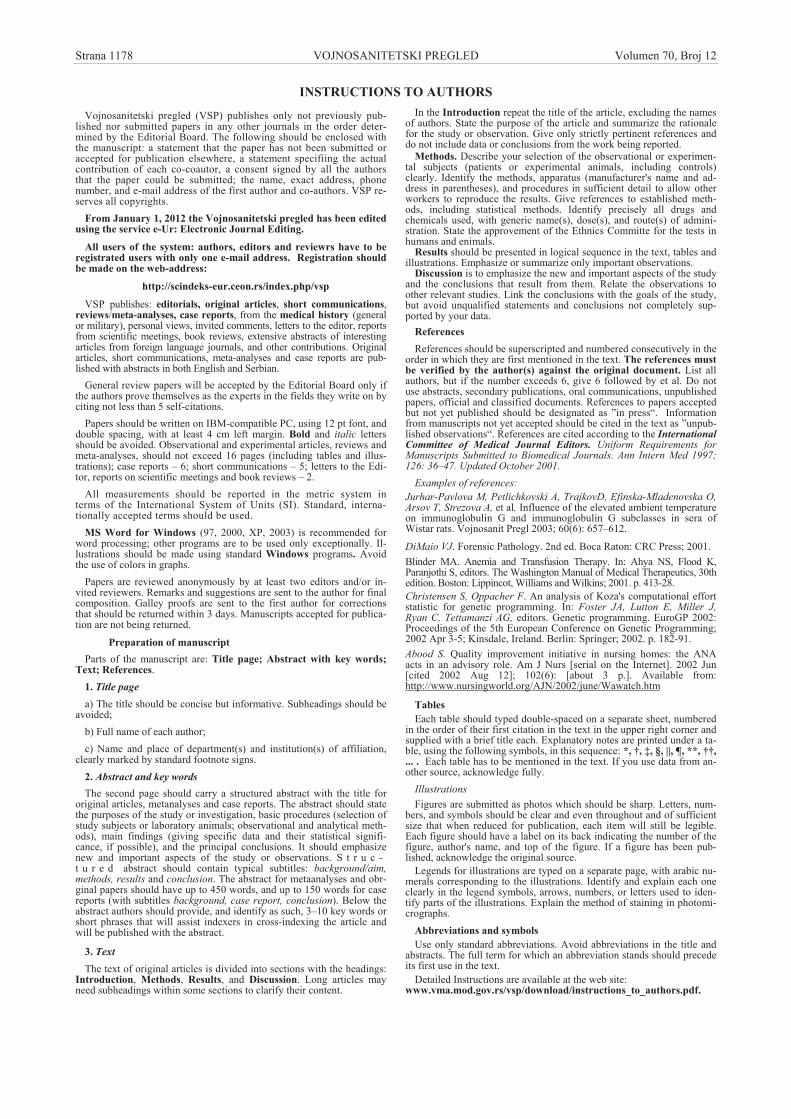
Strana 1178 VOJNOSANITETSKI PREGLED Volumen 70, Broj 12
INSTRUCTIONS TO AUTHORS
Vojnosanitetski pregled (VSP) publishes only not previously pub-lished nor submitted papers in any other journals in the order deter-mined by the Editorial Board. The following should be enclosed withthe manuscript: a statement that the paper has not been submitted oraccepted for publication elsewhere, a statement specifiing the actualcontribution of each co-coautor, a consent signed by all the authorsthat the paper could be submitted; the name, exact address, phonenumber, and e-mail address of the first author and co-authors. VSP re-serves all copyrights.
From January 1, 2012 the Vojnosanitetski pregled has been editedusing the service e-Ur: Electronic Journal Editing.
All users of the system: authors, editors and reviewrs have to beregistrated users with only one e-mail address. Registration shouldbe made on the web-address:
http://scindeks-eur.ceon.rs/index.php/vsp
VSP publishes: editorials, original articles, short communications,reviews/meta-analyses, case reports, from the medical history (generalor military), personal views, invited comments, letters to the editor, reportsfrom scientific meetings, book reviews, extensive abstracts of interestingarticles from foreign language journals, and other contributions. Originalarticles, short communications, meta-analyses and case reports are pub-lished with abstracts in both English and Serbian.
General review papers will be accepted by the Editorial Board only ifthe authors prove themselves as the experts in the fields they write on byciting not less than 5 self-citations.
Papers should be written on IBM-compatible PC, using 12 pt font, anddouble spacing, with at least 4 cm left margin. Bold and italic lettersshould be avoided. Observational and experimental articles, reviews andmeta-analyses, should not exceed 16 pages (including tables and illus-trations); case reports – 6; short communications – 5; letters to the Edi-tor, reports on scientific meetings and book reviews – 2.
All measurements should be reported in the metric system interms of the International System of Units (SI). Standard, interna-tionally accepted terms should be used.
MS Word for Windows (97, 2000, XP, 2003) is recommended forword processing; other programs are to be used only exceptionally. Il-lustrations should be made using standard Windows programs. Avoidthe use of colors in graphs.
Papers are reviewed anonymously by at least two editors and/or in-vited reviewers. Remarks and suggestions are sent to the author for finalcomposition. Galley proofs are sent to the first author for correctionsthat should be returned within 3 days. Manuscripts accepted for publica-tion are not being returned.
Preparation of manuscriptParts of the manuscript are: Title page; Abstract with key words;
Text; References.1. Title pagea) The title should be concise but informative. Subheadings should be
avoided;b) Full name of each author;c) Name and place of department(s) and institution(s) of affiliation,
clearly marked by standard footnote signs.2. Abstract and key wordsThe second page should carry a structured abstract with the title for
original articles, metanalyses and case reports. The abstract should statethe purposes of the study or investigation, basic procedures (selection ofstudy subjects or laboratory animals; observational and analytical meth-ods), main findings (giving specific data and their statistical signifi-cance, if possible), and the principal conclusions. It should emphasizenew and important aspects of the study or observations. S t r u c -t u r e d abstract should contain typical subtitles: background/aim,methods, results and conclusion. The abstract for metaanalyses and obr-ginal papers should have up to 450 words, and up to 150 words for casereports (with subtitles background, case report, conclusion). Below theabstract authors should provide, and identify as such, 3–10 key words orshort phrases that will assist indexers in cross-indexing the article andwill be published with the abstract.
3. TextThe text of original articles is divided into sections with the headings:
Introduction, Methods, Results, and Discussion. Long articles mayneed subheadings within some sections to clarify their content.
In the Introduction repeat the title of the article, excluding the namesof authors. State the purpose of the article and summarize the rationalefor the study or observation. Give only strictly pertinent references anddo not include data or conclusions from the work being reported.
Methods. Describe your selection of the observational or experimen-tal subjects (patients or experimental animals, including controls)clearly. Identify the methods, apparatus (manufacturer's name and ad-dress in parentheses), and procedures in sufficient detail to allow otherworkers to reproduce the results. Give references to established meth-ods, including statistical methods. Identify precisely all drugs andchemicals used, with generic name(s), dose(s), and route(s) of admini-stration. State the approvement of the Ethnics Committe for the tests inhumans and enimals.
Results should be presented in logical sequence in the text, tables andillustrations. Emphasize or summarize only important observations.
Discussion is to emphasize the new and important aspects of the studyand the conclusions that result from them. Relate the observations toother relevant studies. Link the conclusions with the goals of the study,but avoid unqualified statements and conclusions not completely sup-ported by your data.
ReferencesReferences should be superscripted and numbered consecutively in the
order in which they are first mentioned in the text. The references mustbe verified by the author(s) against the original document. List allauthors, but if the number exceeds 6, give 6 followed by et al. Do notuse abstracts, secondary publications, oral communications, unpublishedpapers, official and classified documents. References to papers acceptedbut not yet published should be designated as ”in press“. Informationfrom manuscripts not yet accepted should be cited in the text as ”unpub-lished observations“. References are cited according to the InternationalCommittee of Medical Journal Editors. Uniform Requirements forManuscripts Submitted to Biomedical Journals. Ann Intern Med 1997;126: 36–47. Updated October 2001.
Examples of references:Jurhar-Pavlova M, Petlichkovski A, TrajkovD, Efinska-Mladenovska O,Arsov T, Strezova A, et al. Influence of the elevated ambient temperatureon immunoglobulin G and immunoglobulin G subclasses in sera ofWistar rats. Vojnosanit Pregl 2003; 60(6): 657–612.
DiMaio VJ. Forensic Pathology. 2nd ed. Boca Raton: CRC Press; 2001.Blinder MA. Anemia and Transfusion Therapy. In: Ahya NS, Flood K,Paranjothi S, editors. The Washington Manual of Medical Therapeutics, 30thedition. Boston: Lippincot, Williams and Wilkins; 2001. p. 413-28.Christensen S, Oppacher F. An analysis of Koza's computational effortstatistic for genetic programming. In: Foster JA, Lutton E, Miller J,Ryan C, Tettamanzi AG, editors. Genetic programming. EuroGP 2002:Proceedings of the 5th European Conference on Genetic Programming;2002 Apr 3-5; Kinsdale, Ireland. Berlin: Springer; 2002. p. 182-91.Abood S. Quality improvement initiative in nursing homes: the ANAacts in an advisory role. Am J Nurs [serial on the Internet]. 2002 Jun[cited 2002 Aug 12]; 102(6): [about 3 p.]. Available from:http://www.nursingworld.org/AJN/2002/june/Wawatch.htm
TablesEach table should typed double-spaced on a separate sheet, numbered
in the order of their first citation in the text in the upper right corner andsupplied with a brief title each. Explanatory notes are printed under a ta-ble, using the following symbols, in this sequence: *, †, ‡, §, ||, ¶, **, ††,... . Each table has to be mentioned in the text. If you use data from an-other source, acknowledge fully.
IllustrationsFigures are submitted as photos which should be sharp. Letters, num-
bers, and symbols should be clear and even throughout and of sufficientsize that when reduced for publication, each item will still be legible.Each figure should have a label on its back indicating the number of thefigure, author's name, and top of the figure. If a figure has been pub-lished, acknowledge the original source.
Legends for illustrations are typed on a separate page, with arabic nu-merals corresponding to the illustrations. Identify and explain each oneclearly in the legend symbols, arrows, numbers, or letters used to iden-tify parts of the illustrations. Explain the method of staining in photomi-crographs.
Abbreviations and symbolsUse only standard abbreviations. Avoid abbreviations in the title and
abstracts. The full term for which an abbreviation stands should precedeits first use in the text.
Detailed Instructions are available at the web site:www.vma.mod.gov.rs/vsp/download/instructions_to_authors.pdf.
VO
JNO
SAN
ITE
TSK
I P
RE
GL
ED
VO
JNO
ME
DIC
INSK
A A
KA
DE
MIJ
ACr
notra
vska
17,
110
40 B
eogr
ad, S
rbija
Tel/
Fax:
+38
1 11
266
9689
vmain
i1@
EU
net.r
svm
avsp
@ho
tmail
.com
asop
is „V
ojno
sani
tets
ki p
regl
ed“
izlaz
i god
išnje
u 12
bro
jeva
.G
odišn
ja pr
etpl
ata
za 2
014.
god
inu
iznos
i: 5
000
dina
ra z
a gr
aan
e Sr
bije,
10 0
00 d
inar
a za
usta
nove
iz S
rbije
i 15
0 €
za st
rane
drž
avlja
ne i
usta
nove
. Pre
tplat
e:Ži
ro r
aun
br.
840-
3148
49-7
0 M
O –
Sre
dstv
a ob
jedin
jene
napl
ate
– V
MA
(za
Voj
nosa
nite
tski p
regl
ed),
poziv
na
broj
122
7423
1295
5214
15. U
plat
nicu
(do
kaz
oup
lati)
dosta
viti
lino
ili p
ošto
m (p
ismom
, fak
som
, -m
ail-o
m).
Za z
apos
lene
u M
O i
Voj
sci S
rbije
mog
ua
je i p
retp
lata
u 12
mes
eni
h ra
ta p
utem
tra
jnog
nalo
ga, t
j.„o
dbija
njem
od
plat
e“. P
opun
jen o
braz
ac p
oslat
i na a
dres
u V
SP-a
.
PR
IJA
VA
ZA
PR
ET
PL
AT
U N
A
ASO
PIS
„VO
JNO
SAN
ITE
TSK
I P
RE
GL
ED
“
Ime
i pre
zim
e ili
naz
iv u
stan
ove
Jedi
nstv
eni m
ati
ni b
roj g
raan
aPo
resk
i ide
ntifi
kaci
oni b
roj (
PIB)
za u
stan
ove
Mes
toU
lica
i bro
jTe
lefo
n /
tele
faks
Pret
plat
a na
as
opis
„Voj
nosa
nite
tski
pre
gled
“ (z
aokr
užiti
):1.
Lino
. Dok
az o
pre
tplat
i dos
tavl
jam u
z ov
u pr
ijavu
.2.
Za
prip
adni
ke M
O i
Voj
ske
Srbi
je:
Daje
m s
aglas
nost
da
sepr
iliko
m is
plat
e pl
ata
u Ra
unov
odst
veno
m c
entru
MO
iz m
ojih
prin
adle
žnos
ti ob
usta
vlja
izno
s mes
ene
rate
(pre
tplat
e).
3.V
irman
om p
o pr
ijem
u pr
ofak
ture
.
Dat
um _
____
____
____
___
Potp
is__
____
____
____
____
____
VO
JNO
SAN
ITE
TSK
I P
RE
GL
ED
VO
JNO
ME
DIC
INSK
A A
KA
DE
MIJ
ACr
notra
vska
17,
110
40 B
eogr
ad, S
rbija
Tel/
Fax:
+38
1 11
266
9689
vmain
i1@
EU
net.r
svm
avsp
@ho
tmail
.com
asop
is „V
ojno
sani
tets
ki p
regl
ed“
izlaz
i god
išnje
u 1
2 br
ojev
a.G
odišn
ja pr
etpl
ata
za 2
014.
god
inu
iznos
i: 5
000
dina
ra z
a gr
aan
e Sr
bije,
10 0
00 d
inar
a za
usta
nove
iz S
rbije
i 15
0 €
za st
rane
drž
avlja
ne i
usta
nove
. Pre
tplat
e:žir
o ra
un b
r. 84
0-31
4849
-70
MO
– S
reds
tva
objed
injen
e na
plat
e –
VM
A (
zaV
ojno
sani
tetsk
i pre
gled
), po
ziv n
a br
oj 1
2274
2312
9552
1415
. Upl
atni
cu (
doka
z o
uplat
i) do
stavi
ti li
no il
i poš
tom
(pism
om, f
akso
m,
-om
). Za
zap
oslen
e u
MO
iV
ojsc
i Srb
ije m
ogu
a je
i pre
tplat
a u
12 m
ese
nih
rata
put
em t
rajn
og n
aloga
, tj.
„odb
ijanj
em o
d pl
ate“
. Pop
unjen
obr
azac
pos
lati n
a adr
esu
VSP
-a.
PR
IJA
VA
ZA
PR
ET
PL
AT
U N
A
ASO
PIS
„VO
JNO
SAN
ITE
TSK
I P
RE
GL
ED
“
Ime
i pre
zim
e ili
naz
iv u
stan
ove
Jedi
nstv
eni m
ati
ni b
roj g
raan
aPo
resk
i ide
ntifi
kaci
oni b
roj (
PIB)
za u
stan
ove
Mes
toU
lica
i bro
jTe
lefo
n /
tele
faks
Pret
plat
a na
as
opis
„Voj
nosa
nite
tski
pre
gled
“ (z
aokr
užiti
):1.
Lino
. Dok
az o
pre
tplat
i dos
tavl
jam u
z ov
u pr
ijavu
.2.
Za
prip
adni
ke M
O i
Voj
ske
Srbi
je:
Daje
m s
aglas
nost
da
sepr
iliko
m i
splat
e pl
ata
u Ra
unov
odst
veno
m c
entru
MO
iz
moj
ih p
rinad
ležno
sti o
bust
avlja
izno
s mes
ene
rate
(pre
tplat
e).
3.V
irman
om p
o pr
ijem
u pr
ofak
ture
.
Dat
um _
____
____
____
___
Potp
is__
____
____
____
____
____