476 Oral Abstract Session, Fri, 1:30 PM-3:00 PM ... - Amazon S3
126
476 Oral Abstract Session, Fri, 1:30 PM-3:00 PM Patient-reported outcomes (PROs) from the Phase III IMbrave150 trial of atezolizumab (atezo) + bevacizumab (bev) vs sorafenib (sor) as first-line treatment (tx) for patients (pts) with unresectable hepatocellular carcinoma (HCC). Peter R. Galle, Richard S. Finn, Shukui Qin, Masafumi Ikeda, Andrew X. Zhu, Tae-You Kim, Masatoshi Kudo, Valeriy Vladimirovich Breder, Philippe Merle, Ahmed Omar Kaseb, Daneng Li, Sohail Mulla, Wendy Verret, Derek-Zhen Xu, Sairy Hernandez, Juan Liu, Chen Huang, Ho Yeong Lim, Ann-Lii Cheng, Michel Ducreux; University Medical Center Mainz, Mainz, Germany; University of California Los Angeles, Los Angeles, CA; People’s Liberation Army Cancer Center, Nanjing, China; National Cancer Center Hospital East, Kashiwa, Japan; Massachusetts General Hospital, Boston, MA; Department of Internal Medicine, Seoul National University Hospital, Seoul National University College of Medicine, Seoul, South Korea; Kindai University Faculty of Medicine, Osaka, Japan; Russian Oncological Research Center, N.N. Blokhin of Ministry of Health, Moscow, Russian Federation; Lyon University, Lyon, France; The University of Texas MD Anderson Cancer Center, Houston, TX; City of Hope National Medical Center, Duarte, CA; Genentech, Inc., South San Francisco, CA; Roche Product Development, Shanghai, China; Division of Hematology-Oncology, Department of Medicine, Samsung Medical Center, Seoul, South Korea; National Taiwan University Hospital and National Taiwan University Cancer Center, Taipei, Taiwan; Gustave Roussy Cancer Campus Grand Paris, Villejuif, France Background: Atezo + bev in pts with unresectable HCC who had not received prior systemic therapy has shown statistically significant and clinically meaningful improvement in OS and PFS per inde- pendent review facility-assessed RECIST 1.1 vs sor in the Phase III IMbrave150 study (Cheng ESMO Asia 2019). Here, we report PRO data from this trial to show pt perspectives on the overall clinical benefit of atezo + bev. Methods: Pts were randomized 2:1 to receive either atezo 1200 mg IV q3w + bev 15 mg/kg IV q3w or sor 400 mg PO BID until loss of clinical benefit or unacceptable toxicity. Pts completed the EORTC QLQ-C30 and EORTC QLQ-HCC18 questionnaires before tx, every 3 wk on tx, and every 3 mo after tx discontinuation or disease progression. A pre-specified secondary endpoint was time to deterioration (TTD; first $ 10-point decrease from baseline held for 2 consecutive assessments or 1 assessment followed by death within 3 wk) of pt-reported quality of life (QOL), physical functioning, and role functioning. Pre-specified exploratory analyses included TTD of and proportion of pts with a clinically meaningful change ($ 10 points from baseline) in key pt-reported symptoms. Results: Questionnaire completion rates were $ 92% in both arms from baseline through most of the tx period. Compared with sor, atezo + bev delayed TTD of pt-reported QOL (median TTD, 11.2 vs 3.6 mo; HR, 0.63 [95% CI: 0.46, 0.85]), physical functioning (median TTD, 13.1 vs 4.9 mo; HR, 0.53 [95% CI: 0.39, 0.73]), and role functioning (median TTD, 9.1 vs 3.6 mo; HR, 0.62 [95% CI: 0.46, 0.84]). Atezo + bev also delayed TTD in pt-reported appetite loss, fatigue, pain, and diarrhea vs sor; a lower proportion of pts on atezo + bev experienced clinically meaningful dete- rioration in each of these symptoms vs sor. Conclusions: High-quality PRO results from IMbrave150 showed large and consistent benefits in key aspects of the pt experience with atezo + bev, further supporting its overall clinical benefit in pts with unresectable HCC who have not received prior systemic therapy. Clinical trial information: NCT03434379. Research Sponsor: F. Hoffmann-La Roche, Ltd. © 2020 American Society of Clinical Oncology. Visit gicasym.org and search by abstract for disclosure information. HEPATOBILIARY CANCER
-
Upload
khangminh22 -
Category
Documents
-
view
0 -
download
0
Transcript of 476 Oral Abstract Session, Fri, 1:30 PM-3:00 PM ... - Amazon S3

476 Oral Abstract Session, Fri, 1:30 PM-3:00 PM
Patient-reported outcomes (PROs) from the Phase III IMbrave150 trial of atezolizumab (atezo) +bevacizumab (bev) vs sorafenib (sor) as first-line treatment (tx) for patients (pts) withunresectable hepatocellular carcinoma (HCC).
Peter R. Galle, Richard S. Finn, Shukui Qin, Masafumi Ikeda, Andrew X. Zhu, Tae-You Kim, Masatoshi Kudo,Valeriy Vladimirovich Breder, Philippe Merle, Ahmed Omar Kaseb, Daneng Li, Sohail Mulla, Wendy Verret, Derek-Zhen Xu,Sairy Hernandez, Juan Liu, Chen Huang, Ho Yeong Lim, Ann-Lii Cheng, Michel Ducreux; University Medical Center Mainz, Mainz,Germany; University of California Los Angeles, Los Angeles, CA; People’s Liberation Army Cancer Center, Nanjing, China;National Cancer Center Hospital East, Kashiwa, Japan; Massachusetts General Hospital, Boston, MA; Department of InternalMedicine, Seoul National University Hospital, Seoul National University College of Medicine, Seoul, South Korea; KindaiUniversity Faculty of Medicine, Osaka, Japan; Russian Oncological Research Center, N.N. Blokhin of Ministry of Health, Moscow,Russian Federation; Lyon University, Lyon, France; The University of Texas MD Anderson Cancer Center, Houston, TX; City ofHope National Medical Center, Duarte, CA; Genentech, Inc., South San Francisco, CA; Roche Product Development, Shanghai,China; Division of Hematology-Oncology, Department of Medicine, Samsung Medical Center, Seoul, South Korea; NationalTaiwan University Hospital and National Taiwan University Cancer Center, Taipei, Taiwan; Gustave Roussy Cancer Campus GrandParis, Villejuif, France
Background:Atezo + bev in pts with unresectable HCCwho had not received prior systemic therapyhas shown statistically significant and clinically meaningful improvement in OS and PFS per inde-pendent review facility-assessed RECIST 1.1 vs sor in the Phase III IMbrave150 study (Cheng ESMOAsia 2019). Here, we report PRO data from this trial to show pt perspectives on the overall clinicalbenefit of atezo + bev.Methods: Pts were randomized 2:1 to receive either atezo 1200 mg IV q3w +bev 15 mg/kg IV q3w or sor 400 mg PO BID until loss of clinical benefit or unacceptable toxicity. Ptscompleted the EORTC QLQ-C30 and EORTC QLQ-HCC18 questionnaires before tx, every 3 wk on tx,and every 3 mo after tx discontinuation or disease progression. A pre-specified secondary endpointwas time to deterioration (TTD; first $ 10-point decrease from baseline held for 2 consecutiveassessments or 1 assessment followed by death within 3 wk) of pt-reported quality of life (QOL),physical functioning, and role functioning. Pre-specified exploratory analyses included TTD of andproportion of pts with a clinically meaningful change ($ 10 points from baseline) in key pt-reportedsymptoms. Results: Questionnaire completion rates were $ 92% in both arms from baselinethrough most of the tx period. Compared with sor, atezo + bev delayed TTD of pt-reported QOL(median TTD, 11.2 vs 3.6mo; HR, 0.63 [95%CI: 0.46, 0.85]), physical functioning (median TTD, 13.1 vs4.9 mo; HR, 0.53 [95% CI: 0.39, 0.73]), and role functioning (median TTD, 9.1 vs 3.6 mo; HR, 0.62[95% CI: 0.46, 0.84]). Atezo + bev also delayed TTD in pt-reported appetite loss, fatigue, pain, anddiarrhea vs sor; a lower proportion of pts on atezo + bev experienced clinically meaningful dete-rioration in each of these symptoms vs sor. Conclusions: High-quality PRO results from IMbrave150showed large and consistent benefits in key aspects of the pt experience with atezo + bev, furthersupporting its overall clinical benefit in pts with unresectable HCC who have not received priorsystemic therapy. Clinical trial information: NCT03434379. Research Sponsor: F. Hoffmann-LaRoche, Ltd.
© 2020 American Society of Clinical Oncology. Visit gicasym.org and search by abstract for disclosure information.
HEPATOBILIARY CANCER
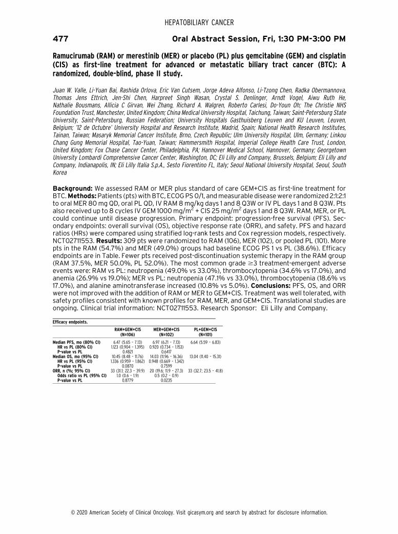
477 Oral Abstract Session, Fri, 1:30 PM-3:00 PM
Ramucirumab (RAM) or merestinib (MER) or placebo (PL) plus gemcitabine (GEM) and cisplatin(CIS) as first-line treatment for advanced or metastatic biliary tract cancer (BTC): Arandomized, double-blind, phase II study.
Juan W. Valle, Li-Yuan Bai, Rashida Orlova, Eric Van Cutsem, Jorge Adeva Alfonso, Li-Tzong Chen, Radka Obermannova,Thomas Jens Ettrich, Jen-Shi Chen, Harpreet Singh Wasan, Crystal S. Denlinger, Arndt Vogel, Aiwu Ruth He,Nathalie Bousmans, Allicia C Girvan, Wei Zhang, Richard A. Walgren, Roberto Carlesi, Do-Youn Oh; The Christie NHSFoundation Trust, Manchester, United Kingdom; China Medical University Hospital, Taichung, Taiwan; Saint-Petersburg StateUniversity, Saint-Petersburg, Russian Federation; University Hospitals Gasthuisberg Leuven and KU Leuven, Leuven,Belgium; ’12 de Octubre’ University Hospital and Research Institute, Madrid, Spain; National Health Research Institutes,Tainan, Taiwan; Masaryk Memorial Cancer Institute, Brno, Czech Republic; Ulm University Hospital, Ulm, Germany; LinkouChang Gung Memorial Hospital, Tao-Yuan, Taiwan; Hammersmith Hospital, Imperial College Health Care Trust, London,United Kingdom; Fox Chase Cancer Center, Philadelphia, PA; Hannover Medical School, Hannover, Germany; GeorgetownUniversity Lombardi Comprehensive Cancer Center, Washington, DC; Eli Lilly and Company, Brussels, Belgium; Eli Lilly andCompany, Indianapolis, IN; Eli Lilly Italia S.p.A., Sesto Fiorentino FL, Italy; Seoul National University Hospital, Seoul, SouthKorea
Background: We assessed RAM or MER plus standard of care GEM+CIS as first-line treatment forBTC.Methods:Patients (pts)with BTC, ECOGPS0/1, andmeasurable diseasewere randomized2:1:2:1to oral MER 80mgQD, oral PL QD, IV RAM 8mg/kg days 1 and 8 Q3W or IV PL days 1 and 8 Q3W. Ptsalso received up to 8 cycles IV GEM 1000mg/m2 + CIS 25mg/m2 days 1 and 8 Q3W. RAM,MER, or PLcould continue until disease progression. Primary endpoint: progression-free survival (PFS). Sec-ondary endpoints: overall survival (OS), objective response rate (ORR), and safety. PFS and hazardratios (HRs) were compared using stratified log-rank tests and Cox regression models, respectively.NCT02711553. Results: 309 pts were randomized to RAM (106), MER (102), or pooled PL (101). Morepts in the RAM (54.7%) and MER (49.0%) groups had baseline ECOG PS 1 vs PL (38.6%). Efficacyendpoints are in Table. Fewer pts received post-discontinuation systemic therapy in the RAM group(RAM 37.5%, MER 50.0%, PL 52.0%). The most common grade $3 treatment-emergent adverseevents were: RAM vs PL: neutropenia (49.0% vs 33.0%), thrombocytopenia (34.6% vs 17.0%), andanemia (26.9% vs 19.0%); MER vs PL: neutropenia (47.1% vs 33.0%), thrombocytopenia (18.6% vs17.0%), and alanine aminotransferase increased (10.8% vs 5.0%). Conclusions: PFS, OS, and ORRwere not improved with the addition of RAM orMER to GEM+CIS. Treatment was well tolerated, withsafety profiles consistent with known profiles for RAM,MER, and GEM+CIS. Translational studies areongoing. Clinical trial information: NCT02711553. Research Sponsor: Eli Lilly and Company.
Efficacy endpoints.
RAM+GEM+CIS(N=106)
MER+GEM+CIS(N=102)
PL+GEM+CIS(N=101)
Median PFS, mo (80% CI) 6.47 (5.65 – 7.13) 6.97 (6.21 – 7.13) 6.64 (5.59 – 6.83)HR vs PL (80% CI) 1.123 (0.904 – 1.395) 0.920 (0.734 – 1.153)P-value vs PL 0.4821 0.6417
Median OS, mo (95% CI) 10.45 (8.48 – 11.76) 14.03 (11.96 – 16.36) 13.04 (11.40 – 15.31)HR vs PL (95% CI) 1.336 (0.959 – 1.862) 0.948 (0.669 – 1.342)P-value vs PL 0.0870 0.7599
ORR, n (%; 95% CI) 33 (31.1; 22.3 – 39.9) 20 (19.6; 11.9 – 27.3) 33 (32.7; 23.5 – 41.8)Odds ratio vs PL (95% CI) 1.0 (0.6 – 1.9) 0.5 (0.2 – 0.9)P-value vs PL 0.8779 0.0235
© 2020 American Society of Clinical Oncology. Visit gicasym.org and search by abstract for disclosure information.
HEPATOBILIARY CANCER
478 Rapid Abstract Session, Fri, 7:00 AM-7:45 AM and Poster Session(Board #A1), Fri, 12:00 PM-1:30 PM and 4:30 PM-5:30 PM
Nivolumab (NIVO) + ipilimumab (IPI) + cabozantinib (CABO) combination therapy in patients(pts) with advanced hepatocellular carcinoma (aHCC): Results from CheckMate 040.
Thomas Yau, Vittorina Zagonel, Armando Santoro, Mirelis Acosta-Rivera, Su Pin Choo, Ana Matilla, Aiwu Ruth He,Antonio Cubillo Gracian, Anthony B. El-Khoueiry, Bruno Sangro, Tarek Eldawy, Jordi Bruix, Giovanni Frassineti,Gina M. Vaccaro, Marina Tschaika, Christian Scheffold, Yun Shen, Jaclyn Neely, Fabio Piscaglia; The University at HongKong, Hong Kong, China; Istituto Oncologico Veneto IOV-IRCCS, Padua, Italy; Istituto Clinico Humanitas, Rozzano, Italy;Fundacion de Investigacion, San Juan, PR; National Cancer Center Singapore, Curie Oncology, Singapore, Singapore; Serviciode Digestivo, Hospital General Universitario Gregorio Mara~non and CIBEREHD, Madrid, Spain; Georgetown UniversityHospital, Washington, DC; Hospital HM Universitario Sanchinarro, Centro Integral Oncologico Clara Campal (HM-CIOCC),Departamento de Ciencias Medicas Clınicas, Universidad San Pablo CEU, Madrid, Spain; USC Norris Comprehensive CancerCenter, Los Angeles, CA; Clinica Universidad de Navarra and CIBEREHD, Pamplona, Spain; Sacred Heart Health Systems,Pensacola, FL; BCLC, Hospital Clınic of Barcelona, IDIBAPS, University of Barcelona, CIBEREHD, Barcelona, Spain; IstitutoScientifico Romagnolo per lo Studio e la Cura dei Tumori (IRST-IRCCS), Meldola, Italy; Oregon Health & Science University,Portland, OR; Bristol-Myers Squibb, Princeton, NJ; Exelixis, Inc., Alameda, CA; University of Bologna, Bologna, Italy
Background: The programmed death-1 inhibitor NIVO had durable responses and a manageablesafety profile in pts with aHCC in CheckMate 040 (NCT01658878; El-Khoueiry et al. Lancet 2017)and is approved in the United States, Canada, Australia, and elsewhere for sorafenib (SOR)-treatedpts with aHCC. In another CheckMate 040 cohort, NIVO + IPI combination therapy had durableresponses in SOR-treated pts with aHCC, with objective response rates (ORRs) . 30% in eachdosing arm (Yau et al. J Clin Oncol 2019). CABO is also approved for SOR-treated pts with aHCC; apivotal phase 3 trial reported median overall survival (OS) of 10.2 mo (Abou-Alfa et al.N Engl J Med2018). This is the first report of efficacy and safety of NIVO + CABO +/- IPI (doublet and triplet)combinations in pts with aHCC. Methods: SOR-naive or -experienced pts with aHCC were ran-domized to 2 arms: [1] NIVO 240mg Q2W + CABO 40mg daily or [2] NIVO 3mg/kg Q2W + IPI 1 mg/kg Q6W+CABO40mg daily. Treatment continued until intolerable toxicity or disease progression.Primary endpoints included ORR (investigator assessed using RECIST v1.1) and safety/tolerability.Data cutoff was January 2019.Results: 71 pts were randomized to NIVO + CABO (n = 36) or NIVO +IPI + CABO (n = 35). Investigator-assessed ORR was 17% (6 pts with partial response [PR]) in theNIVO + CABO arm and 26% (9 pts with PR) in the NIVO + IPI + CABO arm. Disease control rate was81% for the NIVO + CABO arm and 83% for the NIVO + IPI + CABO arm; median progression-freesurvival was 5.5mo for theNIVO+CABO arm and 6.8mo for the NIVO + IPI + CABO arm.Median OSwas not reached in either arm. Grade 3-4 treatment-related adverse events (TRAEs) were reportedin 15 pts (42%) in the NIVO + CABO arm and 25 pts (71%) in the NIVO + IPI + CABO arm and led todiscontinuation in 1 (3%) and 7 (20%) pts, respectively. No new safety signals were observed ineither arm. Updated data describing the efficacy and safety of the combinations will be shown.Conclusions: In pts with aHCC, NIVO + CABO +/- IPI combination therapy led to clinically meaningfulresponses. Although the triplet had a higher rate of TRAEs observed than the doublet regimen, themajority of AEs weremanageable and reversible. Clinical trial information: NCT01658878. ResearchSponsor: Bristol-Myers Squibb.
© 2020 American Society of Clinical Oncology. Visit gicasym.org and search by abstract for disclosure information.
HEPATOBILIARY CANCER

479 Rapid Abstract Session, Fri, 7:00 AM-7:45 AM and Poster Session(Board #A2), Fri, 12:00 PM-1:30 PM and 4:30 PM-5:30 PM
Comprehensive molecular profiling of IDH1/2 mutant biliary cancers (BC).
Francesca Battaglin, Joanne Xiu, Yasmine Baca, Anthony Frank Shields, Richard M. Goldberg, Andreas Seeber, Diane Habib,Shivani Soni, Alberto Puccini, Ryuma Tokunaga, Hiroyuki Arai, Jingyuan Wang, Martin D. Berger, Igor A. Astsaturov,Albert Craig Lockhart, Wu Zhang, John Marshall, W. Michael Korn, Heinz-Josef Lenz, Anthony B. El-Khoueiry; Division ofMedical Oncology, USC Norris Comprehensive Cancer Center, Keck School of Medicine, Los Angeles, CA; Caris Life Sciences,Phoenix, AZ; Karmanos Cancer Institute, Wayne State University, Detroit, MI; West Virginia University Cancer Institute,Morgantown, WV; Department of Internal Medicine V (Hematology and Oncology), Innsbruck, Austria; Norris ComprehensiveCancer Center, University of Southern California, Los Angeles, CA; USC Keck School of Medicine, Los Angeles, CA; ChibaCancer Center, Chibashi, Japan; Division of Medical Oncology, Norris Comprehensive Cancer Center, Keck School of Medicine,University of Southern California, Los Angeles, CA; Fox Chase Cancer Center, Philadelphia, PA; University of Miami SylvesterCancer Center, Miami, FL; Georgetown University, Washington, DC; University of Southern California, Los Angeles, CA
Background: Isocitrate dehydrogenases (IDH) play a key role in energetic metabolism and IDHmutations (mut) promote oncogenesis via epigenetic and genetic changes. Data addressing themolecular contexture of IDH1/2 mut in BC are lacking. We aimed to characterize the molecularprofile of IDH1/2mutant (mIDH) GI cancers with a focus on BC.Methods: 27954 GI cancer samplescollected between August 2000 and July 2019 were included: 2057 BC (1159 ICC, 277 extrahepaticCC, 573 gallbladder, 48 unspecified CC), 13807 colorectal, 4183 gastric/esophageal, 3060 other.Samples were analyzed using NextGen DNA sequencing, in situ hybridization, RNA sequencing andimmunohistochemistry (Caris Life Sciences, Phoenix, AZ). Tumor mutational burden (TMB) wascalculated based on somatic nonsynonymous missense mut. MMR/MSI status was evaluated by acombination of IHC, Fragment Analysis and NGS. Results: mIDH frequency in BC was 10.3% (211/2057), with higher prevalence of IDH1 mut (8.2%). ICC showed the highest mut prevalence: IDH113.5%, IDH2 4%. Mut rates in other GI cancers types were, 1%, except for HCC (1.9%, 11/582) andsmall bowel (1.1%, 8/736). When compared to IDHwild type (WT), mIDH BC showed lower mut ratesin TP53 (13 vs 43%), KRAS (8 vs 19%), CDKN2A (1 vs 9%), and SMAD4 (0 vs 9%), whereas PBRM1mut were higher (14 vs 5%) (P , .001 for all comparisons). There was a trend towards higherfrequency of ARID1A and BAP1 in mIDH BC. HER2 expression and amplification rates were lower inmIDH vsWTBC (0.5 vs 3%, P = .048 and 0 vs 6%,P = .002). FGFR2 fusionwas detected in 7%ofWTvs 2% of mIDH BC. mIDH BC showed a lower TMB (0.7 vs 3.7%, P = .048) and a trend for lower MSIrates (0.6 vs 3%, P = .06) vs WT BC. Conversely, IDHmut were associated with higher TMB and MSI(P , .001) and higher PD-L1 expression in other GI cancers. Conclusions: This is the largest andmost extensive profiling study to investigate themolecular makeup ofmIDH BC and GI tumors. Ourdata show distinct gene alteration patterns characterizing mIDH BC, involving genes related tochromatin remodeling and DNA repair, and a differential expression of immune related markerscompared to other mIDH GI tumors. These findings can contribute to the development of rationalcombination therapies and to improved patient selection in the future. Research Sponsor: NationalCancer Institute grant number P30CA014089, the Gloria Borges WunderGlo Foundation-TheWunder Project, the Dhont Family Foundation, the San Pedro Peninsula Cancer Guild, the DanielButler Research Fund, the Call to Cure Research Fund and the Fong.
© 2020 American Society of Clinical Oncology. Visit gicasym.org and search by abstract for disclosure information.
HEPATOBILIARY CANCER
480 Poster Session (Board #A5), Fri, 12:00 PM-1:30 PM and4:30 PM-5:30 PM
Immune checkpoint blockade (ICB) response evaluation with MRI/MR elastography (MRE) insurgical and nonsurgical patients with HCC.
Aliya Qayyum, Rony Avritscher, Rizwan Aslam, Jingfei Ma, Mark David Pagel, Jia Sun, Yehia I. Abugabal, Manal Hassan,Hesham M. Amin, Asif Rashid, Sunyoung S. Lee, Robert A. Wolff, James C. Yao, Richard Ehman, Dan G. Duda,Ahmed Omar Kaseb; The University of Texas MD Anderson Cancer Center, Houston, TX; The University of Texas, MDAnderson Cancer Center, Houston, TX; The University of Texas, Md Anderson Cancer Center, Houston, TX; University of TexasMD Anderson Cancer Center, Houston, TX; The University of Texas-MD Anderson Cancer Center, Houston, TX; Roswell ParkComprehensive Cancer Center, Buffalo, NY; Mayo Clinic, Rochester, MN; Massachusetts General Hospital, Boston, MA
Background: Currently, there is a lack of imaging biomarkers of immunotherapy outcome inhepatocellular carcinoma (HCC). The study aim was to determine if HCC enhancement on MRI andstiffness change measured by magnetic resonance elastography (MRE) can predict immunother-apy response. Methods: This was a prospective, Institutional Review Board approved study of 38patients with HCC treated with immune checkpoint blockade (ICB) therapy. All patients had liverMRI/MRE and HCC biopsy at baseline, andMRI/MREwith biopsy or resection after 6weeks therapy.HCC stiffness (kPa) was measured on MRE elastograms (liver stiffness maps). HCC enhancementand change in stiffness were compared with treatment response to ICB in 1) non-surgical patients(pembrolizumab), and 2) surgical patients (nivolumab +/- ipilimumab). For non-surgical patients,treatment response was defined as overall survival .1 year. For surgical patients, treatmentresponse was defined as ,50% viable tumor at time of resection. Analysis was performed usingdescriptive statistics and Spearman correlation; p-value ,0.05 was considered statistically sig-nificant. Results: Twenty-five patients were evaluable. Median age was 67 years (32, 78). Etiologyof liver disease was NASH (n=8), HCV (n=8), HBV (n=2) and unknown (n=7). Treatment responseoccurred in 11/25 (44%) patients. Median HCC size and change in size were 4.7 cm (1.2, 14.0) and–0.32 cm, respectively. Median baseline HCC stiffness and change in stiffness were 5 kPa (2.2, 12.4)and –0.1 kPa (–2.2, 1.5), respectively. Median change in HCC size for responders and non-responderswas –1.2 cm (–4.8, 0.4) and 0 cm (–1.5, 1.1), respectively (p = 0.02). Treatment response wasassociated with absence of portal venous phase capsular enhancement and increase in HCCstiffness, (p,0.001).Conclusions:Capsular enhancement andMRE stiffness changemay be usefulbiomarkers of immune cell activated response to ICB therapy. Research Sponsor: None.
© 2020 American Society of Clinical Oncology. Visit gicasym.org and search by abstract for disclosure information.
HEPATOBILIARY CANCER

481 Poster Session (Board #A6), Fri, 12:00 PM-1:30 PM and4:30 PM-5:30 PM
Clinical predictors of progression of a hepatic lesion from Li-RADS (LR) 3 to LR5 among patients(pts) at risk of hepatocellular carcinoma (HCC).
Lindsay Marie Hannan, William Proctor Harris, Patricia I. Ojeda, James O. Park, Rebecca Mieloszyk, Puneet Bhargava,Guy E. Johnson; University of Washington/Fred Hutchinson Cancer Center, Seattle, WA; University of Washington School ofMedicine, Seattle, WA; University of Washington, Seattle, WA
Background: We sought to identify predictors of progression of LR3 lesions (i.e. indeterminate forHCC) to LR5 lesions (i.e. definitely HCC) on follow-up imaging among cirrhotic pts.Methods: Imagingreports with LR assignments were identified among pts seen at the University of Washington, 2013-2017. Cirrhotic pts with a LR3 lesion and follow-up scan within 1 year (yr) of LR3 lesion date wereincluded (n = 313). Clinical features were abstracted from chart review. Survival analyses employinginterval censoring were performed. Variables as potentially predictive of LR3 progression wereidentified in univariate analyses, with backwards elimination done (p , 0.05) to obtain the finalmultivariatemodel.Results: 20.4%of LR3 lesions progressed to LR5within 1 yr; 73%were still LR3,8% progressed to LR4. The population was predominantly male (61%), Caucasian (71%), older than55 (63%). The most common cirrhotic etiologies were HCV (46.7%), alcohol (32.6%), and NASH(12.8%), not mutually exclusive. AFP at the time of LR3 scan was low if available (39%with AFP,5,16% 5-10, 28% unknown). 22.7% had impaired liver function (ALBI grade 3); 19.5% lacked data tocalculate ALBI grade. CT scanwas themost common exam (56%). Multiple LR3 lesions were seen on51%of scans. Most LR3 lesions were right sided (75%),, 1 cm (51%); 7%of lesions were. 2cm. Men(HR2.0, p =0.02), earlier scan yr (HR0.47per yr, p,0.0001), older age (HR 1.42 per 15 yr, p =0.047),lesion size (HR 1.21 for 2cm+, global p = 0.02) appeared as independent predictors of LR3 to LR5progression basedon the finalmodel. Of 16 variables examined,menweremore likely to have chronicHCV, history of alcohol use and less likely to have autoimmune hepatitis. No other differences wereseen. In an a priori analysis, risk of male sex (HR 1.99, p = 0.03) persisted despite control for HCV,alcohol, age, race, scan yr, lesion size, and number of lesions. Conclusions: Identification of clinicalfactors associated with LR3 progression may allow for risk modeling tools that may assist indetermining imaging frequency and timing of intervention. The increased risk amongmen vswomenis not explained by clinical or radiographic features listed above. Research Sponsor: None.
© 2020 American Society of Clinical Oncology. Visit gicasym.org and search by abstract for disclosure information.
HEPATOBILIARY CANCER
482 Poster Session (Board #A7), Fri, 12:00 PM-1:30 PM and4:30 PM-5:30 PM
The comparisons of the outcomes between surgical resection and proton beam therapy forsingle primary hepatocellular carcinoma.
Shunsuke Tamura, Yukiyasu Okamura, Teiichi Sugiura, Takaaki Ito, Yusuke Yamamoto, Ryo Ashida, Katsuhisa Ogi,Shigeyuki Murayama, Katsuhiko Uesaka; Division of Hepato-Biliary-Pancreatic Surgery, Shizuoka Cancer Center Hospital,Shizuoka, Japan; Division of Hepato-Biliary-Pancreatic Surgery, Shizuoka Cancer Center, Shizuoka, Japan; Shizuoka GeneralHospital Cancer Center, Shizuoka, Japan
Background: There aremany treatment choices for hepatocellular carcinoma (HCC). Proton beamtherapy (PBT) is considered a treatment option for HCC. The purpose of this study was to comparesurgical resection (SR) and PBT in order to clarify the prognostic factors for operable HCC based ona single institution’s database. Methods: Patients with single primary nodular HCC # 100 mmwithout vessel invasion on pretreatment imaging were divided into the SR group and PBT group. Inthe PBT group, the patients with unresectable HCC due to their liver function and/or performancestatus (PS) were excluded. Results: There were 314 and 31 patients who underwent SR and PBT,respectively. The median survival time in the SR group was significantly better than in the PBTgroup (104.1 vs. 64.6 months, p = 0.008). Regarding the relapse-free survival (RFS), there was nosignificant difference between the SR andPBT groups (33.8 vs. 14.0months, p = 0.099).Conclusions:In RFS, the PBT group and the SR group were comparable. However, the PBT group was significantlyworse than SR group in overall survival. SRmay therefore be favorable as an initial treatment for HCCcompared to PBT. Clinical trial information: 1856. Research Sponsor: None.
© 2020 American Society of Clinical Oncology. Visit gicasym.org and search by abstract for disclosure information.
HEPATOBILIARY CANCER

483 Poster Session (Board #A8), Fri, 12:00 PM-1:30 PM and4:30 PM-5:30 PM
CheckMate 459: Health-related quality of life (HRQoL) in a randomized, multicenter phase IIIstudy of nivolumab (NIVO) versus sorafenib (SOR) as first-line (1L) treatment in patients (pts)with advanced hepatocellular carcinoma (aHCC).
Julien Edeline, Thomas Yau, Joong-Won Park, Masatoshi Kudo, Kwang-Hyub Han, Philippe Mathurin, Philippe Merle,Richard S. Finn, Tobias Muller, Fiona Taylor, Mike Greenwood, Damir Begic, Marina Tschaika, Christine Yip, Emma Pranschke,Kim Cocks, Gwilym Thompson, Steven I Blum, Tami Wisniewski, Bruno Sangro; Medical Oncology, Centre Eugene Marquis,Rennes, France; The University at Hong Kong, Hong Kong, China; Center for Liver Cancer, National Cancer Center Korea,Goyang-Si, South Korea; Kindai University Faculty of Medicine, Osaka, Japan; Severance Hospital, Yonsei University, Seoul,South Korea; Centre Hospitalo-Universitaire Claude Huriez, Service d’Hepatologie, Lille, France; Hepatology Unit, Croix-Rousse Hospital, Lyon, France; Geffen School of Medicine, UCLA Medical Center, Santa Monica, CA; Charite-Universitats-medizin Berlin, Campus Virchow Klinikum, Berlin, Germany; Adelphi Values, Boston, MA; Adelphi Values, Bollington, UnitedKingdom; Bristol-Myers Squibb, Princeton, NJ; Bristol-Meyers Squibb, Uxbridge, United Kingdom; Clinica Universidad deNavarra and CIBEREHD, Pamplona, Spain
Background: SOR is approved as 1L therapy for ptswith aHCC, but there is still an unmet need to helpimproveormaintainHRQoL. This phase3 study comparedHRQoLofNIVOvsSORas 1L therapy in ptswith aHCC as an exploratory endpoint.Methods: FACT-Hepwas administered cycle 1, day 1 and everyother cycle. The effect of NIVO vs SOR on HRQoL using FACT-Hep was assessed via repeatedmeasures mixed models (MMRM). Kaplan–Meier curves and Cox proportional-hazards models de-termined between-treatment differences in time to first and time until definitive deterioration (TTD/TUDD) based on prespecified thresholds for minimally important differences. The GP5 item fromFACT-Hep was used to assess the burden associated with treatment side effects. Results: 743 ptswith aHCCwere randomized toNIVO (n = 371) or SOR (n = 372). MedianOSwas 16.4mo for NIVO, 14.7mo for SOR (HR 0.85 [95% CI 0.72–1.02]; P = 0.0752). ORR was 15% for NIVO, 7% for SOR (OR 2.41[95%CI 1.48–3.92]). HRQoL scoreswere completed at baseline by 94.6%and 92.5%of participants,respectively, and were similar (FACT-Hep total: NIVO 140.7 [SD 21.5] and SOR 140. 6 [SD 19.1].Questionnaire compliance rates exceeded 70% at most visits. MMRM analyses yielded clinicallymeaningful and statistically significant least squaresmeans differences favoring NIVO on FACT-Heptotal (10.1 [95% CI 7.3–13.0]), physical well-being (PWB; 2.0 [95% CI 1.4–2.6]), and functional well-being (FWB; 2.5 [95% CI 1.7–3.2]) scores. No sub-scales favored sorafenib. TTD was significantlydelayed in NIVO for FACT-Hep total (HR 0.62 [95% CI 0.51–0.74]), PWB (HR 0.62 [95% CI0.52–0.74]), FWB (HR 0.73 [95% CI 0.61–0.88]), and hepatobiliary cancer subscale (HR 0.57[95% CI 0.48–0.69]). TUDD results were consistent with TTD. A greater proportion of NIVO ptsdidnot experience increasedburden of side effects (50%–67.7%) comparedwith SOR (26.8%–45%)based on the GP5 item. Conclusions: These patient-reported findings demonstrate that pts takingNIVO had superior HRQoL and reduced side effect burden, further supporting clinical data showing atreatment benefit for 1L NIVO in aHCC. Clinical trial information: NCT02576509. Research Sponsor:Bristol-Myers Squibb.
© 2020 American Society of Clinical Oncology. Visit gicasym.org and search by abstract for disclosure information.
HEPATOBILIARY CANCER
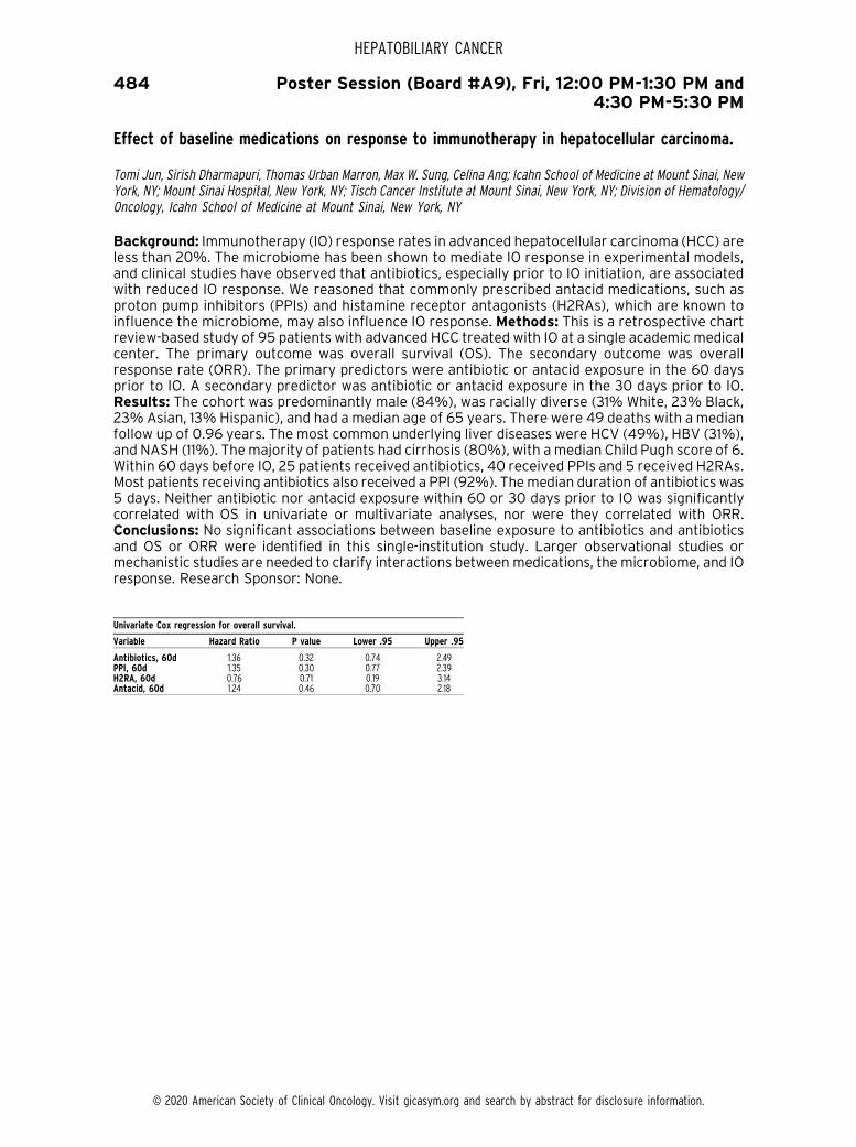
484 Poster Session (Board #A9), Fri, 12:00 PM-1:30 PM and4:30 PM-5:30 PM
Effect of baseline medications on response to immunotherapy in hepatocellular carcinoma.
Tomi Jun, Sirish Dharmapuri, Thomas Urban Marron, Max W. Sung, Celina Ang; Icahn School of Medicine at Mount Sinai, NewYork, NY; Mount Sinai Hospital, New York, NY; Tisch Cancer Institute at Mount Sinai, New York, NY; Division of Hematology/Oncology, Icahn School of Medicine at Mount Sinai, New York, NY
Background: Immunotherapy (IO) response rates in advanced hepatocellular carcinoma (HCC) areless than 20%. The microbiome has been shown to mediate IO response in experimental models,and clinical studies have observed that antibiotics, especially prior to IO initiation, are associatedwith reduced IO response. We reasoned that commonly prescribed antacid medications, such asproton pump inhibitors (PPIs) and histamine receptor antagonists (H2RAs), which are known toinfluence the microbiome, may also influence IO response.Methods: This is a retrospective chartreview-based study of 95 patients with advanced HCC treated with IO at a single academic medicalcenter. The primary outcome was overall survival (OS). The secondary outcome was overallresponse rate (ORR). The primary predictors were antibiotic or antacid exposure in the 60 daysprior to IO. A secondary predictor was antibiotic or antacid exposure in the 30 days prior to IO.Results: The cohort was predominantly male (84%), was racially diverse (31% White, 23% Black,23% Asian, 13% Hispanic), and had a median age of 65 years. There were 49 deaths with a medianfollow up of 0.96 years. The most common underlying liver diseases were HCV (49%), HBV (31%),and NASH (11%). Themajority of patients had cirrhosis (80%), with amedian Child Pugh score of 6.Within 60 days before IO, 25 patients received antibiotics, 40 received PPIs and 5 received H2RAs.Most patients receiving antibiotics also received a PPI (92%). Themedian duration of antibiotics was5 days. Neither antibiotic nor antacid exposure within 60 or 30 days prior to IO was significantlycorrelated with OS in univariate or multivariate analyses, nor were they correlated with ORR.Conclusions: No significant associations between baseline exposure to antibiotics and antibioticsand OS or ORR were identified in this single-institution study. Larger observational studies ormechanistic studies are needed to clarify interactions between medications, the microbiome, and IOresponse. Research Sponsor: None.
Univariate Cox regression for overall survival.
Variable Hazard Ratio P value Lower .95 Upper .95
Antibiotics, 60d 1.36 0.32 0.74 2.49PPI, 60d 1.35 0.30 0.77 2.39H2RA, 60d 0.76 0.71 0.19 3.14Antacid, 60d 1.24 0.46 0.70 2.18
© 2020 American Society of Clinical Oncology. Visit gicasym.org and search by abstract for disclosure information.
HEPATOBILIARY CANCER

485 Poster Session (Board #A10), Fri, 12:00 PM-1:30 PM and4:30 PM-5:30 PM
Potential use of lenvatinib for patients with unresectable hepatocellular carcinoma beyondprogression of sorafenib treatment: A real-world evidence and in vitro assessment with proteinphosphorylation array.
Yasushi Sato, Tetsu Tomonari, Hironori Tanaka, Takahiro Tanaka, Akihiro Hirao, Koichi Okamoto, Hiroshi Miyamoto,Naoki Muguruma, Harumi Kagiwada, Masashi Kitazawa, Kazuhiko Fukui, Katsuhisa Horimoto, Tetsuji Takayama; Departmentof Community Medicine for Gastroenterology and Oncology, Tokushima University Graduate School of Biomedical Sciences,Tokushima, Japan; Department of Gastroenterology and Oncology, Institute of Biomedical Sciences, Tokushima UniversityGraduate School, Tokushima, Japan; SOCIUM Inc, Tokyo, Japan; National Institute of Advanced Industrial Science andTechnology, Tokyo, Japan
Background:No information is available on the efficacy and safety of lenvatinib (LEN) as a second/third-line treatment for unresectable hepatocellular carcinoma (HCC) after sorafenib (SOR)therapy. We evaluated the characteristics and the therapeutic efficacy and safety of LEN as asecond- and third-line treatment as well as first- treatment for unresectable HCC patients in clinicalsettings. Moreover, to rationalize these clinical findings in vitro, we assessed the anti-tumoractivity of LEN on SOR-resistant cell line and performed a comprehensive phosphorylated proteinarray analysis associated with 377 signal transduction pathways using SOR-resistant and parentalHCC cells. Methods: We retrospectively enrolled 51 unresectable HCC patients. Radiologic re-sponses in 41 patients were evaluated by modified RECIST. Active signal transduction pathways inthe cells were identified by protein array analysis, including 1205 proteins. Results: The evaluatedpatients comprised 25 TKI-naive (first- line), 7 intolerant to SOR (second-line), and 9 patientsresistant to regorafenib (third-line). The ORRs were 64% in first-line, 42.8% in second-line, and22.2% in third-line groups (first-line vs. third-line p, 0.05). The OS in the first-line was significantlylonger than that in third-line group (p, 0.05). Patients with better liver functional reserve (Childscore, ALBI grade) exhibited higher ORR and longer OS. LEN was well-tolerated in the second/third-line treatment. The IC50 value of LEN against PLC/PRF5-R2 (30 mM) was significantly higherthan that against PLC/PRF5 (6.4 mM). LEN significantly inhibited more signal transduction path-ways related to FRS2, a crucial FGFR downstream molecule, in PLC/PRF5 than in PLC/PRF5-R2cells. Conclusions: Our study indicates that LEN was active and safe in the second/third-linetreatment for unresectable HCC. LEN seems more effective for HCC patients with better hepaticreserve function, or before TKI-resistance is acquired because of the partial cross-resistance toSOR. Research Sponsor: None.
© 2020 American Society of Clinical Oncology. Visit gicasym.org and search by abstract for disclosure information.
HEPATOBILIARY CANCER
486 Poster Session (Board #A11), Fri, 12:00 PM-1:30 PM and4:30 PM-5:30 PM
Randomized, open-label, perioperative phase II study evaluating nivolumab alone or nivolumabplus ipilimumab in patients with resectable HCC.
Ahmed Omar Kaseb, Dan G. Duda, Hop Sanderson Tran Cao, Yehia I. Abugabal, Luis M. Vence, Asif Rashid, Roberto Pestana,Jorge M. Blando, Shalini Singh, Jean-Nicolas Vauthey, Manal Hassan, Hesham M. Amin, Aliya Qayyum, Yun Shin Chun,Ching-Wei David Tzeng, Divya Sakamuri, Robert A. Wolff, James C. Yao, James Patrick Allison, Padmanee Sharma; TheUniversity of Texas MD Anderson Cancer Center, Houston, TX; Massachusetts General Hospital, Boston, MA; Baylor College ofMedicine, Houston, TX; University of Texas MD Anderson Cancer Center, Houston, TX; MD Anderson Cancer Center, S~ao Paulo,Brazil; Department of Immunology, The University of Texas MD Anderson Cancer Center, Houston, TX; The University ofTexas-MD Anderson Cancer Center, Houston, TX; University of Kentucky, Lexington, KY
Background: In HCC, surgical resection is associated with high recurrence rates, and no effectiveneoadjuvant or adjuvant therapies currently exist. Immunotherapy using anti-PD-1 antibodies hasshown promised but limited increase in survival in advanced disease. To maximize the benefit, weare studying the efficacy and safety of anti–PD-1 (nivolumab) and anti–CTLA-4 (ipilimumab)antibodies against HCC for resectable HCC. Methods: This is a randomized phase II trial ofnivolumab (Arm A) or nivolumab + ipilimumab (Arm B) as pre-operative treatment for patientswith HCC who are eligible for surgical resection. Pts are given nivolumab 240 mg every 2 weeks(wks) for a total of 6wks. Pt in ArmB are treated concurrently with ipilimumab 1mg/kg every 6wks.Surgical resection occurs within 4 wks after last cycle of therapy. Pts continue adjuvant immu-notherapy for up to 2 years after resection. The primary objective is the safety/tolerability ofnivolumab +/- ipilimumab. Secondary objectives include overall response rate, complete responserate and time to progression. Exploratory objectives include evaluating the pre- and post-treatment immunological changes in tumor tissues and peripheral blood. Results: Twenty-sixpatients were enrolled at the time of this interim analysis, of which 20 have evaluable data. Mostpts (55%) were between 60-70yo and male (75%). Four pts were HCV-positive, 6 had HBV and 10had no hepatitis. 20 patients proceeded with resection as planned, surgery was aborted for 5patients (1 for frozen abdomen and 2 development of contralateral liver nodule). Three are stillreceiving preoperative therapy. Pathologic complete response (pCR) was observed in 5/20evaluable patients – 2 in Arm A and 3 Arm B (25% pCR rate). Five patients in Arm B and 1 inArm A experienced grade 3 or higher toxicity prior to surgery. No grade 4 or higher toxicity wereobserved. Conclusions: We report a pCR rate of 25% for resectable HCC after preoperativeimmunotherapy in a randomized phase II pilot trial. Treatment was safe and surgical resection wasnot delayed. The study is ongoing. These promising results may contribute to a paradigm shift inthe perioperative treatment of resectable HCC. Clinical trial information: NCT03510871. ResearchSponsor: Bristol-Myers Squibb SPORE.
© 2020 American Society of Clinical Oncology. Visit gicasym.org and search by abstract for disclosure information.
HEPATOBILIARY CANCER
487 Poster Session (Board #A12), Fri, 12:00 PM-1:30 PM and4:30 PM-5:30 PM
Economic burden and patterns of care in patients with advanced hepatocellular carcinoma.
Abdalla Aly, Elisabetta Malangone-Monaco, Virginia Noxon, Caroline Henriques, Fernando Benavente, Amy K. Kim;AstraZeneca, Gaithersburg, MD; IBM Watson Health, Cambridge, MA; Johns Hopkins School of Medicine, Baltimore, MD
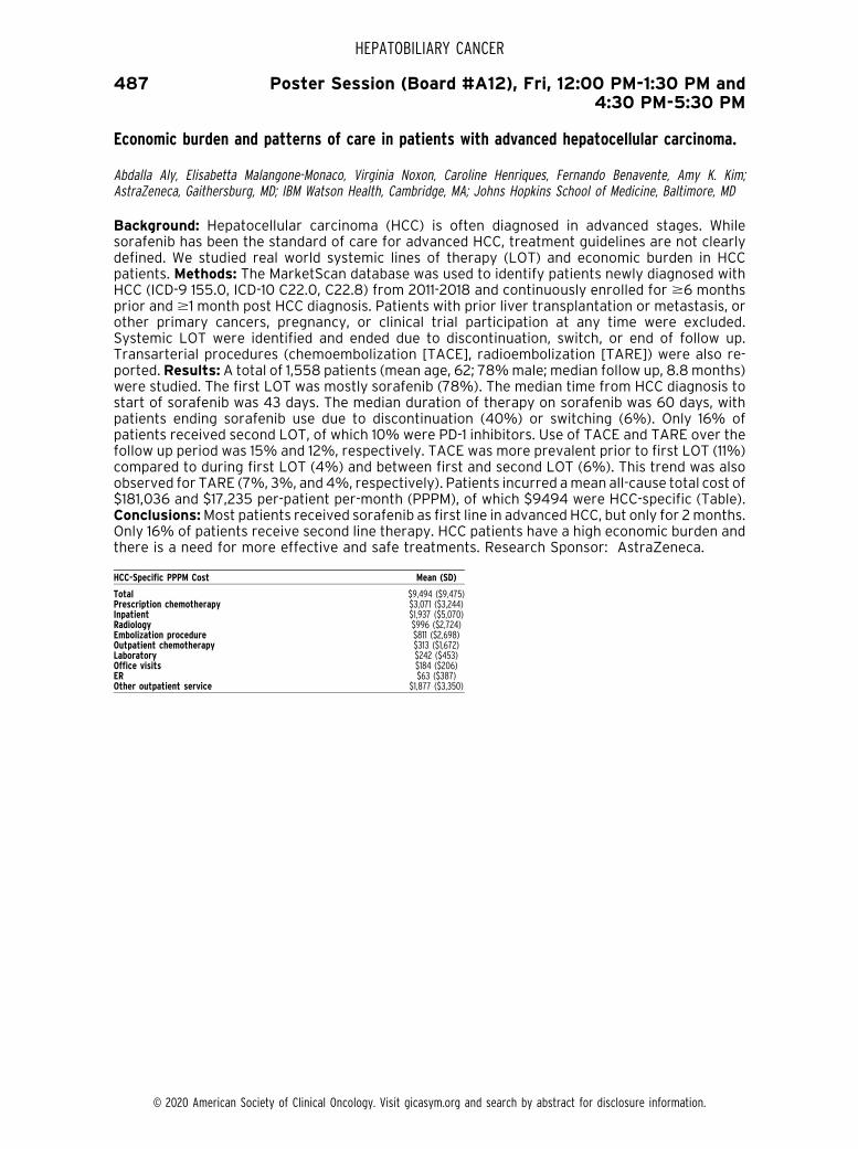
Background: Hepatocellular carcinoma (HCC) is often diagnosed in advanced stages. Whilesorafenib has been the standard of care for advanced HCC, treatment guidelines are not clearlydefined. We studied real world systemic lines of therapy (LOT) and economic burden in HCCpatients. Methods: The MarketScan database was used to identify patients newly diagnosed withHCC (ICD-9 155.0, ICD-10 C22.0, C22.8) from 2011-2018 and continuously enrolled for $6 monthsprior and $1 month post HCC diagnosis. Patients with prior liver transplantation or metastasis, orother primary cancers, pregnancy, or clinical trial participation at any time were excluded.Systemic LOT were identified and ended due to discontinuation, switch, or end of follow up.Transarterial procedures (chemoembolization [TACE], radioembolization [TARE]) were also re-ported.Results: A total of 1,558 patients (mean age, 62; 78%male; median follow up, 8.8 months)were studied. The first LOT was mostly sorafenib (78%). The median time from HCC diagnosis tostart of sorafenib was 43 days. The median duration of therapy on sorafenib was 60 days, withpatients ending sorafenib use due to discontinuation (40%) or switching (6%). Only 16% ofpatients received second LOT, of which 10% were PD-1 inhibitors. Use of TACE and TARE over thefollow up period was 15% and 12%, respectively. TACE was more prevalent prior to first LOT (11%)compared to during first LOT (4%) and between first and second LOT (6%). This trend was alsoobserved for TARE (7%, 3%, and 4%, respectively). Patients incurred amean all-cause total cost of$181,036 and $17,235 per-patient per-month (PPPM), of which $9494 were HCC-specific (Table).Conclusions:Most patients received sorafenib as first line in advanced HCC, but only for 2months.Only 16% of patients receive second line therapy. HCC patients have a high economic burden andthere is a need for more effective and safe treatments. Research Sponsor: AstraZeneca.
HCC-Specific PPPM Cost Mean (SD)
Total $9,494 ($9,475)Prescription chemotherapy $3,071 ($3,244)Inpatient $1,937 ($5,070)Radiology $996 ($2,724)Embolization procedure $811 ($2,698)Outpatient chemotherapy $313 ($1,672)Laboratory $242 ($453)Office visits $184 ($206)ER $63 ($387)Other outpatient service $1,877 ($3,350)
© 2020 American Society of Clinical Oncology. Visit gicasym.org and search by abstract for disclosure information.
HEPATOBILIARY CANCER
488 Poster Session (Board #A13), Fri, 12:00 PM-1:30 PM and4:30 PM-5:30 PM
IGF-1 Child-Turcotte-Pugh score as a predictor of overall survival to therapy in CTP-A, BCLCstage C patients with advanced hepatocellular carcinoma.
Yehia I. Abugabal, Aliya Qayyum, Manal Hassan, Lianchun Xiao, Dan G. Duda, Rikita Hatia, Sunyoung S. Lee, Robert A. Wolff,Roberto Pestana, Jeffrey Morris, James C. Yao, Hesham M. Amin, Ahmed Omar Kaseb; University of Texas MD AndersonCancer Center, Houston, TX; The University of Texas MD Anderson Cancer Center, Houston, TX; The University of Texas-MDAnderson Cancer Center, Houston, TX; Massachusetts General Hospital, Boston, MA; Roswell Park Comprehensive CancerCenter, Buffalo, NY; MD Anderson Cancer Center, S~ao Paulo, Brazil; Department of Biostatistics, The University of Texas MDAnderson Cancer Center, Houston, TX
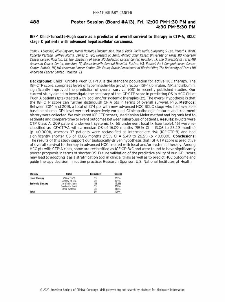
Background: Child-Turcotte-Pugh (CTP) A is the standard population for active HCC therapy. TheIGF-CTP score, comprises levels of type 1 insulin-like growth factor (IGF-1), bilirubin, INR, and albumin,significantly improved the prediction of overall survival (OS) in recently published studies. Ourcurrent study aimed to investigate the accuracy of the IGF-CTP score in predicting OS in HCC Child-Pugh A patients (pts) treatedwith local and/or systemic therapies (tx). The overall hypothesis is thatthe IGF-CTP score can further distinguish CP-A pts in terms of overall survival, PFS. Methods:Between 2014 and 2018, a total of 274 pts with new advanced HCC BCLC stage who had availablebaseline plasma IGF-1 level were retrospectively enrolled. Clinicopathologic features and treatmenthistorywere collected.We calculated IGF-CTP scores, used Kaplan-Meiermethod and log rank test toestimate and compare time to event outcomes between subgroups of patients.Results: 198ptswereCTP Class A, 209 patient underwent systemic tx, 65 underwent local tx [see table]; 161 were re-classified as IGF-CTP-A with a median OS of 16.09 months (95% CI = 13.06 to 23.29 months)(p ,0.0001), whereas 37 patients were reclassified as intermediate risk (IGF-CTP-B) and hadsignificantly shorter OS of 10.66 months (95% CI = 5.49 to 26.51) (p ,0.0001). Conclusions:The results of this study support our biologically-driven hypothesis that IGF-CTP score is predictiveof overall survival to therapy in advanced HCC treated with local and/or systemic therapy. AmongHCC pts with CTP-A class, some are reclassified as IGF-CP-B/C and were found to have significantlypoorer prognosis in terms of shorter OS. Future validation of the predictive ability of our IGF-1 scoremay lead to adopting it as a stratification tool in clinical trials as well as to predict HCC outcome andguide therapy decision in routine practice. Research Sponsor: U.S. National Institutes of Health.
Therapy Name Frequency Percent
Local therapy Y90 or TACE 35 12.7%Surgery or RFA 30 10.9%
Systemic therapy Sorafenib alone 136 49.6%Sorafenib+ Local 35 12.8%Other systemic 38 13.8%
Total 274 100%
© 2020 American Society of Clinical Oncology. Visit gicasym.org and search by abstract for disclosure information.
HEPATOBILIARY CANCER
489 Poster Session (Board #A14), Fri, 12:00 PM-1:30 PM and4:30 PM-5:30 PM
Impact of population center (PC) size on access to care in advanced hepatocellular carcinoma(HCC).
Irene S. Yu, Shiru Lucy Liu, Valeriya O. Zaborska, Tyler Raycraft, Janine Marie Davies; BC Cancer, Vancouver, BC, Canada;University of British Columbia, Vancouver, BC, Canada; British Columbia Cancer Agency-Centre for the Southern Interior,Kelowna, BC, Canada
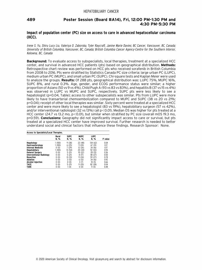
Background: To evaluate access to subspecialists, local therapies, treatment at a specialized HCCcenter, and survival in advanced HCC patients (pts) based on geographical distribution. Methods:Retrospective chart review was performed on HCC pts who received sorafenib in British Columbiafrom 2008 to 2016. Pts were stratified by Statistics Canada PC size criteria: large urban PC (LUPC),medium urban PC (MUPC), and small urban PC (SUPC). Chi-square tests and Kaplan Meier were usedto analyze the groups. Results: Of 288 pts, geographical distribution was: LUPC 75%, MUPC 16%,SUPC 8%, and rural 0.3%. Age, gender, and ECOG performance status were similar; a higherproportion ofAsians (50 vs9 vs4%), Child PughA (93vs83vs83%), and hepatitis B (37 vs 15vs4%)was observed in LUPC vs MUPC and SUPC, respectively. SUPC pts were less likely to see ahepatologist (p=0.04, Table); access to other subspecialists was similar. Pts from LUPC were morelikely to have transarterial chemoembolization compared to MUPC and SUPC (38 vs 20 vs 21%;p=0.04); receipt of other local therapies was similar. Sixty percent were treated at a specialized HCCcenter and were more likely to see a hepatologist (83 vs 19%), hepatobiliary surgeon (57 vs 42%),and/or interventional radiologist (32 vs 13%) (all p,0.01). Median OS was higher for pts treated at aHCC center (24.7 vs 13.2 mo, p,0.01), but similar when stratified by PC size (overall mOS 19.3 mo,p=0.59). Conclusions: Geography did not significantly impact access to care or survival, but ptstreated at a specialized HCC center have improved survival. Further research is needed to betterunderstand social and clinical factors that influence these findings. Research Sponsor: None.
Access to Specialists/Local Therapies.
RuralN, %
SUPCN, %
MUPCN, %
LUPCN, % P value
Hepatology 1 (100) 9 (38) 22 (48) 134 (62) 0.04Gastroenterology 1 (100) 6 (25) 9 (20) 67 (31) 0.17Internal Medicine 0 (0) 7 (29) 12 (26) 34 (16) 0.17Hepatobiliary 1 (100) 13 (54) 20 (44) 112 (52) 0.55General Surgery 0 (0) 5 (21) 10 (22) 28 (13) 0.36Interventional Radiology 0 (0) 5 (21) 8 (17) 48 (27) 0.50Resection 0 (0) 8 (33) 11 (24) 59 (27) 0.78Ablation 0 (0) 3 (13) 6 (13) 34 (16) 0.92Alcohol Injection 0 (0) 0 (0) 3 (7) 10 (5) 0.66TACE 0 (0) 5 (21) 9 (20) 82 (38) 0.04TARE 0 (0) 1 (4) 0 (0) 11 (5) 0.48
© 2020 American Society of Clinical Oncology. Visit gicasym.org and search by abstract for disclosure information.
HEPATOBILIARY CANCER
490 Poster Session (Board #A15), Fri, 12:00 PM-1:30 PM and4:30 PM-5:30 PM
Efficacy and safety of lenvatinib (LEN) in Korean patients (pts) with advanced hepatocellularcarcinoma (aHCC): Multicenter retrospective analysis.
Jaekyung Cheon, Changhoon Yoo, Yeonghak Bang, Hong Jae Chon, Baek-Yeol Ryoo; Division of Hematology and Oncology,Department of Internal Medicine, Ulsan University Hospital, University of Ulsan College of Medicine, Ulsan, South Korea;Department of Oncology, Asan Medical Center, University of Ulsan College of Medicine, Seoul, South Korea; Department ofMedical Oncology, CHA Bundang Medical Center, CHA University, Seongnam, South Korea; Asan Medical Center, University ofUlsan College of Medicine, Seoul, South Korea
Background: LEN has demonstrated the efficacy and safety in pts with aHCC as first-line treatmentin the pivotal REFLECT trial. Further evaluation in real-world setting is necessary to measure theclinical outcomes of LEN in daily practice.Methods: This is amulticenter retrospective analysis from3 Korean referral cancer institutions. Between September 2018 and August 2019, a total of 74 ptsreceived LEN for the management of BCLC B or C aHCC, and 66 pts who had at least one follow-upvisit after the start of LEN were included in this analysis. Results:Median age was 58 years (range,19-81), and 46 pts (69.7%)weremale. Baseline characteristics were as follows; Child-Pugh class A/B/C in 46 (69.7%)/14 (21.2%)/6 (9.1%), BCLC B/C/D in 1 (1.5%)/63 (95.5%)/2 (3.0%), prior systemictherapy in 25 (37.9%) including 14 (21.2%) with prior immune checkpoint inhibitors (ICIs). LEN wasused as first/second/third to fourth lines of therapy in 41 (62.1%)/13 (19.7%)/12 (18.2%) pts, and 27(40.9%) had extensive disease extent excluded in the REFLECT trial. With a median follow-upduration of 4.8 months (95%CI, 3.4–6.1), the median PFS and OSwere 4.6 (95% CI, 3.2-6.0) and 7.5months (mo) (95%CI, 3.7–11.2), respectively, in overall pts: first-line setting, 4.2 (95%CI, 3.2-5.2) and6.5 mo (95% CI, 5.0-8.1), respectively; $ second-line setting, 6.1 mo (95% CI, 3.6–8.5) and notreached, respectively. In pts with prior ICIs, median PFS was 6.1 mo (95% CI, 1.8-8.4) and median OSwas not reached. According to the RECIST v 1.1, response rates and disease control rate were 12.1%and 71.2%, respectively, in overall pts. The most common grade 3-4 toxicities were hyperbilirubi-nemia (n=9, 13.6%), AST elevation (n=5, 7.6%), diarrhea (n=4, 6.1%) and fatigue (n=4, 6.1%).Conclusions: LEN was effective and well tolerated in pts with aHCC in Korean real-life setting.Research Sponsor: None.
© 2020 American Society of Clinical Oncology. Visit gicasym.org and search by abstract for disclosure information.
HEPATOBILIARY CANCER
491 Poster Session (Board #A16), Fri, 12:00 PM-1:30 PM and4:30 PM-5:30 PM
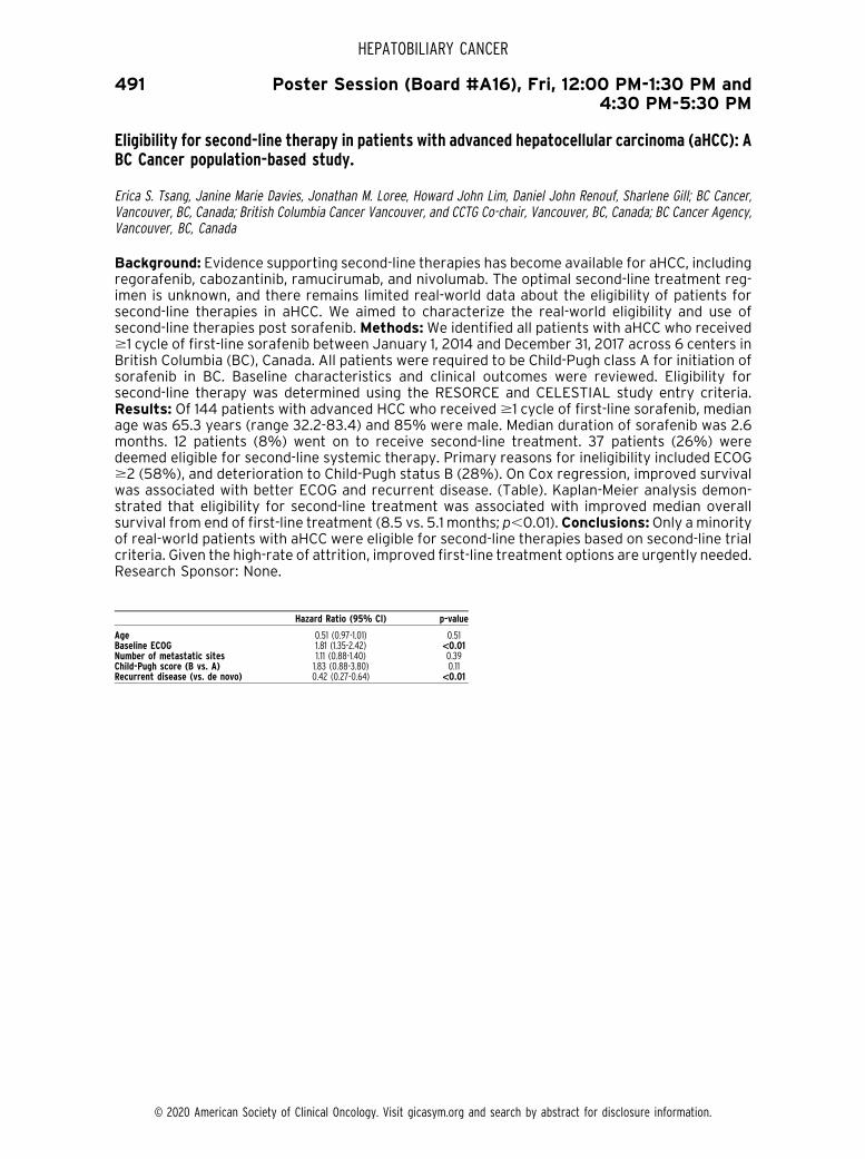
Eligibility for second-line therapy in patients with advanced hepatocellular carcinoma (aHCC): ABC Cancer population-based study.
Erica S. Tsang, Janine Marie Davies, Jonathan M. Loree, Howard John Lim, Daniel John Renouf, Sharlene Gill; BC Cancer,Vancouver, BC, Canada; British Columbia Cancer Vancouver, and CCTG Co-chair, Vancouver, BC, Canada; BC Cancer Agency,Vancouver, BC, Canada
Background: Evidence supporting second-line therapies has become available for aHCC, includingregorafenib, cabozantinib, ramucirumab, and nivolumab. The optimal second-line treatment reg-imen is unknown, and there remains limited real-world data about the eligibility of patients forsecond-line therapies in aHCC. We aimed to characterize the real-world eligibility and use ofsecond-line therapies post sorafenib.Methods:We identified all patients with aHCC who received$1 cycle of first-line sorafenib between January 1, 2014 and December 31, 2017 across 6 centers inBritish Columbia (BC), Canada. All patients were required to be Child-Pugh class A for initiation ofsorafenib in BC. Baseline characteristics and clinical outcomes were reviewed. Eligibility forsecond-line therapy was determined using the RESORCE and CELESTIAL study entry criteria.Results: Of 144 patients with advanced HCC who received$1 cycle of first-line sorafenib, medianage was 65.3 years (range 32.2-83.4) and 85% were male. Median duration of sorafenib was 2.6months. 12 patients (8%) went on to receive second-line treatment. 37 patients (26%) weredeemed eligible for second-line systemic therapy. Primary reasons for ineligibility included ECOG$2 (58%), and deterioration to Child-Pugh status B (28%). On Cox regression, improved survivalwas associated with better ECOG and recurrent disease. (Table). Kaplan-Meier analysis demon-strated that eligibility for second-line treatment was associated with improved median overallsurvival from end of first-line treatment (8.5 vs. 5.1 months; p,0.01). Conclusions:Only a minorityof real-world patients with aHCC were eligible for second-line therapies based on second-line trialcriteria. Given the high-rate of attrition, improved first-line treatment options are urgently needed.Research Sponsor: None.
Hazard Ratio (95% CI) p-value
Age 0.51 (0.97-1.01) 0.51Baseline ECOG 1.81 (1.35-2.42) <0.01Number of metastatic sites 1.11 (0.88-1.40) 0.39Child-Pugh score (B vs. A) 1.83 (0.88-3.80) 0.11Recurrent disease (vs. de novo) 0.42 (0.27-0.64) <0.01
© 2020 American Society of Clinical Oncology. Visit gicasym.org and search by abstract for disclosure information.
HEPATOBILIARY CANCER

492 Poster Session (Board #A17), Fri, 12:00 PM-1:30 PM and4:30 PM-5:30 PM
Biliary tract cancer (BTC) in the elderly: A real-world tertiary cancer center experience.
Massimiliano Salati, Francesco Caputo, Krisida Cerma, Andrea Spallanzani, Fabio Gelsomino, Chiara Santini,Alessandro Bocconi, Maria Laura Riggi, Gabriele Luppi, Andrea Casadei Gardini, Massimo Dominici; PhD Program in Clinicaland Experimental Medicine, University of Modena and Reggio Emilia, Modena, Italy; Department of Oncology andHematology, University Hospital of Modena, Modena, Italy; Department of Oncology, University of Modena and ReggioEmilia, Modena, Italy; University Hospital of Modena, Modena, Italy; Department of Oncology, University Hospital of Modena,Forli, Italy
Background: Although BTC is mostly a disease of the elderly, only limited data ara available on theoptimal management of this patient (pt) population. In fact, older pts are underepresented inclinical trials and results are seldom reported by age group. In this study, we aimed at evaluatingpattern of care and treatment outcome in BTC aged $ 70 years and comparing them with theiryounger counterparts. Methods: Medical records of BTC followed at the Modena Cancer Centrefrom 2007 and 2019 were retrospectively reviewed.. Overall survival (OS) was estimated with theKaplan-Meier curves and compared by log-rank test. Differences between categorical variableswere assessed using the chi square test. Univariate and multivariate analyses were performed toassess the impact of covariates on survival. Results: A total of 120 BTC patients (49%)$ 70 wereincluded in the analysis. 54% (64) were female, 47% (56) had iCCA, 41% (49) GBC, and 12% (15)eCCA. 68% (81) had unresectable locally advanced or metastatic disease. 32% (39) underwentsurgical resection, 60% (72) were treated with first-line chemotherapy (1L), while 29% (21) of themwent on to receive second-line (2L). No differences in terms of both chance to receive surgery(p=0.59) and survival (p=0.25) were recorded compared to youngers. In the advanced-diseasesetting, median OS was 8 months and was significantly worse than that of the younger counter-parts (p,0.001). Older patients were less likely to receive 1L (p,0.001) and 2L (p,0.001) che-motherapy and doublet regimens (p,0.001). Female gender (p=0.031), ECOG PS 0 (p,0.001),stage III (p,0.001) andNLR.3 (p,0.001) were independently associatedwith a better prognosis inolder BTC receiving 1L, with 1-year OS of 82% (95%CI 68-91, p=0.031). Conclusions: In this real-world study, no survival difference was found between older and non-older surgically-treatedpatients. Contrariwise, elderly BTC were less frequently treated with chemotherapy for advanceddisease and their outcome is poorer than youngers. However, clinical and biochemical prognostichave been identified that may assist in selecting older pts more likely to benefit from systemictreatment, both in clinical trials and daily practice. Research Sponsor: None.
© 2020 American Society of Clinical Oncology. Visit gicasym.org and search by abstract for disclosure information.
HEPATOBILIARY CANCER
493 Poster Session (Board #A18), Fri, 12:00 PM-1:30 PM and4:30 PM-5:30 PM
The impact of skeletal muscle loss for hepatocellular carcinoma treated with lenvatinib.
Mao Okada, Hiroyuki Nakanishi, Masayuki Kurosaki, Kento Inada, Sakura Kirino, Koji Yamashita, Shuhei Sekiguchi,Yuka Hayakawa, Leona Osawa, Wan Wang, Mayu Higuchi, Kenta Takaura, Chiaki Maeyashiki, Shun Kaneko,Nobuharu Tamaki, Yutaka Yasui, Kaoru Tsuchiya, Jun Itakura, Yuka Takahashi, Namiki Izumi; Musashino Red CrossHospital, Tokyo, Japan; Musaino Red Cross Hospital, Tokyo, Japan; Department of Gastroenterology and Hepatology,Musashino Red Cross Hospital, Musashino, Tokyo, Japan
Background: Many previous reports have shown that skeletal muscle loss (SML) is one of theprognostic factors for hepatocellular carcinoma (HCC) patients treated with sorafenib. However,there are few reports about the impact of SML for the HCC patients treated with lenvatinib.Therefore, we evaluated the relation between SML and overall survival (OS) of HCC patients treatedwith lenvatinib (LEN).Methods:We retrospectively analyzed 50HCC patients treatedwith LEN fromApril 2018 to February 2019. We included 36 patients who continued LEN more than 8 weeks andevaluated CT scans before treatment and after 8 weeks. Skeletal muscle area wasmeasured on axialimage at the level of the third lumber vertebra (L3) using sliceOmatic. Skeletal Mass Index (SMI) wascalculated by dividing the muscle area (㎠) with square of height (㎡). The definition of myopenia isbasedon the guideline describedby theJapanSociety ofHepatology (42㎠/㎡ inmenand38㎠/㎡ inwomen). DSMI is a chronological change of SMI for 8 weeks. We calculated decreasing rate of DSMI.We evaluated the relation between chronological change of SMI and OS. Results: The patients withmyopenia at baseline were 12 (33.3 %). The decreasing rate of DSMI at 8 weeks was -2.57 % [-5.9,0.2]. SMI had decreased in 27 patients (75 %) for 8 weeks. There was no significant differencebetween OS and baselinemyopenia (p = 0.2), ALBI grade (p = 0.2), BCLC stage (p = 0.5), up to 7 in orout (p = 0.35), previous TKI treatment (p = 0.15), metastasis (p = 0.91), or vascular invasion (p = 0.12).However, the patients who had decreased SMI had significantly poor prognosis (p = 0.028). Inbackgrounds, there was no significant difference between patients with or without decreasing ofDSMI, such asbaselinemyopenia (p =0.7), ALBI grade (p =0.4), BCLCstage (p = 1.0), Child Pugh score(p = 0.8), age (p = 0.6), sex (p = 0.3), up to7 in or out (p = 1.0), previous TKI treatment (p = 0.3), andrelative dose intensity at 4 weeks (p = 0.9). Conclusions: There was no significant correlationbetween baseline myopenia and OS. However, chronological decreasing of SMI for 8 weeks was aprognostic factor of HCC patients treated with LEN. Therefore, monitoring and preventing ofdecreasing of skeletal muscle mass may be important. Research Sponsor: None.
© 2020 American Society of Clinical Oncology. Visit gicasym.org and search by abstract for disclosure information.
HEPATOBILIARY CANCER

494 Poster Session (Board #A19), Fri, 12:00 PM-1:30 PM and4:30 PM-5:30 PM
Clinical outcome associated with neoadjuvant chemoradiation and orthotopic liver transplantationversus definitive chemoradiation in 49 patients with unresectable, hilar, or extrahepaticcholangiocarcinoma.
Brady S. Laughlin, Molly M. Petersen, Jonathan Ben Ashman, William G. Rule, Mitesh J. Borad, Bashar A. Aqel,Mohamad Bassam Sonbol, Tanios S. Bekaii-Saab, Daniel H. Ahn, Nathan Y. Yu, Todd DeWees, Terence Tai Weng Sio; MayoClinic Arizona, Phoenix, AZ; Mayo Clinic, Rochester, MN; Mayo Clinic, Scottsdale, AZ; Mayo Clinic, Phoenix, AZ; Mayo ClinicCancer Center, Scottsdale, AZ; Ohio State University Arthur G. James Cancer Hospital and Richard J. Solove ResearchInstitute, Columbus, OH; Washington University in St. Louis, St. Louis, MO
Background: Our aim was to compare survival between patients receiving neoadjuvant chemo-radiation and orthotopic liver transplantation (OLT group) versus definitive chemoradiation (CRTgroup) for extrahepatic or hilar cholangiocarcinoma.Methods:49patients (20 inOLT group vs. 29 inCRT group) with unresectable hilar/extrahepatic cholangiocarcinoma were treated at Mayo ClinicArizona between Feb. 1998–Sep. 2019. Treatment included external beam radiation therapy (median4500cGy) and boost (median 900cGy)with either continuous5-flurouracil (dose range 180–225mg/m2) or capecitabine (dose range 825–1000 mg/m2 BID) prior to or without OLT. Radiation boostswere delivered with EBRT or bile duct brachytherapy. Patients were between 27.9–84.3 years(median 64.3) at diagnosis. 18 patients had previous diagnosis of PSC. Results: Between Feb.1998–Sep. 2019, 31(63%) of 49 patients died by the end of follow-up. Of patients treated withneoadjuvant therapy and OLT, 7(35%) of 20 patients died. 24(86%) of 28 patients treated withdefinitive therapy died. The OLT cohort were younger (mean age 56.5 vs. 69.0 years), more likely tohave PSC and UC (65% vs. 17%), and had a lower CA 19-9 (median 43 vs. 535)(P, 0.003). From theend date of radiation, median overall survival was 76.8 months vs. 15.6 months for the OLT and CRTgroups, respectively. Survival rates at 3 and 5 years were 78% and 69% in the OLT group comparedto 19%and 6% in the CRT group (HR 7.73; 3.04-19.65:(P, 0.0001)). Progression-free survival (89%vs. 30% at 3 years), and distant metastasis-free survival (88% vs. 66% at 3 years) favored OLTversus CRT alone (HR5.74;1.12-29.34:(P, 0.02)). Univariate analysis demonstrated that themethodof treatment (OLT vs. CRT) was the only variable associated with better clinical outcomes.Conclusions: In patients with unresectable extrahepatic/hilar cholangiocarcinoma, survival washigher in those who underwent chemoradiation and OLT. Patients who received definitive chemo-radiation in the absence of OLT were expected to have worse overall, progression-free, andmetastasis-free survival. Research Sponsor: None.
© 2020 American Society of Clinical Oncology. Visit gicasym.org and search by abstract for disclosure information.
HEPATOBILIARY CANCER
495 Poster Session (Board #A20), Fri, 12:00 PM-1:30 PM and4:30 PM-5:30 PM
Variations in surgical treatment of stage I gallbladder carcinoma impacts survival.
Anthony Joseph Scholer, Mary Garland-Kledzik, Mansen Wang, Adam Khader, Juan Santamaria-Barria, Zeljka Jutric,Ronald Wolf, Melanie Goldfarb; John Wayne Cancer Institute at Providence St. John’s Health Center, Santa Monica, CA;Providence Health & Services Medical Data Research Center, Portland, OR; University of California Irvine, Irvine, CA
Background:Variations in surgical care for stage I gallbladder carcinoma (GBC)may be associatedwith inferior outcomes. The aim of this study was to identify the variations in surgical treatment ofGBC. Methods: All patients diagnosed with stage I GBC by AJCC 8 criteria from 2004-2013 wereidentified in theNCDB. Surgical treatmentwas categorized as cholecystectomy (C), cholecystectomywith lymph node dissection (C+LND), or radical cholecystectomy (RC). Independent predictors ofimproved overall survival (OS) and extent of surgery were identified by multinomial regressionanalyses.Results:Of 1756 patients with stage I GBC, 26%were T1a, 56%T1b, and 18.5% T1NOS. Themajority were White non-Hispanic (61.8%) and female (68.5%), with 55.1%. 70 years of age. Two-thirds of T1a tumors were treated with more aggressive surgery (28% C+LND, 4.2% RC), which didnot differ by age. However, only 44.4% of patients with T1b tumors had more aggressive surgery,which was significantly less likely in patients . 70 years, even after controlling for other factors(C+LND (OR:0.60; CI:0.44-0.81), RC (OR:0.52; CI:0.29-0.91)). Five-year OS was 54.34% for T1a and43.05% for T1b (p = 0.02). After controlling for other factors, both C+LND (HR:0.46, CI:0.26-0.81)and RC (HR:0.31, CI:0.16-0.62) significantly improved 5-year OS for T1b tumors, whereas RC alsoimproved 5-year OS for all patients . 70 years old (p = 0.04). Conclusions: A majority of patientswith T1b GBC had less than adequate surgery by the current AJCC staging, which significantlydecreased survival in all patients. Thiswas especially evident in older patientswhowere also the leastlikely to receive more aggressive surgery. Research Sponsor: None.
© 2020 American Society of Clinical Oncology. Visit gicasym.org and search by abstract for disclosure information.
HEPATOBILIARY CANCER
496 Poster Session (Board #A21), Fri, 12:00 PM-1:30 PM and4:30 PM-5:30 PM
The adherence to The American Association for The Study of Liver Disease (AASLD) guidelines intreating patients with hepatocellular carcinoma: Institutional experience.
Ashish Manne, Daisy E. Escobar, Pranitha Prodduturvar, Phillip Henderson, Osama Abdul-Rahim, Zeiad Hussain, Spencer Liles,Annabelle Fonseca, John Harrison Howard, Wadad Mneimneh, Robert Gilbert, Sachin Gopalkrishna Pai, Moh’d M. Khushman;Medical Oncology, Mitchell Cancer Institute, The University of South Alabama, Mobile, AL; Internal Medicine, The University ofSouth Alabama, Mobile, AL; Gastroenetrology, The University of South Alabama, Mobile, AL; Interventional RAdiology, TheUniversity of South Alabama, Mobile, AL; Interventional Radiology, The University of South Alabama, Mobile, AL; SurgicalOncology, The University of South Alabama, Mobile, AL; Department of Pathology/The University of South Alabama, Mobile,AL; Radiation Oncology, The University of South Alabama, Mobile, AL; Robert H. Lurie Cancer Center of NorthwesternUniversity, Chicago, IL; Medical Oncology, The University of South Alabama, Mitchell Cancer Institute, Mobile, AL
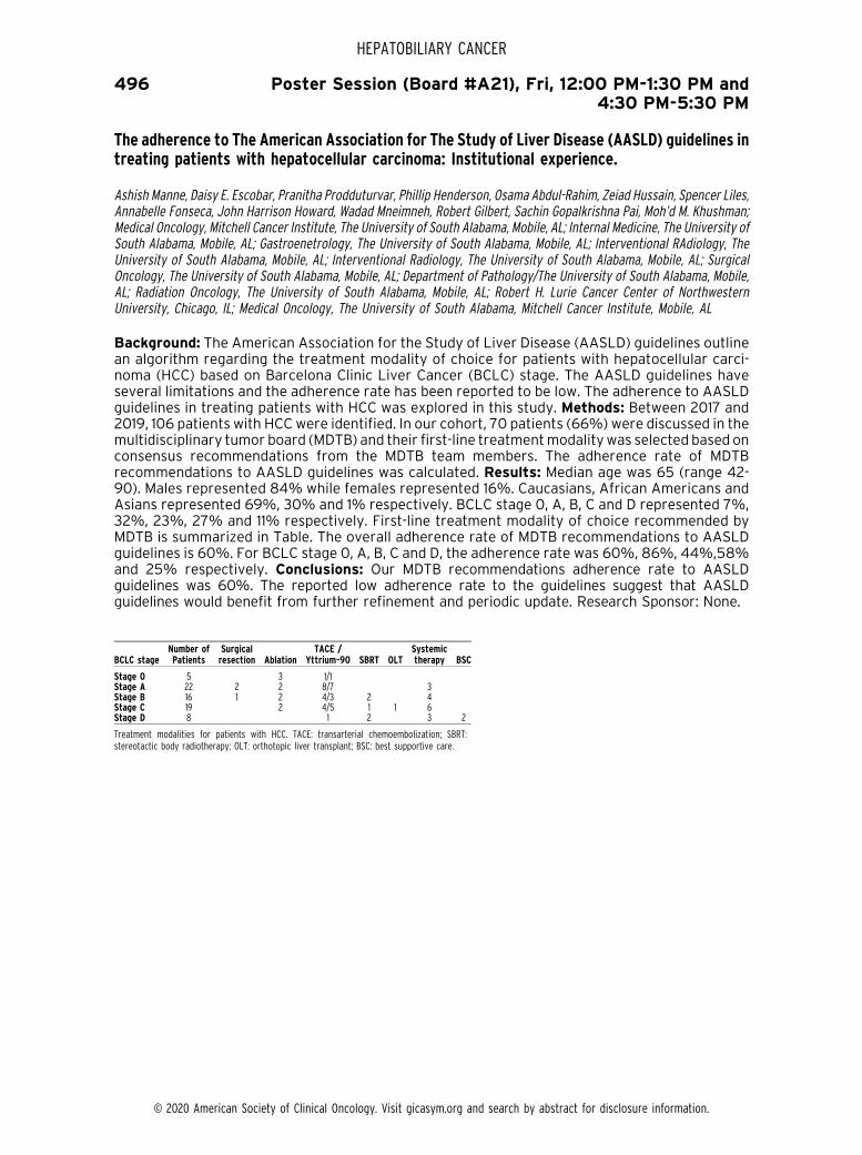
Background: The American Association for the Study of Liver Disease (AASLD) guidelines outlinean algorithm regarding the treatment modality of choice for patients with hepatocellular carci-noma (HCC) based on Barcelona Clinic Liver Cancer (BCLC) stage. The AASLD guidelines haveseveral limitations and the adherence rate has been reported to be low. The adherence to AASLDguidelines in treating patients with HCC was explored in this study. Methods: Between 2017 and2019, 106 patients with HCC were identified. In our cohort, 70 patients (66%) were discussed in themultidisciplinary tumor board (MDTB) and their first-line treatmentmodalitywas selected based onconsensus recommendations from the MDTB team members. The adherence rate of MDTBrecommendations to AASLD guidelines was calculated. Results: Median age was 65 (range 42-90). Males represented 84% while females represented 16%. Caucasians, African Americans andAsians represented 69%, 30% and 1% respectively. BCLC stage 0, A, B, C and D represented 7%,32%, 23%, 27% and 11% respectively. First-line treatment modality of choice recommended byMDTB is summarized in Table. The overall adherence rate of MDTB recommendations to AASLDguidelines is 60%. For BCLC stage 0, A, B, C and D, the adherence rate was 60%, 86%, 44%,58%and 25% respectively. Conclusions: Our MDTB recommendations adherence rate to AASLDguidelines was 60%. The reported low adherence rate to the guidelines suggest that AASLDguidelines would benefit from further refinement and periodic update. Research Sponsor: None.
BCLC stageNumber ofPatients
Surgicalresection Ablation
TACE /Yttrium-90 SBRT OLT
Systemictherapy BSC
Stage 0 5 3 1/1Stage A 22 2 2 8/7 3Stage B 16 1 2 4/3 2 4Stage C 19 2 4/5 1 1 6Stage D 8 1 2 3 2
Treatment modalities for patients with HCC. TACE: transarterial chemoembolization; SBRT:stereotactic body radiotherapy; OLT: orthotopic liver transplant; BSC: best supportive care.
© 2020 American Society of Clinical Oncology. Visit gicasym.org and search by abstract for disclosure information.
HEPATOBILIARY CANCER
497 Poster Session (Board #A22), Fri, 12:00 PM-1:30 PM and4:30 PM-5:30 PM
Survival by line of therapy of older patients with advanced biliary tract cancer (BTC).
Sarah Bobiak, Mark D. Danese, Deborah P. Lubeck, Michelle L. Gleeson; EMD Serono, Inc: A Business of Merck Kgaa, Darmstadt,Germany, Billerica, MA; Outcomes Insights, Inc., Westlake Village, CA
Background:Overall survival (OS) in advancedormetastatic BTChas not beenadequately describedoutside the clinical trial setting. Further, real-world descriptions of OS by line of therapy, including inpatients who do not receive systemic chemotherapy, are not widely available. In this study, we useddata from a recent cohort of US patients available in the SEER-Medicare linked database to examineOS from diagnosis of advanced or metastatic BTC as well as from initiation of first- and second-linetreatment. Methods: Patients with advanced or metastatic BTC diagnosed between 2010 and 2013were identified in SEER-Medicare, with follow-up through 2014. Demographic and clinical charac-teristics were analyzed. The Kaplan-Meier estimator was used to describe OS from diagnosis amongall patients, OS from diagnosis among patients who did not receive systemic treatment, and OS byline of treatment, from date of treatment initiation. The Cox proportional hazardsmodel was used toidentify demographic and clinical factors associated with survival. Results: Of the 1,461 eligiblepatients aged $66 years, 39% had gallbladder, 22% had intrahepatic, 22% had extrahepatic, and9% had ampulla of Vater cancer. More than two-thirds of patients had stage IV disease, and 38% ofpatients (n = 558) received systemic chemotherapy. Systemic treatment patients were somewhatyounger, more likely to be white, have stage IV cancer and less likely to have mobility limitations(24%vs. 38%) than patientswho did not receive systemic treatment. Among all patients, unadjustedmedian OS from diagnosis was 5.6 months (95% CI 5.0-6.1). Among patients who were not treated,unadjustedmedian survival was 3.3 months (n = 903; 95%CI 2.8-4.0) from diagnosis. When OSwasevaluated by line of treatment, median OS was 8.2 months (n = 558; 95% CI 7.6-9.0) from first-lineinitiation and 5.6months (n = 220; 95%CI 4.6-6.5) from second-line initiation.Conclusions:Amongnewly diagnosed, older US patients, less than half receive systemic treatment for their advancedBTC, and outcomes among both treated and untreated patients remain poor. There is an immediateneed for better therapies to treat patients with advanced BTC. Research Sponsor: Merck KGaA,Darmstadt, Germany and GlaxoSmithKline.
© 2020 American Society of Clinical Oncology. Visit gicasym.org and search by abstract for disclosure information.
HEPATOBILIARY CANCER
498 Poster Session (Board #B1), Fri, 12:00 PM-1:30 PM and4:30 PM-5:30 PM
Primary liver angiosarcoma and factors associated with improved outcomes: An analysis of theNational Cancer Database.
Prantesh Jain, Gino Cioffi, Nirav Patil, Aman Opneja, Asrar Alahmadi, David Lawrence Bajor, Joel N. Saltzman, Patrick J. Getty,John Brian Ammori, Aashish D. Bhatt, David B. Mansur, Richard T. Lee, Ankit Mangla; Department of Hospital Medicine,Cleveland Clinic, OH, Cleveland, OH; Department of Population and Quantitative Health Sciences, Case Western ReserveUniversity School of Medicine, Cleveland, OH; University Hospitals Cleveland Medical Center, Case Western ReserveUniversity, Cleveland, OH; University Hospitals Seidman Cancer Center, Cleveland, OH; University Hospitals Seidman CancerCenter, Case Comprehensive Cancer Center, Case Western Reserve University, Cleveland, OH; Case Western ReserveUniversity, School of Medicine, Cleveland, OH; Univ Hosp Case Medcl Ctr, Cleveland, OH; Univ Hosps Seidman Cancer Ctr,Cleveland, OH; The University of Texas MD Anderson Cancer Center, Houston, TX
Background: Primary liver angiosarcoma (LAS) is a rare and aggressive tumor of the liver. In thisanalysis of the national cancer database (NCDB) we sought the risk of mortality and factorsassociated with survival amongst patient diagnosed with LAS. Methods: Patients diagnosed withhepatocellular carcinoma (HCC) or LAS from 2004 – 2014 were identified in the NCDB. The Kaplan-Meier method with the log-rank test was used to calculate survival for HCC and LAS patients.Additional analyses were performed on the cohort with LAS to assess the impact of surgery,chemotherapy, radiation therapy (RT) and facility type on overall survival (OS). Multivariableanalyses using cox proportionalmethods, adjusted for age, sex, Charlson/Deyo score, race, ethnicity,insurance status, facility location and type, surgery status, and chemotherapy status were per-formed to obtain adjusted hazard ratio (aHR). Results: Total of 118,066 patients with HCC and 346patients with LAS were identified in the database. Median survival for HCC patients was 11.9 months(95%CI: 11.7-12.2) and 2.0months for LASpatients (95%CI: 1.8 – 2.4). Risk ofmortalitywas higher forpatients with LAS compared to those with HCC (aHR (95% CI): 2.23 (1.97 - 2.53), p, .0001). Amongthe LAS patients, those who received surgery had a median survival of 8.6 months (95% CI: 5.6 -17.3), and 1.8months for thosewhodidnot (95%CI: 1.48 - 1.94). Risk ofmortalitywas lower in patientswho received surgery compared to those who did not (aHR (95% CI): 0.23 (0.15 - 0.37), p, .0001).Patients treated at and academic center had a higher median survival (3.3 months, 95% CI: 2.2 - 4.1)then those treated at a non-academic center (1.5 months, 95% CI: 1.2 - 1.8). Though, there was nosignificant difference in OS (aHR (95% CI): 0.48 (0.21 - 1.10), p = 0.082). A very small number ofpatients received chemotherapy or RT to conduct a meaningful analysis. Conclusions: Patientsdiagnosed with primary LAS have a worse OS compared to those with HCC. Amongst patients withprimary LAS, surgical resection is associatedwith best survival outcomes. Treatment at an academiccenter is associated with better median survival, although OS did not reach statistical significance inour analysis. Research Sponsor: None.
© 2020 American Society of Clinical Oncology. Visit gicasym.org and search by abstract for disclosure information.
HEPATOBILIARY CANCER
499 Poster Session (Board #B2), Fri, 12:00 PM-1:30 PM and4:30 PM-5:30 PM
Real-world experience of regorafenib in patients with hepatocellular carcinoma: A multicenterUnited Kingdom study.
Paul J. Ross, Yuk Ting Ma, Daniel H. Palmer, Mark Peter Lythgoe, Sophie Merrick, Adel Samson, Ankit Rohit Rao, Bristi Basu,Debi Prasad, Tony Dhillon, David Lau, Suzanne Darby, James Orr, Jane Margetts, Richard Hubner, Shobhit Baijal,Rohini Sharma, Olusola Olusesan Faluyi, Lennard Lee, Kiruthikah Thillai; Guy’s & St Thomas’ NHS Foundation Trust andKing’s College Hospital NHS Foundation Trust, London, United Kingdom; University of Birmingham, Birmingham, UnitedKingdom; University of Liverpool and Clatterbridge Cancer Centre, Liverpool, United Kingdom; Imperial College London,London, United Kingdom; East & North Herts NHS Trust, Stevenage, United Kingdom; University of Leeds, Leeds, UnitedKingdom; Nottingham City Hospital, Nottingham, United Kingdom; University of Cambridge and Cambridge UniversityHospitals NHS Foundation Trust, Cambridge, United Kingdom; Kings College Hospital, London, United Kingdom; Royal SurreyHospital/University of Surrey, Guildford, United Kingdom; Royal Marsden NHS Foundation Trust, Sutton, United Kingdom;Weston Park Hospital, Sheffield, United Kingdom; Bristol Royal Infirmary, Bristol, United Kingdom; Newcastle upon TyneHospitals NHS Foundation trust, Newcastle upon Tyne, United Kingdom; Christie NHS Foundation Trust, Manchester, UnitedKingdom; Birmingham Heartlands Hospital, Birmingham, United Kingdom; Clatterbridge Cancer Centre, Wirral, UnitedKingdom
Background: Regorafenib was the first treatment to demonstrate a survival benefit in patientswith HCC after progression on sorafenib. The RESORCE trial found that regorafenib improvedoverall survival with acceptable toxicity, in patients with disease progression on sorafenib whotolerated$400mg sorafenib daily and had Child-Pugh A liver function.Methods:We performed amulticentre, retrospective, observational study of patients with HCC receiving regorafenib in theUK, following its availability in April 2018. Results: Data on a total of 104 patients were includedfrom April 2018–August 2019, and 80.8% were male. Age was collected in 85 patients, with amedian of 68 years (range 22–86). 23.5% had NAFLD, 21.2% had ALD, 12.9% had HBV, and 3.5%had HCV. Prior management included sorafenib (100%), TACE (30.8%), resection (12.9%). Dura-tion of sorafenib treatment was evaluable in 99/104 patients, and reported amedian of 8.7months(range 1.8–76.6). Duration of regorafenib treatmentwas evaluable in 92/104 patients, and reporteda median of 3.9 months (range 0.0–15.7). Following treatment with regorafenib, 6 patients (5.8%)achieved partial response, 37 (35.6%) achieved stable disease and 45 (43.3%) had progressivedisease as the best response. 15 (14.4%) were not assessed and 1 (1.1%) had mixed response.Survival data is immature with 62/101 (61.4%) patients alive at the time of census with mediansurvival currently 6.5 months. Fatigue was the most frequent AE, with 69/88 patients (85.2%) forall grades. 12/88 patients (14.8%) had Grade 3 fatigue. Other significant AEs include hand-footsyndrome (6/85 patients [7.3%] had Grade 3) and diarrhoea (4/83 patients [4.9%] had Grade 3).Conclusions: The population in our real-world experience of regorafenib for HCC had a similarduration of prior sorafenib to those in the RESORCE trial. However, there was different balance ofaetiologies with a lower proportion of patients with HBV and HCV. The rate of partial response issimilar to the RESORCE trial with fewer patients achieving stable disease. The incidence of fatiguewas higher, but the incidence of hand-foot syndrome and diarrhoea were lower. Further expansionand follow-up of this population is warranted. Research Sponsor: Bayer Pharmaceutical/BiotechCompany.
© 2020 American Society of Clinical Oncology. Visit gicasym.org and search by abstract for disclosure information.
HEPATOBILIARY CANCER
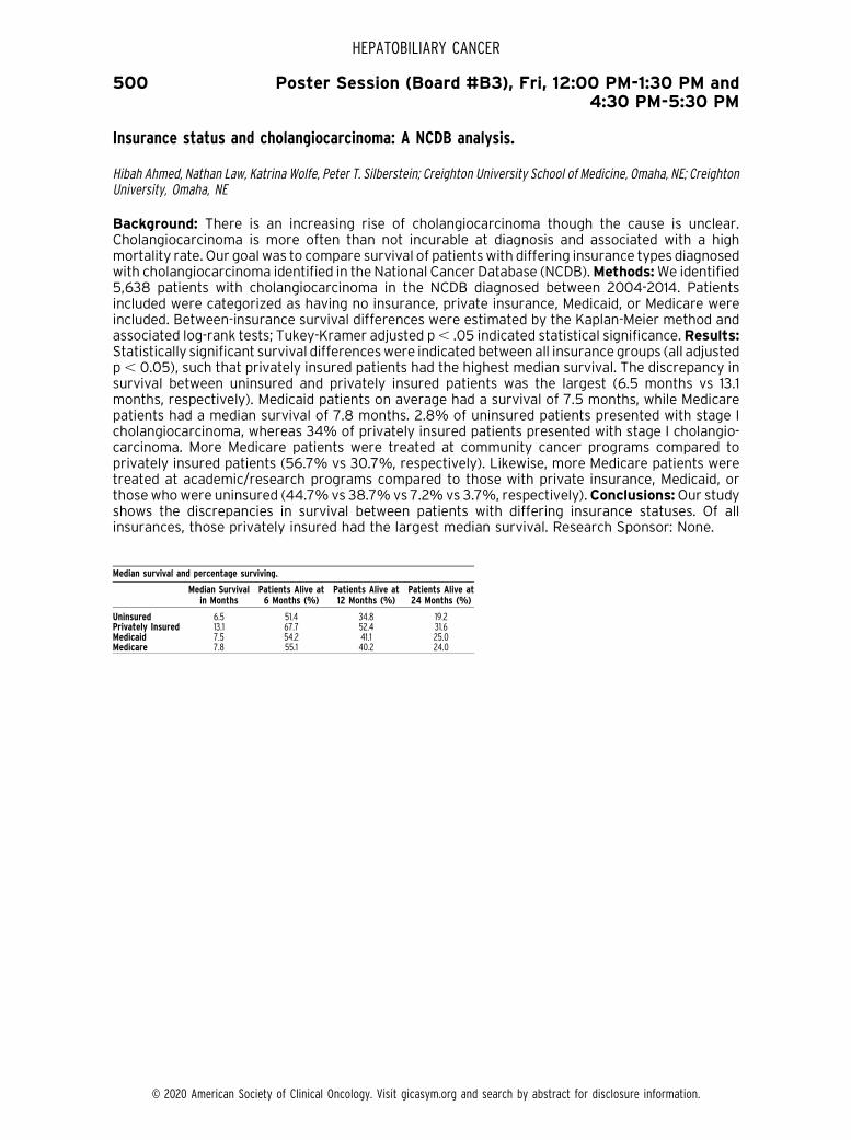
500 Poster Session (Board #B3), Fri, 12:00 PM-1:30 PM and4:30 PM-5:30 PM
Insurance status and cholangiocarcinoma: A NCDB analysis.
Hibah Ahmed, Nathan Law, Katrina Wolfe, Peter T. Silberstein; Creighton University School of Medicine, Omaha, NE; CreightonUniversity, Omaha, NE
Background: There is an increasing rise of cholangiocarcinoma though the cause is unclear.Cholangiocarcinoma is more often than not incurable at diagnosis and associated with a highmortality rate. Our goal was to compare survival of patientswith differing insurance types diagnosedwith cholangiocarcinoma identified in the National Cancer Database (NCDB).Methods:We identified5,638 patients with cholangiocarcinoma in the NCDB diagnosed between 2004-2014. Patientsincluded were categorized as having no insurance, private insurance, Medicaid, or Medicare wereincluded. Between-insurance survival differences were estimated by the Kaplan-Meier method andassociated log-rank tests; Tukey-Kramer adjusted p, .05 indicated statistical significance.Results:Statistically significant survival differenceswere indicated between all insurance groups (all adjustedp, 0.05), such that privately insured patients had the highest median survival. The discrepancy insurvival between uninsured and privately insured patients was the largest (6.5 months vs 13.1months, respectively). Medicaid patients on average had a survival of 7.5 months, while Medicarepatients had a median survival of 7.8 months. 2.8% of uninsured patients presented with stage Icholangiocarcinoma, whereas 34% of privately insured patients presented with stage I cholangio-carcinoma. More Medicare patients were treated at community cancer programs compared toprivately insured patients (56.7% vs 30.7%, respectively). Likewise, more Medicare patients weretreated at academic/research programs compared to those with private insurance, Medicaid, orthosewhowere uninsured (44.7%vs 38.7%vs 7.2% vs 3.7%, respectively).Conclusions:Our studyshows the discrepancies in survival between patients with differing insurance statuses. Of allinsurances, those privately insured had the largest median survival. Research Sponsor: None.
Median survival and percentage surviving.
Median Survivalin Months
Patients Alive at6 Months (%)
Patients Alive at12 Months (%)
Patients Alive at24 Months (%)
Uninsured 6.5 51.4 34.8 19.2Privately Insured 13.1 67.7 52.4 31.6Medicaid 7.5 54.2 41.1 25.0Medicare 7.8 55.1 40.2 24.0
© 2020 American Society of Clinical Oncology. Visit gicasym.org and search by abstract for disclosure information.
HEPATOBILIARY CANCER
501 Poster Session (Board #B4), Fri, 12:00 PM-1:30 PM and4:30 PM-5:30 PM
Role of hepatologists in management of hepatocellular carcinoma in the new era of transplantoncology.
Divya Ashat, Mark Karwal, Shiliang Sun, Sandeep Laroia, Tomohiro Tanaka; University of Iowa Hospitals and Clinics, Iowa City,IA; Univeristy of Iowa Hospitals and Clinics, Iowa City, IA
Background: There have been global efforts to manage hepatobiliary malignancies such ashepatocellular carcinoma (HCC) in a multidisciplinary setting under the concept of transplantoncology. We published preliminary data of hepatology-directed treatment of HCC (Ann Hepatol2019). However, the actual role of hepatologists in this setting is not well defined. Methods: Weevaluated 107 patients with newly diagnosed HCC, undergoing locoregional therapy (LRT) as a firsttherapy (microwave ablation, TACE, TARE or SBRT) in our institution between 1/2017 to 2/2019 andbeing followed until 8/2019. Patients were divided into three groups based on referral pathways:outside referral directly to oncologists (O-group, n=24), internal referral from hepatologists tooncologists (H/O-group, n=62) and hepatologist directed HCC treatment (H-group, n=21). Thehepatologist performed all microwave ablations in H-group; rest of the LRTs were performed byeither interventional radiology or radiation oncology. Results: The baseline gender, etiology ofliver disease, MELD score, Child-Pugh score, BCLC stage, CLIP score, AFP and proportion ofpatients within Milan criteria were similar between 3 groups (p=n.s.). However, O-group includedolder patients (median 70 vs 63/62 y.o., p,0.01), and had larger HCC size (median diameter 41 vs26/28mm, p,0.01). In H-group, there weremore cases discussed inmultidisciplinary tumor boards(77% vs 46%, p=0.012) and referrals for liver transplantation (71% versus 50%, p=0.046). Timebetween the diagnosis and the first procedurewas shorter in H-group than others (median 53 vs 69days, p=0.048). The rate of complete response/partial response per mRECIST criteria was highestin H-group (91 vs 66%, p=0.024). The 2-year cumulative survival was comparable among threegroups (70, 74 and 76% in O-group, H/O-group and H-group, respectively, p=0.4). Conclusions:Hepatologists are often the first point of contact and can play a key (and even more direct) role insubsequent management of HCC. To further accomplish the concept of multidisciplinary approachand transplant oncology, primary/secondary care institutions might be a potential target forintervention for outreach. Research Sponsor: None.
© 2020 American Society of Clinical Oncology. Visit gicasym.org and search by abstract for disclosure information.
HEPATOBILIARY CANCER
502 Poster Session (Board #B5), Fri, 12:00 PM-1:30 PM and4:30 PM-5:30 PM
Association of socioeconomic disparities with underutilization of palliative care in patients withmetastatic foregut cancer.
Michelle Ju, Matthew R. Porembka; UT Southwestern, Dallas, TX; Department of Surgery, University of Texas SouthwesternMedical Center, Dallas, TX
Background: Metastatic foregut cancers (MFC) are frequently associated with debilitating symp-toms that have significant negative impact on patients’ quality of life. Palliative care (PC) iseffective inmitigating disease-, psychosocial-, and treatment-related effects. However, PC remainsheavily underutilized. The aim of our study was to characterize the rate of PC utilization inMFC anddetermine the impact of various clinicopathologic and socioeconomic factors associated with thereceipt of PC. Methods: We conducted a retrospective review of 277,957 National Cancer Data-base patients diagnosed with MFC between 2004-2013. Chi-squared tests were used to analyzedifferences between groups. Logistic regression was performed to assess the impact of factors onthe likelihood of receiving PC.Results: PC utilization increased among all groups over time (12.3%2004-2006 vs. 14.7%2007-2010 vs. 16.4%2011-2013 for all cancers). Female sex, Medicaid,medianincome , $46,000/year, higher education level, higher Charlson/Deyo Score, and pancreatic orbiliary cancerswere associatedwith increased likelihood of PC interventions. Additionally, patientstreated at an academic center or integrated network cancer program were more likely to receivePC than patients treated in the community setting. When receipt of PC was stratified by race,Hispanics were significantly less likely to have undergone palliative interventions compared tonon-Hispanic Whites (OR 0.70, 95% CI 0.66-0.73). Patients with Medicare or private insurancewere less likely to receive PC than uninsured patients (OR 0.92, 95%CI 0.87-0.97 and 0.81, 95%CI0.77-0.89, respectively). Conclusions: Differences in palliative care receipt rates exist withregards to racial/ethnic and socioeconomic factors such as insurance status, median householdincome, and education level. Patients receiving care in the community setting were also less likelyto receive palliative care than those treated at academic or integrated network cancer programcenters. Further studies are needed to delineate why these disparities exist with regards topalliative care utilization. Research Sponsor: None.
© 2020 American Society of Clinical Oncology. Visit gicasym.org and search by abstract for disclosure information.
HEPATOBILIARY CANCER
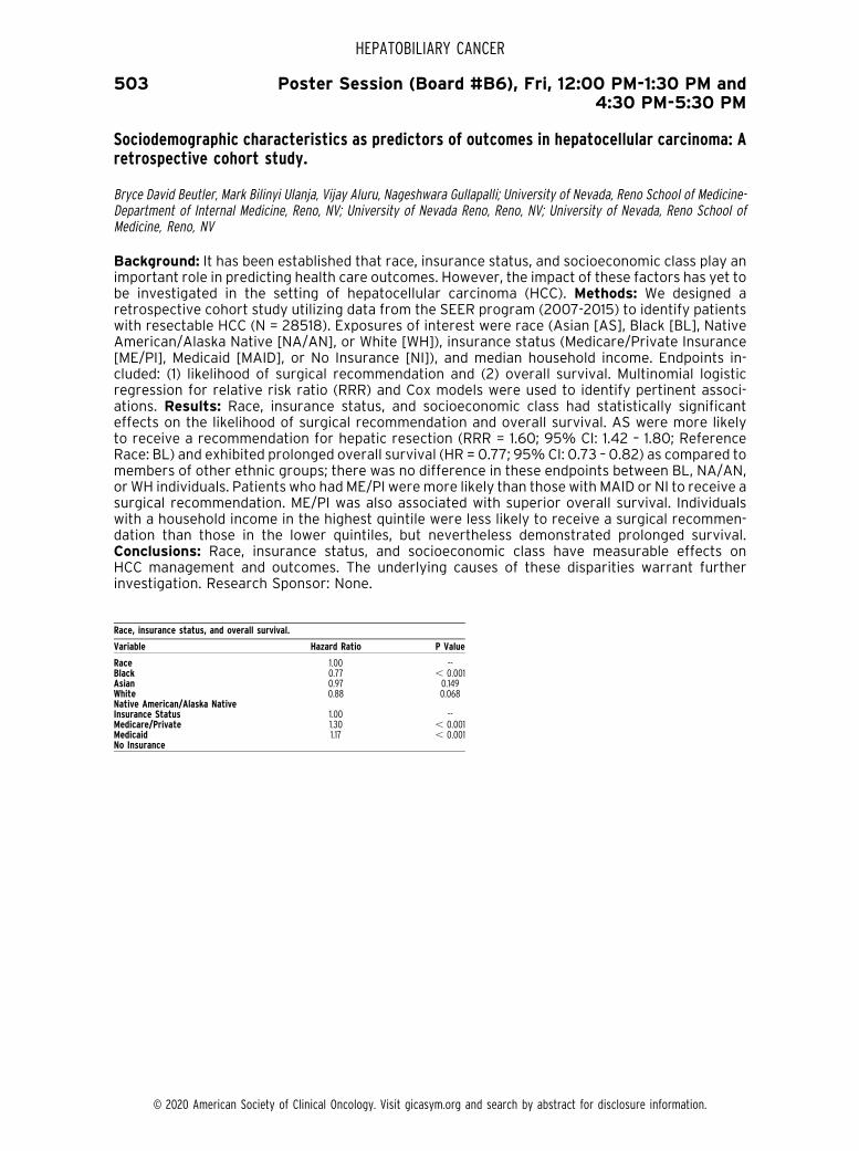
503 Poster Session (Board #B6), Fri, 12:00 PM-1:30 PM and4:30 PM-5:30 PM
Sociodemographic characteristics as predictors of outcomes in hepatocellular carcinoma: Aretrospective cohort study.
Bryce David Beutler, Mark Bilinyi Ulanja, Vijay Aluru, Nageshwara Gullapalli; University of Nevada, Reno School of Medicine-Department of Internal Medicine, Reno, NV; University of Nevada Reno, Reno, NV; University of Nevada, Reno School ofMedicine, Reno, NV
Background: It has been established that race, insurance status, and socioeconomic class play animportant role in predicting health care outcomes. However, the impact of these factors has yet tobe investigated in the setting of hepatocellular carcinoma (HCC). Methods: We designed aretrospective cohort study utilizing data from the SEER program (2007-2015) to identify patientswith resectable HCC (N = 28518). Exposures of interest were race (Asian [AS], Black [BL], NativeAmerican/Alaska Native [NA/AN], or White [WH]), insurance status (Medicare/Private Insurance[ME/PI], Medicaid [MAID], or No Insurance [NI]), and median household income. Endpoints in-cluded: (1) likelihood of surgical recommendation and (2) overall survival. Multinomial logisticregression for relative risk ratio (RRR) and Cox models were used to identify pertinent associ-ations. Results: Race, insurance status, and socioeconomic class had statistically significanteffects on the likelihood of surgical recommendation and overall survival. AS were more likelyto receive a recommendation for hepatic resection (RRR = 1.60; 95% CI: 1.42 – 1.80; ReferenceRace: BL) and exhibited prolonged overall survival (HR = 0.77; 95%CI: 0.73 – 0.82) as compared tomembers of other ethnic groups; there was no difference in these endpoints between BL, NA/AN,orWH individuals. Patients who hadME/PI weremore likely than those with MAID or NI to receive asurgical recommendation. ME/PI was also associated with superior overall survival. Individualswith a household income in the highest quintile were less likely to receive a surgical recommen-dation than those in the lower quintiles, but nevertheless demonstrated prolonged survival.Conclusions: Race, insurance status, and socioeconomic class have measurable effects onHCC management and outcomes. The underlying causes of these disparities warrant furtherinvestigation. Research Sponsor: None.
Race, insurance status, and overall survival.
Variable Hazard Ratio P Value
Race 1.00 --Black 0.77 , 0.001Asian 0.97 0.149White 0.88 0.068Native American/Alaska NativeInsurance Status 1.00 --Medicare/Private 1.30 , 0.001Medicaid 1.17 , 0.001No Insurance
© 2020 American Society of Clinical Oncology. Visit gicasym.org and search by abstract for disclosure information.
HEPATOBILIARY CANCER

504 Poster Session (Board #B7), Fri, 12:00 PM-1:30 PM and4:30 PM-5:30 PM
Impact of facility type, insurance status, and income on use of single agent chemotherapy(SACT) for advanced hepatocellular carcinoma (AHCC): Analysis of National Cancer Database(NCDB).
Richard T. Lee, Gino Cioffi, Aman Opneja, Asrar Alahmadi, Nirav Patil, Nelroy Jones, David Lawrence Bajor, Joel N. Saltzman,Amr Mohamed, Ankit Mangla, Jill Barnholtz-Sloan; The University of Texas MD Anderson Cancer Center, Houston, TX;Department of Population and Quantitative Health Sciences, Case Western Reserve University School of Medicine, Cleveland,OH; University Hospitals Cleveland Medical Center, Case Western Reserve University, Cleveland, OH; University HospitalsSeidman Cancer Center, Cleveland, OH; University Hospitals, Cleveland, OH; University Hospitals Seidman Cancer Center,Case Comprehensive Cancer Center, Case Western Reserve University, Cleveland, OH; University Hospitals-Seidman CancerCenter, Cleveland, OH; Case Comprehensive Cancer Center and Department of Population and Quantitative Health Sciences,Case Western Reserve University School of Medicine, Cleveland, OH
Background: This study analyzes the pattern of use of SACT in the treatment and survival of AHCCbefore and after sorafenibwas FDA approved in late 2007.Methods:Adult patients diagnosedwithHCC and treated with only chemotherapy (CT) from 2004 – 2014 were identified in NCDB database.Patients were analyzed during 3 time frames: 2004–2006 (pre-sorafenib (PS), 2007–2011 (earlysorafenib (ES) and 2012–2014 (late sorafenib (LS)). Cox proportional hazards models and Kaplan-Meier method were used for analyses. Results: The NCDB contained 31,107 patients with HCCdiagnosed from 2004–2014 and treated with CT alone. Patients were generally men (77.3%),.50years of age (92.5%), and with a variety of T-stages - T1 (31.0%), T2 (23.9%), T3 (28.3%), and T4(16.9%). The use of SACT was only 6.2% in the PS period, increased to 15.5% in the ES period, andto 22.3% in the LS period (p,0.0001). During this later period, the highest proportion of SACT isamong academic and integrated network facilities (23.4%) as compared to community facilities(16.4%, p,0.0001). The MS of patients with AHCC treated only with CT has improved significantlyover the study periods from 10months (m) (95% CI: 9.5-10.6) to 12.5m (12.0-12.9) to 16m (15.6-16.4,p, 0.001). Significant differences in MS were found between facility types in all time frames(Table). Multivariate analysis indicates worse outcomes for patients treated at community cancerprograms (HR 1.66, 1.53-1.79) as compared to academic programs as well as for no insurance (HR1.13, 1.05-1.22) and estimated household income of ,$63,000 (HR 1.09, 1.05-1.13). Conclusions:Despite an overall improvement in survival for AHCC patients treated with only CT, significantdifferences in the utilization of SACT and survival exist by facility type, insurance status, andincome. Research Sponsor: None.
MS of HCC patients treated with SACT.
Facility Type
Year of cancer diagnosis
2004-2006 2007-2010 2011-2014
Academic/Integrated Network Cancer 11.9 (11.0-12.5) 15.6 (15.0-16.3) 19.9 (19.2-20.5)Community Cancer 6.4 (5.1-8.6) 6.0 (5.4-6.7) 8.1 (7.5-9.6)Comprehensive Community Cancer 7.9 (7.1-8.8) 8.7 (8.3-9.4) 11.3 (10.6-12)
© 2020 American Society of Clinical Oncology. Visit gicasym.org and search by abstract for disclosure information.
HEPATOBILIARY CANCER
505 Poster Session (Board #B8), Fri, 12:00 PM-1:30 PM and4:30 PM-5:30 PM
Early nutritional risk assessment by NRS 2002 to predict survival in patients with advancedbiliary tract cancer.
Se Eung Oh, Hei-Cheul Jeung; Gangnam Severance Hospital, Seoul, South Korea; Yonsei University College of Medicine,Gangnam Severance Hospital, Seoul, South Korea
Background: Biliary tract cancer (BTC) has heterogenous disease with dismal prognosis. Weinvestigated the predictors of overall survival (OS) among Korean patients with advanced BTCaccording to their baseline nutritional risks estimated by Nutritional Risk Screening (NRS) 2002score.Methods: From September 2006 to July 2017, we retrospectively reviewed the data of 601patients with BTC. Data on demographic and clinical parameters were collected from electronicmedical records, and overall survival (OS) and progression-free survival (PFS) was estimated usingthe Kaplan–Meier method. Stepwise Cox regression analysis was used to determine the factorsassociated with survival. Patients with a NRS 2002 score #2 were classified as “no-risk; ” thosewith a score of 3were classified as “moderate-risk; ” and thosewith a score of$4were classified as“high-risk.” Results: Following nutritional screening at baseline, 333 patients (55%) were clas-sified as the “no risk” group; 109 patients (18%), as the “moderate risk” group; and 159 patients(27%) as the “high risk” group. Survival analysis showed significant differences in the median OSaccording to the NRS 2002 groups: “no risk” group: 12.6 months (95% CI: 11.5–13.7); “moderaterisk” group: 6.1 months (95% CI: 4.3–8.0); and “high risk” group: 3.9 months (95% CI: 3.2 – 4.6)(p,0.001). On the Cox’s regression analysis, NRS 2002 score came out as the most independentfactor for OS (for “moderate-risk”HR 1.610, 95%CI 1.288-2.027, p,0.001; for “high-risk”, HR 2.121,95% CI 1.728-2.612, p,0.001), compared with other prognostic factors including liver metastasis,peritoneal seeding, white blood cell count, platelet count, neutrophil-to-lymphocyte ratio, choles-terol, CEA, and CA19-9. Conclusions: Our study demonstrated OS of advanced BTC was stronglyrelated to their baseline nutritional status assessed by NRS 2002. Constitutional nutritionalassessment can help to improve patient prognosis through proactive and individualized nutritionalintervention. Baseline nutritional status should be integrated for implementing prognostic scoringsystem, which can provide more sophisticated risk stratification of patients with metastatic BTC.Research Sponsor: None.
© 2020 American Society of Clinical Oncology. Visit gicasym.org and search by abstract for disclosure information.
HEPATOBILIARY CANCER
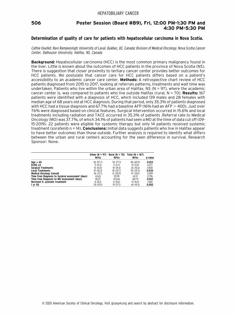
506 Poster Session (Board #B9), Fri, 12:00 PM-1:30 PM and4:30 PM-5:30 PM
Determination of quality of care for patients with hepatocellular carcinoma in Nova Scotia.
Cathie Ouellet, Ravi Ramjeesingh; University of Laval, Quebec, QC, Canada; Division of Medical Oncology, Nova Scotia CancerCenter, Dalhousie University, Halifax, NS, Canada
Background: Hepatocellular carcinoma (HCC) is the most common primary malignancy found inthe liver. Little is known about the outcomes of HCC patients in the province of Nova Scotia (NS).There is suggestion that closer proximity to tertiary cancer center provides better outcomes forHCC patients. We postulate that cancer care for HCC patients differs based on a patient’saccessibility to an academic cancer care center. Methods: A retrospective chart review of HCCpatients diagnosed from 2015 to 2017, looking at referrals patterns, treatments and wait time wasundertaken. Patients who live within the urban area of Halifax, NS (N = 97), where the academiccancer center is, was compared to patients who live outside Halifax (rural, N = 70). Results: 167patients were identified with a diagnosis of HCC, which included 139 males and 28 females withmedian age of 68 years old at HCC diagnosis. During that period, only 35.3% of patients diagnosedwith HCC had a tissue diagnosis and 67.7% had a baseline AFP (16% had an AFP. 400). Just over76% were diagnosed based on clinical features. Surgical intervention occurred in 15.6% and localtreatments including radiation and TACE occurred in 35.3% of patients .Referral rate to MedicalOncology (MO) was 37.7%, of which 34.1% of patients had seen aMO at the time of data cut off (09-15-2019). 22 patients were eligible for systemic therapy but only 14 patients received systemictreatment (sorafenib n = 14). Conclusions: Initial data suggests patients who live in Halifax appearto have better outcomes than those outside. Further analysis is required to identify what differsbetween the urban and rural centers accounting for the seen difference in survival. ResearchSponsor: None.
Urban (N = 97)N(%)
Rural (N = 70)N(%)
Total (N = 167)N(%) p-value
Age > 65 56 (57.7) 54 (77.1) 110 (65.9) 0.013ECOG ‡2 11 (11.3) 4 (5.7) 15 (9.0) 0.277Surgical Treatments 16 (16.5) 10 (14.3) 26 (15.6) 0.830Local Treatments 41 (42.3) 18 (25.7) 59 (35.3) 0.033Medical Oncology Consult 36 (37.1) 21 (30.0) 57 (34.1) 0.409Time from Diagnosis to Surgical assessment (days) 63.65 59.98 62.21 0.796Time from Diagnosis to MO assessment (days) 82.07 225.66 164.74 0.012Received 1L systemic treatment 8 (8.2) 6 (8.6) 14 (8.4) 1.0001 yr OS 50 (51.5) 19 (27.1) 69 (41.3) 0.002
© 2020 American Society of Clinical Oncology. Visit gicasym.org and search by abstract for disclosure information.
HEPATOBILIARY CANCER
507 Poster Session (Board #B10), Fri, 12:00 PM-1:30 PM and4:30 PM-5:30 PM
Comparison of the frequency of adverse events requiring interventions determined bytelephone follow up in hepatocellular carcinoma patients treated with sorafenib and lenvatinib.
Koki Morishita, Hidetaka Suzuki, Junko Tauchi, Misaki K Takeno, Kohei Hayashi, Masafumi Ikeda; Department ofPharmacy,National Cancer Center Hospital East, Kashiwa, Japan; Department of Pharmacy, National Cancer Center HospitalEast, Kashiwa, Japan; Department of Pharmacy, National Cancer Center Hospital East, Kashiwa City, Japan; Department ofHepatobiliary and Pancreatic Oncology, National Cancer Center Hospital East, Kashiwa, Japan; National Cancer CenterHospital East, Kashiwa, Japan
Background: The REFLECT trial demonstrated that lenvatinib is non-inferior to sorafenib for first-line treatment of unresectable hepatocellular carcinoma (uHCC). However, no comparison of thefrequency of adverse events (AEs) requiring interventions has been reported yet between uHCCpatients receiving sorafenib and those receiving lenvatinib. At the National Cancer Center HospitalEast, Japan, pharmacists conduct telephone follow-up (TF) during the first month after the start oftreatment with sorafenib or lenvatinib in uHCC patients, for the purpose of detecting and treatingAEs early. The aim of this studywas to reveal the frequency of AEs requiring interventions betweenpatients receiving sorafenib and those receiving lenvatinib, based on TF. Methods: The charac-teristics, AEs and contents of intervention by TF of 56 uHCC patients who had been started ontreatmentwith sorafenib and lenvatinib were reviewed retrospectively. The study subjects were 33patients initiated on sorafenib treatment and monitored by TF from March 2017 to March 2018(Group S) and 23 patients initiated on lenvatinib treatment and monitored by TF from March 2018toMarch 2019 (Group L).Results: The total numbers of TFs in Group S and Group Lwere 91 and 48,respectively. The rate of AEs requiring interventionswas significantly higher inGroup S as comparedto Group L (Group S, 17.6% (16/91); Group L, 4.2% (2/48); p = 0.032). The frequencies of theinterventions, including use of supportive treatments (A), withdrawal of sorafenib or lenvatinib (B),and medical examination (C), differed between the two groups (A/B/C: Group S, 8/5/3 times vs.Group L, 2/0/0 times). The most frequently observed AE that necessitated intervention in Group Swas the hand-foot syndrome (HFS) (75.0%, 12/16). Conclusions: The frequency of interventionsfor AEs appears to be higher in uHCC patients receiving sorafenib than in those receivinglenvatinib. Although a great number of patients taking sorafenib had symptomatic AEs, suchas HFS, early detection of the symptoms through TF contributed to prevention of treatmentwithdrawal on account of AEs. Research Sponsor: None.
© 2020 American Society of Clinical Oncology. Visit gicasym.org and search by abstract for disclosure information.
HEPATOBILIARY CANCER

508 Poster Session (Board #B11), Fri, 12:00 PM-1:30 PM and4:30 PM-5:30 PM
Hepatic adverse events during treatment with immune checkpoint inhibitors (ICI) in cancerpatients: A territory-wide patient cohort study.
Stephen Lam Chan, Terry Cheuk-Fung Yip, Vincent Wai-Sun Wong, Anthony W.H. Chan, Cathy Ka Kiu Tong, Yee Kit Tse,Becky Wing-Yan Yuen, Hester Wing-Sum Luk, Grace Chung-Yan Lui, Henry L.Y. Chan, Tony S. K. Mok, Grace Lai-Hung Wong;Department of Clinical Oncology, Prince of Wales Hospital, The Chinese University of Hong Kong, Hong Kong, China;Department of Medicine and Therapeutics, The Chinese University of Hong Kong, Shatin, China; Department of Anatomicaland Cellular Pathology, The Chinese University of Hong Kong, Shatin, China; Department of Statistics, The Chinese Universityof Hong Kong, Shatin, China; Department of Clinical Oncology The Chinese University of Hong Kong, Hong Kong, China
Background: Hepatic adverse events (AEs) are commonly encountered during ICI treatment incancer. We evaluated the incidence and impact of hepatic AEs in a territory-wide cohort of 1509patients who received ICI for cancer treatment. Methods: This is a territory-wide retrospectiveobservational cohort study in Hong Kong. We identified patients through the regional hospitaldatabase (CDARS), based on the drug record of ICIs from 1 Jan 2014 to 31 Oct 2018. Serial liverfunctions before, during and at 3-month after ICI, were retrieved. Hepatic AEs were gradedaccording to CTCAE 4.0. Results: The mean age was 60 years and 65.4% were male with thecommonest malignancies being lung cancer (37.0%), liver cancer (17.0%) and gastrointestinal (GI)cancer (8.4%). Grade 1-2 and grade 3-4 hepatic AE occurred 39.8% and 23.3% of patients,respectively, during or within 3 months after ICI. During ICI, 39.5% developed grade 1-2 and 13.0%had grade 3-4 hepatic AE. Themost commonmanifestations of hepatic AE occurred as elevation ofALT/AST (grade 1-2: 38.7%; grade 3-4: 10.3%). The median time of duration from ICI 1st dose tohepatic $ Grade 3 AE was 54 days (IQR: 22-124). Patients with liver cancer were more likely todevelop hepatic AE (grade 1-2: 37.4%; grade 3-4: 55.5%). In all patients and cancer subgroup,patients with grade 3-4 hepatic AE had worse OS than grade 1-2 hepatic AE (Table). Conclusions:Hepatic AE occurs in more than half of the patients receiving ICI, with over 20% being grade 3-4AE. Regardless of tumor types, development of hepatic AE during ICI is associated with poorprognosis. Research Sponsor: None.
12-month overall survival rate.
PatientsWithout
hepatic AEWith
hepatic AE P value*Grade 1-2hepatic AE
Grade 3-4hepatic AE P value#
All 48.9%(42.3%–55.1%)
32.7%(29.5%–36.0%)
,0.001 38.9%(34.8%–42.9%)
18.5%(14.1%–23.5%)
,0.001
Lung cancer 45.3%(35.7%–54.4%)
32.4%(26.9%–38.0%)
0.003 36.4%(30.2%–42.6%)
9.9%(3.2%–21.0%)
,0.001
Liver cancer 79.3%(47.8%–92.9%)
36.5%(29.2%–43.8%)
0.005 55.6%(44.3%–65.6%)
18.3%(10.9%–27.2%)
,0.001
GI cancer 24.2%(6.8%–47.3%)
22.7%(14.9%–31.4%)
0.927 28.8%(18.3%–40.2%)
11.3%(3.6%–24.0%)
0.009
© 2020 American Society of Clinical Oncology. Visit gicasym.org and search by abstract for disclosure information.
HEPATOBILIARY CANCER

509 Poster Session (Board #B12), Fri, 12:00 PM-1:30 PM and4:30 PM-5:30 PM and Poster Walks, Fri, 4:45 PM-5:30 PM
Celecoxib versus parecoxib versus oxycodone in pain control for transcatheter chemoembolizationin patients with hepatocellular carcinoma.
Ming Zhao, Ning Lyu, Yanan Kong, Xiaoxian Li; Sun Yat-sen University Cancer Center, Guangzhou, China; Zhongshan School ofMedicine, Sun Yat-sen University, Guangzhou, China
Background: Abdominal pain is one of the most common side effects of transarterial chemo-embolization (TACE) in patients with unresectable hepatocellular carcinoma. Previous studiesreported that perioperative controlled-release oxycodone (CRO) intake or administration ofparecoxib resulted in adequate pain control after TACE. However, there are currently no studiescomparing opioids with nonsteroidal anti-inflammatory drugs (NSAIDs) in controlling postoper-ative pain. Therefore, we conducted a clinical trial to compare the analgesic effect and safetyamong celecoxib (oral COX-2 inhibitor), parecoxib (injectable COX-2 inhibitor), and CRO (oralopioids) in patients undergoing TACE. Methods: The study was a prospective, randomized,paralleled trial in which 213 patients were enrolled between September 2016 and March 2019.Patients were randomly assigned at the ratio of 1:1:1 to receive celecoxib, parecoxib or CRO 1 hbefore TACE (T0) and once every 12 h for 2 days after TACE. Pain level, morphine consumption andadverse events were evaluated and compared among the three regimens. Results: Highestincidence of pain occurred within the first 12 hours (T12) after TACE. Analysis of pain controlshowed no significant difference among the mean highest pain scores, percentage distribution ofpain categories andmeanmorphine consumption in the three groups at T0, T12, T24, T36, and T48.At T24, 11 patients (15.7%) in oxycodone group had fever, which was higher than parecoxib regimen(1 patient [1.5%], P = 0.003). At T36, 13 patients (18.6%) in oxycodone regimen had fever, whichwashigher than celecoxib regimen (2 patients [2.9%], P = 0.003) and parecoxib regimen (1 patient[1.5%], P , 0.001). At T48, 11 patients (15.7%) in oxycodone regimen had fever, which was higherthan celecoxib regimen (2 patients [2.9%], P =0.010) and parecoxib regimen (0 patients, P =0.001).Conclusions: The results suggested that patients obtained celecoxib, parecoxib or CRO once every12 hours can have the same level of analgesic effect during each time period of TACE. However,body temperature balance in oxycodone regimen was significantly worse than celecoxib regimenand parecoxib regimen. Clinical trial information: NCT03059238. Research Sponsor: Sun Yat-senUniversity Clinical Research 5010 Program.
© 2020 American Society of Clinical Oncology. Visit gicasym.org and search by abstract for disclosure information.
HEPATOBILIARY CANCER

510 Poster Session (Board #B13), Fri, 12:00 PM-1:30 PM and4:30 PM-5:30 PM
Early experiences with triple immunochemotherapy in young adults with high-risk fibrolamellarcarcinoma.
Claire O’Grady, Ariel Gliksberg, Paul Kent; Rush University Medical Center, Chicago, IL
Background: Fibrolamellar Carcinoma (FLC) is a rare liver cancer affecting young adults withoutunderlying liver disease. Surgery is the only proven therapy, and recurrence is common. There areno proven systemic treatments, especially for high-risk FLC (unresectable, relapse, progression,metastatic). Research suggests that immunotherapy may work. We share our experience usingsystemic “triple immunochemotherapy” (TT): 2 week cycles of 7 days continuous infusion 5FU ororal capecitabine, interferon alpha-2b on days 1,3,5,7 or PEG-Interferon and nivolumab on day 1.Methods: Data from all patients who received TT from 5/2018 to 9/2019 was reviewed to assesstolerability, survival and toxicity.Results: 14 patients were treated with TT of which 10 (8F,2M witha median age of 20) were evaluable. They received a median of 13 cycles (6-31). At time of analysis,the median progression free survival was 6 months, 22% longer than prior to TT, with 80% ofpatients (8) stable or improving, 1 progression, and 1 who died 2months after initiating TT. For the 4patients who achieved surgical remission, none have relapsed (PFS 9 months). Overall objectiveresponse (CR+PR) and tumor control rate (CR+PR+SD) were 60% and 80%, respectively. Therewere no withdrawals from treatment due to side effects, though 2 had dose adjustments. Allexperienced mild adverse effects, most commonly fever and headache, but only 2 patients hadgrade 3 toxicity. Conclusions: Our early results of TT for high-risk FLC are promising, with goodtolerability and treatment response, particularly in patients who have achieved surgical CR.Further longitudinal data is needed to confirm outcomes, especially in patients still early in theirtreatment. Research Sponsor: None.
Patient #Clinical Statusat start of TT
Typeof TT
Cyclesof TT
RECISTResponse
# of PreviousRelapses
Shortest Timeto Relapse Priorto TT (months)
PFS afterTT (months)
TTStatus
# of Grade3+ AEs
1 CR A 6 SD 1 12 4 O 02 PD N + A 19 CR 2 1 10 O 03 PD N 18 PR 2 5 8 O 04 PD N 12 SD 4 2 6 O 05 PD N 7 SD 4 9 5 O 26 SD N 14 PR 0 n/a 8 DC 17 SD N + A 31 CR 1 5 16 O 08 SD A 14 CR 0 n/a 6 O 09 SD A 6 death 0 n/a 2 DC 010 SD A 8 PR 0 n/a 4 O 0
CR = Clinical Remission, SD = Stable Disease, PD = Progressive disease, PR = Partial Response, N = Neo-adjuvant, A = Adjuvant O = Ongoing,DC = Discontinued.
© 2020 American Society of Clinical Oncology. Visit gicasym.org and search by abstract for disclosure information.
HEPATOBILIARY CANCER
511 Poster Session (Board #B14), Fri, 12:00 PM-1:30 PM and4:30 PM-5:30 PM
Effect of neoadjuvant immunotherapy and targeted therapies on surgical resection in patientswith solid tumors: A systematic review and meta-analysis.
Pablo Emilio Serrano Aybar, Sameer Parpia, Leyo Ruo, Kasia Tywonek, Sandra Lee, Connor O’Neill, Noor Faisal, Ali Alfayyadh,Marylrose Gundayao, Brandon M. Meyers; McMaster University, Department of Surgery, Division of General Surgery,Hamilton, ON, Canada; Ontario Clinical Oncology Group, Hamilton, ON, Canada; Western University, London, ON, Canada;Royal College of Surgeons in Ireland, Dublin, Ireland; Juravinski Cancer Centre, Hamilton, ON, Canada
Background: Neoadjuvant immunotherapy with anti-programmed cell death protein-1 (PD-1) oranti-programmed cell death ligand-1 (PD-L1) and tyrosine kinase inhibitor (TKI) therapy is currentlybeing used to treat certain solid tumours prior to surgery. Neoadjuvant therapy may cause delaysto resection potentially losing a window of opportunity. We explored the pooled proportion ofpatients with solid tumours receiving neoadjuvant therapy who completed planned surgicalresection. Methods: Medline, CENTRAL and Embase databases were searched for single armor randomized controlled trials studying neoadjuvant PD-1/PD-L1 immunotherapy or TKI therapy.Random-effectsmodel was used to estimate the pooled proportion of patients undergoing plannedresection, and weights were estimated using inverse variance method. Statistical heterogeneitywas calculated using the I2 and chi-squared test. Results: From 368 relevant articles, elevenstudies with a total of 382 patients receiving neoadjuvant PD-1 immunotherapy (n = 234) orneoadjuvant TKI therapy (n = 148) were analyzed. The types of tumours included hepatocellularcarcinoma (1 study), renal cell carcinoma (8 studies), bladder carcinoma (1 study) or non-small celllung cancer (1 study). The pooled proportion of patients who completed planned surgery afterneoadjuvant therapy was 95% (95% CI 0.92 to 0.99). The overall partial response rate prior tosurgery was 12% (95% CI 0.07 to 0.16) in the PD-1 therapy group and 46% (95% CI -0.12 to 1.03) inthe TKI group. The pooled serious adverse events rate was 17% (95% CI 0.02 to 0.32) in the PD-1therapy group and 29% (95% CI -0.10 to 0.68) in the TKI group. For all patients receivingneoadjuvant therapy, the pooled median overall survival was 23.41 months (95% CI 16.21 to 30.62)and median progression free survival was 7.46 months (95% CI 4.41 to 10.51). Conclusions:Neoadjuvant PD-1 or TKI therapy prior to surgery for solid tumours is safe, does not delay surgicalresection and can result in a partial radiological response prior to surgery. Research Sponsor:None.
© 2020 American Society of Clinical Oncology. Visit gicasym.org and search by abstract for disclosure information.
HEPATOBILIARY CANCER

512 Poster Session (Board #B15), Fri, 12:00 PM-1:30 PM and4:30 PM-5:30 PM
Nivolumab (NIVO) + ipilimumab (IPI) combination therapy in patients (pts) with advancedhepatocellular carcinoma (aHCC): Subgroup analyses from CheckMate 040.
Aiwu Ruth He, Thomas Yau, Chiun Hsu, Yoon-Koo Kang, Tae-You Kim, Armando Santoro, Bruno Sangro, Ignacio Melero,Masatoshi Kudo, Ming-Mo Hou, Ana Matilla, Francesco Tovoli, Jennifer J. Knox, Bassel F. El-Rayes, Mirelis Acosta-Rivera,Jaclyn Neely, Yun Shen, Marina Tschaika, Anthony B. El-Khoueiry; Georgetown University Hospital, Washington, DC; TheUniversity at Hong Kong, Hong Kong, China; National Taiwan University Hospital, Taipei, Taiwan; Asan Medical Center,University of Ulsan, Seoul, South Korea; Seoul National University, Seoul, South Korea; Istituto Clinico Humanitas, Rozzano,Italy; Clinica Universidad de Navarra and CIBEREHD, Pamplona, Spain; Universidad de Navarra, Pamplona, Spain; KindaiUniversity Faculty of Medicine, Osaka, Japan; Chang Gung Memorial Hospital, Taipei, Taiwan; Servicio de Digestivo, HospitalGeneral Universitario Gregorio Mara~non y CIBEREHD, Madrid, Spain; Department of Medical & Surgical Sciences, University ofBologna, Bologna, Italy; Princess Margaret Cancer Centre, Toronto, ON, Canada; Emory University Winship Center, Atlanata,GA; Fundacion de Investigacion, San Juan, PR; Bristol-Myers Squibb, Princeton, NJ; USC Norris Comprehensive Cancer Center,Los Angeles, CA
Background: NIVO monotherapy is approved in the United States and other countries for pts withHCC treated with sorafenib (SOR) based on CheckMate 040 (NCT01658878) results, whichreported 14% objective response rate (ORR) and 16-month median overall survival (mOS; El-Khoueiry et al. Lancet 2017). Primary efficacy and safety of NIVO + IPI in pts with aHCC previouslytreated with SOR were presented recently (Yau et al. J Clin Oncol 2019). Here, we will presentsubgroup analyses from this study. Methods: Pts were randomized to 3 arms: [A] NIVO 1 mg/kg +IPI 3 mg/kg Q3W (4 doses) or [B] NIVO 3mg/kg + IPI 1 mg/kg Q3W (4 doses), each followed by NIVO240 mg Q2W, or [C] NIVO 3 mg/kg Q2W + IPI 1 mg/kg Q6W. Treatment continued until intolerabletoxicity or disease progression. Primary endpoints included safety/tolerability, ORR, and durationof response (DOR; investigator assessment per RECIST v1.1). Key secondary endpoints includeddisease control rate (DCR), OS, and progression-free survival (blinded independent central review[BICR] per RECIST v1.1); key exploratory endpoints included ORR (BICR per RECIST v1.1). Data cutoffwas January 2019.Results:A total of 148 pts were randomized. MinimumOS follow-up from last ptrandomization date to data cutoff was 28months. At baseline, 34%of all pts had vascular invasion;82% had extrahepatic spread; and 91% had Barcelona Clinic Liver Cancer stage C; 84% discon-tinued SOR because of disease progression and 14% because of toxicity. For all treated pts, ORRwas 31% (7 had complete response), with median DOR of 17 months; DCR was 49%; the 30-monthOS rate was 37%. NIVO + IPI was well tolerated; 38% of pts had grade 3–4 treatment-relatedadverse events (TRAEs; most common any grade: pruritus and rash; most common grade 3–4:aspartate aminotransferase increase and lipase increase); 5% had grade 3–4 TRAEs leading todiscontinuation. Subgroup analyses based on duration of prior SOR therapy and other pt char-acteristics will be presented. Conclusions: NIVO + IPI led to clinically meaningful benefits, with amanageable safety profile in pts previously treated with SOR. NIVO + IPI may provide a newtreatment option for these pts. Clinical trial information: NCT01658878. Research Sponsor:Bristol-Myers Squibb.
© 2020 American Society of Clinical Oncology. Visit gicasym.org and search by abstract for disclosure information.
HEPATOBILIARY CANCER

513 Poster Session (Board #B16), Fri, 12:00 PM-1:30 PM and4:30 PM-5:30 PM
A phase Ib study of lenvatinib (LEN) plus nivolumab (NIV) in patients (pts) with unresectablehepatocellular carcinoma (uHCC): Study 117.
Masatoshi Kudo, Masafumi Ikeda, Kenta Motomura, Takuji Okusaka, Naoya Kato, Corina E. Dutcus, Takashi Hisai,Mayumi Suzuki, Hiroki Ikezawa, Takeyuki Iwata, Hiromitsu Kumada, Masahiro Kobayashi; Department of Gastroenterologyand Hepatology, Kindai University School of Medicine, Osaka, Japan; Department of Hepatobiliary and Pancreatic Oncology,National Cancer Center Hospital East, Kashiwa, Japan; Aso Iizuka Hospital, Fukuoka, Japan; Department of Hepatobiliary andPancreatic Oncology, National Cancer Center Hospital, Tokyo, Japan; Department of Gastroenterology, Graduate School ofMedicine, Chiba University Hospital, Chiba, Japan; Eisai Inc., Woodcliff Lake, NJ; Eisai Co. Ltd., Tokyo, Japan; OnoPharmaceutical Co., Ltd., Osaka, Japan; Toranomon Hospital, Tokyo, Japan
Background: LEN, a multikinase inhibitor of VEGFR 1–3, FGFR 1–4, PDGFRa, RET, and KIT withimmunomodulatory activity, is approved in multiple countries for first-line treatment of uHCC. NIVis an anti-PD-1 monoclonal antibody approved in multiple countries as a second-line treatment forHCC. We report early results from a phase 1b trial of LEN + NIV in pts with uHCC.Methods: In thisopen-label study, pts with uHCC, BCLC stage B (not eligible for transarterial chemoembolization) orC, Child-Pugh class A, and ECOG PS# 1 received LEN (bodyweight$ 60 kg: 12 mg/day;, 60 kg: 8mg/day) PO QD and 240 mg NIV IV Q2W. The primary endpoint was tolerability and safety of thecombination. Objective response rate (ORR; mRECIST by investigator) was a secondary endpoint.Tolerability was evaluated by assessing dose-limiting toxicities (DLTs) during the first cycle in ptsfor whom no other appropriate therapy was available (Part 1). Once tolerability was confirmed,additional pts with no prior systemic therapy for uHCC were enrolled (Part 2). Results: At datacutoff (May 17, 2019), 30 pts had received LEN + NIV (Part 1: n=6; Part 2: n=24). Pts had BCLC stageB (n=17) or C (n=13) and Child-Pugh scores of 5 (n=23) or 6 (n=7). 4 pts in Part 1 (66.7%) hadreceived prior anticancer medications (3 pts had 1; 1 pt had $3). No DLTs were reported in Part 1.TEAEs leading to discontinuation of LEN were reported in 2 (6.7%) pts; 4 (13.3%) pts had TEAEsleading to discontinuation of NIV. TEAEs occurred in all 30 pts; the most common were palmar-plantar erythrodysesthesia (56.7%) and dysphonia (53.3%). Adverse events were manageable.Efficacy outcomes are reported (Table). Conclusions: LEN + NIV was well tolerated with encour-aging anti-tumor activity in pts with uHCC. Clinical trial information: NCT03418922. ResearchSponsor: Eisai Inc., Woodcliff Lake, NJ, USA and Ono Pharmaceutical Co., Ltd.
Efficacy summary.
Parameter, n (%)
LEN + NIV (N=30)mRECIST by Investigator
Part 1 (n=6) Part 2 (n=24) Overall (N=30)
Best Overall Responsea,n (%)Complete response (CR)
0 4 (16.7) 4 (13.3)
Partial response (PR) 4 (66.7) 15 (62.5) 19 (63.3)Stable disease 2 (33.3) 4 (16.7) 6 (20.0)Progressive disease 0 1 (4.2) 1 (3.3)Not evaluable 0 0 0ORRb, n (%) 4 (66.7) 19 (79.2) 23 (76.7)95% CI (22.3–95.7) (57.8–92.9) (57.7–90.1)aConfirmation not required.bORR = Proportion of CR + PR.
© 2020 American Society of Clinical Oncology. Visit gicasym.org and search by abstract for disclosure information.
HEPATOBILIARY CANCER
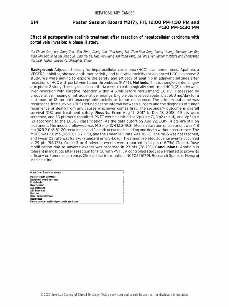
514 Poster Session (Board #B17), Fri, 12:00 PM-1:30 PM and4:30 PM-5:30 PM
Effect of postoperative apatinib treatment after resection of hepatocellular carcinoma withportal vein invasion: A phase II study.
Hui-Chuan Sun, Xiao-Dong Zhu, Jian Zhou, Qiang Gao, Ying-Hong Shi, Zhen-Bing Ding, Cheng Huang, Shuang-Jian Qiu,Ning Ren, Guo-Ming Shi, Jian Sun, Qing-Hai Ye, Xiao-Wu Huang, Xin-Rong Yang, Jia Fan; Liver Cancer Institute and ZhongshanHospital, Fudan University, Shanghai, China
Background: Adjuvant therapy for hepatocellular carcinoma (HCC) is an unmet need. Apatinib, aVEGFR2 inhibitor, showed antitumor activity and tolerable toxicity for advanced HCC in a phase 2study. We were aiming to explore the safety and efficacy of apatinib in adjuvant settings afterresection of HCCwith portal vein tumor thrombosis (PVTT).Methods:This is a single-center single-arm phase 2 study. The key inclusion criteria were: (1) pathologically confirmedHCC; (2) underwentliver resection with curative intention within 4-6 wk before recruitment; (3) PVTT assessed bypreoperative imaging or intraoperative findings. Eligible pts received apatinib at 500mg/day for amaximum of 12 mo until unacceptable toxicity or tumor recurrence. The primary outcome wasrecurrence-free survival (RFS) defined as the interval between surgery and the diagnosis of tumorrecurrence or death from any causes whichever comes first. The secondary outcome is overallsurvival (OS) and treatment safety. Results: From Aug 17, 2017 to Dec 18, 2018, 49 pts werescreened, and 30 pts were recruited. PVTT were classified as Vp1 (n = 7), Vp2 (n = 11), and Vp3 (n =12) according to the LCSGJ classification. As the data cutoff on Aug 22, 2019, 4 pts are still ontreatment. Themedian follow-up was 14.3mo (IQR 12.3-19.3). Median duration of treatment was 4.8mo (IQR 2.0–8.8). 20 recurrence and 2 death occurred including one death without recurrence. ThemRFS was 7.6 mo (95% CI, 3.7-11.5), and the 1-year RFS rate was 36.1%. The mOS was not reached,and 1-year OS rate was 93.3% (standard error, 4.6%). Treatment-related adverse events occurredin 29 pts (96.7%). Grade 3 or 4 adverse events were reported in 14 pts (46.7%) (Table). Dosemodification due to adverse events was recorded in 23 pts (76.7%). Conclusions: Apatinib istolerant in most pts after resection for HCC with PVTT. A controlled study is warranted to prove itsefficacy on tumor recurrence. Clinical trial information: NCT03261791. Research Sponsor: HengruiMedicine Inc.
Grade 3 or 4 adverse events n
Platelet count decrease 5Neutrophil count decrease 4Proteinuria 3Hypertension 3ALT increased 1AST increased 1Diarrhea 1Upper GI hemorrhage 1Hoarseness 1Palmar-plantar erythrodysesthesia syndrome 1
© 2020 American Society of Clinical Oncology. Visit gicasym.org and search by abstract for disclosure information.
HEPATOBILIARY CANCER
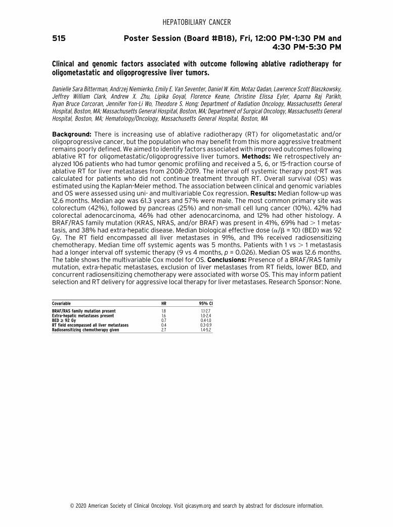
515 Poster Session (Board #B18), Fri, 12:00 PM-1:30 PM and4:30 PM-5:30 PM
Clinical and genomic factors associated with outcome following ablative radiotherapy foroligometastatic and oligoprogressive liver tumors.
Danielle Sara Bitterman, Andrzej Niemierko, Emily E. Van Seventer, Daniel W. Kim, Motaz Qadan, Lawrence Scott Blaszkowsky,Jeffrey William Clark, Andrew X. Zhu, Lipika Goyal, Florence Keane, Christine Elissa Eyler, Aparna Raj Parikh,Ryan Bruce Corcoran, Jennifer Yon-Li Wo, Theodore S. Hong; Department of Radiation Oncology, Massachusetts GeneralHospital, Boston, MA; Massachusetts General Hospital, Boston, MA; Department of Surgical Oncology, Massachusetts GeneralHospital, Boston, MA; Hematology/Oncology, Massachusetts General Hospital, Boston, MA
Background: There is increasing use of ablative radiotherapy (RT) for oligometastatic and/oroligoprogressive cancer, but the population whomay benefit from this more aggressive treatmentremains poorly defined. We aimed to identify factors associated with improved outcomes followingablative RT for oligometastatic/oligoprogressive liver tumors. Methods: We retrospectively an-alyzed 106 patients who had tumor genomic profiling and received a 5, 6, or 15-fraction course ofablative RT for liver metastases from 2008-2019. The interval off systemic therapy post-RT wascalculated for patients who did not continue treatment through RT. Overall survival (OS) wasestimated using the Kaplan-Meier method. The association between clinical and genomic variablesand OS were assessed using uni- and multivariable Cox regression. Results:Median follow-up was12.6 months. Median age was 61.3 years and 57% were male. The most common primary site wascolorectum (42%), followed by pancreas (25%) and non-small cell lung cancer (10%). 42% hadcolorectal adenocarcinoma, 46% had other adenocarcinoma, and 12% had other histology. ABRAF/RAS family mutation (KRAS, NRAS, and/or BRAF) was present in 41%, 69% had . 1 metas-tasis, and 38% had extra-hepatic disease. Median biological effective dose (a/b = 10) (BED) was 92Gy. The RT field encompassed all liver metastases in 91%, and 11% received radiosensitizingchemotherapy. Median time off systemic agents was 5 months. Patients with 1 vs . 1 metastasishad a longer interval off systemic therapy (9 vs 4 months, p = 0.026). Median OS was 12.6 months.The table shows the multivariable Cox model for OS. Conclusions: Presence of a BRAF/RAS familymutation, extra-hepatic metastases, exclusion of liver metastases from RT fields, lower BED, andconcurrent radiosensitizing chemotherapy were associated with worse OS. This may inform patientselection and RT delivery for aggressive local therapy for livermetastases. Research Sponsor: None.
Covariable HR 95% CI
BRAF/RAS family mutation present 1.8 1.1-2.7Extra-hepatic metastases present 1.6 1.0-2.4BED ‡ 92 Gy 0.7 0.4-1.0RT field encompassed all liver metastases 0.4 0.3-0.9Radiosensitizing chemotherapy given 2.7 1.4-5.2
© 2020 American Society of Clinical Oncology. Visit gicasym.org and search by abstract for disclosure information.
HEPATOBILIARY CANCER
516 Poster Session (Board #B19), Fri, 12:00 PM-1:30 PM and4:30 PM-5:30 PM
Major impact of personalized dosimetry using 90Y loaded glass microspheres SIRT in HCC: Finaloverall survival analysis of a multicenter randomized phase II study (DOSISPHERE-01).
Etienne Garin, Lambros Tzelikas, Boris Guiu, Julia Chalaye, Julien Edeline, Thierry De Baere, Vania Tacher, Corentin Robert,Eric Assenat, Marie Terroir-Cassou-Mounat, Helene Regnault, Xavier Palard, Sophie Laffont, Boris Campillo-Gimenez,Yann Rolland; Cancer Institute Eugene Marquis, Rennes, France; Medical Imaging, Gustave Roussy, Villejuif, France;University Hospital, Radiology Department, Montpellier, France; CHU Henri Mondor, Creteil, France; Medical Oncology, CentreEugene Marquis, Rennes, France; Institut Gustave Roussy, Villejuif, France; CHU Montpellier, Montpellier, France; GustaveRoussy, Villejuif, France; Centre Eugene-Marquis, Rennes, France; Centre Eugene-Marquis, Rennes, France
Background: 90Y loaded microsphere SIRT (radioembolization) is a treatment option in advancedHCC. However, no personalized dosimetric endpoints are currently used. The goal of this study wasto compare the efficacy of 90Y loaded glass microsphere SIRT in HCC using a standard versus apersonalized dosimetric approach. Methods: DOSISPHERE-01 was a multicenter, randomizedphase 2 trial in unresectable HCC patients with at least one tumor $7cm. Treatment arm wasrandomly assigned (1:1) to standard dosimetry arm (SDA), with a goal to deliver 120620Gy to thetreated volume or to personalized dosimetry arm (PDA) with a goal to deliver at least 205Gy to theindex lesion. The primary endpoint was the response rate (RR) of the index lesion according toEASL criteria. Secondary endpoints included dose response evaluation, safety and overall survival(OS). Results: Sixty HCC patients were randomized (PDA 31, SDA 29, intent to treat population-ITTP-), and 56 treated (28 in each arm). RR was significantly increased in the PDA versus the SDA,in the ITTP, respectively 64.5% versus 31% (p=0.0095) as in the safety population -SP- (treatmenteffectively received, personalized 35, standard 21), respectively 74.3% versus 14.3% (p,0.0001).Median OS was significantly increased in the PDA versus the SDA, in the ITTP, respectively 26.7m(CI 95%:11.7-NR) versus 10.6m (CI 95%:6-16.8), p=0.0096, HR=0.421 (95%CI:0.215-0.826),p=0.0119, as in the SP, respectively 26.7m (CI 95%:11.7- NR) versus 9.5m (CI 95%:4.8-14.9),p=0.0015, HR=0.342 (95%CI:0.171-0.683), p=0.0023. Median OS was 26.7m (CI 95%:13.5-NR)versus 6.0m (CI 95%:3.8-14.9) for the patients who received a tumor dose $205 Gy or ,205 Gyrespectively, p=0.0106, HR=0.336 (95%CI:0.154-0.735), p=0.0063. Treatment-related clinicallyrelevant hepatic $grade 3 AEs were observed in 5.7% and 14.2% of the patients of the PDA andSDA arms, respectively, (p=ns).Conclusions:MAASPECT/CT based personalized dosimetry is safeand dramatically increased RR andOS of HCC patients. These results question the interpretation ofall phase 3 trials of SIRT designedwithout personalized dosimetry in HCC. Clinical trial information:2015-A00894-45. Research Sponsor: BTG UK LtD.
© 2020 American Society of Clinical Oncology. Visit gicasym.org and search by abstract for disclosure information.
HEPATOBILIARY CANCER

517 Poster Session (Board #B20), Fri, 12:00 PM-1:30 PM and4:30 PM-5:30 PM
Sex difference in patients with biliary tract cancer receiving chemotherapy: Post hoc analysis ofABC-01, -02, -03, -04, BILCAP.
Eiichiro Suzuki, John A. Bridgewater, Juan W. Valle, John Neil Primrose, Anna Dorothea Wagner, Andre Lopes, Richard Fox;Department of Gastroenterology, Graduate School of Medicine, Chiba University, Chiba, Japan; University College LondonCancer Institute, London, United Kingdom; The Christie NHS Foundation Trust, Manchester, United Kingdom; University ofSouthampton, Southampton, United Kingdom; Lausanne University Hospital, Lausanne, Switzerland; Cancer Research UKand UCL Cancer Trials Centre, London, United Kingdom; University of Birmingham, Birmingham, United Kingdom
Background: The relationship between toxicity from chemotherapy and clinical outcome in biliarytract cancer (BTC) is uncertain. Aim: This post hoc analysis evaluated differences by sex in thefrequency of adverse events (AEs) and overall survival (OS) and its impact on progression-freesurvival (PFS)/recurrence-free survival (RFS) for BTC patients. Methods: Individual patient datawere retrieved from ABC -01, -02, -03, -04, and BILCAP study. AEs were graded according toNational Cancer Institute’s Common Toxicity Criteria v 4.02 and odds ratios along with 95%CI andp-values derived from logistic regression were used to assess the effect of sex on the risk of AEs.Time to event outcomeswere evaluated using Cox regression and plotted using Kaplan-Meier plots.All statistical tests were two-sided. Results: Overall 994 patients-data were examined: 86 in ABC-01, 324 in-02, 124 in -03, 13 in -04 and 447 in BILCAP. A total of 484 (49%) were males (M) and 510(51%) were females (F). 770 patients were evaluable for AEs because a total of 224 patients inBILCAP study belonged to the observation group. Urinary tract infection (M, 1.6%; F, 5.5%), nausea(M, 50.7%; F, 69.9%), vomiting (M, 29.1%; F, 46.1%), alopecia (M, 11.3%; F, 27.3%), are dominant inF, hyperbilirubinaemia (M, 36.7%; F, 29.1%) and thrombocytopenia (M, 43.1%; F, 34.3%) andhiccups (M, 2.4%; F, 0.5%) are dominant inM at any grade. Vomiting (M, 3.5%; F, 7.0%) and fatigue(M, 4.0%; F, 8.5%) are higher in F than in M for grade 3-5. The median OS (M, 16.2 months (Mo); F,17.5 Mo), PFS (M, 6.4 Mo; F, 6.5 Mo) and RFS (M, 20.8 Mo; F 19.4 Mo) were similar. Amongst thesubgroup of patients with gallbladder, F achieved longer OS (M, 11.5 Mo; F 13.3 Mo, 0.73 (95%CI:0.54,0.99), p = 0.041) and RFS than M (M, 20.8 Mo; median PFS for F not reached, HR:0.52 (95%CI:0.27,1.02), p = 0.057). Conclusions: Females with BTC have tended to have more AEs, especiallygrade 3+. Although no difference was observed in OS, PFS, and RFS betweenmales and females forthe overall cohort of patients, females with gallbladder cancer had an improved OS and RFScompared with males. These findings suggest, in BTC, sex may play a role when designing clinicaltrials as well as in making treatment decisions. Research Sponsor: None.
© 2020 American Society of Clinical Oncology. Visit gicasym.org and search by abstract for disclosure information.
HEPATOBILIARY CANCER
518 Poster Session (Board #B21), Fri, 12:00 PM-1:30 PM and4:30 PM-5:30 PM
Updated efficacy and safety of KEYNOTE-224: A phase II study of pembrolizumab (pembro) inpatients with advanced hepatocellular carcinoma (HCC).
Masatoshi Kudo, Richard S. Finn, Julien Edeline, Stephane Cattan, Sadahisa Ogasawara, Daniel H. Palmer, Chris Verslype,Vittorina Zagonel, Laetitia Fartoux, Arndt Vogel, Debashis Sarker, Gontran Verset, Stephen Lam Chan, Jennifer J. Knox,Bruno Daniele, Ellen B Gurary, Abby B. Siegel, Lokesh Jain, Ann-Lii Cheng, Andrew X. Zhu, for the KEYNOTE-224 Investigators;Kindai University Faculty of Medicine, Osaka, Japan; University of California Los Angeles, Los Angeles, CA; Medical Oncology,Centre Eugene Marquis, Rennes, France; Centre Hospitalier Universitaire, Lille, France; Chiba University Graduate School ofMedicine, Chiba, Japan; University of Liverpool, Liverpool, United Kingdom; University Hospitals Leuven, Leuven, Belgium;Medical Oncology Unit 1, Department of Oncology, Istituto Oncologico Veneto IOV IRCCS, Padova, Italy; Hopital de la PitieSalpetriere, Paris, France; Hannover Medical School, Hannover, Germany; King’s College Hospital, Institute of Liver Studies,London, United Kingdom; ULB-Hopital Erasme, Brussels, Belgium; State Key Laboratory in Oncology of South China, TheChinese University of Hong Kong, Shatin, China; Princess Margaret Cancer Centre and University of Toronto, Toronto, ON,Canada; Azienda Ospedaliera G. Rummo, Benevento, Italy; Merck & Co., Inc., Kenilworth, NJ; National Taiwan UniversityHospital and National Taiwan University Cancer Center, Taipei, Taiwan; Massachusetts General Hospital Cancer Center,Harvard Medical School, Boston, MA
Background: Pembro received accelerated approval in pts with advanced HCC in the second-linesetting based on results of the KEYNOTE-224 trial. Results of a 2 y follow-up analysis of the efficacyand safety in this trial are presented here.Methods: Eligible pts had histologically confirmed HCC,radiographic progression on/intolerance to sorafenib and disease not amenable to curativetreatment, Child Pugh A, ECOG PS 0-1 and BCLC stage C or B. Pts received pembro 200 mg IVQ3W for 2 y or until disease progression, unacceptable toxicity, consent withdrawal or investigatordecision. Response was assessed every 9 wk. Primary endpoint was ORR (RECIST v1.1, centralreview). Secondary endpoints were DOR, DCR, PFS, OS and safety. Results: Efficacy and safetywere assessed in 104 pts. The median time from randomization to data cutoff (Jun 05, 2019) was31.2mo (27.5-35.5mo). Pt characteristics were: median age 68 y (43-87), 21.2%HBV+, 25%HCV+,94.2% Child Pugh A, 79.8% had PD on sorafenib, 17.3% had MVI and 64.4% had extrahepaticdisease. ORR was 18.3% (95%CI 11.4-27.1) and was similar across subgroups. Median DOR was 21.0mo (3.1-28+mo); 77% had responses lasting$12 mo (Kaplan Meier). Best overall responses were 4(3.8%) CRs, 15 (14.4%) PRs, 45 (43.3%) SDs and 34 (32.7%) PDs; DCR was 61.5%. The median PFS(95% CI) was 4.9 mo (3.5-6.7) and OS was 13.2 mo (9.7-15.3). PFS 24 mo rate was 11.3% and OS 24mo rate was 30.8%. ORR was shown to be a predictor of longer OS by landmark analysis.Treatment-related AEs occurred in 76 (73.1%) pts; the most common AEs were fatigue, increasedaspartate aminotransferase, pruritus and diarrhea observed in $10% pts. Grade $3 treatmentrelated AEs occurred in 27 (26.0%) pts. Immune-mediated hepatitis occurred in 3 (2.9%) pts; nocases of HBV/HCV flare were identified. Conclusions: At 2 y follow-up, pembro continued toprovide durable anti-tumor activity and prolonged survival (30.8% OS, 24 mo rate), furthersupporting its use in previously treated pts with advanced HCC. With longer follow-up, increases inORR (18.3% vs 17.0%), DOR $12 mo (77.0% vs 61.4%) and CR rates (3.8% vs 1%) were seen. Thesafety profile was similar to the primary analysis. Clinical trial information: NCT02702414.Research Sponsor: Merck & Co., Inc., Kenilworth, NJ, USA.
© 2020 American Society of Clinical Oncology. Visit gicasym.org and search by abstract for disclosure information.
HEPATOBILIARY CANCER
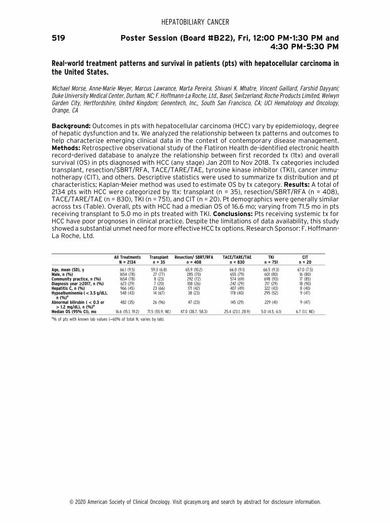
519 Poster Session (Board #B22), Fri, 12:00 PM-1:30 PM and4:30 PM-5:30 PM
Real-world treatment patterns and survival in patients (pts) with hepatocellular carcinoma inthe United States.
Michael Morse, Anne-Marie Meyer, Marcus Lawrance, Marta Pereira, Shivani K. Mhatre, Vincent Gaillard, Farshid Dayyani;Duke University Medical Center, Durham, NC; F. Hoffmann-La Roche, Ltd., Basel, Switzerland; Roche Products Limited, WelwynGarden City, Hertfordshire, United Kingdom; Genentech, Inc., South San Francisco, CA; UCI Hematology and Oncology,Orange, CA
Background: Outcomes in pts with hepatocellular carcinoma (HCC) vary by epidemiology, degreeof hepatic dysfunction and tx. We analyzed the relationship between tx patterns and outcomes tohelp characterize emerging clinical data in the context of contemporary disease management.Methods: Retrospective observational study of the Flatiron Health de-identified electronic healthrecord–derived database to analyze the relationship between first recorded tx (1tx) and overallsurvival (OS) in pts diagnosed with HCC (any stage) Jan 2011 to Nov 2018. Tx categories includedtransplant, resection/SBRT/RFA, TACE/TARE/TAE, tyrosine kinase inhibitor (TKI), cancer immu-notherapy (CIT), and others. Descriptive statistics were used to summarize tx distribution and ptcharacteristics; Kaplan-Meier method was used to estimate OS by tx category. Results: A total of2134 pts with HCC were categorized by 1tx: transplant (n = 35), resection/SBRT/RFA (n = 408),TACE/TARE/TAE (n = 830), TKI (n = 751), and CIT (n = 20). Pt demographics were generally similaracross txs (Table). Overall, pts with HCC had a median OS of 16.6 mo; varying from 71.5 mo in ptsreceiving transplant to 5.0 mo in pts treated with TKI. Conclusions: Pts receiving systemic tx forHCC have poor prognoses in clinical practice. Despite the limitations of data availability, this studyshoweda substantial unmet need formore effectiveHCC tx options. Research Sponsor: F. Hoffmann-La Roche, Ltd.
All TreatmentsN = 2134
Transplantn = 35
Resection/ SBRT/RFAn = 408
TACE/TARE/TAEn = 830
TKIn = 751
CITn = 20
Age, mean (SD), y 66.1 (9.5) 59.3 (6.8) 65.9 (10.2) 66.0 (9.1) 66.5 (9.3) 67.0 (7.5)Male, n (%) 1654 (78) 27 (77) 285 (70) 655 (79) 601 (80) 16 (80)Community practice, n (%) 1654 (78) 8 (23) 292 (72) 574 (69) 698 (93) 17 (85)Diagnosis year ‡2017, n (%) 623 (29) 7 (20) 108 (26) 242 (29) 217 (29) 18 (90)Hepatitis C, n (%) 966 (45) 23 (66) 171 (42) 407 (49) 322 (43) 8 (40)Hypoalbuminemia ( < 3.5 g/dL),n (%)a
548 (43) 14 (67) 38 (23) 178 (40) 295 (52) 9 (47)
Abnormal bilirubin ( < 0.3 or> 1.2 mg/dL), n (%)a
482 (35) 26 (96) 47 (23) 145 (29) 229 (41) 9 (47)
Median OS (95% CI), mo 16.6 (15.1, 19.2) 71.5 (55.9, NE) 47.0 (38.7, 58.3) 25.4 (23.1, 28.9) 5.0 (4.5, 6.1) 6.7 (1.1, NE)a% of pts with known lab values (»60% of total N, varies by lab).
© 2020 American Society of Clinical Oncology. Visit gicasym.org and search by abstract for disclosure information.
HEPATOBILIARY CANCER
520 Poster Session (Board #C1), Fri, 12:00 PM-1:30 PM and4:30 PM-5:30 PM
Subsequent anticancer procedures following first-line lenvatinib (LEN): A post hoc analysis fromthe phase III REFLECT study in unresectable hepatocellular carcinoma (uHCC).
Angel Alsina, Masatoshi Kudo, Arndt Vogel, Ann-Lii Cheng, Won Young Tak, Baek-Yeol Ryoo, T.R. Jeffry Evans,Carlos Lopez Lopez, Bruno Daniele, Jean-Frederic Blanc, Min Ren, Rae Lynn Baldwin, Namiki Izumi, Shukui Qin,Richard S. Finn; Tampa General Hospital, Tampa, FL; Kindai University Faculty of Medicine, Osaka, Japan; HannoverMedical School, Hannover, Germany; National Taiwan University Hospital and National Taiwan University Cancer Center,Taipei, Taiwan; Kyungpook National University Hospital, Daegu, South Korea; Asan Medical Center, University of UlsanCollege of Medicine, Seoul, South Korea; University of Glasgow, Beatson West of Scotland Cancer Centre, Glasgow, UnitedKingdom; Marques de Valdecilla University Hospital, Santander, Spain; Azienda Ospedaliera G. Rummo, Benevento, Italy;Hospital Saint-Andre, Bourdeaux, France; Eisai Inc., Woodcliff Lake, NJ; Musashino Red Cross Hospital, Tokyo, Japan; NanjingBayi Hospital, Nanjing, China; Geffen School of Medicine, UCLA Medical Center, Santa Monica, CA
Background: The availability of newer therapeutic options has shifted the treatment paradigm foruHCC, evoking questions regarding the sequencing of anticancer therapies. REFLECT was a phase 3study comparing the efficacy and safety of LEN versus sorafenib (SOR) in first-line treatment ofpatients (pts) who have uHCC. In this post hoc analysis, we investigated overall survival (OS) duringthe follow-up survival period between treatment arms for ptswho did and did not receive subsequentanticancer procedures and for responders to first-line LEN who received subsequent anticancerprocedures. Methods: Pts in REFLECT were randomized 1:1 to LEN or SOR. The follow-up periodcommenced at the first visit after stopping study medications. Pts were followed-up every 12 weeksuntil data cutoff (Nov 13, 2016) or death. OS was estimated using the Kaplan–Meier method andpresentedwith a 95%confidence interval (CI). Pts receiving subsequent radiotherapywere excludedfrom theOSanalysis.Results:Of the954pts enrolled inREFLECT, 122/478 (26%) LEN- and 130/476(27%) SOR-treated pts received subsequent anticancer procedures, with the most common beingtransarterial chemotherapy embolization (LENn = 69 [14.4%]; SOR n = 81 [17.0%]) and hepatic intra-arterial chemotherapy (LENn= 23 [4.8%]; SORn= 25 [5.3%]). EasternCooperativeOncologyGroupperformance status scores and laboratory assessments, including liver function, were comparablebetween arms at first-line treatment discontinuation. Of the pts who received subsequent anticancerprocedures, those randomized to first-line LEN (n = 99) had a longermedianOS (23.0 vs 19.6months,respectively; hazard ratio [HR]: 0.71; 95% CI: 0.51–1.01) than those randomized to first-line SOR (n =112). Responders to first-line LEN who received subsequent anticancer procedures (n = 45) had amedian OS of 27.2 months (95% CI: 20.7–29.8). There were too few SOR responders (n = 10) whoreceived subsequent anticancer procedures to evaluate OS. Conclusions: Pts randomized to first-line LEN who received subsequent anticancer procedures had longer OS compared with the similarsequence for pts taking first-line SOR. Clinical trial information: NCT01761266. Research Sponsor:This study was sponsored by Eisai Inc., Woodcliff Lake, NJ, USA, and Merck Sharp & Dohme Corp., asubsidiary of Merck & Co. Inc., Kenilworth, NJ, USA.
© 2020 American Society of Clinical Oncology. Visit gicasym.org and search by abstract for disclosure information.
HEPATOBILIARY CANCER
521 Poster Session (Board #C2), Fri, 12:00 PM-1:30 PM and4:30 PM-5:30 PM
Optimal timing of radiotherapy for incomplete transarterial chemoembolization in BCLC stage Bhepatocellular carcinoma.
Hwa Kyung Byun, Nalee Kim, Jinsil Seong; Yonsei Cancer Center, Yonsei University College of Medicine, Seoul, South Korea;Yonsei University College of Medicine, Seoul, South Korea
Background: Numerous studies have reported on the efficacy of radiotherapy (RT) after incom-plete transarterial chemoembolization (TACE). However, the optimal timing of RT remains unclear.This study investigated the optimal time of initiating RT for incomplete TACE in patients with BCLCstage B hepatocellular carcinoma (BCLC-B HCC). Methods: Between 2001 and 2016, 116 lesions in104 patients with BCLC-B HCC were treated with RT after TACE. The time interval between the lastsession of TACE and initiation of RT was obtained from medical records and analyzed retrospec-tively. The optimal cut-off time-interval that maximized the difference in local failure-free rate(LFFR) was determined using maximally selected rank statistics. Results: The median durationbetween TACE and RT was 26 (range: 2–165) days. Median number of TACE treatments on thetarget lesion before RT was 2; median tumor size was 7 cm. At a median follow-up of 18 (range:3–160) months, the median overall survival was 18 months. The probability of local controlincreased as the time interval between TACE and RT decreased. The optimal cut-off value ofthe time interval was 5 weeks. With the cut-off of 5 weeks, 65 and 39 patients were classified intoearly and late RT groups, respectively. The early RT group had significantly poorer Child-Pugh classand higher alpha-fetoprotein levels. Most characteristics including tumor size (7 cm vs. 6 cm; P =.144)were not significantly different between the groups. One-year LFFRwas significantly higher inthe early RT group (94.6% vs. 70.8%; P = .005). On multivariate analysis, early RT was anindependent predictor of favorable LFFR (hazard ratio: 3.82, 95% confidence interval: 1.64–8.88,P = .002).Conclusions: The optimal time for the administration of RT for incomplete TACE iswithin5 weeks following TACE. Early administration of RT within 5 weeks after TACE was associated withbetter local control. Research Sponsor: None.
© 2020 American Society of Clinical Oncology. Visit gicasym.org and search by abstract for disclosure information.
HEPATOBILIARY CANCER
522 Poster Session (Board #C3), Fri, 12:00 PM-1:30 PM and4:30 PM-5:30 PM
Initial lenvatinib therapy with no prior TACE in patients with intermediate-stage hepatocellularcarcinoma beyond up-to-seven criteria and Child-Pugh A liver function: A proof-of-conceptstudy.
Masatoshi Kudo, Kazuomi Ueshima, Stephen Lam Chan, Tomoko Aoki, Satoru Hagiwara, Yasunori Minami, Hiroshi Ida,Mamoru Takenaka, Tomohiro Watanabe, Masahiro Morita, Chikara Ogawa, Yoshiyuki Wada, Masafumi Ikeda, Hiroshi Ishii,Namiki Izumi, Atsushi Hiraoka, Hiroshi Aikata, Naoshi Nishida; Kindai University Faculty of Medicine, Osaka, Japan; KindaiUniversity Faculty of Medicine, Osakasayama, Japan; State Key Laboratory in Oncology of South China, The ChineseUniversity of Hong Kong, Shatin, China; Department of Gastroenterology and Hepatology, Kindai University Faculty ofMedicine, Osakasayama, Japan; Takamatsu Red Cross Hospital, Takamatsu, Japan; National Hospital Organization KyushuMedical Center, Fukuoka, Japan; National Cancer Center Hospital East, Kashiwa, Japan; Cancer Institute Hospital, Kohtoh-Ku,Japan; Musashino Red Cross Hospital, Tokyo, Japan; Ehime Prefectural Central Hospital, Matsuyama, Japan; Department ofGastroenterology and Metabolism, Hiroshima University Hospital, Hiroshima, Japan; Kindai University, Osaka, Japan
Background: Intermediate-stage HCC is a heterogenous population with subgroups of patients thatdo not benefit from TACE. Hence, we conducted a matched cohort study to compare the efficacy oflenvatinib to TACE in intermediate-stageHCC patientswith large ormultinodular tumours exceedingthe up-to-7 criteria. Methods: This study first identified 642 consecutive patients with HCC initiallytreated with lenvatinib or conventional TACE (cTACE) between January 2006 and December 2018.Of these patients, 176 who received lenvatinib or cTACE as an initial treatment andmet the eligibilitycriteria [unresectable, beyond the up-to-7 criteria, without prior TACE/systemic therapy, no vascularinvasion, no extrahepatic spread and Child-Pugh A liver function] were selected. Propensity scorematching was used to adjust for patients’ demographics. Results: After propensity-score matching,outcome of 30 patients prospectively treated with lenvatinib (14 in clinical trials [1 Phase II and 13Phase III REFLECT trial], 1 in early access program and 15 in real world setting) and 60 patientstreated with cTACE as the initial treatment was compared. The lenvatinib group showed a higherobjective response rate (73.3% vs. 33.3%; p , 0.001) and longer median progression-free survivalthan the cTACE group (16.0 vs. 3.0 months; p, 0.001). Median overall survival was also significantlylonger in the lenvatinib group than in the cTACE group (37.9 vs. 21.3 months; hazard ratio: 0.48, p,0.01). The change of ALBI score from baseline to the end of treatment were -2.61 to -2.61 for 30patients in lenvatinib group (p = 0.254) and -2.66 to -2.09 in cTACE group (p , 0.01), respectively.Conclusions: In patients with large or multinodular intermediate-stage HCC exceeding the up-to-7criteria with Child-Pugh A liver function, who usually do not benefit from TACE, lenvatinib isassociated with better outcome than TACE. In particular, leventinib is associated with preservationof hepatic function during treatment while TACE is associated with deterioration of hepatic function.Research Sponsor: None.
© 2020 American Society of Clinical Oncology. Visit gicasym.org and search by abstract for disclosure information.
HEPATOBILIARY CANCER
523 Poster Session (Board #C4), Fri, 12:00 PM-1:30 PM and4:30 PM-5:30 PM
Analysis of OX40 agonist antibody (PF-04518600) in patients with hepatocellular carcinoma.
Anthony B. El-Khoueiry, Jean-Philippe Spano, ERIC ANGEVIN, Toshihiko Doi, Andrea J. Bullock, William Proctor Harris,Omid Hamid, Paul Gougis, Alison Forgie, Wenjing Yang, Ken Liao, Ruifeng Li, Carrie T. Taylor, Jeffrey Chou, Ferry Eskens;University of Southern California Norris Comprehensive Cancer Center, Los Angeles, CA; Pitie-Salpetriere Hospital, SorbonneUniversity, Cancer University Institute, Paris, France; Drug Development Department (DITEP), Institut Gustave Roussy,Villejuif, France; National Cancer Center Hospital East, Chiba, Japan; Beth Israel Deaconess Medical Center, Boston, MA;University of Washington School of Medicine, Seattle, WA; The Angeles Clinic and Research Institute, Los Angeles, CA; GroupeHospitalier Pitie-Salpetriere Oncologie Medicale, Paris, France; Pfizer Inc., San Francisco, CA; Pfizer Inc., San Diego, CA; PfizerInc., Cambridge, MA; Pfizer Inc., New York, NY; Erasmus MC Cancer Institute, Rotterdam, Netherlands
Background:PF-04518600 (PF-8600) is a humanizedagonist IgG2monoclonal antibody to the tumornecrosis factor superfamily receptor OX40. PF-8600 was given to patients (pts) with advanced/metastatic hepatocellular carcinoma (HCC) in dose expansion of a phase 1 study (NCT02315066).Safety and tolerability were primary endpoints and exploratory endpoints included biomarker ana-lyses.Methods: Pts received either 30mg (Arm 1) or 250mg (Arm 2) of PF-8600 intravenously Q2W.Pts had pathologic diagnosis of advancedHCC, a Child-Pugh score of A or B7, and had#2 prior lines oftherapy, or if treatment naıve, had declined standard of care. Radiological tumor assessments wereconducted Q8W. Biopsy samples collected at baseline and wk 6 were analyzed by immunohistochem-istry and RNA sequencing for pharmacodynamic (PD) analyses. Whole blood samples were collectedlongitudinally for DNA extraction for high-throughput sequencing of the T cell receptor b-chain.Results:Arm 1 enrolled 16 pts (mean age 65.6 yrs; range 54-81 yrs; prior PD-1/PD-L1: 5 pts) and Arm 2enrolled 19 pts (meanage61.7 yrs; range26-79 yrs; prior PD-1/PD-L1: 3 pts). All grade treatment relatedadverse events (TRAEs) occurred in69%ofpts inArm 1 and58%ofpts inArm2. The rate of$Grade3TRAEs was 31% and 16% in Arms 1 and 2, respectively. For both arms combined, the most common($10%) TRAEs were rashes and pruritus. In Arm 1, 8 pts (50%) and in Arm 2, 10 pts (53%) achievedstable disease (SD), with a mean duration of 18.4 (range: 14.0-30.3 wks) and 17.4 wks (range: 8.0-31.9wks), respectively. PD effectsweremore evident in Arm 1 thanArm2, including increasedOX40 tumorexpression and positive changes in gene signatures, reflecting an active anti-tumor immune response.Conclusions: PF-8600 was generally well tolerated and provided meaningful disease control. Whilesafety and efficacy were not significantly different between the 2 doses, there were potentialdifferences in the PD data. The safety and relative durability of SD in HCC pts may provide a rationalefor exploration of combination therapy in this pt population. Clinical trial information: NCT02315066.Research Sponsor: Pfizer Inc.
© 2020 American Society of Clinical Oncology. Visit gicasym.org and search by abstract for disclosure information.
HEPATOBILIARY CANCER
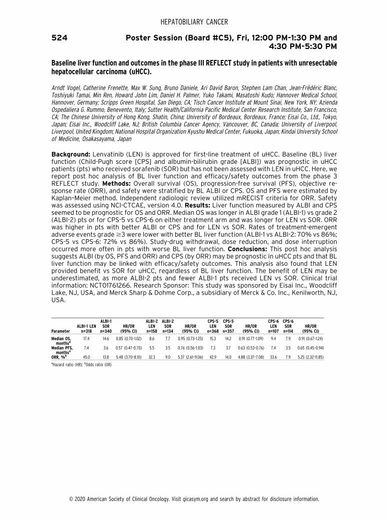
524 Poster Session (Board #C5), Fri, 12:00 PM-1:30 PM and4:30 PM-5:30 PM
Baseline liver function and outcomes in the phase III REFLECT study in patients with unresectablehepatocellular carcinoma (uHCC).
Arndt Vogel, Catherine Frenette, Max W. Sung, Bruno Daniele, Ari David Baron, Stephen Lam Chan, Jean-Frederic Blanc,Toshiyuki Tamai, Min Ren, Howard John Lim, Daniel H. Palmer, Yuko Takami, Masatoshi Kudo; Hannover Medical School,Hannover, Germany; Scripps Green Hospital, San Diego, CA; Tisch Cancer Institute at Mount Sinai, New York, NY; AziendaOspedaliera G. Rummo, Benevento, Italy; Sutter Health/California Pacific Medical Center Research Institute, San Francisco,CA; The Chinese University of Hong Kong, Shatin, China; University of Bordeaux, Bordeaux, France; Eisai Co., Ltd., Tokyo,Japan; Eisai Inc., Woodcliff Lake, NJ; British Columbia Cancer Agency, Vancouver, BC, Canada; University of Liverpool,Liverpool, United Kingdom; National Hospital Organization Kyushu Medical Center, Fukuoka, Japan; Kindai University Schoolof Medicine, Osakasayama, Japan
Background: Lenvatinib (LEN) is approved for first-line treatment of uHCC. Baseline (BL) liverfunction (Child-Pugh score [CPS] and albumin-bilirubin grade [ALBI]) was prognostic in uHCCpatients (pts) who received sorafenib (SOR) but has not been assessed with LEN in uHCC. Here, wereport post hoc analysis of BL liver function and efficacy/safety outcomes from the phase 3REFLECT study. Methods: Overall survival (OS), progression-free survival (PFS), objective re-sponse rate (ORR), and safety were stratified by BL ALBI or CPS. OS and PFS were estimated byKaplan–Meier method. Independent radiologic review utilized mRECIST criteria for ORR. Safetywas assessed using NCI-CTCAE, version 4.0. Results: Liver function measured by ALBI and CPSseemed to be prognostic for OS and ORR. Median OS was longer in ALBI grade 1 (ALBI-1) vs grade 2(ALBI-2) pts or for CPS-5 vs CPS-6 on either treatment arm and was longer for LEN vs SOR. ORRwas higher in pts with better ALBI or CPS and for LEN vs SOR. Rates of treatment-emergentadverse events grade$3 were lower with better BL liver function (ALBI-1 vs ALBI-2: 70% vs 86%;CPS-5 vs CPS-6: 72% vs 86%). Study-drug withdrawal, dose reduction, and dose interruptionoccurred more often in pts with worse BL liver function. Conclusions: This post hoc analysissuggests ALBI (by OS, PFS and ORR) and CPS (by ORR) may be prognostic in uHCC pts and that BLliver function may be linked with efficacy/safety outcomes. This analysis also found that LENprovided benefit vs SOR for uHCC, regardless of BL liver function. The benefit of LEN may beunderestimated, as more ALBI-2 pts and fewer ALBI-1 pts received LEN vs SOR. Clinical trialinformation: NCT01761266. Research Sponsor: This study was sponsored by Eisai Inc., WoodcliffLake, NJ, USA, and Merck Sharp & Dohme Corp., a subsidiary of Merck & Co. Inc., Kenilworth, NJ,USA.
ParameterALBI-1 LENn=318
ALBI-1SORn=340
HR/OR(95% CI)
ALBI-2LENn=158
ALBI-2SORn=134
HR/OR(95% CI)
CPS-5LEN
n=368
CPS-5SORn=357
HR/OR(95% CI)
CPS-6LENn=107
CPS-6SORn=114
HR/OR(95% CI)
Median OS,monthsa
17.4 14.6 0.85 (0.70–1.02) 8.6 7.7 0.95 (0.73–1.25) 15.3 14.2 0.91 (0.77–1.09) 9.4 7.9 0.91 (0.67–1.24)
Median PFS,monthsa
7.4 3.6 0.57 (0.47–0.70) 5.5 3.5 0.76 (0.56–1.03) 7.3 3.7 0.63 (0.53–0.76) 7.4 3.5 0.65 (0.45–0.94)
ORR, %b 45.0 13.8 5.48 (3.70–8.10) 32.3 9.0 5.37 (2.61–11.06) 42.9 14.0 4.88 (3.37–7.08) 33.6 7.9 5.25 (2.32–11.85)aHazard ratio (HR); bOdds ratio (OR)
© 2020 American Society of Clinical Oncology. Visit gicasym.org and search by abstract for disclosure information.
HEPATOBILIARY CANCER
525 Poster Session (Board #C6), Fri, 12:00 PM-1:30 PM and4:30 PM-5:30 PM
Nivolumab (NIVO) and drug eluting bead transarterial chemoembolization (deb-TACE): Preliminaryresults from a phase I study of patients (pts) with liver limited hepatocellular carcinoma (HCC).
James J. Harding, Hooman Yarmohammadi, Kim Anna Reiss, Joanne F. Chou, Marinela Capanu, Richard K. G. Do, Danny Khalil,Imane H. El Dika, Joseph Daniel Giardina, Taha Merghoub, William R. Jarnagin, Gregory Nadolski, Joseph Patrick Erinjeri,Michael C. Soulen, Benjamin R. Tan, Ghassan K. Abou-Alfa; Memorial Sloan Kettering Cancer Center, New York, NY; Universityof Pennsylvania Abramson Cancer Center, Philadelphia, PA; Memorial Sloan Kettering Cancer Center, New York City, NY;Washington University, St. Louis, MO; University of Pennsylvania, Philadelphia, PA; Washington University School of Medicine,St. Louis, MO
Background: Regional therapies in HCC impact the immune microenvironment and may augmentthe effects of immune checkpoint inhibitors.Methods: This is a multicenter phase 1 study of NIVOand deb-TACE in unresectable HCC pts (BCLC Stage B) and Child Pugh A cirrhosis (NCT03143270).The primary objective is to assess safety. Secondary objectives include response rate by RECISTv1.1, progression-free and overall survival by Kaplan-Meier methodology, and blood/tumor immunecorrelates. A 3 + 3 design sequentially evaluates 3 cohorts of differing schedules of NIVO relative todeb-TACE. Deb-TACE (75mg of doxorubicin) is administered on Day 0. NIVO is dosed at 240mg IVevery 14 days for 1 year (Cohort 1: NIVO begins day +14 after deb-TACE; Cohort 2, interrupted NIVOdosing begins at Day -28 but is held on the Day 0 then restarted on Day +14; Cohort 3, continuousNIVO dosing begins on Day -28 without interruption). Results: As of July 2019, 9 pts have beentreated [median 65 years (range: 54-76), male (89%), viral (44%;1 HBV, 3 HCV), non-viral (56%;2EtOH, 1 NASH, 2 unknown), prior resection (44%), prior regional therapy (44%), 3 pts in eachcohort]. No cases of treatment related liver failure, dose-limiting toxicity, or Grade 5 adverseevents (AEs) were observed. Grade $3 AEs possibly related to nivolumab, deb-TACE, or bothincluded: transaminase elevation (1 pt: day 1 post TACE resolved in 7 days without treatment; 2 pts:$30 days post TACE resolved with steroids between 20-41 days), post-embolization syndrome (1pt: resolved in 5 days), asymptomatic lipase increase (1 pt: resolved in 14 days), post-proceduralgroin hematoma (1 pt: resolved in 2 days). All 9 pts were evaluable for efficacy: 2 (22%) confirmedPR and 7 (78%) SD. 4/9 pts remain on study with SD or better—2 pts continue . 18 months postembolization with durable PRs. 12months OS rate was 71%. Ongoing correlates will be presented ata separate meeting. Conclusions: Nivolumab given at various times relative to deb-TACE appearssafe and tolerable. Cohort 3 continues to accrue to provide a better estimate of safety andantitumor activity of the combination. Clinical trial information: NCT03143270. Research Sponsor:Bristol Myer Squibb.
© 2020 American Society of Clinical Oncology. Visit gicasym.org and search by abstract for disclosure information.
HEPATOBILIARY CANCER
526 Poster Session (Board #C7), Fri, 12:00 PM-1:30 PM and4:30 PM-5:30 PM
Phase III study of pembrolizumab (pembro) versus best supportive care (BSC) for second-linetherapy in advanced hepatocellular carcinoma (aHCC): KEYNOTE-240 Asian subgroup.
Masatoshi Kudo, Ho Yeong Lim, Ann-Lii Cheng, Yee Chao, Thomas Yau, Sadahisa Ogasawara, Masayuki Kurosaki,Naoki Morimoto, Kazuyoshi Ohkawa, Tatsuya Yamashita, Kyung-Hun Lee, Erluo Chen, Abby B. Siegel, Baek-Yeol Ryoo;Kindai University, Osaka, Japan; Samsung Medical Center, Sungkyunkwan University School of Medicine, Seoul, South Korea;National Taiwan University Hospital Cancer Center, Taipei City, Taiwan; Taipei Veterans General Hospital, Taipei City, Taiwan;University of Hong Kong, Queen Mary Hospital, Hong Kong, China; Chiba University Graduate School of Medicine, Chiba,Japan; Musashino Red Cross Hospital, Tokyo, Japan; Jichi Medical University Hospital, Tochigi, Japan; Department ofHepatobiliary and Pancreatic Oncology, Osaka International Cancer Institute, Osaka, Japan; Kanazawa University Hospital,Kanazawa, Japan; Seoul National University Hospital, Seoul, South Korea; Merck & Co., Inc., Kenilworth, NJ; Asan MedicalCenter, University of Ulsan College of Medicine, Seoul, South Korea
Background: Pembro received accelerated approval for second-line therapy in aHCC based onKEYNOTE-224 (phase 2). KEYNOTE-240 (NCT02702401) is a randomized, phase 3 study of pembrov BSC in previously treated aHCC.We report outcomes for the Asian subgroup.Methods:Pts with aradiographic/pathologic HCC diagnosis, radiographic progression with/intolerance to sorafenib,Child-Pugh A disease, and ECOG PS 0/1 were randomized 2:1 to pembro (200 mg) + BSC or PBO +BSC Q3W (#35 cycles or until confirmed PD/unacceptable toxicity). Pts were stratified bygeographic region (Asia without Japan; non-Asia with Japan), AFP, and macrovascular invasion.Response was assessed Q6W (RECIST v1.1, central review). Primary end points: OS, PFS; secondaryend points: ORR, DOR, safety. Data cutoff: Jan 2, 2019. Results: 413 pts were randomized (overallcohort: n = 278 pembro, n = 135 PBO; Asian subgroup [Hong Kong, Japan, Philippines, S Korea,Taiwan, Thailand]: n = 107, n = 50). HBV+ status and BCLC stage C were higher in Asian subgroup(HBV+: 51% v 24.5% overall; stage C: 86.6% v 79.4%). Median follow-up: pembro (21.3 mo overall;23.5 mo); PBO (21.5 mo overall; 23.0 mo). Pembro improved OS v PBO (median OS [95% CI]: 13.9[11.6-16.0] v 10.6 [8.3-13.5] mo; HR: 0.781; 95% CI, 0.611-0.998; P = 0.0238) and PFS (HR: 0.718;0.570-0.904; P = 0.0022) for overall cohort and Asian subgroup (median OS: 13.8 [10.1-16.9] v 8.3[6.3-11.8] mo; HR: 0.548; 0.374-0.804; P = 0.0009; PFS: HR: 0.475; 0.324-0.696; P , 0.0001).Differences did not meet prespecified significance level for overall cohort. ORR in overall cohortwas 18.3% (14.0-23.4) for pembro; 4.4% (1.6-9.4; P = 0.00007) for PBO; in Asian subgroup, 20.6%(13.4-29.5) and 2.0% (0.1-10.6; P = 0.00135). Safety was consistent with that previously reported inpembro studies. No HBV/HCV flares were identified. Conclusions: Pembro reduced risk for deathby 22% in overall cohort and 45% in Asian subgroup and improved PFS v PBO. Safety wascomparable to that of pembro monotherapy. Results are consistent with KEYNOTE-224 andmagnitude of OS benefit was enhanced in Asian subgroup, supporting a favorable risk-benefitbalance for second-line pembro in HCC. Clinical trial information: NCT02702401. Research Spon-sor: Merck Sharp & Dohme Corp., a subsidiary of Merck & Co., Inc., Kenilworth, NJ, USA.
© 2020 American Society of Clinical Oncology. Visit gicasym.org and search by abstract for disclosure information.
HEPATOBILIARY CANCER
527 Poster Session (Board #C8), Fri, 12:00 PM-1:30 PM and4:30 PM-5:30 PM
Postoperative adjuvant transarterial infusion chemotherapy with FOLFOX could improve outcomesof hepatocellular carcinoma patients with microvascular invasion: A preliminary report of a phaseIII, randomized, controlled clinical trial.
No Firstname [email protected], Wei Wei, Qiaoxuan Wang, Zhixing Guo, Jie Mei, Lianghe Lu, Yihong Ling, Li Xu,Minshan Chen, Wenping Lin, Jingwen Zou, Yuhua Wen, Rongping Guo; Sun Yat-sen University Cancer Center, Guangzhou,China; Sun Yat-sen University Cancer Center, Guangzhou City, China
Background: Microvascular invasion (MVI) is a risk factor for recurrence after hepatectomy forhepatocellular carcinoma (HCC) patients. This study aimed to investigate to efficacy and safety ofpostoperative adjuvant transarterial infusion chemotherapy (TAI) with FOLFOX regimen for HCCpatients with MVI. Methods: In this prospective, phase III, randomized, open-labeled, controlledclinical trial, HCC patients with histologically confirmed MVI were randomly assigned (1:1) afterhepatectomy to receive either 1-2 cycles adjuvant TAI (AT group) or follow-upwithout any adjuvanttreatment (FU group). The primary endpoint was disease-free survival (DFS), the secondaryendpoints are overall survival (OS) and safety. Results: Between June, 2016 and April 2019,127 patients were randomly assigned to AT group (n=63) or FU group (n=64). Clinicopathologicalcharacteristics were balanced between the two groups. The 6-, 12-, and 18-month OS rates for ATgroup were 100.0%, 97.7%, and 97.7%, and were 94.5%, 89.6%, and 78.5% for FU group,respectively. The 6-, 12-, and 18-month DFS rates for AT group were 84.7%, 61.8%, and 58.7%, andwere 62.9%, 48.1%, and 38.6% for FU group, respectively. The OS and DFS were significantlybetter in AT group than in FU group (p=0.037 and 0.023, respectively). No patients in AT groupexperienced grade 3 or more severe adverse events (AEs). Conclusions: Adjuvant TAI afterhepatectomy may bring survival benefits of OS and DFS for HCC patients with MVI. Clinical trialinformation: NCT03192618. Research Sponsor: None.
© 2020 American Society of Clinical Oncology. Visit gicasym.org and search by abstract for disclosure information.
HEPATOBILIARY CANCER
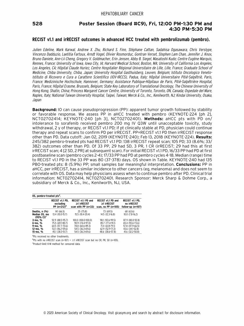
528 Poster Session (Board #C9), Fri, 12:00 PM-1:30 PM and4:30 PM-5:30 PM
RECIST v1.1 and irRECIST outcomes in advanced HCC treated with pembrolizumab (pembro).
Julien Edeline, Mark Karwal, Andrew X. Zhu, Richard S. Finn, Stephane Cattan, Sadahisa Ogasawara, Chris Verslype,Vincenzo Dadduzio, Laetitia Fartoux, Arndt Vogel, Olivier Rosmorduc, Gontran Verset, Stephen Lam Chan, Jennifer J. Knox,Bruno Daniele, Ann-Lii Cheng, Gregory V. Goldmacher, Erin Jensen, Abby B. Siegel, Masatoshi Kudo; Centre Eugene Marquis,Rennes, France; University of Iowa, Iowa City, IA; Harvard Medical School, Boston, MA; University of California Los Angeles,Los Angeles, CA; Hopital Claude Huriez, Centre Hospitalier Regional Universitaire de Lille, Lille, France; Graduate School ofMedicine, Chiba University, Chiba, Japan; University Hospital Gasthuisberg, Leuven, Belgium; Istituto Oncologico Veneto-Istituto di Ricovero e Cura a Carattere Scientifico (IOV-IRCCS), Padua, Italy; Hopital Universitaire Pitie-Salpetrie, Paris,France; Medizinische Hochschule, Hannover, Germany; Assistance Publique-Hopitaux de Paris, Pitie-Salpetriere Hospital,Paris, France; Hopital Erasme, Brussels, Belgium; State Key Laboratory of Translational Oncology, The Chinese University ofHong Kong, Shatin, China; Princess Margaret Cancer Centre, University of Toronto, Toronto, ON, Canada; Ospedale del Mare,Naples, Italy; National Taiwan University Hospital, Taipei, Taiwan; Merck & Co., Inc., Kenilworth, NJ; Kindai University, Osaka,Japan
Background: IO can cause pseudoprogression (PP): apparent tumor growth followed by stabilityor favorable response. We assess PP in aHCC treated with pembro (KEYNOTE-224 [ph 2],NCT02702414; KEYNOTE-240 [ph 3], NCT02702401). Methods: aHCC pts with PD on/intolerance to sorafenib received pembro 200 mg IV Q3W until unacceptable toxicity, studywithdrawal, 2 y of therapy, or RECIST v1.1 PD; if pt clinically stable at PD, physician could continuetherapy and repeat scans to confirm PD per irRECIST. PP=RECIST v1.1 PD then irRECIST responseother than PD. Data cutoff: Jan 02, 2019 (KEYNOTE-240); Feb 13, 2018 (KEYNOTE-224). Results:245/382 pembro-treated pts had RECIST v1.1 PD: 138 irRECIST repeat scan; 105 PD; 33 (8.6%; 33/382) outcomes other than PD. Of 33 PP, 29 had SD, 3 PR, 1 CR (irRECIST; 29 had this at firstirRECIST scan; 4 [2 SD, 2 PR] at subsequent scan). For initial RECIST v1.1 PD, 16/33 PP had PD at firstpostbaseline scan (pembro cycles 2-4); 17/33 PP had PD at pembro cycles 4-18. Median (range) timeto RECIST v1.1 PD in the 33 PP was 80 (37-378) days. OS shown in Table. KEYNOTE-240 had 135PBO-treated pts: 8 (5.9%) PP; small samples bar meaningful interpretation. Conclusions: PP inaHCC, per irRECIST, has a similar incidence to other cancers (eg, melanoma) and does not seem tocorrelate with OS. Datamay help physicians assess when to continue pembro after PD. Clinical trialinformation: NCT02702414, NCT02702401. Research Sponsor: Merck Sharp & Dohme Corp., asubsidiary of Merck & Co., Inc., Kenilworth, NJ, USA.
OS, pembro-treated ptsa.
RECIST v1.1 PD,excluding
PP (n=212)b
RECIST v1.1 PD and‡1 irRECIST
scan with PP (n=33)
RECIST v1.1 PD and‡1 irRECIST
scan, no PP (n=105)
RECIST v1.1 PD,no irRECIST
follow-up (n=107)
Deaths, n (%) 141 (66.5) 25 (75.8) 73 (69.5) 68 (63.6)Median OS, mo(95% CI)c
13.4 (10.0-15.7) 15.5 (10.4-20.4) 14.5 (12.3-16.8) 10.0 (7.8-16.2)
3-mo, % 92.9 (88.5-95.7) 100.0 (100.0-100.0) 98.1 (92.6-99.5) 87.9 (80.0-92.8)6-mo, % 75.5 (69.1-80.7) 90.9 (74.4-97.0) 85.7 (77.4-91.1) 65.4 (55.6-73.6)9-mo, % 64.5 (57.7-70.6) 78.8 (60.6-89.3) 71.4 (61.8-79.1) 57.8 (47.9-66.5)12-mo, % 53.1 (46.2-59.6) 54.5 (36.3-69.6) 62.9 (52.9-71.3) 43.6 (34.1-52.8)15-mo, % 45.1 (38.3-51.7) 54.5 (36.3-69.6) 48.6 (38.6-57.8) 41.6 (32.2-50.8)aPts received no other treatments.bPts with no irRECIST scan (n=107) + $1 irRECIST scan but no CR, PR, SD (n=105).cProduct-limit K-M method for censored data.
© 2020 American Society of Clinical Oncology. Visit gicasym.org and search by abstract for disclosure information.
HEPATOBILIARY CANCER

529 Poster Session (Board #C10), Fri, 12:00 PM-1:30 PM and4:30 PM-5:30 PM and Poster Walks, Fri, 4:45 PM-5:30 PM
Cisplatin plus irinotecan versus cisplatin plus gemcitabine in the treatment of advanced ormetastaticgallbladder or biliary tract cancer: Results of a randomized phase II trial (NCT01859728)– the Gambittrial.
Lucas Vieira dos Santos, Gustavo Sanches Faria Pinto, Mauricio Wagner Souto Ferraz, Arinilda Bragagnoli, Florinda Santos,Sarah Haddad, Ana Barros, Isadora Clarissa Cordeiro Dias, Joao Paulo SN Lima, Kathia C Abdalla; Beneficencia Portuguesa deS~ao Paulo, S~ao Paulo, Brazil; Barretos Cancer Hospital, Barretos, Brazil; Barretos Cancer Center, Barretos, Brazil; Hospital deCancer de Barretos, Barretos, Brazil; AC Camargo Cancer Center, S~ao Paulo, Brazil; Fundaç~ao Faculdade de Medicina de SaoJose do Rio Preto, S~ao Jose Do Rio Preto, Brazil
Background: The combination of gemcitabine-cisplatin (GC) is the current standard of carechemotherapy for metastatic/unresectable biliary tract cancer (BTC). However, the prognosisremains poor. This randomized trial aimed to evaluate the efficacy and safety of irinotecan pluscisplatin (IP) versus GC in advanced or metastatic BTC. Methods: Patients with biopsy-proven,chemo-naıve, unresectable or metastatic BTC, ECOG 0-2, measurable disease per RECIST 1.1,adequate organ function and written informed consent were stratified by ECOG (0 or 1 vs 2) anddistant metastases and randomized to receive irinotecan 65 mg/m2 IV D1 and D8 plus cisplatin 60mg/m2 D1 repeated every 3weeks (IP) or gemcitabine 1000mg/m2 IVD1 andD8 plus cisplatin 25mg/m2 IV D1 and D8 repeated every 3 weeks, until disease progression or unacceptable toxicity. Theprimary endpointwas overall response rate (ORR). Results: BetweenJanuary 2013 andApril 2018, 47pts were randomized (1:1) to receive IP (N = 24) or GC (N = 23). Overall, groups were well balancedaccording to prognostic factors. The ORR was 35% (complete response 5%, partial response 30%)and 31.8% in IP and GC arms, respectively. Median progression-free survival were 5.3 vs 7.8 months(HR= 1.165, 95%CI0.628-2.161, p =0.628) andmedian overall survivalwere 11.9 and9.8months (HR=0.859, 95%CI 0.431 – 1.710, p = 0.665) for IP and GC, respectively. Adverse events were notstatistically different between arms, and results were consistent with previous experiences withthese regimens. No therapy-related death were reported. Conclusions: Irinotecan-cisplatin combi-nation is active in BTC, with similar ORR, PFS and OS when compared to gemcitabine-cisplatin.Irinotecan-cisplatinwerewell tolerated, and adverse eventsweremanageable. Irinotecan-cisplatin couldbe considered as an alternative to gemcitabine-cisplatin. Clinical trial information: NCT01859728.Research Sponsor: Barretos Cancer Hospital.
© 2020 American Society of Clinical Oncology. Visit gicasym.org and search by abstract for disclosure information.
HEPATOBILIARY CANCER

531 Poster Session (Board #C12), Fri, 12:00 PM-1:30 PM and4:30 PM-5:30 PM
Impact of baseline and concomitant corticosteroid therapy on the outcomes of hepatocellularcarcinoma treated with immune checkpoint inhibitor therapy.
David James Pinato, Ahmed Omar Kaseb, Yinghong Wang, Anwaar Saeed, David Szafron, Tomi Jun, Abdul Rafeh Naqash,ChiehJu Lee, Uqba Khan, Neil Satish Nimkar, Sonal D Paul, Dominik Bettinger, Hannah Hildebrand, Tiziana Pressiani,Yehia I. Abugabal, Nicola Personeni, Yi-Hsiang Huang, Lorenza Rimassa, Celina Ang, Thomas Urban Marron; Imperial CollegeLondon, London, United Kingdom; The University of Texas MD Anderson Cancer Center, Houston, TX; University of KansasMedical Center, Kansas City, KS; MD Anderson Cancer Centre, Houston, TX; Mount Sinai Hospital, New York, NY; East CarolinaUniversity/Vidant Cancer Center, Greenville, NC; Taipei Veterans General Hospital, Taiwan, Taiwan; Weill Cornell MedicalCollege, New York, NY; New York Methodist Hospital, Brooklyn, NY; New York-Presbyterian Brooklyn Methodist Hospital,Brooklyn, NY; University of Freiburg, Freiburg, Germany; Humanitas Clinical and Research Center, Rozzano, Italy; Universityof Texas MD Anderson Cancer Center, Houston, TX; Humanitas University, Pieve Emanuele, Italy; Division of Gastroenterologyand Hepatology, Taipei Veterans General Hospital, Institute of Clinical Medicine, National Yang-Ming University, Taipei,Taiwan; Humanitas Clinical and Research Center-IRCCS, Humanitas University, Rozzano, Milan, Italy; Memorial SloanKettering Cancer Center, New York, NY
Background: The impact of corticosteroid treatment (CT) on the efficacy of immune checkpointinhibitors (ICI) in hepatocellular carcinoma (HCC) is undefined. We evaluated whether CT admin-istered at baseline (bCT) or concurrently to ICI (cCT) influences clinical outcomes of HCC patientstreated with ICI. Methods: This retrospective, multi-center observational study was conductedacross 9 tertiary academic referral centers collected 341 HCC patients who received ICI across 3continents between January 1, 2016 and April 1, 2019. Outcomemeasures included overall (OS) andprogression-free survival (PFS) calculated from time of ICI commencement and overall responserates (ORR) defined by Response Evaluation Criteria in Solid Tumors (v1.1) on 6-8 weekly periodicrestaging.Results:Of 331 eligible patients, 254 (76%) had BCLC-C stage HCC and receivedmostlyPD(L)-1 ICI monotherapy (n=250, 85%). Median OS was 12.1 months (95%CI 9.2-15.0 months) andmedian PFS was 8.1 months (95%CI 6.3-10 months). In total 81 patients (24%) received .10 mgprednisone equivalent daily either as bCT (n=15, 4%) or cCT (n=66, 20%). Indications for CTincluded procedure/prophylaxis (n=37, 45%), management of irAE (n=31, 37%), cancer-relatedsymptoms (n=5, 2%) or comorbidities (n=8, 3%). Neither overall CT, bCT nor cCT predicted forworse OS, PFS nor ORR in uni- and multi-variable analyses (p.0.05). CT for cancer-relatedindications predicted for shorter PFS (2.4 vs. 11.3 months, p=0.01), OS (4.5 vs. 12.8 months, p=0.05)and reducedORR (p=0.03) compared to cancer-unrelated indications.Conclusions: This is the firststudy to demonstrate that neither bCT nor cCT appear to influence response andOS following ICI inHCC. Worse survival and ORR in CT recipients for cancer-related indications appears driven by thepoor prognosis associated with symptomatic HCC. Research Sponsor: None.
© 2020 American Society of Clinical Oncology. Visit gicasym.org and search by abstract for disclosure information.
HEPATOBILIARY CANCER
532 Poster Session (Board #C13), Fri, 12:00 PM-1:30 PM and4:30 PM-5:30 PM
Respiratory-gated magnetic resonance image guided radiation therapy for hepatocellularcarcinoma: A pilot study.
Hyun-Cheol Kang, Jin Ho Kim, Eui Kyu Chie; Department of Radiation Oncology, Seoul National University Hospital, Seoul,South Korea; Seoul National University College of Medicine, Seoul, South Korea
Background: Hypofractionated RT has shown encouraging results in hepatocellular carcinoma(HCC) patients. However, HCC adjacent to the gastrointestnal (GI) tract should be carefully treatedusing the high-precision irradiation technique due to risk of radiation damage. We evaluated thefeasibility of a respiratory-gated magnetic resonance image guided radiation therapy (RgMRg-RT)for hepatocellular carcinoma (HCC).Methods: Thirty-three patients with HCC underwent RgMRg-RT in our hospital from 2015 to February 2019, including patients with Child-Pugh A/B7 cirrhosisand unresectable tumors near the gastrointestinal tract. The median radiation dose was 50 Gy(range, 25–60) and median fraction number was 5 (range, 4–15). Gating was performed based onreal-timemagnetic resonance imagewithout an external surrogate.Results:Themedian follow-upperiod was 11.7 months (range, 3.6–37.9months). The rates of local control of the target tumor at 6months and 1 year were 90.2 and 86.9%, respectively. The overall survival rates at 6months, 1, and2 years were 84.3, 76.3, and 61%, respectively. The median distances from gross tumor to theesophagus, stomach, duodenum and colon were 6.1 (range, 1.9-14.3), 6.4 (0-13.4), 3.8 (0-13.5) and4cm (0.3-13), respectively. A total of 15 tumors (45.5%) were located within 2 cm of the gastro-intestinal tract and 9 tumors (27.3%) within 1cm. Grades 3 treatment-related bleeding wasobserved in one patient and one patient had radiation-induced liver disease. Conclusions:Hypofractionated RgMRg-RT was a safe and potentially ablative therapy for HCC. RgMRg-RT isa good alternative treatment for patients with HCCs that are unsuitable for surgical resection orlocal ablative therapy. Research Sponsor: None.
© 2020 American Society of Clinical Oncology. Visit gicasym.org and search by abstract for disclosure information.
HEPATOBILIARY CANCER
533 Poster Session (Board #C14), Fri, 12:00 PM-1:30 PM and4:30 PM-5:30 PM
Immunotherapy versus biologics as second-line therapy in advanced hepatocellular carcinoma(HCC).
Mohammed Al-Jumayli, Hannah Hildebrand, Saqib Abbasi, Anup Kasi, Anwaar Saeed; Kansas University Cancer Center,Kansas, KS; University of Kansas Medical Center, Kansas City, KS; Kansas University Cancer Center, Westwood,, KS; Universityof Kansas Cancer Center, Westwood, KS
Background: Sorafenib or lenvatinib are the current frontline options for advanced HCC. Multiplebiologic agents including multi-Tyrosine Kinase Inhibitors (TKI) cabozantinib & regorafenib havebeen recently approved for the previously treated population. Immunotherapy (IO) agent Nivo-lumab have also been approved in the same setting. Due to lack of prospective head to headcomparison, there is no standardized way for alternating those agents beyond frontline.Methods:We performed a retrospective review of KU cancer registry. Patients with advanced HCC weredivided into 2 groups based on the 2nd line systemic regimen (IO vs non-IO). Kaplan–Meier and Coxproportional hazards models were utilized to evaluate progression free survival (PFS) and overallsurvival (OS). Results: Between 2016-2019, 98 patients were identified, 41 received IO, while 57received biologics. All patients had sorafinib as 1st line therapy. Most patients have ECOG 0-1 andChild-Pugh class A or B. 55% had hepatitis C, 6% hepatitis B and 27 % have history of alcoholmisuse. Almost 50%of patients have received prior liver directed therapy. Comparing IO vs non-IOgroups,median PFSwas 3.9months vs 3months andmedianOSwas 10months vs 10months. Therewas no statistically significant difference in PFS & OS but there was a delay separation in thesurvival curve favoring IO. Similar outcome was seen in a sub-group analysis of the hepatitis Cpatients. Conclusions: This retrospective comparative review of current 2nd line regimens is oneof the first & largest studies reported. In this study population, IO was not superior to multi-TKI as a2nd line regimen. The late survival curve separation favoring IOmight suggest a delay IO effect in asubgroup of patients. Future studies should define specific biomarkers that could predict responseand allow treatment to be optimized depending on patient and tumor characteristics. Furthermore,combining IO plus multi-TKI might be a promising next step in development. A number of ongoingtrials are testing this strategy including our CAMILLA trial of Cabozantinib plus Durvalumab,currently enrolling patients at the University of Kansas Cancer Center NCT03539822. ResearchSponsor: None.
© 2020 American Society of Clinical Oncology. Visit gicasym.org and search by abstract for disclosure information.
HEPATOBILIARY CANCER
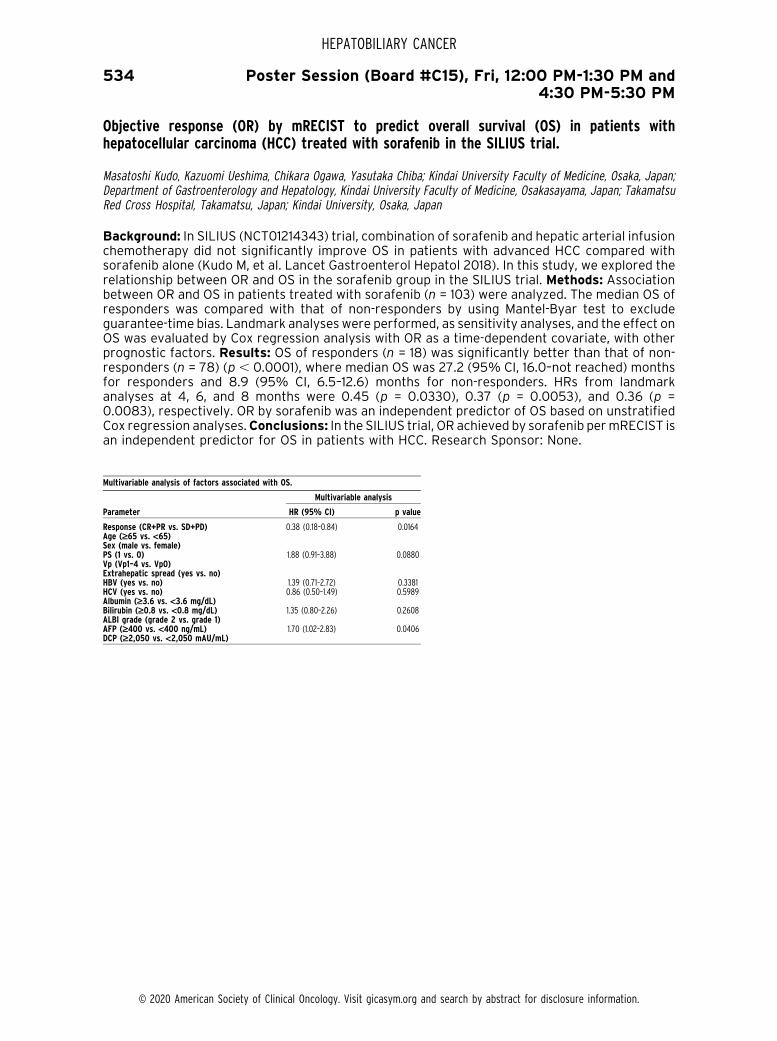
534 Poster Session (Board #C15), Fri, 12:00 PM-1:30 PM and4:30 PM-5:30 PM
Objective response (OR) by mRECIST to predict overall survival (OS) in patients withhepatocellular carcinoma (HCC) treated with sorafenib in the SILIUS trial.
Masatoshi Kudo, Kazuomi Ueshima, Chikara Ogawa, Yasutaka Chiba; Kindai University Faculty of Medicine, Osaka, Japan;Department of Gastroenterology and Hepatology, Kindai University Faculty of Medicine, Osakasayama, Japan; TakamatsuRed Cross Hospital, Takamatsu, Japan; Kindai University, Osaka, Japan
Background: In SILIUS (NCT01214343) trial, combination of sorafenib and hepatic arterial infusionchemotherapy did not significantly improve OS in patients with advanced HCC compared withsorafenib alone (Kudo M, et al. Lancet Gastroenterol Hepatol 2018). In this study, we explored therelationship between OR and OS in the sorafenib group in the SILIUS trial. Methods: Associationbetween OR and OS in patients treated with sorafenib (n = 103) were analyzed. The median OS ofresponders was compared with that of non-responders by using Mantel-Byar test to excludeguarantee-time bias. Landmark analyses were performed, as sensitivity analyses, and the effect onOS was evaluated by Cox regression analysis with OR as a time-dependent covariate, with otherprognostic factors. Results: OS of responders (n = 18) was significantly better than that of non-responders (n = 78) (p , 0.0001), where median OS was 27.2 (95% CI, 16.0–not reached) monthsfor responders and 8.9 (95% CI, 6.5–12.6) months for non-responders. HRs from landmarkanalyses at 4, 6, and 8 months were 0.45 (p = 0.0330), 0.37 (p = 0.0053), and 0.36 (p =0.0083), respectively. OR by sorafenib was an independent predictor of OS based on unstratifiedCox regression analyses.Conclusions: In the SILIUS trial, OR achieved by sorafenib permRECIST isan independent predictor for OS in patients with HCC. Research Sponsor: None.
Multivariable analysis of factors associated with OS.
Parameter
Multivariable analysis
HR (95% CI) p value
Response (CR+PR vs. SD+PD) 0.38 (0.18–0.84) 0.0164Age (‡65 vs. <65)Sex (male vs. female)PS (1 vs. 0) 1.88 (0.91–3.88) 0.0880Vp (Vp1–4 vs. Vp0)Extrahepatic spread (yes vs. no)HBV (yes vs. no) 1.39 (0.71–2.72) 0.3381HCV (yes vs. no) 0.86 (0.50–1.49) 0.5989Albumin (‡3.6 vs. <3.6 mg/dL)Bilirubin (‡0.8 vs. <0.8 mg/dL) 1.35 (0.80–2.26) 0.2608ALBI grade (grade 2 vs. grade 1)AFP (‡400 vs. <400 ng/mL) 1.70 (1.02–2.83) 0.0406DCP (‡2,050 vs. <2,050 mAU/mL)
© 2020 American Society of Clinical Oncology. Visit gicasym.org and search by abstract for disclosure information.
HEPATOBILIARY CANCER
535 Poster Session (Board #C16), Fri, 12:00 PM-1:30 PM and4:30 PM-5:30 PM and Poster Walks, Fri, 4:45 PM-5:30 PM
SHR-1210 combined with GEMOX as first-line treatment in patients with advanced biliary tractcancer.
Xiaofeng Chen, Xiaofeng Wu, Hao Wu, Qianwen Shao, Feipeng Zhu, Xiaofeng Qian, Fengjiao Zhao, Weidong Mao, Jing Sun,Jian Wang, Gaohua Han, Changxian Li, Poshita kumari Seesaha, Quanren Wang, Huajun Li, Yang Shao, Yanhong Gu,Xiangcheng Li, Yongqian Shu; The First Affiliated Hospital of Nanjing Medical University, Nanjing, China; Jiangyin People’sHospital, Jiangyin, China; Taizhou People’s Hospital Affiliated to Nantong University, Taizhou, China; Jiangsu HengruiMedicine Co., Ltd., Lianyungang, China; Nanjing Geneseeq Technology Inc., School of Public Health, Nanjing MedicalUniversity, Nanjing, China
Background: This study aimed to evaluate the efficacy and safety of SHR-1210 (a humanized anti-programmed cell death receptor 1 antibody) plus gemcitabine and oxaliplatin (GEMOX) as first linetreatment in patients (pts) with biliary tract cancer (BTC).Methods: This was a single-arm, single-center, exploratory trial, which included advanced BTC pts. Pts received SHR-1210 (3mg/kg, totaldose #200mg, ivd, D1/2W) combined with gemcitabine (800 mg/m2, ivd, D1/2W) and oxaliplatin(85mg/m2, ivd, D2/2W). Combined chemotherapy lasted for no more than 12 cycles. Once che-motherapy intolerance occurred or at end of 12-cycle combined chemotherapy, pts with stabledisease or objective response would continue to take SHR-1210 as single agent until diseaseprogression or intolerable toxicity. The primary endpoint was the 6-month progression freesurvival (PFS) rate. Results: From February 2018 to April 2019, 37 eligible pts were enrolled.The median age was 64 (range 41-74) years, male/female was 70.3/29.7%, and bile duct cancer/gallbladder cancer was 59.5/40.5%. All 37 pts were included in the safety analysis. The overall AEincidence rate was 97.3%. The incidence of grade $3 AEs was 73.0%, which mainly includedincreased GGT (gammaglutamyltransferase, 18.9%), hypokalemia (18.9%), and fatigue (16.2%).Particularly, the incidence of fever is 73.0%, in which 2 pts experienced grade 3/4 fever. Among 36evaluable pts, 19 pts got partial response (PR, 52.8%), 14 pts stable disease (SD, 38.9%), and 3 ptsprogressive disease (PD, 8.3%) at best. The primary endpoint 6-month PFS rate was 50.0% (95%CI 32.4-65.4), which indicated that the primary endpoint of the study was reached, and mPFS was6.2 months (95% CI 4.2-7.1). The 12-month overall survival (OS) rate was 50.5% (95% CI 30.6-67.4), and mOS was 12.1 months (95% CI 8.0-NA). Conclusions: This study has reached the pre-defined primary endpoint with a high response rate. Predictive biomarker analysis was reported inanother abstract. Further study is needed to validate the efficacy of this combination. Clinical trialinformation: NCT03486678. Research Sponsor: None.
© 2020 American Society of Clinical Oncology. Visit gicasym.org and search by abstract for disclosure information.
HEPATOBILIARY CANCER
536 Poster Session (Board #C17), Fri, 12:00 PM-1:30 PM and4:30 PM-5:30 PM
Biomarker exploration for SHR-1210 plus GEMOX as first-line treatment in advanced biliary tractcancer.
Xiaofeng Chen, Hao Wu, Xiaofeng Wu, Qianwen Shao, Feipeng Zhu, Xiaofeng Qian, Fengjiao Zhao, Weidong Mao, Jing Sun,Jian Wang, Gaohua Han, Huajun Li, Dongqin Zhu, Liuqing Zhu, Changxian Li, Poshita kumari Seesaha, Yang Shao, Yanhong Gu,Xiangcheng Li, Yongqian Shu; The First Affiliated Hospital of Nanjing Medical University, Nanjing, China; Jiangyin People’sHospital, Jiangyin, China; Taizhou People’s Hospital Affiliated to Nantong University, Taizhou, China; Jiangsu HengruiMedicine Co., Ltd., Lianyungang, China; Nanjing Geneseeq Technology Inc., Nanjing, China; Nanjing Geneseeq Technology Inc.,School of Public Health, Nanjing Medical University, Nanjing, China
Background: We conducted a trial to evaluate the efficacy and safety of SHR-1210 (a humanizedanti-programmed cell death receptor 1 antibody) plus gemcitabine and oxaliplatin (GEMOX) as inuntreated patients (pts) with biliary tract cancer (BTC) (NCT03486678). This study is to explorethe predictive biomarkers for efficacy. Methods: Baseline lymphocyte count and lactate dehy-drogenase (LDH) level were obtained from routine tests. Gene mutation and tumor mutationburden (TMB) from baseline tissue and blood samples were tested by the next generationsequencing (NGS) with a 425-gene panel. The expressions of PD-L1 and markers for lymphocyte,natural killer cells, and macrophages in baseline tumor tissue samples were analyzed by immu-nohistochemistry (IHC). Results: The median progression free survival (PFS) and overall survival(OS) in this trial was 6.2m and 12.1m, respectively. Firstly, pts with normal LDH level (#271 U/L) hada tendency for longer PFS (6.2m vs 5.0m, p = 0.053) and significantly longer OS (p = 12.6m vs 6.8m,p, 0.001) than thosewith elevated LDH (. 271 U/L). Low baseline lymphocyte count (# 1.13109/L)was related to worse OS (12.6m vs 6.9m, p , 0.001) and PFS (6.2m vs 3.9m, p = 0.021). Secondly,baseline tissue and ctDNA gene mutations were detected in 33 and 30 pts, respectively. Tissueanalysis showed that pts with STK11 (p = 0.0254), CTNNB1 (p , 0.001) and SMARCA4 (p = 0.0181)wild type showed significantly longer PFS than those with mutations. Pts with ARID1A gene wildtype showed a tendency for longer PFS (p = 0.0634) and significantly longer OS (p = 0.0149). Genemutations from baseline ctDNA revealed that pts with wild type SMARCA4, CTNNB1, STK11, and NF1had longer PFS than thosewithmutations. Lastly, IHCmeant that PD-L1 positivitymay be related tolonger PFS (TPS . 1%, p = 0.08; IPS . 1%, p = 0.05). Besides, pts with CD68+ HLA-DR+macrophages . 0.01%, CD68+ HLA-DR- macrophages.2.5%, and CD56bright.1.7% andCD56dim . 0.05 also got PFS benefits (all p , 0.05). TMB (cutoff = 7 muts/mbp) was notassociatedwith PFS.Conclusions:Despite limited sample size, biomarkers from routine blood test,gene mutation and immune microenvironment can be helpful to stratify pts who are sensitive toimmunotherapy in advanced BTC. Clinical trial information: NCT03486678. Research Sponsor:None.
© 2020 American Society of Clinical Oncology. Visit gicasym.org and search by abstract for disclosure information.
HEPATOBILIARY CANCER
537 Poster Session (Board #C18), Fri, 12:00 PM-1:30 PM and4:30 PM-5:30 PM
Treatment of hepatocellular carcinoma and liver metastases with hafnium oxide nanoparticlesactivated by SBRT: A phase I/II trial.
Nicolas Jaksic, Marc Pracht, Yann Rolland, Thierry De Baere, Jerome Durand-Labrunie, France Nguyen,Jean-Pierre Bronowicki, Veronique Vendrely, Antonio Sa Cunha, Valerie Croise-Laurent, Emmanuel Rio,Samuel Le Sourd, Patricia Said, Sebastian Freund, Edwina Baskin-Bey, Pierre Gustin, Christophe Perret, Didier Peiffert,Eric Deutsch, Enrique Chajon; Centre Eugene Marquis, Rennes Cedex, Rennes, France; Oncodermatology Unit, EugeneMarquis Center CHU-CLCC, Rennes, France; Centre Eugene-Marquis, Rennes, France; Institut Gustave Roussy, Villejuif,France; Hopital de Brabois Adultes, Vandœuvre-Les-Nancy, France; CHU Bordeaux, Bordeaux, France; Centre Hepato-Biliaire,AP-HP, Hopital Paul Brousse, Villejuif, France; Institut de Cancerologie de Lorraine, Nancy, France; ICO-Site ReneGauducheau, Saint-Herblain, France; Nanobiotix, Paris, France; Nanobiotix, Cambridge, MA; Institut de Cancerologie del’Ouest, Nantes, France; Centre Eugene Marquis, Rennes, France
Background: Treatment of unresectable liver cancer or liver metastases (mets) by stereotacticbody radiotherapy is well tolerated but limited by the need to preserve liver function. Increasingenergy deposition in the tumor while at the same time maintaining the dose in healthy tissueremains a major challenge in radiation oncology that could be achieved by NBTXR3 (hafnium oxidenanoparticles) when activated by radiotherapy (RT). NBTXR3 augments energy dose deposit withintumor cells, increasing tumor cell death compared to RT alone, while sparing healthy tissues.Patients (pts) with hepatocellular carcinoma (HCC) or liver metastasis (mets) may benefit from themode of action of NBTXR3. A phase I/II clinical trial has been conducted to evaluate NBTXR3activated by SBRT in these pts [NCT02721056].Methods: The Phase I used a 3+3 dose escalationscheme with 5 NBTXR3 dose levels: 10, 15, 22, 33, and 42% of baseline tumor volume. NBTXR3 wasadministered by intratumoral injection (ITI) followed by SBRT (45 Gy / 3 fractions / 5 to 7 days or50 Gy / 5 fractions / 15 days). Primary endpoints were identification of the recommended Phase IIDose and early DLTs. Secondary endpoints included safety profile, liver function evaluated byChild-Pugh score (CPS), AST to Platelet Ratio Index (APRI), and early efficacy by response rate(mRECIST/RECIST 1.1).Results: The dose escalation levels of 10, 15, 22 and 33% are completed (n =17): 6 pts at 10% (2 SBRT doses tested due to organ constraints), 4 pts each at 15% and 22% (due tofiducial displacement and ITI shift) and 3 pts at 33%. No early DLT was observed and only one SAE(bile duct stenosis) related to NBTXR3 and RT occurred. CPS and APRI did not show clinicallymeaningful changes post-treatment and CT-scan showed no leakage of NBTXR3 into surroundingtissues. Best response for HCC (n = 8) were 5CR, 3PR and for mets (n = 6) the results were: 3 PR,3SD. Conclusions: ITI of NBTXR3 is feasible, demonstrated a very good safety and tolerabilityprofile up to the 33% dose level. Recruitment needs to be finalized at the 42%dose level. Based onearly efficacy results NBTXR3 has the potential to address an unmet medical need in pts withunresectable primary or metastatic liver cancer. Clinical trial information: NCT02721056. Re-search Sponsor: Nanobiotix, S.A.
© 2020 American Society of Clinical Oncology. Visit gicasym.org and search by abstract for disclosure information.
HEPATOBILIARY CANCER
538 Poster Session (Board #C19), Fri, 12:00 PM-1:30 PM and4:30 PM-5:30 PM
Expansion part of phase I study of E7090 in patients with cholangiocarcinoma harboring FGFR2gene fusion and with gastric cancer harboring FGFR2 gene amplification or FGFR2 protein highexpression.
Chigusa Morizane, Makoto Ueno, Tatsuya Ioka, Masahiro Tajika, Masafumi Ikeda, Kensei Yamaguchi, Hiroki Hara,Hiroshi Yabusaki, Atsushi Miyamoto, Satoru Iwasa, Manabu Muto, Tsutomu Takashima, Keiko Minashi, Yoshito Komatsu,Tomohiro Nishina, Takako Eguchi Nakajima, Takatoshi Sahara, Setsuo Funasaka, Masakazu Yashiro, Junji Furuse; NationalCancer Center Hospital, Tokyo, Japan; Kanagawa Cancer Center, Yokohama, Japan; Osaka International Cancer Institute,Osaka, Japan; Department of Endoscopy, Aichi Cancer Center Hospital, Nagoya, Japan; National Cancer Center Hospital East,Kashiwa, Japan; Department of Gastroenterology, The Cancer Institute Hospital, Japanese Foundation for Cancer Research,Tokyo, Japan; Department of Gastroenterology, Saitama Cancer Center, Saitama, Japan; Niigata Cancer Center Hospital,Niigata, Japan; Department of Surgery, National Hospital Organization Osaka National Hospital, Osaka, Japan; Department ofExperimental Therapeutics, National Cancer Center Hospital, Tokyo, Japan; Department of Clinical Oncology, KyotoUniversity Hospital, Kyoto, Japan; Osaka City University Graduate School of Medicine, Osaka, Japan; Clinical Trial PromotionDepartment, Chiba Cancer Center, Chiba, Japan; Hokkaido University Hospital, Sapporo, Japan; National Hospital Orga-nization Shikoku Cancer Center, Matsuyama, Japan; Department of Clinical Oncology, St. Marianna University School ofMedicine, Kawasaki, Japan; Eisai Co., Ltd, Tokyo, Japan; Kyorin University, Tokyo, Japan
Background: E7090 is a selective tyrosine kinase inhibitor against FGFR1-3. This first-in-humanphase I study has been conducted in Japan and consists of 2 parts. Based on the toxic, pharma-cokinetic, and pharmacodynamic profiles in Part 1, dose escalation study, the recommended dosewas determined to be 140 mg QD for Part 2, an expansion study part restricted to patients withtumors harboring FGFR gene alterations. Here we provide the interim analysis results of Part 2.Methods: In Part 2, patients with cholangiocarcinoma harboring FGFR2 gene fusion (CCA cohort)and gastric cancer harboring FGFR2 gene amplification or FGFR2 protein high expression (GCcohort) were enrolled. Patients received oral dosing of E7090 until disease progression orunacceptable toxicity. Safety was assessed using CTCAE version 4.03. Tumor response wasevaluated by site investigators using RECIST 1.1.Results: As of 31 March 2019, 16 patients including6 patients in CCA cohort and 10 patients in GC cohort received E7090 in Part2. Median age was 63years, 25% were female, ECOG PS of 0 and 1 were 50% respectively. 5 patients (83%) in CCAcohort and 1 patient (10%) in GC cohort achieved partial response as the best overall response. Thedisease control rates were 100% in CCA cohort and 30% in GC cohort, respectively. Medianprogression-free survivals were 8.3 months in CCA cohort and 2.8 months in GC cohort at thecut-off date. 2 patients with CCA remain on treatment. The most common treatment-related TEAEsoccurring in 30% or more of patients in Part 2 were hyperphosphatemia (100%), palmar–plantarerythrodysesthesia syndrome (56%), paronychia (50%), dysgeusia (38%), stomatitis (31%), diar-rhea (31%), increased AST (31%) and blood creatinine increased (31%). Grade$3 treatment-relatedTEAEs were reported in 2 out of 16 patients (13%); they were increased AST, lipase increased andretinopathy.Conclusions: This study indicated that E7090 has amanageable safety profile in Part 2and the promising clinical activity in CCA patients with FGFR2 gene fusion. Research Sponsor: Eisaico., Ltd.
© 2020 American Society of Clinical Oncology. Visit gicasym.org and search by abstract for disclosure information.
HEPATOBILIARY CANCER
539 Poster Session (Board #C20), Fri, 12:00 PM-1:30 PM and4:30 PM-5:30 PM
Pharmacokinetics/pharmacodynamics (PK/PD) of ivosidenib in patients with mutant IDH1advanced cholangiocarcinoma from the phase III ClarIDHy study.
Bin Fan, Ghassan K. Abou-Alfa, Andrew X. Zhu, Shuchi Sumant Pandya, Hongxia Jia, Feng Yin, Liewen Jiang, Camelia Gliser,Hua Yang; Agios Pharmaceuticals, Inc., Cambridge, MA; Memorial Sloan Kettering Cancer Center & Weill Cornell MedicalCollege, New York, NY; Massachusetts General Hospital Cancer Center, Harvard Medical School, Boston, MA
Background: Mutations in isocitrate dehydrogenase 1 (mIDH1) occur in up to 20% of intrahepaticcholangiocarcinomas (CC), leading to accumulation of 2-hydroxyglutarate (2-HG) and epigeneticdysregulation, promoting oncogenesis. Ivosidenib (IVO; AG-120), a first in-class, oral, targetedinhibitor of the mIDH1 enzyme, showed improved progression-free survival and a positive trend inoverall survival versus placebo (PBO) in ClarIDHy, a global, phase 3,multicenter, double-blind study(Abou-Alfa et al. ESMO 2019 LBA10_PR; NCT02989857). Methods: Patients (pts) with unresect-able or metastatic mIDH1 CC were randomized 2:1 to IVO (500 mg once daily in continuous 28-daycycles) or matched PBO, stratified by number of prior systemic therapies (1 or 2). Crossover fromPBO to IVO was permitted at radiographic progressive disease. Blood samples for PK/PD analyses,a secondary endpoint, were collected predose, 0.5, 2, and 4 h postdose on day (D) 1 of cycles (C) 1–2,predose and 2 h postdose on D15 of C1–2, and predose on D1 fromC3 onwards. Plasma IVO and 2-HGwere measured using validated or qualified LC-MS/MSmethods. Results: As of 31Jan2019, 185 ptswere randomized to IVO (n = 124) or PBO (n = 61); 35 pts crossed over to IVO. PK/PD analysis wasavailable from 156 pts receiving IVO. IVO was absorbed rapidly following single and multiple oraldoses; exposure, as measured by Cmax and AUC, was higher at C2D1 than after a single dose, withlow accumulation. Plasma IVO reached steady state within C1 of daily dosing. In pts receiving IVO,baseline mean plasma 2-HG concentration was reduced from 1108 ng/mL to 97.7 ng/mL at C2D1,close to levels in healthy subjects (72.6621.8 ng/mL). 2-HG inhibition was robust and persistent upto Cycle 19. An average 2-HG inhibition of 75.0% (up to 97.3%) was observed at steady-state aftermultiple IVO administrations. No plasma 2-HG decreases were seen with PBO. Analyses of plasma2-HG levels and association with clinical outcomes will be presented. Conclusions: In pts withadvancedmIDH1 CC, oral IVO 500mg once-daily demonstrated good exposure, andmaintained theinhibition of 2-HG to levels observed in healthy subjects, whereas 2-HG remained elevated withPBO. Clinical trial information: NCT02989857. Research Sponsor: Agios Pharmaceuticals, Inc.
© 2020 American Society of Clinical Oncology. Visit gicasym.org and search by abstract for disclosure information.
HEPATOBILIARY CANCER
540 Poster Session (Board #D1), Fri, 12:00 PM-1:30 PM and4:30 PM-5:30 PM
Role of para-aortic lymph node sampling for patients with potentially resectable biliary cancer.
Hiromichi Ito, Yoshihiro Ono, Takafumi Sato, Yosuke Inoue, Yu Takahashi; Japanese Foundation for Cancer Research, Tokyo,Japan
Background:While paraaortic lymph node (PALN) metastasis has been known as poor prognosticindicator for patients with biliary cancer and is regarded as distant metastasis in AJCC stagingsystem. However, preoperative diagnosis of PALN metastasis by current imaging studies is notaccurate and the incidence of PALN metastasis among patients with potentially resectable biliarycancer and its impact on their long-term outcomes remain unclear. Methods: The patients whounderwent exploratory laparotomy with PALN sampling for potentially resectable biliary cancer atour institution from 2006 through 2018 were included. All patients were appropriately stagedpreoperatively with CT/MRI and patients with suspected PALNmetastasis preoperatively were notconsidered resectable disease, and thus, such patients were not included. The incidence of PALNmetastasis and long-term outcomes (recurrence-free and overall survivals [RFS, OS]) for patientswith/without PALN metastasis were compared. Results: Total 383 patients with three types ofbiliary cancers (164 perihilar cholangiocarcinoma [PHCC], 115 distal cholangiocarcinoma [DCC] and104 gallbladder cancer [GBCA]) were included. The median age was 71 years and 65% were man.Majority of them (362 patients [95%]) completed planned resection and 9 patients (2%) died ofpost-operative complications. PALN metastasis was confirmed on 33 patients (9%) among theentire cohort; the yield of positive PALN sampling was the highest in the patients with GBCA (14%),followed by 9% in those with PHCC and 4% in those with DCC. Among 33 patients with positivePALN, 20 underwent tumor resection. Median RFS and OS following resection for the patients withPALN metastasis were 11 months and 22 months, respectively, compared to 46 months and 56months for those without, respectively (p, 0.001 for both RFS and OS). There were no survivorsbeyond 5-years among those with PALN metastasis. Conclusions: The yield of routine intra-operative PALN sampling is not small even among patients with potentially resectable biliarycancer and positive PALN indicates poor long-term outcomes. This procedure can provide theopportunity to avoidmorbid operation for patients who unlikely benefits. Research Sponsor: None.
© 2020 American Society of Clinical Oncology. Visit gicasym.org and search by abstract for disclosure information.
HEPATOBILIARY CANCER

541 Poster Session (Board #D2), Fri, 12:00 PM-1:30 PM and4:30 PM-5:30 PM
Irreversible electroporation of the bile duct in swine: A pilot study.
Sang Hyun Kim, Jae Min Lee, Kang Won Lee, Sang Hoon Kim, Se Hyun Jang, Han Jo Jeon, Seong Ji Choi, Hyuk Soon Choi,Eun Sun Kim, Bora Keum, Yoon Tae Jeen, Hoon Jai Chun, Hong Sik Lee, Chang Duck Kim, Department of Gastroenterology,Korea University Anam Hospital; Department of Gastroenterology, Korea University Anam Hospital, Seoul, South Korea;Korea University College of Medicine, Seoul, South Korea; Korea University Anam Hospital, Seoul, South Korea
Background: Irreversible electroporation (IRE) is a relatively new ablative method. However, theapplication of IRE ablation has not been attempted for the treatment of biliary disease. Minimallyinvasive approach using endoscopic retrograde cholangio-pancreatography (ERCP) can be a noveltherapeutic modality for IRE ablation. In this study, we investigated the feasibility and effect ofendoscopic IRE for biliary tract in animalmodel.Methods:Anew catheter electrodewas developedfor endoscopic IRE ablation of biliary tract. The electrode for IRE ablation has two band-shapedelectrodes on catheter tip. We performed ERCP and endoscopic IRE ablations on normal commonbile duct in 6 Yorkshire pigs. Experimental parameters of IRE were 500V/cm, 1000V/cm and2000V/cm (under 50 pulses, 100 ms length). Animals were sacrificed after 24 hours and ablatedbile duct were collected. H & E stain, immunohistochemistry and western blot were performed.Results: Well-demarcated focal color changes were observed on the mucosa of the common bileduct under all experimental parameters. After IRE ablation, bile duct epithelium was disappearedaround ablated area and it showed fibrotic change in H&E stain. Depth of change after IRE wasdifferent between each experimental parameters. Apoptotic change of bile duct was localizedaround mucosa in 500V. Diffuse transmural fibrosis of bile duct was shown after IRE ablation with2000V. TUNEL immunohistochemistry showed the cell death of bile duct mucosa and submucosaalong the electrode. Within 24 hours, no complication was observed in pigs after endoscopic IREablation. Conclusions: Endoscopic IRE ablation using ERCP was successfully performed on com-mon bile duct by using catheter-shaped electrode. It can be a potential therapeutic option asminimally invasive ablation for treatment of biliary tumors. Research Sponsor: None.
© 2020 American Society of Clinical Oncology. Visit gicasym.org and search by abstract for disclosure information.
HEPATOBILIARY CANCER

542 Poster Session (Board #D3), Fri, 12:00 PM-1:30 PM and4:30 PM-5:30 PM
Regorafenib in patients with unresectable hepatocellular carcinoma (uHCC) in routine clinicalpractice: Interim analysis of the prospective, observational REFINE trial.
Ho Yeong Lim, Philippe Merle, Richard S. Finn, Catherine Frenette, Gianluca Masi, Masafumi Ikeda, Hong Zebger-Gong,Sabine Fiala-Buskies, Kirhan Ozgurdal, Masatoshi Kudo, Shukui Qin; Samsung Medical Center, Sungkyunkwan UniversitySchool of Medicine, Seoul, South Korea; Groupement Hospitalier Lyon Nord, Hepatology Unit, Lyon, France; David GeffenSchool of Medicine at UCLA, Los Angeles, CA; Scripps Green Hospital, La Jolla, CA; Azienda Ospedaliero-Universitaria Pisana,Pisa, Italy; National Cancer Center Hospital East, Kashiwa, Japan; Bayer AG, Berlin, Germany; Bayer AG, Wuppertal, Germany;Bayer, Basel, Switzerland; Kindai University Faculty of Medicine, Osaka, Japan; Chinese People’s Liberation Army CancerCenter of Nanjing Bayi Hospital, Nanjing, China
Background: Regorafenib significantly improved overall survival (OS) versus placebo in patients(pts) with uHCC who progressed on prior sorafenib therapy in the phase 3 RESORCE trial. Themulticenter, international REFINE trial was designed to evaluate the safety and effectiveness ofregorafenib in uHCC in the real-world setting.Methods: This prospective, observational trial aimsto recruit 1000 pts with uHCC for whom a decision to treat with regorafenib has been made by thetreating physician prior to enrollment according to the local health authority approved label. Theprimary endpoint is the incidence of treatment-emergent adverse events (TEAEs) and dosemodifications due to TEAEs (NCI-CTCAE v4.03). Secondary endpoints are OS, progression-freesurvival, time to progression, best overall tumor response, and duration of treatment. Tumorresponse and progression are assessed per investigator according to local standard. An interimanalysis includes the first 500 pts observed for $4 months. We report baseline characteristics,starting dose, and treatment duration for the interim analysis. Results: Of 498 pts evaluable foranalysis, median age was 67 years (range: 22–91) and 84% were male. 59% were from Asia.Proportions of pts with ECOG performance status 0–1/$2 were 82%/5% (missing 13%); 67% and11% of pts had Child–Pugh A and B liver function, respectively (missing/not evaluable 21%). 60% ofpts hadmetastases and 33%had vascular invasion. HCC etiology included hepatitis B 37%, alcoholuse 26%, hepatitis C 23%, and NASH 7%. Baseline alpha-fetoprotein levels were, 400/$400 ng/mL in 44%/26% of pts (missing 31%). The initial regorafenib dose was 160 mg/day (d) in 58% ofpts, 120 mg/d in 13%, and 80 mg/d in 28% (8 pts initiated at 40 mg/d). The mean (SD) initial dailydose was 131 mg/d (37). Median duration of treatment was 3.7 months (IQR: 1.9–8.5). Conclusions:In this interim analysis of the observational REFINE study, some differences were observed in thecharacteristics of pts treated with regorafenib from the phase 3 RESORCE trial, reflecting lessstringent inclusion criteria in a real-world study. Efficacy and safety analyses are ongoing. Clinicaltrial information: NCT03289273. Research Sponsor: Bayer.
© 2020 American Society of Clinical Oncology. Visit gicasym.org and search by abstract for disclosure information.
HEPATOBILIARY CANCER
543 Poster Session (Board #D4), Fri, 12:00 PM-1:30 PM and4:30 PM-5:30 PM
Capecitabine and cisplatin as a second-line chemotherapy for patients with advanced biliarytract cancer.
Kabsoo Shin, In Seok Lee, Tae Ho Hong, Joori Kim, Myung Ah Lee; The Catholic University of Korea, Seoul St. Mary’s Hospital,Seoul, South Korea; Catholic University of Korea, Seoul, South Korea; Department of Surgery, Seoul St. Mary’s Hospital,Seoul, South Korea; Seoul St Mary’s Hospital, Seoul, South Korea; Seoul St. Mary’s Hospital, The Catholic University of Korea,Seoul, South Korea
Background: Few data are available on second-line chemotherapy for patients with advancedbiliary tract cancer. We retrospectively analyzed the efficacy and safety of capecitabine andcisplatin combination as second-line chemotherapy for advanced biliary tract cancer. Methods:Between Mar, 2014 and Dec, 2018, advanced BTC patients who received second-line capecitabineand cisplatin after the failure of the gemcitabine-platinum combination were analyzed. Progres-sion-free survival (PFS) and overall survival (OS) were estimated with the Kaplan-Meier method.Cox models were used for multivariate analyses. Results: Of total 40 patients, male: female was24(60%) to 16(40%), and the median age was 68 years old (range: 45-77) . As primary tumor site,8(20%) was intrahepatic, 16(40%) was extrahepatic and 16(40%) was gallbladder. Initially met-astatic disease was 22(55%), and recurrent disease after curative surgery was 17(42.5%) andlocally advanced unresectable disease was 1(2.5%). 30(75%) patients had ECOG performancestatus of 0-1. The mean number of the chemotherapy cycles was 3.3 6 2.0. Objective responserates and stable disease were 12.5% and 25%, respectively. Median PFS and median OS from thebeginning of the capcitabine and cisplatin combination were 2.8 and 7.0 months, respectively.Grade 3-4 adverse event were neutropenia (n = 7, 17.5%), anemia(n = 3, 7.5%), hand-footsyndrome (n = 3, 7.5%), nausea and vomiting(n = 2, 5%), peripheral neuropathy (n = 2, 5%)and mucositis(n = 1, 2.5%). And there was no treatment related death. Conclusions: This studyshowed the possible survival benefit of capecitabine and cisplatin combination as second-linechemotherapy for advanced biliary tract cancer. Research Sponsor: None.
© 2020 American Society of Clinical Oncology. Visit gicasym.org and search by abstract for disclosure information.
HEPATOBILIARY CANCER
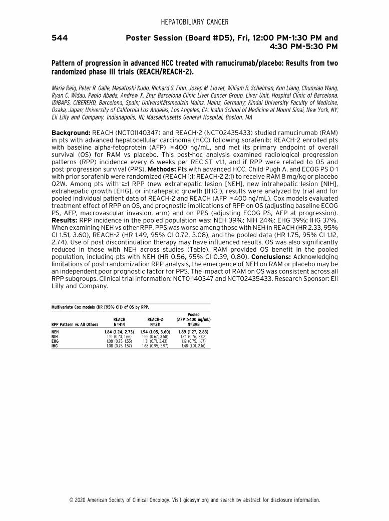
544 Poster Session (Board #D5), Fri, 12:00 PM-1:30 PM and4:30 PM-5:30 PM
Pattern of progression in advanced HCC treated with ramucirumab/placebo: Results from tworandomized phase III trials (REACH/REACH-2).
Maria Reig, Peter R. Galle, Masatoshi Kudo, Richard S. Finn, Josep M. Llovet, William R. Schelman, Kun Liang, Chunxiao Wang,Ryan C. Widau, Paolo Abada, Andrew X. Zhu; Barcelona Clinic Liver Cancer Group, Liver Unit, Hospital Clınic of Barcelona,IDIBAPS, CIBEREHD, Barcelona, Spain; Universitatsmedizin Mainz, Mainz, Germany; Kindai University Faculty of Medicine,Osaka, Japan; University of California Los Angeles, Los Angeles, CA; Icahn School of Medicine at Mount Sinai, New York, NY;Eli Lilly and Company, Indianapolis, IN; Massachusetts General Hospital, Boston, MA
Background: REACH (NCT01140347) and REACH-2 (NCT02435433) studied ramucirumab (RAM)in pts with advanced hepatocellular carcinoma (HCC) following sorafenib; REACH-2 enrolled ptswith baseline alpha-fetoprotein (AFP) $400 ng/mL, and met its primary endpoint of overallsurvival (OS) for RAM vs placebo. This post-hoc analysis examined radiological progressionpatterns (RPP) incidence every 6 weeks per RECIST v1.1, and if RPP were related to OS andpost-progression survival (PPS).Methods: Pts with advanced HCC, Child-Pugh A, and ECOG PS 0-1with prior sorafenib were randomized (REACH 1:1; REACH-2 2:1) to receive RAM8mg/kg or placeboQ2W. Among pts with $1 RPP (new extrahepatic lesion [NEH], new intrahepatic lesion [NIH],extrahepatic growth [EHG], or intrahepatic growth [IHG]), results were analyzed by trial and forpooled individual patient data of REACH-2 and REACH (AFP$400 ng/mL). Cox models evaluatedtreatment effect of RPP on OS, and prognostic implications of RPP on OS (adjusting baseline ECOGPS, AFP, macrovascular invasion, arm) and on PPS (adjusting ECOG PS, AFP at progression).Results: RPP incidence in the pooled population was: NEH 39%; NIH 24%; EHG 39%; IHG 37%.When examiningNEH vs other RPP, PPSwasworse among thosewith NEH inREACH (HR2.33, 95%CI 1.51, 3.60), REACH-2 (HR 1.49, 95% CI 0.72, 3.08), and the pooled data (HR 1.75, 95% CI 1.12,2.74). Use of post-discontinuation therapy may have influenced results. OS was also significantlyreduced in those with NEH across studies (Table). RAM provided OS benefit in the pooledpopulation, including pts with NEH (HR 0.56, 95% CI 0.39, 0.80). Conclusions: Acknowledginglimitations of post-randomization RPP analysis, the emergence of NEH on RAM or placebo may bean independent poor prognostic factor for PPS. The impact of RAM on OS was consistent across allRPP subgroups. Clinical trial information: NCT01140347 and NCT02435433. Research Sponsor: EliLilly and Company.
Multivariate Cox models (HR [95% CI]) of OS by RPP.
RPP Pattern vs All OthersREACH REACH-2
Pooled(AFP ‡400 ng/mL)
N=414 N=211 N=398
NEH 1.84 (1.24, 2.73) 1.94 (1.05, 3.60) 1.89 (1.27, 2.83)NIH 1.10 (0.73, 1.66) 1.55 (0.67, 3.58) 1.24 (0.76, 2.02)EHG 1.08 (0.75, 1.55) 1.31 (0.71, 2.43) 1.12 (0.75, 1.67)IHG 1.08 (0.75, 1.57) 1.68 (0.95, 2.97) 1.48 (1.01, 2.16)
© 2020 American Society of Clinical Oncology. Visit gicasym.org and search by abstract for disclosure information.
HEPATOBILIARY CANCER
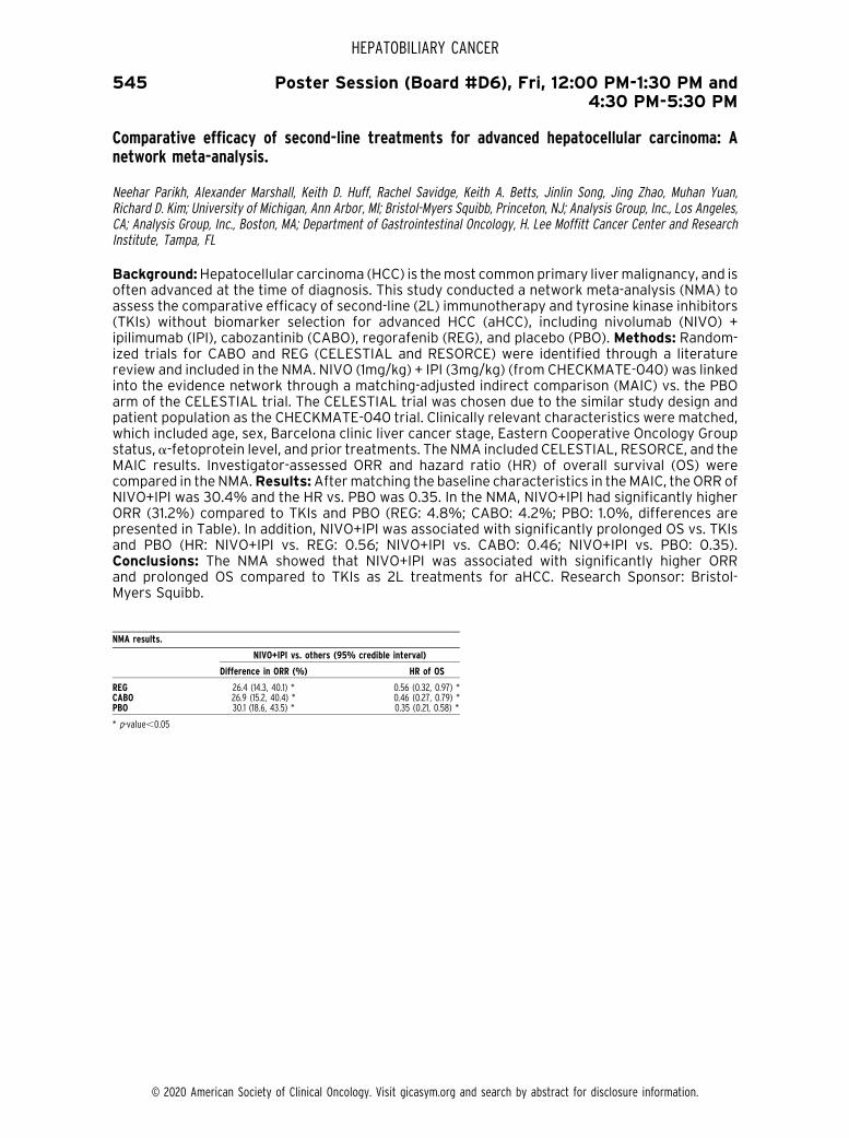
545 Poster Session (Board #D6), Fri, 12:00 PM-1:30 PM and4:30 PM-5:30 PM
Comparative efficacy of second-line treatments for advanced hepatocellular carcinoma: Anetwork meta-analysis.
Neehar Parikh, Alexander Marshall, Keith D. Huff, Rachel Savidge, Keith A. Betts, Jinlin Song, Jing Zhao, Muhan Yuan,Richard D. Kim; University of Michigan, Ann Arbor, MI; Bristol-Myers Squibb, Princeton, NJ; Analysis Group, Inc., Los Angeles,CA; Analysis Group, Inc., Boston, MA; Department of Gastrointestinal Oncology, H. Lee Moffitt Cancer Center and ResearchInstitute, Tampa, FL
Background:Hepatocellular carcinoma (HCC) is themost common primary livermalignancy, and isoften advanced at the time of diagnosis. This study conducted a network meta-analysis (NMA) toassess the comparative efficacy of second-line (2L) immunotherapy and tyrosine kinase inhibitors(TKIs) without biomarker selection for advanced HCC (aHCC), including nivolumab (NIVO) +ipilimumab (IPI), cabozantinib (CABO), regorafenib (REG), and placebo (PBO). Methods: Random-ized trials for CABO and REG (CELESTIAL and RESORCE) were identified through a literaturereview and included in the NMA. NIVO (1mg/kg) + IPI (3mg/kg) (from CHECKMATE-040) was linkedinto the evidence network through a matching-adjusted indirect comparison (MAIC) vs. the PBOarm of the CELESTIAL trial. The CELESTIAL trial was chosen due to the similar study design andpatient population as the CHECKMATE-040 trial. Clinically relevant characteristics were matched,which included age, sex, Barcelona clinic liver cancer stage, Eastern Cooperative Oncology Groupstatus, a-fetoprotein level, and prior treatments. The NMA included CELESTIAL, RESORCE, and theMAIC results. Investigator-assessed ORR and hazard ratio (HR) of overall survival (OS) werecompared in the NMA.Results:Aftermatching the baseline characteristics in theMAIC, the ORR ofNIVO+IPI was 30.4% and the HR vs. PBO was 0.35. In the NMA, NIVO+IPI had significantly higherORR (31.2%) compared to TKIs and PBO (REG: 4.8%; CABO: 4.2%; PBO: 1.0%, differences arepresented in Table). In addition, NIVO+IPI was associated with significantly prolonged OS vs. TKIsand PBO (HR: NIVO+IPI vs. REG: 0.56; NIVO+IPI vs. CABO: 0.46; NIVO+IPI vs. PBO: 0.35).Conclusions: The NMA showed that NIVO+IPI was associated with significantly higher ORRand prolonged OS compared to TKIs as 2L treatments for aHCC. Research Sponsor: Bristol-Myers Squibb.
NMA results.
NIVO+IPI vs. others (95% credible interval)
Difference in ORR (%) HR of OS
REG 26.4 (14.3, 40.1) * 0.56 (0.32, 0.97) *CABO 26.9 (15.2, 40.4) * 0.46 (0.27, 0.79) *PBO 30.1 (18.6, 43.5) * 0.35 (0.21, 0.58) *
* p-value,0.05
© 2020 American Society of Clinical Oncology. Visit gicasym.org and search by abstract for disclosure information.
HEPATOBILIARY CANCER

546 Poster Session (Board #D7), Fri, 12:00 PM-1:30 PM and4:30 PM-5:30 PM
Hepatic and hematological toxicity associated with selective internal radiation therapy (SIRT):A meta-analysis of randomized clinical trials.
Ashish Manne, Michael Behring, Upender Manne, Rojymon Jacob, Ravi Kumar Paluri; Medical Oncology, Mitchell CancerInstitute, The University of South Alabama, Mobile, AL; University of Alabama at Birmingham, Birmingham, AL
Background: For advanced hepatocellular cancers (HCCs) and colorectal cancers (CRCs) with livermetastasis, the impact of selective internal radiation therapy(SIRT) with yttrium-90 on survivaloutcomes is not established. A meta-analysis of randomized clinical trials (RCT) was performed todetermine the relative risk (RR) of hepatic and hematological toxicities with the use of SIRT,compared to therapies not including SIRT. For patients with advanced HCC or CRC, we assessed theRR of high grade (grades 3 and 4) hyperbilirubinemia, fatigue, leucopenia, thrombocytopenia andelevated liver enzymes (AST and ALT) with use of SIRT.Methods: Citations from PubMed/Medline,clinical trials.gov, package inserts, and abstracts frommajor conferences were reviewed to includeRCTs comparing arms with or without SIRT. Potential publication bias was assessed using theEgger test for funnel plot asymmetry. There was no publication bias and the trials were of highquality per Jadad scoring. Patients in control arms received trans-arterial chemo-embolization(TACE) or sorafenib for HCC or FOLFOX for CRC. The proportion and 95% confidence intervals(CIs) for patients with adverse events were derived for each arm of each study and used tocalculate the RR. For themeta-analysis, both the fixed-effectsmodel and the random-effectsmodelwere considered; the method proposed by DerSimonian and Laird was used to estimate therandom-effects model.Results: The RR of grade 3/4 leucopenia was consistently high across highwith the use of SIRT across all the studies (RR 2.05, 95% CI (1.22-3.42), p-value 0.027). The risk ofhepatic dysfunction and fatigue was higher with SIRT but not statistically significant. Conclusions:Since SIRT is associated with increased risk of high-grade leukopenia, caution is advised inselecting patients with HCC’s with underlying decompensated cirrhosis or with CRCs and oncytotoxic therapy. Proper selection of patients would reduce toxicities from SIRT alone or SIRT incombination with systemic chemotherapy. Research Sponsor: None.
© 2020 American Society of Clinical Oncology. Visit gicasym.org and search by abstract for disclosure information.
HEPATOBILIARY CANCER

547 Poster Session (Board #D8), Fri, 12:00 PM-1:30 PM and4:30 PM-5:30 PM
A pilot study of durvalumab/tremelimumab (durva/treme) and radiation (XRT) for metastaticbiliary tract cancer (mBTC): Preliminary safety and efficacy.
Theodore S. Hong, Lipika Goyal, Aparna Raj Parikh, Beow Y. Yeap, Christine A. Ulysse, Lorraine C. Drapek, Jill N. Allen,Jeffrey William Clark, Benjamin Christopher, Christine Bolton, David P. Ryan, Ryan Bruce Corcoran, Jeffrey A. Meyerhardt,Jennifer Yon-Li Wo, Andrew X. Zhu; Department of Radiation Oncology, Massachusetts General Hospital, Boston, MA;Massachusetts General Hospital, Boston, MA; University of California San Francisco, San Francisco, CA; Hematology/Oncology, Massachusetts General Hospital, Boston, MA; Dana-Farber Cancer Institute/Partners Cancer Care, Boston, MA
Background: Metastatic biliary tract cancer (mBTC) is a lethal malignancy with median 5 year OSof less than 10%. Immunotherapy, particularly single agent anti-PD-1/PD-L1, has limited efficacy inmBTCwith ORR~9-15%. Recently presented data shows responses inmetastatic MSS pancreatic orcolon cancer with combination anti-PD-1/CTLA-4 and radiation (XRT) to produce systemic re-sponse (abscopal effect) (Parikh A, GI ASCO 2019, ASCO 2019.). We evaluate safety and efficacy ofdual PD-1/CTLA-4 inhibition with XRT in MSS mBTC. Methods: 15 of a planned 15 mBTC patientswere enrolled. Eligible pts had histologically-confirmed mBTC, ECOG-PS 0/1, and must haveprogressed on at least one line of previous therapy or refused standard therapy. Safety cohortof 6 pts of durva 1500 mg/treme 75 mg q4w was enrolled. If . 2 DLTs, patients were enrolledsubsequently to dose level -1 (durva 1125 mg/ treme 75 mg q4w). 3 fractions of 8 Gy of radiation atC2D1 every other day to a single metastatic site. Durva/treme continued for 4 cycles, followed by 4cycles of maintenance durva until progressive disease, discontinuation or withdrawal. Endpointsinclude disease control rate (DCR (SD+PR+CR)), PFS and OS and safety. Radiological evaluationswere done q2 mo. Results: 15 mBTC pts enrolled and evaluable from May 2018 to March 2019.Median age 63 years (range 48-75), 47% male. DLTs occurred in 3 patients during the safety run-in. One patient experienced DLT at dose level -1 and subsequent expansion. 3 patients did NOTreach radiation therapy. DCR was 27%with a 13% PR and 7% CR. Of those who reached radiation,DCRwas 33%with a 17%PRand 8%CR. At time of analysis,median PFSwas 54 days for ITTmBTC.Duration of response for 4 patients with DCR was 26, 52, 122, 254+ days. Treatment-relatedadverse events were reported in 12/15 patients (80%). Grade $3 toxicities were seen in 9/15 pts(60%) with lymphopenia (5 grade 3) and elevated LFTs (2 grade 4 and 4 grade 2) being the mainadverse events. All patients with disease control were not MSI. Conclusions: Combination ofdurva/treme XRT is feasible and shows preliminary activity inmetastatic BTC. An expansion cohortis being planned to confirm activity. Clinical trial information: NCT03482102. Research Sponsor:Astra Zeneca.
© 2020 American Society of Clinical Oncology. Visit gicasym.org and search by abstract for disclosure information.
HEPATOBILIARY CANCER

548 Poster Session (Board #D9), Fri, 12:00 PM-1:30 PM and4:30 PM-5:30 PM
The efficacy and safety of lenvatinib in patients with intermediate-stage hepatocellularcarcinoma: A nationwide multicenter study in Japan.
Kaoru Tsuchiya, Masayuki Kurosaki, Azusa Sakamoto, Hiroyuki Marusawa, Chikara Ogawa, Masahiko Kondo, Kouji Joko,Keiji Tsuji, Hiroyuki Kimura, Shuichi Wada, Haruhiko Kobashi, Yasushi Uchida, Koichiro Furuta, Takehiro Akahane,Atsunori Kusakabe, Hideo Yoshida, Tomomichi Matsushita, Takehiko Abe, Tetsuro Sohda, Namiki Izumi; Department ofInternal Medicine III, Division of Gastroenterology and Hepatology, Medical University of Vienna, Vienna, Austria; MusashinoRed Cross Hospital, Tokyo, Japan; Osaka Red Cross Hospital, Osaka, Japan; Takamatsu Red Cross Hospital, Takamatsu,Japan; Japanese Red Cross Otsu Hospital, Otsu, Japan; Matsuyama Red Cross Hospital, Mastuyama, Japan; Hiroshima RedCross Hospital & Atomic-Bomb Survivors Hospital, Hiroshima, Japan; Japanese Red Cross Kyoto Daiichi Hospital, Kyoto,Japan; Nagano Red Cross Hospital, Nagano, Japan; Okayama Red Cross Hospital, Okayama, Japan; Matsue Red CrossHospital, Matsue, Japan; Masuda Red Cross Hospital, Masuda, Japan; Ishinomaki Red Cross Hospital, Ishinomaki, Japan;Japanese Red Cross Nagoya Daini Hospital, Nagoya, Japan; Japanese Red Cross Medical Center, Tokyo, Japan; Gifu Red CrossHospital, Gifu, Japan; Maebashi Red Cross Hospital, Maebashi, Japan; Fukuoka Red Cross Hospital, Fukuoka, Japan
Background: Lenvatinib (LEN) has been used in patients with unresectable hepatocellular car-cinoma (u-HCC) since Mar 2018 in Japan. We conducted a nationwide multicenter study andespecially focused on the efficacy and safety in the patients with intermediate-stage u-HCC.Methods: A total of 240 patients received LEN from March 2018 at 15 sites in Japan was enrolled.Tumour assessments in accordance with modified RECIST were done using dynamic CT or MRIwithin 4-8 weeks and every 6-8 weeks thereafter. Results: In this study, 88 of 240 (36.7%)patients were BCLC stage B. Among them 76 (86.3%) patients received TACE before LEN and themedian number of TACE was 2 (1-10). Only 4 patients were TKI experienced and other 84 (95.5%)patients received LEN as a 1st line therapy. The median pretreatment ALBI score was -2.35 and 75(85.2%) patients were Child-Pugh A. In this cohort, 73 (83.0%) patients were beyond up-to-sevencriteria and themedian pretreatment AFPwas 38.2 (2-12870) ng/mL. Themedian observation timewas 8.5months and 16 patients died. Themedian progression free survival was 8.7months, and themedian overall survival (OS) was not reached. Objective response rate (ORR) and disease controlrate (DCR) were 48.5% and 80.3%. AFP decrease ( . 20%) after 1 month was observed in 52(59.0%) patients. Child-Pugh B patients (n = 13) had significantly shorter OS than Child-Pugh A (p =0.02) and median OS in Child-Pugh B patients was 8.8 months. The patients received . 6 timesTACE before LEN had significantly shorter OS than patients received # 6 times TACE (p = 0.02).Additional TACE was performed in 8 patients and The median time of restarting LEN was 19 days.ThemedianALBI score before additional TACE, Day 1 after TACE andDay 28 after TACEwere -2.38,-2.07, and -2.36.There was no severe adverse event associated with additional TACE. The medianduration of LEN in patients treated with LEN and additional TACE was 8.5 months. Conclusions:The ORR and DCR of LEN in Child-Pugh A patients with intermediate-stage HCC were 46.6% and79.3%. The therapeutic strategies for intermediate-stage HCC should be discussed based on theliver function, tumor states, and treatment course about TACE. Research Sponsor: None.
© 2020 American Society of Clinical Oncology. Visit gicasym.org and search by abstract for disclosure information.
HEPATOBILIARY CANCER
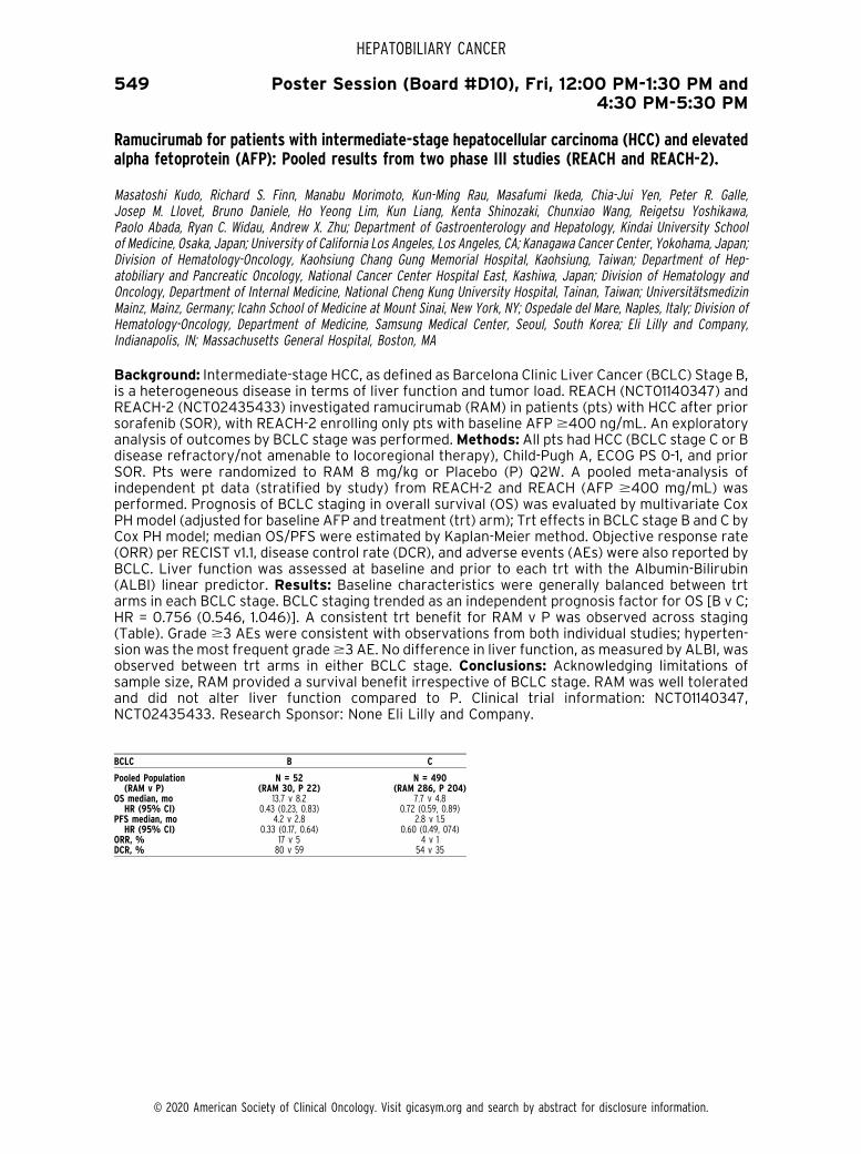
549 Poster Session (Board #D10), Fri, 12:00 PM-1:30 PM and4:30 PM-5:30 PM
Ramucirumab for patients with intermediate-stage hepatocellular carcinoma (HCC) and elevatedalpha fetoprotein (AFP): Pooled results from two phase III studies (REACH and REACH-2).
Masatoshi Kudo, Richard S. Finn, Manabu Morimoto, Kun-Ming Rau, Masafumi Ikeda, Chia-Jui Yen, Peter R. Galle,Josep M. Llovet, Bruno Daniele, Ho Yeong Lim, Kun Liang, Kenta Shinozaki, Chunxiao Wang, Reigetsu Yoshikawa,Paolo Abada, Ryan C. Widau, Andrew X. Zhu; Department of Gastroenterology and Hepatology, Kindai University Schoolof Medicine, Osaka, Japan; University of California Los Angeles, Los Angeles, CA; Kanagawa Cancer Center, Yokohama, Japan;Division of Hematology-Oncology, Kaohsiung Chang Gung Memorial Hospital, Kaohsiung, Taiwan; Department of Hep-atobiliary and Pancreatic Oncology, National Cancer Center Hospital East, Kashiwa, Japan; Division of Hematology andOncology, Department of Internal Medicine, National Cheng Kung University Hospital, Tainan, Taiwan; UniversitatsmedizinMainz, Mainz, Germany; Icahn School of Medicine at Mount Sinai, New York, NY; Ospedale del Mare, Naples, Italy; Division ofHematology-Oncology, Department of Medicine, Samsung Medical Center, Seoul, South Korea; Eli Lilly and Company,Indianapolis, IN; Massachusetts General Hospital, Boston, MA
Background: Intermediate-stage HCC, as defined as Barcelona Clinic Liver Cancer (BCLC) Stage B,is a heterogeneous disease in terms of liver function and tumor load. REACH (NCT01140347) andREACH-2 (NCT02435433) investigated ramucirumab (RAM) in patients (pts) with HCC after priorsorafenib (SOR), with REACH-2 enrolling only pts with baseline AFP$400 ng/mL. An exploratoryanalysis of outcomes by BCLC stage was performed.Methods: All pts had HCC (BCLC stage C or Bdisease refractory/not amenable to locoregional therapy), Child-Pugh A, ECOG PS 0-1, and priorSOR. Pts were randomized to RAM 8 mg/kg or Placebo (P) Q2W. A pooled meta-analysis ofindependent pt data (stratified by study) from REACH-2 and REACH (AFP $400 mg/mL) wasperformed. Prognosis of BCLC staging in overall survival (OS) was evaluated by multivariate CoxPHmodel (adjusted for baseline AFP and treatment (trt) arm); Trt effects in BCLC stage B and C byCox PH model; median OS/PFS were estimated by Kaplan-Meier method. Objective response rate(ORR) per RECIST v1.1, disease control rate (DCR), and adverse events (AEs) were also reported byBCLC. Liver function was assessed at baseline and prior to each trt with the Albumin-Bilirubin(ALBI) linear predictor. Results: Baseline characteristics were generally balanced between trtarms in each BCLC stage. BCLC staging trended as an independent prognosis factor for OS [B v C;HR = 0.756 (0.546, 1.046)]. A consistent trt benefit for RAM v P was observed across staging(Table). Grade $3 AEs were consistent with observations from both individual studies; hyperten-sion was the most frequent grade$3 AE. No difference in liver function, as measured by ALBI, wasobserved between trt arms in either BCLC stage. Conclusions: Acknowledging limitations ofsample size, RAM provided a survival benefit irrespective of BCLC stage. RAM was well toleratedand did not alter liver function compared to P. Clinical trial information: NCT01140347,NCT02435433. Research Sponsor: None Eli Lilly and Company.
BCLC B C
Pooled Population(RAM v P)
N = 52(RAM 30, P 22)
N = 490(RAM 286, P 204)
OS median, mo 13.7 v 8.2 7.7 v 4.8HR (95% CI) 0.43 (0.23, 0.83) 0.72 (0.59, 0.89)
PFS median, mo 4.2 v 2.8 2.8 v 1.5HR (95% CI) 0.33 (0.17, 0.64) 0.60 (0.49, 074)
ORR, % 17 v 5 4 v 1DCR, % 80 v 59 54 v 35
© 2020 American Society of Clinical Oncology. Visit gicasym.org and search by abstract for disclosure information.
HEPATOBILIARY CANCER
550 Poster Session (Board #D11), Fri, 12:00 PM-1:30 PM and4:30 PM-5:30 PM
Hyperprogressive disease during PD-1 blockade in patients with advanced hepatocellularcarcinoma.
Hongjae Chon, Chang Gon Kim, Sangeun Yoon, Chan Kim, Beodeul Kang, Hye Jin Choi, Ho Yeong Lim; CHA Hospital,Seongnam, South Korea; Graduate School of Medical Science and Engineering, KAIST, Daejon, South Korea; Samsung MedicalCenter, Sungkyunkwan University School of Medicine, Seoul, South Korea; CHA Bundang Medical Center, Seongnam, SouthKorea; Medical Oncology, Bundang Medical Center, CHA University, Seongnam, South Korea; Yonsei Cancer Center, Seoul,South Korea; Division of Hematology-Oncology, Department of Medicine, Samsung Medical Center, Seoul, South Korea
Background: Immune checkpoint blockade with PD-1 inhibitors has shown promising clinicalefficacy in patients with hepatocellular carcinoma (HCC). However, emerging evidences showthat PD-1 blockade can sometimes lead to a flare-up of tumor growth with rapid clinical deteri-oration (hyperprogressive disease, HPD). This study aimed to evaluate the incidence and pattern ofHPD in amulticenter, real-world cohort of East Asian patients with advanced HCC treated with PD-1blockade. Methods: We enrolled 148 advanced HCC patients treated with nivolumab betweenMarch 2016 and December 2018 in Korea. Clinicopathologic variables, tumor growth dynamic, andtreatment outcomes were comprehensively analyzed. Progressive disease was assessed usingtumor growth kinetics (TGK), tumor growth rate (TGR), and time to treatment failure (TTF), andpatient with HPD were defined as those who met the criteria of progressive disease by both TGKand TGR. Results: In this large cohort of HCC patients, the median age was 60 years and themajority were male (85%) and HBV-infected (72%). The objective response rate was 17.6%including two complete responders (1.4%). Ongoing responses were seen in 46% of responders atdata cut-off. The incidence of HPD after PD-1 blockade was 23%. HCC patients with HPD had dismalprognosis with worse progression-free survival (PFS) and overall survival (OS) (HR, 1.947; 95% CI,1.226-3.093 and HR, 1.839; 95% CI, 1.108-3.055, respectively) than progressive disease withoutHPD. Among various baseline clinicopatholgic parameters, elevated neutrophil-to-lymphocyteratio (NLR) was only significantly associated with HPD. The optimal cut-off value of NLR for HPDpredictionwas 3.74 determined byROC curves, andNLR. 3.74was associatedwithworse PFS andOS. Conclusions: The real-world efficacy of PD-1 blockade in HCC patients was consistent withprevious studies. However, there was also a corresponding risk of HPD as well as a clinical benefit.Therefore, careful patient selection using immunologic biomarkers, such as NLR, could enhancethe therapeutic benefit of PD-1 blockade in clinical trials and real-world practice of HCC.
© 2020 American Society of Clinical Oncology. Visit gicasym.org and search by abstract for disclosure information.
HEPATOBILIARY CANCER

551 Poster Session (Board #D12), Fri, 12:00 PM-1:30 PM and4:30 PM-5:30 PM
In vitro and in vivo efficacy and safety of tumor treating fields (TTFields) and sorafenibcombination in hepatocellular carcinoma.
Shiri Davidi, Catherine Tempel-Brami, Mijal Munster, Karnit Gotlib, Einav Zeevi, Rosa S Schneiderman, Tali Voloshin,Moshe Giladi, Adrian Kinzel, Eilon D. Kirson, Uri Weinberg, Yoram Palti; Novocure Ltd, Haifa, Israel; Novocure GmbH, Munich,Germany; Novocure Ltd, Haifa, NY, Israel
Background:Hepatocellular carcinoma (HCC) is a leading global cause of cancer-relatedmortality.Sorafenib (oral multikinase inhibitor) is approved in patients with advanced HCC, yet survivalbenefit is limited. Tumor Treating Fields (TTFields) are an effective, anti-neoplastic treatmentmodality delivered via noninvasive, low intensity (1-3 V/cm), intermediate frequency (100-500kHz), alternating electric fields. The study aim was to explore in vitro and in vivo effects of TTFieldsalone and combined with sorafenib for HCC treatment.Methods: HCC (HepG2 and Huh-7D12) cellswere TTFields treated with at frequencies of 100-400 kHz for 72 hr using the inovitro system.Efficacy of TTFields and sorafenib combined treatment was tested at optimal frequency withvarious sorafenib concentrations. Cell counts, apoptosis induction, and clonogenic potential weredetermined. Healthy rats were used to assess safety of TTFields applied to the abdomen. N1S1 HCCcells were injected into the left hepatic lobe of Sprague Dawley rat; after 1 week, TTFields (1.2 V/cm)and sorafenib (10mg/kg) were applied for 6 days. Tumor growthwas evaluated usingMRI.Results:The optimal TTFields frequency was 150 kHz in HepG2 and Huh-7D12 HCC cells. TTFields 150 kHztreatment (1.0 - 1.7 V/cm, 72 hr) led to cell count reductions (53-55%) and further decreases inclonogenic potential (65-69%). TTFields and sorafenib combination treatment led to a significantreduction in cell count (2-wayANOVA,P,0.05) vs either treatment alone. Also, tumor growthwassignificantly reduced in the combined treatment group vs the control group (student t test, P ,0.01). Tumor volume (fold increase) in the combination treatment group (1.6) was significantlylower vs control (5.9,P,0.0001), TTFields alone (3.3,P,0.01), and sorafenib alone (2.3,P,0.05)groups. Safety studies did not reveal any TTFields related adverse events with delivery to the ratabdomen. Conclusions: In vitro and in vivo data demonstrated efficacy and safety of TTFields inHCC; and improved efficacy in combination with sorafenib. A phase 2 study (HEPANOVA;NCT03606590) will explore the clinical potential of TTFields 150 kHz plus sorafenib. ResearchSponsor: Novocure.
© 2020 American Society of Clinical Oncology. Visit gicasym.org and search by abstract for disclosure information.
HEPATOBILIARY CANCER
552 Poster Session (Board #D13), Fri, 12:00 PM-1:30 PM and4:30 PM-5:30 PM
Role of precision medicine for patients with advanced biliary tract cancers.
Antoine Hollebecque, Oumrou Nabi, Michel Ducreux, Valerie Boige, Jean-Yves Scoazec, Etienne Rouleau, Alice Boileve,Cristina Smolenschi, Loic Verlingue, Margarida Matias, Pascal Burtin, Caroline Prieux-Klotz, Christophe Massard, David Malka;Gustave Roussy Cancer Campus, Villejuif, France; Gustave Roussy, Villejuif, France; Digestive Oncology, Gustave Roussy,Villejuif, France; Department of Biopathology, Gustave Roussy Cancer Campus, Villejuif, France; Institut Gustave Roussy,Villejuif, France; Gustave Roussy, Universite Paris-Saclay, Villejuif, France; Drug Development Unit, Institut Gustave RoussyCancer Centre, Villejuif, France; Gustave Roussy, Universite Paris-Saclay, Departement de Medecine Oncologique, Villejuif,France
Background: Biliary tract cancers (BTC) are rare and heterogeneous cancers with poor prognosis.Several actionable genomic targets have been described in BTC but data on the efficacy oftargeted therapies remain limited. The main objectives of this retrospective study was to evaluatethe frequency of actionable genomic alterations among BTC and the impact of targeted therapy.Methods: We performed a retrospective analysis on BTC patients seen at Gustave Roussy (IGR)from Dec 2011 to Jul 2019. All clinical and genomic reports were reviewed. Results: The studypopulation included 212 patients with the main following characteristics: median age 61 years,female 51%, intrahepatic cholangiocarcinoma 57%, median of 2 previous lines. Of 212 BTCpatients, 170 patients had a genomic profile based on archival tissue or a new tumor biopsy(IGR panel n = 120; Foundation One panel n = 92 ). 124 patients (73%) had at least one genomicalteration and 68 (40%) patients had genomic alteration considered as actionable. The mostcommon actionable targets were FGFR2 rearrangement/mutation (n = 24, 35.3%), HER2/3mutations (n = 9, 13.2%) and IDH1/2 mutations (n = 7, 10.3%). Of those 68 patients, 58 receivedthe matched targeted therapy: FGFR inhibitor n = 24, HER2/3 inhibitors n = 9, Akt/PIK3CA/mTORinhibitors n = 7, IDH1 inhibitor n = 6. In the treated population, the objective response rate was36.2% and the disease control rate 85.1%. Progression-free survival (PFS) was 6.2 monthscompared to 2.8 months (p = 0.02) for patients who did not received targeted treatment. Overallsurvival (OS) was 17.7 months compared to 11.0 months (p = 0.03) for patients who did not receivedtargeted treatment. Conclusions: Actionable genomic targets are frequent among BTC. Profiling-directed therapies resulted in a 36% response rate, a 85% tumor control rate and a 6.2 monthsPFS which compare favorably to second-line chemotherapy. A randomized trial is required toconfirm the benefit of precision medicine in BTC. Research Sponsor: None.
© 2020 American Society of Clinical Oncology. Visit gicasym.org and search by abstract for disclosure information.
HEPATOBILIARY CANCER
553 Poster Session (Board #D14), Fri, 12:00 PM-1:30 PM and4:30 PM-5:30 PM
The impact of inflammatory biomarkers, BMI, and sarcopenia on survival in advanced hepatocellularcarcinoma treated with immunotherapy.
Mehmet Akce, Yuan Liu, Katerina Mary Zakka, Dylan J. Martini, Amber Draper, Olatunji B. Alese, Walid Labib Shaib,Christina Wu, Joel Wedd, Marty T. Sellers, Mehmet Asim Bilen, Bassel F. El-Rayes; Winship Cancer Institute, Atlanta, GA;Departments of Biostatistics and Bioinformatics, Emory University, Atlanta, GA; Winship Cancer Institute of EmoryUniversity, Atlanta, GA; Department of Hematology and Medical Oncology, Emory University School of Medicine, Atlanta, GA;Ohio State University Arthur G. James Cancer Hospital and Richard J. Solove Research Institute, Columbus, OH; EmoryUniversity School of Medicine, Atlanta, GA; Winship Cancer Institute, Emory University, Atlanta, GA
Background: Sarcopenia and inflammation are independently associated with worse survival incancer patients. This study aims to determine the impact of inflammatory biomarkers, BMI andsarcopenia on survival in advanced hepatocellular carcinoma (HCC) patients treated with immu-notherapy.Methods:We performed a retrospective review of advanced HCC patients treated withimmunotherapy-based therapies at Winship Cancer Institute between 2015 and 2019. Baselinecomputed tomography and magnetic resonance imaging scans were collected at mid-L3 level,assessed for skeletal muscle density using SliceOmatic (TomoVision, version 5.0) and converted toskeletal muscle index (SMI) by dividing it by height (m)2. Gender-specific sarcopenia was defined bymedian value of SMI. The optimal cut for continuous inflammation biomarker was determined bybias-adjusted log-rank test. Overall Survival (OS) was set as primary outcome and Cox proportionalhazard model was performed. Results: 57 patients were included; 77.2% male, 52.6% Caucasian,58.5% ECOG PS 0-1, 80.7% Child Pugh A. Treatment was second line and beyond in 71.9%. Themedian follow-up time was 6 months. Sarcopenia cut-off for males and females was SMI of 43 and39, respectively. 49.1% of patients had sarcopenia. Median OS was 5 vs. 14.3 months in sarcopenicvs. non-sarcopenic patients (p=0.054). Median OS was 5 and 17.5 months in patients with BMI,25and BMI $25 respectively (p=0.034). Median OS was 3.6 and 14.3 months for patients withneutrophil to lymphocyte ratio (NLR) $ 5.15 vs. NLR , 5.15 (p,0.001). In multivariable Coxregression model, higher baseline NLR was associated with worse OS (HR: 4.17, 1.52-11.39,p=0.005). Gender specific sarcopenia showed a trend of worse OS (HR: 1.71, 0.73-4.00,p=0.215) but was not statistically significant. BMI,25 was associated with worse OS (HR: 2.73,1.15-6.53, p=0.023). In the association with PFS, neither baseline BMI nor gender specific sarco-penia showed statistical significance. Conclusions: Baseline BMI and NLR may predict OS afterimmunotherapy treatment. After controlling for baseline Child Pugh Score and NLR, genderspecific sarcopenia was not associated with OS significantly. Research Sponsor: None.
© 2020 American Society of Clinical Oncology. Visit gicasym.org and search by abstract for disclosure information.
HEPATOBILIARY CANCER
554 Poster Session (Board #D15), Fri, 12:00 PM-1:30 PM and4:30 PM-5:30 PM and Poster Walks, Fri, 4:45 PM-5:30 PM
First-in-human phase I trial of small activating RNA (saRNA) oligonucleotide MTL-CEBPA incombination with sorafenib in patients with advanced hepatocellular carcinoma (HCC).
Debashis Sarker, Mikael Sodergren, Elizabeth Ruth Plummer, Bristi Basu, Tim Meyer, Kai-Wen Huang, T.R. Jeffry Evans,Duncan Spalding, Yuk Ting Ma, Daniel H. Palmer, Cheng Ean Chee, David James Pinato, Vikash Reebye, Daniel McVeigh,Nina Raulf, Jenni Vasara, Pinelopi Andrikakou, Robert Habib, David Blakey, Nagy A. Habib; King’s College London, London,United Kingdom; Imperial College London, London, United Kingdom; Northern Centre for Cancer Care, Newcastle-upon-Tyne, United Kingdom; Addenbrooke’s Hospital, Cambridge, United Kingdom; University College London Cancer Institute,London, United Kingdom; National Taiwan University Hospital, Taipei, Taiwan; University of Glasgow, Beatson West ofScotland Cancer Centre, Glasgow, United Kingdom; Department of Hepatobiliary Oncology, New Queen Elizabeth HospitalBirmingham, Birmingham, United Kingdom; University of Liverpool, Liverpool, United Kingdom; National UniversityCancer Institute, Singapore, Singapore; MINA Therapeutics, London, United Kingdom; Imperial College, London, UnitedKingdom; MiNA Therapeutics, London, United Kingdom
Background: MTL-CEBPA is the first saRNA to enter clinical trials and targets the transcriptionfactor C/EBPa, a master regulator of myeloid cell differentiation. We have previously reported afavourable safety profile of MTL-CEBPA given as a single agent QWx3 every 28 days, in patientswith HCC (Sarker D et al, ASCO 2018). After discontinuation of MTL-CEBPA, three out of fivepatients treated with sorafenib off study have hadmaintained complete radiological response (CR)of 7-18 months duration; 2 patients demonstrated resolution of lung metastases. 1 year. Here weprovide updated data on phase 1 patients treated with sorafenib off study as well as subsequentcombination cohorts. Methods: MTL-CEBPA 130mg/m2 QWx3 or BIW and sorafenib 400mg bdwere administered to patients with HCC using combination or sequential dosing regimens, incohorts either tyrosine kinase inhibitor naive or resistant. On treatment liver biopsies evaluatedchanges in M2 macrophages (CD163). Flow cytometry of peripheral blood determined changes inmyeloid cell populations.Results: 12 patients have been treated with MTL-CEBPA co-administeredwith sorafenib and 14 patients treatedwithMTL-CEBPA followed by sorafenib (23M/3F,median age65.5years, range 44-83, ECOG PS 0/1: 18/8). Themost common treatment-related AEs (all grades/grade 3) in this group include facial flushing (4/0), raised AST (3/1) raised ALT (2/1), fatigue (5/0),raised ALP (2/0), and anaemia (2/2), diarrhoea (3/0), rash (2/0) and anorexia (1/0). 1 TKI naıvepatient in the co-administration cohort has maintained CR at 7 months and two patients have SD(ongoing at 3 & 4 months both in sequential cohort). IHC in the patient with CR has demonstrated95% reduction in M2 macrophages with significant decrease in frequency of immature CD10-neutrophils (-85.7%; p = 0.0078), PMN-MDSCs (-49.3%; p = 0.00145) and M-MDSCs (-18.4%; P =0.0072). All responding patients have underlying HBV or HCV.Conclusions:MTL-CEBPA is a novelsaRNA targetingmyeloid cells whichmay result in a significantly enhanced oncological response inHCC of viral aetiology. Updated safety and efficacy data will be presented. Clinical trial information:NCT02716012. Research Sponsor: MINA Therpaeutics.
© 2020 American Society of Clinical Oncology. Visit gicasym.org and search by abstract for disclosure information.
HEPATOBILIARY CANCER

555 Poster Session (Board #D16), Fri, 12:00 PM-1:30 PM and4:30 PM-5:30 PM
Predictors of immunotherapy (IO) response in hepatocellular carcinoma (HCC).
Samantha Ann Armstrong, Bhavana Pendurthi Singh, Monika Kulasekaran, Petra Prins, Aiwu Ruth He; Indiana UniversitySchool of Medicine, Indianapolis, IN; Medstar Georgetown University Hospital, Washington, DC; MedStar Harbor Hospital,Baltimore, MD; Georgetown University Lombardi Comprehensive Cancer Center, Washington, DC
Background: Despite advances in understanding the molecular pathways of HCC, therapeuticoptions are limited and patient survival is dismal. IO is a promising HCC treatment. There arecurrently no indicators to identify which patients (pts) will have a prolonged response.Methods: Inthis single-institutional retrospective analysis, pts received one of five IO containing regimens withnivolumab, pembrolizumab, atezolizumab plus bevacizumab, durvalumab or cemiplimab untildisease progression (PD) or unacceptable toxicity. Relevant factors including: stage, viral etiology,vascular invasion (VI), tumor thrombus (TT), multifocal disease, toxicity grade, steroid use for IOmediated toxicities and derived Neutrophil-to Lymphocyte ratio (dNLR), were correlated to clinicaloutcome: progression free survival (PFS), overall survival (OS), response rate (RR), using Pear-son’s chi-square test or student’s t-test . Responses were assessed using RECIST v 1.1 criteria forstable disease (SD), partial response (PR) and PD were correlated with best response and PFS. OSwas calculated by the Kaplan-Meier method. Results: Cohort demographics (n = 76) were: 72%male; 38% African American, 30% Caucasian and 16% Asian; 29% of pts had HBV, 41% had HCV,1% had both HBV/HCV and 13% had no viral etiology (n = 64). The majority of pts were stage III(43%) or IV (38%). At the start of IO, 32% had VI, 32% had TT and 80% had multifocal ormetastatic disease. 65% of pts experienced IO toxicity, with 24.3% at grade 3 or higher, and 34%requiring steroids. Best response to IOwas SD in 65.7%of pts, PR in 25.7% and PD in 8.6%.MedianOS was 13m (95% CI 7.9-18.1) from the start of IO and median PFS (n = 65) was 14m (95% CI 6.8-21.2). Median OS and PFSwere significantly improved in pts with PR compared to PD (45 vs 8m, p,0.0005, PFS 15 vs 3m p = 0.007). Both OS and PFS showed benefits for SD of$2months comparedto those with PD (11 vs 8m, p , .0005, PFS 5 vs 3m p = .007). VI, TT, stage, viral etiology, toxicitygrade or dNLR did not correlate with OS, PFS and RR, however need of steroid treatment trendedtoward worse outcome. Conclusions: PR and SD are independent predictors for prolonged PFSand OS in HCC pts receiving IO therapy. Absence of steroid use for toxicity trended towardimproved IO response. Research Sponsor: None.
© 2020 American Society of Clinical Oncology. Visit gicasym.org and search by abstract for disclosure information.
HEPATOBILIARY CANCER
556 Poster Session (Board #D17), Fri, 12:00 PM-1:30 PM and4:30 PM-5:30 PM
Phase II study of copanlisib in combination with gemcitabine and cisplatin in advancedcholangiocarcinoma.
Elaine Tan, Dae Won Kim, Rutika Mehta, Biwei Cao, Jongphil Kim, Angel Meroni, Barbara Centeno, Richard D. Kim; University ofSouth Florida, Tampa, FL; The University of Texas MD Anderson Cancer Center, Houston, TX; Roswell Park Cancer Institute,Buffalo, NY; H. Lee Moffitt Cancer Center & Research Institute, Tampa, FL; Moffitt Cancer Center, Tampa, FL; Department ofGastrointestinal Oncology, H. Lee Moffitt Cancer Center and Research Institute, Tampa, FL
Background: First line therapy for advanced cholangiocarcinoma (CCA) is currently gemcitabineand cisplatin. However, survival rarely exceeds one year with this regimen. PI3K/AKT activationhas been shown to increase resistance to chemotherapy in CCA; therefore, inhibiting this pathwaymay improve chemotherapy’s efficacy. This phase II study evaluated the safety and efficacy ofcopanlisib, a potent and reversible pan-class I PI3K inhibitor, with gemcitabine and cisplatin inadvanced CCA. Methods: Between July 2016 and April 2019, pts with histologically confirmedadvanced/unresectable CCA received cisplatin (25 mg/m2), gemcitabine (1000mg/m2), andcopanlisib 60mg on day 1 and 8, every 21 days as first line treatment. The primary endpointwas PFS at 6 months. Secondary endpoints were RR, median OS and PFS, and safety profile. Asingle-arm Simon’s two-stageminimax design with one-sided 10% type I error and 80% power wasused. Based on ABC-01 and ABC-02 studies, PFS6 for gemcitabine and cisplatin were 57.1% and59.3%, respectively. Therefore, PFS6 of 57% was considered not to warrant further study and$72% to warrant further investigation.Results: Twenty-four pts received at least one dose of thestudy drug (62.5% female, median age 64 years), with 70.8% intrahepatic, 16.7% extrahepatic,and 12.5% gallbladder cancer. For all pts, median OS was 13.9 months (95% CI: 6.8-17.9) andmedian PFS was 6.2 months (95% CI: 1.3-11.1). PFS at 6 and 12 months was 57.0% and 42.2%, and 6and 12-month OS was 73.9% and 53.2%, respectively. Only 19 pts were considered evaluable forRR. Five pts were either lost to follow up, withdrew consent, or died before a second scanwas done.Six pts achieved PR (31.5%) and 11 (57.9%) had SD. Grade 3 or higher adverse events (AE) occurredin 75% of pts. The most common grade 3/4 AEs were decreased neutrophil count (40%) andincreased lipase (20%). Treated related AEs led to drug discontinuation for 3 pts (12.5%) and dosemodification for 7 pts (29.2%). Conclusions: Gemcitabine, cisplatin, and copanlisib in combinationdid notmeet the primary endpoint of 6-month PFS. However, additional correlativework is ongoingto identify a possible biomarker for copanlisib. Clinical trial information: NCT02631590. ResearchSponsor: Bayer AG.
© 2020 American Society of Clinical Oncology. Visit gicasym.org and search by abstract for disclosure information.
HEPATOBILIARY CANCER
557 Poster Session (Board #D18), Fri, 12:00 PM-1:30 PM and4:30 PM-5:30 PM
Clinical characteristics and outcomes of patients with advanced hepatocellular carcinomatreated with immunotherapy: A real-world retrospective study.
Adel Chergui, Eswar Gadde, Seda Serra Tolu, Ana Acuna-Villaorduna, Rafi Kabarriti, Sanjay Goel, Andreas Kaubisch; JacobiMedical Center, Bronx, NY; Montefiore Medical Center, Bronx, NY; Montefiore Medical Center, Albert Einstein College ofMedicine, Bronx, NY
Background: Advanced HCC is an aggressive malignancy with dismal prognosis. Newer agents,including immunotherapy (IT), have been granted accelerated approval. Information outsideclinical trials is scarce. This study is aimed to describe the clinical characteristics and outcomesof HCC patients treated with IT. Methods: Patients with HCC treated with IT were identified usingthe institutional data-mining software, Clinical Looking Glass. Patient demographics, clinical, andtreatment characteristicswere collected. Progression-free survival (PFS) was defined as time fromtreatment initiation to disease progression or death, and overall-survival (OS) as time fromdiagnosis of advanced disease to death. PFS and OS were plotted using Kaplan-Meier curves.Results: A total of 52 patients; median age 64 years; male predominance (38, 73.1%) wereidentified. There were 24 (54.5%) Hispanics, 9 (20.5%) Non-Hispanic Blacks, 7 (15.9%) Non-Hispanic White and 4 (9.1%) Asians. Cirrhosis was seen in 41 (83.7%), and median MELD score was8 (IQ: 7-10). Hepatitis B and C infection were encountered in 12 (24.5%) and 22 (44%) patients,respectively. Imaging evidence of intravascular invasion was seen in 16 (34.8%) and extrahepaticmetastases in 7 (14.9%) cases. Local treatment was provided to 29 (59.2%) and radiationtreatment to 14 (28.6%) patients. Nivolumab was used in all the cases, as first-line treatmentin 17 (32.7%) and as$ second line in 35(67.3%). Themedian PFS was 6.2 (3.1-10.6) months and wassimilar in first-line and$ second line treatment (8 vs 5.9months, p=0.90). Themedian OSwas 24.2(18-28) months; there was a tendency towards higher survival rates in patients that were treated in$ second line (16.8 vs 25.2 months, 0=0.07). Conclusions: In this multiethnic cohort, the “realworld” experience of the benefit of IT in HCC is encouraging, with a median OS exceeding twoyears. Expanded data may elucidate the differences if any, between use of IT as front vs. secondline therapy, in PFS and OS. Research Sponsor: None.
© 2020 American Society of Clinical Oncology. Visit gicasym.org and search by abstract for disclosure information.
HEPATOBILIARY CANCER
558 Poster Session (Board #D19), Fri, 12:00 PM-1:30 PM and4:30 PM-5:30 PM
Intensity-modulated proton therapy using dose-painting pencil beam scanning for high-riskhepatocellular carcinoma.
Stephanie K. Schaub, Stephen R. Bowen, Matthew J. Nyflot, Smith Apisarnthanarax; University of Washington, Seattle, WA;Department of Radiation Oncology, University of Washington, Seattle, WA
Background: High rates of local control are achievable with hypofractionated proton therapy withpassive techniques for hepatocellular carcinoma (HCC), but may have limitations when tumors areadjacent to organs-at-risk (OARs), whichmay result in tumor underdosage and lead to inferior localcontrol. We present the first reported series of HCC patients treated with pencil beam scanning(PBS) intensity-modulated proton therapy (IMPT) using a simultaneous-integrated boost andprotection (SIB/SIP) technique to escalate tumor dose while protecting adjacent OARs.Methods: Twenty-five HCC patients were treated between 2015-2019 with a 15-fraction regimenusing IMPT SIB/SIP. SIB/SIP dose levels generally ranged from 36.0-67.5 GyRBE to minimize doseto OARs at their respective dose-limiting thresholds (e.g. luminal gastrointestinal organs, chestwall). Radiation-induced liver disease (RILD) was defined by a Child-Pugh (CP) score increase of 2or greater and/or any RTOG grade 3 enzyme elevation. Other toxicities were graded byCTCAEv5.0. Overall survival (OS), progression-free survival (PFS), and local control were calcu-lated using the Kaplan-Meier method. Results: Patients most commonly had BCLC stage B or Cdisease (84%) and CP-A (80%) andALBI grade 2 (60%) liver function. Median gross tumor volume(GTV) size and volumewere 12.3 cm (range 2.17-20.57) and 461 cc (range 4.68-2439), and 32%hadgross vascular invasion. Median mean and minimum dose delivered to the gross tumor volume(GTV) was 64.0 GyRBE (EQD2 76.1, BED 91.3, range 54.3-69.6) and 45.1 GyRBE (EQD2 48.9, BED58.7, range 33.4-67.7), respectively. Median mean dose to liver minus GTV was 15.0 GyRBE (range,8.2-19.6). 1-year OS, PFS, and local control were 66%, 32%, and 84%, respectively. No isolatedlocal failures occurred. Two patients experienced RILD with no RILD-related deaths. Two grade 3non-GI toxicities occurred: 1 rib fracture and 1 pneumonitis. No acute or late GI grade$2 occurred.Conclusions: In our series of HCC patients with large tumors near OARs, IMPT SIB/SIP allows fortumor dose escalation while sparing of OARs and results in favorable local control and acceptabletoxicities. Research Sponsor: None.
© 2020 American Society of Clinical Oncology. Visit gicasym.org and search by abstract for disclosure information.
HEPATOBILIARY CANCER
559 Poster Session (Board #D20), Fri, 12:00 PM-1:30 PM and4:30 PM-5:30 PM
Real-world efficacy and safety of immune checkpoint inhibitors in advanced hepatocellularcarcinoma: Experience of a tertiary Asian center.
Kennedy Ng, Lawrence Wen Jun Wong, Su Pin Choo, David Wai-Meng Tai, Sze Huey Tan, Andrea Jing Shi Ang, Joycelyn Jie Xin Lee;National Cancer Centre Singapore, Singapore, Singapore; National University of Singapore, Singapore, Singapore; National CancerCenter Singapore, Curie Oncology, Singapore, Singapore; National Cancer Center Singapore, Singapore, Singapore; Division ofClinical Trials & Epidemiological Sciences, National Cancer Centre Singapore, Singapore, Singapore; Department of Medical Oncology,National Cancer Centre Singapore, Singapore, Singapore
Background: Immune checkpoint inhibitor (ICI) use in advanced hepatocellular carcinoma (HCC) isincreasing. Real-world data on efficacy and safety however is lacking, more so when used inpatients who fall out of standard clinical trial criteria. Methods: We conducted a retrospectivereview of all patients with advanced HCC seen at our centrewho received at least one dose of an ICIbetween May 2015 - June 2018. Data cutoff was 31 Dec 2018. Responses were evaluated usingRECIST v1.1 criteria. Results: 114 patients fulfilled inclusion criteria. Median age was 66 years and88.6% were male. 96.5% had an ECOG PS of 0 – 1. 64.9% received an ICI within a clinical trialsetting. 62.3% received monotherapy ICI. 19.6% of patients had Child-Pugh B disease on initiationof ICI, and 69.3% had an ALBI Grade of 2. 50.0% were known to have hepatitis B and 11.4% hadhepatitis C. Baseline HBV VL ranged from undetectable to 8210000 IU/mL. 30.7% received priorsystemic treatment, most commonly sorafenib (82.9%). Over a median follow-up duration of 5.7months (0.03 - 42.4), ORR was 18.4%, and disease control rate (DCR) was 51.8%. Median PFS was2.6 months (1.7 - 3.9), and median OS was 13.9 months (7.0 - 16.2). 5 patients (23.8%) had responseduration of more than 18 months. 35.1% received further systemic therapy after ICI. On multi-variable analyses, age $ 65 years, higher albumin level and lower bilirubin level were associatedwith increased OS. 68.0% of patients experienced adverse events (AEs) of any grade, 12.0% ofthese being grade 3 - 4. No grade 5 adverse events were observed. Use of antiviral therapy wasassociated with a lower risk of hepatic AEs (p = 0.04) whilst high baseline HBV VL was notassociated with an increased risk of reactivation or hepatic AEs. Conclusions: In the real-worldsetting, responses and adverse event profiles to ICI use are comparable to those observed inclinical trials despite amore heterogenous population base. The expansion of indications for ICI usein advanced HCC beyond current approvals warrants greater study. Research Sponsor: None.
© 2020 American Society of Clinical Oncology. Visit gicasym.org and search by abstract for disclosure information.
HEPATOBILIARY CANCER
560 Poster Session (Board #D21), Fri, 12:00 PM-1:30 PM and4:30 PM-5:30 PM
Clinical outcomes of hepatocellular carcinoma patients with Child-Pugh class B treated withstereotactic body radiation therapy.
J Richelcyn Baclay, Diego Augusto Santos Toesca, Rie von Eyben, Madeline Minneci, Erqi L. Pollom, Daniel Tandel Chang;Stanford University, Stanford, CA; Stanford University School of Medicine, Stanford, CA; Stanford Cancer Institute, Palo Alto,CA
Background: Caution is usually employed in the treatment of patients with hepatocellular car-cinoma (HCC) due to the inherent liver radiosensitivity, especially in patients with Child Pugh (CP) Band C classes. This study aims to review the outcomes of patients treated with SBRT for CP class Bwith HCC. Methods: Medical records of all patients with HCC and compromised liver function (CPclass B) treated with SBRT between 2003 and 2018 were retrieved after institutional review boardapproval. Clinical, laboratory, and treatment-related data were collected and analyzed for theircorrelation to toxicity and survival. Liver function was assessed prior to SBRT and at 1, 3 and 6months after treatment using the CP score classification. Patients were censored for toxicity afterextensive tumor progression in the liver, new liver-directed therapies, or liver transplant. Time-to-events were calculated from date of SBRT. Results: A total of 22 patients were identified, but 3were excluded for incomplete follow-up. Median follow-up time was 33 months (range: 11-95months). At baseline, 13 (68%) patients had a CP score of 7, and 6 (32%) had a CP score of 8. Themedian PTV volume was 94 cc (range: 14-710 cc). The median prescribed dose was in 5 fractions(range: 35-45 Gy in 3-5 fractions). After SBRT, 8 (42%) patients presented with worsening in CPscore, with amean increase of 1.5 points (95%CI, 0.6-2.5; p = 0.005) at the firstmonth of follow-up,but followed by recovery in liver function with change in CP score not statistically different frombaseline at 3- or 6-month follow-up times (p = 0.35 and p = 0.13, respectively). Eight patients (42%)presented with acute hepatobiliary toxicity, with six of those presenting with $grade 2 toxicity.Patients with CP score change $2 points (n = 6) showed a significantly higher incidence of acutegrade 2 or higher hepatobiliary toxicity (p = 0.001) with a trend toward worse overall survival (33vs. 51 months, p = 0.45). Conclusions: In our cohort, SBRT demonstrated to be safe for patientswith Child Pugh Class B liver function. Research Sponsor: None.
© 2020 American Society of Clinical Oncology. Visit gicasym.org and search by abstract for disclosure information.
HEPATOBILIARY CANCER
561 Poster Session (Board #D22), Fri, 12:00 PM-1:30 PM and4:30 PM-5:30 PM
Proteomic features of HCC tumors reveal clinically distinct subtypes independent of somaticmutations.
Sun Young Yim, Jaejun Shim, Yun Seong Jeong, Bo Hwa Sohn, Ji Hoon Kim, Ju-Seog Lee; Korea University Medical Center,Seoul, South Korea; Kyunghee University Medical Center, Seoul, South Korea; University of Texas MD Anderson CancerCenter, Houston, TX; Departments of Systems Biology, Houston, TX; Systems of Biology, The University of Texas MD AndersonCancer Center, Houston, TX
Background: Recent massive sequencing studies of HCC genomes revealed many new geneticalterations that might be accountable for HCC development and provided comprehensive view ofmalignant disease. However, genomic profiling of tumors is limited by a loose correlation betweengenetic alterations and their functional products such as proteins and metabolites. To overcomecurrent limitation, we generated genomic and proteomic data together from HCC tumors andperformed integrated analysis of both data sets. Methods: We analyzed proteomic data andgenomic data such as somatic mutations, mRNA expression, miRNA expression, and copy numberalterations from 300 HCC tumors to uncover most correlated genomic alterations with proteins.Clinical significance of identified key protein features were validated in multiple independentcohorts of HCC patients. Results: Analysis revealed three subtypes of HCC with substantialdifference in proteomic patterns. Based on proteomic characteristics, three subtypes were namedto mesenchymal (MES), metabolically active (MA), and kinase-active and genome stable (KAGS)subtypes. When assessed clinical relevance, the overall survival rate of patients in MES subtypewas significantly worse than those in MA and KAGS subtype (P = 0.001). Poor clinical outcomes ofmesenchymal subtype is validated in multiple independent cohorts (in total of .500 patients).Gene network analysis with integrated genomic and proteomic data further revealed association ofsubtypes with currently available treatments of HCC such as sorafenib and immunotherapy. Inaddition, multiple in-depth analysis of integrated data identified potential therapeutic target can-didates for each subtype. Functional validation with cell lines demonstrated that some of candidatesare essential for survival of HCC cells. Conclusions: HCC is classified into three subtypes byintegrating genomic and proteomic data. These analyses has identified potential therapeutic targetsaswell as biomarkers associatedwith therapeutic targets. Our study demonstratedmerit of integratedanalysis of proteomic data with genomic data to uncover potential driver genes of HCC development.Research Sponsor: MD Anderson Cancer Center.
© 2020 American Society of Clinical Oncology. Visit gicasym.org and search by abstract for disclosure information.
HEPATOBILIARY CANCER
562 Poster Session (Board #E1), Fri, 12:00 PM-1:30 PM and4:30 PM-5:30 PM
Exploring the genomic landscape of hepatobiliary cancers to establish a novel molecularsubtype classification.
Anthony Joseph Scholer, Mary Garland-Kledzik, Debopyria Ghosh, Juan Santamaria-Barria, Adam Khader, Javier Orozco,Melanie Goldfarb, Diego M. Marzese; John Wayne Cancer Institute at Providence St. John’s Health Center, Santa Monica, CA;Applied Probability and Data Analytics Lab, Rutgers University, Newark, NJ; John Wayne Cancer Institute at Providence SaintJohn’s Health Center, Santa Monica, CA
Background: The current understanding of the genomic landscape of hepatobiliary cancer (HBC) islimited. Recent genomic and epigenomic studies have demonstrated that various cancers ofdifferent tissue origins can have similar molecular phenotypes. Therefore, the aim of this study isto evaluate the genomic alterations of HBCs as a first step towards creating a novel molecularsubtype classification. Methods: A multidimensional analysis of next-generation sequencing forthe genomic landscape of HBCs was conducted using mutational data from the AACR-GenomicsEvidence Neoplasia Information Exchange database (v. 5.0). From 61 gene mutation platforms, wefound 42 genes common to all HBC cases. Associations between histomolecular characteristics ofHBCs (hepatocellular (HCC), cholangiocarcinoma (CCA), and gallbladder carcinoma (GBC)) withgene mutations (classified by COSMIC CENSUS) were analyzed using Pearson’s x2 test.Results: Atotal of 1,017 alterations were identified in 61 genes (516 missense variant, 157 gene amplifications,101 inactivating mutations, 106 truncating mutations, 84 upstream gene variants, 37 gene homo-zygous deletions, 16 gene rearrangements) in 329 patients: 115 (35%) CCA, 87 (26.4%) GBC, and127 (38.6%) HCC. Themajority 77.8% (256) of tumors harbored at least twomutations and 38.9%(128) had at least one alteration, with GBC having a higher average number of alterations (3.28)than HCC (3.23) and CCA (2.49) However, HCCs had the higher maximum number of alterationscompared to CCA and GBC (p , 0.05). The ten genes most frequently altered across all the HBCswere TP53, TERT, CTNNB1, KRAS, ARID1A, CDKN2A, IDH1, PIK3CA,MYC, and SMAD4with disparitiesin the distribution of genes altered repeatedly observed (p , 0.001). IDH1 mutations wereassociated with CCA, CTNNB1 and TERT mutations with HCC, and TP53 mutations with bothHCC and GBC. Conclusions: HBC subtypes appear to have unique mutational landscapes, but alsosignificant overlap of genetic signatures. Therefore, further exploratory genetic and epigenomicresearch is needed to develop a histomolecular classification algorithm that can be used forprognostic and therapeutic stratification of these cancers. Research Sponsor: None.
© 2020 American Society of Clinical Oncology. Visit gicasym.org and search by abstract for disclosure information.
HEPATOBILIARY CANCER
563 Poster Session (Board #E2), Fri, 12:00 PM-1:30 PM and4:30 PM-5:30 PM
Apurinic/apyrimidinic endonuclease-1 and hepatocellular carcinoma.
Nicholas James Skill, Mary Alice Maluccio; Indiana University, Indianapolis, IN; Louisiana State University, New Orleans, LA
Background: The enzyme apurinic/apyrimidinic endonuclease-1 (APE1) is associated with protec-tion against DNA damage and oxidative damage congruent with cancer. The purpose of this studyis to investigate hepatic APE1 levels in association with hepatocellular carcinoma (HCC) in order tobetter stratify patients with underlying liver disease for the development of HCC and identify noveltargets for therapy. Methods: Hepatic APE1 levels were determined by immunohistochemistrystaining and ELISA within liver and tumor samples from patients with HepC6HCC. Hepatic APE1staining was semi-quantitated using a scale of 0-100. SerumAPE1 levels were determined by ELISA.In addition, APE1 staining was quantified in hepatic paraffin embedded sections from MDR22/2
mice with HCC and within MDR22/+ mice controls that do not develop HCC. Results: HepatocyteAPE1 staining was lower in livers from patients with HepC and HCCwhen compared to patients withHepCwithout HCC. In a similarmanner, hepatic APE1 levels were significantly lower in patients withHepC and HCC patients when compared to HepC controls. In contrast, serum APE1 level wasgreater in patients with HepC and HCC when compared to patients with HepC and no HCC.Moreover, APE1 levels were greater in HCC tumors when compared to non-malignant liver tissue.Hepatocyte APE1 staining in MDR2/2 mice with HCC was lower when compared to MDR22/+ micethat do not develop HCC. In addition, cytosolic APE1 staining was increased in HCC tumor ofMDR22/2 mice when compared to controls. Conclusions: Increased APE1 is a potential biomarkerof HCC risk in patientswith underlying liver disease and is a novel target for therapy in patients withunderlying liver disease whom have a higher risk of developing HCC. Consequently, targeted APE1inhibition may increase chemotherapy response by reducing tolerance to DNA damage. Additionalstudies are required to better understand the role of APE1 inhibition in HCC in the face of reducedbackground hepatic APE1 levels. Research Sponsor: None.
© 2020 American Society of Clinical Oncology. Visit gicasym.org and search by abstract for disclosure information.
HEPATOBILIARY CANCER
564 Poster Session (Board #E3), Fri, 12:00 PM-1:30 PM and4:30 PM-5:30 PM
Phase Ib study of regorafenib (REG) plus pembrolizumab (PEMBRO) for first-line treatment ofadvanced hepatocellular carcinoma (HCC).
Anthony B. El-Khoueiry, Richard D. Kim, William P. Harris, Max W. Sung, Dirk Waldschmidt, Syma Iqbal, Xiaojing (Amily) Zhang,Keiko Nakajima, Peter R. Galle; University of Southern California, Norris Comprehensive Cancer Center, Los Angeles, CA; H.Lee Moffitt Cancer Center, Tampa, FL; University of Washington/Seattle Cancer Care Alliance (SCCA), Seattle, WA; TischCancer Institute at Mount Sinai, New York, NY; University Hospital Cologne, Cologne, Germany; Bayer HealthCarePharmaceuticals, Whippany, NJ; Bayer HealthCare Pharmaceuticals, Inc., Whippany, NJ; University Medical Center Mainz,Mainz, Germany
Background: REG is a multikinase inhibitor with immunomodulatory activity and PEMBRO is ananti-PD-1 monoclonal antibody. Both are approved as monotherapy for patients (pts) with HCCpreviously treated with sorafenib. Based on their potential synergistic effects, we conducted aphase 1b study of REG plus PEMBRO for first-line treatment of advanced HCC.Methods: This is anongoing, open-label, dose-escalation study in pts with advanced HCC who had no prior systemictherapy. In the first cohort, pts received REG 120 mg/day PO for 3 weeks on/1 week off plusPEMBRO 200 mg IV q 3 weeks. In later cohorts, the REG dose could be escalated (160 mg) orreduced (80 mg) based on the modified toxicity probability interval design; the PEMBRO dose isfixed. The primary objective is safety and tolerability. Secondary objectives are to define themaximum tolerated dose (MTD) and recommended phase 2 dose, and to assess antitumor activity.Results: As of August 23, 2019, 29 pts have been treated at the REG 120mg level. Median age is 65years (range 32–81); 41%/55%of pts are BCLC stage B/C; 100% are Child–Pugh A; ECOG status 0/1is 72%/28%. Dose-limiting toxicities occurred in 4/18 evaluable pts: grade (Gr) 3 increased ALT/AST with Gr 2 increased bilirubin (n = 2); Gr 3 rash (n = 2). The MTD of REG in the combination was120 mg. Most common Gr 3 or 4 treatment-emergent adverse events (TEAEs) are shown (n = 29).There were no Gr 5 TEAEs. 59%/31% of pts had REG/PEMRO-related Gr 3 or 4 TEAEs. Dosemodifications (reductions or interruptions) of REG/PEMBRO for drug-related TEAEs occurred in59%/31% of pts. Of 23 evaluable pts, 7 (30%) had a partial response (PR) and 14 (61%) had stabledisease (RECIST v1.1); 1 additional pt had PR by mRECIST. Conclusions: The combination of REGplus PEMBRO for first-line treatment of advanced HCC showed no unexpected safety signals andencouraging antitumor activity. Accrual is continuing at REG 120mg dose and an expansion cohortevaluating REG80mg plus PEMBRO is planned. Clinical trial information: NCT03347292. ResearchSponsor: Bayer.
TEAEs (Gr 3/4 in ‡10% pts), n (%) Gr 3 Gr 4
AST increased 7 (24) 0Hypertension 5 (17) 0ALT increased 3 (10) 1 (3)Bilirubin increased 4 (14) 0Lipase increased 4 (14) 1 (3)Hyperglycemia 3 (10) 0Hyponatremia 3 (10) 0MedDRA v22.0; CTCAE v4.03 grade
© 2020 American Society of Clinical Oncology. Visit gicasym.org and search by abstract for disclosure information.
HEPATOBILIARY CANCER
565 Poster Session (Board #E4), Fri, 12:00 PM-1:30 PM and4:30 PM-5:30 PM
Association of CXCR2+ granulocytic myeloid derived suppressor cells with the development ofcholangiocarcinoma: A possible target for intervention.
Nicholas A. Ullman, Luis I. Ruffolo, Katherine M. Jackson, Alexander Chacon, Rachel Jewell, Mary Georger, Brian A. Belt,David Linehan; University of Rochester Medical Center, Rochester, NY; James P. Wilmot Cancer Institute, University ofRochester, Rochester, NY
Background: Cholangiocarcinoma (CCA) is the second most common primary liver malignancy,with increasing incidence. Currently, surgical resection offers the only chance for cure, howeverthe prognosis remains poor in part due to high rates of unresectability, recurrence, and poorresponse to conventional therapy. Thus, new systemic therapies represent an unmetmedical need.Few preclinical models exist for identifying and testing new targeted or immune based therapies.Here we present our findings of the immune infiltrate in human CCA tumor microenvironment(TME) and a spontaneous murine model that faithfully recapitulates human disease. Methods:Histology and immunohistochemistry (IHC) staining was performed on human CCA and adjacentnormal liver. Mice with targeted hepatic Kras activation and loss of p53 (KPPC) spontaneouslydevelop CCA. KPPC hepatic tumors and normal livers from littermate controls underwent histo-logical and gene expression studies. Flow cytometric analysis was performed on bone marrow,spleen, peripheral blood, CCA tumors and normal littermate livers. Results: Digital IHC quanti-fication of archival human CCA specimens demonstrated elevated levels of CD15+CXCR2+ gran-ulocytic myeloid derived suppressor cells (G-MDSC) compared to adjacent normal liver (p =, 0.05). In addition, the CXCR2 ligand, CXCL5, was significantly elevated in CCA tumorscompared to adjacent normal liver. In KPPC mice, flow cytometric analysis of hepatic tumorsshowed an abundance of CD45+ leukocytes comprised of immunosuppressive G-MDSC vs normallittermate controls (p = 0.0007) which recapitulates human disease. qRT-PCR demonstratedsignificantly increased expression of G-csf, Csf1, Cxcl1, Cxcl2, and Cxcl5 (p = , 0.0001) in CCAKPPC tumors compared to normal livers. Accordingly, granulocytes in KPPC mice were elevatedin both the bone marrow and blood compared to normal littermate controls. Conclusions: Thesedata suggest CCA co-opts the ELR+ cytokine/CXCR2 axes to mobilize and recruit immunosup-pressive G-MDSC to the TME. Targeted therapy against tumor infiltrating neutrophils can betested in this pre-clinical model to inform clinical translation. Research Sponsor: URMC Depart-ment of Surgery.
© 2020 American Society of Clinical Oncology. Visit gicasym.org and search by abstract for disclosure information.
HEPATOBILIARY CANCER

566 Poster Session (Board #E5), Fri, 12:00 PM-1:30 PM and4:30 PM-5:30 PM
Survival outcomes according to the tumor mutation burden and PD-L1 expression in hepatobiliarytumors.
Jorge Sanchez-Garcıa, Fidel Lopez-Verdugo, Andrew Gagnon, Diane Alonso, Shiro Fujita, Manuel Rodriguez-Davalos,Mark Andrew Lewis, David Michael Gill, Richard Gilroy, Derrick S. Haslem, Timothy Joseph Yeatman, Jake Krong, Li Dong,Sean Dow, Ivan R Zendejas-Ruiz, Ramya Thota; Intermountain Medical Center, Murray, UT; Intermountain Precision Genomics,Murray, UT; University of Utah Hunstman Cancer Institute, Salt Lake City, UT; Precision Genomics Program, IntermountainHealthcare, St. George, UT; Gibbs Cancer Center and Research Institute, Spartanburg, SC; Intermountain Healthcare, Murray,UT
Background: Hepatobiliary (HB) tumors are aggressive tumors with emerging evidence forincreasing sensitivity to immune checkpoint inhibitors (ICI). Tumor mutation burden (TMB) wasfound to be a quantitative biomarker associated with production of neoantigens within the tumorand predict the sensitivity to immune therapy. Herein, we explore the TMB, microsatellite insta-bility (MSI) and PD-L1 expression as a potential biomarker of response to immune therapy in HBtumors. Methods: We retrospectively assessed all patients with hepatobiliary malignancies whohave undergone next generation sequencing (NGS) between October 2009 and June 2019. Wethen analysed the tumor mutation burden and PD-L1 of these tumors and also identified frequencyof patients with no clinically actionable mutations.Results: In our total 61 patients with HB tumorspredominantly were male (62.3%) with mean age of 63 years. Thirty-four patients had hepato-cellular carcinoma, 22 patients had cholangiocarcinoma and 5 patients had gallbladder carcinoma.Themost common risk factors were smoking status, cirrhosis, alcohol consumption and hepatitis Cvirus. The mean TMB reported was 3.2 (1.16 – 7.35). MSI was identified in 13 patients and one wasindeterminate. Only 17 patients had PD-L1 positive. At least, 37 patients had one clinically action-able mutation while 24 patients had no clinically actionable mutations. Mean overall survival was16.6 months, but no statistically significant difference was found by high PD-L1 (3 vs 3.7 months,p=0.3) expression. Conclusions: Our data suggests the TMB in HB tumors is low in generalirrespective of their underlying risk factors. We also noted more than half hadmicrosatellite stabletumors and PD-L1 expression. Future larger studies are needed to evaluate TMB, MSI and PD-L1 as apotential biomarker in hepatobiliary tumors to help select patients that will benefit from immunetherapy. Research Sponsor: None.
© 2020 American Society of Clinical Oncology. Visit gicasym.org and search by abstract for disclosure information.
HEPATOBILIARY CANCER
567 Poster Session (Board #E6), Fri, 12:00 PM-1:30 PM and4:30 PM-5:30 PM
Therapeutic targeting of extracellular FGFR2 activating deletions in intrahepatic cholangiocarcinoma.
James M. Cleary, Srivatsan Raghavan, Yvonne Y. Li, Liam Spurr, Qibiao Wu, Lei Shi, Lauren K. Brais, Zunelly Odhiambo,Lipika Goyal, Anuj K. Patel, Atul B. Shinagare, Thomas E. Clancy, Geoffrey Shapiro, Ethan Cerami, William R. Sellers,William C. Hahn, Andrew D. Cherniack, Nabeel Bardeesy, Matthew Meyerson, Brian M. Wolpin; Dana-Farber Cancer Institute,Boston, MA; Dana-Farber Cancer Institute, Cambridge, MA; Massachusetts General Hospital, Boston, MA; Dana-Farber CancerInst., Boston, MA; Brigham and Women’s Hospital, Boston, MA; Novartis Inst for Biomedcl Rsrch, Cambridge, MA; BroadInstitute of MIT and Harvard, Cambridge, MA; Massachusetts General Hospital Cancer Center, Boston, MA
Background: Fibroblast growth factor receptor (FGFR) pathway alterations have been identified inapproximately 20% of patients (pts) with intrahepatic cholangiocarcinoma (IHCC), most com-monly by FGFR2 fusions. Early phase clinical trials have demonstrated encouraging efficacy ofFGFR inhibitors in pts with FGFR2-translocated cholangiocarcinoma, but efficacy in pts with otherFGFR2 activating alterations is less clear.Methods: Pts with cholangiocarcinoma underwent CLIA-certified next generation DNA sequencing (NGS) to identify actionable alterations. FGFR2 fusionsand other FGFR2 genomic events were assessed, with genomic characterization performed beforeand after treatment with FGFR inhibitors in appropriate pts. Novel extracellular domain in-framedeletions (INDELs) of FGFR2 and apparent resistance mutations were investigated for oncogenicactivity and inhibitor resistance in vitro and in vivo. Results: Cholangiocarcinomas from 284 pts(136 male, 148 female; median age, 64 [20-89], including 139 IHCCs, were sequenced. Among theIHCCs, 16 (11.5%) had FGFR2 fusions, with 9 different gene partners. Surprisingly, 5 (3.6%) IHCCsharbored extracellular domain FGFR2 INDELs. Two of these IHCCs harbored an exon 5 deletionFGFR2 p.H167_N173del. Expression of FGFR2 p.H167_N173del in 3T3 cells resulted in oncogenictransformation. In the clinic, two pts with FGFR2 p.H167_N173del were treated with Debio1347, anoral FGFR-1/2/3 inhibitor. Both patients achieved a durable partial response (PR) of 11 months, withone of the pts still on active treatment with Debio-1347. The patient who developed acquiredresistance underwent repeat biopsy, and NGS identified a secondary mutation (FGFR2 p. L617F) inthe kinase domain. In vitro studies demonstrated that this mutation confers resistance toDebio1347. This patient was subsequently treated with another FGFR inhibitor and again expe-rienced a PR lasting 17months. A third biopsy after disease progression demonstrated a previouslyundetected L597Q BRAF mutation. Conclusions: Extracellular domain FGFR2 in-frame deletionsare a novel genomic alteration in IHCC that are transforming and predict clinical sensitivity to FGFRinhibitors. Research Sponsor: Target Cancer Foundation Pharmaceutical/Biotech Company.
© 2020 American Society of Clinical Oncology. Visit gicasym.org and search by abstract for disclosure information.
HEPATOBILIARY CANCER
568 Poster Session (Board #E7), Fri, 12:00 PM-1:30 PM and4:30 PM-5:30 PM
Total skeletal, psoas, and rectus abdominis muscle mass as prognostic factors for patients withadvanced hepatocellular carcinoma.
Yu Yun Shao, Chih-Horng Wu, Po-Chin Liang, Chih-Hung Hsu, Tiffany Ting-Fang Shih; Department of Oncology, NationalTaiwan University Hospital, Taipei, Taiwan; National Taiwan University Hospital, Taipei City, Taiwan; National TaiwanUniversity Cancer Center, Taipei City, Taiwan; Department of Medical Imaging and Radiology, National Taiwan UniversityHospital, Taipei, Taiwan
Background: We investigated whether low skeletal muscle mass (LSMM) defined according todifferent muscle groups on computed tomography (CT) scans could predict prognosis of advancedhepatocellular carcinoma (HCC).Methods:We analyzed patients who received first-line sorafenibtreatment for advanced HCC in a prospective patient cohort between 2007 and 2012. The muscelsareas of total skeletal muscle (TSM), paraspinal muscle (PS), psoas muscle (PM), rectus abdominis(RA), and abdominal wall (AW) were evaluated using a single CT slice at the third lumbar vertebrabefore treatment. LSMM was determined according to the TSM, PS, PM, RA and AW indices, whichwas calculated as the parameters divided by the square of the body height. Results: We enrolled137 patients, with a mean age of 57.5 years; 120 were male and 17 were female. Liver diseaseetiology was hepatitis B virus in 94 (68.6%) patients and hepatitis C virus in 28 (20.4%) patients.All patients had Child–Pugh class A liver reserve. Women had significantly lower TSM index thanmen did (p , .001). Among men, the optimal cut points of the TSM, PM and RA indices for LSMMdiagnosis were 39.1, 8.3 and 2.9 cm2/m2, respectively. Patients with LSMM exhibited poorer overallsurvival than patients without LSMM, whether LSMM was defined by TSM index (median 5.1 vs. 8.0months, p =0.007), PM index (median 5.8 vs. 11.8months, p,0.001), or RA index (median 7.2 vs. 8.1months, p = 0.003). After adjusting for clinical variables including underweight, age, tumor extent,and performance status, LSMM defined by TSM index (hazard ratio [HR]: 2.123, 95% confidenceinterval [CI]: 1.124-4.010, p = 0.020), PM index (HR: 1.855, 95% CI: 1.163-2.959, p = 0.009), or RAindex (HR: 1.650, 95% CI: 1.004-2.710, p = 0.048) remained independent predictors for poor OS.Conclusions: LSMM defined by TSM, PM and RA indices are independent predictors for poorprognosis of advanced HCC, even after adjusted for body weight. Research Sponsor: Ministry ofScience and Technology, Taiwan.
© 2020 American Society of Clinical Oncology. Visit gicasym.org and search by abstract for disclosure information.
HEPATOBILIARY CANCER
569 Poster Session (Board #E8), Fri, 12:00 PM-1:30 PM and4:30 PM-5:30 PM
Impact of baseline hepatitis B viremia and management on outcomes in patients (Pts) withadvanced hepatocellular carcinoma (HCC) and elevated alpha-fetoprotein (AFP): Outcomes fromREACH-2.
Peter R. Galle, Masatoshi Kudo, Josep M. Llovet, Richard S. Finn, Mark Karwal, Denis Pezet, Tae-You Kim, Tsai-Sheng Yang,Vittorina Zagonel, Jiri Tomasek, Jean Marc Phelip, Yann Touchefeu, Su-Jin Koh, Guido Stirnimann, Chunxiao Wang,Kenyon Ogburn, Paolo Abada, Ryan C. Widau, Andrew X. Zhu; University Medical Center, Mainz, Germany; Kindai UniversityFaculty of Medicine, Osaka, Japan; Icahn School of Medicine at Mount Sinai, New York, NY; University of California LosAngeles, Los Angeles, CA; University of Iowa Hospitals and Clinics, Iowa City, IA; CHU Clermont-Fd, Clermond-Ferrand, France;Department of Internal Medicine, Seoul National University Hospital, Seoul National University College of Medicine, Seoul,South Korea; Chang Gung Memorial Hospital, Taoyuan, Taiwan; Istituto Oncologico Veneto IOV-IRCCS, Padua, Italy; MasarykMemorial Cancer Institute, Brno, Czech Republic; Saint Etienne University Hospital, Saint Priest en Jarez, France; CHUNantes, Nantes, France; Ulsan University Hospital, University of Ulsan College of Medicine, Ulsan, South Korea; UniversityClinic for Visceral Surgery, Freiburgstrasse, Switzerland; Eli Lilly and Company, Indianapolis, IN; Massachusetts GeneralHospital, Boston, MA
Background: REACH (NCT01140347) and REACH-2 (NCT02435433) were global, randomized,blinded, placebo (PL)-controlled phase 3 trials of ramucirumab (RAM) in pts with advanced HCCfollowing sorafenib. REACH-2 limited enrollment to pts with AFP$400 ng/mL, andmet its primaryOS endpoint, consistent with the prespecified REACH subgroup with baseline AFP $400 ng/mL.Analysis of pooled individual pt data from REACH (AFP $400 ng/mL) and REACH-2 showedimproved OS with RAM vs PL for pts with hepatitis B virus (HBV) etiology (7.7 vs 4.5 mos; HR 0.74,95% CI 0.55, 0.99). Here we investigate survival and liver function in REACH-2 pts with HBVetiology tested for serum HBV DNA.Methods: Pts had advanced HCC, Child-Pugh A, ECOG PS 0/1,AFP$400ng/mL, prior sorafenib treatment, andwere randomized (2:1) to receive RAM8mg/kg orPL Q2W. Pretreatment serum HBV DNA was quantified by HBV-specific PCR (Roche) by a centrallab. HBVDNA. 15 IU/mLwere detectable (HBVDNA+),, 15 IU/mLwere undetectable (HBVDNA-).OS in pooled treatment arms was evaluated using Kaplan-Meier method and Cox proportionalhazards model. Liver function was assessed at baseline and before each cycle with the ALBI linearpredictor. Outcomes were assessed by concomitant antiviral therapy. Adverse events (AEs) weregraded by NCI-CTCAE v4.0.Results:Of 107 REACH-2 pts with HBV etiology, 106 had available PCRsamples andwere included in a pooled analysis (70 RAMand 36PL pts). 48 pts wereHBVDNA+ and58 pts were HBV DNA-. HBV DNA+ pts had poorer median OS vs HBV DNA- pts (5.3 vs 10.1 mos,unstratified HR 1.45 95% CI 0.93, 2.28). HBV DNA+ pts taking concomitant antiviral therapy (n =36) had numerically improved OS compared with those without (n = 12) (5.8 vs 4.0 mos). Nodifference in OS was noted for HBV DNA- pts by antiviral therapy use (n = 39 antiviral; n = 19 noantiviral) (10.2 vs 9.7 mos for yes vs no antiviral). In pts taking antiviral therapy, regardless of HBVDNA serology, liver function was improved and liver injury/failure related AEs were less frequent.Conclusions: Our data reinforce the use of antiviral therapy to improve outcomes in pts withadvanced HBV-associated HCC and elevated AFP. Clinical trial information: NCT02435433. Re-search Sponsor: Eli Lilly and Company.
© 2020 American Society of Clinical Oncology. Visit gicasym.org and search by abstract for disclosure information.
HEPATOBILIARY CANCER
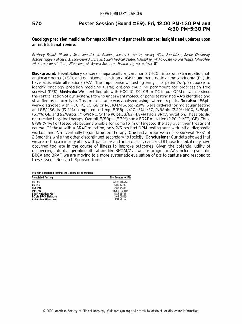
570 Poster Session (Board #E9), Fri, 12:00 PM-1:30 PM and4:30 PM-5:30 PM
Oncology precision medicine for hepatobiliary and pancreatic cancer: Insights and updates uponan institutional review.
Geoffrey Bellini, Nicholas Sich, Jennifer Jo Godden, James L. Weese, Wesley Allan Papenfuss, Aaron Chevinsky,Antony Ruggeri, Michael A. Thompson; Aurora St. Luke’s Medical Center, Milwaukee, WI; Advocate Aurora Health, Milwaukee,WI; Aurora Health Care, Milwaukee, WI; Aurora Advanced Healthcare, Wauwatosa, WI
Background: Hepatobiliary cancers - hepatocellular carcinoma (HCC), intra or extrahepatic chol-angiocarcinoma (I/EC), and gallbladder carcinoma (GB) - and pancreatic adenocarcinoma (PC) dohave actionable alterations (AA). The importance of testing early in a patient’s (pts) course toidentify oncology precision medicine (OPM) options could be paramount for progression freesurvival (PFS). Methods: We identified pts with HCC, IC, EC, GB or PC in our OPM database sincethe centralization of our system. Pts who underwent molecular panel testing had AA’s identified andstratified by cancer type. Treatment course was analyzed using swimmers plots. Results: 456ptswere diagnosed with HCC, IC, EC, GB or PC. 104/456pts (23%) were ordered for molecular testingand 88/456pts (19.3%) completed testing: 18/88pts (20.4%) I/EC, 2/88pts (2.3%) HCC, 5/88pts(5.7%) GB, and 63/88pts (71.6%) PC. Of the PC pts, 3/63 (4.8%) had a BRCAmutation. These pts didnot receive targeted therapy. Overall, 5/88pts (5.7%) had a BRAFmutation (2 PC, 2 I/EC, 1GB). Thus,8/88 (9.1%) of tested pts became eligible for some form of targeted therapy over their treatmentcourse. Of those with a BRAF mutation, only 2/5 pts had OPM testing sent with initial diagnosticworkup, and 2/5 eventually began targeted therapy. One had a progression free survival (PFS) of2.5months while the other discontinued secondary to toxicity. Conclusions: Our data showed thatwe are testing aminority of ptswith pancreas and hepatobiliary cancers. Of those tested, itmay haveoccurred too late in the course of illness to improve outcomes. Given the potential utility ofuncovering potential germline alterations like BRCA1/2 as well as pragmatic AAs including somaticBRCA and BRAF, we are moving to a more systematic evaluation of pts to capture and respond tothese issues. Research Sponsor: None.
Pts with completed testing and actionable alterations.
Completed Testing N = Number of Pts
PC Pts 63/88 (71.6%)GB Pts 5/88 (5.7%)HCC Pts 2/88 (2.3%)I/EC Pts 18/88 (20.4%)BRAF Mutation Pts 5/88 (5.7%)PC pts BRCA Mutation 3/63 (4.8%)Actionable Alterations 8/88 (9.1%)
© 2020 American Society of Clinical Oncology. Visit gicasym.org and search by abstract for disclosure information.
HEPATOBILIARY CANCER
571 Poster Session (Board #E10), Fri, 12:00 PM-1:30 PM and4:30 PM-5:30 PM
Preoperative serum artemin (ARTN) as a predictive biomarker of recurrence following curativeresection for hepatocellular carcinoma (HCC).
Sophie Feng, Hao-Wen Sim, Roxana Bucur, Reenika Aggarwal, Phillipe Abreu, Monali Ray, Michael Herman, Dangxiao Cheng,Zhuo Chen, Wenjiang Zhang, Paul David Greig, Alice Chia-chi Wei, Carol-Anne Moulton, Ian M. McGilvray, Sean P. Cleary,Steven Gallinger, Gonzalo Sapisochin, Jennifer J. Knox, Geoffrey Liu, Eric Xueyu Chen; Princess Margaret Cancer Centre,Toronto, ON, Canada; The Kinghorn Cancer Centre, St Vincent’s Hospital Sydney, Sydney, Australia; Princess Margaret-University Health Network, Toronto, ON, Canada; Princess Margaret Cancer Centre Division of Medical Oncology andHematology, Toronto, ON, Canada; Toronto General Hospital, University Health Network, Toronto, ON, Canada; University ofToronto, Toronto, ON, Canada; Princess Margaret Cancer Centre, University Health Network, Toronto, ON, Canada; TorontoGeneral Hospital, Toronto, ON, Canada; Division of General Surgery, University Health Network, University of Toronto,Toronto, ON, Canada; Mayo Clinic, Rochester, MN
Background: Tumor-induced generation of splenic erythroblast- like cells (Ter-cells) has beenshown to promote tumor progression. These Ter-cells produce the glial-line derived neurotropicfactor, ARTN.We investigated the association of pre-operative serumARTNand the risk of recurrencein HCC patients (pts) undergoing curative resection.Methods: Blood samples were collected prior tosurgery as part of an institutional molecular epidemiologic study. Serum ARTN concentration wasmeasured using an enzyme-linked immunosorbent assay (ELISA). Demographics, pathological var-iables known to be associated with outcomes and clinical outcomes were collected. Uni- and multi-variate analysis were conducted. The optimal cutpoint method was used to define high and low ARTNconcentrations. Coxmodels (hazard ratios, HR) were used to compare progression-free (PFS, primaryendpoint), and overall survival (OS) of pts with high vs low serumARTN.Results: Pre-operative bloodsamples were available for 58 pts. Median agewas 63 years (range: 25-85 years); 74%weremale and50%wereAsian. Etiology of liver diseasewas hepatitis B (43%) and hepatitis C (22%); 43%of tumorswere#5cm, and vascular invasionwas seen in 62%.A baseline alpha-fetoprotein (AFP) of. 100mcg/Lwas observed in 36%pts.Median follow-upwas 18.9months.MedianARTN concentrationwas 0.322ng/mL. The optimal ARTN concentration cut-off was 0.206 ng/mL. Median PFS for pts with high( . 0.206 ng/mL) vs low (#0.206 ng/mL) ARTN was 15.7 vs 55 months (p = 0.04) respectively.Three year PFSwas 34%vs 55%, and three year OS 54%vs 91% for high vs low groups. Univariateanalysis found that high ARTN concentration (HR 2.44, p = 0.05) and multifocal tumors wereassociated with a worse PFS. In a multivariate analysis adjusted for AFP . 100mcg/L, vascularinvasion, hepatitis status andmultifocal tumors, high ARTN remained a negative prognostic factorfor PFS, aHR 3.32 (95% CI: 1.22-9.05, p = 0.02). Conclusions: A high pre-surgery serum ARTN isassociated with earlier recurrence in HCC pts undergoing curative resection. ARTN should befurther studied in HCC to determine its value as a prognostic marker. Research Sponsor: None.
© 2020 American Society of Clinical Oncology. Visit gicasym.org and search by abstract for disclosure information.
HEPATOBILIARY CANCER

572 Poster Session (Board #E11), Fri, 12:00 PM-1:30 PM and4:30 PM-5:30 PM
Circulating levels of soluble urokinase plasminogen activator receptor (suPAR) to predictoutcome after resection of biliary tract cancer.
Sven H Loosen, Annemarie Breuer, Frank Tacke, Patrick Hamid Alizai, Tom Florian Ulmer, Christian Trautwein,Thomas Longerich, Christoph Roderburg, Ulf Peter Neumann, Tom Luedde; University Hospital RWTH Aachen, Aachen,Germany; Charite–Universitatsmedizin Berlin, Berlin, Germany; RWTH Aachen University, Aachen, Germany; UniversityHospital Heidelberg, Heidelberg, Germany
Background: Surgical resection is the only curatively intended therapy for patients with biliary tractcancer (BTC), but 5-year survival rates after tumor resection have remained below 30%, corrob-orating the need for better preoperative stratification tools to identify the ideal surgical candidates.The soluble urokinase plasminogen activator receptor (suPAR) represents a mediator of inflam-mation and has recently been associated with cancer. In this study, we evaluated a potential role ofsuPAR as a novel biomarker in patients undergoing resection of BTC.Methods: Tumor expression ofuPAR, themembrane bound source of suPAR,was analyzed by IHC in 108BTC samples. Serum levelsof suPAR were analyzed by ELISA in a training and validation cohort comprising a total of 117 BTCpatients and 76 healthy controls. Results: A high tumoral uPAR expression was associated with anadverse outcome after BTC resection. In line, circulating levels of suPAR were significantly elevatedin BTC patients compared to healthy controls and patients with primary sclerosing cholangitis (PSC).Using a small training set, we established an optimal prognostic suPAR cut-off value of 3.72ng/ml forBTCpatients. Importantly, preoperative suPARserum levels above this cut-off valuewere associatedwith significantly impaired overall survival in both the training and validation cohort. MultivariateCox-regression analysis including clinicopathological parameters such as the tumor stage, markersof systemic inflammation or organ dysfunction and established tumormarkers revealed suPARas anindependent prognostic marker following BTC resection. Finally, high preoperative suPAR levelswere indicative for acute kidney injury after tumor resection. Conclusions: Circulating suPARrepresents a previously unrecognized biomarker in patients with resectable BTC, which might beuseful to preoperatively identify the ideal candidates for tumor resection. Research Sponsor:German Research Foundation.
© 2020 American Society of Clinical Oncology. Visit gicasym.org and search by abstract for disclosure information.
HEPATOBILIARY CANCER

573 Poster Session (Board #E12), Fri, 12:00 PM-1:30 PM and4:30 PM-5:30 PM
Role of Wnt5a in suppressing invasiveness of hepatocellular carcinoma via epithelialmesenchymal transition.
Kazuki Wakizaka, Toshiya Kamiyama, Tatsuya Orimo, Shingo Shimada, Akihisa Nagatsu, Yoh Asahi, Yuzuru Sakamoto,Hirofumi Kamachi, Tomoko Mitsuhashi, Akinobu Taketomi; Department of Gastroenterological Surgery I, Graduate School ofMedicine, Hokkaido University, Sapporo, Japan; Department of Gastoenterological Surgery I, Hokkaido University GraduateSchool of Medicine, Sapporo, Japan; Hokkaido University Graduate School of Medicine, Sapporo, Japan; Department ofGastroenterological Surgery I, Hokkaido University Graduate School of Medicine, Sapporo, Japan
Background:Wnt signaling pathway includes canonical pathway and non-canonical pathway. Wnt/b-Catenin pathway as canonical pathway is associated with the development of hepatocellularcarcinoma (HCC). On the other hand, the association between aberrant activation of non-canonicalpathway activated by Wnt5a and tumor progression of HCC is not well-known. We investigated thesignificance of the expression of Wnt5a in HCC.Methods: Immunohistochemical staining of Wnt5awas performed on the specimen of 243 patients who underwent hepatic resection for HCC. Weinvestigated whether the expression of Wnt5a correlated with the clinicopathological factors,survival, and recurrence in HCC patients. The expression of Wnt5a in human HCC cell lines HLE,HLF, HepG2 and Huh7 was investigated by western blotting. The effects of overexpression orknockdown of Wnt5a on cell lines were evaluated by proliferation assay and invasion assay andchanges in epithelial mesenchymal transition (EMT) related molecules were studied by westernblotting. Results: The Wnt5a expression was positive in 63 patients (25.9%) and negative in 180patients (74.1%). The Wnt5a negative was significantly associated with poorly differentiation (P =0.003) and vascular invasion positive (P =0.046). Byunivariate analysis,Wnt5a negative (P =0.020)was identified as a significant prognostic factor of OS.Multivariate analysis ofOS showed thatWnt5anegative (HR 1.895, 95% CI 1.053-3.409, P = 0.033) was identified as an independent prognosticfactor. In theHCC cell lines, theWnt5a expressionwas lower inHLE andHLF than inHepG2andHuh7.Knockdown of Wnt5a by shRNA increased the proliferation and invasiveness in Huh7 with highexpression ofWnt5a. As a result, the expression of E-cadherin decreased. In HLFwith low expressionof Wnt5a, overexpression of Wnt5a inhibited the invasiveness and the expression of vimentindecreased. Conclusions: Wnt5a negative was associated with poorly differentiation and vascularinvasion, and was independent poor prognostic factor in HCC patients. Wnt5a may be a tumorsuppressor involved in EMT mediated changes of invasiveness. Research Sponsor: None.
© 2020 American Society of Clinical Oncology. Visit gicasym.org and search by abstract for disclosure information.
HEPATOBILIARY CANCER
574 Poster Session (Board #E13), Fri, 12:00 PM-1:30 PM and4:30 PM-5:30 PM
NTRK gene fusions in biliary tract cancers.
Anne Demols, Laureen Rocq, Manon Charry, Nancy De Neve, Audrey Verrellen, Ali Ramadhan, Claude Van Campenhout,Sarah De Clercq, Isabelle Salmon, Nicky D’Haene; CUB Hopital Erasme, Bruxelles, Belgium; CUB Hopital Erasme, PathologyDepartment, Anderlecht, Belgium; Pathology Department, Erasme Hospital, Universite libre de Bruxelles (ULB), Brussels,Belgium
Background: Gene fusions involving one of the 3 neurotrophic tyrosine receptor kinases (NTRK)have been identified in approximately 1% of solid tumors and inhibitors of TRK (e.g. larotrectinib)have been shown to have anti-tumor activity regardless of tumor type. NTRK gene fusions havebeen previously reported in bilio-pancreatic cancers. It is of interest therefore to determine theincidence and molecular characteristics of NTRK gene fusions in patients with bilio-pancreaticcancers. Methods: Formalin-fixed paraffin-embedded archival blocks from surgical resections,biopsies or cytological samples of biliary tract tumors including intra-hepatic cholangiocarcinoma(IH), extra-hepatic cholangiocarcinoma (EH), perihilar cholangiocarcinoma (PH) and gallbladdertumors (G) were selected/retrieved from the tumor bank of the CUB Hopital Erasme betweenJanuary 2010 and July 2019. A two-step diagnostic method incorporating immunohistochemistry(IHC) screening followed by NGS analysis was used. Pan–TRK IHC (monoclonal antibody cloneEPR17341 [AbCam, Cambridge,MA])was used for the screeningmethod.Staining intensity (negative,weak, moderate or strong) and localization (cytoplasmic or nuclear) were evaluated. The presence ofat least weak staining tumor cells led to testing by a RNA-based NGS panel (Oncomine Focus Assay,ThermoFisher scientific). Results: 145 archival tumors samples (81 surgical resections, 48 biopsiesand 16 cytology) have been selected, including 61 IH, 32 PH, 26 EH and 26 G (67 female and 78male).134 samples were suitable to perform IHC. 17 samples were IHC positive. Intensity of staining wasweak in 16 samples and moderate in one. Staining location was cytoplasmic (14/17), nuclear (2/17),and nuclear+cytoplasmic (1/17). NGS testing of the 17 IHC positive samples revealed a single NTRK3gene fusion (ETV6(4)-NTRK3(14)). In this case (female patient with a poorly differentiated PH,deceased), IHC had aweak focal cytoplasmic and nuclear staining. Overall in the patients screened byIHC and confirmed by NGS, the percentage of NTRK fusions was 0.75%. Conclusions: NTRK genefusions are rare in biliary cancers but testing is of high interest due to the emergence of possibletreatment with specific TRK inhibitors. Research Sponsor: Bayer Healthcare.
© 2020 American Society of Clinical Oncology. Visit gicasym.org and search by abstract for disclosure information.
HEPATOBILIARY CANCER

575 Poster Session (Board #E14), Fri, 12:00 PM-1:30 PM and4:30 PM-5:30 PM
Genomic characterization of HBV-infected hepatocellular carcinoma patients using circulatingtumor DNA.
Daniel Lin, Zhong Ye, Chun Wang, Qiang Wei, Li Bingshan, James Posey, Hushan Yang; Thomas Jefferson University,Philadelphia, PA; Thomas Jefferson University Hospital, Philadelphia, PA; Vanderbilt University, Nashville, TN
Background:Hepatocellular carcinoma (HCC) is a leading cause of mortality, with Hepatitis B virus(HBV) infection as a dominant etiology. Surgery or ablation may be curative for early-stage HCC.Thus, effective detection strategies are needed. We investigated genomic aberrations in circu-lating tumor DNA (ctDNA) as a potential diagnostic marker of HCC in HBV-infected patients.Methods:We identified early stage (BCLC 0-A) HCC cases (n = 21) and cancer-free controls (n = 15)from a cohort of Asian patients with HBV, undergoing surveillance at Thomas Jefferson UniversityHospital between 2013-2017. Blood samples were collected. Circulating cell-free DNA was isolatedfrom plasma and assayed by capture-based next-generation sequencing of a targeted panel of 23genes implicated in HCC pathogenesis. Sequencing data analysis and somatic mutation identifi-cation were conducted using a computational pipeline. Using area under the curve (AUC) inreceiver operating characteristic analysis, we evaluated gene alterations and clinical factors (age,gender, cirrhosis) in an exploratory early detection HCC model. Results: Mutant ARID1A, ATM,CDKN2A, CTNNB1, ERBB2, TP53 genes were increased in HCC cases relative to non-cancer patients(85.7% vs53.3%,P =0.058; 42.9% vs6.7%,P=0.025; 38.1% vs6.7%,P=0.051; 42.9% vs0%,P=0.005; 52.4% vs 13.3%, P = 0.016; 100% vs 66.7%, P = 0.008, respectively). HCC patients hadhigher prevalence of cirrhosis than controls (90.5% vs. 60%, P = 0.046). Using the 6mutant genesalone, the AUC for discriminating HCC from non-cancer patients was 0.827 (95% confidenceinterval [CI]: 0.701-0.953), which was greater than the AUC for discriminating cirrhosis from non-cirrhosis (0.531). When the 6 mutant genes were combined with clinical factors, the AUC of theexploratory HCC detection model increased to 0.914 (P= 0.045). Conclusions: We identified 6genomic aberrations in ctDNA that were more prevalent in HCC patients compared with non-cancer patients. Combining these alterations with clinical factorsmay identify HCC in HBV-infectedpatients at an early stage. These findings warrant further validation in future studies. ResearchSponsor: American Cancer Society Institutional Research Grant.
© 2020 American Society of Clinical Oncology. Visit gicasym.org and search by abstract for disclosure information.
HEPATOBILIARY CANCER
577 Poster Session (Board #E16), Fri, 12:00 PM-1:30 PM and4:30 PM-5:30 PM
Gamma-synuclein as a potential novel prognostic marker promoting tumor cell migration inbiliary tract carcinoma.
Yusuke Takemura, Hidenori Ojima, Go Oshima, Masahiro Shinoda, Minoru Kitago, Hiroshi Yagi, Yuta Abe, Shutaro Hori,Yoko Nishimura, Naoto Kubota, Yuki Masuda, Taizo Hibi, Michiie Sakamoto, Yuko Kitagawa; Department of Surgery, KeioUniversity School of Medicine, Tokyo, Japan; Department of Pathology, Keio University School of Medicine, Tokyo, Japan;Department of Pediatric Surgery and Transplantation, Kumamoto University Graduate School of Medical Sciences,Kumamoto, Japan
Background: Biliary tract carcinoma (BTC) is a highly malignant tumor, but the detailed patho-logical mechanism of its development and progression remain ill-defined. Gamma-synuclein(SNCG) promotes invasive behavior in pancreatic cancer and a range of other cancers, and SNCGexpression is reportedly a prognostic factor. However, the role of SNCG in BTC remains unknown.Consequently, we investigated the clinicopathological significance and function of SNCG in BTC.Methods: Using surgically resected BTC specimens from 149 patients with adenocarcinoma[extrahepatic cholangiocarcinoma (ECC) (n =98); intrahepatic cholangiocarcinoma (ICC) (n =51)],we immunohistochemically evaluated SNCG positivity and checked for correlations with clinico-pathological factors and outcomes. Furthermore, cell lines with high expressions of SNCG wereselected from 17 BTC cell lines by using immunohistochemistry and qPCR. Cell proliferation andmigration assays were then performed in the presence and absence of SNCG (silenced using).Results: SNCG expression was found in 32 of 149 (21.4%) BTC patients. SNCG expression wassignificantly correlated with poorly differentiated tumor (P = 0.001) and lymph node metastases(P = 0.001). SNCG positivity was also correlated with poorly differentiated tumor in both ECC andICC (P = 0.008 and P = 0.03, respectively), but was correlated with lymph node metastases only inECC (P = 0.003). Multivariate analyses identified SNCG expression as an independent prognosticfactor of poor overall survival (P = 0.008) and poor recurrence-free survival (RFS) (P = 0.006).SNCG expression in both ECC and ICCwas significantly associatedwith poor prognosis in univariateanalysis, andmultivariate analysis identified SNCGas an independent prognostic factor of poor RFSfor ECC (P = 0.03). High SNCG expression was found in 3 BTC cell lines (NCC-BD1, NCC-BD3, andNCC-CC6-1). Functional analysis revealed that SNCG silencing by siRNA suppressed cell migrationsignificantly in NCC-BD1 and NCC-CC6-1 and downregulated cell proliferation in NCC-CC6-1.Conclusions: Our results suggest that SNCG may promote tumor cell activity and is potentiallya novel prognostic marker in BTC. Research Sponsor: JSPS KAKENHI grant No. 16K10609.
© 2020 American Society of Clinical Oncology. Visit gicasym.org and search by abstract for disclosure information.
HEPATOBILIARY CANCER
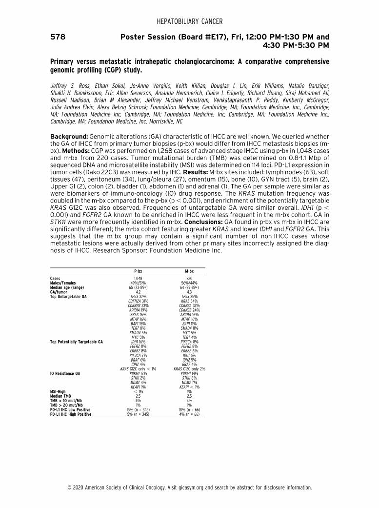
578 Poster Session (Board #E17), Fri, 12:00 PM-1:30 PM and4:30 PM-5:30 PM
Primary versus metastatic intrahepatic cholangiocarcinoma: A comparative comprehensivegenomic profiling (CGP) study.
Jeffrey S. Ross, Ethan Sokol, Jo-Anne Vergilio, Keith Killian, Douglas I. Lin, Erik Williams, Natalie Danziger,Shakti H. Ramkissoon, Eric Allan Severson, Amanda Hemmerich, Claire I. Edgerly, Richard Huang, Siraj Mahamed Ali,Russell Madison, Brian M Alexander, Jeffrey Michael Venstrom, Venkataprasanth P. Reddy, Kimberly McGregor,Julia Andrea Elvin, Alexa Betzig Schrock; Foundation Medicine, Cambridge, MA; Foundation Medicine, Inc., Cambridge,MA; Foundation Medicine Inc, Cambridge, MA; Foundation Medicine, Inc, Cambridge, MA; Foundation Medicine Inc.,Cambridge, MA; Foundation Medicine, Inc, Morrisville, NC
Background: Genomic alterations (GA) characteristic of IHCC are well known. We queried whetherthe GA of IHCC from primary tumor biopsies (p-bx) would differ from IHCCmetastasis biopsies (m-bx).Methods: CGPwas performed on 1,268 cases of advanced stage IHCC using p-bx in 1,048 casesand m-bx from 220 cases. Tumor mutational burden (TMB) was determined on 0.8-1.1 Mbp ofsequenced DNA and microsatellite instability (MSI) was determined on 114 loci. PD-L1 expression intumor cells (Dako 22C3) wasmeasured by IHC.Results:M-bx sites included: lymph nodes (63), softtissues (47), peritoneum (34), lung/pleura (27), omentum (15), bone (10), GYN tract (5), brain (2),Upper GI (2), colon (2), bladder (1), abdomen (1) and adrenal (1). The GA per sample were similar aswere biomarkers of immuno-oncology (IO) drug response. The KRAS mutation frequency wasdoubled in them-bx compared to the p-bx (p, 0.001), and enrichment of the potentially targetableKRAS G12C was also observed. Frequencies of untargetable GA were similar overall. IDH1 (p ,0.001) and FGFR2 GA known to be enriched in IHCC were less frequent in the m-bx cohort. GA inSTK11 were more frequently identified in m-bx. Conclusions: GA found in p-bx vs m-bx in IHCC aresignificantly different; the m-bx cohort featuring greater KRAS and lower IDH1 and FGFR2 GA. Thissuggests that the m-bx group may contain a significant number of non-IHCC cases whosemetastatic lesions were actually derived from other primary sites incorrectly assigned the diag-nosis of IHCC. Research Sponsor: Foundation Medicine Inc.
P-bx M-bx
Cases 1,048 220Males/Females 49%/51% 56%/44%Median age (range) 65 (23-89+) 64 (29-89+)GA/tumor 4.2 4.3Top Untargetable GA TP53 32% TP53 35%
CDKN2A 31% KRAS 34%CDKN2B 23% CDKN2A 32%ARID1A 19% CDKN2B 24%KRAS 16% ARID1A 16%MTAP 16% MTAP 16%BAP1 15% BAP1 11%TERT 8% SMAD4 11%SMAD4 5% MYC 5%MYC 5% TERT 4%
Top Potentially Targetable GA IDH1 16% PIK3CA 8%FGFR2 11% FGFR2 8%ERBB2 8% ERBB2 6%PIK3CA 7% IDH1 6%BRAF 6% IDH2 5%IDH2 4% BRAF 4%
KRAS G12C only , 1% KRAS G12C only 2%IO Resistance GA PBRM1 12% PBRM1 14%
STK11 2% STK11 8%MDM2 4% MDM2 7%KEAP1 1% KEAP1 , 1%
MSI-High , 1% 1%Median TMB 2.5 2.5TMB > 10 mut/Mb 4% 4%TMB > 20 mut/Mb 1% 1%PD-L1 IHC Low Positive 15% (n = 345) 18% (n = 66)PD-L1 IHC High Positive 5% (n = 345) 4% (n = 66)
© 2020 American Society of Clinical Oncology. Visit gicasym.org and search by abstract for disclosure information.
HEPATOBILIARY CANCER
579 Poster Session (Board #E18), Fri, 12:00 PM-1:30 PM and4:30 PM-5:30 PM
Circulating free DNA (cfDNA) and tissue next-generation sequencing analysis in a phase II studyof infigratinib (BGJ398) for cholangiocarcinoma with FGFR2 fusions.
Shalini Makawita, Robin Kate Kelley, Sameek Roychowdhury, Karl Heinz Weiss, Ghassan K. Abou-Alfa, Teresa Macarulla,Saeed Sadeghi, Dirk Waldschmidt, Andrew X. Zhu, Lipika Goyal, Wei-Peng Yong, Ivan Borbath, Anthony B. El-Khoueiry,Philip Agop Philip, Susan Moran, Yining Ye, Harris S. Soifer, Gary Li, Craig Berman, Milind M. Javle; The University of Texas MDAnderson Cancer Center, Houston, TX; University of California San Francisco, San Francisco, CA; Ohio State ComprehensiveCancer Center/James Cancer Hospital, Columbus, OH; University Hospital Heidelberg, Heidelberg, Germany; Memorial SloanKettering Cancer Center, New York, NY; Hospital Vall d’Hebron, Barcelona, Spain; University of California at Los Angeles,Santa Monica, CA; Klinikum der Universitaet zu Koln, Koln, Germany; Massachusetts General Hospital, Boston, MA; NationalUniversity Cancer Institute, Singapore, Singapore; Cliniques Universitaires St Luc Bruxelles, Brussels, Belgium; USC/KennethNorris Comprehensive Cancer Center, Los Angeles, CA; Karmanos Cancer Institute, Detroit, MI; QED Therapeutics Inc, SanFrancisco, CA; University of Texas MD Anderson Cancer Center, Houston, TX
Background: Fibroblast growth factor receptor 2 (FGFR2) alterations occur in 11% of cholangio-carcinomas, 85% of which are fusions. A multicenter, open-label, phase II study is currentlyevaluating the efficacy of infigratinib, a selective FGFR1–3 tyrosine kinase inhibitor, in patients withpreviously treated advanced cholangiocarcinoma containing FGFR2 fusions. We report detailedbiomarker analyses from this study. Methods: Patients with advanced or metastatic cholangio-carcinoma containing FGFR2 fusions whose disease had progressed following cisplatin- orgemcitabine-based therapy were eligible. Patients received oral infigratinib 125 mg once dailyon days 1–21 every 28 days. Comprehensive genomic profiling (CGP) was performed on tumortissue and cfDNA collected prior to the start of therapy. The primary endpoint was investigator-assessed overall response rate (ORR) [RECIST version 1.1]. Data cut-off (prespecified): August 8,2018. Trial registration: NCT02150967. Results: At data cut-off, 71 patients with FGFR2 fusionswere included (62%women;median age 53 years; 55% received$2 prior lines of therapy). Medianduration of treatment was 5.5 months. ORR (confirmed and unconfirmed) was 31.0% (95% CI20.5–43.1%) and confirmed ORR was 26.9% (95% CI 16.8–39.1%). 33 unique FGFR2 fusion geneswere identified in 71 enrolled patients. The most common fusion gene partner was BICC1 (32%; 23/71). Pathogenic variants in 9 other druggable genes were identified in 32% of patients (13/37) whounderwent CGP. FGFR2 fusions were concordant in 67% (8/12) of patients with tumor tissue andcfDNA at screening. Conclusions: The large assortment of FGFR2 fusion genes identified in thisstudy underscores the diversity of FGFR2 rearrangements that may drive cholangiocarcinoma.Although cfDNA analysis was performed in a minority, these preliminary data suggest that cfDNAanalysis may be valuable for the identification of FGFR2 fusions and to study intratumoralheterogeneity. Clinical trial information: NCT02150967. Research Sponsor: QED Therapeutics.
© 2020 American Society of Clinical Oncology. Visit gicasym.org and search by abstract for disclosure information.
HEPATOBILIARY CANCER
580 Poster Session (Board #E19), Fri, 12:00 PM-1:30 PM and4:30 PM-5:30 PM
Comparison of the clinical features, treatment patterns, and tumor mutations of patients withintrahepatic (ICC) and extrahepatic (ECC) cholangiocarcinoma.
Leon Pappas, Stephanie Reyes, Aishwarya Lanka, Rachna T. Shroff, Tri Minh Le, Osama E. Rahma, Andrea Grace Bocobo,Mitesh J. Borad, Thomas DeLeon, Kabir Mody, Marc Thomas Roth, Mary Linton Bounetheau Peters, Laura Williams Goff,Kylie Boyhen, Christine VanCott, Nora Horick, Andrew X. Zhu, Milind M. Javle, Robin Kate Kelley, Lipika Goyal; MassachusettsGeneral Hospital, Boston, MA; University of Michigan Medical School, Ann Arbor, MI; MD Anderson, Houston, TX; University ofArizona Cancer Center, Tucson, AZ; University of Virginia, Charlottesville, VA; Dana-Farber Cancer Institute, Boston, MA; UCSan Francisco Helen Diller Family Comprehensive Cancer Center, San Francisco, CA; Mayo Clinic Cancer Center, Scottsdale,AZ; Mayo Clinic, Scottsdale, AZ; Mayo Clinic, Jacksonville, FL; Vanderbilt University Medical Center, Nashville, TN; Beth IsraelDeaconess Medical Center, Cambridge, MA; Yale University, New Haven, CT; St. Vincent’s Medical Center, Bridgeport, CT;Massachusetts General Hospital Biostatistics Center, Boston, MA; University of Texas MD Anderson Cancer Center, Houston,TX; University of California San Francisco, San Francisco, CA
Background: Though studies indicate that the genomic profiles of ICC and ECC are distinct, theclinical features that differentiate them still remain to be well characterized. The purpose of thisstudy was to further analyze these differences and patient treatment patterns in a multi-centercohort. Methods: A retrospective chart review was performed at 8 institutions on patients (pts)with ICC or ECC diagnosed after June 2009. Data on demographics, risk factors, treatments,pathology and overall survival (OS) were collected. Tumor genotyping results from CLIA-certifiedtissue assays were analyzed. Fisher’s exact, Wilcoxon rank sum and log-rank tests were used tocompare sub-groups. Results: In a database of 737 pts with cholangiocarcinoma, 538(73%) hadICC and 199(27%) had ECC. Pts with ICCmore often presented in later stages, had tumors. 5cm atresection (p, 0.0001) and had metastases to the liver, lymph nodes, lung and/or bone (p, 0.01).Pts with ICC more often received liver directed therapy, targeted therapy and multiple lines ofsystemic therapy and they more often enrolled in a clinical trial (all p , 0.01). Pts with ECC weremore likely to be male, undergo surgery, receive adjuvant chemotherapy and/or chemoradiation(all p, 0.05). Mutation profiling performed in 381 (52%) pts (ICC/ECC = 301/80) showed that ptswith ICC were more likely to have IDH1 mutations and FGFR2 fusions, whereas pts with ECC weremore likely to have KRAS, APC, SMAD4, WNT, TGFb and TP53 mutations (all, 0.05). Factors thatdid not differ significantly between pts with ICC and ECC include race, rates of primary sclerosingcholangitis, median diagnosis CA19-9 levels and R1 resection rate. Median OS from diagnosis was18.9 months in ICC and 17.3 months in ECC (p = 0.8471). Conclusions: While pts with ICC and ECChave some similarities in their clinical features, differences in metastases patterns and molecularprofiling significantly impact their management such that pts with ICC receive more liver-directedtherapy, targeted therapy and more lines of systemic therapy. Further prospective studies areneeded as referral patterns to tertiary care centers may have impacted these results. ResearchSponsor: None.
© 2020 American Society of Clinical Oncology. Visit gicasym.org and search by abstract for disclosure information.
HEPATOBILIARY CANCER
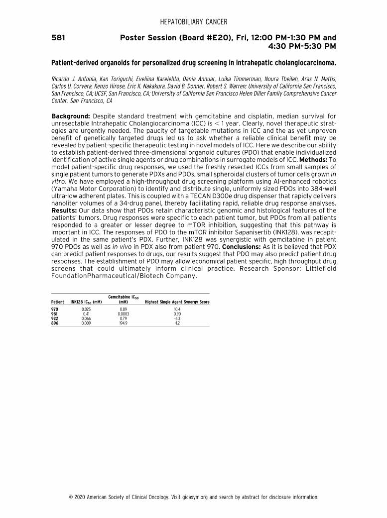
581 Poster Session (Board #E20), Fri, 12:00 PM-1:30 PM and4:30 PM-5:30 PM
Patient-derived organoids for personalized drug screening in intrahepatic cholangiocarcinoma.
Ricardo J. Antonia, Kan Toriguchi, Eveliina Karelehto, Dania Annuar, Luika Timmerman, Noura Tbeileh, Aras N. Mattis,Carlos U. Corvera, Kenzo Hirose, Eric K. Nakakura, David B. Donner, Robert S. Warren; University of California San Francisco,San Francisco, CA; UCSF, San Francisco, CA; University of California San Francisco Helen Diller Family Comprehensive CancerCenter, San Francisco, CA
Background: Despite standard treatment with gemcitabine and cisplatin, median survival forunresectable Intrahepatic Cholangiocarcinoma (ICC) is , 1 year. Clearly, novel therapeutic strat-egies are urgently needed. The paucity of targetable mutations in ICC and the as yet unprovenbenefit of genetically targeted drugs led us to ask whether a reliable clinical benefit may berevealed by patient-specific therapeutic testing in novel models of ICC. Here we describe our abilityto establish patient-derived three-dimensional organoid cultures (PDO) that enable individualizedidentification of active single agents or drug combinations in surrogatemodels of ICC.Methods: Tomodel patient-specific drug responses, we used the freshly resected ICCs from small samples ofsingle patient tumors to generate PDXs and PDOs, small spheroidal clusters of tumor cells grown invitro. We have employed a high-throughput drug screening platform using AI-enhanced robotics(Yamaha Motor Corporation) to identify and distribute single, uniformly sized PDOs into 384-wellultra-low adherent plates. This is coupled with a TECAN D300e drug dispenser that rapidly deliversnanoliter volumes of a 34-drug panel, thereby facilitating rapid, reliable drug response analyses.Results: Our data show that PDOs retain characteristic genomic and histological features of thepatients’ tumors. Drug responses were specific to each patient tumor, but PDOs from all patientsresponded to a greater or lesser degree to mTOR inhibition, suggesting that this pathway isimportant in ICC. The responses of PDO to the mTOR inhibitor Sapanisertib (INK128), was recapit-ulated in the same patient’s PDX. Further, INK128 was synergistic with gemcitabine in patient970 PDOs as well as in vivo in PDX also from patient 970. Conclusions: As it is believed that PDXcan predict patient responses to drugs, our results suggest that PDO may also predict patient drugresponses. The establishment of PDO may allow economical patient-specific, high throughput drugscreens that could ultimately inform clinical practice. Research Sponsor: LittlefieldFoundationPharmaceutical/Biotech Company.
Patient INK128 IC50 (mM)Gemcitabine IC50
(mM) Highest Single Agent Synergy Score
970 0.025 0.89 10.4981 0.41 0.0003 0.90922 0.066 0.79 -6.3896 0.009 194.9 -1.2
© 2020 American Society of Clinical Oncology. Visit gicasym.org and search by abstract for disclosure information.
HEPATOBILIARY CANCER
582 Poster Session (Board #E21), Fri, 12:00 PM-1:30 PM and4:30 PM-5:30 PM
Hepatocyte-derived intrahepatic cholangiocarcinoma requires Yap and Sox9: A clinical andpreclinical analysis.
Satdarshan Monga, Sungjin Ko, Laura Molina, Junyan Tao, Aatur D. Singhi, Aaron Bell; University of Pittsburgh MedicalCenter, Pittsburgh, PA; University of Pittsburgh, Pittsburgh, PA
Background: Intrahepatic cholangiocarcinoma (ICC) is a liver tumor of increasing incidence anddevastating prognosis. WGS andWES have identified numerousmolecular pathways and fusions inICC. Recent studies have also suggested hepatocyte as a cell source in a subset of ICCs, especiallythose associated with chronic liver insult such as non-alcoholic steatohepatitis (NASH) or primarysclerosing cholangitis (PSC). Methods: Since co-expression of myristoylated AKT (myrAKT) &Notch intracellular domain (NICD) in hepatocytes using sleeping beauty transposon/transposase-based hydrodynamic tail vein injection (SB-HTVI) lead to ICC, we initiated a comprehensive analysisof mechanism of ICC development in patients and in this preclinical model. Results: Over 90% ofCC samples exhibited high levels of nuclear SOX9 & YAP, in addition to a significant positivity forpAKT in ICCs as compared to extrahepatic CC. We also identified upregulation of p-AKT, SOX9 &YAP in hepatocytes of patients with PSC and NASH. This was also seen in many murine models ofcholestatic injury and NASH. While co-expression of myrAKT+NICD led to hepatocyte-derived ICC,conditional deletion of either Yap or Sox9 significantly delayed and almost completely abrogatedICC development. While Yap deletion impaired the initial HC-to-BEC fate conversion, Sox9 elim-ination had no such effect on reprograming. Interestingly, following deletion of either Yap or Sox9we observed a few AKT/NICD-driven ICC tumors expressing either Sox9 or Yap but not both. Thisalso occurred in a small subset of human CC tumors which may be Sox9+Yap- (4%) or Sox9-Yap+(3.7%), showing that deletion of Yap or Sox9 is not sufficient to completely abrogate ICCdevelopment. We finally demonstrated that conditional deletion of both Yap & Sox9 completelyblocked development of ICC tumors in the myrAKT+NICDmodel. Conclusions: Thus, we show thatcholestatic injury or NASH in humans and mice induces hepatocyte-to-cholangiocyte reprogram-ing to increase the risk of ICC development. We also provide evidence for critical but distinct rolesof Yap and Sox9 in ICC development and demonstrate the therapeutic potential of targeting thesefactors for treatment of subsets of ICC. Research Sponsor: U.S. National Institutes of HealthIntramural Endowed Chair.
© 2020 American Society of Clinical Oncology. Visit gicasym.org and search by abstract for disclosure information.
HEPATOBILIARY CANCER
583 Poster Session (Board #E22), Fri, 12:00 PM-1:30 PM and4:30 PM-5:30 PM
Beta-catenin mutations in hepatocellular cancer, tumor cell metabolism, and the response ofthese tumors to mTOR inhibition.
Satdarshan Monga, Mitesh J. Borad, Junyan Tao; University of Pittsburgh Medical Center, Pittsburgh, PA; Mayo Clinic CancerCenter, Scottsdale, AZ; University of Pittsburgh, Pittsburgh, PA
Background:Hepatocellular cancer (HCC) continues to grow in incidence despite approval ofmanynew therapies including immune checkpoint inhibitors (ICIs) and TKIs. Genomic landscape of HCC isbecoming increasing apparent through WGS and WES. One major driver of HCC is the Wnt/b-catenin pathway. Mutations in CTNNB1, which encodes for b-catenin, are evident in 26-37% of allhuman HCCs. However, expression of mutant-b-catenin in liver in mice is insufficient for HCCdevelopment. Indeed, analysis of HCC cases has revealed CTNNB1 mutations to significantly co-exist with other aberrations including activation/overexpression of Met and Myc and mutations inTERT promoter or mutations in NFE2L2/KEAP1, APOB and ARID2.Methods: Using sleeping beautytransposon/transposase and hydrodynamic tail vein injection, we co-expressed mutant-CTNNB1(S45Y, S33Y or T41A) and one other clinically relevant co-occurrence to study significance andbiology of HCC. Any novel findings in the preclinical model were validated in HCC patient cohorts.Results: Co-expression of mutant-CTNNB1 and one relevant co-occurrence led to development ofHCC in mice in 6-10 weeks. All HCC in these models showed a dramatic increase in glutaminesynthetase (GS), encoded by Glul, a known target of the Wnt/b-catenin signaling pathway in theliver. This led to an increase in glutamine levels in the tumor-bearing livers. Increased glutamine inthe tumors in turn led to increased levels of phospho-mTOR-S2448, a marker of mTORC1 acti-vation. In fact, examination of ~400 patient HCCs showed a significant correlation betweenpositive GS and p-mTOR-S2448 staining. Treatment of Met-b-catenin model with Rapamycin led tonotable and significant decrease in HCC burden. Conclusions: Our studydemonstrates b-catenin-mutated HCC to be positive for both GS and in turn mTORC1 active. This provides a novelopportunity for personalized medicine in HCC and CTNNB1-mutated HCCs may be vulnerable totherapeutic targeting by mTOR and more specially, mTORC1 inhibitors. Research Sponsor: U.S.National Institutes of Health Intramural Endowed Chair.
© 2020 American Society of Clinical Oncology. Visit gicasym.org and search by abstract for disclosure information.
HEPATOBILIARY CANCER
584 Poster Session (Board #F1), Fri, 12:00 PM-1:30 PM and4:30 PM-5:30 PM
Novel therapeutic avenues for cholangiocarcinoma treatment: A meta-analysis.
Nabeal Aljabban, Jihad Aljabban, Merve Gurakar, Kamal Khorfan, Saad A. Syed, Adam Gayar, Hajra Khan, Dexter Hadley,Behnam Saberi; Penn State School of Medicine, Hershey, PA; The Ohio State University College of Medicine, Columbus, OH;Johns Hopkins, Baltimore, MD; Henry Ford Health System, Detroit, MI; Stanford Medicine, Stanford, CA; University ofCincinnati College of Medicine, Cincinnati,, OH; Detroit Medical Center, Detroit, MI; University of California San Francisco, SanFrancisco, CA; Mount Sinai, New York, NY
Background: Cholangiocarcinoma (CCA) is a rare cancer of the bile ducts but has been increasingin incidence. The mainstay of treatment of CCA is resection or chemoradiation for more advanceddisease, with immunotherapy being an evolving field in treatment. A better understanding of CCApathogenesis will pave new avenues for treatment.Methods:We employed our STARGEO platformto conduct a meta-analysis of public data from NCBI’s Gene Expression Omnibus. We performedmeta-analysis with 259 CCA tumor samples against 16 normal intrahepatic duct samples as acontrol. We then analyzed the signature in Ingenuity Pathway Analysis. Results: Our analysisrevealed FXR/RXR and LXR/RXR activation as top canonical pathways. Top upstream regulatorsidentified included HNF1A (with predicted inhibition) and ERBB2 (with predicted activation). Themost upregulated genes included several extracellular matrix proteins implicated in cancerincluding COL1A1, LAMC2 (correlated with poor prognosis in CCA), KRT17 (a keratin implicatedin various malignancies but not well described in CCA), and LAMB3 (exerts tumorigenesis throughPI3k/Akt signaling). Additionally, we found stark upregulation of the immunophillin FKPBP1A,which is involved in mTOR activation. We also noted upregulation of ubiquitin-associated geneUBASH3B, which inhibits endocytosis in EGFR and has been described in breast cancer but notCCA. From our investigation of immune checkpoint inhibitors, we found upregulation of classicallydescribed inhibitors such as CTLA4, TIGIT, and BTLA. In addition, we found upregulation of SIGLEC7,which has been recently shown to suppress immune function by binding to terminal sialic acid onglycans on the surface of immune cells. Conclusions: Our analysis highlights the possible role ofERBB2 and several extracellular genes in the pathogenesis of CCA.We also identify the role of genesnot previously described in CCA such as FKBP1A and UBASH3B. Lastly, our results promote thepromise of immunotherapy in CCA treatment. Research Sponsor: None.
© 2020 American Society of Clinical Oncology. Visit gicasym.org and search by abstract for disclosure information.
HEPATOBILIARY CANCER

585 Poster Session (Board #F2), Fri, 12:00 PM-1:30 PM and4:30 PM-5:30 PM
The world-wide incidence of biliary tract cancer (BTC).
Huifen Wang, Ping Sun, Katherine Baria; AstraZeneca, Gaithersburg, MD; AstraZeneca Pharmaceuticals, Gaithersburg, MD
Background: Existing epidemiologic research for BTC is limited to certain subtypes, eg gallbladdercancer (GBC), or combines BTC with liver cancer, and only reports regional statistics. Main sourcesfor global BTC epidemiology (particularly anatomic sites) are literature reviews, which can besubject to limitations like inconsistent classification of subtypes, and varying standard populationadjustment and comparison timeframes. Given the increasing incidence of BTC, it is critical tomonitor global epidemiology to provide insight to public health management and clinical programdevelopment.Methods:We evaluated BTC incidence in 15 countries using the Cancer Incidence inFive Continents database Volume XI (CI5 XI) from the International Agency for Research on Cancer.Most CI5 XI registries cover data from 2008-12 with some variations. Incidence was examined bycountry, region, and anatomic site (extrahepatic cholangiocarcinoma, ECC; intrahepatic cholangio-carcinoma, ICC;GBC; ampulla ofVater cancer, AVC; and unspecified BTCs). Incidencewas adjusted tostandard world population and reported as age-standardized rate (ASR; per 100,000 person-years)with standard error (SE).Results: The highest incidence of total BTC was in South Korea (SK; ASR =3.00, SE = 0.02) and lowest in the UK (ASR = 0.66, SE = 0.01). Overall, BTC incidence was higher inAsian countries than Western, and in Asian-Americans than the general US population (ASR = 1.00,SE = 0.02 and 0.77, SE. 0.01, respectively). In Thailand, BTC incidence was higher in the North thanSouth (ASR = 1.93, SE = 0.05 and 1.05, SE = 0.03, respectively). GBC and ECC are the top commonsubtypes in most countries reported. SK had the highest incidence of all subtypes, with ASR . 2 inGBC, ECC, and ICC. Outside of SK, GBC and ECC were most common in Japan (ASR = 1.93, SE = 0.02and 2.66, SE= 0.03, respectively); ICCwasmost common in Thailand (ASR= 1.73, SE =0.05) and highin France (ASR = 1.01, SE = 0.03). Conclusions: These registry data support past reports of higherBTC incidence in Asian vs Western countries as well as regional variation within countries. Data alsosuggests that high BTC rates in Asian populations may not be entirely geographical as Asian-Americans suffer a higher BTC incidence than the overall US population. Research Sponsor:AstraZeneca.
© 2020 American Society of Clinical Oncology. Visit gicasym.org and search by abstract for disclosure information.
HEPATOBILIARY CANCER
586 Poster Session (Board #F3), Fri, 12:00 PM-1:30 PM and4:30 PM-5:30 PM
Association of objective response by mRECIST with better overall survival (OS) in patients withadvanced hepatocellular carcinoma (HCC) treated with systemic therapies: A systematic reviewand meta-analysis of randomized controlled trials.
Masatoshi Kudo, Yasutaka Chiba, Tim Meyer, Riccardo Lencioni, Josep M. Llovet; Kindai University Faculty of Medicine, Osaka,Japan; Kindai University, Osaka, Japan; University College London Cancer Institute, London, United Kingdom; University ofMiami Miller School of Medicine, Sylvester Comprehensive Cancer Center, Miami, FL; Icahn School of Medicine at Mount Sinai,New York, NY
Background: EASL guidelines for management of HCC recommends assessing tumor responseaccording to mRECIST at all stages of the disease (EASL guidelines, J Hep 2018). Several studieshave reported that objective response by mRECIST predicted overall survival (OS) but definitivedata are still lacking.Methods: The PubMed database and ASCOmeeting library were searched forfull reports of randomized trials in patients with advanced HCC treated by systemic therapy up toAugust 31, 2018. We search strategy used the following terms: HCC, mRECIST, OS and objectiveresponse rate (ORR). We assess the association between ORR and OS in a meta-analysis of pooleddata by using random effectsmodel comparing patients achieving objective response (complete orpartial response) versus non responders (stable disease, progressive disease) and displayed theresults as per hazard ratio (HR, 95%CI).Results:Among 14 articles assessing response bymRECISTto systemic therapies in randomized studies in advanced HCC, 4 studies (5 trials) including 1,463patients were considered eligible. Systemic therapies tested included lenvatinib, sorafenib, brivaniband nintedanib. Overall, ORR as per mRECIST ranged from 11.5% to 18.8%, being the median OS forresponders of 18.5 to 27.2 mo (as opposed 8.9 to 11.4 for non-responders). As per random effectsmodel, the HR for overall survival (responders versus non responders) was 0.47 (95% confidenceinterval 0.34–0.66, p,0.001).Conclusions:Objective response bymRECIST to systemic therapies inpatients with advanced HCC is significantly and strongly associated to OS. Patients achieving anobjective response can expect a significantly longer OS. Research Sponsor: None.
© 2020 American Society of Clinical Oncology. Visit gicasym.org and search by abstract for disclosure information.
HEPATOBILIARY CANCER
587 Poster Session (Board #F4), Fri, 12:00 PM-1:30 PM and4:30 PM-5:30 PM
Nationwide survey of pancreaticobiliary maljunction focusing on biliary cancer incidence inJapan.
Mitsuo Shimada, Hiroki Ishibashi, Yuji Morine, Masayuki Kubota, Hideki Fujii, Japanese Study Group on PancreaticobiliaryMaljunction; Tokushima University, Tokushima, Japan; Niigata University, Niigata, Japan; Kofu Municipal Hospital, Kofu-City,Japan
Background:Pancreaticobiliarymaljunction (PBM) is known to be a unique entity closely related tobiliary carcinogenesis. We herein report update analysis of PBM focusing on biliary cancer as thirdreport of Japan-nationwide survey. Methods: From 1990 to 2015, 3,419 patients with PBM wereregistered at 141 medical institutions in Japan. 3,289 (1,344 pediatric and 1,945 adult patients) outof 3,419 patients were fully investigated, according to presence of bile duct dilatation (BDD), age(pediatric or adult), etc.Results: In pediatric patients, only 3 cases with BDD (0.124%) had bile ductcancers. On the other hand, in adults, biliary cancer incidence was 21.1% in patients with BDD and43.5% in patients without BDD. The rates were extremely high in comparison with 0.0174% ofJapanese prevalence of biliary cancer 2013. The cancer incidences of bile duct, gallbladder andboth in adults with BDD/without BDD were 6.7%/4.1%, 13.0%/37.3%, and 1.4%/2.1%, respectively.In patients without associated biliary cancers, extrahepatic bile duct resection (EHBDRx) combinedwith cholecystectomy was performed in 89.5% of adults with BDD, while, in only 31.2% adultswithout BDD. Regarding the new biliary cancer occurrence of patients having follow-up data, therate was 0.3% in 354 patients with BDD who underwent EHBDRx, while that was 4.0% in 75patients without BDD who underwent simple cholecystectomy. In patients without BDD, cancerincidence (6.7%) in the late period (2010 to 2015) was higher than those (3.3% and 3.5%) in earlyandmiddle period (1990 to 1999, and 2000 to 2009).Conclusions: This updated nationwide surveyof PBM revealed characteristics of associated and newly occurred biliary cancers, and could bewidely used as a reference data for diagnosis and treatment of PBM. Research Sponsor: None.
© 2020 American Society of Clinical Oncology. Visit gicasym.org and search by abstract for disclosure information.
HEPATOBILIARY CANCER
588 Poster Session (Board #F5), Fri, 12:00 PM-1:30 PM and4:30 PM-5:30 PM
Progression-free survival in patients with cholangiocarcinoma with FGFR2 fusions orrearrangements: An exploration of response to systemic therapy.
Kristen Bibeau, Luis Feliz, Scott Barrett, Ling Na, Christine Francis Lihou, Ekatherine Asatiani; Incyte Corporation,Wilmington, DE
Background:Most cholangiocarcinoma (CCA) patients (pts) are diagnosed with advanced diseaseand are ineligible for surgery. FGFR2 fusions or rearrangements are present in 10–16% of pts withintrahepatic CCA (iCCA) and are reported to be oncogenic drivers. However, little data areavailable on the role of FGFR2 genetic alterations in the response to systemic cancer therapy.FIGHT-202 is a phase 2 study of pemigatinib (a selective, potent, oral FGFR1–3 inhibitor) in pts withpreviously treated advanced/metastatic CCA (NCT02924376); primary results were reported atESMO 2019. FIGHT-202 enrolled pts who progressed on$1 prior therapy, allowing the examinationof the role of FGFR2 alterations on the response to prior therapy. The objective of this post hocanalysis was to evaluate progression free survival (PFS) on standard systemic therapy receivedprior to study enrollment among pts with CCA harboring FGFR2 fusions or rearrangements(FGFR2+).Methods: Case report forms were reviewed to determine disease history and exposureto prior lines of systemic cancer therapies (LOSCT) in the advanced setting before receivingpemigatinib. Only pts with sufficient data on prior LOSCTwere included in this analysis. Median PFSwas calculated using the Kaplan-Meier method. Results: 102 pts were included in this analysis(median age 54.5, 61.8% female). Median PFS on first-line therapy was 5.5 (95% CI: 4.0, 8.0)months. Among the 38 pts (37.3%) with$2 prior LOSCT, median PFS on second-line therapy was4.4 (95% CI: 3.0, 5.3) months. Conclusions: This analysis provides data about PFS on standardsystemic therapies for pts with FGFR2+ CCA. Median PFS on first-line therapy was lower thanhistorical published data, and median PFS on second-line therapy was slightly longer than pre-viously reported, in unselected CCA populations. Limitations of this analysis include retrospectiveexamination of investigator reported data, and that clinical trial participants may not truly reflect ageneral CCA patient population. The short PFS on standard therapies in pts with FGFR2+ CCAhighlights the need for development of other options including targeted therapies to improveoutcomes. Research Sponsor: Incyte Corporation.
© 2020 American Society of Clinical Oncology. Visit gicasym.org and search by abstract for disclosure information.
HEPATOBILIARY CANCER
589 Poster Session (Board #F6), Fri, 12:00 PM-1:30 PM and4:30 PM-5:30 PM
Increased frequency of PD-1+CD57+Siglec-7- dysfunctional NK cells in patients with nonalcoholicfatty liver disease.
Yuzuru Sakamoto, Sachiyo Yoshio, Akihisa Nagatsu, Yoh Asahi, Shingo Shimada, Tatsuya Orimo, Hirofumi Kamachi,Toshiya Kamiyama, Tatsuya Kanto, Akinobu Taketomi; Department of Gastoenterological Surgery I, Hokkaido UniversityGraduate School of Medicine, Sapporo, Japan; National Center for Global Health and Medicine, The Research Center forHepatitis and Immunology, Ichikawa, Japan
Background: The proportion of non-alcoholic fatty liver disease (NAFLD) has been increasing as acause of hepatocellular carcinoma (HCC) worldwide. Natural killer (NK) cells are involved in thefirst line of immune defense against cancer. NK cell function is regulated by activating and inhibitoryNK cell receptors. However, the role of NK cells in the pathogenesis of NAFLD andNAFLD-HCC is stilllargely unknown. In this study, we aimed to clarify the phenotypic and functional features of NK cellsin NAFLD and NAFLD-HCC patients. Methods: We performed mass cytometry (CyTOF) and flowcytometry analysis ofNKcells in 33NAFLDpatients (22 chronic hepatitis (CH), 8 liver cirrhosis (LC), 3with HCC (HCC)) and 9 healthy donors (HDs).We compared surfacemarkers onNK cells in cancerousand non-cancerous intrahepatic lymphocytes (IHLs). We also measured NK cell function in thepresence of IL-12 and IL-18.Results:The frequency ofNKcellswas lower inNAFLDpatients comparedto HDs. PD-1, CD57, ILT2 were highly expressed, and Siglec-7, NKp30, NKp46 were downregulated onNK cells from NAFLD patients, compared with those of HDs. In NAFLD patients, Siglec-7 levels on NKcells were negatively correlatedwith PD-1 andCD57, and positively correlatedwithNKp30 andNKp46.The other inhibitory markers (NKG2A, KIR3DL1 and KIRDL2/L3), activating markers (CD69 andNKG2D) and checkpoint markers (Tim-3 and TIGIT) were comparable between NAFLD patients andHDs. PD-1 and CD57 expression levels on NK cells were also significantly upregulated in NAFLD-HCCpatients than those in HDs. CD57was rarely expressed onNK cells in non-cancerous IHLs, on the otherhand, highly expressed in cancerous IHLs. The IFNg production and CD107a expression on NK cellswere also decreased in NAFLD patients. PD-1+CD57+Siglec-72 NK cells were observed in NAFLDpatients, rarely in HDs. PD-1+CD57+Siglec-72 NK cells were functionally impaired compared to otherNK subsets. Conclusions: In patients with NAFLD, NK cells are reduced and functionally impaired, thereason of which may be the increased proportion of dysfunctional PD-1+CD57+Siglec-72 NK subset,and dysfunctional NK cells might be related to impairment of surveillance for HCC. Research Sponsor:None.
© 2020 American Society of Clinical Oncology. Visit gicasym.org and search by abstract for disclosure information.
HEPATOBILIARY CANCER

590 Poster Session (Board #F7), Fri, 12:00 PM-1:30 PM and4:30 PM-5:30 PM
Significance of frailty in prognosis after hepatectomy in older patients with hepatocellularcarcinoma.
Shinichiro Yamada, Mitsuo Shimada, Yuji Morine, Satoru Imura, Tetsuya Ikemoto, Yusuke Arakawa, Yu Saito,Masato Yoshikawa, Katsuki Miyazaki; Tokushima University, Tokushima, Japan; Department of Surgery, TokushimaUniversity, Tokushima, Japan
Background: An aging society has come, and “Frailty” is becoming increasingly important insurgery. Recently, clinical frail scale (CFS), which is simple criteria for frailty, has been reported tobe useful for prognostic prediction of non-cardiac surgery (Ann Surg. 2018). Herein we report anew knowledge about frailty for patients with hepatocellular carcinoma (HCC) undergoing hep-atectomy. Methods: Eighty-one patients over 75 years who underwent hepatectomy for HCCbetween 2007 and 2018 were enrolled in this study. Frailty was diagnosed as CFS$4, and patientwere divided into 2 groups, frailty (n = 17) and no frailty (n = 64). Clinicopathological factors werecompared between 2 groups. Results: Patients’ background, such as age, gender, preoperativecomorbidity, and liver function showed no significant difference between 2 groups. Regardingtumor factors, frailty group showed significant larger tumor diameter, more advanced stage (p ,0.05) and tendency of high PIVKA-II (p = 0.15) comparedwith no frailty group. Frailty group showedsignificant high CRP level (p, 0.01), highmodified Glasgow prognostic score (mGPS, p = 0.04) andtendency of high neutrophil-lymphocyte ratio (NLR, p = 0.14). Frailty group also showed tendencyof higher rate of postoperative complication (p = 0.11) and longer postoperative hospital stay (p =0.09). Overall and disease-free survival rate were significantly worse in frailty group (p = 0.03). Inunivariate analysis for overall survival, AFP$10, PIVKA-II$400, frailty and mGPS high (1, 2) wereprognostic factor. Multivariate analysis revealed that frailty was independent prognostic factor. Inunivariate analysis of disease-free survival, only frailty was detected as prognostic factor.Conclusions: Frailty is an independent prognostic factor for HCC patients who underwent hep-atectomy. Research Sponsor: None.
© 2020 American Society of Clinical Oncology. Visit gicasym.org and search by abstract for disclosure information.
HEPATOBILIARY CANCER
TPS591 Trials in Progress Poster Session (Board #P4),Fri, 12:00 PM-1:30 PM and 4:30 PM-5:30 PM
Phase II trial of the PARP inhibitor, niraparib, in BAP1 and other DNA damage response (DDR)pathway deficient neoplasms including cholangiocarcinoma.
Thomas J. George, David L. DeRemer, Hiral D. Parekh, Ji-Hyun Lee, Merry Jennifer Markham, Karen Colleen Daily,Frederic J. Kaye, Stephen Staal, Dennie V. Jones, Bruce K. Stechmiller, Carmen Joseph Allegra, Robert A. Hromas; Universityof Florida Health Cancer Center, Gainesville, FL; University of Florida/UF Health Cancer Center, Gainesville, FL; UF HealthCancer Center, Gainesville, FL; Univ of Florida, Gainesville, FL; University of Texas Health Science Center at San Antonio, SanAntonio, TX
Background: BRCA1-Associated Protein 1 (BAP1) is a critical regulator of the cell cycle, cellulardifferentiation, cell death, and DNA damage response. It also acts as a tumor suppressor. Pre-clinical models demonstrate significant synthetic lethality in BAP1 mutant cell lines and patientxenografts when treated with PARP inhibitors, independent of underlying BRCA status, suggestingthis mutation confers a BRCA-like phenotype. BAP1 is mutated, leading to a loss of functionalprotein, in up to 30% of cholangiocarcinomas as well as several other solid tumors.Methods: Thisphase 2, open-label, single arm study aims to exploit the concept of synthetic lethality with the useof the PARP inhibitor niraparib in pts with metastatic relapsed or refractory solid tumors. Eligiblepts with measurable metastatic and incurable solid tumors are assigned to one of two cohorts:Cohort A (histology-specific): tumors harboring suspected BAP1 mutations including cholangio-carcinoma, uveal melanoma, mesothelioma or clear cell renal cell carcinoma with tissue availablefor BAP1mutational assessment via NGS or Cohort B (histology-agnostic): tumors with known DNAdamage response (DDR) mutations (Table) confirmed by CLIA-approved NGS. Other key eligibilitycriteria include age $18 years, adequate cardiac, renal, hepatic function and ECOG performancestatus of 0 to 1. Pts with known BRCA1 or BRCA2 mutations or prior PARPi exposure are excluded.Pts receive niraparib 200-300mg daily (depending on weight and/or platelet count) continuously.Primary endpoint is objective response rate with secondary endpoints of PFS, OS, toxicity andexploratory biomarker determinations. Radiographic response by RECIST criteria is measuredevery 8 weeks on treatment. Enrollment continues to a maximum of 47 evaluable subjects withexpansion cohorts allowable for histologic ormolecular subtypesmeeting pre-specified responses.Clinical trial information: NCT03207347. Research Sponsor: Tesaro University of Florida HealthCancer Center.
DNA damage response genes with mutations eligible for Cohort B.
ARID1A ATM ATR BACH1 (BRIP1) BAP1 BARD1
BLM CHEK1 CHEK2 CDK2 CDK4 ERCCFAM175A FEN1 IDH1 IDH2 MRE11A NBN (NBS1)PALB2 POLD1 PRKDC (DNA-PK) PTEN RAD50 RAD51RAD52 RAD54 RPA1 SLX4 WRN XRCC
© 2020 American Society of Clinical Oncology. Visit gicasym.org and search by abstract for disclosure information.
HEPATOBILIARY CANCER
TPS592 Trials in Progress Poster Session (Board #P5),Fri, 12:00 PM-1:30 PM and 4:30 PM-5:30 PM
FIGHT-302: Phase III study of first-line (1L) pemigatinib (PEM) versus gemcitabine (GEM) pluscisplatin (CIS) for cholangiocarcinoma (CCA) with FGFR2 fusions or rearrangements.
Tanios S. Bekaii-Saab, Juan W. Valle, Eric Van Cutsem, Lorenza Rimassa, Junji Furuse, Tatsuya Ioka, Davide Melisi,Teresa Macarulla, John A. Bridgewater, Harpreet Singh Wasan, Mitesh J. Borad, Christine F. Lihou, Huiling Zhen, Luis Feliz,Ekatherine Asatiani, Ping Jiang, Arndt Vogel; Mayo Clinic, Phoenix, AZ; The Christie NHS Foundation Trust, Manchester,United Kingdom; University Hospitals Gasthuisberg Leuven and KU Leuven, Leuven, Belgium; Humanitas Clinical andResearch Center-IRCCS, Humanitas University, Rozzano, Milan, Italy; Kyorin University, Tokyo, Japan; Osaka InternationalCancer Institute, Osaka, Japan; University of Verona, Verona, Italy; Vall d’Hebron University Hospital and Vall d’HebronInstitute of Oncology, Barcelona, Spain; University College London Cancer Institute, London, United Kingdom; HammersmithHospital, Imperial College Health Care Trust, London, United Kingdom; Incyte Corporation, Wilmington, DE; Hannover MedicalSchool, Hannover, Germany
Background: For advanced CCA, standard of care 1L systemic treatment is GEM + CIS. Geneticalterations in intrahepatic CCA provide potential therapeutic targets. Fibroblast growth factorreceptor (FGFR) 2 gene rearrangements driving CCA tumorigenesis were identified almost ex-clusively in intrahepatic CCA patients (pts) (incidence, 10–16%). In phase 2, PEM (INCB054828), aselective, potent, oral FGFR1–3 inhibitor elicited an objective response rate (ORR) of 35.5% andmedian progression-free survival (PFS) of 6.9 months (mo) in previously treated, locally advancedor metastatic CCA with FGFR2 rearrangements (NCT02924376). FIGHT-302, a randomized, open-label, phase 3 study will evaluate efficacy and safety of 1L PEM vs GEM + CIS in unresectable/metastatic CCA with FGFR2 fusions or rearrangements (NCT03656536).Methods: Eligible pts areadults with confirmed unresectable/metastatic CCA; no prior systemic therapy for advanceddisease , 6 mo before enrollment; radiographically measurable/evaluable disease (per RECISTv1.1); ECOG PS #1; documented FGFR2 fusions or rearrangements. Exclusions include clinicallysignificant corneal or retinal disorder; history of calcium and phosphate homeostasis disorder orsystemic mineral imbalance with ectopic soft tissue calcification; untreated CNS metastases orhistory of uncontrolled seizures. Pts will be randomized (1:1; stratified by region and tumor burden)to PEM 13.5mgQD on a 21-day (d) cycle or GEM (1000mg/m2) + CIS (25mg/m2) on D1 and D8 of 21-dcycles (max 8). Crossover to PEM allowed after confirmed progression. PEM titration to 18mg fromcycle 2 allowed for pts without hyperphosphatemia (serum phosphate. 5.5mg/dL) and Grade$2treatment-related adverse events during cycle 1. Hyperphosphatemia will be managed with dietmodifications, phosphate binders, diuretics, or dose adjustments. Treatment will continue untilprogression or unacceptable toxicity. Primary endpoint is PFS (by independent review). Secondaryendpoints are ORR, overall survival, duration of response, disease control rate, safety, and qualityof life. Four pts (target N = 432) are enrolled as of Sep 25, 2019. Clinical trial information:NCT03656536. Research Sponsor: Incyte Corporation.
© 2020 American Society of Clinical Oncology. Visit gicasym.org and search by abstract for disclosure information.
HEPATOBILIARY CANCER

TPS593 Trials in Progress Poster Session (Board #P6),Fri, 12:00 PM-1:30 PM and 4:30 PM-5:30 PM
Phase II study of fluorouracil (FU), leucovorin (LV), and nanoliposomal irinotecan (nal-IRI) inpreviously treated advanced biliary tract cancer (NAPOLI-2).
Benjamin Adam Weinberg, Hongkun Wang, Katrina Pedersen, Amikar Sehdev, Max W. Sung, Jimmy J. Hwang; Ruesch Centerfor the Cure of Gastrointestinal Cancers, Lombardi Comprehensive Cancer Center, Georgetown University, Washington, DC;Mayo School of Graduate Medical Education, Rochester, MN; Indiana University, Indianapolis, IN; Tisch Cancer Institute atMount Sinai, New York, NY; Levine Cancer Institute, Atrium Health, Charlotte, NC
Background: Biliary tract cancers (BTCs) are rare and aggressive malignancies. The currentstandard of care for advanced BTC is gemcitabine (GEM) plus cisplatin . Although there is noestablished second-line treatment, regimens such as FOLFOX, XELOX, FOLFIRI, XELIRI, GEM, andcapecitabine have activity. Nal-IRI contains IRI free base encapsulated in liposome nanoparticleswhich shelter IRI from conversion to its active metabolite (SN-38) and increase intratumoral levelsof SN-38 compared with IRI. FU/LV/nal-IRI has shown overall survival benefit and acceptabletoxicity in patients (pts) with metastatic pancreatic adenocarcinoma following GEM-based therapyin theNAPOLI-1 trial.Methods:This is a single arm, open label, multicenter phase II study of pts withadvanced BTC previously treated with gemcitabine plus platinum chemotherapy. Pts will receivenal-IRI 70mg/m2 IV over 90minutes, LV 400mg/m2 IV over 30minutes, and FU 2400mg/m2 over46 hours, every 14 days. The primary objective is to determine progression-free survival (PFS) rateat 4months (4mo) using RECIST v. 1.1 criteria and central radiology review. Response assessmentswill occur using imaging every 8 weeks. All pts who receive at least 1 dose of the study treatmentwill be eligible for the primary analysis. We will substitute pts who screen fail or do not begintreatment. Median PFS reported for pts receiving second-line 5-FU doublet chemotherapy is 3months with a PFS4mo of 30%. FU/LV/nal-IRI would be of interest if it could increase the PFS4mo to50% or higher. We will use a 2-stage Simon Minimax design. Using a one-sided a of 0.05 and 80%power, 39 pts will be required to detect a difference in PFS4mo between 30% and 50%. Assuming adropout rate of 10%, 44 pts will be enrolled across the 5 study sites. Enrollment began in Q2 2019.Clinical trial information: NCT04005339. Research Sponsor: Ipsen.
© 2020 American Society of Clinical Oncology. Visit gicasym.org and search by abstract for disclosure information.
HEPATOBILIARY CANCER
TPS594 Trials in Progress Poster Session (Board #P7),Fri, 12:00 PM-1:30 PM and 4:30 PM-5:30 PM
Phase II trial of trifluridine/tipiracil and irinotecan for the treatment of advanced refractorybiliary tract cancer.
Hao Xie, Mitesh J. Borad, Daniel H. Ahn, Tanios S. Bekaii-Saab, Nguyen H. Tran, Zhaohui Jin, Henry C. Pitot, Lucas J. Huebner,Qian Shi, Jaclynn Wessling, Lori M. Durgin, Minetta C. Liu, Tara L. Hogenson, William J Phillips, Martin Fernandez-Zapico,Steven R Alberts, Amit Mahipal; Mayo Clinic, Rochester, MN; Mayo Clinic Cancer Center, Scottsdale, AZ; Mayo Clinic, Phoenix,AZ
Background: Effective treatment options are very limited for patients with advanced refractorybiliary tract cancer (BTC). Fluoropyrimidine-based chemotherapy regimen such as 5-fluorouraciland irinotecan are frequently utilized for these patients after first-line therapy despite lack of FDAapproval. Trifluridine/tipiracil (FTD/TPI) is a novel oral nucleoside with antitumor activity in bothfluoropyrimidine sensitive and resistant tumors due to its unique mechanisms of action. Givenearly toxicity and efficacy data from our previous study on single-agent trifluridine/tipiracil (FTD/TPI) in advanced BTC, the clinical evaluation of its combination with irinotecan represents arational approach for the treatment of advanced refractory BTC. Methods: This is a single-armphase II trial with a two-stage design to assess the efficacy of trifluridine/tipiracil (FTD/TPI) andirinotecan in advanced refractory BTC. Key eligibility criteria include histologically confirmedadvanced, unresectable BTC who have progressed on at least one line of systemic therapy andhave measurable disease per RECIST v1.1. Target accrual is 25. Treatment includes trifluridine/tipiracil (FTD/TPI) 25 mg/m2 on days 1-5 and irinotecan 180 mg/m2 on day 1 in 14-day cycles.Patients will be evaluated for response every 4 cycles and in the absence of disease progression,therapymay be given up to 2 years. The primary end point is the progression-free survival rate at 16weeks. Secondary endpoints include overall response rate, disease control rate, progression-freesurvival, overall survival, and incidence of adverse events. Correlative biomarker studies includeevaluations of circulating tumor DNA and circulating tumor cells at baseline, after 4 cycles and atprogression; and development of patient-derived tumor organoids from pre-treatment biopsies forparallel treatments. This study was approved and funded in part by the National ComprehensiveCancer Network (NCCN) Oncology Research Program from general research support provided byTaiho Oncology, Inc. Clinical trial information: NCT 04072445. Research Sponsor: Taiho Oncology,Inc National Comprehensive Cancer Network (NCCN).
© 2020 American Society of Clinical Oncology. Visit gicasym.org and search by abstract for disclosure information.
HEPATOBILIARY CANCER
TPS595 Trials in Progress Poster Session (Board #P8),Fri, 12:00 PM-1:30 PM and 4:30 PM-5:30 PM
Phase I study of a new concept cancer vaccine composed artificial intelligence (AI)-designedshared-antigen peptides plus combined synergistically activating antigen-specific CTL reaction(CYT001) in patients with advanced hepatocellular carcinoma (CRESCENT 1).
Sadahisa Ogasawara, Hiroaki Kanzaki, Keisuke Koroki, Kengo Kanayama, Susumu Maruta, Kazufumi Kobayashi, Souichiro Kiyono,Masato Nakamura, Naoya Kanogawa, Takayuki Kondo, Eiichiro Suzuki, Yoshihiko Ooka, Shingo Nakamoto, Akinobu Tawada,Tetsuhiro Chiba, Makoto Arai, Hiroyuki Nakada, Nobuko Yamaguchi, Hideki Hanaoka, Naoya Kato; Department of Gastroenterology,Graduate School of Medicine, Chiba University, Chiba, Japan; Department of Gastroenterology, Graduate School of Medicine, Chiba,Chiba, Japan; Department of Gastroenterology, Graduate School of Medicine, Chiba, Chiba, Chiba, Japan; Translational Researchand Development Center, Chiba University Hospital, Chiba, Japan; Clinical Research Centre, Chiba University Hospital, Chiba, Japan;Department of Gastroenterology, Graduate School of Medicine, Chiba University Hospital, Chiba, Japan
Background: CYT001 (CYTLIMIC Inc.) is a novel cancer vaccine involving artificial intelligence (AI)-designed shared-antigen peptides and optimal combined adjuvants that boost the cancer-immunity cycle. The two multi-HLA reactive peptides heat shock protein 70 (HSP70) and glypican3 (GPC3) were screened by an AI-based prediction system according to the proteome, mRNA, andhistopathology data fromhuman samples. These immunogenic peptides were confirmed to showcross-reactivity to HLA-A 24:02, 02:01, and 02:06. Poly-ICLC (Oncovir Inc.) binds to Toll-likereceptor 3 (TLR3) and melanoma differentiation antigen 5 (MDA5) on antigen-presenting cells(APCs)and activates APCs. LAG-3Ig (Immutep Inc.) binds to the major histocompatibility complex(MHC) class II molecules of APCs and activates APCs. Both poly-ICLC and LAG-3Ig synergisticallyactivate antigen-specific CTL reactions as effective combination adjuvants. The present study aimsto evaluate the safety and tolerability of CYT001 (mixture of HSP70 peptide [2.0mg], GPC3 peptide[2.0 mg], poly-ICLC [1.0 mg], and LAG-3Ig [1.4 mg]) in patients with advanced hepatocellularcarcinoma (HCC). Methods: This is a single-center, phase 1, open-label, single-arm, investigator-initiated clinical trial of CYT001 for advanced HCC patients with no eligible standard systemictherapy, Child–Pugh A liver disease, and HLA-A 24:02, 02:01, or 02:06. Enrolled patients willreceive CYT001 as a subcutaneous injection on days 1, 8, 15, and 21 in the 1st and 2nd cycles, days 1and 15 in the 3rd and 4th cycles, and day 1 in the 5th cycle or later of 28-day cycles. The primaryendpoint is dose-limiting toxicity, and the secondary endpoints are safety and the response rate.The transition of the CTL reactions of both the HSP70 and GPC3 peptides will be evaluated usingblood samples of the subjects. Exploratory analyses include investigation of candidate biomarkersfor treatment efficacy using liver tumor biopsy samples (baseline and after the 1st cycle) and bloodsamples (baseline and every days of administration). Clinical trial information: jRCT2031190072.Research Sponsor: CYTLIMIC Inc.
© 2020 American Society of Clinical Oncology. Visit gicasym.org and search by abstract for disclosure information.
HEPATOBILIARY CANCER
TPS596 Trials in Progress Poster Session (Board #P9),Fri, 12:00 PM-1:30 PM and 4:30 PM-5:30 PM
Phase Ib/II study of sorafenib (SOR) and pembrolizumab (PEM) in advanced hepatocellularcancer (HCC).
Rohit Gosain, Sarbajit Mukherjee, Sunyoung S. Lee, Austin Miller, Hans Minderman, Orla Maguire, Junko Matsuzaki,Danielle Casucci, Devalingam Mahalingam, Renuka V. Iyer; Roswell Park Comprehensive Cancer Center, Buffalo, NY; RoswellPark Cancer Institute, Buffalo, NY; Northwestern University, Chicago, IL
Background: SOR has been the backbone of advanced HCC therapy with poor outcome. Anti-PD-1therapy is approved as a second-line treatment option in HCC due to its promising efficacy andsafety. Our preclinical work showed that SOR is immunomodulatory and may be synergistic whencombinedwith anti-PD-1 therapy. Guided by this data, we initiated thismulticenter study of SOR andPEM in advanced HCC patients (pts). Methods: Pts who have Child-Pugh Class A, ECOG PS of 0/1,biopsy-proven measurable HCC that is unresectable or metastatic, are included. Pts must notreceive either SOR or anti-PD-1 therapy before. A total of 27 pts will be enrolled from 2 sites. Ptsmust have the following lab values: ANC$ 1,500/ mc; Hgb$ 8.5 g/dL; Plts$75,000/ mcL; serumtotal bilirubin# 2.0 mg/dL; AST/ALT# 5 X ULN; serum creatinine# 1.5 X ULN. Pts with active hepB must be on antiviral therapy. Pts would be on a 4-week run-in of SOR alone at 400mg BID toensure tolerability and stable dose (minimum 200 BID) before beginning PEM 200mg IV q3 weeks.Both drugs would be administered until progression or unacceptable toxicity with responseassessment q6weeks byRECIST 1.1 criteria. Primary endpoint: response rate. Secondary endpoints:safety, overall survival, and progression-free survival. Correlative Endpoints: Pre-treatment levelsof immunosuppressive cells and the functional activity of effector T cells would be compared topost-treatment blood and tumor samples. The first 6 pts who completed 4 weeks of SOR-onlytreatment and began the combination therapy (addition of PEM at a fixed dose of 200 mg Q3W)would comprise the safety lead-in. Pts who withdrew before initiation of combination therapy (forreasons other than DLT) would be replaced. Dose-Limiting toxicity was defined as any $ grade 3clinically significant toxicity, which is deemed possibly treatment-related and occurs within the firstcycle of combination therapy. Toxicity would be assessed by NCI CTCAEV4.0. Status: First patientenrollment on 12/19/2017. On 11/28/2018, our 6th patient completed the SOR lead-in phase andbegan combination treatment with SOR and PEM. As of Sept 12, 2019, thirteen pts have enrolled,and 9 pts have received combination treatment. Support: Merck. Clinical trial information:NCT03211416. Research Sponsor: Merck.
© 2020 American Society of Clinical Oncology. Visit gicasym.org and search by abstract for disclosure information.
HEPATOBILIARY CANCER
TPS597 Trials in Progress Poster Session (Board #P10),Fri, 12:00 PM-1:30 PM and 4:30 PM-5:30 PM
FIDES-01, a phase II study of derazantinib in patients with unresectable intrahepaticcholangiocarcinoma (iCCA) and FGFR2 fusions and mutations or amplifications (M/A).
Milind M. Javle, Walid Labib Shaib, Stephan Braun, Marc Engelhardt, Mitesh J. Borad, Ghassan K. Abou-Alfa,Andrea Boncompagni, Silke Friedmann, Christoph Georg Gahlemann; University of Texas MD Anderson Cancer Center,Houston, TX; Winship Cancer Institute of Emory University, Atlanta, GA; Basilea Pharmaceutica International Ltd., Basel, NJ,Switzerland; Mayo Clinic Cancer Center, Scottsdale, AZ; Memorial Sloan Kettering Cancer Center, New York, NY
Background: Deregulation of the FGFR signaling pathway is implicated in various cancers. In iCCA,FGFR genetic aberrations include FGFR2 fusions and, less commonly, FGFR2 M/A. iCCA prognosisis poor, and chemotherapeutic and targeted treatment options are limited. While FGFR2 fusionsare acknowledged oncogenic drivers, the oncogenic potential of FGFR2 M/A is less well defined.Derazantinib (DZB) is an investigational, oral small-molecule kinase inhibitor with activity againstFGFR1, 2 and 3, which demonstrated antitumor activity in patients with unresectable iCCA withFGFR2 fusions. Based on preliminary efficacy data demonstrating durable responses of . 6months and a clinicallymeaningful progression-free survival in a subset of iCCA patients harboringFGFR2 M/A (NCT01752920), the multicenter, multicohort open-label phase 2 study FIDES-01 isevaluating the effect of DZB in separate cohorts of iCCA patients with FGFR2 fusions or FGFR2 M/A. Methods: The new cohort evaluates 300 mg once daily dosing of DZB in patients withunresectable iCCA with FGFR2 M/A per liquid or tissue biopsy-based next generation sequencingand at least one previous systemic therapy. Treatment will continue until progressive disease,intolerance,withdrawal of informed consent, or death.Using aSimon’s two-stage design, the primaryendpoint to assess the antitumor activity of DZB is the proportion of patients with PFS at 3 months(PFS3; per RECIST 1.1 central review). Secondary objectives are evaluation of median PFS, objectiveresponse rate, duration of response, safety profile, quality of life (incl., QLQ-C30, QLQ-BIL21, EQ-5D),and symptom response from baseline. Current status: The study was initiated in July 2019 withplanned enrollment of 43 patients with confirmed FGFR2 M/A. Clinical trial information:NCT03230318. Research Sponsor: Basilea Pharmaceutica International Ltd. Pharmaceutical/Biotech Company.
© 2020 American Society of Clinical Oncology. Visit gicasym.org and search by abstract for disclosure information.
HEPATOBILIARY CANCER
TPS598 Trials in Progress Poster Session (Board #P11),Fri, 12:00 PM-1:30 PM and 4:30 PM-5:30 PM
A phase II, randomized, controlled trial of nivolumab in combination with BMS-986253 orcabiralizumab in advanced hepatocellular carcinoma (HCC) patients.
Theodore Welling, Nina Beri, Despina Siolas, Deirdre Jill Cohen, Daniel Jacob Becker, Hua Zhong, Jennifer J. Wu,Paul Eliezer Oberstein, Thomas Benjamin Karasic; Laura and Isaac Perlmutter Cancer Center, NYU Langone Health,New York, NY; Perlmutter Cancer Center, NYU Langone Health, New York, NY; New York University, Mattituck, NY; Laura andIsaac Perlmutter Cancer Center, NYU Langone Health, and ECOG-ACRIN, New York, NY; Department of Medical Oncology, NYULangone Medical Center, New York, NY; NYU Langone Health, New York, NY; University of Pennsylvania Abramson CancerCenter, Philadelphia, PA
Background: Tyrosine kinase inhibitors can prolong survival in advanced HCC patients, butresponse rates have been minimal. Recently, immune checkpoint inhibition with nivolumab (nivo)demonstrated objective response rates (ORR) of 15% (escalation phase) and 20% (expansionphase) in the Checkmate 040 study. Pre-clinical and translational studies have demonstrated thatIL-8 and tumor associated macrophages (TAMs) contribute to HCC progression and recurrencefollowing treatment. Therefore, rationale exists to evaluate combinatorial approaches to targetTAM function combined with checkpoint inhibitory therapy. This phase II, randomized study willevaluate the safety and efficacy of combined anti-CSF1R (Cabiralizumab) or anti-IL-8 (BMS-986253) in combination with Nivo in advanced HCC. Methods: Advanced HCC patients withoutprior systemic treatment and disease measurable by RECISTv1.1 with Childs A liver function areeligible. Patients will be enrolled (n=25 per arm) to Nivo 240 mg IV Q2 weeks monotherapy, Nivo240mg IV + BMS-986253 1200mg IV Q2 weeks, or Nivo 240mg IV + Cabiralizumab 4mg/kg IV Q2weeks. Primary endpoints include safety and ORR determined by RECISTv1.1. Secondary endpointsinclude time to response, duration of response, progression free survival, and overall survival.Exploratory endpoints include analysis of tumor microenvironment immune and tumor cell pro-filing of pre- and on-treatment tumor tissue. Clinical trial information: NCT04050462. ResearchSponsor: Bristol-Myers Squibb.
© 2020 American Society of Clinical Oncology. Visit gicasym.org and search by abstract for disclosure information.
HEPATOBILIARY CANCER

TPS599 Trials in Progress Poster Session (Board #P12),Fri, 12:00 PM-1:30 PM and 4:30 PM-5:30 PM
A phase II clinical trial of the phosphatidylserine targeting antibody, bavituximab, in combinationwith pembrolizumab in patients with advanced hepatocellular carcinoma.
Muhammad Shaalan Beg, Hao Zhu, David Hsieh, Syed Mohammad Ali Kazmi, Aravind Sanjeevaiah, Amit G. Singal,Radhika Kainthla, Leticia Khosama, Kimberli Crane, Rolf A. Brekken, Adam Charles Yopp; The University of TexasSouthwestern Medical Center, Dallas, TX; San Juan Onc Assoc, Farmington, NM; Alaska Native Medical Center, Anchorage,AK; University of Texas Southwestern Medical Center, Dallas, TX; Division of Surgical Oncology Department of Surgery,Hamon Center for Therapeutic Oncology Research, Dallas, TX; Department of Surgery, University of Texas SouthwesternMedical Center, Dallas, TX
Background: Phosphatidylserine (PS) is an immunosuppressive lipid segregated to the innerleaflet of the plasmamembrane of normal cells. PS is externalized to the outer leaflet of the plasmamembrane on cells that line tumor blood vessels, tumor cells and exosomes in the tumormicroenvironment creating a specific target for anticancer treatments. Bavituximab is a PStargeting antibody that binds to PS via b2 glycoprotein-1 (b2GP1), an abundant serum glycoprotein.Bavituximab can modulate the tumor immune microenvironment to promote anti-tumor immuneactivity. This clinical trial is evaluating the effect of combining bavituxumab and the checkpointinhibitor pembrolizumab in patients with advanced hepatocellular carcinoma (HCC).Methods: Thisis a single arm phase 2 clinical trial for patients with histologically confirmed HCC which is noteligible for curative and/or loco-regional therapy. No prior systemic therapy for HCC is allowed.Patients should be . 18 years, have Child-Pugh Score A, ECOG 0-1 and meet the followinglaboratory criteria: total bilirubin # 2.0, INR # 1.7; Hgb $ 8.5 g/dl; AST, ALT #5 times ULN;platelet$ 50,000/mm3; and albumin$ 2.5 g/dl. Major exclusion criteria include thromboembolicevent within the last 6 months, clinically evident ascites, GI bleeding within 2 months, uncontrolledhypertension, HIV and autoimmune disease (except DM1, childhood allergies and thyroid abnor-malities if adequately controlled. Dosing is as follows: Bavituximab: 3 mg/kg IV weekly, pembro-lizumab: 200mg IV every 3 weeks. The primary objective is to determine the overall response rate(ORR) of therapy. Secondary objectives are to determine the safety, tolerability and othermeasures of clinical efficacy (overall survival, 6 month PFS, and duration of response). Tumorand blood samples are being collected to assess for pharmacodynamic biomarkers. Aminimax two-stage method will be used to analyze ORR after the first 15 patients are accrued. If 3 or more of thefirst 15 patients have either a complete or partial response, 13 additional patients will be enrolled.ORR will be analyzed by RECIST 1.1 criteria. Clinical trial information: NCT03519997. ResearchSponsor: Merck, Oncologie.
© 2020 American Society of Clinical Oncology. Visit gicasym.org and search by abstract for disclosure information.
HEPATOBILIARY CANCER
TPS600 Trials in Progress Poster Session (Board #P13),Fri, 12:00 PM-1:30 PM and 4:30 PM-5:30 PM
A phase III study of futibatinib (TAS-120) versus gemcitabine-cisplatin (gem-cis) chemotherapyas first-line (1L) treatment for patients (pts) with advanced (adv) cholangiocarcinoma (CCA)harboring fibroblast growth factor receptor 2 (FGFR2) gene rearrangements (FOENIX-CCA3).
Mitesh J. Borad, John A. Bridgewater, Chigusa Morizane, Rachna T. Shroff, Do-Youn Oh, Markus H. Moehler, Junji Furuse,Karim A. Benhadji, Helen He, Juan W. Valle; Mayo Clinic Cancer Center, Scottsdale, AZ; University College London CancerInstitute, London, United Kingdom; National Cancer Center Hospital, Tokyo, Japan; University of Arizona Cancer Center,Tucson, AZ; Seoul National University Hospital, Seoul, South Korea; Johannes Gutenberg-University Clinic, Mainz, Germany;Kyorin University, Tokyo, Japan; Taiho Oncology, Princeton, NJ; The Christie NHS Foundation Trust, Manchester, UnitedKingdom
Background: Pts with adv CCA have poor survival outcomes, and chemotherapy offers limitedsurvival benefit (5-year survival rates, 5–10%; median overall survival [OS], 8–12 months). FGFR2gene rearrangements are known to be early drivers of oncogenesis in ~15%of pts with intrahepatic(i) CCA. Futibatinib, an oral, highly selective, irreversible FGFR1-4 inhibitor has shown antitumoractivity against a broad spectrum of FGFR-deregulated tumors in preclinical studies. In a previousstudy, futibatinib demonstrated clinical activity and tolerability in heavily pretreated pts with advCCA harboring FGFR2 gene rearrangements. This phase 3 trial (FOENIX-CCA3) is designed toevaluate futibatinib vs gem-cis as 1L therapy for pts with adv iCCA harboring FGFR2 rearrange-ments. Methods: FOENIX-CCA3 is a multicenter, open-label, randomized phase 3 study that will beconducted in pts with metastatic or unresectable iCCA harboring FGFR2 rearrangements (assessedat screening by a central laboratory). Ptsmust have an ECOGperformance status of0 or 1 and shouldnot have received previous systemic anticancer therapy for adv disease (adjuvant/neoadjuvanttherapy $6 mo prior to randomization is permissible). Pts with clinically-significant alterations incalcium-phosphorus homeostasis or ectopic mineralization/calcification will be excluded. Approx-imately 216 pts will be randomized (1:1 ratio) to receive 20 mg futibatinib once daily until diseaseprogression or other discontinuation criteria aremet or gem-cis (on days 1 and8 of a 21-day cycle) for8 cycles or until disease progression, whichever occurs first. Pts will be stratified by prior surgicalexcision (yesvsno), geographic region, and locally adv vsmetastatic disease. Theprimary endpoint isprogression-free survival (PFS) assessed by independent central review (ICR). Secondary endpointsinclude objective response rate and disease control rate based on ICR, OS, PFS per investigatorassessment, and safety. The anticipated start date is in April, 2020. Research Sponsor: TaihoOncology.
© 2020 American Society of Clinical Oncology. Visit gicasym.org and search by abstract for disclosure information.
HEPATOBILIARY CANCER
TPS601 Trials in Progress Poster Session (Board #P14),Fri, 12:00 PM-1:30 PM and 4:30 PM-5:30 PM
A phase II randomized placebo-controlled study investigating the combination of yiv-906 andsorafenib (SORA) in HBV (+) patients (Pts) with advanced hepatocellular carcinoma (HCC).
James J. Harding, Ghassan K. Abou-Alfa, Yuankai Shi, Jacqueline Whang-Peng, Man Fung Yuen, Wasif M. Saif, Aiping Tian,Shanzhi Gu, Wing Lam, Shwu-Huey Liu, Yung-Chi Cheng, Edward Chu, Yun Yen; Memorial Sloan Kettering Cancer Center, NewYork, NY; National Cancer Center/Cancer Hospital, Chinese Academy of Medical Sciences & Peking Union Medical College,Beijing Key Laboratory of Clinical Study on Anticancer Molecular Targeted Drugs, Beijing, China; Taipei Medical University,Taipei, Taiwan; Queen Mary Hospital, The University of Hong Kong, Hong Kong, China; Northwell Health Cancer Institute, LakeSuccess, NY; Cancer Institute and Hospital, Chinese Academy of Medical Sciences, Beijing, China; Department ofInterventional Radiology,Hunan Cancer Hospital, Changsha, China; Yale School of Medicine, Yale University, New Haven,CT; Yiviva Inc., New York, NY; University of Pittsburgh Medical Center, Pittsburgh, PA; TMU Research Center of CancerTranslational Medicine, Taipei Medical University, Taipei, Taiwan
Background: First-line systemic treatment options for advanced HCC pts are limited to the multi-targeted tyrosine kinase inhibitors, SORA and lenvatinib. Both agents improve outcomes for ptswith advanced disease, but are associated with increased rate of grade $ 3 treatment-relatedadverse events. YIV-906 (PHY906, KD018) is derived from Huang-Qin-Tang, a traditional Chinesemedicine documented 1800 years ago to treat gastrointestinal ailments. Preclinical data indicateYIV-906 increases inflammation in the tumor microenvironment by M1 macrophages activation/proliferation resulting in HCC tumor rejection in vivo and reduces SORA associated toxicity. Clinicalexperience with YIV-906 plus SORA suggests safety and potential clinical benefit to HCC pts withchronic HBV infection. Methods: This is a proof-of-concept, international, multicenter, double-blind, placebo-controlled, randomized phase 2 study designed to compare the efficacy of YIV-906and SORA to SORA alone in advanced HCC pts (NCT04000737). Key eligibility criteria include age$ 18 years, HBV-associated HCC,$ 1 measurable untreated lesion, Child-Pugh A liver function, andno prior systemic therapy. An estimated 125 pts will be randomized 2:1 to receive the investiga-tional (YIV-906 plus SORA) or control (placebo plus SORA) arm until disease progression orunacceptable toxicity. Pts will be stratified by metastatic status (extrahepatic/vascular invasion vsnone) and ECOG performance status (0 vs. 1). The primary endpoint is progression-free survival(PFS). Secondary endpoints include objective response rate and disease control rate by mRECIST,time to progression, overall survival, quality of life, and safety by CTCAE version 4.0. Translationalcorrelatives include pharmacokinetics, effects on oral/gut microbiota, and exploratory solublebiomarkers analysis. For the primary endpoint, sample size of 41 pts in control arm and 84pts in theinvestigational arm achieves 90% power at a 0.05% significance level to detect a hazard ratio of0.5 assuming the median PFS of the control SORA arm is 3.6 months and that of the combinationarm is 7.3 months. Clinical trial information: NCT04000737. Research Sponsor: Yiviva Inc.
© 2020 American Society of Clinical Oncology. Visit gicasym.org and search by abstract for disclosure information.
HEPATOBILIARY CANCER
TPS602 Trials in Progress Poster Session (Board #P15),Fri, 12:00 PM-1:30 PM and 4:30 PM-5:30 PM and Poster Walks,
Fri, 4:45 PM-5:30 PM
NUC-1031 in combination with cisplatin for first-line treatment of patients with advanced biliarytract cancer (NuTide:121).
Jennifer J. Knox, Mairead Geraldine McNamara, Lipika Goyal, Mark Doherty, Christoph Springfeld, Joon Oh Park,Aimery De Gramont, Helena Verdaguer, John Raymond Zalcberg, Daniel H. Palmer, T.R. Jeffry Evans, Paul J. Ross,Juan W. Valle; Princess Margaret Cancer Centre, University Health Network, Toronto, ON, Canada; Princess Margaret CancerCentre, Toronto, ON, Canada; Massachusetts General Hospital, Boston, MA; Department of Medical Oncology, UniversityCollege Hospital Galway, Galway, Ireland; Heidelberg University Hospital, Medical Oncology, National Center for TumorDiseases, Heidelberg, Germany; Samsung Medical Center, Sungkyunkwan University School of Medicine, Seoul, South Korea;Franco-British Institute, Levallois-Perret, France; Vall d’Hebron Institute of Oncology, Barcelona, Spain; Peter MacCallumCancer Centre, Melbourne, Australia; University of Liverpool, Liverpool, United Kingdom; University of Glasgow, BeatsonWest of Scotland Cancer Centre, Glasgow, United Kingdom; Guy’s Hospital, London, United Kingdom; The Christie NHSFoundation Trust, Manchester, United Kingdom
Background: Biliary tract cancer (BTC) carries a poor prognosis and has no approved treatments.Although gemcitabine + cisplatin (GemCis) is accepted as the global standard of care (SoC) for 1st-line treatment, the reported unconfirmed ORR and OS from randomized studies of this combi-nation are low at 18.5-26.1% and 11.2-11.7 months, respectively. NUC-1031, a phosphoramidatetransformation of gemcitabine, is designed to overcome key cancer resistance mechanismsassociated with gemcitabine. Promising signs of efficacy have been observed with single-agentNUC-1031 in a Phase I study in advanced solid tumors (Blagden et al 2018) and in the Phase Ib ABC-08 study of NUC-1031 + cisplatin 25mg/m2 on days 1 and 8 of a 21-day cycle for the 1st-line treatmentof advanced BTC. Of 14 patients (pts) enrolled in 2 cohorts (NUC-1031: 625mg/m2 and 725mg/m2), 1pt achieved a CR and 6 pts achieved PR, giving an unconfirmed ORR of 50% and representing anapproximate doubling of ORR over SoC. The combination was well-tolerated with no unexpectedadverse events or dose-limiting toxicities. The RP2D of NUC-1031 in combination with cisplatin is725 mg/m2. The tolerability profile together with robust efficacy signals suggested NUC-1031 +cisplatin may represent a more effective therapy than GemCis for BTC and led to initiation of aglobal Phase III study. Methods: A Phase III, open-label, randomized head-to-head study of NUC-1031 + cisplatin versus GemCis for 1st-line treatment of advanced BTCwill include pts$18 years withhistologically- or cytologically-proven BTC (including cholangiocarcinoma, gallbladder, or ampul-lary cancer), who have had no prior systemic chemotherapy for locally advanced/metastaticdisease. A total of 828 pts will be randomized (1:1) to either 725 mg/m2 NUC-1031 + 25 mg/m2
cisplatin or 1000mg/m2 gemcitabine + 25mg/m2 cisplatin, administered on days 1 and 8 of a 21-daycycle. Primary objectives are OS and ORR. Secondary objectives include further measurements ofefficacy, safety, pharmacokinetics, and patient-reported quality of life. The study will be conductedat approximately 120 sites across North America, Europe and Asia Pacific countries. Clinical trialinformation: NCT04163900. Research Sponsor: NuCana.
© 2020 American Society of Clinical Oncology. Visit gicasym.org and search by abstract for disclosure information.
HEPATOBILIARY CANCER
TPS603 Trials in Progress Poster Session (Board #P16),Fri, 12:00 PM-1:30 PM and 4:30 PM-5:30 PM
Phase II HEPANOVA trial of tumor treating fields concomitant with sorafenib for advancedhepatocellular carcinoma.
Anca Grosu, Eleni Gkika, Thomas B. Brunner, Robert Thimme; German Cancer Consortium (DKTK) Core Center Heidelberg andDKTK Partner Site Freiburg, Freiburg, Germany; German Cancer Research Center (DKFZ), Heidelberg, Germany and GermanCancer Consortium (DKTK) partner site Essen, Essen, Germany; Department of Radiation Oncology, University HospitalsFreiburg, Freiburg Im Breisgau, Germany; University of Freiburg, Freiburg, Germany
Background: Tumor Treating Fields (TTFields) are a non-invasive, regional antimitotic treatmentmodality, which has been approved for the treatment of glioblastoma and malignant pleural meso-thelioma by the FDA. TTFields predominantly act by disrupting the formation of the mitotic spindleduring metaphase. TTFields were effective in multiple preclinical models of hepatocellular carcinoma(HCC), leading to a significant increase in cell death. ThePhase 2HEPANOVA [NCT03606590] study isthe first trial testing TTFields in HCC patients, and is designed to test the safety and efficacy of addingTTFields to sorafenib in advanced HCC. Methods: (N = 25) with unresectable HCC who are notamenable to any local treatmentwill be enrolled in this prospective, single-armstudy. The studyenrollspatientswith ECOGscore of0-2 andBarcelona clinic liver cancer (BCLC) stage0-C. Patientsmust havea measurable disease per RECIST Criteria. Having implanted electronic devices in the torso isexclusionary. Sorafenib will be administered at standard dose (400 mg twice daily). TTFields (150kHz) will be delivered for 18 hours/day until local disease progression per RECIST Criteria. Clinicalfollow up will be performed q4w, and a CT/MRI scan q12w. Following disease progression in the liver,patients will discontinue TTFields and be followed monthly for survival. Overall response rate will bethe primary endpoint and in-field control rate, progression-free survival rate at 12 month (PFS12), OSrate at 1 year and toxicity will all be secondary endpoints. Sample size was calculated using an Exacttest for proportions considering the weighted average of ORR of patients who had either complete orpartial response per RECIST criteria in historical studies with sorafenib is 4.5%. A sample size of 25patients was required to achieve a power of approximately 80% at a one-sided alpha level of 0.05using a single sample Exact test for proportions. Clinical trial information: NCT03606590. ResearchSponsor: Novocure.
© 2020 American Society of Clinical Oncology. Visit gicasym.org and search by abstract for disclosure information.
HEPATOBILIARY CANCER