3D computational imaging of the petrosal of a new multituberculate mammal from the Late Cretaceous...
12
C. R. Palevol 9 (2010) 319–330 Contents lists available at ScienceDirect Comptes Rendus Palevol www.sciencedirect.com General palaeontology 3D computational imaging of the petrosal of a new multituberculate mammal from the Late Cretaceous of China and its paleobiologic inferences Imagerie en trois dimensions d’un os pétreux d’un nouveau mammifère multituberculé du Crétacé supérieur de Chine et ses implications paléobiologiques Sandrine Ladevèze a,∗ , Christian de Muizon b , Matthew Colbert c , Thierry Smith a a Department of Paleontology, Royal Belgian Institute of Natural Sciences, 29, rue Vautier, 1000 Brussels, Belgium b USM203-UMR7207 CR2P, département histoire de la Terre, Muséum national d’histoire naturelle de Paris, 8, rue Buffon, 75005 Paris, France c Jackson School of Geosciences, University of Texas, Austin, Texas 78705, USA article info Article history: Received 11 March 2010 Accepted after revision 28 July 2010 Available online 30 October 2010 Written on invitation of the Editorial Board Keywords: Bony labyrinth Computed tomography Ear Evolution Mammalia Petrosal bone abstract The derived middle and inner ears of mammals are the major features distinguishing them from non-mammalian vertebrates. Among them, multituberculate mammals represent an important transitional stage and a groundplan for further therian ear evolution. We present the reconstruction of petrosal features of a new multituberculate from the Late Cretaceous of Inner Mongolia (China) based on high resolution computed tomography and three-dimensional imaging analysis. Besides questioning some aspects of previous inter- pretations, this study reveals a combination of derived and primitive characters, such as a therian-like vascular and nervous pattern and internal acoustic meatus, and a monotreme- like inner ear, but with a derived semicircular canal planarity. The possible presence of a primary bony lamina for the basilar membrane could demonstrate that the first step in the elaboration of a coiled cochlea was already present in multituberculates. Auditory capabilities can be deduced for this animal, which was certainly terrestrial and possibly fossorial. © 2010 Académie des sciences. Published by Elsevier Masson SAS. All rights reserved. Mots clés : Labyrinthe osseux Tomographie assistée par ordinateur Oreille Évolution Mammalia Anatomie du périotique résumé Les oreilles moyenne et interne dérivées des mammifères représentent une caractéristique majeure qui les distingue des vertébrés non mammaliens. Parmi eux, les mammifères mul- tituberculés constituent une étape transitoire importante et un plan d’organisation pour l’évolution des thériens. Nous présentons la reconstruction d’un os pétreux d’un nouveau multituberculé du Crétacé supérieur de Mongolie intérieure (Chine), fondée sur des tomo- graphies (CT) à haute résolution et des analyses d’imagerie en trois dimensions. Cette étude permet de remettre en question certains points d’interprétations proposées précédemment et de mettre en évidence une combinaison de caractères primitifs et dérivés, tels qu’un pattern vasculaire et nerveux et un méat acoustique interne de type thérien, une oreille ∗ Corresponding author. E-mail address: [email protected] (S. Ladevèze). 1631-0683/$ – see front matter © 2010 Académie des sciences. Published by Elsevier Masson SAS. All rights reserved. doi:10.1016/j.crpv.2010.07.008
-
Upload
independent -
Category
Documents
-
view
4 -
download
0
Transcript of 3D computational imaging of the petrosal of a new multituberculate mammal from the Late Cretaceous...
G
3mi
Im
Sa
b
c
a
ARAA
W
KBCEEMP
MLTOÉMA
1d
C. R. Palevol 9 (2010) 319–330
Contents lists available at ScienceDirect
Comptes Rendus Palevol
www.sc iencedi rec t .com
eneral palaeontology
D computational imaging of the petrosal of a new multituberculateammal from the Late Cretaceous of China and its paleobiologic
nferences
magerie en trois dimensions d’un os pétreux d’un nouveau mammifèreultituberculé du Crétacé supérieur de Chine et ses implications paléobiologiques
andrine Ladevèzea,∗, Christian de Muizonb, Matthew Colbertc, Thierry Smitha
Department of Paleontology, Royal Belgian Institute of Natural Sciences, 29, rue Vautier, 1000 Brussels, BelgiumUSM203-UMR7207 CR2P, département histoire de la Terre, Muséum national d’histoire naturelle de Paris, 8, rue Buffon, 75005 Paris, FranceJackson School of Geosciences, University of Texas, Austin, Texas 78705, USA
r t i c l e i n f o
rticle history:eceived 11 March 2010ccepted after revision 28 July 2010vailable online 30 October 2010
ritten on invitation of the Editorial Board
eywords:ony labyrinthomputed tomographyarvolutionammalia
etrosal bone
a b s t r a c t
The derived middle and inner ears of mammals are the major features distinguishing themfrom non-mammalian vertebrates. Among them, multituberculate mammals representan important transitional stage and a groundplan for further therian ear evolution. Wepresent the reconstruction of petrosal features of a new multituberculate from the LateCretaceous of Inner Mongolia (China) based on high resolution computed tomography andthree-dimensional imaging analysis. Besides questioning some aspects of previous inter-pretations, this study reveals a combination of derived and primitive characters, such as atherian-like vascular and nervous pattern and internal acoustic meatus, and a monotreme-like inner ear, but with a derived semicircular canal planarity. The possible presence ofa primary bony lamina for the basilar membrane could demonstrate that the first stepin the elaboration of a coiled cochlea was already present in multituberculates. Auditorycapabilities can be deduced for this animal, which was certainly terrestrial and possiblyfossorial.
© 2010 Académie des sciences. Published by Elsevier Masson SAS. All rights reserved.
ots clés :
r é s u m é
Les oreilles moyenne et interne dérivées des mammifères représentent une caractéristique
abyrinthe osseuxomographie assistée par ordinateurreillevolutionammalianatomie du périotique
majeure qui les distingue des vertébrés non mammaliens. Parmi eux, les mammifères mul-tituberculés constituent une étape transitoire importante et un plan d’organisation pourl’évolution des thériens. Nous présentons la reconstruction d’un os pétreux d’un nouveaumultituberculé du Crétacé supérieur de Mongolie intérieure (Chine), fondée sur des tomo-graphies (CT) à haute résolution et des analyses d’imagerie en trois dimensions. Cette étudepermet de remettre en question certains points d’interprétations proposées précédemmentet de mettre en évidence une combinaison de caractères primitifs et dérivés, tels qu’unpattern vasculaire et nerveux et un méat acoustique interne de type thérien, une oreille
∗ Corresponding author.E-mail address: [email protected] (S. Ladevèze).
631-0683/$ – see front matter © 2010 Académie des sciences. Published by Elsevier Masson SAS. All rights reserved.oi:10.1016/j.crpv.2010.07.008
320 S. Ladevèze et al. / C. R. Palevol 9 (2010) 319–330
interne de type monotrème, mais dont la planarité des canaux semi-circulaires est dérivée.La présence éventuelle d’une lame osseuse primaire pour la membrane basilaire pourraitattester que le premier pas dans l’élaboration d’une cochlée enroulée était déjà effectuéchez les multituberculés. Les capacités auditives peuvent être inférées chez cet animal, qui
eut-êtrémie d
était terrestre et p© 2010 Acad
1. Introduction
The ear region has undergone major changes in the his-tory of mammals, one of the most fundamental being theevolution of the coiled cochlea. While the origin and evo-lution of the mammalian ear has long been of interest toembryologists and comparative anatomists (e.g., Gaupp,1913; Goodrich, 1930), significant new perspectives havemore recently been advanced by vertebrate palaeontolo-gists. The fossil record provides the only direct evidenceconcerning the course of evolution of the ear, presentinga rich source of novel characters in a temporal context,and accordingly can contribute to both phylogenetic recon-struction and functional interpretation. Fossils can addressspecific questions regarding the origin and evolution of themammalian ear (Luo et al., 2007; Martin and Luo, 2005),such as the evolutionary steps in the coiling of the the-rian cochlear canal and duct (Ruf et al., 2009). Furthermore,fossils provide new and relevant information about thevascular and nervous pathways on the petrosal bone as dic-tated by grooves, foramina, or other bony indicators of softanatomical features (e.g., MacIntyre, 1972; Rougier et al.,1992; Wible and Rougier, 2000).
Multituberculate mammals have played a major rolein the understanding of the evolution of the ear struc-tures among Mammalia, and in the possible patterns oftransformation in the petrosal and inner ear structuresfrom non-cynodont mammaliaforms to therian mam-mals. Because they are phylogenetically nested betweenmonotremes and living therians, and because they are partof the paraphyletic stem group to cladotherians, multitu-berculates represent a ground plan for the evolution of thecladotherian or even therian ear morphology.
Despite several detailed works on the petrosal, how-ever, the anatomy of the multituberculate ear still suffersfrom limited and sometimes controversial information (seebelow). Unlike the external morphology of the petrosalbone that has been studied in a wide range of Mesozoicmammals, the inner ear of early mammals is much moredifficult to investigate (because of the small number ofspecimens in collections, its inaccessible location withinthe petrosal, and because of the delicacy of these inter-nal structures). This is unfortunate because the inner earis crucial for the study of hearing specializations.
Multituberculates currently known from their petrosalanatomy include the North American ptilodontid Ptilo-dus (Simpson, 1937), neoplagiaulacid Mesodma (Wible andHopson, 1995), taeniolabid Catopsalis (Kielan-Jaworowskaet al., 1986; Wible and Hopson, 1995), paulchoffatiid
(Hahn, 1988; Lillegraven and Hahn, 1993), the Mongo-lian djadochtatherioids Kryptobaatar (Wible and Rougier,2000), Chulsanbaatar, Nemegtbaatar (Hurum et al., 1996;Hurum, 1998; Kielan-Jaworowska et al., 1986), taenio-e fouisseur.es sciences. Publie par Elsevier Masson SAS. Tous droits réservés.
labidoid Lambdopsalis (Miao, 1988), and the sloanbaataridKamptobaatar (Hurum et al., 1996). However, their petrosalanatomy remains incompletely documented as this boneis rarely isolated from the skull and the cerebellar part ishidden.
Despite the difficulties in accessing the inner ear, softtissue anatomy of the inner ear of early mammals has beenreconstructed based on the internal bony architecture ofthe petrosal using naturally exposed inner ears, brokenpetrosals, or internal molds (e.g., Fox and Meng, 1997;Lillegraven and Hahn, 1993; Meng and Wyss, 1995), serialsections of fossils (e.g., Hurum, 1998; Kielan-Jaworowskaet al., 1986), and early attempts using medical CT scan-ning and 3D reconstruction (e.g., Luo and Ketten, 1991).It is only in the last decades that the use of high-resolutionCT has allowed a better documentation and knowledge ofthe detailed inner ear structure and anatomy of mammals(e.g., Ladevèze et al., 2008; Ruf et al., 2009; Schmelzle et al.,2007; Spoor et al., 2002), but this has not yet been appliedto multituberculates.
According to various authors, multituberculates havea rod-like and straight or slightly curved cochlea, of alength relative to the skull length about the same as forMorganucodon (Luo and Ketten, 1991; Luo et al., 1995;Miao, 1988), and they are said to be characterized by aninflated vestibule, which may represent a synapomorphyof Multituberculata (Luo and Ketten, 1991), or a subcladeof some multituberculates but not Multituberculata as awhole (Fox and Meng, 1997). The proposed reconstructionsof the ancestral inner ear characters, such as vestibule size,would require further studies of all multituberculates andother theriiforms.
The use of state-of-the-art micro-CT scanning technol-ogy (�CT) not only improves knowledge of the ear anatomyof multituberculates, but also verifies the accuracy of pre-vious studies, and presents data germane to fundamentalissues. The �CT scanning of the skull of a new multitubercu-late mammal from the Late Cretaceous of Inner Mongolia(China) reveals not only many characters of the petrosalbut also the structure of the bony labyrinth of the innerear, including the cochlea for hearing and the vestibuleand semicircular canals for balance and equilibrium. Thestudy of the tympanic surface of the petrosal reveals a novelpattern of vascular and nervous pathways. The reconstruc-tion of the vessels and nerves on the petrosal bone hasbeen made for only a few multituberculates and conflict-ing accounts are recorded in the literature (e.g., Fox andMeng, 1997; Hurum, 1998; Kielan-Jaworowska et al., 1986;Wible and Hopson, 1995; Wible and Rougier, 2000). Here
we offer a different view of the previously proposed vas-cular and nervous patterning in multituberculates. Thanksto this new data, the evolution of the auditory apparatusin early mammals is discussed and a view about the life
R. Palevol 9 (2010) 319–330 321
sp
2
IatiBlotradtdct(hera1
rfpspe2DvflolnsTR1gaIa
UaiswCsteam
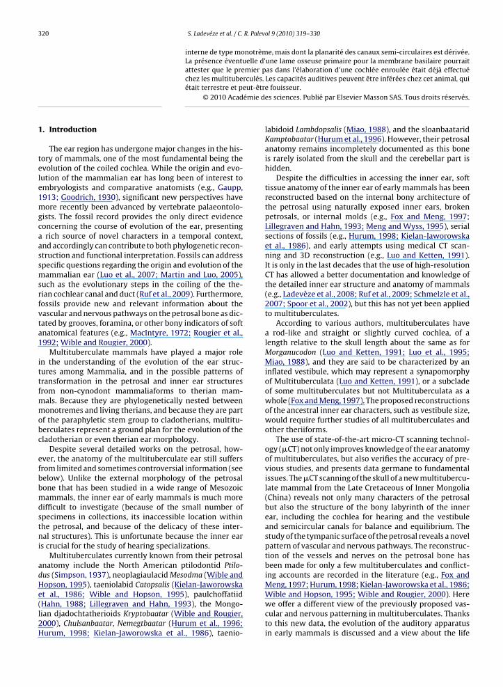
Fig. 1. Skull of cf. Tombaatar sp. (IMM 99BM-IV/4) in coronal view, slicedat the level of the auditory region. A: 3D reconstruction (VG Studio max)showing the endocast and the basicranium with the cochlear canals. Thebackground shows the two first incisors, pointing ventrally. B: The cor-responding slide (+006.46) and C: an additional coronal slide (+006.41),with a zoom of the boxed area. A bony lamina (arrow) is visible into the leftcochlear canal and is interpreted as primary bony lamina for attachmentof the medial part of the basilar membrane.Fig. 1. Crâne de cf. Tombaatar sp. (IMM 99BM-IV/4), en vue coronale,coupé au niveau de la région auditive. A: Reconstruction 3D montrantla cavité endocrânienne et la base du crâne avec les canaux cochléaires.L’arrière-plan montre les deux premières incisives qui pointent ventrale-ment. B: La coupe correspondante (+006,46) et C: une coupe coronaleadditionnelle (+006,41), avec un agrandissement de la zone encadrée.Une lame osseuse (flèche) est visible dans le canal cochléaire gauche etest interprétée comme la lame osseuse primaire pour l’attachement de lapartie médiale de la membrane basilaire.Abbreviations: A: b: bottom; l: left; r: right; t, top; B: cochlear canal;fcn: foramen for cochlear nerve; fv: fenestra vestibuli; fvn: foramen forthe vestibular nerve; pbl: primary bony lamina; pr: promontorium; sbl?:
S. Ladevèze et al. / C.
tyle of Asian Late Cretaceous multituberculates is pro-osed.
. Material and methods
The skull of the multituberculate mammal (IMM 99BM-V/4, Fig. 1A) from which the petrosal is here reconstructednd described was discovered in September 1999 duringhe Sino-Belgian expedition in the southern Gobi desertn Inner Mongolia at Bayan Mandahu, Urad Houqi Banner,ayan Nor League. The Late Cretaceous Bayan Mandahu
ocality is situated about 50 km north-west from the cityf Urad Houqi in the southern part of the Gobi Basin nearhe Lang Shan Mountains in China. The Bayan Mandahuedbeds extend there over 12 km length and 2 km widthnd have yielded a large quantity of vertebrates includinginosaurs, lizards, snakes, turtles and mammals. Amonghe latter, some multituberculate mammals have beenescribed (Smith et al., 2001). The deposits are generallyorrelated with the Campanian aged Djadokhta Forma-ion of South-central Mongolia, 350 km to the north-westJerzykiewicz et al., 1993). The red sandstone depositsave been interpreted to represent an eolian sedimentarynvironment but local alluvial and lacustrine componentepresenting a proximal to distal depositional gradient insemi-arid climate have also been documented (Eberth,
993).The specimen is identified as a typical djadochtathe-
ioid multituberculate (crown group Cimolodonta) by theollowing: large frontals, inserted between the nasals andointed anteriorly in the middle, U-shaped frontoparietaluture, sharp edge between the lateral and palatal walls ofremaxilla (rounded in other multituberculates), and pari-tal postorbital process (Kielan-Jaworowska and Hurum,001). The specimen can further be referred to the familyjadochtatheriidae by the subtrapezoidal snout in dorsaliew, with wide anterior margin and lateral margins con-uent with zygomatic arches rather than incurved in frontf the arches. The snout extends for about 50% of the skullength and shows two pairs of vascular foramina on theasals. P2 is absent and replaced by a small diastema, aynapomorphy shared with Catopsbaatar and Tombaatar.he skull is 44 mm long, which is smaller than T. sabuliougier et al. 1997 and C. catapsaloides Kielan-Jaworowska,994. Although this new specimen may represent a newenus, the tooth morphology is quite similar to Tombaatarnd it is thus provisionally referred to cf. Tombaatar n. sp.ts description and precise systematic attribution will beddressed in a work in progress by some of the authors.
CT scanning of IMM 99BM-IV/4 was performed at theniversity of Texas CT facility at Austin. The slice thicknessnd interslice spacing was 0.04275 mm, and the in-planenterpixel spacing was 0.03906 mm, for a total of 1030lices along the coronal axis of the skull. The MIMICS soft-are (®Materialise 2010, Release 13.1; license UMR 7207NRS-MNHN, Paris) was used to complete visualization,
egmentation and 3D rendering. MIMICS allows differentypes of segmentation to be performed. Regions of inter-st (i.e., petrosal bone and inner ear) can be selected withccuracy using threshold method to create segmentationasks and, when necessary, manual outlining operations.maybe the secondary bony lamina for the basilar membrane.
322 S. Ladevèze et al. / C. R. Palevol 9 (2010) 319–330
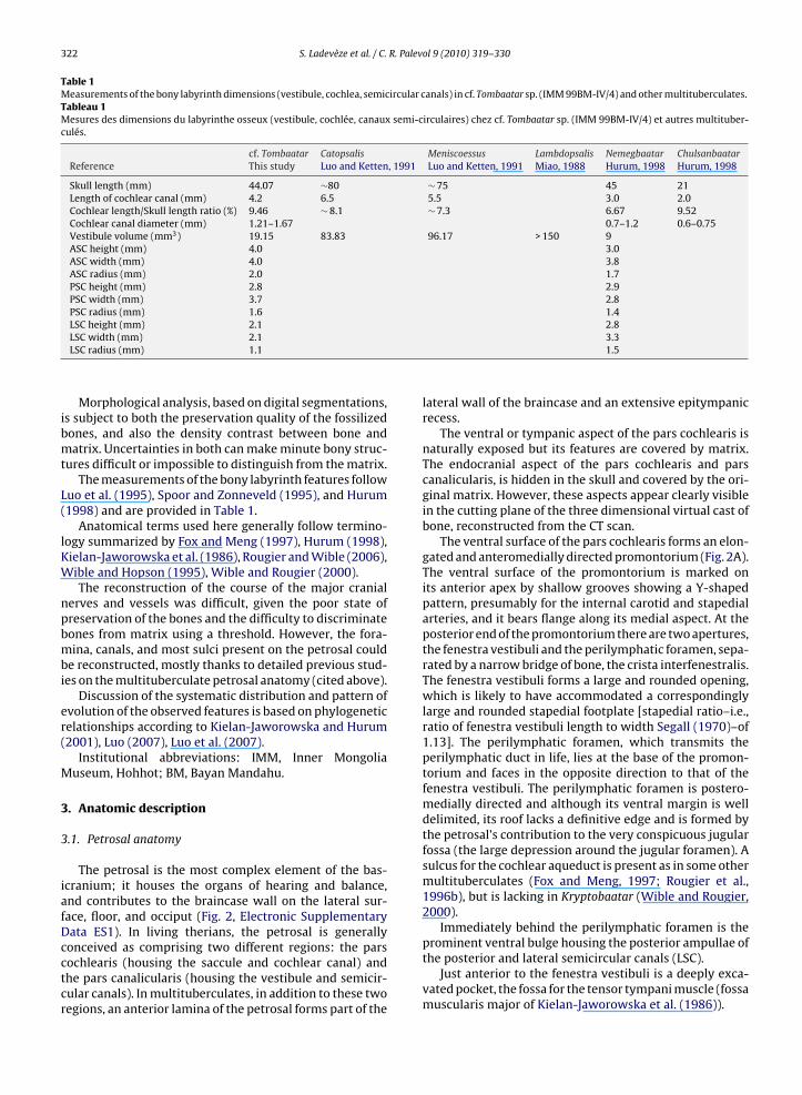
Table 1Measurements of the bony labyrinth dimensions (vestibule, cochlea, semicircular canals) in cf. Tombaatar sp. (IMM 99BM-IV/4) and other multituberculates.Tableau 1Mesures des dimensions du labyrinthe osseux (vestibule, cochlée, canaux semi-circulaires) chez cf. Tombaatar sp. (IMM 99BM-IV/4) et autres multituber-culés.
cf. Tombaatar Catopsalis Meniscoessus Lambdopsalis Nemegbaatar ChulsanbaatarReference This study Luo and Ketten, 1991 Luo and Ketten, 1991 Miao, 1988 Hurum, 1998 Hurum, 1998
Skull length (mm) 44.07 ∼80 ∼ 75 45 21Length of cochlear canal (mm) 4.2 6.5 5.5 3.0 2.0Cochlear length/Skull length ratio (%) 9.46 ∼ 8.1 ∼ 7.3 6.67 9.52Cochlear canal diameter (mm) 1.21–1.67 0.7–1.2 0.6–0.75Vestibule volume (mm3) 19.15 83.83 96.17 > 150 9ASC height (mm) 4.0 3.0ASC width (mm) 4.0 3.8ASC radius (mm) 2.0 1.7PSC height (mm) 2.8 2.9PSC width (mm) 3.7 2.8PSC radius (mm) 1.6 1.4LSC height (mm) 2.1 2.8
LSC width (mm) 2.1LSC radius (mm) 1.1Morphological analysis, based on digital segmentations,is subject to both the preservation quality of the fossilizedbones, and also the density contrast between bone andmatrix. Uncertainties in both can make minute bony struc-tures difficult or impossible to distinguish from the matrix.
The measurements of the bony labyrinth features followLuo et al. (1995), Spoor and Zonneveld (1995), and Hurum(1998) and are provided in Table 1.
Anatomical terms used here generally follow termino-logy summarized by Fox and Meng (1997), Hurum (1998),Kielan-Jaworowska et al. (1986), Rougier and Wible (2006),Wible and Hopson (1995), Wible and Rougier (2000).
The reconstruction of the course of the major cranialnerves and vessels was difficult, given the poor state ofpreservation of the bones and the difficulty to discriminatebones from matrix using a threshold. However, the fora-mina, canals, and most sulci present on the petrosal couldbe reconstructed, mostly thanks to detailed previous stud-ies on the multituberculate petrosal anatomy (cited above).
Discussion of the systematic distribution and pattern ofevolution of the observed features is based on phylogeneticrelationships according to Kielan-Jaworowska and Hurum(2001), Luo (2007), Luo et al. (2007).
Institutional abbreviations: IMM, Inner MongoliaMuseum, Hohhot; BM, Bayan Mandahu.
3. Anatomic description
3.1. Petrosal anatomy
The petrosal is the most complex element of the bas-icranium; it houses the organs of hearing and balance,and contributes to the braincase wall on the lateral sur-face, floor, and occiput (Fig. 2, Electronic SupplementaryData ES1). In living therians, the petrosal is generally
conceived as comprising two different regions: the parscochlearis (housing the saccule and cochlear canal) andthe pars canalicularis (housing the vestibule and semicir-cular canals). In multituberculates, in addition to these tworegions, an anterior lamina of the petrosal forms part of the3.31.5
lateral wall of the braincase and an extensive epitympanicrecess.
The ventral or tympanic aspect of the pars cochlearis isnaturally exposed but its features are covered by matrix.The endocranial aspect of the pars cochlearis and parscanalicularis, is hidden in the skull and covered by the ori-ginal matrix. However, these aspects appear clearly visiblein the cutting plane of the three dimensional virtual cast ofbone, reconstructed from the CT scan.
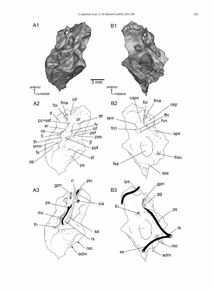
The ventral surface of the pars cochlearis forms an elon-gated and anteromedially directed promontorium (Fig. 2A).The ventral surface of the promontorium is marked onits anterior apex by shallow grooves showing a Y-shapedpattern, presumably for the internal carotid and stapedialarteries, and it bears flange along its medial aspect. At theposterior end of the promontorium there are two apertures,the fenestra vestibuli and the perilymphatic foramen, sepa-rated by a narrow bridge of bone, the crista interfenestralis.The fenestra vestibuli forms a large and rounded opening,which is likely to have accommodated a correspondinglylarge and rounded stapedial footplate [stapedial ratio–i.e.,ratio of fenestra vestibuli length to width Segall (1970)–of1.13]. The perilymphatic foramen, which transmits theperilymphatic duct in life, lies at the base of the promon-torium and faces in the opposite direction to that of thefenestra vestibuli. The perilymphatic foramen is postero-medially directed and although its ventral margin is welldelimited, its roof lacks a definitive edge and is formed bythe petrosal’s contribution to the very conspicuous jugularfossa (the large depression around the jugular foramen). Asulcus for the cochlear aqueduct is present as in some othermultituberculates (Fox and Meng, 1997; Rougier et al.,1996b), but is lacking in Kryptobaatar (Wible and Rougier,2000).
Immediately behind the perilymphatic foramen is the
prominent ventral bulge housing the posterior ampullae ofthe posterior and lateral semicircular canals (LSC).Just anterior to the fenestra vestibuli is a deeply exca-vated pocket, the fossa for the tensor tympani muscle (fossamuscularis major of Kielan-Jaworowska et al. (1986)).
S. Ladevèze et al. / C. R. Palevol 9 (2010) 319–330 323
324 S. Ladevèze et al. / C. R. Palev
ol 9 (2010) 319–330The crista interfenestralis extends posteriorly and con-tacts the paroccipital process of the petrosal, which isa long, prominent and slanted process. Posterior to thecrista interfenestralis and medial to the paroccipital pro-cess is a broad depression in the tympanic roof, the fossafor the stapedius muscle. This fossa is conspicuous andits size is at least twice the surface area of the fenestravestibuli.
The paroccipital process probably represented the siteof attachment for the long muscle of the neck, the ster-nomastoid muscle.
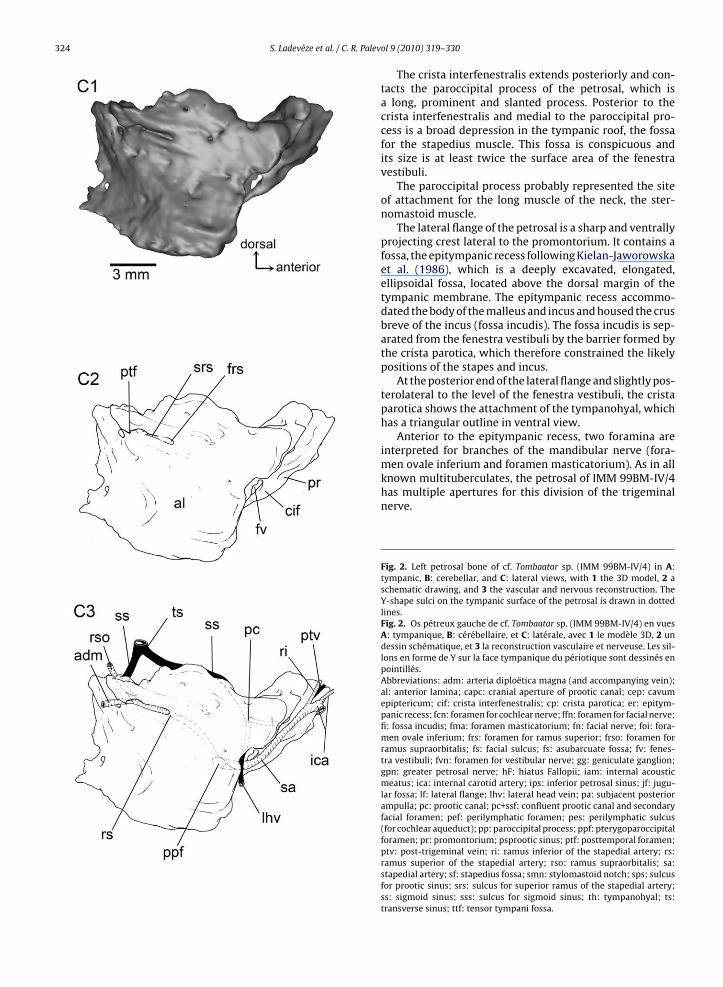
The lateral flange of the petrosal is a sharp and ventrallyprojecting crest lateral to the promontorium. It contains afossa, the epitympanic recess following Kielan-Jaworowskaet al. (1986), which is a deeply excavated, elongated,ellipsoidal fossa, located above the dorsal margin of thetympanic membrane. The epitympanic recess accommo-dated the body of the malleus and incus and housed the crusbreve of the incus (fossa incudis). The fossa incudis is sep-arated from the fenestra vestibuli by the barrier formed bythe crista parotica, which therefore constrained the likelypositions of the stapes and incus.
At the posterior end of the lateral flange and slightly pos-terolateral to the level of the fenestra vestibuli, the cristaparotica shows the attachment of the tympanohyal, whichhas a triangular outline in ventral view.
Anterior to the epitympanic recess, two foramina areinterpreted for branches of the mandibular nerve (fora-
men ovale inferium and foramen masticatorium). As in allknown multituberculates, the petrosal of IMM 99BM-IV/4has multiple apertures for this division of the trigeminalnerve.Fig. 2. Left petrosal bone of cf. Tombaatar sp. (IMM 99BM-IV/4) in A:tympanic, B: cerebellar, and C: lateral views, with 1 the 3D model, 2 aschematic drawing, and 3 the vascular and nervous reconstruction. TheY-shape sulci on the tympanic surface of the petrosal is drawn in dottedlines.Fig. 2. Os pétreux gauche de cf. Tombaatar sp. (IMM 99BM-IV/4) en vuesA: tympanique, B: cérébellaire, et C: latérale, avec 1 le modèle 3D, 2 undessin schématique, et 3 la reconstruction vasculaire et nerveuse. Les sil-lons en forme de Y sur la face tympanique du périotique sont dessinés enpointillés.Abbreviations: adm: arteria diploëtica magna (and accompanying vein);al: anterior lamina; capc: cranial aperture of prootic canal; cep: cavumepiptericum; cif: crista interfenestralis; cp: crista parotica; er: epitym-panic recess; fcn: foramen for cochlear nerve; ffn: foramen for facial nerve;fi: fossa incudis; fma: foramen masticatorium; fn: facial nerve; foi: fora-men ovale inferium; frs: foramen for ramus superior; frso: foramen forramus supraorbitalis; fs: facial sulcus; fs: asubarcuate fossa; fv: fenes-tra vestibuli; fvn: foramen for vestibular nerve; gg: geniculate ganglion;gpn: greater petrosal nerve; hF: hiatus Fallopii; iam: internal acousticmeatus; ica: internal carotid artery; ips: inferior petrosal sinus; jf: jugu-lar fossa; lf: lateral flange; lhv: lateral head vein; pa: subjacent posteriorampulla; pc: prootic canal; pc+ssf: confluent prootic canal and secondaryfacial foramen; pef: perilymphatic foramen; pes: perilymphatic sulcus(for cochlear aqueduct); pp: paroccipital process; ppf: pterygoparoccipitalforamen; pr: promontorium; psprootic sinus; ptf: posttemporal foramen;ptv: post-trigeminal vein; ri: ramus inferior of the stapedial artery; rs:ramus superior of the stapedial artery; rso: ramus supraorbitalis; sa:stapedial artery; sf: stapedius fossa; smn: stylomastoid notch; sps: sulcusfor prootic sinus; srs: sulcus for superior ramus of the stapedial artery;ss: sigmoid sinus; sss: sulcus for sigmoid sinus; th: tympanohyal; ts:transverse sinus; ttf: tensor tympani fossa.
R. Palev
omtttpaafort
tmotah
cAsca(itcfimchftttiotvg
ih
mdcttmfit
btfl
S. Ladevèze et al. / C.
A rounded foramen lies just posterior to the foramenvale inferium and is separated from it by a crest. This fora-en is interpreted as the prootic canal for the small vein of
he prootic sinus supplying the lateral head vein. Throughhis aperture also passed the hyomandibular branch ofhe facial nerve, which took the facial sulcus, medial andarallel to the medial crested border of the lateral flange,nd left the middle ear via a stylomastoid notch immedi-tely posterior to the tympanohyal. Anteriorly to the largeoramen for the prootic vein and the hyomandibular branchf the facial nerve is a smaller hiatus Fallopii, opened ante-iorly onto a distinct sulcus for the greater petrosal nerve,he palatine branch of the facial nerve.
Just posterior to the fenestra vestibuli and medial to theympanohyal is a recessed area that contains one large fora-
en leading into a canal within the petrosal. This foramenpens into a posterolaterally directed channel and likelyransmitted one of the two end branches of the stapedialrtery, the ramus superior, into the ventral ascending canal,ere into the pterygoparoccipital foramen.
Two dominant features are evident on the dorsal orerebellar view of the petrosal of IMM 99BM-IV/4 (Fig. 2B).nteromedially, the internal acoustic meatus forms aubcircular depression and lies on the roof of the parsochlearis. At the posteromedial edge of the meatus is annteroventrally directed foramen for the cochlear nervepart of cranial nerve VIII). In the deeper part of the meatuss a broad fossa containing two foramina: one posteroven-rally directed aperture for the vestibular nerve (part ofranial nerve VIII) and one anteroventrally directed for theacial nerve (cranial nerve VII). The canal for the facial nerves directed anteromedially and its anterior opening, the pri-
ary facial foramen, opens directly into the back of theavum epiptericum where the geniculate ganglion wouldave been located. A separate cavum supracochleare for the
acial ganglion is thus lacking in IMM 99BM-IV/4, as in Kryp-obaatar (Wible and Rougier, 2000). The secondary exit ofhe facial nerve from the cavum epiptericum is in the pos-erior part of the cavum’s floor and opens anteroventrallynto the hiatus Fallopii, the conduit for the anterior branchf the facial nerve, the greater petrosal nerve. Lateral tohe internal acoustic meatus is the cavum epiptericum, theery deep fossa that lodged the trigeminal (or semilunar)anglion of cranial nerve V.
At the anterior apex of the petrosal and anterior to thenternal acoustic meatus is a large groove, which probablyas received the inferior petrosal sinus.
Posterodorsally, the subarcuate fossa, which accom-odated the paraflocculus of the cerebellum, is a large,
eep, subspherical depression that opens into the brain-ase through an elliptical aperture. This fossa is bounded byhe three semicircular canals, with the anterior boundinghe entrance of the subarcuate fossa. On the posterior-most
argin of the subarcuate fossa is a broad sulcus, probablyor the sigmoid sinus. Anterolateral to the subarcuate fossan the anterior lamina is the deep, broad, and anteroven-
rally directed sulcus for the prootic sinus.In lateral view (Fig. 2C), the petrosal is mostly formedy the anterior lamina, which is ventrally continuous withhe tympanic surface of the petrosal through the lateralange, but posteriorly overlapped by the squamosal. The
ol 9 (2010) 319–330 325
anterior lamina wholly or partially formed a number of pas-sageways for nerves and vessels leaving the braincase forthe orbitotemporal fossa, and also provided a major area ofattachment for several muscles of mastication. The dorsalpart of the anterior lamina is pierced by a foramen whichopens caudally into a deep sulcus. This foramen is for theexit of the superior ramus of the stapedial artery, whichdivided into two branches. The posterior branch ran intothe rounded posttemporal foramen and transmitted thediploëtic vessels (arteria diploëtica magna and accompa-nying vein). The dorsoposterior branch ran into a dorsalforamen and transmitted the ramus supraorbitalis.
3.2. Bony labyrinth anatomy
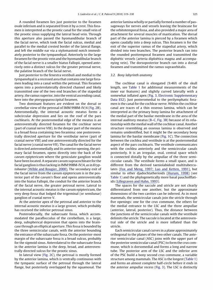
The cochlear canal is elongated (9.46% of the skulllength, see Table 1 for additional measurements of theinner ear features) and slightly curved laterally with asomewhat inflated apex (Fig. 3, Electronic SupplementaryData ES2). Just posteromedial to the perilymphatic fora-men is the canal for the cochlear nerve. Within the cochlearcanal are traces of a thin osseous lamina, which can beinterpreted as the primary bony lamina for attachment ofthe medial part of the basilar membrane in the area of theinternal auditory meatus (B–C, Fig. 3B) because of its rela-tionship with the medial wall of the cochlear canal. Anotherstructure resembling an osseous lamina is observed andremains unidentified, but it might be the secondary bonylamina for the basilar membrane (Fig. 1C). The connectionbetween the cochlea and vestibule is at the posteromedialaspect of the pars cochlearis. The vestibule communicateswith the cochlea anteriorly and the semicircular canalsposteriorly. It is an irregular, oval, central space whichis connected distally by the ampullae of the three semi-circular canals. The vestibule forms a small space, and isdifferent from the derived taeniolabidoid multitubercu-lates (Fox and Meng, 1997; Luo and Ketten, 1991), butsimilar to other djadochtatherioids (Hurum, 1998) (seeTable 1) and the phylogenetically more basal paucholffati-ids (Lillegraven and Hahn, 1993).
The spaces for the saccule and utricle are not clearlydifferentiated from one another, but the approximatedimensions of the two cavities can be inferred. As in mostmammals, the semicircular canals join the utricle throughfive openings: one for the crus commune, the others forthe medial entrance to the LSC and the three ampullae(anterior, lateral, posterior). Thus, the distance betweenthe junctions of the semicircular canals with the vestibuledelimits the utricle. The saccule is located at the anteroven-tral side of the utricle and is more inflated than theutricle.
Each semicircular canal curves in a plane approximatelyorthogonal to the planes of the two other canals. The ante-rior semicircular canal (ASC) joins with the upper part ofthe posterior semicircular canal (PSC) to form the crus com-mune, which is dorsomedial and forms a long and narrow
tube. The posterior arm of the LSC and the inferior armof the PSC build a bony second crus commune, a variablestructure among mammals. The ASC is the longest (Table 1)and forms an almost complete half-circle before it ends inthe anterior ampullar recess (Fig. 3). The LSC is distinctly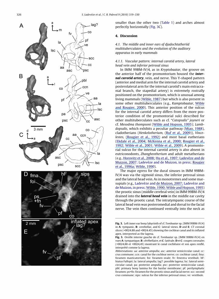
326 S. Ladevèze et al. / C. R. Palev
ol 9 (2010) 319–330smaller than the other two (Table 1) and arches almostperfectly horizontally (Fig. 3C).
4. Discussion
4.1. The middle and inner ears of djadochtatheriidmultituberculates and the evolution of the auditoryapparatus in early mammals
4.1.1. Vascular pattern: internal carotid artery, lateralhead vein and inferior petrosal sinus
In IMM 99BM-IV/4, as in Kryptobaatar, the groove onthe anterior half of the promontorium housed the inter-nal carotid artery, vein, and nerve. This Y-shaped pattern(anterior and medial arm for the internal carotid artery andposterolateral arm for the internal carotid’s main extracra-nial branch, the stapedial artery) is extremely rostrallypositioned on the promontorium, which is unusual amongliving mammals (Wible, 1987) but which is also present insome other multituberculates (e.g., Kamptobaatar, Wibleand Rougier, 2000). This anterior position of the sulcusfor the internal carotid artery differs from the more pos-terior condition of the promontorial sulci described forother multituberculates such as cf. “Catopsalis” joyneri orcf. Mesodma thompsoni (Wible and Hopson, 1995); Lamb-dopsalis, which exhibits a peculiar pathway (Miao, 1988),cladotherians (Henkelotherium, (Ruf et al., 2009)), Vince-lestes, (Rougier et al., 1992) and most basal eutherians(Ekdale et al., 2004; McKenna et al., 2000; Rougier et al.,1992; Wible et al., 2001; Wible et al., 2009). A promonto-rial sulcus for the internal carotid artery is also absent ineutriconodonts, Zhangheotherium and adult metatherians(e.g., Horovitz et al., 2008; Hu et al., 1997; Ladevèze and deMuizon, 2007; Ladevèze and de Muizon, in press; Rougieret al., 1996a; Wible, 1990).
The major egress for the dural sinuses in IMM 99BM-IV/4 was via the sigmoid sinus, the inferior petrosal sinusand the lateral head vein. As in monotremes and some mar-supials (e.g., Ladevèze and de Muizon, 2007; Ladevèze andde Muizon, in press; Wible, 1990; Wible and Hopson, 1995)
the prootic sinus (middle cerebral vein) in IMM 99BM-IV/4drained into the lateral head vein in the middle ear cavitythrough the prootic canal. The intratympanic course of thelateral head vein was posteromedial and dorsal to the facialnerve. The vein then continued ventrally into the neck asFig. 3. Left inner ear bony labyrinth of cf. Tombaatar sp. (IMM 99BM-IV/4)in A: tympanic, B: cerebellar, and C: lateral views. D and E: CT coronalslices (+0024.88 and +0024.45) showing the cochlear canal and its inflatedapex, interpreted as the lagena.Fig. 3. Oreille interne gauche de cf. Tombaatar sp. (IMM 99BM-IV/4) envues A: tympanique, B: cérébellaire, et C: latérale. D et C: coupes coronales(+0024,88 et +0024,45) montrant le canal cochléaire et son apex renflé,interprété comme la lagena.Abbreviations: aa: anterior ampulla; asc: anterior semicircular canal; cc:crus commune; ccn: canal for the cochlear nerve; co: cochlear canal; fma:foramen masticatorium; foi: foramen ovale; fv: fenestra vestibuli; hF:hiatus Fallopii; la: lateral ampulla; lag?: possible lagena; lsc: lateral semi-circular canal; pa: posterior ampulla; psc: posterior semicircular canal;pbl: primary bony lamina for the basilar membrane; pf: perilymphaticforamen; ps+fn: foramen for the prootic sinus and facial nerve; scc: secondcrus commune; sips: sulcus for the inferior petrosal sinus; ve: vestibule.
R. Palev
tbt(KihaHtas
gmtstttamaTrpdtR
etanitrepttatcraaptalccat1r
4
a
S. Ladevèze et al. / C.
he internal jugular vein. The lateral head vein most proba-ly did not leave the tympanic cavity with the facial nervehrough the stylomastoid notch but via a more medial exitWible and Hopson, 1995; Wible and Rougier, 2000; contraielan-Jaworowska et al., 1986; Miao, 1988). A posttrigem-
nal vein (the other major tributary of the lateral head vein)as been reconstructed accompanying the ramus inferior,s in the other multituberculates described by Wible andopson (1995) and Wible and Rougier (2000). However,
he presence of the posttrigeminal vein in IMM 99BM-IV/4nd other multituberculates remains equivocal, withoutpecific osteological evidence.
The inferior petrosal sinus ran in a large, superficialroove on the anteromedial region of the petrosal. In extantammals, this sinus guides the cavernous sinus blood to
he internal jugular vein along the petrosal-basioccipitaluture (Rougier et al., 1996b). Given the broad size ofhe jugular foramen in IMM 99BM-IV/4, we believe thathis served as the exit for the inferior petrosal sinus fromhe cranial cavity. This contrasts with Kryptobaatar (Wiblend Rougier, 2000) in which the inferior petrosal sinusust have left the cranial cavity via the foramen magnum
s vertebral veins, as in monotremes (Hochstetter, 1896).his pathway more closely resembles that of marsupials,epresenting a derived condition relative to the plesiomor-hic course of this sinus (via the foramen magnum) asescribed for other non-therian mammals (Morganucodon,riconodontids Vincelestes, other multituberculates; seeougier et al., 1996a, b).
In IMM 99BM-IV/4, the prootic sinus enters the middlear cavity through the prootic canal, which also housedhe facial nerve (secondary facial foramen). The cranialperture of the prootic canal is anterolateral to the inter-al acoustic meatus and apparently leads the prootic sinus
nto the cavum epiptericum space. Actually, the preserva-ion of the fossil does not allow us to clearly identify andeconstruct a separate canal for the prootic sinus. How-ver, we believe that, as in other multituberculates, therootic sinus was enclosed into a canal and separated fromhe cavum epiptericum space. It is assumed that, in mul-ituberculates, the cranial aperture of the prootic canal ist the anterodorsal margin of the subarcuate fossa, andhe tympanic aperture is confluent with the pterygoparoc-ipital foramen (ventral ascending canal for the superioramus of the stapedial artery) (Rougier et al., 1996b; Wiblend Rougier, 2000). Nonetheless, cf. Tombaatar sp. exhibitsdifferent pattern, in which the cranial aperture of the
rootic canal is anterolateral to the internal acoustic mea-us and the pterygoparoccipital foramen is a separateperture, much more posterior to the prootic canal, lyingateral and posterodorsal to the fenestra vestibuli. The non-onfluence of the pterygoparoccipital foramen and prooticanal is also found in Chulsanbaatar (Kielan-Jaworowska etl., 1986), “Catopsalis” joyneri, and in an isolated petrosalentatively assigned to Meniscoessus (Wible and Hopson,995). The condition in paulchoffatiids and Nemegtbaatar
emains uncertain..1.2. Neural anatomy: facial and trigeminal nervesThe facial nerve entered the middle ear through the
nterolateral foramen in the deepest part of the internal
ol 9 (2010) 319–330 327
acoustic meatus. It differs from Kryptobaatar (Wible andRougier, 2000), in that the facial foramen is separated fromthe vestibular foramen, and the primary facial foramenopens into the cavum supracochleare, which is not closedby an osseous floor and contained the geniculate ganglionat the back of the cavum epiptericum, as in other describedmultituberculates.
According to Miao (1993), the internal acoustic mea-tus of multituberculates, even in the most derived formssuch as Lambdopsalis, is shallow and not as distinct asthat of extant mammals, whereas a well-defined and deepinternal acoustic meatus is only found in monotremes andtherians. However, the internal acoustic meatus of IMM99BM-IV/4 is conspicuous, with a depression housing twoseparated areas for nervous circulation. In this aspect, IMM99BM-IV/4 is more similar to recent mammals than to mor-ganucodontids.
The greater petrosal nerve or palatine ramus of the facialnerve leaves the anterior part of the geniculate ganglionand via the hiatus Fallopii, which opens in a tympanic posi-tion in IMM 99BM-IV/4. This condition is unusual amongmultituberculates. In Kryptobaatar, the entrance of thegreater petrosal nerve is unclear, with no obvious tympanicaperture for the greater petrosal nerve (Wible and Rougier,2000). The ‘hiatus Fallopii’ (original quotes) described byKielan-Jaworowska et al. (1986) for Chulsanbaatar is morelikely to be the posterior aperture of the carotid canal, assuggested by Wible and Rougier (2000).
An interesting character is the position of the foram-ina for the branches of the trigeminal nerve (foramenovale inferium and foramen masticatorium). Among theCretaceous multituberculates, the anterior lamina in Sloan-baatar has only the foramen ovale inferium, whereas theanterior lamina has both foramina masticatorium andovale inferium in Kryptobaatar, Kamptobaatar, Nemegt-baatar, Chulsanbaatar and Catopsbaatar (Wible and Rougier,2000). The foramina are roughly at the same level in Kryp-tobaatar, Kamptobaatar and Nemegtbaatar, but the foramenovale inferium is more posterior in Sloanbaatar, Chulsan-baatar, and Catopsbaatar, as in IMM 99BM-IV/4.
4.1.3. Inner ear morphologyThe 3D reconstruction of the inner ear of IMM 99BM-
IV/4 and its comparison with other multituberculatesprovides more accurate distribution of the phylogeneticcharacters. The first concerns the vestibule size. Thevestibules described in ?Meniscoessus and ?Catopsalis byLuo and Ketten (1991) and the vestibule of Lambdopsalis(known by an endocast of the inner ear; Meng and Wyss,1995) are extremely large, suggesting that the saccule, utri-cle, or both were inflated to a much larger extent thanin other mammals. This inflated vestibule has thus beenregarded as a possible synapomorphy of Multituberculata(Luo and Ketten, 1991) or a uniquely derived feature onlyfor one or more subgroups within the Multituberculata(Fox and Meng, 1997). The vestibules of paulchoffatiids,
Nemegtbaatar and Chulsanbaatar, however, are not moreinflated than in most typical mammals (Hurum, 1998;Lillegraven and Hahn, 1993; contra Luo and Ketten, 1991).The new 3D restoration of cf. Tombaatar sp. (IMM 99BM-IV/4) shows the vestibule is not enlarged, and this is
R. Palev
328 S. Ladevèze et al. / C.consistent with the phylogenetically more basal multi-tuberculates who show a typical mammalian vestibulesize.
The cochlear canal of IMM 99BM-IV/4 is slightlylaterally curved. This condition is found in all known mul-tituberculates, in which the cochlea is definitely curved,and the direction of curvature matches that in other mam-mals (clockwise in the right ear and counterclockwise inthe left ear). However, according to Fox and Meng (1997),the degree of curvature in multituberculates differs amongdifferent taxa. The �CT scans reveal two particularly inter-esting features of the cochlear canal. A thin bony laminais observed (Fig. 1B–C), connected to the medial wall ofthe cochlear canal, near the cochlear nerve entry point(B–C, Fig. 3B). This lamina may be interpreted as the pri-mary bony lamina for support of the basilar membrane,as described in Ornithorhynchus (Pritchard, 1881) and Chul-sanbaatar (Hurum, 1998). The dimensions of the basilarmembrane cannot be estimated because of the possibleabsence of a secondary bony lamina. The other featureof interest of the cochlear canal is the marked inflationof its apical region (Fig. 3), which suggests the presenceof a lagena or lagena-like structure, previously knownonly in monotremes among mammals and one multitu-berculate specimen (UALVP 26039) (Fox and Meng, 1997).Previous attempts at reconstructing the inner ear of mul-tituberculates from X-ray tomographies (Luo & Ketten,1991) indicated that the cochlear canal was distally narrow,rather than expanded, in some multituberculates and inmonotremes. This supports the assertion of Fox and Meng(1997) in that these 3D reconstructions of the cochlearcanal based on serial sections had been too coarse to cap-ture details of its actual shape. The presence of a lagena atthe distal end of the cochlear duct is a specialization com-parable to that in reptiles and birds (see Fox and Meng,1997 for more details). Fernández and Schmidt (1963) pro-posed that the lagena was lost in the common ancestor ofmarsupials and placentals as a result of the coiling of thecochlea.
The conformation of the semicircular canals of IMM99BM-IV/4 approaches that of therian mammals in thatthey are more planar (= they follow the arc of a true circlemore accurately) than are monotremes. This is also the casein two other multituberculates, Nemegtbaatar and Chulsan-baatar (Hurum, 1998).
4.1.4. Phylogenetic implicationsAt first sight, the overall ear morphology of the
djadochtatheriid cf. Tombaatar sp. (IMM 99BM-IV/4) isnot strikingly similar to that of any other multitubercu-lates. The promontorium shape only resembles to thatof other djadochtatherioid multituberculates. The vascu-lar and nervous patterns are quite different from thepreviously described patterns of arterial, venous andnervous circulation in other multituberculates, and par-ticularly the djadochtatheriid Kryptobaatar, suggesting
that some previous interpretations and reconstructions(Kielan-Jaworowska et al., 1986; Wible and Hopson, 1995;Wible and Rougier, 2000) may have been biased by thefossil preservation and by the difficulty in accessing thesecryptic morphologies without intrusive technology, suchol 9 (2010) 319–330
as the �CT scanning. However, the study of the rela-tive positions of the foramina for the branches of thetrigeminal nerve indicates a morphological affinity withCatopsbaatar, the other djadochtatheriid. The inner earmorphology of cf. Tombaatar sp. (IMM 99BM-IV/4) is typicalfor multituberculate mammals and we here highlight anew appraisal of the vestibule size and shape and con-firm the previously suspected retention of a lagena inmultituberculates.
The �CT technology allows reevaluation of the ear mor-phology for multituberculates, with the djadochtatheriidcf. Tombaatar sp. (IMM 99BM-IV/4) showing a mosaicof plesiomorphic and derived characters, with a vas-cular and nervous pattern more cladotherian-like thanmultituberculate-like, a therian-like internal acoustic mea-tus, and a monotreme-like (or even sauropsid-like) innerear (slightly coiled cochlear canal, lagena, small vestibule,long and thin crus commune), but with a derived andtherian-like semicircular canal planarity.
4.2. Functional implications: auditory capabilities andbehaviour
The semicircular canal system of vertebrates helps coor-dinate body movements, including stabilization of gazeduring locomotion, and can be used as a proxy for inferringlocomotion habits in vertebrates (e.g., Spoor et al., 2007;Walsh et al., 2009). The 3D reconstruction of the semicir-cular canals of IMM 99BM-IV/4 shows hitherto unknowncondition for multituberculates. The LSC is indeed veryshort and thin with almost perfectly vertical arches,whereas the ASC is large and arched in a round half cir-cle. A small LSC can be regarded as consistent with less agilebehavior (Silcox et al., 2009; Spoor et al., 2007). Moreover ithas been concluded that the reduction and verticality of theLSC in humans was related to their upright posture (Spooret al., 1994). Another analogy can be made with some cimo-lestan mammals (Aphronorus and Eoryctes), in which theLSC is also the shortest semicircular canal, and which werelikely to have been terrestrial or fossorial animals (Boyerand Georgi, 2007).
Another relevant inner ear feature that potentially canbe used to infer hearing capability relates to the cochlearanatomy. The length of the basilar membrane is related tothe upper and lower frequency limits of therian hearing(West, 1985). The cochlear canal of cf. Tombaatar sp. hasa primary bony lamina but apparently lacks a secondarybony lamina in support of the basilar membrane, whichwas thus probably less efficient in responding to high-frequency airborne vibrations (see Fox and Meng 1997).However this structure is extremely variable among mam-mals (Fleischer, 1973; Ruf et al., 2009). The cochlea ofcf. Tombaatar sp. is likely to have supported the basilarmembrane and contained at its end a lagena macula, asin monotremes. Because of its coma-shaped and apicallyexpanded cochlea, cf. Tombaatar sp. was likely to have been
more sensitive to low-frequency sounds (Manoussaki et al.,2008). As a matter of fact, the mammalian cochlea performsanalysis of the frequency spectrum of auditory signalsby processing different frequencies at different locationsalong the cochlear duct, the low-frequency sounds lead-
R. Palev
i(
isfsb(ftwmmswdsfsntMirciToft
bahbh1amtKE
TotGooTtosel
A
r
S. Ladevèze et al. / C.
ng to maximum vibrations at the apical end of the cochleatonotopy principle, Békésy, 1960).
The study of the middle ear anatomy provides othernformation as to the auditory capability of cf. Tombaatarp. The stapedial footplate (estimated from the size of theenestra vestibuli) was very large, suggesting cf. Tombaatarp. might have been fossorial, as stapedial footplates maye significantly larger with respect to body size in fossorialand low-frequency hearing) species compared to non-ossorial species (Burda et al., 1992). However, in contrasto other fossorial species (e.g., most fossorial placentals),hich have large stapedial footplates and degeneratediddle ear muscles (Burda et al., 1992), the stapediususcle is not reduced in cf. Tombaatar sp. This differs
omewhat from the presumed fossorial Lambdopsalis bulla,hich is reported to have a large fenestra vestibuli, stronglyeveloped middle ear ossicles, but a reduced fossa for thetapedius muscle (Miao, 1988). Furthermore, while theossa for the stapedius muscle is broad in cf. Tombaatarp., the fossa for the tensor tympani muscle is small andarrow, as compared to other multituberculates in whichhe tensor tympani muscle occupied a deep recess (Fox and
eng, 1997; Wible and Rougier, 2000). A similar conditions present in the fossorial marsupials Necrolestes and Noto-yctes (Ladevèze et al., 2008), in which the cochlea is little-oiled (1.1 and 1.6, respectively) and the stapedius muscles strongly developed. These observations suggest that cf.ombaatar sp. and the marsupial moles might have devel-ped an alternative specialization to a fossorial way of liferom the one reported in placental forms (i.e., strong reduc-ion of the middle ear muscles, see Ladevèze et al., 2008).
Most Asian multituberculates, whose lifestyles haveeen reconstructed based primarily on postcranialnatomy have been interpreted as terrestrial. However, itas been suggested that some multituberculates may haveeen fossorial, while some later multituberculates mightave been arboreal (Kielan-Jaworowska and Gambaryan,994; Kielan-Jaworowska et al., 2004). Specializations tofossorial lifestyle have been suggested for two Asianultituberculates: a possible djadochtatherioid from
he Djadokhta Formation (Kielan-Jaworowska, 1989;ielan-Jaworowska et al., 2004) and Lambdopsalis from theocene of China (Kielan-Jaworowska et al., 2004).
The semicircular canal system of the new specimen (cf.ombaatar sp.) described here is consistent with a low levelf agility, and, as expected for a Late Cretaceous multi-uberculate from Mongolia (see Kielan-Jaworowska andambaryan, 1994), it is likely that it was terrestrial. More-ver, the anatomy of both the petrosal and bony labyrinthf cf. Tombaatar sp. is compatible with fossorial habits.hese preliminary conclusions nonetheless need to be fur-her tested by improved and combined statistical analysesf the degree of correlation of metric parameters from theemicircular canal system with locomotion habits (Davidt al., 2010, this issue) to infer with more confidence aocomotor pattern for multituberculates.
cknowledgements
We thank the High-Resolution X-ray Computed Tomog-aphy Facility at The University of Texas at Austin - UTCT
ol 9 (2010) 319–330 329
for producing the CT scans. The three dimensional modelswere reconstructed thanks to Volume Graphics© (VG Stu-dio Max, licence Univ. Texas, Austin), Materialise© (MIMICSv. 13, licence UMR 7207, MNHN Paris). We are indebted tothe 3D team of the UMR7207 (Gaël Clement, Didier Geffard-Kuriyama, Florent Goussard, Apollo XIII, Skylab, and JP).We are grateful to Romain David (UMR7207, USM203,MNHN Paris) for his help and fruitful discussions aboutthe inner ear anatomy, and Renaud Vacant (UMR7207,USM203, MNHN Paris) for the preparation of the multi-tuberculate skull. This work was supported by ResearchProjects MO/36/001 and MO/36/004 and bilateral coop-eration project BL/36/C12 of the Belgian Federal SciencePolicy Office. We wish to thank Gaël Clement and DidierGeffard-Kuriyama for inviting us to propose a manuscriptfor this special volume, and Zhe-Xi Luo and an anonymousreviewer for their helpful comments that greatly improvedthe manuscript.
Appendix A. Supplementary data
Electronic Supplementary Data associated withthis article can be found, in the online version, atdoi:10.1016/j.crpv.2010.07.008.
References
Békésy, G.V., 1960. Experiments in Hearing. McGraw-Hill, New York.Boyer, D.M., Georgi, J.A., 2007. Cranial morphology of a pantolestid
eutherian mammal from the Eocene Bridger Formation, Wyoming,USA: implications for relationships and habitat. J. Mamm. Evol. 14,239–280.
Burda, H., Bruns, V., Hickman, G.C., 1992. The ear in subterraneanInsectivora and Rodentia in comparison with ground-dwelling repre-sentatives. 1. Sound conducting system of the middle ear. J. Morphol.214, 49–61.
David, R., Droulez, J., Allain, R., Berthoz, A., Janvier, P., Bennequin, D., 2010.Motion from the past. A new method to infer vestibular capacities ofin extinct species. C. R. Palevol. 9, 397–410.
Eberth, D.A., 1993. Depositional environments and facies transitionsof dinosaur-bearing Upper Cretaceous redbeds at Bayan Mandahu(Inner Mongolia People’s Republic of China). Can. J. Earth Sci. 30,2196–2213.
Ekdale, E.G., Archibald, J.D., Averianov, A.O., 2004. Petrosal bones of pla-cental mammals from the Late Cretaceous of Uzbekistan. Acta Pal. Pol.49, 161–176.
Fernández, C., Schmidt, R.S., 1963. The opossum ear and evolution of thecoiled cochlea. J. Comp. Neurol. 121, 151–159.
Fleischer, G., 1973. Studies of the skeleton of the auditory organs ofmammals including humans. Saeugetierkundliche Mitteilungen 21,131–239.
Fox, R.C., Meng, J., 1997. An X-radiographic and SEM study of the osseousinner ear of multituberculates and monotremes (Mammalia): impli-cations for mammalian phylogeny and evolution of hearing. Zool. J.Linn. Soc. 121, 249–291.
Gaupp, E., 1913. Die Reichertsche Theorie (Hammer-Amboss- und Kiefer-frage). Archiv für Anatomie und Entwicklungsgeschichte 1912, 1–416.
Goodrich, E.S. 1930. Studies on the structure and development of verte-brates. Macmillan, London, 837 p.
Hahn, G., 1988. Die Ohr-Region der Paulchoffatiidae (MultituberculataOber-Jura). Palaeovertebrata 18, 155–185.
Hochstetter, F., 1896. Beiträge zur Anatomie und Entwickelungs-geschichte des Blutgefäßsystem der Monotremen. Semon’s Zoologis-che Forschungsreisen in Australien 5, 189–243.
Horovitz, I., Ladevèze, S., Argot, C., Hooker, J.J., Macrini, T.E., Martin, T.,
Kurz, C., Muizon, C.D., Sánchez-Villagra, M.R., 2008. The anatomy ofHerpetotherium fugax Cope 1873, a metatherian from the Oligocene ofNorth America. Palaeontographica Abt. A 284, 109–141.Hu, Y., Wang, Y., Luo, Z.-X., Li, C., 1997. A new symmetrodont mammalfrom China and its implications for mammalian evolution. Nature 390,137–142.
R. Palev
330 S. Ladevèze et al. / C.Hurum, J.H., 1998. The inner ear of two Late Cretaceous multituberculatemammals, and its implications for multituberculate hearing. J. Mamm.Evol. 5, 65–93.
Hurum, J.H., Presley, R., Kielan-Jaworowska, Z., 1996. The middle ear inmultituberculate mammals. Acta Pal. Pol. 41, 253–275.
Jerzykiewicz, T., Currie, P.J., Eberth, D.A., Johnston, P.A., Koster, E.H., Zheng,J.-J., 1993. Djadokhta Formation correlative strata in Chinese InnerMongolia: an overview of the stratigraphy, sedimentary geology, andpalaeontology and comparisons with the type locality in the pre-AltaiGobi. Can. J. Earth Sci. 30, 2180–2195.
Kielan-Jaworowska, Z., 1989. Postcranial skeleton of a Cretaceous multi-tuberculate mammal. Acta Pal. Pol. 34, 15–18.
Kielan-Jaworowska, Z., Gambaryan, P.P., 1994. Postcranial anatomy andhabits of Asian multituberculate mammals. Fossils Strata 36, 1–92.
Kielan-Jaworowska, Z., Hurum, J.H., 2001. Phylogeny and systematics ofmultituberculate mammals. Palaeontology 44, 389–429.
Kielan-Jaworowska, Z., Cifelli, R.L., Luo, Z.-X., 2004. Mammals from the ageof dinosaurs: origins, evolution, and structure. Columbia UniversityPress, New York (i-xv, 1-630 p).
Kielan-Jaworowska, Z., Presley, R., Poplin, C., 1986. The cranial vascularsystem in taeniolabidoid multituberculate mammals. Phil. Trans. Roy.Soc. B. 313, 525–602.
Ladevèze, S., de Muizon, C., 2007. The auditory region of Early PaleocenePucadelphydae (Mammalia, Metatheria) from Tiupampa, Bolivia, withphylogenetic implications. Palaeontology 50 (Part 5), 1123–1154.
Ladevèze, S., de Muizon, C. 2010. Evidence of early evolution ofAustralidelphia (Metatheria, Mammalia) in South America: phylo-genetic relationships of the metatherians from the Late Palaeoceneof Itaboraí (Brazil) based on teeth and petrosal bones. ZoologicalJournal of the Linnean Society 159, 746–784.doi: 10.1111/j.1096-3642.2009.00577.x.
Ladevèze, S., Asher, R.J., Sánchez-Villagra, M.R., 2008. Petrosal anatomy inthe fossil mammal Necrolestes: evidence for metatherian affinities andcomparisons with the extant marsupial mole. J. Anat. 213, 686–696.
Lillegraven, J.A., Hahn, G., 1993. Evolutionary analysis of the middle andinner ear of Late Jurassic multituberculates. J. Mamm. Evol. 1, 47–74.
Luo, Z.X., 2007. Transformation and diversification in early mammal evo-lution. Nature 450, 1011–1019.
Luo, Z.X., Ketten, D.R., 1991. CT scanning and computerized reconstruc-tions of the inner ear of multituberculate mammals. J. Vert. Paleont.11, 220–228.
Luo, Z.X., Chen, P., Li, G., Chen, M., 2007. A new eutriconodont mammal andevolutionary development in early mammals. Nature 446, 288–293.
Luo, Z.X., Crompton, A.W., Lucas, S.G., 1995. Evolutionary origins of themammalian promontorium and cochlea. J. Vert. Paleont. 15, 113–121.
MacIntyre, G.T., 1972. The trisulcate petrosal pattern of mammals. In:Dobzhansky, T., Hecht, M.K., Steere, W.C. (Eds.), Evolutionary Biology.Plenum Press, New York, pp. 275–303.
Manoussaki, D., Chadwick, R.S., Ketten, D.R., Arruda, J., Dimitriadis, E.K.,O’Malley, J.T., 2008. The influence of cochlear shape on low-frequencyhearing. PNAS 115, 6162–6166.
Martin, T., Luo, Z.X., 2005. Homoplasy in the mammalian ear. Science 307,861–862.
McKenna, M.C., Kielan-Jaworowska, Z., Meng, J., 2000. Earliest eutherianmammal skull, from the Late Cretaceous (Coniacian) of Uzbekistan.Acta Pal. Pol. 45, 1–54.
Meng, J., Wyss, A.R., 1995. Monotreme affinities and low-frequency hear-ing suggested by multituberculate ear. Nature 377, 141–144.
Miao, D. 1988. Skull morphology of Lambdopsalis bulla (Mammalia,Multituberculata) and its implications to mammalian evolution, Con-tribution to Geology, Volume Special Paper 4. University of Wyoming,Laramie (viii + 104 p).
Miao, D., 1993. Cranial morphology and multituberculate relationships. In:Szalay, F.S., Novacek, M.J., McKenna, M.C. (Eds.), Mammal phylogeny:Mesozoic differentiation, multituberculates, monotremes, early the-
rians, and marsupials. Springer-Verlag, New York, pp. 63–74.Pritchard, U, 1881. The cochlea of the Ornithorhynchus platypus comparedwith that of ordinary mammals and birds. Philos. Trans. Roy. Soc.London B 172, 267–282.
Rougier, G.W., Wible, J.R., 2006. Major changes in the ear region and basi-cranium of early mammals. In: Carrano, M., Gaudin, T.J., Blob, R., Wible,
ol 9 (2010) 319–330
J.R. (Eds.), Amniote Paleobiology: Phylogenetic and Functional Per-spectives on the Evolution of Mammals, Birds and Reptiles. Universityof Chicago Press, Chicago, pp. 269–311.
Rougier, G.W., Wible, J.R., Hopson, J.A., 1992. Reconstruction of the cra-nial vessels in the Early Cretaceous mammal Vincelestes neuquenianus:implications for the evolution of the mammalian cranial vascular sys-tem. J. Vert. Paleont. 12, 188–216.
Rougier, G.W., Wible, J.R., Hopson, J.A., 1996a. Basicranial anatomy of Pria-codon fruitaensis (Triconodontidae, Mammalia) from the Late Jurassicof Colorado, and a reappraisal of mammaliaform interrelationships.Am. Mus. Novitates 3183, 1–38.
Rougier, G.W., Wible, J.R., Novacek, M.J., 1996b. Middle-ear ossicles of themultituberculate Kryptobaatar from the Mongolian Late Cretaceous:implications for mammaliamorph relationships and the evolution ofthe auditory apparatus. Am. Mus. Novitates 3187, 1–43.
Ruf, I., Luo, Z.X., Wible, J.R., Martin, T., 2009. Petrosal anatomy and inner earstructures of the Late Jurassic Henkelotherium (Mammalia, Cladothe-ria, Dryolestoidea): insight into the early evolution of the ear regionin cladotherian mammals. J. Anat 214, 679–693.
Schmelzle, T., Maier, W., Sánchez-Villagra, M.R., 2007. Vestibular labyrinthevolution in diprotodontian marsupial mammals. Mammal Study 32,83–97.
Segall, W., 1970. Morphological parallelisms of the bulla and auditoryossicles in some insectivores and marsupials. Fieldiana: Zoology 51,169–205.
Silcox, M.T., Bloch, J.I., Boyer, D.M., Godinot, M., Ryan, T.M., Spoor, F.,Walker, A., 2009. Semicircular canal system in early primates. J. Hum.Evol. 56, 315–327.
Simpson, G.G., 1937. Skull structure of the Multituberculata. Bull. Am. Mus.Nat. Hist. 73, 727–763.
Smith, T., Guo, D.Y., Sun, Y., 2001. A new species of Kryptobaatar (Multi-tuberculata): the first Late Cretaceous mammal from Inner Mongolia(P.R. China). Bull. Inst. R. Sci. Nat. Belg. 71 (Suppl.), 29–50.
Spoor, F., Zonneveld, F., 1995. Morphometry of the primate bony labyrinth:a new method based on high-resolution computed tomography. J.Anat. 186, 271–286.
Spoor, F., Wood, B., Zonneveld, F., 1994. Implications of early hominidlabyrinthine morphology for evolution of human bipedal locomotion.Nature 369, 645–648.
Spoor, F., Bajpai, S., Hussain, S.T., Kumar, K., Thewissen, J.G.M., 2002.Vestibular evidence for the evolution of aquatic behaviour in earlycetaceans. Nature 417, 163–166.
Spoor, F., Garland, T., Krovitz, G., Ryan, T.M., Silcox, M.T., Walker, A., 2007.The primate semicircular canal system and locomotion. PNAS 104,10808–10812.
Walsh, S.A., Barrett, P.M., Milner, A.C., Manley, G., Witmer, L.M., 2009. Innerear anatomy is a proxy for deducing auditory capability and behaviourin reptiles and birds. Proc. R. Soc. B 276, 1355–1360.
West, C.D., 1985. The relationship of the spiral turns of the cochleaand the length of the basilar membrane to the range of audiblefrequencies in ground dwelling mammals. J. Acoust. Soc. Am. 77,1091–1101.
Wible, J.R., 1987. The eutherian stapedial artery: character analysis andimplications for superordinal relationships. Zool. J. Linn. Soc. 91,107–135.
Wible, J.R., 1990. Petrosals of Late Cretaceous marsupials from NorthAmerica, and a cladistic analysis of the petrosal in therian mammals.J. Vert. Paleont. 10, 183–205.
Wible, J.R., Hopson, J.A., 1995. Homologies of the prootic canal in mammalsand non-mammalian cynodonts. J. Vert. Paleont. 15, 331–356.
Wible, J.R., Rougier, G.W., 2000. Cranial anatomy of Krypto-baatar dashzevegi (Mammalia, Multituberculata); and its bearing onthe evolution of mammalian characters. Bull. Am. Mus. Nat. Hist. 247,1–124.
Wible, J.R., Rougier, G.W., Novacek, M.J., McKenna, M.C., 2001. Earliest
eutherian ear region: a petrosal referred to Prokennalestes from theEarly Cretaceous of Mongolia. Am. Mus. Novit 3322, 1–44.Wible, J.R., Rougier, G.W., Novacek, M.J., Asher, R.J., 2009. The eutherianmammal Maelestes gobiensis from the Late Cretaceous of Mongolia andthe phylogeny of Cretaceous Eutheria. Bull. Am. Mus. Nat. Hist. 327,1–123.