309564.pdf - Repositório Aberto da Universidade do Porto
168
Universidade do Porto Faculdade de Engenharia An injectable Sr-releasing hybrid system to promote bone regeneration PhD Thesis Ana Filipa Henriques Ferreira Lourenço 2018
-
Upload
khangminh22 -
Category
Documents
-
view
1 -
download
0
Transcript of 309564.pdf - Repositório Aberto da Universidade do Porto

Universidade do Porto
Faculdade de Engenharia
An injectable Sr-releasing hybrid system to promote bone regeneration
PhD Thesis
Ana Filipa Henriques Ferreira Lourenço
2018

Ana Filipa Henriques Ferreira Lourenço
An injectable Sr-releasing hybrid system to promote bone regeneration
Dissertação submetida à Faculdade de Engenharia da
Universidade do Porto para obtenção do grau de Doutor
em Engenharia Biomédica.
Supervisor: Maria Cristina de Castro Ribeiro
Categoria: Professor adjunto
Afiliação: Instituto de investigação e Inovação em
Saúde/Instituto de Engenharia Biomédica (i3S/INEB);
Instituto Superior de Engenharia do Porto (ISEP)
Co-supervisor: Cristina Maria Santos Alves de Carvalho
Barrias
Categoria: Investigador principal
Afiliação: Instituto de investigação e Inovação em
Saúde/Instituto de Engenharia Biomédica (i3S/INEB)


Funding
Thesis performed at INEB – Instituto de Engenharia Biomédica/ i3s – Instituto de
Investigação e Inovação em Saúde.
This work was financed by FEDER - Fundo Europeu de Desenvolvimento Regional
funds through the COMPETE 2020 - Operacional Programme for Competitiveness and
Internationalisation (POCI), Portugal 2020, and by Portuguese funds through FCT -
Fundação para a Ciência e a Tecnologia/ Ministério da Ciência, Tecnologia e Inovação
in the framework of the project "Institute for Research and Innovation in Health
Sciences" (POCI-01-0145-FEDER-007274) and project PTDC/CTM/103181/2008. AHL
acknowledges FCT for PhD grant: SFRH/BD/87192/2012.


“Nada se ganha, nada se perde, tudo se transforma.”
Lei da conservação de massa, Lavoisier


Acknowledgements
Gostaria de agradecer em primeiro lugar às instituições que me acolheram
(FEUP e INEB/i3S) e permitiram que desenvolvesse o trabalho aqui presente. Ao Prof.
Mário Barbosa agradeço a possibilidade de integrar uma equipa como o grupo
Microenvironments for New Therapies (INEB). Agradeço ao Prof. Fernando Jorge
Monteiro por toda a sua ajuda e disponibilidade.
Gostaria de agradecer às minhas orientadoras por toda a ajuda e amparo que
sempre me deram. Cristina Ribeiro, o meu mais profundo agradecimento por todas as
suas palavras, que foram sempre de um carinho incrível. Agradeço todas as
oportunidades que me deu de crescer, e agradeço principalmente a sua paciência. O
seu coração do tamanho do mundo e a sua amabilidade para com todos são uma
inspiração para mim. Cristina Barrias, muito obrigada pelo seu apoio constante, tanto a
nível académico como pessoal. Estarei sempre disponível para fazer uma massagem.
Muito obrigada às duas por aceitarem entrar comigo neste desafio.
A todos os que de alguma maneira contribuíram para esta tese, o meu mais
sincero obrigado. Foi uma tarefa difícil, mas dos maiores investimentos e
aprendizagens que já fiz. A todos os amigos que fiz no INEB, obrigada pelo
companheirismo e pelas gargalhadas e por me acompanharem desde sempre.
Definitivamente o caminho faz-se andando e vocês ajudaram a trilhar o meu caminho.
Sílvia, Raquelita e Keila muito obrigada por tudo o que me ensinaram.
Obrigada Estrelinha, Caty, Dani V, Dani Sousa, Ana Luísa, Bia, AR Bento,
Ritinha, Fred, Ké, Aidinha, Mary Valente, Ana Freire, David Gomes, Janeca, Laúndos,
L. Pires, Meriem, Ricardo Vidal, Ana Paula Filipe, Vasco, João Pedro Martins,
Bernardo, Joana Silva, Rui Ribeiro, Tatiana, Cátia Lopes, Inês Estevão, Ana Paula
Lima, Andreia Pereira, Patrícia Henriques, Mariana Neves, Chiara, Daniel
Vasconcelos.
Um agradecimento especial aos meninos do laboratório: Andreia, Zé, João
Brás, Xú, Morena, Dudu, Ana João, Mafalda, João Vasco, que fazem sempre tudo
parecer melhor, que dão sempre uma ajuda e a quem nunca falta um sorriso. Muito
obrigada mesmo!
Um obrigado especial ao Rúben. Foste um exemplo de dedicação e trabalho.

À minha Claudete, minha pequena Bey-sister, um obrigada especial pela tua
ajuda, paciência e overall glamness.
Às meninas do prof. FJ: Ângela, Marta R, Tatiana, Lili G, Catarina Coelho,
Joana Barros e Eri, muito obrigada pelo apoio e companhia, em especial para a
Ângela, por seres o exemplo neste final complexo.
Aos que o meu coração escolheu ao pormenor, Sara, Francisca A, Filipa
Sousa, Aninhas, D. Salvador, Grace, Luís Leitão, muito muito obrigada. As vossas
palavras e apoio incondicionais foram o que me manteve à tona.
Às minhas garotas, Tininha e Idália, um obrigada por se manterem perto e me
ajudarem a manter a sanidade e a perspectiva. À minha Sara Neto, obrigada por seres
a minha amiga de sempre.
Aos meus pais, muito obrigada por todas as oportunidades que me
proporcionaram e tudo o que me permitiram ser. Ao meu irmão, ensinaste-me mais do
que o que imaginas e lembra-te que podes fazer tudo o que quiseres e que gosto de ti
tal como és. Às minhas primas (Clarinha e Irene), são os meus exemplos. São vocês
as minhas maiores incentivadoras e não teria conseguido chegar aqui sem vocês.
Obrigada! À minha avó Micas um grande obrigada pelo teu amor. Para a árvore
crescer tem que fortificar as raízes.

Publications
The work performed in the frame of this thesis resulted in the following publications:
A. Henriques Lourenço, A. L. Torres, D. P. Vasconcelos, C Ribeiro-Machado, J. N.
Barbosa, M. A. Barbosa, C. C. Barrias, C. C. Ribeiro, Osteoimmunomodulatory
properties of a strontium-rich injectable hybrid scaffold for bone repair (submitted) –
Chapter III
A. Henriques Lourenço*, N. Neves*, C. Ribeiro-Machado, S.R. Sousa, M. Lamghari,
C.C. Barrias, A. Trigo Cabral, M.A. Barbosa, C.C. Ribeiro, Injectable hybrid system for
strontium local delivery promotes bone regeneration in a rat critical-sized defect model,
Scientific Reports 7(1) (2017) 5098. (*equal contribution) – Chapter IV
Other publications complementary to the thesis:
C. Ribeiro-Machado, A. Henriques Lourenço, N. Neves, N. Alexandre, M. Lamghari,
M. A. Barbosa, Cristina C. Ribeiro, Evaluation of an injectable hybrid system for
strontium local delivery in a sheep vertebrae model (manuscript in preparation).
AHL contributed to data interpretation and manuscript revision.
L. M. Baptista, A. Henriques Lourenço, M. A. Barbosa, I. C. Gonçalves, C. C. Ribeiro,
Antimicrobial properties of Sr-rich microspheres for bone regeneration (manuscript in
preparation).
AHL contributed with microspheres preparation and revision of the manuscript.


xiii
Abstract
Injectable bone substitutes are very attractive for bone tissue regeneration/repair
strategies, since they can be applied via minimally invasive surgical procedures and
can perfectly fill irregular defects created in cases of trauma, tumor resection and bone
pathologies, namely osteoporosis. These materials should combine adequate
injectability and mechanical properties with the ability to induce new bone formation.
Incorporating strontium (Sr) in biomaterials for bone substitution stands out as a
promising strategy to achieve high local Sr concentrations at lesion sites and promote
new bone formation, by taking advantage of its osteoanabolic and anti-osteoclastic
activity. Furthermore, recent evidences of Sr immunomodulatory properties highlight an
additional benefit for its use, as these might synergistically contribute to more efficient
bone repair/regeneration.
In this context, the aim of the present work was to evaluate the in vitro and in vivo
performance of a designed Sr-hybrid injectable system for bone regeneration,
consisting of hydroxyapatite Sr-doped microspheres combined with an in situ
crosslinkable Sr-alginate hydrogel. The system showed to be able to release clinically
relevant Sr2+ amounts, providing a dual delivery, owing to the incorporation of Sr in both
alginate and microspheres, with different release rates due to the different degradation
kinetics of the polymer network (faster) and the ceramic particles (slower). In vitro
results showed that the developed Sr-rich hybrid system was able to support
mesenchymal stem cells (MSC) survival and osteogenic differentiation, with increase in
alkaline phosphatase (ALP) activity, while inhibiting monocyte adhesion and osteoclast
(OC) formation, with decreased tartrate-resistant acid phosphatase (TRAP) production.
For in vivo studies, two different rodent models were used, to evaluate both the
inflammatory response and the bone regeneration potential upon material implantation.
To this end, an air-pouch model and critical-sized bone defect model were used,
respectively, where non Sr-doped similar materials (Ca-hybrid system) and empty
defects were used as controls. In the air-pouch model, an increase in F4/80+/CD206+
cells (M2 macrophages) in inflammatory exudates was observed upon Sr-hybrid
implantation, as compared to the controls. Importantly, cytokine levels upon
implantation were indicative of a mild inflammatory response when compared to Ca-
hybrid system, with no increase in fibrous capsule thickness. The characteristic M2
macrophage phenotype observed is known to be pro-regenerative, namely by
promoting angiogenesis and tissue repair. Furthermore, in the critical-sized bone defect
model, the Sr-hybrid system led to higher material degradation with increased collagen

xiv
fibers deposition and new bone formation in the center of the defect, as opposed to
bone formation restricted to the periphery in the control Ca-hybrid system.
Furthermore, no alterations were observed in the Sr levels in systemic organs in both in
vivo studies, assuring the safety of the proposed system regarding recent
cardiovascular concerns of SrRan products. Most importantly, the hybrid system
provided a scaffold for cell migration and offered structural support for tissue ingrowth,
further improving effective local bone formation.
Overall, results obtained in this thesis suggest that the proposed hybrid system is able
to modulate the inflammatory response, where Sr acts as a pro-resolution mediator
through M2 macrophage polarization. Combined with the osteoanabolic and anti-
osteoclastic activity of Sr, this leads to increased bone formation and enhanced
osteointegration of the biomaterial. Therefore, this system might provide a promising
multifunctional approach for bone regeneration, especially under osteoporotic
conditions.

xv
Resumo
Os materiais injetáveis constituem uma alternativa muito interessante para utilização
em regeneração/reparação de tecido ósseo pois podem ser aplicados usando
procedimentos cirúrgicos minimamente invasivos, permitindo preencher perfeitamente
defeitos irregulares criados em casos de trauma, ressecção tumoral e patologias
ósseas, nomeadamente osteoporose. Estes materiais devem combinar injectabilidade
e propriedades mecânicas adequadas com a capacidade de induzir a formação de
novo osso. A incorporação de estrôncio (Sr) em biomateriais para substituição óssea
destaca-se como uma estratégia promissora, que permite obter elevadas
concentrações locais de Sr no local da lesão e promover a formação de novo osso,
beneficiando da atividade osteoanabólica e anti-osteoclástica deste elemento metálico.
Para além disso, evidências recentes de propriedades imunomodulatórias associadas
ao Sr, constituem um benefício adicional para a sua utilização, podendo assim
contribuir sinergisticamente para uma reparação/regeneração óssea mais eficiente.
Neste contexto, o objectivo do presente trabalho foi avaliar o desempenho in vitro e in
vivo de um sistema injectável híbrido rico em Sr para regeneração óssea, consistindo
em microesferas de hidroxiapatite dopadas com Sr, combinadas com um hidrogel de
alginato reticulado in situ também com Sr. O sistema mostrou ser capaz de libertar
quantidades clinicamente relevantes de Sr2+, fornecendo uma dupla entrega, em
consequência da incorporação de Sr tanto no alginato como nas microesferas, com
diferentes taxas de libertação devido às distintas cinéticas de degradação da rede
polimérica (mais rápida) e das partículas cerâmicas (mais lenta). Resultados in vitro
mostraram que o sistema híbrido desenvolvido rico em Sr, foi capaz de suportar a
sobrevivência de células estaminais mesenquimatosas (MSC) e a sua diferenciação
osteogénica, com aumento de actividade da fosfatase alcalina (ALP), inibindo a
adesão de monócitos e formação de osteoclastos (OC), com diminuição da produção
de fosfatase ácida resistente ao tartarato (TRAP). Foram realizados estudos in vivo,
utilizando dois modelos animais distintos (bolsa de ar e defeito ósseo de tamanho
crítico), com o objectivo de avaliar respectivamente a resposta inflamatória e o
potencial de regeneração óssea após implantação do material. Nos estudos in vivo
foram usados como controlos um material similar, não dopado com Sr (sistema Ca-
híbrido), e defeitos vazios. No modelo de bolsa de ar, foi observado um aumento das
células F4/80+/CD206+ (macrófagos M2) em exsudados inflamatórios após
implantação do híbrido de Sr, relativamente aos controlos. Os níveis de citocinas após
implantação indicaram uma reduzida resposta inflamatória comparativamente ao

xvi
verificado com o sistema híbrido de Ca, sem que tenha ocorrido aumento da
espessura da cápsula fibrosa. Sabe-se que o fenótipo característico de macrófagos
M2 é pró-regenerativo, promovendo a angiogénese e reparação de tecidos. Verificou-
se igualmente que no modelo de defeito ósseo de tamanho crítico, o sistema híbrido
de Sr levou a uma maior degradação do material com maior deposição de fibras de
colagénio e formação de novo osso no centro do defeito, em oposição à formação
óssea restrita à periferia, verificada no sistema híbrido de Ca usado como controlo.
Para além disso não foram observadas alterações nos níveis de Sr em órgãos
sistémicos em ambos os estudos in vivo, garantindo a segurança do sistema proposto
em relação a preocupações cardiovasculares recentes, associadas à utilização de
fármacos de Ranelato de Sr. Mais importante ainda, é de destacar o facto de o
sistema híbrido ter fornecido um suporte estrutural que viabiliza a migração celular e o
crescimento de tecido, melhorando a formação de novo osso.
Concluindo, os resultados obtidos nesta tese sugerem que o sistema híbrido proposto
é capaz de modular a resposta inflamatória, onde o Sr atua como um mediador pró-
resolução através da polarização de macrófagos M2. Este facto, combinado com a
atividade osteoanabólica e anti-osteoclástica do Sr, leva ao aumento da formação
óssea e melhora a osteointegração do biomaterial. Assim sendo, o sistema híbrido
injetável rico em Sr estudado pode fornecer uma abordagem multifuncional deveras
promissora para a regeneração óssea, especialmente em condições osteoporóticas.

xvii
Table of Contents
Abstract xiii
Resumo xv
List of abbreviations xxi
Chapter I – General Introduction 1
1. Bone composition and dynamics 3
1.1. Bone structure and composition 3
1.2. Bone Remodeling 4
1.3. Bone repair and regeneration 6
2. Bone repair/regeneration strategies: injectable bone substitutes 8
2.1. Immunomodulatory biomaterials 8
2.2 Requirements for bone substitute materials 9
2.3. Ceramics 10
2.4. Hydrogels as bone substitutes 12
2.5. Injectable hydrogel-ceramic hybrid materials 13
2.6. Injectable bone substitutes as controlled-release systems 17
3. Strontium-based therapeutics and biomaterials 19
3.1. Sr as a treatment for bone diseases 19
3.2. Sr-based biomaterials for bone regeneration 22
References 24
Chapter II – Aim of the thesis 31
Chapter III - Osteoimmunomodulatory properties of a strontium-rich
injectable hybrid scaffold for bone repair
35
Abstract 39
1. Introduction 40
2. Materials and Methods 43
2.1. Preparation of Sr-HAp microspheres 43
2.2. Synthesis of RGD-alginate 43
2.3. Preparation of Sr-hybrid system 44
2.4. Quantification of Sr2+ released from Sr-hybrid and Sr-HAp
microspheres
45
2.5. Cell Culture Procedures 45

xviii
2.6. Effect of Sr2+ on human MSC 47
2.7. Human OC biofunctionality tests and Sr2+ effect 48
2.8. In vivo inflammatory response 51
2.9. Statistical Analysis 53
3. Results 54
3.1. Sr-hybrid system promotes sustained Sr2+ release 54
3.2. Sr2+ enhances MSC osteogenic differentiation in monolayer culture 55
3.3. Sr2+ decreases OC adhesion and fusion with decreased
functionality
57
3.4. The Sr-hybrid system supports MSC and OC adhesion 59
3.5. The Sr-hybrid system promotes osteogenic differentiation of MSC 60
3.6. Sr-hybrid system decreases OC adhesion, fusion and activity 62
3.7. Sr-hybrid system promotes M2 macrophage polarization in vivo 63
4. Discussion 68
5. Conclusions 74
References 76
Supplementary Data 81
Chapter IV - Injectable hybrid system for strontium local delivery
promotes bone regeneration in a rat critical-sized defect model
85
Abstract 89
1. Introduction 90
2. Materials and Methods 93
Preparation of the injectable hybrid materials 93
Animal surgical procedure 94
Sample collection 95
Radiographic analysis 95
Micro-computed tomography (micro-CT) analysis 96
Histological Analysis 96
Systemic Sr quantification by Inductively Coupled Plasma - Atomic
Emission Spectroscopy (ICP-AES)
98
Statistical Analysis 99
3. Results 100
Radiographical analysis of bone and biomaterial 100
Micro-CT morphometric 3D evaluation 101
Histological evaluation of bone/biomaterial interface 102

xix
Histological evaluation of the center of the defect 105
Evaluation of Sr systemic effect 107
4. Discussion 109
5. Conclusions 114
References 116
Supplementary Data 122
Chapter V – General discussion and future perspectives 125
References 133
Appendix 137

xx

xxi
List of abbreviations
3D – three-dimensional
ALP – alkaline phosphatase activity
BC – buffy coats
BM – basal medium
BMMSC - bone marrow mesenchymal stem cells
BSA – bovine serum albumin
BV - bone volume
CD – cluster of differentiation
CSLM - confocal scanning laser microscope
DMEM - Dulbecco's modified eagle medium
DNA – deoxyribonucleic acid
ECM - extracellular matrix
EDS - energy-dispersive X-ray spectroscopy
EMA - European Medicines Agency
FACS - fluorescence-activated cell sorting
FBS - fetal bovine serum
GDL - glucone delta-lactone
GPC/SEC - gel permeation chromatography/size exclusion chromatography
H&E – hematoxylin and eosin
HAp - hydroxyapatite
ICP-AES - inductively coupled plasma - atomic emission spectroscopy
IL- interleukin
LG - light green
LOD - limit of detection
LOQ - limit of quantification
M-CSF - macrophage-colony stimulating factor
MicroCT – micro computerized tomography

xxii
MIP-1gamma - macrophage inflammatory protein 1 gamma
MSC - mesenchymal stem cells
MT - masson’s trichrome
NF-κB – nuclear factor kB
OC - osteoclasts
OM - osteogenic medium
PBMC - peripheral blood mononuclear cells
PBS - phosphate buffer saline
PFA - paraformaldehyde
PMN - polymorphonuclear neutrophils
PSR - picrosirius red
RANKL - receptor activator of nuclear factor-k
RANTES - regulated upon activation normal T cell expressed and secreted
RGD - arginine-glycine-aspartic acid peptide
ROI – region of interest
RT – room temperature
SEM - scanning electron microscopy
TBS - tris-buffered saline
TCPS – tissue culture polystyrene
TNF-RI - tumor necrosis factor receptor 1
TNF- tumor necrosis factor
TRAP – tartrate-resistant acid phosphatase
TV - total volume
UP - ultra-pure
VOI - volume of interest
α-MEM - α-minimal essential medium
β-TCP – β tricalcium phosphate

xxiii

xxiv

Chapter I
General Introduction


Chapter I – General Introduction
3
1. Bone composition and dynamics
1.1. Bone structure and composition
Bone is formed by specialized cells and a mineralized collagenous extracellular matrix
(ECM) containing collagen type I, non-collagenous proteins and proteoglycans (organic
part), along with calcium and phosphorus, arranged as hydroxyapatite nanocrystals
(inorganic part). These nanocrystals are embedded in the collagenous matrix.
Morphologically, bone presents two different types of architectures, designated as
trabecular bone (spongy, cancellous) and compact bone (cortical) (Figure 1) [1].
Compact bone is characterized by a dense arrangement of osteons, concentric layers
of collagen fibers (lamellae) surrounding a central canal (Haversian canal) that contains
blood vessels and nerves. Compact bone has only aprox. 3–5% of voids and unique
mechanical properties, accounting for nearly 80% of the total bone mass. Trabecular
bone has high porosity (50-90%), where interstitial spaces are filled with bone marrow.
The specific and hierarchical organization of bone tissue provides adequate
mechanical properties, such as rigidity and strength, but also elasticity. Bone functions
include locomotion, protection of vital organs, hematopoiesis and mineral homeostasis.
Figure 1 – Bone structure and composition [2]. At a nanostructure, bone is composed of collagen fibrils combined with hydroxyapatite nanocrystals and macrostructurally bone can be classified into trabecular bone or compact bone.
Bone structure is maintained through continuous formation and destruction of bone
tissue by a balanced action of different cells that form the bone tissue. Bone cells
include bone lining cells, osteoblasts, osteocytes, and osteoclasts (Figure 2). Bone

Chapter I – General Introduction
4
lining cells are quiescent flat-shaped osteoblastic cells covering the bone surface
creating a functional barrier between bone and bone marrow. These cells also
participate in bone remodeling by releasing matrix metalloproteinases that help upon
osteoclastic degradation of the matrix and are thought also to deposit a thin layer of
collagen before osteoblastic activity [3]. Osteoblasts are cuboidal cells located along
the bone surface that secrete the osteoid, which will give raise to the bone matrix.
These cells are responsible for bone formation and derive from mesenchymal
stem/stromal cells (MSC). Several factors determine the commitment of MSC towards
the osteoblastic lineage, in a timely programmed fashion, namely the expression of
Runt-related transcription factors (Runx) 2 and osterix (Osx). Osteoblast progenitors
exhibit alkaline phosphatase (ALP) activity and Osx and secrete bone matrix proteins
such as osteocalcin (OCN), bone sialoprotein (BSP) I/II, and collagen type I [4]. They
also initiate mineralization by secreting enzyme-rich vesicles that will accumulate
inorganic ions and trigger the process [4]. During the final phase of bone remodeling,
osteoblasts undergo apoptosis or become incorporated into the mineralized bone
matrix as osteocytes. Osteocytes are stellate-shape cells that remain in contact with
each other and with cells on the bone surface via gap junction-coupled cell processes.
These cell processes pass through the matrix via small channels, the canaliculi, that
connect the cell body-containing lacunae with each other and with exterior environment
[5]. They are mechanosensory cells and play a pivotal role in functional adaptation of
bone [5]. Osteoclasts are terminally differentiated multinucleated cells derived from
pluripotent hematopoietic cells and are responsible for resorption of the mineralized
bone matrix. Among the main factors influencing osteoclast differentiation are
macrophage colony-stimulating factor (M-CSF), secreted by osteoprogenitor
mesenchymal cells and osteoblasts, and receptor activator of nuclear factor κ B
(RANK) ligand, secreted by osteoblasts, osteocytes, and stromal cells. When exposed
to these factors, osteoclast precursors fuse and maturate into fully functional
osteoclasts.
1.2. Bone Remodeling
Bone is a dynamic and metabolically active tissue, undergoing constant remodeling.
The process of bone remodeling occurs in distinct stages, namely resorption and
formation, consisting respectively on the removal of mineralized old bone and its
replacement by an equivalent amount of new bone matrix, which then undergoes
mineralization (Figure 2).

Chapter I – General Introduction
5
The removal of mineralized bone, or bone resorption, is performed by osteoclasts.
These, migrate towards the bone surface to be resorbed, mainly adhering via
interactions between integrin αvβ3 and bone matrix protein vitronectin. Afterwards, αvβ3
integrin-mediated binding promotes a cytoskeletal reorganization, comprising the
formation of podosomes and the formation of an actin ring, which seals the area to be
resorbed. Thereafter, osteoclasts develop ruffled border membranes at sites of active
bone resorption [6, 7]. Ruffled borders are complex structures, rich in acidic vesicles
with proton pumps and hydrolases, secreting H+ and Cl- ions through the ruffled border
into the resorptive cavity, in order to lower the pH and dissolve the mineralized matrix.
In addition, during bone resorption, osteoclasts secrete proteolytic enzymes, such as
tartrate resistant acid phosphate (TRAP), cathepsin K and matrix metalloproteinase-9
(MMP-9) that digest collagen fibers and other matrix proteins, under acidic conditions.
After resorption, bone debris is transported from the extracellular space under the
ruffled border through the osteoclast, leaving the cell by transcytosis.
After bone has been resorbed, the process of bone formation begins, as osteoclasts
stimulate osteoblasts to migrate into the area and start to produce new tissue, in order
to fill the resorption pits. The resorption lacuna becomes filled with new bone (osteoid),
which is gradually mineralized. As mineralization proceeds, osteoblasts become
trapped within the matrix that they are producing, leading to their differentiation into
osteocytes. Finally, bone formation/mineralization stops and bone lining cells remain in
a quiescent state at the bone surface [8].
Figure 2 – Schematic representation of bone remodeling, bone cells and its precursors [9]. Bone resorption is performed by osteoclasts, derived from hematopoietic stem cells, whereas bone formation is accomplished by osteoclasts, derived from mesenchymal stem cells. Osteocytes are entrapped within the mineralized matrix with a stellate-shape and bone lining cells are present at the bone surface.

Chapter I – General Introduction
6
Although with distinct functions, there is a close interaction between the different bone
cells and their progenitors. In particular, osteoblasts and osteoclasts activities are
tightly regulated to ensure the maintenance of bone mass along remodeling [1, 10, 11].
Cells of the myeloid progenitor-macrophage-osteoclast and MSC-osteoblast
differentiation axis modulate each other via different pathways, mainly paracrine. One
example of these interactions is the RANKL/RANK/OPG system. RANKL is a TNF
superfamily cell-surface cytokine expressed normally by osteoblasts and osteocytes
and pathologically by lymphocytes. RANKL binds to RANK present in osteoclast
precursors and dendritic cells, promoting osteoclasts survival and inducing their
maturation and activation. Osteoprotegerin (OPG), a soluble decoy receptor produced
by B cells, dendritic cells, MSCs, and osteoblasts, can block these effects through
competitive binding with RANKL. Thus, the RANKL/OPG ratio is an important
determinant in osteoclast activity [4].
1.3. Bone repair and regeneration
Upon injury, bone is able to heal to a certain extent without the formation of a scar,
through a tightly orchestrated cascade of events (Figure 3) [12]. Immediately after
trauma, a hematoma is formed and an acute inflammatory response is initiated, crucial
for the regenerative process. The blood coagulation cascade is activated with the
formation of a provisional fibrin matrix and activation of local macrophages [4]. The
secretion of pro-inflammatory factors, such as tumor necrosis factor-α (TNF-α),
interleukin-1 (IL-1), IL-6, IL-11 and IL-18, further recruits inflammatory cells and
promotes angiogenesis. The acute inflammatory response peaks within the first 24 h
and is completed after 7 days. Neutrophils and macrophages are recruited, the
clearance of necrotic tissue occurs and a provisional matrix is formed [4]. In the second
phase, MSC are recruited from the surrounding soft tissues, cortex, periosteum, bone
marrow and systemic mobilization. MSC proliferate and differentiate into osteoblastic
cells. Then, there is the formation of a cartilaginous soft callus, dependent on the
recruitment of MSC, with production of a matrix rich in collagen-I and collagen-II, along
with several signaling molecules. Afterwards, chondrocyte apoptosis and cartilaginous
degradation allows for the blood vessel in-growth at the repair site, leading to proper
revascularization and neoangiogenesis at the fracture site [12]. A hard callus is formed
through mineralization and resorption of the soft callus, a process that combines
cellular proliferation and differentiation, and increased matrix deposition. After this
process, bone undergoes a remodeling process involving a balance between hard
callus resorption by osteoclasts, and lamellar bone deposition by osteoblasts, a

Chapter I – General Introduction
7
process that is initiated after 3–4 weeks and can take up to several years to be
completed [12].
Figure 3 – Bone fracture repair [13]. Time frame and cellular players of the different phases of fracture repair.
Under normal physiological conditions, this process of bone repair and regeneration
leads to the formation of new fully functional bone tissue, and to the reestablishment of
bone structure and function. But the impairment of this cascade of events may lead to
unsuccessful fracture healing and painful non-union fractures. Several factors can
influence the success of this healing such as mechanical stability and relative proximity
of the fracture ends, the influx of MSC/osteoprogenitor cells and inflammatory cells that
regulate this process by secreting a repertoire of inflammatory and chemotactic
mediators and growth factors [4]. Furthermore, other general conditions such as the
presence of an infection, smoking, and other comorbidities such as diabetes mellitus or
osteoporosis may also compromise the regeneration of the tissue, prompting medical
intervention.

Chapter I – General Introduction
8
2. Bone repair/regeneration strategies: injectable bone
substitutes
2.1. Immunomodulatory biomaterials
Current strategies for bone tissue repair/regeneration aim to achieve osteointegration
of the implanted biomaterial, i.e., the complete integration of the material into the host
bone tissue, without a fibrous interface, to fully restore bone architecture and
functionality. For this purpose, the immune response to the material should not be
overlooked, since inflammatory cellular and molecular interactions at the host-implant
interface will determine the final outcome to a great extent [4]. As described previously,
inflammation integrates the first stages of healing response to fractures, being also a
key biological event upon biomaterial implantation. It represents a first-line protective
response of the host and a favorable resolution towards tissue regeneration, dictating
the final outcome and integration of the implant. A recent trend in the regenerative
medicine field is the use of biomaterials that have favorable immunomodulatory
properties and can shift the default response to a foreign body implant (i.e. scar tissue
formation or fibrous encapsulation) towards one of tissue integration and functional
remodeling. The default response to a foreign body (i.e., a biomaterial implant)
commonly results in dense scar tissue formation surrounding and infiltrating the
implant, limiting its interaction with the surrounding native tissue [14].
The immune response to a biomaterial implant begins with the innate immune system,
which includes neutrophils and macrophages and the complex network of cytokines
they release, which in turn triggers a cascade of diverse immune responders [14].
Macrophages can either reside in tissues, remnant from embryonic development, or
circulate in peripheral blood as monocytes, that become activated and differentiate into
macrophages following migration into inflamed tissue, where they exhibit a spectrum of
transient polarization states related to their functional diversity [15]. At one end of the
spectrum there is the pro-inflammatory M1 phenotype and at the other end there is the
anti-inflammatory M2 phenotype.
The M1 phenotype (classically activated) emerges as a result of macrophage response
to pro-inflammatory signals, such as interferon-γ (IFN-γ), and microbial products such
as lipopolysaccharide (LPS). In the context of biomaterial implantation, while the initial
presence of M1 macrophages promotes the necessary inflammatory response, a
prolonged M1 presence leads to a severe foreign body reaction, characterized by

Chapter I – General Introduction
9
granuloma formation and fibrous encapsulation resulting in chronic inflammatory
events and failure of biomaterial integration [15].
The M2 phenotype (alternatively activated) is the result of activation by signals (e.g., IL-
4, IL-13) from basophils, mast cells and other granulocytes. M2 macrophages
consistently express scavenger and mannose receptors (CD206), release anti-
inflammatory cytokines such as IL-10, and encompass a range of different subsets
(i.e., M2a, M2b, M2c). Within the M2 subsets, the M2a (induced by IL-4 and IL-13) and
M2b (induced by immune complexes and Toll-like receptor (TLR) agonists) subsets
perform immune regulatory functions by initiating Th2 lymphocyte anti-inflammatory
responses (through the secretion of IL-10, IL-1ra and IL-6). Alternatively, the M2c
subset is induced by IL-10 and plays a major role in tissue remodeling and suppression
of inflammatory immune reactions by secreting transforming growth factor-β (TGF-β)
and IL-10. The presence of such anti-inflammatory cytokines and the tissue remodeling
response can aid in the vascularization of regenerative biomaterials by inhibiting
fibrous tissue formation, which greatly improves the integration of the biomaterial and
enables it to fulfill its intended function [15].
Therefore, the modulation of the immune response towards an M2 phenotype upon
biomaterial implantation is desired, towards more efficient tissue regeneration
strategies, which might be achieved using rationally designed immunomodulatory
materials.
2.2. Requirements for bone substitute materials
In pathological conditions where bone repair and regeneration does not occur
successfully, different strategies are being employed in the clinics, or are currently
under research, to substitute the injured bone. Bone grafts and/or biomaterials are the
most commonly used bone substitutes for the clinical management of bone lesions in
traumatology, oncologic surgery, revision prosthetic surgery, spine surgery and
dentistry [16, 17].
The available materials can be broadly categorized into natural bone-derived materials,
artificial (based on ceramics, metals or polymers) or the combination of different
materials (composite materials) [16]. Bone-derived materials can be autografts (same
individual), allografts (cadaver, usually demineralized bone matrix) and xenografts
(animal source, usually bovine or porcine bone).

Chapter I – General Introduction
10
The gold standard material used as bone substitute is the autologous bone graft, being
osteoconductive, osteoinductive, and osteogenic (carrying osteogenic cells). The
concepts of osteoconduction and osteoinduction regard the ability of a material to allow
bone growth on its surface and into its pores/channels, and to stimulate the
differentiation of progenitor cells into bone-forming cells, respectively [18]. Moreover,
autologous grafts provide structural support without raising histocompatibility issues
[17, 19]. However, bone grafting requires tissue harvesting at a secondary place,
usually the iliac crest, which has associated risks, such as donor site morbidity and
increased time of surgery. The shape and quantity of bone that can be harvested are
also limiting factors. In addition, the harvest procedure can eventually lead to chronic
pain, superficial infection, hematoma and nerve lesions, among others [19]. Other
limitations of autologous bone grafts include the age, as in elderly or pediatric patients,
and the presence of diseases, as in patients with malignant disease, that might lead to
the failure of the autologous graft on clinical practice [20]. The problems associated
with bone harvesting and the finite supply of this tissue has prompted the search for
artificial materials as an alternative. The ideal bone substitute should not only provide
mechanical stabilization, but also be osteoconductive, osteoinductive, osteogenic and
an off-the-shelf product [16].
Bone substitutes can be synthesized in different formulations, but this thesis will focus
on injectable bone substitutes, which can be used to fill-in irregular defects and be
implanted via minimally invasive surgical interventions. They are particularly appealing
since such procedures reduce patient discomfort and health costs, resulting also in
reduced tissue damage and limited exposure to infectious agents [21, 22]. The
degradation rate and biocompatibility of both the biomaterial and its degradation
products should be carefully taken into account. Other requirements for these materials
are sterilizability, adequate viscosity and ease of handling, and adequate setting
conditions and time [22]. In general, synthetic bone substitutes show advantages over
autografts and allografts in terms of unlimited supply, easy sterilization, and storage.
2.3. Ceramics
Calcium phosphate ceramics, such as hydroxyapatite (HAp) and tricalcium phosphate
beta (β-TCP), have been widely studied as bone substitutes due to their similarity to
the mineral phase of bone, biocompatibility, osteoconductivity and bioactivity [23].
Furthermore, these types of materials can easily be produced in large quantities at a
low cost, being an off-the-shelf product. However, the main limitation of ceramic

Chapter I – General Introduction
11
materials is their brittle nature, relatively slow biodegradation and low mechanical
strength limiting their application in bone tissue engineering, especially at load-bearing
sites [23, 24]. High crystallinity, low porosity and small grain size tend to give higher
stiffness, compressive and tensile strengths, and greater fracture toughness [24],
achieved by sintering. Crystalline HAp exhibits the slowest degradation rate, compared
with other calcium phosphates (Amorphous HAp > α-TCP>β-TCP>crystalline HAp).
For clinical use in orthopedics and dentistry, these materials are available as powders,
granules, blocks and hydraulic cements, which are mixtures of calcium phosphates and
water [23]. Ceramic blocks are hard and not easy to handle and to fit into irregular
surgical places. The use of cements that harden in situ may be advantageous as
injectable bone substitutes. However several parameters must be finely tuned. On one
hand, pastes should have adequate rheological properties to be properly extruded by
the physician through a cannula. On the other hand, the setting rate should be
appropriate to allow the injection of the material, but still harden at adequate time for
the surgeon to close the defect. However, the use of such pastes may lack adequate
porosity for cell ingrowth and proper bone regeneration.
Particles or granules are frequently used, where the intergranular space can be rapidly
invaded by newly-formed bone, and ceramic resorption can proceed fast and
throughout the defect [25]. The size and shape of such particles dictate their spatial
rearrangement at the implant site. Higher sphericity is associated with higher packing
abilities and the use of spherical particles with uniform size leads to regular
interparticular porosity (Figure 4A) [25]. This configuration is claimed to promote
efficient osteoconduction. The use of particles with broader size distribution creates
inhomogeneous and dense packing at the defect site, which might obstruct new tissue
ingrowth and vascularization (Figure 4B). Irregular and dense particles may increase
the inflammatory response [26, 27].

Chapter I – General Introduction
12
Figure 4 – Packing of spherical particles of different sizes. Adapted from [28].
The production of such spherical particles can be performed by numerous methods
achieving several different ranges of diameters [25]. Importantly, the use of these
microspheres in combination with hydrogels can improve cohesion of the particles. The
lack of cohesion may lead to the leakage of particles to surrounding tissues, causing
adverse reactions. Moreover, microspheres combined with a vehicle can be
administered by injection using a narrow gauge needle, thus enabling the filling of
defects of various shapes and sizes.
2.4. Hydrogels as bone substitutes
Hydrogels are polymeric materials with high water content, structured as hydrated
three-dimensional (3D) networks that closely resemble the natural ECM. They can be
of natural origin (e.g. alginate, chitosan, hyaluronate) or synthetic origin (e.g.
poly(ethylene glycol) (PEG), poly(vinyl alcohol) (PVA)). Different strategies can be
employed to obtain in situ crosslinking of an injectable hydrogel-precursor, through
chemical or physical crosslinking mechanisms and/or different activation routes (e.g.
ionic-, thermal- or photo-activation) [21]. The use of hydrogels for tissue regeneration
strategies is very appealing since they act as 3D ECM-analogs that can be used as
space filling agents, and delivery vehicles for bioactive molecules or even cells.
Moreover, they are versatile and can be functionalized with specific bioactive cues to
modulate cell behavior [29].
In the scope of this thesis, alginate hydrogels were used. Alginates are biodegradable
and biocompatible natural polymers extracted from brown algae, extensively studied for
biomedical applications [30, 31]. They are linear co-polymers composed of (1-4)-linked

Chapter I – General Introduction
13
-D-mannuronic acid (M units) and -L-guluronic acid (G units) monomers, arranged
into M-blocks, G-blocks and/or MG-blocks, and are able to form hydrogels under mild
conditions, in the presence of divalent cations through a cytocompatible physical
gelation process. Cations, such as calcium (Ca) or strontium (Sr), cooperatively bind
negatively-charged alginate chains, primarily between G-blocks, creating ionic
interchain bridges which cause gelling of alginate solutions [31]. Alginate hydrogels are
highly versatile, and different strategies can be used to modulate its biochemical and
biophysical properties. While cells are not able to specifically interact with the alginate
network, ‘‘bioactive’’ alginate derivatives can be easily obtained by chemical grafting of
cell-instructive moieties, such as peptides. Peptide-grafted polysaccharides represent
clear breakthrough alternatives for mimicking key features of the natural extracellular
matrices. For instance, the grafting of arginine-glycine-aspartic acid (RGD) peptides to
the polymer backbone by aqueous carbodiimide chemistry has been widely used as a
strategy to provide appropriate guidance signals to promote cell adhesion and cell-
matrix crosstalk [32-35].
However, as a hydrogel, alginate cannot meet the mechanical properties of bone tissue
and therefore, the combination of ceramics and polymers have been used to fine tune
properties such as injectability, setting time, porosity, mechanical properties and
degradation time. Herein, we provide a brief overview on injectable bone substitutes
based on hydrogel matrices combined with ceramic fillers.
2.5. Injectable hydrogel-ceramic hybrid materials
The combination of hydrogels and ceramics for bone regeneration strategies is a very
interesting biomimetic approach since bone is, in fact, a composite material. The
ceramic mimics the inorganic counterpart of bone, whilst the hydrogel creates a
hydrated ECM-like matrix that acts as the organic part, permitting cell invasion and
colonization. Furthermore, the use of in situ crosslinking hydrogels allows for the
injectability of the composite material, with improved handling, while the ceramic
provides mechanical reinforcement of the hydrogel properties. Several studies have
used this approach, either using micro or nano-sized ceramic particles, as summarized
in Table 1. In this way, ceramic particles cohesiveness is assured, during injection and
after implantation in the bone defect.
A wide range of hydrogels have been considered for the production of polymer-ceramic
composites, such as alginate [34, 36-42], chitosan [43-47], gelatin [48], different types

Chapter I – General Introduction
14
of cellulose and derivatives [49-53], hyaluronan [54-56] and fibrin [57], among others.
Furthermore, different polymerization/crosslinking strategies have been used, for
example to develop thermoresponsive and photosensitive hydrogels. The use of
thermoresponsive hydrogels is very attractive due to sol-gel transition at body
temperature, allowing gelation to occur in situ upon injection at the site of interest [43,
44, 47, 49, 52, 54]. Photosensitive hydrogels have also been used [48]. Although
photopolymerization strategies are also interesting, by allowing for the spatial and
temporal control over polymerization, with fast curing rates, there are some concerns
regarding the light penetration, specially at deeper and in irregular sites.

Chapter I – General Introduction
15
Table 1. Injectable bone substitute based in reinforced hydrogels.
Hydrogel Reinforcement Size of particles Additional bioactivity Ref.
Alginate/
Alginate-based
Glass-reinforced HAp granules of a 500–1000 μm size range [40]
microbeads of HAp, alginate + lactose-modified
Chitosan/Silver Nanoparticles average size of 990 ± 60 μm Silver ions [36]
HAp and gelatin microspheres HAp, particle size: 60 nm/ GM with drug
with average size 10 um
Tetracycline
hydrochloride [42]
HAp microspheres 500-560 um [38]
HAp microspheres 500-560 um Sr ions [34, 41]
β-TCP 710-850 μm [39]
Calcium Silicate 100-150 µm Silicon ions [37]
Chitosan/
Chitosan-based
nanoHAp + collagen nano-sized [44]
nanoHAp + collagen + Zinc nano-sized Zinc ions (Zinc dopped
Chitosan) [43]
Bioactive glass nanoparticles 87±5 nm and predominant spherical
shape [45]
tetracalcium phosphate (TTCP), dicalcium phosphate
anhydrous (DCPA) and calcium sulfate hemihydrate
(CSH)
1–50 μm [47]
HAp MSC encapsulation [46]

Chapter I – General Introduction
16
Gelatin
methacryloyl HAp and whitlochite nanoparticles
[48]
Cellulose and
derivatives
BCP 40–80 μm and 200–500 μm [50, 53]
CP nanoparticles CaP NPs with a size in the range 40–
50 nm [52]
bioceramic 1–50 μm. [47, 49]
BCP + Poly ε-caprolactone microspheres with vancomycin 80–200 μm granules of BCP, same for
PCL vancomycin [51]
Hyaluronan-
based
β-TCP up to 1.4 mm rhBMP-2 and DEX [54]
HAp + BMP-2 average particle size of 3.39 µm BMP-2 [55]
Octacalcium phosphate diameters ranging from 300 to 500 μm [56]
Fibrin bTCP [57]

Chapter I – General Introduction
17
The tuning of the injectability and cohesion of particles within the injectable material
can also be achieved by varying the particle size and geometry. Different particle sizes
have been used ranging from nano-sized up to 1.4 mm (Table 1). This aspect is of
particular interest since the intergranular space should allow for blood vessel in-growth,
and cellular infiltration, ultimately leading to ceramic resorption and bone ingrowth [25].
Injectability is defined as the mass of extrudate relative to the mass of the initial paste
[28]. Several approaches can be used to optimize cohesion and injectability of these
composite materials, namely: increase the hydrogel viscosity, since it improves the
cohesion and reduces phase separation; use spherical particles and decrease its mean
particle size [25]. As described in the previous section, the use of spherical particles is
also particularly interesting since the uniform packing of the particles allows for the
creation of adequate and defined intergranular space. Furthermore, the use of
spherical particles leads to enhanced injectability compared to irregular particles [58]
once smother surfaces may increase the flow of the composite.
The injectability of a composite biomaterial arises as the combination of the different
parameters herein discussed, where the rheological properties of the composite should
allow the injection through the cannula in a orthopedic surgery scenario with a force up
to 100 N [59], limit commonly referred for manual procedures, filling the cavity without
extravasation and providing adequate cohesion to prevent disintegration [28].
2.6. Injectable bone substitutes as controlled-release systems
The use of a carrier to deliver a bioactive compound has several advantages since it
can downscale the dosages used, which have implications in the cost of the treatment.
Moreover, the mechanical and chemical properties of the carrier may influence the
release profile and bioactivity of the factor to be delivered, affecting its site-specific
pharmacological action. For biodegradable materials, these properties are dynamic,
changing with time, due to degradation and new tissue formation [55]. Moreover this
strategy allows for in situ, localized delivery, reducing systemic exposure, while
protecting the bioactive agent from clearance.
When using injectable hydrogel-ceramic composites both materials can be used as
carriers for the delivery of bioactive agents, and different/combined release kinetics can
be achieved, taking into account the properties of each material. Different strategies
can be used for the incorporation of bioactive compounds in the system, namely
covalent binding, physical entrapment or adsorption and incorporation into
micro/nanospheres [60]. Also, hydrogels can physically slow down diffusion of both

Chapter I – General Introduction
18
hydrogel-entrapped and ceramic-associated drugs, therefore further modulating overall
release kinetics [61]. The combination of different strategies enables better control over
drug release profiles.
Combining osteoconductive materials with the delivery of osteoinductive factors is of
particular interest, improving their potential for bone regeneration strategies. For
instance, Petta and colleagues combined a hyaluronan-based/β-TCP composite with
recombinant human bone morphogenetic protein-2 (rhBMP-2) and dexamethasone
(DEX) [54]. The potency of BMP-2 in inducing bone is well recognized but, after years
of clinical use, serious issues of efficacy and safety have been posed [62]. To
accomplish its therapeutic action, rhBMP-2 needs to be released at the appropriate
dose and time. Most of the observed issues arise from suboptimal administration,
generally characterized by the use of high doses of rhBMP-2 over a short period of
time. The composite presented in the above mentioned study was effective in slowing
down the release of rhBMP-2 [54], increasing its therapeutic action as compared to the
free compound. Stenfelt et al also described a study were BMP-2 was combined with
an hyaluronan-based/HAp injectable bone substitute that effectively slowed down
BMP-2 release [55].
Local delivery of antibiotics, such as vancomycin, using injectable bone substitutes has
also been attempted to tackle infection-related problems such as osteomyelitis. Work
from Iooss and coworkers have used poly(ε-caprolactone) encapsulated vancomycin in
a biphasic calcium phosphate/hydroxypropyl methylcellulose (HPMC) injectable bone
substitute. In vitro studies revealed that after 14 days, only 28% of vancomycin have
been released showing the ability of the system to promote prolonged delivery [51].
Work from Yan and colleagues have used another antibiotic, tetracycline hydrochloride,
encapsulated within gelatin microspheres, which were subsequently combined with an
alginate/HAp composite to enhance bioactivity of the system. Comparing to hydrogel
alone, the composite hydrogel scaffolds exhibited significantly delayed burst release
behavior (46.4% released after 21days), suggesting that the combined system was
more effective in modulating release kinetics [42]. In another work, the incorporation of
antimicrobial Silver nanoparticles in an alginate-based/HAp material, was also
evaluated as a strategy to tackle bone infection issues, with the system showing less
than 6% of silver release after 1 week, in vitro [36].
Other works have used ion incorporation as a strategy to stimulate osteoblastogenesis,
inhibiting osteoclastogenesis, such as zinc in a Chitosan-based/nanoHAp material [43].
In Dhivya et al work, the Zinc incorporation effect was not particularly studied, the focus

Chapter I – General Introduction
19
being made in the HAp reinforcement of chitosan injectable bone substitute. In our
group, we developed a Sr rich hybrid material incorporating Sr both in alginate and
HAp, in order to achieve a dual release strategy [34, 41]. The decision to incorporate Sr
in the system relies on the growing evidence that this trace element has beneficial
effects on bone remodeling and consequently may have potential benefits in the
treatment of osteopenic disorders and osteoporosis [63]. In vivo studies showed that
doping calcium phosphate cements and other ceramics with Sr promote bone repair
[64-66].
Apart from delivering drugs, another application that raises great interest is to use
injectable bone substitutes as cell delivery vehicles, namely for MSC. This might confer
osteogenic capacity to the system, while improving the grafting potential of the
delivered cells. In a study from Ressler et al, MSC combined with a chitosan/HAp
composite were evaluated. Cells were homogeneously dispersed within the material
and osteogenic differentiation of encapsulated MSC was confirmed in vitro [46].
In summary, different strategies can be used to achieve higher functionality of
injectable biomaterials.
3. Strontium-based therapeutics and biomaterials
3.1. Sr as treatment for bone diseases
Osteoporosis is a systemic skeletal disease characterized by low bone density and
microarchitectural deterioration of bone tissue. The consequent increase in bone
fragility greatly increases the risk of fractures, which represent the major relevant
clinical aspects of the disease. Osteoporosis affects mainly post-menopausal women
but also men, in either primary or secondary forms. There are three major fracture sites
in osteoporosis – the hip, the vertebrae, and the distal radius (although other sites can
also be affected) [67]. Osteoporotic fractures represent an emerging medical and
socioeconomic threat [68, 69]. The numbers are quite alarming and indicate that 50%
of women and 20% of men with more than 50 years old are estimated to have a fragility
fracture within their lifetime [68]. Women are particularly prone to this impairment due
to estrogen deficiency at menopause, which induces imbalanced bone turnover with
excessive bone resorption and insufficient bone formation [70, 71]. Aging is also
associated with decreased bone formation relative to bone resorption, thereby

Chapter I – General Introduction
20
accentuating bone loss [72]. Extrinsic causes involved in the defective age-related
bone formation include the decline in physical activity, insufficient protein intake,
excessive alcohol and tobacco consumption and long-term glucocorticoid treatment
[72].
Current therapy for osteoporosis includes dietary supplementation of calcium and
vitamin D, in addition to treatments with pharmaceutical drugs [70, 73]. There are two
main strategies for the treatment of osteoporosis, where the compounds can be
categorized as being anti-resorptive drugs, which inhibit osteoclast differentiation or
resorption efficiency, inducing their death; and anabolic agents, which will ultimately
lead to an increased osteoblastogenesis [10]. Amongst the available drugs are
bisphosphonates, parathyroide agonists, parathyroid antagonists, steroid hormones,
selective estrogen receptor modulators (SERM), statins, monoclonal antibodies
(denosumab, romosozumab, blosozumab), cathepsin K inhibitors and strontium
ranelate [74].
Sr is a divalent alkaline earth metal from the second group of the periodic table, to
which calcium (Ca) and magnesium (Mg) also belong. Sr is able to form divalent
cations in biological fluids, having protein binding properties to serum or plasma of the
same order of magnitude as Ca [75]. Sr uptake occurs mainly in food from vegetables
and cereals, being however negligible when compared to Ca, and is excreted by the
urine. Ca has a preferential absorption in the intestinal tract and renal tubular
reabsorption than Sr (in part due to lower size of Ca) [75]. Stable Sr (84Sr, 86Sr, 87Sr,
88Sr) should not be confused with its radioactive isotopes (85Sr, 87mSr, 89Sr, 90Sr).
Administered Sr is almost exclusively deposited in bone, in small amounts [75]. Sr
incorporation into the bone occurs by two mechanisms. One is the surface exchange or
ionic substitution, by an initial rapid mode, where Sr, binding to pre-osteoid proteins,
exchanges Ca, and the other is a slower mode involving the incorporation of Sr into the
crystal lattice of the bone mineral. However, in the hydroxyapatite structure only a
theoretical maximum of one Ca atom out of ten can be substituted by a Sr atom [73].
The uptake of Sr in old bone is mainly due to adsorption and exchanges at the crystal
surface, whereas Sr taken up by heteroionic substitution during remodeling is more
firmly linked to bone mineral substance [73]. Elimination of Sr from bone takes place by
a combination of three different processes: clearance from exchangeable pools of
bone, displacement of Sr, presumably by calcium, from sites within the apatite crystal,
by long-term exchange processes, and volume removal from the mineral phase and
the matrix by osteoclastic resorption [73].

Chapter I – General Introduction
21
Sr Ranelate (SrRan) has been used as a therapeutic strategy in osteoporosis due to its
anti-resorptive and anabolic effect, as a dual-action drug [10, 63, 70, 74, 76-79]. SrRan
is a compound with two atoms of stable Sr and ranelic acid that has been administered
orally for many years. Clinical follow-ups confirmed its contribution to increase the bone
mineral density and reduce the risk of fracture [67], and it has shown effectiveness in
the prevention of both vertebral and non-vertebral osteoporotic fractures [80, 81]. It was
found to act by dissociating bone resorption and bone formation in vitro through the
activation of several signaling pathways in both osteoclasts and osteoblasts. These
pharmacological effects were shown to translate into beneficial effects on bone mass,
bone quality and bone resistance in osteopenic models and in osteoporotic patients
[77].
Several in vitro studies have suggested that Sr, either in the form of SrRan or other Sr
salts, has the potential to increase the proliferation and differentiation of osteoblasts,
osteoprogenitors and MSC [82-95]. Moreover, Sr has been shown to inhibit the
formation, maturation and resorptive behavior of OC [84, 96-98].
Some pre-clinical studies performed in both normal and osteopenic/osteoporotic animal
models confirmed these in vitro results, showing the beneficial effects of SrRan on
bone formation and remodeling, with an increase in bone mass and bone strength [99-
103]
Although it has not yet been fully understood, several molecular targets and action
mechanisms of Sr in bone have already been proposed. Due to Sr similarity to Ca it is
supposed that they play a role in similar cellular targets. Sr binds to calcium-sensing
receptor (CaSR, a G-protein coupled receptor) activating its downstream effectors
leading to osteoblast proliferation, differentiation and survival, and inducting apoptosis
in osteoclasts. Sr induces increased production of nuclear factor of activated Tc
(NFATc)/Wnt signaling, prostaglandin E2 (PGE2), activation of fibroblast growth factor
receptor (FGFR) in osteoblastic cells, and reduction of sclerostin expression (a Wnt
antagonist produced by osteocytes) [104]. Sr also plays a role in the
RANK/RANKL/OPG system. The receptor activator of nuclear factor-kappaB ligand
(RANKL) binds RANK expressed on osteoclast precursor cells and thereby promotes
signaling leading to increased osteoclast differentiation. Sr reduces RANKL expression
and increases the expression of osteoprotegerin (OPG) by osteoblasts/stromal cells, a
decoy soluble factor that antagonises the soluble RANKL, resulting in reduced pre-
osteoclast differentiation into osteoclasts [72].

Chapter I – General Introduction
22
However, recent reports have shown a small but significant increase in non-fatal
myocardial infarctions upon oral administration of SrRan [81, 105-107]. Therefore,
SrRan is now contraindicated in patients with a history of cardiovascular disease, i.e. in
patients with a history of ischaemic heart disease, peripheral artery disease, and/or
cerebrovascular disease and in those with uncontrolled hypertension [81, 107].
3.2. Sr-based biomaterials for bone regeneration
Alternatively to the oral administration of SrRan, the use of biomaterial-based systems
able to promote Sr local delivery in bone tissue may be advantageous, surpassing
therefore systemic effects. Several in vitro studies have shown an osteoinductive effect
of Sr when incorporated into different types of biomaterials [64-66, 108-112]. Also, in
vivo studies, using mainly Sr-rich phosphate cements or Sr-doped HAp, revealed
enhanced local bone formation both at the center and surface of the implant [64, 113].
Banerjee et al studied the effect of doping ß-TCP with MgO/SrO on bone formation in
Sprague-Dawley rats [114]. Doped ß-TCP promoted more osteogenesis and faster
bone formation than pure ß-TCP. In critical calvaria defects of an ovariectomized rat
model, macroporous Sr-substituted scaffolds showed superior osteoinductive activity to
enhance early bone formation, and could also stimulate angiogenesis compared with
calcium silicate scaffolds [115]. A recent systematic review from Neves et al, showed
that all the in vivo works described in the literature presented similar or increased effect
of Sr in bone formation and/or regeneration, in both healthy and osteoporotic models.
No study found a decreased effect [116]. This review revealed the safety and
effectiveness of Sr-enriched biomaterials for stimulating bone formation and
remodeling in animal models. The effect seems to increase over time and is impacted
by the concentration used. In an era of rising medical and socioeconomic challenges of
an aging population, where deficient bone healing is expected to occur, when native
bone does not provide an optimal structure for surgical implantation procedures,
osseointegration-stimulating properties of a bone substitute are of particular interest.
Furthermore, the combination of Sr beneficial effects, by a sustained delivery system
for local release of Sr ions, can surpass systemic complications with similar rates of
bone formation at the site of implantation.
Moreover, and as already described previously, a recent area of research has been
emerging in the biomaterials field, with the design of immunomodulatory materials able
to regulate the host inflammatory response [117]. Although scarce, recent evidences
have shown that Sr may have the potential to modulate the polarization of
Chapter I – General Introduction
23
macrophages towards an M2 phenotype [118-120]. In studies of Yuan and coworkers,
modification of titanium surfaces with Ca and Sr, either alone or in combination at
different Ca:Sr ratios, have shown that the presence of Sr was able to increase the
number of M2 macrophages in an in vivo model, although no differences were seen
between coatings of Ca and Sr alone [118]. Another work showed that a Sr-substituted
sub-micron bioactive glass (SBG) significantly increased the number of M2
macrophages as compared to bare SBG, in vivo [119]. In studies from Zhao and
colleagues, Sr in the form of Sr-substituted bioactive glass microspheres and Sr
chloride (control) was shown to promote M1 to M2 polarization in a murine macrophage
cell line using after 3 days of culture. An increased number of M2 macrophages
(CD206+ and arginase I+) was also detected upon implantation of the Sr-substituted
material at a bone augmentation model comparing to a non Sr substituted material
[120].

Chapter I – General Introduction
24
References
[1] P.A. Downey, M.I. Siegel, Bone Biology and the Clinical Implications for Osteoporosis, Physical Therapy 86(1) (2006) 77-91. [2] X. Wang, S. Xu, S. Zhou, W. Xu, M. Leary, P. Choong, M. Qian, M. Brandt, Y.M. Xie, Topological design and additive manufacturing of porous metals for bone scaffolds and orthopaedic implants: A review, Biomaterials 83 (2016) 127-141. [3] M.N. Wein, Bone Lining Cells: Normal Physiology and Role in Response to Anabolic Osteoporosis Treatments, Curr Mol Bio Rep (2017) 3: 79. (2017). [4] F. Loi, L.A. Córdova, J. Pajarinen, T.-h. Lin, Z. Yao, S.B. Goodman, Inflammation, fracture and bone repair, Bone 86 (2016) 119-130. [5] E.M. Aarden, P.J. Nijweide, E.H. Burger, Function of osteocytes in bone, Journal of Cellular Biochemistry 55(3) (1994) 287-299. [6] S.L. Teitelbaum, Osteoclasts: What Do They Do and How Do They Do It?, The American Journal of Pathology 170(2) (2007) 427-435. [7] S.L. Teitelbaum, Bone Resorption by Osteoclasts, Science 289(5484) (2000) 1504-1508. [8] L.G. Raisz, Physiology and Pathophysiology of Bone Remodeling, Clinical Chemistry 45(8) (1999) 1353-1358. [9] Y. Imai, M.-Y. Youn, K. Inoue, I. Takada, A. Kouzmenko, S. Kato, Nuclear Receptors in Bone Physiology and Diseases, Physiological Reviews 93(2) (2013) 481-523. [10] G. Mazziotti, J. Bilezikian, E. Canalis, D. Cocchi, A. Giustina, New understanding and treatments for osteoporosis, Endocrine 41(1) (2012) 58-69. [11] S.C. Manolagas, R.L. Jilka, Bone marrow, cytokines, and bone remodeling. Emerging insights into the pathophysiology of osteoporosis, The New England journal of medicine 332(5) (1995) 305-311. [12] R. Marsell, T.A. Einhorn, THE BIOLOGY OF FRACTURE HEALING, Injury 42(6) (2011) 551-555. [13] T.A. Einhorn, L.C. Gerstenfeld, Fracture healing: mechanisms and interventions, Nature Reviews Rheumatology 11 (2014) 45. [14] J.L. Dziki, S.F. Badylak, Immunomodulatory biomaterials, Current Opinion in Biomedical Engineering 6 (2018) 51-57. [15] R. Sridharan, A.R. Cameron, D.J. Kelly, C.J. Kearney, F.J. O’Brien, Biomaterial based modulation of macrophage polarization: a review and suggested design principles, Materials Today 18(6) (2015) 313-325. [16] V. Campana, G. Milano, E. Pagano, M. Barba, C. Cicione, G. Salonna, W. Lattanzi, G. Logroscino, Bone substitutes in orthopaedic surgery: from basic science to clinical practice, Journal of Materials Science. Materials in Medicine 25(10) (2014) 2445-2461. [17] F. Y., J. J., Bone grafts and their substitutes, The Bone & Joint Journal 98-B(1_Supple_A) (2016) 6-9. [18] T. Albrektsson, C. Johansson, Osteoinduction, osteoconduction and osseointegration, European Spine Journal 10(2) (2001) S96-S101. [19] K. T., P.R. G., S.B. E., Bone graft substitutes currently available in orthopaedic practice, The Bone & Joint Journal 95-B(5) (2013) 583-597. [20] P.V. Giannoudis, H. Dinopoulos, E. Tsiridis, Bone substitutes: An update, Injury 36(3, Supplement) (2005) S20-S27. [21] J.D. Kretlow, L. Klouda, A.G. Mikos, Injectable matrices and scaffolds for drug delivery in tissue engineering, Advanced Drug Delivery Reviews 59(4) (2007) 263-273. [22] J.S. Temenoff, A.G. Mikos, Injectable biodegradable materials for orthopedic tissue engineering, Biomaterials 21(23) (2000) 2405-2412. [23] L.K. Ling, T.S. Huat, Z.S.H. Sharif, R.J. A., M. Viviana, B.A. R., Calcium phosphate-based composites as injectable bone substitute materials, Journal of Biomedical Materials Research Part B: Applied Biomaterials 94B(1) (2010) 273-286.

Chapter I – General Introduction
25
[24] K. Rezwan, Q.Z. Chen, J.J. Blaker, A.R. Boccaccini, Biodegradable and bioactive porous polymer/inorganic composite scaffolds for bone tissue engineering, Biomaterials 27(18) (2006) 3413-3431. [25] M. Bohner, S. Tadier, N. van Garderen, A. de Gasparo, N. Döbelin, G. Baroud, Synthesis of spherical calcium phosphate particles for dental and orthopedic applications, Biomatter 3(2) (2013) e25103. [26] D.J. Misiek, J.N. Kent, R.F. Carr, Soft tissue responses to hydroxylapatite particles of different shapes, Journal of Oral and Maxillofacial Surgery 42(3) (1984) 150-160. [27] M.P. Ginebra, M. Espanol, E.B. Montufar, R.A. Perez, G. Mestres, New processing approaches in calcium phosphate cements and their applications in regenerative medicine, Acta Biomaterialia 6(8) (2010) 2863-2873. [28] R. O'Neill, H.O. McCarthy, E.B. Montufar, M.P. Ginebra, D.I. Wilson, A. Lennon, N. Dunne, Critical review: Injectability of calcium phosphate pastes and cements, Acta Biomaterialia 50 (2017) 1-19. [29] J.L. Drury, D.J. Mooney, Hydrogels for tissue engineering: Scaffold design variables and applications, Biomaterials 24(24) (2003) 4337-4351. [30] K.Y. Lee, D.J. Mooney, Alginate: Properties and biomedical applications, Progress in Polymer Science 37(1) (2012) 106-126. [31] J.A. Rowley, G. Madlambayan, D.J. Mooney, Alginate hydrogels as synthetic extracellular matrix materials, Biomaterials 20(1) (1999) 45-53. [32] S.J. Bidarra, C.C. Barrias, K.B. Fonseca, M.A. Barbosa, R.A. Soares, P.L. Granja, Injectable in situ crosslinkable RGD-modified alginate matrix for endothelial cells delivery, Biomaterials 32(31) (2011) 7897-7904. [33] M.B. Evangelista, S.X. Hsiong, R. Fernandes, P. Sampaio, H.J. Kong, C.C. Barrias, R. Salema, M.A. Barbosa, D.J. Mooney, P.L. Granja, Upregulation of bone cell differentiation through immobilization within a synthetic extracellular matrix, Biomaterials 28(25) (2007) 3644-3655. [34] A. Henriques Lourenço, N. Neves, C. Ribeiro-Machado, S.R. Sousa, M. Lamghari, C.C. Barrias, A. Trigo Cabral, M.A. Barbosa, C.C. Ribeiro, Injectable hybrid system for strontium local delivery promotes bone regeneration in a rat critical-sized defect model, Scientific Reports 7(1) (2017) 5098. [35] F.R. Maia, A.H. Lourenço, P.L. Granja, R.M. Gonçalves, C.C. Barrias, Effect of Cell Density on Mesenchymal Stem Cells Aggregation in RGD-Alginate 3D Matrices under Osteoinductive Conditions, Macromolecular Bioscience (2014) n/a-n/a. [36] P. Davide, T. Andrea, T. Gianluca, C. Matteo, B. Massimiliano, D. Ivan, P. Sergio, A. Gianpiero, M. Eleonora, Antibacterial-nanocomposite bone filler based on silver nanoparticles and polysaccharides, Journal of Tissue Engineering and Regenerative Medicine 12(2) (2018) e747-e759. [37] Y. Han, Q. Zeng, H. Li, J. Chang, The calcium silicate/alginate composite: Preparation and evaluation of its behavior as bioactive injectable hydrogels, Acta Biomaterialia 9(11) (2013) 9107-9117. [38] O.S. M., B.C. C., A.I. F., C.P. C., F.M.R. Pena, B.M. F., B.M. A., Injectability of a bone filler system based on hydroxyapatite microspheres and a vehicle with in situ gel-forming ability, Journal of Biomedical Materials Research Part B: Applied Biomaterials 87B(1) (2008) 49-58. [39] T. Matsuno, Y. Hashimoto, S. Adachi, K. Omata, Y. Yoshitaka, Y. Ozeki, Y. Umezu, Y. Tabata, M. Nakamura, T. Satoh, Preparation of injectable 3D-formed β-tricalcium phosphate bead/alginate composite for bone tissue engineering, Dental Materials Journal 27(6) (2008) 827-834. [40] D.S. Morais, M.A. Rodrigues, T.I. Silva, M.A. Lopes, M. Santos, J.D. Santos, C.M. Botelho, Development and characterization of novel alginate-based hydrogels as vehicles for bone substitutes, Carbohydrate Polymers 95(1) (2013) 134-142. [41] N. Neves, B.B. Campos, I.F. Almeida, P.C. Costa, A.T. Cabral, M.A. Barbosa, C.C. Ribeiro, Strontium-rich injectable hybrid system for bone regeneration, Materials Science and Engineering: C 59 (2016) 818-827.

Chapter I – General Introduction
26
[42] J. Yan, Y. Miao, H. Tan, T. Zhou, Z. Ling, Y. Chen, X. Xing, X. Hu, Injectable alginate/hydroxyapatite gel scaffold combined with gelatin microspheres for drug delivery and bone tissue engineering, Materials Science and Engineering: C 63 (2016) 274-284. [43] S. Dhivya, S. Saravanan, T.P. Sastry, N. Selvamurugan, Nanohydroxyapatite-reinforced chitosan composite hydrogel for bone tissue repair in vitro and in vivo, Journal of Nanobiotechnology 13(1) (2015) 40. [44] Z. Huang, J. Tian, B. Yu, Y. Xu, Q. Feng, A bone-like nano-hydroxyapatite/collagen loaded injectable scaffold, Biomedical materials (Bristol, England) 4(5) (2009) 055005. [45] C.D.F. Moreira, S.M. Carvalho, H.S. Mansur, M.M. Pereira, Thermogelling chitosan–collagen–bioactive glass nanoparticle hybrids as potential injectable systems for tissue engineering, Materials Science and Engineering: C 58 (2016) 1207-1216. [46] A. Ressler, J. Ródenas-Rochina, M. Ivanković, H. Ivanković, A. Rogina, G.G. Ferrer, Injectable chitosan-hydroxyapatite hydrogels promote the osteogenic differentiation of mesenchymal stem cells, Carbohydrate Polymers. [47] H.-Y. Song, A.H.M. Esfakur Rahman, B.-T. Lee, Fabrication of calcium phosphate-calcium sulfate injectable bone substitute using chitosan and citric acid, Journal of Materials Science: Materials in Medicine 20(4) (2009) 935-941. [48] H. Cheng, R. Chabok, X. Guan, A. Chawla, Y. Li, A. Khademhosseini, H.L. Jang, Synergistic interplay between the two major bone minerals, hydroxyapatite and whitlockite nanoparticles, for osteogenic differentiation of mesenchymal stem cells, Acta Biomaterialia 69 (2018) 342-351. [49] E.D. Demir Oğuz Ö, Rheological and Mechanical Properties of Thermoresponsive Methylcellulose/Calcium Phosphate-Based Injectable Bone Substitutes., Materials Today 11(4):604. (2018). [50] O. Gauthier, J.M. Bouler, P. Weiss, J. Bosco, G. Daculsi, E. Aguado, Kinetic study of bone ingrowth and ceramic resorption associated with the implantation of different injectable calcium-phosphate bone substitutes, Journal of biomedical materials research 47(1) (1999) 28-35. [51] P. Iooss, A.M. Le Ray, G. Grimandi, G. Daculsi, C. Merle, A new injectable bone substitute combining poly(ε-caprolactone) microparticles with biphasic calcium phosphate granules, Biomaterials 22(20) (2001) 2785-2794. [52] M.H. Kim, B.S. Kim, H. Park, J. Lee, W.H. Park, Injectable methylcellulose hydrogel containing calcium phosphate nanoparticles for bone regeneration, International Journal of Biological Macromolecules 109 (2018) 57-64. [53] G. O., K. I., B. J., O. L., B. X., R. C., M. D., B.J. M., A. E., D. G., W. P., Noninvasive bone replacement with a new injectable calcium phosphate biomaterial, Journal of Biomedical Materials Research Part A 66A(1) (2003) 47-54. [54] D. Petta, G. Fussell, L. Hughes, D.D. Buechter, C.M. Sprecher, M. Alini, D. Eglin, M. D'Este, Calcium phosphate/thermoresponsive hyaluronan hydrogel composite delivering hydrophilic and hydrophobic drugs, Journal of Orthopaedic Translation 5 (2016) 57-68. [55] S. Stenfelt, G. Hulsart-Billström, L. Gedda, K. Bergman, J. Hilborn, S. Larsson, T. Bowden, Pre-incubation of chemically crosslinked hyaluronan-based hydrogels, loaded with BMP-2 and hydroxyapatite, and its effect on ectopic bone formation, Journal of Materials Science: Materials in Medicine 25(4) (2014) 1013-1023. [56] K. Suzuki, T. Anada, T. Miyazaki, N. Miyatake, Y. Honda, K.N. Kishimoto, M. Hosaka, H. Imaizumi, E. Itoi, O. Suzuki, Effect of addition of hyaluronic acids on the osteoconductivity and biodegradability of synthetic octacalcium phosphate, Acta Biomaterialia 10(1) (2014) 531-543. [57] S. Reppenhagen, J.C. Reichert, L. Rackwitz, M. Rudert, P. Raab, G. Daculsi, U. Nöth, Biphasic bone substitute and fibrin sealant for treatment of benign bone tumours and tumour-like lesions, International Orthopaedics 36(1) (2012) 139-148.

Chapter I – General Introduction
27
[58] K. Ishikawa, S. Matsuya, M. Nakagawa, K. Udoh, K. Suzuki, Basic properties of apatite cement containing spherical tetracalcium phosphate made with plasma melting method, Journal of Materials Science: Materials in Medicine 15(1) (2004) 13-17. [59] J.K.S.J.F.M.B.G.B.T.S.P.F. Heini, Clinical Measurements of Cement Injection Pressure During Vertebroplasty, Spine. 30(5):E118-E122 (2005). [60] T.-M. De Witte, L.E. Fratila-Apachitei, A.A. Zadpoor, N.A. Peppas, Bone tissue engineering via growth factor delivery: from scaffolds to complex matrices, Regenerative Biomaterials (2018) rby013-rby013. [61] A. Vashist, A. Vashist, Y.K. Gupta, S. Ahmad, Recent advances in hydrogel based drug delivery systems for the human body, Journal of Materials Chemistry B 2(2) (2014) 147-166. [62] J.W. Hustedt, D.J. Blizzard, The Controversy Surrounding Bone Morphogenetic Proteins in the Spine: A Review of Current Research, The Yale Journal of Biology and Medicine 87(4) (2014) 549-561. [63] P.J. Marie, P. Ammann, G. Boivin, C. Rey, Mechanisms of Action and Therapeutic Potential of Strontium in Bone, Calcified Tissue International 69(3) (2001) 121-129. [64] M. Baier, P. Staudt, R. Klein, U. Sommer, R. Wenz, I. Grafe, P.J. Meeder, P.P. Nawroth, C. Kasperk, Strontium enhances osseointegration of calcium phosphate cement: a histomorphometric pilot study in ovariectomized rats, Journal of Orthopaedic Surgery and Research 8(1) (2013) 16. [65] G. Hulsart-Billström, W. Xia, E. Pankotai, M. Weszl, E. Carlsson, C. Forster-Horváth, S. Larsson, H. Engqvist, Z. Lacza, Osteogenic potential of Sr-doped calcium phosphate hollow spheres in vitro and in vivo, Journal of Biomedical Materials Research Part A 101A(8) (2013) 2322-2331. [66] U. Thormann, S. Ray, U. Sommer, T. ElKhassawna, T. Rehling, M. Hundgeburth, A. Henß, M. Rohnke, J. Janek, K.S. Lips, C. Heiss, G. Schlewitz, G. Szalay, M. Schumacher, M. Gelinsky, R. Schnettler, V. Alt, Bone formation induced by strontium modified calcium phosphate cement in critical-size metaphyseal fracture defects in ovariectomized rats, Biomaterials 34(34) (2013) 8589-8598. [67] L. Cianferotti, F. D’Asta, M.L. Brandi, A review on strontium ranelate long-term antifracture efficacy in the treatment of postmenopausal osteoporosis, Therapeutic Advances in Musculoskeletal Disease 5(3) (2013) 127-139. [68] P. Sambrook, C. Cooper, Osteoporosis, The Lancet 367(9527) (2006) 2010-2018. [69] T.D. Rachner, S. Khosla, L.C. Hofbauer, Osteoporosis: now and the future, The Lancet 377(9773) 1276-1287. [70] P.J. Marie, Strontium as therapy for osteoporosis, Current Opinion in Pharmacology 5(6) (2005) 633-636. [71] S. Roux, New treatment targets in osteoporosis, Joint Bone Spine 77(3) (2010) 222-228. [72] Z. Saidak, P.J. Marie, Strontium signaling: Molecular mechanisms and therapeutic implications in osteoporosis, Pharmacology & Therapeutics 136(2) (2012) 216-226. [73] S.G. Dahl, P. Allain, P.J. Marie, Y. Mauras, G. Boivin, P. Ammann, Y. Tsouderos, P.D. Delmas, C. Christiansen, Incorporation and distribution of strontium in bone, Bone 28(4) (2001) 446-53. [74] A. Moshiri, A.M. Sharifi, A. Oryan, Current Knowledge, Drug-Based Therapeutic Options and Future Directions in Managing Osteoporosis, Clinical Reviews in Bone and Mineral Metabolism 15(1) (2017) 1-23. [75] S. Pors Nielsen, The biological role of strontium, Bone 35(3) (2004) 583-588. [76] P.J. Marie, Optimizing bone metabolism in osteoporosis: insight into the pharmacologic profile of strontium ranelate, Osteoporos Int 14 Suppl 3 (2003) S9-12. [77] P.J. Marie, Strontium ranelate: a novel mode of action optimizing bone formation and resorption, Osteoporos Int 16 Suppl 1 (2005) S7-10. [78] P.J. Marie, Strontium ranelate: a physiological approach for optimizing bone formation and resorption, Bone 38(2 Suppl 1) (2006) S10-4.

Chapter I – General Introduction
28
[79] P.J. Marie, Effective doses for strontium ranelate, Osteoporos Int 19(12) (2008) 1813; author reply 1815-7. [80] P.J. Meunier, C. Roux, E. Seeman, S. Ortolani, J.E. Badurski, T.D. Spector, J. Cannata, A. Balogh, E.M. Lemmel, S. Pors-Nielsen, R. Rizzoli, H.K. Genant, J.Y. Reginster, The effects of strontium ranelate on the risk of vertebral fracture in women with postmenopausal osteoporosis, N Engl J Med 350 (2004). [81] J.Y. Reginster, A. Neuprez, N. Dardenne, C. Beaudart, P. Emonts, O. Bruyere, Efficacy and safety of currently marketed anti-osteoporosis medications, Best Practice & Research Clinical Endocrinology & Metabolism 28(6) (2014) 809-834. [82] G.J. Atkins, K.J. Welldon, P. Halbout, D.M. Findlay, Strontium ranelate treatment of human primary osteoblasts promotes an osteocyte-like phenotype while eliciting an osteoprotegerin response, Osteoporosis International 20(4) (2009) 653-664. [83] A. Barbara, P. Delannoy, B.G. Denis, P.J. Marie, Normal matrix mineralization induced by strontium ranelate in MC3T3-E1 osteogenic cells, Metabolism 53(4) (2004) 532-537. [84] E. Bonnelye, A. Chabadel, F. Saltel, P. Jurdic, Dual effect of strontium ranelate: Stimulation of osteoblast differentiation and inhibition of osteoclast formation and resorption in vitro, Bone 42(1) (2008) 129-138. [85] T.C. Brennan, M.S. Rybchyn, W. Green, S. Atwa, A.D. Conigrave, R.S. Mason, Osteoblasts play key roles in the mechanisms of action of strontium ranelate, British Journal of Pharmacology 157(7) (2009) 1291-1300. [86] J. Caverzasio, Strontium ranelate promotes osteoblastic cell replication through at least two different mechanisms, Bone 42(6) (2008) 1131-6. [87] J. Caverzasio, C. Thouverey, Activation of FGF receptors is a new mechanism by which strontium ranelate induces osteoblastic cell growth, Cellular physiology and biochemistry : international journal of experimental cellular physiology, biochemistry, and pharmacology 27(3-4) (2011) 243-50. [88] N. Chattopadhyay, S.J. Quinn, O. Kifor, C. Ye, E.M. Brown, The calcium-sensing receptor (CaR) is involved in strontium ranelate-induced osteoblast proliferation, Biochemical Pharmacology 74(3) (2007) 438-447. [89] S. Choudhary, P. Halbout, C. Alander, L. Raisz, C. Pilbeam, Strontium Ranelate Promotes Osteoblastic Differentiation and Mineralization of Murine Bone Marrow Stromal Cells: Involvement of Prostaglandins, Journal of Bone and Mineral Research 22(7) (2007) 1002-1010. [90] O. Fromigué, E. Haÿ, A. Barbara, P.J. Marie, Essential Role of Nuclear Factor of Activated T Cells (NFAT)-mediated Wnt Signaling in Osteoblast Differentiation Induced by Strontium Ranelate, Journal of Biological Chemistry 285(33) (2010) 25251-25258. [91] Y. Li, J. Li, S. Zhu, E. Luo, G. Feng, Q. Chen, J. Hu, Effects of strontium on proliferation and differentiation of rat bone marrow mesenchymal stem cells, Biochemical and Biophysical Research Communications 418(4) (2012) 725-730. [92] S. Peng, G. Zhou, K.D.K. Luk, K.M.C. Cheung, Z. Li, W.M. Lam, Z. Zhou, W.W. Lu, Strontium Promotes Osteogenic Differentiation of Mesenchymal Stem Cells Through the Ras/MAPK Signaling Pathway, Cellular Physiology and Biochemistry 23(1-3) (2009) 165-174. [93] S.C. Verberckmoes, M.E. De Broe, P.C. D'Haese, Dose-dependent effects of strontium on osteoblast function and mineralization, Kidney Int 64(2) (2003) 534-543. [94] F. Yang, D. Yang, J. Tu, Q. Zheng, L. Cai, L. Wang, Strontium Enhances Osteogenic Differentiation of Mesenchymal Stem Cells and In Vivo Bone Formation by Activating Wnt/Catenin Signaling, STEM CELLS 29(6) (2011) 981-991. [95] L.-L. Zhu, S. Zaidi, Y. Peng, H. Zhou, B.S. Moonga, A. Blesius, I. Dupin-Roger, M. Zaidi, L. Sun, Induction of a program gene expression during osteoblast differentiation with strontium ranelate, Biochemical and Biophysical Research Communications 355(2) (2007) 307-311.

Chapter I – General Introduction
29
[96] R. Baron, Y. Tsouderos, In vitro effects of S12911-2 on osteoclast function and bone marrow macrophage differentiation, European Journal of Pharmacology 450(1) (2002) 11-17. [97] A.S. Hurtel-Lemaire, R. Mentaverri, A. Caudrillier, F. Cournarie, A. Wattel, S. Kamel, E.F. Terwilliger, E.M. Brown, M. Brazier, The Calcium-sensing Receptor Is Involved in Strontium Ranelate-induced Osteoclast Apoptosis: NEW INSIGHTS INTO THE ASSOCIATED SIGNALING PATHWAYS, Journal of Biological Chemistry 284(1) (2009) 575-584. [98] N. Takahashi, T. Sasaki, Y. Tsouderos, T. Suda, S 12911-2 Inhibits Osteoclastic Bone Resorption In Vitro, Journal of Bone and Mineral Research 18(6) (2003) 1082-1087. [99] S.D. Bain, C. Jerome, V. Shen, I. Dupin-Roger, P. Ammann, Strontium ranelate improves bone strength in ovariectomized rat by positively influencing bone resistance determinants, Osteoporosis International 20(8) (2009) 1417-1428. [100] J. Buehler, P. Chappuis, J.L. Saffar, Y. Tsouderos, A. Vignery, Strontium ranelate inhibits bone resorption while maintaining bone formation in alveolar bone in monkeys (Macaca fascicularis), Bone 29(2) (2001) 176-179. [101] P. Delannoy, D. Bazot, P.J. Marie, Long-term treatment with strontium ranelate increases vertebral bone mass without deleterious effect in mice, Metabolism 51(7) (2002) 906-911. [102] A. Patrick, B. Isabelle, B. Sébastien, D. Romain, R. René, Strontium Ranelate Treatment Improves Trabecular and Cortical Intrinsic Bone Tissue Quality, a Determinant of Bone Strength, Journal of Bone and Mineral Research 22(9) (2007) 1419-1425. [103] A. Patrick, S. Victor, R. Bruno, M. Yves, B. Jean-Philippe, R. Rene, Strontium Ranelate Improves Bone Resistance by Increasing Bone Mass and Improving Architecture in Intact Female Rats, Journal of Bone and Mineral Research 19(12) (2004) 2012-2020. [104] S. Bose, G. Fielding, S. Tarafder, A. Bandyopadhyay, Understanding of dopant-induced osteogenesis and angiogenesis in calcium phosphate ceramics, Trends in Biotechnology 31(10) (2013) 594-605. [105] B. Abrahamsen, E.L. Grove, P. Vestergaard, Nationwide registry-based analysis of cardiovascular risk factors and adverse outcomes in patients treated with strontium ranelate, Osteoporosis International 25(2) (2014) 757-762. [106] C. Cooper, K.M. Fox, J.S. Borer, Ischaemic cardiac events and use of strontium ranelate in postmenopausal osteoporosis: a nested case–control study in the CPRD, Osteoporosis International 25(2) (2014) 737-745. [107] A.F. Donneau, J.Y. Reginster, Cardiovascular safety of strontium ranelate: real-life assessment in clinical practice, Osteoporosis International 25(2) (2014) 397-398. [108] J. Hao, A. Acharya, K. Chen, J. Chou, S. Kasugai, N.P. Lang, Novel bioresorbable strontium hydroxyapatite membrane for guided bone regeneration, Clinical Oral Implants Research 26(1) (2015) 1-7. [109] V. Nardone, S. Fabbri, F. Marini, R. Zonefrati, G. Galli, A. Carossino, A. Tanini, M.L. Brandi, Osteodifferentiation of Human Preadipocytes Induced by Strontium Released from Hydrogels, International Journal of Biomaterials 2012 (2012) 10. [110] M. Raucci, D. Giugliano, M.A. Alvarez-Perez, L. Ambrosio, Effects on growth and osteogenic differentiation of mesenchymal stem cells by the strontium-added sol–gel hydroxyapatite gel materials, Journal of Materials Science: Materials in Medicine 26(2) (2015) 1-11. [111] K. Sariibrahimoglu, W. Yang, S.C.G. Leeuwenburgh, F. Yang, J.G.C. Wolke, Y. Zuo, Y. Li, J.A. Jansen, Development of porous polyurethane/strontium-substituted hydroxyapatite composites for bone regeneration, Journal of Biomedical Materials Research Part A (2014) n/a-n/a. [112] Y. Zhu, Y. Ouyang, Y. Chang, C. Luo, J. Xu, C. Zhang, W. Huang, Evaluation of the proliferation and differentiation behaviors of mesenchymal stem cells with partially

Chapter I – General Introduction
30
converted borate glass containing different amounts of strontium in vitro, Molecular medicine reports 7(4) (2013) 1129-36. [113] C.T. Wong, W.W. Lu, W.K. Chan, K.M.C. Cheung, K.D.K. Luk, D.S. Lu, A.B.M. Rabie, L.F. Deng, J.C.Y. Leong, In vivo cancellous bone remodeling on a strontium-containing hydroxyapatite (sr-HA) bioactive cement, Journal of Biomedical Materials Research Part A 68A(3) (2004) 513-521. [114] S.S. Banerjee, S. Tarafder, N.M. Davies, A. Bandyopadhyay, S. Bose, Understanding the influence of MgO and SrO binary doping on the mechanical and biological properties of β-TCP ceramics, Acta Biomaterialia 6(10) (2010) 4167-4174. [115] K. Lin, L. Xia, H. Li, X. Jiang, H. Pan, Y. Xu, W.W. Lu, Z. Zhang, J. Chang, Enhanced osteoporotic bone regeneration by strontium-substituted calcium silicate bioactive ceramics, Biomaterials 34(38) (2013) 10028-10042. [116] N. Neves, D. Linhares, G. Costa, C.C. Ribeiro, M.A. Barbosa, In vivo and clinical application of strontium-enriched biomaterials for bone regeneration: A systematic review, Bone & Joint Research 6(6) (2017) 366-375. [117] A. Vishwakarma, N.S. Bhise, M.B. Evangelista, J. Rouwkema, M.R. Dokmeci, A.M. Ghaemmaghami, N.E. Vrana, A. Khademhosseini, Engineering Immunomodulatory Biomaterials To Tune the Inflammatory Response, Trends in Biotechnology 34(6) (2016) 470-482. [118] X. Yuan, H. Cao, J. Wang, K. Tang, B. Li, Y. Zhao, M. Cheng, H. Qin, X. Liu, X. Zhang, Immunomodulatory Effects of Calcium and Strontium Co-Doped Titanium Oxides on Osteogenesis, Frontiers in Immunology 8 (2017) 1196. [119] W. Zhang, F. Zhao, D. Huang, X. Fu, X. Li, X. Chen, Strontium-Substituted Submicrometer Bioactive Glasses Modulate Macrophage Responses for Improved Bone Regeneration, ACS Applied Materials & Interfaces 8(45) (2016) 30747-30758. [120] F. Zhao, B. Lei, X. Li, Y. Mo, R. Wang, D. Chen, X. Chen, Promoting in vivo early angiogenesis with sub-micrometer strontium-contained bioactive microspheres through modulating macrophage phenotypes, Biomaterials 178 (2018) 36-47.

Chapter II – Aim of the thesis
31
Chapter II
Aim of the thesis

Chapter II – Aim of the thesis
32
Chapter II – Aim of the thesis
33
Motivation
In bone tissue repair/regeneration therapeutic strategies, the use of injectable bone
substitutes is very attractive since these can be applied with minimally invasive surgical
procedures and can perfectly fill irregular defects resulting from trauma, disease,
infection or tumor resection. Bone substitute materials should ideally combine
adequate mechanical properties with the ability to induce/support new bone formation.
Incorporating Sr in injectable biomaterials stands out as a promising strategy to
achieve high local concentrations of this element, which exhibits both osteoanabolic
and anti-osteoclastic activities, for the enhancement of new bone formation. Recent
evidences also suggest that Sr may induce a more pro-regenerative immune response,
important upon biomaterial implantation.
Objectives
The aim of the present work was to evaluate the biological response to a Sr-hybrid
injectable system for bone regeneration, designed by our group, consisting of
hydroxyapatite microspheres doped with Sr and an alginate vehicle crosslinked in situ
with Sr ions. A similar Sr-free system (Ca-hybrid) was used as a control. The main
tasks of this work consisted of:
- Study the release profile of Sr2+ from the designed system and the in vitro
effect of different Sr2+ concentrations on bone marrow MSC and OC derived from
peripheral blood derived monocytes, within the range of the expected values of Sr2+ to
be released from the hybrid system (Chapter III).
- Study the in vitro potential of the system to drive MSC differentiation into
osteoblasts and inhibit OC formation and activity (Chapter III).
- Evaluate the in vivo inflammatory response to the developed system in a
rodent air-pouch model, focusing on tissue histology, flow cytometry analyses of the
cellular infiltrate and cytokine analysis (Chapter III).
- Evaluate the in vivo response to the Sr-hybrid system in a rat critical-sized
defect model, regarding bone tissue formation by radiological techniques (X-ray and
MicroCT) and methylmethacrylate histological study (Chapter IV).

Chapter II – Aim of the thesis
34

Chapter III – Osteoimmunomodulatory properties of Sr-hybrid system for bone repair
35
Chapter III
Osteoimmunomodulatory properties of a strontium-rich injectable
hybrid scaffold for bone repair
Ana Henriques Lourenço, Ana Luísa Torres, Daniela P. Vasconcelos, Cláudia
Ribeiro-Machado, Judite N. Barbosa, Mário A. Barbosa, Cristina C. Barrias, Cristina
C. Ribeiro
Submitted

Chapter III – Osteoimmunomodulatory properties of Sr-hybrid system for bone repair
36
Chapter III – Osteoimmunomodulatory properties of Sr-hybrid system for bone repair
37
Osteoimmunomodulatory properties of a strontium-rich injectable
hybrid scaffold for bone repair
Ana Henriques Lourenço1,2,3 , Ana Luísa Torres1,2,4 , Daniela P. Vasconcelos1,2,4,
Cláudia Ribeiro-Machado1,2, Judite N. Barbosa1,2,4, Mário A. Barbosa1,2,4, Cristina C.
Barrias1,2,4, Cristina C. Ribeiro1,2,5, *
1i3S - Instituto de Investigação e Inovação em Saúde, Universidade do Porto, Rua
Alfredo Allen, 208, 4200 - 135 Porto, Portugal
2INEB - Instituto de Engenharia Biomédica, Universidade do Porto, Rua Alfredo
Allen, 208, 4200 - 135 Porto, Portugal
3Faculdade de Engenharia, Universidade do Porto, Rua Dr. Roberto Frias, s/n, 4200-
465 Porto, Portugal
4ICBAS - Instituto de Ciências Biomédicas de Abel Salazar, Universidade do Porto,
Rua de Jorge Viterbo Ferreira n. 228, 4050-313 Porto, Portugal
5ISEP – Instituto Superior de Engenharia do Porto, Instituto Politécnico do Porto,
Rua Dr. António Bernardino de Almeida 431, 4249-015, Porto, Portugal
* Corresponding author:
Cristina C. Ribeiro ([email protected])
Address: i3S – Instituto de Investigação e Inovação em Saúde da Universidade do
Porto
Rua Alfredo Allen, 208
4200 – 135 Porto

Chapter III – Osteoimmunomodulatory properties of Sr-hybrid system for bone repair
38

Chapter III – Osteoimmunomodulatory properties of Sr-hybrid system for bone repair
39
Abstract
Strontium (Sr) is known to stimulate osteogenesis, while inhibiting
osteoclastogenesis, thus encouraging research on its application as a therapeutic
agent for bone repair/regeneration. It has been suggested that it may possess
immunomodulatory properties, which might act synergistically in bone
repair/regeneration. To further explore this hypothesis we have designed a Sr-hybrid
system composed of an in situ forming Sr-crosslinked RGD-alginate hydrogel
reinforced with Sr-doped hydroxyapatite (HAp) microspheres and studied its in vitro
osteoinductive behaviour and in vivo inflammatory response. The Sr-hybrid scaffold
acts as a dual Sr2+ delivery system, showing a cumulative Sr2+ release of ca. 0.3 mM
after 15 days. In vitro studies using Sr2+ concentrations within this range (0 to 3 mM
Sr2+) confirmed its ability to induce osteogenic differentiation of mesenchymal
stem/stromal cells (MSC), as well as to reduce osteoclastogenesis and osteoclasts
(OC) functionality. In comparison with a similar Sr-free system, the Sr-hybrid system
stimulated osteogenic differentiation of MSC, while inhibiting the formation of OC.
Implantation in an in vivo model of inflammation, revealed an increase in
F4/80+/CD206+ cells, highlighting its ability to modulate the inflammatory response as
a pro-resolution mediator, through M2 macrophage polarization. Therefore, the Sr-
hybrid system is potentially an appealing biomaterial for future clinical applications.
Keywords
Strontium, Hydroxyapatite, Alginate, Osteoclasts, Mesenchymal Stem/Stromal Cells,
Immune response modulation

Chapter III – Osteoimmunomodulatory properties of Sr-hybrid system for bone repair
40
1. Introduction
Bone is a complex and highly dynamic tissue, and the maintenance of its mass is
ensured by a proper balance between bone resorption and bone formation. The
deregulation of this equilibrium may lead to severe pathological conditions, such as
osteoporosis, cancer and Paget’s disease, as well as inflammatory disorders like
rheumatoid arthritis and periodontal disease [1, 2]. These pathological situations are
frequently associated with multiple morbidities, especially in an increasingly older
population, and often lead to non-healing fractures. Autologous bone grafts remain
the gold standard material for the treatment of this kind of fractures, though they
present critical drawbacks, including low tissue availability and high morbidity of the
secondary harvest place. Thus, the development of synthetic bone grafts that
support and stimulate bone repair and regeneration is a major need in clinics [3].
Among those, injectable bone substitutes are attractive options, as they can be
implanted through minimally invasive surgery and can easily fit into irregular bone
defects, providing local support for bone repair [4]. Synthetic hydroxyapatite (HAp)
has been widely used as bone-like ceramic owing to its biocompatibility and
resemblance to the mineral phase of natural bone [5]. Injectable systems are often
composed of ceramic particles embedded within hydrogels, somehow mimicking the
composite nature of bone tissue [6]. Ceramic materials also provide strength and
improve the mechanical properties of the system, as compared to hydrogels alone
[7]. In turn, the hydrogel phase provides a hydrated three-dimensional (3D)
environment, that may allow the entrapment of bioactive factors or even cells, and
may also support host cell colonization and new tissue ingrowth [8]. Gel-precursor
solutions can act as vehicles for the ceramic particles, facilitating their injection, and
preferably reticulating in situ.
Strontium (Sr) ranelate has been administered orally as an anti-osteoporotic drug for
many years, and clinical follow-ups confirmed its contribution to increase the bone
mineral density and reduce the risk of fracture [9]. Several in vitro studies have

Chapter III – Osteoimmunomodulatory properties of Sr-hybrid system for bone repair
41
suggested that Sr, either in the form of Sr ranelate or other Sr salts, has the potential
to increase the proliferation and differentiation of osteoblasts, osteoprogenitors and
mesenchymal stem/stromal cells (MSC) [10-15]. Moreover, Sr has been shown to
inhibit the formation, maturation and resorptive behavior of osteoclasts (OC) [16-18].
However, oral administration of Sr ranelate in patients with osteoporosis and
osteoarthritis has been recently associated with myocardial infarction, due to a
significant increase in the number of non-fatal occurrences [19-21]. Currently, the
prescription of Sr ranelate is restricted to patients with no history of ischemic heart
disease, peripheral arterial disease and/or cerebrovascular disease or uncontrolled
hypertension (European Medicines Agency, EMEA, 2014).
Alternatively to the oral administration of Sr ranelate, the use of biomaterial-based
systems able to promote Sr local delivery in bone tissue may be advantageous,
surpassing therefore systemic effects. Several in vitro studies have shown an
osteoinductive effect of Sr when incorporated into different type of biomaterials [22-
26]. Also, in vivo studies, using mainly Sr-rich phosphate cements or Sr-doped HAp,
revealed enhanced local bone formation both at the center and surface of the implant
[27, 28].
Growing evidence of the importance of inflammation in the regulation of bone healing
[29] highlights the need to consider a global approach for an effective therapy.
Inflammation is a natural process following injury, where macrophages play a key
role. Upon implantation of a material, monocytes differentiate into macrophages that
can polarize into two phenotypes (M1 and M2). M1 macrophages (classically
activated/inflammatory) are pro-inflammatory exerting an immunostimulatory effect,
whereas M2 macrophages (alternatively activated/regenerative) are anti-
inflammatory and promote tissue repair [30-32]. Therefore, the modulation of the
immune response towards an M2 phenotype may lead to more efficient tissue
regeneration strategies and immunomodulatory materials [33-35].

Chapter III – Osteoimmunomodulatory properties of Sr-hybrid system for bone repair
42
In our group we have designed an injectable, viscoelastic hybrid system for bone
regeneration consisting of Sr-rich HAp microspheres and an RGD (arginine-glycine-
aspartic acid)-modified alginate hydrogel that crosslinks in situ at body temperature
when mixed with Sr carbonate, which acts as crosslinking agent. The incorporation of
cell-adhesive RGD peptides is key to promote cell attachment to the hydrogel and
also to enhance the viability and function of osteoblastic cells, as already reported
[36, 37]. In a previous work, we have demonstrated that the incorporation of HAp
microspheres at 35% w/v represented the best compromise between system
injectability and compression strength, and also resulted in a homogeneous
distribution of microspheres throughout the hydrogel phase [38]. Moreover, it was
able to promote bone regeneration in a critical-sized bone defect [39].
The main goals of the present study were to assess the ability of the Sr-hybrid
system to promote sustained release of Sr2+, analyze the effect of Sr2+ in
osteogenesis and osteoclastogenesis as well as to evaluate the in vivo inflammatory
response to the designed material, using a rodent air-pouch model of inflammation.

Chapter III – Osteoimmunomodulatory properties of Sr-hybrid system for bone repair
43
2. Materials and Methods
2.1. Preparation of Sr-HAp microspheres
Sr-HAp or HAp microspheres (control) were obtained as described elsewhere [40].
Briefly, HAp was dispersed in a 3% w/v alginate (Protanal 10/60, FMC BioPolymer)
solution in deionized water, under gentle stirring until a homogeneous paste was
obtained. The paste was extruded dropwise and crosslinked in solutions of Sr
chloride (SrCl2, 0.1 M, Sigma) or calcium (Ca) chloride (CaCl2, 0.1 M, Merck), and
allowed to harden for 30 minutes (min). The size was controlled by regulating the
extrusion flow rate using a syringe pump (Cole-Parmer), and by applying a coaxial air
stream (Encapsulation Unit Var J1–Nisco). Afterwards, microspheres were recovered
and rinsed in deionized water, in order to remove the excess of crosslinking ions and
dried overnight at 60 °C. Microspheres were subsequently sintered at 1200 ºC to
burn-off the polymer and aggregate the ceramic granules. Microspheres were
washed with deionized water, dried and finally sieved to obtain particles with a
uniform spherical shape with 500-560 µm diameter. Sr-enriched and Sr-free
microspheres will be hereafter designated as Sr-HAp microspheres and HAp
microspheres, respectively. Sr-HAp microspheres were imaged by scanning electron
microscopy (SEM) and analysed by SEM/Energy-dispersive X-ray spectroscopy
(SEM/EDS).
2.2. Synthesis of RGD-alginate
Ultra-pure sodium alginate with high content of guluronic acid units (>60%,
NovaMatrix, FMC Biopolymers) and molecular weight of 131±13 kDa (determined by
Gel Permeation Chromatography/size exclusion chromatography (GPC/SEC)) [38]
was used for hydrogel preparation. The cell-adhesion peptide sequence (glycine)-4-
arginine-glycine-aspartic acid-serine-proline (G4RGDSP, GenScript), hereafter
abbreviated as RGD, was coupled to alginate using carbodiimide chemistry, as

Chapter III – Osteoimmunomodulatory properties of Sr-hybrid system for bone repair
44
described in detail in previous studies [41, 42]. Briefly, a 1% w/v alginate solution in
0.1 M MES buffer (Sigma) was prepared and stirred overnight. Thereafter, sulfo-NHS
(Thermo Scientific) and EDC (Sigma) at a molar ratio of 1:2, and RGD peptides (17
mg/g alginate) were sequentially added. After stirring for 24 hours (h), the reaction
was quenched with hydroxylamine (NH2OH, Sigma). Non-reacted species were
removed by dialysis (MWCO 3500 membrane, Spectrum Lab) against solutions of
decreasing concentration of sodium chloride (NaCl, Merck) and finally deionized
water. The recovered solution was lyophilized, sterile filtered and the RGD-alginate
was stored at -20 ºC until further use. The conjugation yield was determined using
the BCA Protein Assay (Pierce), as previously described [43].
Endotoxin levels were measured in RGD-modified alginate using the Food and Drug
Administration (FDA) approved Endosafe™-PTS system (Charles River). The
analysis was performed and certified by an external entity (Analytical Services Unit,
IBET/ITQB), indicating endotoxin levels below 0.1 EU/mL (EU - unit of measurement
for endotoxin activity).
2.3. Preparation of Sr-hybrid system
For the hybrid system, RGD-alginate was combined with Sr-HAp or HAp
microspheres and crosslinked by internal gelation with Sr2+ or Ca2+, respectively
(hereafter designated as Sr-hybrid or Ca-hybrid). The used methodology [38] was
adapted and optimized from previous works using Ca-crosslinked alginate hydrogels
[37, 40, 42, 43]. Briefly, RGD-alginate was thoroughly mixed with an aqueous
suspension of Sr carbonate (SrCO3, Sigma) or Ca carbonate (CaCO3, Fluka) at a
SrCO3/COOH or CaCO3/COOH molar ratio of 1.6. A fresh solution of glucone delta-
lactone (GDL, Sigma) was added to trigger gel formation. The SrCO3/GDL or
CaCO3/GDL molar ratio was set at 0.125 and the total polymer concentration was
3.5% w/v in deionized water, with a final RGD concentration of 235 µM in the
hydrogel. Microspheres were homogeneously mixed with the alginate solution at

Chapter III – Osteoimmunomodulatory properties of Sr-hybrid system for bone repair
45
35% w/v. The final mixture was allowed to polymerize in a mold, yielding a cylindrical
shape with 5.96 mm diameter and 2.5 mm height. For cell culture studies, Sr- or Ca-
hybrid systems were placed in 96-well plates with 200 µL of cell culture medium.
2.4. Quantification of Sr2+ released from Sr-hybrid and Sr-HAp microspheres
Sr2+ levels released to cell culture medium were quantified by Inductively Coupled
Plasma - Atomic Emission Spectroscopy (ICP-AES, Horiba Jobin-Yvon, Ultima
spectrometer, generator RF of 40,68 MHz). Sr-hybrid system or Sr-HAp
microspheres alone were maintained for up to 15 days in Dulbecco's Modified Eagle
Medium with glutamax (DMEM, Gibco), at 37 ºC under a humidified atmosphere of
5% v/v CO2 in air. A ratio of 200 µL of medium per disc (Sr-hybrid system with 35%
w/v Sr-HAp microspheres, corresponding to a total amount of 40.35 mg) or 40.35 mg
Sr-HAp microspheres was maintained, in order to simulate the conditions
subsequently used for cell culture studies. Three replicate readings from each
material per time point were analysed. Cell culture medium was renewed every three
days and stored at -20 ºC until further analysis.
2.5. Cell Culture Procedures
2.5.1. Cultures of human MSC
Human MSC were purchased from Lonza (PT-2501, Lot no. 0F3825, 22Y, female)
and routinely cultured in a basal medium (hereafter designated as BM) composed of
low-glucose DMEM supplemented with 10% v/v fetal bovine serum (FBS, MSC-
qualified, HyClone, GE Healthcare Life Sciences) and 1% v/v penicillin/streptomycin
(Pen/Strep, Gibco). Cultures were maintained at 37 ºC under a humidified
atmosphere of 5% v/v CO2 in air, with culture medium being changed twice a week.
Cells were trypsinized when reaching ca. 70% confluence. Osteogenic differentiation
was induced by culturing cells (passage 6-7) in an osteogenic medium (OM)

Chapter III – Osteoimmunomodulatory properties of Sr-hybrid system for bone repair
46
composed by low-glucose DMEM with 10% v/v FBS (pre-selected batch from PAA),
1%v/v Pen/Strep, 100 nM dexamethasone (Sigma), 10mM β-glycerophosphate
(Sigma) and 0.05 mM 2-phospho-L-ascorbic acid (Fluka) [44], for up to 21 days. In
monoculture studies, SrCl2 was added to cell cultures at different concentrations (0.5,
1 and 3 mM of Sr2+) and renewed in every culture medium replacement, every 2 to 3
days. Cells cultured in the absence of Sr2+ (untreated cells) and in BM were used as
controls. In studies with Sr-hybrid materials, cells were cultured in direct contact and
medium was replaced every 2 to 3 days. Ca-hybrid materials were used as controls.
2.5.2. Human monocyte isolation and OC differentiation
Buffy coats (BC, kindly donated by Hospital S. João, Porto) from healthy blood
donors, were used to isolate monocytes by negative selection, using RosetteSep
isolation kit (Stem Cell Technologies SARL) and a method adapted from the
manufacturer’s instructions [45]. Briefly, BC were centrifuged at 1200xg, for 20 min,
without brake and peripheral blood mononuclear cells (PBMC) collected. Cells were
incubated for 20 min with RosetteSep human monocyte enrichment isolation kit. The
mixture was then diluted at a 1:1 ratio with phosphate buffer saline (PBS): 2% FBS
(Lonza), layered over Histopaque (Sigma Aldrich, System Histopaque 1077) and
centrifuged as previously. The enriched monocyte fraction was carefully collected
from the gradient formed and washed with PBS. Washes were performed at 100xg
for 17 min, to ensure platelet removal. Cells were then resuspended in α-minimal
essential medium (α-MEM, Gibco), supplemented with 10% FBS and 1% Pen/Strep.
Monocyte purity was routinely checked by flow cytometry, as described previously
[45]. Finally, cells were plated on TCPS at 0.63x10⁶ cells/cm2 and allowed to
differentiate into OC in the presence of 50 ng/mL of receptor activator of nuclear
factor-k (RANKL) combined with 30 ng/mL of macrophage-colony stimulating factor
(M-CSF) for up to 21 days at 37 ºC, 5%CO2, as previously described [46]. For cells
cultured in monolayer, culture medium supplemented with cytokines and Sr2+ was

Chapter III – Osteoimmunomodulatory properties of Sr-hybrid system for bone repair
47
replaced every 3 to 4 days, where no Sr2+ addition was used as a control. In studies
with Sr-hybrid materials, cells were cultured in direct contact and media was replaced
every 3 to 4 days. Ca-hybrid materials were used as controls.
2.6. Effect of Sr2+ on human MSC
2.6.1. Metabolic activity and viability
Cell metabolic activity of monolayer cultures was assessed using the resazurin
assay. Cells were incubated with 20% v/v of the stock resazurin solution (0.1 mg/mL
resazurin (Sigma) in PBS) in medium for 2 h at 37 ºC. The supernatant was then
transferred to a 96-well plate black with clear bottom (Greiner) and fluorescence
measurements (Ex 530 nm / Em 590 nm) were carried out using a microplate reader
(Biotek Synergy MX).
Live/Dead assay was used to assess viability of cells cultured on hybrid systems,
according to the manufacturer’s instructions. Briefly, cells were washed three times
with serum-free DMEM without phenol red (Gibco), to remove/dilute esterases, and
then incubated with 1 µM Calcein AM and 2.5 µM ethidium homodimer-1 (EthD-1) for
45 min at 37 ºC, protected from light. Afterwards, the supernatant was removed and
fresh serum-free DMEM without phenol red was added. Calcein AM (Ex 485 nm/ Em
530 nm) stains live cells green and EthD-1 (Ex 530 nm/Em 645 nm) stains dead cells
red. Samples were imaged with confocal scanning laser microscope (CSLM, Leica
SP2 AOBS) using LCS software (Leica). Further image treatment was performed
using ImageJ 1.43u software (Wayne Rasband).
2.6.2. Osteogenic differentiation: ALP and Von Kossa stainings
ALP staining was performed on cells either in monolayer or on the hybrid systems.
Cells were washed three times with tris-buffered saline (TBS) and fixed with 4% v/v

Chapter III – Osteoimmunomodulatory properties of Sr-hybrid system for bone repair
48
PFA in TBS for 20 min at RT, followed by another washing step, and incubated for 30
min in Naphthol AS-MX phosphate/Fast Violet B salt (Sigma) at 37 ºC, protected
from light. For Von Kossa staining, cells in monolayer were washed with PBS, fixed
with 4% PFA v/v in PBS and further washed with deionized water. Afterwards, cells
were incubated in 2.5 wt % silver nitrate (Sigma) for 30 min under UV light, followed
by incubation in 5 wt % sodium thiosulfate (Aldrich) for 3 min, and finally washed in
deionized water. Images were obtained using a stereoscope (SZX10, Olympus) with
a digital camera (DP21, Olympus) or with a Zeiss Axiovert 200M fluorescence
microscope.
2.6.3. Morphology
The morphology of MSC on the hybrid system was observed by CSLM, after F-actin
and nuclei staining. First, culture medium was removed from the wells, cells washed
with TBS and fixed for 20 min with 4% v/v paraformaldehyde (PFA, Sigma) in TBS at
RT. After washing in TBS, cells were permeabilized using 0.1% Triton X-100 in TBS
for 5 min and further washed. Samples were incubated for 1 h with 1% w/v bovine
serum albumin (BSA, Merck) in TBS for blocking, and then incubated for 1 h in 6.6
µM Alexa Fluor 488 Phalloidin (Ex 495 nm/Em 518 nm, Molecular Probes, Invitrogen)
and 2.5 µM EthD-1 in 1% w/v BSA solution. After a final washing step, samples were
kept at -20 ºC and protected from light until being imaged by CSLM.
2.7. Human OC biofunctionality tests and Sr2+ effect
2.7.1. Analysis of multinucleated OC formation
To analyse the formation of multinucleated OC in monolayer and on the hybrid
systems, the cell cytoskeleton and nuclei were stained as previously described
(section 2.6.3). 3D samples were visualized by CLSM and monolayer samples were

Chapter III – Osteoimmunomodulatory properties of Sr-hybrid system for bone repair
49
observed in a fluorescence microscope. For cells cultured in monolayer, quantitative
analysis was performed in 3 independent experiments (3 different individuals). For
each experiment, 4 images per time point were acquired, with an AxioCam MRm
(Zeiss), using the AxioVision Rel. 4.8 software. The analysed area was of 0.357 mm2
per image. Image analysis and nuclei per cell counting were performed with Cell
Note software [47]. For a more comprehensive analysis of the quantified results,
multinucleated cells were divided into three categories based on the number of nuclei
per cell: 2 nuclei, 3 to 9 nuclei and more than 10 nuclei per cell. This division was
intended to distinguish binucleated cells from truly multinucleated ones and within the
multinucleated cells, the smaller osteoclasts, with up to 9 nuclei, from the very large
ones [46].
2.7.2. TRAP staining and quantification of TRAP Release
The TRAP assay (Sigma-Aldrich) was performed according to the manufacturer’s
instructions. OC at days 7, 14 and 21 of differentiation were fixed in citrate/acetone
solution, washed in deionized water and dried. Afterwards, samples were incubated
with a solution containing water, acetate, naphthol and 1 capsule of fast garnet GBC
salt for 1 h, at 37 ºC, in the dark. At the end of the incubation period, cells were
washed in deionized water, counter-stained with acid hematoxylin solution, washed
and finally allowed to air dry. Cells were then analysed with a stereomicroscope,
coupled to a digital camera, or with an inverted fluorescence microscope. Images
(0.357 mm2 per image) were acquired with AxioCam HRc (Zeiss) and using
AxioVision Rel. 4.8 software. This was performed for 3 independent experiments.
Released TRAP was quantified as previously described [48]. At day 20, fresh
complete culture medium was added to the cells and, after 24 h of incubation,
supernatants were removed for analysis. Cell lysates were prepared by washing cells
with PBS, incubating in 100 µL 0.1% v/v Triton X-100 in water, for 10 min. To
quantify the activity of TRAP, the conversion of p-nitrophenyl phosphate

Chapter III – Osteoimmunomodulatory properties of Sr-hybrid system for bone repair
50
(phosphatase substrate, Sigma-Aldrich) into p-nitrophenol, in the presence of sodium
tartrate, was measured. 80 µL of the supernatant or the lysate of each condition was
transferred to a 96-well plate, containing 80 µL of 0.09 M citrate buffer, 20 mM
phosphatase substrate and 80 mM tartaric acid and incubated at RT for 40 min. In
order to stop the reaction, 40 µL of 0.5 M NaOH was added to each well. Serial
dilutions of p-nitrophenol (Sigma-Aldrich) were performed, to create a standard
curve, and the absorbance of all samples was measured at 405 nm on a microplate
reader. The activity of TRAP was measured in the supernatant and lysate of cells
and enzyme activity released into the supernatant was calculated as a percentage of
total enzyme activity [48, 49]. For each condition, 5 replicates were prepared and
from each well triplicate readings were performed.
2.7.3. Resorption assay with dentine slices
To evaluate the effect of Sr2+ in OC resorption activity, cells were cultured on top of
dentine slices and treated with SrCl2 as previously described. Commercially available
sterile dentine slices, with approximately 5 mm of diameter and 0.3 mm of thickness
(IDS, Lucron, Benelux) were placed in a 96-well plate and incubated for 1 h with PBS
prior to cell culture. Cells were seeded, at a density of 0.5x10⁶ cells per slice and
cultured in the presence of 50 ng/mL of RANKL and 30 ng/mL of M-CSF. Cell culture
medium was changed every 3 to 4 days. After 21 days of culture, samples were
prepared for SEM analysis. Cells were washed with PBS at RT and a 2.5% v/v
glutaraldehyde in 0.1 M sodium cacodylate solution was added to each well. Fixation
was performed at RT, for 30 min, under gentle shaking (50 rpm), on an orbital
shaker. Samples were then washed 3 times with cacodylate buffer. Finally, cells
were incubated for 10 min in each of a graded series of ethanol solutions (50, 60, 70,
80, 90 and 99% v/v), in order to dehydrate them, and stored in absolute ethanol, at 4
ºC, until being subjected to critical point drying, mounted onto appropriate supports
with Araldite glue, sputtered-coated with gold and examined by SEM, using a FEI

Chapter III – Osteoimmunomodulatory properties of Sr-hybrid system for bone repair
51
Quanta 400FEG ESEM / EDAX Genesis X4M microscope, at 15.00 kV.
2.8. In vivo inflammatory response
2.8.1. Rodent air-pouch model of inflammation
All animal experiments were conducted following protocols approved by the Ethics
Committee of the Portuguese Official Authority on Animal Welfare and
Experimentation (DGAV). The air-pouch model of inflammation was used [34, 35, 50,
51] in 7-9 weeks old, male BALB/c mice. The formation of an air-pouch was achieved
by subcutaneous injection of 5 mL of sterile air in the dorsal area. The air-pouch was
reinforced with a 3 mL sterile air injection after 5 days. After 24h, mice were
anaesthetized by intraperitoneal injection of ketamine and medetomidine (75 mg/kg
and 1mg/kg, respectively) and the dorsal area shaved and cleaned. A surgical
incision was performed and the material was placed inside the air-pouch followed by
wound suture. Anaesthetic reverser, atipamezole, was administered intraperitoneally.
Three experimental groups were performed: animals implanted with Sr-hybrid
material, animals implanted with Ca-hybrid material and Sham-operated animals. For
each experimental group 6 (Sham-operated) or 7(Sr-hybrid and Ca-hybrid) animals
were used. Two different time-points, 3 and 15 days post-surgery, were analysed.
For inflammatory exudates recovery, after 3 days, animals were anesthetised as
previously described and 4 mL of PBS were injected in the air-pouch cavity. The
cavity was gently shaken and the lavage fluid recovered. For both time-points,
animals were sacrificed and organs (skin, heart, liver, spleen, kidneys) and implanted
materials were recovered.
2.8.2. Flow cytometry analysis
The recovered inflammatory exudates at day 3 were filtered through a 40 µm nylon
mesh and centrifuged at 1200 rpm for 5 min at 4 ºC. Supernatants were stored at -80

Chapter III – Osteoimmunomodulatory properties of Sr-hybrid system for bone repair
52
ºC until further process. Cells recovered were re-suspended in FACS buffer (PBS,
0.5% BSA, 0.1 % Sodium Azide). Cells were counted in a Neubauer chamber. Cells
were incubated with Fc-receptor-blocking antibody (Miltenyi Biotech) for 15 min at
RT. Labelling was performed incubating cells with antibodies for 30 min, 4 ºC in the
dark. The following antibodies from R&D systems were used: Phycoeryhrin (PE)-
conjugated rat monoclonal anti-mouse F4/80/EMR1 (clone 521204, 10µl/106 cells),
Alexa Fluor 488-conjugated polyclonal goat anti-mouse MMR/CD206 (5µl/106 cells),
Allophycocyanin (APC)-conjugated rat monoclonal anti-mouse CCR7 (clone 4B12,
10µl/106 cells). The isotype controls PE-labelled IgG2A, Alexa Fluor 488-conjugated
goat IgG, APC-conjugated Rat IgG2A (clone 54447) were used as negative controls.
A FACS Calibur flow cytometer (BD Biosciences) was used with Cell Quest software.
Analysis was performed using FlowJo software.
2.8.3. Cytokine production
Supernatants recovered from inflammatory exudates at day 3 were analyzed using a
commercially available 62-mouse cytokine array (RayBiotech), according to
manufacturer instructions. A pool of the inflammatory exudates of 6 animals per
group was used, reaching a final volume of 1 mL. Membranes were exposed in a
Chemidoc XRS+ (BioRad). Quantification of the results was generated by quantifying
the mean spot pixel density from the array using image software analyses (ImageLab
4.1; BioRad), and results are shown in comparison to control (Sham-operated
animals).
2.8.4. Histological evaluation
Skin, implanted materials and adjacent tissues were retrieved at days 3 and 15 for
histological evaluation. Samples were fixed in 10% Formalin for 24 to 48 h and
decalcified in formic acid 10% for 3 days, to allow for the sectioning of HAp
microspheres. Heart, spleen, kidneys and liver were also retrieved. All samples were

Chapter III – Osteoimmunomodulatory properties of Sr-hybrid system for bone repair
53
processed and paraffin embedded. Paraffin sections of 3 μm thickness were
sequentially obtained and stained with H&E. Liver and heart samples were also
stained with Masson’s Thricrome (MT) staining. Skin and materials samples were
sequentially sectioned with 5 µm and stained for H&E and MT stainings. Briefly, H&E
sections were stained 3 min in Gill III Hematoxilin and 1 min in alcoholic Eosin. For
MT staining, sections were stained 5 min in Celestine Blue and 5 min Gill III
Hematoxylin, 10 min running water, incubated 1h in Bouin solution at RT, washed
with deionized water, stained with Biebrich Scarlet-Acid Fuchsin solution for 5 min,
washed with deionized water, incubated in fresh phosphomolybdic
acid/phosphotungstic acid solution for 5 min, removed excess, stained with Anilin
Blue for 5 min, differentiated with acetic acid 1% for 2 min, washed in deionized
water, allowed to air dry and mounted in Entellan.
2.9. Statistical Analysis
Statistical analysis was performed using Prism5 software, vs. 5.0a. The
D’Agostino&Pearson omnibus normality test was performed to determine if data was
normally distributed. For normally distributed data One Way ANOVA was used,
followed by the Bonferroni’s Multiple Comparison Test to compare selected pairs of
columns. For non-parametric data, the Mann-Whitney test was used to compare two
samples, whereas comparison between more than two samples was performed
using the Kruskal-Wallis followed by Dunns test. Asterisks indicate statistical
significance: * p< 0.05; ** p<0.01; *** p<0.001.

Chapter III – Osteoimmunomodulatory properties of Sr-hybrid system for bone repair
54
3. Results
3.1. Sr-hybrid system promotes sustained Sr2+ release
By design, the Sr-hybrid system has two Sr2+ sources in order to promote sequential
Sr2+ release. In the hybrid system, Sr carbonate was mixed with the alginate solution,
as crosslinking agent, at 0.6 mg Sr2+ per disc, while Sr-HAp microspheres contained
Sr2+ at approximately 4.12 % w/w, as shown by SEM/EDS semi-quantitative
elemental analysis (Fig. 1A). Sr-hybrid discs were incubated for up to 15 days in cell
culture media, at the same volume ratio of medium-to-disc subsequently used in cell
culture studies. Sr-HAp microspheres alone (same amount present in one disc) were
also analyzed. Under these conditions, the total amount of Sr2+ available per disc
was 112.4 mM (20.3 mM in the hydrogel, 92.1 mM in the microspheres). The delivery
of Sr2+ from the hybrid system and the microspheres alone is depicted in Fig. 1B,
showing a continuous release along 15 days of incubation. At day 15, the Sr-hybrid
showed a cumulative release of ca. 0.3 mM, representing 0.3% of the total available
amount, while microspheres showed a cumulative release of ca. 0.05 mM,
representing 0.054% of the total available amount, which suggests that a sustained
release over longer periods of time is to be expected. The slower release kinetics
observed for the microspheres alone suggests that Sr2+ is more easily released from
the hydrogel crosslinks. The release of Sr2+ along the first 72 h was analyzed in more
detail (Fig. 1C), showing that in both cases there is an initial burst followed by a
sustained release. Altogether these results showed that the system effectively
promotes Sr2+ release, where the alginate hydrogel acts as a faster delivery source
when compared to the Sr-HAp microspheres.
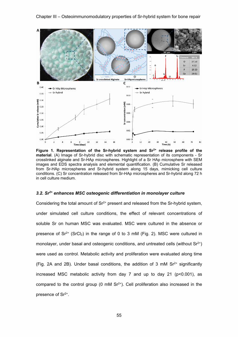
Chapter III – Osteoimmunomodulatory properties of Sr-hybrid system for bone repair
55
Figure 1. Representation of the Sr-hybrid system and Sr2+ release profile of the material. (A) Image of Sr-hybrid disc with schematic representation of its components - Sr crosslinked alginate and Sr-HAp microspheres. Highlight of a Sr HAp microsphere with SEM images and EDS spectra analysis and elemental quantification. (B) Cumulative Sr released from Sr-HAp microspheres and Sr-hybrid system along 15 days, mimicking cell culture conditions. (C) Sr concentration released from Sr-HAp microspheres and Sr-hybrid along 72 h in cell culture medium.
3.2. Sr2+ enhances MSC osteogenic differentiation in monolayer culture
Considering the total amount of Sr2+ present and released from the Sr-hybrid system,
under simulated cell culture conditions, the effect of relevant concentrations of
soluble Sr on human MSC was evaluated. MSC were cultured in the absence or
presence of Sr2+ (SrCl2) in the range of 0 to 3 mM (Fig. 2). MSC were cultured in
monolayer, under basal and osteogenic conditions, and untreated cells (without Sr2+)
were used as control. Metabolic activity and proliferation were evaluated along time
(Fig. 2A and 2B). Under basal conditions, the addition of 3 mM Sr2+ significantly
increased MSC metabolic activity from day 7 and up to day 21 (p<0.001), as
compared to the control group (0 mM Sr2+). Cell proliferation also increased in the
presence of Sr2+.

Chapter III – Osteoimmunomodulatory properties of Sr-hybrid system for bone repair
56
Under osteogenic conditions, a significant increase in metabolic activity was also
observed in the presence of Sr2+, since day 7, with a 5-fold increase in relation to
control at day 21. Regarding MSC proliferation, a decrease at day 7 was observed
for Sr2+ concentrations of 1 mM (p<0.05) and 3 mM (p<0.01). At days 14 and 21 a
significant increase in DNA content was seen in the presence of 0.5 and 1 mM Sr2+
(p<0.001), reaching a 4-fold increase at day 21 upon addition of 0.5 mM Sr2+.
Therefore, although the addition of Sr2+increased metabolic activity, independently of
the concentration used, cell proliferation under osteogenic conditions was shown to
increase in the presence of low Sr2+ doses.
MSC osteogenic differentiation was analysed by measuring ALP activity, an early
osteogenic marker. Quantitatively (Fig. 2C), higher ALP activity (normalized to the
total DNA content) was observed under osteoinductive conditions, compared to basal
conditions, as expected. Under basal conditions, higher ALP activity was detected in
Sr-treated cultures as compared to the control (0 mM Sr2+) at days 14 and 21, with a
significant increase (p<0.05) for 1 mM Sr2+. Under osteogenic conditions, a dose-
dependent increase in ALP activity was observed with increasing Sr2+ concentrations,
with statistical differences observed at all time-points for 1 mM and 3 mM Sr2+. MSC
monolayers were stained for ALP activity (pink staining, Fig. 2D). From day 14
onwards, higher expression of ALP activity was obtained in Sr-treated cultures,
especially at 3 mM Sr2+, whereas in basal conditions a less intense staining was
observed without evident differences among the tested concentrations (data not
shown). The formation of mineralized deposits was assessed by Von Kossa staining
(Fig. 2E). Matrix mineralization, a hallmark of osteogenic differentiation, was detected
under osteoinductive conditions at day 21, being much more evident for the highest
Sr2+ concentrations used (1 and 3 mM).

Chapter III – Osteoimmunomodulatory properties of Sr-hybrid system for bone repair
57
Figure 2. Sr2+ effect on MSC proliferation and osteogenic differentiation. Human MSC cultured on TCPS in the absence (0 mM Sr) or presence of different strontium (Sr2+) concentrations (0.5, 1 and 3 mM) under basal (left) or osteoinductive conditions (right), along 21 days of culture: (A) Metabolic activity, (B) DNA content and (C) ALP activity normalized by DNA, all shown as fold change to day 1 (dashed red line as 1-fold). Results are shown as mean±(standard error of the mean, SE). Asterisks indicate statistical significance performed using parametric unpaired t test: *p< 0.05; **p< 0.01; ***p< 0.001. Hashtags indicate statistical significance performed using non-parametric Mann Whitney U test: # p< 0.05; ## p< 0.01; ### p< 0.001. (D) ALP staining images at days 14 and 21 under osteogenic induction conditions (Magnification 10x). (E) Von Kossa staining images at day 21 of MSC cultured under osteogenic induction conditions (Magnification 10x).
3.3. Sr2+ decreases OC adhesion and fusion with decreased functionality
Human PBMC were allowed to differentiate into OC in monolayer cultures, and the
effect of Sr2+ on the formation of large and multinucleated OC was evaluated (Fig. 3).
As depicted in Fig. 3A, treatment with Sr2+ had a major effect on OC adhesion, which
was higher in the control condition (0 mM Sr2+) and decreased in Sr2+-treated
cultures, in a dose-dependent manner. Along differentiation, cells were able to
merge, originating large and multinucleated OC (Fig. 3B). An increase in OC size

Chapter III – Osteoimmunomodulatory properties of Sr-hybrid system for bone repair
58
throughout the culture time was observed, for all tested conditions, especially in the
absence of Sr2+ or at the lowest Sr2+ concentration (0.5 mM). However, when higher
Sr2+ concentrations were used, the increase in cell size was not so evident. These
differences were categorized/grouped according to the number of nuclei (2, 3-to-9 or
10+ nuclei), a measure of osteoclastogenesis (Fig. C a to c). As shown, in the
absence of Sr2+, the number of multinucleated OC increased along the culture time.
At day 7 (Fig. 3C a), the number of OC with 2 nuclei and 3-9 nuclei, for 0 mM Sr2,
was significantly higher than the number of OC (within the same categories), cultured
with 3 mM of Sr2+. By day 21 (Fig.3C c), the number of multinucleated OC in all
categories was significantly higher in control cultures compared to those with 3 mM
of Sr2+. Also, there were significantly more OC with 10 or more nuclei in Sr2+-free
control cultures than in those with 1 mM of Sr2+.
OC are described as TRAP-producing cells and the only cells able to resorb
mineralized substrates. As shown in Fig. 3D, OC cultured without Sr2+ for 21 days
were able to produce TRAP, with a typical “brownish” staining resulting from TRAP
enzymatic hydrolysis of tartrate substrate. In presence of Sr2+ concentrations,
especially at 3 mM, the amount of TRAP produced by OC decreased, which is in
accordance with the results obtained in the osteoclastogenesis studies. TRAP
quantification results (Fig. 3E), confirmed that OC released a significantly higher
amount of TRAP when cultured without Sr2+. OC were also cultured for 21 days on
top of dentine slices, and their ability to resorb mineralized substrates was evaluated.
As illustrated in Fig. 3F, OC were able to adhere to dentine and degrade its surface,
in all the tested conditions, forming typical resorption pits. However, SEM images
suggest that Sr2+ may have an inhibitory effect on OC resorbing activity, as there is a
clear decrease in the number and size of resorption pits when Sr2+ is present, which
is particularly evident for the higher concentration tested (3 mM).

Chapter III – Osteoimmunomodulatory properties of Sr-hybrid system for bone repair
59
Figure 3. Sr2+ effect on PBMC adhesion and OC formation, TRAP production and resorption capacity. Monocytes were differentiated into OC, in the presence of M-CSF and RANKL, on TCPS without or with different strontium (Sr) concentrations (0.5, 1 and 3 mM) for up to 21 days. (A) Number of nuclei of monocytes adherent to TCPS after 7 days. (B) Staining for the actin cytoskeleton (green) and nuclei (red) at days 7, 14 and 21 and (C) quantification of number of nuclei per cell, at days: 7 (a), 14 (b) and 21 (c). Boxes represent the median and min-max percentiles and whiskers represent 5–95 percentiles. (D) TRAP staining of OC differentiated for 21 days and (E) quantification of TRAP production in 24h, measured in the supernatant and lysate of cells. Statistical significance: *p < 0.05; **p < 0.01; ***p < 0.001. (F) SEM images of resorption pits in the surface of dentine slices created by functional OC.
3.4. The Sr-hybrid system supports MSC and OC adhesion
After establishing the effect of relevant Sr2+ concentrations on MSC and OC
response, the effect of the Sr-hybrid system on osteogenesis and osteoclastogenesis
was evaluated. Combined brightfield/fluorescence imaging of the system with f-

Chapter III – Osteoimmunomodulatory properties of Sr-hybrid system for bone repair
60
actin/nuclei stained MSC and OC, cultured for 21 days on Sr-hybrid system, allowed
for the spatial localization of the microspheres (more opaque within the hydrogel) and
labeled cells (Fig. 4). As shown, both cell types could be cultured in contact with Sr-
hybrid system, being preferably located in the vicinities of the microspheres.
Figure 4 – Cells located in the vicinity of microspheres in Sr-hybrid system. Cells cultured on Sr-hybrid system at day 14, stained for f-actin (in green) and nuclei (in red) and brightfield chanel (showing contour of microspheres, more opaque), imaged by CLSM (Z projection). MSC under osteogenic conditions (A) and OC (B). 3.5. The Sr-hybrid system promotes osteogenic differentiation of MSC
MSC were cultured on Sr-hybrid system, under basal and osteoinductive conditions.
Ca-hybrid was used as control. Live/Dead assay images (Fig. 5A) of MSC cultured
under basal conditions in Ca- or Sr-hybrid at days 1, 7 and 14, show that the viability
was not compromised during the time of culture. Moreover, in both systems, a
progressive increase in the number of cells was observed, suggesting that MSC
were able to proliferate. ALP activity was also evaluated in both systems for MSC
cultured under osteogenic conditions (Fig. 5B). Higher ALP activity was observed for
the Sr-hybrid, as compared to the control, at day 7. As shown in Fig. 5C, when
cultured under osteogenic conditions, MSC adopted a spread shape with polygonal
osteoblast-like morphology. While no differences were observed in MSC viability or

Chapter III – Osteoimmunomodulatory properties of Sr-hybrid system for bone repair
61
morphology in the Sr-hybrid, as compared to Ca-hybrid, increased ALP expression
suggested enhanced osteogenic differentiation.
Figure 5 – Sr-Hybrid effect on MSC survival and osteogenic differentiation. (A) Viability of MSC cultured on Sr-hybrid or Ca-hybrid systems after 1,7 and 14 days imaged by CLSM (Z projection, live cells in green, dead cells in red, scale bars: 200 µm). (B) ALP staining images of MSC cultured on Sr- or Ca-hybrid systems under osteogenic conditions at days 7, 14 and 21 (Scale bar 1 mm). (C) F-actin staining of MSC cultured on Sr- or Ca-hybrid, under basal (BM) or osteogenic conditions (OM), at days 7 and 21, imaged by CLSM (Z-projection, F-actin in green, nuclei in red, scale bars: 200 µm).
Chapter III – Osteoimmunomodulatory properties of Sr-hybrid system for bone repair
62
3.6. Sr-hybrid system decreases OC adhesion, fusion and activity
Human PBMC were allowed to differentiate into OC on Sr-hybrid and Ca-hybrid
systems for 21 days, and both osteoclastogenesis and OC functionality were
evaluated (Fig. 6). Images of f-actin stained OC cultured on both systems at days 7
and 21 are depicted in Fig. 6A, showing that cells were able to adhere and merge to
form multinucleated OC in both systems. Already at day 7, it was possible to detect
more adherent cells in the Ca-hybrid system. The extent of osteoclastogenesis was
decreased in the Sr-hybrid, as compared to the control, where the presence of larger
and multinucleated OC was more evident, especially at day 21. OC functionality was
assessed by TRAP staining, at days 7, 14 and 21 (Fig. 6B). The Sr-hybrid apparently
inhibited the ability of OC to secrete TRAP after 7 days of culture, which was not
observed for the Ca-hybrid system. During the culture period, it was also possible to
observe the presence of “brownish” agglomerates on the Ca-hybrid system,
indicating that OC maintained their functionality when cultured for 21 days in the
absence of Sr2+. These results were further confirmed by quantification of released
TRAP (Fig. 6C), which was significantly lower in Sr-hybrid (p<0.001), suggesting that
OC were more functional on Ca-hybrid than on Sr-hybrid system.

Chapter III – Osteoimmunomodulatory properties of Sr-hybrid system for bone repair
63
Figure 6 - Sr-hybrid system effect on OC formation and TRAP production. PBMC were allowed to differentiate into OC in the presence of M-CSF and RANKL for up to 21 days, on Sr-hybrid or Ca-hybrid systems. (A) OC cytoskeleton (green) and nuclei (red) staining at days 7 and 21 imaged by CLSM (Z projection). (B) TRAP staining and (C) quantification of its production after 24h. Statistical significance: *p < 0.05; **p < 0.01; ***p < 0.001.
3.7. Sr-hybrid system promotes M2 macrophage polarization in vivo
An in vivo air-pouch model of inflammation was used to evaluate the inflammatory
response to the developed Sr-hybrid system, in comparison to Ca-hybrid. Three
different animal groups were performed: Sham-operated, Ca-hybrid-implanted and
Sr-hybrid-implanted animals. Three days after implantation, the inflammatory cells

Chapter III – Osteoimmunomodulatory properties of Sr-hybrid system for bone repair
64
present in the inflammatory exudates retrieved from the air-pouches were analysed
by flow cytometry (Fig. 7A) and in addition, the exudates were evaluated using a
cytokine array (Fig. 7B). The average number of cells recovered from the air-
pouches was higher in the sham-operated animals, with a statistically significant
(p<0.05) decrease for cells retrieved from the Ca-hybrid group (Fig. 7A a).
Macrophages were identified using F4/80, a commonly used murine macrophage
marker, and CCR7 and CD206 were used as specific M1 and M2 markers,
respectively. As depicted in Fig. 7A b, the percentage of F4/80+/CCR7+ cells,
marking M1 macrophages, was low and showed no significant differences between
groups. However, regarding M2 macrophages, in the Sr-hybrid group, a statistically
significant increase (p<0.05) in the percentage of F4/80+/CD206+ cells was
observed when compared to the control (Sham-operated, Fig. 7A c).
The cytokine production by the inflammatory cells was analyzed by a cytokine array
(Supplementary Fig. 1) and the cytokines with at least a 1.5 fold-change to the
control (Sham-operated group) are depicted in Fig. 7B. Sr-hybrid implantation
caused a decrease in inflammatory cytokines, such us TNF-RI, MIP-1gamma and
RANTES, compared to Ca-hybrid. Similar levels of TNF-α were observed for the two
experimental groups and a decrease in IL-6 was also seen in both groups.
Importantly, in the Sr-hybrid group, the cytokine levels were indicative of a mild
inflammatory response, as opposed to Ca-hybrid group, where higher range
variations were observed, from -4 to 2 times fold-change to control in Sr-hybrid group
as opposed to variations from -12 to 15 times fold-change to control in Ca-hybrid
system. These results seem to corroborate the flow cytometry analysis, where the
overall evidences point out to an M2 macrophage response after Sr-hybrid
implantation.
Histological analysis of materials and surrounding tissues at day 3 was performed
and representative H&E stained sections are shown in Fig. 7 C. In sham-operated
animals, normal skin structures can be observed as well as the presence of the

Chapter III – Osteoimmunomodulatory properties of Sr-hybrid system for bone repair
65
collapsed air-pouch surrounding fibroblastic-like cells (Fig. 7C a and b). In the
experimental groups (Fig. 7C c to h), this structure was also present, surrounding the
materials with high cell infiltration in the nearby tissue. These tissues were also
analyzed after 15 days of implantation and Masson’s Trichrome (MT) stained
sections are shown in Fig. 7D, where collagen is stained in blue. A thin fibrous
capsule with low infiltration of inflammatory cells within the scaffolds was observed in
both materials. The thickness of this capsule was measured and results are shown in
Fig. 7E. An average thickness of 150 µm was measured and no statistically
significant differences were observed between Sr-hybrid and Ca-hybrid groups.
Furthermore, histological analysis of liver, kidneys, spleen (Supplementary Fig. 2),
and heart of animals were also performed to assess possible detrimental systemic
effects of the designed system, especially regarding cardiovascular safety due to the
recent restrictions in Sr Ranelate prescription. As shown in Fig. 7F, MT stained
sections of hearts do not show any deposition of collagen in the myocardium or any
noteworthy alterations in the Sr-hybrid implanted group after 15 days, as compared
to the Ca-hybrid group and Sham-operated animals.

Chapter III – Osteoimmunomodulatory properties of Sr-hybrid system for bone repair
66
Figure 7 – Characterization of the in vivo inflammatory response to the implantation of the Sr-hybrid system. (A) Flow cytometry analysis after 3 days of implantation regarding (a) total cell number, (b) percentage of F4/80+/CCR7+ (M1 macrophages) and (c) percentage of F4/80+/CD206+ (M2 macrophages) cells. Results are shown as mean±SE, statistical significance: *p < 0.05. (B) Cytokine production by inflammatory cells present on the retrieved inflammatory exudates evaluated using a cytokine array. Results are shown as fold-change to control (sham-operated animals) of exudates retrieved from Ca-hybrid and Sr-hybrid animal groups. (C) H&E stained sections of the surrounding tissues and implanted materials after 3 days of implantation of sham animals (a, b), Ca-hybrid (c, d ,e) and Sr-hybrid (f, g, h) group. (D) MT stained sections of the surrounding tissues and implanted materials after 15 days of implantation of Ca-hybrid (a, b) and Sr-hybrid (c, d) group. Collagen - blue (E) Thickness of the fibrous capsule found surrounding the implants after 15 days of implantation. n.s. – not statistically significant. (F) MT stained sections of the hearts of the animals retrieved after 15 days (collagen – blue).

Chapter III – Osteoimmunomodulatory properties of Sr-hybrid system for bone repair
67

Chapter III – Osteoimmunomodulatory properties of Sr-hybrid system for bone repair
68
4. Discussion
We have designed a hybrid polymeric-ceramic material, here designated as Sr-
hybrid system, for bone regeneration applications. The system is highly enriched in
Sr, being composed of Sr-HAp microspheres embedded in an RGD-modified alginate
hydrogel that crosslinks in situ in the presence of Sr carbonate, where Sr2+ acts as
the crosslinking agent. This Sr-hybrid system is expected to have a dual role, acting
on one hand as a Sr2+ delivery system and, on the other hand, as a bone-defect
injectable filler for repair/regeneration strategies. In this system, alginate hydrogels
were chemically modified with RGD peptides, providing a more physiologically
relevant microenvironment, which better mimics the natural extracellular matrix
(ECM). The incorporation of RGD is important to promote cell anchorage, in
otherwise non-adhesive hydrogels, and to improve cell survival and function, as we
previously demonstrated using osteoblastic cell lines and MSC, and also other
relevant cell types such as endothelial cells [36, 37, 52]. The alginate hydrogel can
be crosslinked in situ, using a previously described internal gelation strategy [36, 37,
42, 43, 52], enabling the injectability of the system, which is an important advantage
in clinical use. HAp microspheres, embedded within the hydrogel, act as mechanical
reinforcement of the system [38]. Microspheres with a diameter of around 500 µm
were used, as this size results in sufficient interstitial space within packed
microspheres, which is expected to provide an adequate porosity for new tissue
ingrowth upon implantation [38, 53]. Microspheres prepared by the same
methodology were previously studied [40, 54, 55]. MG63 osteoblast-like cells and
human MSC were shown to be able to spread on their surface, produce their own
ECM and even bridge adjacent microspheres [54]. The composite nature of bone has
previously inspired the development of other biomimetic materials for bone
regeneration [5], where hydrogels were combined with calcium phosphates [8]. Apart
from Ca2+, the incorporation of additional metal ions in calcium phosphate ceramics,
such as Sr2+ or Mg2+, is of particular interest, due to their high affinity for HAp and

Chapter III – Osteoimmunomodulatory properties of Sr-hybrid system for bone repair
69
demonstrated bioactivity [56, 57]. Several studies, as systematically reviewed in [58],
have shown increased in vivo bone formation and bone remodelling upon Sr addition
to biomaterials. Particularly, the addition of Sr to HAp in different HAp/alginate
materials has shown to lead to increase in vitro bioactivity [22] and in vivo bone
formation [26, 39, 57].
The hybrid system has two Sr2+ sources, the hydrogel and the microspheres. Results
showed that after 15 days of incubation in cell culture medium, only 0.3%
(corresponding to 0.3 mM) of the total amount of Sr2+ initially present in the Sr-hybrid
had been released. This suggests that the system is able to provide a sustained
release of Sr2+ over long periods of time, which is very appealing for clinical
applications. A more detailed analysis along the first 72 h of incubation, showed that
Sr2+ release profile is characterized by an initial burst, followed by a plateau. By
studying the behavior of the microspheres alone, we were able to conclude that they
release Sr2+ at a slower rate, presumably due to the rather stable nature of Sr-HAp,
while the hydrogel releases Sr2+ at a faster rate, mainly due to ionic exchange with
non-gelling ions present in the medium. The magnitude of the values observed is in
the same range of clinically relevant Sr concentrations, since upon oral
administration of Sr Ranelate therapeutic (2g/day/3 years), serum concentrations of
0.12 mM Sr were found [59].
After establishing the Sr2+ release kinetics profile of our system, we set out to check
the effect of relevant Sr2+ concentrations on MSC and OC response, first by using a
simpler in vitro model of monolayer cultures, and then by testing the system itself.
The presence of Sr2+ in the MSC culture medium showed that under basal conditions
the addition of a concentration of 3 mM led to increased metabolic activity and an
increase in cell proliferation for all concentrations tested. Under osteogenic
conditions, the presence of Sr2+ led to increased metabolic activity and cell
proliferation only for low Sr2+ doses (0.5 and 1 mM Sr2+), after 21 days of culture.

Chapter III – Osteoimmunomodulatory properties of Sr-hybrid system for bone repair
70
Some controversy can be found in the literature regarding the effect of different Sr2+
concentrations on different cell types. For example, Li and colleagues reported that
proliferation of rat bone marrow MSC (BMMSC) was inhibited in the presence of
Sr2+concentrations between 0.1 and 1 mM after 15 days of culture under basal
conditions [11]. In contrast, our results show that 0.5 and 1 mM Sr2+ increase cell
proliferation after 14 days under basal conditions. In another study using human
MSC, and Sr2+ concentrations ranging from 0.001 to 10 mM, an increase in cell
proliferation after 21 days under osteogenic conditions was observed for
concentrations up to 0.1 mM, and the opposite effect was observed for higher
concentrations, from 1 to 10 mM. Similar results were seen in basal conditions,
however, for 1 mM Sr2+ only a slight decrease in cell number was observed [60]. Our
results did not show a decrease in cell proliferation, however a similar trend of higher
proliferation at lower concentrations was also observed.
In terms of MSC osteogenic differentiation, the expression of ALP activity increased
in Sr-treated cultures, particularly under osteogenic conditions and at higher Sr2+
concentrations (1 and 3 mM Sr2+). A similar positive effect was also observed for the
Sr-hybrid system. MSC were able to adhere and spread on the hybrid scaffolds and
remained viable along the culture, showing increased expression of ALP activity from
early time points on the Sr-hybrid system, as compared to the control Ca-hybrid
system. The effect of Sr2+ on MSC and osteoblastic cells has been described in
several studies. For example, Peng and colleagues treated primary murine BMMSC
cultures with Sr2+ concentrations similar to the ones tested herein (1 and 3 mM Sr2+),
showing that both dosages promoted osteogenic differentiation with an increase in
Runx2 (day 7), ALP activity (day 14) and calcium nodules (day 21, Alizarin red
staining), in a dose-dependent manner [15]. Also, studies using human umbilical
cord-derived MSC treated with 2 mM Sr2+ showed a significant increase in ALP
activity (day 10) and mineralization nodules (4 weeks, Von Kossa), as compared to
osteoinductive media without the addition of Sr2+ [10]. This effect was shown to be

Chapter III – Osteoimmunomodulatory properties of Sr-hybrid system for bone repair
71
mainly dose-dependent. Studies from Li et al also demonstrated that rat BMMSC
treated with 0.1 and 1 mM Sr2+ exhibited higher ALP activity after 2 weeks of culture,
as compared to untreated cells [11]. On the other hand, Verberckmoes and co-
workers showed a multiphasic effect of Sr2+ on in vitro bone formation, using primary
cultures of rat calvaria osteoblasts treated with Sr2+ at concentrations ranging from
0.00625 to 1 mM [13]. At low Sr2+ concentrations an inhibitory effect on osteoblastic
differentiation was observed with a decrease in mineralized nodule formation [13].
These results were also supported by studies from Wornham and coworkers that
showed a decrease in mineralization nodules when primary osteoblast cells retrieved
from Sprague Dawly calvaria were cultured in the presence of Sr concentrations of
0.1 and 1 mM [61]. Studies from Schumacher et al showed a deleterious effect in
human MSC osteogenic differentiation for Sr2+ concentrations above 0.1 mM, with a
decrease in ALP activity (day 14) for Sr2+ concentrations of 1 and 5 mM [60].
Regarding OC response, our results showed the inhibitory effect of Sr2+ on the ability
of OC to attach to a substrate and merge. The number of large, multinucleated OC
decreased in the presence of Sr2+, in a dose-dependent manner. OC treated with
higher Sr2+ concentrations also showed decreased functionality, with lower
expression of TRAP activity and impaired ability to resorb mineralized substrates.
Likewise, osteoclastogenesis occurred to a lower extent on Sr-hybrid scaffolds, as
compared to Ca-hybrid control. Although human PBMC could be cultured on both
types of materials and successfully differentiate into OC, more adherent cells were
detected in the Ca-hybrid system, with higher number of multinucleated and larger
OC. The ability of OC to secrete TRAP also decreased in the Sr-hybrid, as compared
to the Ca-hybrid where OC seemed to be more functional. Similar effects of Sr on
OC activity were reported by Schumacher et al, in which higher Sr2+ concentrations
of 5 mM and 10 mM Sr2+ promoted a decrease in the number of adherent cells, while
1 mM did not show significant alterations in cell behavior [62]. Similarly, in another
study, 1 mM only exerted a modest reduction on OC number and resorption area

Chapter III – Osteoimmunomodulatory properties of Sr-hybrid system for bone repair
72
[61]. Moreover, the analysis of the enzymatic activities of TRAP and carbonic
anhydrase type II, and gene expressions of denditric cell-specific transmembrane
protein and CD44, did not show any effect of Sr in osteoclastogenesis besides the
observation of actin disruption in some cells at 0.1 mM Sr2+ concentration [62].
Interestingly, it has been previously demonstrated that 1 mM of Sr2+ was sufficient to
decrease the expression of functional osteoclast markers such as integrin αvβ3, a
vitronectin receptor that has a prominent role in osteoclast adhesion, being involved
in podosomes formation and cytoskeleton re-organization, and is also implicated in
OC motility and in the maintenance of the sealing zone [18]. Accordingly, 1 mM of
Sr2+ was previously found to inhibit the ability of mouse and chicken OC to resorb the
surface of dentine slices [16] and to decrease the number of OC expressing carbonic
anhydrase II, an enzyme that dissolves bone matrix [18]. Importantly, the loss of OC
functionality when cultured in the presence of Sr2+ might be explained by its ability to
disrupt the OC actin-containing sealing zone and the ruffled border, essential for the
OC resorbing activity in vitro and bone resorption [15-17]. Another study
demonstrated that Sr impaired osteoclastogenesis by blocking RANKL-induced
activation of the NF-κB pathway [63].
Evidence in recent years has been shifting concepts in the bone regeneration field,
where inflammatory cells and their mediators are being nowadays considered crucial
players for proper bone healing [29]. Therefore, current strategies include the design
of immunomodulatory biomaterials able to establish a pro-regenerative milieu [34,
35]. Upon biomaterial implantation, macrophages are amongst the first cells to arrive
to the injury site and their polarization and M2/M1 increased ratio may be beneficial
to improve tissue regeneration. Some studies suggested that Sr may have
immunomodulatory properties [63-65], however the information in the literature
concerning the influence of Sr-rich biomaterials in the host immune response is very
scarce, being the present work a significant contribution to the field.

Chapter III – Osteoimmunomodulatory properties of Sr-hybrid system for bone repair
73
Our results show a decrease in the number of inflammatory cells recruited to the
implant site in both materials compared to sham-operated animals, but most
importantly, an increase in the number of M2 macrophages was observed in the Sr-
hybrid group. These results are in agreement with others where Sr has been shown
to promote an M2 polarization of macrophages. In studies of Yuan and coworkers,
surface modification of titanium surfaces with Ca and Sr, either alone or in different
Ca:Sr ratios, have shown that the presence of Sr was able to increase the number of
M2 macrophages in a similar in vivo model, although no differences were seen
between coatings of Ca and Sr alone [64]. Another work using the air-pouch model of
inflammation showed that a Sr-substituted sub-micron bioactive glass (SBG)
significantly increased the number of M2 macrophages as compared to bare SBG
[65]. The cytokine analysis of the inflammatory exudates revealed a mild
inflammatory reaction to Sr-hybrid, when compared to Ca-hybrid system. In Sr-hybrid
system, increased levels of both TNF-α and IL-1β were observed, shown to correlate
with an increase in MSC mineralization potential [66]. Although a decrease in
RANTES and SDF-1α, both known to recruit MSC [67-69], was observed in Sr-hybrid
implanted animals, the material did not show a decreased capacity to promote bone
regeneration. In an in vivo model, Sr-hybrid was seen to promote, 60 days after
implantation, an increase in cell recruitment and in new bone formation at both center
and periphery of the defect, as opposed to Ca-hybrid where new bone formation was
limited to the periphery of the defect [39].
The histological analysis revealed a thin fibrous capsule, with a non-significant
difference between groups, and low numbers of inflammatory cells were observed in
the implanted materials groups. Although neither system induced a massive M1-like
inflammatory response, our results indicate that Sr-hybrid system was able to
promote an M2 regenerative response when compared to the Ca-hybrid system.
Recent concerns regarding Sr ranelate oral administration also highlight the
importance of the use of Sr-releasing materials for local delivery. In this study, the

Chapter III – Osteoimmunomodulatory properties of Sr-hybrid system for bone repair
74
histological evaluation of organs and particularly the hearts of animals did not show
any alteration after 15 days. Furthermore, in an in vivo critical-sized bone defect
model, no alterations were observed in peripheral organs (liver, kidneys, spleen) or in
Sr serum levels of animals implanted with Sr-hybrid [39].
Taken together, our results demonstrate that the Sr-hybrid scaffold promotes long-
term Sr2+ release and exhibits both osteoinductive and anti-osteoclastogenesis
properties. Moreover, it was able to modulate the immune response towards the M2
macrophage phenotype. These results highlight the potential of this injectable
biomaterial for bone repair/regeneration, particularly for the treatment of pathological
conditions where bone remodelling is impaired, namely osteoporosis.
5. Conclusions
Our findings confirmed that the proposed Sr-hybrid, which acted as Sr2+ release
system, has the ability to drive the differentiation of MSC towards the osteoblastic
lineage, while inhibiting OC adhesion/fusion, activity and resorptive behaviour. This
dual effect of Sr2+ is of major interest for the therapeutic management of bone
pathologies characterized by abnormal bone remodelling such as osteoporosis. The
Sr-hybrid also showed to be able to modulate the immune response, inducing
macrophage polarization towards a pro-regenerative M2 phenotype. Moreover, the
system exhibits some unique properties, such as injectability for minimally invasive
surgery, mechanical reinforcement with HAp microspheres, and sustained Sr2+
release over extended periods of time, which are appealing for future clinical
applications.
Acknowledgments
The authors would like to thank Hospital S. João, for kindly donating the buffy coats.
This work was financed by FEDER funds, through the Programa Operacional

Chapter III – Osteoimmunomodulatory properties of Sr-hybrid system for bone repair
75
Factores de Competitividade — COMPETE and by Portuguese funds through FCT
— Fundação para a Ciência e a Tecnologia in the framework of the project
PTDC/CTM/103181/2008. A.H. Lourenço is recipient of a doctoral fellowship from
FCT, ref. SFRH/BD/87192/2012. ALT thanks FCT for the doctoral grant
SFRH/BD/94306/2013 and the BiotechHealth PhD Programme. Daniela P.
Vasconcelos is funded by FCT - Fundação para a Ciência e Tecnologia through the
FCT PhD Programmes and by Programa Operacional Potencial Humano (POCH),
specifically by the BiotechHealth Programme (Doctoral Programme on Cellular and
Molecular Biotechnology Applied to Health Sciences – reference PD/00016/2012),
and through the PhD studentship PD/BD/114011/2015. CB acknowledges her
research position (IF/00296/2015) funded by FCT and POPH/ESF (EC).
Disclosures
The authors declare no conflict of interest.

Chapter III – Osteoimmunomodulatory properties of Sr-hybrid system for bone repair
76
References
[1] X. Feng, J.M. McDonald, Disorders of Bone Remodeling, Annual review of pathology 6 (2011) 121-145. [2] G.A. Rodan, T.J. Martin, Therapeutic Approaches to Bone Diseases, Science 289(5484) (2000) 1508-1514. [3] E.M.M. Van Lieshout, V. Alt, Bone graft substitutes and bone morphogenetic proteins for osteoporotic fractures: what is the evidence?, Injury 47, Supplement 1 (2016) S43-S46. [4] K.L. Low, S.H. Tan, S.H.S. Zein, J.A. Roether, V. Mouriño, A.R. Boccaccini, Calcium phosphate-based composites as injectable bone substitute materials, Journal of Biomedical Materials Research Part B: Applied Biomaterials 94B(1) (2010) 273-286. [5] A.R. Amini, C.T. Laurencin, S.P. Nukavarapu, Bone Tissue Engineering: Recent Advances and Challenges, Critical reviews in biomedical engineering 40(5) (2012) 363-408. [6] M.M. Stevens, Biomaterials for bone tissue engineering, Materials Today 11(5) (2008) 18-25. [7] F. Barrere, C.A. van Blitterswijk, K. de Groot, Bone regeneration: molecular and cellular interactions with calcium phosphate ceramics, Int J Nanomedicine 1(3) (2006) 317-32. [8] M. D’Este, D. Eglin, Hydrogels in calcium phosphate moldable and injectable bone substitutes: Sticky excipients or advanced 3-D carriers?, Acta Biomaterialia 9(3) (2013) 5421-5430. [9] L. Cianferotti, F. D’Asta, M.L. Brandi, A review on strontium ranelate long-term antifracture efficacy in the treatment of postmenopausal osteoporosis, Therapeutic Advances in Musculoskeletal Disease 5(3) (2013) 127-139. [10] F. Yang, D. Yang, J. Tu, Q. Zheng, L. Cai, L. Wang, Strontium Enhances Osteogenic Differentiation of Mesenchymal Stem Cells and In Vivo Bone Formation by Activating Wnt/Catenin Signaling, STEM CELLS 29(6) (2011) 981-991. [11] Y. Li, J. Li, S. Zhu, E. Luo, G. Feng, Q. Chen, J. Hu, Effects of strontium on proliferation and differentiation of rat bone marrow mesenchymal stem cells, Biochemical and Biophysical Research Communications 418(4) (2012) 725-730. [12] J. Caverzasio, C. Thouverey, Activation of FGF receptors is a new mechanism by which strontium ranelate induces osteoblastic cell growth, Cellular physiology and biochemistry : international journal of experimental cellular physiology, biochemistry, and pharmacology 27(3-4) (2011) 243-50. [13] S.C. Verberckmoes, M.E. De Broe, P.C. D'Haese, Dose-dependent effects of strontium on osteoblast function and mineralization, Kidney Int 64(2) (2003) 534-543. [14] Z. Saidak, E. Haÿ, C. Marty, A. Barbara, P.J. Marie, Strontium ranelate rebalances bone marrow adipogenesis and osteoblastogenesis in senescent osteopenic mice through NFATc/Maf and Wnt signaling, Aging Cell 11(3) (2012) 467-474. [15] S. Peng, G. Zhou, K.D.K. Luk, K.M.C. Cheung, Z. Li, W.M. Lam, Z. Zhou, W.W. Lu, Strontium Promotes Osteogenic Differentiation of Mesenchymal Stem Cells Through the Ras/MAPK Signaling Pathway, Cellular Physiology and Biochemistry 23(1-3) (2009) 165-174. [16] N. Takahashi, T. Sasaki, Y. Tsouderos, T. Suda, S 12911-2 Inhibits Osteoclastic Bone Resorption In Vitro, Journal of Bone and Mineral Research 18(6) (2003) 1082-1087. [17] E. Bonnelye, A. Chabadel, F. Saltel, P. Jurdic, Dual effect of strontium ranelate: Stimulation of osteoblast differentiation and inhibition of osteoclast formation and resorption in vitro, Bone 42(1) (2008) 129-138.

Chapter III – Osteoimmunomodulatory properties of Sr-hybrid system for bone repair
77
[18] R. Baron, Y. Tsouderos, In vitro effects of S12911-2 on osteoclast function and bone marrow macrophage differentiation, European Journal of Pharmacology 450(1) (2002) 11-17. [19] J.Y. Reginster, A. Neuprez, N. Dardenne, C. Beaudart, P. Emonts, O. Bruyere, Efficacy and safety of currently marketed anti-osteoporosis medications, Best Practice & Research Clinical Endocrinology & Metabolism 28(6) (2014) 809-834. [20] C. Cooper, K.M. Fox, J.S. Borer, Ischaemic cardiac events and use of strontium ranelate in postmenopausal osteoporosis: a nested case–control study in the CPRD, Osteoporosis International 25(2) (2014) 737-745. [21] B. Abrahamsen, E.L. Grove, P. Vestergaard, Nationwide registry-based analysis of cardiovascular risk factors and adverse outcomes in patients treated with strontium ranelate, Osteoporosis International 25(2) (2014) 757-762. [22] M. Raucci, D. Giugliano, M.A. Alvarez-Perez, L. Ambrosio, Effects on growth and osteogenic differentiation of mesenchymal stem cells by the strontium-added sol–gel hydroxyapatite gel materials, J Mater Sci: Mater Med 26(2) (2015) 1-11. [23] V. Nardone, S. Fabbri, F. Marini, R. Zonefrati, G. Galli, A. Carossino, A. Tanini, M.L. Brandi, Osteodifferentiation of Human Preadipocytes Induced by Strontium Released from Hydrogels, International Journal of Biomaterials 2012 (2012) 10. [24] Y. Zhu, Y. Ouyang, Y. Chang, C. Luo, J. Xu, C. Zhang, W. Huang, Evaluation of the proliferation and differentiation behaviors of mesenchymal stem cells with partially converted borate glass containing different amounts of strontium in vitro, Molecular medicine reports 7(4) (2013) 1129-36. [25] K. Sariibrahimoglu, W. Yang, S.C.G. Leeuwenburgh, F. Yang, J.G.C. Wolke, Y. Zuo, Y. Li, J.A. Jansen, Development of porous polyurethane/strontium-substituted hydroxyapatite composites for bone regeneration, Journal of Biomedical Materials Research Part A (2014) n/a-n/a. [26] J. Hao, A. Acharya, K. Chen, J. Chou, S. Kasugai, N.P. Lang, Novel bioresorbable strontium hydroxyapatite membrane for guided bone regeneration, Clinical Oral Implants Research 26(1) (2015) 1-7. [27] C.T. Wong, W.W. Lu, W.K. Chan, K.M.C. Cheung, K.D.K. Luk, D.S. Lu, A.B.M. Rabie, L.F. Deng, J.C.Y. Leong, In vivo cancellous bone remodeling on a strontium-containing hydroxyapatite (sr-HA) bioactive cement, Journal of Biomedical Materials Research Part A 68A(3) (2004) 513-521. [28] M. Baier, P. Staudt, R. Klein, U. Sommer, R. Wenz, I. Grafe, P. Meeder, P. Nawroth, C. Kasperk, Strontium enhances osseointegration of calcium phosphate cement: a histomorphometric pilot study in ovariectomized rats, Journal of Orthopaedic Surgery and Research 8(1) (2013) 16. [29] F. Loi, L.A. Córdova, J. Pajarinen, T.-h. Lin, Z. Yao, S.B. Goodman, Inflammation, fracture and bone repair, Bone 86(Supplement C) (2016) 119-130. [30] A. Mantovani, A. Sica, S. Sozzani, P. Allavena, A. Vecchi, M. Locati, The chemokine system in diverse forms of macrophage activation and polarization, Trends in Immunology 25(12) 677-686. [31] B.N. Brown, B.M. Sicari, S.F. Badylak, Rethinking Regenerative Medicine: A Macrophage-Centered Approach, Frontiers in Immunology 5 (2014) 510. [32] F.O. Martinez, S. Gordon, The M1 and M2 paradigm of macrophage activation: time for reassessment, F1000Prime Reports 6 (2014) 13. [33] D.P. Vasconcelos, A.C. Fonseca, M. Costa, I.F. Amaral, M.A. Barbosa, A.P. Águas, J.N. Barbosa, Macrophage polarization following chitosan implantation, Biomaterials 34(38) (2013) 9952-9959. [34] D.P. Vasconcelos, M. Costa, I.F. Amaral, M.A. Barbosa, A.P. Águas, J.N. Barbosa, Modulation of the inflammatory response to chitosan through M2 macrophage polarization using pro-resolution mediators, Biomaterials 37(Supplement C) (2015) 116-123.

Chapter III – Osteoimmunomodulatory properties of Sr-hybrid system for bone repair
78
[35] D.P. Vasconcelos, M. Costa, I.F. Amaral, M.A. Barbosa, A.P. Águas, J.N. Barbosa, Development of an immunomodulatory biomaterial: Using resolvin D1 to modulate inflammation, Biomaterials 53(Supplement C) (2015) 566-573. [36] S.l.J. Bidarra, C.C. Barrias, M.r.A. Barbosa, R. Soares, P.L. Granja, Immobilization of Human Mesenchymal Stem Cells within RGD-Grafted Alginate Microspheres and Assessment of Their Angiogenic Potential, Biomacromolecules 11(8) (2010) 1956-1964. [37] S.J. Bidarra, C.C. Barrias, K.B. Fonseca, M.A. Barbosa, R.A. Soares, P.L. Granja, Injectable in situ crosslinkable RGD-modified alginate matrix for endothelial cells delivery, Biomaterials 32(31) (2011) 7897-7904. [38] N. Neves, B.B. Campos, I.F. Almeida, P.C. Costa, A.T. Cabral, M.A. Barbosa, C.C. Ribeiro, Strontium-rich injectable hybrid system for bone regeneration, Materials Science and Engineering: C 59 (2016) 818-827. [39] A. Henriques Lourenço, N. Neves, C. Ribeiro-Machado, S.R. Sousa, M. Lamghari, C.C. Barrias, A. Trigo Cabral, M.A. Barbosa, C.C. Ribeiro, Injectable hybrid system for strontium local delivery promotes bone regeneration in a rat critical-sized defect model, Scientific Reports 7(1) (2017) 5098. [40] C.C. Ribeiro, C.C. Barrias, M.A. Barbosa, Calcium phosphate-alginate microspheres as enzyme delivery matrices, Biomaterials 25(18) (2004) 4363-4373. [41] J.A. Rowley, G. Madlambayan, D.J. Mooney, Alginate hydrogels as synthetic extracellular matrix materials, Biomaterials 20(1) (1999) 45-53. [42] F.R. Maia, A.H. Lourenço, P.L. Granja, R.M. Gonçalves, C.C. Barrias, Effect of Cell Density on Mesenchymal Stem Cells Aggregation in RGD-Alginate 3D Matrices under Osteoinductive Conditions, Macromolecular Bioscience (2014) n/a-n/a. [43] K.B. Fonseca, S.J. Bidarra, M.J. Oliveira, P.L. Granja, C.C. Barrias, Molecularly designed alginate hydrogels susceptible to local proteolysis as three-dimensional cellular microenvironments, Acta Biomaterialia 7(4) (2011) 1674-1682. [44] N. Jaiswal, S.E. Haynesworth, A.I. Caplan, S.P. Bruder, Osteogenic differentiation of purified, culture-expanded human mesenchymal stem cells in vitro, Journal of Cellular Biochemistry 64(2) (1997) 295-312. [45] M.I. Oliveira, S.G. Santos, M.J. Oliveira, A.L. Torres, M.A. Barbosa, Chitosan drives anti-inflammatory macrophage polarisation and pro-inflammatory dendritic cell stimulation, European cells & materials 24 (2012) 136-52; discussion 152-3. [46] A.L. Torres, S.G. Santos, M.I. Oliveira, M.A. Barbosa, Fibrinogen promotes resorption of chitosan by human osteoclasts, Acta Biomaterialia 9(5) (2013) 6553-6562. [47] B.A.C. Lopes, Cell Note: Plataforma para a análise escrita de imagens, University of Porto, Porto, 2010. [48] K. Fuller, J.L. Ross, K.A. Szewczyk, R. Moss, T.J. Chambers, Bone Is Not Essential for Osteoclast Activation, PLOS ONE 5(9) (2010) e12837. [49] B. Kirstein, U. Grabowska, B. Samuelsson, M. Shiroo, T.J. Chambers, K. Fuller, A novel assay for analysis of the regulation of the function of human osteoclasts, Journal of Translational Medicine 4 (2006) 45-45. [50] A.G. Castro, N. Esaguy, P.M. Macedo, A.P. Aguas, M.T. Silva, Live but not heat-killed mycobacteria cause rapid chemotaxis of large numbers of eosinophils in vivo and are ingested by the attracted granulocytes, Infection and Immunity 59(9) (1991) 3009-3014. [51] A.D. Sedgwick, P. Lees, Studies of eicosanoid production in the air pouch model of synovial inflammation, Agents and Actions 18(3) (1986) 429-438. [52] M.B. Evangelista, S.X. Hsiong, R. Fernandes, P. Sampaio, H.J. Kong, C.C. Barrias, R. Salema, M.A. Barbosa, D.J. Mooney, P.L. Granja, Upregulation of bone cell differentiation through immobilization within a synthetic extracellular matrix, Biomaterials 28(25) (2007) 3644-3655.
Chapter III – Osteoimmunomodulatory properties of Sr-hybrid system for bone repair
79
[53] M. Bohner, S. Tadier, N. van Garderen, A. de Gasparo, N. Döbelin, G. Baroud, Synthesis of spherical calcium phosphate particles for dental and orthopedic applications, Biomatter 3(2) (2013) e25103. [54] C.C. Barrias, C.C. Ribeiro, M. Lamghari, C.S. Miranda, M.A. Barbosa, Proliferation, activity, and osteogenic differentiation of bone marrow stromal cells cultured on calcium titanium phosphate microspheres, Journal of Biomedical Materials Research Part A 72A(1) (2005) 57-66. [55] C.C. Barrias, M. Lamghari, P.L. Granja, M.C. Sá Miranda, M.A. Barbosa, Biological evaluation of calcium alginate microspheres as a vehicle for the localized delivery of a therapeutic enzyme, Journal of Biomedical Materials Research Part A 74A(4) (2005) 545-552. [56] F. Jiang, D.P. Wang, S. Ye, X. Zhao, Strontium-substituted, luminescent and mesoporous hydroxyapatite microspheres for sustained drug release, Journal of materials science. Materials in medicine 25(2) (2014) 391-400. [57] J. Hao, J. Chou, S. Kuroda, M. Otsuka, S. Kasugai, N.P. Lang, Strontium hydroxyapatite in situ gel-forming system – a new approach for minimally invasive bone augmentation, Clinical Oral Implants Research (2014) n/a-n/a. [58] N. Neves, D. Linhares, G. Costa, C.C. Ribeiro, M.A. Barbosa, In vivo and clinical application of strontium-enriched biomaterials for bone regeneration: A systematic review, Bone & Joint Research 6(6) (2017) 366-375. [59] P.J. Meunier , C. Roux , E. Seeman , S. Ortolani , J.E. Badurski , T.D. Spector , J. Cannata , A. Balogh , E.-M. Lemmel , S. Pors-Nielsen , R. Rizzoli , H.K. Genant , J.-Y. Reginster The Effects of Strontium Ranelate on the Risk of Vertebral Fracture in Women with Postmenopausal Osteoporosis, New England Journal of Medicine 350(5) (2004) 459-468. [60] M. Schumacher, A. Lode, A. Helth, M. Gelinsky, A novel strontium(II)-modified calcium phosphate bone cement stimulates human-bone-marrow-derived mesenchymal stem cell proliferation and osteogenic differentiation in vitro, Acta Biomaterialia 9(12) (2013) 9547-9557. [61] D.P. Wornham, M.O. Hajjawi, I.R. Orriss, T.R. Arnett, Strontium potently inhibits mineralisation in bone-forming primary rat osteoblast cultures and reduces numbers of osteoclasts in mouse marrow cultures, Osteoporosis International 25(10) (2014) 2477-2484. [62] M. Schumacher, A.S. Wagner, J. Kokesch-Himmelreich, A. Bernhardt, M. Rohnke, S. Wenisch, M. Gelinsky, Strontium substitution in apatitic CaP cements effectively attenuates osteoclastic resorption but does not inhibit osteoclastogenesis, Acta Biomaterialia 37 (2016) 184-194. [63] S. Zhu, X. Hu, Y. Tao, Z. Ping, L. Wang, J. Shi, X. Wu, W. Zhang, H. Yang, Z. Nie, Y. Xu, Z. Wang, D. Geng, Strontium inhibits titanium particle-induced osteoclast activation and chronic inflammation via suppression of NF-κB pathway, Scientific Reports 6 (2016) 36251. [64] X. Yuan, H. Cao, J. Wang, K. Tang, B. Li, Y. Zhao, M. Cheng, H. Qin, X. Liu, X. Zhang, Immunomodulatory Effects of Calcium and Strontium Co-Doped Titanium Oxides on Osteogenesis, Frontiers in Immunology 8 (2017) 1196. [65] W. Zhang, F. Zhao, D. Huang, X. Fu, X. Li, X. Chen, Strontium-Substituted Submicrometer Bioactive Glasses Modulate Macrophage Responses for Improved Bone Regeneration, ACS Applied Materials & Interfaces 8(45) (2016) 30747-30758. [66] N. Charatcharoenwitthaya, S. Khosla, E.J. Atkinson, L.K. McCready, B.L. Riggs, Effect of Blockade of TNF-α and Interleukin-1 Action on Bone Resorption in Early Postmenopausal Women, Journal of Bone and Mineral Research 22(5) (2007) 724-729. [67] B. Zhou, Z.C. Han, M.-C. Poon, W. Pu, Mesenchymal stem/stromal cells (MSC) transfected with stromal derived factor 1 (SDF-1) for therapeutic neovascularization: Enhancement of cell recruitment and entrapment, Medical Hypotheses 68(6) 1268-1271.

Chapter III – Osteoimmunomodulatory properties of Sr-hybrid system for bone repair
80
[68] T.T. Lau, D.-A. Wang, Stromal cell-derived factor-1 (SDF-1): homing factor for engineered regenerative medicine, Expert Opinion on Biological Therapy 11(2) (2011) 189-197. [69] K. Anton, D. Banerjee, J. Glod, Macrophage-Associated Mesenchymal Stem Cells Assume an Activated, Migratory, Pro-Inflammatory Phenotype with Increased IL-6 and CXCL10 Secretion, PLOS ONE 7(4) (2012) e35036.

Chapter III – Osteoimmunomodulatory properties of Sr-hybrid system for bone repair
81
Supplementary figures
Supplementary Figure 1 –Cytokine production by inflammatory cells exudates after 3 days of implantation. A, B, C - Membranes obtained from the cytokine array for the different groups: (A) Sham, (B) Ca-hybrid and (C) Sr-hybrid. D, E- Total results of the array organized as fold change to the control (Sham) of Ca-hybrid (D) and Sr-hybrid (E).

Chapter III – Osteoimmunomodulatory properties of Sr-hybrid system for bone repair
82
Supplementary Figure 2 – Strontium systemic evaluation. Representative H&E stained sections of spleen (1st column), liver (2nd column) and kidney (3rd column) of sham (a,b,c,m,n,o), Sr-hybrid (d,e,f,p,q,r), Ca-hybrid (g,h,i,s,t,u) and Non-operated (N.O., j,k,l,v,w,x), at 3 days (a to l) and 15 days (m to x) post-surgery. No alterations were observed in the spleen and kidney. All liver samples showed areas of microvesicular and macrovesicular steatosis with similar degree of extension.

Chapter III – Osteoimmunomodulatory properties of Sr-hybrid system for bone repair
83

Chapter III – Osteoimmunomodulatory properties of Sr-hybrid system for bone repair
84

Chapter IV – Sr-hybrid system promotes bone regeneration in a rat critical-sized model
85
Chapter IV
Injectable hybrid system for strontium local delivery promotes bone
regeneration in a rat critical-sized defect model
Ana Henriques Lourenço*, Nuno Neves*, Cláudia Ribeiro-Machado, Susana R. Sousa,
Meriem Lamghari, Cristina C. Barrias, Abel Trigo Cabral, Mário A. Barbosa and
Cristina C. Ribeiro
* These authors contributed equally to this work.
Published in Scientific Reports 7(1) (2017) 5098.

Chapter IV – Sr-hybrid system promotes bone regeneration in a rat critical-sized model
86

Chapter IV – Sr-hybrid system promotes bone regeneration in a rat critical-sized model
87
Injectable hybrid system for strontium local delivery promotes bone
regeneration in a rat critical-sized defect model
Ana Henriques Lourenço1,2,3 *, Nuno Neves1,2,4 *, Cláudia Ribeiro-Machado 1,2,
Susana R. Sousa1,2,5, Meriem Lamghari1,2, Cristina C. Barrias1,2, Abel Trigo
Cabral4, Mário A. Barbosa1,2,6 and Cristina C. Ribeiro1,2,5
1i3S - Instituto de Investigação e Inovação em Saúde, Universidade do Porto, Rua
Alfredo Allen, 208, 4200 - 135 Porto, Portugal
2INEB - Instituto de Engenharia Biomédica, Universidade do Porto, Rua Alfredo Allen,
208, 4200 - 135 Porto, Portugal
3Faculdade de Engenharia, Universidade do Porto, Rua Dr. Roberto Frias, s/n, 4200-
465 Porto, Portugal
4Faculdade de Medicina, Universidade do Porto, Serviço de Ortopedia, Alameda Prof.
Hernâni Monteiro, 4200-319 Porto, Portugal
5ISEP – Instituto Superior de Engenharia do Porto, Instituto Politécnico do Porto, Rua
Dr. António Bernardino de Almeida 431, 4249-015, Porto, Portugal
6ICBAS - Instituto de Ciências Biomédicas de Abel Salazar, Universidade do Porto,
Rua de Jorge Viterbo Ferreira n. 228, 4050-313 Porto, Portugal
* These authors contributed equally to this work.

Chapter IV – Sr-hybrid system promotes bone regeneration in a rat critical-sized model
88

Chapter IV – Sr-hybrid system promotes bone regeneration in a rat critical-sized model
89
Abstract
Strontium (Sr) has been described as having beneficial influence in bone strength and
architecture. However, negative systemic effects have been reported on oral
administration of Sr ranelate, leading to strict restrictions in clinical application. We
hypothesized that local delivery of Sr improves osteogenesis without eliciting
detrimental side effects. Therefore, the in vivo response to an injectable Sr-hybrid
system composed of RGD-alginate hydrogel cross-linked in situ with Sr and reinforced
with Sr-doped hydroxyapatite microspheres, was investigated. The system was
injected in a critical-size bone defect model and compared to a similar Sr-free material.
Micro-CT results show a trend towards higher new bone formed in Sr-hybrid group and
major histological differences were observed between groups. Higher cell invasion was
detected at the center of the defect of Sr-hybrid group after 15 days with earlier bone
formation. Higher material degradation with increase of collagen fibers and bone
formation in the center of the defect after 60 days was observed as opposed to bone
formation restricted to the periphery of the defect in the control. These histological
findings support the evidence of an improved response with the Sr enriched material.
Importantly, no alterations were observed in the Sr levels in systemic organs or serum.
Keywords: strontium, bone regeneration, injectable bone substitute, alginate,
hydroxyapatite, ionic delivery system

Chapter IV – Sr-hybrid system promotes bone regeneration in a rat critical-sized model
90
1. Introduction
The management of fractures and bone defects remains a significant challenge, and
there is the need for improved therapeutic strategies [1]. Biological (autografts,
allografts and xenografts) and synthetic bone grafts are currently used in clinical
practice for bone repair. Because of their osteogenic potential and the absence of risks
of immune rejection or disease transfer, autografts are clinically preferred. However,
they are limited in supply, imply the additional morbidity of a harvest surgery and their
properties and shape do not match exactly those of the bone to be replaced [2].
Intensive investigation is being carried out to produce synthetic bone grafts in order to
overcome these problems. The use of injectable materials in bone regeneration,
especially calcium phosphate based materials, presents several advantages, namely
due to their adequate biological responses, osteoconductivity and mechanical
properties [3-6]. These materials can be applied by minimally invasive surgical
procedures, to efficiently fill-in cavities of non-uniform shapes, with no tissue damage
and limited exposure to infectious agents, thus reducing patient discomfort and
procedure-associated health costs. The addition of osteoinductive factors or
osteoprogenitor/stem cells may improve bone repair, particularly in osteoporotic
conditions, characterized by an impaired healing response [7-10].
Oral administration of Strontium (Sr) ranelate has shown effectiveness in the
prevention of both vertebral and non-vertebral osteoporotic fractures [11, 12]. Unlike
other anti-osteoporotic agents widely used in clinical practice, such as
bisphosphonates, estrogen, selective estrogen-receptor modulators (SERMs) and
calcitonin, which inhibit bone resorption [9], Sr ranelate also promotes bone formation
[13-16]. Several in vitro studies show that Sr ranelate decreases bone resorption, by
reducing osteoclast activity [13, 14, 17], decreasing functional osteoclast markers
expression [13], disrupting osteoclasts cytoskeleton [14], and increasing osteoclast
apoptosis [18]. Simultaneously, it induces positive effects on osteoblastogenesis and
osteoblast activity in different in vitro models [19], namely by enhancing replication of

Chapter IV – Sr-hybrid system promotes bone regeneration in a rat critical-sized model
91
preosteoblastic cells [14, 20-23], increasing osteogenesis [14, 20, 24-26], decreasing
osteoblast apoptosis [21, 27], and promoting terminal differentiation of osteoblasts into
osteocytes [20]. Some pre-clinical studies performed in both normal and
osteopenic/osteoporotic animal models confirmed these in vitro results, showing the
beneficial effects of Sr ranelate on bone formation and remodeling [28-32]. Despite
these important effects, cardiovascular safety of orally administered Sr ranelate has
been questioned due to a small but significant increase in non-fatal myocardial
infarctions [12, 33, 34]. Currently, there are strict indications and restrictions to its use
[12].
Nevertheless, Sr incorporation into biomaterials for bone regeneration may improve
their regeneration potential. In vivo studies showed that doping calcium phosphate
cements and other ceramics with Sr promotes bone repair [35-37]. A sustained delivery
system for local release of Sr ions can obviate systemic complications with similar rates
of bone formation at the site of implantation.
Current injectable bone defect methacrylate-based fillers have compression strengths
much higher than that of cancellous bone, and the brittleness of calcium phosphate
cements is a limitation [38, 39]. We have previously developed various types of
injectable biomaterials for bone regeneration, namely calcium phosphate [40-42] and
calcium phosphate/alginate [43] microspheres, as well as different types of bio-
functional alginate hydrogels [44-47]. When combined, alginate can act as an
appropriate vehicle for ceramic microspheres delivery and immobilization at the injury
site. Alginate is a natural linear polysaccharide, biodegradable and biocompatible,
extensively studied for biomedical applications [48, 49]. Although generally regarded as
a bioinert material, since it does not elicit specific cell-matrix interactions, grafting of
alginate with arginine-glycine-aspartic acid (RGD) peptides is an effective strategy to
provide appropriate guidance signals to promote cell adhesion and facilitate cell
colonization [50]. Alginate forms hydrogels under mild chemical conditions, in the
presence of divalent cations, such as Ca and Sr, through a cytocompatible physical

Chapter IV – Sr-hybrid system promotes bone regeneration in a rat critical-sized model
92
gelation process. These cations bind homoguluronic blocks in adjacent alginate chains
in a cooperative manner (egg-box model) producing a crosslinked hydrogel network
[51, 52].
Recently, we developed an injectable hybrid system that consists of ~500 µm diameter
hydroxyapatite (HAp) microspheres doped with Sr, embedded in a functionalized
alginate matrix crosslinked in situ with Sr [53]. As a vehicle for the microspheres
delivery, functionalized alginate with RGD peptides was used, providing a scaffold for
cell adhesion and migration and allowing for the injectability of the system. However,
the use of hydrogels in bone tissue engineering is limited by low mechanical properties
and the non-applicability in load-bearing conditions [54]. The packing of the
microspheres upon delivery raises the compression strength of the material [53], and
alginate creates an interconnected 3D network adequate for the invasion of blood
vessels and cells [55]. Moreover, the presence of Sr in both components of the system
provides two different release routes upon degradation of the materials. This system
presents a clinically relevant compromise between adequate injectability and gelation
time and final compression strength [53]. Moreover, in vitro studies showed that this Sr-
hybrid scaffold promotes sustained release of Sr2+, supports human mesenchymal
stem cells adhesion, survival and osteogenic differentiation, and inhibits osteoclasts
differentiation and activity, as compared to a similar Sr-free system [56].
In the current study we aim to evaluate the in vivo response to the designed Sr-rich
hybrid system and its influence on new bone formation using a rat metaphyseal femoral
critical-sized defect model, compared to a similar Sr-free material. The proposed
system is expected to provide an adequate therapeutic approach to fill-in bone defects
by minimally invasive surgery, while acting as a scaffold for local Sr2+ release to
promote bone regeneration.

Chapter IV – Sr-hybrid system promotes bone regeneration in a rat critical-sized model
93
Materials and Methods
Preparation of the injectable hybrid materials
For the preparation of the hybrid system, RGD-alginate was combined with Sr-doped
hydroxyapatite (HAp) or HAp microspheres and crosslinked by internal gelation with Sr
or Ca carbonate, respectively (hereafter designated as Sr-hybrid or Ca-hybrid). These
formulations and methodologies were adapted and optimized from previous works
using Ca-crosslinked alginate hydrogels [47, 48, 57, 58].
Ultra-pure (UP) LVG Alginate (Pronova FMC Biopolymers, G content ≥60%, MW
131±13 kDa) was functionalized with RGD peptides as previously described [57],
filtered in 0.22 µm Steriflip units (Millipore), lyophilized and stored at -20 °C until further
use. Endotoxin levels were measured in RGD-modified and non-modified UP alginate
using the Food and Drug Administration (FDA) approved Endosafe™-PTS system
(Charles River). The analysis was performed and certified by an external entity
(Analytical Services Unit, IBET/ITQB) revealing endotoxin levels below 0.1 EU/mL (EU
- unit of measurement for endotoxin activity), respecting the US Department of Health
and Human Services guidelines for implantable devices.
Sterile RGD-Alginate was dissolved in 0.9% (w/v) NaCl solution under sterile conditions
to yield a 4% (w/v) solution, which was thoroughly mixed with an aqueous suspension
of SrCO3 (Sigma) or CaCO3 (Fluka) at SrCO3/COOH or CaCO3/COOH molar ratio of
1.6. A fresh solution of glucone delta-lactone (GDL, Sigma) was added to trigger gel
formation at a final polymer concentration of 3.5% (w/v) and a carbonate/GDL molar
ratio of 0.125.
HAp microspheres were prepared as described elsewhere [42]. Briefly, HAp powder
(Plasma Biotal) was dispersed in a 3% (w/v) alginate solution (FMC Biopolymers) with
a ceramic-to-polymer solution ratio of 0.25. The paste was extruded dropwise into 0.1
M SrCl2 (Merck) or 0.1 M CaCl2 (Merck) crosslinking solution, to produce Sr-HAp or
HAp microspheres, respectively. Microspheres were allowed to reticulate for 30 min in
the crosslinking solution, and were then washed in deionized water, dried and sintered

Chapter IV – Sr-hybrid system promotes bone regeneration in a rat critical-sized model
94
at 1200 °C. Upon sintering, the polymer phase is burned out giving rise to a porous
network where Sr or Ca ions are incorporate in the ceramic particles. Microspheres
with spherical shape and diameter of 500-560 µm were retrieved by sieving and
autoclave sterilized for further use. Sterile microspheres were promptly added to the
gelling alginate solution to yield 35% in weight of the total mixture, thoroughly
homogenized and placed in a 1 mL syringe (Terumo) ready for extrusion of the
material.
Animal surgical procedure
All animal experiments were conducted following protocols approved by the Ethics
Committee of the Portuguese Official Authority on Animal Welfare and Experimentation
(DGAV) – reference no. 0420/000/000/2012. We used a critical size metaphyseal bone
defect model adapted from Le Guehennec et al [59], as previously described [60].
Three months old Wistar Han male rats (Charles River Laboratories) with weight
ranging from 300 to 400 g were used. Two different experimental groups (n=5
animals/group) were analysed: bone defect filled with Sr-hybrid material and bone
defect filled with Ca-hybrid material (control material). Animals with empty defects (n=5)
were used as a critical-sized defect model control. Two different time-points were used,
15 days and 60 days, to evaluate the relationship of inflammation and early bone
formation, and new bone formation, respectively. Non-operated animals (n=2, 60 days)
and animas with empty defects were used as control for serum Sr quantification and
organ histological analysis. The analgesic buprenorphine (0.05 mg per kg), was
administrated subcutaneously, 30 minutes before surgery. The animals were then
subjected to volatile anesthesia with isofluorane, in a chamber, according to standard
procedures of the animal facility (inducing anesthesia with 900 cc O2 /min, 5%
Isofluorane), confirmed by loss of posture and reflexes. Animals were then moved to a
clean surgery area and anesthesia was maintained along all time of surgery with a face
mask (300 cc O2 /min, 2.5% Isofluorane). The right knee of each animal was shaved

Chapter IV – Sr-hybrid system promotes bone regeneration in a rat critical-sized model
95
and skin cleaned and disinfected with 70% ethanol. A lateral incision was performed
and both skin and muscles were retracted to expose the articular capsule. After
arthrotomy, a cylindrical defect with 3 mm diameter and depth of approximately 4 mm
was drilled in the anterolateral wall of the lateral condyle of the femur. The defect was
washed with physiological saline solution and either filled with a biomaterial or left
empty. All materials were prepared in sterile conditions and injected in the femur's
critical defect using a 1 mL syringe. Skin and muscle were sutured and the animal was
placed back in its cage. Animals were observed until regaining consciousness. Post-
operative care was carried out for 48 hours, where analgesics were given
(Buprenorphine) in the same dose as before surgery, every 12 hours, with a
subcutaneous injection. Behavior and wound healing were examined along time.
Sample collection
Fifteen and sixty days post-surgery animals were sacrificed. Animals were kept under
volatile anesthesia (Isofluorane) and blood collection was performed by cardiac
puncture. Pentobarbital (Eutasil) was administered for euthanizing animals, and femurs
and organs (liver, left and right kidneys and spleen) were retrieved. Blood was
centrifuged and serum collected and stored at -80 °C until further use. Femurs were
cleaned from surrounding soft tissue and immediately placed in 10% (v/v) formalin
neutral solution for 4 days, rinsed in phosphate buffered saline (PBS) solution and
dehydrated in serial ethanol solutions (50-70%) for 3 days each. Femurs were
maintained in 70% ethanol at 4ºC until further use. Organs were also placed in 10%
(v/v) formalin solution for 24h and further processed for paraffin embedding.
Radiographic analysis
Lateral X-ray of femurs retrieved from animals sacrificed at 15 days post-surgery were
obtained using a radiographic system (Owandy). For the remaining animals, an in vivo

Chapter IV – Sr-hybrid system promotes bone regeneration in a rat critical-sized model
96
lateral X-ray was also performed at 30 days post-surgery, to allow for a follow-up of
defects and materials.
Micro-computed tomography (micro-CT) analysis
Bone defects and adjacent areas were analyzed using a high-resolution micro-CT
(Skyscan 1072 scanner). Specimens (n=5, 60 days post-implantation) were scanned in
high resolution mode, using a pixel size of 19.13 μm and an integration time of 1.7 ms.
The X-ray source was set at 91 keV of energy and 110 μA of current. A 1-mm-thick
aluminum filter and a beam hardening correction algorithm were employed to minimize
beam-hardening artefacts (SkyScan hardware/software).
For all scanned specimens, representative datasets of 1023 slices were used for
morphometric analysis. To quantify new bone formation, a volume of interest (VOI),
corresponding to the femoral defect volume, was delineated using CTAn software
(Skyscan Ltd), to enable quantitative analysis to be performed. Binary images were
created using two different thresholds, 50-255 (corresponding to particles and new
bone) and 90-255 (just particles), and the respective TV (Total volume) determined.
The difference between both TV corresponds to the volume of new bone formed (Bone
volume, BV). Additionally, 3D virtual models were generated using an image
processing software (ANT 3D Creator v 2.4, SkyScan). The micro-CT threshold was
first calibrated from a backscattered image with primarily determined quantitative
histological measurements, which was then applied equally to all samples.
Histological Analysis
Femurs were dehydrated in 100% ethanol for 3 days, at 4°C, followed by immersion in
xylol for 24 h and further embedding in methylmethacrylate and processed for
histological analysis as described elsewhere [61, 62].
Serial 7 µm coronal slides were retrieved and the intermediate region of the defect
(1200-1500 µm) was stained with Hematoxylin and Eosin (H&E), Masson’s Trichrome

Chapter IV – Sr-hybrid system promotes bone regeneration in a rat critical-sized model
97
(MT), Picrosirius Red (PSR) and Tartarate-resistant acid phosphatase (TRAP)-light
green (LG) staining. Briefly, for H&E, undeplastified sections were re-hydrated in
deionized water and incubated in Gill’s Hematoxylin for 6 min and counterstained with
alcoholic Eosin Y for 1 min. For MT staining, an MT kit (Sigma-Aldrich) was used
according to manufacturer’s instructions in undeplastified sections. TRAP staining was
performed according to manufacturer using a TRAP kit (Sigma) and counterstained
with LG 0.1% (v/v) in deplastified section with xylol overnight. Sections were visualized
under a light microscope (DP25, Olympus) and imaged. For PSR staining, sections
were deplastified, hydrated in decreasing ethanol gradient to de-ionized water, stained
for 6 min in Celestine blue and another 6 min in Gill’s Hematoxylin. After a 10 min
washing step in water, sections were stained with Sirius Red for 1 h, washed with
acidified water, dehydrated and mounted. Sections were imaged through polarization
lens under a light microscope (Axiovert 200M, Zeiss) using MosaiX software.
Regarding the retrieved organs, paraffin sections of 3 μm thickness were sequentially
obtained and stained with H&E.
Peripheral bone thickness was determined as the average thickness of twenty different
random locations (arrow in Fig.4A c’’ and d’’) of bone found around the defect area
using MT stained sections. AxioVision software was used for the measurements (n=5
animals/group). The area of residual material found within the defect was measured by
the same user, manually delimiting the area of the hybrid (microspheres and alginate)
in MT stained sections. Alginate and microspheres retain staining and have a different
texture, allowing for the easy identification of the material. MosaicJ (ImageJ software,
n=5 animals/group, 3 sections/animal) was used for the assembly of microscopic
images at 20x magnification and area values were obtained in ImageJ.
Birefrigent green and red fibers were quantified as the percentage of thin/type III and
thicker/type I collagen fibers, respectively [63, 64]. The collagen area within the central
region of the defect (diameter=2.4 mm) was quantified in ImageJ software (n=5

Chapter IV – Sr-hybrid system promotes bone regeneration in a rat critical-sized model
98
animals/group, 3 images/animal). Sections were stained simultaneously and images
acquired in the same day with the same parameters.
Serial 7 µm coronal slides were also analysed by Scanning Electron
Microscopy/Energy-dispersive X-ray spectroscopy (SEM/EDS) using a High Resolution
(Schottky) Environmental Scanning Electron Microscope with X-Ray Microanalysis and
Electron Backscattered Diffraction analysis: Quanta 400 FEG ESEM / EDAX Genesis
X4M. Samples were coated with an Au/Pd thin film, by sputtering, using the SPI
Module Sputter Coater equipment.
Systemic Sr quantification by Inductively Coupled Plasma - Atomic Emission
Spectroscopy (ICP-AES)
Sr levels in serum and organs (spleen, liver and kidneys) were quantified by ICP-AES
(Horiba Jobin-Yvon, Ultima spectrometer, generator RF of 40,68 MHz). Serum samples
(n=6 samples/group/time point and n=2 non-operated) were diluted 5 times in 1%
Suprapur nitric acid (Fluka) as described elsewhere [65]. Spleen, liver and kidneys
were digested in Suprapure nitric acid (n=4 samples/group/time point). Before use, all
glass materials were washed and then immersed in a 20% (v/v) nitric acid solution for
at least 1 day in order to eliminate possible contaminations with Sr or other impurities
from the vessels walls. Organs (~300 mg) were dried in a microwave (MARS-X 1500
W, CEM) configured with a 14 position carousel. An aliquot of 10 mL of Suprapur nitric
acid was added and microwave digestion proceeded during 55 min, according to
microwave digestion program (Supplementary - Table 1). The solutions were
concentrated until 1 mL and preserved at −20 °C until Sr determination. The limit of
detection (LOD – 1 μg/L) and limit of quantification (LOQ – 5 μg/L) for Sr were
adequate for the expected concentration range of the samples.

Chapter IV – Sr-hybrid system promotes bone regeneration in a rat critical-sized model
99
Statistical Analysis
Statistical analysis was performed using non-parametric Mann–Whitney test with
GraphPad Prism Program. A value of p<0.05 was considered statistically significant:
*p<0.05; **p<0.01; *** p<0.001.

Chapter IV – Sr-hybrid system promotes bone regeneration in a rat critical-sized model
100
Results
Radiographical analysis of bone and biomaterial
A cylindrical defect with 3 mm diameter and depth of approximately 4 mm was drilled in
the lateral condyle of the right femur (Figure 1A). The defect was filled with the
biomaterial, injected using a 1 mL syringe (Figure 1B, C). A picture of the Sr-hybrid
material used is shown in Figure 1D, where spherical particles (Sr-HAp microspheres)
are embedded within a transparent hydrogel (Sr crosslinked RGD-alginate).
Figure 1. In vivo intraoperative setting. Critical sized defect created in the distal femur (A). Injection of the hybrid material using a 1 mL syringe (B) and filled defect (C). Detail of the hybrid system, composed of HAp microspheres embedded in a RGD-alginate hydrogel (D).
Rat femurs were imaged by X-ray along the experimental period allowing for a follow-
up at 15 and 30 days. Representative images of defects filled with materials (Sr-hybrid
or Ca-hybrid) or empty defects are shown in Fig. 2. In hybrid-filled defects (Fig. 2A to
D), microspheres are located inside the created bone defect (arrows in the images),
where the higher radiopacity of the HAp microspheres allowed for the easy monitoring
using X-ray. Microspheres were homogeneously distributed within the defects (Fig. 2A
to D) and were still detected at day 30, a non-invasive mid-term follow-up (Fig. 2B and
D). Empty defects were also imaged (Fig. 2E and F) and the defect could still be
observed after 30 days, confirming it to be of a critical size.
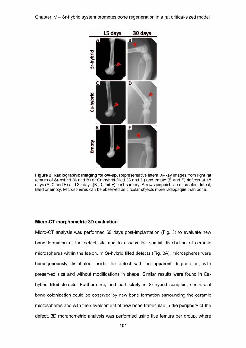
Chapter IV – Sr-hybrid system promotes bone regeneration in a rat critical-sized model
101
Figure 2. Radiographic imaging follow-up. Representative lateral X-Ray images from right rat femurs of Sr-hybrid (A and B) or Ca-hybrid-filled (C and D) and empty (E and F) defects at 15 days (A, C and E) and 30 days (B ,D and F) post-surgery. Arrows pinpoint site of created defect, filled or empty. Microspheres can be observed as circular objects more radiopaque than bone.
Micro-CT morphometric 3D evaluation
Micro-CT analysis was performed 60 days post-implantation (Fig. 3) to evaluate new
bone formation at the defect site and to assess the spatial distribution of ceramic
microspheres within the lesion. In Sr-hybrid filled defects (Fig. 3A), microspheres were
homogeneously distributed inside the defect with no apparent degradation, with
preserved size and without modifications in shape. Similar results were found in Ca-
hybrid filled defects. Furthermore, and particularly in Sr-hybrid samples, centripetal
bone colonization could be observed by new bone formation surrounding the ceramic
microspheres and with the development of new bone trabeculae in the periphery of the
defect. 3D morphometric analysis was performed using five femurs per group, where

Chapter IV – Sr-hybrid system promotes bone regeneration in a rat critical-sized model
102
the ROI was defined in binary images (Fig. 3B) and the percentage of new bone
formed (bone volume fraction, BV/TV) was calculated. Values of (31.5±1.7) % and
(28.6±1.1) % (BV/TV (%), mean±SE) of new bone was measured in animals that
received the Sr-hybrid and Ca-hybrid materials, respectively (Fig. 3C).
Figure 3. Micro-CT analysis of distal femur after 60 days of implantation. 3D reconstructed image and respective orthogonal slices of micro-CT acquisition of the femur with Sr-hybrid filled defect (A). Morphometric analysis approach used to quantify new bone formation: transversal micro-CT slice of a femur injected with Sr-hybrid after 60 days of implantation highlighting ROI (B). C - New bone formed (BV/TV, %) after 60 days of implantation of Sr-hybrid and Ca-hybrid materials. Data presented as box-plot with median and min to max whiskers of n = 5 samples (n.s. – statistically non-significant).
Histological evaluation of bone/biomaterial interface
In Fig. 4A representative images of femurs with defects filled with Sr-hybrid or Ca-
hybrid materials, at days 15 and 60, are portrayed. A global view of the defects and
materials (Fig. 4A a to d), as well as a more detailed view of the periphery of the defect
(Fig. 4A a’ to d’ and magnifications a’’ to d’’), are given. Histological analysis at day 15
post-implantation showed that the created defects exhibited similar diameter and were
filled with approximately 15 to 18 microspheres (Fig. 4A a, b). As early as 15 days post-

Chapter IV – Sr-hybrid system promotes bone regeneration in a rat critical-sized model
103
implantation, all animals showed, to some extent, newly formed bone at the periphery
of the defect (Fig 4A a’, b’). Sr-hybrid implanted defects also showed new bone
formation in close contact with the microspheres, distant from the periphery of native
bone (Fig. 4A b’’, arrow). SEM images and EDS analysis of this newly formed bone in
close vicinity of the microspheres is shown in Fig. 4B. The results confirmed the high
content of calcium and phosphate, and a Ca/P ratio in accordance with normal bone
composition (Z1 in Fig. 4B), and different from the elemental analysis of the
microspheres (Z2 in Fig. 4B), where Sr was also identified.
After 60 days, new bone formation at the periphery was observed in both materials. Sr-
hybrid implanted defects exhibited a thicker trabecular bone structure at the periphery
of the defect (Fig 4A d’, d’’), when compared to the Ca-hybrid group (Fig. 4A c’, c’’).
The quantification of new bone formed at the periphery of the defect revealed a
statistically significant thicker bone structure in Sr-hybrid group (662.4±48 µm) in
contrast to a thinner bone formation (381.1±29 µm) in the Ca-hybrid group (mean±SE,
p<0.001, Fig. 4C).

Chapter IV – Sr-hybrid system promotes bone regeneration in a rat critical-sized model
104
Figure 4. Histological evaluation of bone/biomaterial interface in femurs implanted with Sr-hybrid and Ca-hybrid systems. Coronal histological sections of critical sized defect created in the distal femur, 15 and 60 days post-implantation (A). Global view of the created defects filled with Ca-hybrid (a and c) and Sr-hybrid (b and d) materials, at 15 (a and b) or 60 days (c and d) post-implantation, stained with MT (a to d, dashed yellow line circling the created defect area). Interface bone/biomaterial, 15 days (a’ and b’) and 60 days (c’ and d’) post-implantation, with Ca-hybrid (a’ and c’) and Sr-hybrid (b’ and d’) systems, and higher magnification of square (a’’ to d’’, collagen/bone in blue, yellow dashed line – bone/biomaterial interface, * – alginate, M – microspheres, black arrows – new bone). B - SEM image of histological section of Sr-hybrid filled defect 15 days post-implantation with EDS spectra of new bone (Z1) found near the microsphere and microsphere (Z2). C - Thickness of peripheral bone found around the defect (as represented with arrows in A c’’ and A d’’), 60 days post-implantation of Sr-hybrid and Ca-hybrid systems. Data presented as mean±SEM of 20 different random locations of trabecular bone found around the defect of n=5 animals/group, 3 sections/animal, Asterisks show statistically significant differences (*** p<0.001).

Chapter IV – Sr-hybrid system promotes bone regeneration in a rat critical-sized model
105
Histological evaluation of the center of the defect
Representative images of the center of the defect in both groups are shown in Fig. 5A.
Although no evident differences were observed in the diameter of the microspheres
with time of implantation, alginate showed different behavior between groups. After 15
days, some alginate was observed surrounding microspheres in both experimental
groups (Fig 5A, a and b). In Sr-hybrid group, higher cell invasion at the center was
observed, mainly of polymorphonuclear neutrophils (PMN) (Fig 5A, b and b’).
Figure 5. Histological analysis of the center of the defect in femurs implanted with Sr-hybrid and Ca-hybrid systems. Detailed view of the center of the defect implanted with Ca-hybrid (a and c) and Sr-hybrid (b and d) materials, stained with H&E, after 15 (a and b) and 60 days (c and d) of implantation, with higher magnification of square (a’ to d’, A). B - Area of residual material (microspheres and alginate) found within the defect area, 60 days post-implantation of Sr-hybrid and Ca-hybrid systems. Measurements were performed by delimiting the area of material in MT stained sections using ImageJ software, n=5 animals/group, 3 sections/animal. Data presented as mean±SE and asterisks show statistically significant differences (*** p<0.001). C – TRAP-LG staining images of Ca-hybrid (a) and Sr-hybrid (b) systems filled defects, 60 days post-implantation. (M – microspheres, * – alginate, PMN – Polymorphonuclear neutrophils, GT – Granulation Tissue, BV – blood vessels, red arrows – Osteoclasts).

Chapter IV – Sr-hybrid system promotes bone regeneration in a rat critical-sized model
106
After 60 days, alginate was still present in both groups (Fig. 5A, c and d), although
higher degradation was observed in Sr-hybrid filled defects. A statistically significant
decrease in the area of residual material (alginate + microspheres) present on the
defect site was observed in Sr-hybrid group with an area of 1.77±0.2 x105 µm2
compared to 3.14± 0.1 x105 µm2 in Ca-hybrid group (mean±SE, p<0.001, Fig. 5B). In a
detailed microscopic analysis of the center of the Sr-hybrid filled defects (Fig. 5A, d and
d’) granulation tissue could be observed, with the presence of blood vessels (Fig. 5A
d’) and osteoclasts (Fig. 5C b). In contrast, in Ca-hybrid group osteoclasts were found
only at the periphery of the defect (Fig. 5C a). Furthermore, PSR staining images under
conventional light (Fig. 6A a and c) showed the presence of collagen (in red) within the
area of the defect in Sr-hybrid (Fig. 6A c) at a higher extent than in Ca-hybrid filled
defect after 60 days (Fig. 6A a). The use of PSR-polarization method (Fig. 6A b and d)
allowed for the quantification of different types of collagen fibrils, i.e. green and red,
which are associated with thin/immature/type III and thick/mature/type I collagen,
respectively. The quantification was performed within the central area of the defect
using a fixed ROI (diameter=2.4 mm, yellow circle in Fig. 6A b and d) and results are
shown in Fig. 6B. As expected, an increase in the percentage of red/type I collagen
was observed from day 15 to day 60 in both groups. However, 60 days post-
implantation, a slightly higher percentage of red/type I collagen was measured in the
central defect region in Sr-hybrid group ((3.3±1.3) %, mean±SE) compared to Ca-
hybrid group ((1.9±0.3) %, mean±SE).

Chapter IV – Sr-hybrid system promotes bone regeneration in a rat critical-sized model
107
Figure 6. Quantification of birefringent collagen fibers by PSR-polarization method in the center of the defect. PSR staining visualized under conventional light (a and c) and polarized light (b and d) to identify different collagen fiber types, 60 days post-implantation of Ca-hybrid (a and b) and Sr-hybrid (c and d) systems (A). PSR stains collagen in red (conventional light) and collagen fibers are specifically birefringent in polarized light (green - thin fibers/type III; red - thick fibers/type I, MosaicX image, original magnification 20x). Fixed region of interest (ROI) used for quantification is demonstrated in b and d. B - Quantification of area of green or red birefringent collagen fibers in the center of the defect in Sr-hybrid and Ca-hybrid filled defects, 15 and 60 days post-implantation. Data presented as mean±SE of n=5 animals/group, 3 images/animal. Asterisks show statistically significant differences (p<0.05) between groups for red collagen data.
Evaluation of Sr systemic effect
Sr levels were quantified in serum (Fig. 7A) and organs (Fig. 7B) associated with
excretory/filtration functions, such as liver, spleen and kidneys, by ICP-AES analysis, to
evaluate the safety of the designed Sr-hybrid system. The Sr levels in serum of animals
that were subjected to Sr-hybrid implantation (27.05±2.7 µg/L after 15 days and
20.61±1.3 µg/L after 60 days) were not statistically different from those in empty defect
animals (27.26±3.9 µg/L after 15 days and 23.31±1.9 µg/L after 60 days) or non-
operated animals (28.59±0.8 µg/L, mean±SD). Even after 60 days of implantation, no
increase in Sr was found in serum (Limit of quantification, LOQ – 5 μg/L).

Chapter IV – Sr-hybrid system promotes bone regeneration in a rat critical-sized model
108
Sr quantification in organs at 60 days post-implantation supports results from
measurements in serum. No statistical significant differences were observed between
empty defect animals (0.44±0.1 µg/g kidney, 0.50±0.1 µg/g spleen, 0.48±0.3 µg/g liver,
mean±SD) and Sr-hybrid group (0.69±0.3 µg/g kidney, 0.59±0.2 µg/g spleen, 0.36±0.1
µg/g liver, mean±SD). Moreover, histomorphological analyses were also performed in
histological sections of organs after 60 days (Fig. 7C). In Sr-hybrid implanted group no
morphological alterations at macro or microscopic level were observed, when
compared to non-operated animals. Analysis of Ca-hybrid group presented similar
results, with no alterations observed.
Figure 7. Strontium systemic evaluation. Strontium concentration found in serum in non-operated, empty and Sr-hybrid implanted animals at days 15 and 60 post-surgery (A). Data presented as mean±SD of 6 samples/group/time point and 2 samples non-operated. Strontium concentration found in the liver, kidneys and spleen of animals implanted with Sr-hybrid and empty defect, 60 days post-surgery (B). Data presented as mean±SD of 4 samples/group/time point. Representative histological sections from liver (a to c), kidney (d to f) and spleen (g to i) of non-operated (a, d and g), Sr-hybrid (b, e and h) and Ca-hybrid implanted (c, f, and i) animals, 60 days post-surgery (C).

Chapter IV – Sr-hybrid system promotes bone regeneration in a rat critical-sized model
109
Discussion
In this study the in vivo response to an injectable Sr-rich hybrid system, composed of Sr-
doped HAp microspheres embedded in an Sr-crosslinked RGD-alginate hydrogel, as
compared to a similar Sr-free system (Ca-hybrid material), using a rat metaphyseal
femoral critical size defect model, is presented.
The designed hybrid system aims at providing adequate mechanical support in the early
phases of bone formation and gradual replacement of the artificial scaffold by newly-
formed bone with adequate function and mechanical properties. The use of hydrogels is
a promising approach in skeletal regenerative medicine [49, 66-68]. Alginate has been
used due to its biocompatibility, low toxicity, and mild gelation in the presence of divalent
cations. Therefore alginate gels act as a natural extracellular matrix mimic which can be
tuned to deliver bioactive agents and cells to the desired site, creating space for new
tissue formation and control the structure and function of the engineered tissue [49].
Other works have incorporated alginate in self-setting cements, for improving injectability,
cohesion and compression strength [69-71]. In the present study, the ability of alginate to
form hydrogels in situ acting as a carrier for HAp microspheres under cytocompatible
conditions, was explored. In agreement with our previous results [53], the system
showed to be adequate for minimally invasive implantation. A conventional syringe can
be used to manually inject the material, perfectly filling complex defects and, once set,
creating a 3D matrix with homogeneous distribution of microspheres. Furthermore,
alginate was modified with RGD peptides to provide biological cues for promoting cell
adhesion and colonization [50, 57]. The main disadvantage regarding load-bearing
application is alginate low mechanical properties which can be overcome through the
reinforcement with ceramic components, application in non load-bearing areas, and the
concomitant use of fixation devices [54]. With an alginate-to-microspheres weight ratio of
0.35, and microspheres with average diameter of 530 µm, the hybrid system allows for a
good compromise between mechanical resistance and adequate space between

Chapter IV – Sr-hybrid system promotes bone regeneration in a rat critical-sized model
110
particles (approximately 220 μm), which is expected to facilitate in situ cell colonization
and invasion by blood vessels [53, 55].
Deficient bone healing is expected to occur, especially in osteoporotic conditions. The
use of crystalline HAp in the microspheres, with low degradation rate, ensures its
permanency at the injury site for longer periods post-implantation, therefore allowing for a
mechanical reinforcement of the defect. In this work, the high radiopacity of microspheres
allowed for an easy follow-up in vivo of the material using conventional radiological
imaging. In clinical practice, this comes as highly advantageous since regular X-rays are
required to assess bone healing.
In this system we used alginate crosslinked by internal gelation with Sr2+ as a vehicle for
the Sr-doped HAp microspheres. Sr was incorporated both in the hydrogel and the
microspheres, which present different release kinetic profiles resulting in sustained Sr2+
release for long periods of time (AH Lourenço et al, unpublished results). In other in vivo
studies, Sr has been found to enhance bone formation [35, 37, 72, 73]. For example,
Banerjee et al. studied the effect of doping ß-TCP with MgO/SrO on bone formation in
Sprague-Dawley rats [72]. Doped ß-TCP promoted more osteogenesis and faster bone
formation than pure ß-TCP. In critical calvaria defects of an ovariectomized rat model,
macroporous Sr-substituted scaffolds showed superior osteoinductive activity to enhance
early bone formation, and could also stimulate angiogenesis compared with calcium
silicate scaffolds [73]. The current study showed that Sr-hybrid system presents bioactive
properties, promoting cell migration, implant vascularization and supports bone ingrowth.
Newly formed bone developed in close contact with the material, without any fibrous
interface, growing in a centripetal manner, in continuity with the surrounding host
trabecular bone, indicates a good integration with the host tissue. Newly formed bone
percentages is in agreement to those observed in other works testing similar materials
[74, 75] and a trend towards greater new bone formation was observed in Sr-hybrid filled
defects.

Chapter IV – Sr-hybrid system promotes bone regeneration in a rat critical-sized model
111
Major histological differences were observed between the two groups. Higher cell
invasion was seen at the center of the Sr-hybrid filled defects at 15 days post-
implantation, with the presence of polymorphonuclear neutrophils (PMN). Bone injury
elicits an inflammatory response that is beneficial to healing when acute and highly
regulated. Inflammatory cells are recruited to the site of injury for clearance of
pathogens and maintenance of bone homeostasis [76]. This higher cell invasion
correlates with higher material degradation and bone tissue formation seen after 60
days. We may assume that Sr induces faster bone healing, possibly due to a faster
resolution of inflammation, tissue repair and remodeling. This can be appealing since
the presence of Sr may be modulating the inflammatory response, a current trend in
bone regeneration strategies regarding the development of biomaterials [77-79].
Earlier bone formation was identified in close contact with the microspheres in Sr-
hybrid system, highlighting higher osteoinductivity when compared to Ca-hybrid system
which was evidenced by the thicker trabecular bone structure at the periphery of the
defect observed in Sr-hybrid group 60 days post-implantation. Cardemill et al have also
found major differences in the topological distribution of the formed bone in association
with Sr-doped calcium phosphate or HAp granules, although both materials showed
comparable overall bone formation, when implanted in ovariectomized and non-
ovariectomized rats [80]. A larger amount of mineralized bone was observed in the
center of the defect in HAp group, mainly in ovariectomized rats, whereas at the
periphery of the defect the bone area was higher using Sr-doped granules, irrespective
of ovariectomy.
Granulation tissue with blood vessels and increased collagen deposition were
observed at 60 days post-implantation in Sr-hybrid filled defects. Several studies have
correlated the color of birefringent collagen fibers under polarized light with different
collagen types [63, 64]. Our results show an increase in thicker red collagen fibers in
Sr-hybrid group. These fibers are associated with type I collagen, the main type found
in bone tissue. It has been shown that the incorporation of Sr in biomaterials may

Chapter IV – Sr-hybrid system promotes bone regeneration in a rat critical-sized model
112
decrease the number of osteoclasts significantly, but these were nevertheless closely
associated with newly formed trabeculae, indicating activated bone remodeling [81]. A
quantitative analysis of TRAP positive cells was not performed. However, the presence
of osteoclasts in the center of the defect supports the higher bone remodeling found in
the Sr-hybrid group. Although the microCT calculations did not reveal a statistically
different bone volume, these histological findings sustain the evidence of an improved
response with the Sr enriched material.
The use of methylmethacrylate embedding histological technique allowed for the study
of both bone and material without decalcification. The technique was optimized in our
lab [61] using an exothermic process. One of the disadvantages of the procedure is the
inability to perform immunohistochemistry studies due to loss of antigenicity, which is
worth exploring in future works.
With the use of Sr releasing systems, cardiovascular safety is of concern. Current
guidelines indicate that orally administered Sr ranelate should be avoided in patients
with past or present history of ischemic heart disease, peripheral arterial disease
and/or cerebrovascular disease or uncontrolled hypertension, due to an observed
increase of cardiovascular events [12]. Although previous reports have shown that ionic
Sr can be added to calcium phosphates and ceramics, potentially stimulating bone
formation locally, the risk of systemic adverse effects has been rarely reported. Baier et
al studied the addition of Sr to calcium-phosphate cement in a distal methaphyseal
femoral defect in ovariectomized rat model. Results have shown faster osteointegration
of the implant with the addition of Sr, and Sr serum concentrations of 10.87 ± 4.16 μg/l
were found 1 month post-implantation [35]. The systemic Sr levels were very low when
compared to those found upon oral Sr ranelate treatment [82]. In the present study, Sr
concentration was assessed both in the serum and organs with excretory/filtration
functions, as well as the histology of these organs. Serum Sr concentration in operated
animals was found to be similar to non-operated and in the same range as previously
reported [35]. No statistically significant difference was observed between Sr-hybrid

Chapter IV – Sr-hybrid system promotes bone regeneration in a rat critical-sized model
113
implanted and empty defects, both in serum and organs. Similarly, Sr concentration
levels do not seem to be increased compared to normal levels found in the liver of
Wistar rats (~0.2 ug/g of dry weight [83]) . These results, together with the absence of
morphological changes in histological sections of the organs suggest that Sr release is
restricted to the defect site, corroborating the safety of this osteoinductive hybrid
system.

Chapter IV – Sr-hybrid system promotes bone regeneration in a rat critical-sized model
114
Conclusions
We evaluated the in vivo response of an injectable Sr-rich hybrid system composed of
Sr-doped HAp microspheres embedded in Sr-crosslinked RGD-alginate hydrogel
intended for bone regeneration. Sr-hybrid system led to an increased bone formation in
both center and periphery of a critical size defect compared to a non Sr–doped similar
system, where new bone formation was restricted to the periphery. Besides promoting
earlier new bone formation, Sr-hybrid system was also found to stimulate higher cell
colonization with increased deposition of thick collagen fibers in the center of the
defect. Importantly, our results suggest that only local release of Sr from the material
was obtained, since no statistically significant differences on Sr concentration were
detected in retrieved organs or serum. Together, these data demonstrate that the
incorporation of Sr improved the osteoinductive properties of the hybrid system leading
to higher bone regeneration without inducing detrimental side effects currently
associated with other Sr-based therapeutic strategies. The Sr-hybrid material stands as
a promising approach for bone regeneration strategies through minimally invasive
procedures.

Chapter IV – Sr-hybrid system promotes bone regeneration in a rat critical-sized model
115
Acknowledgements
The authors thankfully acknowledge the use of Micro-CT in the 3B’s Research Group
facilities at University of Minho, the use of microscopy services at Centro de Materiais
da Universidade do Porto (CEMUP) and the technicians responsible for the
experimental techniques mentioned, Vitor Correlo and Daniela Silva, respectively;
Carla Rodrigues from Requimte at FCT-UNL for the help with ICP-AES analysis. This
work was financed by FEDER - Fundo Europeu de Desenvolvimento Regional funds
through the COMPETE 2020 - Operacional Programme for Competitiveness and
Internationalisation (POCI), Portugal 2020, and by Portuguese funds through FCT -
Fundação para a Ciência e a Tecnologia/ Ministério da Ciência, Tecnologia e Inovação
in the framework of the project "Institute for Research and Innovation in Health
Sciences" (POCI-01-0145-FEDER-007274) and project PTDC/CTM/103181/2008. AHL
acknowledges FCT for PhD grant: SFRH/BD/87192/2012, CCB acknowledges FCT for
research position Investigator FCT (IF2013).
Author Contributions
Study design: AHL, NN, ML, CR. Preparation of materials: AHL, CR. Surgical
procedures: AHL, NN. Animal follow-up and data collection: AHL, CR-M. Preparation
for elemental ICP-AES analysis and interpretation: AHL, SRS. Histological analysis and
interpretation: AHL, CR-M, ML, CR. Discussion and interpretation of the results: AHL,
NN, CR-M, SRS, ML, CB, ATC, MB, CR. Drafting manuscript: AHL, NN, CR. All
authors critically revised manuscript content and approved final version of manuscript.
Disclosures: The authors declare no conflict of interest.

Chapter IV – Sr-hybrid system promotes bone regeneration in a rat critical-sized model
116
References
[1] J. Goldhahn, W.H. Scheele, B.H. Mitlak, E. Abadie, P. Aspenberg, P. Augat, M.-L. Brandi, N. Burlet, A. Chines, P.D. Delmas, I. Dupin-Roger, D. Ethgen, B. Hanson, F. Hartl, J.A. Kanis, R. Kewalramani, A. Laslop, D. Marsh, S. Ormarsdottir, R. Rizzoli, A. Santora, G. Schmidmaier, M. Wagener, J.-Y. Reginster, Clinical evaluation of medicinal products for acceleration of fracture healing in patients with osteoporosis, Bone 43(2) (2008) 343-347. [2] V. Campana, G. Milano, E. Pagano, M. Barba, C. Cicione, G. Salonna, W. Lattanzi, G. Logroscino, Bone substitutes in orthopaedic surgery: from basic science to clinical practice, Journal of Materials Science: Materials in Medicine 25(10) (2014) 2445-2461. [3] A. Dupraz, T.P. Nguyen, M. Richard, G. Daculsi, N. Passuti, Influence of a cellulosic ether carrier on the structure of biphasic calcium phosphate ceramic particles in an injectable composite material, Biomaterials 20(7) (1999) 663-673. [4] L.R.A. Iooss P, Grimandi G, Daculsi G, Merle C. , A new injectable bone substitute combining poly(epsilon-caprolactone) microparticles with biphasic calcium phosphate granules., Biomaterials. (22) (2001) 2785-94. [5] S. Larsson, Calcium Phosphates: What Is the Evidence?, Journal of Orthopaedic Trauma 24 (2010) S41-S45. [6] K.L. Low, S.H. Tan, S.H.S. Zein, J.A. Roether, V. Mouriño, A.R. Boccaccini, Calcium phosphate-based composites as injectable bone substitute materials, Journal of Biomedical Materials Research Part B: Applied Biomaterials 94B(1) (2010) 273-286. [7] L. Cao, G. Liu, Y. Gan, Q. Fan, F. Yang, X. Zhang, T. Tang, K. Dai, The use of autologous enriched bone marrow MSCs to enhance osteoporotic bone defect repair in long-term estrogen deficient goats, Biomaterials 33(20) (2012) 5076-5084. [8] S.W. Cho, H.J. Sun, J.-Y. Yang, J.Y. Jung, H.J. Choi, J.H. An, S.W. Kim, S.Y. Kim, K.-J. Park, C.S. Shin, Human Adipose Tissue-Derived Stromal Cell Therapy Prevents Bone Loss in Ovariectomized Nude Mouse, Tissue Engineering Part A 18(9-10) (2012) 1067-1078. [9] H.-Y. Liu, A.T.H. Wu, C.-Y. Tsai, K.-R. Chou, R. Zeng, M.-F. Wang, W.-C. Chang, S.-M. Hwang, C.-H. Su, W.-P. Deng, The balance between adipogenesis and osteogenesis in bone regeneration by platelet-rich plasma for age-related osteoporosis, Biomaterials 32(28) (2011) 6773-6780. [10] Z. Wang, J. Goh, S.D. De, Z. Ge, H. Ouyang, J.S.W. Chong, S.L. Low, E.H. Lee, Efficacy of bone marrow-derived stem cells in strengthening osteoporotic bone in a rabbit model., Tissue engineering. (12(7)) ( 2006) 1753-1761. . [11] P.J. Meunier , C. Roux , E. Seeman , S. Ortolani , J.E. Badurski , T.D. Spector , J. Cannata , A. Balogh , E.-M. Lemmel , S. Pors-Nielsen , R. Rizzoli , H.K. Genant , J.-Y. Reginster The Effects of Strontium Ranelate on the Risk of Vertebral Fracture in Women with Postmenopausal Osteoporosis, New England Journal of Medicine 350(5) (2004) 459-468. [12] J.Y. Reginster, A. Neuprez, N. Dardenne, C. Beaudart, P. Emonts, O. Bruyere, Efficacy and safety of currently marketed anti-osteoporosis medications, Best Practice & Research Clinical Endocrinology & Metabolism 28(6) (2014) 809-834. [13] R. Baron, Y. Tsouderos, In vitro effects of S12911-2 on osteoclast function and bone marrow macrophage differentiation, European Journal of Pharmacology 450(1) (2002) 11-17. [14] E. Bonnelye, A. Chabadel, F. Saltel, P. Jurdic, Dual effect of strontium ranelate: Stimulation of osteoblast differentiation and inhibition of osteoclast formation and resorption in vitro, Bone 42(1) (2008) 129-138. [15] M.D. Grynpas, E. Hamilton, R. Cheung, Y. Tsouderos, P. Deloffre, M. Hott, P.J. Marie, Strontium increases vertebral bone volume in rats at a low dose that does not induce detectable mineralization defect, Bone 18(3) (1996) 253-259.

Chapter IV – Sr-hybrid system promotes bone regeneration in a rat critical-sized model
117
[16] F. Yang, D. Yang, J. Tu, Q. Zheng, L. Cai, L. Wang, Strontium Enhances Osteogenic Differentiation of Mesenchymal Stem Cells and In Vivo Bone Formation by Activating Wnt/Catenin Signaling, STEM CELLS 29(6) (2011) 981-991. [17] N. Takahashi, T. Sasaki, Y. Tsouderos, T. Suda, S 12911-2 Inhibits Osteoclastic Bone Resorption In Vitro, Journal of Bone and Mineral Research 18(6) (2003) 1082-1087. [18] A.S. Hurtel-Lemaire, R. Mentaverri, A. Caudrillier, F. Cournarie, A. Wattel, S. Kamel, E.F. Terwilliger, E.M. Brown, M. Brazier, The Calcium-sensing Receptor Is Involved in Strontium Ranelate-induced Osteoclast Apoptosis: NEW INSIGHTS INTO THE ASSOCIATED SIGNALING PATHWAYS, Journal of Biological Chemistry 284(1) (2009) 575-584. [19] P.J. Marie, D. Felsenberg, M.L. Brandi, How strontium ranelate, via opposite effects on bone resorption and formation, prevents osteoporosis, Osteoporosis International 22(6) (2011) 1659-1667. [20] G.J. Atkins, K.J. Welldon, P. Halbout, D.M. Findlay, Strontium ranelate treatment of human primary osteoblasts promotes an osteocyte-like phenotype while eliciting an osteoprotegerin response, Osteoporosis International 20(4) (2009) 653-664. [21] T.C. Brennan, M.S. Rybchyn, W. Green, S. Atwa, A.D. Conigrave, R.S. Mason, Osteoblasts play key roles in the mechanisms of action of strontium ranelate, British Journal of Pharmacology 157(7) (2009) 1291-1300. [22] J. Caverzasio, Strontium ranelate promotes osteoblastic cell replication through at least two different mechanisms, Bone 42(6) (2008) 1131-1136. [23] N. Chattopadhyay, S.J. Quinn, O. Kifor, C. Ye, E.M. Brown, The calcium-sensing receptor (CaR) is involved in strontium ranelate-induced osteoblast proliferation, Biochemical Pharmacology 74(3) (2007) 438-447. [24] A. Barbara, P. Delannoy, B.G. Denis, P.J. Marie, Normal matrix mineralization induced by strontium ranelate in MC3T3-E1 osteogenic cells, Metabolism 53(4) (2004) 532-537. [25] S. Choudhary, P. Halbout, C. Alander, L. Raisz, C. Pilbeam, Strontium Ranelate Promotes Osteoblastic Differentiation and Mineralization of Murine Bone Marrow Stromal Cells: Involvement of Prostaglandins, Journal of Bone and Mineral Research 22(7) (2007) 1002-1010. [26] L.-L. Zhu, S. Zaidi, Y. Peng, H. Zhou, B.S. Moonga, A. Blesius, I. Dupin-Roger, M. Zaidi, L. Sun, Induction of a program gene expression during osteoblast differentiation with strontium ranelate, Biochemical and Biophysical Research Communications 355(2) (2007) 307-311. [27] O. Fromigué, E. Haÿ, A. Barbara, P.J. Marie, Essential Role of Nuclear Factor of Activated T Cells (NFAT)-mediated Wnt Signaling in Osteoblast Differentiation Induced by Strontium Ranelate, Journal of Biological Chemistry 285(33) (2010) 25251-25258. [28] P. Ammann, I. Badoud, S. Barraud, R. Dayer, R. Rizzoli, Strontium Ranelate Treatment Improves Trabecular and Cortical Intrinsic Bone Tissue Quality, a Determinant of Bone Strength, Journal of Bone and Mineral Research 22(9) (2007) 1419-1425. [29] P. Ammann, V. Shen, B. Robin, Y. Mauras, J.-P. Bonjour, R. Rizzoli, Strontium Ranelate Improves Bone Resistance by Increasing Bone Mass and Improving Architecture in Intact Female Rats, Journal of Bone and Mineral Research 19(12) (2004) 2012-2020. [30] S.D. Bain, C. Jerome, V. Shen, I. Dupin-Roger, P. Ammann, Strontium ranelate improves bone strength in ovariectomized rat by positively influencing bone resistance determinants, Osteoporosis International 20(8) (2009) 1417-1428. [31] J. Buehler, P. Chappuis, J.L. Saffar, Y. Tsouderos, A. Vignery, Strontium ranelate inhibits bone resorption while maintaining bone formation in alveolar bone in monkeys (Macaca fascicularis), Bone 29(2) (2001) 176-179.

Chapter IV – Sr-hybrid system promotes bone regeneration in a rat critical-sized model
118
[32] P. Delannoy, D. Bazot, P.J. Marie, Long-term treatment with strontium ranelate increases vertebral bone mass without deleterious effect in mice, Metabolism 51(7) (2002) 906-911. [33] B. Abrahamsen, E.L. Grove, P. Vestergaard, Nationwide registry-based analysis of cardiovascular risk factors and adverse outcomes in patients treated with strontium ranelate, Osteoporosis International 25(2) (2014) 757-762. [34] C. Cooper, K.M. Fox, J.S. Borer, Ischaemic cardiac events and use of strontium ranelate in postmenopausal osteoporosis: a nested case–control study in the CPRD, Osteoporosis International 25(2) (2014) 737-745. [35] M. Baier, P. Staudt, R. Klein, U. Sommer, R. Wenz, I. Grafe, P.J. Meeder, P.P. Nawroth, C. Kasperk, Strontium enhances osseointegration of calcium phosphate cement: a histomorphometric pilot study in ovariectomized rats, Journal of Orthopaedic Surgery and Research 8(1) (2013) 1-8. [36] G. Hulsart-Billström, W. Xia, E. Pankotai, M. Weszl, E. Carlsson, C. Forster-Horváth, S. Larsson, H. Engqvist, Z. Lacza, Osteogenic potential of Sr-doped calcium phosphate hollow spheres in vitro and in vivo, Journal of Biomedical Materials Research Part A 101A(8) (2013) 2322-2331. [37] U. Thormann, S. Ray, U. Sommer, T. ElKhassawna, T. Rehling, M. Hundgeburth, A. Henß, M. Rohnke, J. Janek, K.S. Lips, C. Heiss, G. Schlewitz, G. Szalay, M. Schumacher, M. Gelinsky, R. Schnettler, V. Alt, Bone formation induced by strontium modified calcium phosphate cement in critical-size metaphyseal fracture defects in ovariectomized rats, Biomaterials 34(34) (2013) 8589-8598. [38] C.P. Friedman CD, Takagi S, Chow LC., BoneSource hydroxyapatite cement: a novel biomaterial for craniofacial skeletal tissue engineering and reconstruction., J Biomed Mater Res (43:428-32.) (1998). [39] Q.J. Xu HH, Calcium phosphate cement containing resorbable fibers for short-term reinforcement and macroporosity., Biomaterials (23:193-202.) (2002). [40] C.C. Barrias, C.C. Ribeiro, M. Lamghari, C.S. Miranda, M.A. Barbosa, Proliferation, activity, and osteogenic differentiation of bone marrow stromal cells cultured on calcium titanium phosphate microspheres, Journal of Biomedical Materials Research Part A 72A(1) (2005) 57-66. [41] S.M. Oliveira, C.C. Barrias, I.F. Almeida, P.C. Costa, M.R.P. Ferreira, M.F. Bahia, M.A. Barbosa, Injectability of a bone filler system based on hydroxyapatite microspheres and a vehicle with in situ gel-forming ability, Journal of Biomedical Materials Research Part B: Applied Biomaterials 87B(1) (2008) 49-58. [42] C.C. Ribeiro, C.C. Barrias, M.A. Barbosa, Preparation and characterisation of calcium-phosphate porous microspheres with a uniform size for biomedical applications, Journal of Materials Science: Materials in Medicine 17(5) (2006) 455-463. [43] C.C. Ribeiro, C.C. Barrias, M.A. Barbosa, Calcium phosphate-alginate microspheres as enzyme delivery matrices, Biomaterials 25(18) (2004) 4363-4373. [44] C.C. Barrias, M. Lamghari, P.L. Granja, M.C. Sá Miranda, M.A. Barbosa, Biological evaluation of calcium alginate microspheres as a vehicle for the localized delivery of a therapeutic enzyme, Journal of Biomedical Materials Research Part A 74A(4) (2005) 545-552. [45] K.B. Fonseca, D.B. Gomes, K. Lee, S.G. Santos, A. Sousa, E.A. Silva, D.J. Mooney, P.L. Granja, C.C. Barrias, Injectable MMP-Sensitive Alginate Hydrogels as hMSC Delivery Systems, Biomacromolecules 15(1) (2014) 380-390. [46] F.R. Maia, M. Barbosa, D.B. Gomes, N. Vale, P. Gomes, P.L. Granja, C.C. Barrias, Hydrogel depots for local co-delivery of osteoinductive peptides and mesenchymal stem cells, Journal of Controlled Release 189 (2014) 158-168. [47] F.R. Maia, A.H. Lourenço, P.L. Granja, R.M. Gonçalves, C.C. Barrias, Effect of Cell Density on Mesenchymal Stem Cells Aggregation in RGD-Alginate 3D Matrices under Osteoinductive Conditions, Macromolecular Bioscience 14(6) (2014) 759-771. [48] M.B. Evangelista, S.X. Hsiong, R. Fernandes, P. Sampaio, H.-J. Kong, C.C. Barrias, R. Salema, M.A. Barbosa, D.J. Mooney, P.L. Granja, Upregulation of bone cell

Chapter IV – Sr-hybrid system promotes bone regeneration in a rat critical-sized model
119
differentiation through immobilization within a synthetic extracellular matrix, Biomaterials 28(25) (2007) 3644-3655. [49] K.Y. Lee, D.J. Mooney, Alginate: properties and biomedical applications, Progress in polymer science 37(1) (2012) 106-126. [50] S. Sakiyama-Elbert, J. Hubbell, Functional Biomaterials: Design of Novel Biomaterials, Annual Review of Materials Research 31(1) (2001) 183-201. [51] T. Andersen, B.L. Strand, K. Formo, E. Alsberg, B.E. Christensen, Alginates as biomaterials in tissue engineering, Carbohydrate Chemistry: Volume 37, The Royal Society of Chemistry2012, pp. 227-258. [52] Ý.A. Mørch, I. Donati, B.L. Strand, Effect of Ca2+, Ba2+, and Sr2+ on Alginate Microbeads, Biomacromolecules 7(5) (2006) 1471-1480. [53] N. Neves, B.B. Campos, I.F. Almeida, P.C. Costa, A.T. Cabral, M.A. Barbosa, C.C. Ribeiro, Strontium-rich injectable hybrid system for bone regeneration, Materials Science and Engineering: C 59 (2016) 818-827. [54] E.D. D'Este M, Hydrogels in calcium phosphate moldable and injectable bone substitutes: Sticky excipients or advanced 3-D carriers?, Acta Biomater (9:5421-30.) (2013). [55] M. Bohner, S. Tadier, N. van Garderen, A. de Gasparo, N. Dobelin, G. Baroud, Synthesis of spherical calcium phosphate particles for dental and orthopedic applications, Biomatter 3(2) (2013). [56] A. Lourenço, A. Torres, S. Santos, M. Barbosa, C. Barrias, C. Ribeiro, Sr-Hybrid system for bone regeneration: an in vitro study, 4th Interrogations at the Biointerface Advanced Summer School, Barcelona, 2014. [57] S.J. Bidarra, C.C. Barrias, K.B. Fonseca, M.A. Barbosa, R.A. Soares, P.L. Granja, Injectable in situ crosslinkable RGD-modified alginate matrix for endothelial cells delivery, Biomaterials 32(31) (2011) 7897-7904. [58] K.B. Fonseca, S.J. Bidarra, M.J. Oliveira, P.L. Granja, C.C. Barrias, Molecularly designed alginate hydrogels susceptible to local proteolysis as three-dimensional cellular microenvironments, Acta Biomaterialia 7(4) (2011) 1674-1682. [59] L. Le Guehennec, E. Goyenvalle, E. Aguado, M. Houchmand-Cuny, B. Enkel, P. Pilet, G. Daculsi, P. Layrolle, Small-animal models for testing macroporous ceramic bone substitutes, Journal of Biomedical Materials Research Part B: Applied Biomaterials 72B(1) (2005) 69-78. [60] S.G. Santos, M. Lamghari, C.R. Almeida, M.I. Oliveira, N. Neves, A.C. Ribeiro, J.N. Barbosa, R. Barros, J. Maciel, M.C.L. Martins, R.M. Gonçalves, M.A. Barbosa, Adsorbed fibrinogen leads to improved bone regeneration and correlates with differences in the systemic immune response, Acta Biomaterialia 9(7) (2013) 7209-7217. [61] C. Carvalho, J. Magalhães, L. Pereira, L. Simões-Silva, I. Castro-Ferreira, J.M. Frazão, Evolution of bone disease after kidney transplantation: A prospective histomorphometric analysis of trabecular and cortical bone, Nephrology 21(1) (2016) 55-61. [62] D.M. Sousa, P.A. Baldock, R.F. Enriquez, L. Zhang, A. Sainsbury, M. Lamghari, H. Herzog, Neuropeptide Y Y1 receptor antagonism increases bone mass in mice, Bone 51(1) (2012) 8-16. [63] G.S. Montes, L.C.U. Junqueira, The use of the Picrosirius-polarization method for the study of the biopathology of collagen, Memórias do Instituto Oswaldo Cruz 86 (1991) 1-11. [64] A.E. Vieira, C.E. Repeke, S.d.B. Ferreira Junior, P.M. Colavite, C.C. Biguetti, R.C. Oliveira, G.F. Assis, R. Taga, A.P.F. Trombone, G.P. Garlet, Intramembranous Bone Healing Process Subsequent to Tooth Extraction in Mice: Micro-Computed Tomography, Histomorphometric and Molecular Characterization, PLoS ONE 10(5) (2015) e0128021.

Chapter IV – Sr-hybrid system promotes bone regeneration in a rat critical-sized model
120
[65] R.K. Fuchs, M.R. Allen, K.W. Condon, S. Reinwald, L.M. Miller, D. McClenathan, B. Keck, R.J. Phipps, D.B. Burr, Strontium ranelate does not stimulate bone formation in ovariectomized rats, Osteoporosis International 19(9) (2008) 1331-1341. [66] N.E. Fedorovich, J. Alblas, J.R. de Wijn, W.E. Hennink, A.J. Verbout, W.J. Dhert, Hydrogels as extracellular matrices for skeletal tissue engineering: state-of-the-art and novel application in organ printing, Tissue Eng 13(8) (2007) 1905-25. [67] A. Khademhosseini, R. Langer, Microengineered hydrogels for tissue engineering, Biomaterials 28(34) (2007) 5087-5092. [68] M. Patenaude, N.M.B. Smeets, T. Hoare, Designing Injectable, Covalently Cross-Linked Hydrogels for Biomedical Applications, Macromolecular Rapid Communications 35(6) (2014) 598-617. [69] G.-S. Lee, J.-H. Park, J.-E. Won, U.S. Shin, H.-W. Kim, Alginate combined calcium phosphate cements: mechanical properties and in vitro rat bone marrow stromal cell responses, Journal of Materials Science: Materials in Medicine 22(5) (2011) 1257-1268. [70] C. Xu, X. Wang, J. Zhou, Z. Huan, J. Chang, Bioactive tricalcium silicate/alginate composite bone cements with enhanced physicochemical properties, Journal of Biomedical Materials Research Part B: Applied Biomaterials (2017) n/a-n/a. [71] S. Sprio, M. Dapporto, M. Montesi, S. Panseri, W. Lattanzi, E. Pola, G. Logroscino, A. Tampieri, Novel Osteointegrative Sr-Substituted Apatitic Cements Enriched with Alginate, Materials 9(9) (2016) 763. [72] S.S. Banerjee, S. Tarafder, N.M. Davies, A. Bandyopadhyay, S. Bose, Understanding the influence of MgO and SrO binary doping on the mechanical and biological properties of β-TCP ceramics, Acta Biomaterialia 6(10) (2010) 4167-4174. [73] K. Lin, L. Xia, H. Li, X. Jiang, H. Pan, Y. Xu, W.W. Lu, Z. Zhang, J. Chang, Enhanced osteoporotic bone regeneration by strontium-substituted calcium silicate bioactive ceramics, Biomaterials 34(38) (2013) 10028-10042. [74] G. Daculsi, A.P. Uzel, P. Weiss, E. Goyenvalle, E. Aguado, Developments in injectable multiphasic biomaterials. The performance of microporous biphasic calcium phosphate granules and hydrogels, Journal of Materials Science: Materials in Medicine 21(3) (2010) 855-861. [75] O. Gauthier, R. Müller, D. von Stechow, B. Lamy, P. Weiss, J.-M. Bouler, E. Aguado, G. Daculsi, In vivo bone regeneration with injectable calcium phosphate biomaterial: A three-dimensional micro-computed tomographic, biomechanical and SEM study, Biomaterials 26(27) (2005) 5444-5453. [76] F. Loi, L.A. Córdova, J. Pajarinen, T.-h. Lin, Z. Yao, S.B. Goodman, Inflammation, fracture and bone repair, Bone 86 (2016) 119-130. [77] D.P. Vasconcelos, M. Costa, I.F. Amaral, M.A. Barbosa, A.P. Águas, J.N. Barbosa, Modulation of the inflammatory response to chitosan through M2 macrophage polarization using pro-resolution mediators, Biomaterials 37 (2015) 116-123. [78] D.P. Vasconcelos, M. Costa, I.F. Amaral, M.A. Barbosa, A.P. Águas, J.N. Barbosa, Development of an immunomodulatory biomaterial: Using resolvin D1 to modulate inflammation, Biomaterials 53 (2015) 566-573. [79] D.M. Vasconcelos, R.M. Gonçalves, C.R. Almeida, I.O. Pereira, M.I. Oliveira, N. Neves, A.M. Silva, A.C. Ribeiro, C. Cunha, A.R. Almeida, C.C. Ribeiro, A.M. Gil, E. Seebach, K.L. Kynast, W. Richter, M. Lamghari, S.G. Santos, M.A. Barbosa, Fibrinogen scaffolds with immunomodulatory properties promote in vivo bone regeneration, Biomaterials 111 (2016) 163-178. [80] C. Cardemil, I. Elgali, W. Xia, L. Emanuelsson, B. Norlindh, O. Omar, P. Thomsen, Strontium-Doped Calcium Phosphate and Hydroxyapatite Granules Promote Different Inflammatory and Bone Remodelling Responses in Normal and Ovariectomised Rats, PLoS ONE 8(12) (2013) e84932. [81] L. Wei, J. Ke, I. Prasadam, R.J. Miron, S. Lin, Y. Xiao, J. Chang, C. Wu, Y. Zhang, A comparative study of Sr-incorporated mesoporous bioactive glass scaffolds for

Chapter IV – Sr-hybrid system promotes bone regeneration in a rat critical-sized model
121
regeneration of osteopenic bone defects, Osteoporosis International 25(8) (2014) 2089-2096. [82] P.J. Marie, M. Hott, D. Modrowski, C. De Pollak, J. Guillemain, P. Deloffre, Y. Tsouderos, An uncoupling agent containing strontium prevents bone loss by depressing bone resorption and maintaining bone formation in estrogen-deficient rats, Journal of Bone and Mineral Research 8(5) (1993) 607-615. [83] S. Takahashi, I. Takahashi, H. Sato, Y. Kubota, S. Yoshida, Y. Muramatsu, Determination of major and trace elements in the liver of Wistar rats by inductively coupled plasma-atomic emission spectrometry and mass spectrometry, Laboratory Animals 34(1) (2000) 97-105.

Chapter IV – Sr-hybrid system promotes bone regeneration in a rat critical-sized model
122
Supplementary Data
Table 1 - Microwave digestion program.
Stages 1 2 3
Power (W) 600 600 600
Time (min.) 5 10 10
Temperature (control, °C ) 50 100 175
Hold (min.) 10 10 15

Chapter IV – Sr-hybrid system promotes bone regeneration in a rat critical-sized model
123

Chapter IV – Sr-hybrid system promotes bone regeneration in a rat critical-sized model
124

Chapter V – General discussion and future perspectives
125
Chapter V
General discussion and future perspectives

Chapter V – General discussion and future perspectives
126

Chapter V – General discussion and future perspectives
127
The use of injectable biomaterials that support and stimulate bone repair/regeneration
are highly attractive options for the clinical management of bone lesions. These can be
implanted through minimally invasive surgery, to efficiently fill-in cavities of non-uniform
shapes, with less tissue damage and limited exposure to infectious agents, thus
reducing patient discomfort and procedure-associated health costs. The addition of
osteoinductive factors may improve bone repair, particularly in osteoporotic conditions,
characterized by an impaired healing response [1-4].
In this context, the aim of this PhD thesis was to evaluate the in vitro and in vivo
response to a Sr-hybrid injectable system for bone regeneration, designed by our
group [5], consisting of hydroxyapatite microspheres doped with Sr and an alginate
vehicle crosslinked in situ with Sr.
The composite nature of bone has inspired the combination of polymeric hydrogels and
calcium phosphates [6]. In a biomimetic approach, RGD-alginate hydrogel was used,
creating a 3D matrix with homogeneous distribution of microspheres. The incorporation
of RGD was important to promote cell anchorage, in otherwise non-adhesive
hydrogels, and to improve cell survival and function, as we previously demonstrated [7-
9]. The alginate hydrogel could be crosslinked in situ, using a previously described
internal gelation strategy [7-11], allowing for the injectability of the system, an important
feature for clinical use, as already discussed. The HAp microspheres, embedded within
the hydrogel, acted as mechanical reinforcement of the system [5]. Microspheres with a
diameter of around 500 µm were used, as this size results in sufficient interstitial space
within packed microspheres (approximately 220 μm), which is expected to provide an
adequate porosity for new tissue ingrowth upon implantation. Therefore, the hybrid
system allows for a good compromise between mechanical resistance and available
space for tissue formation and neo-vascularization to meet the growing tissue nutrient
supply and clearance needs. Under osteoporotic conditions, bone healing is
compromised. Therefore, microspheres were produced with crystalline HAp, which
presents a low degradation rate. This is expected to ensure the permanency of the
microspheres at the injury site for longer periods post-implantation, thus allowing for
mechanical reinforcement of the defect for sufficient time. The viscosity and gelation
time of the hydrogel reinforced with microspheres where also shown to be adequate for
injectable purposes in orthopedic surgery [5].
A similar Sr-free system, using HAp microspheres and Ca-crosslinked alginate was
used as a control (Ca-hybrid), to evaluate the specific effect of Sr incorporation in the
designed materials, throughout all this work.

Chapter V – General discussion and future perspectives
128
On Chapter III, we aimed at studying the Sr2+ release profile from the material. The
hybrid system has two Sr2+ sources, the hydrogel and the microspheres, providing a
sustained release of Sr2+ over long periods of time, which is very appealing for clinical
applications. Sr-HAp microspheres were shown to release Sr2+ at a slower rate,
presumably due to the rather stable nature of Sr-HAp, while the hydrogel released Sr2+
at a faster rate, mainly due to ionic exchange with non-gelling ions present in the
medium. The magnitude of the values observed (ca. 0.3 mM released after 15 days)
were in the same range of clinically relevant Sr concentrations, since upon oral
administration of SrRan therapeutic (2 g per day/3 years), average serum
concentrations of 0.12 mM Sr were found [12]. Importantly, the designed system is able
to reach such values locally whereas in a SrRan therapeutic, the administered dosage
is much higher.
Taken into account these concentrations, and considering that the amount released
was only 0.3% of the total amount available, we studied in vitro the effect of Sr2+ on
MSC proliferation and osteogenic differentiation and on OC formation and functionality
for Sr2+ concentrations of 0.5, 1 and 3 mM. Although higher than the values obtained in
the release studies, we believe these values would be easily achieved after
implantation.
Some controversy can be found in the literature regarding the effect of different Sr2+
concentrations on different cell types. Although it has been generally shown that Sr2+
promotes MSC differentiation towards the osteoblastic lineage [13-19] and inhibits OC
formation and activity [13, 19-23], the range of concentrations tested in the different
studies significantly varied, ranging from 0.00625 mM up to 10 mM of Sr2+. Dose-
dependent effects in osteoblastic cells have been commonly reported [15, 17], however
some studies showed a multiphasic effect of Sr2+ on osteoblastic differentiation and
even deleterious effects on osteoblast mineralization for low Sr2+ concentrations (from
0.00625 mM to 1 mM) [16, 19]. Other studies showed a deleterious effect in human
MSC osteogenic differentiation for Sr2+ concentrations higher than 0.1 mM [21].
Regarding OC formation and functionality, the studies performed have also used broad
ranges of Sr2+ concentrations (from 0.1 mM to 10 mM). In some studies only higher
Sr2+ concentrations (5 mM and 10 mM) where shown to decrease the number of
adherent cells [21], while other studies have shown that at 1 mM Sr2+ decreased the
expression of integrin αVβ3 [20] and decreased the ability of OC to resorb mineralized
substrates [22]. Here the effect of Sr2+ amounts within the range of concentrations
released from the materials was tested (0.5, 1 and 3 mM), confirming the effectiveness
of such concentrations, especially 3 mM, in promoting MSC osteogenic differentiation

Chapter V – General discussion and future perspectives
129
(ALP activity increase and mineralization,) while inhibiting PBMC adherence and OC
formation and activity (decrease nuclei number, TRAP activity and mineralized
substrates resorption). Importantly, the Sr-hybrid presented similar features, showing
ability to promote MSC osteogenic differentiation and inhibit OC differentiation, which
shows that the osteoanabolic and anti-osteoclastic activities of Sr were preserved in
the system. Additional studies of gene expression would help confirming the MSC
differentiation towards osteogenic lineage, as well as immunohistochemistry analysis of
OC enzyme, Cathepsin K, would also further validate these results.
Another question addressed in this chapter was the in vivo inflammatory response to
the developed material. Very few studies described in the literature address the
immunomodulatory properties of Sr and consequently the information is scarce. One
concern that cannot be overlooked is the fact that upon implantation of any biomaterial
an inflammatory response is expected to occur. Furthermore, one promising emergent
strategy in the field of tissue engineering and regenerative medicine is the development
of immunomodulatory biomaterials. These should be able to modulate the immune
response, for example by altering macrophage polarization, from a M1 pro-
inflammatory profile to a M2 pro-regenerative one. In our work we aimed at
understanding, in particular, the effect of incorporated Sr on the inflammatory
response, since little information is currently available, especially regarding in vivo
models of inflammation. Our results have shown a decrease in the number of
inflammatory cells recruited to the implant site in both systems (Sr-hybrid and Ca-
hybrid), as compared to sham-operated animals, but, most importantly, an increase in
the number of M2 macrophages was observed in the Sr-hybrid group. These results
are in agreement with other studies reported in the literature, where Sr has been shown
to promote M2 polarization of macrophages [24-26]. The histological analysis revealed
a thin fibrous capsule, with a non-significant difference between groups, and low
numbers of inflammatory cells were observed in the implanted materials groups. In
summary, although none of the systems induced a massive M1-like inflammatory
response, our results indicate that Sr-hybrid system was able to promote an M2
regenerative response, when compared to the Ca-hybrid system. These results bring
new insights into the emerging field of immunomodulatory biomaterials, and highlight
the promising use of Sr. Future studies of the interaction of these materials with other
immune cell populations would be very interesting, allowing to further understanding
the immune response to this system.
On Chapter IV we aimed at evaluating the in vivo bone regeneration response of the
developed Sr-hybrid system. For this purpose, a rat femoral critical sized defect model

Chapter V – General discussion and future perspectives
130
was used. The material was manually injected with a conventional syringe within the
created bone defect, perfectly filling it, while allowing a homogeneous distribution of the
microspheres, in agreement with our previous results [5]. An advantage of our material
and of major importance in clinical practice was the in vivo traceability using
radiological imaging. In this work, microCT evaluation of new bone formation showed
growth rates that are in agreement with other similar works [27, 28], with a trend to
increased bone formation in Sr-hybrid materials. Histologic analysis revealed higher
cell invasion at the center of the Sr-hybrid filled defects at 15 days post-implantation,
with the presence of polymorphonuclear neutrophils, correlating with higher material
degradation and bone tissue formation after 60 days. These results correlate with the
previous ones (Chapter III), where the Sr-hybrid system promoted M2 macrophage
polarization leading to a pro-regenerative milieu, which ultimately may have resulted in
higher and earlier bone formation observed at the center of the Sr-hybrid filled defects.
The newly formed bone developed in close contact with the material, without any
fibrous interface, growing in a centripetal manner in continuity with the surrounding host
trabecular bone, indicating a good integration with the host tissue. Granulation tissue
with blood vessels and increased collagen deposition were observed at 60 days post-
implantation in Sr-hybrid filled defects. Along this work, one of the main technical
limitations was the use of exothermic methylmethacrylate embedding of bone and
biomaterial samples, which allowed for the adequate sectioning, but limited
immunohistochemical analysis of the samples due to the loss of immunogenicity. The
use of other hard resins or endothermic methylmethacrylate embedding should be
further explored in future studies.
With the use of Sr releasing systems, cardiovascular safety is of concern. Current
guidelines indicate that orally administered SrRan should be avoided in patients with
past or present history of ischemic heart disease, peripheral arterial disease and/or
cerebrovascular disease or uncontrolled hypertension, due to an observed increase of
cardiovascular events [29]. In this work, a thoroughly evaluation of Sr in excretory
organs and serum samples, as well as histological evaluation of organs, including the
heart, was performed. No increase in Sr levels in Sr-hybrid implanted animals was
observed, at any time point in none of the studies. Serum Sr concentration in operated
animals was found to be similar to non-operated and in the same range, as previously
reported [30] (Chapter IV). The absence of morphological changes in histological
sections of the organs suggests that Sr release was restricted to the defect site.
Moreover, careful histological analysis of hearts of animals (Chapter III) did not reveal

Chapter V – General discussion and future perspectives
131
any alteration when comparing to Ca-hybrid or non-operated animals, corroborating the
safety of this osteoinductive hybrid system.
We were able to provide evidence of the promising features of this system both in vitro
and in two different in vivo models. Further testing using an osteoporotic model would
also bring important insights regarding the effectiveness of the material in a more
clinically relevant scenario, and should be considered in future studies. However, the
applicability of this material was also studied in a large animal model (Merino Branco
sheep, C. Ribeiro-Machado et al, manuscript in preparation) where favorable results
were also obtained when using the Sr-hybrid material, with functional new bone formed
inside the defect.
Additionally, in another work of the group the advantages of Sr2+ as an antimicrobial
agent have also been explored. The antimicrobial properties of Sr and the Sr-rich
microspheres were evidenced towards Staphylococcus aureus and Staphylococcus
epidermidis (L.M. Baptista et al, manuscript in preparation) highlighting the potential of
the hybrid material in preventing bone surgery related infections, besides enhancing
bone repair.
The developed Sr-hybrid system brings also the opportunity to incorporate other
bioactive compounds. In particular, the embedding of MSC within the hydrogel is highly
relevant in the field, increasing the osteogenic capacity of the material while increasing
grafting of the cells to be delivered. Another very interesting possibility would be the
use of co-cultures, for instance using endothelial cells cultured on top of the
microspheres and MSC embedded within the hydrogel. Ideally this combination could
favor the neo-vascularization of the implanted material. The versatility of the Sr-hybrid
system, however, allows for the incorporation of other bioactive agents, such as growth
factors, especially within the hydrogel.
One of the largest challenges facing bone tissue engineering is developing
mechanically strong porous scaffolds that retain proper vascularization and host
integration properties. The elastic modulus of the hybrid system is below that of
cancellous bone [5]. Preliminary results of the group (ongoing studies) show that the
incorporation of graphene nanoplatelets in the material significantly increases its
compression strength, a strategy that is being explored in order to increase mechanical
strength.
Overall our results have broadened current knowledge regarding the effect of Sr2+ on
human MSC and human OC, along with its potential immunomodulatory properties.
Chapter V – General discussion and future perspectives
132
The study highlights several advantages of the developed system as a potential
biomaterial to be used in clinics, in bone regeneration applications. Besides providing a
scaffold capable of supporting tissue ingrowth into a porous structure adequate for cell
migration, the hybrid system has the ability to drive the differentiation of MSC towards
the osteoblastic lineage, while inhibiting OC adhesion/fusion, activity and resorptive
behaviour. This dual effect of Sr is of major interest for the therapeutic management of
bone pathologies characterized by abnormal bone remodelling such as osteoporosis.
The Sr-hybrid also showed to be able to modulate the immune response, inducing
macrophage polarization towards a pro-regenerative phenotype. A significant
advantage of the hybrid system is that it can constitute an alternative to the oral
administration of Sr in the form of SrRan, providing the Sr delivery locally, surpassing
systemic effects. Another innovative property of this particular injectable system is that
it provides two different Sr release kinetics (from the alginate and from the
microspheres) for double stimulus of bone regeneration, a feature that can be
especially useful in osteoporotic conditions. Also the bactericide properties of the Sr-
rich microspheres constitute an additional advantage of the hybrid, highlighting its
potential to contribute to bone surgery related infections reduction. Moreover, the
system can also act as a delivery system for biological agents including cells, and its
degradation properties can be tuned according to the application, age of the patient
and health condition, by changing the ceramic composition and the alginate
characteristics, namely its functionalization.
In conclusion, this work opens excellent perspectives for the application of the Sr-
hybrid system in the field of bone regeneration, both as injectable bone filling materials
and drug/cell delivery matrices.

Chapter V – General discussion and future perspectives
133
References
[1] L. Cao, G. Liu, Y. Gan, Q. Fan, F. Yang, X. Zhang, T. Tang, K. Dai, The use of autologous enriched bone marrow MSCs to enhance osteoporotic bone defect repair in long-term estrogen deficient goats, Biomaterials 33(20) (2012) 5076-5084.
[2] S.W. Cho, H.J. Sun, J.-Y. Yang, J.Y. Jung, H.J. Choi, J.H. An, S.W. Kim, S.Y. Kim, K.-J. Park, C.S. Shin, Human Adipose Tissue-Derived Stromal Cell Therapy Prevents Bone Loss in Ovariectomized Nude Mouse, Tissue Engineering Part A 18(9-10) (2012) 1067-1078.
[3] H.-Y. Liu, A.T.H. Wu, C.-Y. Tsai, K.-R. Chou, R. Zeng, M.-F. Wang, W.-C. Chang, S.-M. Hwang, C.-H. Su, W.-P. Deng, The balance between adipogenesis and osteogenesis in bone regeneration by platelet-rich plasma for age-related osteoporosis, Biomaterials 32(28) (2011) 6773-6780.
[4] Z. Wang, J. Goh, S.D. De, Z. Ge, H. Ouyang, J.S.W. Chong, S.L. Low, E.H. Lee, Efficacy of bone marrow-derived stem cells in strengthening osteoporotic bone in a rabbit model., Tissue engineering. (12(7)) ( 2006) 1753-1761. .
[5] N. Neves, B.B. Campos, I.F. Almeida, P.C. Costa, A.T. Cabral, M.A. Barbosa, C.C. Ribeiro, Strontium-rich injectable hybrid system for bone regeneration, Materials Science and Engineering: C 59 (2016) 818-827.
[6] M. D’Este, D. Eglin, Hydrogels in calcium phosphate moldable and injectable bone substitutes: Sticky excipients or advanced 3-D carriers?, Acta Biomaterialia 9(3) (2013) 5421-5430.
[7] S.l.J. Bidarra, C.C. Barrias, M.r.A. Barbosa, R. Soares, P.L. Granja, Immobilization of Human Mesenchymal Stem Cells within RGD-Grafted Alginate Microspheres and Assessment of Their Angiogenic Potential, Biomacromolecules 11(8) (2010) 1956-1964.
[8] S.J. Bidarra, C.C. Barrias, K.B. Fonseca, M.A. Barbosa, R.A. Soares, P.L. Granja, Injectable in situ crosslinkable RGD-modified alginate matrix for endothelial cells delivery, Biomaterials 32(31) (2011) 7897-7904.
[9] M.B. Evangelista, S.X. Hsiong, R. Fernandes, P. Sampaio, H.J. Kong, C.C. Barrias, R. Salema, M.A. Barbosa, D.J. Mooney, P.L. Granja, Upregulation of bone cell differentiation through immobilization within a synthetic extracellular matrix, Biomaterials 28(25) (2007) 3644-3655.
[10] F.R. Maia, A.H. Lourenço, P.L. Granja, R.M. Gonçalves, C.C. Barrias, Effect of Cell Density on Mesenchymal Stem Cells Aggregation in RGD-Alginate 3D Matrices under Osteoinductive Conditions, Macromolecular Bioscience (2014) n/a-n/a.
[11] K.B. Fonseca, S.J. Bidarra, M.J. Oliveira, P.L. Granja, C.C. Barrias, Molecularly designed alginate hydrogels susceptible to local proteolysis as three-dimensional cellular microenvironments, Acta Biomaterialia 7(4) (2011) 1674-1682.
[12] P.J. Meunier, C. Roux, E. Seeman, S. Ortolani, J.E. Badurski, T.D. Spector, J. Cannata, A. Balogh, E.M. Lemmel, S. Pors-Nielsen, R. Rizzoli, H.K. Genant, J.Y. Reginster, The effects of strontium ranelate on the risk of vertebral fracture in women with postmenopausal osteoporosis, N Engl J Med 350 (2004).
[13] E. Bonnelye, A. Chabadel, F. Saltel, P. Jurdic, Dual effect of strontium ranelate: Stimulation of osteoblast differentiation and inhibition of osteoclast formation and resorption in vitro, Bone 42(1) (2008) 129-138.

Chapter V – General discussion and future perspectives
134
[14] Y. Li, J. Li, S. Zhu, E. Luo, G. Feng, Q. Chen, J. Hu, Effects of strontium on proliferation and differentiation of rat bone marrow mesenchymal stem cells, Biochemical and Biophysical Research Communications 418(4) (2012) 725-730.
[15] S. Peng, G. Zhou, K.D.K. Luk, K.M.C. Cheung, Z. Li, W.M. Lam, Z. Zhou, W.W. Lu, Strontium Promotes Osteogenic Differentiation of Mesenchymal Stem Cells Through the Ras/MAPK Signaling Pathway, Cellular Physiology and Biochemistry 23(1-3) (2009) 165-174.
[16] S.C. Verberckmoes, M.E. De Broe, P.C. D'Haese, Dose-dependent effects of strontium on osteoblast function and mineralization, Kidney Int 64(2) (2003) 534-543.
[17] F. Yang, D. Yang, J. Tu, Q. Zheng, L. Cai, L. Wang, Strontium Enhances Osteogenic Differentiation of Mesenchymal Stem Cells and In Vivo Bone Formation by Activating Wnt/Catenin Signaling, STEM CELLS 29(6) (2011) 981-991.
[18] M. Schumacher, A. Lode, A. Helth, M. Gelinsky, A novel strontium(II)-modified calcium phosphate bone cement stimulates human-bone-marrow-derived mesenchymal stem cell proliferation and osteogenic differentiation in vitro, Acta Biomaterialia 9(12) (2013) 9547-9557.
[19] D.P. Wornham, M.O. Hajjawi, I.R. Orriss, T.R. Arnett, Strontium potently inhibits mineralisation in bone-forming primary rat osteoblast cultures and reduces numbers of osteoclasts in mouse marrow cultures, Osteoporosis International 25(10) (2014) 2477-2484.
[20] R. Baron, Y. Tsouderos, In vitro effects of S12911-2 on osteoclast function and bone marrow macrophage differentiation, European Journal of Pharmacology 450(1) (2002) 11-17.
[21] M. Schumacher, A.S. Wagner, J. Kokesch-Himmelreich, A. Bernhardt, M. Rohnke, S. Wenisch, M. Gelinsky, Strontium substitution in apatitic CaP cements effectively attenuates osteoclastic resorption but does not inhibit osteoclastogenesis, Acta Biomaterialia 37 (2016) 184-194.
[22] N. Takahashi, T. Sasaki, Y. Tsouderos, T. Suda, S 12911-2 Inhibits Osteoclastic Bone Resorption In Vitro, Journal of Bone and Mineral Research 18(6) (2003) 1082-1087.
[23] S. Zhu, X. Hu, Y. Tao, Z. Ping, L. Wang, J. Shi, X. Wu, W. Zhang, H. Yang, Z. Nie, Y. Xu, Z. Wang, D. Geng, Strontium inhibits titanium particle-induced osteoclast activation and chronic inflammation via suppression of NF-κB pathway, Scientific Reports 6 (2016) 36251.
[24] X. Yuan, H. Cao, J. Wang, K. Tang, B. Li, Y. Zhao, M. Cheng, H. Qin, X. Liu, X. Zhang, Immunomodulatory Effects of Calcium and Strontium Co-Doped Titanium Oxides on Osteogenesis, Frontiers in Immunology 8 (2017) 1196.
[25] W. Zhang, F. Zhao, D. Huang, X. Fu, X. Li, X. Chen, Strontium-Substituted Submicrometer Bioactive Glasses Modulate Macrophage Responses for Improved Bone Regeneration, ACS Applied Materials & Interfaces 8(45) (2016) 30747-30758.
[26] F. Zhao, B. Lei, X. Li, Y. Mo, R. Wang, D. Chen, X. Chen, Promoting in vivo early angiogenesis with sub-micrometer strontium-contained bioactive microspheres through modulating macrophage phenotypes, Biomaterials 178 (2018) 36-47.
[27] G. Daculsi, A.P. Uzel, P. Weiss, E. Goyenvalle, E. Aguado, Developments in injectable multiphasic biomaterials. The performance of microporous biphasic calcium

Chapter V – General discussion and future perspectives
135
phosphate granules and hydrogels, Journal of Materials Science: Materials in Medicine 21(3) (2010) 855-861.
[28] O. Gauthier, R. Müller, D. von Stechow, B. Lamy, P. Weiss, J.-M. Bouler, E. Aguado, G. Daculsi, In vivo bone regeneration with injectable calcium phosphate biomaterial: A three-dimensional micro-computed tomographic, biomechanical and SEM study, Biomaterials 26(27) (2005) 5444-5453.
[29] J.Y. Reginster, A. Neuprez, N. Dardenne, C. Beaudart, P. Emonts, O. Bruyere, Efficacy and safety of currently marketed anti-osteoporosis medications, Best Practice & Research Clinical Endocrinology & Metabolism 28(6) (2014) 809-834.
[30] M. Baier, P. Staudt, R. Klein, U. Sommer, R. Wenz, I. Grafe, P. Meeder, P. Nawroth, C. Kasperk, Strontium enhances osseointegration of calcium phosphate cement: a histomorphometric pilot study in ovariectomized rats, Journal of Orthopaedic Surgery and Research 8(1) (2013) 16.

136

137
Appendix

138

NPG will consider publishing the Contributionpursuant to the terms set forth herein, includinggranting readers rights to use the Contribution onan open access basis identified below.
CC BY*: This licence allows readers to copy,distribute and transmit the Contribution as long as itis attributed back to the author. Readers arepermitted to alter, transform or build upon theContribution, and to use the article for commercialpurposes. Please read the full licence for furtherdetails at -http://creativecommons.org/licenses/by/4.0.
* The Creative Commons Attribution (CC BY)
Manuscript id: SREP-16-45995B
Proposed title: Injectable hybrid system for strontium local delivery promotes bone
regeneration in a rat critical-sized defect model
The "Author(s)": Ana Henriques Lourenço, Nuno Neves, Cláudia Ribeiro-Machado, Susana R
Sousa, Meriem Lamghari, Cristina Barrias, Abel Trigo Cabral, Mário
Barbosa, Maria Cristina Castro Ribeiro
The "Article": All content of the article, with the Proposed title listed above, including but
not limited to all text, supplementary information, tables, graphs and
images.
The "Journal": Scientific Reports
Licence to publish
Purpose
Open access licence

Licence is preferred by many research fundingbodies. We support use of this licence as it isrecommended for maximum dissemination and useof open access materials.
In consideration of NPG evaluating the Contributionfor publication (and publishing the Contribution ifNPG so decides) the Author(s) grant to NPG for thefull term of copyright and any extensions thereto,except as set out in the Author rights below, the rightand irrevocable licence:
a. to edit, adapt, publish, reproduce, distribute,display and store the Contribution in all forms,formats and media whether now known orhereafter developed (including withoutlimitation in print, digital and electronic form)throughout the world;
b. to translate the Contribution into otherlanguages, create adaptations, summaries orextracts of the Contribution or other derivativeworks based on the Contribution and exerciseall of the rights set forth (a) above in thetranslations, adaptations, summaries, extractsand derivative works;
c. to licence others to do any or all of the above,including but not limited to the right to grantreaders the right to use the Contribution underthe Creative Commons licence listed above;and
d. to re-licence article metadata withoutrestriction (including but not limited to authorname, title, abstract, citation, references,keywords and any additional information, asdetermined by NPG).
Grant of rights

Ownership of the copyright in the Contributionremains with the Author(s).
However, the Author(s)' re-use rights in theContribution are subject to the rights andrestrictions set forth in this Section, and in the Pre-print services and Warranties a) Section. After theAuthor(s) have submitted the Contribution to NPGhereunder, the Author(s)' rights to re-use theContribution shall be the same as those set forth inthe Creative Commons licence listed above, with thefollowing additional re-use rights:
a. to reproduce the Contribution in whole or inpart in any printed volume (book or thesis) ofwhich they are the Author(s); and
b. to reuse figures or tables created by theAuthor(s) and contained in the Contribution inoral presentations and other works created bythem.
NPG acknowledges that an earlier version of theContribution may have been submitted to a pre-print service (in accordance with that service'sstandard licence terms).
The Author(s) warrant and represent that:
a. the Author(s) are the sole Author(s) of and soleowners of the copyright in the Contributionand the Contribution is the original work of theAuthor(s) and not copied (in whole or part)
Author rights
Pre-print services
Warranties

from another work. If however theContribution includes materials from othersources, the Author(s) warrant they haveobtained the necessary rights from the ownersof the copyright in all such materials andhereby license to NPG the rights to use suchmaterials in accordance with the Grant of rightsSection above. Copies of all such grant of rightsfrom third parties have been submitted.
b. all of the facts contained in the Contributionare true and accurate;
c. the signatory (the Author or the employer) whohas signed this Agreement below has full right,power and authority to enter into thisAgreement and grant the rights herein onbehalf of all of the Authors;
d. nothing in the Contribution is obscene,defamatory, libelous, violates any right ofprivacy or publicity, infringes any intellectualproperty rights (including without limitationcopyright, patent, database or trademarkrights) or any other human, personal or otherrights of any person or entity or is otherwiseunlawful; and
e. nothing in the Contribution infringes any dutyof confidentiality which any of the Author(s)may owe to anyone else or violates anycontract, express or implied, of any of theAuthor(s), and all of the institutions in whichwork recorded in the Contribution was createdor carried out, have authorized suchpublication.
The Author(s) authorise NPG to take such steps as itconsiders necessary at its own expense in theAuthor(s) name and on their behalf if NPG believesthat a third party is infringing or is likely to infringecopyright in the Contribution including but not
Dispute resolution

limited to initiating legal proceedings.
The Author(s) grant NPG the perpetual right to edit,correct, or retract the Contribution if NPG considers(in its reasonable opinion) that such actions arerequired. The Author(s) hereby agree that they shallnot object to NPG carrying out any such actions.
The Author(s) shall cooperate fully with NPG inrelation to any legal action that might arise from thepublication of the Contribution and the Author(s)shall give NPG access at reasonable times to anyrelevant accounts, documents and records withinthe power or control of the Author(s).
If the Contribution is rejected by NPG and notpublished, all rights under this licence shall revert tothe Author(s).
This Agreement shall be governed by and construedin accordance with the laws of England and Wales.The parties irrevocably agree that the courts ofEngland and Wales shall have exclusive jurisdictionto settle any dispute or claim that arises out of or inconnection with this Agreement or its subject matteror formation.
Macmillan Publishers Limited (trading as NaturePublishing Group), registered office: Brunel Road,Houndmills, Basingstoke, Hampshire, RG21 6XS,UK. Company number 785998.
Reversion of rights
Law & jurisdiction