2018年12月 - 上海市医学真菌分子生物学重点实验室
270
2018年12月
-
Upload
khangminh22 -
Category
Documents
-
view
0 -
download
0
Transcript of 2018年12月 - 上海市医学真菌分子生物学重点实验室
2018年12月

院士致辞
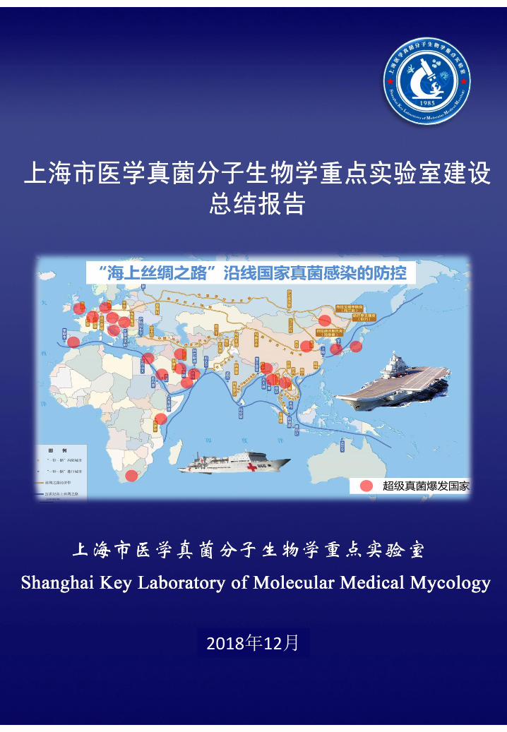
回顾一年来上海市医学真菌分子生物学重点实验室的工作,我们在论文发表、科研项目、人才梯队建设、国内外合作交流、运行管理等方面都取得了不错的成绩。实验室瞄准国家“一带一路”、“军民融合”等重大战略和议题,关注国际学术前沿,紧密结合人民卫生健康的需要,在全球真菌病的流行病学、临床真菌病的早期诊断、医学真菌系统进化与菌种保藏研究、重要病原真菌与宿主相互作用等多方面稳步推进。在承接国家重大课题方面,我们承担了国家传染病重大专项《“海上丝绸之路”重要病原体的威胁感知和监测预警》以及工程院咨询项目《我国及“一带一路”沿线国家真菌病联合防控策略研究》,旨在阐明“海上丝绸之路”沿线重要真菌病,尤其是“超级真菌”的流行情况,揭示相关海域真菌生物多样性组成,建立适用于当地卫生条件的诊断方法,并为我国“一带一路”沿线相关性真菌病输入性感染提供理论依据和防控策略。 同时我们的国自然重大国际合作课题《中国人群高发的难治性暗色真菌感染的生态学和进化起源研究》正在顺利推进,发表了一系列的成果。
逐渐形成了一支以院士以及领军人才作为优秀学科带头人的研究队伍,凝聚了一批具有发展潜力的中青年学术人才。不仅与国内外一流研究机构开展了高效务实的合作交流,而且利用我国在真菌领域的科研优势,逐步影响、提高“一带一路”沿线国家真菌病防控能力。与国内外多家单位联合,建立多个院士工作站和长期合作点,实行优势互补、资源共享,加强研究项目,建立公共研发平台,为包括延安、哈尔滨、邯郸、南昌、杭州、广州、苏州等在内的全国广大学者提供研究场地。
在海军首长、上海市科委、大学领导、医院领导的关心和支持下,以及实验室全体成员的不懈努力下,我们的2018收获满满。同时,我们又将站在以实现自身跨越为目标的新起点,抓住现有的发展契机,发挥自身科研优势,力争在医学真菌学前沿的探索中取得更多系统性原创成果,圆满完成各项科研、临床和政治任务。
在生命科学和生物技术快速发展的新形势下,机遇与挑战并存,实验室的发展建设任重道远,愿团队人员弘扬“探索、奉献、包容、进取”的精神,为理想,追求不断、矢志不渝;为事业,百折不挠、坚韧不拔的态度,扎实工作、团结合作,潜心科研,一起推动实验室各项工作迈向新的台阶!
中国工程院院士
2018年12月21日

参与年报编写的人员名单
总策划:
廖万清
执行编辑:
潘炜华
主要编写人员:
方文捷、杜明威
参与编写人员:
姜伟伟、扈东营、邓宇晨、张蕾、张克明、
洪南、刘伊诺、李 航、朱信霖、陈丽琳
目录
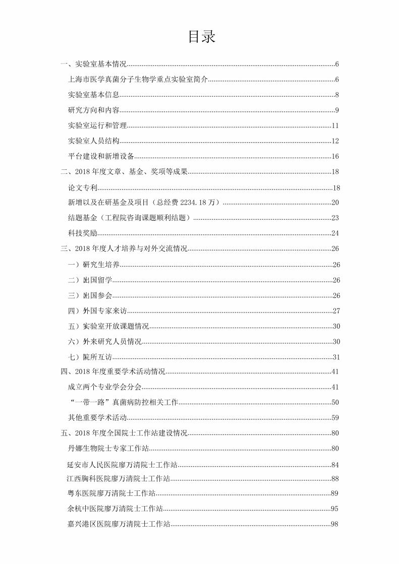
一、实验室基本情况.................................................................................................................6
上海市医学真菌分子生物学重点实验室简介.....................................................................6
实验室基本信息.....................................................................................................................8
研究方向和内容.....................................................................................................................9
实验室运行和管理...............................................................................................................11
实验室人员结构...................................................................................................................12
平台建设和新增设备...........................................................................................................16
二、2018 年度文章、基金、奖项等成果..............................................................................18
论文专利...............................................................................................................................18
新增以及在研基金及项目(总经费 2234.18 万)...........................................................20
结题基金(工程院咨询课题顺利结题)...........................................................................23
科技奖励...............................................................................................................................24
三、2018 年度人才培养与对外交流情况..............................................................................26
一))研究生培养...................................................................................................................26
二))出国留学.......................................................................................................................26
三))出国参会.......................................................................................................................26
四))外国专家来访...............................................................................................................27
五))实验室开放课题情况...................................................................................................30
六))外来研究人员情况.......................................................................................................30
七))院所互访.......................................................................................................................31
四、2018 年度重要学术活动情况..........................................................................................41
成立两个专业学会分会.......................................................................................................41
“一带一路”真菌病防控相关工作...................................................................................50
其他重要学术活动...............................................................................................................59
五、2018 年度全国院士工作站建设情况..............................................................................80
丹娜生物院士专家工作站...................................................................................................80
延安市人民医院廖万清院士工作站...................................................................................84
粤东医院廖万清院士工作站...............................................................................................89
余杭中医院廖万清院士工作站...........................................................................................95
嘉兴港区医院廖万清院士工作站.......................................................................................98
江西胸科医院廖万清院士工作站.......................................................................................88

河北工程大学附属医院廖万清院士工作站.......................................................................99
长江润发集团院士专家工作站.........................................................................................101
大专家.com 院士专家工作站............................................................................................103
六、2018 年度服务军民情况................................................................................................104
一、为军服务.....................................................................................................................104
二、义诊和其他民间交流.................................................................................................107
附件: 2018 年实验室部分 SCI 文章原文............................................................................114
七、《中国真菌学杂志》.....................................................................................................113
一、实验室基本情况
上海市医学真菌分子生物学重点实验室简介
上海市医学真菌分子生物学重点实验室项目 2010 年 5 月经上海市科委批准立项依托上
海长征医院建设,2 年来共投入建设经费约 400 万元,2012 年 12 月通过上海市科委检查验
收,并于 2014 年和 2016 年两次通过实验室评估,获上海市科委滚动支持。现任实验室主任
为中国工程院院士廖万清,学术委员会主任为中国工程院院士陈洪铎。
本实验室依托海军军医大学皮肤病与真菌病研究所和长征医院皮肤科建设,在医学真菌
学基础与临床研究方面基础深厚。40 多年来,廖万清院士领衔的学术团队主要从事隐球菌
及隐球菌病的研究,创建了我国第一个隐球菌专业实验室,对隐球菌的分类及系统进化、毒
力因子及其调控机制、与宿主相互作用及临床治疗方案等领域的进行了系统、深入的研究,
部分研究成果达国际先进水平。
目前,在上海市科委及长征医院各级领导的关心、指导下,实验室整体建设呈现出快速
发展的强劲趋势,拥有团结协作、充满生机的工作氛围和浓郁踏实的学术风气,以研究项目
为牵引,与美国杜克大学、霍普金斯医学院、NIH、荷兰 CBS 真菌多态性研究中心等国际著
名医学真菌研究机构开展了多个务实高效的国际合作研究,产生的一批如隐球菌毒力调控的
分子机制研究、亚洲范围内新生隐球菌临床株特定基因型与耐药趋势的关系、暗色真菌系统
进化研究、病原真菌药物敏感性流行病学调查等重要研究成果,在国内外医学真菌学研究领
域产生了较大影响。近几年,实验室各项目组成员在 New England Journal of Medicine、
Clinical Infectious Disease、Frontiers in Immunology、Plos Neglected Tropical
Diseases 、 Journal of Antimicrobial Chemotherapy 、 Antimicrobial Agents and
Chemotherapy、Emerging Microbes & Infections 等相关领域顶级期刊发表多篇文章。
实验室拥有中国工程院院士 1 人,博士生导师 4 人,硕士生导师 2 人,固定研究人员
28 人,汇聚了包括从荷兰皇家科学院、意大利米兰大学、美国杜克大学、霍普金斯医学院、
NIH、加州大学等归国中青年学术骨干,学科背景优势互补、科研思路活跃,已基本形成具
有一支国际水平、以中青年研究骨干为主体的学术团队。
实验室主要研究方向为重要病原真菌的毒力及其与宿主相互作用、分子流病及系统发育、

我国病原真菌菌株保藏、耐药趋势监测及分子鉴定等。具有国际水平的以基因枪、MALDI-TOF
MS,实时荧光定量 PCR 等仪器为核心的病原真菌基因组学研究技术平台完全能够胜任真菌培
养、细胞分离培养、临床样本收集、分子生物学操作等研究工作。目前,实验室承担了多项
省部级课题,现有科研经费达 2234.18 万元。
实验室秉承“开放、流动、联合、竞争”的运行机制,依托上海长征医院皮肤科和隐球
菌专业实验室的临床和科研优势,加强与国内外科研机构的交流合作,通过实验室门户网站
www.shmedmyco.com 向社会公开招标。
实验室自成立到现在,已经逐步走向正轨。为了记录实验室的发展历程,更好的弘扬实
验室文化,延续既往优良传统,我们编辑了这本 2018 年实验室年报,以记载实验室年度教
学科研及学科人才建设情况,同时也为后人留下实验室可以查阅的历史资料。
实验室的发展和成长,离不开校院党委、上海市科委及卫计委各级领导对我们的关心和
帮助,也凝聚着实验室每个成员的心血和汗水。在此我向曾经为实验室建设与发展做出贡献
的所有人表示衷心的感谢!
实验室基本信息
实验室中文名称:上海市医学真菌分子生物学重点实验室
实验室英文名称:Shanghai Key Laboratory of Molecular Medical Mycology
门户网站:www.shmedmyco.com
微信公众号:上海医学真菌分子生物重点实验室
依托单位:上海长征医院
实验室主任:廖万清
实验室学术委员会主任:陈洪铎
现有固定人员 32 名,其中中国工程院院士 1 人、高级职称 4 人,中级职称 9 人,技术
员 4 人,编辑 3人,护士 5 名等。在读博士研究生 3人、硕士研究生 10 人。
实验室现拥有细胞培养室、DNA 提取室、真菌培养室等实验场所,用于完成真菌的分子
生物学鉴定、真菌功能基因组学、真菌与宿主相互作用等研究。配备莱卡荧光显微镜、高压
气体基因枪、实时荧光定量 PCR 仪、核酸蛋白测定仪、冷冻干燥机、凝胶成像系统、全自动
酶标仪等仪器设备。实验室规章制度健全,管理规范,运行有序,具备承担国家重大科研任
务和开放研究平台的科技支撑条件。
研究方向和内容
为满足我国医学真菌领域临床实践和基础研究的需要,实验室研究方向主要分为一下三
个方向:
(一) 重要病原真菌与宿主细胞的相互作用;
瞄准致病真菌与宿主相互作用这一前沿研究领域,开展针对真菌功能基因组学和宿主抵
御真菌侵袭的分子与细胞机制研究,阐明各种医学真菌致病机制,为未来临床诊治寻找合适
靶点。目前,实验室的研究重点是新型隐球菌毒力调控的分子与细胞机制研究和重要致病真
菌逃逸宿主免疫的机制及信号通路研究。该研究方向目前已获国家重点基础研究发展计划
(“973”计划)《重要侵袭性致病真菌与宿主相互作用的分子与细胞机制研究》的支持和多
项自然基金重点、面上、青年项目支持。
(二) 临床真菌病分子生物学早期快速诊断;
依托围绕目前临床真菌病患者诊治需要,将分子生物学方法用于临床真菌病诊断,做到
真菌病的早诊早治。实验室研究人员通过研发真菌特异性抗体和新型分子生物学诊断方法,
努力提高真菌病患者早期诊断的敏感性和特异性。同时,收集临床真菌菌株建成菌种库,为
后续科学研究做基础。该研究方向目前已获国家卫生部科技重大专项课题《侵袭性真菌感染
现代早期诊断技术体系的研究》、《海上丝绸之路沿线真菌病防控》等重大课题的支持。
(三) 医学真菌系统进化与菌种保藏研究
针对目前全国范围内收集的医学真菌临床菌株和环境菌株进行系统进化学研究,将其与
国外流行地区分离菌株和标准菌株对比,初步探讨我国不同地区真菌流行趋势和原因,并探
索真菌基因分型与抗真菌药物敏感性之间的关系。实验室现正在进行中国格特隐球菌致病菌
株和环境菌株系统进化研究和暗色真菌系统发育学研究,该项研究已获国家自然科学基金国
际重大合作课题以及国自然青年项目支持。
为了更好的开展医学真菌保藏工作,实验室采取冷冻干燥法、沙土保藏法、生理盐水甘
油等多种国际流行菌种保藏方法保藏病原真菌,并已采购冷冻干燥机、液氮冻结菌种保藏库
和-80℃超低温冰箱保藏各种临床分离株和环境株。现已保藏冷冻干燥菌种 1200 余株,包括
从意大利、荷兰、美国和比利时等国家引进的隐球菌标准菌株,生理盐水甘油保存菌株 300
余株,日常斜面移种菌株 300 余株,菌种资源多样性高、新资源丰富,包括标准株、临床株

和环境分离株。菌种类型包括隐球菌、曲霉、念珠菌、暗色真菌、皮肤癣菌等常见及罕见致
病真菌。在实验室建设期间,研究团队新发现 3种新的致病真菌也保藏其中。同时,真菌保
藏中心秉承互惠共享的原则,与上海和其他省市大型医院保持合作,进行菌种鉴定、药敏和
保藏工作,为我国病原真菌研究工作提供坚实保证。实验室拟建成现代化的、亚洲一流的中
国上海真菌保藏中心(Shanghai Center of Mycology Collection, SCMC)和上海疑难真菌
菌种鉴定质控中心,与世界真菌保藏中心美国 ATCC、荷兰 CBS、比利时 BCCM 等国际一流保
藏中心接轨。

实验室运行和管理
本实验室依托上海长征医院皮肤科建设,在原有第二军医大学皮肤病与真菌病研究所
和中国科学院隐球菌专业实验室基础上筹建上海市医学真菌分子生物学重点实验室,其中包
括医学真菌分子生物学实验室、真菌病理实验室和医学真菌临床检验室三部分组成,涵盖医
学真菌病研究的基础、病理、临床和检验四个方面。实验室运行管理实行实验室主任负责制
和岗位责任制,实验室设主任一名。由实验室主任提名,聘任实验室副主任,主管技师,学
术秘书和行政秘书等。学术委员会是实验室的学术指导机构,主要任务是审议实验室的目标、
任务和研究方向,审议实验室的重大活动、年度工作等。实验室工作人员包括:实验室固定
研究人员、客座人员、实验技术人员和管理人员。实验室聘请中国医科大学陈洪铎院士(图
14)、荷兰皇家科学院真菌生物多样性研究中心 Sybren de Hoog 教授、荷兰真菌生物多样性
研究中心 Teun Boekhout 教授和美国杜克大学 John Perfect 教授为实验室客座教授。
实验室实行“开放、流动、联合、竞争”的运行机制,建设依照“边建设、边研究、
边开放”的原则,课题向国内学者公开招标,吸引其他院校学者到实验室作访问学者,邀请
国内外知名学者到实验室进行学术交流,派送学术骨干到国外科研机构开展科学研究。现已
有军事医学科学院、交通大学附属新华医院等单位学者在本实验室进行研究工作。实验室从
实际出发、统筹规划、合理设置,做到建筑设施、仪器设备、技术队伍和科学管理协调发展,
提高投资效益,充分保证科研项目和企业合作实施,全面培养和造就高层次医学真菌学人才。

实验室人员结构
实验室主任、副主任和学术委员会主任
高级职称
主治医师

院士秘书、助理
助教
技术人员

《中国真菌学杂志》编辑团队
护士

研究生:
荷兰皇家科学院真菌多样性研究中心Teun Boekhout
荷兰皇家科学院真菌多样性研究中心Sybren de Hoog
美国杜克大学John Perfect
客座教授
Wenjie Fang
图章
Wenjie Fang
图章
Wenjie Fang
图章
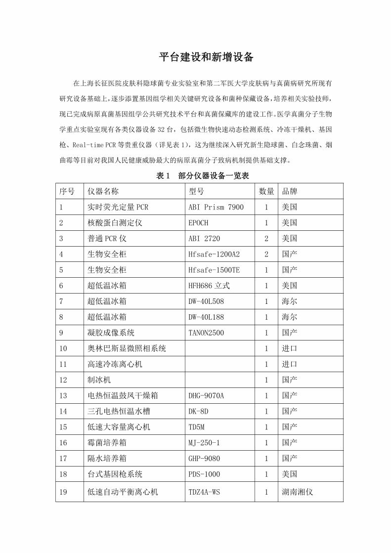
平台建设和新增设备
在上海长征医院皮肤科隐球菌专业实验室和第二军医大学皮肤病与真菌病研究所现有
研究设备基础上,逐步添置基因组学相关关键研究设备和菌种保藏设备,培养相关实验技师,
现已完成病原真菌基因组学公共研究技术平台和真菌保藏库的建设工作。医学真菌分子生物
学重点实验室现有各类仪器设备 32 台,包括微生物快速动态检测系统、冷冻干燥机、基因
枪、Real-time PCR 等贵重仪器(详见表 1),这为继续深入研究新生隐球菌、白念珠菌、烟
曲霉等目前对我国人民健康威胁最大的病原真菌分子致病机制提供基础支撑。
表 1 部分仪器设备一览表
序号 仪器名称 型号 数量 品牌
1 实时荧光定量 PCR ABI Prism 7900 1 美国
2 核酸蛋白测定仪 EPOCH 1 美国
3 普通 PCR 仪 ABI 2720 2 美国
4 生物安全柜 Hfsafe-1200A2 2 国产
5 生物安全柜 Hfsafe-1500TE 1 国产
6 超低温冰箱 HFH686 立式 1 美国
7 超低温冰箱 DW-40L508 1 海尔
8 超低温冰箱 DW-40L188 1 海尔
9 凝胶成像系统 TANON2500 1 国产
10 奥林巴斯显微照相系统 1 进口
11 高速冷冻离心机 1 进口
12 制冰机 1 国产
13 电热恒温鼓凤干燥箱 DHG-9070A 1 国产
14 三孔电热恒温水槽 DK-8D 1 国产
15 低速大容量离心机 TD5M 1 国产
16 霉菌培养箱 MJ-250-1 1 国产
17 隔水培养箱 GHP-9080 1 国产
18 台式基因枪系统 PDS-1000 1 美国
19 低速自动平衡离心机 TDZ4A-WS 1 湖南湘仪

序号 仪器名称 型号 数量 品牌
20 纯水仪 MILI-Q CENTURY 1 国厂和泰
21 微生物快速动态检测系统 MB-80 1 北京金山川
22 (附带 T02 智能恒温仪) 1
23 电子天平 BT125D 2 赛多利斯
24 冷冻干燥机 1 美国
25 医用冷藏、冻箱 HYCD-205 1 海尔
26 通风柜 1.2 米 1 国产
27 二氧化碳培养箱 MCO-15AC 1 日本
28 三洋全自动消毒锅 MLS-3750 1 日本
29 药品保存箱 HYC-360 1 海尔
30 药品保存箱 HYC-326A 1 海尔
研究所新增设备一台:禾信 CM-1600 MALDI-TOF MS 微生物鉴定系统。由广州禾信质谱
提供以用于科学研究。

二、2018 年度文章、基金、奖项等成果
论文发表、授权发明专利
第一和通讯(含并列)发表或接受12篇,IF>3累计6篇。授权发明3项,新型1项。
1、 Arastehfar A, Fang W, Pan W, Lackner M, Liao W, Badiee P, Zomorodian K, Badali
H, Hagen F, Lass-Flörl C, Boekhout T. YEAST PANEL multiplex PCR for
identification of clinically important yeast species: stepwise diagnostic
strategy, useful for developing countries. Diagn Microbiol Infect Dis. 2018 Sep
21. pii: S0732-8893(18)30381-X. doi: 10.1016/j.diagmicrobio.2018.09.007.
[Epub ahead of print] PubMed PMID: 30377018. 【IF=2.3】
2、 Hong N, Chen M, Xu N, Al-Hatmi AMS, Zhang C, Pan WH, Hagen F, Boekhout T,
Xu J, Zou XB, Liao WQ. Genotypic diversity and antifungal susceptibility of
Cryptococcus neoformans isolates from paediatric patients in China. Mycoses.
2018 Oct 19. doi: 10.1111/myc.12863. [Epub ahead of print] PubMed PMID: 30341799.
【IF=2.793】
3、 Arastehfar A, Fang W, Pan W, Liao W, Yan L, Boekhout T. Identification of
nine cryptic species of Candida albicans, C. glabrata, and C. parapsilosis
complexes using one-step multiplex PCR. BMC Infect Dis. 2018 Sep 25;18(1):480.
doi: 10.1186/s12879-018-3381-5. PubMed PMID: 30253748; PubMed Central PMCID:
PMC6156947. 【IF=2.62】
4、 Tang L, Fang W, Lin J, Li J, Wu W, Xu J. Vitamin D protects human
melanocytes against oxidative damage by activation of Wnt/β-catenin
signaling. Lab Invest. 2018 Dec;98(12):1527-1537. doi: 10.1038/
s41374-018-0126-4. Epub 2018 Sep 11. PubMed PMID: 30206310. 【IF=4.2】
5、 Fang W, Wang G, Tang L, Su H, Chen H, Liao W, Xu J. Hydrogen gas inhalation
protects against cutaneous ischaemia/reperfusion injury in a mouse model of
pressure ulcer. J Cell Mol Med. 2018 Sep;22(9):4243-4252. doi:
10.1111/jcmm.13704. Epub 2018 Jun 19. PubMed PMID: 29921037; PubMed Central
PMCID: PMC6111801. 【IF=4.3】
6、 Arastehfar A, Fang W, Badali H, Vaezi A, Jiang W, Liao W, Pan W, Hagen F,
Boekhout T. Low-Cost Tetraplex PCR for the Global Spreading Multi-Drug Resistant
Fungus, Candida auris and Its Phylogenetic Relatives. Front Microbiol. 2018 May
29;9:1119. doi: 10.3389/fmicb.2018.01119. eCollection 2018. PubMed PMID:
29896181; PubMed Central PMCID: PMC5987591. 【IF=4.019】
7、 Sang J, Yang Y, Fan Y, Wang G, Yi J, Fang W, Pan W, Xu J, Liao W. Isolated
iliac cryptococcosis in an immunocompetent patient. PLoS Negl Trop Dis. 2018
Mar 29;12(3):e0006206. doi: 10.1371/journal.pntd.0006206. eCollection 2018 Mar.
PubMed PMID: 29596420; PubMed Central PMCID: PMC5875738. 【IF=4.3】
8、 Deng S, Lei W, de Hoog GS, Yang L, Vitale RG, Rafati H, Seyedmousavi M, Tolooe
A, van der Lee H, Liao W, Verweij PE, Seyedmousavi S. Combination of Amphotericin
B and Terbinafine against Melanized Fungi Associated with Chromoblastomycosis.
Antimicrob Agents Chemother. 2018 May 25;62(6). pii: e00270-18. doi:
10.1128/AAC.00270-18. Print 2018 Jun. PubMed PMID: 29581111; PubMed Central
PMCID: PMC5971613. 【IF=4.2】
9、 Chen M, Xu Y, Hong N, Yang Y, Lei W, Du L, Zhao J, Lei X, Xiong L, Cai L,
Xu H, Pan W, Liao W. Epidemiology of fungal infections in China. Front Med. 2018
Feb;12(1):58-75. doi: 10.1007/s11684-017-0601-0. Epub 2018 Jan 11. Review.
PubMed PMID: 29380297. 【IF=2.0】
10、 Chen M, Kondori N, Deng S, Gerrits van den Ende AHG, Lackner M, Liao
W, de Hoog GS. Direct detection of Exophiala and Scedosporium species in sputa
of patients with cystic fibrosis. Med Mycol. 2018 Aug 1;56(6):695-702. doi:
10.1093/mmy/myx108. PubMed PMID: 29228273. 【IF=2.799】
11、 Novel multiplex qPCR approach for direct detection of Candida auris and
its relatives in spiked serum samples. Amir Arastehfar,1, Wenjie Fang‡, Farnaz
Daneshnia, Abdullah M S Al-Hatmi, Wanqing Liao, Weihua Pan*, Ziauddin Khan,
Suhail Ahmad, Katharina Rosam, Michaela Lackner, Cornelia Lass-Flo¨ rl, Ferry
Hagen & Teun Boekhout Future Microbiology in press 【IF=3.19】
12、 Unequivocal identification of an underestimated opportunistic yeast
species, Cyberlindnera fabianii, and its close relatives using a dual-function

PCR and literature review of published cases Amir Arastehfar, Wenjie Fang,
Abdullah M. S. Al-Hatmi, Mohammad Hosein Afsarian, Farnaz Daneshnia, Mina
Bakhtiari, Sara Khanjari Sadati, Hamid Badali, Sadegh Khodavaysi, Ferry Hagen,
Wanqing Liao, Weihua Pan*, Kamiar Zomorodian6* and Teun Boekhout. Medical
Mycology 【IF=2.799】
隐球菌活力检测试剂盒及检测方法zl.201310627949.X(发明授权)
一种用于核酸扩增的酵母样真菌总DNA无仪器提取方法zl.201610114856.0(发明授权)
一种异丙基胺代氮唑类抗真菌化合物及其制备方法和应用zl.201610007802.4(发明授权)
一种拇外翻矫正鞋201721864805.6 (新型授权)
新增以及在研基金及项目(总经费 2234.18 万)
序号 课题名称 负责人 起止时间 经费(万元) 项目来源
1*
“海上丝绸之路”重
要病原体的威胁感知
和监测预警
廖万清2018.01-2020.1
21104.18 国家科技部
2*
我国及“一带一路”
沿线国家真菌病联合
防控策略研究
廖万清2018.12-2020.1
2100 中国工程院
3* 长征医院“金字塔工
程”——国家优青后
备人才
方文捷 2018-2021 30 长征医院
4
中国人群高发的难治
性暗色真菌感染的生
态学和进化起源研究
廖万清2018.01-2022.1
2232
国家自然科
学基金
5
circ_0091746以内源
性竞争 RNA 方式结合
miR-6848-5p 在巨噬
细胞抗新生隐球菌感
染 M1/M2 极化中的作
用及调控机制
潘炜华2018.01-2021.1
265
国家自然科
学基金
6GM-CSF、TF 在我国非
HIV 感染隐球菌性脑陈江汉
2018.01-2021.1
253
国家自然科
学基金

膜炎患者发病中的作
用及机制研究
7
上海市真菌病与自身
免疫性疾病临床医学
中心建设
廖万清2017.07-2020.0
6325
上海市卫计
委
8 上海市领军人才 潘炜华 2017-2020 40 上海市科委
9 上海市科委扬帆计划 张 超2017.05-2020.0
520 上海市科委
10
已糖载体蛋白家族对
新生隐球菌体内荚膜
动态变化的调控机制
廖万清20151.1.至
2018.12.3180
国家自然科
学基金
11
医学真菌生物样本库
质量控制体系构建研
究
廖万清2016-8-31 至
2019-7-3120
上海市科委
技术标准专
项
12上海市医学真菌分子
生物学重点实验室廖万清 2017-2020 150
上海市科委
重点实验室
13
用于酵母感染核酸诊
断的两项 DNA 提取试
剂盒的研发和产业化
方文捷 2016-2018 5上海长征医
院
14
烟曲霉耐药基因突变
检测试剂盒研发与新
型临床精准诊断策略
探索和验证
方文捷 2017-2019 10海军军医大
学
*2018 年新立项课题
重要新立项课题介绍
1、“海上丝绸之路”重要病原体的威胁感知和监测预警
研究首先在“海上丝绸之路”沿线国家和地区建立传染病病原体联防联控监测体系,结
合当地传染病流行情况,有针对性开展重要传染病病原监测,建立的网络实验室监测预警体

系可应用于“一带一路”沿线相关国家和地区、口岸检疫系统等,也是实现我国应对美国"
空海一体战"制定的军事战略的重要保障。同时,形成有较强实用性的研究成果,将建立一
套病原体的快速检测技术、溯源数据库、研制出一些小型、实用技术设备和一系列简便、快
速的诊断试剂和诊疗产品,包括从现场用的简便快速检测方法到普通实验室用的快速敏感检
测方法和对多种病原高通量的检测方法、以及各种相应诊断试剂和诊疗产品,形成一个多层
面病原体快速检测技术和处置技术体系。这些技术方法、小型设备、诊断试剂和诊疗产品适
用于重大疫情暴发流行的检测。课题开发和集成基因组学、蛋白质组学、生物芯片、生物信
息学、分子病原学等前沿生物技术,建立实用性的未知病原体的高通量的快速鉴别和溯源技
术,可用于新发、突发传染病病原体的综合检测和鉴定,以及应急处置的对应产品。对传染
病流行病学风险评估分析可以获得相关病原体的特征信息,为进一步的特异性检测及鉴别打
下基础。
任务研究内容:阐明“海上丝绸之路”沿线重点真菌病流行情况,揭示相关海域真菌生
物多样性组成,建立适用于当地卫生条件的诊断方法。
考核指标:1.完成 1000 例流行病学调查,30 次海域多样性调查,建立 40 株菌的基因
序列库包含 50 个以上海域地点的微生物宏基因组数据库,包含 300 个菌株可培养微生物数
据库;2.多重 PCR 快速诊断体系 4-5 套,在 2-4 小时以内可以诊断 20 个高致病菌种;建立
微流控平台,;建立首个近红外荧光探针平台;申请国家发明专利 7-10 项。
2、我国及“一带一路”沿线国家真菌病联合防控策略研究
“一带一路”倡议涉及 100 多个国家,覆盖约 44 亿人口,拟构建世界跨度最长、最具
发展潜力的经济走廊。近年来,一些烈性真菌病借由“海上丝绸之路”向沿线国家扩散,而
且主要是由“一带一路”国家传入我国为主。比如近年来新发的多重耐药“超级真菌”(Candida
auris,耳念珠菌)死亡率高达 60%以上。耳念珠菌已在全球 30 个国家出现过感染病例或局
部爆发流行,其中 60%以上分布于我国“一带一路”沿线,对我国存在较大的输入性传播风
险。我国同一带一路国家在真菌病防控方面的联合研究,具有一定的滞后性和急迫性。我国
同相关国家至今未在本领域内建立成熟的合作模式和网络,缺乏合作互信。因此无法对潜在
的高致病真菌病爆发流行进行联合预警和防控。
基于此,本项目拟从以下三个方面,组织调研和策略研究:
(一)国门及“一带一路”沿线病原真菌生物样本库构建策略研究:进行调研国门及“一

带一路”沿线病原真菌生物样本库构建水平;提出国门及“一带一路”沿线真菌病专病登记
平台和生物样本库平台构建策略。
(二)我国及“一带一路”沿线国家真菌病研究机构跨国合作模式探索:进行我国及“一
带一路”沿线国家真菌病研发实力和跨国合作现状调研;提出“一带一路”真菌病防控联合
实验室建设策略。
(三)面向我国及“一带一路”沿线国家的适应性真菌病防控技术研发策略:进行适应
性真菌病防控技术联合研发模式调研和理论创新;提出真菌病防控国产技术在“一带一路”
沿线国家推广策略。
本课题将形成我国及“一带一路”沿线国家真菌病联合防控咨询报告,拟报送工程院医
药卫生学部办公室以及国务院卫生健康委员会,为我国“一带一路”沿线相关性真菌病输入
性感染提供理论依据和防控策略。
结题基金(工程院咨询课题顺利结题)
中国工程院院士咨询项目是国家高端智库为国家建言献策重要组成之一,2018年11月29
日下午14:30-16:00,中国工程院咨询课题《我国抗真菌药物研发策略国内外对比研究》项
目组在上海市医学真菌分子生物学重点实验室会议室召开了项目结题报告会。参加此次项目
结题会的工程院领导和专家有:工程院三局易建局长、三局医药卫生学部办公室副调研员赵
西路、长征医院徐正梅副院长、田诗音助理、廖万清院士、昆明医科大学第二附属医院邓丹
琪副院长、郭芸教授、上海市第一人民医院施伟民主任、上海市新华医院姚志荣主任、军事
科学院军事医学研究院杨英副研究员、海军军医大学海军医学系戚中田教授、海军军医大学
药学院盛春泉副院长、潘炜华教授以及各技术骨干。
会议由会议主席戚中田教授主持。廖院士首先代表项目组向会议介绍了《我国抗真菌药
物研发策略国内外对比研究》项目的研究背景、总体设计及课题设置等基本情况,邓丹琪教
授、杨英教授、方伟博士等分别汇报了各子课题的研究目标、研究内容、研究进展与结果、
财务收支等情况。项目的主要内容涉及我国真菌感染病原谱的流行规律、现有药物再利用以
及我国海洋与中草药抗真菌药物研发策略评估四个方面。会议期间,各位专家主要对本项目
的研究意义、设计思路和研究成果等进行了积极讨论,一致同意该项目按计划顺利结题。
经过讨论,与会专家在以下方面达成共识:
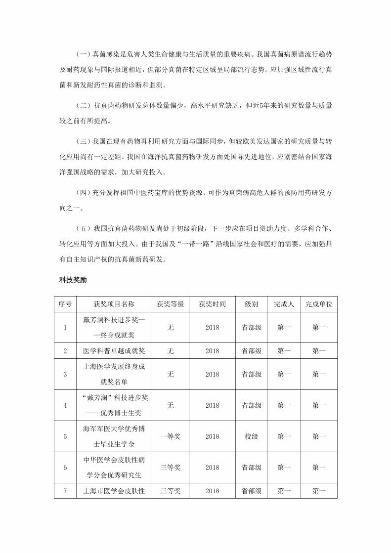
(一)真菌感染是危害人类生命健康与生活质量的重要疾病。我国真菌病原谱流行趋势
及耐药现象与国际报道相近,但部分真菌在特定区域呈局部流行态势。应加强区域性流行真
菌和新发耐药性真菌的诊断和监测。
(二)抗真菌药物研发总体数量偏少,高水平研究缺乏,但近5年来的研究数量与质量
较之前有所提高。
(三)我国在现有药物再利用研究方面与国际同步,但较欧美发达国家的研究质量与转
化应用尚有一定差距。我国在海洋抗真菌药物研发方面处国际先进地位,应紧密结合国家海
洋强国战略的需求,加大研究投入。
(四)充分发挥祖国中医药宝库的优势资源,可作为真菌病高危人群的预防用药研发方
向之一。
(五)我国抗真菌药物研发尚处于初级阶段,下一步应在项目资助力度、多学科合作、
转化应用等方面加大投入。由于我国及“一带一路”沿线国家社会和医疗的需要,应加强具
有自主知识产权的抗真菌新药研发。
科技奖励
序号 获奖项目名称 获奖等级 获奖时间 级别 完成人 完成单位
1戴芳澜科技进步奖—
—终身成就奖无 2018 省部级 第一 第一
2 医学科普卓越成就奖 无 2018 省部级 第一 第一
3上海医学发展终身成
就奖名单无 2018 省部级 第一 第一
4“戴芳澜”科技进步奖
——优秀博士生奖无 2018 省部级 第一 第一
5海军军医大学优秀博
士毕业生学金一等奖 2018 校级 第一 第一
6中华医学会皮肤性病
学分会优秀研究生三等奖 2018 省部级 第一 第一
7 上海市医学会皮肤性 三等奖 2018 省部级 第一 第一

病学分会年度SCI论文
奖
8
上海市医学会皮肤性
病学分会年度SCI论文
奖
优胜奖 2018 省部级 第一 第一

三、2018 年度人才培养与对外交流情况
一、研究生培养
2018年有2名博士,4名硕士顺利通过毕业论文答辩并获得学位。
2018年毕业研究生名单:方文捷(博士)、金怡(博士)、晏亮、徐媛、林文婷、高睿。
二、出国留学
2018年在国外交流人员2名,已回国2名。
方文捷博士在荷兰CBS访学、张蕾博士赴美国约翰.霍普金斯大学留学深造,均已回国。
三、出国参会
1、廖万清院士,潘炜华教授 6 月 30 日参加 20th
Congresses of the International Society for Human
and Animal Mycology (ISHAM)大会
2、5 月 19 日,廖万清院士参加世界华人医师协会理事会换届大会
四、外国专家来访
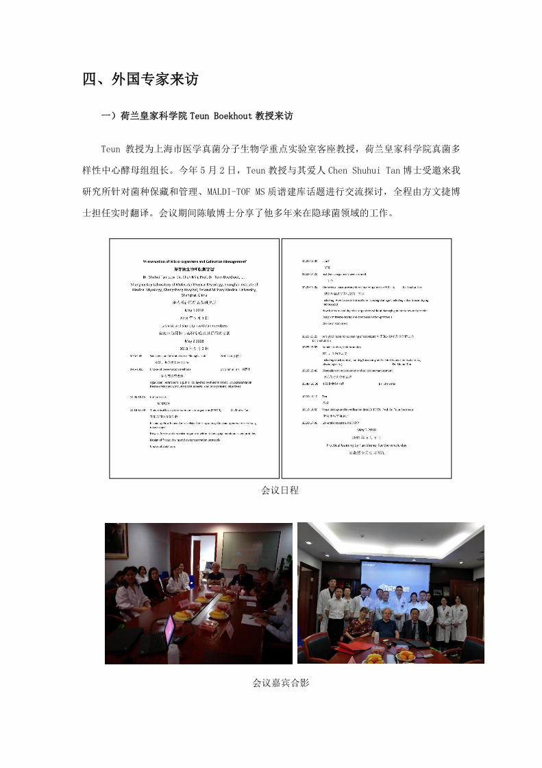
一)荷兰皇家科学院 Teun Boekhout 教授来访
Teun 教授为上海市医学真菌分子生物学重点实验室客座教授,荷兰皇家科学院真菌多
样性中心酵母组组长。今年 5 月 2 日,Teun 教授与其爱人 Chen Shuhui Tan 博士受邀来我
研究所针对菌种保藏和管理、MALDI-TOF MS 质谱建库话题进行交流探讨,全程由方文捷博
士担任实时翻译。会议期间陈敏博士分享了他多年来在隐球菌领域的工作。
会议日程
会议嘉宾合影

陈敏博士分享了他多年来在隐球菌领域的工作
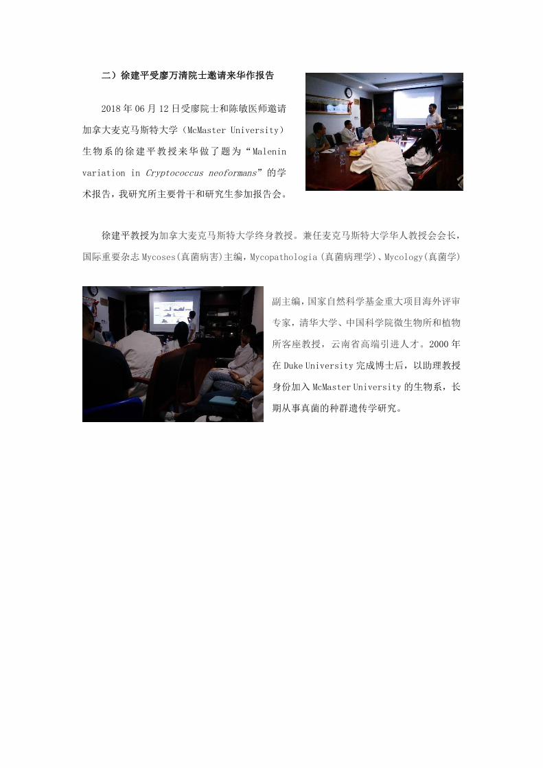
二)徐建平受廖万清院士邀请来华作报告
2018 年 06 月 12 日受廖院士和陈敏医师邀请
加拿大麦克马斯特大学(McMaster University)
生物系的徐建平教授来华做了题为“Malenin
variation in Cryptococcus neoformans”的学
术报告,我研究所主要骨干和研究生参加报告会。
徐建平教授为加拿大麦克马斯特大学终身教授。兼任麦克马斯特大学华人教授会会长,
国际重要杂志 Mycoses(真菌病害)主编,Mycopathologia (真菌病理学)、Mycology(真菌学)
副主编,国家自然科学基金重大项目海外评审
专家,清华大学、中国科学院微生物所和植物
所客座教授,云南省高端引进人才。2000 年
在 Duke University 完成博士后,以助理教授
身份加入 McMaster University 的生物系,长
期从事真菌的种群遗传学研究。
五、实验室开放课题情况
序号 服务资源名称 服务描述 服务范围 开放课题负责
人
1实验室平台对
外开放
为梅县人民医院提供研究
平台
提供实验仪器、
耗材和技术支
持
张东兴
2实验平台对外
开放
为粤东医院提供研究平台 提供实验仪器、
耗材和技术支
持
郑跃
3实验平台对外
开放
为梅州嘉应医学院提供研
究平台
提供实验仪器
和技术支持
肖光文
4 菌株共享为梅州嘉应医学院提供标
准菌株菌株提供
徐美兰
七、外来研究人员情况
2018 年共有 10 人来我研究所,进行长期或者短期的研究工作,分别为:高磊(哈尔滨医科
大学 副主任)、李航(江西胸科医院 研究生)、李帅(延安市人民医院 检验医师)、马涛(延安市
人民医院 皮肤科医生)、李改静(河北工程大学附属医院 皮肤科医生)、姜如金(余杭区人民医院
检验医师)、杨金(复旦大学附属华山医院研究生)曾炫皓(复旦大学附属华山医院 研究生)、王
鹏磊(广东呼吸病研究所 研究生)、陈晓菲(江苏省高新区人民医院 检验医师)
Wenjie Fang
图章

八、院所互访
一)廖院士参加卫生部免疫皮肤病学重点实验室年会
2018 年 10 月 12 日廖院士参加卫生部免疫皮肤病学重点实验室年会。
卫生部免疫皮肤病学重点实验室、教育部免疫皮肤病学重点实验室、辽宁省皮肤病重点
实验室、辽宁省免疫皮肤病学重点实验室、辽
宁省免疫性皮肤病诊疗技术工程实验室学术委
员会会议在中国医科大学附属第一医院举行。
学术委员会由包括廖万清院士在内的四位院士、
三位长江学者及五位业内专家组成。会议由学
术委员会主任委员王正国院士主持。

会上,中国医科大学闻德亮校长
等致辞。实验室常务副主任肖汀教授
汇报免疫皮肤病学实验室年度进展概
况,陈洪铎院士作补充介绍。学术委
员会委员认真听取了汇报,委员们逐
个发表意见及建议,梳理了本实验室
的工作进展,肯定了实验室一年的工
作成绩。对实验室申报和开展更多重
大研究项目、高层次人才队伍建设、实验室的较高层次发展方面提出殷切希望。学术委员会
的意见顺利通过表决,也得到与会各级领导的认可和重视。
二)钟南山院士团队——叶枫主任来访
2018 年 8 月 30 日,钟南山院士团队叶枫教授、李征途博士、王鹏磊医师来访,分享了
该团队在肺隐球菌病诊治和基础研究方面的经验和成果。
李征途博士汇报科研进展

廖万清院士点评

参会人员合影
三)廖万清院士访问东北大学丁辰教授课题组
10 月 12 日,中国工程院院士廖万清
访问东北大学丁辰教授课题组,参观了生
命科学与健康学院实验室并与生命学院青
年教师、学生代表进行座谈。校总会计师
芦延华出席座谈会。
芦延华介绍了生命学院的发展历程、
基本情况和科学研究情况,希望廖万清院
士能够对生命学院的实验室建设、青年人才培养和学术科研提出指导意见。

廖万清院士分享了个人的科研经历,表达了对中国共产党培养、支持的感激之情,希望
学院青年教师和学生积极投身科研,为国家发展建设贡献力量,并表示愿意支持学校、学院
的发展建设,期待更多合作。
廖万清院士同丁辰教授课题组合影

廖万清院士赠与丁辰教授专著《现代真菌病学》
四)2018 年 11 月 26 日廖万清院士、Sybren de Hoog 教授受邀访问贵阳医学院
2018 年 11 月 26 日,贵州省医疗卫生援黔专家团专家、中国工程院院士、海军军医大
学上海长征医院皮肤性病与真菌病研
究所所长廖万清教授,荷兰奈梅亨大
学 Sybren de Hoog 教授一行莅临贵阳
医学院开展学术交流和访问,校党委
书记林昌虎代表学校对廖院士、
Sybren de Hoog 教授一行的到来表示
热烈欢迎。
林昌虎介绍了学校在人才培养、科学研究、服务社会、对外交流与合作等方面工作,并
感谢廖万清院士多年来对我校学科建设、人才培养、重点实验室评估、科技成果、项目申报、

人才基地建设等各方面做出的巨大支持,并希望廖院士能继续关心学校发展和健康贵州的建
设。廖院士对林书记的热情接待表示感谢,他指出,贵州医科大学教育科研工作在学校党委
行政领导的全力推
动下取得了日新月
异的进步,每次来到
学校都能感受到学
校全体员工奋发拼
搏、克服困难、积极
向上的精神风貌,他
将一如既往支持推动学校及医院工作,希望贵州百姓的健康生活质量得到提高。
廖万清院士、Sybren de Hoog 教授还参加了 11 月 27 日由贵州医科大学承办的 2018 年
贵州省微生物学会学术年会暨贵州省医学会微生物与免疫学分会年会。

五)2018 年 11 月 27 日廖万清院士参加贵州省微生物学会学术年会暨贵州省医学会微生物
与免疫学分会年会
11 月 27 至 28 日,2018 年贵州省微生物学会学术年会暨贵州省医学会微生物与免疫学
分会年会召开。大会主席、贵州省医
学会会长杨克勤,贵州省医学会秘书
长甘梁福,大会主席、贵州省微生物
学会理事长刘作易,贵州省卫健委副
主任杨洪,贵州省科协副主席刘炳银
等领导莅临会议指导;贵州医科大学
校长梁贵友出席会议;会议由贵州省
微生物学会常务副理事长康颖倩及
贵州省医学会微免分会主任委员黄山主持。
会上,廖万清院士对贵州省近年来在医疗卫生及科研教育等方面的进步给予了肯定,他
心系贵州,热爱贵州,在今后将一如既往不遗余力支持贵州的发展。
杨洪代表贵州省卫健委对医疗卫生援黔院士专家团专家廖万清院士对我省健康贵州建
设工作的支持表示感谢,并希望通过学术会议的交流,能全面提高我省微生物的研究水平,
为临床医学发挥更大的作用。
会议邀请了中国工程院院
士、海军军医大学廖万清教授,
荷兰奈梅亨大学 Sybren de
Hoog 教授,上海交通大学基础
医学院副院长郭晓奎教授作为
本次大会的主旨演讲特邀嘉宾,
此外还有来自贵州省内各地的
20 位专家学者分别在以医学微生物及微生物生态健康为主题的两个会场进行了精彩的学术
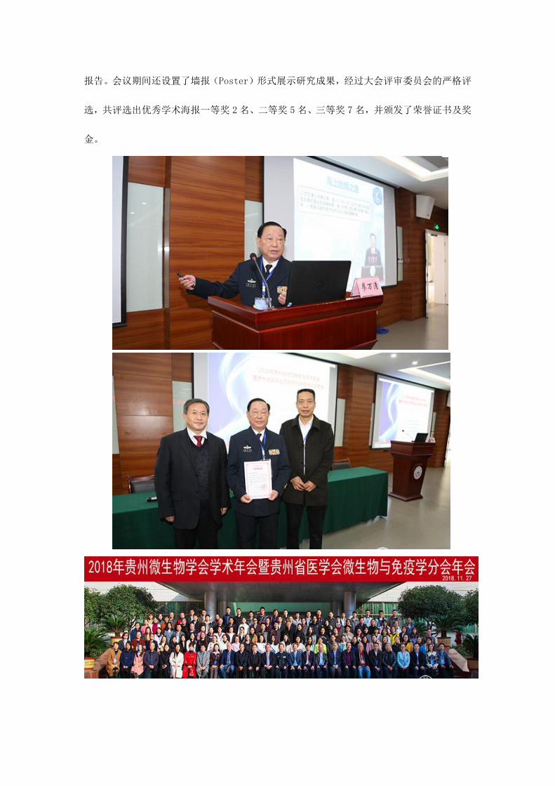
报告。会议期间还设置了墙报(Poster)形式展示研究成果,经过大会评审委员会的严格评
选,共评选出优秀学术海报一等奖 2名、二等奖 5名、三等奖 7名,并颁发了荣誉证书及奖
金。


四、2018 年度重要学术活动情况
成立两个专业学会分会
一)中国整合医学会皮肤病学分会在西安成立,实验室副主任潘炜华教授担任主委
21 世纪,医学进入了从生物医学模式向现代医学模式转变、从科学医学时代向整
合医学时代发展的历史时期。生物医学模式把心身整体的人体机械地分解成各种器官、
组织、细胞与分子,导致临床科室越分越细,让病人成了器官、疾病成了症状,引起
很多临床问题。整合医学还器官为病人、还症状为疾病,实现心身并重、中西医并举、
防治并行、医养并进、人病同治,让医疗回归人文。整合医学是全方位、全周期保障
人类健康的新思维、新的医学观;是未来医学发展的必然方向;是生物医学模式向现
代医学模式转变的必由之路;是健康中国战略的重大理论支撑;是全力推进医疗供给
侧改革、全方位改善医疗服务质量的重要保证。整合医学将引领医学走向新的时代。
2018 中国整合医学大会“整合皮肤病学论坛”4 月 29 日在西安陕西召开。该
大会由整合皮肤病专业委员会(筹)、上海医学真菌分子生物学重点实验室、西京医
院皮肤科联合承办。论坛主
席为本重点实验室副所长、
中国整合医学会皮肤分会主
任委员潘炜华教授。执行主
席为西京皮肤医院副院长李
春英教授。论坛名誉主席为
工程院陈洪铎院士、廖万清
院士。
会议由我院潘炜华教授和西京皮肤病医院副长
李春英教授共同主持

主任委员潘炜华教授主持论坛开幕式,并作皮肤病专业委员会筹备工作报告
名誉主席廖万清院士发言
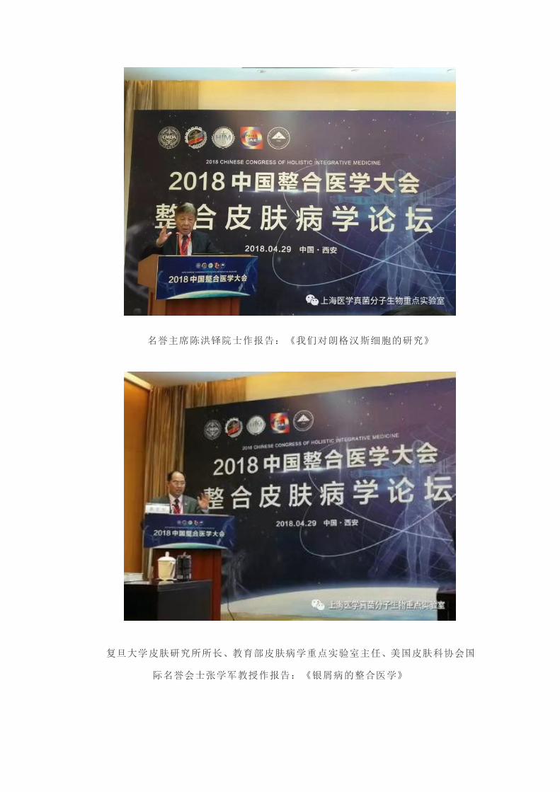
名誉主席陈洪铎院士作报告:《我们对朗格汉斯细胞的研究》
复旦大学皮肤研究所所长、教育部皮肤病学重点实验室主任、美国皮肤科协会国
际名誉会士张学军教授作报告:《银屑病的整合医学》

空军军医大学西京皮肤医院院长、全军皮肤研究所所长、中国皮肤科医师协会候
任会长王刚教授作报告:《皮肤屏障与皮肤病及系统炎症》
交大医学院皮肤病研究所所长、新华医院皮肤科主任、上海市领军人才姚志荣教
授作报告:《特应性皮炎与整合医学》

中华医学会皮肤性病学会候任主委、中南大学皮肤性病研究所所长、湘雅二医院
皮肤科主任陆前进教授作报告:《表观遗传调控与复杂性皮肤病》
广东医学会皮肤病学分会前任主委、中华医学会皮肤病学分会常委委员赖维教授
作报告《整合医学理念在皮肤病 诊疗实践中应用的思考》
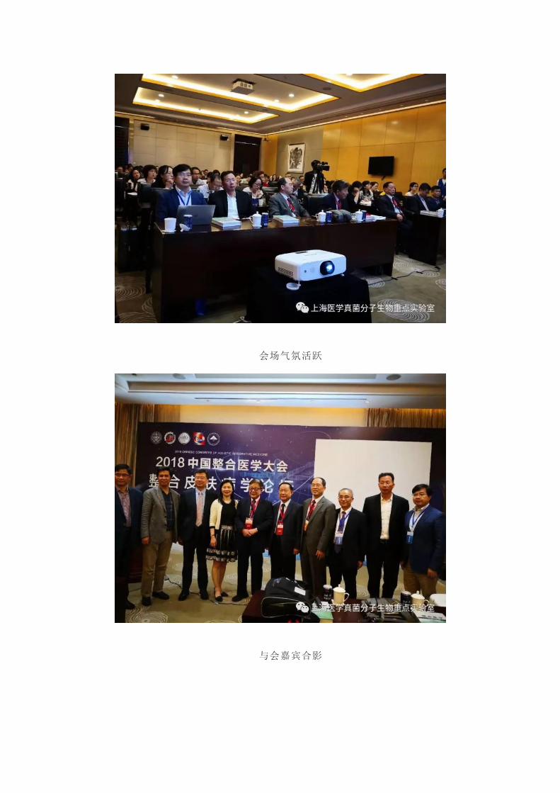
会场气氛活跃
与会嘉宾合影

二)廖万清院士领衔,世界华人皮肤科医师协会成立
世界华人皮肤科医师协会成立大会日前在中国青岛召开。世界华人医师协会会长、
中国医师协会长张雁灵、理事长石丽英,中国工程院陈洪铎院士、廖万清院士等出席
会议。世界华人皮肤科医师协
会全体委员参加会议。海军军
医大学长征医院张殿勇院长
亲临会场并做重要指示。
张雁灵会长代表中国医
师协会、世界华人医师协会向
世界华人皮肤科医师协会成
立,以及世界华人皮肤科医师
协会新当选的荣誉会长陈洪
铎院士,会长廖万清院士、副会长潘炜华、周幼文等 13 位副会长,以及常委、委员
表示热烈祝贺。他指出,皮肤科医师协会的成立,标志着世界华人皮肤科医师有了自
己的国际组织,有了一个共同合作交流的大平台,有了皮肤科病华人医师的家。
张殿勇院长首先代表海军军医大学长征医院,向来自世界各国的华人医师,来自
全国各地的各位皮肤科同仁表示最热烈的欢迎。他指出廖万清院士作为世界华人皮肤
科协会的首任会长,为协会的成立和本次大会的召开做了大量的工作;世界华人皮肤
科医师协会的成立,为华人皮肤科专家的交流合作提供了一个非常好的学术平台,唯
有沟通才能发展,唯有开放才能进步!

张殿勇院长致辞
首任会长廖万清院士致辞

长征医院潘炜华教授作大会报告
世界华人皮肤科医师协会将在廖万清院士的带领下,在副会长、各位委员的努力
下,打造有影响的皮肤病国际组织,为世界人民健康,也为中国人民健康,特别是皮
肤科疾病的预防和治疗作出贡献。
“一带一路”真菌病防控相关工作
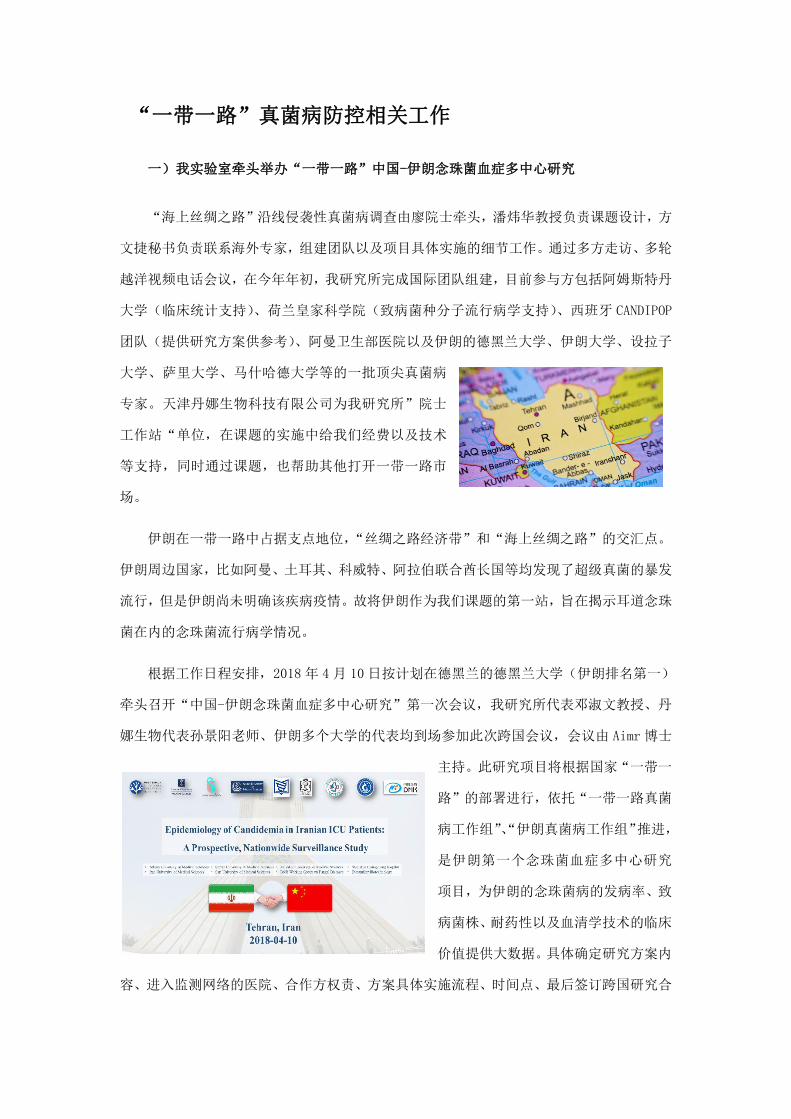
一)我实验室牵头举办“一带一路”中国-伊朗念珠菌血症多中心研究
“海上丝绸之路”沿线侵袭性真菌病调查由廖院士牵头,潘炜华教授负责课题设计,方
文捷秘书负责联系海外专家,组建团队以及项目具体实施的细节工作。通过多方走访、多轮
越洋视频电话会议,在今年年初,我研究所完成国际团队组建,目前参与方包括阿姆斯特丹
大学(临床统计支持)、荷兰皇家科学院(致病菌种分子流行病学支持)、西班牙 CANDIPOP
团队(提供研究方案供参考)、阿曼卫生部医院以及伊朗的德黑兰大学、伊朗大学、设拉子
大学、萨里大学、马什哈德大学等的一批顶尖真菌病
专家。天津丹娜生物科技有限公司为我研究所”院士
工作站“单位,在课题的实施中给我们经费以及技术
等支持,同时通过课题,也帮助其他打开一带一路市
场。
伊朗在一带一路中占据支点地位,“丝绸之路经济带”和“海上丝绸之路”的交汇点。
伊朗周边国家,比如阿曼、土耳其、科威特、阿拉伯联合酋长国等均发现了超级真菌的暴发
流行,但是伊朗尚未明确该疾病疫情。故将伊朗作为我们课题的第一站,旨在揭示耳道念珠
菌在内的念珠菌流行病学情况。
根据工作日程安排,2018 年 4 月 10 日按计划在德黑兰的德黑兰大学(伊朗排名第一)
牵头召开“中国-伊朗念珠菌血症多中心研究”第一次会议,我研究所代表邓淑文教授、丹
娜生物代表孙景阳老师、伊朗多个大学的代表均到场参加此次跨国会议,会议由 Aimr 博士
主持。此研究项目将根据国家“一带一
路”的部署进行,依托“一带一路真菌
病工作组”、“伊朗真菌病工作组”推进,
是伊朗第一个念珠菌血症多中心研究
项目,为伊朗的念珠菌病的发病率、致
病菌株、耐药性以及血清学技术的临床
价值提供大数据。具体确定研究方案内
容、进入监测网络的医院、合作方权责、方案具体实施流程、时间点、最后签订跨国研究合
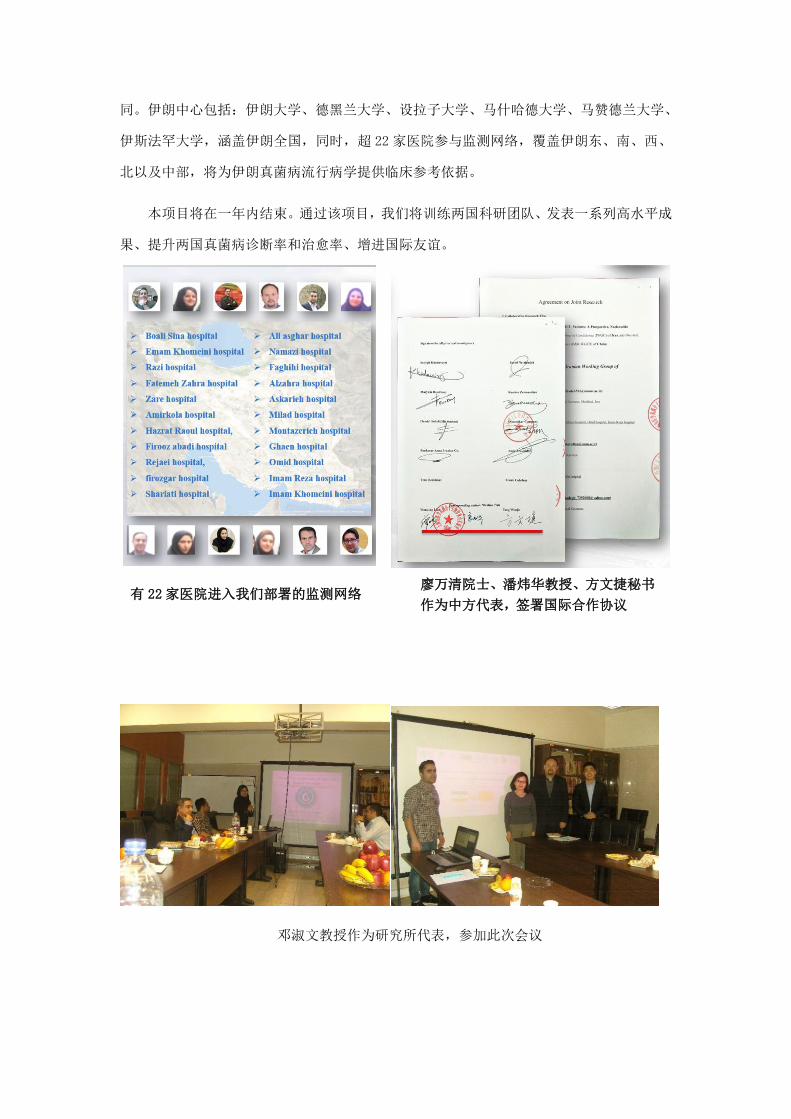
同。伊朗中心包括:伊朗大学、德黑兰大学、设拉子大学、马什哈德大学、马赞德兰大学、
伊斯法罕大学,涵盖伊朗全国,同时,超 22 家医院参与监测网络,覆盖伊朗东、南、西、
北以及中部,将为伊朗真菌病流行病学提供临床参考依据。
本项目将在一年内结束。通过该项目,我们将训练两国科研团队、发表一系列高水平成
果、提升两国真菌病诊断率和治愈率、增进国际友谊。
邓淑文教授作为研究所代表,参加此次会议
廖万清院士、潘炜华教授、方文捷秘书
作为中方代表,签署国际合作协议有 22 家医院进入我们部署的监测网络

会议现场
邓淑文教授同与会嘉宾合影,参观大学实验室

二)上海海洋大学王永杰教授课题组肖劲洲博士一行来访商讨“海上丝绸之路”传染
病防控课题合作
7月4日,上海海洋大学王永杰教授课题组肖劲洲博士一行参观访问了我研究所,并就未
来双方在“海上丝绸之路重大传染病防控”等课题的合作内容和方式进行了深入探讨。
廖万清院士课题组成员带领肖
博士一行六人参观了分子生物学实
验室、真菌保藏中心以及真菌培养室
等实验室功能区块。肖博士对我研究
所的建设、管理和近年来取得的成果
给予了高度的评价,表示下一步将进
一步加强与廖院士课题组的合作交
流,深入探索双方团队人才发展、培
训、使用等方面的新型合作模式。
随后,廖院士团队和肖博士一行
就“一带一路”沿线海洋真菌多样性
测序、宏基因组分析等具体课题进行
了深入会谈。双方就目前各自课题开
展中的遇到的问题进行了交流和解答,
并在具体的合作模式、成果共享形式
等方面达成共识。
海上丝绸之路自秦汉时期
开通以来,一直是沟通东西方经济文化交流的重要桥梁。近年来,“一带一路”在卫
生领域的合作也逐步深化,各国在卫生政策、医学科研、人才培养、医药贸易等方面
交流的广度深度不断拓展,传染病防控、慢病监测、卫生应急、妇幼健康、抗菌药物
耐药、传统医药等领域合作惠及更多民众。我国政府一直希望通过共同努力,促进与
“一带一路”沿线国家等重点合作伙伴开展合作,携手打造“健康丝绸之路”。“一
带一路”伟大战略中“21 世纪海上丝绸之路”为我国海洋相关力量走向远洋,走向深

蓝提出了新的要求。如何提供有力的卫生安全保障是目前我国深蓝计划的重中之重,
其中由致病微生物引起的传染性疾病是健康威胁的重大隐患。
受国家科技部委托,我研究所
2018 年获批“国家科技重大专项课
题:《海上丝绸之路及国门输入重
要传染病军民融合防控技术研究》
(2018ZX10101003;1200 万)。该
独立子课题由廖万清院士牵头,也
是自 2013 年潘炜华教授牵头国家
科技重大专项后,我所第二次牵头
该类项目。
鉴于重要病原体产生的健康危害及对一带一路战略的重大影响,对一带一路相关
海域、地域和港口的重要病原体建立威胁感知系统并针对性进行防控关键技术研究十
分紧迫。通过重点收集和整合“海上丝绸之路”沿岸国家疾病流行数据,对重要病原
体进行采样分析,高通量检测鉴定,建立基于地理信息系统的传染病监测预警数据库,
绘制传染病地理图谱,研究时空分布特征,形成能够集成疾病防控、监测预警于一体
的“传染病医学地理信息预警系统”。对于可能导致严重传染性疾病的病原菌建立快
速检测平台,为疾病发生时的快速诊断和及时救治提供依据,并为今后进一步向相关
海域、港口开展工作建立成熟的组织、技术模式。
在 4 月 10 日,我研究所
牵头在伊朗首都德黑兰成功召
开中伊真菌病防控第一次国
际会议。此次同上海海洋大学
达成合作,是我研究所在一带
一路课题上的又一项重大进
展。

三)“一带一路”免疫缺陷与真菌感染研究进展国际研讨会廖万清院士做重要报告
2018 年 08 月 18 日“一带一路”免疫缺陷与真菌感染研究进展国际研讨会在上海举行。
本次大会由上海市公共卫生临床中心、上海市医学会检验医学分会临床微生物学组和上海医
药行业协会共同主办。该研讨会聚集了中国工程院院士、医学真菌病学专家廖万清、复旦大
学附属华山医院终身教授翁心华、传染病专家卢洪洲、肾移植专家朱同玉等近 300 位国内外
真菌学相关领域专家学者,就如何解决侵袭性真菌感染临床诊治中面临的实际问题交流讨论。
近年来,免疫缺陷疾病,尤其是 HIV 感染,器官移植和恶性肿瘤等的发病率呈上升趋势;
与此同时,侵袭性真菌感染
的发病率和病死率不断升高
而成为影响公众健康的重要
公共卫生问题。“一带一路”
免疫缺陷与真菌感染研究进
展国际研讨会与会专家一致
认为,真菌感染的诊治已经
成为国内外学者关注的热点
和重点。同时,侵袭性真菌
感染的早期诊断、合理用药
是治疗疾病和抢救生命的关键。
大会致辞后,廖院士做了专题报告:一
带一路沿线真菌病的防控。他指出,如何提
供有力的卫生安全保障是目前中国深蓝计划
的重中之重,其中致病微生物引起的传染性
疾病是健康威胁的重大隐患。感染性疾病病
原的发现、鉴定和标准化的诊疗方式,一直
是临床救治和诊断实验室面临的难点问题,
关系到患者能否及时鉴别诊断、及时隔离和
及时救治。而建立高通量、快速、准确、高
性价比的病原体鉴定方法是感染性疾病诊疗的基础。
中国工程院院士、医学真菌病学
专家廖万清做了大会致辞

四)廖万清院士担任一带一路热带医学联盟成立大会主席,并发表作演讲
2018 年 10 月 19 日廖万清院士受邀担任“一带一路”热带医学联盟成立大会暨首
届热带医学论坛主席,发表题为《“海上丝绸之路”沿线国家重要真菌病防控》的主
题报告。
大会开幕
廖万清院士接受中央电视台参访
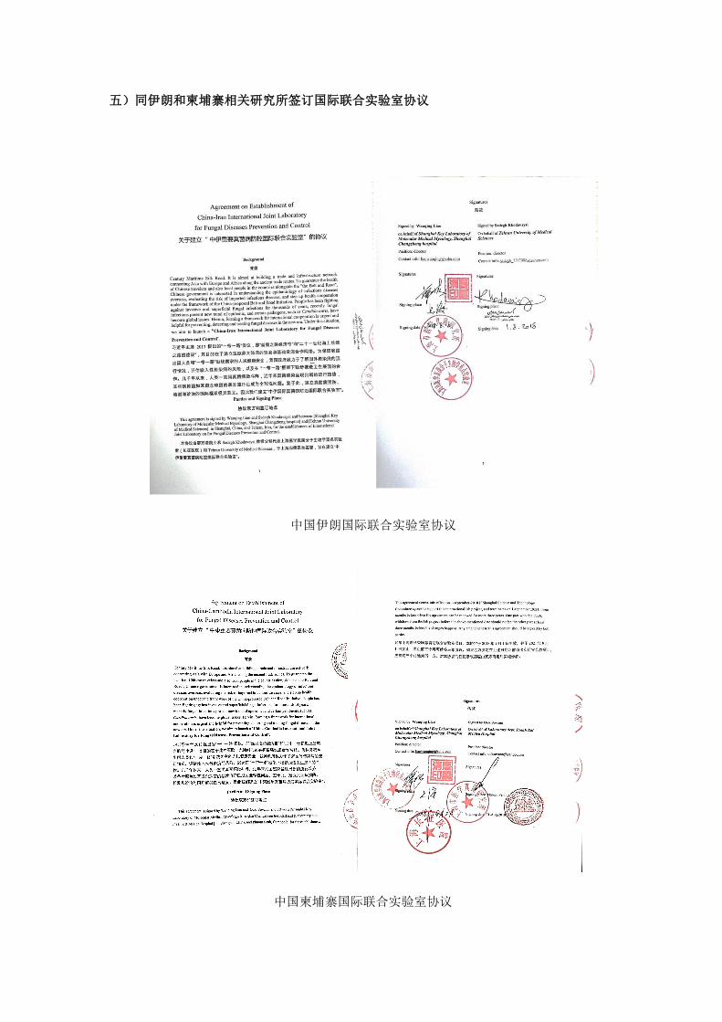
五)同伊朗和柬埔寨相关研究所签订国际联合实验室协议
中国伊朗国际联合实验室协议
中国柬埔寨国际联合实验室协议

中国伊朗真菌监测项目协议

其他重要学术活动
一)廖万清院士获戴芳澜终身成就奖 方文捷博士获戴芳澜优秀博士生
8月 11 日上午,中国菌物学会 2018 年学术年会在山东泰安开幕,此次大会以“组学时
代的菌物科学”为主题,中国工程院院士、海军军医大学上海长征医院廖万清院士,山东农
业大学副校长张然,山东农业大学植保学院院长许永玉,中国菌物学会理事长、中国科学院
微生物研究所研究员郭良栋以及 来自全国 200 多个科研院所、高校、企业的近 800 名专家
学者出席了开幕式。
大会设立了以我国真菌学创始人——“戴芳澜”教授的名字命名的 “戴芳澜科学技术
奖获奖”,该奖是我国真菌研究
领域最高奖项。2018 年戴芳澜
科学技术奖评审委员会由庄文
颖、李玉、白逢彦、边银丙、戴
玉成、郭良栋、姜子德、康冀川、
刘杏忠、图力古尔、王成树、王
源超、席丽艳、张劲松、张克勤、
张修国、朱平组成。
经评审委员会严格评选,决定授予我研究所所长廖万清院士以及中国科学院院士魏江春
2018 年戴芳澜终身成就奖;授予我研究所方文捷等 2018 年戴芳澜优秀研究生奖。同时大会
还评选出了 2018 年戴芳澜杰出成就
奖和优秀青年奖。我研究所以及其
他获奖单位,将再接再厉,为我国
以及世界真菌研究事业继续贡献自
己的力量。
开幕式现场
开幕式现场

廖万清院士在开幕式致辞
廖万清、魏江春院士获 2018 年戴芳澜终身成就奖

廖万清院士在闭幕式发表获奖感言
2018 年戴芳澜优秀研究生获奖代表

廖万清院士和方文捷博士会后合影
戴芳澜(1893 年 05 月 04 日- 1973 年 01 月 03 日)是著名
的真菌学家和植物病理学家,中央研究院院士,中国科学院院士。
在真菌分类学、真菌形态学、真菌遗传学以及植物病理学等方面
作出了突出的贡献。他建立起以遗传为中心的真菌分类体系,确
立了中国植物病理学科研系统;对近代真菌学和植物病理学在我
国的形成和发展起了开创和奠基的作用。

二)《大众医学》七十周年刊庆活动,廖院士获评医学科普卓越成就奖
2016 年 8 月,习近平总书记在全国卫生与健康大会上提出:没有全民健康,就没有全
面小康,要把人民健康放在优先发展的战略地位。党的十九大报告也明确提出实施健康中国
战略,为人民群众提供全方位、全周期的健康服务。要实现全民健康的宏伟目标,除了积极
构建完善的医疗保障体系、提高医疗技术水平以外,
大力普及健康知识(也就是科学普及工作),提高广
大人民群众的健康素养,促使其主动践行健康的生活
方式,做到未病先防、有病早治,也是不可或缺的重
要工作。为激励医学相关专业人员积极从事医学科普
工作,表彰为中国医学科普事业繁荣发展、提升中国人民健康素养的做出突出贡献的专家,
以及在医学科普领域成绩卓著的医疗机构,由中华医
学会科学普及部等指导,中华医学会科学普及分会、
《大众医学》杂志承办的“传播健康 心系大众”医学
科普奖评选活动于 2018 年 8 月 3 日正式启动。截至
2018 年 8 月 31 日,组委会共收到个人自荐、机构推
荐的参评材料 306 份。
为表彰廖万清院士在大众科普方面的贡献,大众医学特授予廖万清院士“传播健康 心
系大众”医学科普奖——卓越成就奖。


三)廖万清院士、巴里·马歇尔(诺奖得主)、侯云德院士、
钟南山院士任《新发传染病电子杂志》名誉主编,陈竺担任该杂志总顾问
2018年7月14日《新发传染病电子杂志》编委会在深圳召
开第一次编委会议。创刊于2016年的《新发传染病电子杂志》
由国家卫健委主管、人民卫生出版社主办,该杂志为传播和交
流新发传染病新知识、新技术搭建了一个很好的学术共享平台,
已出版发行7期,杂志以丰富的内容,全新视角引起海内外传
染病医学领域极大关注。该杂志创刊填补了我国新发传染病学
术交流领域的空白,为处于改革开放前沿的深圳在全国医学系
列期刊中取得了一席之地。
据了解,《新发传染病电子杂志》2016年8月获得国家新闻出版广电总局正式批准,于2017
年1月落地深圳出版发行,由全国人大常委会副委员长陈竺担任该杂志总顾问,幽门螺杆菌
发现者,诺贝尔奖获得者,中国工程
院外籍院士巴里·马歇尔;中国工程
院院士,呼吸道疾病国家重大实验室
主任钟南山;中国工程院院士,重大
传染病防控国家重大科技专项技术总
师侯云德;中国工程院院士,上海医
学真菌分子生物学重点实验室主任廖
万清等任名誉主编。
来自国内外近 100 名流行病医学领域新发传染病防治研究的领军人物、著名专家齐聚一
堂,共商办刊大计。侯云德院士,廖万清院士亲临会场祝贺。因故不能到会的钟南山院士,
中国工程院外籍院士巴里·马歇尔教授远程视频发言,对杂志的出版发行表示祝贺并提出了
希望。
中国工程院院士,上海市医学真菌研究所所长,上海长征医院皮肤病与真菌病研究所所
人民卫生出版社总编辑杜贤编审、主编陆普选教授给名誉主编颁发聘书

长廖万清院士担任该杂志的名誉主编并在大会致辞。他首先表示非常荣幸能够担任该杂志的
名誉主编,也希望杂志能多刊登中国人自己的最新研究成果,让世界都来关注中国的最新成
果。也希望该杂志能成为具有国
际影响力的学术期刊。廖万清院
士说:医生要勇敢面对新发传染
病,100 多年研究史对人类 5000
年历史,是短暂的,杂志为发传
染病提供了重要学术交流平台,
希望大家认真分析、研究、不断
提高研究与学术水平,才能更好
做好新发传染病防治工作。
会者一致认为,该杂志将致力于及时传播新发传染病新技术、新技能和新知识;大力促
进新发传染病早预防、早诊断和早治疗;以认真求实、开拓创新为原则,努力打造好这个与
国际接轨、先进的学术交流平台,服务于全人类。
新发传染病是指新的、刚出现的或呈现抗药性的传染病,其在人群中的发生在过去不断
增加或者有迹象表明在将来其发病有增加的可能。对多数新发传染病,人们目前尚无足够认
识。在过去 30 年中,全球发现了 40 余种新发传染病,平均每年至少出现一种。 多重耐
药耳道念珠菌、SARS、甲型 H1N1、H5N1、H7N9 人禽流感和中东呼吸综合征等是近 10 年来
对人类威胁最大的新发传染病。医务工作者在面对每一次新发传染病到来之际,都会够感到
预防控制措施掌握不够,缺乏精准的诊断知识和治疗技术缺憾。通过创办杂志可以广泛传播
新发传染病相关防
控、诊治知识和新
技术,普及和提高
广大医务工作者应
对新发传染病的水
平和能力,利用专
业杂志的功能及其
特殊性,将新发传
染病防控指南、诊
上海长征医院皮肤病与真菌病研究所所长廖万清院士致辞
《新发传染病电子杂志》第一届编委合影

疗专家共识及对新发传染病和流行病学研究的科学方法与规范的技术传递到每一位医务人
员(包括基层医务工作者)。这对于及时早期预防、早期诊断、及时治疗各种新发传染病,
降低病死率有着极其重要的意义。

四)廖万清院士一行人至上海海洋大学实地调研
2018 年 11 月 28 日中国工程院咨询项目《我国抗真菌药物研发策略国内外对比研究》
子课题《海洋天然抗真菌药物研发策略的调查与评估》项目组廖万清院士一行人至上海海洋
大学实地调研。参加此次实地调研讨论的专家教授有:上海海洋大学海洋生物制药系主任吴
文惠教授、包斌副教授、许剑锋副教授、张朝燕副教授、郭锐华讲师、刘宁讲师、马子宾博
士、张静怡博士、岳恒博士、长征医院廖万清院士、潘炜华教授、方文捷主治医师、扈东营
研究生。
实地调研讨论由上海海洋大学海
洋生物制药系主任吴文惠教授主持。廖
院士首先代表项目组介绍了《海洋天然
抗真菌药物研发策略的调查与评估》课
题的研究背景、研究目标和此次实地调
研的目的等基本情况,潘炜华教授汇报
了课题组前期海洋抗真菌药物研究文
献检索和至海军军医大学药学院实地
调研的情况。会议期间,各位专家教授主要就海洋天然抗真菌药物研发策略进行了积极讨论,
经过讨论,与会专家在以下方面达成共识:
1、海洋微生物具有极其丰富多样的种类,能够生存于低温、缺氧、高压等独特的海洋
生态环境中,因而能够产生具有优良生物活性且结构新颖的天然化合物,从海洋生物资源中
获取这些化合物具有安全性高、成本低及来源丰富等优点,因此,海洋微生物代谢产物已成
为药物创新与新药开发的重要来源。
2、上海海洋大学海洋生物制药系海洋类药物研发主要是从海洋真菌微生物提取具有优
良生物活性的天然化合物,包括青霉属、葡萄穗霉、地霉属、短孢霉属等真菌微生物,然后
用于心脑血管溶栓、医学工程材料和临床特异性食品等三大领域。目前较大的突破是从海洋
真菌微生物长孢葡萄穗霉FG216的代谢产物中发现了一种新型纤溶活性小分子化合物FGFC1,
药效学和毒理学研究已证实 FGFC1 是一种安全有效的溶血栓化合物。
3、上海海洋大学海洋生物制药系主要对海水真菌微生物发酵后产生的初级和次级代谢
产物进行研究,并未涉及海洋天然抗真菌药物的研发。呼吁国家关注海洋天然抗真菌药物研

发,增加对海洋天然抗真菌药物研发相关课题的支持。
廖万清院士课题组同海洋大学课题组成员合影

五)【汤飞凡论坛】廖万清院士纵论“科研之路”
2018 年 4 月 26 日下午,中国工程院院
士、海军军医大学长征医院廖万清教授应邀
访问中南大学湘雅二医院做客“汤飞凡”论
坛,与医院师生分享了题为《机缘型的科研
之路》的学术讲座,讲座由湘雅名医、皮肤
性病科主任陆前进教授主持。
廖万清院士创造性提出科学研究大体
可分为攻关型科学研究、挑战型科学研究、机缘型科学研究三种类型,结合其数十年的临床
科研实践发现,生动分享了格特隐球菌引起脑膜炎、具多育现象米曲霉引起肺曲霉病、小红
酵母引起甲真菌病、聚多曲霉致阻塞支气管曲霉病、胶囊青霉引起的肺青霉球等九种新的病
原真菌及其新的致病类型被发现的故事,并将这些归属于机缘型科学研究。
廖万清院士提到,作为临床医生,机缘型科学研究不但要有良好的科研素质、深厚扎实
的基础知识,并且还要有敏锐的眼光和无边的想象及强大的创造力,要善于从临床工作中发
现问题,抓住稍纵即逝的机会以成就科学发现。从科学研究到临床诊疗工作,廖院士的讲座
都为每一位与会者提供了
宝贵的指导和帮助。
在讲座中,廖万清院士
还和与会者进行了深入的
交流和探讨,并对提出的问
题一一进行详细的解答,师
生们表示受益匪浅。

六)廖万清院士参加季德胜先生诞辰 120 周年暨学术研讨活动
我国著名蛇伤专家季德胜先生诞辰 120 周年暨学术研讨活动今天在北京人民大会堂举
行。廖万清院士、国医大师和北京、上海、广东等地科研院所、各大医院专家、学者及国家
卫健委、江苏南通市政府相关领导近百人出席。
六)廖万清院士受邀参加 2018 第二届中国银屑病大会
2018 年 10 月 26-28 日,中华医学会皮肤性病学分会将联合安徽省医学会在美丽的“大

湖名城,创新高地”安徽省合肥市共同主办第二届“中国银屑病大会”。
中国医科大学附属医院陈洪铎院士、海军医科大学长征医院廖万清院士、复旦大学附属
华山医院王侠生教授、国际银屑病协会委员美国密歇根大学 JamesT.Elder 教授、国际银屑
病协会委员美国华盛顿大学 AnneBowcock.教授、日本银屑病协会前任主席日本东海大学
AKiraOZAWA 教授、国际皮肤科学会联盟常务理事/国际银屑病协会委员/中华医学会银屑病
专业委员会主任委
员张学军教授、国际
银屑病协会委员/中
华医学会银屑病专
业委员会副主任委
员/浙江大学医学院
附属第二医院皮肤
科主任郑敏教授等
国内外数百位全球
顶尖银屑病专家出席。
廖万清院士参与《全国科学技术名词审定委员会
皮肤性病学名词》定稿

七)廖万清院士参加世界温州人大会院士论坛
10 月 29 日,“大健康·新未来”2018 世界温州人大会院士论坛在洞头举行。本次论坛
以助力温州全面参与“一带一路”建设,发
扬世界温州人精神内涵为主旨,邀请了海内
外著名院士和国家级、省部级人才齐聚洞头,
聚焦生命科学主题,发挥名家效应,把脉洞
头康疗智岛发展方向。
上午八点三十分,开幕式在温州医科大
学滨海校区会堂举行,市委常委、统战部部
长施艾珠,温州医科大学校长李校堃,洞头
区委书记王蛟虎等致辞。温籍科学家、美国文理科学院院士、美国国家科学院院士林海帆以
录播视频形式发来致辞。院士论坛主题报告议程,中国科学院院士杨雄里教授、中国工程院
院士张心湜教授、中国工程院院士廖万清教授分别就《对脑科学前景的思考》、《如何成为快
乐成功的医疗工作人员》、《“海上丝绸之路”沿线国家重要真菌病防控》展开专题报告。

八)廖万清院士受邀参加陆军举办高层次学术交流活动
11 月 8日至 9 日,陆军举办高层次学术交流
活动。活动位于山城重庆陆军军医大学。
陆军高层次学术交流活动会场“高朋”满座,
大咖云集。会场共有 20 名军地知名院士,30 名
陆军军事医学领域高层次科技人才和科技英才培
养对象,90 余名各类型医疗卫生单位专家代表。
廖万清院士同陆军军医大学领导和医务人员合影 廖院士同王云贵校长合影

九)廖万清院士参与中国医师协会年会,并为获奖嘉宾颁奖
中国医师协会皮肤科医师分会年会与 11 月 8 日在广州举办,廖万清受邀参加。11 月 9
日,在广州召开的第十四届中国皮肤科医师年会(2018CDA)暨全国美容皮肤科学大会开幕
式上,张会长勉励道:希望 CDA 着力于培养更多的像廖万清、朱学骏、王宝玺、郑志忠、李
若瑜、李恒进等一样的高质量人才,这才是 CDA 最大的成功和诚意!”
廖万清院士为 CDA 杰出贡献专家颁奖

廖万清院士接受《医师报》采访
呼吁全国同行重视“超级真菌”

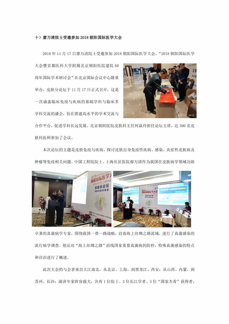
十)廖万清院士受邀参加 2018 朝阳国际医学大会
2018 年 11 月 17 日廖万清院士受邀参加 2018 朝阳国际医学大会。“2018 朝阳国际医学
大会暨首都医科大学附属北京朝阳医院建院 60
周年国际学术研讨会”在北京国际会议中心隆重
举办。皮肤分论坛于 11 月 17 日正式召开,这是
一次涵盖临床免疫与疾病的基础学科与临床多
学科交流的盛会,旨在搭建高水平的学术交流与
合作平台,促进学科长远发展。北京朝阳医院皮肤科主任何焱玲担任论坛主席,近 300 名皮
肤科医师参加了会议。
本次论坛的主题是皮肤免疫与疾病,探讨皮肤自身免疫性疾病、感染、炎症性皮肤病及
肿瘤等免疫相关问题。中国工程院院士、上海长征医院廖万清作为我国在皮肤病学领域功勋
卓著的真菌病学专家,围绕我国一带一路战略,沿着海上丝绸之路流域,进行了真菌感染的
流行病学调查。他还对“海上丝绸之路”沿线国家重要真菌病的防控,特殊真菌感染的特点
和诊治进行了概述。
此次大会的与会者来自大江南北,从北京、上海,到黑龙江、西安;从山西、内蒙,到
苏州、长沙;演讲专家阵容强大,共有 1 位院士、2 位长江学者、3 位“国家杰青”获得者,

代表了当前皮肤科领域和风湿免疫领域的免疫相关性疾病研究方面较高的学术水平;会议内
容丰富,从基础研究到临床研究等多个角度、不同层面对皮肤病学和免疫学研究的最新进展
进行了较为详细丰富的介绍,是国内临床免疫领域一流水平的对接碰撞。与会专家学者均对
此次大会给予了高度评价,认为本次会议学术气氛浓厚、内容新颖丰富,基础与临床联系密
切,为国内皮肤免疫及疾病的科研提供了良好的沟通平台,并将有效地推动国内各地区、各
中心之间的合作与发展。
与会全体嘉宾合影
五、2018 年度全国院士工作站建设情况
丹娜生物院士专家工作站
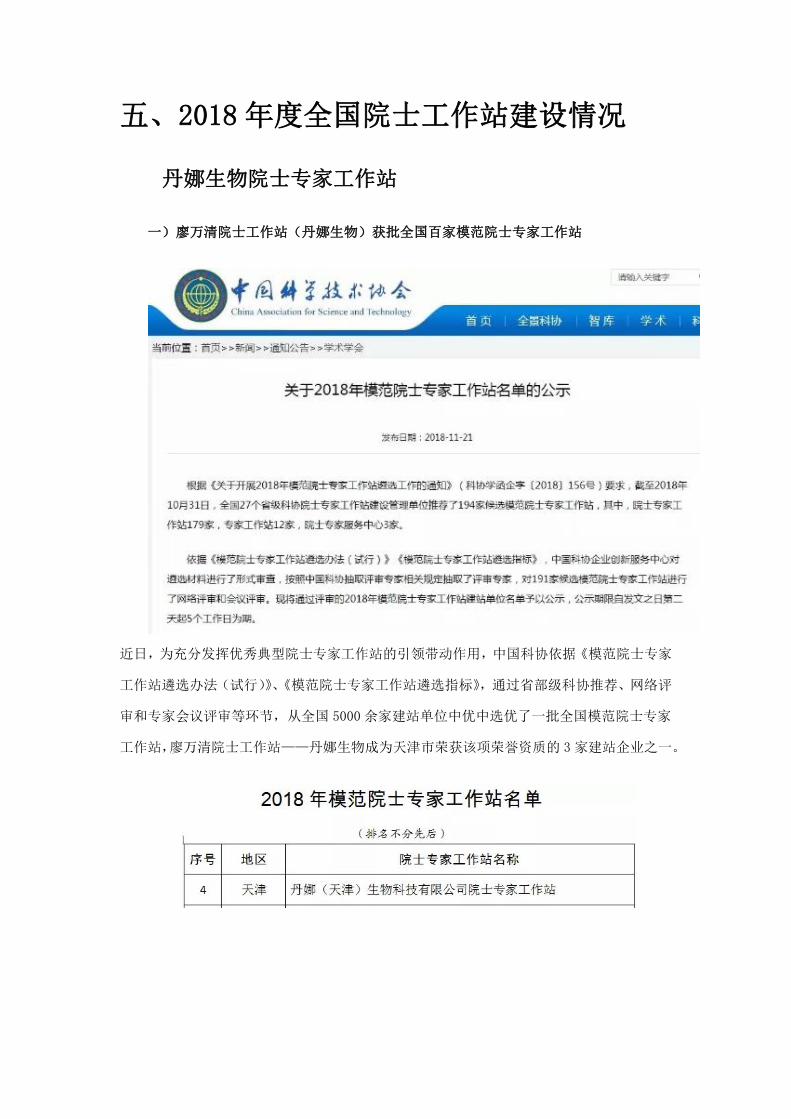
一)廖万清院士工作站(丹娜生物)获批全国百家模范院士专家工作站
近日,为充分发挥优秀典型院士专家工作站的引领带动作用,中国科协依据《模范院士专家
工作站遴选办法(试行)》、《模范院士专家工作站遴选指标》,通过省部级科协推荐、网络评
审和专家会议评审等环节,从全国 5000 余家建站单位中优中选优了一批全国模范院士专家
工作站,廖万清院士工作站——丹娜生物成为天津市荣获该项荣誉资质的 3 家建站企业之一。
在本次评选过程中,评审专家们对丹娜生物院士专家工作站的各项遴选指标给予了很高
评价:
工作站基础条件良好,建站单位诚信经营、具有良好的社会责任感;工作站保
障经费充分,院士专家团队与建站单位联系紧密,“产、学、研”协同效应显著。
工作站管理和运行机制规范,制度健全,运行状况良好。
工作站在提升企业等建站单位创新能力、促进建站单位创新人才培养与引进、
提高建站单位经济效益及社会效益等方面成效突出。
进站院士专家对工作站基础条件、运行管理及取得的成效等方面满意度高。
中国科协开展本次遴选工作,主要目的在于引导全国院士专家工作站规范、有序、可持
续发展,推动各地工作站提质增效。荣获“全国模范院士专家工作站”是中国科协和天津市
科协组织对丹娜生物院士专家工作站几
年来整体运营成效的极大肯定。丹娜生物
董事长周泽奇表示,公司将进一步提高院
士专家工作站的运行质量、加强高水平建
设,真正发挥引领带动作用,持续提升工
作站服务企业技术创新的能力,用更加完
善的联合诊断产品为IFD患者的生命健康
提供优质、高效的服务。

二)丹娜生物举办 2018 年院士专家工作站总结报告会
天津滨海新区丹娜生物在生态城举办了 2018 年院士专家工作站总结报告会。会上,中
国工程院廖万清院士报告了《“海上丝绸之路”沿线国家重要真菌病防控》国际合作项目所
取得的重大进展。 丹娜生物作为这一重大国际合作项目单位之一,在廖万清院士的带领下,
与中东地区 22 家重点标杆医院共同开展的“一带一路”念珠菌血症临床诊断技术研究获得
了重要的阶段性成果。这充分表明了丹娜生物的侵袭性真菌病早期快速检测核心技术与创新
产品具有重要的临床应用价值和很强的国际竞争力。
据介绍,一年以来,在院士专家工作站的带动下,丹娜生物取得了国家级科技型企业、
与廖万清院士团队及伊朗22家重点医院共同开展
“中国-伊朗念珠菌血症多中心研究”、获批天津
市企业人才智力合作项目、G试验获批天津市中小
企业局“专精特新” 产品认定等一系列成就,
科研进展迅速。
丹娜生物董事长周泽奇指出,院士专家
工作站对丹娜生物的发展和创新研究非常重要,
它是吸引人才的高地和智库服务的平台,它对丹
娜生物的快速、健康发展具有强大的推动作用。

丹娜生物将进一步加强与廖万清院士团队的全面深入合作,共同解决好 IFD 精准诊疗领域的
难题,为有效的改善广谱抗生素不合理利用的现状、不断提高 IFD 患者的检出率和治愈率、
减少死亡率提供更加优质高效的服务。
廖万清院士同丹娜全体职工合影

延安市人民医院廖万清院士工作站
廖万清院士专家团队潘炜华教授莅临延安市人民医院指导工作

为进一步深入了解和掌握延安市皮肤科院士专家工作站的建设情况,12 月 22 日,第二
军医大学附属上海长征医院皮肤病与真菌病研究所副所长、上海市医学真菌研究所副所长、
上海领军人才、廖万清院士专家团队成员潘炜华教授莅临延安市人民医院检查指导皮肤科院
士专家工作站工作。市政协副主席、延安市人民医院院长蔺广东出席座谈会并讲话。皮肤科
主任熊林、检验科主任曹云、科教科副科长师丽陪同检查。来自科教科、皮肤科、检验科等
科室近 20 人参加了座谈会。
当天,潘炜华教授在延安市人民医院检验科培训示教室作了题为《临床真菌感染的检测
及诊疗思路》的学术讲座。她分别从基础概念、诊断思路、检测方法与技术、临床应用四个
方面进行了深刻阐述,内容丰富实用,使在场人员深感获益匪浅。
培训结束后,潘炜华教授先后走访了工作站办公场所和真菌实验室,她从资金投入、场
地建设、人员梯队和实验数据四个方面深入考察,并对医院的全方位投入表示了充分肯定。
“大家一定要做好实验标本的采集和保存工作,尤其是在遇到少见、罕见的标本时,可
以及时联系我们并从原始标本里取样寄给我们,通过‘两地实验、一个标本、一个结果’的
实验模式共同促进我们真菌实验的发展。”潘教授在检查中强调:“真菌实验的原始标本和实
验数据的完整保存,是我们开展科学研究和指导临床实践的基础条件,至关重要。”

座谈会上,潘炜华教授从场地建设、科研课题申报、论文发表等方面做了具体指导。她
特别指出,工作站后期的运行要抓住两个重点:一是皮肤科设立浅部真菌实验室,用于浅部
真菌的实验和研究,并以此为契机开展相关新技术新业务;二是检验科要充分利用真菌实验
室开展好深部真菌的实验、培养和研究工作,要切实做到“从临床实验走向基础研究”,为
夯实科研基础走好第一步。

市政协副主席、延安市人民医院院长要求,各相关科室要尽快落实潘炜华教授的具体要
求,积极开展新技术新业务,不断提高临床技术水平和科研能力,全力以赴打造陕北首家真
菌实验中心。
据悉,延安市人民医院皮肤科院士专家工作站为西北首家皮肤专业院士专家工作站,隶
属于陕西省科学技术协会。该工作站设在延安市人民医院皮肤科,其中真菌实验室设在检验
科。现阶段,该工作站已经明确了工作职责,并从标本采集、实验研究、科研学术等方面开
展相关工作。
江西胸科医院院士工作站
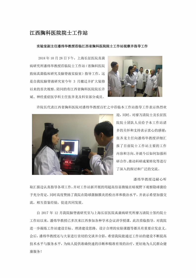
实验室副主任潘炜华教授莅临江西省胸科医院院士工作站视察并指导工作
2018年 10 月 28日下午,上海长征医院真菌
病研究所潘炜华教授莅临院士工作站(省胸科医院
致病真菌临床研究及脑脊液实验室)指导工作,这
是自我院脑脊液研究室今年 3 月搬迁并扩大装修
以来的首次视察。陪同的有江西省胸科医院院长许
斌、神经重症医学科主任张齐龙及科室部分成员。
许院长代表江西省胸科医院对潘炜华教授百忙之中莅临本工作站指导工作表示热烈欢
迎,同时,对廖万清院士及长征医
院院士团队人员给予本工作站诸
多的关怀和支持表示衷心的感谢;
张齐龙主任向潘炜华教授详细汇
报了目前院士工作站主要的工作
内容和方向。并就今后如何加强科
研合作、推动科研成果转化等进行
了深入的探讨和广泛的交流。
潘炜华教授边耐心听
取汇报边认真指导各项工作,并对工作站新开展的用超高倍显微镜在暗视野下观察隐球菌给
予充分肯定,同时高度赞扬了我院在隐球菌脑膜炎的检出率和救治水平,并表示希望加强交
流,相互借鉴经验,促进共同发展。
自 2017 年 12 月我院脑脊液研究室与上海长征医院真菌病研究所廖万清院士签约院士
工作站以来,潘炜华教授已多次来江西参加各种学术会议讲学授课。此次莅临指导,对我院
进一步凝练工作站建设目标、理清建设思路、设计合理的实验课题等都具有重要启发意义。
会后,潘炜华教授还与大家进行亲切的交谈并合影,希望我院能通过工作站的建设不断提高
技术水平与服务水平,为病人提供准确快速的诊断和精准有效的治疗,更好地为人民群众健
康服务!
粤东医院廖万清院士工作站
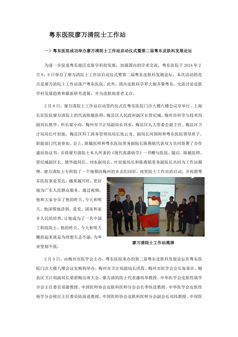
一)粤东医院成功举办廖万清院士工作站启动仪式暨第二届粤东皮肤科发展论坛
为进一步促进粤东地区皮肤学科的发展,加强国内的学术交流,粤东医院于 2018 年 2
月 8、9 日举办了廖万清院士工作站启动仪式暨第二届粤东皮肤科发展论坛。本次活动的亮
点是廖万清院士工作站落户粤东医院,此外,国内皮肤科学界大伽齐聚粤东,交流讨论皮肤
学科发展趋势和最新研究进展,并为皮肤病患者义诊。
2月 8日,廖万清院士工作站启动签约仪式在粤东医院门诊大楼六楼会议室举行。上海
长征医院廖万清院士的代表陈敏医师,梅县区人民政府副区长管纪城,梅州市科学与技术局
副局长熊华、科长梁小向,梅州市卫计局副局长刘水,梅县区人大常委会副主任、梅县区卫
计局局长叶世振,梅县区科工商务管理局局长张云龙、副局长何国材和粤东医院领导班子、
职能部门代表参加。会上,陈敏医师和粤东医院常务副院长陈燕铭代表双方共同签署了合作
建站协议书,并将廖万清院士本人所著的《现代真菌病学》一书赠与医院。随后,陈敏医师、
管纪城副区长、熊华副局长、刘水副局长、叶世振局长和陈燕铭常务副院长共同为工作站揭
牌。廖万清院士专程拍了一个视频向梅州的乡亲们问好,祝贺院士工作站的启动,并祝愿粤
东医院事业发达,越来越兴旺,更好
地为广东人民群众服务。通过视频,
他和大家分享了他的昨天、今天和明
天,他深情地讲到,是党、国家和家
乡人民的培养,让他成为了一名中国
工程院院士,他的昨天、今天和明天
概括起来就是为理想矢志不渝,为事
业坚韧不拔。
2月 9日,由梅州市医学会主办,粤东医院承办的第二届粤东皮肤科发展论坛在粤东医
院门诊大楼八楼会议室顺利举办。梅州市卫计局副局长洪霞、梅州市医学会会长张荣在、梅
县区卫计局副局长梁碧梅出席大会。廖万清的院士代表潘炜华教授、中华医学会皮肤性病学
分会主任委员郑捷教授、中国医师协会皮肤科医师分会会长李恒进教授、中华医学会皮肤性
病学分会候任主任委员陆前进教授、中国医师协会皮肤科医师分会副会长刘玮教授、中国医
廖万清院士工作站揭牌

师协会皮肤科医师分会副会长李利教授、广东省医学会皮肤性病学分会主委曾抗教授、广东
省医师协会皮肤科医师分会副主委李其林教授、邓列华教授和陆春教授等国内皮肤科学界大
伽齐聚粤东带来了一场国家级学术盛宴,吸引了 200 多名来自梅州、惠州、河源、江门和汕
头各级医院的医务人员前来参加。中山大学附属
第三医院皮肤科主任赖维教授主持开幕式。学术
活动启动仪式上,粤东医院还进行了皮肤科成立
的揭牌仪式,市、区领导们、皮肤科学界大伽们
和粤东医院党总支书记沈友权共同为粤东医院皮
肤科开科揭牌。
9日下午 3:30-6:30,赖维、曾抗、韩建德、陆春、李其林、邓列华六位教授在门诊
大楼四楼皮肤科联袂义诊,帮助广大皮肤病患者摆脱疾病的痛苦。梅州、河源、汕头、惠州
等地区的患者慕名前来就诊。专家们耐心地解答每一位群众的疑问,用专业知识细致地为他
们诊治。群众们对专家的医疗水平、服务态度纷纷表示满意。据统计,此次专科义诊为 180
多名患者提供了医疗服务,取得了良好的社会效益。
本次活动取得了圆满的成
功。廖万清院士工作站这一科
研平台落户于粤东医院,将通
过联合研究、联合培养科技创
新型人才、联合开展高层次学
术或技术交流活动等方式,进
一步提升粤东医院在皮肤病相
关领域的临床诊疗水平和科研
能力,更好地服务于社会、服
务于患者。
廖万清院士工作站签约仪式
廖万清院士讲话

领导和皮肤科医务人员合影
论坛现场

皮肤科开科揭牌
义诊现场

二)粤东医院举办廖万清院士工作站启动授牌仪式
为了提升广东粤东地区医学真菌领域的研究水平,促进资源共享和合作,2018 年 8 月
24 日,廖万清院士工作站在粤东医院皮肤科正式挂牌。中国工程院院士廖万清,梅县区委
书记钟光灵,梅县区政协副主
席、粤东医院管理顾问李刚教
授,梅县区卫计局局长钟匡仁,
粤东医院党总支书记沈友权和
中山三院皮肤科赖维教授出席
挂牌仪式并为工作站揭牌。
廖万清院士工作站是由梅州市政府推动、以粤东医院为依托,联合进行科学技术研究的
高层次科技创新平台。工作站将通过联合研究、联合培养科技创新型人才、联合开展高层次
学术或技术交流活动等方式,将粤东医院皮肤科打造成粤东地区乃至广东省一流的皮肤科,
为梅州及周边地区的医疗学术及创新提供强有力的支撑。
廖万清院士表示,工作站建成投入使用后,将逐渐完善内部建设,开展有粤东特色的主
要包括皮肤癣菌病、孢子丝菌病等皮肤
真菌病的研究工作,进一步联合申报省
级以上研究课题,建立专业研究人员的
真菌研究团队,保持并加强皮肤真菌病
方面的诊治优势,推动粤东医院皮肤科
诊疗水平达到国内先进水平,真正做到
造福百姓。

廖院士作专题讲座,题为:我的昨天、今天和明天
来院期间,廖万清院士一行对粤东医院建设、廖万清院士工作站建设等情况进行了详细
考察,并与粤东医院领导班子、相关部门负责人进行座谈。同时,中山三院皮肤科、粤东医
院皮肤科联合举办了粤东地区医学真菌高峰论坛暨前沿技术培训班。培训班上,廖万清院士
和大家分享了他的昨天、今天和明天,感谢梅县家乡人民的养育之恩,讲述了他的成才之路
以及所取得的成就,表达了对真菌研究和医学真菌方面发展的憧憬和希望。随后,上海长征
医院潘炜华教授、中山三院赖维教授等就医学真菌临床和基础研究热点,以及抗真菌药物研
发的最新成果进行授课。此次培训班吸引了近百名粤闽赣周边地区从事病原真菌的基础研究
人员、临床工作者(皮肤科、检验科和血液科
等)特地前来参加,是一场难得的学术盛宴。
所有与会人员更是非常珍惜本次交流学习的机
会,认真听取了专家教授的授课,并对专家的
精彩讲述予以热烈掌声。
会后廖万清院士接受梅县电视台的采访
余杭中医院廖万清院士工作站
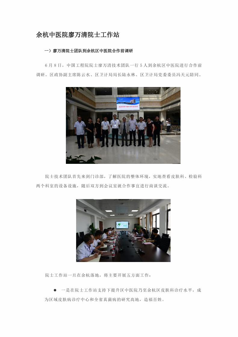
一)廖万清院士团队到余杭区中医院合作前调研
6 月 8 日,中国工程院院士廖万清技术团队一行 5 人到余杭区中医院进行合作前
调研。区政协副主席陈云水、区卫计局局长陆永林、区卫计局党委委员冯天元陪同。
院士技术团队首先来到门诊部,了解医院的整体环境,实地查看皮肤科、检验科
两个科室的设备设施,随后双方到会议室就合作事宜进行商谈交流。
院士工作站一旦在余杭落地,将主要开展五方面工作:
一是在院士工作站支持下提升区中医院乃至余杭区皮肤科诊疗水平,成
为区域皮肤病诊疗中心和全省真菌病的研究高地,造福百姓。

二是借助院士工作站品牌招录高层次人才,解决区域内重症皮肤病患者
病痛,拓宽皮肤病研究领域,填补我区皮肤病基础研究空白。
三是为全区医疗单位开展皮肤科及相关工作人员培训,提升全区皮肤科
工作人员临床技能及科研能力。
四是发挥院士工作站在真菌病领域的研究优势,成立区域真菌检查和治
疗中心,包括深部真菌的诊疗,为全区真菌领域诊疗提供技术科研支撑。
五是借助院士工作站号召力,通过举办大型国家级学术交流等活动,提
升医院及医院皮肤科影响力,并在周期内成功申报杭州市级重点学科乃至浙江省
级重点学科。
座谈交流结束后,廖万清院士还将刚主编出版的《现代真菌学》书籍赠送给了区
中医院。

二)中国工程院廖万清院士工作站落户余杭中医院
2018 年 07 月 21 日中国工程院廖万清院士工作站落户余杭区中医院,这是余杭区公立
医疗机构引进的首个院士工作站,意味着余杭百姓今后在家门口就能享受到“院士级”专家
的医疗服务。副区长许玲娣参加签约仪式。
会议期间,廖万清院士给参会医务人员做了题为“医学真菌研究的前沿和热点”的专题
报告。
院士工作站挂牌后,院士团队专家将每两周一次来余杭区中医院坐诊,院士本人每季度
到余杭区中医院工作一天,为余杭及周边
地区的疑难、重症皮肤病、真菌患者诊疗,
帮助提升余杭区中医院及余杭区皮肤病诊
疗能力和科研水平,推荐或协助高层次人
才招录,促进相关学科建设并成立皮肤病
实验室。院士工作站在真菌领域的研究优
势将为全区真菌病诊疗提供技术支撑,协
助成立区域真菌检查和治疗中心,同时带
动检验、重症医学、肿瘤等相关学科的发展,进一步满足余杭市民对优质医疗资源的需求。

嘉兴港区医院廖万清院士工作站
廖万清院士受邀参加“星耀南湖”精英峰会,参加工作站授牌仪式
连续 10 年举办的“星耀南湖”精英峰会,早已成为嘉兴不忘初心、久久为功,打造人
才高地,推动创新发展的最好注脚。本届峰会将继续携手浦江创新论坛,紧扣“确立人才引
领发展的战略地位”要求,围绕“人才驱动创新、创新引领产业”主题,突出实效性、引领
性、高端性、国际性,推进人才与科技、人才与文化的融合发展,搭建人才与企业、项目与
资金、产品与市场的对接平台,为打造具有国际化品质的现代化网络型田园城市提供有力人
才保障。
在举行授牌、颁证仪式上,嘉兴市
人大常委会副主任沈利农先生,嘉兴市
政协党组副书记、副主席孙建华先生为
院士专家工作站代表授牌。
市领导为中国工程院廖万清院士和上海市东方
医院嘉兴港区医院授牌
廖院士团队和嘉兴港区医院谭军副院长合影
河北工程大学附属医院廖万清院士工作站
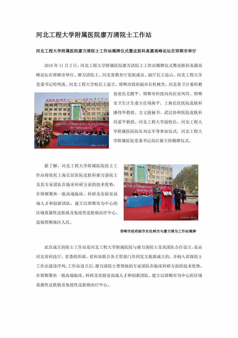
河北工程大学附属医院廖万清院士工作站揭牌仪式暨皮肤科真菌高峰论坛在邯郸市举行
2018 年 11 月 2 日,河北工程大学附属医院廖万清院士工作站揭牌仪式暨皮肤科真菌高
峰论坛在邯郸市举行。廖万清院士、河北省教育厅党组成员、副厅长王廷山、河北工程大学
党委书记哈明虎、河北工程大学校长王延吉、邯郸市政府副市长杜树杰、河北省卫计委科教
处处长尤殿平、邯郸市科技局局长安凤玲、邯郸
市卫生计生委主任周海平、上海长征医院皮肤科
潘炜华教授、方文捷秘书、武汉协和医院皮肤科
冯爱平教授、河北工程大学副校长、河北工程大
学附属医院院长刘志军等参加仪式,河北工程大
学附属医院党委书记高红旗主持揭牌仪式。
据了解,河北工程大学附属医院院士工
作站将依托上海长征医院皮肤科廖万清院士
及其专家团队在临床科研方面的技术优势,
在邯郸聚焦一批高端临床、科研及实验室高
端人才和创新团队,建立以邯郸市为中心的
区域真菌性皮肤病及免疫性皮肤病治疗中心,
造福邯郸地区人民。
此次成立的院士工作站是河北工程大学附属医院与廖万清院士及其团队合作设立,是由
河北省科技厅、省委组织部、省科协联合各主管部门共同发文批准成立的,并纳入省级院士
工作站建设序列。工作站设立后,廖万清院士带领他的专家团队在临床科研方面的技术优势,
在邯郸聚焦一批高端临床、科研及实验室高端人才和创新团队,建立以邯郸市为中心的区域
真菌性皮肤病及免疫性皮肤病治疗中心。
邯郸市政府副市长杜树杰与廖万清为工作站揭牌
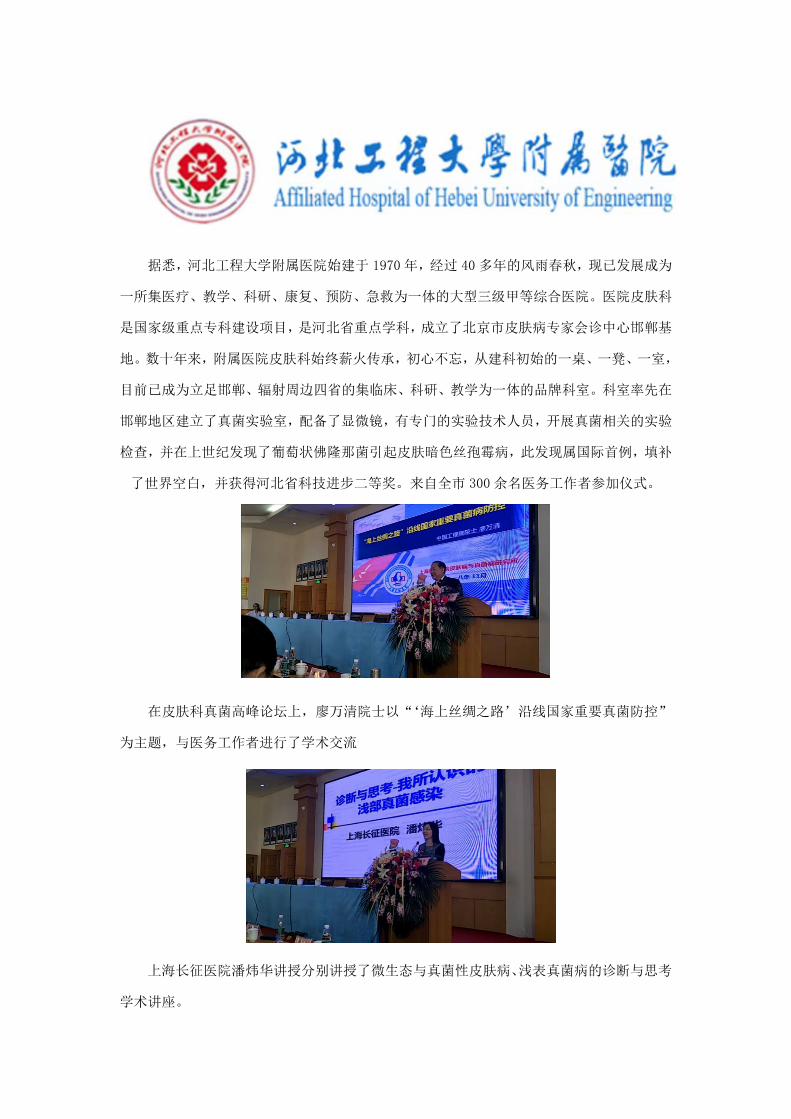
据悉,河北工程大学附属医院始建于 1970 年,经过 40 多年的风雨春秋,现已发展成为
一所集医疗、教学、科研、康复、预防、急救为一体的大型三级甲等综合医院。医院皮肤科
是国家级重点专科建设项目,是河北省重点学科,成立了北京市皮肤病专家会诊中心邯郸基
地。数十年来,附属医院皮肤科始终薪火传承,初心不忘,从建科初始的一桌、一凳、一室,
目前已成为立足邯郸、辐射周边四省的集临床、科研、教学为一体的品牌科室。科室率先在
邯郸地区建立了真菌实验室,配备了显微镜,有专门的实验技术人员,开展真菌相关的实验
检查,并在上世纪发现了葡萄状佛隆那菌引起皮肤暗色丝孢霉病,此发现属国际首例,填补
了世界空白,并获得河北省科技进步二等奖。来自全市 300 余名医务工作者参加仪式。
在皮肤科真菌高峰论坛上,廖万清院士以“‘海上丝绸之路’沿线国家重要真菌防控”
为主题,与医务工作者进行了学术交流
上海长征医院潘炜华讲授分别讲授了微生态与真菌性皮肤病、浅表真菌病的诊断与思考
学术讲座。

长江润发集团院士专家工作站
廖万清院士参观海南海灵药业
10 月 18 日,长江润发集团院士专家工作站主任、中国工程院院士、世界华人皮肤科医
师协会会长、上海长征医院皮肤科教授廖万清一行来我司参观指导。海灵药业执行总裁陆一
峰、长江润发集团总裁助理李一青、海灵药业市场总监曹涛热情接待了来访一行。
在公司展厅,陆总向廖院士详细介绍了长江润发集团的发展及我司的现状、规划及发展
前景。海灵药业秉承“为大众健康孜孜以求”的愿景,以打造中国领先的健康产业平台为目
标,志在为广大临床医师和患者提供更加有效、安全的优质药品。
廖院士在听取了陆总的介绍后,充分认可了海灵药业的优质产品及发展目标。他说,为
人民健康服务是中国制药企业的核心
责任。廖院士对集团的规模及发展情况
表示肯定,对大健康领域的发展也提出
许多宝贵意见,并就双方在抗真菌领域
的合作进行了深入的探讨。
海灵药业一直以来始终致力于抗
感染制剂的研发、生产和销售,是国内
细分医药行业领域的龙头企业。2016 年被长江润发并购之后,不断调整和提升发展思路,
延伸产品领域,并取得了良好的效果,海灵药业持续保持稳健的发展态势。

雅蒂院士专家工作站
廖万清院士到访雅蒂化妆品集团并达成初步合作意向
2018年7月16日,廖万清院士受邀到访汕头市雅蒂化妆品有限公司。雅蒂公司总经理王
泽辉率公司全体管理团队,对廖院士一行的到来表示最热烈的欢迎。
廖院士一行首先依次参观了雅蒂公司的办公场所、产品展厅、实验室以及生产车间,然
后宾主双方在总经理室进行座谈。雅蒂公司总经理王泽辉首先为嘉宾们就企业的愿景、使命、
科研成果等进行简要的汇报。然后双方就肌肤健康管理行业的发展、科技创新、人才培养等
方面进行了热烈、深入的交流,廖院士一一给出了很多前瞻性的指导性意见,整个座谈会现场
探讨交流氛围异常浓烈。

大专家.com 院士专家工作站
2018 年 11 月 23 日大专家.com 来访交流吴翀博士一行来访
大专家.com 吴翀博士一行来访,与廖万清院士课题组成员深入讨论了皮肤病 OK 系统的
构建、真菌大数据的构架等。廖万清院士亲切接待了吴翀博士一行。

六、2018 年度服务军民情况
一、为军服务
一)朱红梅副教授参加“和谐使命-2018”任务。
2018 年 06 月 29 日海军和平方舟医院船在任务指挥员管柏林、秦威率领下,从浙江舟
山某军港解缆起航,赴巴布亚新几内亚、瓦努阿图、斐济、汤加、哥伦比亚、委内瑞拉、格
林纳达、多米尼克、安提瓜和巴布达、多米尼加、厄瓜多尔 11 国进行人道主义医疗服务,
执行“和谐使命-2018”任务,并应邀赴智利参加其海军成立 200 周年庆典活动。这是该医
院船第七次执行“和谐使命”任务。
二)潘炜华教授参加长征医院“移动课堂”
2018 年 12 月上旬,研究所副主
任潘炜华教授带队海军军医大学附属
长征医院“移动课堂”教学小分队来
到上海某部,为舰艇官兵送去了一场
健康保健、疾病防治知识讲座并进行
了义诊活动。本次活动受到了舰艇全
体官兵的一致好评,纷纷称赞专家们
的讲座言简意赅,深入浅出、精彩纷呈,讲座内容贴近舰艇训练和生活,科学实用,指导性
强。潘炜华教授就湿疹、痤疮、虫咬性皮炎、体癣等舰艇环境下常见皮肤病的治疗和预防展
开宣讲,她的授课图文并茂,形象实用。长征医院“移动课堂”巡教项目是贯彻落实习主席、
军委、海军和学校党委的决策指示,对标医院党委扩大会议关于“打造海卫特色教学品牌”
的具体任务,探索将院校教育与部队训
考相结合、医疗服务与教书育人相结合,
教学实践与教改研究相结合的海军卫勤
医教研工作联动推进的创新模式和方法。
“移动课堂”按照“贴近海军、贴近基
层、贴近实战”的基本要求,依托舰艇
现有卫勤保障与战救装备器材,以海军

舰艇单位卫勤保障力量为主要培训对象,探索面向全体官兵拓展、实现整舰全员覆盖,切实
提高受训人员实战技能的有效途径。让舰艇卫勤保障力量“动起来”、让现有列装卫勤装备
“活起来”。目前,小分队在东海舰队已展开巡教活动,并取得初步成效。
三三)其他军事任务
2018 年 1 月-2018 年 4 月 张超参加 2018 年度海军招飞全面检测工作
2018 年 6 月-2018 年 7 月 张超参加 2018 年度海军招飞定选工作
2018 年 11 月- 2020 年 1 月 张超参加中国驻吉布提保障基地保障任务
2018 年 9 月-2018 年 12 月 潘搏参加南海保障任务
2018 年 12 月-2019 年 2 月 陈敏参加 2018 年度海军招飞工作
2018 年 12 月—2019 年 3 月方伟参加海岛任务

四四)廖万清院士前往八路军一二九师司令部旧址。祭拜革命英烈
2018 年 11 月 02 日廖万清驱车前往河北邯郸涉县,于八路军一二九师司令部旧址祭拜
革命英烈。抗日战争时期,涉县是边区根据地的腹心地、首府县,地处华北抗战前哨,为华
北抗战战略要地,八路军 129 师在刘伯承、邓小平等师首长率领下,临危受命、东渡黄河、
挺进太行,运筹涉县赤岸村,浴血千里太行山,打响了抗日战争中长生口、神头岭、响堂铺
和解放战争中上党、平汉等著名战斗、战役,曾有一百一十多个党、政、军、财、文等机关
单位在涉县驻扎长达五年之久。
廖万清院士为八路军英烈敬献花圈,并深情鞠躬

二、义诊和其他民间交流
一)廖万清院士为深圳市慢性病防治中心多名疑难皮肤病患者义诊
7 月 14 日下午,中国工程院院士廖万清,亲
临深圳市慢性病防治中心皮肤病防治研究所为多
名疑难皮肤病患者进行会诊。八十岁高龄的廖万清
院士认真听取了主管医师汇报的每一例患者,并询
问病情,亲自为患者进行全面仔细的体格检查,详
细地分析了病情,为下一步治疗给予指导意见。廖
院士会诊后,还细心叮嘱患者饮食注意事项。
廖院士表示,深圳市慢性病防治中心就诊环境舒适,中心领导十分重视皮肤病研究所的
发展,医护团体强大,积极向上,做到了临床和科研并轨发展,寄望中心医务人员今后继续
努力,活到老学到老,通过科学研究解决临床问题,造福广大群众,为理想要孜孜不倦,为
事业要坚韧不拔。
廖万清院士深圳慢病防治所皮肤科全体员工合影廖万清院士参观深圳慢病防治所

二)廖万清院士参加苏州“寒山检验高峰论坛”,并会疑难病例
2018年6月9日,第二届寒山检验医学高峰论坛在高新区人民医院开幕,中国工程院院士
廖万清、阮长耿与来自全国各地的顶尖专家、学者600余人汇聚一堂就检验医学的前沿发展
进行观点碰撞、智慧交锋。廖万清院士在开幕式上做了大会致辞。
会后,廖万清院士参加了高新区人民医院疑难病例会诊,该患者经院士查房,最终确诊
为罕见的地霉属感染病例,最终得到了正确的治疗。
三)廖万清院士入住皮肤宝网络问诊平台,为患者免费诊治
10 月 10 日,皮肤科院士廖万清教授上线皮肤宝在线问诊平台,这是目前国内互联网医
疗所有的诊疗平台中首次有院士级别专家入驻。据悉,皮肤宝 APP 在 2016 年 3 月上线,由
爱肤宝(上海)信息科技有限公司发布,产品上线以来,开通了医疗“电子处方”、“送药到家”
等多项在线服务。
皮肤科医生通过皮肤宝 APP 在线为患者提供皮肤健康及护肤咨询服务, 一方面,帮助医

生采集门诊患者病例,线上管理,为皮肤疾病+护理的健康管理进行互联网赋能;另一方面,
患者除了能够得到更快捷的诊疗,还能获得更个性化的辅助治疗和护肤建议。同时利用积累
的大数据,推出真正有效改善皮肤问题的精准药妆。
廖万清院士为皮肤宝题词:“祝皮肤宝越办越好,造福于民!”
四)廖万清院士抵深为瑞敏皮肤病医院疑难皮肤病患者会诊
今年 80 岁高龄的廖万清院士是皮肤病学专家、医学
真菌学专家,他是中国菌物学会医学真菌专业委员会副主
任委员、中国医院真菌感染学会副主任委员、中国菌物学
会理事、中国中西医结合学会皮肤性病专业委员会常委兼
真菌病研究学组主任委员等。多年来,他与深圳瑞敏皮肤
科医院形成学术合作,定期来深提供支持。
在当天的会诊中,他为银屑病、黑棘皮病等疑难皮肤
病患者进行会诊,前来就诊的患者中不乏治疗多年、病情
反复,或者此前一直未能获得明确诊断的病人,甚至还有
一位小患者从小
受遗传性过敏性湿疹折磨,生长发育严重受到影响,
治疗了 10 年也未能治愈。廖万清院士为这些患者提
供了治疗方案,并为他们提供了日常生活的健康指
导。
五)廖万清院士参加杨浦区迎重阳、重传统、传美德活动
敬老孝亲庆重阳 墨香雅韵传家风
——廖万清院士参加杨浦区迎重阳、重传统、传美德活动
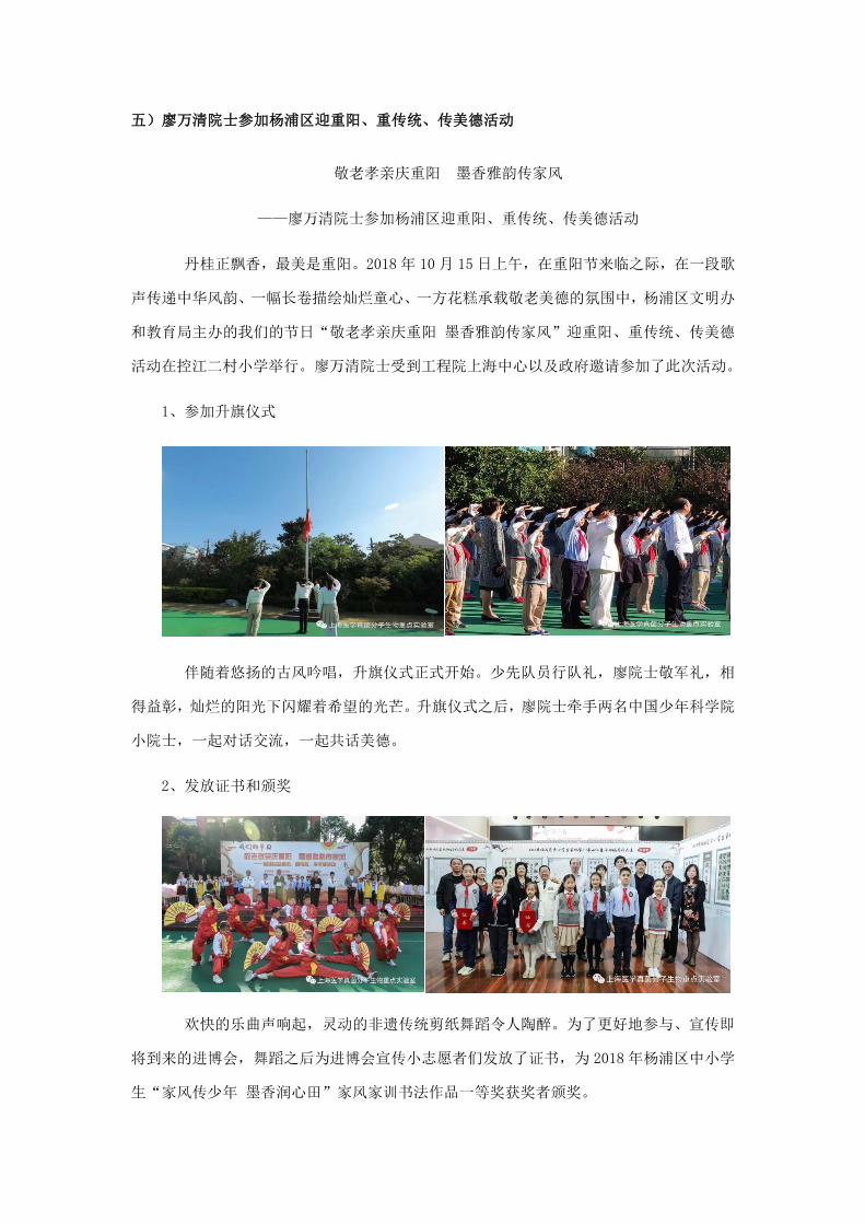
丹桂正飘香,最美是重阳。2018 年 10 月 15 日上午,在重阳节来临之际,在一段歌
声传递中华风韵、一幅长卷描绘灿烂童心、一方花糕承载敬老美德的氛围中,杨浦区文明办
和教育局主办的我们的节日“敬老孝亲庆重阳 墨香雅韵传家风”迎重阳、重传统、传美德
活动在控江二村小学举行。廖万清院士受到工程院上海中心以及政府邀请参加了此次活动。
1、参加升旗仪式
伴随着悠扬的古风吟唱,升旗仪式正式开始。少先队员行队礼,廖院士敬军礼,相
得益彰,灿烂的阳光下闪耀着希望的光芒。升旗仪式之后,廖院士牵手两名中国少年科学院
小院士,一起对话交流,一起共话美德。
2、发放证书和颁奖
欢快的乐曲声响起,灵动的非遗传统剪纸舞蹈令人陶醉。为了更好地参与、宣传即
将到来的进博会,舞蹈之后为进博会宣传小志愿者们发放了证书,为 2018 年杨浦区中小学
生“家风传少年 墨香润心田”家风家训书法作品一等奖获奖者颁奖。

3、参观书法作品展
在小院士们的引导下,廖院士还参
观了杨浦区中小学生“家风传少年 墨香润
心田”家风家训书法作品展,现场还有学
生与书法家泼墨挥毫,分享从“守礼养德”
到“家国天下”凝聚智慧的家风祖训,廖
院士亲切的与大家分享自己的心得和体会。
4、大小院士面对面
廖院士与小院士们面对面零距离交流,引导他们更好地熟悉家风家训,激励他们感恩于
心,回报于行,将严谨的求学问道精神铭记于心。
古人云:年高喜赏登高节,秋老还添不老春。此次活动,通过升旗仪式与书法展示相结
合的方式,通过廖院士和小院士对话的方式,通过非遗展示和家长学生互动的方式,多方融
合,将中华传统节日赋予新的时代内涵,焕发新的文化活力,进一步弘扬了传统美德,极好
地推动杨浦形成孝亲敬老的良好风尚。

六)梅县区委书记钟光灵来访
广东梅县区委书记钟光灵在上海招商引资期间,专程前往长征医院拜访中国工程院院
士廖万清,向他介绍了家乡梅县经济社会发展情况,并致以诚挚的问候和美好的祝福。
钟光灵来到廖万清院士位于上海长征医院的办公室,与他促膝交谈,关切询问他的身体
和生活情况,并详细介绍家乡梅县近年来在经济、社会、医疗、教育等方面取得的成果,虚
心听取他对梅县加快振兴发展的意见建议。钟光灵说,近年来,梅县坚持以习近平新时代中
国特色社会主义思想为指导,持续改善民生
福祉。特别是在廖院士的直接关心支持下,
粤东医院顺利创建三甲医院,该院的皮肤科
也成为品牌科室。他诚挚希望廖万清院士更
多地关心支持家乡建设,发挥在科研方面的
领先技术优势,为梅县培养和引进更多优秀
的医技人才。
廖万清对家乡当前加快发展的态势感到由衷高兴,充分肯定了家乡梅县在经济社会特别
是医疗、教育、新农村建设等方面取得的成就。他表示,将尽力为梅县经济社会和医疗卫生
事业发展贡献自己的一份力量。
会后,钟光灵想院士献上“松口画卷”。有千年
历史的松口古镇,是明末以后客家人出南洋的第一
站,也是孙中山发动辛亥革命的策源地之一,更被
评为广东十大海上丝绸之路文化地理坐标
区人大常委会主任曾庆辉,区委常委、副区长
潘雪松等一同拜访。
七、中国真菌学杂志
中国真菌学杂志贯彻党和国家的卫生工作方针政策,主要报道我国真菌学特别是医
学真菌学的最新研究进展,内容涉及基础医学与临床医学中的大部分专业,以从事皮
肤、感染、血液、呼吸、器官移植、肿瘤、急救、创伤、检验等与真菌感染专业有关的
中高级医务人员及从事微生物学、分子生物学及药学等基础研究的研究人员为主要读者
群,是真菌学工作者之间交流的窗口和平台。
2018年《中国真菌学杂志》一共刊发 6期,录用稿件 107篇。

附件: 2018 年实验室部分 SCI 文章原文
Medical Mycology, 2018, 56, 695–702doi: 10.1093/mmy/myx108
Advance Access Publication Date: 8 December 2017Original Article
Original Article
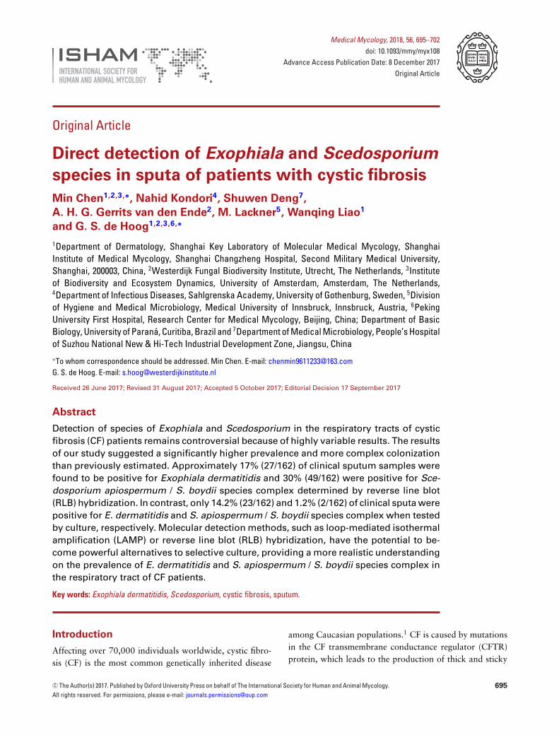
Direct detection of Exophiala and Scedosporium
species in sputa of patients with cystic fibrosis
Min Chen1,2,3,∗, Nahid Kondori4, Shuwen Deng7,
A. H. G. Gerrits van den Ende2, M. Lackner5, Wanqing Liao1
and G. S. de Hoog1,2,3,6,∗
1Department of Dermatology, Shanghai Key Laboratory of Molecular Medical Mycology, ShanghaiInstitute of Medical Mycology, Shanghai Changzheng Hospital, Second Military Medical University,Shanghai, 200003, China, 2Westerdijk Fungal Biodiversity Institute, Utrecht, The Netherlands, 3Instituteof Biodiversity and Ecosystem Dynamics, University of Amsterdam, Amsterdam, The Netherlands,4Department of Infectious Diseases, Sahlgrenska Academy, University of Gothenburg, Sweden, 5Divisionof Hygiene and Medical Microbiology, Medical University of Innsbruck, Innsbruck, Austria, 6PekingUniversity First Hospital, Research Center for Medical Mycology, Beijing, China; Department of BasicBiology, University of Parana, Curitiba, Brazil and 7Department of Medical Microbiology, People’s Hospitalof Suzhou National New & Hi-Tech Industrial Development Zone, Jiangsu, China∗To whom correspondence should be addressed. Min Chen. E-mail: [email protected]. S. de Hoog. E-mail: [email protected]
Received 26 June 2017; Revised 31 August 2017; Accepted 5 October 2017; Editorial Decision 17 September 2017
Abstract
Detection of species of Exophiala and Scedosporium in the respiratory tracts of cysticfibrosis (CF) patients remains controversial because of highly variable results. The resultsof our study suggested a significantly higher prevalence and more complex colonizationthan previously estimated. Approximately 17% (27/162) of clinical sputum samples werefound to be positive for Exophiala dermatitidis and 30% (49/162) were positive for Sce-dosporium apiospermum / S. boydii species complex determined by reverse line blot(RLB) hybridization. In contrast, only 14.2% (23/162) and 1.2% (2/162) of clinical sputa werepositive for E. dermatitidis and S. apiospermum / S. boydii species complex when testedby culture, respectively. Molecular detection methods, such as loop-mediated isothermalamplification (LAMP) or reverse line blot (RLB) hybridization, have the potential to be-come powerful alternatives to selective culture, providing a more realistic understandingon the prevalence of E. dermatitidis and S. apiospermum / S. boydii species complex inthe respiratory tract of CF patients.
Key words: Exophiala dermatitidis, Scedosporium, cystic fibrosis, sputum.
Introduction
Affecting over 70,000 individuals worldwide, cystic fibro-sis (CF) is the most common genetically inherited disease
among Caucasian populations.1 CF is caused by mutationsin the CF transmembrane conductance regulator (CFTR)protein, which leads to the production of thick and sticky
C© The Author(s) 2017. Published by Oxford University Press on behalf of The International Society for Human and Animal Mycology.All rights reserved. For permissions, please e-mail: [email protected]
695
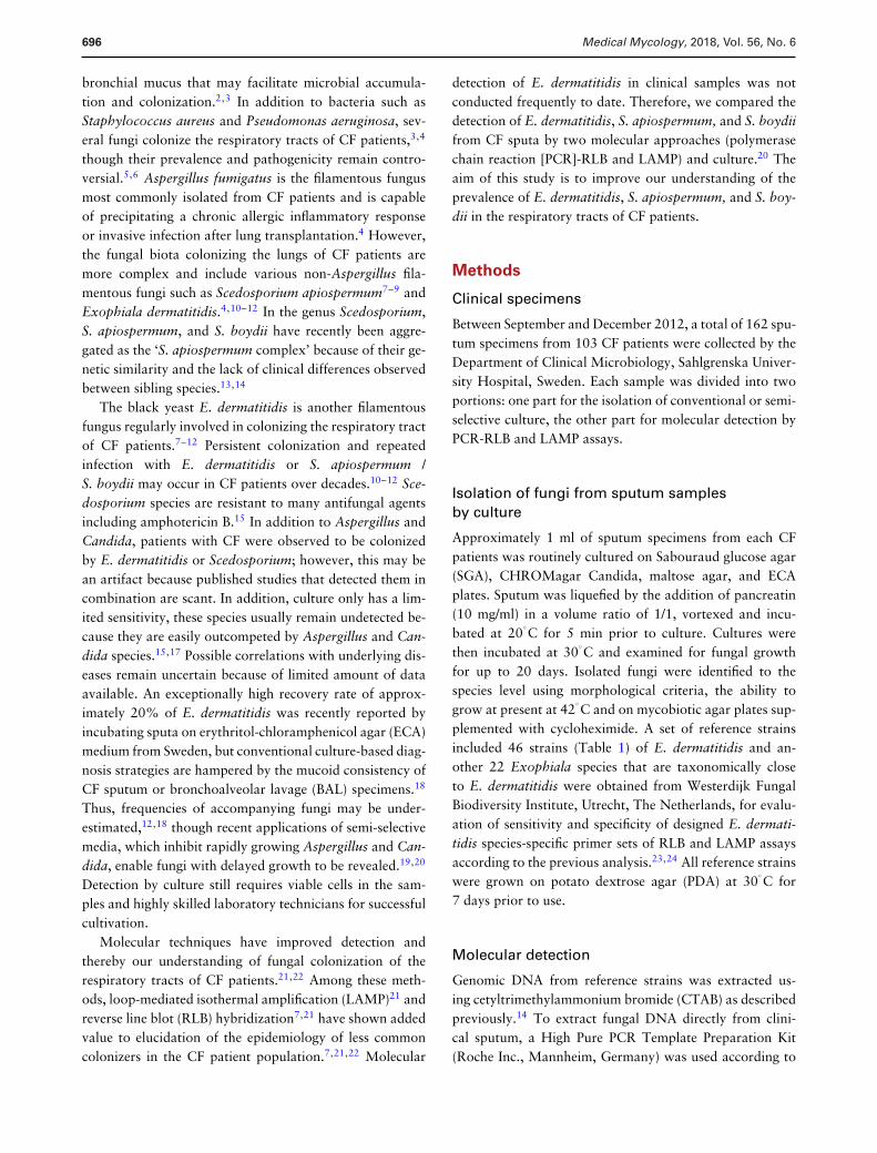
696 Medical Mycology, 2018, Vol. 56, No. 6
bronchial mucus that may facilitate microbial accumula-tion and colonization.2,3 In addition to bacteria such asStaphylococcus aureus and Pseudomonas aeruginosa, sev-eral fungi colonize the respiratory tracts of CF patients,3,4
though their prevalence and pathogenicity remain contro-versial.5,6 Aspergillus fumigatus is the filamentous fungusmost commonly isolated from CF patients and is capableof precipitating a chronic allergic inflammatory responseor invasive infection after lung transplantation.4 However,the fungal biota colonizing the lungs of CF patients aremore complex and include various non-Aspergillus fila-mentous fungi such as Scedosporium apiospermum7–9 andExophiala dermatitidis.4,10–12 In the genus Scedosporium,S. apiospermum, and S. boydii have recently been aggre-gated as the ‘S. apiospermum complex’ because of their ge-netic similarity and the lack of clinical differences observedbetween sibling species.13,14
The black yeast E. dermatitidis is another filamentousfungus regularly involved in colonizing the respiratory tractof CF patients.7–12 Persistent colonization and repeatedinfection with E. dermatitidis or S. apiospermum /S. boydii may occur in CF patients over decades.10–12 Sce-dosporium species are resistant to many antifungal agentsincluding amphotericin B.15 In addition to Aspergillus andCandida, patients with CF were observed to be colonizedby E. dermatitidis or Scedosporium; however, this may bean artifact because published studies that detected them incombination are scant. In addition, culture only has a lim-ited sensitivity, these species usually remain undetected be-cause they are easily outcompeted by Aspergillus and Can-dida species.15,17 Possible correlations with underlying dis-eases remain uncertain because of limited amount of dataavailable. An exceptionally high recovery rate of approx-imately 20% of E. dermatitidis was recently reported byincubating sputa on erythritol-chloramphenicol agar (ECA)medium from Sweden, but conventional culture-based diag-nosis strategies are hampered by the mucoid consistency ofCF sputum or bronchoalveolar lavage (BAL) specimens.18
Thus, frequencies of accompanying fungi may be under-estimated,12,18 though recent applications of semi-selectivemedia, which inhibit rapidly growing Aspergillus and Can-dida, enable fungi with delayed growth to be revealed.19,20
Detection by culture still requires viable cells in the sam-ples and highly skilled laboratory technicians for successfulcultivation.
Molecular techniques have improved detection andthereby our understanding of fungal colonization of therespiratory tracts of CF patients.21,22 Among these meth-ods, loop-mediated isothermal amplification (LAMP)21 andreverse line blot (RLB) hybridization7,21 have shown addedvalue to elucidation of the epidemiology of less commoncolonizers in the CF patient population.7,21,22 Molecular
detection of E. dermatitidis in clinical samples was notconducted frequently to date. Therefore, we compared thedetection of E. dermatitidis, S. apiospermum, and S. boydiifrom CF sputa by two molecular approaches (polymerasechain reaction [PCR]-RLB and LAMP) and culture.20 Theaim of this study is to improve our understanding of theprevalence of E. dermatitidis, S. apiospermum, and S. boy-dii in the respiratory tracts of CF patients.
Methods
Clinical specimens
Between September and December 2012, a total of 162 spu-tum specimens from 103 CF patients were collected by theDepartment of Clinical Microbiology, Sahlgrenska Univer-sity Hospital, Sweden. Each sample was divided into twoportions: one part for the isolation of conventional or semi-selective culture, the other part for molecular detection byPCR-RLB and LAMP assays.
Isolation of fungi from sputum samplesby culture
Approximately 1 ml of sputum specimens from each CFpatients was routinely cultured on Sabouraud glucose agar(SGA), CHROMagar Candida, maltose agar, and ECAplates. Sputum was liquefied by the addition of pancreatin(10 mg/ml) in a volume ratio of 1/1, vortexed and incu-bated at 20
◦C for 5 min prior to culture. Cultures were
then incubated at 30◦C and examined for fungal growth
for up to 20 days. Isolated fungi were identified to thespecies level using morphological criteria, the ability togrow at present at 42
◦C and on mycobiotic agar plates sup-
plemented with cycloheximide. A set of reference strainsincluded 46 strains (Table 1) of E. dermatitidis and an-other 22 Exophiala species that are taxonomically closeto E. dermatitidis were obtained from Westerdijk FungalBiodiversity Institute, Utrecht, The Netherlands, for evalu-ation of sensitivity and specificity of designed E. dermati-tidis species-specific primer sets of RLB and LAMP assaysaccording to the previous analysis.23,24 All reference strainswere grown on potato dextrose agar (PDA) at 30
◦C for
7 days prior to use.
Molecular detection
Genomic DNA from reference strains was extracted us-ing cetyltrimethylammonium bromide (CTAB) as describedpreviously.14 To extract fungal DNA directly from clini-cal sputum, a High Pure PCR Template Preparation Kit(Roche Inc., Mannheim, Germany) was used according to
Chen et al. 697
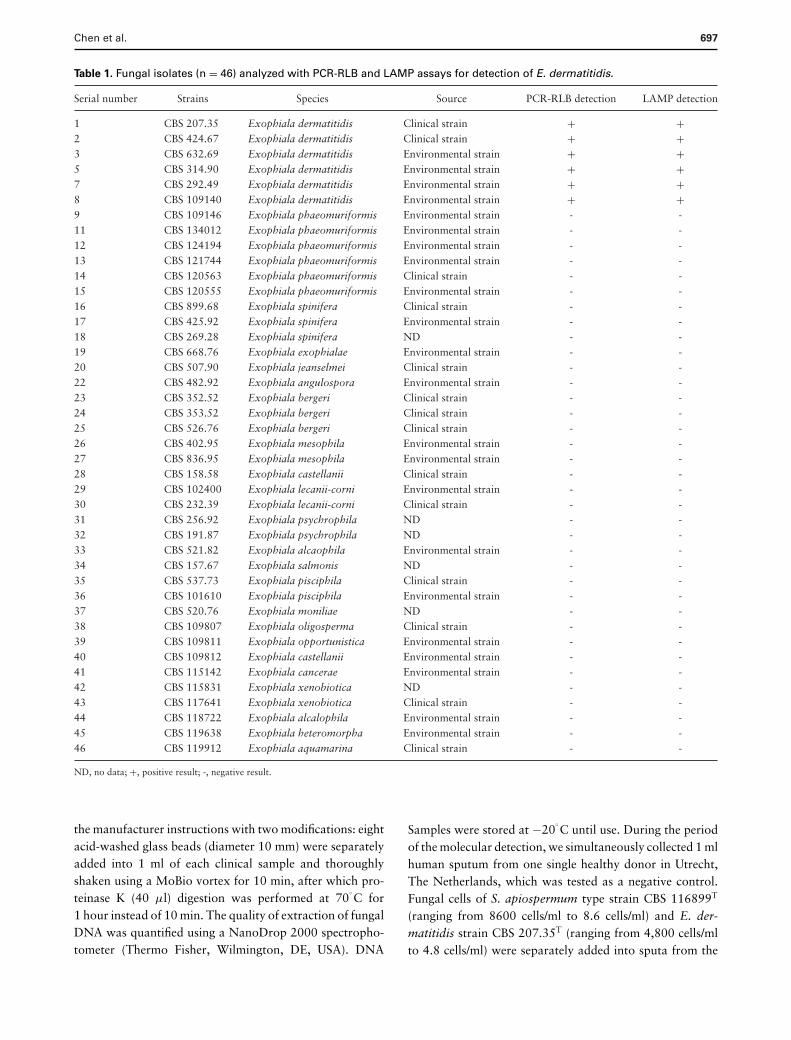
Table 1. Fungal isolates (n = 46) analyzed with PCR-RLB and LAMP assays for detection of E. dermatitidis.
Serial number Strains Species Source PCR-RLB detection LAMP detection
1 CBS 207.35 Exophiala dermatitidis Clinical strain + +2 CBS 424.67 Exophiala dermatitidis Clinical strain + +3 CBS 632.69 Exophiala dermatitidis Environmental strain + +5 CBS 314.90 Exophiala dermatitidis Environmental strain + +7 CBS 292.49 Exophiala dermatitidis Environmental strain + +8 CBS 109140 Exophiala dermatitidis Environmental strain + +9 CBS 109146 Exophiala phaeomuriformis Environmental strain - -11 CBS 134012 Exophiala phaeomuriformis Environmental strain - -12 CBS 124194 Exophiala phaeomuriformis Environmental strain - -13 CBS 121744 Exophiala phaeomuriformis Environmental strain - -14 CBS 120563 Exophiala phaeomuriformis Clinical strain - -15 CBS 120555 Exophiala phaeomuriformis Environmental strain - -16 CBS 899.68 Exophiala spinifera Clinical strain - -17 CBS 425.92 Exophiala spinifera Environmental strain - -18 CBS 269.28 Exophiala spinifera ND - -19 CBS 668.76 Exophiala exophialae Environmental strain - -20 CBS 507.90 Exophiala jeanselmei Clinical strain - -22 CBS 482.92 Exophiala angulospora Environmental strain - -23 CBS 352.52 Exophiala bergeri Clinical strain - -24 CBS 353.52 Exophiala bergeri Clinical strain - -25 CBS 526.76 Exophiala bergeri Clinical strain - -26 CBS 402.95 Exophiala mesophila Environmental strain - -27 CBS 836.95 Exophiala mesophila Environmental strain - -28 CBS 158.58 Exophiala castellanii Clinical strain - -29 CBS 102400 Exophiala lecanii-corni Environmental strain - -30 CBS 232.39 Exophiala lecanii-corni Clinical strain - -31 CBS 256.92 Exophiala psychrophila ND - -32 CBS 191.87 Exophiala psychrophila ND - -33 CBS 521.82 Exophiala alcaophila Environmental strain - -34 CBS 157.67 Exophiala salmonis ND - -35 CBS 537.73 Exophiala pisciphila Clinical strain - -36 CBS 101610 Exophiala pisciphila Environmental strain - -37 CBS 520.76 Exophiala moniliae ND - -38 CBS 109807 Exophiala oligosperma Clinical strain - -39 CBS 109811 Exophiala opportunistica Environmental strain - -40 CBS 109812 Exophiala castellanii Environmental strain - -41 CBS 115142 Exophiala cancerae Environmental strain - -42 CBS 115831 Exophiala xenobiotica ND - -43 CBS 117641 Exophiala xenobiotica Clinical strain - -44 CBS 118722 Exophiala alcalophila Environmental strain - -45 CBS 119638 Exophiala heteromorpha Environmental strain - -46 CBS 119912 Exophiala aquamarina Clinical strain - -
ND, no data; +, positive result; -, negative result.
the manufacturer instructions with two modifications: eightacid-washed glass beads (diameter 10 mm) were separatelyadded into 1 ml of each clinical sample and thoroughlyshaken using a MoBio vortex for 10 min, after which pro-teinase K (40 μl) digestion was performed at 70
◦C for
1 hour instead of 10 min. The quality of extraction of fungalDNA was quantified using a NanoDrop 2000 spectropho-tometer (Thermo Fisher, Wilmington, DE, USA). DNA
Samples were stored at −20◦C until use. During the period
of the molecular detection, we simultaneously collected 1 mlhuman sputum from one single healthy donor in Utrecht,The Netherlands, which was tested as a negative control.Fungal cells of S. apiospermum type strain CBS 116899T
(ranging from 8600 cells/ml to 8.6 cells/ml) and E. der-matitidis strain CBS 207.35T (ranging from 4,800 cells/mlto 4.8 cells/ml) were separately added into sputa from the
698 Medical Mycology, 2018, Vol. 56, No. 6
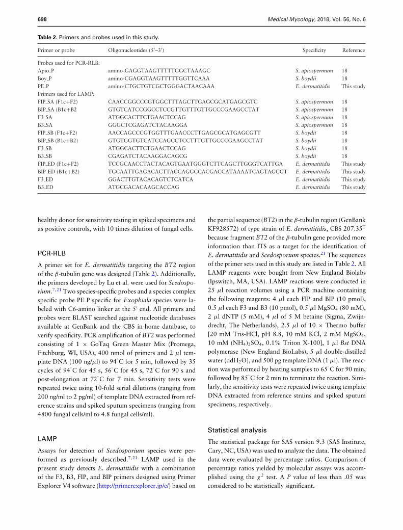
Table 2. Primers and probes used in this study.
Primer or probe Oligonucleotides (5′–3′) Specificity Reference
Probes used for PCR-RLB:Apio P amino-GAGGTAAGTTTTTGGCTAAAGC S. apiospermum 18Boy P amino-CGAGGTAAGTTTTTGGTTCAAA S. boydii 18PE P amino-CTGCTGTCGCTGGGACTAACAAA E. dermatitidis This studyPrimers used for LAMP:FIP SA (F1c+F2) CAACCGGCCCGTGGCTTTAGCTTGAGCGCATGAGCGTC S. apiospermum 18BIP SA (B1c+B2 GTGTCATCCGGCCTCCGTTGTTTGTTGCCCGAAGCCTAT S. apiospermum 18F3 SA ATGGCACTTCTGAACTCCAG S. apiospermum 18B3 SA GGGCTCGAGATCTACAAGGA S. apiospermum 18FIP SB (F1c+F2) AACCAGCCCGTGGTTTGAACCCTTGAGCGCATGAGCGTT S. boydii 18BIP SB (B1c+B2) GTGTGGTGTCATCCAGCCTCCTTTGTTGCCCGAAGCCTAT S. boydii 18F3 SB ATGGCACTTCTGAACTCCAG S. boydii 18B3 SB CGAGATCTACAAGGACAGCG S. boydii 18FIP ED (F1c+F2) TCCGCAACCTACTACAGTGAATGGGTCTTCAGCTTGGGTCATTGA E. dermatitidis This studyBIP ED (B1c+B2) TGCAATTGAGACACTTACCAGGCCACGACCATAAAATCAGTAGCGT E. dermatitidis This studyF3 ED GGACTTGTACACAGTCTCATCA E. dermatitidis This studyB3 ED ATGCGACACAAGCACCAG E. dermatitidis This study
healthy donor for sensitivity testing in spiked specimens andas positive controls, with 10 times dilution of fungal cells.
PCR-RLB
A primer set for E. dermatitidis targeting the BT2 regionof the β-tubulin gene was designed (Table 2). Additionally,the primers developed by Lu et al. were used for Scedospo-rium.7,21 Two species-specific probes and a species complexspecific probe PE P specific for Exophiala species were la-beled with C6-amino linker at the 5′ end. All primers andprobes were BLAST searched against nucleotide databasesavailable at GenBank and the CBS in-home database, toverify specificity. PCR amplification of BT2 was performedconsisting of 1 × GoTaq Green Master Mix (Promega,Fitchburg, WI, USA), 400 nmol of primers and 2 μl tem-plate DNA (100 ng/μl) to 94
◦C for 5 min, followed by 35
cycles of 94◦C for 45 s, 56
◦C for 45 s, 72
◦C for 90 s and
post-elongation at 72◦C for 7 min. Sensitivity tests were
repeated twice using 10-fold serial dilutions (ranging from200 ng/ml to 2 pg/ml) of template DNA extracted from ref-erence strains and spiked sputum specimens (ranging from4800 fungal cells/ml to 4.8 fungal cells/ml).
LAMP
Assays for detection of Scedosporium species were per-formed as previously described.7,21 LAMP used in thepresent study detects E. dermatitidis with a combinationof the F3, B3, FIP, and BIP primers designed using PrimerExplorer V4 software (http://primerexplorer.jp/e/) based on
the partial sequence (BT2) in the β-tubulin region (GenBankKF928572) of type strain of E. dermatitidis, CBS 207.35T
because fragment BT2 of the β-tubulin gene provided moreinformation than ITS as a target for the identification ofE. dermatitidis and Scedosporium species.21 The sequencesof the primer sets used in this study are listed in Table 2. AllLAMP reagents were bought from New England Biolabs(Ipswitch, MA, USA). LAMP reactions were conducted in25 μl reaction volumes using a PCR machine containingthe following reagents: 4 μl each FIP and BIP (10 pmol),0.5 μl each F3 and B3 (10 pmol), 0.5 μl MgSO4 (80 mM),2 μl dNTP (5 mM), 4 μl of 5 M betaine (Sigma, Zwijn-drecht, The Netherlands), 2.5 μl of 10 × Thermo buffer[20 mM Tris-HCl, pH 8.8, 10 mM KCl, 2 mM MgSO4,10 mM (NH4)2SO4, 0.1% Triton X-100], 1 μl Bst DNApolymerase (New England BioLabs), 5 μl double-distilledwater (ddH2O), and 500 pg template DNA (1 μl). The reac-tion was performed by heating samples to 65
◦C for 90 min,
followed by 85◦C for 2 min to terminate the reaction. Simi-
larly, the sensitivity tests were repeated twice using templateDNA extracted from reference strains and spiked sputumspecimens, respectively.
Statistical analysis
The statistical package for SAS version 9.3 (SAS Institute,Cary, NC, USA) was used to analyze the data. The obtaineddata were evaluated by percentage ratios. Comparison ofpercentage ratios yielded by molecular assays was accom-plished using the χ2 test. A P value of less than .05 wasconsidered to be statistically significant.
Chen et al. 699
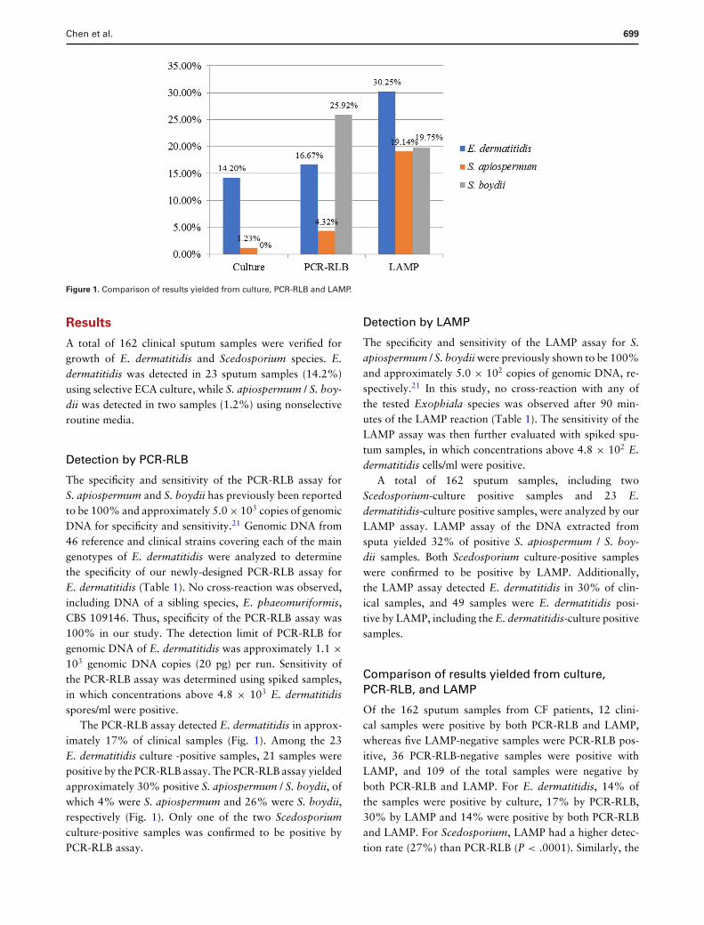
Figure 1. Comparison of results yielded from culture, PCR-RLB and LAMP.
Results
A total of 162 clinical sputum samples were verified forgrowth of E. dermatitidis and Scedosporium species. E.dermatitidis was detected in 23 sputum samples (14.2%)using selective ECA culture, while S. apiospermum / S. boy-dii was detected in two samples (1.2%) using nonselectiveroutine media.
Detection by PCR-RLB
The specificity and sensitivity of the PCR-RLB assay forS. apiospermum and S. boydii has previously been reportedto be 100% and approximately 5.0 × 10³ copies of genomicDNA for specificity and sensitivity.21 Genomic DNA from46 reference and clinical strains covering each of the maingenotypes of E. dermatitidis were analyzed to determinethe specificity of our newly-designed PCR-RLB assay forE. dermatitidis (Table 1). No cross-reaction was observed,including DNA of a sibling species, E. phaeomuriformis,CBS 109146. Thus, specificity of the PCR-RLB assay was100% in our study. The detection limit of PCR-RLB forgenomic DNA of E. dermatitidis was approximately 1.1 ×10³ genomic DNA copies (20 pg) per run. Sensitivity ofthe PCR-RLB assay was determined using spiked samples,in which concentrations above 4.8 × 10³ E. dermatitidisspores/ml were positive.
The PCR-RLB assay detected E. dermatitidis in approx-imately 17% of clinical samples (Fig. 1). Among the 23E. dermatitidis culture -positive samples, 21 samples werepositive by the PCR-RLB assay. The PCR-RLB assay yieldedapproximately 30% positive S. apiospermum / S. boydii, ofwhich 4% were S. apiospermum and 26% were S. boydii,respectively (Fig. 1). Only one of the two Scedosporiumculture-positive samples was confirmed to be positive byPCR-RLB assay.
Detection by LAMP
The specificity and sensitivity of the LAMP assay for S.apiospermum / S. boydii were previously shown to be 100%and approximately 5.0 × 10² copies of genomic DNA, re-spectively.21 In this study, no cross-reaction with any ofthe tested Exophiala species was observed after 90 min-utes of the LAMP reaction (Table 1). The sensitivity of theLAMP assay was then further evaluated with spiked spu-tum samples, in which concentrations above 4.8 × 10² E.dermatitidis cells/ml were positive.
A total of 162 sputum samples, including twoScedosporium-culture positive samples and 23 E.dermatitidis-culture positive samples, were analyzed by ourLAMP assay. LAMP assay of the DNA extracted fromsputa yielded 32% of positive S. apiospermum / S. boy-dii samples. Both Scedosporium culture-positive sampleswere confirmed to be positive by LAMP. Additionally,the LAMP assay detected E. dermatitidis in 30% of clin-ical samples, and 49 samples were E. dermatitidis posi-tive by LAMP, including the E. dermatitidis-culture positivesamples.
Comparison of results yielded from culture,PCR-RLB, and LAMP
Of the 162 sputum samples from CF patients, 12 clini-cal samples were positive by both PCR-RLB and LAMP,whereas five LAMP-negative samples were PCR-RLB pos-itive, 36 PCR-RLB-negative samples were positive withLAMP, and 109 of the total samples were negative byboth PCR-RLB and LAMP. For E. dermatitidis, 14% ofthe samples were positive by culture, 17% by PCR-RLB,30% by LAMP and 14% were positive by both PCR-RLBand LAMP. For Scedosporium, LAMP had a higher detec-tion rate (27%) than PCR-RLB (P < .0001). Similarly, the
700 Medical Mycology, 2018, Vol. 56, No. 6
LAMP assay also provided a higher detection rate (30%)for E. dermatitidis (P = .0039).
Discussion
Previous studies revealed a highly variable prevalence of Ex-ophiala and Scedosporium species in the respiratory tractsof CF patients (Table 3).8,12,16,17,25–31 Lack of standard-ization of the procedures for detection on filamentous fungiin sputum samples from CF patients may be a possiblecause for the variable reported data.31 Moreover, routineprocessing procedures for isolating filamentous fungi fromrespiratory sputum samples may underestimate fungalprevalence.18 Notably, LAMP technology can rapidly andaccurately amplify genomic DNA in an isothermal stepfrom partially processed and/or nonprocessed samples inapproximately 1 hour without the need for sophisticatedequipment and the products can be assessed by the nakedeye.32 The PCR-RLB assay has been used to detect Sce-dosporium species in the respiratory samples of CF pa-tients.7 Thus, E. dermatitidis-specific and Scedosporium-specific molecular assays, PCR-RLB and LAMP, and cultureassays were used to detect the prevalence of E. dermatitidisand Scedosporium species in 162 clinical sputa in a doubleblind experiment.
We noticed that only two samples were positive forS. apiospermum / S. boydii and 23 samples were positivefor E. dermatitidis upon culture assay in our study. Thismay due to cultivation of Exophiala by selective medium,and Scedosporium culture was nonselective. Some filamen-tous fungi such as A. fumigatus can overlap other fungisuch as Scedosporium species on non-selective medium be-cause they grow faster than other non-Aspergillus fungithat colonize the respiratory tract of CF patients.4 Gener-ally, our results are similar to the recovery rates reportedusing comparable media for Scedosporium species and E.dermatitidis.10–12,33,34 In contrast, positive recovery ratesfor Scedosporium tend to increase at approximately 15%when the sputum samples processed by benomyl-basedmedia.33
In addition to A. fumigatus, E. dermatitidis and speciesof the S. apiospermum complex are the most frequent moldsrecovered from respiratory secretions of CF patients, buttheir recovery rates vary greatly.18,34,35 With its abilityto grow at 37
◦C, E. dermatitidis has a worldwide distri-
bution in hot environments, not only in CF respiratorytracts10,26,36,37 but also warm indoor environments suchas steambaths38 and dishwashers.39 Although the signifi-cance of E. dermatitidis in causing disease in patients withCF remains unclear, this fungus is known to cause severeinvasive fungal infections.40 With selective ECA medium,the prevalence of E. dermatitidis ranges from 5% in
Germany to 19% in Sweden.11,12,18 Published recoveryrates in Sweden were exceptionally high, around 17–19%by selective culture.12,37 Our data, which were also fromSweden, showed a high positive rate of 14%, confirmingthat this detection rate is rather consistent. PCR-RLB andLAMP assays yielded 15 and 30% of E. dermatitis detec-tion, respectively, which was significantly higher than cul-ture. The prevalence in Sweden is 2 to almost 20 timeshigher than in European countries, which is possibly be-cause of differences in indoor conditions. For a better under-standing of E. dermatitidis prevalence among CF patients,further multi-center epidemiological studies are needed.
Regarding the positive rate for Scedosporium species,the results of our molecular assays showed that approxi-mately 10.5% of the CF airway samples were positive forthe S. apiospermum complex using the PCR-RLB assay, andthe positive rate yielded by the LAMP assay was approx-imately 31.5%. This positive rate for S. apiospermum / S.boydii was lower than the maximum reported rate of 61.5%that was obtained using PCR-RLB.7 The high detection rateyielded by LAMP is likely closest to actual value, and LAMPtechnology would be the most efficient for amplification oftarget DNA from partially processed and/or non-processedCF specimens.32 The selective ECA medium yielded a com-paratively high positive recovery rate of E. dermatitidis,and this figure might be tripled to approach the real preva-lence generated by molecular methods. However, LAMPmay have a higher risk of false positive results than PCR-RLB, which might be caused by pre-PCR contamination ofsamples, as the method is highly sensitive. Careful precau-tions to prevent cross-contamination need to be taken dur-ing sample collection, and the methods should be executedusing separated rooms and filtered tips. In general, analysisof larger numbers of samples is required to determine theirprevalence in clinical samples, and the method should berepeated several times to enable for reliable determinationof the presence in a single patient. The recent application ofnext-generation sequencing (NGS) technology has allowedus to conduct meta-genomic studies,41 which may providemore comprehensive insight into chronic fungi colonizationin airways of CF patients. Finally, we noticed two sampleswith positive culture results for E. dermatitidis were nega-tive upon both PCR-RLB and LAMP assays. The quality ofthe fungi DNA extracted from respiratory sputum samplesis likely poor, and sputa from CF patients might containPCR inhibiting substances, both of which could influencethe performance of the molecular assays.
In summary, current data on the prevalence of Exophialaand Scedosporium in the respiratory tract of CF patientsremains variable and controversial. The results of our com-parative detection study suggested a significantly higherprevalence and more complex colonization than previously

Chen et al. 701
Table 3. Overview of E. dermatitidis and Scedosporium detection in sputum of CF patients.
Time Geography Samples (n) E. dermatitidis Scedosporium species Reference
1991 Germany 121 SGA + CDA: 9% NA 251998 Australia 52 NA IGS-PCR: 10% (S. apiospermum complex) 262000 France 128 NA Culture using filtrate antigens:8.6%
(S. apiospermum complex)16
2003 Germany 94 SGA + ECA: 1% NA 272004 Germany 81 ECA: 6.2% NA 322009 Germany 42 NA SceSel: 14% (S. apiospermum complex) 282009 Australia, Germany,
France, Spain, USA162 NA SGA: 14% (S. prolificans) 17
2010 Belgium 154 ECA: 5.8% NA 112011 Sweden 97 ECA: 19% NA 122011 France 59 NA SceSel + PCR-RLB: 61.5% (S. apiospermum
complex)7
2013 France 50 NA SGA + YPGA: 24% (S. apiospermum complex) 82015 Germany 2346 NA SceSel: 3.1% (S. apiospermum complex) 202016 Germany 3186 ECA: 2.8% SceSel: 2.4% (S. apiospermum complex) 302017 Sweden 162 SGA, ECA, LAMP,
RLB: 17–30%SGA, LAMP, RLB: 30–39% (S. apiospermumcomplex)
This study
Abbreviations used: CDA, Czapeck Dox Agar; ECA, Erythritol Chloramphenicol Agar; NA, not applied; SGA, Sabouraud’s Glucose Agar; SceSel, ScedosporiumSelect Agar; YPGA, Yeast Peptone Glucose Agar.
estimated. Molecular detection methods such as the LAMPor NGS technology have the potential to become power-ful tools that can be used in place of selective culture forto provide a more realistic description of the prevalence ofExophiala and Scedosporium in the respiratory tracts of CFpatients, upon the improvement of the protocols for the ex-traction of filamentous fungi DNA from clinical specimens.
Acknowledgments
This study was funded in part with the grants from Ma-jor National R&D Projects of the National Health Department(2014ZX09J14106-02A), and the National Natural Science Foun-dation of China (81201269).
Declaration of interest
The authors report no conflicts of interest. The authors alone areresponsible for the content and the writing of the paper.
Supplementary material
Supplementary data are available at MMYCOL online.
References
1. Bellis G, Cazes MH, Parant A et al. Cystic fibrosis mortality trends inFrance. J Cyst Fibros. 2007; 6: 179–186.
2. Girodon E, Cazeneuve C, Lebargy F et al. CFTR gene mutations in adultswith disseminated bronchiectasis. Eur J Hum Genet. 1997; 5: 149–155.
3. O’Sullivan BP, Freedman SD. Cystic fibrosis. Lancet. 2009; 373: 1891–1904.
4. Lipuma JJ. The changing microbial epidemiology in cystic fibrosis. ClinMicrobiol Rev. 2010; 23: 299–323.
5. de Vrankrijker AM, Wolfs TF, van der Ent CK. Challenging and emergingpathogens in cystic fibrosis. Paediatr Respir Rev. 2010; 11: 246–254.
6. Chotirmall SH, McElvaney NG. Fungi in the cystic fibrosis lung: by-standers or pathogens? Int J Biochem Cell Biol. 2014; 52: 161–173.
7. Lu Q, van den Ende AH, de Hoog GS et al. Reverse line blot hybridisationscreening of Pseudallescheria/Scedosporium species in patients with cysticfibrosis. Mycoses. 2011; 54: 5–11.
8. Zouhair R, Rougeron A, Razafimandimby B, Kobi A, Bouchara JP, Gi-raud S. Distribution of the different species of the Pseudallescheria boy-dii/Scedosporium apiospermum complex in French patients with cysticfibrosis. Med Mycol. 2013; 51: 603–613.
9. Harun A, Blyth CC, Gilgado F, Middleton P, Chen SC, Meyer W. Devel-opment and validation of a multiplex PCR for detection of Scedosporiumspp. in respiratory tract specimens from patients with cystic fibrosis. J ClinMicrobiol. 2011; 49: 1508–1512.
10. Haase G, Skopnik H, Kusenbach G. Exophiala dermatitidis infection incystic fibrosis. Lancet. 1990; 336: 188–189.
11. Lebecque P, Leonard A, Huang D et al. Exophiala (Wangiella) dermatitidisand cystic fibrosis: prevalence and risk factors. Med Mycol. 2010; 48:S4–9.
12. Kondori N, Gilljam M, Lindblad A, Jonsson B, Moore ER, Wenneras C.High rate of Exophiala dermatitidis recovery in the airways of patientswith cystic fibrosis is associated with pancreatic insufficiency. J Clin Mi-crobiol. 2011; 49: 1004–1009.
13. Cortez KJ, Roilides E, Quiroz-Telles F et al. Infections caused by Sce-dosporium spp. Clin Microbiol Rev. 2008; 21: 157–197.
14. Chen M, Zeng J, De Hoog GS et al. The ‘species complex’ issue in clinicallyrelevant fungi: a case study in Scedosporium apiospermum. Fungal Biol.2016; 120: 137–146.
15. Guarro J, Kantarcioglu AS, Horre R et al. Scedosporium apiospermum:changing clinical spectrum of a therapy-refractory opportunist. Med My-col. 2006; 44: 295–327.
16. Cimon B, Carrere J, Vinatier JF, Chazalette JP, Chabasse D,Bouchara JP. Clinical significance of Scedosporium apiospermum inpatients with cystic fibrosis. Eur J Clin Microbiol Infect Dis. 2000;19 53–56.

702 Medical Mycology, 2018, Vol. 56, No. 6
17. Rodriguez-Tudela JL, Berenguer J, Guarro J et al. Epidemiology and out-come of Scedosporium prolificans infection, a review of 162 cases. MedMycol. 2009; 47: 359–370.
18. Pashley CH, Fairs A, Morley JP et al. Routine processing procedures forisolating filamentous fungi from respiratory sputum samples may under-estimate fungal prevalence. Med Mycol. 2012; 50: 433–438.
19. Pham T, Giraud S, Schuliar G, Rougeron A, Bouchara JP. Scedo-Select III:a new semi-selective culture medium for detection of the Scedosporiumapiospermum species complex. Med Mycol. 2015; 53: 512–519.
20. Sedlacek L, Graf B, Schwarz C et al. Prevalence of Scedosporium speciesand Lomentospora prolificans in patients with cystic fibrosis in a multi-center trial by use of a selective medium. J Cyst Fibros. 2015; 14 237–241.
21. Lu Q, Gerrits van den Ende AH, Bakkers JM et al. Identification of Pseu-dallescheria and Scedosporium species by three molecular methods. J ClinMicrobiol. 2011; 49: 960–967.
22. Lackner M, Klaassen CH, Meis JF, van den Ende AH, de Hoog GS.Molecular identification tools for sibling species of Scedosporium andPseudallescheria. Med Mycol. 2012; 50: 497–508.
23. Hoog GS, Guarro J, Gene J, Figueras MJ. Atlas of Clinical Fungi, CD-rom3rd ed. Utrecht / Reus: CBS-KNAW Fungal Biodiversity Centre/ Universi-tat Rovirai Virgili, 2009.
24. Najafzadeh MJ, Dolatabadi S, Saradeghi Keisari M, Naseri A, Feng P, deHoog GS. Detection and identification of opportunistic Exophiala speciesusing the rolling circle amplification of ribosomal internal transcribedspacers. J Microbiol Methods. 2013; 94: 338–342.
25. Haase G, Skopnik H, Groten T, Kusenbach G, Posselt HG. Long-termfungal cultures from sputum of patients with cystic fibrosis. Mycoses.1991; 34: 373–376.
26. Williamson EC, Speers D, Arthur IH, Harnett G, Ryan G, Inglis TJ. Molec-ular epidemiology of Scedosporium apiospermum infection determined byPCR amplification of ribosomal intergenic spacer sequences in patientswith chronic lung disease. J Clin Microbiol. 2001; 39: 47–50.
27. Bakare N, Rickerts V, Bargon J, Just-Nubling G. Prevalence of Aspergillusfumigatus and other fungal species in the sputum of adult patients withcystic fibrosis. Mycoses. 2003; 46: 19–23.
28. Horre R, Marklein G, Siekmeier R, Nidermajer S, Reiffert SM. Selec-tive isolation of Pseudallescheria and Scedosporium species from respi-ratory tract specimens of cystic fibrosis patients. Respiration. 2009; 77:320–324.
29. Lackner M, Rezusta A, Villuendas MC, Palacian MP, Meis JF, KlaassenCH. Infection and colonisation due to Scedosporium in Northern Spain:
an in vitro antifungal susceptibility and molecular epidemiology study of60 isolates. Mycoses. 2011; 54: 12–21.
30. Ziesing S, Suerbaum S, Sedlacek L. Fungal epidemiology and diversity incystic fibrosis patients over a 5-year period in a national reference center.Med Mycol. 2016; 54: 781–786.
31. Borman AM, Palmer MD, Delhaes L et al. Lack of standardization inthe procedures for mycological examination of sputum samples from CFpatients: a possible cause for variations in the prevalence of filamentousfungi. Med Mycol. 2010; 48: S88–97.
32. Kaneko H, Kawana T, Fukushima E, Suzutani T. Tolerance of loop-mediated isothermal amplification to a culture medium and biologicalsubstances. Journal of biochemical and biophysical methods. 2007; 70:499–501.
33. Horre R, Schaal KP, Siekmeier R, Sterzik B, de Hoog GS, Schnit-zler N. Isolation of fungi, especially Exophiala dermatitidis, in patientssuffering from cystic fibrosis: a prospective study. Respiration. 2004;71: 360–366.
34. Burns JL, Emerson J, Stapp JR et al. Microbiology of sputum from patientsat cystic fibrosis centers in the United States. Clin Infect Dis. 1998; 27:158–163.
35. Nagano Yuriko, Millar B Cherie et al. Fungal infections in patients withcystic fibrosis. Rev Med Microbiol. 2007; 18: 11–16.
36. Kusenbach G, Skopnik H, Haase G, Friedrichs F, Dohmen H. Exophialadermatitidis pneumonia in cystic fibrosis. Eur J Pediatr. 1992; 151: 344–346.
37. Kondori N, Lindblad A, Welinder-Olsson C, Wenneras C, Gilljam M.Development of IgG antibodies to Exophiala dermatitidis is associatedwith inflammatory responses in patients with cystic fibrosis. J Cyst Fibros.2014; 13: 391–399.
38. Sudhadham M, Prakitsin S, Sivichai S et al. The neurotropic black yeastExophiala dermatitidis has a possible origin in the tropical rain forest.Stud Mycol. 2008; 61: 145–155.
39. Zalar P, Novak M, de Hoog GS, Gunde-Cimerman N. Dishwashers–aman-made ecological niche accommodating human opportunistic fungalpathogens. Fungal Biol. 2011; 115: 997–1007.
40. Sood S, Vaid VK, Sharma M, Bhartiya H. Cerebral phaeohyphomy-cosis by Exophiala dermatitidis. Indian J Med Microbiol. 2014;32: 188–190.
41. Koboldt DC, Steinberg KM, Larson DE, Wilson RK, Mardis ER. Thenext-generation sequencing revolution and its impact on genomics. Cell.2013; 155: 27–38.
Epidemiology of fungal infections in China
Min Chen1,2,*, Yuan Xu1,2,*, Nan Hong1,2,*, Yali Yang1, Wenzhi Lei1, Lin Du1, Jingjun Zhao3, Xia Lei4, Lin Xiong5,
Langqi Cai6, Hui Xu7, Weihua Pan (✉)1,a, Wanqing Liao (✉)2,b
1Department of Dermatology, Changzheng Hospital, Second Military Medical University, Shanghai 200003, China; 2Shanghai KeyLaboratory of Molecular Medical Mycology, Changzheng Hospital, Second Military Medical University, Shanghai 200003, China;3Department of Dermatology, Tongji Hospital, Tongji University, Shanghai 200442, China; 4Department of Dermatology, Daping Hospital,Third Military Medical University, Chongqing 400042, China; 5Department of Dermatology, the People’s Hospital of Yan’an, Yan’an 716000,China; 6Department of Dermatology, the First Affiliated Hospital of Xiamen University, Xiamen 361003, China; 7Department of Dermatology,Shanghai Ninth People’s Hospital, Shanghai Jiao Tong University School of Medicine, Shanghai 200011, China
© Higher Education Press and Springer-Verlag GmbH Germany, part of Springer Nature 2018
Abstract With the increasing number of immunocompromised hosts, the epidemiological characteristics offungal infections have undergone enormous changes worldwide, including in China. In this paper, we reviewed theexisting data on mycosis across China to summarize available epidemiological profiles. We found that the generalincidence of superficial fungal infections in China has been stable, but the incidence of tinea capitis has decreasedand the transmission route has changed. By contrast, the overall incidence of invasive fungal infections hascontinued to rise. The occurrence of candidemia caused by Candida species other than C. albicans and includingsome uncommon Candida species has increased recently in China. Infections caused by Aspergillus have alsopropagated in recent years, particularly with the emergence of azole-resistant Aspergillus fumigatus. An increasingtrend of cryptococcosis has been noted in China, with Cryptococcus neoformans var. grubii ST 5 genotype isolatesas the predominant pathogen. Retrospective studies have suggested that the epidemiological characteristics ofPneumocystis pneumonia in China may be similar to those in other developing countries. Endemic fungalinfections, such as sporotrichosis in Northeastern China, must arouse research, diagnostic, and treatmentvigilance. Currently, the epidemiological data on mycosis in China are variable and fragmentary. Thus, anationwide epidemiological research on fungal infections in China is an important need for improving thecountry’s health.
Keywords fungi; infection; epidemiology; China
Introduction
Fungi are eukaryotic organisms found throughout nature.They emerged about 1.6 million years ago and serve theimportant function of returning the nutrients removed byplants to the soil [1]. Historically, fungi are majorpathogens of plants, rotifers, insects, and amphibians,with a relative few causing infections in humans. However,human modification of natural environments has intensi-fied the dispersal of fungal infections [2]. Global warming
has also augmented the prevalence of fungal infections inmammals by its selection of adaptive thermo-tolerantfungal species possessing significant pathogenic potentialdespite their current nonpathogenicity due to beingrestricted by mammalian temperatures [3]. To date,approximately 400 fungal species have been reported tobe pathogenic to humans, with emerging pathogenic fungalspecies recorded annually [4].The patterns of fungal infections are diverse and vary
worldwide depending on the fungal species, host immunestatus, and infection site. For example, superficial fungalinfections (SFIs), such as tinea capitis, are closely related tohost lifestyle and socioeconomic conditions [5–7], whereasinvasive fungal infections (IFIs), such as candidemia, areclosely linked to host immune status and fungal species [8].Since the 1950s, SFIs have steadily declined in incidenceglobally, especially in fast-developing countries, such as
REVIEW
Received August 12, 2017; accepted October 23, 2017
Correspondence: [email protected];[email protected]
*These authors contributed equally to this work and share the first author
position.
Front. Med. 2018, 12(1): 58–75https://doi.org/10.1007/s11684-017-0601-0
China, because of improving sanitary conditions [6,7]. Bycontrast, IFIs have been recognized as an increasing majorthreat to human health because of the growing prevalence ofimmunocompromised populations in recent decades [8]. Forexample, the human immunodeficiency virus (HIV) epidemicin Uganda has dramatically increased the incidence of IFIsthat were once considered rare, such as cryptococcosis anddisseminated histoplasmosis [9–11].China, one of the largest countries in the world, is
located at the western part of the Pacific Ocean. Thiscountry is rich in diversity of geographical environmentsand microorganisms. As the nation with the world’s largestpopulation (1400 million people), China has also report-edly steadily increased in the number of immunocompro-mised hosts [12,13]. A retrospective research based onsingle-center autopsy data (3447 cases) in China indicatedthat the prevalence of IFIs has risen steadily for decades[13]. The drastic socioeconomic lifestyle transformationamong Chinese people may have altered the epidemiolo-gical characteristics of fungal infections in the country.Nonetheless, epidemiological data on fungal infections inChina remain scarce. In the present research, we system-atically reviewed the epidemiological data on fungalinfections across China to obtain the epidemiologicalprofiles and trends of Chinese fungal infections.
Superficial fungal infections
SFIs are fungal infections limited to the outermost layers ofthe skin and appendages. These infections are believed toaffect approximately 25% of the world’s population[5,6,14]. SFIs can be classified by infection site, forexample tinea corporis, tinea pedis, tinea cruris, tineamanuum, onychomycosis, tinea capitis, and tinea faciei.The chief causative agents of SFIs are dermatophytes andyeasts. Dermatophytes are a group of filamentous fungithat infect keratin-rich tissues, such as the skin. Thegeographic distribution of dermatophytes is variable [5,6].Currently, Trichophyton rubrum, Trichophyton interdigi-tale, Trichophyton tonsurans, and Microsporum canis arethe major dermatophyte species globally [15]. Meanwhile,Malassezia spp. and Candida spp. are the principal yeastpathogens that cause SFIs [5,6]. To date, the majority ofepidemiological studies on SFIs in China were conductedin separate disease categories or segmental regions [7].Consequently, the epidemiological profile of SFIs in Chinaremains thin and fragmented. In this portion of ourresearch, we sought to summarize the epidemiologicaldata on SFIs in China, particularly tinea capitis andonychomycosis.Previous studies demonstrated that the patterns of SFIs
and their chief causative agents vary across the manydifferent geographical regions of China. Tinea corporis andtinea cruris (53.4%; 1682/3152) are the most prevalent
types of SFIs in Northern China [16], whereas onycho-mycosis is the most prevalent in Eastern (55.2%;5277/9566) and Southern China (28.6%; 199/697)[17,18]. Notably, tinea faciei is the most frequent SFI inSouthern China among patients aged below 22 years(median age of 9 years). In Central China, tinea corporis(32.4%; 67/207) is the most prevalent type of SFI, and T.rubrum (50.7%; 34/67) is the predominant fungal patho-gen [19]. In Northwestern and Northeastern China, tineapedis (33.1%, 471/1422; 20.0%, 164/818, respectively) isthe most prevalent type of SFI, and T. rubrum is again thepredominant fungal pathogen [20,21]. In SouthwesternChina, tinea cruris (30.1%; 385/1279) is the most prevalenttype of SFI, and T. rubrum is the predominant fungalpathogen [22].In summary, dermatophytes, such as T. rubrum, remain
as the most common fungal pathogens causing SFIs,especially onychomycosis, tinea pedis, and tinea cruris, inthe majority of China. Similar observations were describedby investigators in Brazil, America, Sweden, and France[23–26]. Tinea pedis is also the most prevalent type of SFIin Northern China, a finding compatible with previousepidemiological studies from Singapore [27].
Tinea capitis
Tinea capitis, an infection of scalp hair follicles and thesurrounding skin caused mainly by dermatophyte fungi[28], is the most common SFI among children of schoolage, especially in developing countries [7]. From the 1950sthrough the 1960s, hyperendemic favus, generally a severeform of tinea capitis, was dominant in some provinces ofChina, such as Hubei, Jiangsu, and Jiangxi [7]. Forexample, in 1965, the prevalence of favus in Jiangxireached 3410 per 100 000 [29]. However, after aprevention and treatment project for tinea capitis wasinitiated successfully in the 1960s, the incidence of favusdecreased significantly, down from 1160 per 100 000 in the1970s to below 1 per 100 000 in the 2000s [7,30–32]. Overtime, the prevalence of tinea capitis gradually resurged insome regions of China. For example, in Shanghai, thenumber of tinea capitis cases increased from 25 cases in1993 to 105 cases in 2001 [32]. These changes in incidencewere probably related to the changing lifestyles of thepopulation. Although finding a nationwide epidemiologicstudy on tinea capitis in China is difficult, a recent reviewsuggested that anthropophilic fungi overwhelmed thezoophiles across vast regions of China before 1985 andthat the dominant species nationwide was T. schoenleiniidespite the variability of endemic species across differentgeographic regions [7]. Notably, the predominant etiologyswitched from T. schoenleinii to T. violaceum in Europeduring 1963–1993, whereas the zoophile M. canisincreased dramatically since 2000 [33]. Similarly, a shiftof etiological agents from anthroponoses to zoonoses also
Min Chen et al. 59
occurred in contemporary China. Pets became the mostlikely sources of tinea capitis in modern society, replacingthe earlier human-to-human transmission mode [7]. Thedetails are provided in Fig. 1.
Onychomycosis
Onychomycosis is a chronic fungal infection involving thenails, nail bed, nail plate, and matrix. This infection isresponsible for almost 50% of all nail diseases [34]. In2015, Wang et al. reported that the dominant form ofonychomycosis in China is distal lateral subungualonychomycosis (DLSO: 54%; 531/978), followed bytotal dystrophy (TD: 24%; 237/978), superficial whiteonychomycosis (SWO: 16%; 155/978), and proximalsubungual onychomycosis (PSO: 6%; 55/978) [35]. Thedetails are provided in Fig. 2. The global prevalence ofonychomycosis was estimated to be 5.5% [36], but noaccurate data exist about the incidence rate of onychomy-cosis in China. Onychomycosis can be caused bydermatophytes, non-dermatophyte molds, or yeasts. InChina, the pathogenic fungi that cause onychomycosiswere found to be most commonly composed of dermato-phytes (84.0%; 672/800), followed by yeasts (11.4%;91/800) and non-dermatophyte molds (4.6%; 37/800) [35].These observations are consistent with those of someprevious reports [6,34,37]. Among the dermatophytespecies, T. rubrum was dominant (80.9%; 647/800) [35].These observations are compatible with previous epide-miological studies from China and other countries, such asIndia and Italy [34,37–40], but different from the findings
in Greece, Iran, and Brazil, where yeasts were thepredominant agents of onychomycosis [41–43]. Thesedifferences may be attributed to the geography, regionalclimate, or population migration, among other factors, inthe regions studied [38]. C. glabrata was the mostfrequently found pathogenic yeasts (16.8%; 135/805) inGuangdong Province [44]. Increasing reports of onycho-mycosis in China have also implicated a new yeastpathogen as a causative agent. For example, our teamreported the first case of onychomycosis caused byRhodotorula minuta in an immunocompetent girl in 2013[45]. A nationwide epidemiology survey on onychomy-cosis in China is needed in the future.
Invasive fungal infections
Invasive fungal infections (IFIs) are opportunistic fungalpathogens that infect deep solid organs and/or the blood-stream mainly in immunocompromised patients, such asthose with prolonged neutropenia or cancer [8,46]. IFIs arecaused primarily by Candida spp., Aspergillus spp.,Cryptococcus spp., and others, all of which are distributeduniversally in the environment [8,47–49]. In general, theepidemiological profile of IFIs is characterized bygeographical and temporal variability, with differentincidence rates and new emergence incidents revealedover the past 20 years [50]. Notably, two separate single-center autopsy studies conducted in the 2000s showed thatthe incidence of IFIs increased steadily over time [51,52].Until recently, the epidemiology of IFIs in China was
Fig. 1 Dynamic change of fungal etiology of tinea capitis in China.
60 Epidemiology of fungal infections in China
poorly understood because of insufficient and inaccuratedata. Epidemiological studies on IFIs were reported mainlyin numerous, separate case studies, and most of thesestudies were written in Chinese [53,54]. Consequently,information about the epidemiology of IFIs in Chinaremains limited. In the following sections, we summarizethe epidemiological data of IFIs in China with focus on thefive main IFIs caused by Candida, Aspergillus, Crypto-coccus, Mucorales, and Pneumocystis species.
Invasive candidiasis (IC)
Candida species are important nosocomial yeast pathogensthat cause IC, including candidemia. These pathogens maybe disseminated to internal organs [8,55,56]. The mortalityrates can reach 35%–80% among immunocompromised
and other seriously ill patients [57–60], a finding that issimilar to the mortality rates reported by several studies(36.6%–60%) of IC in China [56,61–64]. Risk factors forIC include the extensive use of invasive procedures anddevices, broad-spectrum antimicrobial agents, advancedlife support, and aggressive chemotherapy [62,65].Improved detection may also have raised the reportednumber of cases. The risk factors for Chinese patientsinfected with IC [56] are similar to the factors found bystudies of patients from Spain and Canada [66,67], exceptfor some unique risk factors. One such factor was ICcaused by Candida albicans commonly found in patientswith subclavian vein catheters or peritoneal drainage tubes[61]. The details are provided in Fig. 3.For the past two decades, the incidence of candidemia
doubled and currently ranks as the fourth and sixth most
Fig. 2 Clinical type and fungal etiology of onychomycosis in China.
Fig. 3 Risk factors and dynamic change of species composition in invasive candidiasis in China.
Min Chen et al. 61
common nosocomial bloodstream infection in Americanand European studies, respectively [68,69]. Notably, theincidence of IC in intensive care units was 0.32% (32 per1000 admissions) in a multicenter prospective observa-tional study in China from November 2009 to April 2011.This rate was consistent with the global incidence rates of0.03%–0.5% in hospital-based studies [59].Although C. albicans remains the most common
etiological agent worldwide, Candida species other thanC. albicans have been encountered more often thanpreviously reported [8,64,70–76]. In some countries,these species account for increased number of episodesof IC, including candidemia, relative to C. albicans[50,56,77–79]. In China, C. albicans (40.1%; 156/389)also remains the most common causative agent of IC inrecent decades, followed by C. parapsilosis (21.3%;83/389), C. tropicalis (17.2%; 67/389), C. glabrata(12.9%; 50/389), C. krusei (2%; 1/389), and others(8.2%; 32/389) [56,61,78,80,81]. These findings differslightly from the results based on data of ARTEMIS DISKGlobal Antifungal Surveillance Program, associated withChina from 1997 to 2007, which showed that C. tropicaliswas the second most common Candida species found in ICpatients within the Asia-Pacific regions, including China’sTaiwan [71,82,83]. Moreover, C. parapsilosis was themost common Candida isolates from blood in China(33.2%; 107/322) [78].Significant geographical differences exist in the dis-
tribution of Candida species involved in IC worldwide.Similarly, variations are present in the distribution amongdifferent geographic regions in China [62,84–89]. Ingeneral, C. albicans is the strain most commonly isolatedfrom candidemia in China, particularly in Southern China,but C. tropicalis (28.6%; 38/133) is the most commoncause of candidemia in Nanjing [62,84–89].Recently, uncommon Candida species, such as C.
guilliermondii, C. rugosa, C. quercitrusa, and C. auris,have emerged worldwide, including in China [90–93]. Forexample, C. guilliermondii has been recently reported tocause candidemia in Taibei, Shanghai, and Nanjing and hasbeen proven to be more resistant to both fluconazole (FLU)and voriconazole (VRC) in the Asia-Pacific region than inother regions [62,71,94,95]. More recently, C. aurisemerged globally as a nosocomial pathogen, which is themultidrug-resistant yeast that exhibits resistance to FLUand shows a markedly variable susceptibility to otherazoles, including amphotericin B (AMB) and echinocan-dins [93,96–98]. To date, IC caused by C. auris remainsunreported in China, but this new yeast pathogen has beenfound recently in India and Oman and hence must still bemonitored by mycologists [99,100].In China, the majority of studies on the antifungal
susceptibility patterns of Candida spp.-associated IC weredefined partially in specific regions and/or particular
populations [80,86,101–103]. Consequently, resultsreflecting the distribution and the associated antifungalsusceptibility patterns of Candida species frequentlydiffered among these studies. Because many non-albicansCandida spp. are resistant or mildly susceptible toantifungal agents [90–92], the timely and continuousdetermination of the antifungal susceptibility patterns ofthese yeasts is required. In the multicenter nationwideChina Hospital Invasive Fungal Surveillance Net (CHIF-NET) study, most ( > 94%) of the isolates of C. albicans,C. tropicalis, and C. parapsilosis were susceptible to FLUand VRC, although such susceptibility varied with thespecies [78]. Again, this result was consistent with those ofother studies, such as the ARTEMIS DISK GlobalAntifungal Surveillance Program [56,66,72,87,104]. Nota-bly, 12.2% of the C. glabrata isolates were FLU resistant,and 17.8% exhibited a non-wild-type susceptibility toVRC [78].The China Survey of Candidiasis reported a widespread
resistance to itraconazole (ITC) [56]. Only 0.6% C.albicans showed susceptibility to ITC, and 96.0% of C.glabrata were ITC resistant. No resistance to caspofungin(CFG) was identified in any Candida strain isolated fromChina [56]. Regarding ketoconazole (KTC), the resistancerate was particularly high in Candida spp. (61.9%), andalmost all isolates were resistant to KTC [62]. AMB and 5-flucytosine (5-FC) remained close to 100% effectiveagainst common Candida spp. in China, except for C.krusei and C. guilliermondii (which were only 20% and71.4% susceptible to 5-FC, respectively) [72,85]. Notably,the resistance to FLU and VRC of Candida species otherthan C. albicans in elderly patients was approximatelydouble that of younger patients (30.6% vs. 15.1% and8.3% vs. 3.8%, respectively). However, the distribution ofCandida species did not differ between the elderly andyounger patients in China [86].In conclusion, C. albicans remains the most common
causative agent of IC in China, whereas the number ofcases of IC caused by other Candida species has increasedin the last decade. C. parapsilosis is the most commonpathogen causing candidemia, but with geographicalvariation in China. FLU and VRC demonstrated goodactivity against C. albicans, C. parapsilosis, and C.tropicalis but not against C. glabrata. Cross-resistance toboth azoles was noted in C. glabrata and in uncommonCandida strains, such as C. auris. Continued surveillanceof IC in China is warranted. Details are shown in Fig. 3.
Invasive aspergillosis
Invasive aspergillosis (IA) is a serious opportunisticinfection that mainly affects immunocompromisedpatients, with an extremely high mortality rate rangingbetween 40% and 90% [105,106]. Inevitably, people inhale
62 Epidemiology of fungal infections in China

Aspergillus spores or conidia daily; these particles caninduce the occurrence of the following four main types ofIA: invasive pulmonary aspergillosis (IPA), chronicnecrotizing aspergillosis (CNA), allergic bronchopulmon-ary aspergillosis (ABPA), and aspergilloma [107].Approximately 60 000 IA cases per year occurred inEurope, whereas more than 160 000 IA patients per yearwere estimated to have occurred in China, suggesting aheavy burden [108].In China, the most dominant pattern of aspergillosis is
IPA (approximately 15%), followed by ABPA (approxi-mately 4%), CNA (approximately 3%), and aspergilloma(approximately 1%) [107]. IPA is the most severe type ofpulmonary aspergillosis, with a high mortality in immu-nocompromised individuals (39%–100%), especially inpatients with malignant hematological disorders, such asneutropenia, or who have undergone hematopoietic stemcell transplantation (HSCT) [109–111]. The mortality rateof IPA in China is similar to the rates found in variouscountries of Europe, including Italy [50,112].Previous studies revealed that the overall IA infection rate
in China ranged from 0.29%–14% depending on differentunderlying diseases or conditions [13,110,111,113]. Overall,this rate is similar to the rates exhibited in other countries,such as Italy (0.2%; 13/5561), and in Europe (6.9%;127/1850) [50,112]. In addition, the lung (71.9%;1047/1457) is the most frequent site of aspergillosis infectionin China [53], followed by sinus infection (18.7%;273/1457), eye infection (5.0%; 72/1457), and others(4.4%; 64/1457), all of which is consistent with reportsfrom other nations [106,114]. However, the proportion oflung infections in China was lower than noted in aninternational, multicenter observational study in which lunginfections accounted for 94% of the cases [114].Currently, more than 30 Aspergillus species have been
implicated in IA, the most common are the following: A.fumigatus, A. flavus, A. niger, A. terreus, and A. nidulans[115]. In China, A. fumigatus was the most commonlyisolated Aspergillus species (59.3%; 153/258), followed byA. flavus (27.5%; 71/258), A. niger (5.8%; 15/258), A.terreus (2%; 5/258), A. sydowii (2%; 5/258), and others(3.4%; 9/258) [53]. These preponderance levels are higherthan those found by an Italian study, where A. fumigatusaccounted for 41.7% (5/12) [50], but lower than those in aninternational, multicenter observational study and aEuropean study, where A. fumigatus accounted for 92.2%(519/563) and 96.1% (98/102), respectively [112,114].The occurrence of IA caused by Aspergillus species
other than A. fumigatus has increased in China recently[53]. For example, Li et al. reported an elevated number ofIA cases caused by A. niger and A. tubingensis, the mainblack Aspergillus species present in clinical and environ-mental samples in China [116]. Wang et al. also found thatfor patients with HBV-related liver failure, the number of
IPA occurrences caused by A. flavus (37.9%; 25/66 cases)was greater than that attributed to A. fumigatus (27.3%;18/66 cases) [117].Since 1997, azole-resistant A. fumigatus has been widely
identified in clinical isolates from all over the world [118].Currently, most A. fumigatus azole-resistant strains havebeen associated with mutations of cyp51A, followed byTR34/L98H and TR46/Y121F/T289A mutations [118]. In2011, the ARTEMIS DISKGlobal Antifungal SurveillanceProgram first reported that the TR34/L98H mutation canbe found in A. fumigatus isolates collected in China [115].Subsequently, Liu et al. found that the TR34/L98Hmutation (or isolates containing the S297T/F495I muta-tion) remains the predominant mutation in China and isfairly common in Europe and some other Asian countries[118,119]. Studies in China found that mutations in A.fumigatus isolates were the TR34/L98H/S297T/F495Imutation, TR34/L98H mutation, G432A, M220I mutation,and TR46/Y121F/T289A mutation [115,118,120,121]. Atpresent, azole-resistant A. fumigatus has spread mainlyacross Southeastern and Northern China [108]. Untilrecently, no infection caused by A. fumigatus in China hasdeveloped the TR/L98H mutation under pressure oftriazole therapy, which has proposed that the increase infrequency of this particular mutation in clinical isolatesfrom the Netherlands is driven by the use of azolecompounds as fungicides in agricultural practice [115].In summary, the incidence of IA in China has increased
over the past 20 years, with new emerging Aspergillusspecies and azole-resistant A. fumigatus. The lung is themost frequent site of IA in China. Molecular methods areessential for the identification of uncommon Aspergilluspathogens, such as azole-resistant Aspergillus species, inChina. Further nationwide surveillance of IA is needed inChina.
Cryptococcosis
Cryptococcosis is a life-threatening infection afflicting bothimmunosuppressed and immunocompetent individuals,which is caused primarily by two sibling basidiomycetousyeasts, Cryptococcus neoformans and Cryptococcus gattii[122]. In general, C. neoformans and C. gattii have beenfurther subdivided into several genotypes, such as VN I –VN IV genotypes in C. neoformans and VG I – VG IVgenotypes in C. gattii [12,123,124]. Recent phylogenetictools have improved our understanding of molecularepidemiological cryptococcosis. In 2009, multilocussequence typing (MLST) was recommended by theInternational Society of Human and Animal Mycoses asthe preferred method for typing cryptococcal strains [125].According to research based on the MLST analysis of 305Chinese clinical C. neoformans isolates, sequence type 5(ST 5) was the predominant sequence type (89.2%) in C.
Min Chen et al. 63
neoformans isolates, followed by ST 31 (6.2%) [126]. Thepredominance of ST 5 genotype in Chinese clinical C.neoformans isolates was also reported by Dou et al. (94.9%;75/79) andWu et al. (82.9%; 34/41) [127]. Actually, ST 5 isthe major sequence type in C. neoformans isolates fromEast Asian countries where cryptococcal data wereavailable, including China, Japan, and South Korea [128–130]. However, in Thailand, ST 4 and ST 6 were found tobe the major MLST types, whereas ST 93 was dominant inIndia and Indonesia [128,131]. Recent MLST analysis hasindicated that the evolutionary origin of C. neoformans var.grubii in Thailand is in Africa [131]. The C. neoformansvar. grubii in China was of the same lowmolecular diversityas the C. neoformans var. grubii in Thailand. Thus, the C.neoformans var. grubii in China may have the sameevolutionary origin followed by a global expansion and ispotentially vector transmitted by avian migration.In China, cryptococcosis caused by C. gattii has been
reportedly increasing mostly in patients living in sub-tropical and tropical regions [132,133]. VG I genotypestrains were predominant in Chinese C. gattii isolates,whereas the VG IIb genotype strain has been reported inrecent studies [134]. The C. gattii isolates from China maybe distantly related to the highly virulent strain (VG IIagenotype) that caused the outbreaks of cryptococcalinfection in western North America. In 2010, the VG IIagenotype strain was reported to have caused infections inJapan, which is adjacent to China [135]. This observationsuggests that the VG IIa genotype strain has spread alreadyto the Asia Pacific as a result of international travel andcommerce and animal migration. Xue et al. [132] statedthat if a greater number of laboratories undertook MLSTanalysis, more cases of C. gattii would be diagnosed.Although an accurate incidence rate of cryptococcosis in
China is unavailable, the number of reported cryptococ-cosis cases in China has increased gradually over the pasttwo decades [12]. A recent survey of invasive yeastinfections has indicated that cryptococcosis has becomethe second most common invasive yeast infection (7.7%)in China [78]. Moreover, meningitis is the most frequentmeningoencephalitis in cryptococcosis in China. Accord-ing to the latest literature review of cryptococcosis inChinese mailand (1985–2010), central nervous system(CNS) infections occurred in 83.4% (7315/8769) ofcryptococcosis patients [136]. A high CNS prevalenceinvolving cryptococcosis was also reported in China’sTaiwan (58.9%) and Hong Kong (67.4%) [137,138].Cryptococcosis is an opportunistic fungal infectionbecause it occurs mainly in immunocompromised popula-tions, such as patients with AIDS, organ transplantrecipients, and patients with autoimmune diseases. Nota-bly, a significantly high proportion of cryptococcosis caseshave been reported in immunocompetent individuals inChina [139–141], which might be the result of apredisposition in the ethnic Chinese population [142].
However, in accordance with a recent meta-analysis ofcryptococcosis in China based on 8769 cases in 1032reports, only 17% of the cases were without identifiableunderlying diseases [136]. Therefore, a large-scale epide-miological study is necessary for further understanding ofcryptococcosis in China.Research on environmental Cryptococcus strains has
been relatively lacking in China. Existing research is eitherlimited in geographic area [143] or lacking the applicationof the latest molecular typing techniques [144]. Soilenriched with pigeon excreta, decaying wood, and treedetritus such as Eucalyptus species and Laurus species areecological niches for C. neoformans and C. gattii,respectively [144]. Our research group has already isolatedC. gattii from eucalyptus trees in Yunnan Province(unpublished data). Further high-density sampling of theenvironmental strain of Cryptococcus is needed.Until today, our understanding on the epidemiological
features of Cryptococcus and cryptococcosis in China wasmainly based on single-center retrospective studies, whichcannot reflect the overall prevalence and fungal burden ofcryptococcosis in China. The large numbers of reportedcryptococcosis in immunocompetent patients but lownumbers in immunocompromised hosts in China warrantre-evaluation. Thus, an effective nationwide surveillanceof cryptococcosis in China is necessary.
Mucormycosis
Mucormycosis (previously called zygomycosis) is anopportunistic infection caused by fungi belonging to theorder Mucorales and the family Mucoraceae [145,146].Rhizopus, Mucor, and Lichtheimia (formerly Absidia) arethe most common genera that cause mucormycosis [147].With the augmented size of immunocompromised popula-tions, the prevalence of mucormycosis has also increasedannually.In places such as California and Spain, the annual
incidence of mucormycosis was 0.43–1.7 cases per 1 millionindividuals [148–150]. By contrast, the morbidity in China isunclear. A review conducted in 2016 found that theprevalence of diabetes combined with mucormycosisincreased in Chinese mainland from 10 cases before 2000to 28 cases in 2010–2016 [151]. Several researchers reportedthat the overall mortality rate was 29.4% (126/428)–40.8%(40/98) [151–153], which is similar to the rate reported bynon-Chinese sources. For example, the rate for Europeranged from 23.5% (125/531) to 54.3% (504/929)[145,154,155]. The most frequent pathogen of mucormy-cosis in China is Mucor spp. (54.3%; 19/35), followed byRhizopus spp. (28.6%; 10/35) [152]. These statistics varyfrom the findings in Europe, where Rhizopus spp. accountedfor 33.7% (58/172), Mucor spp. accounted for 19.2%(33/172), and Lichtheimia spp. accounted for 18.6% (32/172) [145].
64 Epidemiology of fungal infections in China

According to one report external to China [147], thevariability of susceptibility to AMB, along with resistanceto most other conventional antifungal agents, leads to highmortality. Currently, no large sample studies in Chinaconducted the antifungal susceptibility testing of Mucor-ales.Because mucormycosis is an opportunistic infectious
disease, patients with underlying diseases are more likelyto be infected than healthy individuals [151]. Internation-ally, mucormycosis is most common in patients withmalignant tumors, diabetes, or organ transplants [154].However, in China, this disease is more common inpatients with diabetes, HIV infection, and viral hepatitisand in individuals who are long-term steroid or immuno-suppressant users [152]. The close connection of mucor-mycosis with diabetes is worth noting because China hasseen a rising burden of diabetes (92.4 million adults abovethe age of 20 years) [156].In China, mucormycosis has occurred mainly on the
coast and in humid areas [152]. The most common sites ofinfection are pulmonary (36.5%; 27/74), followed byrhinocerebral (32.4%; 24/74), skin (10.8%; 8/74), intra-cranial (6.8%; 5/74), and others (13.5%; 10/74) [151]. Thisfinding is similar to the observations in Europe [145] butdiffers from the results of an international review thatreported the sinus area as the most common site (39%;359/929) [154].To date, epidemiological data for this type of mucormy-
cosis are scant in China. Most reports about mucormycosislack evidence of molecular biological diagnosis. Addi-tional effort should be exerted to identify species bymolecular biology, and antifungal susceptibility testing isneeded. Mucormycosis and diabetes are closely related;thus, attention must be focused on preventing andcontrolling diabetes.
Pneumocystis pneumonia
Pneumocystis pneumonia (PCP) is a potentially fatalpulmonary infection that occurs in immunocompromisedindividuals, especially in AIDS patients with a low CD4cell count (below 200/mm3) [157,158]. PCP in humans isonly caused by Pneumocystis jirovecii, which has recentlybeen reclassified as fungal species [159–163]. Given thewidespread use of PCP prophylaxis and highly activeantiretroviral therapy, the incidence of PCP has declinedsignificantly in developed countries [164].However, PCP remains a common opportunistic infec-
tion in HIV-infected patients in developing countries,including China [165–167]. The earliest HIV/AIDS-associated PCP case in China was reported in 1985[167]. From 1985 to 2009, the number of PCP patientsafflicted by HIV/AIDS increased dramatically in China,apparently 70.2% (1646/2344) of which were identifiedalong with HIV infection [167]. PCP also affects other
immunocompromised patients [168]. In China, the pre-valence of P. jirovecii colonization in patients with chronicpulmonary diseases is apparently 63.3% (62/98), which ishigher than previous reports in North Lebanon (17.3%;4/23) and Iran (7.9%; 7/89) [169]. However, the incidenceof PCP in China is rare among HIV-positive and HIV-negative hosts because all performed epidemiologicalstudies on PCP in China were retrospective studies [170].The mortality of PCP is diverse among different studies
in the world, including China. A retrospective study inBeijing showed that mortality (15.2% vs. 12.4%) did notsignificantly differ between the PCP cases of HIV-positiveand HIV-negative populations [171], which differ fromstudies in Beijing. Moreover, the mortality of non-HIV–PCP (30%–60%) in other countries, such as Japan, ishigher than that of HIV–PCP (11.3%–20%) [157,172–174]. The difference in mortality may be associated withthe different genetic backgrounds of P. jirovecii and anti-PCP treatment strategy.Although trimethoprim (TMP)–sulfamethoxazole
(SMX) remains the first-line agent for PCP [170],mutations in the dihydrofolate reductase (DHFR) anddihydropteroate synthase (DHPS) genes of P. jirovecii withresistance to TMP and SMX, respectively, have progres-sively emerged [170]. Currently, the prevalence of P.jirovecii DHPS and DHFR mutations in HIV-positivepatients in China remains low, which is similar to that inother developing countries [175]. Approximately 60% ofP. jirovecii isolates harbor nucleotide mutations in theDHFR gene in China. The majority ( > 90%) of thesemutations were synonymous, similar to the data reported inJapan and Thailand [175–177]. Meanwhile, the prevalenceof P. jirovecii DHPS mutations in China was 12.0% (3/25)[175], which is similar to those reported from otherdeveloping countries, such as South Africa (3.8%; 2/53)[178], but lower than those determined from developedcountries, such as the United States (40%; 58/145) [179].In summary, the epidemiological data on PCP are
limited in China to date, although the above-mentionedretrospective studies suggested that the epidemiologicalcharacteristics of PCP in China are similar to those in otherdeveloping countries. A prospective epidemiologicalsurvey focused on PCP in China is needed.
Endemic fungal infections
Sporotrichosis
Sporotrichosis is a subcutaneous mycosis caused byseveral Sporothrix species, namely, S. brasiliensis, S.globosa, S. mexicana, and S. luriei, besides the classicalspecies S. schenckii [180]. Sporotrichosis is globallydistributed, but Sporothrix species have shown highdegrees of endemicity [180]. Given the data in existing
Min Chen et al. 65
literature, the most endemic regions are China (3299cases), South Africa (3154 cases), and Brazil (5814 cases)[181]. In China, since sporotrichosis was first found in1916 [182], the disease has existed nationwide but hasbeen observed mainly in Northeastern China, includingJilin Province, Heilongjiang Province, and LiaoningProvince [180,183]. S. globosa exhibits a global distribu-tion with nearly identical genotypes, and this species is theonly pathogenic Sporothrix species that has been reportedto date in China [180,183–186]. Such pattern is similar tothose found in India and Spain [181].Although the transmission mode of sporotrichosis
remains unclear, trauma may be strongly implicated.Contact with decaying plant material was frequentlynoted in S. globosa and S. schenckii, whereas S.brasiliensis was significantly associated with transmissionby cats [181]. Relative immune impairment and underlyingmetabolic diseases are important risk factors for sporo-trichosis [180].Sporotrichosis presents as the following three main
clinical types: lymphocutaneous, fixed cutaneous, andmultifocal or disseminated cutaneous sporotrichosis [187].Globally, the most common clinical form is lymphocuta-neous sporotrichosis, but in China, the fixed cutaneousform is prevalent, followed by the lymphocutaneous form[180,181,187,188]. Classically, sporotrichosis occurs intemperate and subtropical climates of relatively highhumidity. By contrast, the climate is relatively cold in thehyperendemic area of Northeastern China, where S.globosa is prevalent [181].As the number of immunocompromised patients rises,
the epidemiological monitoring of sporotrichosis isincreasingly needed, and research is necessary to indicatewhether S. schenckii or other Sporothrix species exist inChina. Additional protective measures should be under-taken to avoid infection during agricultural activities.
Penicilliosis
Penicilliosis is a deep fungal infection caused mainly bythe dimorphic fungus Talaromyces marneffei (formerlyknown as Penicillium marneffei), a species endemic toSoutheast Asia, including some districts in China (e.g.,Guangdong, Guangxi, Yunnan, Fujian, Yunnan, and HongKong) [189–191]. The organism was discovered in 1956 inbamboo rats [192], and the first natural human case ofinfection was reported in 1973 [193]. In China, the firstcase of T. marneffei infection was noted in 1984 [194].After the 1990s, with the prevalence of AIDS in SoutheastAsia, the number of patients with T. marneffei infectionincreased rapidly in this area, including Chinese mainland[195,196]. More than 87% of the number of reported T.marneffei infections was noted with HIV infection in China[196], and nearly 16% of the patients with AIDS were
infected with T. marneffei in Guangxi [197]. To date, morethan 600 cases of T. marneffei infection have beenidentified in China, with 82% of the cases reported inGuangxi and Guangdong [196,198]. The mortality of T.marneffei infection is higher in patients without antifungaltherapy (50.6%; 45/89) than in patients with antifungaltherapy (24.3%; 138/569) [196]. Cao et al. confirmed thatthe T. marneffei isolates from humans are similar to thosefrom infected bamboo rats, and in some cases, the isolatesare identical, which parallel the data from Thailand andIndia [197,199,200]. Although the potential sources ofinfection are rodent species, particularly bamboo rats, thetransmission route of T. marneffei to humans remains amystery [197]. In recent years, T. marneffei infection hasincreasingly impacted immunocompromised patients inSouthern China, especially individuals with HIV/AIDS[196]. Additional studies should be conducted to determinethe transmission route of T. marneffei, and raised attentionshould be paid to immunocompromised patients, espe-cially those in endemic areas.
Histoplasmosis
Histoplasmosis, caused by the soil-based dimorphicfungus Histoplasma capsulatum, is a common endemicmycosis in midwestern United States and in CentralAmerica [201]. However, sporadic cases of autochthonoushistoplasmosis have been encountered in China, a regiontraditionally considered non-endemic for H. capsulatum[202]. The first case of histoplasmosis in China wasreported in 1958 in an individual returning from the USA[203]. Since 1990, 300 cases of histoplasmosis have beenreported in China, of which only 17 cases were potentiallyimported cases [204]. The majority of these cases (75.0%;225/300) occurred in nine provinces and regions traversedby the Yangtze River. Among these regions, YunnanProvince accounted for more than 27.7% (83/300) of thereported cases of histoplasmosis [204]. Pan et al. foundthat Chinese H. capsulatum isolates may have originatedfrom Australia or other continents, such as North America[202].Underlying diseases associated with histoplasmosis
include HIV infection (22.0%; 38/173), diabetes mellitus(10.4%; 18/173), and liver diseases (7.5%; 13/173).Diabetes and malignancy are common in patients withpulmonary infection, but HIV-infected patients are proneto systemic dissemination. This information suggests thatthe clinical spectrum of histoplasmosis depends on theunderlying host immune status [204]. Although histoplas-mosis may prefer infecting individuals with underlyingillness, research demonstrated that 49.1% (85/173) ofpatients lack an identifiable underlying disease [204].At the time of our research, the epidemiology and
ecology of H. capsulatum in China were unknown. As
66 Epidemiology of fungal infections in China

international travel becomes increasingly accessible, H.capsulatum may progressively spread globally from Chinaor other endemic countries. A further detailed epidemio-logic investigation of H. capsulatum is worthwhile.
Coccidioidomycosis
Coccidioidomycosis (CM) is a deep mycotic infectionendemic to the Americas [205]. Although China is not aknown endemic area for CM, the number of case reports ofCM has risen [206]. Since 1958 when the first case wasreported, 30 CM cases, of which 27 cases were fromSouthern China, have been recorded in China. Risk factorsfor infection include residence in or travel to a CM-endemic region, occupations with high exposure risk,immunocompromising conditions, or other underlyingdiseases [206]. However, the majority of Chinese patients(80.0%; 24/30) presented with no history of exposure toCM-endemic areas [206]. Since CM has been reportedrarely in China, the possible local sources of CM in suchregion or in other countries remain uncertain.
Rare fungal infections
With the development of molecular diagnosis approaches,the number of case reports on fungal infections caused byrare fungal species has been increasing in China since 2000[45,207–219]. For example, Pan et al. reported a case ofmeningitis caused by azole- and FLU-resistant Filobasi-dium uniguttulatum in 2012 [211]. Chen et al. alsopublished the first case of a fungus ball (Penicilliumcapsulatum) in the left lung of a patient with type 2diabetes [214]. Fungal infections caused by newly reportedspecies should be monitored in China in the future. Thedetails are provided in Table 1.
Conclusions
We systematically reviewed the epidemiological data offungal infections across China to understand the relatedepidemiological profiles and trends. Our analysis demon-strated that the overall incidence of SFIs in China wasstable. The overall incidence of IFIs in China continued toincrease in both immunocompromised and immunocom-petent individuals, with rising numbers caused by Candidaspecies besides C.albicans, azole-resistant A. fumigatus,and Cryptococcus species. Moreover, epidemiologicalstudies on mucormycosis and Pneumocystis are scarce inChina. As regards the endemic fungal infections in China,sporotrichosis, penicilliosis, and histoplasmosis showedobvious regional distribution characteristics with raisednumber of cases. In addition, the occurrence of fungalinfections caused by rare fungal species has augmented inChina during the last decade. Considering these findings,we conclude that a nationwide epidemiological researchfocused on fungal infections in China is direly needed.
Acknowledgements
This study was funded in part by grants from the Severe Infectious
Diseases Specific Projects from China’s Ministry of Health (No.2013ZX10004612-7), the 973 Program (Nos. 2013CB531601 and
2013CB531606), and the National Natural Science Foundation ofChina (No. 81201269).
Compliance with ethics guidelines
Min Chen, Yuan Xu, Nan Hong, Yali Yang, Wenzhi Lei, Lin Du,Jingjun Zhao, Xia Lei, Lin Xiong, Langqi Cai, Hui Xu, Weihua Pan,and Wanqing Liao declare no conflict of interest. This manuscript
does not involve a research protocol requiring approval from arelevant institutional review board or ethics committee.
Table 1 Recent reports of fungal infections caused by rare fungal species in ChinaCase Date Species Infective sites Prognosis Reference
1 2004 Arthrographis kalrae Eye and sinuses Blindness [207]
2 2007 Prototheca wickerhamii Brain Cured [208]
3 2010 Coniosporium epidermidis Toes Cured [210]
4 2011 Exophiala asiatica Brain Died [209]
5 2012 Filobasidium uniguttulatum Brain Cured [211]
6 2013 Pichia fabianii Blood Cured [212]
7 2013 Trichophyton tonsurans Face Cured [213]
8 2013 Penicillium capsulatum Lung Cured [214]
9 2013 Rhinocladiella basitona Face Cured [215]
10 2014 Rhodotorula minuta Nail Cured [45]
11 2014 Phialemonium curvatum Brain Cured [216]
12 2015 Veronaea botryosa Cutaneous Cured [217]
13 2016 Bipolaris oryzae Corneal Cured [218]
14 2017 Chrysosporium keratinophilum Subcutaneous Cured [219]
Min Chen et al. 67
References
1. Wang DY, Kumar S, Hedges SB. Divergence time estimates for theearly history of animal phyla and the origin of plants, animals andfungi. Proc Biol Sci 1999; 266(1415): 163–171
2. Fisher MC, Henk DA, Briggs CJ, Brownstein JS, Madoff LC,McCraw SL, Gurr SJ. Emerging fungal threats to animal, plant and
ecosystem health. Nature 2012; 484(7393): 186–194
3. Garcia-Solache MA, Casadevall A. Global warming will bring newfungal diseases for mammals. MBio 2010; 1(1): e00061-10
4. Taylor LH, Latham SM, Woolhouse ME. Risk factors for humandisease emergence. Philos Trans R Soc Lond B Biol Sci 2001; 356(1411): 983–989
5. Kaushik N, Pujalte GG, Reese ST. Superficial fungal infections.Prim Care 2015; 42(4): 501–516
6. Ameen M. Epidemiology of superficial fungal infections. ClinDermatol 2010; 28(2): 197–201
7. Zhan P, Li D, Wang C, Sun J, Geng C, Xiong Z, Seyedmousavi S,Liu W, de Hoog GS. Epidemiological changes in tinea capitis overthe sixty years of economic growth in China. Med Mycol 2015; 53(7): 691–698
8. Miceli MH, Díaz JA, Lee SA. Emerging opportunistic yeastinfections. Lancet Infect Dis 2011; 11(2): 142–151
9. Adams P. Cryptococcal meningitis: a blind spot in curbing AIDS.Lancet 2016; 387(10028): 1605–1606
10. Bahr NC, Sarosi GA, Meya DB, Bohjanen PR, Richer SM,Swartzentruber S, Halupnick R, Jarrett D, Wheat LJ, BoulwareDR. Seroprevalence of histoplasmosis in Kampala, Uganda. MedMycol 2016; 54(3): 295–300
11. Parkes-Ratanshi R, Achan B, Kwizera R, Kambugu A, Meya D,Denning DW. Cryptococcal disease and the burden of other fungaldiseases in Uganda; Where are the knowledge gaps and how canwe fill them? Mycoses 2015; 58(Suppl 5): 85–93
12. Wu SX, Guo NR, Li XF, Liao WQ, Chen M, Zhang QQ, Li CY, LiRY, Bulmer GS, Li DM, Xi LY, Lu S, Liu B, Zheng YC, Ran YP,Kuan YZ. Human pathogenic fungi in China—emerging trendsfrom ongoing national survey for 1986, 1996, and 2006.Mycopathologia 2011; 171(6): 387–393
13. Liao Y, Chen M, Hartmann T, Yang RY, Liao WQ. Epidemiologyof opportunistic invasive fungal infections in China: review ofliterature. Chin Med J (Engl) 2013; 126(2): 361–368
14. Havlickova B, Czaika VA, Friedrich M. Epidemiological trends inskin mycoses worldwide. Mycoses 2008; 51(Suppl 4): 2–15
15. Zhan P, Liu W. The changing face of dermatophytic infectionsworldwide. Mycopathologia 2017; 182(1-2): 77–86
16. Li GZ, Liu YH, Chen XW. Etiologic analysis of 4000 cases ofsuperficial mycosis in Tianjin region. China J Lepr Skin Dis(Zhongguo Ma Feng Pi Fu Bing Za Zhi) 2010; 26(8): 602 (inChinese)
17. Cai W, Lu C, Li X, Zhang J, Zhan P, Xi L, Sun J, Yu X.Epidemiology of superficial fungal infections in Guangdong,Southern China: a retrospective study from 2004 to 2014.Mycopathologia 2016; 181(5-6): 387–395
18. Zhu JH, Han DM, Zhao Y, Li L, Zhang QQ. Etiologic analysis of9566 cases of superficial mycosis in Shanghai region. Chin JMycol (Zhongguo Zhen Jun Xue Za Zhi) 2016; 11(3): 178–180 (in
Chinese)
19. LI LN. Zhang SM, Liu HW, Gao L, Lei DC, Li ZL, Yu HQ, Li JG.
Analysis of superficial mycoses and pathogenic fungi in 668 cases.Chin J Derm Venereol (Zhongguo Pi Fu Xing Bing Xue Za Zhi)2016; 30(3): 259–260 (in Chinese)
20. Ma XN, Zhang H, Han XH, Wang HD, Shi LQ, Ren Y. Clinicalanalysis of 1422 cases of superficial mycoses in Yan’an Area. Chin
J Derm Venereol (Zhongguo Pi Fu Xing Bing Xue Za Zhi) 2016;30(9): 908–910 (in Chinese)
21. Xu W, Li Y. Clinical analysis of 818 cases of superficial mycosis.Guide China Med (Zhongguo Yi Yao Zhi Nan) 2010; 8(13): 131–132 (in Chinese)
22. Xiong Y, Zhou CJ, Li QJ, Huang XY, Huang YH, Zhong BY,Tang SQ, Dai W, Hao F. Etiologic analysis of 2135 cases ofsuperficial mycosis in Chongqing region. J Clin Dermatol (LinChuang Pi Fu Ke Za Zhi) 2008; 37(11): 711–713 (in Chinese)
23. Silva-Rocha WP, de Azevedo MF, Chaves GM. Epidemiology andfungal species distribution of superficial mycoses in NortheastBrazil. J Mycol Med 2017; 27(1): 57–64
24. Foster KW, Ghannoum MA, Elewski BE. Epidemiologic surveil-lance of cutaneous fungal infection in the United States from 1999to 2002. J Am Acad Dermatol 2004; 50(5): 748–752
25. Drakensjö IT, Chryssanthou E. Epidemiology of dermatophyteinfections in Stockholm, Sweden: a retrospective study from 2005–2009. Med Mycol 2011; 49(5): 484–488
26. Simonnet C, Berger F, Gantier JC. Epidemiology of superficialfungal diseases in French Guiana: a three-year retrospectiveanalysis. Med Mycol 2011; 49(6): 608–611
27. Tan HH. Superficial fungal infections seen at the National SkinCentre, Singapore. Nihon Ishinkin Gakkai Zasshi 2005; 46(2): 77–80
28. Fuller LC, Barton RC, Mohd Mustapa MF, Proudfoot LE, PunjabiSP, Higgins EM. British Association of Dermatologists’ guidelinesfor the management of tinea capitis 2014. Br J Dermatol 2014; 171(3): 454–463
29. Zhan P, Geng C, Li Z, Jin Y, Jiang Q, Tao L, Luo Y, Xiong Z, WuS, Li D, Liu W, de Hoog GS. Evolution of tinea capitis in the
Nanchang area, Southern China: a 50-year survey (1965–2014).Mycoses 2015; 58(5): 261–266
30. Liu ZS, Fu XQ, Xie SW, Wang Y, Sun JB. Analysis of situation oftinea capitis in Hubei from 1995 to 2000. China J Lepr Skin Dis(Zhongguo Ma Feng Pi Fu Bing Za Zhi) 2003; 14(4): 54–55 (inChinese)
31. Yu J, Li R, Bulmer G. Current topics of tinea capitis in China.Nihon Ishinkin Gakkai Zasshi 2005; 46(2): 61–66
32. Zhu M, Li L, Wang J, Zhang C, Kang K, Zhang Q. Tinea capitis in
Southeastern China: a 16-year survey. Mycopathologia 2010; 169(4): 235–239
33. Korstanje MJ, Staats CG. Tinea capitis in Northwestern Europe1963–1993: etiologic agents and their changing prevalence. Int JDermatol 1994; 33(8): 548–549
34. Sigurgeirsson B, Baran R. The prevalence of onychomycosis in the
global population: a literature study. J Eur Acad Dermatol Venereol2014; 28(11): 1480–1491
35. Wang AP, Yu J, Wan Z, Li FQ, Zeng JS, Liu WD, Zhang QQ, HaoF, Ran YP, Xi LY, Lai W, Li RY. Multi-center epidemiologicalsurvey of pathogenic fungi of onychomycosis in China. Chin JMycol (Zhongguo Zhen Jun Xue Za Zhi) 2015; 10(4): 197–202 (in
68 Epidemiology of fungal infections in China

Chinese)
36. Gupta AK, Versteeg SG, Shear NH. Onychomycosis in the 21stcentury: an update on diagnosis, epidemiology, and treatment. JCutan Med Surg 2017 Jun 1 [Epub ahead of print] https://doi.org/10.1177/1203475417716362
37. Papini M, Piraccini BM, Difonzo E, Brunoro A. Epidemiology ofonychomycosis in Italy: prevalence data and risk factor identifica-tion. Mycoses 2015; 58(11): 659–664
38. Ribeiro CS, Zaitz C, Framil VM, Ottoboni TS, Tonoli MS, RibeiroRP. Descriptive study of onychomycosis in a hospital in São Paulo.Braz J Microbiol 2015; 46(2): 485–492
39. Gupta C, Jongman M, Das S, Snehaa K, Bhattacharya SN,Seyedmousavi S, van Diepeningen AD. Genotyping and in vitro
antifungal susceptibility testing of fusarium isolates from onycho-mycosis in India. Mycopathologia 2016; 181(7-8): 497–504
40. Segal R, Shemer A, Hochberg M, Keness Y, Shvarzman R,Mandelblat M, Frenkel M, Segal E. Onychomycosis in Israel:epidemiological aspects. Mycoses 2015; 58(3): 133–139
41. Maraki S, Mavromanolaki VE. Epidemiology of onychomycosis
in Crete, Greece: a 12-year study. Mycoses 2016; 59(12): 798–802
42. Hashemi SJ, Gerami M, Zibafar E, Daei M, Moazeni M, NasrollahiA. Onychomycosis in Tehran: mycological study of 504 patients.Mycoses 2010; 53(3): 251–255
43. Souza LK, Fernandes OF, Passos XS, Costa CR, Lemos JA, SilvaMR. Epidemiological and mycological data of onychomycosis in
Goiania, Brazil. Mycoses 2010; 53(1): 68–71
44. Yin SC, Zhang YQ, Tan YF, Yang JY, Huang HQ, Li H, Lai W.Analysis of pathogenic fungi of 805 cases with onychomycosis.Chin J Mycol (Zhongguo Zhen Jun Xue Za Zhi) 2013; 8(4): 214–216 (in Chinese)
45. Zhou J, Chen M, Chen H, Pan W, Liao W. Rhodotorula minuta as
onychomycosis agent in a Chinese patient: first report and literaturereview. Mycoses 2014; 57(3): 191–195
46. Meersseman W, Van Wijngaerden E. Invasive aspergillosis in theICU: an emerging disease. Intensive Care Med 2007; 33(10):1679–1681
47. Pappas PG, Alexander BD, Andes DR, Hadley S, Kauffman CA,
Freifeld A, Anaissie EJ, Brumble LM, Herwaldt L, Ito J,Kontoyiannis DP, Lyon GM, Marr KA, Morrison VA, Park BJ,Patterson TF, Perl TM, Oster RA, Schuster MG, Walker R, WalshTJ, Wannemuehler KA, Chiller TM. Invasive fungal infectionsamong organ transplant recipients: results of the Transplant-Associated Infection Surveillance Network (TRANSNET). ClinInfect Dis 2010; 50(8): 1101–1111
48. Gavaldà J, Meije Y, Fortún J, Roilides E, Saliba F, Lortholary O,Muñoz P, Grossi P, Cuenca-Estrella M; ESCMID Study Group for
Infections in Compromised Hosts. Invasive fungal infections insolid organ transplant recipients. Clin Microbiol Infect 2014; 20(Suppl 7): 27–48
49. Brown GD, Denning DW, Gow NA, Levitz SM, Netea MG, WhiteTC. Hidden killers: human fungal infections. Sci Transl Med 2012;4(165): 165rv13
50. Montagna MT, Caggiano G, Lovero G, De Giglio O, Coretti C,Cuna T, Iatta R, Giglio M, Dalfino L, Bruno F, Puntillo F.Epidemiology of invasive fungal infections in the intensive careunit: results of a multicenter Italian survey (AURORA Project).Infection 2013; 41(3): 645–653
51. Feng WL, Yang J, Xi ZQ, Wang YQ, Zhang RM, Ji Y, Wu Y, Jia
XQ. Epidemiologic study on patients with invasive fungalinfections. Chin J Epidemiol (Zhonghua Liu Xing Bing Xue ZaZhi) 2009; 30(10): 1043–1046 (in Chinese)
52. Liu ZY, Sheng RY, Li XL, Li TS, Wang AX. Nosocomial fungalinfections, analysis of 149 cases. Natl Med J China (Zhonghua YiXue Za Zhi) 2003; 83(5): 399–402 (in Chinese)
53. Gao LY, Yu J, Li RY. Epidemiology of aspergillosis in mainlandChina. Chin J Mycol (Zhongguo Zhen Jun Xue Za Zhi) 2010; 5(4): 247–251 (in Chinese)
54. Liu YN, She DY, Sun TY, Tong ZH, He B, Xiao Y, He LX, QuJM, Liu XQ, Li ER, Chen P, Ma ZS, Shi Y, Feng YL, Jiang SJ,Xiong SD, Hu CP. A multicentre retrospective study of pulmonarymycosis clinically proven from 1998 to 2007. Chin J Tubere RespirDis (Zhonghua Jie He He Hu Xi Za Zhi) 2011; 34(2): 86–90 (inChinese)
55. Seneviratne CJ, Rajan S, Wong SS, Tsang DN, Lai CK,Samaranayake LP, Jin L. Antifungal susceptibility in serum andvirulence determinants of Candida bloodstream isolates fromHong Kong. Front Microbiol 2016; 7: 216
56. Guo F, Yang Y, Kang Y, Zang B, Cui W, Qin B, Qin Y, Fang Q,Qin T, Jiang D, Li W, Gu Q, Zhao H, Liu D, Guan X, Li J, Ma X,
Yu K, Chan D, Yan J, Tang Y, Liu W, Li R, Qiu H; China-SCAN
Team. Invasive candidiasis in intensive care units in China: amulticentre prospective observational study. J Antimicrob Che-mother 2013; 68(7): 1660–1668
57. Méan M, Marchetti O, Calandra T. Bench-to-bedside review:Candida infections in the intensive care unit. Crit Care 2008; 12(1): 204
58. Kett DH, Azoulay E, Echeverria PM, Vincent JL; ExtendedPrevalence of Infection in ICU Study (EPIC II) Group ofInvestigators. Candida bloodstream infections in intensive careunits: analysis of the extended prevalence of infection in intensivecare unit study. Crit Care Med 2011; 39(4): 665–670
59. Falagas ME, Roussos N, Vardakas KZ. Relative frequency ofalbicans and the various non-albicans Candida spp. amongcandidemia isolates from inpatients in various parts of the world:
a systematic review. Int J Infect Dis 2010; 14(11): e954–e966
60. Horn DL, Neofytos D, Anaissie EJ, Fishman JA, Steinbach WJ,Olyaei AJ, Marr KA, Pfaller MA, Chang CH, Webster KM.Epidemiology and outcomes of candidemia in 2019 patients: datafrom the prospective antifungal therapy alliance registry. ClinInfect Dis 2009; 48(12): 1695–1703
61. Gong X, Luan T, Wu X, Li G, Qiu H, Kang Y, Qin B, Fang Q, CuiW, Qin Y, Li J, Zang B. Invasive candidiasis in intensive care unitsin China: Risk factors and prognoses of Candida albicans and non-albicans Candida infections. Am J Infect Control 2016; 44(5):e59–e63
62. Ma CF, Li FQ, Shi LN, Hu YA, Wang Y, Huang M, Kong QQ.Surveillance study of species distribution, antifungal susceptibilityand mortality of nosocomial candidemia in a tertiary care hospital
in China. BMC Infect Dis 2013; 13(1): 337
63. Li S, An YZ. Retrospective analysis of invasive fungal infection insurgical intensive care unit. Natl Med J China (Zhonghua Yi XueZa Zhi) 2010; 90(6): 382–385 (in Chinese)
64. Cao B, Wang H, Wu L, Sun WJ, Li F, Liu YM. Epidemiologicalstudy of invasive nosocomial candidiasis in 2 teaching hospitals in
Min Chen et al. 69
Beijing. Natl Med J China (Zhonghua Yi Xue Za Zhi) 2008; 88(28): 1970–1973 (in Chinese)
65. Poikonen E, Lyytikäinen O, Anttila VJ, Koivula I, Lumio J,Kotilainen P, Syrjälä H, Ruutu P. Secular trend in candidemia andthe use of fluconazole in Finland, 2004–2007. BMC Infect Dis2010; 10(1): 312
66. Almirante B, Rodríguez D, Park BJ, Cuenca-Estrella M, Planes
AM, Almela M, Mensa J, Sanchez F, Ayats J, Gimenez M, SaballsP, Fridkin SK, Morgan J, Rodriguez-Tudela JL, Warnock DW,Pahissa A; Barcelona Candidemia Project Study Group. Epide-miology and predictors of mortality in cases of Candida blood-stream infection: results from population-based surveillance,Barcelona, Spain, from 2002 to 2003. J Clin Microbiol 2005; 43(4): 1829–1835
67. Paphitou NI, Ostrosky-Zeichner L, Rex JH. Rules for identifying
patients at increased risk for candidal infections in the surgicalintensive care unit: approach to developing practical criteria forsystematic use in antifungal prophylaxis trials. Med Mycol 2005;43(3): 235–243
68. Marchetti O, Bille J, Fluckiger U, Eggimann P, Ruef C, Garbino J,Calandra T, Glauser MP, Täuber MG, Pittet D; Fungal InfectionNetwork of Switzerland. Epidemiology of candidemia in Swisstertiary care hospitals: secular trends, 1991–2000. Clin Infect Dis2004; 38(3): 311–320
69. Wisplinghoff H, Bischoff T, Tallent SM, Seifert H, Wenzel RP,Edmond MB. Nosocomial bloodstream infections in US hospitals:analysis of 24,179 cases from a prospective nationwide surveil-lance study. Clin Infect Dis 2004; 39(3): 309–317
70. González GM, Treviño-Rangel RdeJ, Palma-Nicolás JP, MartínezC, González JG, Ayala J, Caballero A, Morfín-Otero R, Rodríguez-
Noriega E, Velarde F, Ascencio EP, Tinoco JC, Vázquez JA, CanoMA, León-Sicairos N, González R, Rincón J, Elías MA, BonifazA. Species distribution and antifungal susceptibility of blood-stream fungal isolates in paediatric patients in Mexico: a nation-wide surveillance study. J Antimicrob Chemother 2013; 68(12):2847–2851
71. Pfaller MA, Diekema DJ, Gibbs DL, Newell VA, Ellis D, Tullio V,Rodloff A, Fu W, Ling TA; Global Antifungal Surveillance Group.
Results from the ARTEMIS DISK Global Antifungal SurveillanceStudy, 1997 to 2007: a 10.5-year analysis of susceptibilities ofCandida species to fluconazole and voriconazole as determined byCLSI standardized disk diffusion. J Clin Microbiol 2010; 48(4):1366–1377
72. Pu S, Niu S, Zhang C, Xu X, Qin M, Huang S, Zhang L.Epidemiology, antifungal susceptibilities, and risk factors forinvasive candidiasis from 2011 to 2013 in a teaching hospital insouthwest China. J Microbiol Immunol Infect 2017; 50(1): 97–
103
73. Li F, Wu L, Cao B, Zhang Y, Li X, Liu Y. Surveillance of theprevalence, antibiotic susceptibility, and genotypic characterizationof invasive candidiasis in a teaching hospital in China between2006 to 2011. BMC Infect Dis 2013; 13(1): 353
74. Zhang Z, Zhao SL, Pang LJ, Wang H, Liu WY, Yan W. The
analysis and survey of 475 strains of fungi from clinical samples.Chin J Lab Med (Zhonghua Jian Yan Yi Xue Za Zhi) 1996; 19(5):267–269 (in Chinese)
75. Yu J, Li RY, Wang D, Zhao M, Jiang H, Chen W, Wang AP, WangDL. Analysis on species distribution and risk factors of nosocomial
invasive Candida infection. China J Lepr Skin Dis (Zhongguo MaFeng Pi Fu Bing Za Zhi) 2000; 16(4): 211–215 (in Chinese)
76. Wang H, Xu YC, Hsueh PR. Epidemiology of candidemia andantifungal susceptibility in invasive Candida species in the Asia-Pacific region. Future Microbiol 2016; 11(11): 1461–1477
77. Nucci M, Queiroz-Telles F, Tobón AM, Restrepo A, Colombo AL.Epidemiology of opportunistic fungal infections in Latin America.
Clin Infect Dis 2010; 51(5): 561–570
78. Wang H, Xiao M, Chen SC, Kong F, Sun ZY, Liao K, Lu J, ShaoHF, Yan Y, Fan H, Hu ZD, Chu YZ, Hu TS, Ni YX, Zou GL, XuYC. In vitro susceptibilities of yeast species to fluconazole andvoriconazole as determined by the 2010 National China HospitalInvasive Fungal Surveillance Net (CHIF-NET) study. J ClinMicrobiol 2012; 50(12): 3952–3959
79. Puig-Asensio M, Pemán J, Zaragoza R, Garnacho-Montero J,Martín-Mazuelos E, Cuenca-Estrella M, Almirante B; ProspectivePopulation Study on Candidemia in Spain (CANDIPOP) Project;Hospital Infection Study Group (GEIH); Medical Mycology StudyGroup (GEMICOMED) of the Spanish Society of InfectiousDiseases and Clinical Microbiology (SEIMC); Spanish Networkfor Research in Infectious Diseases. Impact of therapeuticstrategies on the prognosis of candidemia in the ICU. Crit CareMed 2014; 42(6): 1423–1432
80. Wu JQ, Zhu LP, Ou XT, Xu B, Hu XP, Wang X, Weng XH.Epidemiology and risk factors for non-Candida albicans candide-
mia in non-neutropenic patients at a Chinese teaching hospital.Med Mycol 2011; 49(5): 552–555
81. LiuW, Tan J, Sun J, Xu Z, Li M, Yang Q, Shao H, Zhang L, LiuW,Wan Z, Cui W, Zang B, Jiang D, Fang Q, Qin B, Qin T, Li W, GuoF, Liu D, Guan X, Yu K, Qiu H, Li R; China-SCAN team. Invasive
candidiasis in intensive care units in China: in vitro antifungalsusceptibility in the China-SCAN study. J Antimicrob Chemother2014; 69(1): 162–167
82. Chen TC, Chen YH, Tsai JJ, Peng CF, Lu PL, Chang K, Hsieh HC,Chen TP. Epidemiologic analysis and antifungal susceptibility ofCandida blood isolates in southern Taiwan. J Microbiol ImmunolInfect 2005; 38(3): 200–210
83. Chen PL, Lo HJ, Wu CJ, Lee HC, Chang CM, Lee NY, Wang AH,Lin WL, Ko NY, Lee CC, Ko WC. Species distribution andantifungal susceptibility of blood Candida isolates at a tertiaryhospital in southern Taiwan, 1999–2006. Mycoses 2011; 54(4):e17–e23
84. Li Y, Du M, Chen LA, Liu Y, Liang Z. Nosocomial bloodstreaminfection due to Candida spp. in China: species distribution,clinical features, and outcomes. Mycopathologia 2016; 181(7-8):
485–495
85. Yang ZT, Wu L, Liu XY, Zhou M, Li J, Wu JY, Cai Y, Mao EQ,Chen EZ, Lortholary O. Epidemiology, species distribution andoutcome of nosocomial Candida spp. bloodstream infection inShanghai. BMC Infect Dis 2014; 14(1): 241
86. Wang H, Liu N, Yin M, Han H, Yue J, Zhang F, Shan T, Guo H,
Wu D. The epidemiology, antifungal use and risk factors of deathin elderly patients with candidemia: a multicentre retrospectivestudy. BMC Infect Dis 2014; 14(1): 609
87. Zhang XB, Yu SJ, Yu JX, Gong YL, Feng W, Sun FJ.Retrospective analysis of epidemiology and prognostic factorsfor candidemia at a hospital in China, 2000–2009. Jpn J Infect Dis2012; 65(6): 510–515
70 Epidemiology of fungal infections in China
88. Yap HY, Kwok KM, Gomersall CD, Fung SC, Lam TC, Leung
PN, Hui M, Joynt GM. Epidemiology and outcome of Candidabloodstream infection in an intensive care unit in Hong Kong.Hong Kong Med J 2009; 15(4): 255–261
89. Chen PY, Chuang YC, Wang JT, Sheng WH, Yu CJ, Chu CC,Hsueh PR, Chang SC, Chen YC. Comparison of epidemiology andtreatment outcome of patients with candidemia at a teachinghospital in Northern Taiwan, in 2002 and 2010. J MicrobiolImmunol Infect 2014; 47(2): 95–103
90. Chen SC, Marriott D, Playford EG, Nguyen Q, Ellis D, Meyer W,Sorrell TC, Slavin M; Australian Candidaemia Study. Candidae-mia with uncommon Candida species: predisposing factors,outcome, antifungal susceptibility, and implications for manage-ment. Clin Microbiol Infect 2009; 15(7): 662–669
91. Pfaller MA, Diekema DJ, Colombo AL, Kibbler C, Ng KP, GibbsDL, Newell VA. Candida rugosa, an emerging fungal pathogen
with resistance to azoles: geographic and temporal trends from theARTEMIS DISK antifungal surveillance program. J Clin Micro-biol 2006; 44(10): 3578–3582
92. Xiao M, Wang H, Lu J, Chen SC, Kong F, Ma XJ, Xu YC. Threeclustered cases of candidemia caused by Candida quercitrusa andmycological characteristics of this novel species. J Clin Microbiol2014; 52(8): 3044–3048
93. Chowdhary A, Sharma C, Meis JF. Candida auris: a rapidlyemerging cause of hospital-acquired multidrug-resistant fungalinfections globally. PLoS Pathog 2017; 13(5): e1006290
94. Wu Z, Liu Y, Feng X, Liu Y, Wang S, Zhu X, Chen Q, Pan S.Candidemia: incidence rates, type of species, and risk factors at atertiary care academic hospital in China. Int J Infect Dis 2014; 22:4–8
95. Huang YT, Liu CY, Liao CH, Chung KP, Sheng WH, Hsueh PR.Antifungal susceptibilities of Candida isolates causing blood-stream infections at a medical center in Taiwan, 2009–2010.Antimicrob Agents Chemother 2014; 58(7): 3814–3819
96. Chowdhary A, Sharma C, Duggal S, Agarwal K, Prakash A, SinghPK, Jain S, Kathuria S, Randhawa HS, Hagen F, Meis JF. Newclonal strain of Candida auris, Delhi, India. Emerg Infect Dis
2013; 19(10): 1670–1673
97. Magobo RE, Corcoran C, Seetharam S, Govender NP. Candidaauris-associated candidemia, South Africa. Emerg Infect Dis 2014;20(7): 1250–1251
98. Vallabhaneni S, Kallen A, Tsay S, Chow N, Welsh R, Kerins J,Kemble SK, Pacilli M, Black SR, Landon E, Ridgway J, Palmore
TN, Zelzany A, Adams EH, Quinn M, Chaturvedi S, Greenko J,Fernandez R, Southwick K, Furuya EY, Calfee DP, Hamula C,Patel G, Barrett P; MSD,Lafaro P, Berkow EL, Moulton-MeissnerH, Noble-Wang J, Fagan RP, Jackson BR, Lockhart SR,Litvintseva AP, Chiller TM. Investigation of the first sevenreported cases of Candida auris, a globally emerging invasive,multidrug-resistant fungus — United States, May 2013–August2016. MMWR Morb Mortal Wkly Rep 2016; 65(44): 1234–1237
99. Mohsin J, Hagen F, Al-Balushi ZAM, de Hoog GS, Chowdhary A,Meis JF, Al-Hatmi AMS. The first cases of Candida auriscandidaemia in Oman. Mycoses 2017; 60(9): 569–575
100. Chowdhary A, Anil Kumar V, Sharma C, Prakash A, Agarwal K,Babu R, Dinesh KR, Karim S, Singh SK, Hagen F, Meis JF.Multidrug-resistant endemic clonal strain of Candida auris in
India. Eur J Clin Microbiol Infect Dis 2014; 33(6): 919–926
101. Wang FJ, Zhang D, Liu ZH, Wu WX, Bai HH, Dong HY. Speciesdistribution and in vitro antifungal susceptibility of vulvovaginalCandida isolates in China. Chin Med J (Engl) 2016; 129(10):1161–1165
102. Ding X, Yan D, Sun W, Zeng Z, Su R, Su J. Epidemiology and riskfactors for nosocomial non-Candida albicans candidemia in adult
patients at a tertiary care hospital in North China. Med Mycol2015; 53(7): 684–690
103. Chen J, Jiang Y, Wei B, Ding Y, Xu S, Qin P, Fu J. Epidemiologyof and risk factors for neonatal candidemia at a tertiary carehospital in western China. BMC Infect Dis 2016; 16(1): 700
104. Pfaller MA, Castanheira M, Messer SA, Moet GJ, Jones RN.
Echinocandin and triazole antifungal susceptibility profiles forCandida spp., Cryptococcus neoformans, and Aspergillus fumiga-tus: application of new CLSI clinical breakpoints and epidemio-logic cutoff values to characterize resistance in the SENTRYAntimicrobial Surveillance Program (2009). Diagn MicrobiolInfect Dis 2011; 69(1): 45–50
105. Montagna MT, Lovero G, Coretti C, Martinelli D, Delia M, DeGiglio O, Caira M, Puntillo F, D’Antonio D, Venditti M, Sambri V,
Di Bernardo F, Barbui A, Lo Cascio G, Concia E, Mikulska M,Viscoli C, Maximova N, Candoni A, Oliveri S, Lombardi G,Pitzurra L, Sanguinetti M, Masciari R, Santantonio T, Andreoni S,Barchiesi F, Pecile P, Farina C, Viale P, Specchia G, Caggiano G,Pagano L. SIMIFF study: Italian fungal registry of mold infectionsin hematological and non-hematological patients. Infection 2014;42(1): 141–151
106. Lin SJ, Schranz J, Teutsch SM. Aspergillosis case-fatality rate:systematic review of the literature. Clin Infect Dis 2001; 32(3):
358–366
107. Zhang S, Wang S, Wan Z, Li R, Yu J. The diagnosis of invasive andnoninvasive pulmonary aspergillosis by serum and bronchoalveo-lar lavage fluid galactomannan assay. Biomed Res Int 2015; 2015:943691
108. Chen Y, Lu ZY, Jin Y, Han L, Huang LY. Progress of research on
azole resistance in Aspergillus fumigatus. Chin J Epidemiol(Zhonghua Liu Xing Bing Xue Za Zhi) 2016; 37(12): 1687–1692 (in Chinese)
109. He H, Ding L, Li F, Zhan Q. Clinical features of invasivebronchial-pulmonary aspergillosis in critically ill patients withchronic obstructive respiratory diseases: a prospective study. CritCare 2011; 15(1): R5
110. Chen J, Yang Q, Huang J, Li L. Clinical findings in 19 cases ofinvasive pulmonary aspergillosis with liver cirrhosis. MultidiscipRespir Med 2014; 9(1): 1
111. Chen J, Yang Q, Huang J, Li L. Risk factors for invasivepulmonary aspergillosis and hospital mortality in acute-on-chronicliver failure patients: a retrospective-cohort study. Int J Med Sci2013; 10(12): 1625–1631
112. Meersseman W, Vandecasteele SJ, Wilmer A, Verbeken E,Peetermans WE, Van Wijngaerden E. Invasive aspergillosis incritically ill patients without malignancy. Am J Respir Crit CareMed 2004; 170(6): 621–625
113. Yan X, Li M, Jiang M, Zou LQ, Luo F, Jiang Y. Clinicalcharacteristics of 45 patients with invasive pulmonary aspergillo-sis: retrospective analysis of 1711 lung cancer cases. Cancer 2009;
Min Chen et al. 71

115(21): 5018–5025
114. Taccone FS, Van den Abeele AM, Bulpa P, Misset B, MeerssemanW, Cardoso T, Paiva JA, Blasco-Navalpotro M, De Laere E,Dimopoulos G, Rello J, Vogelaers D, Blot SI; AspICU StudyInvestigators. Epidemiology of invasive aspergillosis in criticallyill patients: clinical presentation, underlying conditions, andoutcomes. Crit Care 2015; 19(1): 7
115. Lockhart SR, Frade JP, Etienne KA, Pfaller MA, Diekema DJ,Balajee SA. Azole resistance in Aspergillus fumigatus isolates fromthe ARTEMIS global surveillance study is primarily due to the TR/L98H mutation in the cyp51A gene. Antimicrob Agents Che-mother 2011; 55(9): 4465–4468
116. Li Y, Wan Z, Liu W, Li R. Identification and susceptibility ofAspergillus section nigri in china: prevalence of species andparadoxical growth in response to echinocandins. J Clin Microbiol
2015; 53(2): 702–705
117. Wang W, Zhao CY, Zhou JY, Wang YD, Shen C, Zhou DF, YinHZ. Invasive pulmonary aspergillosis in patients with HBV-relatedliver failure. Eur J Clin Microbiol Infect Dis 2011; 30(5): 661–667
118. Liu M, Zeng R, Zhang L, Li D, Lv G, Shen Y, Zheng H, Zhang Q,Zhao J, Zheng N, Liu W. Multiple cyp51A-based mechanisms
identified in azole-resistant isolates of Aspergillus fumigatus fromChina. Antimicrob Agents Chemother 2015; 59(7): 4321–4325
119. Chen Y, Lu Z, Zhao J, Zou Z, Gong Y, Qu F, Bao Z, Qiu G, SongM, Zhang Q, Liu L, Hu M, Han X, Tian S, Zhao J, Chen F, ZhangC, Sun Y, Verweij PE, Huang L, Han L. Epidemiology andmolecular characterizations of azole resistance in clinical andenvironmental Aspergillus fumigatus isolates from China. Anti-microb Agents Chemother 2016; 60(10): 5878–5884
120. Chen J, Li H, Li R, Bu D, Wan Z. Mutations in the cyp51A geneand susceptibility to itraconazole in Aspergillus fumigatus seriallyisolated from a patient with lung aspergilloma. J AntimicrobChemother 2005; 55(1): 31–37
121. Chen Y, Wang H, Lu Z, Li P, Zhang Q, Jia T, Zhao J, Tian S, HanX, Chen F, Zhang C, Jia X, Huang L, Qu F, Han L. Emergence ofTR46/Y121F/T289A in an Aspergillus fumigatus isolate from a
Chinese patient. Antimicrob Agents Chemother 2015; 59(11):7148–7150
122. Sloan DJ, Parris V. Cryptococcal meningitis: epidemiology andtherapeutic options. Clin Epidemiol 2014; 6: 169–182
123. Gullo FP, Rossi SA, Sardi JdeC, Teodoro VL, Mendes-GianniniMJ, Fusco-Almeida AM. Cryptococcosis: epidemiology, fungal
resistance, and new alternatives for treatment. Eur J Clin MicrobiolInfect Dis 2013; 32(11): 1377–1391
124. Cogliati M. Global molecular epidemiology of Cryptococcusneoformans and Cryptococcus gattii: an atlas of the moleculartypes. Scientifica (Cairo) 2013; 2013: 675213
125. Meyer W, Aanensen DM, Boekhout T, Cogliati M, Diaz MR,
Esposto MC, Fisher M, Gilgado F, Hagen F, Kaocharoen S,Litvintseva AP, Mitchell TG, Simwami SP, Trilles L, Viviani MA,Kwon-Chung J. Consensus multi-locus sequence typing schemefor Cryptococcus neoformans and Cryptococcus gattii. MedMycol2009; 47(6): 561–570
126. Fan X, Xiao M, Chen S, Kong F, Dou HT, Wang H, Xiao YL,Kang M, Sun ZY, Hu ZD, Wan Z, Chen SL, Liao K, Chu YZ, HuTS, Zou GL, Hou X, Zhang L, Zhao YP, Xu YC, Liu ZY.Predominance of Cryptococcus neoformans var. grubii multilocus
sequence type 5 and emergence of isolates with non-wild-typeminimum inhibitory concentrations to fluconazole: a multi-centrestudy in China. Clin Microbiol Infect 2016; 22(10): 887.e1–887.e9
127. Dou HT, Xu YC, Wang HZ, Li TS. Molecular epidemiology ofCryptococcus neoformans and Cryptococcus gattii in Chinabetween 2007 and 2013 using multilocus sequence typing andthe DiversiLab system. Eur J Clin Microbiol Infect Dis 2015; 34(4): 753–762
128. Khayhan K, Hagen F, Pan W, Simwami S, Fisher MC,Wahyuningsih R, Chakrabarti A, Chowdhary A, Ikeda R, Taj-Aldeen SJ, Khan Z, Ip M, Imran D, Sjam R, Sriburee P, Liao W,Chaicumpar K, Vuddhakul V, Meyer W, Trilles L, van Iersel LJ,Meis JF, Klaassen CH, Boekhout T. Geographically structured
populations of Cryptococcus neoformans variety grubii in Asiacorrelate with HIV status and show a clonal population structure.PLoS One 2013; 8(9): e72222
129. Park SH, Kim M, Joo SI, Hwang SM. Molecular epidemiology ofclinical Cryptococcus neoformans isolates in Seoul, Korea.Mycobiology 2014; 42(1): 73–78
130. Mihara T, Izumikawa K, Kakeya H, Ngamskulrungroj P,Umeyama T, Takazono T, Tashiro M, Nakamura S, Imamura Y,Miyazaki T, Ohno H, Yamamoto Y, Yanagihara K, Miyzaki Y,Kohno S. Multilocus sequence typing of Cryptococcus neofor-mans in non-HIV associated cryptococcosis in Nagasaki, Japan.Med Mycol 2013; 51(3): 252–260
131. Simwami SP, Khayhan K, Henk DA, Aanensen DM, Boekhout T,Hagen F, Brouwer AE, Harrison TS, Donnelly CA, Fisher MC.Low diversity Cryptococcus neoformans variety grubii multilocus
sequence types from Thailand are consistent with an ancestralAfrican origin. PLoS Pathog 2011; 7(4): e1001343
132. Xue X, Wu H, Wang K, Cao J, Shen D. Cryptococcosis byCryptococcus gattii in China. Lancet Infect Dis 2015; 15(10):1135–1136
133. Kronstad JW, Attarian R, Cadieux B, Choi J, D’Souza CA,
Griffiths EJ, Geddes JM, Hu G, JungWH, Kretschmer M, Saikia S,Wang J. Expanding fungal pathogenesis: Cryptococcus breaks outof the opportunistic box. Nat Rev Microbiol 2011; 9(3): 193–203
134. Feng X, Yao Z, Ren D, Liao W, Wu J. Genotype and mating typeanalysis of Cryptococcus neoformans and Cryptococcus gattiiisolates from China that mainly originated from non-HIV-infectedpatients. FEMS Yeast Res 2008; 8(6): 930–938
135. Okamoto K, Hatakeyama S, Itoyama S, Nukui Y, Yoshino Y,Kitazawa T, Yotsuyanagi H, Ikeda R, Sugita T, Koike K.Cryptococcus gattii genotype VGIIa infection in man, Japan,2007. Emerg Infect Dis 2010; 16(7): 1155–1157
136. Yuchong C, Fubin C, Jianghan C, Fenglian W, Nan X, Minghui Y,Yalin S, Zhizhong Z. Cryptococcosis in China (1985–2010):review of cases from Chinese database. Mycopathologia 2012; 173(5-6): 329–335
137. Lui G, Lee N, Ip M, Choi KW, Tso YK, Lam E, Chau S, Lai R,Cockram CS. Cryptococcosis in apparently immunocompetent
patients. QJM 2006; 99(3): 143–151
138. Tseng HK, Liu CP, Ho MW, Lu PL, Lo HJ, Lin YH, Cho WL,Chen YC; Taiwan Infectious Diseases Study Network forCryptococcosis. Microbiological, epidemiological, and clinicalcharacteristics and outcomes of patients with cryptococcosis inTaiwan, 1997–2010. PLoS One 2013; 8(4): e61921
72 Epidemiology of fungal infections in China
139. Chen J, Varma A, Diaz MR, Litvintseva AP, Wollenberg KK,
Kwon-Chung KJ. Cryptococcus neoformans strains and infectionin apparently immunocompetent patients, China. Emerg Infect Dis2008; 14(5): 755–762
140. Chen YY, Lai CH. Nationwide population-based epidemiologicstudy of cryptococcal meningitis in Taiwan. Neuroepidemiology2011; 36(2): 79–84
141. Lu CH, Chang WN, Chang HW, Chuang YC. The prognosticfactors of cryptococcal meningitis in HIV-negative patients. J HospInfect 1999; 42(4): 313–320
142. Fang W, Fa Z, Liao W. Epidemiology of Cryptococcus andcryptococcosis in China. Fungal Genet Biol 2015; 78: 7–15
143. Dou H, Wang H, Xie S, Chen X, Xu Z, Xu Y. Molecular
characterization of Cryptococcus neoformans isolated from theenvironment in Beijing, China. Med Mycol 2017; 55(7):737–747
144. Li AS, Pan WH, Wu SX, Hideaki T, Guo NR, Shen YN, Lu GX,Pan RG, ZhuMC, Chen M, Shi WM, LiaoWQ. Ecological surveysof the Cryptococcus species complex in China. Chin Med J (Engl)2012; 125(3): 511–516
145. Skiada A, Pagano L, Groll A, Zimmerli S, Dupont B, Lagrou K,Lass-Florl C, Bouza E, Klimko N, Gaustad P, Richardson M,Hamal P, Akova M, Meis JF, Rodriguez-Tudela JL, Roilides E,Mitrousia-Ziouva A, Petrikkos G; European Confederation ofMedical Mycology Working Group on Zygomycosis. Zygomy-cosis in Europe: analysis of 230 cases accrued by the registry of theEuropean Confederation of Medical Mycology (ECMM) WorkingGroup on Zygomycosis between 2005 and 2007. Clin MicrobiolInfect 2011; 17(12): 1859–1867
146. Ribes JA, Vanover-Sams CL, Baker DJ. Zygomycetes in humandisease. Clin Microbiol Rev 2000; 13(2): 236–301
147. Katragkou A, Walsh TJ, Roilides E. Why is mucormycosis moredifficult to cure than more common mycoses? Clin MicrobiolInfect 2014; 20(Suppl 6): 74–81
148. Rees JR, Pinner RW, Hajjeh RA, Brandt ME, Reingold AL. The
epidemiological features of invasive mycotic infections in the SanFrancisco Bay area, 1992–1993: results of population-basedlaboratory active surveillance. Clin Infect Dis 1998; 27(5):1138–1147
149. Torres-Narbona M, Guinea J, Martínez-Alarcón J, Muñoz P, GadeaI, Bouza E; MYCOMED Zygomycosis Study Group. Impact ofzygomycosis on microbiology workload: a survey study in Spain. JClin Microbiol 2007; 45(6): 2051–2053
150. Bitar D, Van Cauteren D, Lanternier F, Dannaoui E, Che D,Dromer F, Desenclos JC, Lortholary O. Increasing incidence ofzygomycosis (mucormycosis), France, 1997–2006. Emerg InfectDis 2009; 15(9): 1395–1401
151. Zhao JY, Fang W, Yang YL, Pan WH, Liao WQ, Tang YC.Retrospective analysis of diabetes combined with zygomycosis inmainland China: a review of 74 cases. Chin J Mycol (Zhongguo
Zhen Jun Xue Za Zhi) 2016; 11(1): 33–36 (in Chinese)
152. Wang SB, Li RY, Yu J. Epidemiology of zygomycosis in mainlandChina. Chin J Mycol (Zhongguo Zhen Jun Xue Za Zhi) 2013; 8(3):163–168 (in Chinese)
153. Zhao JY, Wang GZ, Zhang JY, Yang YL, Jia HL, Fang W, PanWH, Liao WQ. Retrospective analysis of pulmonary zygomycosis
in mainland China: a review of 102 cases. Chin J Mycol(Zhongguo Zhen Jun Xue Za Zhi) 2014; 9(3): 150–154 (in
Chinese)
154. Roden MM, Zaoutis TE, Buchanan WL, Knudsen TA, SarkisovaTA, Schaufele RL, Sein M, Sein T, Chiou CC, Chu JH,Kontoyiannis DP, Walsh TJ. Epidemiology and outcome ofzygomycosis: a review of 929 reported cases. Clin Infect Dis2005; 41(5): 634–653
155. Cornely OA, Arikan-Akdagli S, Dannaoui E, Groll AH, Lagrou K,
Chakrabarti A, Lanternier F, Pagano L, Skiada A, Akova M,Arendrup MC, Boekhout T, Chowdhary A, Cuenca-Estrella M,Freiberger T, Guinea J, Guarro J, de Hoog S, Hope W, Johnson E,Kathuria S, Lackner M, Lass-Flörl C, Lortholary O, Meis JF,Meletiadis J, Muñoz P, Richardson M, Roilides E, Tortorano AM,Ullmann AJ, van Diepeningen A, Verweij P, Petrikkos G;European Society of Clinical Microbiology and InfectiousDiseases Fungal Infection Study Group; European Confederationof Medical Mycology. ESCMID and ECMM joint clinicalguidelines for the diagnosis and management of mucormycosis2013. Clin Microbiol Infect 2014; 20(Suppl 3): 5–26
156. YangW, Lu J, Weng J, Jia W, Ji L, Xiao J, Shan Z, Liu J, Tian H, JiQ, Zhu D, Ge J, Lin L, Chen L, Guo X, Zhao Z, Li Q, Zhou Z, ShanG, He J; China National Diabetes and Metabolic Disorders StudyGroup. Prevalence of diabetes among men and women in China. NEngl J Med 2010; 362(12): 1090–1101
157. Thomas CF Jr, Limper AH. Pneumocystis pneumonia. N Engl J
Med 2004; 350(24): 2487–2498
158. Thomas CF Jr, Limper AH. Current insights into the biology andpathogenesis of Pneumocystis pneumonia. Nat Rev Microbiol2007; 5(4): 298–308
159. Edman JC, Kovacs JA, Masur H, Santi DV, Elwood HJ, SoginML. Ribosomal RNA sequence shows Pneumocystis carinii to be a
member of the fungi. Nature 1988; 334(6182): 519–522
160. Stringer SL, Stringer JR, Blase MA, Walzer PD, Cushion MT.Pneumocystis carinii: sequence from ribosomal RNA implies aclose relationship with fungi. Exp Parasitol 1989; 68(4): 450–461
161. Gigliotti F, Limper AH, Wright T. Pneumocystis. Cold SpringHarb Perspect Med 2014; 4(12): a019828
162. Lu JJ, Lee CH. Pneumocystis pneumonia. J Formos Med Assoc2008; 107(11): 830–842
163. Catherinot E, Lanternier F, Bougnoux ME, Lecuit M, Couderc LJ,Lortholary O. Pneumocystis jirovecii pneumonia. Infect Dis ClinNorth Am 2010; 24(1): 107–138
164. Kaplan JE, Hanson D, Dworkin MS, Frederick T, Bertolli J,Lindegren ML, Holmberg S, Jones JL. Epidemiology of humanimmunodeficiency virus-associated opportunistic infections in theUnited States in the era of highly active antiretroviral therapy. ClinInfect Dis 2000; 30(s1 Suppl 1): S5–S14
165. Ruxrungtham K, Brown T, Phanuphak P. HIV/AIDS in Asia.Lancet 2004; 364(9428): 69–82
166. Beck JM, Cushion MT. Pneumocystis workshop: 10th anniversarysummary. Eukaryot Cell 2009; 8(4): 446–460
167. Wang XL, Wang XL, Wei W, An CL. Retrospective study ofPneumocystis pneumonia over half a century in mainland China. JMed Microbiol 2011; 60(Pt 5): 631–638
168. Liu Y, Su L, Jiang SJ, Qu H. Risk factors for mortality fromPneumocystis carinii pneumonia (PCP) in non-HIV patients: ameta-analysis. Oncotarget 2017; 8(35): 59729–59739
169. Wang DD, Zheng MQ, Zhang N, An CL. Investigation of
Min Chen et al. 73
Pneumocystis jirovecii colonization in patients with chronicpulmonary diseases in the People’s Republic of China. Int JChron Obstruct Pulmon Dis 2015; 10: 2079–2085
170. Huang YS, Yang JJ, Lee NY, Chen GJ, Ko WC, Sun HY, HungCC. Treatment of Pneumocystis jirovecii pneumonia in HIV-infected patients: a review. Expert Rev Anti Infect Ther 2017; 15(9): 873–892
171. Guo F, Chen Y, Yang SL, Xia H, Li XW, Tong ZH. Pneumocystispneumonia in HIV-infected and immunocompromised non-HIVinfected patients: a retrospective study of two centers in China.PLoS One 2014; 9(7): e101943
172. Chen M, Tian X, Qin F, Zhou J, Liu J, Wang M, Xu KF.Pneumocystis pneumonia in patients with autoimmune diseases: aretrospective study focused on clinical characteristics and prog-nostic factors related to death. PLoS One 2015; 10(9):
e0139144
173. Lemiale V, Debrumetz A, Delannoy A, Alberti C, Azoulay E.Adjunctive steroid in HIV-negative patients with severe Pneumo-cystis pneumonia. Respir Res 2013; 14(1): 87
174. Asai N, Motojima S, Ohkuni Y, Matsunuma R, Nakashima K,Iwasaki T, Nakashita T, Otsuka Y, Kaneko N. Early diagnosis and
treatment are crucial for the survival of Pneumocystis pneumoniapatients without human immunodeficiency virus infection. J InfectChemother 2012; 18(6): 898–905
175. Deng X, Zhuo L, Lan Y, Dai Z, Chen WS, Cai W, Kovacs JA, MaL, Tang X. Mutational analysis of Pneumocystis jiroveciidihydropteroate synthase and dihydrofolate reductase genes inHIV-infected patients in China. J Clin Microbiol 2014; 52(11):4017–4019
176. Takahashi T, Endo T, Nakamura T, Sakashitat H, Kimurat K,Ohnishit K, Kitamura Y, Iwamoto A. Dihydrofolate reductase genepolymorphisms in Pneumocystis carinii f. sp. hominis in Japan. JMed Microbiol 2002; 51(6): 510–515
177. Siripattanapipong S, Leelayoova S, Mungthin M, Worapong J,Tan-Ariya P. Study of DHPS and DHFR genes of Pneumocystisjirovecii in Thai HIV-infected patients. Med Mycol 2008; 46(4):
389–392
178. Robberts FJ, Chalkley LJ, Weyer K, Goussard P, Liebowitz LD.Dihydropteroate synthase and novel dihydrofolate reductase genemutations in strains of Pneumocystis jirovecii from South Africa. JClin Microbiol 2005; 43(3): 1443–1444
179. Kazanjian PH, Fisk D, Armstrong W, Shulin Q, Liwei H, Ke Z,
Meshnick S. Increase in prevalence of Pneumocystis cariniimutations in patients with AIDS and P. carinii pneumonia, in theUnited States and China. J Infect Dis 2004; 189(9): 1684–1687
180. Zhang Y, Hagen F, Wan Z, Liu Y, Liu Y, Wang Q, de Hoog GS, LiR, Zhang J. Two cases of sporotrichosis of the right upperextremity in right-handed patients with diabetes mellitus. RevIberoam Micol 2016; 33(1): 38–42
181. Zhang Y, Hagen F, Stielow B, Rodrigues AM, Samerpitak K, ZhouX, Feng P, Yang L, Chen M, Deng S, Li S, Liao W, Li R, Li F,Meis JF, Guarro J, Teixeira M, Al-Zahrani HS, Pires de CamargoZ, Zhang L, de Hoog GS. Phylogeography and evolutionarypatterns in Sporothrix spanning more than 14 000 human andanimal case reports. Persoonia 2015; 35(1): 1–20
182. Chen YC. Sporotrichosis. Chin J Mycol (Zhongguo Zhen Jun XueZa Zhi) 2008; 3(4): 233–241 (in Chinese)
183. Liu TT, Zhang K, Zhou X. Molecular identification of Sporothrix
clinical isolates in China. J Zhejiang Univ Sci B 2014; 15(1): 100–108
184. Yu X, Wan Z, Zhang Z, Li F, Li R, Liu X. Phenotypic and
molecular identification of Sporothrix isolates of clinical origin inNortheast China. Mycopathologia 2013; 176(1-2): 67–74
185. Zhao MD, Zhou X, Liu TT, Yang ZB. Morphological and
physiological comparison of taxa comprising the Sporothrixschenckii complex. J Zhejiang Univ Sci B 2015; 16(11): 940–947
186. Marimon R, Cano J, Gené J, Sutton DA, Kawasaki M, Guarro J.Sporothrix brasiliensis, S. globosa, and S. mexicana, three newSporothrix species of clinical interest. J Clin Microbiol 2007; 45(10): 3198–3206
187. Mahajan VK. Sporotrichosis: an overview and therapeutic options.Dermatol Res Pract 2014; 2014: 272376
188. Pappas PG, Tellez I, Deep AE, Nolasco D, Holgado W,Bustamante B. Sporotrichosis in Peru: description of an area ofhyperendemicity. Clin Infect Dis 2000; 30(1): 65–70
189. Sirisanthana T, Supparatpinyo K. Epidemiology and managementof penicilliosis in human immunodeficiency virus-infectedpatients. Int J Infect Dis 1998; 3(1): 48–53
190. Li HR, Cai SX, Chen YS, Yu ME, Xu NL, Xie BS, Lin M, Hu XL.Comparison of Talaromyces marneffei infection in humanimmunodeficiency virus-positive and human immunodeficiencyvirus-negative patients from Fujian, China. Chin Med J (Engl)2016; 129(9): 1059–1065
191. Zheng J, Gui X, Cao Q, Yang R, Yan Y, Deng L, Lio J. A clinicalstudy of acquired immunodeficiency syndrome associated Peni-cillium marneffei infection from a non-endemic area in China.PLoS One 2015; 10(6): e0130376
192. Capponi M, Segretain G, Sureau P. Penicillosis from Rhizomyssinensis. Bull Soc Pathol Exot 1956; 49(3): 418–421
193. DiSalvo AF, Fickling AM, Ajello L. Infection caused byPenicillium marneffei: description of first natural infection inman. Am J Clin Pathol 1973; 60(2): 259–263
194. Deng ZL. Progressive disseminated penicilliosis caused byPenicillium marneffei .J Guangxi Med Col (Gangxi Yi Xue YuanXue Bao) 1984; 1(1): 1–4 (in Chinese)
195. Chan JF, Lau SK, Yuen KY, Woo PC. Talaromyces (Penicillium)marneffei infection in non-HIV-infected patients. Emerg MicrobesInfect 2016; 5(3): e19
196. Hu Y, Zhang J, Li X, Yang Y, Zhang Y, Ma J, Xi L. Penicilliummarneffei infection: an emerging disease in mainland China.Mycopathologia 2013; 175(1-2): 57–67
197. Cao C, Liang L, WangW, Luo H, Huang S, Liu D, Xu J, Henk DA,Fisher MC. Common reservoirs for Penicillium marneffei infectionin humans and rodents, China. Emerg Infect Dis 2011; 17(2): 209–214
198. Huang X, He G, Lu S, Liang Y, Xi L. Role of Rhizomys pruinosusas a natural animal host of Penicillium marneffei in Guangdong,China. Microb Biotechnol 2015; 8(4): 659–664
199. Gugnani H, Fisher MC, Paliwal-Johsi A, Vanittanakom N, Singh I,Yadav PS. Role of Cannomys badius as a natural animal host ofPenicillium marneffei in India. J Clin Microbiol 2004; 42(11):5070–5075
200. Fisher MC, Hanage WP, de Hoog S, Johnson E, Smith MD, White
74 Epidemiology of fungal infections in China
NJ, Vanittanakom N. Low effective dispersal of asexual genotypesin heterogeneous landscapes by the endemic pathogen Penicilliummarneffei. PLoS Pathog 2005; 1(2): e20
201. Kauffman CA. Histoplasmosis: a clinical and laboratory update.Clin Microbiol Rev 2007; 20(1): 115–132
202. Wang Y, Pan B, Wu J, Bi X, Liao W, Pan W, Gu J. Detection andphylogenetic characterization of a case of Histoplasma capsulatum
infection in mainland China. Am J Trop Med Hyg 2014; 90(6):1180–1183
203. Ge L, Zhou C, Song Z, Zhang Y, Wang L, Zhong B, Hao F.Primary localized histoplasmosis with lesions restricted to themouth in a Chinese HIV-negative patient. Int J Infect Dis 2010; 14(Suppl 3): e325–e328
204. Pan B, Chen M, Pan W, Liao W. Histoplasmosis: a new endemicfungal infection in China? Review and analysis of cases. Mycoses2013; 56(3): 212–221
205. Stevens DA. Coccidioidomycosis. N Engl J Med 1995; 332(16):1077–1082
206. Wang XL, Wang S, An CL. Mini-review of published reports on
coccidioidomycosis in China. Mycopathologia 2015; 180(5-6):299–303
207. Xi L, Fukushima K, Lu C, Takizawa K, Liao R, Nishimura K. Firstcase of Arthrographis kalrae ethmoid sinusitis and ophthalmitis inthe People’s Republic of China. J Clin Microbiol 2004; 42(10):4828–4831
208. Zhang QQ, Zhu LP, Weng XH, Li L, Wang JJ. Meningitis due toPrototheca wickerhamii: rare case in China. Med Mycol 2007; 45(1): 85–88
209. Li DM, Li RY, de Hoog GS, Sudhadham M, Wang DL. FatalExophiala infections in China, with a report of seven cases.Mycoses 2011; 54(4): e136–e142
210. Li DM, Chen XR. A new superficial fungal infection caused byConiosporium epidermidis. J Am Acad Dermatol 2010; 63(4):
725–727
211. Pan W, Liao W, Hagen F, Theelen B, Shi W, Meis JF, Boekhout T.Meningitis caused by Filobasidium uniguttulatum: case report andoverview of the literature. Mycoses 2012; 55(2): 105–109
212. Wu Y, Wang J, Li W, Jia H, Che J, Lu J, Liu L, Cheng Y. Pichiafabianii blood infection in a premature infant in China: case report.BMC Res Notes 2013; 6(1): 77
213. Fu M, Ge Y, ChenW, Feng S, She X, Li X, LiuW. Tinea faciei in anewborn due to Trichophyton tonsurans. J Biomed Res 2013; 27(1): 71–74
214. Chen M, Houbraken J, Pan W, Zhang C, Peng H, Wu L, Xu D,Xiao Y, Wang Z, Liao W. Pulmonary fungus ball caused byPenicillium capsulatum in a patient with type 2 diabetes: a case
report. BMC Infect Dis 2013; 13(1): 496
215. Cai Q, Lv GX, Jiang YQ, Mei H, Hu SQ, Xu HB, Wu XF, ShenYN, Liu WD. The first case of phaeohyphomycosis caused byRhinocladiella basitona in an immunocompetent child in China.Mycopathologia 2013; 176(1-2): 101–105
216. Zou Y, Bi Y, Bu H, He Y, Guo L, Shi D. Infective meningitis
caused by Phialemonium curvatum. J Clin Microbiol 2014; 52(8):3111–3113
217. Zhu CY, Yang YP, Sheng P, Li W, Huang WM, Fan YM.Cutaneous chromoblastomycosis caused by Veronaea botryosa in apatient with pemphigus vulgaris and review of published reports.Mycopathologia 2015; 180(1-2): 123–129
218. Wang L, Al-Hatmi AM, Lai X, Peng L, Yang C, Lai H, Li J, MeisJF, de Hoog GS, Zhuo C, Chen M. Bipolaris oryzae, a novel fungalopportunist causing keratitis. Diagn Microbiol Infect Dis 2016; 85(1): 61–65
219. Mijiti J, Pan B, de Hoog S, Horie Y, Matsuzawa T, Yilifan Y, LiuY, Abliz P, Pan W, Deng D, Guo Y, Zhang P, Liao W, Deng S.Severe chromoblastomycosis-like cutaneous infection caused byChrysosporium keratinophilum. Front Microbiol 2017; 8: 83
Min Chen et al. 75

OR I G I N A L A R T I C L E
Hydrogen gas inhalation protects against cutaneousischaemia/reperfusion injury in a mouse model of pressureulcer
Wei Fang1,2,3 | Guizhen Wang4 | Luyan Tang1,2 | Huilin Su1 | Huyan Chen1 |
Wanqing Liao3 | Jinhua Xu1,2
1Department of Dermatology, Huashan
Hospital, Fudan University, Shanghai, China
2The Shanghai Institute of Dermatology,
Shanghai, China
3Department of Dermatology and
Venereology, Changzheng Hospital,
Shanghai Key Laboratory of Molecular
Medical Mycology, Shanghai, China
4Emergency room, Shanghai Tenth People’sHospital of Tongji University, Shanghai,
China
Correspondence
Jinhua Xu, Department of Dermatology,
Huashan Hospital, Fudan University,
Shanghai, China.
Email: [email protected]
Funding information
This study was supported by the Grant of
Shanghai Key Medical Discipline for
Dermatology (2017ZZ02002), the Grant of
Shanghai leading talent project and China
Postdoctoral Science Foundation Grant
(2016M600286).
Abstract
Pressure ulcer formation depends on various factors among which repetitive ischae-
mia/reperfusion(I/R) injury plays a vital role. Molecular hydrogen (H2) was reported
to have protective effects on I/R injuries of various internal organs. In this study,
we investigated the effects of H2 inhalation on pressure ulcer and the underlying
mechanisms. H2 inhalation significantly reduced wound area, 8-oxo-dG level (oxida-
tive DNA damage) and cell apoptosis rates in skin lesions. H2 remarkably decreased
ROS accumulation and enhanced antioxidant enzymes activities by up-regulating
expression of Nrf2 and its downstream components in wound tissue and/or H2O2-
treated endothelia. Meanwhile, H2 inhibited the overexpression of MCP-1, E-selectin,
P-selectin and ICAM-1 in oxidant-induced endothelia and reduced inflammatory cells
infiltration and proinflammatory cytokines (TNF-a, IL-1, IL-6 and IL-8) production in
the wound. Furthermore, H2 promoted the expression of pro-healing factors (IL-22,
TGF-b, VEGF and IGF1) and inhibited the production of MMP9 in wound tissue in
parallel with acceleration of cutaneous collagen synthesis. Taken together, these
data indicated that H2 inhalation suppressed the formation of pressure ulcer in a
mouse model. Molecular hydrogen has potentials as a novel and alternative therapy
for severe pressure ulcer. The therapeutic effects of molecular hydrogen might be
related to its antioxidant, anti-inflammatory, pro-healing actions.
K E YWORD S
hydrogen, inflammation, oxidative stress, pressure ulcer, reperfusion injury, wound healing
1 | INTRODUCTION
Pressure ulcer is a chronic inflammatory dermatosis primarily occur-
ring over bony prominences (such as the sacrum, trochanter and the
heels) of senior bedridden patients. Without prompt treatment, pres-
sure ulcer may progress to cellulitis, osteomyelitis, sepsis and even
death. Pressure ulcer has become a serious public health problem
owing to aging of the population.1
There are several hypotheses for the mechanisms underlying
chronic pressure ulcer, such as local tissue hypoxia, repetitive ischae-
mia/reperfusion (I/R), wound bacterial colonization, etc.2,3 Among
these factors, I/R injury is thought to be a principal causative fac-
tor.4,5 Long-time recumbent position interrupts arteriolar capillary
blood flow and thus induces local ischaemia in cutaneous tissue withWei Fang and Guizhen Wang contributed equally to this work.
- - - - - - - - - - - - - - - - - - - - - - - - - - - - - - - - - - - - - - - - - - - - - - - - - - - - - - - - - - - - - - - - - - - - - - - - - - - - - - - - - - - - - - - - - - - - - - - - - - - - - - - - - - - - - - - - - - - - - - - - - - - - - - - - - - - - - - - - - - - - - - - - - - - - - - - - - - - - - - - - - - - - - -This is an open access article under the terms of the Creative Commons Attribution License, which permits use, distribution and reproduction in any medium,
provided the original work is properly cited.
© 2018 The Authors. Journal of Cellular and Molecular Medicine published by John Wiley & Sons Ltd and Foundation for Cellular and Molecular Medicine.
Received: 19 January 2018 | Accepted: 25 April 2018
DOI: 10.1111/jcmm.13704
J Cell Mol Med. 2018;22:4243–4252. wileyonlinelibrary.com/journal/jcmm | 4243

prominent bony protrusions. After a change of body position, reper-
fusion of blood to the ischaemic skin initiates a series of harmful
events because of a large increase in reactive oxygen species (ROS).6
Excessive ROS cause direct damage on lipids, proteins and nucleic
acids, leading to cell apoptosis and tissue injury. Meanwhile, these
free radicals induce the development of inflammatory responses
such as endothelial dysfunction, neutrophil and macrophage infiltra-
tion, production of proinflammatory cytokines and, thereafter, tis-
sues necrosis.6,7 Consistently, several studies demonstrated that skin
ulcers induced by cutaneous I/R were inhibited by treatment with
antioxidants such as vitamin E, melatonin and deferoxamine.8-10
Therefore, antioxidant administration may be a good therapeutic
strategy for promoting healing of pressure ulcers.
In the past decade, H2 as a novel medical gas has gained wide
attention. In 2007, Ohsawa and colleagues found that H2 affords
neuroprotection against brain I/R injury by selectively neutralizing
hydroxyl radicals and peroxynitrite.11 Later studies demonstrated
similar protective effects of hydrogen on I/R injuries of other organs
such as liver, heart and intestines.12-14 The potential mechanisms
might be involved in its antioxidant and anti-inflammatory and anti-
apoptotic properties.15 These biological effects further enhanced the
potential of H2 in clinical application of various organ system dis-
eases.15,16 Moreover, molecular hydrogen displayed high safety
in vivo even at high concentration and high pressure, which could
effectively reach target tissues and cells by gaseous diffusion but
has no effect on physiological variables such as pH, oxygen satura-
tion and blood pressure.17,18 Taken together, we hypothesized that
H2 could act as an effective treatment for pressure ulcer induced by
repeated cutaneous I/R injury.
A recent study reported that hydrogen–water intake promoted
wound size reduction and early recovery in 22 elderly in-patients
with severe pressure ulcer.19 However, there has been no experi-
mental evidence of the beneficial effects of molecular hydrogen on
I/R-induced pressure ulcer using animal models. This study aimed to
determine the possible protective effects of H2 on pressure ulcer
and the underlying mechanisms.
2 | MATERIALS AND METHODS
2.1 | Animals and cells
Female C57BL/6 mice (8 to 12 weeks old) were purchased from
Changzhou Cavens Laboratory Animal Ltd. (Changzhou, China). All
experiments were approved by the Ethical Committee for Animal
Experiments of Fudan University, and strictly carried out in accordance
with the approved guidelines. HUVEC cells were purchased from
ATCC (Manassan, VA), and maintained in RPMI 1640 containing 10%
foetal bovine serum (Thermo Fisher Scientific Inc., Waltham, MA).
2.2 | I/R cycles and analysis
The cutaneous I/R model was established according to previously
published reports.20-22 Briefly, all mice were anaesthetized, and their
backs were shaved and cleaned with 75% ethanol. The dorsal skin
was gently pulled up and placed between two round ferrite magnetic
plates that had a 12-mm diameter and 5-mm thickness (NeoMag Co,
Ichikawa, Japan). A single I/R cycle was initiated with a 12-hour per-
iod of magnet placement, and followed by a release or rest period of
12 hours. After three I/R cycles, all of the mice developed two circu-
lar ulcers separated by a bridge of normal skin. For analysis, each
wound site was digitally photographed after wounding, and wound
areas were measured on photographs using Image J (version 1.48,
NIH, Bethesda, MD) as previously described.20,23 To assess the
effects of hydrogen gas on wound healing, the mice were housed in
a specific airtight device producing air mixture including 2% or 75%
H2 and 21%O2 for one week (6 hours per day) before the beginning
of I/R cycles. H2 treatment was continuously performed until the
wounds completely healed. The hydrogen-producing device was pro-
vided by Shanghai Asclepius Meditec Co. Ltd (Shanghai, China).
Wound sites were digitally photographed at various time-points after
wounding, and wound areas were measured on the images using
ImageJ software version 1.46r (NIH, Bethesda, MD).
2.3 | Histological and immunohistochemicalexaminations
The wounds were harvested with a 5-mm rim of unwounded skin
tissue from sacrificed mice. Skin samples were fixed in 10%
paraformaldehyde and embedded in paraffin. Sections (6 lm) were
stained with HE and Masson or processed for subsequent immunos-
taining. For immunohistochemistry, deparaffinized sections were
incubated with 3% H2O2 for 5 minutes to block endogenous peroxi-
dase activity. After blocking with 10% foetal bovine serum, sections
were stained with primary antibodies of interest followed by sec-
ondary Abs. Sections were washed three times with PBS buffer. The
colour was developed using DAB substrate-chromogen solution (Bio-
care Medical). The sections were then counterstained with HE.
2.4 | ROS measurement
ROS levels were measured in wound homogenates using ROS
ELISA Kit (J&L Bio., Shanghai, China). For ROS detection in vitro,
HUEVC cells were incubated in 2% or 75% H2 incubator (Shanghai
Asclepius Meditec Co. Ltd) for 24 hours and then stimulated with
0.25 mmol/L H2O2 (100 mL/well) for 2 hours as previous report.23
The control group was without H2 pretreatment. The ROS levels
were then examined with dihydroethidium (DHE) (Beyotime, Shang-
hai, China) according to the manufacturer’ protocol. Fluorescent
pictures were collected by fluorescence microscope (Olympus
IX71), and fluorescence intensity was analysed by Image J software
(NIH, Bethesda, MD).
2.5 | CCK8 assay
The rate of cell proliferation was detected using CCK8 assay kit
(Beyotime, Shanghai, China). After H2 preincubation and 0.25 mmol/
4244 | FANG ET AL.
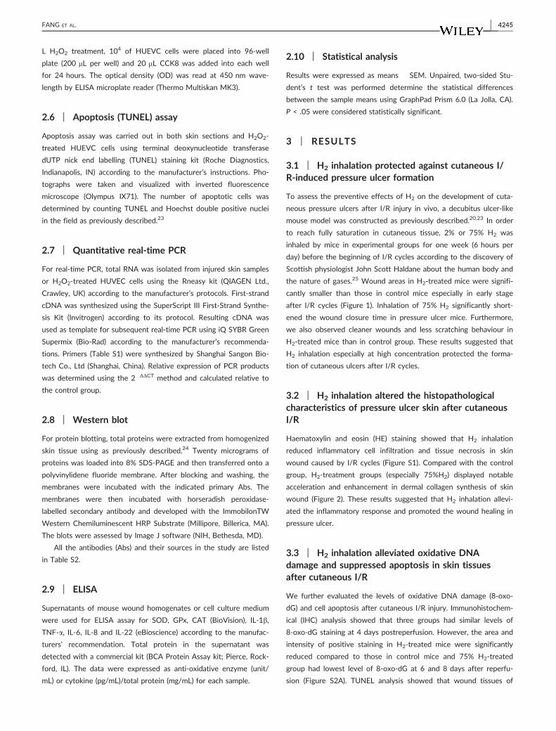
L H2O2 treatment, 104 of HUEVC cells were placed into 96-well
plate (200 lL per well) and 20 lL CCK8 was added into each well
for 24 hours. The optical density (OD) was read at 450 nm wave-
length by ELISA microplate reader (Thermo Multiskan MK3).
2.6 | Apoptosis (TUNEL) assay
Apoptosis assay was carried out in both skin sections and H2O2-
treated HUEVC cells using terminal deoxynucleotide transferase
dUTP nick end labelling (TUNEL) staining kit (Roche Diagnostics,
Indianapolis, IN) according to the manufacturer’s instructions. Pho-
tographs were taken and visualized with inverted fluorescence
microscope (Olympus IX71). The number of apoptotic cells was
determined by counting TUNEL and Hoechst double positive nuclei
in the field as previously described.23
2.7 | Quantitative real-time PCR
For real-time PCR, total RNA was isolated from injured skin samples
or H2O2-treated HUVEC cells using the Rneasy kit (QIAGEN Ltd.,
Crawley, UK) according to the manufacturer’s protocols. First-strand
cDNA was synthesized using the SuperScript III First-Strand Synthe-
sis Kit (Invitrogen) according to its protocol. Resulting cDNA was
used as template for subsequent real-time PCR using iQ SYBR Green
Supermix (Bio-Rad) according to the manufacturer’s recommenda-
tions. Primers (Table S1) were synthesized by Shanghai Sangon Bio-
tech Co., Ltd (Shanghai, China). Relative expression of PCR products
was determined using the 2�DDCT method and calculated relative to
the control group.
2.8 | Western blot
For protein blotting, total proteins were extracted from homogenized
skin tissue using as previously described.24 Twenty micrograms of
proteins was loaded into 8% SDS-PAGE and then transferred onto a
polyvinylidene fluoride membrane. After blocking and washing, the
membranes were incubated with the indicated primary Abs. The
membranes were then incubated with horseradish peroxidase-
labelled secondary antibody and developed with the ImmobilonTW
Western Chemiluminescent HRP Substrate (Millipore, Billerica, MA).
The blots were assessed by Image J software (NIH, Bethesda, MD).
All the antibodies (Abs) and their sources in the study are listed
in Table S2.
2.9 | ELISA
Supernatants of mouse wound homogenates or cell culture medium
were used for ELISA assay for SOD, GPx, CAT (BioVision), IL-1b,
TNF-a, IL-6, IL-8 and IL-22 (eBioscience) according to the manufac-
turers’ recommendation. Total protein in the supernatant was
detected with a commercial kit (BCA Protein Assay kit; Pierce, Rock-
ford, IL). The data were expressed as anti-oxidative enzyme (unit/
mL) or cytokine (pg/mL)/total protein (mg/mL) for each sample.
2.10 | Statistical analysis
Results were expressed as means � SEM. Unpaired, two-sided Stu-
dent’s t test was performed determine the statistical differences
between the sample means using GraphPad Prism 6.0 (La Jolla, CA).
P < .05 were considered statistically significant.
3 | RESULTS
3.1 | H2 inhalation protected against cutaneous I/R-induced pressure ulcer formation
To assess the preventive effects of H2 on the development of cuta-
neous pressure ulcers after I/R injury in vivo, a decubitus ulcer-like
mouse model was constructed as previously described.20,23 In order
to reach fully saturation in cutaneous tissue, 2% or 75% H2 was
inhaled by mice in experimental groups for one week (6 hours per
day) before the beginning of I/R cycles according to the discovery of
Scottish physiologist John Scott Haldane about the human body and
the nature of gases.25 Wound areas in H2-treated mice were signifi-
cantly smaller than those in control mice especially in early stage
after I/R cycles (Figure 1). Inhalation of 75% H2 significantly short-
ened the wound closure time in pressure ulcer mice. Furthermore,
we also observed cleaner wounds and less scratching behaviour in
H2-treated mice than in control group. These results suggested that
H2 inhalation especially at high concentration protected the forma-
tion of cutaneous ulcers after I/R cycles.
3.2 | H2 inhalation altered the histopathologicalcharacteristics of pressure ulcer skin after cutaneousI/R
Haematoxylin and eosin (HE) staining showed that H2 inhalation
reduced inflammatory cell infiltration and tissue necrosis in skin
wound caused by I/R cycles (Figure S1). Compared with the control
group, H2-treatment groups (especially 75%H2) displayed notable
acceleration and enhancement in dermal collagen synthesis of skin
wound (Figure 2). These results suggested that H2 inhalation allevi-
ated the inflammatory response and promoted the wound healing in
pressure ulcer.
3.3 | H2 inhalation alleviated oxidative DNAdamage and suppressed apoptosis in skin tissuesafter cutaneous I/R
We further evaluated the levels of oxidative DNA damage (8-oxo-
dG) and cell apoptosis after cutaneous I/R injury. Immunohistochem-
ical (IHC) analysis showed that three groups had similar levels of
8-oxo-dG staining at 4 days postreperfusion. However, the area and
intensity of positive staining in H2-treated mice were significantly
reduced compared to those in control mice and 75% H2-treated
group had lowest level of 8-oxo-dG at 6 and 8 days after reperfu-
sion (Figure S2A). TUNEL analysis showed that wound tissues of
FANG ET AL. | 4245
control mice had a large number of apoptotic cells, which was signif-
icantly reduced by H2 inhalation (especially 75% H2) at day 8
postreperfusion (Figure S2B and Figure 2C). These results suggested
that H2 inhalation could alleviate the oxidative DNA damage and
suppress apoptosis of skin tissues, and high-concentration H2 had
optimal protective effect against cutaneous I/R injury.
3.4 | H2 inhalation reduced ROS accumulation andup-regulated antioxidant enzyme activities in skintissue after cutaneous I/R
As ROS is essential mediators of reperfusion induced tissue dam-
age,26-29 we assessed ROS levels and the activities of major
F IGURE 1 H2 inhalation protected against ulcer formation in cutaneous I/R injury mice model. A, Representative photographs of woundsafter cutaneous I/R in control or H2-treated mice at 4, 6, 8, 10 and 13 days after reperfusion. B, Relative wound area after I/R injury in normalC57BL/6 mice with or without H2 inhalation (N = 8 for each time point and groups). The ulcer size in control mice at 4 days after reperfusionwas assigned a value of 100%. *P < .05; **P < .01; ***P < .001 compared to control
F IGURE 2 H2 treatment promoted dermal collagen synthesis in skin wound caused by I/R cycles. Skin tissue samples from pressure ulcermice of each group were fixed and paraffin embedded. The tissue sections were subjected to Masson staining. Representative images from thewound skin tissues are shown. Scale bar, 20 lm
4246 | FANG ET AL.
antioxidant enzymes including superoxide dismutase (SOD), catalase
(CAT) and glutathione peroxidase (GPx) in the wound tissue at day 6
postreperfusion. H2 suppressed the ROS accumulation in skin tissue
caused by I/R injury (P < .01) in a concentration-dependent manner
(Figure 3A). Activities of SOD, CAT and GPx in mice inhaled H2
were significantly higher than in control mice (Figure 3B-D).
3.5 | H2 activated the NRF2-ARE pathway in skintissue after cutaneous I/R
We next investigated whether NRF2/ARE antioxidant pathway medi-
ated the inhibition of ROS level of pressure ulcer tissue by H2. Quanti-
tative real-time PCR analysis showed that H2 significantly up-regulated
the mRNA levels of NRF2 and its target genes (heme oxygenase-1,
HO-1; NADH quinone oxidoreductase 1, NQO1; aldo-keto reductase,
AKR1C1) in a concentration-dependent manner in the skin tissue (Fig-
ure 4A). Immunoblotting also confirmed the enhanced expression of
NRF2-ARE pathway components in the skin tissue of H2 treatment
group (especially 75%) (Figure 4B). These results indicated that H2
activated the NRF2 pathway in skin lesions after cutaneous I/R.
3.6 | H2 inhalation inhibited cutaneous I/R-inducedinflammation
As proinflammatory cytokines are important for tissue damage dur-
ing I/R injury, the levels of proinflammatory cytokines in the
wounded skin were assessed. At day 6 post-I/R, H2 treatment signif-
icantly reduced the relative mRNA and protein expression of TNF-a,
IL-1b, IL-6 and IL-8 in wounded tissue, and their expression was
lowest in pressure ulcer mice treated with 75% H2 (Figure 5A and
B). IHC further confirmed that the protein levels of TNF-a, IL-1b, IL-
6 and IL-8 in cutaneous I/R injured skin tissue of mice inhaled H2
were substantially lower than those of control mice (Figure 5C).
Meanwhile, H2 treatment significantly up-regulated the expression
of IL-22, an essential pro-healing cytokine involved in repair events
on different models of epithelial regeneration,30,31 at both mRNA
and protein levels and 75% H2 treated group displayed the highest
level of IL-22 (Figure 5), which was mainly expressed in the dermal
layer of perilesional skin tissues in mice treated by H2 (Figure 5C).
These results indicated that hydrogen inhalation reversed proinflam-
matory effects of skin wound environment after cutaneous I/R.
3.7 | H2 inhalation modulated the expression ofhealing-associated molecules in vivo after cutaneousI/R
The metalloprotease MMP9 and pro-healing factors TGF-b, VEGF
and IGF-1 play crucial roles during proliferative and remodelling
phases of wound healing. To investigate how H2 improves wound
healing in pressure ulcer mice, its effects on expression of aforemen-
tioned molecules were analysed by quantitative PCR and
immunoblotting. H2 inhalation significantly decreased the expression
level of MMP9 and enhanced the expression of TGF-b1, VEGF and
IGF-1 at both mRNA and protein levels in the wound. Mice treated
with 75% H2 had the lowest level of MMP9 and highest expression
of pro-healing factors in skin tissue among three groups (Figure 6).
F IGURE 3 H2 inhalation reduced ROS accumulation and enhanced antioxidant enzyme activities in skin tissue. ELISA assays for detectionof ROS production (A) and the activities of SOD (B), GPx (C), and CAT (D). *P < .05; **P < .01 compared to control
FANG ET AL. | 4247
3.8 | H2 reversed the adverse effect of H2O2 oncell proliferation and apoptosis of vascular endotheliacells
I/R injury is closely associated with microvascular dysfunction, which
is largely a consequence of endothelia cell impairment.7 Thus, we
further examine the effects of H2 on human umbilical vein endothe-
lial cells (HUVECs) against oxidative damage. Preincubation with H2
for 24 hours significantly relieved the 0.25 mmo/L H2O2-induced
proliferation inhibition of HUVECs in a concentration-dependent
manner (P < .01) (Figure S3A). After exposure to 0.25 mmol/L H2O2,
HUVECs showed a 2.5-folds increase in the apoptosis rate, which
was markedly reduced by H2 pretreatment (especially 75%H2)
(P < .01) (Figure S3B and C).
3.9 | H2 rectified the imbalance of oxidation-reduction system in H2O2-treated vascular endotheliacells through the activation of Nrf2/ARE pathway
To further understand the mechanism of H2-mediated protective
effects on endothelial cells during cutaneous I/R, a series of in vitro
studies were performed in oxidant-treated HUVECs. H2O2 notably
up-regulated intracellular ROS production (Figure S4A) and sup-
pressed the expression of various anti-oxidative enzymes (SOD, GPx,
and CAT) (Figure S4B-D) in HUVECs, suggesting of a severe distur-
bance of intracellular oxidation-reduction system. Preincubation with
H2 resulted in a trend towards normalization of both intracellular
ROS and anti-oxidative enzyme in H2O2-treated HUVECs
(Figure S4A-D). Quantitative PCR assay demonstrated that H2 signif-
icantly up-regulated the expression of NRF2 and its target genes
HO-1, AKR1C1 and NQO1 in HUVECs exposed to H2O2 in a concen-
tration-dependent manner (Figure S4E-H). Together, these results
suggested that H2 might maintain intracellular homoeostasis of
oxidation-reduction system in vascular endothelial cells through the
activation of Nrf2/ARE pathway.
3.10 | H2 inhibited the expression of severalchemokine and adhesion molecules in vascularendothelial cells stimulated by H2O2
Endothelial damage is a critical event in the early phase of inflamma-
tory responses induced by cutaneous I/R, which could drive the up-
regulation of various chemokines and adhesion molecules and then
mediate leucocyte recruit, adhesion and emigration.7,32 Vascular
endothelial cells more than doubled the transcriptional levels of
MCP-1(monocyte chemoattractant protein-1), E-selectin, P-selectin
and ICAM-1(intercellular cell adhesion molecule-1) after H2O2 expo-
sure (Figure 7). Pretreatment with H2 significantly suppressed the
elevation of their expression in H2O2-exposed HUEVCs (P < .01)
(Figure 7). The inhibitory effects were positively correlated with H2
concentration. Our results suggested that H2 might exert anti-inflam-
matory effects against cutaneous I/R injury by down-regulating the
expression of endothelial chemokines and adhesion molecules.
4 | DISCUSSION
In the present study, we first demonstrated the protective effects of
H2 inhalation on pressure ulcers in a murine model. H2 treatment
significantly reduced the wound area in a concentration-dependent
F IGURE 4 Hydrogen activated the NRF2-ARE pathway in wounded tissue. A, The mRNA levels of NRF2, HO-1, NQO1 and AKR1C1 wereanalysed by quantitative RT-PCR assay. B, Immunoblots of NRF2, HO-1, NQO1, AKR1C1 and b-actin. *P < .05; **P < .01 compared to control
4248 | FANG ET AL.
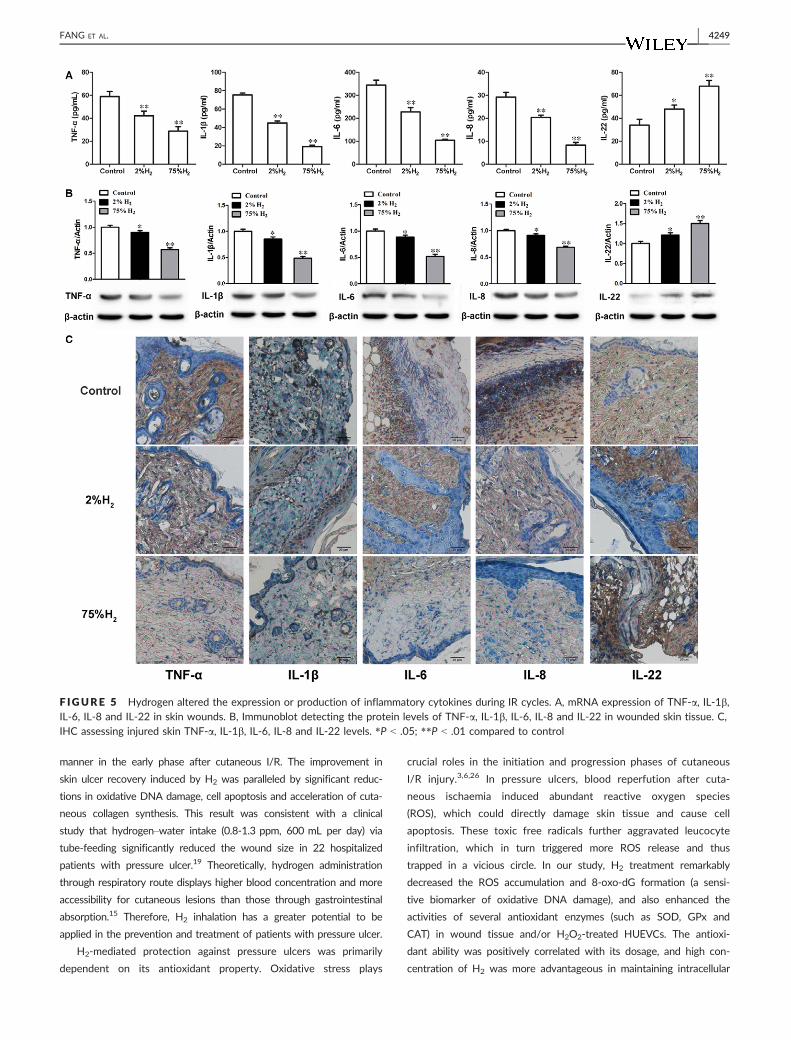
manner in the early phase after cutaneous I/R. The improvement in
skin ulcer recovery induced by H2 was paralleled by significant reduc-
tions in oxidative DNA damage, cell apoptosis and acceleration of cuta-
neous collagen synthesis. This result was consistent with a clinical
study that hydrogen–water intake (0.8-1.3 ppm, 600 mL per day) via
tube-feeding significantly reduced the wound size in 22 hospitalized
patients with pressure ulcer.19 Theoretically, hydrogen administration
through respiratory route displays higher blood concentration and more
accessibility for cutaneous lesions than those through gastrointestinal
absorption.15 Therefore, H2 inhalation has a greater potential to be
applied in the prevention and treatment of patients with pressure ulcer.
H2-mediated protection against pressure ulcers was primarily
dependent on its antioxidant property. Oxidative stress plays
crucial roles in the initiation and progression phases of cutaneous
I/R injury.3,6,26 In pressure ulcers, blood reperfution after cuta-
neous ischaemia induced abundant reactive oxygen species
(ROS), which could directly damage skin tissue and cause cell
apoptosis. These toxic free radicals further aggravated leucocyte
infiltration, which in turn triggered more ROS release and thus
trapped in a vicious circle. In our study, H2 treatment remarkably
decreased the ROS accumulation and 8-oxo-dG formation (a sensi-
tive biomarker of oxidative DNA damage), and also enhanced the
activities of several antioxidant enzymes (such as SOD, GPx and
CAT) in wound tissue and/or H2O2-treated HUEVCs. The antioxi-
dant ability was positively correlated with its dosage, and high con-
centration of H2 was more advantageous in maintaining intracellular
F IGURE 5 Hydrogen altered the expression or production of inflammatory cytokines during IR cycles. A, mRNA expression of TNF-a, IL-1b,IL-6, IL-8 and IL-22 in skin wounds. B, Immunoblot detecting the protein levels of TNF-a, IL-1b, IL-6, IL-8 and IL-22 in wounded skin tissue. C,IHC assessing injured skin TNF-a, IL-1b, IL-6, IL-8 and IL-22 levels. *P < .05; **P < .01 compared to control
FANG ET AL. | 4249
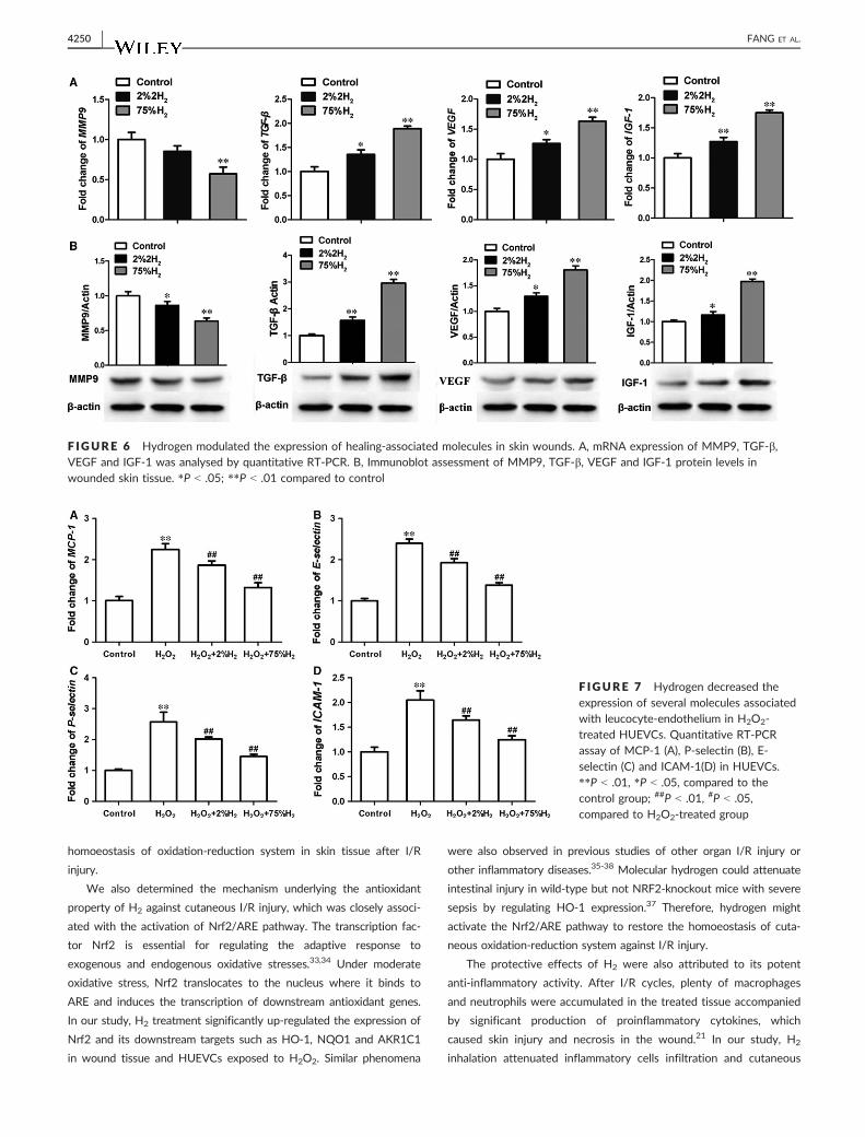
homoeostasis of oxidation-reduction system in skin tissue after I/R
injury.
We also determined the mechanism underlying the antioxidant
property of H2 against cutaneous I/R injury, which was closely associ-
ated with the activation of Nrf2/ARE pathway. The transcription fac-
tor Nrf2 is essential for regulating the adaptive response to
exogenous and endogenous oxidative stresses.33,34 Under moderate
oxidative stress, Nrf2 translocates to the nucleus where it binds to
ARE and induces the transcription of downstream antioxidant genes.
In our study, H2 treatment significantly up-regulated the expression of
Nrf2 and its downstream targets such as HO-1, NQO1 and AKR1C1
in wound tissue and HUEVCs exposed to H2O2. Similar phenomena
were also observed in previous studies of other organ I/R injury or
other inflammatory diseases.35-38 Molecular hydrogen could attenuate
intestinal injury in wild-type but not NRF2-knockout mice with severe
sepsis by regulating HO-1 expression.37 Therefore, hydrogen might
activate the Nrf2/ARE pathway to restore the homoeostasis of cuta-
neous oxidation-reduction system against I/R injury.
The protective effects of H2 were also attributed to its potent
anti-inflammatory activity. After I/R cycles, plenty of macrophages
and neutrophils were accumulated in the treated tissue accompanied
by significant production of proinflammatory cytokines, which
caused skin injury and necrosis in the wound.21 In our study, H2
inhalation attenuated inflammatory cells infiltration and cutaneous
F IGURE 6 Hydrogen modulated the expression of healing-associated molecules in skin wounds. A, mRNA expression of MMP9, TGF-b,VEGF and IGF-1 was analysed by quantitative RT-PCR. B, Immunoblot assessment of MMP9, TGF-b, VEGF and IGF-1 protein levels inwounded skin tissue. *P < .05; **P < .01 compared to control
F IGURE 7 Hydrogen decreased theexpression of several molecules associatedwith leucocyte-endothelium in H2O2-treated HUEVCs. Quantitative RT-PCRassay of MCP-1 (A), P-selectin (B), E-selectin (C) and ICAM-1(D) in HUEVCs.**P < .01, *P < .05, compared to thecontrol group; ##P < .01, #P < .05,compared to H2O2-treated group
4250 | FANG ET AL.
necrosis in the wound induced by I/R injury. Proinflammatory cytoki-
nes such as TNF-a, IL-1, IL-6 and IL-8 were decreased while IL-22
was increased in the wounded skin of H2-treated mice. Distinct from
proinflammatory cytokines, IL-22 mediates a crosstalk between
immune system and cutaneous cells (such as fibroblasts and ker-
atinocytes) and plays a pro-healing role in wound repairment.30,39
Our data demonstrated that H2 reversed the excessive inflammatory
response induced by cutaneous I/R injury.
The anti-inflammatory property of hydrogen might be partially
attributed to its protection on vascular endothelial cells against
oxidative stress. During I/R injury, endothelial damage initiated leu-
cocyte adhesion, recruitment and infiltration by up-regulating the
expression of chemokines and adhesion molecules.6,7,32 Our in vitro
assay showed that H2 treatment significantly alleviated the oxida-
tive injury of endothelial cells and reduced the overexpression of
MCP-1, E-selectin, P-selectin and ICAM-1 in HUEVCs exposed to
H2O2. MCP-1 is a critical molecule for chemotaxis and activation of
macrophage, which is a significant source of proinflammatory
cytokines and contributes to I/R injury of skin and other
organs.21,40-42 The adhesion molecules studied here were mainly
responsible for leucocyte rolling, localization and adhesion to the
endothelium.43,44 Hence, hydrogen might modulate those molecules
associated with leucocyte–endothelium interaction to inhibit subse-
quent inflammatory reaction caused by cutaneous I/R injury.
In pressure ulcer, the wound sites were constantly in the
dynamic pathological alterations of inflammatory injury and tissue
repair. Outcome of wound healing process was mainly determined
by the presence and concentration of the healing-associated factors
such as MMPs, TGF-b, VEGF and IGF1.45-47 Overproduction or high
activity of MMP9 and suppressed expression of TGF-b were identi-
fied as indicators of poor healing in skin samples of chronic
ulcers.46,48 In our study, H2 promoted the expression of pro-healing
factors (TGF-b, VEGF and IGF1) and inhibited the production of
MMP9 in wound tissue of pressure ulcer, accompanied by accelera-
tion of dermal collagen synthesis. Hence, hydrogen had a wound
healing promoting effect against cutaneous I/R injury.
Taken together, the present results indicated that hydrogen
suppressed the formation of decubitus ulcers by its antioxidant, anti-
inflammatory, pro-healing activities against cutaneous I/R injury.
Similar therapeutic effects of hydrogen were also reported in common
senile diseases concomitant with decubitus, such as cerebral or
myocardial infarctions, COPD, diabetes, hyperlipaemia, malignant
tumours.11,36,49-54 Furthermore, H2 had no cytotoxicity in vivo in
human body even at a high concentration.17 Therefore, hydrogen gas
has a great potential for preventing and/or treating pressure ulcer.
There were still some limitations in our study. Firstly, we did not
detect the cutaneous hydrogen concentration, which might provide
direct evidence to support the dosage-dependent protection of
hydrogen gas against cutaneous I/R injury. In addition, the exact
mechanisms by which hydrogen modulates oxidative stress, inflam-
mation and wound repair in pressure ulcer were still unclear. More
experiments are needed to work out these problems before the clini-
cal trial of H2 in pressure ulcers.
ACKNOWLEDGEMENTS
We extend our sincerest gratitude to the staff of Shanghai Asclepius
Meditec Co., Ltd. for their vital technical support in the hydrogen–
oxygen nebulizer device. We also thank Prof. Zhimin Kang of Shang-
hai Huikang Hydrogen Medical Research Center for technical assis-
tance in study design.
CONFLICT OF INTEREST
The authors have no conflicts of interest to report.
ORCID
Wei Fang http://orcid.org/0000-0002-0749-298X
REFERENCES
1. Gorecki C, Brown JM, Nelson EA, et al. Impact of pressure ulcers on
quality of life in older patients: a systematic review. J Am Geriatr Soc.
2009;57:1175-1183.
2. Mustoe T. Understanding chronic wounds: a unifying hypothesis on
their pathogenesis and implications for therapy. Am J Surg.
2004;187:65S-70S.
3. Mustoe TA, O’Shaughnessy K, Kloeters O. Chronic wound pathogen-
esis and current treatment strategies: a unifying hypothesis. Plast
Reconstr Surg. 2006;117:35S-41S.
4. Salcido R, Donofrio JC, Fisher SB, et al. Histopathology of pressure
ulcers as a result of sequential computer-controlled pressure sessions
in a fuzzy rat model. Adv Wound Care. 1994;7:23-24, 26, 28 passim.
5. Peirce SM, Skalak TC, Rodeheaver GT. Ischemia-reperfusion injury in
chronic pressure ulcer formation: a skin model in the rat. Wound
Repair Regen. 2000;8:68-76.
6. Eltzschig HK, Collard CD. Vascular ischaemia and reperfusion injury.
Br Med Bull. 2004;70:71-86.
7. Seal JB, Gewertz BL. Vascular dysfunction in ischemia-reperfusion
injury. Ann Vasc Surg. 2005;19:572-584.
8. Houwing R, Overgoor M, Kon M, Jansen G, van Asbeck BS, Haal-
boom JR. Pressure-induced skin lesions in pigs: reperfusion injury
and the effects of vitamin E. J Wound Care. 2000;9:36-40.
9. Sener G, Sert G, Ozer SA, Arbak S, Gedik N, Ayano�glu-D€ulger G.
Melatonin protects against pressure ulcer-induced oxidative injury of
the skin and remote organs in rats. J Pineal Res. 2006;40:280-287.
10. Duscher D, Neofytou E, Wong VW, et al. Transdermal deferoxamine
prevents pressure-induced diabetic ulcers. Proc Natl Acad Sci U S A.
2015;112:94-99.
11. Ohsawa I, Ishikawa M, Takahashi K, et al. Hydrogen acts as a thera-
peutic antioxidant by selectively reducing cytotoxic oxygen radicals.
Nat Med. 2007;13:688-694.
12. Liu Q, Shen WF, Sun HY, et al. Hydrogen-rich saline protects against
liver injury in rats with obstructive jaundice. Liver Int. 2010;30:
958-968.
13. Sun Q, Kang Z, Cai J, et al. Hydrogen-rich saline protects myocar-
dium against ischemia/reperfusion injury in rats. Exp Biol Med (May-
wood). 2009;234:1212-1219.
14. Zheng X, Mao Y, Cai J, et al. Hydrogen-rich saline protects against
intestinal ischemia/reperfusion injury in rats. Free Radic Res.
2009;43:478-484.
15. Ge L, Yang M, Yang NN, Yin XX, Song WG. Molecular hydrogen: a
preventive and therapeutic medical gas for various diseases. Onco-
target. 2017;8:102653-102673.
FANG ET AL. | 4251
16. Ostojic SM. Molecular hydrogen: an inert gas turns clinically effec-
tive. Ann Med. 2015;47:301-304.
17. Abraini JH, Gardette-Chauffour MC, Martinez E, Rostain JC, Lemaire
C. Psychophysiological reactions in humans during an open sea dive
to 500 m with a hydrogen-helium-oxygen mixture. J Appl Physiol.
1994;76:1113-1118.
18. Ono H, Nishijima Y, Adachi N, et al. A basic study on molecular
hydrogen (H2) inhalation in acute cerebral ischemia patients for
safety check with physiological parameters and measurement of
blood H2 level. Med Gas Res. 2012;2:21.
19. Li Q, Kato S, Matsuoka D, Tanaka H, Miwa N. Hydrogen water intake
via tube-feeding for patients with pressure ulcer and its reconstructive
effects on normal human skin cells in vitro. Med Gas Res. 2013;3:20.
20. Uchiyama A, Yamada K, Perera B, et al. Protective effect of MFG-E8
after cutaneous ischemia-reperfusion injury. J Invest Dermatol.
2015;135:1157-1165.
21. Saito Y, Hasegawa M, Fujimoto M, et al. The loss of MCP-1 attenu-
ates cutaneous ischemia-reperfusion injury in a mouse model of
pressure ulcer. J Invest Dermatol. 2008;128:1838-1851.
22. Stadler I, Zhang RY, Oskoui P, Whittaker MS, Lanzafame RJ. Devel-
opment of a simple, noninvasive, clinically relevant model of pres-
sure ulcers in the mouse. J Invest Surg. 2004;17:221-227.
23. Uchiyama A, Yamada K, Perera B, et al. Protective effect of botulinum
toxin A after cutaneous ischemia-reperfusion injury. Sci Rep. 2015;5:9072.
24. Long M, de la Vega MR, Wen Q, et al. An essential role of NRF2 in
diabetic wound healing. Diabetes. 2016;65:780-793.
25. Bove A, Davis J. Bove and davis’ diving medicine (4th edn.). New
York, NY: Saunders; 2003:53-76.
26. Siemionow M, Arslan E. Ischemia/reperfusion injury: a review in rela-
tion to free tissue transfers. Microsurgery. 2004;24:468-475.
27. Chouchani ET, Pell VR, James AM, et al. A unifying mechanism for
mitochondrial superoxide production during ischemia-reperfusion
injury. Cell Metab. 2016;23:254-263.
28. Minutoli L, Puzzolo D, Rinaldi M, et al. ROS-mediated NLRP3 inflam-
masome activation in brain, heart, kidney, and testis ischemia/reper-
fusion injury. Oxid Med Cell Longev. 2016;2016:2183026.
29. Granger DN, Kvietys PR. Reperfusion injury and reactive oxygen
species: the evolution of a concept. Redox Biol. 2015;6:524-551.
30. Avitabile S, Odorisio T, Madonna S, et al. Interleukin-22 promotes
wound repair in diabetes by improving keratinocyte pro-healing
functions. J Invest Dermatol. 2015;135:2862-2870.
31. Pickert G, Neufert C, Leppkes M, et al. STAT3 links IL-22 signaling
in intestinal epithelial cells to mucosal wound healing. J Exp Med.
2009;206:1465-1472.
32. Toledo-Pereyra LH, Toledo AH, Walsh J, Lopez-Neblina F. Molecular
signaling pathways in ischemia/reperfusion. Exp Clin Transplant.
2004;2:174-177.
33. Kensler TW, Wakabayashi N, Biswal S. Cell survival responses to
environmental stresses via the Keap1-Nrf2-ARE pathway. Annu Rev
Pharmacol Toxicol. 2007;47:89-116.
34. Jaramillo MC, Zhang DD. The emerging role of the Nrf2-Keap1 sig-
naling pathway in cancer. Genes Dev. 2013;27:2179-2191.
35. Yu J, Zhang W, Zhang R, et al. Molecular hydrogen attenuates
hypoxia/reoxygenation injury of intrahepatic cholangiocytes by acti-
vating Nrf2 expression. Toxicol Lett. 2015;238:11-19.
36. Song G, Zong C, Zhang Z, et al. Molecular hydrogen stabilizes
atherosclerotic plaque in low-density lipoprotein receptor-knockout
mice. Free Radic Biol Med. 2015;87:58-68.
37. Yu Y, Yang Y, Bian Y, et al. Hydrogen gas protects against intestinal
injury in wild type but not NRF2 knockout mice with severe sepsis
by regulating HO-1 and HMGB1 release. Shock. 2017;48:364-370.
38. Tamaki N, Orihuela-Campos RC, Fukui M, Ito HO. Hydrogen-rich
water intake accelerates oral palatal wound healing via activation of
the Nrf2/antioxidant defense pathways in a rat model. Oxid Med Cell
Longev. 2016;2016:5679040.
39. McGee HM, Schmidt BA, Booth CJ, et al. IL-22 promotes fibroblast-
mediated wound repair in the skin. J Invest Dermatol. 2012;133:
1321-1329.
40. Leonard EJ, Yoshimura T. Human monocyte chemoattractant pro-
tein-1 (MCP-1). Immunol Today. 1990;11:97-101.
41. Hayasaki T, Kaikita K, Okuma T, et al. CC chemokine receptor-2
deficiency attenuates oxidative stress and infarct size caused by
myocardial ischemia-reperfusion in mice. Circ J. 2006;70:342-351.
42. Furuichi K, Wada T, Iwata Y, et al. CCR2 signaling contributes to
ischemia-reperfusion injury in kidney. J Am Soc Nephrol.
2003;14:2503-2515.
43. Sawaya DE, Zibari GB, Minardi A, et al. P-selectin contributes to the
initial recruitment of rolling and adherent leukocytes in hepatic
venules after ischemia/reperfusion. Shock. 1999;12:227-232.
44. Kurose I, Anderson DC, Miyasaka M, et al. Molecular determinants
of reperfusion-induced leukocyte adhesion and vascular protein leak-
age. Circ Res. 1994;74:336-343.
45. Ravanti L, K€ah€ari VM. Matrix metalloproteinases in wound repair (re-
view). Int J Mol Med. 2000;6:391-407.
46. Pakyari M, Farrokhi A, Maharlooei MK, Ghahary A. Critical role of
transforming growth factor beta in different phases of wound heal-
ing. Adv Wound Care (New Rochelle). 2013;2:215-224.
47. Barrientos S, Stojadinovic O, Golinko MS, Brem H, Tomic-Canic M.
Growth factors and cytokines in wound healing. Wound Repair
Regen. 2008;16:585-601.
48. Yager DR, Zhang LY, Liang HX, Diegelmann RF, Cohen IK. Wound
fluids from human pressure ulcers contain elevated matrix metallo-
proteinase levels and activity compared to surgical wound fluids. J
Invest Dermatol. 1996;107:743-748.
49. Liu SL, Liu K, Sun Q, Liu WW, Tao HY, Sun XJ. Hydrogen therapy
may be a novel and effective treatment for COPD. Front Pharmacol.
2011;2:19.
50. Kawai D, Takaki A, Nakatsuka A, et al. Hydrogen-rich water pre-
vents progression of nonalcoholic steatohepatitis and accompanying
hepatocarcinogenesis in mice. Hepatology. 2012;56:912-921.
51. Dole M, Wilson FR, Fife WP. Hyperbaric hydrogen therapy: a possi-
ble treatment for cancer. Science. 1975;190:152-154.
52. Zong C, Song G, Yao S, et al. Administration of hydrogen-saturated
saline decreases plasma low-density lipoprotein cholesterol levels
and improves high-density lipoprotein function in high-fat diet-fed
hamsters. Metabolism. 2012;61:794-800.
53. Hayashida K, Sano M, Ohsawa I, et al. Inhalation of hydrogen gas
reduces infarct size in the rat model of myocardial ischemia-reperfu-
sion injury. Biochem Biophys Res Commun. 2008;373:30-35.
54. Kamimura N, Nishimaki K, Ohsawa I, Ohta S. Molecular hydrogen
improves obesity and diabetes by inducing hepatic FGF21 and stim-
ulating energy metabolism in db/db mice. Obesity (Silver Spring).
2011;19:1396-1403.
SUPPORTING INFORMATION
Additional supporting information may be found online in the
Supporting Information section at the end of the article.
How to cite this article: Fang W, Wang G, Tang L, et al.
Hydrogen gas inhalation protects against cutaneous
ischaemia/reperfusion injury in a mouse model of pressure
ulcer. J Cell Mol Med. 2018;22:4243–4252. https://doi.org/
10.1111/jcmm.13704
4252 | FANG ET AL.
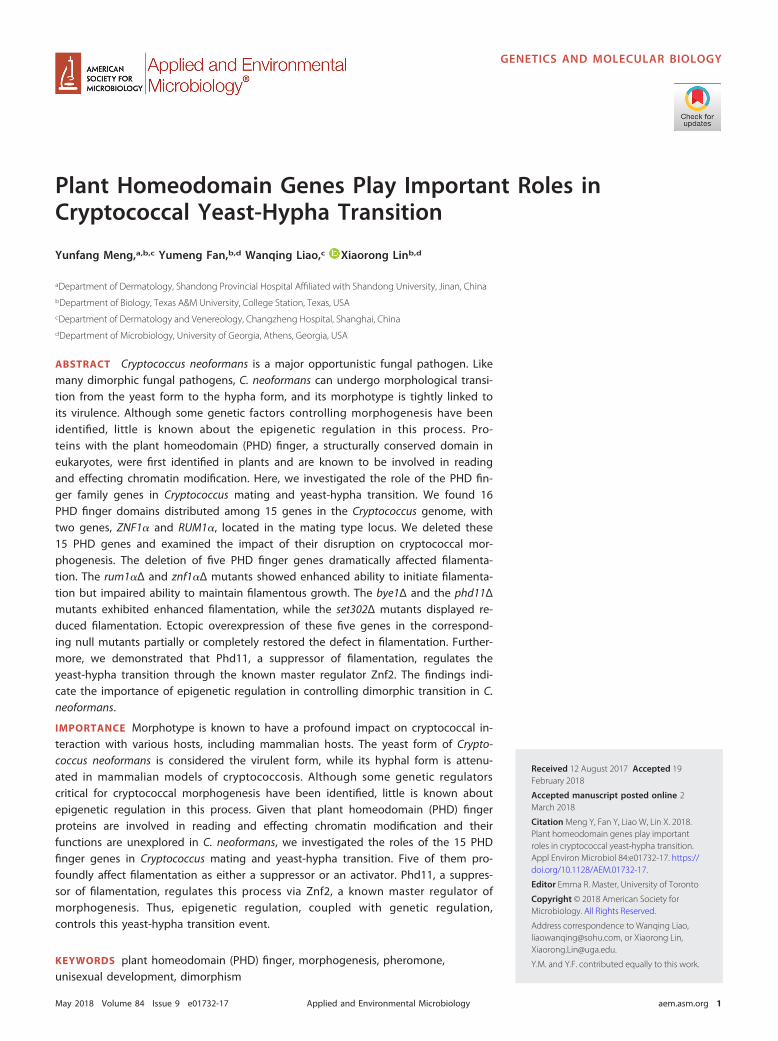
Plant Homeodomain Genes Play Important Roles inCryptococcal Yeast-Hypha Transition
Yunfang Meng,a,b,c Yumeng Fan,b,d Wanqing Liao,c Xiaorong Linb,d
aDepartment of Dermatology, Shandong Provincial Hospital Affiliated with Shandong University, Jinan, ChinabDepartment of Biology, Texas A&M University, College Station, Texas, USAcDepartment of Dermatology and Venereology, Changzheng Hospital, Shanghai, ChinadDepartment of Microbiology, University of Georgia, Athens, Georgia, USA
ABSTRACT Cryptococcus neoformans is a major opportunistic fungal pathogen. Likemany dimorphic fungal pathogens, C. neoformans can undergo morphological transi-tion from the yeast form to the hypha form, and its morphotype is tightly linked toits virulence. Although some genetic factors controlling morphogenesis have beenidentified, little is known about the epigenetic regulation in this process. Pro-teins with the plant homeodomain (PHD) finger, a structurally conserved domain ineukaryotes, were first identified in plants and are known to be involved in readingand effecting chromatin modification. Here, we investigated the role of the PHD fin-ger family genes in Cryptococcus mating and yeast-hypha transition. We found 16PHD finger domains distributed among 15 genes in the Cryptococcus genome, withtwo genes, ZNF1� and RUM1�, located in the mating type locus. We deleted these15 PHD genes and examined the impact of their disruption on cryptococcal mor-phogenesis. The deletion of five PHD finger genes dramatically affected filamenta-tion. The rum1�Δ and znf1�Δ mutants showed enhanced ability to initiate filamenta-tion but impaired ability to maintain filamentous growth. The bye1Δ and the phd11Δmutants exhibited enhanced filamentation, while the set302Δ mutants displayed re-duced filamentation. Ectopic overexpression of these five genes in the correspond-ing null mutants partially or completely restored the defect in filamentation. Further-more, we demonstrated that Phd11, a suppressor of filamentation, regulates theyeast-hypha transition through the known master regulator Znf2. The findings indi-cate the importance of epigenetic regulation in controlling dimorphic transition in C.neoformans.
IMPORTANCE Morphotype is known to have a profound impact on cryptococcal in-teraction with various hosts, including mammalian hosts. The yeast form of Crypto-coccus neoformans is considered the virulent form, while its hyphal form is attenu-ated in mammalian models of cryptococcosis. Although some genetic regulatorscritical for cryptococcal morphogenesis have been identified, little is known aboutepigenetic regulation in this process. Given that plant homeodomain (PHD) fingerproteins are involved in reading and effecting chromatin modification and theirfunctions are unexplored in C. neoformans, we investigated the roles of the 15 PHDfinger genes in Cryptococcus mating and yeast-hypha transition. Five of them pro-foundly affect filamentation as either a suppressor or an activator. Phd11, a suppres-sor of filamentation, regulates this process via Znf2, a known master regulator ofmorphogenesis. Thus, epigenetic regulation, coupled with genetic regulation,controls this yeast-hypha transition event.
KEYWORDS plant homeodomain (PHD) finger, morphogenesis, pheromone,unisexual development, dimorphism
Received 12 August 2017 Accepted 19February 2018
Accepted manuscript posted online 2March 2018
Citation Meng Y, Fan Y, Liao W, Lin X. 2018.Plant homeodomain genes play importantroles in cryptococcal yeast-hypha transition.Appl Environ Microbiol 84:e01732-17. https://doi.org/10.1128/AEM.01732-17.
Editor Emma R. Master, University of Toronto
Copyright © 2018 American Society forMicrobiology. All Rights Reserved.
Address correspondence to Wanqing Liao,[email protected], or Xiaorong Lin,[email protected].
Y.M. and Y.F. contributed equally to this work.
GENETICS AND MOLECULAR BIOLOGY
crossm
May 2018 Volume 84 Issue 9 e01732-17 aem.asm.org 1Applied and Environmental Microbiology

Cryptococcus neoformans is an opportunistic fungal pathogen that claims hundredsof thousands of lives each year globally (1, 2). The fungus can undergo morpho-
logical transition from the yeast form to the hypha form. Like many dimorphic fungalpathogens (3, 4), the morphotype of Cryptococcus is tightly linked to its virulence (5, 6).To understand the biology and pathology of Cryptococcus, some important geneticfactors that regulate yeast-hypha transition have been identified. Morphological tran-sition in Cryptococcus occurs during unisexual development or during bisexual a-�mating (7–9), which is controlled by the pheromone signaling pathway under mating-inducing conditions (10). The HMG domain transcription factor Mat2 is an effector ofthe pheromone pathway (11). Mat2 activates the zinc finger transcription factor Znf2under mating-inducing conditions, which ultimately determines the yeast-hypha tran-sition (11). Znf2 is the decision maker in filamentation, and it also bridges morphologyand virulence potential in Cryptococcus (6, 11, 12).
The mating-type locus of the Cryptococcus genome carries many genes that controlthe mating process (13), including several components of the pheromone pathway,such as the pheromone gene MF�/a (14, 15), the pheromone receptor gene STE3�/a(14, 16), STE20�/a, and elements of the MAPK cascade STE11�/a and STE12�/a (9, 17).The mating type locus also harbors the cell identity factor gene SXI1�/2a (13, 18). Twouncharacterized plant homeodomain (PHD) finger genes, RUM1 �/a and ZNF1 �/a, arealso located in the mating type locus (13, 15). The PHD finger was first identified in theplant Arabidopsis (19). The highly conserved small PHD fold consists of 50 to 80 aminoacid residues and a cross-brace topology in which two zinc atoms are anchored bythe Cys4-His-Cys3 motif. PHD finger proteins are important readers of histonemodifications and are involved in epigenetic regulation in plants and mammals (20).Although PHD finger proteins are universally distributed in eukaryotes, their bio-logical functions in the fungal kingdom are poorly understood. In another humanfungal pathogen, Candida albicans, histone modification contributes to the yeast-hypha transition (21), indicating that epigenetic factors can be important in regu-lating fungal morphogenesis. However, nothing is known about epigenetic regu-lation of morphogenesis in Cryptococcus.
Here, we systematically analyzed the PHD finger genes in the genome of C.neoformans. We identified 16 PHD finger domains harbored by 15 genes. We generatedand characterized 15 PHD gene knockout mutants and 5 PHD gene overexpressionstrains. These five selected PHD genes exhibit important roles in filamentation, indi-cating that some PHD proteins are critical for Cryptococcus morphotype transition.
RESULTSIdentification of PHD finger genes in C. neoformans. As mentioned in the
introduction, two PHD finger genes, RUM1 and ZNF1, are located in the mating typelocus. To identify additional putative PHD finger-containing genes, we used the PHDdomain sequences from ZNF1� as the query sequences in a BLAST search against thegenome of C. neoformans strain JEC21 (serotype D). Based on the Pfam database, weobtained 16 putative PHD finger domains carried by 15 genes in the genome of JEC21(Table 1). Corresponding genes also exist in the genome of C. neoformans strain H99(serotype A). The predicted protein sequences of these PHD genes were compared withthose of PHD proteins in other eukaryotic species, including budding yeast, fissionyeast, mouse, and human. However, these homologues are not conserved with cryp-tococcal PHD genes except for some conserved domains. The closest homologues ofsome PHD proteins in Saccharomyces cerevisiae that have been previously characterizedare provided in Table 1.
Alignment of these PHD finger domain sequences using the DNAMAN software(Lynnon, CA, USA) indicated that they share a feature with the same basic PHD fingertopology—two zinc atoms harbored by the Cys4-His-Cys3 motif (Fig. 1A)—as expectedbased on previous studies (20). The motif scan results revealed additional importantdomains related to histone modification in Phd11 and Set302 (Fig. 1B). Phd11 carries aMOZ-SAS domain, which may potentially possess lysine acetyltransferase activity (22).
Meng et al. Applied and Environmental Microbiology
May 2018 Volume 84 Issue 9 e01732-17 aem.asm.org 2
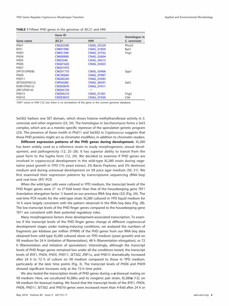
Set302 harbors one SET domain, which shows histone methyltransferase activity in S.cerevisiae and other organisms (23, 24). The homologue in Saccharomyces forms a Set3complex, which acts as a meiotic-specific repressor of the sporulation genetic program(23). The presence of these motifs in Phd11 and Set302 in Cryptococcus suggests thatthese PHD proteins might act as chromatin modifiers, in addition to chromatin readers.
Different expression patterns of the PHD genes during development. XL280has been widely used as a reference strain to study morphogenesis, sexual devel-opment, and pathogenicity (12, 25–28). It has superior ability to transit from theyeast form to the hypha form (12, 29). We decided to examine if PHD genes areinvolved in cryptococcal development in the wild-type XL280 strain during vege-tative yeast growth in YPD (1% yeast extract, 2% Bacto Peptone, and 2% dextrose)medium and during unisexual development on V8 juice agar medium (30, 31). Wefirst examined their expression patterns by transcriptome sequencing (RNA-Seq)and real-time (RT) PCR.
When the wild-type cells were cultured in YPD medium, the transcript levels of thePHD finger genes were 25- to 29-fold lower than that of the housekeeping gene TEF1(translation elongation factor 1) based on our previous RNA-Seq data (32) (Fig. 2A). Thereal-time PCR results for the wild-type strain XL280 cultured in YPD liquid medium for16 h were largely consistent with the pattern observed in the RNA-Seq data (Fig. 2B).The low transcript levels of the PHD finger genes compared to the housekeeping geneTEF1 are consistent with their potential regulatory roles.
Many morphogenesis factors show development-associated transcription. To exam-ine if the transcript levels of the PHD finger genes change at different cryptococcaldevelopment stages under mating-inducing conditions, we analyzed the numbers offragments per kilobase per million (FPKM) of the PHD genes from our RNA-Seq dataobtained from wild-type XL280 cultured alone on YPD medium (yeast growth) and onV8 medium for 24 h (initiation of filamentation), 48 h (filamentation elongation), or 72h (filamentation and initiation of sporulation). Interestingly, although the transcriptlevels of PHD finger genes remained low under all the conditions tested, the transcriptlevels of BYE1, PHD4, PHD5, PHD11, SET302, ZNF1�, and PHD15 dramatically increasedafter 24 h to 72 h of culture on V8 medium compared to those in YPD medium,particularly at the later time points (Fig. 3). The transcript levels of PHD6 and PHD9showed significant increases only at the 72-h time point.
We also tested the transcription levels of PHD genes during �-a bisexual mating onV8 medium. Here, we cocultured XL280� and its congenic pair strain, XL280a (12), onV8 medium for bisexual mating. We found that the transcript levels of the BYE1, PHD4,PHD6, PHD11, SET302, and PHD16 genes were increased more than 4-fold after 24 h or
TABLE 1 Fifteen PHD genes in the genomes of JEC21 and H99
Gene name
Gene IDHomologue inS. cerevisiaeJEC21 H99
PHD1 CNG02500 CNAG_03329 Pho23BYE1 CNK01990 CNAG_01859 Bye1PHD3 CNK01390 CNAG_07532 Yng1PHD4 CNK00900 CNAG_02604PHD5 CNI02340 CNAG_04315PHD6 CNG01620 CNAG_03423PHD7 CNG01970SPP101(PHD8) CNG01770 CNAG_03406 Spp1PHD9 CNC00260 CNAG_07967PHD11 CNG00240 CNAG_03583SET302(PHD12) CNF04280 CNAG_06591 Set3RUM1(PHD13) CND05870 CNAG_07411ZNF1(PHD14) CND05730 �a
PHD15 CND04210 CNAG_01301 Yng2PHD16 CND03810 CNAG_07430 Cti6aZNF1 exists in H99 (13), but there is no annotation of the gene in the current genome database.
PHD Genes Regulate Cryptococcus Morphotype Transition Applied and Environmental Microbiology
May 2018 Volume 84 Issue 9 e01732-17 aem.asm.org 3
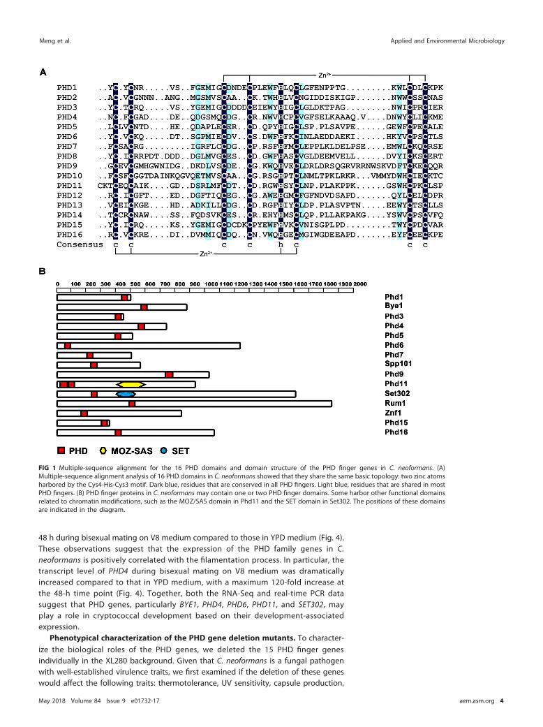
48 h during bisexual mating on V8 medium compared to those in YPD medium (Fig. 4).These observations suggest that the expression of the PHD family genes in C.neoformans is positively correlated with the filamentation process. In particular, thetranscript level of PHD4 during bisexual mating on V8 medium was dramaticallyincreased compared to that in YPD medium, with a maximum 120-fold increase atthe 48-h time point (Fig. 4). Together, both the RNA-Seq and real-time PCR datasuggest that PHD genes, particularly BYE1, PHD4, PHD6, PHD11, and SET302, mayplay a role in cryptococcal development based on their development-associatedexpression.
Phenotypical characterization of the PHD gene deletion mutants. To character-ize the biological roles of the PHD genes, we deleted the 15 PHD finger genesindividually in the XL280 background. Given that C. neoformans is a fungal pathogenwith well-established virulence traits, we first examined if the deletion of these geneswould affect the following traits: thermotolerance, UV sensitivity, capsule production,
FIG 1 Multiple-sequence alignment for the 16 PHD domains and domain structure of the PHD finger genes in C. neoformans. (A)Multiple-sequence alignment analysis of 16 PHD domains in C. neoformans showed that they share the same basic topology: two zinc atomsharbored by the Cys4-His-Cys3 motif. Dark blue, residues that are conserved in all PHD fingers. Light blue, residues that are shared in mostPHD fingers. (B) PHD finger proteins in C. neoformans may contain one or two PHD finger domains. Some harbor other functional domainsrelated to chromatin modifications, such as the MOZ/SAS domain in Phd11 and the SET domain in Set302. The positions of these domainsare indicated in the diagram.
Meng et al. Applied and Environmental Microbiology
May 2018 Volume 84 Issue 9 e01732-17 aem.asm.org 4

and melanization. Many fungal species cannot tolerate or grow at mammalian bodytemperature (33), a prerequisite for systemic infections. Consequently, temperature-sensitive cryptococcal mutants, such as calcineurin mutants or RAM pathway mutants,are often avirulent (34–36). Most of the PHD gene deletion mutants grew well at 30°C,37°C, or 39°C, similar to the wild type (WT) (Fig. 5A). The bye1Δ mutant showed modestgrowth reduction at 37°C, while the phd3Δ and phd15Δ mutants showed severe growthdefects at both 30°C and 37°C (Fig. 5A). Colonies of the phd3Δ and the phd15Δ mutantswere smaller than those of the wild type even at 30°C, and their growth defectsappeared to be much more severe at higher temperatures. At 39°C, there was barelyany growth observed in the phd3Δ mutant or the phd15Δ mutant, and some growthwas recovered after temperature downshift from 39°C to 30°C (Fig. 5B). To determineif the smaller colony size was due to a lower growth rate or a smaller cell size at 30°C,we examined the generation time of the phd3Δ mutant and its cell size. We did notobserve any significant difference in cell size in the phd3Δ mutant compared to the wildtype (Fig. 5C). As expected, the generation time for the phd3Δ mutant was twice as longas that of the wild type at 30°C in YPD liquid culture (Fig. 5D). We also tested thesensitivities of all the PHD finger deletion mutants to UV radiation. Most had sensitivityto UV radiation similar to that of the WT strain (Fig. 6A), with the exception of the phd3Δmutant, which showed increased resistance (Fig. 6A and B). Interestingly, the otherslow-growing phd15Δ mutant did not show significantly increased resistance to UVradiation, suggesting that Phd3 and Phd15 are not functionally identical. Melanin andcapsule production are two other major virulence traits of Cryptococcus. All the mutants
FIG 2 Transcript levels of PHD genes during yeast growth. (A) Transcript levels for all PHD genes arerelatively low during vegetative yeast growth in YPD medium based on our RNA-Seq data (32). The FPKMvalue of the housekeeping gene TEF1 was used as a reference. RUM1� (15) was not included in theRNA-Seq database due to its omission in the current genome annotation file. (B) The transcript level ofeach PHD finger gene in the wild-type strain XL280 cultured in YPD medium for 16 h was measured byreal-time PCR. The transcript level of TEF1 was used as a reference. The error bars indicate standarddeviations. dCt (delta cycle threshold) � CT (PHD gene) � CT (TEF1).
PHD Genes Regulate Cryptococcus Morphotype Transition Applied and Environmental Microbiology
May 2018 Volume 84 Issue 9 e01732-17 aem.asm.org 5
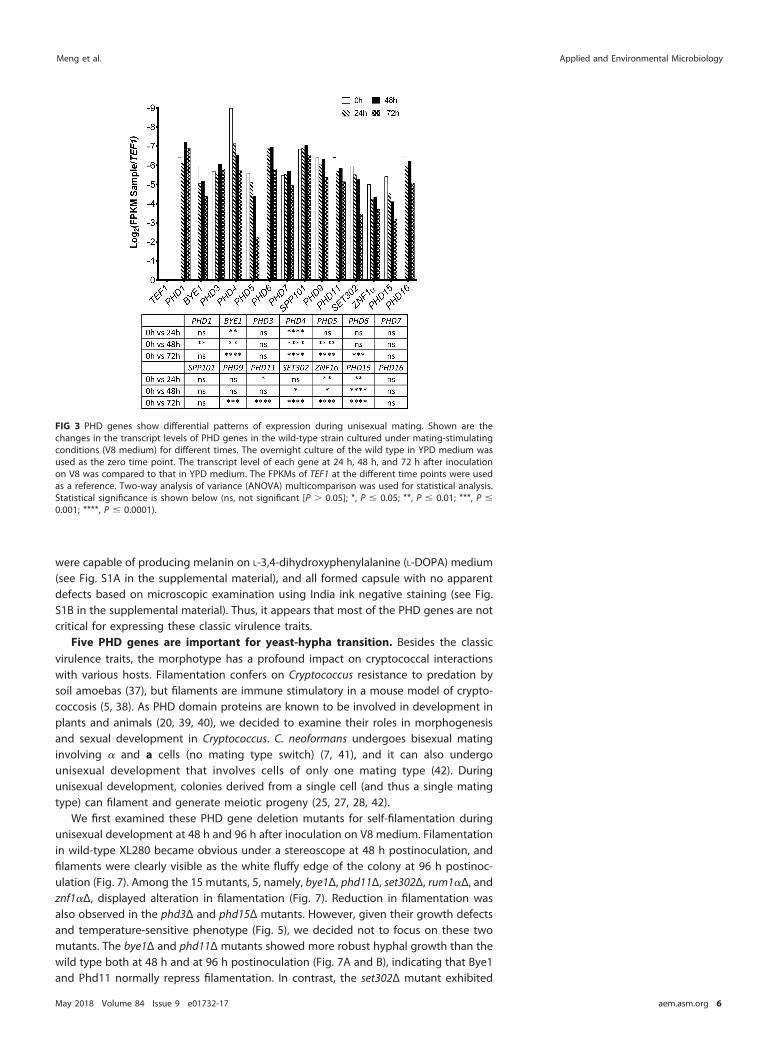
were capable of producing melanin on L-3,4-dihydroxyphenylalanine (L-DOPA) medium(see Fig. S1A in the supplemental material), and all formed capsule with no apparentdefects based on microscopic examination using India ink negative staining (see Fig.S1B in the supplemental material). Thus, it appears that most of the PHD genes are notcritical for expressing these classic virulence traits.
Five PHD genes are important for yeast-hypha transition. Besides the classicvirulence traits, the morphotype has a profound impact on cryptococcal interactionswith various hosts. Filamentation confers on Cryptococcus resistance to predation bysoil amoebas (37), but filaments are immune stimulatory in a mouse model of crypto-coccosis (5, 38). As PHD domain proteins are known to be involved in development inplants and animals (20, 39, 40), we decided to examine their roles in morphogenesisand sexual development in Cryptococcus. C. neoformans undergoes bisexual matinginvolving � and a cells (no mating type switch) (7, 41), and it can also undergounisexual development that involves cells of only one mating type (42). Duringunisexual development, colonies derived from a single cell (and thus a single matingtype) can filament and generate meiotic progeny (25, 27, 28, 42).
We first examined these PHD gene deletion mutants for self-filamentation duringunisexual development at 48 h and 96 h after inoculation on V8 medium. Filamentationin wild-type XL280 became obvious under a stereoscope at 48 h postinoculation, andfilaments were clearly visible as the white fluffy edge of the colony at 96 h postinoc-ulation (Fig. 7). Among the 15 mutants, 5, namely, bye1Δ, phd11Δ, set302Δ, rum1�Δ, andznf1�Δ, displayed alteration in filamentation (Fig. 7). Reduction in filamentation wasalso observed in the phd3Δ and phd15Δ mutants. However, given their growth defectsand temperature-sensitive phenotype (Fig. 5), we decided not to focus on these twomutants. The bye1Δ and phd11Δ mutants showed more robust hyphal growth than thewild type both at 48 h and at 96 h postinoculation (Fig. 7A and B), indicating that Bye1and Phd11 normally repress filamentation. In contrast, the set302Δ mutant exhibited
FIG 3 PHD genes show differential patterns of expression during unisexual mating. Shown are thechanges in the transcript levels of PHD genes in the wild-type strain cultured under mating-stimulatingconditions (V8 medium) for different times. The overnight culture of the wild type in YPD medium wasused as the zero time point. The transcript level of each gene at 24 h, 48 h, and 72 h after inoculationon V8 was compared to that in YPD medium. The FPKMs of TEF1 at the different time points were usedas a reference. Two-way analysis of variance (ANOVA) multicomparison was used for statistical analysis.Statistical significance is shown below (ns, not significant [P � 0.05]; *, P � 0.05; **, P � 0.01; ***, P �0.001; ****, P � 0.0001).
Meng et al. Applied and Environmental Microbiology
May 2018 Volume 84 Issue 9 e01732-17 aem.asm.org 6

drastically reduced filamentation at both time points examined (Fig. 7A and B), sug-gesting that Set302 is an activator for filamentation. Interestingly, the rum1�Δ andznf1�Δ mutants showed slightly increased robustness in filamentation at 48 h butended up with shorter filaments than the wild type at 96 h (Fig. 7A and B). This suggeststhat Rum1 and Znf1 repress the initiation of filamentation but are required for sus-tained hyphal growth.
Filamentation precedes sporulation in Cryptococcus. To examine if the five PHDgenes that regulate filamentation also play a role in sporulation, we examined thecorresponding gene deletion mutants for their ability to sporulate. The wild-type strainproduced basidia with four long chains of spores after 1 week of incubation on V8medium (see Fig. S2 in the supplemental material). The set302Δ mutant rarely yieldedspore chains at this point but eventually produced spores after prolonged incubation.This delay in sporulation was likely due to its severely impaired hyphal growth (Fig. 7Aand B). All the other mutants were able to produce normal and abundant spore chains(see Fig. S2 in the supplemental material). This result demonstrates that PHD geneswere not specifically required for sporulation.
Based on the results obtained with the gene deletion mutants, five PHD genes,namely, BYE1, PHD11, SET302, RUM1�, and ZNF1�, play important roles in regulatingfilamentation. To further examine their roles in filamentation, we decided to constitu-tively express these PHD genes using the promoter of the housekeeping gene GPD1.For Phd11, we also fused the fluorescence protein mCherry in frame to its C terminus.We then introduced these PHD gene overexpression constructs ectopically into thecorresponding gene deletion mutants. Increased transcript levels of the PHD genes in
FIG 4 Transcript levels of PHD genes during bisexual mating. The transcript levels of the PHD genesduring XL280 a-� bisexual mating were measured by real-time PCR at 3 h, 10 h, 24 h, 48 h, and 72 h afterinoculation on V8 mating-inducing medium. The transcript level of TEF1 was used as a reference.Two-way ANOVA multicomparison was used for statistical analysis. The transcript level of each gene at24 h, 48 h, and 72 h postinoculation on V8 medium was compared to that in YPD medium. Statisticalsignificance is shown below. None of the data at 3 h and 10 h showed significant difference from the 0-htime point. ns, not significant (P � 0.05); *, P � 0.05; **, P � 0.01; ****, P � 0.0001. The error bars indicatestandard deviations.
PHD Genes Regulate Cryptococcus Morphotype Transition Applied and Environmental Microbiology
May 2018 Volume 84 Issue 9 e01732-17 aem.asm.org 7
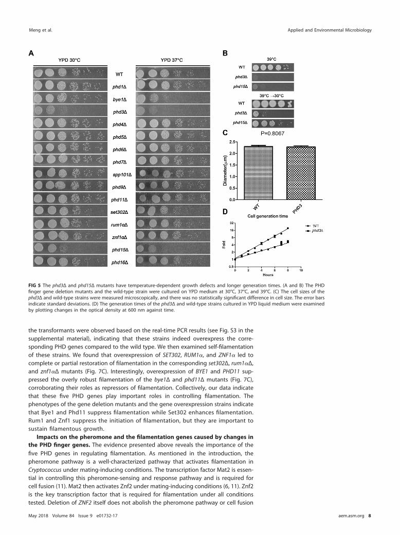
the transformants were observed based on the real-time PCR results (see Fig. S3 in thesupplemental material), indicating that these strains indeed overexpress the corre-sponding PHD genes compared to the wild type. We then examined self-filamentationof these strains. We found that overexpression of SET302, RUM1�, and ZNF1� led tocomplete or partial restoration of filamentation in the corresponding set302Δ, rum1�Δ,and znf1�Δ mutants (Fig. 7C). Interestingly, overexpression of BYE1 and PHD11 sup-pressed the overly robust filamentation of the bye1Δ and phd11Δ mutants (Fig. 7C),corroborating their roles as repressors of filamentation. Collectively, our data indicatethat these five PHD genes play important roles in controlling filamentation. Thephenotypes of the gene deletion mutants and the gene overexpression strains indicatethat Bye1 and Phd11 suppress filamentation while Set302 enhances filamentation.Rum1 and Znf1 suppress the initiation of filamentation, but they are important tosustain filamentous growth.
Impacts on the pheromone and the filamentation genes caused by changes inthe PHD finger genes. The evidence presented above reveals the importance of thefive PHD genes in regulating filamentation. As mentioned in the introduction, thepheromone pathway is a well-characterized pathway that activates filamentation inCryptococcus under mating-inducing conditions. The transcription factor Mat2 is essen-tial in controlling this pheromone-sensing and response pathway and is required forcell fusion (11). Mat2 then activates Znf2 under mating-inducing conditions (6, 11). Znf2is the key transcription factor that is required for filamentation under all conditionstested. Deletion of ZNF2 itself does not abolish the pheromone pathway or cell fusion
FIG 5 The phd3Δ and phd15Δ mutants have temperature-dependent growth defects and longer generation times. (A and B) The PHDfinger gene deletion mutants and the wild-type strain were cultured on YPD medium at 30°C, 37°C, and 39°C. (C) The cell sizes of thephd3Δ and wild-type strains were measured microscopically, and there was no statistically significant difference in cell size. The error barsindicate standard deviations. (D) The generation times of the phd3Δ and wild-type strains cultured in YPD liquid medium were examinedby plotting changes in the optical density at 600 nm against time.
Meng et al. Applied and Environmental Microbiology
May 2018 Volume 84 Issue 9 e01732-17 aem.asm.org 8
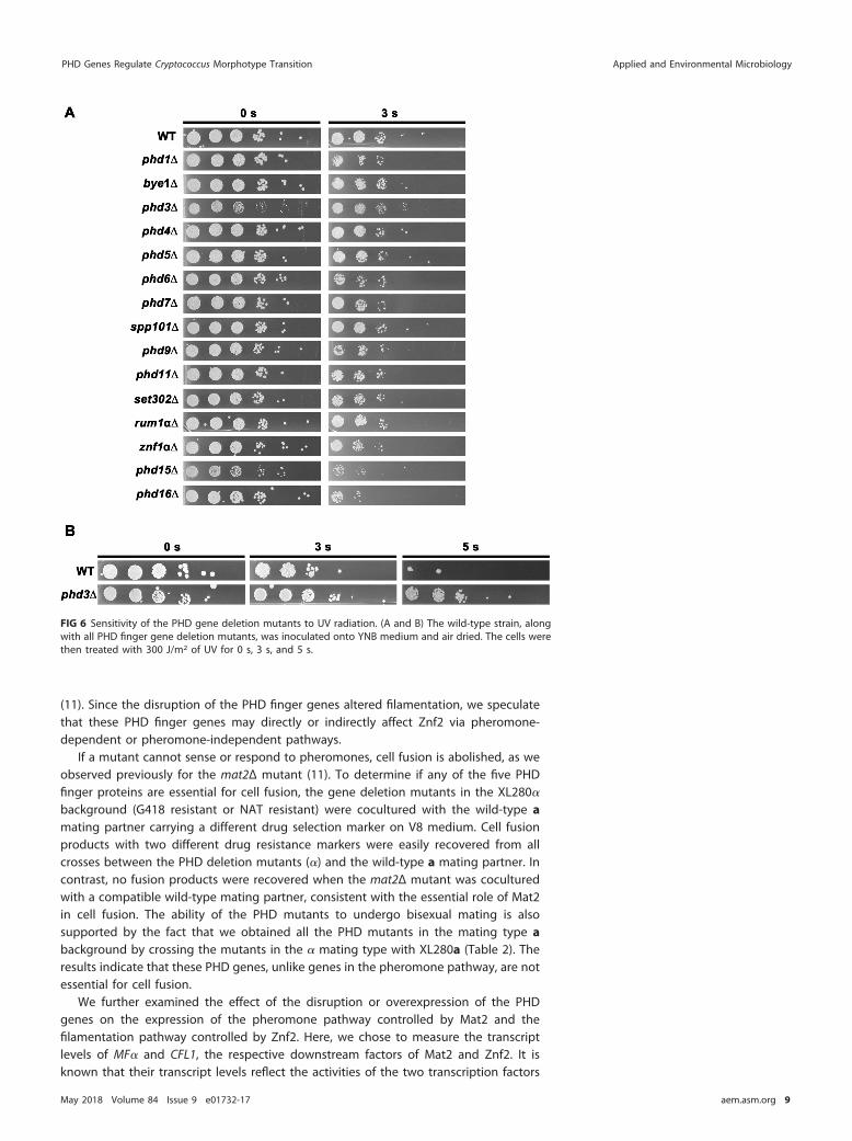
(11). Since the disruption of the PHD finger genes altered filamentation, we speculatethat these PHD finger genes may directly or indirectly affect Znf2 via pheromone-dependent or pheromone-independent pathways.
If a mutant cannot sense or respond to pheromones, cell fusion is abolished, as weobserved previously for the mat2Δ mutant (11). To determine if any of the five PHDfinger proteins are essential for cell fusion, the gene deletion mutants in the XL280�
background (G418 resistant or NAT resistant) were cocultured with the wild-type amating partner carrying a different drug selection marker on V8 medium. Cell fusionproducts with two different drug resistance markers were easily recovered from allcrosses between the PHD deletion mutants (�) and the wild-type a mating partner. Incontrast, no fusion products were recovered when the mat2Δ mutant was coculturedwith a compatible wild-type mating partner, consistent with the essential role of Mat2in cell fusion. The ability of the PHD mutants to undergo bisexual mating is alsosupported by the fact that we obtained all the PHD mutants in the mating type abackground by crossing the mutants in the � mating type with XL280a (Table 2). Theresults indicate that these PHD genes, unlike genes in the pheromone pathway, are notessential for cell fusion.
We further examined the effect of the disruption or overexpression of the PHDgenes on the expression of the pheromone pathway controlled by Mat2 and thefilamentation pathway controlled by Znf2. Here, we chose to measure the transcriptlevels of MF� and CFL1, the respective downstream factors of Mat2 and Znf2. It isknown that their transcript levels reflect the activities of the two transcription factors
FIG 6 Sensitivity of the PHD gene deletion mutants to UV radiation. (A and B) The wild-type strain, alongwith all PHD finger gene deletion mutants, was inoculated onto YNB medium and air dried. The cells werethen treated with 300 J/m2 of UV for 0 s, 3 s, and 5 s.
PHD Genes Regulate Cryptococcus Morphotype Transition Applied and Environmental Microbiology
May 2018 Volume 84 Issue 9 e01732-17 aem.asm.org 9
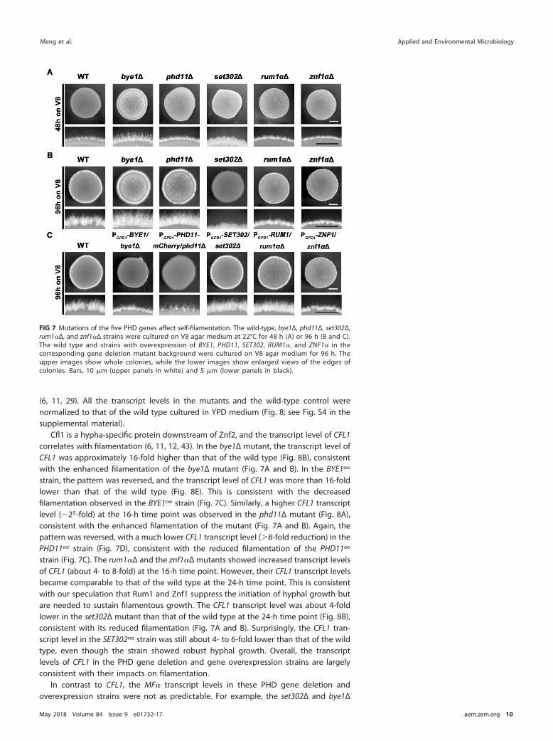
(6, 11, 29). All the transcript levels in the mutants and the wild-type control werenormalized to that of the wild type cultured in YPD medium (Fig. 8; see Fig. S4 in thesupplemental material).
Cfl1 is a hypha-specific protein downstream of Znf2, and the transcript level of CFL1correlates with filamentation (6, 11, 12, 43). In the bye1Δ mutant, the transcript level ofCFL1 was approximately 16-fold higher than that of the wild type (Fig. 8B), consistentwith the enhanced filamentation of the bye1Δ mutant (Fig. 7A and B). In the BYE1oe
strain, the pattern was reversed, and the transcript level of CFL1 was more than 16-foldlower than that of the wild type (Fig. 8E). This is consistent with the decreasedfilamentation observed in the BYE1oe strain (Fig. 7C). Similarly, a higher CFL1 transcriptlevel (�25-fold) at the 16-h time point was observed in the phd11Δ mutant (Fig. 8A),consistent with the enhanced filamentation of the mutant (Fig. 7A and B). Again, thepattern was reversed, with a much lower CFL1 transcript level (�8-fold reduction) in thePHD11oe strain (Fig. 7D), consistent with the reduced filamentation of the PHD11oe
strain (Fig. 7C). The rum1�Δ and the znf1�Δ mutants showed increased transcript levelsof CFL1 (about 4- to 8-fold) at the 16-h time point. However, their CFL1 transcript levelsbecame comparable to that of the wild type at the 24-h time point. This is consistentwith our speculation that Rum1 and Znf1 suppress the initiation of hyphal growth butare needed to sustain filamentous growth. The CFL1 transcript level was about 4-foldlower in the set302Δ mutant than that of the wild type at the 24-h time point (Fig. 8B),consistent with its reduced filamentation (Fig. 7A and B). Surprisingly, the CFL1 tran-script level in the SET302oe strain was still about 4- to 6-fold lower than that of the wildtype, even though the strain showed robust hyphal growth. Overall, the transcriptlevels of CFL1 in the PHD gene deletion and gene overexpression strains are largelyconsistent with their impacts on filamentation.
In contrast to CFL1, the MF� transcript levels in these PHD gene deletion andoverexpression strains were not as predictable. For example, the set302Δ and bye1Δ
FIG 7 Mutations of the five PHD genes affect self-filamentation. The wild-type, bye1Δ, phd11Δ, set302Δ,rum1�Δ, and znf1�Δ strains were cultured on V8 agar medium at 22°C for 48 h (A) or 96 h (B and C).The wild type and strains with overexpression of BYE1, PHD11, SET302, RUM1�, and ZNF1� in thecorresponding gene deletion mutant background were cultured on V8 agar medium for 96 h. Theupper images show whole colonies, while the lower images show enlarged views of the edges ofcolonies. Bars, 10 �m (upper panels in white) and 5 �m (lower panels in black).
Meng et al. Applied and Environmental Microbiology
May 2018 Volume 84 Issue 9 e01732-17 aem.asm.org 10
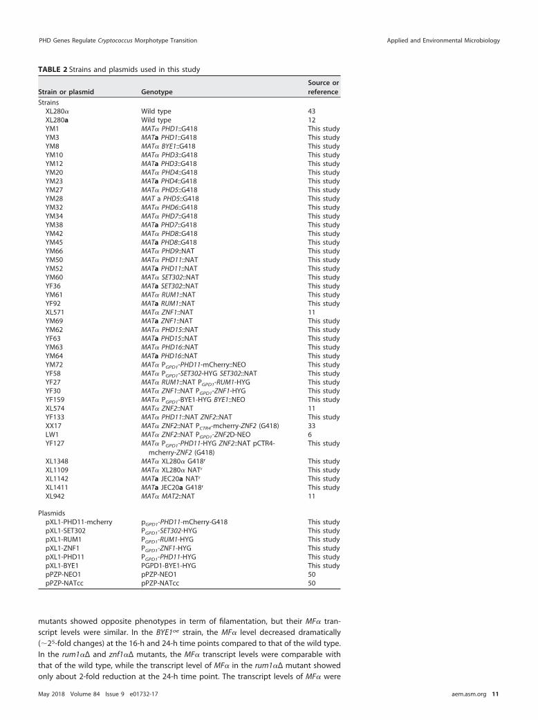
mutants showed opposite phenotypes in term of filamentation, but their MF� tran-script levels were similar. In the BYE1oe strain, the MF� level decreased dramatically(�25-fold changes) at the 16-h and 24-h time points compared to that of the wild type.In the rum1�Δ and znf1�Δ mutants, the MF� transcript levels were comparable withthat of the wild type, while the transcript level of MF� in the rum1�Δ mutant showedonly about 2-fold reduction at the 24-h time point. The transcript levels of MF� were
TABLE 2 Strains and plasmids used in this study
Strain or plasmid GenotypeSource orreference
StrainsXL280� Wild type 43XL280a Wild type 12YM1 MAT� PHD1::G418 This studyYM3 MATa PHD1::G418 This studyYM8 MAT� BYE1::G418 This studyYM10 MAT� PHD3::G418 This studyYM12 MATa PHD3::G418 This studyYM20 MAT� PHD4::G418 This studyYM23 MATa PHD4::G418 This studyYM27 MAT� PHD5::G418 This studyYM28 MAT a PHD5::G418 This studyYM32 MAT� PHD6::G418 This studyYM34 MAT� PHD7::G418 This studyYM38 MATa PHD7::G418 This studyYM42 MAT� PHD8::G418 This studyYM45 MATa PHD8::G418 This studyYM66 MAT� PHD9::NAT This studyYM50 MAT� PHD11::NAT This studyYM52 MATa PHD11::NAT This studyYM60 MAT� SET302::NAT This studyYF36 MATa SET302::NAT This studyYM61 MAT� RUM1::NAT This studyYF92 MATa RUM1::NAT This studyXL571 MAT� ZNF1::NAT 11YM69 MATa ZNF1::NAT This studyYM62 MAT� PHD15::NAT This studyYF63 MATa PHD15::NAT This studyYM63 MAT� PHD16::NAT This studyYM64 MATa PHD16::NAT This studyYM72 MAT� PGPD1-PHD11-mCherry::NEO This studyYF58 MAT� PGPD1-SET302-HYG SET302::NAT This studyYF27 MAT� RUM1::NAT PGPD1-RUM1-HYG This studyYF30 MAT� ZNF1::NAT PGPD1-ZNF1-HYG This studyYF159 MAT� PGPD1-BYE1-HYG BYE1::NEO This studyXL574 MAT� ZNF2::NAT 11YF133 MAT� PHD11::NAT ZNF2::NAT This studyXX17 MAT� ZNF2::NAT PCTR4-mcherry-ZNF2 (G418) 33LW1 MAT� ZNF2::NAT PGPD1-ZNF2D-NEO 6YF127 MAT� PGPD1-PHD11-HYG ZNF2::NAT pCTR4-
mcherry-ZNF2 (G418)This study
XL1348 MAT� XL280� G418r This studyXL1109 MAT� XL280� NATr This studyXL1142 MATa JEC20a NATr This studyXL1411 MATa JEC20a G418r This studyXL942 MAT� MAT2::NAT 11
PlasmidspXL1-PHD11-mcherry pGPD1-PHD11-mCherry-G418 This studypXL1-SET302 PGPD1-SET302-HYG This studypXL1-RUM1 PGPD1-RUM1-HYG This studypXL1-ZNF1 PGPD1-ZNF1-HYG This studypXL1-PHD11 PGPD1-PHD11-HYG This studypXL1-BYE1 PGPD1-BYE1-HYG This studypPZP-NEO1 pPZP-NEO1 50pPZP-NATcc pPZP-NATcc 50
PHD Genes Regulate Cryptococcus Morphotype Transition Applied and Environmental Microbiology
May 2018 Volume 84 Issue 9 e01732-17 aem.asm.org 11
significantly different in the phd11Δ and PHD11oe strains at the 16-h time point (see Fig.S4 in the supplemental material), and the difference became smaller at the later timepoint. The inconsistency of the MF� transcript levels in these strains with the mutantphenotype in filamentation is consistent with the idea that the pheromone pathway isnot the major effector of these PHD proteins.
Phd11 functions upstream of Znf2 in regulating filamentation. Although Znf2 iswell established in its essential role for hyphal growth (6, 11, 12), the regulatory circuitscontrolling Znf2, especially factors that suppress filamentation, are unclear. Given thephenotypes of the PHD11 gene deletion mutant (enhanced filamentation) and theoverexpression strain (reduced filamentation) (Fig. 7A to C), Phd11 works as a suppres-sor of filamentation. Here, we decided to further investigate the relationship betweenPhd11 and Znf2. First, we examined the impacts of deletion and overexpression of ZNF2on the transcript level of PHD11. As shown in Fig. 9A, the PHD11 transcript levels in thewild-type strain, the znf2Δ mutant, and the ZNF2oe strain were similar regardless ofwhether cells were cultured in YPD or on V8 medium. This suggests that Znf2 does notaffect Phd11 at the transcript level. We then examined the transcript level of ZNF2 inthe wild type, the phd11Δ mutant, and the PHD11oe strain. As expected, the transcriptlevel of ZNF2 was low when wild-type cells were cultured in YPD and it was drasticallyincreased (�25-fold higher) when the wild-type cells were cultured on V8 medium (Fig.9B). Although the deletion of PHD11 did not show much impact on the ZNF2 transcriptlevel, the overexpression of PHD11 led to a drastically reduced transcript level of ZNF2(�26-fold reduction) on V8 medium when ZNF2 is normally induced in the wild type.Altogether, Phd11 may act upstream of Znf2 and negatively affect the ZNF2 transcrip-tional level.
To further investigate the relationship between PHD11 and ZNF2, we made thephd11Δ znf2Δ double mutant. The phd11Δ single mutant showed enhanced filamen-tation, while the znf2Δ single mutant had filamentation abolished (Fig. 9C). The phd11Δ
FIG 8 The transcript level of the filamentation marker CFL1 shows good correlation with the filamentation phenotypes of the PHD gene deletion oroverexpression strains. The transcript levels of CFL1 were measured by real-time PCR in the wild type, the five PHD gene deletion mutants (bye1Δ, phd11Δ,set302Δ, rum1�Δ, and znf1�Δ), and overexpression strains (BYE1oe/bye1Δ, PHD11oe/phd11Δ, and SET302oe/set302Δ). Overnight cultures of these strains in YPDliquid medium were considered the zero time point. The cells were then cultured on V8 agar medium for 16 h and 24 h. The CFL1 transcript level in the wildtype at the zero time point was used for normalization. The y axes show the log2 changes in the transcript level of CFL1. The error bars indicate standarddeviations.
Meng et al. Applied and Environmental Microbiology
May 2018 Volume 84 Issue 9 e01732-17 aem.asm.org 12
znf2Δ double mutant showed no hyphal growth, similar to the znf2Δ single mutant(Fig. 9C). Thus, the deletion of PHD11, although it increased filamentation in a wild-typebackground, failed to confer filamentation on the znf2Δ mutant.
To further examine the relationship between Phd11 and Znf2, we constructed astrain with constitutive overexpression of PHD11 and inducible expression of ZNF2. Wehypothesized that if Phd11 functions upstream of Znf2, then overexpression of PHD11when ZNF2 is not expressed would recapitulate the znf2Δ-like nonfilamentous pheno-type. On the other hand, overexpression of the downstream factor ZNF2 shouldovercome the suppressive effect of the overexpression of PHD11 and drive robustfilamentation. For this purpose, we had PHD11 driven by the constitutively active GPD1promoter and ZNF2 by the inducible promoter of the copper transporter gene CTR4(Fig. 9D). Because V8 medium itself is slightly copper limiting, the CTR4 promoter wouldbe activated under these conditions, and consequently, ZNF2 would be expressed.Indeed, the PCTR4-ZNF2/znf2Δ strain showed robust hyphal growth on V8 medium (Fig.9E). The addition of copper to V8 medium suppresses the CTR4 promoter, and ZNF2 isnot expressed. Consistently, the PCTR4-ZNF2/znf2Δ strain grew only yeast colonies in thepresence of copper (Fig. 9E). When PHD11 was constitutively overexpressed, thePGPD1-PHD11/phd11Δ strain showed repressed hyphal growth compared to the wildtype (Fig. 9E). When both PHD11 and ZNF2 were expressed on V8 medium, thePGPD1-PHD11/PCTR4-ZNF2/znf2Δ strain showed robust hyphal growth, similar to theZNF2oe strain (Fig. 9E). When excess copper ion was added to the V8 medium,the expression of ZNF2 was suppressed. This resulted in the nonfilamentous phenotypeof the PGPD1-PHD11/PCTR4-ZNF2/znf2Δ strain (Fig. 9E), as we predicted. These resultsagain corroborate the essential role of Znf2 in filamentation and demonstrate thatPhd11 indeed functions upstream of Znf2 in controlling the yeast-to-hypha morpho-logical change.
FIG 9 Phd11 functions upstream of the master morphogenesis regulator Znf2. (A) WT, znf2Δ, and ZNF2oe cells were cultured in YPD liquid mediumovernight or on V8 agar medium for 24 h. The transcript levels of PHD11 were measured by real-time PCR. The PHD11 transcript level in theWT cultured in YPD medium was used as a reference. (B) WT, phd11Δ, and PHD11oe cells were cultured in YPD liquid medium overnight oron V8 medium for 16 h. The transcript levels of ZNF2 were measured by RT-PCR. The ZNF2 transcript level of the WT cultured in YPD wasused as a reference. (C) WT, phd11Δ, znf2Δ, and phd11Δznf2Δ double-mutant cells were cultured on V8 agar medium for 3 days. (D) Diagramof the strain with constitutive expression of PHD11 driven by the GPD1 promoter and the inducible expression of ZNF2 driven by the CTR4promoter. The expression of ZNF2 is induced by copper limitation and suppressed by excess copper. (E) The WT, the PGPD1-PHD11/phd11Δstrain, the PCTR4-ZNF2/znf2Δ strain, and the PGPD1-PHD11/PCTR4-ZNF2/znf2Δ strain were cultured on copper-limited V8 juice agar medium(ZNF2-inducing condition) and on V8 agar supplemented with 50 �M CuSO4 (ZNF2-suppressing condition). The error bars indicate standarddeviations.
PHD Genes Regulate Cryptococcus Morphotype Transition Applied and Environmental Microbiology
May 2018 Volume 84 Issue 9 e01732-17 aem.asm.org 13

DISCUSSION
The PHD finger is found in many chromatin-remodeling proteins in eukaryotes (20,39, 40), and their activities could lead to altered gene expression and, consequently,regulate various biological processes. In our study, we systematically deleted PHDfinger genes in the environmental opportunistic fungal pathogen C. neoformans. Wefound that PHD3 and PHD15 are critical for thermotolerance. Given that growth atmammalian body temperature is a prerequisite for cryptococcal pathogenesis, it wouldbe interesting to identify the effector genes and to investigate their potential impactson cryptococcal virulence in the future. Interestingly, we identified five PHD genes,namely, BYE1, PHD11, SET302, RUM1, and ZNF1, that contribute to the yeast-hyphamorphological switch in Cryptococcus. Accordingly, we found that the deletion oroverexpression of these five PHD genes alters the expression of filamentation-relatedgenes. Among the products of the five PHD finger genes, Bye1 and Phd11 work assuppressors of filamentation and Set302 functions as an activator of filamentation.Rum1 and Znf1, two PHD proteins encoded in the MAT locus, suppress the initiationof filamentation but are important for maintaining robust hyphal growth. We choseto investigate Set302 and Phd11 further because set302Δ and phd11Δ strainsshowed the most dramatic and opposing phenotypes in terms of filamentation.Set302 and Phd11 are predicted to be nuclear proteins due to the presence of anuclear localization signal (NLS). While attempts to construct mCherry-fused Set302failed, the generated Phd11-mCherry indeed yielded stable and clear fluorescencethat colocalized with DAPI (4=,6-diamidino-2-phenylindole) staining (Fig. 10). Thenuclear localization of Phd11 is consistent with its predicted nuclear function as achromatin reader. Phd11 harbors a predicted histone acetyltransferase MOZ-SASdomain, while Set302 contains a SET domain. The unique domain structures inthese two PHD proteins suggest that Phd11 and Set302 may act not only as readersof histone modifications, due to their PHD domains, but also as writers of histonemodifications. Further mechanistic studies are warranted to fully understand theirmodes of action.
MATERIALS AND METHODSStrains and growth conditions. The strains used in this study are listed in Table 2. All the strains
were stored as glycerol stocks at �80°C. For experiments, cells were streaked out from freezer stocks andcultured on YPD medium at 30°C unless otherwise indicated.
Construction of gene deletion mutants and overexpression strains. For gene deletion, we usedthe method of double-joint PCR as described previously (44). Briefly, two partially overlapping parts ofthe chosen drug selection marker (NEO, NAT, or HYG) were fused with the 5= or 3= flanking region (1 kb)of the open reading frame (ORF) of each gene of interest. The mixture of the pair of fusion products wasthen introduced into the recipient strain by biolistic transformation as described previously (45).Transformants grown on the drug-selective medium (YPD plus NAT, YPD plus NEO, or YPD plus HYG)were checked for stability, and stable transformants were then screened for homologous replacementevents based on diagnostic PCR, as described previously (46). The primers used in generating geneknockout strains and for screening the homologous replacement events are listed in Table 3. For geneoverexpression, the ORF of the gene of interest was amplified using the genomic DNA of the wild-typestrain XL280 as a template. The resulting amplicons were digested with restriction enzymes FseI and PacI,and the digested products were then inserted into the plasmid pXL1 after the GPD1 promoter asdescribed previously (6). The resulting plasmids were linearized after restriction enzyme digestion andthen introduced into C. neoformans cells by biolistic transformation as described previously (45).Transformants grown on the drug-selective medium (YPD plus NAT, YPD plus NEO, or YPD plus HYG)were examined for stability after 5 passages on nonselective medium, and the stable transformants wereconfirmed by phenotypic assay, diagnostic PCR, or real-time PCR. For the inducible expression system,the construct was generated by the same method except that the amplicon was inserted after the CTR4promoter as described previously (6).
FIG 10 Phd11 is located in the nucleus. The Phd11-mCherry strain was cultured in YPD medium. Cellswere fixed and stained with DAPI. DIC, differential inference contrast.
Meng et al. Applied and Environmental Microbiology
May 2018 Volume 84 Issue 9 e01732-17 aem.asm.org 14
TABLE 3 Primers used in this study
Primer no. Name Sequence
M13F GTAAAACGACGGCCAGM13R AACAGCTATGACCATG
Linlab2091 PHD1-Left Forward CTACATGTGACACTTACCGTACTGLinlab2092 PHD1-M13F complement�Left Reverse CTGGCCGTCGTTTTACCTGGTAGATGAGCGCTATGLinlab2093 PHD1-M13R complement�Right Forward GTCATAGCTGTTTCCTGCCTATCCATACCCCTTATAATGLinlab2094 PHD1-Right Reverse AAGAATTGGAGGATTTACAAAGLinlab2095 PHD1-Far left forward TCGTCTCGAGAGGTAGTTGTCLinlab2096 BYE1-Left Forward ACATGCAGCTTTGTGATACGLinlab2097 BYE1-M13F complement�Left Reverse CTGGCCGTCGTTTTACAAGCTGTGTCTATCCCGCLinlab2098 BYE1-M13R complement�Right Forward GTCATAGCTGTTTCCTGGTATTGTTGCACGTGTTCAGTCLinlab2099 BYE1-Right Reverse AGCGCTGTTATTTCTTATCGLinlab2100 BYE1-Far left forward ATGCGACTCCACCACTACTAGLinlab2288 BYE1-Left-forward-nested CATGGCTTTGTTGCTTCACLinlab2101 PHD3-Left Forward CACGAAAGTATCTTCATTCATTGLinlab2102 PHD3-M13F complement�Left Reverse CTGGCCGTCGTTTTACTGTAGGGCGAATTATATGGLinlab2103 PHD3-M13R complement�Right Forward GTCATAGCTGTTTCCTGTACTATCGTTACTGGCACATACLinlab2104 PHD3-Right Reverse GAAACTCACTGCCGTAGAGGLinlab2105 PHD3-Far left forward GCTACCTCTTTGTCTACTACTGCLinlab2106 PHD4-Left Forward CCTATCGATGGAGTAGAGCCLinlab2107 PHD4-M13F complement�Left Reverse CTGGCCGTCGTTTTACATTCATGCCCTCGGAGCLinlab2108 PHD4-M13R complement�Right Forward GTCATAGCTGTTTCCTGATCCTGCCTTTCTCAATGCLinlab2109 PHD4-Right Reverse CATCCATCACACTGCATACTCLinlab2110 PHD4-Far left forward GCATCCAGGACAATTACATCLinlab2225 PHD4-Right reverse2 CGCCTTTCATGACTCTCGLinlab2289 PHD4-Left-forward-nested TGTTCAGCCCTTCTCGTGLinlab2160 PHD5-Left Forward GGACGAGAGAGAGGACGAGLinlab2161 PHD5-M13F complement�Left Reverse CTGGCCGTCGTTTTACAGATGCACCGATGAGCGLinlab2162 PHD5-M13R complement�Right Forward GTCATAGCTGTTTCCTGCCATCGGCGTTTGACTGLinlab2163 PHD5-Right Reverse ATCCGAGCTGTCGTTGGLinlab2164 PHD5-Far left forward CCATCACCTCCGAGCTLinlab2165 PHD6-Left Forward AGCAACGCTGGATCTGGLinlab2166 PHD6-M13F complement�Left Reverse CTGGCCGTCGTTTTACATACAGCGATGGAAAGTGGLinlab2167 PHD6-M13R complement�Right Forward GTCATAGCTGTTTCCTGGTGCATAGGCATATACTTGGLinlab2168 PHD6-Right Reverse GGTACCGCCTGCACAAGLinlab2169 PHD6-Far left forward TGGAGGTCCTCGAACTCTGLinlab2348 PHD6-Left Forward-nested GGCGACAACAGTTGCATAGLinlab2349 PHD6-Right reverse-nested TCAGCAGACTCAATCAGCGLinlab2170 PHD7-Left Forward AAACGGTATTGATCTTCGCLinlab2171 PHD7-M13F complement�Left Reverse CTGGCCGTCGTTTTACACTGATGCGATGTTATGCCLinlab2172 PHD7-M13R complement�Right Forward GTCATAGCTGTTTCCTGCGTTCACCGAACTGTACGLinlab2173 PHD7-Right Reverse CTGTTGTTGCCAACCCTGLinlab2174 PHD7-Far left forward AGAGCCGCTGAGTCCTTCLinlab2175 PHD8-Left Forward CGGTCATGGCGTAGAGTCLinlab2176 PHD8-M13F complement�Left Reverse CTGGCCGTCGTTTTACAAGACAAGGGCAAAGCGLinlab2177 PHD8-M13R complement�Right Forward GTCATAGCTGTTTCCTGGCATGTGCATCTCCTCTCCLinlab2178 PHD8-Right Reverse CCCAGAGCCATCTAGCGLinlab2179 PHD8-Far left forward AGGCGACTTTCGTGATTGLinlab2356 PHD9-Left forward-nested GGCTGCGAAGGACAAAGLinlab2357 PHD9-Right reverse-nested TGGCTTCCTGTCACTTGCLinlab2220 PHD9-Left Forward CACGAGCACTTGGATGGLinlab2221 PHD9-M13F complement�Left Reverse CTGGCCGTCGTTTTACATTGACAACAGGTGGATGCLinlab2222 PHD9-M13R complement�Right Forward GTCATAGCTGTTTCCTGCCTCCAGATCCGAGATTGLinlab2223 PHD9-Right Reverse AGTTCACTGGGGTGTTGCLinlab2224 PHD9-Far left forward GCGAGGAATTTGAACCAGLinlab2190 PHD11-Left Forward CATCAAACTCTGCCACAGGLinlab2191 PHD11-M13F complement�Left Reverse CTGGCCGTCGTTTTACCGTGTCTCATGCATGGAGLinlab2192 PHD11-M13R complement�Right Forward GTCATAGCTGTTTCCTGCAAGTTTCGGGCTTTGGLinlab2193 PHD11-Right Reverse GTGGACCGAGGCGAAAGLinlab2194 PHD11-Far left forward GGCCGTGAAGCGTGTAGLinlab2195 SET302-Left Forward GGTCCGACATTTTCCAGGLinlab2196 SET302-M13F complement�Left Reverse CTGGCCGTCGTTTTACATCTACAAGCGGGTCAGGLinlab2197 SET302-M13R complement�Right Forward GTCATAGCTGTTTCCTGGCGTTACCGTTGTTGTCCLinlab2198 SET302-Right Reverse CCCCTTTTCGCTTTTGCLinlab2199 SET302-Far left forward AAGCGTTGGGATCCCAGLinlab2350 SET302-Left forward-nested CTCGCTTTCGGGATGAGLinlab2351 SET302-Right reverse-nested CAACGGCTTAACCCTGTC
(Continued on next page)
PHD Genes Regulate Cryptococcus Morphotype Transition Applied and Environmental Microbiology
May 2018 Volume 84 Issue 9 e01732-17 aem.asm.org 15
TABLE 3 (Continued)
Primer no. Name Sequence
Linlab2200 RUM1-Left Forward GCCACAACTCGTCCGTGLinlab2201 RUM1-M13F complement�Left Reverse CTGGCCGTCGTTTTACAAAGGGAGTGCGTGCTGLinlab2202 RUM1-M13R complement�Right Forward GTCATAGCTGTTTCCTGCCTTCCAGATTCGTGAGCLinlab2203 RUM1-Right Reverse GAGCTGCTCGATGTACCACLinlab2204 RUM1-Far left forward CGTCACCATCATTTCCGLinlab2205 ZNF1-Left Forward AACTCCTGGGCTCAACGLinlab2206 ZNF1-M13F complement�Left Reverse CTGGCCGTCGTTTTACTTTGCACAGGGTGACCAGLinlab2207 ZNF1-M13R complement�Right Forward GTCATAGCTGTTTCCTGATTCCGGCTGATGCTTCLinlab2208 ZNF1-Right Reverse CCAAGCTTGGCAATTCGLinlab2209 ZNF1-Far left forward GCAAGCGTTGGCTCAACLinlab2210 PHD15-Left Forward TGTGAAGCCGAGGGACCLinlab2211 PHD15-M13F complement�Left Reverse CTGGCCGTCGTTTTACAGATGGATGAGTGGCGGLinlab2212 PHD15-M13R complement�Right Forward GTCATAGCTGTTTCCTGGCGAGATGGTTGCTTTTGLinlab2213 PHD15-Right Reverse TCCAACCTCCAACACCACLinlab2214 PHD15-Far left forward GCTTCCGGGGATAAACCLinlab2352 PHD15-Left forward-nested TGGCTAGAGGAATGGCTGLinlab2353 PHD15-Right reverse-nested TGTGTTTTGCGGTGTTGAGLinlab2354 PHD16-Left forward-nested CACCGAGTACAGCTGCAACLinlab2355 PHD16-Right reverse-nested GGCGTTCAGACTCCTTTGLinlab2176 PHD8-M13F complement�Left Reverse CTGGCCGTCGTTTTACAAGACAAGGGCAAAGCGLinlab2177 PHD8-M13R complement�Right Forward GTCATAGCTGTTTCCTGGCATGTGCATCTCCTCTCCLinlab2178 PHD8-Right Reverse CCCAGAGCCATCTAGCGLinlab2179 PHD8-Far left forward AGGCGACTTTCGTGATTGLinlab2356 PHD9-Left forward-nested GGCTGCGAAGGACAAAGLinlab2357 PHD9-Right reverse-nested TGGCTTCCTGTCACTTGCLinlab2220 PHD9-Left Forward CACGAGCACTTGGATGGLinlab2221 PHD9-M13F complement�Left Reverse CTGGCCGTCGTTTTACATTGACAACAGGTGGATGCLinlab2222 PHD9-M13R complement�Right Forward GTCATAGCTGTTTCCTGCCTCCAGATCCGAGATTGLinlab2223 PHD9-Right Reverse AGTTCACTGGGGTGTTGCLinlab2224 PHD9-Far left forward GCGAGGAATTTGAACCAGLinlab2190 PHD11-Left Forward CATCAAACTCTGCCACAGGLinlab2191 PHD11-M13F complement�Left Reverse CTGGCCGTCGTTTTACCGTGTCTCATGCATGGAGLinlab2192 PHD11-M13R complement�Right Forward GTCATAGCTGTTTCCTGCAAGTTTCGGGCTTTGGLinlab2193 PHD11-Right Reverse GTGGACCGAGGCGAAAGLinlab2194 PHD11-Far left forward GGCCGTGAAGCGTGTAGLinlab2195 SET302-Left Forward GGTCCGACATTTTCCAGGLinlab2196 SET302-M13F complement�Left Reverse CTGGCCGTCGTTTTACATCTACAAGCGGGTCAGGLinlab2197 SET302-M13R complement�Right Forward GTCATAGCTGTTTCCTGGCGTTACCGTTGTTGTCCLinlab2198 SET302-Right Reverse CCCCTTTTCGCTTTTGCLinlab2199 SET302-Far left forward AAGCGTTGGGATCCCAGLinlab2350 SET302-Left forward-nested CTCGCTTTCGGGATGAGLinlab2351 SET302-Right reverse-nested CAACGGCTTAACCCTGTCLinlab2200 RUM1-Left Forward GCCACAACTCGTCCGTGLinlab2201 RUM1-M13F complement�Left Reverse CTGGCCGTCGTTTTACAAAGGGAGTGCGTGCTGLinlab2202 RUM1-M13R complement�Right Forward GTCATAGCTGTTTCCTGCCTTCCAGATTCGTGAGCLinlab2203 RUM1-Right Reverse GAGCTGCTCGATGTACCACLinlab2204 RUM1-Far left forward CGTCACCATCATTTCCGLinlab2205 ZNF1-Left Forward AACTCCTGGGCTCAACGLinlab2206 ZNF1-M13F complement�Left Reverse CTGGCCGTCGTTTTACTTTGCACAGGGTGACCAGLinlab2208 ZNF1-Right Reverse CCAAGCTTGGCAATTCGLinlab2207 ZNF1-M13R complement�Right Forward GTCATAGCTGTTTCCTGATTCCGGCTGATGCTTCLinlab2209 ZNF1-Far left forward GCAAGCGTTGGCTCAACLinlab2210 PHD15-Left Forward TGTGAAGCCGAGGGACCLinlab2211 PHD15-M13F complement�Left Reverse CTGGCCGTCGTTTTACAGATGGATGAGTGGCGGLinlab2212 PHD15-M13R complement�Right Forward GTCATAGCTGTTTCCTGGCGAGATGGTTGCTTTTGLinlab2213 PHD15-Right Reverse TCCAACCTCCAACACCACLinlab2214 PHD15-Far left forward GCTTCCGGGGATAAACCLinlab2352 PHD15-Left forward-nested TGGCTAGAGGAATGGCTGLinlab2353 PHD15-Right reverse-nested TGTGTTTTGCGGTGTTGAGLinlab2354 PHD16-Left forward-nested CACCGAGTACAGCTGCAACLinlab2355 PHD16-Right reverse-nested GGCGTTCAGACTCCTTTGLinlab2215 PHD16-Left Forward CGATGCTGATTTACCCGLinlab2216 PHD16-M13F complement�Left Reverse CTGGCCGTCGTTTTACTGGATGAAGAGTTGCTCGLinlab2217 PHD16-M13Rcomplement�Right Forward GTCATAGCTGTTTCCTGCGGGATATCGGGTGTTCLinlab2218 PHD16-Right Reverse AGCCTCGGACGATCCTGLinlab2219 PHD16-Far left forward GTCTTGGAAGCAACCTCG
(Continued on next page)
Meng et al. Applied and Environmental Microbiology
May 2018 Volume 84 Issue 9 e01732-17 aem.asm.org 16

Construction of mCherry-fused Phd11. The PHD11 ORF was digested with the restriction enzymesFseI and AsisI and then introduced into the plasmid pXL1-mCherry between the GPD1 promoter andmCherry in frame (6). Linearized plasmids were introduced into the corresponding phd11Δ mutant bybiolistic transformation. Stable transformants were examined for the presence of mCherry fluorescenceand for the increased transcript level of PHD11 by real-time PCR. To examine the subcellular localizationof Phd11-mCherry, the strain was cultured in liquid YPD at 30°C overnight. The cells were fixed withformaldehyde and stained or not with DAPI as described previously (42). Images were acquired using aZeiss Imager M2 with an Axiocam 506 camera with Zen 11 software (Carl Zeiss Microscopy). The mCherrysignal was visualized using the FL filter set 43 HE Cy3 (Carl Zeiss Microscopy).
Mating and isolation of recombinant progeny. For the self-filamentation assay, the strains werefirst cultured in YPD liquid medium overnight. Cells were collected, washed with water, andresuspended to achieve a cell density (optical density at 600 nm [OD600]) of 3.0. Then, 5 �l of thecell suspension was spotted onto V8 juice agar (5% V8 juice, 0.5 g/liter KH2PO4, 4% agar, pH 7), andthe cells were incubated at 22°C in the dark for the indicated time before images of the colonieswere taken. For overexpression strains driven by the copper-inducible promoter, cells were main-tained and cultured in YPD medium plus 200 �M bovine calf serum (BCS), washed, and resuspended
TABLE 3 (Continued)
Primer no. Name Sequence
Linlab2669 Fse1�PHD11 Left Forward TGATCTGGCCGGCCGACAGAATCAAACTCTCCATGCLinlab2724 Asisi�PHD11 Right Reverse ATATGCGATCGCAACATCATAGTCAATATACTCCTCATCLinlab3279 Pac1�PHD11Right Reverse GTTTAATTAATCAAACATCATAGTCAATATACTCCLinlab2828 FseI�BYE1-LeftForward TGATCTGGCCGGCCATGGCAGGTCCAGTCATCLinlab3219 PacI�BYE1-Right Reverse GTTTAATTAACTATTCCTCCTCCTTTTCTTCLinlab2901 Fse1�SET302-Left Forward TGATCTGGCCGGCCAAATGGACACCATCAACCLinlab3220 PacI�SET302-Right Reverse GTTTAATTAATTATCCACCTCTCCCACGLinlab2903 Fse1�RUM1�Left Forward TGATCTGGCCGGCCAATGCTGCCATCAAGCCLinlab3137 PacI�RUM1�Right Reverse GTTTAATTAATTATTGGTTCTCCATAGTAACATCLinlab2949 Fse1�ZNF1�Left Forward TGATCTGGCCGGCCATGTGTATGTTTCACTACCTTGLinlab3138 PacI�ZNF1�Right Reverse GTTTAATTAACTAATTTTGGTTCTTAAACLinlab2405 PHD1-RT-left forward GCAAGGTGGAAGAAAACTGLinlab2406 PHD1-RT-right reverse CGTTATCACAGCCAATCATCLinlab2787 PHD2-RT-left forward CCTCGATGGCCTAGTCTACLinlab2788 PHD2-RT-right reverse GAACTCGACCGGATACTACTCLinlab2407 PHD3-RT-left forward GTATGCCGAGATGGAGATGLinlab2408 PHD3-RT-right reverse TCTTTCGCCTCTCAATACACLinlab2789 PHD4-RT-left forward AGAAAGGGAGCTTTTCGAGLinlab2790 PHD4-RT-right reverse CAGCGTCATAAGTTACCAATCLinlab2791 PHD5-RT-left forward CTGAAGCTGCCGTCTCTGLinlab2792 PHD5-RT-right reverse CTTCTTGCCTGACCCTTGLinlab2793 PHD6-RT-left forward CAGAATCGTCAGCAAGAGCLinlab2794 PHD6-RT-right reverse CGTTTCCATCCTCCATTGLinlab2795 PHD7-RT-left forward GATGCGCAAGAGGTCGTLinlab2796 PHD7-RT-right reverse TGTACGTGAGTGAATTTGAGTGLinlab2629 PHD8-RT-left forward TGGGAAGCTAGCCTGATCLinlab2630 PHD8-RT-right-reverse ATTCTCCTGCAGGCTTTCLinlab2805 PHD9-RT-left forward CACTGCTAGATACCCACACCLinlab2806 PHD9-RT-right reverse CTGCTGACGAGTGTCTTGCLinlab2409 PHD11-RT-left forward ACCTGTCAAGCCTGTTACCLinlab2410 PHD11-RT-right reverse CTCCTTCTGCATCGTCATCLinlab2797 SET302 RT-left forward CGTTTAGTGGTTCGGGAGLinlab2798 SET302-RT-right reverse TCCTCTTGACAATCCCAGACLinlab2799 RUM1-RT-left forward GATTCACGTGAAGGCTCTGLinlab2800 RUM1-RT-right reverse CTCCATAGTAACATCTTCATCGLinlab2821 ZNF1-RT-left forward CGACGGAACCGTACAATCLinlab2822 ZNF1-RT-Right reverse GGCAGAGCAGTCGCAATLinlab2801 PHD15-RT-left forward CACGTCAAATGTGTCAACATCLinlab2802 PHD15-RT-right reverse GTTGCCCTTGTCATTTGAGLinlab2803 PHD16-RT-left forward CAGGTAAGGGATTGGACGLinlab2804 PHD16-RT-right reverse CCTGAGTGATTTCCTCTACCTLinlab1345 ZNF2-RT-Left Forward GCCATCTTACCCCTACCATCTACLinlab1346 ZNF2-RT-Right Reverse TGGACATAGGAACGCTGACAATLinlab329 TEF1-RT-Left forward CGTCACCACTGAAGTCAAGTLinlab330 TEF1-RT-Right Reverse AGAAGCAGCCTCCATAGGLinlab1341 CFL1-RT- Left forward CTCCACTCTCGTGCTCCTGAALinlab1342 CFL1-RT-Right Reverse AGTTCGCTTGCCTTTTCCTTTLinlab1267 MF�-RT-Left Forward ATCTTCACCACCTTCACTTCTLinlab1268 MF�-RT-Right Reverse CTAGGCGATGACACAAAGG
PHD Genes Regulate Cryptococcus Morphotype Transition Applied and Environmental Microbiology
May 2018 Volume 84 Issue 9 e01732-17 aem.asm.org 17
to achieve a cell density (OD600) of 3.0. The cells were inoculated onto V8 or V8 plus 50 �M BCS agarand then incubated as described above. The filamentation and the fruiting structure produced byunisexual development of each mutant were examined under an Olympus CX41 microscope with a20� objective after 2 weeks.
For bisexual mating, the � and a mating pair in equal cell numbers were mixed and cocultured onV8 juice agar at 22°C in the dark. Bisexual mating was monitored by examining the production of matinghyphae and spores microscopically. Spores were isolated by microdissection. The genotype of the geneof interest (deletion or overexpression) of the dissected progeny was confirmed by diagnostic PCR andby the presence or absence of the selective drug marker. The mating type of the dissected progeny wasdetermined by their ability to mate with reference strains JEC21� and JEC20a, as described previously(12). Mendelian segregation of the mating type and the drug marker (gene deletion) was confirmed priorto their use in the subsequent phenotypical analyses.
Cell fusion assays. Cell fusion assays were performed as previously reported (6, 11). Strains were firstcultured on YPD solid medium for 2 days. Cells were collected and resuspended in water. The cellconcentration was measured by spectrophotometer, and the OD600 was adjusted to 3.00. Equal amountsof mating partner cells were mixed, and 5 �l of the mixture was dropped onto V8 juice agar medium (pH7.0). The bye1Δ � mutant and the control wild-type XL280� strain with the G418 resistance marker(XL1348) were cocultured with the wild-type mating partner JEC20a marked with NATr (XL1142).Similarly, the phd11Δ�, set302Δ�, rum1�Δ, znf1�Δ, and wild-type XL280� strains with the NAT resistancemarker (XL1109) were cocultured with the wild-type mating partner JEC20a marked with G418r (XL1411).XL942� (mat2::NATr) and XL574� (znf2::NATr) cocultured with JEC20a marked with G418r (XL1411) wereused as the negative and positive controls. After 16 h of incubation at 22°C in the dark, colonies werecut off from the plates and suspended in 1 ml water. After 45 s of vortexing, the cells were plated on YPDselective medium with NAT and G418 drugs and examined for growth of the fusion products after 2 daysof incubation.
In vitro phenotypic assays. In vitro phenotypic assays were performed as reported previously (47,48). Briefly, cryptococcal strains were cultured in YPD liquid medium at 30°C with shaking overnight andthen washed twice with sterile water. All strains were adjusted to the same cell optical density (OD600 �3) and then 10� serially diluted. Then, the serial dilutions of each strain (4 �l) were spotted onto variousagar media for phenotypical analyses as described previously (49). To test melanin production, yeast cellswere spotted onto L-DOPA medium and incubated at 30°C and 37°C. To characterize the production ofcapsule, cells were spotted onto RPMI medium and incubated at 37°C with or without 5% CO2. Capsulewas visualized microscopically as a halo surrounding the yeast cell by India ink exclusion. For thetemperature sensitivity test, yeast cells were spotted onto YPD medium and incubated at 30°C, 37°C, or39°C. For the test for susceptibility to UV radiation, cells of different strains at the same concentrationwere spotted onto yeast nitrogen base (YNB) agar medium and air dried. Then, the cells were exposedto 300 J/m2 UV radiation for 0 s, 3 s, and 5 s in a UV cross-linker. The treated cells were incubated at 30°Cfor an additional 2 to 3 days before images of the colonies were taken.
Growth curve and generation time. The phd3Δ mutant and wild-type cells were inoculated in YPDliquid medium at the same cell density. Cultures were incubated at 30°C with shaking at 225 rpm.Measurements at OD600 with a spectrophotometer (SmartSpec Plus; Bio-Rad) were taken at 1-h intervals.The experiment was carried out with three independent biological replicates. Data analysis was per-formed with GraphPad software.
RNA purification and quantitative PCR (qPCR) analyses. Strains were cultured in YPD liquidmedium at 30°C overnight and washed twice with distilled water. To examine the transcript levelsduring bisexual mating, equal numbers of cells from the wild-type mating pair XL280� and XL280a(OD600 � 1) were mixed and spotted onto V8 juice agar medium. The cells were incubated at 22°Cin the dark. Cells from the coculture were collected from the V8 medium at 0 h, 3 h, 10 h, 24 h, 48h, and 72 h after incubation. The 0-h time point was used as a reference. Total RNA samples werecollected from three biologically independent replicates for each time point using the PureLink RNAminikit (Life Technology) according to the manufacturer’s instructions. Synthesis of the first-strandcDNA was carried out using a Superscript III cDNA synthesis kit (Invitrogen). Real-time PCR wasperformed using SYBR Fast qPCR master mix (KAPA Biosystems, Wilmington, MA) on a realplex2
instrument (Eppendorf). The housekeeping gene TEF1 was used to normalize the gene transcriptlevel as described previously (32). To examine the transcript levels of the genes of interest duringunisexual mating, the wild-type XL280�, the gene deletion mutants, and the gene overexpressionstrains in the mating type � background (OD � 1) were spotted onto V8 juice agar medium. Cellswere collected at 0 h, 16 h, 24 h, and 48 h postinoculation. Overnight liquid culture of XL280� in YPDmedium was used as a reference. The extraction of total RNAs and synthesis of the first-strand cDNAwere carried out using the same procedures described above. The relative levels of transcripts werequantified by real-time PCR as described previously (6, 28). Primers for real-time PCR are included inTable 3.
SUPPLEMENTAL MATERIAL
Supplemental material for this article may be found at https://doi.org/10.1128/AEM.01732-17.
SUPPLEMENTAL FILE 1, PDF file, 0.4 MB.
Meng et al. Applied and Environmental Microbiology
May 2018 Volume 84 Issue 9 e01732-17 aem.asm.org 18
ACKNOWLEDGMENTSWe thank Lin laboratory members for their helpful suggestions.This work was supported by the National Institutes of Health (http://www.niaid.nih
.gov) (R01AI097599 to X.L.) and by the China Scholarship Council (201306580029 toY.M.). X.L. holds an Investigator Award in the Pathogenesis of Infectious Disease fromthe Burroughs Wellcome Fund (http://www.bwfund.org/) (1012445 to X.L.). Thefunders had no role in study design, data collection and interpretation, or thedecision to submit the work for publication.
Y.M., Y.F., and X.L. conceived and designed the experiments; Y.M. and Y.F. performedthe experiments; Y.M., Y.F., and X.L. analyzed the data; X.L. contributed reagents/materials/analysis tools; Y.M., Y.F., and X.L. wrote the paper. Y.M., Y.F., W.L., and X.L.edited the paper.
REFERENCES1. Park BJ, Wannemuehler KA, Marston BJ, Govender N, Pappas PG, Chiller
TM. 2009. Estimation of the current global burden of cryptococcalmeningitis among persons living with HIV/AIDS. AIDS 23:525–530.https://doi.org/10.1097/QAD.0b013e328322ffac.
2. Rajasingham R, Smith RM, Park BJ, Jarvis JN, Govender NP, Chiller TM,Denning DW, Loyse A, Boulware DR. 2017. Global burden of disease ofHIV-associated cryptococcal meningitis: an updated analysis. LancetInfect Dis 17:873– 881. https://doi.org/10.1016/S1473-3099(17)30243-8.
3. Wang L, Lin X. 2012. Morphogenesis in fungal pathogenicity: shape, size,and surface. PLoS Pathog 8:e1003027. https://doi.org/10.1371/journal.ppat.1003027.
4. Sil A, Andrianopoulos A. 2014. Thermally dimorphic human fungalpathogens-polyphyletic pathogens with a convergent pathogenicitytrait. Cold Spring Harb Perspect Med 5:a019794. https://doi.org/10.1101/cshperspect.a019794.
5. Zhai B, Wozniak KL, Masso-Silva J, Upadhyay S, Hole C, Rivera A, Wormley FL,Lin X. 2015. Development of protective inflammation and cell-mediatedimmunity against Cryptococcus neoformans after exposure to hyphal mu-tants. mBio 6:e01433-15. https://doi.org/10.1128/mBio.01433-15.
6. Wang L, Zhai B, Lin X. 2012. The link between morphotype transition andvirulence in Cryptococcus neoformans. PLoS Pathog 8:e1002765. https://doi.org/10.1371/journal.ppat.1002765.
7. Kwon-Chung KJ. 1976. Morphogenesis of Filobasidiella neoformans,the sexual state of Cryptococcus neoformans. Mycologia 68:821– 833.https://doi.org/10.2307/3758800.
8. Heitman J. 2010. Evolution of eukaryotic microbial pathogens via covertsexual reproduction. Cell Host Microbe 8:86 –99. https://doi.org/10.1016/j.chom.2010.06.011.
9. Idnurm A, Bahn YS, Nielsen K, Lin X, Fraser JA, Heitman J. 2005. Deci-phering the model pathogenic fungus Cryptococcus neoformans. Nat RevMicrobiol 3:753–764. https://doi.org/10.1038/nrmicro1245.
10. Davidson RC, Nichols CB, Cox GM, Perfect JR, Heitman J. 2003. A MAPkinase cascade composed of cell type specific and non-specific elementscontrols mating and differentiation of the fungal pathogen Cryptococcusneoformans. Mol Microbiol 49:469 – 485. https://doi.org/10.1046/j.1365-2958.2003.03563.x.
11. Lin X, Jackson JC, Feretzaki M, Xue C, Heitman J. 2010. Transcriptionfactors Mat2 and Znf2 operate cellular circuits orchestrating opposite-and same-sex mating in Cryptococcus neoformans. PLoS Genet6:e1000953. https://doi.org/10.1371/journal.pgen.1000953.
12. Zhai B, Zhu P, Foyle D, Upadhyay S, Idnurm A, Lin X. 2013. Congenicstrains of the filamentous form of Cryptococcus neoformans for studies offungal morphogenesis and virulence. Infect Immun 81:2626 –2637.https://doi.org/10.1128/IAI.00259-13.
13. Lengeler KB, Fox DS, Fraser JA, Allen A, Forrester K, Dietrich FS, HeitmanJ. 2002. Mating-type locus of Cryptococcus neoformans: a step in theevolution of sex chromosomes. Eukaryot Cell 1:704 –718. https://doi.org/10.1128/EC.1.5.704-718.2002.
14. Shen W-C, Davidson RC, Cox GM, Heitman J. 2002. Pheromones stimu-late mating and differentiation via paracrine and autocrine signaling inCryptococcus neoformans. Eukaryot Cell 1:366 –377. https://doi.org/10.1128/EC.1.3.366-377.2002.
15. Fraser JA, Diezmann S, Subaran RL, Allen A, Lengeler KB, Dietrich FS,Heitman J. 2004. Convergent evolution of chromosomal sex-
determining regions in the animal and fungal kingdoms. PLoS Biol2:e384. https://doi.org/10.1371/journal.pbio.0020384.
16. Chung S, Karos M, Chang YC, Lukszo J, Wickes BL, Kwon-Chung KJ. 2002.Molecular analysis of CPRalpha, a MATalpha-specific pheromone recep-tor gene of Cryptococcus neoformans. Eukaryot Cell 1:432– 439. https://doi.org/10.1128/EC.1.3.432-439.2002.
17. Wang L, Lin X. 2011. Mechanisms of unisexual mating in Cryptococcusneoformans. Fungal Genet Biol 48:651– 660. https://doi.org/10.1016/j.fgb.2011.02.001.
18. Hull CM, Davidson RC, Heitman J. 2002. Cell identity and sexual devel-opment in Cryptococcus neoformans are controlled by the mating-type-specific homeodomain protein Sxi1�. Genes Dev 16:3046 –3060. https://doi.org/10.1101/gad.1041402.
19. Schindler U, Beckmann H, Cashmore AR. 1993. HAT3.1, a novel Ara-bidopsis homeodomain protein containing a conserved cysteine-richregion. Plant J 4:137–150. https://doi.org/10.1046/j.1365-313X.1993.04010137.x.
20. Sanchez R, Zhou M-M. 2011. The PHD finger: a versatile epigenomereader. Trends Biochem Sci 36:364 –372.
21. Kim J, Lee JE, Lee JS. 2015. Histone deacetylase-mediated morphologicaltransition in Candida albicans. J Microbiol 53:805– 811. https://doi.org/10.1007/s12275-015-5488-3.
22. Dreveny I, Deeves SE, Fulton J, Yue B, Messmer M, Bhattacharya A,Collins HM, Heery DM. 2014. The double PHD finger domain of MOZ/MYST3 induces �-helical structure of the histone H3 tail to facilitateacetylation and methylation sampling and modification. Nucleic AcidsRes 42:822– 835. https://doi.org/10.1093/nar/gkt931.
23. Pijnappel WW, Schaft D, Roguev A, Shevchenko A, Tekotte H, Wilm M,Rigaut G, Seraphin B, Aasland R, Stewart AF. 2001. The S. cerevisiae SET3complex includes two histone deacetylases, Hos2 and Hst1, and is ameiotic-specific repressor of the sporulation gene program. Genes Dev15:2991–3004. https://doi.org/10.1101/gad.207401.
24. Hnisz D, Majer O, Frohner IE, Komnenovic V, Kuchler K. 2010. TheSet3/Hos2 histone deacetylase complex attenuates cAMP/PKA signalingto regulate morphogenesis and virulence of Candida albicans. PLoSPathog 6:e1000889. https://doi.org/10.1371/journal.ppat.1000889.
25. Lin X, Huang JC, Mitchell TG, Heitman J. 2006. Virulence attributes andhyphal growth of C. neoformans are quantitative traits and the MATalphaallele enhances filamentation. PLoS Genet 2:e187. https://doi.org/10.1371/journal.pgen.0020187.
26. Feretzaki M., Hardison SE., Wormley FL., Jr., Heitman J. 2014. Cryptococ-cus neoformans hyperfilamentous strain is hypervirulent in a murinemodel of cryptococcal meningoencephalitis. PLoS One 9:e104432.https://doi.org/10.1371/journal.pone.0104432.
27. Ni M, Feretzaki M, Li W, Floyd-Averette A, Mieczkowski P, Dietrich FS,Heitman J. 2013. Unisexual and heterosexual meiotic reproductiongenerate aneuploidy and phenotypic diversity de novo in the yeastCryptococcus neoformans. PLoS Biol 11:e1001653. https://doi.org/10.1371/journal.pbio.1001653.
28. Wang L, Tian X, Gyawali R, Upadhyay S, Foyle D, Wang G, Cai JJ, Lin X.2014. Morphotype transition and sexual reproduction are geneticallyassociated in a ubiquitous environmental pathogen. PLoS Pathog 10:e1004185. https://doi.org/10.1371/journal.ppat.1004185.
29. Gyawali R, Zhao Y, Lin J, Fan Y, Xu X, Upadhyay S, Lin X. 2017. Pheromone
PHD Genes Regulate Cryptococcus Morphotype Transition Applied and Environmental Microbiology
May 2018 Volume 84 Issue 9 e01732-17 aem.asm.org 19
independent unisexual development in Cryptococcus neoformans. PLoSGenet 13:e1006772. https://doi.org/10.1371/journal.pgen.1006772.
30. Erke KH. 1976. Light microscopy of basidia, basidiospores, and nuclei inspores and hyphae of Filobasidiella neoformans (Cryptococcus neofor-mans). J Bacteriol 128:445– 455.
31. Kent CR, Ortiz-Bermudez P, Giles SS, Hull CM. 2008. Formulation of adefined V8 medium for induction of sexual development of Cryptococcusneoformans. Appl Environ Microbiol 74:6248 – 6253. https://doi.org/10.1128/AEM.00970-08.
32. Chacko N, Zhao Y, Yang E, Wang L, Cai JJ, Lin X. 2015. The lncRNA RZE1controls Cryptococcal morphological transition. PLoS Genet 11:e1005692. https://doi.org/10.1371/journal.pgen.1005692.
33. Bergman A, Casadevall A. 2010. Mammalian endothermy optimally re-stricts fungi and metabolic costs. mBio 1:e00212-10. https://doi.org/10.1128/mBio.00212-10.
34. Chen Y-L, Lehman VN, Lewit Y, Averette AF, Heitman J. 2013. Calcineuringoverns thermotolerance and virulence of Cryptococcus gattii. G3 (Be-thesda) 3:527–539. https://doi.org/10.1534/g3.112.004242.
35. Walton FJ, Heitman J, Idnurm A. 2006. Conserved elements of the RAMsignaling pathway establish cell polarity in the basidiomycete Crypto-coccus neoformans in a divergent fashion from other fungi. Mol Biol Cell17:3768 –3780. https://doi.org/10.1091/mbc.E06-02-0125.
36. Magditch DA, Liu T-B, Xue C, Idnurm A. 2012. DNA mutations mediatemicroevolution between host-adapted forms of the pathogenic fungusCryptococcus neoformans. PLoS Pathog 8:e1002936. https://doi.org/10.1371/journal.ppat.1002936.
37. Lin J, Idnurm A, Lin X. 2015. Morphology and its underlying geneticregulation impact the interaction between Cryptococcus neoformans andits hosts. Med Mycol 53:493–504. https://doi.org/10.1093/mmy/myv012.
38. Lin X. 2009. Cryptococcus neoformans: morphogenesis, infection, andevolution. Infect Genet Evol 9:401– 416. https://doi.org/10.1016/j.meegid.2009.01.013.
39. Musselman CA, Kutateladze TG. 2009. PHD fingers: epigenetic effectorsand potential drug targets. Mol Interv 9:314 –323. https://doi.org/10.1124/mi.9.6.7.
40. Bienz M. 2006. The PHD finger, a nuclear protein-interaction domain.Trends Biochem Sci 31:35– 40. https://doi.org/10.1016/j.tibs.2005.11.001.
41. Kwon-Chung KJ. 1975. A new genus, Filobasidiella, the perfect state ofCryptococcus neoformans. Mycologia 67:1197–1200. https://doi.org/10.2307/3758842.
42. Lin X, Hull CM, Heitman J. 2005. Sexual reproduction between partnersof the same mating type in Cryptococcus neoformans. Nature 434:1017–1021. https://doi.org/10.1038/nature03448.
43. Gyawali R, Upadhyay S, Way J, Lin X. 2017. A family of secretoryproteins is associated with different morphotypes in Cryptococcusneoformans. Appl Environ Microbiol 83:e02967-16. https://doi.org/10.1128/AEM.02967-16.
44. Kim MS, Kim SY, Yoon JK, Lee YW, Bahn YS. 2009. An efficient gene-disruption method in Cryptococcus neoformans by double-joint PCR withNAT-split markers. Biochem Biophys Res Commun 390:983–988. https://doi.org/10.1016/j.bbrc.2009.10.089.
45. Toffaletti DL, Rude TH, Johnston SA, Durack DT, Perfect JR. 1993.Gene transfer in Cryptococcus neoformans by use of biolistic deliveryof DNA. J Bacteriol 175:1405–1411. https://doi.org/10.1128/jb.175.5.1405-1411.1993.
46. Lin X, Chacko N, Wang L, Pavuluri Y. 2015. Generation of stable mutantsand targeted gene deletion strains in Cryptococcus neoformans throughelectroporation. Med Mycol 53:225–234. https://doi.org/10.1093/mmy/myu083.
47. Lin X, Litvintseva AP, Nielsen K, Patel S, Floyd A, Mitchell TG, Heitman J.2007. Alpha AD alpha hybrids of Cryptococcus neoformans: evidence ofsame-sex mating in nature and hybrid fitness. PLoS Genet 3:1975–1990.https://doi.org/10.1371/journal.pgen.0030186.
48. Lin X, Patel S, Litvintseva AP, Floyd A, Mitchell TG, Heitman J. 2009.Diploids in the Cryptococcus neoformans serotype A population homozy-gous for the � mating type originate via unisexual mating. PLoS Pathog5:e1000283. https://doi.org/10.1371/journal.ppat.1000283.
49. Lin X, Nielsen K, Patel S, Heitman J. 2008. Impact of mating type,serotype, and ploidy on the virulence of Cryptococcus neoformans. InfectImmun 76:2923–2938. https://doi.org/10.1128/IAI.00168-08.
50. Walton FJ, Idnurm A, Heitman J. 2005. Novel gene functions required formelanization of the human pathogen Cryptococcus neoformans. Mol Micro-biol 57:1381–1396. https://doi.org/10.1111/j.1365-2958.2005.04779.x.
Meng et al. Applied and Environmental Microbiology
May 2018 Volume 84 Issue 9 e01732-17 aem.asm.org 20

Combination of Amphotericin B and Terbinafine againstMelanized Fungi Associated with Chromoblastomycosis
S. Deng,a W. Lei,b G. S. de Hoog,b,c,d,e L. Yang,c R. G. Vitale,f H. Rafati,g M. Seyedmousavi,g A. Tolooe,g H. van der Lee,e
W. Liao,b P. E. Verweij,e S. Seyedmousavie,g,h*
aDepartment of Medical Microbiology, People's Hospital of Suzhou National New & Hi-Tech IndustrialDevelopment Zone, Jiangsu, China
bShanghai Institute of Medical Mycology, Department of Dermatology, Changzheng Hospital, Second MilitaryMedical University, Shanghai, China
cWesterdijk Fungal Biodiversity Institute, Utrecht, The NetherlandsdBasic Pathology Department, Federal University of Paraná State, Curitiba, Paraná, BrazileDepartment of Medical Microbiology, Radboud University Medical Center, and Center of Expertise inMycology Radboudumc/CWZ, Nijmegen, The Netherlands
fCONICET and Ramos Mejia Hospital, Parasitology Unit, Mycology Section, Buenos Aires, ArgentinagCenter of Excellence for Infection Biology and Antimicrobial Pharmacology, Tehran, IranhInvasive Fungi Research Center, School of Medicine, Mazandaran University of Medical Sciences, Sari, Iran
ABSTRACT Our in vitro studies showed that a combination of amphotericin B andterbinafine had synergistic effects against the majority of melanized fungi associatedwith chromoblastomycosis (CBM) and similar infections, including those with Clado-phialophora carrionii, Cladophialophora arxii, Exophiala dermatitidis, Exophiala spinifera,Fonsecaea monophora, Fonsecaea nubica, Fonsecaea pedrosoi, and Phialophora verru-cosa. This drug combination could provide an option for the treatment of severe orunresponsive cases of CBM, particularly in cases due to species of Fonsecaea andCladophialophora.
KEYWORDS combination therapy, amphotericin B, terbinafine, melanized fungi,chromoblastomycosis
Chromoblastomycosis (CBM) is a serious fungal skin disease associated with signif-icant morbidity (1). The disease is characterized histologically by muriform cells that
cause chronic inflammation of the skin and subcutaneous tissues (2, 3). The infectionleads to excessive proliferation of host tissue, formation of cauliflower-like eruptions onthe skin, or hyperkeratosis, or it may exhibit intermediate forms, depending on the typeof interaction between host and fungal cells (4, 5). Because of chronicity, the CBMlesions may also undergo neoplastic transformation leading to skin cancer (6). Thechronic nature of the infections seems to be due to inadequate innate recognition andsubsequent failure to mount protective inflammatory responses (7).
The disease has worldwide distribution, mainly in tropical and subtropical climates(8). Species in humid climates, particularly members of the genus Fonsecaea (F. pe-drosoi, F. monophora, and F. nubica), are prevalent agents of CBM (9). Cladophialophoracarrionii is the predominant agent of the disease under arid desert-like climate condi-tions (10). Sporadic cases of CBM-like infections have also been reported for Clado-phialophora arxii (11), Exophiala dermatitidis (12), Exophiala spinifera (13), Phialophoraverrucosa (14), and Veronaea botryose (15), although attribution to this disease categoryhas not been confirmed.
CBM is extremely difficult to treat due to its recalcitrant nature, and there is noconsensus regarding the treatment of choice (16). Based on open clinical studies andexpert opinions, itraconazole is the first-line recommended therapy for CBM (17),
Received 8 February 2018 Returned formodification 2 March 2018 Accepted 15March 2018
Accepted manuscript posted online 26March 2018
Citation Deng S, Lei W, de Hoog GS, Yang L,Vitale RG, Rafati H, Seyedmousavi M, Tolooe A,van der Lee H, Liao W, Verweij PE,Seyedmousavi S. 2018. Combination ofamphotericin B and terbinafine againstmelanized fungi associated withchromoblastomycosis. Antimicrob AgentsChemother 62:e00270-18. https://doi.org/10.1128/AAC.00270-18.
Copyright © 2018 American Society forMicrobiology. All Rights Reserved.
Address correspondence to S. Seyedmousavi,[email protected].
* Present address: S. Seyedmousavi, MolecularMicrobiology Section, Laboratory of ClinicalImmunology and Microbiology, NationalInstitute of Allergy and Infectious Diseases,National Institutes of Health, Bethesda,Maryland, USA.
S.D. and W.L. contributed equally as firstauthor.
PHARMACOLOGY
crossm
June 2018 Volume 62 Issue 6 e00270-18 aac.asm.org 1Antimicrobial Agents and Chemotherapy
followed by terbinafine (18). However, infections by F. pedrosoi strains resistant toitraconazole have been reported (19). Cure rates with itraconazole and terbinafinemonotherapy may range from 15 to 80%, which on average, is insufficient (20). Whenpossible, the addition of physical therapeutic methods, such as laser and photodynamictherapy, is recommended (21, 22), which is still associated with rather low cure ratesand high refractory rates.
Alternative therapeutic strategies employing newer antifungal agents and/or com-bination of drugs (23–26) might be promising to treat CBM more efficiently. In a recentstudy, we also demonstrated that amphotericin B in combination with flucytosine mayhave a role in the treatment of primary cerebral infections caused by other melanizedfungi of the order Chaetothyriales (27). We therefore sought to investigate the in vitroantifungal activity of amphotericin B in combination with terbinafine against a collec-tion of black fungi obtained from patients with CBM.
A collection of 46 isolates of melanized fungi associated with CBM or similar skininfections were studied, including C. carrionii (n � 10), C. arxii (n � 1), Exophialadermatitidis (n � 9), E. spinifera (n � 3), Fonsecaea monophora (n � 7), F. nubica (n �
5), F. pedrosoi (n � 5), Phialophora verrucosa (n � 3), and Veronaea botryosa (n � 3). Theidentities of the organisms were confirmed by sequencing of the internal transcribedspacer regions of ribosomal DNA (rDNA), as described previously (28). All isolates weresubcultured on malt extract agar (MEA) at 25°C. Conidial suspensions were harvestedand suspended in normal saline containing 0.025% Tween 20. Supernatants wereadjusted spectrophotometrically at 530-nm wavelengths to optical densities (ODs)that ranged from 0.15 to 0.17 (68 to 71% transmission) for all isolates, except E.dermatitidis, whose ODs ranged from 0.09 to 0.13 (80 to 83% transmission), asdescribed previously (27).
Amphotericin B (Sigma-Aldrich, St. Louis, MO, USA) and terbinafine (Novartis, Arn-hem, The Netherlands) were obtained as standard pure powders, and serial dilutionswere prepared according to the Clinical and Laboratory Standards Institute (CLSI) brothmicrodilution guidelines (29). Antifungal susceptibility and drug interaction testingwere performed by using the broth microdilution checkerboard (2-dimensional, 8-by-12) method (27). The final concentrations of the antifungal agents ranged from 0.125to 8 �g/ml for amphotericin B and 0.008 to 8 �g/ml for terbinafine. To assess the natureof in vitro interactions between amphotericin B and terbinafine, the data obtainedwere analyzed using nonparametric approaches of the following two no (zero)-interaction theories: Loewe additivity, defined as the fractional inhibitory concentration(FIC), and the Bliss independence (BI) parameter, obtained from response surfaceanalysis (30), as described previously (27). Drug interactions were defined as synergisticif the FIC index was �1, additive if the FIC index was 1, and antagonistic if the FIC indexwas �1 (31). The BI drug interactions were considered synergistic if ΔE was �0 (positiveΔE), indifferent if ΔE was 0, or antagonistic if ΔE was �0 (negative ΔE) (32). Allexperiments were performed in three independent replicates on different days. All dataanalyses were performed by using the software package GraphPad Prism version 5.0 forWindows (GraphPad Software, San Diego, CA, USA). A P value of �0.05 was consideredsignificant (two-tailed).
The mean (range) MICs were 4.46 (0.125 to �8) �g/ml for amphotericin B across allisolates and 0.86 (0.16 to �8) for terbinafine (Table 1). For the amphotericin B andterbinafine combinations, the geometric mean FIC indices, in increasing order, were0.41 for F. monophora (�FIC range, 0.25 to 0.5), 0.5 for E. spinifera (�FIC range, 0.25 to1), 0.63 for E. dermatitidis (�FIC range, 0.25 to 1), 0.7 for C. carrionii (�FIC range, 0.5 to1), 0.72 for P. verrucosa (�FIC range, 0.5 to 1), 0.76 for F. nubica (�FIC range, 0.25 to 1),0.76 for F. pedrosoi (�FIC range, 0.5 to 1), and 1 for C. arxii (n � 1), which indicatesynergy and additivity for these strains. However, antagonism was noted in V. botryosaisolates, with a mean FIC value of 1.4 (�FIC range, 1 to 2).
The Bliss independence drug interaction analysis for the amphotericin B and terbi-nafine combination resulted in a synergistic interaction for 71.74% (33/46) of the strainstested. The degree of synergy was the highest among the C. carrionii strains (sum ΔE,
Deng et al. Antimicrobial Agents and Chemotherapy
June 2018 Volume 62 Issue 6 e00270-18 aac.asm.org 2

TABLE 1 MIC, FIC indices, and Bliss independence results for the in vitro combination of AmB and TBF against melanized fungiassociated with chromoblastomycosis
Strainno. Fungal species Strain Source Origin
MIC(�g/ml)a
FICindex
BlissindependenceindexTBF AmB
1 Cladophialophora carrionii CBS 131844 Human, chromoblastomycosis China 0.031 1 0.75 111.32 CBS 131854 Human, chromoblastomycosis Madagascar 0.063 8 0.5 69.243 CBS 131833 Human, chromoblastomycosis China 1 8 1 �4.484 CBS 131847 Human, chromoblastomycosis China 0.031 4 0.5 410.735 CBS 160.54 Human, chromoblastomycosis Australia 0.031 1 0.75 �8.296 CBS 859.96 Dry plant debris Venezuela, arid zone west
of Coro0.016 1 0.75 �40.42
7 CBS 863.96 Dry plant debris Venezuela, arid zone westof Coro
0.016 2 0.75 6.37
8 CBS 131736 Soil Venezuela, arid zone westof Coro
0.031 4 0.5 75.15
9 CBS 860.96 Dry plant debris Venezuela, arid zone westof Coro
0.016 4 0.75 893.89
10 CBS 861.96 Dry plant debris Venezuela, arid zone westof Coro
0.125 8 1 143.51
11 Cladophialophora arxii CBS 102461 Human, brain abscess USA 0.5 4 1 90.212 Exophiala dermatitidis CBS 120542 Human or animal, stool Slovenia 0.5 4 0.5 138.2613 CBS 120562 Human, keratitis USA 0.5 0.25 1 151.714 CBS 120473 Human, brain USA 0.25 0.5 1 �4,73615 CBS 424.67 Human, chromoblastomycosis Germany 0.5 0.125 1 �11.7816 CBS 550.9 Human, sputum, cystic
fibrosisGermany 0.031 2 1 258.1
17 CBS 126590 Human, sputum, cysticfibrosis
The Netherlands 0.5 1 0.25 89.2
18 CBS 120550 Steam bath Austria 0.5 2 0.5 133.0519 CBS 120483 Flying fox’s feces Thailand 0.25 4 0.25 346.0320 CBS 109138 Hall of sauna complex The Netherlands 0.5 4 1 �43.3521 Exophiala spinifera CBS 899.68 Human, nasal granuloma USA 2 2 1 62.9422 CBS 269.28 Human, chromoblastomycosis Unknown 0.5 8 0.5 64.923 CBS 194.61 Human, systemic mycosis India 0.5 �8 0.25 450.1124 Fonsecaea monophora CBS 117236 Human, brain abscess USA 0.5 8 0.5 30525 CBS 269.37 Unknown,
chromoblastomycosisUnknown 0.25 8 0.5 209.7
26 CBS 117238 Unknown, brain England 5 8 0.25 49.727 CBS 122742 Human, chromoblastomycosis China 0.5 8 0.5 14.9828 CBS 100430 Human, brain Africa 0.5 8 0.5 311.0229 CBS 102229 Decaying vegetable Piraquara, Paraná, Brazil 0.5 4 0.5 27.630 CBS 289.93 Animal, lymph node,
aspiration biopsyThe Netherlands 8 8 0.25 526.72
31 Fonsecaea nubica CBS 277.29 Human, chromoblastomycosis Brazil 1 4 1 154.132 CBS 444.62 Human, chromoblastomycosis Suriname 0.5 8 1 �7.0733 CBS 122733 Human, chromoblastomycosis China 0.25 4 1 �69.434 CBS 269.64 Human, chromoblastomycosis Cameroon 0.5 8 0.75 �79.635 CBS 125198 Human, chromoblastomycosis China 0.25 8 0.25 �223.1736 CBS 127264 Human, chromoblastomycosis Mexico 1 4 1 �271.737 Fonsecaea pedrosoi CBS 102247 Human, chromoblastomycosis Paraná, Brazil 0.5 4 0.5 123.7638 CBS 285.47 Human, chromoblastomycosis Puerto Rico 0.5 4 0.5 298.939 CBS 122739 Human, chromoblastomycosis Mexico 0.5 4 1 114.340 CBS 117910 Human, chromoblastomycosis Venezuela 0.5 4 1 297.2741 CBS 671.66 Soil Venezuela 0.5 2 1 64.142 Phialophora verrucosa CBS 120349 Plant China 0.5 4 1 123.7643 CBS 262.93 Exudate from right hand
(human or animal)Germany 0.016 0.5 0.5 52941
44 CBS 115.89 Disseminated (human oranimal)
Libya 0.25 8 0.75 �47.99
45 Veronaea botryosa CBS 122826 Railway tie treated withcreosote for 20 years
Brazil 1 4 2 �271.7
46 CBS 121506 Cutaneous lesion, wrist Japan �8 2 1 160.1aThe final concentration range for TBF was 0.008 to 8 �g/ml, and that for AmB was 0.125 to 8 �g/ml.
Combination of Antifungals against Chromoblastomycosis Antimicrobial Agents and Chemotherapy
June 2018 Volume 62 Issue 6 e00270-18 aac.asm.org 3

1,546%), followed by F. monophora (sum ΔE, 1,140%), F. pedrosoi (sum ΔE, 775%), E.spinifera (sum ΔE, 515%), P. verrucosa (sum ΔE, 481%), E. dermatitidis (sum ΔE, 449%),and C. arxii (sum ΔE, 90%). The strongest synergistic interactions were found atamphotericin B and terbinafine concentration ranges of 0.125 to 0.5 �g/ml and 0.008to 0.5 �g/ml, respectively. Examples of Bliss independence 3-dimensional plots for thesynergistic and antagonistic interactions of amphotericin B and flucytosine are shownin Fig. 1.
Overall, our results show that the amphotericin B and terbinafine combinationhas synergistic effects against the majority of melanized fungi associated with CBM,including C. carrionii, C. arxii, E. dermatitidis, E. spinifera, F. monophora, F. nubica, F.pedrosoi, and P. verrucosa. The results of FIC analysis were supported by responsesurface analysis using a Bliss independence no-interaction model for the isolatestested.
Terbinafine is one of most commonly used antifungal agents in the treatment ofpatients with CBM (18), due to its high degree of effectiveness and tolerability. In anathymic murine model of CBM caused by F. pedrosoi, terbinafine, especially at thehighest dose, was able to reduce the inflammatory response to the infection to levelssimilar to those with azoles (33), although a total cure in patients with CBM remainsdifficult to achieve (26, 34). On the other hand, various formulations of amphotericin Bhave been developed and are now available in most countries (35). The compound isnevertheless not recommended as a first-line therapy in chronic infections because ofits adverse effects, such as nephrotoxicity, neurotoxicity, hematological side effects, andallergic reactions (36). However, the use of combination therapy can reduce cost- andtoxicity-related effects and may prevent the emergence of resistance (35). Combinationtherapy is also recommended in salvage therapy scenarios for patients with antifungal-resistant and invasive refractory mycoses (37). Few studies have reported data on theefficacy of antifungal combination therapy in the treatment of severe and refractoryCBM. Treatment with amphotericin B and a subsequent combination of flucytosine anditraconazole was shown to be effective in a patient with a CBM-like infection caused byP. verrucosa (23). Combinations of itraconazole with flucytosine (24, 25) and itracona-
FIG 1 Interaction surfaces obtained from response surface analysis of Bliss independence no-interaction model for in vitro combination of amphotericinB (AmB) plus terbinafine (TBF). The x and y axis represent the efficacy of AmB and TBF, respectively. The z axis is the ΔE (%). The 0-plane represents Blissindependent interactions, whereas the volumes above the 0-plane represent statistically significantly synergistic (positive ΔE) interactions. Themagnitude of interactions is directly related to ΔE. The different tones in three dimensional plots represent different percentile bands of synergy. Thehighest level of synergistic interactions was found between 0.25 �g/ml amphotericin B and terbinafine concentrations in the range of 0.008 to 0.5 �g/ml.(A) Synergistic interaction of AmB plus TBF against an Exophiala spinifera strain (CBS 194.61) (AmB MIC, 0.5 �g/ml; TBF MIC, �8 �g/ml). The mean ΔE �standard error of the mean and sum ΔE were 5.36% � 1.81% and 450.11%, respectively. The highest level of synergistic interactions was found between0.25 �g/ml amphotericin B and terbinafine concentrations in the range of 0.008 to 0.5 �g/ml. (B) Antagonistic interaction of AmB plus TBF against a Veronaeabotryosa strain (CBS 122826) (AmB MIC, 1 �g/ml; TBF MIC, 4 �g/ml). The mean ΔE � standard error of the mean and sum ΔE were �3.23% � 1.70% and �271.70%,respectively.
Deng et al. Antimicrobial Agents and Chemotherapy
June 2018 Volume 62 Issue 6 e00270-18 aac.asm.org 4
zole with terbinafine have also shown better efficacy than monotherapy for CBMcaused by F. pedrosoi (26) and F. monophora (38). In general, however, combinationtherapy still is inadequate, requiring long-term therapy at high doses, and treatmentfailure of CBM remains common. The in vitro results obtained in the present studyconfirmed that terbinafine is active against the majority of strains tested. Of the ninespecies investigated, Cladophialophora carrionii and Phialophora verrucosa were moresensitive to terbinafine than species of Fonsecaea and Exophiala. The three species ofFonsecaea showed similar degrees of susceptibility. As in previous reports (39–41), inour study, E. spinifera and V. botryosa were resistant to terbinafine and amphotericin Bwhen used alone. Although a synergistic interaction was found in a combinationsetting for E. spinifera, the combination of terbinafine and amphotericin B exhibited anindifferent interaction for tested isolates of Veronaea botryosa. In the current study, awide range of amphotericin B MICs (0.125 to �8 �g/ml) was observed for agents ofCBM. Exophiala dermatitidis and P. verrucosa were the species being relatively suscep-tible, which is in agreement with previous studies (27, 42). When terbinafine andamphotericin B were used in combination, the highest synergy was shown for F.monophora and E. spinifera, followed by E. dermatitidis, C. carrionii, F. nubica, and F.pedrosoi. Our findings agree with those of Daboit et al. (43), demonstrating in vitrosynergy between amphotericin B and terbinafine for Fonsecaea spp., C. carrionii, and P.verrucosa. Biancalana et al. (44) also reported 96.5% in vitro synergy between terbin-afine and amphotericin B against clinical isolates obtained from cases of phaeohypho-mycosis and CBM, including F. pedrosoi, Curvularia spp., Exophiala jeanselmei, Alternariaalternata, Cladophialophora bantiana, and Bipolaris species. In contrast, Yu et al. (45) didnot find an interaction for this combination against agents for CBM.
Overall, the management of CBM is complicated and requires long-term antifungaltherapy, surgery, thermotherapy, chemotherapy, or combinations of these (3). Impor-tantly, the clinical experience with posaconazole and voriconazole is limited for CBM.However, the good in vitro activities and in vivo efficacies of these agents againstdematiaceous fungi (46–48), together with the tolerance of the drug in long-termtherapies, suggest that further studies are warranted to evaluate the potential use ofthese drugs for the treatment of CBM.
Collectively, the present study demonstrated that the combination with terbin-afine allows a significant reduction in amphotericin B MICs and could be an optionfor severe or unresponsive cases of CBM, particularly in cases due to Fonsecaea andCladophialophora species, and in E. spinifera. Our results therefore suggest that acombination of amphotericin B and terbinafine may have a promising role in thetreatment of CBM.
ACKNOWLEDGMENTSP.E.V. has served as a consultant and has received research grants from Astellas,
Basilea, Gilead Sciences, Merck, and Pfizer.We declare no conflicts of interest. We alone are responsible for the content and the
writing of the paper.This study was supported in part by the National Natural Science Foundation of
China (grant 81260236), the 973 Program of China (grant 2013CB531601), NationalScience and Technology Major Projects of China grant 2013ZX10004612, ShanghaiScience and Technology Committee grant 14DZ2272900, the Center of Excellence forInfection Biology and Antimicrobial Pharmacology, Tehran, Iran, the Department ofMedical Microbiology, Radboudumc, Nijmegen, The Netherlands, and the WesterdijkFungal Biodiversity Center, Utrecht, The Netherlands.
REFERENCES1. Queiroz-Telles F, de Hoog S, Santos DW, Salgado CG, Vicente VA, Bonifaz
A, Roilides E, Xi L, Azevedo CM, da Silva MB, Pana ZD, Colombo AL, WalshTJ. 2017. Chromoblastomycosis. Clin Microbiol Rev 30:233–276. https://doi.org/10.1128/CMR.00032-16.
2. Bonifaz A, Carrasco-Gerard E, Saul A. 2001. Chromoblastomycosis: clini-cal and mycologic experience of 51 cases. Mycoses 44:1–7. https://doi.org/10.1046/j.1439-0507.2001.00613.x.
3. Seyedmousavi S, Netea MG, Mouton JW, Melchers WJG, Verweij PE, de
Combination of Antifungals against Chromoblastomycosis Antimicrobial Agents and Chemotherapy
June 2018 Volume 62 Issue 6 e00270-18 aac.asm.org 5

Hoog GS. 2014. Black yeasts and their filamentous relatives: principles ofpathogenesis and host defense. Clin Microbiol Rev 27:527–542. https://doi.org/10.1128/CMR.00093-13.
4. Carrión AL. 1950. Chromoblastomycosis. Ann N Y Acad Sci 50:1255–1282. https://doi.org/10.1111/j.1749-6632.1950.tb39826.x.
5. Carrión AL. 1975. Chromoblastomycosis and related infections: new con-cepts, differential diagnosis, and nomenclatorial implications. Int J Dermatol14:27–32. https://doi.org/10.1111/j.1365-4362.1975.tb00074.x.
6. Queiroz-Telles F. 2015. Chromoblastomycosis: a neglected tropical dis-ease. Rev Inst Med Trop Sao Paulo 57(Suppl 19):S46 –S50. https://doi.org/10.1590/S0036-46652015000700009.
7. Sousa Mda G, Reid DM, Schweighoffer E, Tybulewicz V, Ruland J, Lang-horne J, Yamasaki S, Taylor PR, Almeida SR, Brown GD. 2011. Restorationof pattern recognition receptor costimulation to treat chromoblastomy-cosis, a chronic fungal infection of the skin. Cell Host Microbe9:436 – 443. https://doi.org/10.1016/j.chom.2011.04.005.
8. Gomes RR, Vicente VA, Azevedo CM, Salgado CG, da Silva MB, Queiroz-Telles F, Marques SG, Santos DW, de Andrade TS, Takagi EH, Cruz KS,Fornari G, Hahn RC, Scroferneker ML, Caligine RB, Ramirez-Castrillon M,de Araujo DP, Heidrich D, Colombo AL, de Hoog GS. 2016. Molecularepidemiology of agents of human chromoblastomycosis in Brazil withthe description of two novel species. PLoS Negl Trop Dis 10:e0005102.https://doi.org/10.1371/journal.pntd.0005102.
9. Najafzadeh MJ, Sun J, Vicente VA, Klaassen CH, Bonifaz A, Gerrits vanden Ende AH, Menken SB, de Hoog GS. 2011. Molecular epidemiologyof Fonsecaea species. Emerg Infect Dis 17:464 – 469. https://doi.org/10.3201/eid1703.100555.
10. de Hoog GS, Nishikaku AS, Fernandez-Zeppenfeldt G, Padin-Gonzalez C,Burger E, Badali H, Richard-Yegres N, van den Ende AH. 2007. Molecularanalysis and pathogenicity of the Cladophialophora carrionii complex,with the description of a novel species. Stud Mycol 58:219 –234. https://doi.org/10.3114/sim.2007.58.08.
11. Brischetto A, Kidd S, Baird R. 2015. First reported Australian case ofCladophilophora arxii: features consistent with possible primary pulmo-nary chromoblastomycosis. Am J Trop Med Hyg 92:791–793. https://doi.org/10.4269/ajtmh.14-0711.
12. Matsumoto T, Matsuda T, McGinnis MR, Ajello L. 1993. Clinical andmycological spectra of Wangiella dermatitidis infections. Mycoses 36:145–155. https://doi.org/10.1111/j.1439-0507.1993.tb00743.x.
13. Barba-Gómez JF, Mayorga J, McGinnis MR, Gonzalez-Mendoza A. 1992.Chromoblastomycosis caused by Exophiala spinifera. J Am Acad Derma-tol 26:367–370. https://doi.org/10.1016/0190-9622(92)70058-N.
14. Takeuchi A, Anzawa K, Mochizuki T, Takehara K, Hamaguchi Y. 2015.Chromoblastomycosis caused by Phialophora verrucosa on the hand. EurJ Dermatol 25:274 –275.
15. Zhu CY, Yang YP, Sheng P, Li W, Huang WM, Fan YM. 2015. Cutaneouschromoblastomycosis caused by Veronaea botryosa in a patient withpemphigus vulgaris and review of published reports. Mycopathologia180:123–129. https://doi.org/10.1007/s11046-015-9887-0.
16. Queiroz-Telles F, Santos DW. 2013. Challenges in the therapy ofchromoblastomycosis. Mycopathologia 175:477– 488. https://doi.org/10.1007/s11046-013-9648-x.
17. Restrepo A, Gonzalez A, Gomez I, Arango M, de Bedout C. 1988. Treat-ment of chromoblastomycosis with itraconazole. Ann N Y Acad Sci544:504 –516. https://doi.org/10.1111/j.1749-6632.1988.tb40448.x.
18. Bonifaz A, Saul A, Paredes-Solis V, Araiza J, Fierro-Arias L. 2005. Treatment ofchromoblastomycosis with terbinafine: experience with four cases. J Der-matol Treat 16:47–51. https://doi.org/10.1080/09546630410024538.
19. Andrade TS, Castro LG, Nunes RS, Gimenes VM, Cury AE. 2004. Suscep-tibility of sequential Fonsecaea pedrosoi isolates from chromoblastomy-cosis patients to antifungal agents. Mycoses 47:216 –221. https://doi.org/10.1111/j.1439-0507.2004.00984.x.
20. Queiroz-Telles F, Esterre P, Perez-Blanco M, Vitale RG, Salgado CG,Bonifaz A. 2009. Chromoblastomycosis: an overview of clinical manifes-tations, diagnosis and treatment. Med Mycol 47:3–15. https://doi.org/10.1080/13693780802538001.
21. Castro LG, Pimentel ER, Lacaz CS. 2003. Treatment of chromomycosis bycryosurgery with liquid nitrogen: 15 years’ experience. Int J Dermatol42:408 – 412. https://doi.org/10.1046/j.1365-4362.2003.01532.x.
22. Bonifaz A, Martinez-Soto E, Carrasco-Gerard E, Peniche J. 1997. Treat-ment of chromoblastomycosis with itraconazole, cryosurgery, and acombination of both. Int J Dermatol 36:542–547. https://doi.org/10.1046/j.1365-4362.1997.00085.x.
23. Park SG, Oh SH, Suh SB, Lee KH, Chung KY. 2005. A case of chromoblas-
tomycosis with an unusual clinical manifestation caused by Phialophoraverrucosa on an unexposed area: treatment with a combination ofamphotericin B and 5-flucytosine. Br J Dermatol 152:560 –564. https://doi.org/10.1111/j.1365-2133.2005.06424.x.
24. Antonello VS, Appel da Silva MC, Cambruzzi E, Kliemann DA, Santos BR,Queiroz-Telles F. 2010. Treatment of severe chromoblastomycosis withitraconazole and 5-flucytosine association. Rev Inst Med Trop Sao Paulo52:329 –331. https://doi.org/10.1590/S0036-46652010000600008.
25. Paniz-Mondolfi AE, Colella MT, Negrin DC, Aranzazu N, Oliver M, Reyes-Jaimes O, Perez-Alvarez AM. 2008. Extensive chromoblastomycosiscaused by Fonsecaea pedrosoi successfully treated with a combination ofamphotericin B and itraconazole. Med Mycol 46:179 –184. https://doi.org/10.1080/13693780701721856.
26. Gupta AK, Taborda PR, Sanzovo AD. 2002. Alternate week and combi-nation itraconazole and terbinafine therapy for chromoblastomycosiscaused by Fonsecaea pedrosoi in Brazil. Med Mycol 40:529 –534. https://doi.org/10.1080/mmy.40.5.529.534.
27. Deng S, Pan W, Liao W, de Hoog GS, Gerrits van den Ende AH, Vitale RG,Rafati H, Ilkit M, Van der Lee AH, Rijs AJ, Verweij PE, Seyedmousavi S.2016. Combination of amphotericin B and flucytosine against neu-rotropic species of melanized fungi causing primary cerebral phaeohy-phomycosis. Antimicrob Agents Chemother 60:2346 –2351. https://doi.org/10.1128/AAC.02526-15.
28. de Hoog GS, Guarro J, Gene J, Figueras MJ. 2009. Atlas of clinical fungi:the ultimate benchtool for diagnostics. Centraalbureau voor Schimmel-cultures, KNAW Fungal Biodiversity Center/Universitat Rovira i Virgili,Utrecht, The Netherlands.
29. CLSI. 2008. Reference method for broth dilution antifungal susceptibilitytesting of filamentous fungi; approved standard, 2nd ed. CLSI documentM38-A2. Clinical and laboratory Standards Institute, Wayne, PA.
30. Hindler J. 1995. Antimicrobial susceptibility testing, p 5.18.11–15.18.20.In Isenberg HD (ed), Clinical microbiology procedures handbook. ASMPress, Washington, DC.
31. Berenbaum MC. 1978. A method for testing for synergy with any num-ber of agents. J Infect Dis 137:122–130. https://doi.org/10.1093/infdis/137.2.122.
32. Prichard MN, Prichard LE, Shipman C, Jr. 1993. Strategic design andthree-dimensional analysis of antiviral drug combinations. AntimicrobAgents Chemother 37:540 –545. https://doi.org/10.1128/AAC.37.3.540.
33. Calvo E, Pastor FJ, Mayayo E, Hernandez P, Guarro J. 2011. Antifungaltherapy in an athymic murine model of chromoblastomycosis by Fon-secaea pedrosoi. Antimicrob Agents Chemother 55:3709 –3713. https://doi.org/10.1128/AAC.01662-10.
34. Minotto R, Bernardi CD, Mallmann LF, Edelweiss MI, Scroferneker ML.2001. Chromoblastomycosis: a review of 100 cases in the state of RioGrande do Sul, Brazil. J Am Acad Dermatol 44:585–592. https://doi.org/10.1067/mjd.2001.112220.
35. Seyedmousavi S, Rafati H, Ilkit M, Tolooe A, Hedayati MT, Verweij P. 2017.Systemic antifungal agents: current status and projected future devel-opments. Methods Mol Biol 1508:107–139. https://doi.org/10.1007/978-1-4939-6515-1_5.
36. Hamill RJ. 2013. Amphotericin B formulations: a comparative review ofefficacy and toxicity. Drugs 73:919 –934. https://doi.org/10.1007/s40265-013-0069-4.
37. Seyedmousavi S, Meletiadis J, Melchers WJ, Rijs AJJM, Mouton JW,Verweij PE. 2013. In vitro interaction of voriconazole and anidulafunginagainst triazole-resistant Aspergillus fumigatus. Antimicrob Agents Che-mother 57:796 – 803. https://doi.org/10.1128/AAC.00980-12.
38. Zhang JM, Xi LY, Zhang H, Xie Z, Sun JF, Li XQ, Lu S. 2009. Synergisticeffects of terbinafine and itraconazole on clinical isolates of Fonse-caea monophora. Eur J Dermatol 19:451– 455. https://doi.org/10.1684/ejd.2009.0728.
39. Daboit TC, Duquia RP, Magagnin CM, Mendes SD, Castrillon MR, SteglichR, dos Santos IS, Vettorato G, Valente P, Scroferneker ML. 2012. A case ofExophiala spinifera infection in southern Brazil: molecular identificationand antifungal susceptibility. Med Mycol Case Rep 1:72–75. https://doi.org/10.1016/j.mmcr.2012.08.006.
40. Vitale RG, Perez-Blanco M, De Hoog GS. 2009. In vitro activity of anti-fungal drugs against Cladophialophora species associated with humanchromoblastomycosis. Med Mycol 47:35– 40. https://doi.org/10.1080/13693780802566333.
41. Duarte APM, Pagnocca FC, Baron NC, Melhem MDC, Palmeira GA, deAngelis DD, Attili-Angelis D. 2013. In vitro susceptibility of environmental
Deng et al. Antimicrobial Agents and Chemotherapy
June 2018 Volume 62 Issue 6 e00270-18 aac.asm.org 6
isolates of Exophiala dermatitidis to five antifungal drugs. Mycopatholo-gia 175:455– 461. https://doi.org/10.1007/s11046-012-9597-9.
42. Sun Y, Liu W, Wan Z, Wang XH, Li RY. 2011. Antifungal activity ofantifungal drugs, as well as drug combinations against Exophiala der-matitidis. Mycopathologia 171:111–117. https://doi.org/10.1007/s11046-010-9358-6.
43. Daboit TC, Massotti Magagnin C, Heidrich D, Czekster Antochevis L,Vigolo S, Collares Meirelles L, Alves K, Scroferneker ML. 2014. In vitrosusceptibility of chromoblastomycosis agents to five antifungal drugsand to the combination of terbinafine and amphotericin B. Mycoses57:116 –120. https://doi.org/10.1111/myc.12111.
44. Biancalana FSC, Lyra L, Schreiber AZ. 2011. In vitro evaluation of the type ofinteraction obtained by the combination of terbinafine and itraconazole,voriconazole, or amphotericin B against dematiaceous molds. AntimicrobAgents Chemother 55:4485–4487. https://doi.org/10.1128/AAC.01015-10.
45. Yu J, Li R, Zhang M, Liu L, Wan Z. 2008. In vitro interaction of terbinafinewith itraconazole and amphotericin B against fungi causing chromo-blastomycosis in China. Med Mycol 46:745–747. https://doi.org/10.1080/13693780802163438.
46. Calvo E, Pastor FJ, Salas V, Mayayo E, Capilla J, Guarro J. 2012. Histopa-thology and antifungal treatment of experimental murine chromoblas-tomycosis caused by Cladophialophora carrionii. J Antimicrob Che-mother 67:666 – 670. https://doi.org/10.1093/jac/dkr537.
47. Negroni R, Tobon A, Bustamante B, Shikanai-Yasuda MA, Patino H,Restrepo A. 2005. Posaconazole treatment of refractory eumycetomaand chromoblastomycosis. Rev Inst Med Trop Sao Paulo 47:339 –346.https://doi.org/10.1590/S0036-46652005000600006.
48. Bonifaz A, Paredes-Solis V, Saul A. 2004. Treating chromoblastomycosiswith systemic antifungals. Expert Opin Pharmacother 5:247–254. https://doi.org/10.1517/14656566.5.2.247.
Combination of Antifungals against Chromoblastomycosis Antimicrobial Agents and Chemotherapy
June 2018 Volume 62 Issue 6 e00270-18 aac.asm.org 7
SYMPOSIUM
Isolated iliac cryptococcosis in an
immunocompetent patient
Junjun Sang1,2☯, Yali Yang1☯, Yibin Fan3, Guizhen Wang4, Jiu Yi1, Wei Fang1,5*,
Weihua Pan1*, Jinhua Xu5, Wanqing Liao1*
1 Shanghai Key Laboratory of Molecular Medical Mycology, Department of Dermatology and Venereology,
Changzheng Hospital, Shanghai, China, 2 Department of dermatology, Fuzhou General Hospital, Fuzhou,
China, 3 Department of dermatology, Zhejiang Provincial People’s Hospital, Hangzhou, Zhejiang, China,
4 Emergency room, Shanghai Tenth People’s Hospital of Tongji University, Shanghai, China, 5 Department
of Dermatology, Huashan Hospital, Fudan University, Shanghai, China
☯ These authors contributed equally to this work.
* [email protected] (WF); [email protected] (WP); [email protected] (WL)
Introduction
Overview
Cryptococcal osteomyelitis is an infrequent infection that is usually associated with dissemi-
nated cryptococcosis or underlying immunocompromised conditions. Here we describe a rare
case of isolated iliac cryptococcosis in an immunocompetent patient. Through histological,
microbial, and molecular biological examinations, the pathogen was finally identified as Cryp-tococcus neoformans VNI genotype, which likely originated from environmental bird drop-
pings. The clinical isolate was hypomelanized but fully virulent in a mouse infection model.
The patient displayed a lower CD4+ T-lymphocyte ratio, reduced serum interferon gamma
(IFN-γ) and interleukin 12 (IL-12) levels, and a dysregulated transcriptional profile of blood
leukocytes compared with the healthy host. After surgical excision and 34 weeks of antifungal
treatment, the patient achieved clinical cure. Our study suggests that cryptococcosis develop-
ment is closely associated with the interaction of the fungal agent and host immunity. Accurate
diagnosis of bone cryptococcosis depends mainly on histological and fungal examinations. A
combination of an antifungal agent treatment regimen and surgery are highly effective for
resolving bone cryptococcosis.The C. neoformans species complex primarily causes opportu-
nistic infections in immunocompromised patients, commonly involving the lungs and central
nervous system [1]. Skeletal involvement is infrequent and usually associated with dissemi-
nated cryptococcosis or underlying predisposing conditions such as sarcoidosis, tuberculosis,
or other systemic disorders [2,3]. We present here a rare case of isolated skeletal cryptococcosis
in an immunocompetent host and further discuss the manifestation of the infection from both
pathogen and host perspectives by in vitro and in vivo assays. Informed written consent was
obtained prior to publication of the case details.
Case report
A previously healthy 46-year-old woman presented with left hip pain of undetermined origin
for 2 months. The pain was not alleviated after symptomatic treatment. Physical examination
showed slight swelling of the left hip with no broken skin, and the patient presented a limp.
There was no obvious spine deformity or muscle atrophy nor tenderness or a mass detected
upon palpation. The Patrick sign and ankle or knee jerk reflexes were normal. The patient had
no fever, cough, coma, headaches, or weight loss.
PLOS Neglected Tropical Diseases | https://doi.org/10.1371/journal.pntd.0006206 March 29, 2018 1 / 6
a1111111111
a1111111111
a1111111111
a1111111111
a1111111111
OPENACCESS
Citation: Sang J, Yang Y, Fan Y, Wang G, Yi J,
Fang W, et al. (2018) Isolated iliac cryptococcosis
in an immunocompetent patient. PLoS Negl Trop
Dis 12(3): e0006206. https://doi.org/10.1371/
journal.pntd.0006206
Editor: Angel Gonzalez, Universidad de Antioquia,
COLOMBIA
Published: March 29, 2018
Copyright: © 2018 Sang et al. This is an open
access article distributed under the terms of the
Creative Commons Attribution License, which
permits unrestricted use, distribution, and
reproduction in any medium, provided the original
author and source are credited.
Funding: This study was supported by the National
Natural Science Foundation of China (81401651,
81501728, and 81471926), the National Key Basic
Research Programs of China (2013CB531601),
China Postdoctoral Science Foundation Grant
(2016M600286), and Zhejiang Provincial Natural
Science Foundation (LQ14H190002). The funders
had no role in study design, data collection and
analysis, decision to publish, or preparation of the
manuscript.
Competing interests: The authors have declared
that no competing interests exist.
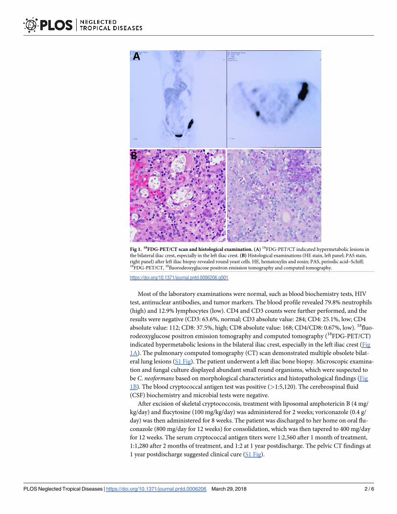
Most of the laboratory examinations were normal, such as blood biochemistry tests, HIV
test, antinuclear antibodies, and tumor markers. The blood profile revealed 79.8% neutrophils
(high) and 12.9% lymphocytes (low). CD4 and CD3 counts were further performed, and the
results were negative (CD3: 63.6%, normal; CD3 absolute value: 284; CD4: 25.1%, low; CD4
absolute value: 112; CD8: 37.5%, high; CD8 absolute value: 168; CD4/CD8: 0.67%, low). 18fluo-
rodeoxyglucose positron emission tomography and computed tomography (18FDG-PET/CT)
indicated hypermetabolic lesions in the bilateral iliac crest, especially in the left iliac crest (Fig
1A). The pulmonary computed tomography (CT) scan demonstrated multiple obsolete bilat-
eral lung lesions (S1 Fig). The patient underwent a left iliac bone biopsy. Microscopic examina-
tion and fungal culture displayed abundant small round organisms, which were suspected to
be C. neoformans based on morphological characteristics and histopathological findings (Fig
1B). The blood cryptococcal antigen test was positive (>1:5,120). The cerebrospinal fluid
(CSF) biochemistry and microbial tests were negative.
After excision of skeletal cryptococcosis, treatment with liposomal amphotericin B (4 mg/
kg/day) and flucytosine (100 mg/kg/day) was administered for 2 weeks; voriconazole (0.4 g/
day) was then administered for 8 weeks. The patient was discharged to her home on oral flu-
conazole (800 mg/day for 12 weeks) for consolidation, which was then tapered to 400 mg/day
for 12 weeks. The serum cryptococcal antigen titers were 1:2,560 after 1 month of treatment,
1:1,280 after 2 months of treatment, and 1:2 at 1 year postdischarge. The pelvic CT findings at
1 year postdischarge suggested clinical cure (S1 Fig).
Fig 1. 18FDG-PET/CT scan and histological examination. (A) 18FDG-PET/CT indicated hypermetabolic lesions in
the bilateral iliac crest, especially in the left iliac crest. (B) Histological examinations (HE stain, left panel; PAS stain,
right panel) after left iliac biopsy revealed round yeast cells. HE, hematoxylin and eosin; PAS, periodic acid–Schiff;18FDG-PET/CT, 18fluorodeoxyglucose positron emission tomography and computed tomography.
https://doi.org/10.1371/journal.pntd.0006206.g001
PLOS Neglected Tropical Diseases | https://doi.org/10.1371/journal.pntd.0006206 March 29, 2018 2 / 6
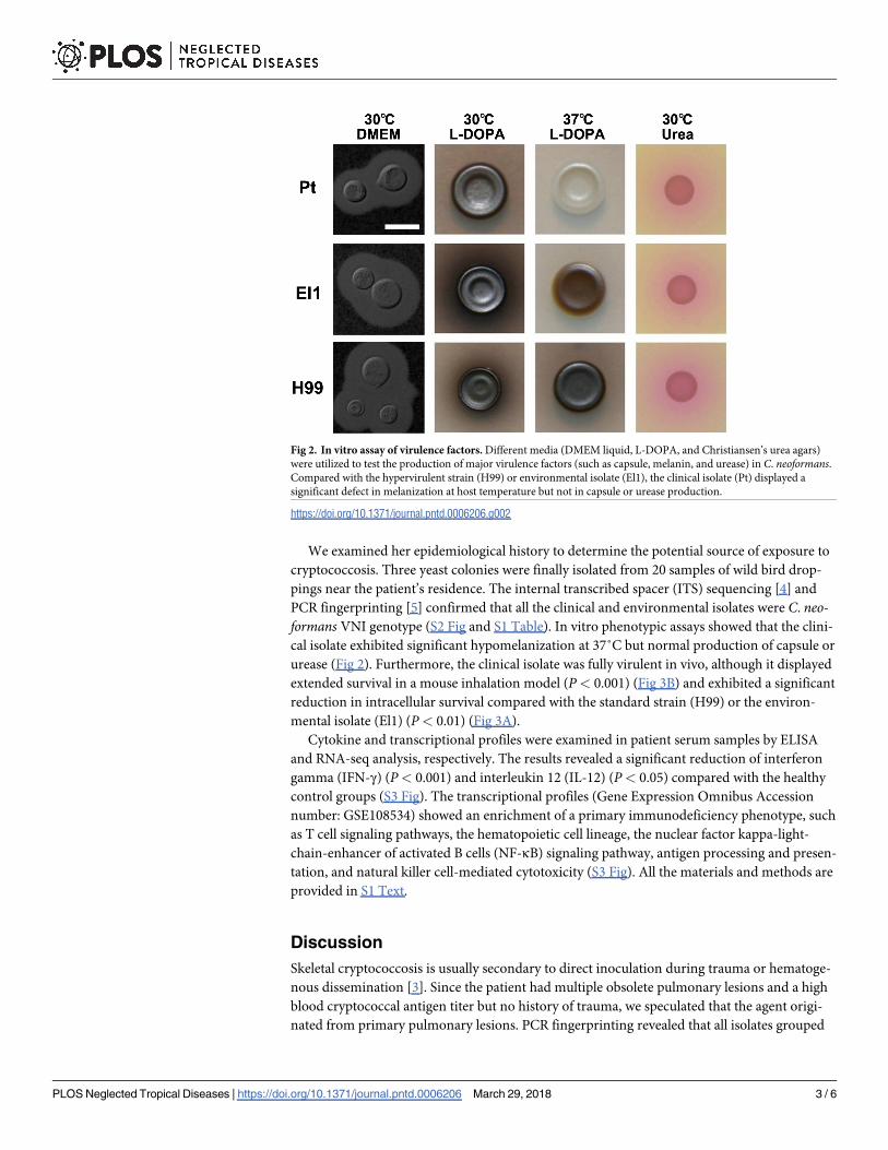
We examined her epidemiological history to determine the potential source of exposure to
cryptococcosis. Three yeast colonies were finally isolated from 20 samples of wild bird drop-
pings near the patient’s residence. The internal transcribed spacer (ITS) sequencing [4] and
PCR fingerprinting [5] confirmed that all the clinical and environmental isolates were C. neo-formans VNI genotype (S2 Fig and S1 Table). In vitro phenotypic assays showed that the clini-
cal isolate exhibited significant hypomelanization at 37˚C but normal production of capsule or
urease (Fig 2). Furthermore, the clinical isolate was fully virulent in vivo, although it displayed
extended survival in a mouse inhalation model (P< 0.001) (Fig 3B) and exhibited a significant
reduction in intracellular survival compared with the standard strain (H99) or the environ-
mental isolate (El1) (P< 0.01) (Fig 3A).
Cytokine and transcriptional profiles were examined in patient serum samples by ELISA
and RNA-seq analysis, respectively. The results revealed a significant reduction of interferon
gamma (IFN-γ) (P< 0.001) and interleukin 12 (IL-12) (P< 0.05) compared with the healthy
control groups (S3 Fig). The transcriptional profiles (Gene Expression Omnibus Accession
number: GSE108534) showed an enrichment of a primary immunodeficiency phenotype, such
as T cell signaling pathways, the hematopoietic cell lineage, the nuclear factor kappa-light-
chain-enhancer of activated B cells (NF-κB) signaling pathway, antigen processing and presen-
tation, and natural killer cell-mediated cytotoxicity (S3 Fig). All the materials and methods are
provided in S1 Text.
Discussion
Skeletal cryptococcosis is usually secondary to direct inoculation during trauma or hematoge-
nous dissemination [3]. Since the patient had multiple obsolete pulmonary lesions and a high
blood cryptococcal antigen titer but no history of trauma, we speculated that the agent origi-
nated from primary pulmonary lesions. PCR fingerprinting revealed that all isolates grouped
Fig 2. In vitro assay of virulence factors. Different media (DMEM liquid, L-DOPA, and Christiansen’s urea agars)
were utilized to test the production of major virulence factors (such as capsule, melanin, and urease) in C. neoformans.Compared with the hypervirulent strain (H99) or environmental isolate (El1), the clinical isolate (Pt) displayed a
significant defect in melanization at host temperature but not in capsule or urease production.
https://doi.org/10.1371/journal.pntd.0006206.g002
PLOS Neglected Tropical Diseases | https://doi.org/10.1371/journal.pntd.0006206 March 29, 2018 3 / 6

into the VNI genotype [6]. Thus, an environmental carrier (bird droppings) of C. neoformansmight be the ultimate source of skeletal infection in this case.
Pathogenesis and progression of cryptococcosis depends on the interaction between the
fungal agent and host immunity. The clinical agent in this study showed complete virulence in
vivo and ex vivo, although its virulence was inferior to the standard strain (H99) or environ-
mental isolates, which might be partially attributed to its hypomelanization at host tempera-
ture. Several laboratory test indices, such as HIV, autoimmune antibodies, and tumor
markers, were normal, and the patient’s medical history showed no medical use of steroids or
immunosuppressants. The patient displayed a lower CD4+ T-lymphocyte ratio, significantly
reduced serum Th1 cytokine (IFN-γ and IL-12) responses, and a dysregulated transcriptional
profile of blood leukocytes, as compared with healthy populations. Two potential scenarios
may explain this phenomenon. On the one hand, the presence of cryptococcal antigens (such
as glucuronoxylomannan) or previously applied anti-inflammatory therapy might cause a
slight compromise of host cellular immunity [7,8]. On the other hand, the abnormalities in
Fig 3. Macrophage killing assay and murine inhalation model. Both macrophage (A) (P< 0.01) and animal (B)
(P< 0.001) infection experiments displayed attenuated virulence of the clinical isolate compared with the
hypervirulent strain H99.
https://doi.org/10.1371/journal.pntd.0006206.g003
PLOS Neglected Tropical Diseases | https://doi.org/10.1371/journal.pntd.0006206 March 29, 2018 4 / 6
immunological indices might originate from host subclinical immune impairment, which
remains to be further confirmed.
We reviewed the literature on immunological analyses in apparently normal individuals
with cryptococcosis. The IL12RB1 gene mutation has been reported in pediatric patients with
cryptococcal osteomyelitis or disseminated cryptococcosis [9,10]. Several studies have also
suggested that genetic polymorphisms of some immune molecules (such as mannose-binding
lectin, dectin-2, and Fcγ receptor IIB) are closely associated with cryptococcosis in immuno-
competent populations [11,12,13]. All these factors were associated with host immune func-
tions essential for fungal control and containment, such as phagocytic activity and T cell
responses [14,15]. Thus, subclinical immune impairment might be a non-negligible factor in
apparently healthy patients with cryptococcosis.
The clinical symptoms and radiological findings of skeletal cryptococcosis were nonspe-
cific. A correct diagnosis still relied on histopathological and fungal examinations of lesion
specimens. After focal cleaning operations, multiple antifungal agents were sequentially
administered, such as liposomal amphotericin B, flucytosine, and azole agents, and the patient
achieved clinical cure.
Conclusions
Isolated skeletal cryptococcosis is rare but may occur in healthy individuals after daily expo-
sure to the organism. Herein, the clinical isolate likely originated from environmental bird
droppings disseminated from the pulmonary lesion to the hip. The development of crypto-
coccosis was closely associated with the interaction of the fungal agent and host immunity.
Accurate diagnosis of bone cryptococcosis depends mainly on histological and fungal exami-
nations. A combination antifungal agent treatment regimen and surgery were quite effective
for resolving bone cryptococcosis.
Ethics statement
This study was approved by the Committee on Ethics of Biomedicine Research, Changzheng
Hospital. All subjects gave written informed consent in accordance with the Declaration of
Helsinki. The animal experiments were carried out in strict accordance with the recommenda-
tions in the Regulations for the Administration of Affairs concerning Experimental Animals of
the State Science and Technology Commission (China). The protocol was approved by the
Animal Experiment Center of Second Military Medical University.
Supporting information
S1 Text. Materials and methods.
(DOCX)
S1 Fig. CT scans. (A) The pulmonary CT scan on admission showed multiple obsolete lesions
in the bilateral upper lobes and lower lobe of the left lung. (B) The pelvic CT scan at 1 year
post-discharge suggested the infectious lesions had disappeared. CT, computed tomography.
(TIF)
S2 Fig. Molecular-type identification of clinical and environmental isolates by PCR finger-
printing. All isolates were identified as VNI genotype. Pt is the cryptococcal strain isolated
from the iliac lesion of the patient. EI denotes environmental isolates from the patient’s area of
residence.
(TIF)
PLOS Neglected Tropical Diseases | https://doi.org/10.1371/journal.pntd.0006206 March 29, 2018 5 / 6
http://journals.plos.org/plosntds/article/asset?unique&id=info:doi/10.1371/journal.pntd.0006206.s001
http://journals.plos.org/plosntds/article/asset?unique&id=info:doi/10.1371/journal.pntd.0006206.s002

S3 Fig. Serum cytokine and transcriptional profile analyses. (A) The serum cytokine assay
revealed a significant reduction of IFN-γ and IL-12 compared with the control groups. ���
means P< 0.001. � means P< 0.05. (B) Bioinformatics analysis of RNA sequencing in the
patient serum sample displayed an enrichment of several immunological pathways. IFN-γ,
interferon gamma; IL-12, interleukin 12.
(TIF)
S1 Table. Strains used in this study. Pt, cryptococcal strain isolated from the biopsy speci-
men; EI-1,2,3, cryptococcal strains isolated from bird droppings near the patient’s residence.
The other strains were provided by our laboratory.
(DOCX)
References1. Maziarz EK, Perfect JR. Cryptococcosis. Infect Dis Clin North Am. 2016; 30: 179–206. https://doi.org/
10.1016/j.idc.2015.10.006 PMID: 26897067
2. Amenta PS, Stead J, Kricun ME. Case report 226: isolated Cryptococcus neoformans osteomyelitis of
femur. Skeletal Radiol. 1983. 9(4): 263–5. PMID: 6867777
3. Zhou HX, Lu L, Chu T, et al. Skeletal cryptococcosis from 1977 to 2013. Front Microbiol. 2014; 5: 740.
https://doi.org/10.3389/fmicb.2014.00740 PMID: 25642211
4. Feng X, Yao Z, Ren D, Liao W, Wu J. Genotype and mating type analysis of Cryptococcus neoformans
and Cryptococcus gattii isolates from China that mainly originated from non-HIV-infected patients.
FEMS Yeast Res. 2008. 8(6): 930–8. https://doi.org/10.1111/j.1567-1364.2008.00422.x PMID:
18671745
5. Meyer W, Castaneda A, Jackson S, Huynh M, Castaneda E. Molecular typing of IberoAmerican Crypto-
coccus neoformans isolates. Emerg Infect Dis. 2003; 9: 189–95. https://doi.org/10.3201/eid0902.
020246 PMID: 12603989
6. Fang W., Fa Z., and Liao W. (2015). Epidemiology of Cryptococcus and cryptococcosis in China. Fun-
gal Genet. Biol. 78, 7–15. doi: https://doi.org/10.1016/j.fgb.2014.10.017 PMID: 25445309
7. Albuquerque PC, Fonseca FL, Dutra FF, Bozza MT, Frases S, et al. (2014) Cryptococcus neoformans
glucuronoxylomannan fractions of different molecular masses are functionally distinct. Future Microbiol
9: 147–161. https://doi.org/10.2217/fmb.13.163 PMID: 24571070
8. Ratzinger F, Haslacher H, Poeppl W, Hoermann G, Kovarik JJ, et al. (2014) Azithromycin suppresses
CD4(+) T-cell activation by direct modulation of mTOR activity. Sci Rep 4: 7438. https://doi.org/10.
1038/srep07438 PMID: 25500904
9. Rezai M. S., Khotael G., Kheirkhah M., Hedayat T., Geramishoar M., and Mahjoub F. (2008). Crypto-
coccosis and deficiency of interleukin12r. Pediatr. Infect. Dis. J. 27, 673. doi: https://doi.org/10.1097/
INF.0b013e318179263a PMID: 18520441
10. Jirapongsananuruk O., Luangwedchakarn V., Niemela J. E., Pacharn P., Visitsunthorn N., Thepthai C.,
et al. (2012). Cryptococcal osteomyelitis in a child with a novel compound mutation of the IL12RB1
gene. Asian Pac. J. Allergy Immunol. 30, 79–82. PMID: 22523911
11. Ou X. T., Wu J. Q., Zhu L. P., Guan M., Xu B., Hu X. P., et al. (2011). Genotypes coding for mannose-
binding lectin deficiency correlated with cryptococcal meningitis in HIV-uninfected Chinese patients. J.
Infect. Dis. 203, 1686–1691. doi: https://doi.org/10.1093/infdis/jir152 PMID: 21592999
12. Hu X. P., Wu J. Q., Zhu L. P., Wang X., Xu B., Wang R. Y., et al. (2012). Association of Fcγ receptor IIB
polymorphism with cryptococcal meningitis in HIV-uninfected Chinese patients. PLoS ONE 7, e42439.
doi: https://doi.org/10.1371/journal.pone.0042439 PMID: 22879986
13. Hu X. P., Wang R. Y., Wang X., Cao Y. H., Chen Y. Q., Zhao H. Z., et al. (2015). Dectin-2 polymorphism
associated with pulmonary cryptococcosis in HIV-uninfected Chinese patients. Med. Mycol. 53, 810–
816. doi: https://doi.org/10.1093/mmy/myv043 PMID: 26129889
14. Heitman J KTR, Kwon-Chung KJ PJR, Casadevall A. Cryptococcus: from human pathogen to model
yeast. Washington, DC: ASM Press. 2011.
15. Rohatgi S., and Pirofski L. A. (2015). Host immunity to Cryptococcus neoformans. Future Microbiol 10,
565–581. doi: https://doi.org/10.2217/fmb.14.132 PMID: 25865194
PLOS Neglected Tropical Diseases | https://doi.org/10.1371/journal.pntd.0006206 March 29, 2018 6 / 6
http://journals.plos.org/plosntds/article/asset?unique&id=info:doi/10.1371/journal.pntd.0006206.s004

See discussions, stats, and author profiles for this publication at: https://www.researchgate.net/publication/325430079
Low-Cost Tetraplex PCR for the Global Spreading Multi-Drug Resistant
Fungus, Candida auris and Its Phylogenetic Relatives
Article in Frontiers in Microbiology · May 2018
DOI: 10.3389/fmicb.2018.01119
CITATIONS
0
READ
1
5 authors, including:
Some of the authors of this publication are also working on these related projects:
Mutation detection conferring resistance to antibiotics View project
Taxonomy and Nomenclature of Human Pathogenic Fungi (Yeasts and moulds) View project
Amir Arastehfar
Westerdijk Fungal Biodiversity Institute
3 PUBLICATIONS 4 CITATIONS
SEE PROFILE
Wenjie Fang
Second Military Medical University, Shanghai
32 PUBLICATIONS 42 CITATIONS
SEE PROFILE
Hamid Badali
Mazandaran University of Medical Sciences, Sari Iran
176 PUBLICATIONS 1,485 CITATIONS
SEE PROFILE
Afsane Vaezi
Mazandaran University of Medical Sciences
21 PUBLICATIONS 97 CITATIONS
SEE PROFILE
All content following this page was uploaded by Amir Arastehfar on 29 May 2018.
The user has requested enhancement of the downloaded file.

fmicb-09-01119 May 29, 2018 Time: 17:58 # 1
ORIGINAL RESEARCHpublished: 29 May 2018
doi: 10.3389/fmicb.2018.01119
Edited by:Dominique Sanglard,
Université de Lausanne, Switzerland
Reviewed by:Alix Thérèse Coste,
Centre Hospitalier UniversitaireVaudois (CHUV), Switzerland
Guillermo Quindós,University of the Basque Country
(UPV/EHU), Spain
*Correspondence:Weiwei Jiang
†These authors have contributedequally to this work.
Specialty section:This article was submitted to
Infectious Diseases,a section of the journal
Frontiers in Microbiology
Received: 23 February 2018Accepted: 11 May 2018Published: 29 May 2018
Citation:Arastehfar A, Fang W, Badali H,
Vaezi A, Jiang W, Liao W, Pan W,Hagen F and Boekhout T (2018)
Low-Cost Tetraplex PCRfor the Global Spreading Multi-Drug
Resistant Fungus, Candida aurisand Its Phylogenetic Relatives.
Front. Microbiol. 9:1119.doi: 10.3389/fmicb.2018.01119
Low-Cost Tetraplex PCR for theGlobal Spreading Multi-DrugResistant Fungus, Candida auris andIts Phylogenetic RelativesAmir Arastehfar1†, Wenjie Fang1,2,3†, Hamid Badali4, Afsane Vaezi5, Weiwei Jiang2,3* ,Wanqing Liao2,3, Weihua Pan2,3* , Ferry Hagen1 and Teun Boekhout1,3,6
1 Westerdijk Fungal Biodiversity Institute, Utrecht, Netherlands, 2 Department of Dermatology, Shanghai ChangzhengHospital, Second Military Medical University, Shanghai, China, 3 Shanghai Key Laboratory of Molecular Medical Mycology,Shanghai Institute of Medical Mycology, Shanghai Changzheng Hospital, Second Military Medical University, Shanghai,China, 4 Department of Medical Mycology/Invasive Fungi Research Center, School of Medicine, Mazandaran University ofMedical Sciences, Sari, Iran, 5 Student Research Committee, Mazandaran University of Medical Sciences, Sari, Iran,6 Institute for Biodiversity and Ecosystem Dynamics, University of Amsterdam, Amsterdam, Netherlands
Candida auris, C. haemulonii, C. duobushaemulonii, and C. pseudohaemulonii areclosely related and highly multidrug resistant yeast pathogens. The high cost andlow accuracy of current diagnostics may underestimate their prevalence, especiallyin medical resource-limited regions. In this study, we used 172 C. auris stains andits relatives and 192 other fungal strains to establish and validate a novel multiplexend-point PCR. A prospective and a retrospective clinical screenings using this assaywere further performed in China and Iran respectively. We identified the first isolateof C. pseudohaemulonii in China and the first isolate of C. haemulonii in Iran from821 clinical isolates in total, without any false positive. Animal models of C. auris andC. haemulonii were established for validation. The overall positive rates of the assay formice blood and tissue were 28.6 and 92.9%, respectively. Compared with previouslydeveloped assays, our assay is more available and affordable to the developingcountries, and may contribute to a better understanding of the epidemiology of C. aurisand its relatives in these regions.
Keywords: end-point PCR, Candida auris, multi-drug resistance, animal model, clinical validation, moleculardiagnosis
INTRODUCTION
The evolving epidemiology of non-albicans Candida species and other emerging opportunisticpathogenic yeasts contributes to an increasing morbidity and mortality globally (Miceli et al.,2011; Fang et al., 2017). Among them, Candida auris and its medically important relatives in theMetschnikowiaceae clade, e.g., C. haemulonii, C. duobushaemulonii, and C. pseudohaemulonii, arewell known as highly multidrug resistant pathogens that can lead to both superficial and deep-seated infections (Cendejas-Bueno et al., 2012). C. auris appears to be the most notable speciesat present due to its outbreak potential and high mortality rate (30–50%) (Calvo et al., 2016; deAlmeida et al., 2016). After the first isolate from the external ear canal of a Japanese inpatient in2009, a number of severe C. auris cases occurred subsequently in 17 countries from five continents
Frontiers in Microbiology | www.frontiersin.org 1 May 2018 | Volume 9 | Article 1119
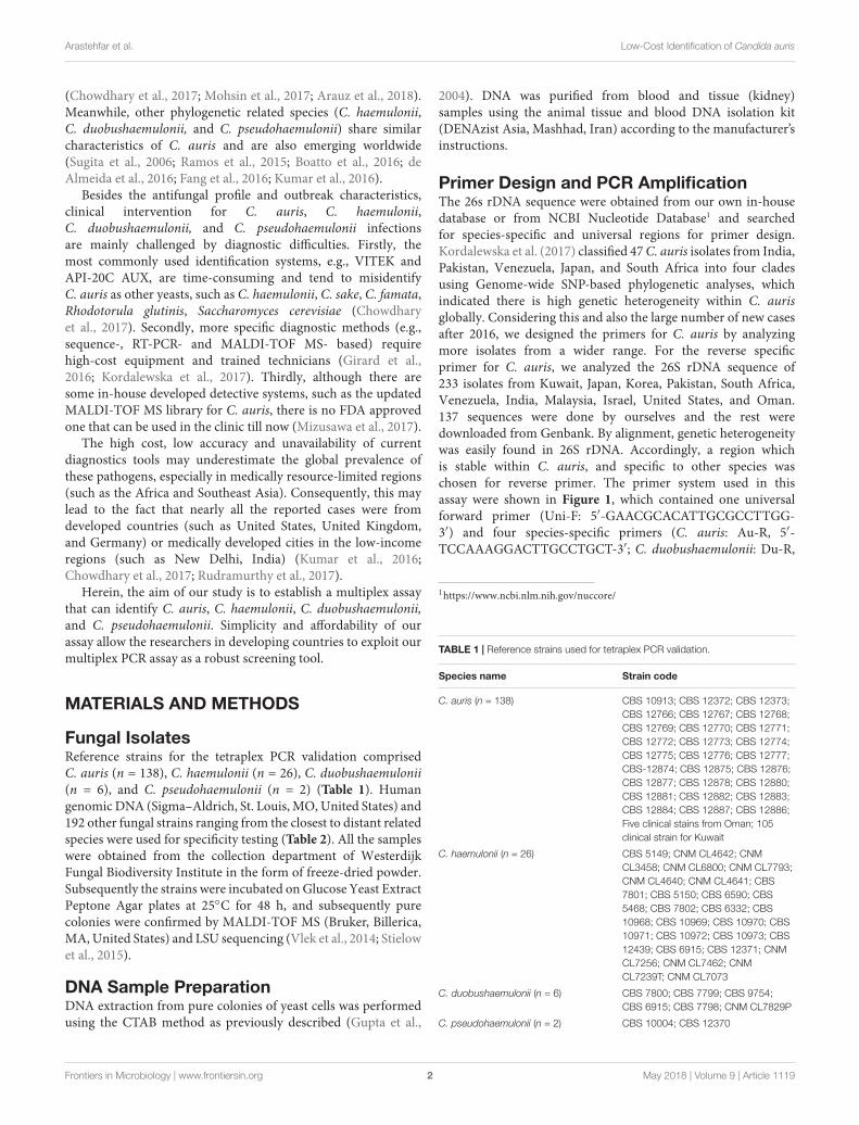
fmicb-09-01119 May 29, 2018 Time: 17:58 # 2
Arastehfar et al. Low-Cost Identification of Candida auris
(Chowdhary et al., 2017; Mohsin et al., 2017; Arauz et al., 2018).Meanwhile, other phylogenetic related species (C. haemulonii,C. duobushaemulonii, and C. pseudohaemulonii) share similarcharacteristics of C. auris and are also emerging worldwide(Sugita et al., 2006; Ramos et al., 2015; Boatto et al., 2016; deAlmeida et al., 2016; Fang et al., 2016; Kumar et al., 2016).
Besides the antifungal profile and outbreak characteristics,clinical intervention for C. auris, C. haemulonii,C. duobushaemulonii, and C. pseudohaemulonii infectionsare mainly challenged by diagnostic difficulties. Firstly, themost commonly used identification systems, e.g., VITEK andAPI-20C AUX, are time-consuming and tend to misidentifyC. auris as other yeasts, such as C. haemulonii, C. sake, C. famata,Rhodotorula glutinis, Saccharomyces cerevisiae (Chowdharyet al., 2017). Secondly, more specific diagnostic methods (e.g.,sequence-, RT-PCR- and MALDI-TOF MS- based) requirehigh-cost equipment and trained technicians (Girard et al.,2016; Kordalewska et al., 2017). Thirdly, although there aresome in-house developed detective systems, such as the updatedMALDI-TOF MS library for C. auris, there is no FDA approvedone that can be used in the clinic till now (Mizusawa et al., 2017).
The high cost, low accuracy and unavailability of currentdiagnostics tools may underestimate the global prevalence ofthese pathogens, especially in medically resource-limited regions(such as the Africa and Southeast Asia). Consequently, this maylead to the fact that nearly all the reported cases were fromdeveloped countries (such as United States, United Kingdom,and Germany) or medically developed cities in the low-incomeregions (such as New Delhi, India) (Kumar et al., 2016;Chowdhary et al., 2017; Rudramurthy et al., 2017).
Herein, the aim of our study is to establish a multiplex assaythat can identify C. auris, C. haemulonii, C. duobushaemulonii,and C. pseudohaemulonii. Simplicity and affordability of ourassay allow the researchers in developing countries to exploit ourmultiplex PCR assay as a robust screening tool.
MATERIALS AND METHODS
Fungal IsolatesReference strains for the tetraplex PCR validation comprisedC. auris (n = 138), C. haemulonii (n = 26), C. duobushaemulonii(n = 6), and C. pseudohaemulonii (n = 2) (Table 1). Humangenomic DNA (Sigma–Aldrich, St. Louis, MO, United States) and192 other fungal strains ranging from the closest to distant relatedspecies were used for specificity testing (Table 2). All the sampleswere obtained from the collection department of WesterdijkFungal Biodiversity Institute in the form of freeze-dried powder.Subsequently the strains were incubated on Glucose Yeast ExtractPeptone Agar plates at 25◦C for 48 h, and subsequently purecolonies were confirmed by MALDI-TOF MS (Bruker, Billerica,MA, United States) and LSU sequencing (Vlek et al., 2014; Stielowet al., 2015).
DNA Sample PreparationDNA extraction from pure colonies of yeast cells was performedusing the CTAB method as previously described (Gupta et al.,
2004). DNA was purified from blood and tissue (kidney)samples using the animal tissue and blood DNA isolation kit(DENAzist Asia, Mashhad, Iran) according to the manufacturer’sinstructions.
Primer Design and PCR AmplificationThe 26s rDNA sequence were obtained from our own in-housedatabase or from NCBI Nucleotide Database1 and searchedfor species-specific and universal regions for primer design.Kordalewska et al. (2017) classified 47C. auris isolates from India,Pakistan, Venezuela, Japan, and South Africa into four cladesusing Genome-wide SNP-based phylogenetic analyses, whichindicated there is high genetic heterogeneity within C. aurisglobally. Considering this and also the large number of new casesafter 2016, we designed the primers for C. auris by analyzingmore isolates from a wider range. For the reverse specificprimer for C. auris, we analyzed the 26S rDNA sequence of233 isolates from Kuwait, Japan, Korea, Pakistan, South Africa,Venezuela, India, Malaysia, Israel, United States, and Oman.137 sequences were done by ourselves and the rest weredownloaded from Genbank. By alignment, genetic heterogeneitywas easily found in 26S rDNA. Accordingly, a region whichis stable within C. auris, and specific to other species waschosen for reverse primer. The primer system used in thisassay were shown in Figure 1, which contained one universalforward primer (Uni-F: 5′-GAACGCACATTGCGCCTTGG-3′) and four species-specific primers (C. auris: Au-R, 5′-TCCAAAGGACTTGCCTGCT-3′; C. duobushaemulonii: Du-R,
1https://www.ncbi.nlm.nih.gov/nuccore/
TABLE 1 | Reference strains used for tetraplex PCR validation.
Species name Strain code
C. auris (n = 138) CBS 10913; CBS 12372; CBS 12373;CBS 12766; CBS 12767; CBS 12768;CBS 12769; CBS 12770; CBS 12771;CBS 12772; CBS 12773; CBS 12774;CBS 12775; CBS 12776; CBS 12777;CBS-12874; CBS 12875; CBS 12876;CBS 12877; CBS 12878; CBS 12880;CBS 12881; CBS 12882; CBS 12883;CBS 12884; CBS 12887; CBS 12886;Five clinical stains from Oman; 105clinical strain for Kuwait
C. haemulonii (n = 26) CBS 5149; CNM CL4642; CNMCL3458; CNM CL6800; CNM CL7793;CNM CL4640; CNM CL4641; CBS7801; CBS 5150; CBS 6590; CBS5468; CBS 7802; CBS 6332; CBS10968; CBS 10969; CBS 10970; CBS10971; CBS 10972; CBS 10973; CBS12439; CBS 6915; CBS 12371; CNMCL7256; CNM CL7462; CNMCL7239T; CNM CL7073
C. duobushaemulonii (n = 6) CBS 7800; CBS 7799; CBS 9754;CBS 6915; CBS 7798; CNM CL7829P
C. pseudohaemulonii (n = 2) CBS 10004; CBS 12370
Frontiers in Microbiology | www.frontiersin.org 2 May 2018 | Volume 9 | Article 1119

fmicb-09-01119 May 29, 2018 Time: 17:58 # 3
Arastehfar et al. Low-Cost Identification of Candida auris
TABLE 2 | Fungal isolates for specificity test.
Species name Strain code
Candida albicans CBS 2704; CBS 2691; CBS 2697;CBS 1893; CBS 2712; CBS 5703;CBS 6552; CBS 2689; CBS 2690;CBS 2691; CBS 2695; CBS 2698;CBS 2696; CBS 5137; CBS 2702;CBS 1912; CLF-2; CLF-11; CLF-32;CLF-41; CLF-52; CLF-61; CLF-71;CLF-82; CLF-91; CLF-844; CLF-177;CLF-282; CLF-283; CLF-378;CLF-403; CLF-435; CLF-539;CLF-Llama 1; CLF-Llama 2; CLF-Llama11
Candida africana CBS 8781
Candida tropicalis CBS 2313; CBS 94; CBS 1920; CLF-6;CLF-15; CLF-26; CLF-36; CLF-45;CLF-56; CLF-65; CLF-75; CLF-86
Candida parapsilosis CBS 11045; CBS 7154; CBS 604;CBS 2197; CBS 2915; CBS 11059;CBS 11920; CBS 8050; CBS 2216;CBS 1954; CBS 2194; CBS 2211;CBS 11043; CBS 11359; CBS 7248;CBS 7156; CBS 10947; CBS 11130;CBS 7157; CBS 8836; CBS 2215;CBS 8181; CBS 12025; CBS 7155;CBS 6318; CBS 2196; CBS 2195;CLF-8; CLF-17; CLF-47; CLF-58;CLF-67; CLF-77; CLF-88; CLF-97
Candida metapsilosis CBS 10907; CBS 2916; CBS 10747;CBS 11127; CBS 2315
Candida orthopsilosis CBS 8825; CBS 9894; CBS 9348;CBS 10741; CBS 10906; CBS 11337;CBS 9347; CBS 10743; CBS 2212;CBS 10744; CBS 11698; CBS 10745;CBS 8548
Candida glabrata CLF-4; CLF-13; CLF-24; CLF-34;CLF-43; CLF-63; CLF-73; CLF-83;CLF-84; CLF-93; CLF-310; CLF-611A;CLF-Llama 3; CLF-Llama 24; CLF-Llama 63
Candida krusei CBS 5147; CLF-30; CLF-40; CLF-60;CLF-69; CLF-79; CLF-99; CLF-611B
Candida norvegensis CBS 6564
Pichia cactophila CBS 6926
Candida lusitaniae CBS 6936; CLF-19
Candida famata CBS 767
Candida dubliniensis CLF-10; CLF-49
Candida guilliermondii CBS 7099
Candida kefyr STA#63
Candida sake CBS 159
Candida rugosa CBS 613
Candida zeylanoides CBS 619
Yarrowia lipolytica CBS 6124
Pichia fermentans CBS 187
Pichia kluyveri CBS 188
Pichia membranifaciens CBS 107
Pseudozyma thailandica CBS 10006
Kluyveromyces lactis var. lactis CBS 683
Kluyveromyces marxianus CBS 712
Kodamaea ohmeri CBS 5367
(Continued)
TABLE 2 | Continued
Species name Strain code
Lindnera fabianii CBS 5640
Lindnera jadinii CBS 1600
Lodderomyces elongisporus CBS 2605
Magnusiomyces capitatus CBS 162.8
Metschnikowia pulcherrima CBS 5833
Meyerozyma caribbica CBS 9966
Millerozyma farinosa CBS 185
Ogataea polymorpha CBS 4732
Cryptococcus neoformans CBS 8710; CBS 6885
Cryptococcus deneoformans CBS6900; CBS10511
Cryptococcus gattii CBS 7229; CBS 6289
Cryptococcus bacillisporus CBS 6955; CBS 8755
Cryptococcus deuterogattii CBS 10514; CBS 7750
Cryptococcus tetragattii CBS 10101;
Cryptococcus decagattii CBS 11687
Cryptococcus amylolentus CBS 6039
Rhodotorula mucilaginosa CBS 316
Saccharomyces cerevisiae CBS 1171
Saccharomyces paradoxus CBS 432
Trichosporon asahii CBS 2479
Trichosporon inkin CBS 5585
Debaryomyces nepalensis CBS 5921
Exophiala dermatitidis CBS 207.35
Galactomyces candidus CBS 615.84
Hortaea werneckii CBS 107.67
Hyphopichia burtonii CBS 2352; CBS 2532
Torulaspora globosa CBS 764
Wickerhamomyces anomalus CBS 5759
Wickerhamomyces onychis CBS 5587
Zygosaccharomyces rouxii CBS 732
Schizosaccharomyces pombe CBS 356
Schizosaccharomyces japonicus CBS 354
Aspergillus fumigatus CBS 114.55
Aspergillus flavus CBS 107.45
Aspergillus niger CBS 102.12
Aspergillus terreus CBS 106.25
Aspergillus nidulans CBS 100.20
Fusarium moniliforme CBS 130180
Fusarium solani CBS 101427
Aspergillus versicolor CBS 106.57
Fusarium oxysporum CBS 100.97
5′-GTAGACTTCGCTGCGGATATGTTA-3′; C. haemulonii: Ha-R, 5′-ATTGCGCCAGCATCCTTATTG-3′; C. pseudohaemulonii:Ps-R, 5′-GCACCCGATGCTGACAGTCTAC-3′). Primers weresynthesized by Integrated DNA Technologies, Coralville, IA,United States.
A total volume of 50 µl PCR mixture containing 5 µl of buffer,1.5 mM magnesium chloride, 2.5 units of Taq polymerase enzyme(BIO-21040, BioLine Company, London, United Kingdom),0.2 mM dNTP (BIO-39043, BioLine Company, London,United Kingdom), 5 pM Uni-F primer, 5 pM Du-R, 2 pMPs-R, 3 pM Au-R, and 10 pM Ha-R, 1 µl template DNA
Frontiers in Microbiology | www.frontiersin.org 3 May 2018 | Volume 9 | Article 1119

fmicb-09-01119 May 29, 2018 Time: 17:58 # 4
Arastehfar et al. Low-Cost Identification of Candida auris
FIGURE 1 | Primer sequences and positions used in the tetraplex PCR. Uni-F: Universal forward primer for Candida auris, C. duobushaemulonii, C. haemulonii, andC. pseudohaemulonii; Du-R: C. duobushaemulonii specific reverse primer; Au-R: C. auris specific reverse primer; Ps-R: C. pseudohaemulonii specific reverseprimer; Ha-R: C. haemulonii specific reverse primer.
or single colony pick (≈1 mm3), and 38.3 µl Mili-Q water(Millipore Corporation, Billerica, MA, United States) was usedfor amplification of DNA from target species.
The PCR was performed using a 2720 thermal cycler (AppliedBiosystems, Waltham, MA, United States), and the PCR programconsisted of 5 min pre-denaturation at 94◦C, followed by 35cycles of 30 s at 94◦C, 30 s at 62◦C, 30 s at 72◦C and 8 min at72◦C as final extension.
Validation via Animal ModelWhen certain infected human samples are not easily availablefor diagnostic validation, animal model can be an alternative(Bialek et al., 2001). To evaluate its potential use for infectedhuman blood or tissue, animal models infected by the mostimportant pathogens in Metschnikowiaceae clade, i.e., C. aurisand C. haemulonii, were developed for diagnostic purpose. Allprocedures related to animal experiments were approved bythe ethics and research committee of Mazandaran University ofMedical Sciences, Sari, Iran.
Candida auris (CBS 10913, type strain), C. haemulonii (CBS5149, type strains) and C. albicans (ATCC 29008, control) werecultured twice at 35◦C for 48 h on Sabouraud dextrose agar (SDA,Difco). All isolates were subcultured in brain heart infusion brothand incubated at 37◦C with shaking at 150 rpm. Supernatantswere carefully removed and washed twice in sterile phosphatebuffered saline (PBS). After centrifugation, the cells were washedin PBS and a hemocytometer was used to count the yeast cell.The yeast cell concentration was adjusted to an inoculum size of5× 105 yeast/ml. The viable counts of isolates were confirmed by10-fold serially diluting the cell suspension on SDA plates.
Immunocompetent female ICR (CD-1 specific pathogen- free)mice with a mean weight of 22 g (purchased from Royan Institute,Tehran, Iran) were used in the study. Animals were housed at theaccredited Animal Experimentation Facility in standard cages,received sterilized food and were monitored daily (Fakhim et al.,2018). Seven mice were allocated into each of the three mice
groups infected with C. auris. C. haemulonii and C. albicans.Infection was induced in the mice for each group with aninoculum of 5 × 10⁵ CFU/mouse in a volume of 0.2 ml intothe lateral tail vein. No immunosuppressive scheme was used.Mice were checked daily and euthanized when symptoms ofdisseminated infection were detected. Any animal that had morethan one of the criteria including, decreased activity, hunchedposture, torticollis or barrel rolling, inability to eat or drink andhypothermia was humanly euthanized by intracardiac punctureunder general anesthesia (Conti et al., 2014). Tissue sections werealso stained with hematoxylin and eosin (H&E) for microscopicexamination. Tissue (kidney) and blood samples were recoveredunder aseptic conditions. Tissue samples were homogenized insterile saline and 100 µl of homogenate was cultured on SDAat 35◦C to confirm Candida infection and determine the colonyforming units (CFU) were determine (Fakhim et al., 2018). Allyeast cells cultures were identified by our tetraplex PCR. Inaddition, 100 µl of each EDTA-blood sample was cultured onSDA plates. The rest of the homogenate and blood samples wereused for tetraplex PCR test.
Application in Clinical SettingA single-center prospective clinical screening for C. auris,C. duobushaemulonii, C. haemulonii and C. pseudohaemuloniiwas performed during 2017.05.01 to 2017.11.01 in ShanghaiChangzheng Hospital, China. The newly developed multiplexPCR system worked as a supplementary tool to BD BACTECTM
FX40 Instrument (Becton, Dickinson and Company,Franklin Lakes, NJ, United States), CHROMagarTM Candida(CHROMagar Microbiology, Paris, France) and microscopicexamination to finding infection cases by the targeted species.In order to further test the specificity of our assay using moreclinical isolates, we used a wide range of sample types forscreening, including blood, sputum, feces, bronchoalveolarlavage fluid and oral swab. We test all the clinical yeast isolatesthat are identified by CHROMagarTM Candida as non-albicans
Frontiers in Microbiology | www.frontiersin.org 4 May 2018 | Volume 9 | Article 1119

fmicb-09-01119 May 29, 2018 Time: 17:58 # 5
Arastehfar et al. Low-Cost Identification of Candida auris
Candida species. As soon as the clinical culture is obtained byconventional diagnostic methods, single colony (≈1 mm3) istested by this multiplex PCR system. DNA extraction and ITSsequencing are done as well to confirm the accuracy of the assayin the clinical settings as described before (Stielow et al., 2015).
Two hundred and fifty five clinical non-albicans Candidaisolates (identified by CHROMagarTM Candida) fromMazandaran University of Medical Sciences, Iran, were usedas a retrospective screening sample in this study. The isolateswere collected from blood, sputum, feces, oral swab and vaginalsamples during 2016.1–2017.6. Multiplex PCR were performed,and MALDI-TOF and LSU sequencing were done as well toconfirm the accuracy of the assay in the clinical settings (Vleket al., 2014; Stielow et al., 2015).
All the tests were approved by the ethics and researchcommittees of Shanghai Changzheng Hospital and MazandaranUniversity of Medical Sciences.
RESULTS
Analytical ValidationFigure 2 showed that four amplicons exhibited major bands thatcould be differentiated from each other. The gel figure for allthe strains of C. auris and its relatives tested in this study wasshown in Supplementary Figure S1. Sequencing results of PCRamplicons were 100% matched with the targeted species. Humangenomic DNA and 192 other fungal species were tested, and nocross-reactivity was found (100% specificity).
Animal Model TestingFigure 3 showed the detailed information of the animal modeltesting.
FIGURE 2 | Agarose gel electrophoresis (2% agarose) of PCR amplicons.Lanes 1–4 are C. auris (CBS 10913 type, 331 bp), C. duobushaemulonii (CBS7798 type, 115 bp), C. haemulonii (CBS 5149 type, 696 bp) andC. pseudohaemulonii (CBS 10004 type, 576 bp), respectively. Lane M,HyperLadderTM 100 bp (Bioline, London, United Kingdom).
Application of the tetraplex PCR in blood showed a higheroverall positive rate (4 of 14, 28.6%) than blood culturepositive rate (1 of 14, 7.1%) in animal models of C. auris andC. haemulonii. Specifically, three C. auris (3/7, on the 3rd, 4th,and 5th day post-infection (PI)) and one C. haemulonii (1/7, onthe 5th day, PI) infected mice showed positive PCR results fromblood, without cross-reactivity in C. albicans group (0/7). Cultureresults were positive only in one blood samples of C. auris (1/7,on the 4th day, PI), negative in all C. haemulonii infected mice(0/7), and positive in three C. albicans infected mice (3/7, on the3rd, 4th, and 5th day PI), with CFUs ranging from 1 to 6 coloniesin 100 µl.
The overall positive PCR rate of mice kidney samples was92.8% (13/14) among C. auris and C. haemulonii, without cross-reactivity in C. albicans group (0/7). Among the kidney samplesfrom 21 mice examined by culture, 19 were positive due toC. auris (7/7), haemulonii (6/7), and C. albicans (6/7) with CFUsranging from 8.3 × 10 to 4.3 × 10⁴ colonies in 100 µl (mean3.5× 103 colonies).
Clinical ValidationApplying our assay in a prospective (566 clinical isolates) anda retrospective cohorts (255 clinical isolates), revealed oneC. haemulonii isolate (S49AF, accession number: KY112738)from Iran and one C. haemulonii isolate (SCZ90793,accession number: MG637448), one C. duobushaemuloniiisolate (SCZ91445, accession number: MG963993) andone C. pseudohaemulonii isolate (SCZ90800, accessionnumber: MG242063) from China. Specifically, the ChineseC. pseudohaemulonii was isolated from sputum of a 65-year-old male who suffered from lower esophageal resection(Shanghai, China). To our knowledge this the first isolateof C. pseudohaemulonii from China. Whereas the IranianC. haemulonii isolate was recovered from toenail sample of a36-year-old diabetic male in 2016 (Shiraz, Iran), and also was thefirst C. haemulonii isolate from Iran.
DISCUSSION
Achieving a timely and accurate diagnosis of infectionscaused by C. auris and its phylogenetic relatives is of greatdifficulty in clinical settings (Mizusawa et al., 2017). Nearlyall the C. auris isolates were misdiagnosed as C. haemulonii,C. sake, C. famata, Rhodotorula glutinis, Saccharomyces cerevisiaeby clinical commercial methods such as Vitek-2 YST IDsystem, API-20C AUX, AuxaColor 2, BD Phoenix, andMicroScan (Chowdhary et al., 2017). Studies also showedthat C. pseudohaemulonii failed to be distinguished fromC. haemulonii by these methods (Kim et al., 2009). All theidentifications from the biochemical assays should be confirmedby sequencing. The Centers for Disease Control of Americaspeculated that the unavailability of appropriate diagnosticmethod underestimate its prevalence in many countries (Sarmaand Upadhyay, 2017), which includes medical resource-limitedregions. Hence, we developed a novel end-point tetraplex PCRsystem in a low-cost, rapid and accurate format. This novel assay
Frontiers in Microbiology | www.frontiersin.org 5 May 2018 | Volume 9 | Article 1119

fmicb-09-01119 May 29, 2018 Time: 17:58 # 6
Arastehfar et al. Low-Cost Identification of Candida auris
FIGURE 3 | Lanes B1–B7 are PCR results of blood sample from seven mice infected with C. auris; Lanes B8-B14 are PCR results of blood sample from seven miceinfected with C. haemulonii; Lanes B15-B21 are PCR results of blood sample from seven mice infected with C. albicans; Lane BC1–4 are PCR results of fourcultured strains from blood samples of all the 21 mice; Lanes T1–T7 are PCR results of kidney tissue sample from seven mice infected with C. auris; Lanes T8–T14are PCR results of kidney tissue sample from seven mice infected with C. haemulonii; Lanes T15–T21 are PCR results of kidney tissue sample from seven miceinfected with C. albicans; Lane TC1–7 are PCR results of seven cultured strains from kidney samples of mice infected with C. auris; Lane TC8–13 are PCR results ofsix cultured strains from kidney samples of mice infected with C. haemulonii; Lane TC14–19 are PCR results of six cultured strains from kidney samples of miceinfected with C. albicans; Lane M, GeneRuler 100 bp Plus DNA Ladder (Thermo Fisher, Waltham, MA, United States).
has been validated by a large number of CBS/clinical isolates,diagnostic animal models and clinical application. We hope thisassay can help to understand the global clinical impact of C. aurisand its relatives, especially in the under-served and poor regions.
Our new end-point tetraplex PCR was designed to identify anddifferentiate the most medically important and related species toC. auris. Recently, two real-time PCR based assays for C. auriswere developed (Kordalewska et al., 2017; Leach et al., 2017).Compared with the real-time PCR method, our end-point PCRis non-automated and labor-intensive. The electrophoresis stepof our assay normally required 45 min, which is more time-consuming than real-time PCRs. Additionally, since the natureof end-point PCR, our assay is unable to quantify the fungal loadof host. However, the advantages of our assay over theirs are alsoobvious.
Kordalewska et al. (2017) established the first SYBR Greenreal-time PCR system for C. auris, C. duobushaemulonii,
C. haemulonii, and C. lusitaniae; While Leach et al. (2017)developed a probe-based singleplex real-time PCR onlyfor C. auris. Considering the high misdiagnostic ratebetween C. auris, C. haemulonii, C. duobushaemulonii,and C. pseudohaemulonii, and higher resistance ofC. pseudohaemulonii to AMB than C. auris, C. haemulonii,C. duobushaemulonii, and C. lusitaniae (Kim et al., 2009;Shin et al., 2012), we used a multiplex strategy for this assayand included C. pseudohaemulonii instead of C. lusitaniae.Among the selected species, C. haemulonii initially wasknown as a multi-antifungal resistant pathogen since the firstcase report in 1984, and it can cause outbreaks and infectimmunocompetent individuals (Loosova et al., 2001; Khan et al.,2007). Subsequently, C. haemulonii was recognized as a complexspecies and reclassified as C. haemulonii, C. duobushaemulonii,and C. haemulonii var. vulnera (Cendejas-Bueno et al., 2012).C. auris and C. pseudohaemulonii were described in 2009 and
Frontiers in Microbiology | www.frontiersin.org 6 May 2018 | Volume 9 | Article 1119

fmicb-09-01119 May 29, 2018 Time: 17:58 # 7
Arastehfar et al. Low-Cost Identification of Candida auris
2006, respectively. Both of them were multi-antifungal resistantpathogens and the latter is spreading across the world at anaccelerated pace (Sugita et al., 2006; Chowdhary et al., 2017).Correct species-level identification for the four species is neededdue to the clinical importance and heterogeneity in terms oftaxonomy, spreading prevalence and antifungal susceptibilityprofile.
Our multiplex PCR system is the first assay for C. auristhat has been validated by animal model testing. Theoverall PCR positive rates of mice blood and tissue were28.6 and 92.9%, respectively, which showed slightly higherperformance than culture based identification. And PCR/culturefrom tissue samples showed higher value than those fromblood for diagnosing infections caused by these pathogens.Moreover, our new assay is much faster than culture basedidentification. PCR directly from tissue/blood only requires3 h from sample preparation to result read-out; while culturebased identification is more time-consuming (normally 1–3 days).
Our new assay is also featured by its low cost and potentialto be used in medical resource-limited settings. Due to thepoor economic condition, lack of basic medical facilities andthe relatively poor population health, the developing countrieswere reported to have a much higher risk of infections thanthe developed ones (Pittet et al., 2008). Among the 17 countriesreported with C. auris prevalence, seven are the developing ones(Oman, Venezuela, Colombia, South Africa, Kenya, Pakistan,and India). Considering this, our new assay was establishedto help to extend the global knowledge of the epidemiologyof these important and emerging pathogens, especially formedical resource-limited countries. Compared with MALDI-TOF and real-time PCR cycler, end-point PCR cycler used inthis assay is more portable and much cheaper ($130–$4,000per cycler) (Wong et al., 2015). For clinical validation, we usedisolates from two clinical settings that can represent differentsituations of medical care in developing countries (Shanghai,China and Sari, Iran). In Shanghai, specific diagnostic toolssuch as MALDI-TOF and sequencing are available to mosthospitals. However, since China is a developing country, thereare still a large number of patients that cannot afford thesetests. While in Iran, to our knowledge, there is no MALDI-TOF and sequencing machine. There was only one epidemiologystudy related to C. haemulonii and C. duobushaemulonii inChina and no epidemiology data is available in Iran (Hou et al.,2016). Hence, we hold the prevalence of C. auris and its closerelated relatives are underestimated due to the lack of low-costand accurate assays in both countries. By using this assay, wesuccessfully found the first clinical isolates of C. haemuloniiin Iran and first clinical isolates of C. pseudohaemuloniiin China. However, no C. auris isolate was found in bothcohorts.
CONCLUSION
The novel end-point tetraplex PCR for C. auris and itsphylogenetic relatives is rapid and 100% specific. Clinicalvalidation and animal model testing further proved its clinicalperformance. This assay only requires the simple devices of end-point PCR cycler and electrophoresis instrument, showing itsavailability and affordability to most of the developing countries,and can contribute to a better understanding of the clinicaloccurrence of C. auris and its relatives.
ETHICS STATEMENT
This study was carried out in accordance with therecommendations of Guide for the Care and Use of LaboratoryAnimals, committee of the Update of the Guide of the Care andUse of Laboratory Animals. The protocol was approved by theEthics and Research Committee of Mazandaran University ofMedical Sciences, Sari, Iran (No. 2321).
AUTHOR CONTRIBUTIONS
AA and WF participated in primer design, PCR optimization,data collection, and drafted the manuscript. WP, FH, and TBparticipated in designing this study and revising the manuscript.HB and AV participated in animal model testing and collectingIranian clinical isolates. WJ participated in clinical validationin China. All authors contributed to the writing of the finalmanuscript.
FUNDING
This work was supported by the Major National R&D Projects ofthe National Health Department (2018ZX10101003), EuropeanUnion’s Horizon 2020 research and innovation program underthe Marie Sklodowska-Curie grant agreement No. 642095,National Natural Science Foundation of China (31770161),Shanghai Science and Technology Committee (Grant Nos.14DZ2272900 and 14495800500).
SUPPLEMENTARY MATERIAL
The Supplementary Material for this article can be foundonline at: https://www.frontiersin.org/articles/10.3389/fmicb.2018.01119/full#supplementary-material
FIGURE S1 | Gel figure for all the strains of C. auris and its relatives tested in thisstudy.
REFERENCESAraúz, A. B., Caceres, D. H., Santiago, E., Armstrong, P., Arosemena, S., Ramos, C.,
et al. (2018). Isolation of Candida auris from 9 patients in Central America:
importance of accurate diagnosis and susceptibility testing. Mycoses 61, 44–47.doi: 10.1111/myc.12709
Bialek, R., Fischer, J., Feucht, A., Najvar, L. K., Dietz, K., Knobloch, J., et al.(2001). Diagnosis and monitoring of murine histoplasmosis by a nested
Frontiers in Microbiology | www.frontiersin.org 7 May 2018 | Volume 9 | Article 1119

fmicb-09-01119 May 29, 2018 Time: 17:58 # 8
Arastehfar et al. Low-Cost Identification of Candida auris
PCR assay. J. Clin. Microbiol. 39, 1506–1509. doi: 10.1128/jcm.39.4.1506-1509.2001
Boatto, H. F., Cavalcanti, S. D., Del Negro, G. M., Girao, M. J., Francisco,E. C., Ishida, K., et al. (2016). Candida duobushaemulonii: an emerging rarepathogenic yeast isolated from recurrent vulvovaginal candidiasis in Brazil.Mem. Inst. Oswaldo Cruz 111, 407–410. doi: 10.1590/0074-02760160166
Calvo, B., Melo, A. S., Perozo-Mena, A., Hernandez, M., Francisco, E. C.,Hagen, F., et al. (2016). First report of Candida auris in America: clinical andmicrobiological aspects of 18 episodes of candidemia. J. Infect. 73, 369–374.doi: 10.1016/j.jinf.2016.07.008
Cendejas-Bueno, E., Kolecka, A., Alastruey-Izquierdo, A., Theelen, B.,Groenewald, M., Kostrzewa, M., et al. (2012). Reclassification of the Candidahaemulonii complex as Candida haemulonii (C. haemulonii group I),C. duobushaemulonii sp. nov. (C. haemulonii group II), and C. haemuloniivar. vulnera var. nov.: three multiresistant human pathogenic yeasts. J. Clin.Microbiol. 50, 3641–3651. doi: 10.1128/JCM.02248-12
Chowdhary, A., Sharma, C., and Meis, J. F. (2017). Candida auris: a rapidlyemerging cause of hospital-acquired multidrug-resistant fungal infectionsglobally. PLoS Pathog. 13:e1006290. doi: 10.1371/journal.ppat.1006290
Conti, H. R., Huppler, A. R., Whibley, N., and Gaffen, S. L. (2014). Animal modelsfor candidiasis. Curr. Protoc. Immunol. 105, 11–17. doi: 10.1002/0471142735.im1906s105
de Almeida, J. N. Jr., Assy, J. G., Levin, A. S., Del Negro, G. M., Giudice, M. C.,Tringoni, M. P., et al. (2016). Candida haemulonii complex species. Emerg.Infect. Dis. 22, 561–563. doi: 10.3201/eid2203.151610
Fakhim, H., Vaezi, A., Dannaoui, E., Chowdhary, A., Nasiry, D., Faeli, L., et al.(2018). Comparative virulence of Candida auris with Candida haemulonii,Candida glabrata and Candida albicans in a murine model. Mycoses 61,377–382. doi: 10.1111/myc.12754
Fang, S. Y., Wei, K. C., Chen, W. C., Lee, S. J., Yang, K. C., Wu, C. S., et al. (2016).Primary deep cutaneous candidiasis caused by Candida duobushaemuloniiin a 68-year-old man: the first case report and literature review. Mycosesdoi: 10.1111/myc.12540 [Epub ahead of print].
Fang, W., Zhang, L., Liu, J., Denning, D. W., Hagen, F., Jiang, W., et al. (2017).Tuberculosis/cryptococcosis co-infection in China between 1965 and 2016.Emerg. Microbes Infect. 6:e73. doi: 10.1038/emi.2017.61
Girard, V., Mailler, S., Chetry, M., Vidal, C., Durand, G., van Belkum, A., et al.(2016). Identification and typing of the emerging pathogen Candida auris bymatrix-assisted laser desorption/ionisation time of flight mass spectrometry.Mycoses 59, 535–538. doi: 10.1111/myc.12519
Gupta, A. K., Boekhout, T., Theelen, B., Summerbell, R., and Batra, R. (2004).Identification and typing of Malassezia species by amplified fragment lengthpolymorphism and sequence analyses of the internal transcribed spacer andlarge-subunit regions of ribosomal DNA. J. Clin. Microbiol. 42, 4253–4260.doi: 10.1128/JCM.42.9.4253-4260.2004
Hou, X., Xiao, M., Chen, S. C., Wang, H., Cheng, J. W., Chen, X. X., et al. (2016).Identification and antifungal susceptibility profiles of Candida haemuloniispecies complex clinical isolates from a multicenter study in China. J. Clin.Microbiol. 54, 2676–2680. doi: 10.1128/jcm.01492-16
Khan, Z. U., Al-Sweih, N. A., Ahmad, S., Al-Kazemi, N., Khan, S., Joseph, L., et al.(2007). Outbreak of fungemia among neonates caused by Candida haemuloniiresistant to amphotericin B, itraconazole, and fluconazole. J. Clin. Microbiol. 45,2025–2027. doi: 10.1128/JCM.00222-07
Kim, M. N., Shin, J. H., Sung, H., Lee, K., Kim, E. C., Ryoo, N., et al. (2009).Candida haemulonii and closely related species at 5 university hospitals inKorea: identification, antifungal susceptibility, and clinical features. Clin. Infect.Dis. 48, e57–e61. doi: 10.1086/597108
Kordalewska, M., Zhao, Y., Lockhart, S. R., Chowdhary, A., Berrio, I., and Perlin,D. S. (2017). Rapid and accurate molecular identification of the emergingmultidrug resistant pathogen Candida auris. J. Clin. Microbiol. 55, 2445–2452.doi: 10.1128/JCM.00630-17
Kumar, A., Prakash, A., Singh, A., Kumar, H., Hagen, F., Meis, J. F., et al. (2016).Candida haemulonii species complex: an emerging species in India and itsgenetic diversity assessed with multilocus sequence and amplified fragment-length polymorphism analyses. Emerg. Microbes Infect. 5:e49. doi: 10.1038/emi.2016.49
Leach, L., Zhu, Y., and Chaturvedi, S. (2017). Development and validation of a real-time PCR assay for rapid detection of Candida auris from surveillance samples.J. Clin. Microbiol. 56:e01223-17. doi: 10.1128/jcm.01223-17
Loosova, G., Jindrak, L., and Kopacek, P. (2001). Mortality caused by experimentalinfection with the yeast Candida haemulonii in the adults of Ornithodorosmoubata (Acarina: argasidae). Folia Parasitol. 48, 149–153. doi: 10.14411/fp.2001.023
Miceli, M. H., Diaz, J. A., and Lee, S. A. (2011). Emerging opportunistic yeastinfections. Lancet Infect. Dis. 11, 142–151. doi: 10.1016/S1473-3099(10)70218-8
Mizusawa, M., Miller, H., Green, R., Lee, R., Durante, M., Perkins, R., et al.(2017). Can multidrug-resistant Candida auris be reliably identified in clinicalmicrobiology laboratories? J. Clin. Microbiol. 55, 638–640. doi: 10.1128/jcm.02202-16
Mohsin, J., Hagen, F., Al-Balushi, Z. A. M., de Hoog, G. S., Chowdhary, A., Meis,J. F., et al. (2017). The first cases of Candida auris candidaemia in Oman.Mycoses 60, 569–575. doi: 10.1111/myc.12647
Pittet, D., Allegranzi, B., Storr, J., Bagheri Nejad, S., Dziekan, G., Leotsakos, A.,et al. (2008). Infection control as a major world health organization priorityfor developing countries. J. Hosp. Infect. 68, 285–292. doi: 10.1016/j.jhin.2007.12.013
Ramos, L. S., Figueiredo-Carvalho, M. H., Barbedo, L. S., Ziccardi, M., Chaves,A. L., Zancope-Oliveira, R. M., et al. (2015). Candida haemulonii complex:species identification and antifungal susceptibility profiles of clinical isolatesfrom Brazil. J. Antimicrob. Chemother. 70, 111–115. doi: 10.1093/jac/dku321
Rudramurthy, S. M., Chakrabarti, A., Paul, R. A., Sood, P., Kaur, H., Capoor,M. R., et al. (2017). Candida auris candidaemia in Indian ICUs: analysisof risk factors. J. Antimicrob. Chemother. 72, 1794–1801. doi: 10.1093/jac/dkx034
Sarma, S., and Upadhyay, S. (2017). Current perspective on emergence, diagnosisand drug resistance in Candida auris. Infect. Drug Resist. 10, 155–165. doi:10.2147/idr.s116229
Shin, J. H., Kim, M. N., Jang, S. J., Ju, M. Y., Kim, S. H., Shin, M. G., et al. (2012).Detection of amphotericin B resistance in Candida haemulonii and closelyrelated species by use of the Etest, Vitek-2 yeast susceptibility system, and CLSIand EUCAST broth microdilution methods. J. Clin. Microbiol. 50, 1852–1855.doi: 10.1128/JCM.06440-11
Stielow, J. B., Levesque, C. A., Seifert, K. A., Meyer, W., Iriny, L., Smits, D., et al.(2015). One fungus, which genes? Development and assessment of universalprimers for potential secondary fungal DNA barcodes. Persoonia 35, 242–263.doi: 10.3767/003158515x689135
Sugita, T., Takashima, M., Poonwan, N., and Mekha, N. (2006). Candidapseudohaemulonii Sp. Nov., an amphotericin B-and azole-resistant yeastspecies, isolated from the blood of a patient from Thailand. Microbiol. Immunol.50, 469–473. doi: 10.1111/j.1348-0421.2006.tb03816.x
Vlek, A., Kolecka, A., Khayhan, K., Theelen, B., Groenewald, M., Boel, E., et al.(2014). Interlaboratory comparison of sample preparation methods, databaseexpansions, and cutoff values for identification of yeasts by matrix-assisted laserdesorption ionization-time of flight mass spectrometry using a yeast test panel.J. Clin. Microbiol. 52, 3023–3029. doi: 10.1128/JCM.00563-14
Wong, G., Wong, I., Chan, K., Hsieh, Y., and Wong, S. (2015). A rapid andlow-cost PCR thermal cycler for low resource settings. PLoS One 10:e0131701.doi: 10.1371/journal.pone.0131701
Conflict of Interest Statement: The authors declare that the research wasconducted in the absence of any commercial or financial relationships that couldbe construed as a potential conflict of interest.
Copyright © 2018 Arastehfar, Fang, Badali, Vaezi, Jiang, Liao, Pan, Hagen andBoekhout. This is an open-access article distributed under the terms of the CreativeCommons Attribution License (CC BY). The use, distribution or reproduction inother forums is permitted, provided the original author(s) and the copyright ownerare credited and that the original publication in this journal is cited, in accordancewith accepted academic practice. No use, distribution or reproduction is permittedwhich does not comply with these terms.
Frontiers in Microbiology | www.frontiersin.org 8 May 2018 | Volume 9 | Article 1119
View publication statsView publication stats
Laboratory Investigationhttps://doi.org/10.1038/s41374-018-0126-4
ARTICLE
Vitamin D protects human melanocytes against oxidative damageby activation of Wnt/β-catenin signaling
Luyan Tang1,2● Wei Fang1,3
● Jinran Lin1,2● Jian Li1,2 ● Wenyu Wu1,2
● Jinhua Xu1,2
Received: 1 April 2018 / Revised: 6 July 2018 / Accepted: 9 August 2018© United States & Canadian Academy of Pathology 2018
AbstractVitamin D analogs have been widely utilized for the treatment of vitiligo, but the molecular mechanism underlyingtheir pharmacological effects (especially their antioxidant properties) has not yet been investigated. We evaluated therelationship between serum vitamin D level and oxidative damage severity in vitiligo patients, and investigated themolecular mechanism of vitamin D in protecting melanocytes against oxidative stress. Serum levels of 25-hydroxyvitamin D and malondialdehyde (MDA) were first measured in patients. A variety of in vitro experiments suchas intracellular reactive oxygen species (ROS), cellular viability, migration, and apoptotic assays were then performedto detect the effects of vitamin D or β-catenin silencing on H2O2-treated melanocytes. Expression of Wnt/β-catenin,Nrf2, apoptotic, and MITF pathways was finally examined using quantitative real-time PCR and western blot. In thisstudy, we initially found that vitamin D insufficiency was closely associated with the severity of oxidative stress invitiligo patients. Using ex vivo cell models, we further showed that vitamin D positively modulated β-cateninsignaling at both translational and posttranslational levels in melanocytes under oxidative stress. Like WNT agonists,vitamin D significantly inhibited ROS accumulation and cell apoptosis in H2O2-treated melanocytes and promotedtheir proliferative and migratory activity, while the protective effects of vitamin D against oxidative stress wereabolished by β-catenin silencing in melanocytes. Furthermore, β-catenin deficiency also blocked the activation of Nrf2and MITF as well as the inhibition of apoptosis induced by vitamin D. Taken together, vitamin D insufficiency wasassociated with severity of oxidative stress in vitiligo patients. Our work also provides new insights into themechanism of vitamin D against vitiligo, in which vitamin D protects melanocytes against oxidative stress byactivating Wnt/β-catenin signaling.
Introduction
Vitiligo is an acquired skin disease characterized by pro-gressive depigmentation due to localized destruction ofepidermal melanocytes. The etiology of vitiligo is still notfully understood, but several factors have been implicated inthe melanocyte destruction, such as oxidative stress, auto-immunity, and genetic predisposition [1–4]. Among them,oxidative stress has been attracting increasing attention. Itcan directly damage lipids and proteins, leading to func-tional impairment or cell apoptosis of melanocytes, and alsoinitiate the autoimmune response that subsequently med-iates melanocyte cytotoxicity 5, 6]. Therefore, antioxidanttreatment has become a promising therapeutic direction forvitiligo.
Vitamin D exerts potent physiological and pharmacolo-gical actions through its nuclear receptor (VDR) in variouscell types, and thus it has great potential for treating a
These authors contributed equally: Luyan Tang, Wei Fang
* Wenyu [email protected]
* Jinhua [email protected]
1 Department of Dermatology, Huashan Hospital, Fudan University,Shanghai, China
2 The Shanghai Institute of Dermatology, Shanghai, China3 Shanghai Key Laboratory of Molecular Medical Mycology,
Department of Dermatology and Venereology, ChangzhengHospital, Second Military Medical University, Shanghai, China
Electronic supplementary material The online version of this article(https://doi.org/10.1038/s41374-018-0126-4) contains supplementarymaterial, which is available to authorized users.
1234
5678
90();,:
1234567890();,:

variety of diseases such as osteoporosis, cancer, and auto-immune diseases [7]. Vitamin D analogs (particularly cal-cipotriol and tacalcitol) have been widely utilized as topicaltherapeutic agents in vitiligo [8, 9]. In addition, serumvitamin D insufficiency and VDR gene polymorphismswere also reported in vitiligo patients, further highlightingthe significance of vitamin D against vitiligo [10–12]. Themultipronged effect of vitamin D analogs against vitiligohas been confirmed in several studies. For example, vitaminD compounds can modulate the proliferation, differentia-tion, migration, and apoptosis of melanocytes, and influencethe local immune response by regulating T cell activation invitiligo [8, 13–15]. A recently proposed mechanism invol-ving the pharmacological action of vitamin D against viti-ligo is based on its antioxidant properties since oxidativestress is crucial during the onset and progression of vitiligo[16, 17]. However, the molecular mechanism by whichvitamin D protects human melanocytes against oxidativedamage is still not clear.
Canonical Wnt signaling is a fundamental mechanismthat governs cell proliferation, differentiation, and apop-tosis by regulating the amount of the transcriptional co-activator β-catenin [18–20]. In the absence of Wnt sig-naling, cytoplasmic β-catenin is constitutively degradedvia destruction of the Axin complex. When Wnt binds toFrizzled receptor (Fz) and its co-receptor (LRP6 orLRP5), Wnt/β-catenin signaling is activated. Formation ofthe Wnt–Fz–LRP6 complex together with recruitmentof the scaffolding protein Dishevelled (Dvl) furthermediates the disassembly of the destruction complex, andthus the stabilization of β-catenin, which accumulates andtravels to the nucleus and activates target gene expression.β-catenin stabilization is negatively associated with theactivity of glycogen synthase kinase 3β (GSK3β), whichis an essential component of the destruction complex [20].Recently, some studies have found that Wnt/β-cateninsignaling is inhibited in H2O2-treated keratinocytes andmelanocytes, while Wnt agonists trigger melanocyte dif-ferentiation and melanogenesis in vitiligo [21, 22]. Inaddition, Wnt/β-catenin pathway has also been shown tobe responsible for the protection of vitamin D againstoxidative stress in other types of cells such as osteoblastsand nephrocytes [23, 24]. Thus, we hypothesized thatvitamin D protects human melanocytes from oxidativestress by activating Wnt/β-catenin signaling.
Materials and methods
Patient and control specimens
After obtaining informed written consent, blood sampleswere collected from 53 vitiligo patients with non-segmental
disease (29 men and 24 women; 37 active and 16 stable;age range, 11–57 years; mean age, 30 years) and from23 healthy controls (10 men and 13 women; agerange, 18–60 years; mean age, 33 years). The lesionalarea in all vitiligo patients was more than 3% of thebody area. None of the patients underwent any systemic ortopical treatment 3 months before the blood sampleswere obtained. This study was approved by the localethics committee of Huashan Hospital and performedin strict compliance with the principles of the Declarationof Helsinki.
Serum MDA and vitamin D measurement
Serum MDA levels were tested using the thiobarbituric acid(TBA) reactive substances assay (Nanjing JianchengBioengineering Institute, Nanjing, China) [25]. MDA is anoxidative degradation product of lipids, which reacts withTBA and then forms red complexes with the absorbent at532 nm. Serum vitamin D (25-hydroxyvitamin D) wasdetected using an enzyme-linked immunosorbent assaykit (Groundwork Biotechnology Diagnosticate Ltd, SanDiego, CA, USA), according to the manufacturer’s proto-col. The 25-hydroxyvitamin D levels were divided into‘sufficiency’ (>75 nmol/L), ‘insufficiency’ (≥25 nmol/L but<75 nmol/L), and ‘deficiency’ (<25 nmol/L) as previouslyreported [26].
Cell culture and treatment
The immortalized human epidermal melanocyte cell linesPIG1 (normal melanocytes) and PIG3V (vitiligo melano-cytes) were gifts from Dr. Caroline Le Poole (LoyolaUniversity Chicago, Maywood, IL) and cultured in Medium254 (Cascade Biologics/Invitrogen, Portland, OR, USA)supplemented with human melanocyte growth supplement(Cascade Biologics/Invitrogen), 5% fetal bovine serum(Invitrogen, CA, USA), and penicillin–streptomycin anti-biotic mix (Invitrogen) at 37 °C in the presence of 5% CO2
[27]. Melanocytes were pretreated with 1 nM 1,25-dihy-droxyvitamin D3 (D1530, Sigma) or 40 μM SKL2001(S8230, Selleck, USA) for 24 h as in previous studies [21,28]. Oxidative stress was then induced by addition of 0.5mM H2O2 (Sigma) for another 24 h.
Determination of cell viability by the CCK-8 assay
Cell viability was measured using the CCK-8 assay (CellCounting Kit-8; Beyotime Institute of Biotechnology, Hai-men, China) to count living cells following the manu-facturer’s protocol. PIG1 or PIG3V cells were seeded into96-well plates (3 × 10³ cells per well) in quadruplicate.After treatment with the indicated agents, 10 μL of CCK-8
L. Tang et al.

reagent was added to 90 μL medium and incubated for 90min at 37 °C. Cell viability was determined by monitoringthe color change using an ELISA plate reader (Bio-Rad,Hercules, CA, USA) at an absorbance of 450 nm.
Apoptosis detection by Annexin V-FITC
Melanocytes were cultured in six-well plates at a density of1.2 × 106 cells for PIG1 and 0.8 × 10⁶ cells for PIG3V. Cellapoptosis was evaluated using the Annexin V-FITCApoptosis Detection Kit (MaiBio, Shanghai, China). FITCfluorescence was detected using a flow cytometer (BeckmanCoulter, Miami, USA) and analyzed with theExpo32 software (Beckman Coulter).
Transwell migration assay
Cell migration was evaluated in Transwell cell culturechambers (Costar 3422; Cambridge, MA, USA) as pre-viously described [29]. PIG1 and PIG3V cells were treatedwith 0.5 mM H2O2 for 24 h and then seeded into the topchambers of the Transwell in medium containing 1 nM1,25-dihydroxyvitamin D3 or 40 μM SKL2001 for 48 h.The bottom chambers of the Transwell were filled with500 μL 10% FBS supplemented with different treatments.Non-migrating cells on the top surface of the membranewere scraped with a cotton swab, and the cells that hadtraveled to the bottom side (migrating cells) were fixedwith cold 4% paraformaldehyde for 10 min. Migratingcells were stained with 0.1% hexamethyl pararosaniline,imaged with a Nikon microscope, and then quantified bymanual counting.
Measurement of intracellular reactive oxygenspecies (ROS)
Melanocytes were seeded into 96-well plates at a density of6 × 103 cells for PIG1 and 3 × 10³ cells for PIG3V. Fol-lowing the indicated treatments, intracellular ROS levelswere determined using the fluorescent probe H2DCFDA(Thermofisher, MA, USA) according to the manufacturer’sprotocol.
RNA isolation and quantitative real-time PCR
Total RNA was isolated using TRIzol reagent (Invitrogen)and then reverse-transcribed into cDNA using the Super-Script III First-Strand Synthesis Kit (Invitrogen). Sub-sequent real-time PCR was performed using iQ SYBRGreen Supermix (Bio-Rad) per the manufacturer’s recom-mendations. Primers (Supplementary Material, Table S1)were synthesized by Shanghai Sangon Biotech Co., Ltd(Shanghai, China).
Western blot analysis
Nuclear-cytoplasmic fractionation was carried out using theNE-PER Nuclear and Cytoplasmic Extraction Reagents kit(Thermo Fisher Scientific, USA). Proteins were extractedfrom cells after the indicated treatment and quantified usingthe BCA protein assay kit (Pierce, Rockford, IL, USA).Equal amounts of proteins were loaded into 8% SDS-PAGEand then transferred onto a polyvinylidene fluoride mem-brane (Millipore, Billerica, MA, USA). After blocking andwashing, the membranes were incubated with the indicatedprimary Abs and then with horseradish peroxidase-labeledsecondary antibody. The blots were assessed using ImageJsoftware (NIH, Bethesda, MD). All antibodies (Abs) andtheir sources are listed in Table S2.
RNA interference
The siRNA for beta-catenin (Supplementary Material,Table S1) was purchased from Genepharm technology,China. Melanocyte cells were seeded at 2 × 10⁵ cells perwell for 24 h before transfection. Cells were transfectedwith beta-catenin siRNA and an irrelevant siRNA control at50 nM (GenePharma, China) with LipofectamineTM 2000(Invitrogen, Carlsbad, USA) following the manufacturer’sprotocol.
Statistical analyses
The data represent the mean ± SD of at least three inde-pendent experiments. All statistical analyses were per-formed using the GraphPad Prism version 5.0 software(GraphPad Software, San Diego, CA, USA). Dual com-parisons were carried out with the two-tailed Student’sunpaired t test. Groups of three or more were analyzed byone-way analysis of variance (ANOVA) with Dunnett’spost-tests. The Pearson correlation coefficient was calcu-lated to examine the association between serum MDA andvitamin D levels. P values < 0.05 were considered statisti-cally significant.
Results
Decreased serum vitamin D levels correlated withincreased serum MDA in vitiligo patients
Malondialdehyde (MDA) is a specific indicator of lipidperoxidation, reflecting the extent of oxidative damage invitiligo patients. To investigate the possible role of VitaminD in the pathogenesis of vitiligo, serum levels of vitamin Dand MDA were first tested in 53 patients with non-segmental vitiligo (lesional area ≥ 3%) and 23 healthy
Vitamin D protects human melanocytes against oxidative damage by activation of Wnt/β-catenin signaling

control subjects by using ELISA. Median serum MDAlevels were significantly higher in the patients with vitiligo(8.02 ± 3.22 nmol/mL) than in healthy controls (6.54 ± 1.68nmol/mL) (Fig. 1a). However, the overall level of serumvitamin D in vitiligo patients (34.23 ± 13.57 nmol/L) wasclearly lower than that in healthy controls (45.80 ± 12.01nmol/L; Fig. 1b). The rate of vitamin D deficiency was24.5% in vitiligo patients as compared with none in healthycontrols. Moreover, we also assessed the relationshipbetween serum vitamin D levels and MDA levels in vitiligopatients. Statistical analysis suggested that the level ofserum vitamin D was inversely correlated with that of serumMDA in vitiligo patients (r=−0.3185, P= 0.0201, n= 53;Fig. 1c).
Since vitamin D exerted pleiotropic biological effects onthe growth and differentiation of cutaneous cells via thenuclear vitamin D receptor (VDR) [30], we further assessedthe expression of VDR in both normal (PIG1) and vitiligomelanocytes (PIG3V). Protein blotting showed that theprotein level of VDR was significantly lower in PIG3Vthan PIG1 (P < 0.001; Fig. 1d). Taken together, theseresults indicated that serum vitamin D insufficiency andfunctional impairment was involved in vitiligo pathogen-esis, which might be associated with oxidative damage inmelanocytes.
Vitamin D activated the WNT/β-catenin pathway inhuman melanocytes under oxidative stress
Since the β-catenin pathway was involved in the protectionof vitamin D against various skin diseases [30, 31], we nextevaluated the effect of vitamin D on the expression of β-catenin pathway components in both normal and vitiligomelanocytes. A concentration of 1 nM vitamin D (1,25-dihydroxyvitamin D3) was selected for dosing according toour previous study, using the Wnt agonist SKL2001 (40μM) as a positive control [21, 28, 32]. Melanocytes werepretreated with vitamin D or SKL2001 and then challengedwith 0.5 mM H2O2 for 24 h. As shown in Fig. 2, 0.5 mMH2O2 significantly downregulated the expression of β-catenin and its downstream effector CDH3 at both mRNAand protein levels in both PIG1 and PIG3V cells. Vitamin Dpartially reversed the inhibitory effect of oxidative stress onthe expression of β-catenin in both H2O2-treated PIG1 andPIG3V, similarly to SKL2001 (Fig. 2a-d). We furthershowed that vitamin D significantly enhanced the nuclear/cytosolic ratio of β-catenin in these two melanocytes with orwithout oxidative stress. Consistently, vitamin D alsoinduced a significant enhancement of CDH3 expression atboth mRNA and protein levels in both PIG1 and PIG3Vunder oxidative stress, demonstrating the functional
`Fig. 1 Serum vitamin Dinsufficiency or decreased VDRexpression was associated withvitiligo pathogenesis.a Decreased serum vitamin Dlevels and (b) increased serumMDA levels in patients withvitiligo. Ctrl healthy controls,PV progressive vitiligo, SVstable vitiligo, Total totalpatients. c Association betweenserum vitamin D and MDAlevels. Serum vitamin D levelswere inversely correlated withserum MDA levels in vitiligopatients (r=−0.3185, p=0.0201, n= 53, Spearman’scorrelation test). The solid linerepresents the regression line.d Vitiligo melanocytes showedlower expression of vitamin Dreceptor (VDR) than normalmelanocytes. *P < 0.05, **P <0.01, ***P < 0.001
L. Tang et al.
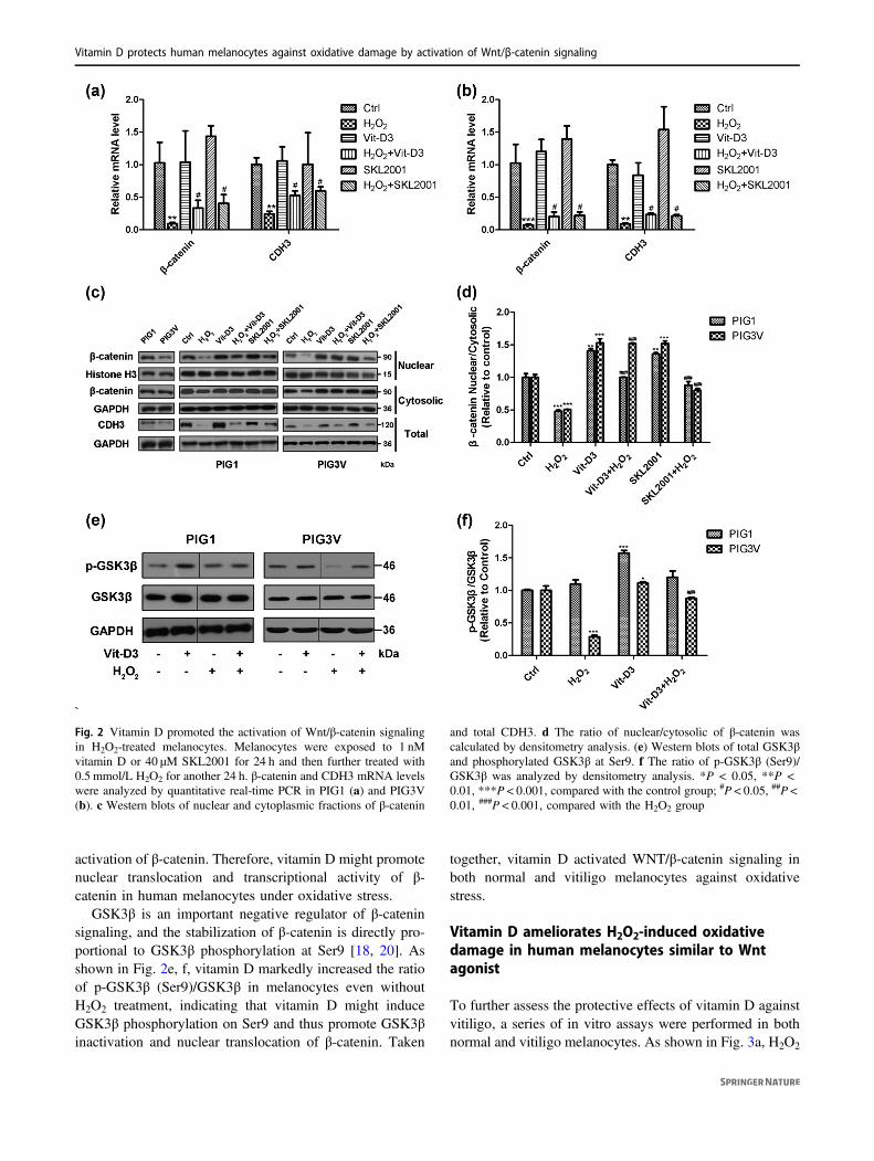
activation of β-catenin. Therefore, vitamin D might promotenuclear translocation and transcriptional activity of β-catenin in human melanocytes under oxidative stress.
GSK3β is an important negative regulator of β-cateninsignaling, and the stabilization of β-catenin is directly pro-portional to GSK3β phosphorylation at Ser9 [18, 20]. Asshown in Fig. 2e, f, vitamin D markedly increased the ratioof p-GSK3β (Ser9)/GSK3β in melanocytes even withoutH2O2 treatment, indicating that vitamin D might induceGSK3β phosphorylation on Ser9 and thus promote GSK3βinactivation and nuclear translocation of β-catenin. Taken
together, vitamin D activated WNT/β-catenin signaling inboth normal and vitiligo melanocytes against oxidativestress.
Vitamin D ameliorates H2O2-induced oxidativedamage in human melanocytes similar to Wntagonist
To further assess the protective effects of vitamin D againstvitiligo, a series of in vitro assays were performed in bothnormal and vitiligo melanocytes. As shown in Fig. 3a, H2O2
`Fig. 2 Vitamin D promoted the activation of Wnt/β-catenin signalingin H2O2-treated melanocytes. Melanocytes were exposed to 1 nMvitamin D or 40 μM SKL2001 for 24 h and then further treated with0.5 mmol/L H2O2 for another 24 h. β-catenin and CDH3 mRNA levelswere analyzed by quantitative real-time PCR in PIG1 (a) and PIG3V(b). c Western blots of nuclear and cytoplasmic fractions of β-catenin
and total CDH3. d The ratio of nuclear/cytosolic of β-catenin wascalculated by densitometry analysis. (e) Western blots of total GSK3βand phosphorylated GSK3β at Ser9. f The ratio of p-GSK3β (Ser9)/GSK3β was analyzed by densitometry analysis. *P < 0.05, **P <0.01, ***P < 0.001, compared with the control group; #P < 0.05, ##P <0.01, ###P < 0.001, compared with the H2O2 group
Vitamin D protects human melanocytes against oxidative damage by activation of Wnt/β-catenin signaling
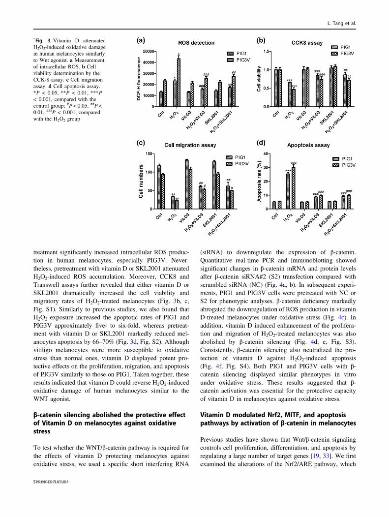
treatment significantly increased intracellular ROS produc-tion in human melanocytes, especially PIG3V. Never-theless, pretreatment with vitamin D or SKL2001 attenuatedH2O2-induced ROS accumulation. Moreover, CCK8 andTranswell assays further revealed that either vitamin D orSKL2001 dramatically increased the cell viability andmigratory rates of H2O2-treated melanocytes (Fig. 3b, c,Fig. S1). Similarly to previous studies, we also found thatH2O2 exposure increased the apoptotic rates of PIG1 andPIG3V approximately five- to six-fold, whereas pretreat-ment with vitamin D or SKL2001 markedly reduced mel-anocytes apoptosis by 66–70% (Fig. 3d, Fig. S2). Althoughvitiligo melanocytes were more susceptible to oxidativestress than normal ones, vitamin D displayed potent pro-tective effects on the proliferation, migration, and apoptosisof PIG3V similarly to those on PIG1. Taken together, theseresults indicated that vitamin D could reverse H2O2-inducedoxidative damage of human melanocytes similar to theWNT agonist.
β-catenin silencing abolished the protective effectof Vitamin D on melanocytes against oxidativestress
To test whether the WNT/β-catenin pathway is required forthe effects of vitamin D protecting melanocytes againstoxidative stress, we used a specific short interfering RNA
(siRNA) to downregulate the expression of β-catenin.Quantitative real-time PCR and immunoblotting showedsignificant changes in β-catenin mRNA and protein levelsafter β-catenin siRNA#2 (S2) transfection compared withscrambled siRNA (NC) (Fig. 4a, b). In subsequent experi-ments, PIG1 and PIG3V cells were pretreated with NC orS2 for phenotypic analyses. β-catenin deficiency markedlyabrogated the downregulation of ROS production in vitaminD-treated melanocytes under oxidative stress (Fig. 4c). Inaddition, vitamin D induced enhancement of the prolifera-tion and migration of H2O2-treated melanocytes was alsoabolished by β-catenin silencing (Fig. 4d, e, Fig. S3).Consistently, β-catenin silencing also neutralized the pro-tection of vitamin D against H2O2-induced apoptosis(Fig. 4f, Fig. S4). Both PIG1 and PIG3V cells with β-catenin silencing displayed similar phenotypes in vitrounder oxidative stress. These results suggested that β-catenin activation was essential for the protective capacityof vitamin D in melanocytes against oxidative stress.
Vitamin D modulated Nrf2, MITF, and apoptosispathways by activation of β-catenin in melanocytes
Previous studies have shown that Wnt/β-catenin signalingcontrols cell proliferation, differentiation, and apoptosis byregulating a large number of target genes [19, 33]. We firstexamined the alterations of the Nrf2/ARE pathway, which
`Fig. 3 Vitamin D attenuatedH2O2-induced oxidative damagein human melanocytes similarlyto Wnt agonist. a Measurementof intracellular ROS. b Cellviability determination by theCCK-8 assay. c Cell migrationassay. d Cell apoptosis assay.*P < 0.05, **P < 0.01, ***P< 0.001, compared with thecontrol group; #P < 0.05, ##P <0.01, ###P < 0.001, comparedwith the H2O2 group
L. Tang et al.
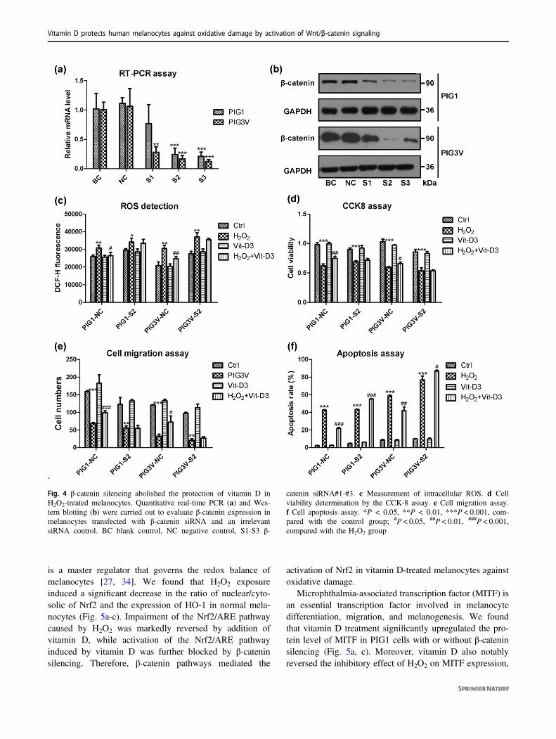
is a master regulator that governs the redox balance ofmelanocytes [27, 34]. We found that H2O2 exposureinduced a significant decrease in the ratio of nuclear/cyto-solic of Nrf2 and the expression of HO-1 in normal mela-nocytes (Fig. 5a-c). Impairment of the Nrf2/ARE pathwaycaused by H2O2 was markedly reversed by addition ofvitamin D, while activation of the Nrf2/ARE pathwayinduced by vitamin D was further blocked by β-cateninsilencing. Therefore, β-catenin pathways mediated the
activation of Nrf2 in vitamin D-treated melanocytes againstoxidative damage.
Microphthalmia-associated transcription factor (MITF) isan essential transcription factor involved in melanocytedifferentiation, migration, and melanogenesis. We foundthat vitamin D treatment significantly upregulated the pro-tein level of MITF in PIG1 cells with or without β-cateninsilencing (Fig. 5a, c). Moreover, vitamin D also notablyreversed the inhibitory effect of H2O2 on MITF expression,
`Fig. 4 β-catenin silencing abolished the protection of vitamin D inH2O2-treated melanocytes. Quantitative real-time PCR (a) and Wes-tern blotting (b) were carried out to evaluate β-catenin expression inmelanocytes transfected with β-catenin siRNA and an irrelevantsiRNA control. BC blank control, NC negative control, S1-S3 β-
catenin siRNA#1-#3. c Measurement of intracellular ROS. d Cellviability determination by the CCK-8 assay. e Cell migration assay.f Cell apoptosis assay. *P < 0.05, **P < 0.01, ***P < 0.001, com-pared with the control group; #P < 0.05, ##P < 0.01, ###P < 0.001,compared with the H2O2 group
Vitamin D protects human melanocytes against oxidative damage by activation of Wnt/β-catenin signaling

but the protection of vitamin D was abolished by β-catenindeficiency. The results indicated that vitamin D mightpositively modulate MITF to promote melanocyte pro-liferation and migration by activating the β-cateninpathway.
In addition to Nrf2 and MITF, the apoptotic pathway isanother important target of the WNT/β-catenin pathway[33]. Thus, expression of caspase 3 and its cleaved formwere tested in melanocytes. Consistent with the increasedapoptotic rates of melanocytes, H2O2 exposure induced aten-fold upregulation of the ratio of cleaved-caspase3/cas-pase3 (Fig. 5a, d). Vitamin D treatment remarkably clearlyinhibited the upregulation of cleaved caspase 3, while itsprotective role was further blocked by β-catenin silencing.These results suggested that the β-catenin pathway wasrequired for the anti-apoptotic effect of vitamin D on humanmelanocytes.
Discussion
Recent evidence indicates that treatment with vitamin Danalogs enhances repigmentation in vitiligo, especially incombination with UV light or corticosteroids [8]. However,
the molecular mechanisms underlying the pharmacologicalaction of vitamin D compounds against vitiligo have not yetbeen investigated. In the present study, we revealed a newmechanism of vitamin D as an antioxidative agent forvitiligo treatment. We found that vitamin D protectedmelanocytes against oxidative damage by activating theWNT/β-catenin pathway. The β-catenin pathway wasrequired for vitamin D to modulate downstream targets suchas Nrf2/ARE, MITF, and apoptotic pathways.
Oxidative stress is a critical factor in the onset and pro-gression of vitiligo [1, 2, 6, 27]. Consistent with previousstudies [10, 35–37], we found that patients with vitiligo hadhigher MDA levels and lower vitamin D levels than thehealthy control group. Intriguingly, the decreased serumvitamin D level was inversely correlated with serum MDAlevels in vitiligo patients, indicative of the potential anti-oxidant effect of vitamin D. A series of in vitro experimentswas conducted and further confirmed that vitamin D atte-nuated H2O2-induced ROS accumulation in human mela-nocytes and ameliorated the effect of oxidative stress onproliferation, migration, and apoptosis of melanocytes.Furthermore, vitiligo melanocytes displayed a significantdecrease in protein levels of VDR compared with normalmelanocytes. Our results were highly consistent with
`Fig. 5 β-catenin activation was required for vitamin D to modulateNrf2, MITF, and apoptosis pathways in melanocytes. a Westernblotting was performed to assess the expression of downstream com-ponents in Wnt/β-catenin signaling. The ratio of nuclear/cytosolic of
Nrf2 (b), total HO-1 and MITF (c), and the ratio of cleaved-caspase3/total caspase 3 (d) were analyzed by densitometry analysis. *P <0.05, **P < 0.01, ***P < 0.001, compared with the control group;#P < 0.05, ##P < 0.01, ###P < 0.001, compared with the H2O2 group
L. Tang et al.

previous studies which revealed that VDR expression wasremarkably decreased in the lesional/perilesional skin whilerepigmentation was accompanied with upregulated VDRexpression in vitiligo patients [38, 39]. The VDR genepolymorphisms were also reported to be associated withserum 25-hydroxyvitamin D levels in Chinese patients withgeneralized vitiligo [12]. Therefore, vitamin D insufficiencyor functional impairment of the vitamin D pathway mightattenuate the antioxidant capacity of patients and thusrepresent a risk factor for vitiligo.
It has been reported that vitiligo patients with comorbidautoimmune illness have lower levels of 25-hydroxyvitaminD [11]. The mechanism by which vitamin D modulatesautoimmunity is unknown, which might be explained by thefollowing possible reasons. On the one hand, vitamin D hasmultiple effects on innate and adaptive immune responsesby regulating various immune cells in vitro [40]. On theother hand, oxidative stress has been shown to be animportant initiate factor in the pathogenesis of variousautoimmune diseases [6, 41, 42], and vitamin D amelioratesoxidative damage and thus reduces the incidence andseverity of autoimmune responses. The antioxidant propertyof vitamin D has been validated in various human cells,such as cardiac cells, endothelial cells, gastric epithelialcells, and melanocytes [16, 43–45]. In the present study, wefurther showed that vitamin D could protect melanocytesagainst oxidative damage by activating the canonical WNT/β-catenin pathway.
A previous study by Regazzetti et al has demonstrated aninhibition of the WNT/β-catenin pathway in keratinocytesand melanocytes under oxidative stress [21]. Moreover,lesional vitiligo skin is also characterized by downregulatedexpression of WNT pathway components such as LEF1,CDH2, CDH3. Similarly, in our study, vitiligo melanocytesdisplayed lower expression of CDH3 (p-cadherin) andnuclear β-catenin than normal melanocytes. Because β-catenin is a transcription factor, nuclear translocation of β-catenin is critical for its dissociation from GSK3β andtranscriptional activation of multiple target genes [18, 33].We further found that in addition to downregulation ofCDH3 and β-catenin expression, H2O2 also decreased thenuclear/cytosolic ratio of β-catenin in both PIG1 and PIG3Vand prevented GSK3β phosphorylation on Ser9 in PIG3V,indicating that oxidative stress inhibited the nuclear trans-location of β-catenin. The inhibitory effects on β-cateninsignaling induced by H2O2 were reversed by addition ofvitamin D or WNT agonist (SKL2001). Thus, vitamin Dpositively modulated β-catenin signaling at both transla-tional and posttranslational levels in melanocytes underoxidative stress.
A previous study has shown that WNT agonist inducesthe differentiation of resident stem cells into pre-melanocytes in vitiligo lesional skin ex vivo [21]. Our
study further highlighted that WNT pathway activation wasessential for the antioxidant properties of vitamin D inmelanocytes. Like the WNT agonist, vitamin D sig-nificantly inhibited ROS accumulation and cell apoptosis inH2O2-treated melanocytes, and promoted their proliferativeand migratory activity. The protective effects of vitamin Dagainst oxidative stress were abolished by β-catenin silen-cing in melanocytes. Similar protective effects andmechanisms of vitamin D have been reported in other cellsand tissues [23, 24, 46, 47]. For example, activated β-catenin signaling is required for vitamin D to promoteosteogenesis in oxidative stress, while inhibition of theWNT pathway is involved in the protection of vitamin Dagainst colorectal tumorigenesis [23, 47]. Thus, vitamin Dmight utilize different modulatory patterns of the WNT/β-catenin pathway to protect against various diseases.
The transcription factor Nrf2 is a critical regulator ofoxidant metabolism in melanocytes, and it is required forthe effectiveness of aspirin and simvastatin as novel ther-apeutic strategies against vitiligo [27, 48–50]. Several stu-dies also revealed that Nrf2 signaling is involved in theprotective effects of vitamin D in various disease models[51–53]. Consistently, our results also showed that vitaminD promoted the upregulation of nuclear Nrf2 and itsdownstream component HO-1 in melanocytes. Furthermore,activation of Nrf2 signaling was completely blocked by β-catenin silencing, indicating that the β-catenin pathwaymediated Nrf2 activation in vitamin D-treated melanocytes.Recently, a number of studies have explored the relation-ship between the Wnt/β-catenin and Nrf2 pathways [54–57]. For example, WNT-3A controls the antioxidant meta-bolism of hepatocytes by activating Nrf2 in a β-catenin-independent manner, and Nrf2 pathway disruption inducesneurogenic impairment in Parkinsonian mice via Wnt/β-catenin dysregulation [54, 57]. Therefore, Wnt/β-cateninsignaling has complex interactions with Nrf2 signaling thatmight be specific to the cell type and disease model. Thepotential mechanism by which β-catenin activatesNrf2 signaling in melanocytes remains to be furtherinvestigated.
In addition to Nrf2 signaling, we also found that MITFand apoptotic pathways were regulated by β-catenin invitamin D-treated melanocytes. MITF is a central regulatorof the differentiation, migration, survival, and melanogen-esis of melanocytes [58–60]. Previous studies have shownthat MITF is significantly upregulated by WNT/β-cateninpathway activation in both vitiligo lesional skin ex vivo andhair follicle melanocytes in vitro [21, 61]. Consistently, weobserved that vitamin D treatment enhanced the expressionof MITF in human melanocytes. In our study, however, β-catenin deficiency abolished the upregulation of MITF inmelanocytes under oxidative stress, but not in melanocyteswithout H2O2 exposure. A previous study by Watabe et al.
Vitamin D protects human melanocytes against oxidative damage by activation of Wnt/β-catenin signaling

once showed that vitamin D induced MITF expression bystimulating endothelin B receptor signaling [13]. Thus, wespeculated that vitamin D could modulate MITF throughmultiple pathways and Wnt/β-catenin signaling might becrucial for MITF regulation in melanocytes under oxidativestress. These hypotheses remain to be validated in furtherexperiments. We also showed that β-catenin activation wasessential for vitamin D to modulate apoptotic pathway inmelanocytes. When β-catenin was silenced, vitamin D couldnot reverse the increase in cleaved caspase 3 expression orapoptotic rates in H2O2-treated melanocytes.
In conclusion, our study shows that vitamin D protectshuman melanocytes against oxidative damage by activationof Wnt/β-catenin signaling, indicating targeting Wnt/β-catenin signaling could be a useful approach to improvetreatment of vitiligo. Serum vitamin D insufficiency orfunctional impairment of the vitamin D pathway might be arisk factor for vitiligo due to impairment of the antioxidantsystem in vivo. Further studies using vitiligo mice arenecessary to confirm the therapeutic potential of systemicadministration of vitamin D for vitiligo.
Acknowledgements This study was supported by grants from Muni-cipal Hospital Emerging Frontier Technology United Key Projects(SHDC12016112), the Shanghai Key Medical Discipline for Derma-tology (2017ZZ02002), Shanghai leading talent project, and ChinaPostdoctoral Science Foundation (2016M600286). The authors thankDr. Caroline Le Poole (Loyola University Chicago, Maywood, IL, US)for providing the human melanocyte cell lines PIG1 and PIG3V andDr. Chunying Li (Xijing Hospital, Fourth Military Medical University,Xi’an, Shaanxi, China) for assisting with the cell transfer. The authorsalso thank the vitiligo patients and healthy donors for their participa-tion in this study.
Conflict of Interest The authors declare that they have no financialconflict of interest.
References
1. Colucci R, Dragoni F, Moretti S. Oxidative stress and immunesystem in vitiligo and thyroid diseases. Oxid Med Cell Longev.2015;2015:631927.
2. Denat L, Kadekaro AL, Marrot L, et al. Melanocytes as instigatorsand victims of oxidative stress. J Invest Dermatol.2014;134:1512–8.
3. Speeckaert R, Speeckaert MM, van Geel N. Why treatments do(n’t) work in vitiligo: An autoinflammatory perspective. Auto-immun Rev. 2015;14:332–40.
4. Jin Y, Birlea SA, Fain PR, et al. Genome-wide analysis identifiesa quantitative trait locus in the MHC class II region associatedwith generalized vitiligo age of onset. J Invest Dermatol.2011;131:1308–12.
5. Dell’Anna ML, Ottaviani M, Albanesi V, et al. Membrane lipidalterations as a possible basis for melanocyte degeneration invitiligo. J Invest Dermatol. 2007;127:1226–33.
6. Li S, Zhu G, Yang Y, et al. Oxidative stress drives CD8+ T-cellskin trafficking in patients with vitiligo through CXCL16 upre-gulation by activating the unfolded protein response in keratino-cytes. J Allergy Clin Immunol. 2017;140(1):177–189.
7. Sintov AC, Yarmolinsky L, Dahan A, et al. Pharmacologicaleffects of vitamin D and its analogs: recent developments. DrugDiscov Today. 2014;19:1769–74.
8. Birlea SA, Costin GE, Norris DA. New insights on therapy withvitamin D analogs targeting the intracellular pathways that controlrepigmentation in human vitiligo. Med Res Rev. 2009;29:514–46.
9. Parsad D, Kanwar AJ. Topical vitamin D analogues in the treat-ment of vitiligo. Pigment Cell Melanoma Res. 2009;22:487–8.
10. Upala S, Sanguankeo A. Low 25-hydroxyvitamin D levels areassociated with vitiligo: a systematic review and meta-analysis.Photodermatol Photoimmunol Photomed. 2016;32:181–90.
11. Silverberg JI, Silverberg AI, Malka E, et al. A pilot study asses-sing the role of 25 hydroxy vitamin D levels in patients withvitiligo vulgaris. J Am Acad Dermatol. 2010;62:937–41.
12. Li K, Shi Q, Yang L, et al. The association of vitamin D receptorgene polymorphisms and serum 25-hydroxyvitamin D levels withgeneralized vitiligo. Br J Dermatol. 2012;167:815–21.
13. Watabe H, Soma Y, Kawa Y, et al. Differentiation of murinemelanocyte precursors induced by 1,25-dihydroxyvitamin D3 isassociated with the stimulation of endothelin B receptor expres-sion. J Invest Dermatol. 2002;119:583–9.
14. Birlea SA, Costin GE, Norris DA. Cellular and molecularmechanisms involved in the action of vitamin D analogs targetingvitiligo depigmentation. Curr Drug Targets. 2008;9:345–59.
15. Kawakami T, Ohgushi A, Hirobe T, et al. Effects of 1,25-dihy-droxyvitamin D3 on human epidermal melanocytes and melano-blasts. J Dermatol Sci. 2014;76:72–4.
16. Gong Q, Li X, Sun J, et al. The effects of calcipotriol on thedendritic morphology of human melanocytes under oxidativestress and a possible mechanism: is it a mitochondrial protector? JDermatol Sci. 2015;77:117–24.
17. Li QL, Wu YH, Niu M, et al. Protective effects of tacalcitolagainst oxidative damage in human epidermal melanocytes. Int JDermatol. 2017;56:232–8.
18. MacDonald BT, Tamai K, He X. Wnt/beta-catenin signaling:components, mechanisms, and diseases. Dev Cell. 2009;17:9–26.
19. Logan CY, Nusse R. The Wnt signaling pathway in developmentand disease. Annu Rev Cell Dev Biol. 2004;20:781–810.
20. Kimelman D, Xu W. beta-catenin destruction complex: insightsand questions from a structural perspective. Oncogene.2006;25:7482–91.
21. Regazzetti C, Joly F, Marty C, et al. Transcriptional analysis ofvitiligo skin reveals the alteration of WNT pathway: a promisingtarget for repigmenting vitiligo patients. J Invest Dermatol.2015;135:3105–14.
22. Yamada T, Hasegawa S, Inoue Y, et al. Wnt/beta-catenin and kitsignaling sequentially regulate melanocyte stem cell differentia-tion in UVB-induced epidermal pigmentation. J Invest Dermatol.2013;133:2753–62.
23. Xiong Y, Zhang Y, Xin N, et al. 1alpha,25-Dihydroxyvitamin D3promotes osteogenesis by promoting Wnt signaling pathway. JSteroid Biochem Mol Biol. 2017;174:153–60.
24. Guo J, Lu C, Zhang F, et al. VDR activation reduces proteinuriaand high-glucose-induced injury of kidneys and podocytes byregulating wnt signaling pathway. Cell Physiol Biochem.2017;43:39–51.
25. Ohkawa H, Ohishi N, Yagi K. Assay for lipid peroxides in animaltissues by thiobarbituric acid reaction. Anal Biochem.1979;95:351–8.
26. Xu X, Fu WW, Wu WY. Serum 25-hydroxyvitamin D deficiencyin Chinese patients with vitiligo: a case-control study. PLoS ONE.2012;7:e52778.
27. Jian Z, Li K, Song P, et al. Impaired activation of the Nrf2-AREsignaling pathway undermines H2O2-induced oxidative stressresponse: a possible mechanism for melanocyte degeneration invitiligo. J Invest Dermatol. 2014;134:2221–30.
L. Tang et al.
28. Tang Luyan, Fu Wenwen, Zhang Yong, et al. Effect of calcipo-triol on melanin synthesis of human melanocytes. Chin J Der-matol. 2009;42:771–3.
29. Lei TC, Vieira WD, Hearing VJ. In vitro migration of melano-blasts requires matrix metalloproteinase-2: implications to vitiligotherapy by photochemotherapy. Pigment Cell Res.2002;15:426–32.
30. Slominski AT, Janjetovic Z, Kim TK, et al. Novel non-calcemicsecosteroids that are produced by human epidermal keratinocytesprotect against solar radiation. J Steroid Biochem Mol Biol.2015;148:52–63.
31. Shah S, Islam MN, Dakshanamurthy S, et al. The molecular basisof vitamin D receptor and beta-catenin crossregulation. Mol Cell.2006;21:799–809.
32. Gwak J, Hwang SG, Park HS, et al. Small molecule-based dis-ruption of the Axin/beta-catenin protein complex regulatesmesenchymal stem cell differentiation. Cell Res. 2012;22:237–47.
33. Vlad A, Rohrs S, Klein-Hitpass L, et al. The first five years of theWnt targetome. Cell Signal. 2008;20:795–802.
34. Jian Z, Li K, Liu L, et al. Heme oxygenase-1 protects humanmelanocytes from H2O2-induced oxidative stress via the Nrf2-ARE pathway. J Invest Dermatol. 2011;131:1420–7.
35. Ines D, Sonia B, Riadh BM, et al. A comparative study of oxidant-antioxidant status in stable and active vitiligo patients. ArchDermatol Res. 2006;298:147–52.
36. Khan R, Satyam A, Gupta S, et al. Circulatory levels of anti-oxidants and lipid peroxidation in Indian patients with generalizedand localized vitiligo. Arch Dermatol Res. 2009;301:731–7.
37. Saleh HM, Abdel Fattah NS, Hamza HT. Evaluation of serum 25-hydroxyvitamin D levels in vitiligo patients with and withoutautoimmune diseases. Photodermatol Photoimmunol Photomed.2013;29:34–40.
38. Doss RW, El-Rifaie AA, Gohary YM, et al. Vitamin D receptorexpression in vitiligo. Indian J Dermatol. 2015;60:544–8.
39. El-Hanbuli HM, Dawoud NM, Mahmoud RH. Narrow-band UVBeffects on cutaneous vitamin D receptor expression and serum 25-hydroxyvitamin D in generalized vitiligo. Photodermatol Photo-immunol Photomed. 2018;34:175–83.
40. Adorini L, Penna G. Control of autoimmune diseases by thevitamin D endocrine system. Nat Clin Pract Rheumatol.2008;4:404–12.
41. Adamczyk B, Adamczyk-Sowa M. New Insights into the role ofoxidative stress mechanisms in the pathophysiology and treatmentof multiple sclerosis. Oxid Med Cell Longev.2016;2016:1973834.
42. Harris JE. Cellular stress and innate inflammation in organ-specific autoimmunity: lessons learned from vitiligo. ImmunolRev. 2016;269:11–25.
43. Farhangi MA, Nameni G, Hajiluian G, et al. Cardiac tissue oxi-dative stress and inflammation after vitamin D administrations inhigh fat- diet induced obese rats. BMC Cardiovasc Disord.2017;17:161.
44. Haas MJ, Jafri M, Wehmeier KR, et al. Inhibition of endoplasmicreticulum stress and oxidative stress by vitamin D in endothelialcells. Free Radic Biol Med. 2016;99:1–10.
45. Uberti F, Bardelli C, Morsanuto V, et al. Role of vitamin D3combined to alginates in preventing acid and oxidative injury incultured gastric epithelial cells. BMC Gastroenterol. 2016;16:127.
46. Lee H, Bae S, Yoon Y. Anti-adipogenic effects of 1,25-dihy-droxyvitamin D3 are mediated by the maintenance of thewingless-type MMTV integration site/beta-catenin pathway. Int JMol Med. 2012;30:1219–24.
47. Groschel C, Aggarwal A, Tennakoon S, et al. Effect of1,25-dihydroxyvitamin D3 on the Wnt pathway in non-malignantcolonic cells. J Steroid Biochem Mol Biol. 2016;155:224–30.(Pt B)
48. Natarajan VT, Singh A, Kumar AA, et al. Transcriptional upre-gulation of Nrf2-dependent phase II detoxification genes in theinvolved epidermis of vitiligo vulgaris. J Invest Dermatol.2010;130:2781–9.
49. Jian Z, Tang L, Yi X, et al. Aspirin induces Nrf2-mediated tran-scriptional activation of haem oxygenase-1 in protection of humanmelanocytes from H2O2 -induced oxidative stress. J Cell MolMed. 2016;20:1307–18.
50. Chang Y, Li S, Guo W, et al. Simvastatin protects human mela-nocytes from H2O2-induced oxidative stress by activating Nrf2. JInvest Dermatol. 2017;137:1286–96.
51. Wang Z, Zhang H, Sun X, et al. The protective role of vitamin D3in a murine model of asthma via the suppression of TGF-beta/Smad signaling and activation of the Nrf2/HO-1 pathway. MolMed Rep. 2016;14:2389–96.
52. Nakai K, Fujii H, Kono K, et al. Vitamin D activates the Nrf2-Keap1 antioxidant pathway and ameliorates nephropathy in dia-betic rats. Am J Hypertens. 2014;27:586–95.
53. Teixeira TM, da Costa DC, Resende AC, et al. Activation of Nrf2-antioxidant signaling by 1,25-dihydroxycholecalciferol preventsleptin-induced oxidative stress and inflammation in humanendothelial cells. J Nutr. 2017;147:506–13.
54. Rada P, Rojo AI, Offergeld A, et al. WNT-3A regulates an Axin1/NRF2 complex that regulates antioxidant metabolism in hepato-cytes. Antioxid Redox Signal. 2015;22:555–71.
55. Long MJ, Lin HY, Parvez S, et al. beta-TrCP1 Is a vacillatoryregulator of Wnt signaling. Cell Chem Biol. 2017;24:944–957.e947.
56. Manigandan K, Manimaran D, Jayaraj RL, et al. Taxifolin curbsNF-kappaB-mediated Wnt/beta-catenin signaling via up-regulating Nrf2 pathway in experimental colon carcinogenesis.Biochimie. 2015;119:103–12.
57. L’Episcopo F, Tirolo C, Testa N, et al. Aging-induced Nrf2-AREpathway disruption in the subventricular zone drives neurogenicimpairment in parkinsonian mice via PI3K-Wnt/beta-catenindysregulation. J Neurosci. 2013;33:1462–85.
58. Levy C, Khaled M, Fisher DE. MITF: master regulator of mela-nocyte development and melanoma oncogene. Trends Mol Med.2006;12:406–14.
59. Cheli Y, Ohanna M, Ballotti R, et al. Fifteen-year quest formicrophthalmia-associated transcription factor target genes. Pig-ment Cell Melanoma Res. 2010;23:27–40.
60. Seberg HE, Van Otterloo E, Cornell RA. Beyond MITF: Multipletranscription factors directly regulate the cellular phenotype inmelanocytes and melanoma. Pigment Cell Melanoma Res.2017;30:454–66.
61. Guo H, Yang K, Deng F, et al. Wnt3a promotes melanin synthesisof mouse hair follicle melanocytes. Biochem Biophys Res Com-mun. 2012;420:799–804.
Vitamin D protects human melanocytes against oxidative damage by activation of Wnt/β-catenin signaling

RESEARCH ARTICLE Open Access
Identification of nine cryptic species ofCandida albicans, C. glabrata, and C.parapsilosis complexes using one-stepmultiplex PCRAmir Arastehfar1†, Wenjie Fang1,2†, Weihua Pan2*, Wanqing Liao2, Liang Yan2 and Teun Boekhout1,2,3*
Abstract
Background: Candida albicans, Candida glabrata, and Candida parapsilosis are three prevalent causes of candidiasis,worldwide. These species are considered as nine medically important complex species. Limited knowledge aboutthese newly recognized species prompted us to develop a one-step, multiplex PCR to detect and identify them inclinical settings.
Methods: Primers targeting Hyphal Wall Protein I gene for the C. albicans, C. dubliniensis, C. africana, Intergenic Spacerfor the C. glabrata, C. nivariensis, C. bracarensis, and Intein and ITS rDNA for the C. parapsilosis, C. orthopsilosis, and C.metapsilosis were designed. Using 168 CBS reference strains and 280 clinical isolates, the specificity and reproducibilityof the developed assay were evaluated.
Results: Our developed assay successfully identified and distinguished all the nine species. No cross-reaction withclosely- and distantly-related yeast species, Aspergillus species and human DNA was observed, resulting in 100%specificity. The ambiguous results obtained by MALDI-TOF for C. albicans and C. africana were corrected by our 9-plexPCR assay. This assay identified all the cryptic complex species from two test sets from Iran and China, correctly.
Conclusions: Our developed multiplex assay is accurate, specific, cost/time-saving, and works without the tedious DNAextraction steps. It could be integrated into routine clinical laboratories and as a reliable identification tool and has thepotential to be implemented into epidemiological studies to broaden the limited knowledge of cryptic speciescomplexes.
Keywords: Cryptic species, Candida, Molecular diagnosis, Multiplex PCR
IntroductionCandida albicans, C. glabrata, and C. parapsilosis werereported to be the three most clinically importantCandida species [1]. Due to the inclusion of newlydesignated species through taxonomic studies, thesethree Candida species are now considered crypticcomplex species, including C. albicans, C. africana,
and C. dubliniensis (as C. albicans complex) [2, 3], C.glabrata, C. nivariensis, and C. bracarensis (as C.glabrata complex) [4, 5], and C. parapsilosis, C.orthopsilosis, and C. metapsilosis (as C. parapsilosis com-plex) [6]. Despite the fact that for more than a decade thesecryptic species have been introduced, still limited knowledgeexists on their distribution, pathogenicity, and antifungalsusceptibility pattern. Moreover, from an evolutionary stand-point, identification and discrimination of these crypticspecies complexes could shed light on pathogenicity acquisi-tion, as Pryszcz et al. (2015) found C. metapsilosis as a highlyheterozygous opportunistic pathogen arose from a twoparental lineages that were not pathogenic [7].
* Correspondence: [email protected];[email protected]†Amir Arastehfar and Wenjie Fang contributed equally to this work.2Department of Dermatology, Shanghai Key Laboratory of Molecular MedicalMycology, Shanghai Institute of Medical Mycology, Shanghai ChangzhengHospital, Second Military Medical University, Shanghai 200003, China1Westerdijk Fungal Biodiversity Institute, Utrecht 3584, the NetherlandsFull list of author information is available at the end of the article
© The Author(s). 2018 Open Access This article is distributed under the terms of the Creative Commons Attribution 4.0International License (http://creativecommons.org/licenses/by/4.0/), which permits unrestricted use, distribution, andreproduction in any medium, provided you give appropriate credit to the original author(s) and the source, provide a link tothe Creative Commons license, and indicate if changes were made. The Creative Commons Public Domain Dedication waiver(http://creativecommons.org/publicdomain/zero/1.0/) applies to the data made available in this article, unless otherwise stated.
Arastehfar et al. BMC Infectious Diseases (2018) 18:480 https://doi.org/10.1186/s12879-018-3381-5
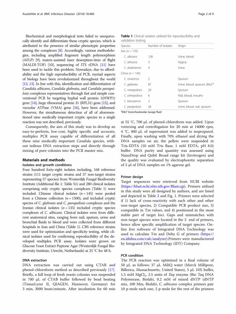
Biochemical and morphological tests failed to unequivo-cally identify and differentiate these cryptic species, which isattributed to the presence of similar phenotypic propertiesamong the complexes [8]. Accordingly, various methodolo-gies, including amplified fragment length polymorphism(AFLP) [9], matrix-assisted laser desorption-time of flight(MALDI-TOF) [10], sequencing of ITS rDNA [11] havebeen used to tackle this problem. Nowadays, due to afford-ability and the high reproducibility of PCR, myriad aspectsof biology have been revolutionized throughout the world[12, 13]. In line with this, identification and differentiation ofCandida albicans, Candida glabrata, and Candida parapsi-losis complexes representatives through fast and simple con-ventional PCR by targeting hyphal wall protein 1(HWP1)gene [14], large ribosomal protein 31 (RPL31) gene [15], andvacuolar ATPase (VMA) gene [16], have been addressed.However, the simultaneous detection of all of aforemen-tioned nine medically important cryptic species in a singlereaction was not described, previously.Consequently, the aim of this study was to develop an
easy-to-perform, low-cost, highly specific and accurate,multiplex PCR assay capable of differentiation of allthese nine medically important Candida species, with-out tedious DNA extraction steps and directly throughmixing of pure colonies into the PCR master mix.
Materials and methodsIsolates and growth conditionsFour hundred forty-eight isolates including, 168 referencestrains (111 target cryptic strains and 57 non-target strainsrepresenting 57 species) fromWesterdijk Fungal BiodiversityInstitute (Additional file 1: Table S1) and 280 clinical isolatescomprising only cryptic species complexes (Table 1) wereincluded. Chinese clinical isolates (n = 145) were pooledfrom a Chinese collection (n= 1500), and included crypticspecies of C. glabrata and C. parapsilosis complexes and theIranian clinical isolates (n = 135) included cryptic speciescomplexes of C. albicans. Clinical isolates were from diffe-rent anatomical sites, ranging from nail, sputum, urine andbronchial fluids to blood and were collected from differenthospitals in Iran and China (Table 1). CBS reference strainswere used for optimization and specificity testing, while cli-nical isolates used for confirming reproducibility of the de-veloped multiplex PCR assay. Isolates were grown onGlucose Yeast Extract Peptone Agar (Westerdijk Fungal Bio-diversity Institute, Utrecht, Netherlands) at 25 °C for 48 h.
DNA extractionDNA extraction was carried out using CTAB andphenol-chloroform method as described previously [17].Briefly, a full loop of fresh yeasts colonies was suspendedin 700 μL of CTAB buffer followed by bead beating(TissueLyzer II, QIAGEN, Hannover, Germany) for3 min, 3000 beats/minute. After incubation for 60 min
at 55 °C, 700 μL of phenol-chloroform was added. Uponvortexing and centrifugation for 20 min at 14000 rpm,4 °C, 400 μL of supernatant was added to isopropanol.Finally, upon washing with 70% ethanol and drying theDNA samples on air, the pellets were suspended inTris-EDTA (10 mM Tris Base, 1 mM EDTA, pH 8.0)buffer. DNA purity and quantity was assessed usingNanoDrop and Qubit Broad range kit (Invitrogen) andthe quality was evaluated by electrophoretic separationof 5 μl of DNA samples on 1% agarose gel.
Primer designTarget sequences were retrieved from NCBI website(https://blast.ncbi.nlm.nih.gov/Blast.cgi). Primers utilizedin this study were all designed by authors, and are listedand depicted in Table 2 and Fig. 1. Primers were selectedif 1) lack of cross-reactivity with each other and othernon-target species, 2) Compatible PCR product size, 3)compatible in Tm values, and 4) positioned in the moststable part of target loci. Gaps and mismatches withnon-target species were located in the 3΄ end of primers,hence allow specific amplification of target species. On-line free software of Integrated DNA Technology wasused to calculate Tm and Delta G of primers (https://eu.idtdna.com/calc/analyzer).Primers were manufacturedby Integrated DNA Technology (IDT) Company.
PCR conditionThe PCR reaction was optimized in a final volume of50 μL as follows: 37 μL MiliQ water (Merck Millipore,Billerica, Massachusetts, United States), 5 μL 10X buffer,1.5 mM MgCl2, 2.5 units of Taq enzyme (Bio Taq DNAPolymerase, Biolab), 0.2 mM of mixed dNTP (dNTPmix, 100 Mm, Biolab), C. albicans complex primers pair10 p-mole each one, 5 p-mole for the rest of the primers
Table 1 Clinical isolates utilized for reproducibility andvalidation testing
Species Number of Isolates Origin
Iran (n = 135)
C. albicans 128 Urine, blood
C. africana 3 Vagina
C. dubliniensis 4 Urine
China (n = 145)
C. nivarensis 2 Sputum
C. glabrata 87 Urine, blood, sputum, BALFa
C. metapsilosis 20 Sputum
C. orthopsilosis 6 Nail, blood, mouths
C. bracarensis 2 Sputum
C. parapsilosis 28 Urine, blood, nail, sputumaBALF bronchoalveolar lavage fluid
Arastehfar et al. BMC Infectious Diseases (2018) 18:480 Page 2 of 9
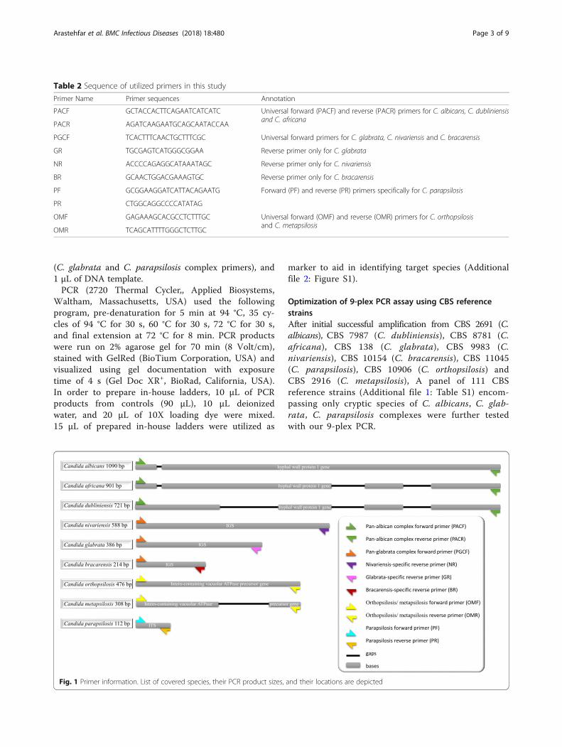
(C. glabrata and C. parapsilosis complex primers), and1 μL of DNA template.PCR (2720 Thermal Cycler,, Applied Biosystems,
Waltham, Massachusetts, USA) used the followingprogram, pre-denaturation for 5 min at 94 °C, 35 cy-cles of 94 °C for 30 s, 60 °C for 30 s, 72 °C for 30 s,and final extension at 72 °C for 8 min. PCR productswere run on 2% agarose gel for 70 min (8 Volt/cm),stained with GelRed (BioTium Corporation, USA) andvisualized using gel documentation with exposuretime of 4 s (Gel Doc XR+, BioRad, California, USA).In order to prepare in-house ladders, 10 μL of PCRproducts from controls (90 μL), 10 μL deionizedwater, and 20 μL of 10X loading dye were mixed.15 μL of prepared in-house ladders were utilized as
marker to aid in identifying target species (Additionalfile 2: Figure S1).
Optimization of 9-plex PCR assay using CBS referencestrainsAfter initial successful amplification from CBS 2691 (C.albicans), CBS 7987 (C. dubliniensis), CBS 8781 (C.africana), CBS 138 (C. glabrata), CBS 9983 (C.nivariensis), CBS 10154 (C. bracarensis), CBS 11045(C. parapsilosis), CBS 10906 (C. orthopsilosis) andCBS 2916 (C. metapsilosis), A panel of 111 CBSreference strains (Additional file 1: Table S1) encom-passing only cryptic species of C. albicans, C. glab-rata, C. parapsilosis complexes were further testedwith our 9-plex PCR.
Table 2 Sequence of utilized primers in this study
Primer Name Primer sequences Annotation
PACF GCTACCACTTCAGAATCATCATC Universal forward (PACF) and reverse (PACR) primers for C. albicans, C. dubliniensisand C. africana
PACR AGATCAAGAATGCAGCAATACCAA
PGCF TCACTTTCAACTGCTTTCGC Universal forward primers for C. glabrata, C. nivariensis and C. bracarensis
GR TGCGAGTCATGGGCGGAA Reverse primer only for C. glabrata
NR ACCCCAGAGGCATAAATAGC Reverse primer only for C. nivariensis
BR GCAACTGGACGAAAGTGC Reverse primer only for C. bracarensis
PF GCGGAAGGATCATTACAGAATG Forward (PF) and reverse (PR) primers specifically for C. parapsilosis
PR CTGGCAGGCCCCATATAG
OMF GAGAAAGCACGCCTCTTTGC Universal forward (OMF) and reverse (OMR) primers for C. orthopsilosisand C. metapsilosis
OMR TCAGCATTTTGGGCTCTTGC
Fig. 1 Primer information. List of covered species, their PCR product sizes, and their locations are depicted
Arastehfar et al. BMC Infectious Diseases (2018) 18:480 Page 3 of 9
Specificity testingTo evaluate the specificity of the 9-plex PCR assay, 100DNA samples containing 57 closely- and distantly-relatedyeast species, Aspergillus spp., and human DNA (fromblood) and 43 target strains pooled from CBS reference(optimization test set) were prepared in a blinded fashion(blind test set) (Table 1). For preparation of the specificitytest set, two technicians were involved. The first techni-cian randomly dispersed DNA samples and serially codedthem from one to 100. The second technician subjectedthe specificity blind test set to the optimized multiplexPCR. The first technician was provided with the resultsderived from PCR and the consistency of the results waschecked with the CBS identity of each strain.
Pure colony testingIn order to eliminate the DNA extraction step andconfirm the compatibility of the multiplex assay, singlecolonies of yeast species derived from the blind test setwere subjected to PCR as a template (except for Rhodo-torula mucilaginosa). Briefly, a single colony (≈ 1 mm3)was suspended in the prepared PCR master mix (50 μl)as mentioned earlier.
Validation of 9-plex multiplex PCR using clinical isolatesUpon optimization of the multiplex PCR assay usingCBS reference strains, specificity and pure colony test-ing, a panel of clinical isolates (n = 280) (Table 1) pre-pared in a blind fashion were subjected to the 9-plexassay. One technician performed the LSU sequencing andMALDI-TOF. Upon, identification, the first techniciancoded the yeast culture plates from 1 to 280, and then anew random number (integer number from 1 to 280) foreach plate was generated by SPSS random number gene-rator (version 21, International Business Machines Corp,Armonk, New York, United States) and used as blind testnumber. The second technician subjected the blind testset to the optimized multiplex PCR. The first technicianwas provided with the results derived from PCR and theconsistency of these results with MALDI-TOF andsequencing were evaluated.
MALDI-TOFThe full extraction method using MALDI-TOF MS (Bru-ker Biotyper, MicroFlex, LT, Bruker Daltonics, Bremen,Germany) was followed, according to the manufacturer’sinstructions (https://www.bruker.com/fileadmin/user_up-load/8-PDF-Docs/Separations_MassSpectrometry/Instruc-tionForUse/IFU_268711_267615_226413_MALDI_Biotar-get_48_Rev1.pdf). Briefly, after suspending of purecolonies in 300 μL of MiliQ water, 900 μL of absoluteethanol was added. After centrifugation step (14,000 rpm,3 min, room temperature) the supernatant was discardedand another centrifugation step was repeated to totally
discard the supernatant. Subsequently, 70% formic acid(Sigma Aldrich, St. Louis, Missouri, United States) wasadded and upon five minutes incubation at roomtemperature equal amount of Acetonitrile was added.After a final centrifugation step (14,000 rpm, 3 min, roomtemperature) 1 μL of clear supernatant was transferredonto the 96-well target plate. Subsequently, 1 μL of matrixwas overlaid on the top of air dried samples. Finally, the96 well target plate was loaded into the MALDI-TOFdevice.
SequencingIn order to ensure that the designed primers targetedthe right genes, the amplified PCR products were subjectto bidirectional dideoxy chain terminated Sangersequencing using the respective primers. Obtainedsequences were subjected to online searching databaseof NCBI (https://blast.ncbi.nlm.nih.gov/Blast.cgi).The ribosomal DNA large subunit (LSU) primers in-
cluding LROR (5΄-ACCCGCTGAACTTAAGC-3΄) andLR5 (5΄-TCCTGAGGGAAACTTCG-3΄) were exploitedto address the identity of each isolate [18]. Sequencesobtained for each isolate was subject to BLAST onlinedatabase tool (https://blast.ncbi.nlm.nih.gov/Blast.cgi).
ResultsDevelopment of 9-plex PCR assay using CBS referencestrainsSubjecting 111 CBS reference strains belonging to the spe-cies complexes of C. albicans (n = 55), C. glabrata (n = 28),and C. parapsilosis (n = 28) successfully differentiated andidentified the nine target species (Fig. 2). Although, thelengths of PCR products were different from those were de-signed, target species were unequivocally distinguishablefrom one another (Figs. 1 and 2 and Table 3). In order toprevent misidentifications it is advised to prepare in-housemaster mixes in large scale by mixing PCR products oftarget species and storing them at 4 °C for future identifica-tion purposes.
Specificity testing using the blind test setSubjecting 100 DNA samples (containing 66 fungal species)to our 9-plex PCR assay ranging from closely- anddistantly-related yeast species, Aspergillus spp. resulted in100% specificity and no cross-reaction with any otherfungal species. Additionally, subjecting human DNA to our9-plex PCR assay, showed no cross-reaction either.
Pure colony testingSubjecting 105 yeast pure colonies from 61 yeast speciespooled from the optimization test set (Additional file 1:Table S1) resulted in successful amplification of all ninetarget species. No PCR inhibition by cell components wasobserved, indicating that pure colonies are compatible
Arastehfar et al. BMC Infectious Diseases (2018) 18:480 Page 4 of 9
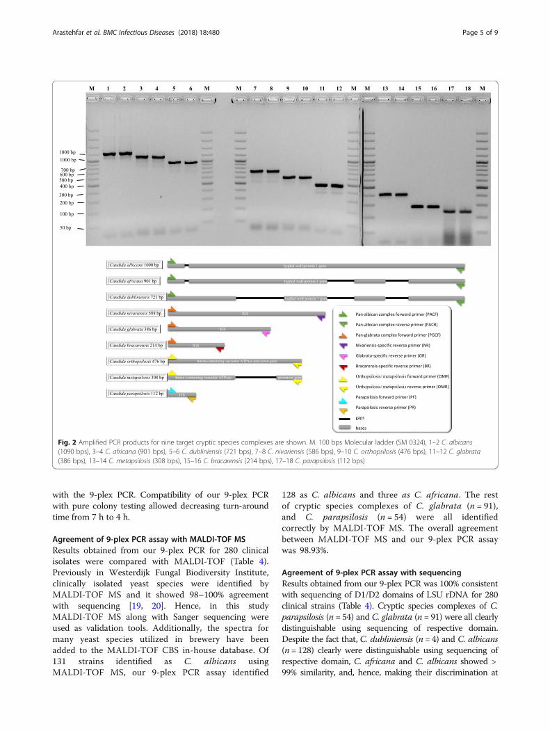
with the 9-plex PCR. Compatibility of our 9-plex PCRwith pure colony testing allowed decreasing turn-aroundtime from 7 h to 4 h.
Agreement of 9-plex PCR assay with MALDI-TOF MSResults obtained from our 9-plex PCR for 280 clinicalisolates were compared with MALDI-TOF (Table 4).Previously in Westerdijk Fungal Biodiversity Institute,clinically isolated yeast species were identified byMALDI-TOF MS and it showed 98–100% agreementwith sequencing [19, 20]. Hence, in this studyMALDI-TOF MS along with Sanger sequencing wereused as validation tools. Additionally, the spectra formany yeast species utilized in brewery have beenadded to the MALDI-TOF CBS in-house database. Of131 strains identified as C. albicans usingMALDI-TOF MS, our 9-plex PCR assay identified
128 as C. albicans and three as C. africana. The restof cryptic species complexes of C. glabrata (n = 91),and C. parapsilosis (n = 54) were all identifiedcorrectly by MALDI-TOF MS. The overall agreementbetween MALDI-TOF MS and our 9-plex PCR assaywas 98.93%.
Agreement of 9-plex PCR assay with sequencingResults obtained from our 9-plex PCR was 100% consistentwith sequencing of D1/D2 domains of LSU rDNA for 280clinical strains (Table 4). Cryptic species complexes of C.parapsilosis (n = 54) and C. glabrata (n = 91) were all clearlydistinguishable using sequencing of respective domain.Despite the fact that, C. dubliniensis (n = 4) and C. albicans(n = 128) clearly were distinguishable using sequencing ofrespective domain, C. africana and C. albicans showed >99% similarity, and, hence, making their discrimination at
Fig. 2 Amplified PCR products for nine target cryptic species complexes are shown. M. 100 bps Molecular ladder (SM 0324), 1–2 C. albicans(1090 bps), 3–4 C. africana (901 bps), 5–6 C. dubliniensis (721 bps), 7–8 C. nivariensis (586 bps), 9–10 C. orthopsilosis (476 bps), 11–12 C. glabrata(386 bps), 13–14 C. metapsilosis (308 bps), 15–16 C. bracarensis (214 bps), 17–18 C. parapsilosis (112 bps)
Arastehfar et al. BMC Infectious Diseases (2018) 18:480 Page 5 of 9

the species level difficult. However, our 9-plex PCRassay clearly discriminated the two closely relatedspecies complexes of C. albicans and C. africana.Consistently, 9-plex PCR assay distinctly identified allof the cryptic species complexes as distinct fragmenton the gel. The overall agreement between sequencingof D1/D2 LSU rDNA was 100%.
DiscussionRepresentatives of the cryptic species of Candida albicans,Candida glabrata, and Candida parapsilosis complexesaccount for the majority of candidiasis cases [21]. Appro-priate identification and differentiation of cryptic speciescomplexes is clinically relevant, as not only there are dif-ferences on virulence and antifungal susceptibility patternsamong species within the same complex, but also contra-dictory observations for antifungal susceptibility patternsamong different studies have been reported [5, 22–25].Due to facing these contradictory results along with thelimited epidemiological data for cryptic species complexes,the genuine distribution and antifungal susceptibility pro-files of these species in different geographical locationsremained unclear. Moreover, as antifungal susceptibility
profile of cryptic species within the same complex are va-ried, identification down to the species level is imperativeto establish the appropriate antifungal therapy [24, 26, 27].In line with this, providing epidemiologists and small la-boratories with a fast, accurate, specific, and cheap meansof identification to disclose the prevalence and antifungalsusceptibility profiles of isolates belonging to crypticCandida species. Accordingly, we have developed andvalidated an inexpensive, reliable, accurate, specific, anduser friendly multiplex PCR assays capable of identifyingnine cryptic species in one assay.From different perspectives including, time needed to
finish the experiments, required expenses and need oftrained technicians our assays is comparable to otherPCR-based assay and platforms such as MALDI-TOF.Application of cryptic species complexes strains iso-
lated in clinical settings with our 9-plex PCR assay andits comparison with results obtained by Sanger sequen-cing of D1/D2 domain of LSU rDNA, revealed 100%consistency between these techniques. However, due tothe high similarity between C. albicans and C. africanain the sequences of the D1/D2 and ITS rDNA fragments(99.3–100%), distinguishing these two species is difficult
Table 3 Expected and actual PCR products on gel are shown. Despite of deviation of amplicon sizes in both individual PCRproducts and home-made ladder containing mixture of PCR products, target species were clearly distinguishable from each other
Target species Expecetd PCRproduct size
Actual home-made ladderPCR product sizes on gel (approximately)
Actual indidividual PCR productsize on gel (approximately)
Candida albicans 1090 bps 950 bps 1200 bps
Candida africana 901 bps 800 bps 1100 bps
Candida dubliniensis 721 bps 600 bps 950 bps
Candida nivariensis 588 bps 500 bps 700 bps
Candida orthopsilosis 476 bps > 400 bps 576 bps
Candida glabrata 386 bps > 350 bps 486 bps
Candida metapsilosis 308 bps ˜280 bps 400 bps
Candida bracarensis 214 bps ˜200 bps 300 bps
Candida parapsilosis 112 bps 100 bps 250 bps
Table 4 Comparison of 9-plex PCR with CBS in-house MALDI-TOF database and sequencing of D1/D2 LSU rDNA
Species Multiplex PCR MALDI-TOF Sequencing
C. albicans (n = 128) C. albicans (n = 128) C. albicans (n = 128) C. albicans (n = 128)
C. dubliniensis (n = 4) C. dubliniensis (n = 4) C. dubliniensis (n = 4) C. dubliniensis (n = 4)
C. africana (n = 3) C. africana (n = 3) C. albicans (n = 3) C. africana (n = 3)a
C. glabrata (n = 87) C. glabrata (n = 87) C. glabrata (n = 87) C. glabrata (n = 87)
C. nivariensis (n = 2) C. nivariensis (n = 2) C. nivariensis (n = 2) C. nivariensis (n = 2)
C. bracarensis (n = 2) C. bracarensis (n = 2) C. bracarensis (n = 2) C. bracarensis (n = 2)
C. parapsilosis (n = 28) C. parapsilosis (n = 28) C. parapsilosis (n = 28) C. parapsilosis (n = 28)
C. orthopsilosis (n = 6) C. orthopsilosis (n = 6) C. orthopsilosis (n = 6) C. orthopsilosis (n = 6)
C. metapsilosis (n = 20) C. metapsilosis (n = 20) C. metapsilosis (n = 20) C. metapsilosis (n = 20)a Similarity between C. albicans and C. africana using sequencing of D1/D2 LSU rDNAwas more than 99%This library has been enriched with a diverse range of yeast species utilized in brewery
Arastehfar et al. BMC Infectious Diseases (2018) 18:480 Page 6 of 9
[28]. Consistently, the sequencing of LSU rDNA frag-ment in C. africana showed > 99% similarity with C.albicans. Importantly, application of our 9-plex PCRcould discriminate these two species. Like DNA micro-array [29] and pyrosequencing [30], Sanger sequencing re-quires highly trained technicians and more turn-aroundtime [8], while our multiplex PCR assay is straightforwardand running of the whole application (from master mixpreparation to visualization on the gel) only consumesfour hours. In terms of expenses, only 0.75–1 euro isenough to finalize the results on the gel electrophoresis,however, sequencing require specific devices and is moreexpensive. However, in order to resolve the issue of dis-crepancy between the length of predicted and actual PCRproducts we suggest to use either our in-house ladder orto run the amplicons of each control species individuallyin a separate lane.Our multiplex PCR successfully identified all C. africana
isolates and, hence, improved the results obtained fromMALDI-TOF. Except for C. africana strains (n = 3),Bruker MALDI-TOF MS could identify the rest of crypticspecies complexes, resulting in 98.93% agreement withour multiplex PCR assay. Variability in accuracy ofcommercial MALDI-TOF MS database for identificationof uncommon and cryptic Candida species [9, 31, 32],inability of the Bruker MALDI-TOF to distinguish C.africana from C. albicans, and incompetence of VITEKMS systems for identification of C. bracarensis, C. niva-riensis, and C. orthopsilosis and low cut-off value of BrukerMS systems (< 1.700) for identification of C. bracarensiscomplex reinforced the urge for molecular identificationtools [22, 33]. On the other hand, unlike the dominationof PCR even in developing countries [12, 13], MALDI-TOF is a newer introduced platform, mainly restricted tolarge or reference laboratories [34–36].Unlike, AFLP [9] and RFLP [37], there is no need for
restriction of the PCR products, several visualizationsteps and reading of the results are straightforward.Muriel Cornet et al. (2011) used PCR-RFLP, to identifyall eight cryptic species except for C. africana. However,this method used three primers targeting ribosomalintergenic spacer (IGS), tedious post-PCR restriction andrequired electrophoretic visualization twice making thisexperiment daunting, time consuming, and expensive[37]. Additionally, digestion of PCR products with re-striction enzymes generated multiple fragments, in turn,makes the interpretation difficult when compared tobanding pattern of reference strains.The successful identification of all nine cryptic species
in one tube, complements previous studies identifyingmembers of the species complexes of Candida albicans[14], Candida glabrata [15], and Candida parapsilosis[16] using three separate tubes, and, hence, is time- andcost-saving.
Application of broad diversity of yeast species, five Asper-gillus species, and human DNA for specificity testing andinclusion of an extensive number of CBS reference strainsand clinical isolates, showed that our 9-plex PCR is 100%specific. As a result, our assay circumvents the imperfec-tions of phenotypic assays (CHROMagar, VITEK 2 and ID32C) that suffer from the lack of specificity [38, 39].Additionally, subjecting various populations of the samespecies to phenotypic assay could showed various results[32, 40, 41]. Accordingly, as suggested by Griseo et al.(2015), small clinical laboratories can take advantage of spe-cific phenotypic methods supplemented with easy-to-perform PCR-based approaches to identify and reportisolated cryptic Candia species [8].
ConclusionDue to the problems with the identification and discrimin-ation of cryptic Candida species from their closest relatives,there is still uncertainty and unclarity about their epide-miology, pathogenicity and antifungal susceptibility pattern[43,44]. Consequently, developing reliable, specific, cost andlabor effective methods is necessary. Successful testing ofspecificity and validation using a broad range of CBSreference strains and clinical isolates, revealed thepotential of this assay to be implemented in routinediagnostics and epidemiological studies. As C. albi-cans, C. glabrata, and C. parapsilosis complex speciesconstitute 80–90% of candidiasis cases, identificationof all nine cryptic species within one multiplex PCRassay, could be of a great assistance.
Additional files
Additional file 1: Table S1. CBS reference strains utilized foroptimization of 9-plex PCR. (DOCX 20 kb)
Additional file 2: Figure S1. Comparison of home-made ladder andThermofisher commercial ladder (SM0323). Obviously, amplicons of alltarget species are distinguished and differentiated from one another.(JPG 79 kb)
AcknowledgementsWe would like to Thank Dr. Kamiar Zomorodian, Sadegh Khodavaisy, HamidBadali, Weiwei Jiang, and Ferry Hagen for kind provision of clinical strainsand revision of the manuscript.
FundingThis project has received funding from the European Union’s Horizon 2020research and innovation programme under the Marie Sklodowska-Curiegrant agreement No 642095, National Health Department of China[2018ZX10101003], National Natural Science Foundation of China [31770161],Second Military Medical University [2017JZ47] and Shanghai Science andTechnology Committee [14DZ2272900 and 14495800500].
Availability of data and materialsAll data are presented in this manuscript.
Authors’ contributionsAA and WJF have designed the study participated in study design andpractical works, and drafted the manuscript. WHP and TB have contributed
Arastehfar et al. BMC Infectious Diseases (2018) 18:480 Page 7 of 9

in study design and revision. WL and LY helped with study design, strainprovision, drafting and revising manuscript. All authors read and approvedthe final version.
Ethics approval and consent to participateNot applicable.
Consent for publicationNot applicable.
Competing interestsWeihua Pan is the editor of BMC Infectious Diseases and we declare that herco-authorship has not influenced the editorial processes.
Publisher’s NoteSpringer Nature remains neutral with regard to jurisdictional claims inpublished maps and institutional affiliations.
Author details1Westerdijk Fungal Biodiversity Institute, Utrecht 3584, the Netherlands.2Department of Dermatology, Shanghai Key Laboratory of Molecular MedicalMycology, Shanghai Institute of Medical Mycology, Shanghai ChangzhengHospital, Second Military Medical University, Shanghai 200003, China.3Institute of Biodiversity and Ecosystem Dynamics, University of Amsterdam,Amsterdam 1012 WX, the Netherlands.
Received: 13 February 2018 Accepted: 13 September 2018
References1. Magill SS, Edwards JR, Bamberg W, Beldavs ZG, Dumyati G, Kainer MA, et al.
Multistate point-prevalence survey of health care–associated infections. NEngl J Med. 2014;370:1198–208. https://doi.org/10.1056/NEJMoa1306801.
2. Tietz H-J, Hopp M, Schmalreck a, Sterry W, Czaika V. Candida africana sp.nov., a new human pathogen or a variant of Candida albicans? Mycoses.2001;44:437–45. https://doi.org/10.1046/j.1439-0507.2001.00707.x.
3. Sullivan DJ, Westerneng TJ, Haynes KA, Bennett DE, Coleman DC. Candidadubliniensis Sp-Nov - phenotypic and molecular characterization of a novelspecies associated with Oral Candidosis in Hiv-infected individuals.Microbiology-Uk. 1995;141:1507–21.
4. Alcoba-flórez J, Méndez-álvarez S, Guarro J, Pérez-roth E, Arévalo P,Cano J, et al. Phenotypic and molecular characterization of Candidanivariensis sp . Nov ., a possible new opportunistic fungus. J ClinMicrobiol. 2005;43:4107–11.
5. Bishop JA, Chase N, Magill SS, Kurtzman CP, Fiandaca MJ, Merz WG. Candidabracarensis detected among isolates of Candida glabrata by peptide nucleicacid fluorescence in situ hybridization: susceptibility data anddocumentation of presumed infection. J Clin Microbiol. 2008;46:443–6.
6. Tavanti A, Davidson AD, Gow NAR, MCJ M, Odds FC. Candida orthopsilosisand Candida metapsilosis spp. nov. to replace Candida parapsilosis groups IIand III. J Clin Microbiol. 2005;43:284–92.
7. Pryszcz LP, Németh T, Saus E, Ksiezopolska E, Hegedűsová E, Nosek J, et al.The genomic aftermath of hybridization in the opportunistic pathogenCandida metapsilosis. PLoS Genet. 2015;11:1–29.
8. Criseo G, Scordino F, Romeo O. Current methods for identifying clinicallyimportant cryptic Candida species. J Microbiol Methods. 2015;111:50–6.https://doi.org/10.1016/j.mimet.2015.02.004.
9. De Carolis E, Hensgens LAM, Vella A, Posteraro B, Sanguinetti M, Senesi S, etal. Identification and typing of the Candida parapsilosis complex: MALDI-TOFMS vs. AFLP. J Music Ther. 2015;52:123–30.
10. Sendid B, Ducoroy P, François N, Lucchi G, Spinali S, Vagner O, et al.Evaluation of MALDI-TOF mass spectrometry for the identification ofmedically-important yeasts in the clinical laboratories of Dijon and Lillehospitals. Med Mycol. 2013;51:25–32.
11. TM P SP, ID K, GW C. Rapid identification of fungi by sequencing the ITSland ITS2 regions using an automated capillary electrophoresis system. MedMycol. 2003;41:369–81.
12. Ragheb SM, Jimenez L. Polymerase chain reaction/rapid methods aregaining a foothold in developing countries. PDA J Pharm Sci Technol. 2014;68:239–55.
13. World Health Organization. Establishment of PCR laboratory in developingcountries. 2016. http://apps.who.int/iris/bitstream/10665/249549/5/9789290225317-Eng.pdf?ua=1.
14. Romeo O, Criseo G. First molecular method for discriminating betweenCandida africana, Candida albicans, and Candida dubliniensis by using hwp1gene. Diagn Microbiol Infect Dis. 2008;62:230–3.
15. Enache-Angoulvant A, Guitard J, Grenouillet F, Martin T, Durrens P,Fairhead C, et al. Rapid discrimination between Candida glabrata,Candida nivariensis, and Candida bracarensis by use of a singleplex PCR.J Clin Microbiol. 2011;49:3375–9.
16. Prandini THR, Theodoro RC, Bruder-Nascimento ACMO, Scheel CM, BagagliE. Analysis of inteins in the Candida parapsilosis complex for simple andaccurate species identification. J Clin Microbiol. 2013;51:2830–6.
17. Theelen B, Silvestri M, Guého E, Belkum A, Boekhout T. Identification andtyping of Malassezia yeasts using amplified fragment length polymorphism(AFLPTm), random amplified polymorphic DNA (RAPD) and denaturinggradient gel electrophoresis (DGGE). FEMS Yeast Res. 2001;1:79–86. https://doi.org/10.1111/j.1567-1364.2001.tb00018.x.
18. Stielow JB, Lévesque CA, Seifert KA, Meyer W, Irinyi L, Smits D, et al. Onefungus, which genes? Development and assessment of universal primers forpotential secondary fungal DNA barcodes. Persoonia - Mol Phylogeny EvolFungi. 2015;35:242–63. https://doi.org/10.3767/003158515X689135.
19. Taj-Aldeen SJ, Kolecka A, Boesten R, Alolaqi A, Almaslamani M, Chandra P, etal. Epidemiology of candidemia in Qatar, the Middle East: performance ofMALDI-TOF MS for the identification of Candida species, species distribution,outcome, and susceptibility pattern. Infection. 2014;42:393–404.
20. Kolecka A, Khayhan K, Groenewald M, Theelen B, Arabatzis M, Velegraki A, etal. Identification of medically relevant species of arthroconidial yeasts by useof matrix-assisted laser desorption ionization-time of flight massspectrometry. J Clin Microbiol. 2013;51:2491–500.
21. Guinea J. Global trends in the distribution of Candida species causingcandidemia. Clin Microbiol Infect. 2014;20:5–10. https://doi.org/10.1111/1469-0691.12539.
22. Hou X, Xiao M, Chen SC, Wang H, Yu S, Fan X. Identification and AntifungalSusceptibility Profiles of Candida nivariensis and Candida bracarensis in aMulti-Center Chinese Collection of Yeasts. 2017;8 January:1–8.
23. Romeo O, Scordino F, Pernice I, Lo Passo C, Criseo G. A multiplex PCRprotocol for rapid identification of Candida glabrata and its phylogeneticallyrelated species Candida nivariensis and Candida bracarensis. J MicrobiolMethods. 2009;79:117–20. https://doi.org/10.1016/j.mimet.2009.07.016.
24. Borman AM, Petch R, Linton CJ, Palmer MD, Bridge PD, Johnson EM.Candida nivariensis, an emerging pathogenic fungus with multidrugresistance to antifungal agents. J Clin Microbiol. 2008;46:933–8.
25. Yazdanparast SA, Khodavaisy S, Fakhim H, Shokohi T, Haghani I, Nabili M, etal. Molecular characterization of highly susceptible Candida africana fromvulvovaginal candidiasis. Mycopathologia. 2015;180:317–23.
26. Gácser A, Schäfer W, Nosanchuk JS, Salomon S, Nosanchuk JD. Virulence ofCandida parapsilosis, Candida orthopsilosis, and Candida metapsilosis inreconstituted human tissue models. Fungal Genet Biol. 2007;44:1336–41.
27. Borman AM, Szekely A, Linton CJ, Palmer MD, Brown P, Johnson EM.Epidemiology, antifungal susceptibility, and pathogenicity of Candidaafricana isolates from the United Kingdom. J Clin Microbiol. 2013;51:967–72.
28. Rodríguez-Leguizamón G, Fiori A, López LF, Gómez BL, Parra-Giraldo CM,Gómez-López A, et al. Characterising atypical Candida albicans clinicalisolates from six third-level hospitals in Bogotá, Colombia. BMC Microbiol2015;15:1–10. doi:https://doi.org/10.1186/s12866-015-0535-0.
29. Campa D, Tavanti A, Gemignani F, Mogavero CS, Bellini I, Bottari F, et al.DNA microarray based on arrayed-primer extension technique foridentification of pathogenic fungi responsible for invasive and superficialmycoses. J Clin Microbiol. 2008;46:909–15.
30. Borman AM, Linton CJ, Oliver D, Palmer MD, Szekely A, Odds FC, et al.Pyrosequencing analysis of 20 nucleotides of internal transcribed spacer 2discriminates Candida parapsilosis, Candida metapsilosis, and Candidaorthopsilosis. J Clin Microbiol. 2009;47:2307–10.
31. Jamal WY, Ahmad S, Khan ZU, Rotimi VO. Comparative evaluation of twomatrix-assisted laser desorption/ionization time-of-flight mass spectrometry(MALDI-TOF MS) systems for the identification of clinically significant yeasts.Int J Infect Dis. 2014;26:167–70. https://doi.org/10.1016/j.ijid.2014.05.031.
32. Kim T-H, Kweon OJ, Kim HR, Lee M-K. Identification of uncommon Candidaspecies using commercial identification systems. J Microbiol Biotechnol.2016;26:2206–13. https://doi.org/10.4014/jmb.1609.09012.
Arastehfar et al. BMC Infectious Diseases (2018) 18:480 Page 8 of 9
33. Sow D, Fall B, Ndiaye M, Ba BS, Sylla K, Tine R, et al. Usefulness of MALDI-TOF mass spectrometry for routine identification of Candida species in aresource-poor setting. Mycopathologia. 2015;180:173–9.
34. Posteraro B, Efremov L, Leoncini E, Amore R, Posteraro P, Ricciardi W, et al.Are the conventional commercial yeast identification methods still helpfulin the era of new clinical microbiology diagnostics? A meta-analysis of theiraccuracy. J Clin Microbiol. 2015;53:2439–50.
35. Posteraro B, De Carolis E, Vella A, Sanguinetti M. MALDI-TOF massspectrometry in the clinical mycology laboratory: identification of fungi andbeyond. Expert Rev Proteomics 2013;10:151–164.
36. Clark AE, Kaleta EJ, Arora A, Wolk DM. Matrix-assisted laser desorptionionization-time of flight mass spectrometry: a fundamental shift in the routinepractice of clinical microbiology. Clin Microbiol Rev. 2013;26:547–603.
37. Cornet M, Sendid B, Fradin C, Gaillardin C, Poulain D, Nguyen HV. Molecularidentification of closely related Candida species using two ribosomalintergenic spacer fingerprinting methods. J Mol Diagnostics. 2011;13:12–22.https://doi.org/10.1016/j.jmoldx.2010.11.014.
38. Bishop JA, Chase N, Lee R, Kurtzman CP, Merz WG. Production of whitecolonies on CHROMagar Candida medium by members of the Candidaglabrata clade and other species with overlapping phenotypic traits. J ClinMicrobiol. 2008;46:3498–500.
39. Cendejas-Bueno E, Gomez-Lopez A, Mellado E, Rodriguez-Tudela JL,Cuenca-Estrella M. Identification of pathogenic rare yeast species in clinicalsamples: comparison between phenotypical and molecular methods. J ClinMicrobiol. 2010;48:1895–9.
40. Albaina O, Sahand IH, Brusca MI, Sullivan DJ, De Larrinoa IF, Moragues MD.Identification and characterization of nine atypical Candida dubliniensisclinical isolates. J Med Microbiol 2015;64:147–156.
41. Romeo O, Criseo G. Candida africana and its closest relatives. Mycoses. 2011;54:475–86.
Arastehfar et al. BMC Infectious Diseases (2018) 18:480 Page 9 of 9
Mycoses. 2018;1–10. wileyonlinelibrary.com/journal/myc | 1© 2018 Blackwell Verlag GmbH
Received:1August2018 | Revised:20September2018 | Accepted:14October2018DOI:10.1111/myc.12863
O R I G I N A L A R T I C L E
Genotypic diversity and antifungal susceptibility of Cryptococcus neoformans isolates from paediatric patients in China
Nan Hong1,2 | Min Chen1,2 | Nan Xu3 | Abdullah M. S. Al-Hatmi4,5,6 | Chao Zhang1,2 | Wei H. Pan1,2 | Ferry Hagen4 | Teun Boekhout1,2,7,8 | Jianping Xu9 | Xian B. Zou10 | Wan Q. Liao1,2
1DepartmentofDermatology,ChangzhengHospital,SecondMilitaryMedicalUniversity,Shanghai,China2ShanghaiKeyLaboratoryofMedicalMycology,ChangzhengHospital,SecondMilitaryMedicalUniversity,Shanghai,China3DepartmentofDermatology,ShanghaiEastHospital,TongjiUniversitySchoolofMedicine,Shanghai,China4DepartmentofMedicalMycology,WesterdijkFungalBiodiversityInstitute,Utrecht,TheNetherlands5CentreofExpertiseinMycology,RadboudUniversityMedicalCentre/Canisius-WilhelminaHospital,Nijmegen,TheNetherlands6MinistryofHealth,DirectorateGeneralofHealthServices,Ibri,Oman7DepartmentofYeastandBasidiomyceteResearch,WesterdijkFungalBiodiversityInstitute,Utrecht,TheNetherlands8InstituteofBiodiversityandEcosystemDynamics,UniversityofAmsterdam,Amsterdam,TheNetherlands9DepartmentofBiology,McMasterUniversity,Hamilton,Ontario,Canada10DepartmentofDermatology,FirstAffiliatedHospitalofPLAGeneralHospital,Beijing,China
NanHong,MinChenandNanXucontributedequallytothisworkandsharethefirstco-authorship.
CorrespondenceWanQ.Liao,DepartmentofDermatology,ShanghaiKeyLaboratoryofMolecularMedicalMycology,ChangzhengHospital,SecondMilitaryMedicalUniversity,Shanghai,China.Email:[email protected],DepartmentofDermatology,FirstAffiliatedHospitalofPLAGeneralHospital,Beijing,China.Email:[email protected]
Funding informationNationalHealthDepartment,Grant/AwardNumber:2014ZX09J14106-02Aand2018ZX10101003;NationalNaturalScienceFoundationofChina,Grant/AwardNumber:81201269;ShanghaiScienceandTechnology,Grant/AwardNumber:14DZ22201000
AbstractCryptococcosisisalife-threateningmycosisprimarilyoccurringinadultpatientspar-ticularlythosewithimmunosuppressionsuchasHIVinfection/AIDS.Thenumberofreportedcasesofpaediatriccryptococcosishasincreasedinthelastdecadearoundtheworld, includingChina.However,current informationonthecharacteristicsofcryptococcosis in children,particularly thegenotypicdiversity andantifungal sus-ceptibilityoftheisolates,islimited.Inthepresentstudy,atotalof25paediatriciso-latesofCryptococcus neoformansweregenotypedusingtheISHAM-MLSTscheme.Invitrosusceptibilitytoantifungalagentsofthe22isolateswastestedusingtheCLSIM27-A3method.OuranalysesrevealedthatthegenotypicdiversityofC. neoformans isolatesfromChinesepaediatricpatientswaslow,withST5(80%)andST31(12%)being the twomajor sequence types.Reduced susceptibility to fluconazole (FLU),5-flucytosine(5-FC)anditraconazole(ITR)wasobservedamongC. neoformans iso-lates fromChinesepaediatricpatients, particularly among theST5 isolates,whichwas similar to observations made on C. neoformans isolatesfromChineseadultpa-tients. Inaddition, themajorityof isolates (3/4,75%)obtained fromdeceasedpa-tients showed decreased antifungal susceptibility, which indicates that furthermonitoringofantifungalsusceptibilityofCryptococcus isolates is warranted in man-agementofpaediatriccryptococcosis.
2 | HONG et al.
1 | INTRODUC TION
Cryptococcus neoformans and Cryptococcus gattii are two major ba-sidiomycetesyeastswhichformaspeciescomplexandareassoci-atedwithcryptococcosis,a life-threateningmycosis involvingbothimmunosuppressed and immunocompetent hosts.1,2 Cryptococcus neoformanshasaworldwidedistribution,occurringnaturallyinavianexcretaandcausingover80%ofcryptococcosiscaseseachyear.3 Cryptococcus gattiiismainlyassociatedwithtreesandhasemergedasapathogenofimmunocompetenthumansintemperateregionsofNorthAmerica.4
Therecentdevelopmentofmoleculartechniques,suchasmulti-locussequencetyping(MLST),has improvedourunderstandingofthe genetic diversity and taxonomy ofC. neoformans and C. gattii asbothare recognisedas species complexes.5-7 Cryptococcus neo-formans (serotypeA) has beendivided into threemajormoleculartypes, namelyAFLP1/VNI,AFLP1A/VNII/VNBandAFLP1B/VNII;moleculartypeofCryptococcus deneoformans(serotypeD)isAFLP2/VNIV;moleculartypeofthehybridisolates(serotypeAD)isAFLP3/VNIII.7 In addition, C. neoformans and C. gattii have two oppositematingtypes,MATα and MATa,withMATαbeingtheprevalentmat-ingtypeofclinicalandenvironmentalisolates.8,9
The rapid emergence of multidrug-resistant pathogenic fungihas become a major threat to human health.10 Currently, azoles,particularly fluconazole (FLU), are generally recommended for thetreatment of cryptococcosis during the consolidation andmainte-nancephases.11However, cryptococcosis causedbyFLU-resistantCryptococcusisolateshasrecentlybeenreported.12Moreover,anti-fungalsusceptibilityofCryptococcusisolateshasbeennotedtovaryamonggenotypesandgeographicregions.1,5
CryptococcosisisprimarilyobservedinadultpatientswithHIV/AIDS,whilecasesinvolvingchildrenarerarelyreported.13-16Ithasbeen documented that paediatric cryptococcosis accounted for0.9%ofallcryptococcosiscasesinSouthAfrica.17Thenumberofreportedcryptococcosisinchildren,however,hasincreasedworld-wide inthe lastdecade, includingChina.16,18Apreviousstudy in-dicatedthatthemajority(approximately70%)ofchildren(>2yearsold)intheBronx,NewYork,USAhadserologicevidenceforC. neo-formansinfection,19particularlythoseolderthan5yearsold.20
To date, data on the characteristics of cryptococcosis in chil-dren,particularlythegenotypicdiversityandantifungalsusceptibil-ityof the isolates,are limited.To thebestofourknowledge,onlyonestudyfromSouthAfricahasinvestigatedisolatesfrompaediat-riccryptococcosiscases, indicatingahighgeneticdiversityamongC. neoformansisolatesfrompaediatricpatients.Herewereportthefirst genotypingandantifungal susceptibilityprofilesofpaediatriccryptococcosisinChina.
2 | MATERIAL S AND METHODS
2.1 | Isolates
The paediatric cryptococcosis cases in the present study were allconfirmedbyapositiveculture.Patientswhoseageatthetimeofdi-agnosiswas<16yearswereincludedinthisstudy.Twenty-twoC. neo-formansstrainsisolatedfrompaediatricpatientswereincludedinourstudy.AlltheseisolateswerecollectedfromsixuniversityhospitalsinChinanamely:ChangzhengHospital (n=15),XinhuaHospital (n=1)andHuashanHospital(n=1)inShanghai;WestChinaHospital(n=2)inChengdu,SichuanProvince;theFirstAffiliatedHospitalofZhejiangUniversity (n=2) in Hangzhou, Zhejiang Province; and GuangzhouGeneralMilitaryHospital(n=1)inGuangzhou,GuangdongProvince.Inaddition,wealsoincludedtheMLSTsequenceandclinicalinforma-tionofthreepaediatricisolates(PU18,PU31andPU158)whichwerereportedpreviouslyfromBeijing21forcomparisonanalyses.
2.2 | Mating type
Todetermine themating typeof theC. neoformans isolates, PCRswereperformedaspreviouslydescribed.22 Cryptococcus neoformans strainsCBS10512 (MATa;AFLP1/VNI),CBS10515 (MATα;AFLP1/VNI),andC. deneoformansstrainsCBS10511(MATa;AFLP2/VNIV),CBS10513(MATα;AFLP2/VNIV)wereusedascontrols.
2.3 | DNA extraction, PCR and sequencing
DNA extraction of C. neoformans isolates was performed follow-ing the instructions of the UltraClean® microbial DNA isolation kit(MoBIOLaboratories,Carlsbad,CA,USA).Eachlocus(CAP59,GPD1,IGS1,LAC1,PLB1,SOD1 and URA5)inC. neoformanswasamplifiedina 25 μLPCRvolumeaccording to the ISHAM-MLSTscheme.23PCRproductswerepurifiedwithSephadexG-50fine(GEHealthcareBio-Sciences, Uppsala, Sweden) and were sequenced from both direc-tions. Sequence reads were manually edited using SeqMan v8.0.2(DNASTAR,Madison,WI,USA)andeachMLSTsequencewasalignedusingMUSCLEversion3.8.3124andconcatenatedbytheFASconCATprogram,25andthenqueriedagainsttheonlineMLSTdatabase(http://mlst.mycologylab.org)toassignalleletype(AT)foreachofthesevenloci. The final sequence type (ST) for each isolate was determinedbasedonthecombinedallelicassignmentsatthesevensequencedloci.
2.4 | Genotyping
Principal component analysis (PCA) of the sequence data ofC. neoformans isolates was performed using the Adegenet 2.1.1
K E Y W O R D S
antifungalsusceptibility,China,Cryptococcus neoformans,genotype,paediatric
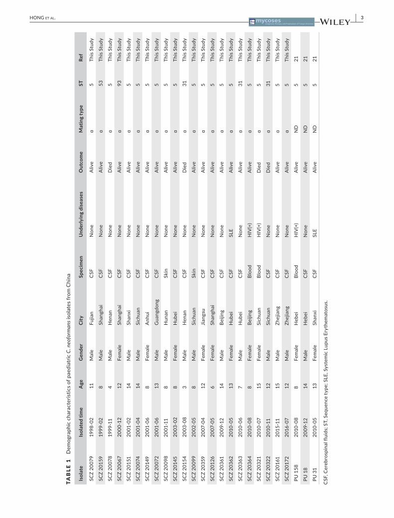
| 3HONG et al.
TABLE 1 DemographiccharacteristicsofpaediatricC
. neo
form
ansisolatesfromChina
Isol
ate
Isol
ated
tim
eA
geG
ende
rCi
tySp
ecim
enU
nder
lyin
g di
seas
esO
utco
me
Mat
ing
type
STRe
f
SCZ20079
1998-02
11Male
Fujian
CSF
None
Aliv
eɑ
5ThisStudy
SCZ20159
1999-02
8Male
Shanghai
CSF
None
Aliv
eɑ
53ThisStudy
SCZ20078
1999-11
4Male
Henan
CSF
None
Die
dɑ
5ThisStudy
SCZ20067
2000-12
12Female
Shanghai
CSF
None
Aliv
eɑ
93ThisStudy
SCZ20151
2001-02
14Male
Shanxi
CSF
None
Aliv
eɑ
5ThisStudy
SCZ20074
2001-04
14Male
Sichuan
CSF
None
Aliv
eɑ
5ThisStudy
SCZ20149
2001-06
8Female
Anhui
CSF
None
Aliv
eɑ
5ThisStudy
SCZ20072
2001-06
13Male
Guangdong
CSF
None
Aliv
eɑ
5ThisStudy
SCZ20098
2001-11
8Male
Hunan
Skin
None
Aliv
eɑ
5ThisStudy
SCZ20145
2003-02
8Female
Hubei
CSF
None
Aliv
eɑ
5ThisStudy
SCZ20154
2003-08
3Male
Henan
CSF
None
Die
dɑ
31ThisStudy
SCZ20099
2002-05
8Male
Sichuan
Skin
None
Aliv
eɑ
5ThisStudy
SCZ20359
2007-04
12Female
Jiangsu
CSF
None
Aliv
eɑ
5ThisStudy
SCZ20126
2007-05
6Female
Shanghai
CSF
None
Aliv
eɑ
5ThisStudy
SCZ20361
2009-12
14Male
Beijing
CSF
None
Aliv
eɑ
5ThisStudy
SCZ20362
2010-05
13Female
Hubei
CSF
SLE
Aliv
eɑ
5ThisStudy
SCZ20363
2010-06
7Male
Hubei
CSF
None
Aliv
eɑ
31ThisStudy
SCZ20364
2010-08
8Female
Beijing
Blood
HIV(+)
Aliv
eɑ
5ThisStudy
SCZ20321
2010-07
15Female
Sichuan
Blood
HIV(+)
Die
dɑ
5ThisStudy
SCZ20322
2010-11
12Male
Sichuan
CSF
None
Die
dɑ
31ThisStudy
SCZ20161
2015-11
15Male
Zhejiang
CSF
None
Aliv
eɑ
5ThisStudy
SCZ20172
2016-07
12Male
Zhejiang
CSF
None
Aliv
eɑ
5ThisStudy
PU158
2010-08
8Female
Hebei
Blood
HIV(+)
Aliv
eND
521
PU18
2009-12
14Male
Hebei
CSF
None
Aliv
eND
521
PU31
2010-05
13Female
Shanxi
CSF
SLE
Aliv
eND
521
CSF,Cerebrospinalfluids;ST,Sequencetype;SLE,SystemicLupusErythematosus.
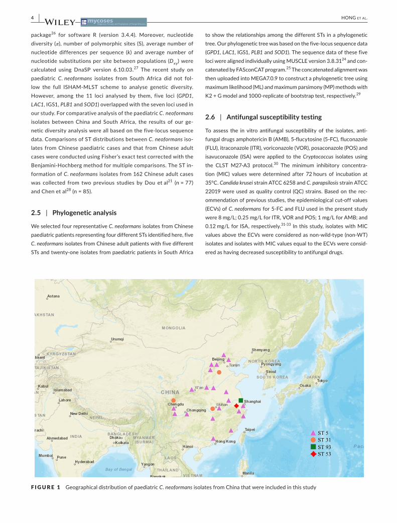
4 | HONG et al.
package26 for software R (version 3.4.4). Moreover, nucleotidediversity(π),numberofpolymorphicsites(S),averagenumberofnucleotide differences per sequence (k) and average number ofnucleotidesubstitutionspersitebetweenpopulations(Dxy)werecalculated using DnaSP version 6.10.03.27 The recent study onpaediatric C. neoformans isolates from South Africa did not fol-low the full ISHAM-MLST scheme to analyse genetic diversity.However, among the 11 loci analysed by them, five loci (GPD1,LAC1,IGS1,PLB1 and SOD1)overlappedwiththesevenlociusedinourstudy.ForcomparativeanalysisofthepaediatricC. neoformans isolates betweenChina and SouthAfrica, the results of our ge-neticdiversityanalysiswereallbasedonthefive-locussequencedata.ComparisonsofSTdistributionsbetweenC. neoformans iso-lates fromChinesepaediatriccasesand that fromChineseadultcaseswereconductedusingFisher’sexacttestcorrectedwiththeBenjamini-Hochbergmethodformultiplecomparisons.TheSTin-formationofC. neoformansisolatesfrom162Chineseadultcaseswas collected from two previous studies byDou etal21 (n=77)andChenetal28(n=85).
2.5 | Phylogenetic analysis
WeselectedfourrepresentativeC. neoformansisolatesfromChinesepaediatricpatientsrepresentingfourdifferentSTsidentifiedhere,fiveC. neoformansisolatesfromChineseadultpatientswithfivedifferentSTsandtwenty-oneisolatesfrompaediatricpatientsinSouthAfrica
toshowtherelationshipsamongthedifferentSTsinaphylogenetictree.Ourphylogenetictreewasbasedonthefive-locussequencedata(GPD1,LAC1,IGS1,PLB1 and SOD1).ThesequencedataofthesefivelociwerealignedindividuallyusingMUSCLEversion3.8.3124 and con-catenatedbyFASconCATprogram.25TheconcatenatedalignmentwasthenuploadedintoMEGA7.0.9toconstructaphylogenetictreeusingmaximumlikelihood(ML)andmaximumparsimony(MP)methodswithK2+Gmodeland1000-replicateofbootstraptest,respectively.29
2.6 | Antifungal susceptibility testing
To assess the in vitro antifungal susceptibility of the isolates, anti-fungaldrugsamphotericinB(AMB),5-flucytosine(5-FC),fluconazole(FLU),itraconazole(ITR),voriconazole(VOR),posaconazole(POS)andisavuconazole (ISA)were applied to theCryptococcus isolates using the CLST M27-A3 protocol.30 The minimum inhibitory concentra-tion (MIC) valueswere determined after 72hours of incubation at35°C.Candida kruseistrainATCC6258andC. parapsilosisstrainATCC22019wereusedasqualitycontrol (QC)strains.Basedontherec-ommendationofpreviousstudies,theepidemiologicalcut-offvalues(ECVs)ofC. neoformansfor5-FCandFLUusedinthepresentstudywere8mg/L;0.25mg/LforITR,VORandPOS;1mg/LforAMB;and0.12mg/Lfor ISA,respectively.31-33 Inthisstudy, isolateswithMICvaluesabovetheECVswereconsideredasnon-wild-type(non-WT)isolatesandisolateswithMICvaluesequaltotheECVswereconsid-eredashavingdecreasedsusceptibilitytoantifungaldrugs.
F IGURE 1 GeographicaldistributionofpaediatricC. neoformansisolatesfromChinathatwereincludedinthisstudy

| 5HONG et al.
3 | RESULTS
3.1 | Demographic data of the paediatric isolates
Theisolatesincludedinourstudywerecollectedfrom25paediatricpa-tientslivingin13differentprovincesacrossChinaasshowninTable1.ThemajorityoftheisolateswereobtainedfromSichuan(16%,5/25),followed by Shanghai (12%, 3/25), Hubei (12%, 3/25), Beijing (8%,2/25),Hebei(8%,2/25),Henan(8%,2/25),Shanxi(8%,2/25),Zhejiang(8%,2/25),Fujian(4%,1/25),Anhui(4%,1/25),Guangdong(4%,1/25),Hunan (4%,1/25)andJiangsu (4%,1/25).Themajorityof these iso-lateswererecoveredfromcerebrospinalfluid(CSF)(80%,20/25),fol-lowedbyblood(12%,3/25),andskin(8%,2/25).Themeanageofthese
patients was 10.4±3.5years (range: 3-15years), with the majoritywithintheagerangeof11-15years(56%,14/25).Themale/femalegen-derratiowas1.5(15:10).Themajorityofthepaediatricpatients(80%,20/25)hadnounderlyingdiseases.OnlythreepaediatricpatientswereHIV-infected, and two patients had systematic lupus erythematous(SLE).PatientcharacteristicsarepresentedinTable1andFigure1.
3.2 | Genotypes and mating types
Allof thepaediatric isolates fromChinawere identifiedasC. neo-formans(AFLP1/VNI,MATα).Amongthese,fourSTswereidentifiedwithST5(80%,20/25)beingpredominant,followedbyST31(12%,
F IGURE 2 AphylogenetictreefortherepresentativepaediatricC. neoformansisolatesfromChinaandSouthAfricausingMLandMPmethodsbasedonthesequencesoffiveloci(GPD1,LAC1,IGS1,PLB1 and SOD1).SequencetypesinbluederivefromtheISHAM-MLSTscheme;sequencetypesinblackderivefrom11-lociMLSTscheme.STrepresentssequencetype

6 | HONG et al.
3/25),ST53(4%,1/25)andST93(4%,1/25).ThedetailsareshowninTable1.
3.3 | Phylogenetic analysis
The phylogenetic tree revealed that the predominant ST5 isolate(basedon the ISHAM-MLST) inChinesepaediatric isolates (repre-sentedbystrainSCZ20172)clusteredtogetherwithaST9 isolate(basedon11-lociMLST)fromtheSouthAfricastudy(representedbystrainRSA1162).Moreover,themajorST8isolate(basedon11lociMLST)fromSouthAfrica(representedbystrainRSA1195)clusteredtogetherwithST93isolate(basedontheISHAM-MLST)fromChina(representedbystrainSCZ20067).DetailsofthephylogenetictreeareshowninFigure2.
3.4 | Comparison of genetic diversity of paediatric isolates between China and South Africa
ThePCAwasusedtoassessthegeneticstructureofthepaediatricC. neoformansAFLP1/VNIisolatesfromChinaandSouthAfrica.Thegenetic structure captured by the first two principal componentsshowedacombinedcontributionof77.8%.Thepaediatric isolatesfromChinaclustered intofourgroups,whereasthepaediatric iso-lates fromSouthAfricaclustered into10groups.ThePCAresultsagreedwiththeresultsofthephylogeneticanalysis,whichrevealedthattheST5(basedontheISHAM-MLST)isolatesfromChinawerecloselyrelatedtoST9(basedon11-lociMLST)isolatesfromSouthAfrica,andST93(basedonthe ISHAM-MLST) isolatesfromChinawereclosely related toST8 (basedon11-lociMLST) isolates fromSouthAfrica.
The nucleotide diversity (π) of paediatric isolates from SouthAfrica(π=0.00300,n=82)wassignificantlyhigherthanthatfromChina(π=0.00044,n=25).Theaveragenumberofnucleotidedif-ferences per sequence (k-value) of paediatric isolates from SouthAfrica (k=8.321) was also higher than that of paediatric isolatesfromChina (k=3.493). Togetherwith the paediatric isolates fromChinaandSouthAfrica,thenucleotidesequencesofallfivelocihad
polymorphicsitesrangingfrom1to12.LocusIGS1hadthehighestnucleotidediversity(π=0.00597)andthehighestaveragenumberof nucleotide differences per sequence (k=4.307), followed byLAC1 (π=0.00288, k=1.355) and GPD1 (π=0.00202, k=1.050).Therewasa significantlyhigherpercentageofST31 isolates fromChinesepaediatriccasesthanthatfromChineseadultcases(Fisher’sExacttest,calibratedP = 0.036).However,therewasnosignificantdifferenceinST5distributionbetweenC. neoformans isolatesfromChinesepaediatriccasesandthatfromChinesadultcases(calibratedP = 0.183).ThedetailsareshowninTable2,Table3andFigure3.
3.5 | In vitro antifungal drug susceptibility
TheMICvaluesofQCstrainATCC6258inthisstudywere1mg/LforAMB,16mg/Lfor5-FC,32mg/LforFLU,0.5mg/LforITRandPOS, 0.25mg/L for VOR and ISA. TheMIC values ofQC strainATCC22019inthisstudywere1mg/LforAMB,2mg/LforFLU,0.125mg/L for POS, 0.063mg/L for VOR, 0.031mg/L for ISA,0.25mg/Lfor5-FCandITR.Ourresultsareconsistentwithwhathavebeenreportedforthesetwostrains inCLSIM27-A3andinprevious studies.31-33 The MIC values of the paediatric isolatesofthepresentstudyweredeterminedforsevenantifungalcom-pounds,namelyAMB,5-FC,FLU,ITR,VOR,POSandISA.Forthe22isolates,theinvitroantifungalMIC90andsusceptibilityrangeswere0.5mg/L(0.25-0.50mg/L)forAMB,8mg/L(2-8mg/L)for5-FC,4mg/L(0.5-8mg/L)forFLU,0.125mg/L(0.031-0.125mg/L)for VOR, 0.125mg/L (0.008-0.125mg/L) for POS, 0.25μg/mL(0.031-0.250mg/L) for ITR and 0.031mg/L (0.004-0.063mg/L)for ISA.Noneof the isolates in this study showed resistance toFLUand5-FC,whicharethetwomostcommonlyusedantifungaldrugs in thetreatmentofcryptococcosis.FiveST5 isolates fromChinesepaediatricpatientswereobservedtohavedecreasedsus-ceptibilitytoITR,andsevenisolateswerefoundtohavedecreasedsusceptibility to 5-FC. One ST5 paediatric isolate (SCZ 20172)wasobservedtohavedecreasedsusceptibility toFLU,5-FCandITR.TheST93andST53paediatricisolatesinourstudywerenotobserved tobe resistantorhavedecreased susceptibility to the
TABLE 2 NucleotidepolymorphismsineachlocusandconcatenatedsequencesofpaediatricC. neoformansisolatesfromChinaandSouthAfrica
Locus
Paediatric isolates from ChinaPaediatric isolates from South Africa
Paediatric isolates from China and South Africa
DxyS π k S π k S π k
PLB1 1 0.00015 0.080 6 0.00208 1.106 6 0.00192 1.021 0.00202
LAC1 1 0.00017 0.333 7 0.00298 1.398 7 0.00288 1.355 0.00315
GPD1 1 0.00054 0.280 6 0.00201 1.044 6 0.00202 1.050 0.00231
IGS1 10 0.00388 2.800 12 0.00654 4.714 12 0.00597 4.307 0.00560
SOD1 0 0 0 1 0.00011 0.059 1 0.00008 0.043 0.00006
Concatenated 13 0.00044 3.493 32 0.00300 8.321 32 0.00280 7.776 0.00282
S,numberofpolymorphicsites;π,nucleotidediversity;k,averagenumberofnucleotidedifferencespersequence;Dxy,averagenumberofnucleotidesubstitutionspersitebetweenpopulations(paediatricisolatesfromChinavspaediatricisolatesfromSouthAfrica).
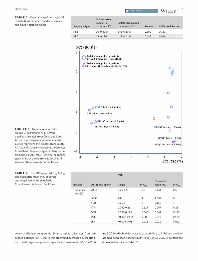
| 7HONG et al.
seven antifungal compounds. Most paediatric isolates from de-ceasedpatients(3/4,75%)inthisstudyhaddecreasedsusceptibil-itytoantifungalcompounds.Specifically,twoisolates(SCZ20154
andSCZ20078)haddecreasedsusceptibilityto5-FCandoneiso-latehaddecreasedsusceptibilityto ITR(SCZ20321).DetailsareshowninTable4andTableS1.
Sequence type
Isolates from paediatric cases (n = 25)
Isolates from adult cases (n = 162) P- value Calibrated P- value
ST5 20(0.800) 145(0.895) 0.183 0.183
ST31 3(0.120) 2(0.012) 0.018 0.036
TABLE 3 ComparisonoftwomajorSTdistributionsbetweenpaediatricisolatesandadultisolatesinChina
F IGURE 3 Geneticrelationshipsamong C. neoformansAFLP1/VNIpaediatricisolatesfromChinaandSouthAfricabyprincipalcomponentanalysis.CirclesrepresenttheisolatesfromSouthAfrica,andtrianglesrepresenttheisolatesfromChina.SequencetypesinbluederivefromtheISHAM-MLSTscheme;sequencetypesinblackderivefrom11-lociMLSTscheme.SArepresentsSouthAfrica
Isolates Antifungal agents
MIC
Range MIC50
Geometric mean MIC MIC90
Thisstudy(n=22)
AMB 0.25-0.5 0.5 0.441 0.5
5-FC 2-8 4 4.832 8
FLU 0.25-8 2 2.130 4
ITR 0.031-0.25 0.125 0.097 0.25
VOR 0.031-0.125 0.063 0.069 0.125
POS <0.008-0.125 <0.008 0.019 0.125
ISA <0.004-0.063 0.015 0.012 0.031
TABLE 4 TheMICrange,MIC50,MIC90 andgeometricmeanMICofsevenantifungalagentsforpaediatricC. neoformansisolatesfromChina

8 | HONG et al.
4 | DISCUSSION
Cryptococcosisisalethalfungalinfectionthatprimarilyaffectsadultpatients,especiallythosewithAIDSorsufferingfromotherunderly-ing diseases.3,34Incontrast,cryptococcosisisrareinchildren,eventhosewhoareHIV-infected.14,35However,thenumberofreportedcasesofcryptococcosisinchildrenhasincreasedinAfrica,Europe,South America and Asia, including China.15,16,18,36-40 Except fortherecentstudyinSouthAfrica,37dataonthegenotypicdiversityandantifungalsusceptibilityofpaediatricCryptococcus isolates are limited.
In this study, cryptococcosismainly occurred in older childrenwithameanageof10.4±3.5yearsold,which issimilartothatofpreviousstudies.14,37Onepossibleexplanationfortheolderonsetage is that the amount of cryptococcal exposure is important forcryptococcal infection.20 Repeated exposure of C. neoformans in earlychildhoodmaybenecessarytoestablishalatentfungalburdenwhichcanbereactivatedatolderage.20Sixtypercentofpaediatriccryptococcosispatientsweremale,whichissimilartopreviousstud-ies,13,14 supporting the theory thatC. neoformans infectionsmightbe gender-related.40,41 In terms of infection profiles of paediatriccryptococcosis,themostfrequent(84%)symptomwasCryptococcus meningitis,followedbydisseminatedcryptococcosis(12%)andskinlesions(4%).ThemajorityofourC. neoformansisolates(80.0%)wererecovered from apparently immunocompetent children, and thisagreeswithpreviousstudiesinChina(83.6%,92/110),13,16,18,40 but differsfromstudiesinUSA(20.6%,13/63)14andSouthAfrica(4.4%,2/45).36 Earlier investigations suggested that cryptococcosis pri-marilyoccursinapparentlyimmunocompetentadultsinChina.21,42 However,inChina,AIDSpatientsaregenerallytransferredtospecialhospitals for treatment and the clinical data of these patients areunavailable. Thus, the incidence of cryptococcosis inHIV-positivepatientsinChinamightbeunderestimated.
A considerable percentage of paediatric cryptococcosis casescaused bymembers of theC. gattii species complex has been re-ported in Colombia (5.9%),15 South Africa (7.0%)43 and Brazil(29.6%).44Incontrast,cryptococcalisolatesfromChinesepaediatricpatientswereallC. neoformansgenotypeAFLP1/VNIandcouldbedividedintofourSTs(ST5,ST31,ST53andST93),withST5asthepredominantone.ThisresultisnotsurprisingbecauseST5isolateisalsopredominantamongcryptococcal isolates fromChineseadultpatients.1,6 This resultmight also be related to the predominanceof C. neoformans AFLP1/VNI in China’s natural environment.45 Notably,ourstudyindicatedasignificantdifferenceinthedistribu-tionofST31betweenC. neoformansisolatesfromChinesepaediatricandadultcases(calibratedP = 0.036).MorestudiesonthevirulenceofST31isolatesareneededtoexplainthedifferentcapabilitythatST31 isolates showed in infectingChinesepaediatricpatients andinfecting Chinese adult patients in this study. In addition, all thepaediatricisolateswerematingtypeα,whichagreeswithpreviousobservationsthatover90%ofbothclinicalandenvironmentaliso-latesofC. neoformansarematingtypeα,8,9 including isolates fromChina.42,46 The skewed distribution ofMATα isolates might result
fromclonalexpansionbyasexualreproduction,self-fruiting,and/orsame-sexmatingandreproductionamongtheseisolates.47
Inthepresentstudy,lowgenotypicdiversityamongisolatesob-tainedfromChinesepaediatricpatientswasobserved.Currently,atotalof24STsofC. neoformanshavebeenidentifiedwithinisolatesobtained fromChinese adult patientswithpredominant sequencetypeST5.6,21,28,46 Inourstudy,onlyfourSTshavebeen identified,andST5wasalsothepredominantsequencetype(80%,20/25).Thisresult isreasonablebecauseadultandpaediatricpatients inChinamaybeexposedtothesamenaturalenvironmentharbouringC. neo-formans isolates.ThegenotypicdiversityamongpaediatricisolatesinChinaislowerthanthatobservedinSouthAfrica.37Bothπ and k valuesofthepaediatricisolatesfromSouthAfricawerehigherthanthoseof the isolates fromChina,which is similar to the resultsofpreviousstudies.48,49Sinceonlyfivelocisequencesweresharedbe-tweenour study and the study fromSouthAfrica,37 phylogeneticanalysisandPCAwerebasedonthesefivesharedloci.PhylogeneticanalysissuggestedthatthepredominantST5(basedontheISHAM-MLST) isolates from China are closely related to ST9 (based on11-lociMLST) isolates fromSouthAfrica, andST93 (basedon theISHAM-MLST) isolates fromChina are closely related to the pre-dominantST8 (basedon11-lociMLST) isolatesfromSouthAfrica.The PCA results agreewith our phylogenetic findings, suggestingsignificant differences in the population structure of paediatricCryptococcus isolates betweenChina and SouthAfrica.Moreover,wesuggestthattheISHAM-MLSTschemeshouldbeutilisedinin-vestigatingthegenotypicdiversityofmorepaediatricC. neoformans isolatesinthefuture.
Thenumberof reportedC. neoformans isolateswithFLUresis-tancehasincreasedinthepastdecade12andvarieswithgenotypeandgeographicregion.31,32Accordingtothecurrentclinicalguide-lines for cryptococcosis,11 AMB and 5-FC were recommended totreatcentralnervoussystem(CNS)cryptococcal infectionanddis-seminatedcryptococcosisinchildrenandFLUisusedinmaintenancetherapyofcryptococcosisinchildren.WhenweapplytherecentlypublishedECVstoevaluatethecurrentresults,noneoftheisolatesin this study showed resistance to FLU and 5-FC, however, eightST5 paediatric isolates and two ST31 paediatric isolates showeddecreased susceptibility to 5-FC, and/or FLU, and/or ITR. Theseresultsagreewiththefindingsofapreviousstudythatshowedin-creasingratesofC. neoformansisolatesthatareresistanttoFLUinChina,particularlytheST5isolates.6Moreover,mostpaediatriciso-latesfromdeceasedpatients(3/4,75%)inthisstudyhaddecreasedsusceptibilitytoantifungalcompounds.Twopaediatricisolateshaddecreasedsusceptibilityto5-FCandonepaediatricisolatehadde-creased susceptibility to ITR, suggesting that decreased suscepti-bilitytoantifungaldrugsofC. neoformansisolatesmightberelatedtotherapeuticfailure.Thus,antifungaldrugsusceptibilitytestingofC. neoformansisolatesshouldberecommendedtoimproveprogno-sisofpaediatriccryptococcosispatients.
In summary, the genotypic diversity of paediatric C. neofor-mans isolates fromChina is low, and the twomost frequent se-quence types were ST5 and ST31. Decreased susceptibility to

| 9HONG et al.
FLU,5-FCandITRwereobservedamongpaediatricC. neoformans isolatesfromChina,particularlyamongtheST5 isolates.FurtherinvestigationsofpaediatricC. neoformansisolatesfromaroundtheworld,particularlyrelatingtogenotypicdiversityanddrugresis-tance,areneededforbetterpreventionandtreatmentofpaediat-riccryptococcosis.
ACKNOWLEDG EMENTS
ThisstudywasfundedinpartwiththegrantsfromMajorNationalR&DProjectsoftheNationalHealthDepartment(2014ZX09J14106-02A),theNationalHealthDepartment(2018ZX10101003),NationalNaturalScienceFoundationofChina(No.81201269)andShanghaiScience and Technology counsel projects of 14DZ22201000 sup-portedthisstudy.
CONFLIC T OF INTERE S TS
Nonedeclared.
AUTHORS’ CONTRIBUTIONS
NH,MCandNXparticipatedinlaboratorytesting,statisticalanaly-sis,anddraftingofthemanuscript.TB,WHP,FH,JPXandAMSpar-ticipatedindesigningthisstudyandrevisingthemanuscript.WQLandXBZconceivedanddesignedthestudy.Alloftheauthorshavereadandapprovedthefinalmanuscript.
ORCID
Min Chen http://orcid.org/0000-0002-0629-9264
Wei H. Pan http://orcid.org/0000-0001-8077-023X
Jianping Xu http://orcid.org/0000-0003-2915-2780
R E FE R E N C E S
1. Cogliati M. Global Molecular Epidemiology of Cryptococcus neo-formans and Cryptococcus gattii: an atlas of themolecular types.Scientifica.2013;2013:675213.
2. HagenF,LumbschHT,ArsicArsenijevicV,etal.Importanceofre-solvingfungalnomenclature:thecaseofmultiplepathogenicspe-ciesintheCryptococcusgenus.mSphere.2017;2(4):e00238-17.
3. Mitchell TG, Perfect JR. Cryptococcosis in the era of AIDS–100 years after the discovery of Cryptococcus neoformans. Clin Microbiol Rev.1995;8(4):515-548.
4. ChenSC,MeyerW,SorrellTC.Cryptococcus gattii infections.Clin Microbiol Rev.2014;27(4):980-1024.
5. KhayhanK,HagenF,PanW,etal.Geographicallystructuredpop-ulationsofCryptococcus neoformans variety grubii in Asia correlate withHIVstatusandshowaclonalpopulationstructure.PLoS ONE. 2013;8(9):e72222.
6. FanX,XiaoM,ChenS,etal.PredominanceofCryptococcus neo-formans var. grubiimultilocus sequence type5andemergenceofisolates with non-wild-type minimum inhibitory concentrationstofluconazole:amulti-centrestudy inChina.Clin Microbiol Infect. 2016;22(10):887-e881.
7. HagenF,KhayhanK,TheelenB,etal.Recognitionofsevenspeciesin the Cryptococcus gattii/Cryptococcus neoformans species com-plex.Fungal Genet Biol.2015;78:16-48.
8. Kwon-ChungKJ,BennettJE.Distributionofalphaandalphamatingtypes ofCryptococcus neoformans among natural and clinical iso-lates. Am J Epidemiol.1978;108(4):337-340.
9. Campbell LT, Fraser JA, Nichols CB, Dietrich FS, Carter D,Heitman J. Clinical and environmental isolates of Cryptococcus gattii from Australia that retain sexual fecundity. Eukaryot Cell. 2005;4(8):1410-1419.
10. BastosRW,CarneiroHCS,OliveiraLVN,et al.Environmental tri-azoleinducescross-resistancetoclinicaldrugsandaffectsmorph-ophysiologyandvirulenceofCryptococcus gattii and C. neoformans. Antimicrob Agents Chemother.2018;62(1):e01179-17.
11. PerfectJR,DismukesWE,DromerF,etal.Clinicalpracticeguide-lines for the management of cryptococcal disease: 2010 up-datebythe infectiousdiseasessocietyofamerica.Clin Infect Dis. 2010;50(3):291-322.
12. CheongJW,McCormackJ.Fluconazoleresistanceincryptococcaldisease: emerging or intrinsic? Med Mycol.2013;51(3):261-269.
13. YuanjieZ,JianghanC,NanX,etal.Cryptococcalmeningitisinim-munocompetentchildren.Mycoses.2012;55(2):168-171.
14. JoshiNS,FisherBT,PrasadPA,ZaoutisTE.Epidemiologyofcryp-tococcal infection in hospitalized children. Pediatr Infect Dis J. 2010;29(12):e91-e95.
15. LizarazoJ,EscandonP,AgudeloCI,CastanedaE.CryptococcosisinColombianchildrenand literaturereview.Mem Inst Oswaldo Cruz. 2014;109(6):797-804.
16. Luo FL, Tao YH, Wang YM, Li H. Clinical study of 23 pediat-ric patients with cryptococcosis. Eur Rev Med Pharmacol Sci. 2015;19(20):3801-3810.
17. McCarthy KM, Morgan J, Wannemuehler KA, et al. Population-based surveillance for cryptococcosis in an antiretroviral-naiveSouth African province with a high HIV seroprevalence. AIDS. 2006;20(17):2199-2206.
18. LiuL,GuoL,LiuY,etal.ClinicalcharacteristicsandprognosisofpediatriccryptococcosisinBeijingChildren’sHospital,2002-2014.Eur J Pediatr.2017;176(9):1235-1244.
19. Goldman DL, Khine H, Abadi J, et al. Serologic evidence forCryptococcus neoformans infection in early childhood. Pediatrics. 2001;107(5):E66.
20. Ramsay S. C neoformans common in urban children. Lancet. 2001;357(9267):1507.
21. Dou HT, Xu YC, Wang HZ, Li TS. Molecular epidemiology ofCryptococcus neoformans and Cryptococcus gattiiinChinabetween2007and2013usingmultilocussequencetypingandtheDiversiLabsystem. Eur J Clin Microbiol Infect Dis.2015;34(4):753-762.
22. Chaturvedi S, Rodeghier B, Fan J, McClelland CM, Wickes BL,Chaturvedi V. Direct PCR of Cryptococcus neoformans MATalphaand MATapheromonestodeterminematingtype,ploidy,andvari-ety:atoolforepidemiologicalandmolecularpathogenesisstudies.J Clin Microbiol.2000;38(5):2007-2009.
23. Meyer W, Aanensen DM, Boekhout T, et al. Consensus multi-locus sequence typing scheme for Cryptococcus neoformans and Cryptococcus gattii. Med Mycol.2009;47(6):561-570.
24. EdgarRC.MUSCLE:multiplesequencealignmentwithhighaccu-racyandhighthroughput.Nucleic Acids Res.2004;32(5):1792-1797.
25. KuckP,LongoGC.FASconCAT-G:extensivefunctionsformultiplesequencealignmentpreparationsconcerningphylogeneticstudies.Front Zool.2014;11(1):81.
26. JombartT.adegenet:aRpackage for themultivariateanalysisofgeneticmarkers.Bioinformatics.2008;24(11):1403-1405.
27. RozasJ,Ferrer-MataA,Sanchez-DelBarrioJC,etal.DnaSP6:DNAsequencePolymorphismanalysisof largedata sets.Mol Biol Evol. 2017;34(12):3299-3302.
10 | HONG et al.
28. ChenYH,YuF,BianZY,etal.MultilocusSequencetypingrevealsbothsharedanduniquegenotypesofCryptococcus neoformans in JiangxiProvince,China.Sci Rep.2018;8(1):1495.
29. Kumar S, Stecher G, Tamura K. MEGA7: Molecular evolutionarygenetics analysis version 7.0 for bigger datasets. Mol Biol Evol. 2016;33(7):1870-1874.
30. PanW,KhayhanK,HagenF,etal.ResistanceofAsianCryptococcus neoformansserotypeAisconfinedtofewmicrosatellitegenotypes.PLoS ONE.2012;7(3):e32868.
31. Espinel-Ingroff A, Aller AI, Canton E, et al. Cryptococcus neoformans-Cryptococcus gattii species complex: an interna-tional study of wild-type susceptibility endpoint distributionsand epidemiological cutoff values for fluconazole, itraconazole,posaconazole, and voriconazole. Antimicrob Agents Chemother. 2012;56(11):5898-5906.
32. Espinel-Ingroff A, Chowdhary A, Cuenca-Estrella M, et al.Cryptococcus neoformans-Cryptococcus gattii species complex: aninternational study of wild-type susceptibility endpoint distribu-tionsandepidemiologicalcutoffvaluesforamphotericinBandflu-cytosine. Antimicrob Agents Chemother.2012;56(6):3107-3113.
33. Espinel-IngroffA,ChowdharyA,GonzalezGM,etal.MulticenterstudyofisavuconazoleMICdistributionsandepidemiologicalcut-offvaluesfortheCryptococcus neoformans-Cryptococcus gattiispe-ciescomplexusingtheCLSIM27-A3brothmicrodilutionmethod.Antimicrob Agents Chemother.2015;59(1):666-668.
34. SloanDJ,ParrisV.Cryptococcalmeningitis:epidemiologyandther-apeuticoptions.Clin Epidemiol.2014;6:169-182.
35. Speed BR, Kaldor J. Rarity of cryptococcal infection in children.Pediatr Infect Dis J.1997;16(5):536-537.
36. LamanM,HwaihwanjeI,DavisTM,ManningL.Cryptococcalmen-ingitis in immunocompetent Papua New Guinean children. Trop Doct.2010;40(1):61-63.
37. Miglia KJ, Govender NP, Rossouw J, Meiring S, Mitchell TG.Analyses of pediatric isolates of Cryptococcus neoformans fromSouthAfrica.J Clin Microbiol.2011;49(1):307-314.
38. Adonis-Koffy L, Diarrassouba G, Mouba J, et al. Cryptococcalmeningitis in two HIV-negative children. Arch Pediatr. 2010;17(7):1072-1073.
39. Gupta N, Sachdev A, Gupta D, Radhakrishnan N. Disseminatedcryptococcosis in an immunocompetent toddler. Indian Pediatr. 2017;54(2):145-146.
40. GuoJ,ZhouJ,ZhangS,etal.Acase-controlstudyofriskfactorsfor HIV-negative children with cryptococcal meningitis in ShiJiazhuang,China.BMC Infect Dis.2012;12:376.
41. GuessTE,RosenJA,McClellandEE.AnoverviewofsexbiasinC. neoformansInfections.J Fungi (Basel).2018;4(2):49.
42. Chen J, Varma A, Diaz MR, Litvintseva AP, Wollenberg KK,Kwon-Chung KJ. Cryptococcus neoformans strains and infectioninapparently immunocompetentpatients.China. Emerg Infect Dis. 2008;14(5):755-762.
43. Meiring ST, Quan VC, Cohen C, et al. A comparison of casesof paediatric-onset and adult-onset cryptococcosis detectedthrough population-based surveillance, 2005-2007. AIDS. 2012;26(18):2307-2314.
44. SeveroCB,XavierMO,GazzoniAF,SeveroLC.Cryptococcosis inchildren.Paediatr Respir Rev.2009;10(4):166-171.
45. ChenM,XuY,HongN,etal.EpidemiologyoffungalinfectionsinChina.Front Med.2018;12(1):58-75.
46. WuSY,LeiY,KangM,XiaoYL,ChenZX.MolecularcharacterisationofclinicalCryptococcus neoformans and Cryptococcus gattii isolates fromSichuanprovince.China. Mycoses.2015;58(5):280-287.
47. Sun S, Billmyre RB, Mieczkowski PA, Heitman J. Unisexual re-production drives meiotic recombination and phenotypic andkaryotypic plasticity in Cryptococcus neoformans. PLoS Genet. 2014;10(12):e1004849.
48. SimwamiSP,KhayhanK,HenkDA,etal.LowdiversityCryptococcus neoformans variety grubiimultilocussequencetypesfromThailandare consistent with an ancestral African origin. PLoS Pathog. 2011;7(4):e1001343.
49. LitvintsevaAP,MitchellTG.PopulationgeneticanalysesrevealtheAfricanoriginandstrainvariationofCryptococcus neoformans var. grubii. PLoS Pathog.2012;8(2):e1002495.
SUPPORTING INFORMATION
Additional supporting information may be found online in theSupportingInformationsectionattheendofthearticle.
Table S1.DetailsoftheMICforChinesepediatricC. neoformans iso-latesforsevenantifungals
How to cite this article:HongN,ChenM,XuN,etal.GenotypicdiversityandantifungalsusceptibilityofCryptococcus neoformansisolatesfrompaediatricpatientsinChina.Mycoses. 2018;00:1–10. https://doi.org/10.1111/myc.12863

Diagnostic Microbiology and Infectious Disease xxx (xxxx) xxx–xxx
DMB-14676; No of Pages 8
Contents lists available at ScienceDirect
Diagnostic Microbiology and Infectious Disease
j ourna l homepage: www.e lsev ie r .com/ locate /d iagmicrob io
YEAST PANEL multiplex PCR for identification of clinically importantyeast species: stepwise diagnostic strategy, useful fordeveloping countries
Amir Arastehfar a,1,2,Wenjie Fang a,b,c,2,Weihua Pan b,c,⁎, Michaela Lackner d,Wanqing Liao b,c, Parisa Badiee e,Kamiar Zomorodian f, Hamid Badali g, Ferry Hagen a, Cornelia Lass-Flörl d, Teun Boekhout a,c,h,⁎⁎a Westerdijk Fungal Biodiversity Institute, Utrecht, the Netherlandsb Department of Dermatology, Shanghai Changzheng Hospital, Second Military Medical University, Shanghai, Chinac Shanghai Key Laboratory of Molecular Medical Mycology, Shanghai Institute of Medical Mycology, Shanghai Changzheng Hospital, Second Military Medical University, Shanghai, Chinad Division of Hygiene and Medical Microbiology, Medical University of Innsbruck, Innsbruck, Austriae Prof. Alborzi Clinical Microbiology Research Center, Shiraz University of Medical Sciences, Shiraz, Iranf Basic Sciences in Infectious Diseases Research Center, and Department of Medical Mycology and Parasitology, School of Medicine, Shiraz University of Medical Sciences, Shiraz, Irang Department of Medical Mycology and Parasitology, School of Medicine, Mazandaran University of Medical Sciences, Sari, Iranh Institute of Biodiversity and Ecosystem Dynamics, University of Amsterdam, Amsterdam, the Netherlands
a b s t r a c ta r t i c l e i n f o
⁎ Corresponding author. Tel.: +86-2181885494; fax: +⁎⁎ Corresponding author. Tel.: +31-30-212-2600; fax: +
E-mail addresses: [email protected] (W. Pan)[email protected] (T. Boekhout).
1 Present address: Westerdijk Fungal Biodiversity InstiShanghai Changzheng Hospital, Second Military Medical U
2 AA andWF have equally contributed to this work.
https://doi.org/10.1016/j.diagmicrobio.2018.09.0070732-8893/© 2018 The Author(s). Published by Elsevier I
Please cite this article as: Arastehfar A, et al, Ytic strategy, useful for devel..., Diagn Microbi
Article history:Received 24 April 2018Received in revised form 7 September 2018Accepted 12 September 2018Available online xxxx
Keywords:Developing countriesYeast infectionMultiplex PCRMolecular identificationDeveloping countries
Identification of opportunistic yeasts in developing countries is mainly performed by phenotypic assays, whichare time-consuming and prone to errors. Wrong species identification may result in suboptimal treatment andinaccurate epidemiological data. To improve rapidity and accuracy of species identification, a diagnostic strategyusing a stepwise “YEAST PANEL multiplex PCR assays” targeting 21 clinically important yeast species of Candida,Trichosporon, Rhodotorula, Cryptococcus, and Geotrichumwas designed. Four hundred CBS reference strains wereused for optimization and specificity testing. Eight hundred clinical species were prepared in blinded sets formultiplex polymerase chain reaction (PCR) and matrix-assisted laser desorption time of flight mass spectropho-tometry (MALDI-TOF MS) investigation. Results obtained from YEAST PANEL multiplex PCR assay were 100%consistent with those of MALDI-TOF MS. Utilization of pure colony testing showed distinct amplicons for eachspecies, thus eliminating the need for DNA extraction. The targeted yeast species of this assay are responsiblefor 95% of the yeast infections. In conclusion, due to the high accuracy and coverage of a broad range of yeasts,this assay could be useful for identification in routine laboratories and epidemiological studies.
86-2181885493.31-30-21-22-601.,
tute, Utrecht, Netherlands, andniversity, Shanghai, China.
nc. This is an open access article under the CC BY-NC-ND l
EAST PANELmultiplex PCR for identification ofol Infect Dis (2018), https://doi.org/10.1016/j.d
© 2018 The Author(s). Published by Elsevier Inc. This is anopen access article under the CC BY-NC-ND license (http://
creativecommons.org/licenses/by-nc-nd/4.0/).
1. Introduction
The number of patients with invasive fungal infections (IFIs)continues to rise (Pfaller and Diekema, 2007), and the majority of theinfections are attributable to members of yeast genera, e.g., Candida,Trichosporon, Rhodotorula, Cryptococcus, and Geotrichum (Pappas et al.,2010; Pfaller and Diekema, 2007). Candida species account for themajority of candidiasis in patients with a broad range of underlyingconditions (Miceli et al., 2011). Trichosporon species are the second
leading cause of fungemia among patientswith hematological disorders(Miceli et al., 2011). Rhodotorula species are linked to catheter-relatedinfections (Miceli et al., 2011), while Cryptococcus species are morelikely to be found in HIV/AIDS-infected patients (Park et al., 2009).Geotrichum species are associated with infections in severely immuno-compromised patients (Miceli et al., 2011). Depending on the species,mortality of invasive yeast infections ranges from 15 to 80% (Pfallerand Diekema, 2007; Richardson and Lass-Flörl, 2008). Additionally,the growing number of non-albicans Candida species that are less sus-ceptible to fluconazole is worrisome (Cleveland et al., 2015;Vallabhaneni et al., 2016).
Historically, phenotypic and biochemical assays are frequently usedin developing countries for species identification (Posteraro et al.,2015). However, these assays are time-consuming and prone to error(Kathuria et al., 2015). Despite the rising popularity of matrix-assistedlaser desorption time of flight mass spectrophotometry (MALDI-TOF
icense (http://creativecommons.org/licenses/by-nc-nd/4.0/).
clinically important yeast species: stepwise diagnos-iagmicrobio.2018.09.007
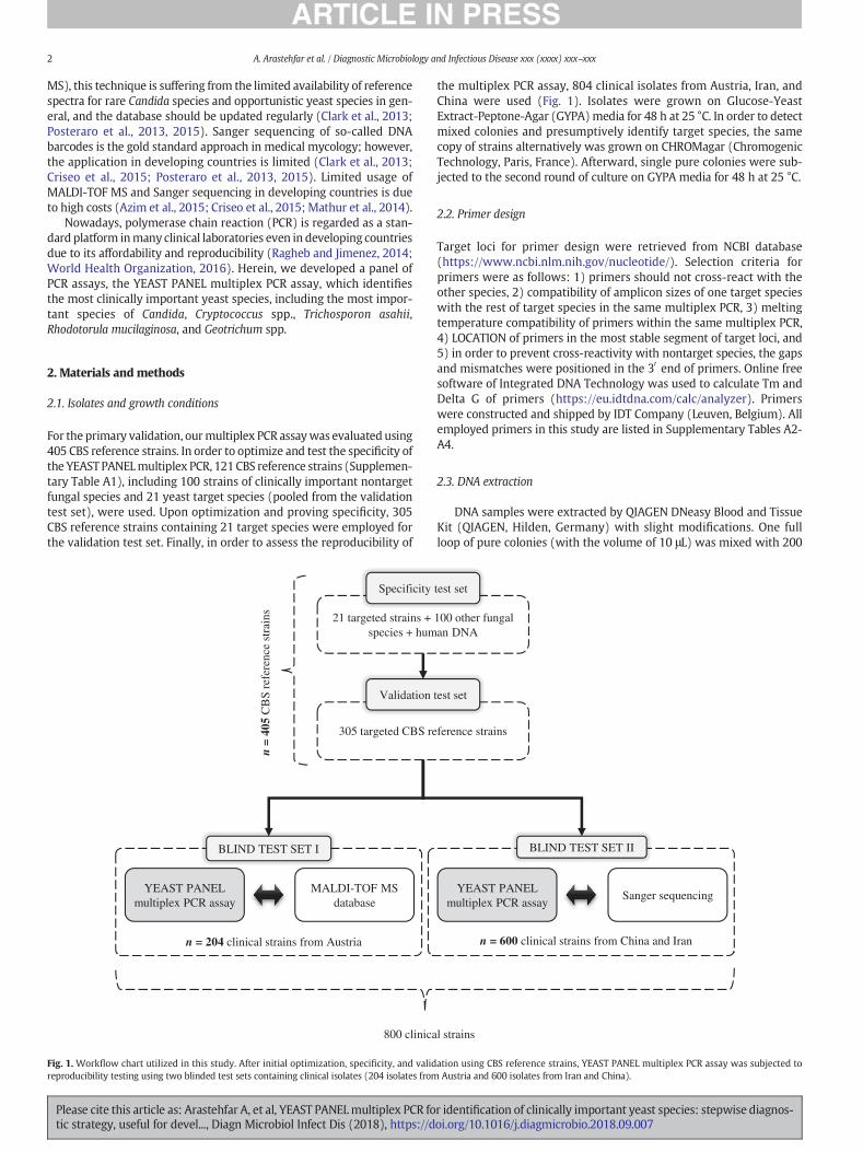
2 A. Arastehfar et al. / Diagnostic Microbiology and Infectious Disease xxx (xxxx) xxx–xxx
MS), this technique is suffering from the limited availability of referencespectra for rare Candida species and opportunistic yeast species in gen-eral, and the database should be updated regularly (Clark et al., 2013;Posteraro et al., 2013, 2015). Sanger sequencing of so-called DNAbarcodes is the gold standard approach in medical mycology; however,the application in developing countries is limited (Clark et al., 2013;Criseo et al., 2015; Posteraro et al., 2013, 2015). Limited usage ofMALDI-TOF MS and Sanger sequencing in developing countries is dueto high costs (Azim et al., 2015; Criseo et al., 2015; Mathur et al., 2014).
Nowadays, polymerase chain reaction (PCR) is regarded as a stan-dard platform inmany clinical laboratories even in developing countriesdue to its affordability and reproducibility (Ragheb and Jimenez, 2014;World Health Organization, 2016). Herein, we developed a panel ofPCR assays, the YEAST PANEL multiplex PCR assay, which identifiesthe most clinically important yeast species, including the most impor-tant species of Candida, Cryptococcus spp., Trichosporon asahii,Rhodotorula mucilaginosa, and Geotrichum spp.
2. Materials and methods
2.1. Isolates and growth conditions
For the primary validation, ourmultiplex PCR assaywas evaluated using405 CBS reference strains. In order to optimize and test the specificity ofthe YEASTPANELmultiplex PCR, 121CBS reference strains (Supplemen-tary Table A1), including 100 strains of clinically important nontargetfungal species and 21 yeast target species (pooled from the validationtest set), were used. Upon optimization and proving specificity, 305CBS reference strains containing 21 target species were employed forthe validation test set. Finally, in order to assess the reproducibility of
21 targeted strains + species + hum
BLIND TEST SET I
YEAST PANEL multiplex PCR assay
MALDI-TOF MS database
n =
405
CB
S re
fere
nce
stra
ins
800 clinica
n = 204 clinical strains from Austria
Specificity
305 targeted CBS re
Validation
Fig. 1. Workflow chart utilized in this study. After initial optimization, specificity, and validreproducibility testing using two blinded test sets containing clinical isolates (204 isolates from
Please cite this article as: Arastehfar A, et al, YEAST PANELmultiplex PCR fotic strategy, useful for devel..., Diagn Microbiol Infect Dis (2018), https://d
the multiplex PCR assay, 804 clinical isolates from Austria, Iran, andChina were used (Fig. 1). Isolates were grown on Glucose-YeastExtract-Peptone-Agar (GYPA)media for 48 h at 25 °C. In order to detectmixed colonies and presumptively identify target species, the samecopy of strains alternatively was grown on CHROMagar (ChromogenicTechnology, Paris, France). Afterward, single pure colonies were sub-jected to the second round of culture on GYPA media for 48 h at 25 °C.
2.2. Primer design
Target loci for primer design were retrieved from NCBI database(https://www.ncbi.nlm.nih.gov/nucleotide/). Selection criteria forprimers were as follows: 1) primers should not cross-react with theother species, 2) compatibility of amplicon sizes of one target specieswith the rest of target species in the same multiplex PCR, 3) meltingtemperature compatibility of primers within the same multiplex PCR,4) LOCATION of primers in the most stable segment of target loci, and5) in order to prevent cross-reactivity with nontarget species, the gapsand mismatches were positioned in the 3′ end of primers. Online freesoftware of Integrated DNA Technology was used to calculate Tm andDelta G of primers (https://eu.idtdna.com/calc/analyzer). Primerswere constructed and shipped by IDT Company (Leuven, Belgium). Allemployed primers in this study are listed in Supplementary Tables A2-A4.
2.3. DNA extraction
DNA samples were extracted by QIAGEN DNeasy Blood and TissueKit (QIAGEN, Hilden, Germany) with slight modifications. One fullloop of pure colonies (with the volume of 10 μL) was mixed with 200
100 other fungal an DNA
Sanger sequencing
BLIND TEST SET II
YEAST PANEL multiplex PCR assay
l strains
n = 600 clinical strains from China and Iran
test set
ference strains
test set
ation using CBS reference strains, YEAST PANEL multiplex PCR assay was subjected toAustria and 600 isolates from Iran and China).
r identification of clinically important yeast species: stepwise diagnos-oi.org/10.1016/j.diagmicrobio.2018.09.007
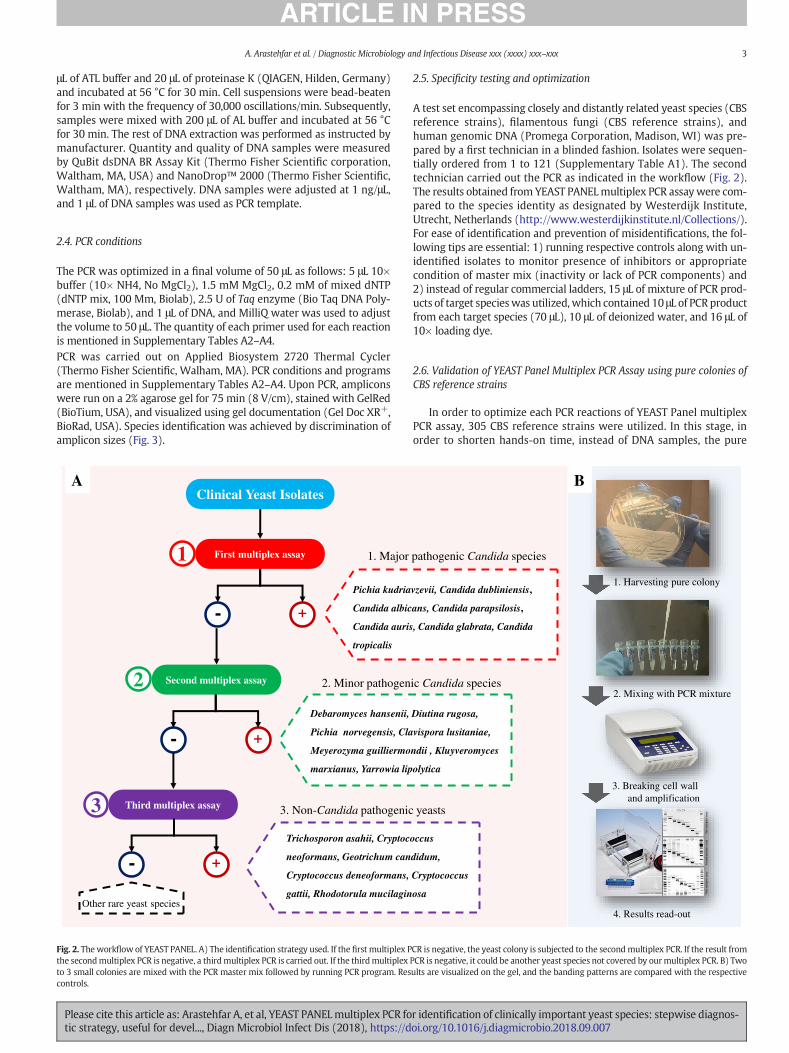
3A. Arastehfar et al. / Diagnostic Microbiology and Infectious Disease xxx (xxxx) xxx–xxx
μL of ATL buffer and 20 μL of proteinase K (QIAGEN, Hilden, Germany)and incubated at 56 °C for 30 min. Cell suspensions were bead-beatenfor 3 min with the frequency of 30,000 oscillations/min. Subsequently,samples were mixed with 200 μL of AL buffer and incubated at 56 °Cfor 30 min. The rest of DNA extraction was performed as instructed bymanufacturer. Quantity and quality of DNA samples were measuredby QuBit dsDNA BR Assay Kit (Thermo Fisher Scientific corporation,Waltham, MA, USA) and NanoDrop™ 2000 (Thermo Fisher Scientific,Waltham, MA), respectively. DNA samples were adjusted at 1 ng/μL,and 1 μL of DNA samples was used as PCR template.
2.4. PCR conditions
The PCR was optimized in a final volume of 50 μL as follows: 5 μL 10×buffer (10× NH4, No MgCl2), 1.5 mM MgCl2, 0.2 mM of mixed dNTP(dNTP mix, 100 Mm, Biolab), 2.5 U of Taq enzyme (Bio Taq DNA Poly-merase, Biolab), and 1 μL of DNA, and MilliQ water was used to adjustthe volume to 50 μL. The quantity of each primer used for each reactionis mentioned in Supplementary Tables A2–A4.PCR was carried out on Applied Biosystem 2720 Thermal Cycler(Thermo Fisher Scientific, Walham, MA). PCR conditions and programsare mentioned in Supplementary Tables A2–A4. Upon PCR, ampliconswere run on a 2% agarose gel for 75 min (8 V/cm), stained with GelRed(BioTium, USA), and visualized using gel documentation (Gel Doc XR+,BioRad, USA). Species identification was achieved by discrimination ofamplicon sizes (Fig. 3).
Clinical Yeast Isolates
First multiplex assay
+
Second multiplex assay
Third multiplex assay
-
+-
+-
Pichia kudria
Candida albic
Candida auris
tropicalis
Debaromyces hansenii,
Pichia norvegensis, Cla
Meyerozyma guilliermo
marxianus, Yarrowia lip
Trichosporon asahii, Cryptoc
neoformans, Geotrichum can
Cryptococcus deneoformans,
gattii, Rhodotorula mucilagin
1. Major
2. Minor pathogen
3. Non-Candida pathogenic
1
2
3
Other rare yeast species
A
Fig. 2. Theworkflow of YEAST PANEL. A) The identification strategy used. If the first multiplex Pthe secondmultiplex PCR is negative, a thirdmultiplex PCR is carried out. If the thirdmultiplexto 3 small colonies are mixed with the PCR master mix followed by running PCR program. Rescontrols.
Please cite this article as: Arastehfar A, et al, YEAST PANELmultiplex PCR fotic strategy, useful for devel..., Diagn Microbiol Infect Dis (2018), https://d
2.5. Specificity testing and optimization
A test set encompassing closely and distantly related yeast species (CBSreference strains), filamentous fungi (CBS reference strains), andhuman genomic DNA (Promega Corporation, Madison, WI) was pre-pared by a first technician in a blinded fashion. Isolates were sequen-tially ordered from 1 to 121 (Supplementary Table A1). The secondtechnician carried out the PCR as indicated in the workflow (Fig. 2).The results obtained from YEAST PANELmultiplex PCR assaywere com-pared to the species identity as designated by Westerdijk Institute,Utrecht, Netherlands (http://www.westerdijkinstitute.nl/Collections/).For ease of identification and prevention of misidentifications, the fol-lowing tips are essential: 1) running respective controls along with un-identified isolates to monitor presence of inhibitors or appropriatecondition of master mix (inactivity or lack of PCR components) and2) instead of regular commercial ladders, 15 μL of mixture of PCR prod-ucts of target specieswas utilized, which contained 10 μL of PCR productfrom each target species (70 μL), 10 μL of deionized water, and 16 μL of10× loading dye.
2.6. Validation of YEAST Panel Multiplex PCR Assay using pure colonies ofCBS reference strains
In order to optimize each PCR reactions of YEAST Panel multiplexPCR assay, 305 CBS reference strains were utilized. In this stage, inorder to shorten hands-on time, instead of DNA samples, the pure
vzevii, Candida dubliniensis,
ans, Candida parapsilosis,
, Candida glabrata, Candida
Diutina rugosa,
vispora lusitaniae,
ndii , Kluyveromyces
olytica
occus
didum,
Cryptococcus
osa
pathogenic Candida species
ic Candida species
yeasts
1. Harvesting pure colony
2. Mixing with PCR mixture
3. Breaking cell wall and amplification
4. Results read-out
B
CR is negative, the yeast colony is subjected to the secondmultiplex PCR. If the result fromPCR is negative, it could be another yeast species not covered by ourmultiplex PCR. B) Twoults are visualized on the gel, and the banding patterns are compared with the respective
r identification of clinically important yeast species: stepwise diagnos-oi.org/10.1016/j.diagmicrobio.2018.09.007
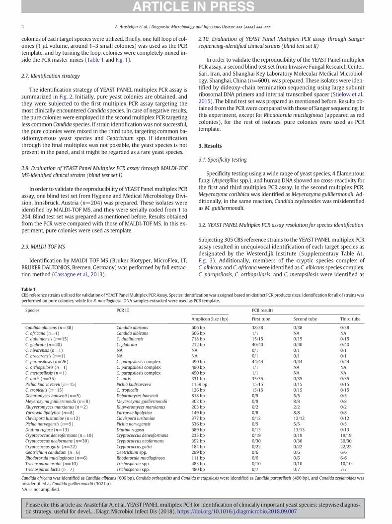
4 A. Arastehfar et al. / Diagnostic Microbiology and Infectious Disease xxx (xxxx) xxx–xxx
colonies of each target species were utilized. Briefly, one full loop of col-onies (1 μL volume, around 1–3 small colonies) was used as the PCRtemplate, and by turning the loop, colonies were completely mixed in-side the PCR master mixes (Table 1 and Fig. 1).
2.7. Identification strategy
The identification strategy of YEAST PANEL multiplex PCR assay issummarized in Fig. 2. Initially, pure yeast colonies are obtained, andthey were subjected to the first multiplex PCR assay targeting themost clinically encountered Candida species. In case of negative results,the pure colonies were employed in the secondmultiplex PCR targetingless common Candida species. If strain identification was not successful,the pure colonies were mixed in the third tube, targeting common ba-sidiomycetous yeast species and Geotrichum spp. If identificationthrough the final multiplex was not possible, the yeast species is notpresent in the panel, and it might be regarded as a rare yeast species.
2.8. Evaluation of YEAST Panel Multiplex PCR assay through MALDI-TOFMS-identified clinical strains (blind test set I)
In order to validate the reproducibility of YEAST Panelmultiplex PCRassay, one blind test set from Hygiene and Medical Microbiology Divi-sion, Innsbruck, Austria (n=204) was prepared. These isolates wereidentified by MALDI-TOF MS, and they were serially coded from 1 to204. Blind test set was prepared as mentioned before. Results obtainedfrom the PCR were compared with those of MALDI-TOF MS. In this ex-periment, pure colonies were used as template.
2.9. MALDI-TOF MS
Identification by MALDI-TOF MS (Bruker Biotyper, MicroFlex, LT,BRUKER DALTONIOS, Bremen, Germany) was performed by full extrac-tion method (Cassagne et al., 2013).
Table 1CBS reference strains utilized for validation of YEAST PanelMultiplex PCRAssay. Species identificperformed on pure colonies, while for R. mucilaginosa, DNA samples extracted were used as PC
Species PCR ID
Am
Candida albicans (n=38) Candida albicans 606C. africana (n=1) Candida albicans 606C. dubliniensis (n=15) C. dubliniensis 718C. glabrata (n=20) C. glabrata 212C. nivarensis (n=1) NA NAC. bracarensis (n=1) NA NAC. parapsilosis (n=26) C. parapsilosis complex 490C. orthopsilosis (n=1) C. parapsilosis complex 490C. metapsilosis (n=1) C. parapsilosis complex 490C. auris (n=35) C. auris 331Pichia kudriavzevii (n=15) Pichia kudriavzevii 115C. tropicalis (n=15) C. tropicalis 126Debaromyces hansenii (n=5) Debaromyces hansenii 818Meyerozyma guilliermondii (n=8) Meyerozyma guilliermondii 302Kluyveromyces marxianus (n=2) Kluyveromyces marxianus 203Yarrowia lipolytica (n=8) Yarrowia lipolytica 149Clavispora lusitaniae (n=12) Clavispora lusitaniae 377Pichia norvegensis (n=5) Pichia norvegensis 536Diutina rugosa (n=13) Diutina rugosa 689Cryptococcus deneoformans (n=19) Cryptococcus deneoformans 235Cryptococcus neoformans (n=30) Cryptococcus neoformans 392Cryptococcus gattii (n=22) Cryptococcus gattii 184Geotrichum candidum (n=6) Geotrichum spp. 299Rhodotorula mucilaginosa (n=6) Rhodotorula mucilaginosa 111Trichosporon asahii (n=10) Trichosporon spp. 483Trichosporon lactis (n=7) Trichosporon spp. 480
Candida africana was identified as Candida albicans (606 bp), Candida orthopsilsis and Candidamisidentified as Candida guilliermondii (302 bp).NA = not amplified.
Please cite this article as: Arastehfar A, et al, YEAST PANELmultiplex PCR fotic strategy, useful for devel..., Diagn Microbiol Infect Dis (2018), https://d
2.10. Evaluation of YEAST Panel Multiplex PCR assay through Sangersequencing-identified clinical strains (blind test set II)
In order to validate the reproducibility of the YEAST Panel multiplexPCR assay, a second blind test set from Invasive Fungal Research Center,Sari, Iran, and Shanghai Key Laboratory Molecular Medical Microbiol-ogy, Shanghai, China (n=600), was prepared. These isolates were iden-tified by dideoxy-chain termination sequencing using large subunitribosomal DNA primers and internal transcribed spacer (Stielow et al.,2015). The blind test set was prepared asmentioned before. Results ob-tained from the PCRwere comparedwith those of Sanger sequencing. Inthis experiment, except for Rhodotorula mucilaginosa (appeared as redcolonies), for the rest of isolates, pure colonies were used as PCRtemplate.
3. Results
3.1. Specificity testing
Specificity testing using a wide range of yeast species, 4 filamentousfungi (Aspergillus spp.), and human DNA showed no cross-reactivity forthe first and third multiplex PCR assay. In the second multiplex PCR,Meyerozyma caribbica was identified as Meyerozyma guilliermondii. Ad-ditionally, in the same reaction, Candida zeylanoides was misidentifiedasM. guiiliermondii.
3.2. YEAST PANEL Multiplex PCR assay resolution for species identification
Subjecting 305 CBS reference strains to the YEAST PANELmultiplex PCRassay resulted in unequivocal identification of each target species asdesignated by the Westerdijk Institute (Supplementary Table A1,Fig. 3). Additionally, members of the cryptic species complex ofC. albicans and C. africanawere identified as C. albicans species complex.C. parapsilosis, C. orthopsilosis, and C. metapsilosis were identified as
ationwas assigned based on distinct PCRproducts sizes. Identification for all of strainswasR template.
PCR results
plicon Size (bp) First tube Second tube Third tube
bp 38/38 0/38 0/38bp 1/1 NA NAbp 15/15 0/15 0/15bp 40/40 0/40 0/40
0/1 0/1 0/10/1 0/1 0/1
bp 44/44 0/44 0/44bp 1/1 NA NAbp 1/1 NA NAbp 35/35 0/35 0/359 bp 15/15 0/15 0/15bp 15/15 0/15 0/15bp 0/5 5/5 0/5bp 0/8 8/8 0/8bp 0/2 2/2 0/2bp 0/8 8/8 0/8bp 0/12 12/12 0/12bp 0/5 5/5 0/5bp 0/13 13/13 0/13bp 0/19 0/19 19/19bp 0/30 0/30 30/30bp 0/22 0/22 22/22bp 0/6 0/6 6/6bp 0/6 0/6 6/6bp 0/10 0/10 10/10bp 0/7 0/7 7/7
metapsilosiswere identified as Candida parapsilosis (490 bp), and Candida zeylanoideswas
r identification of clinically important yeast species: stepwise diagnos-oi.org/10.1016/j.diagmicrobio.2018.09.007
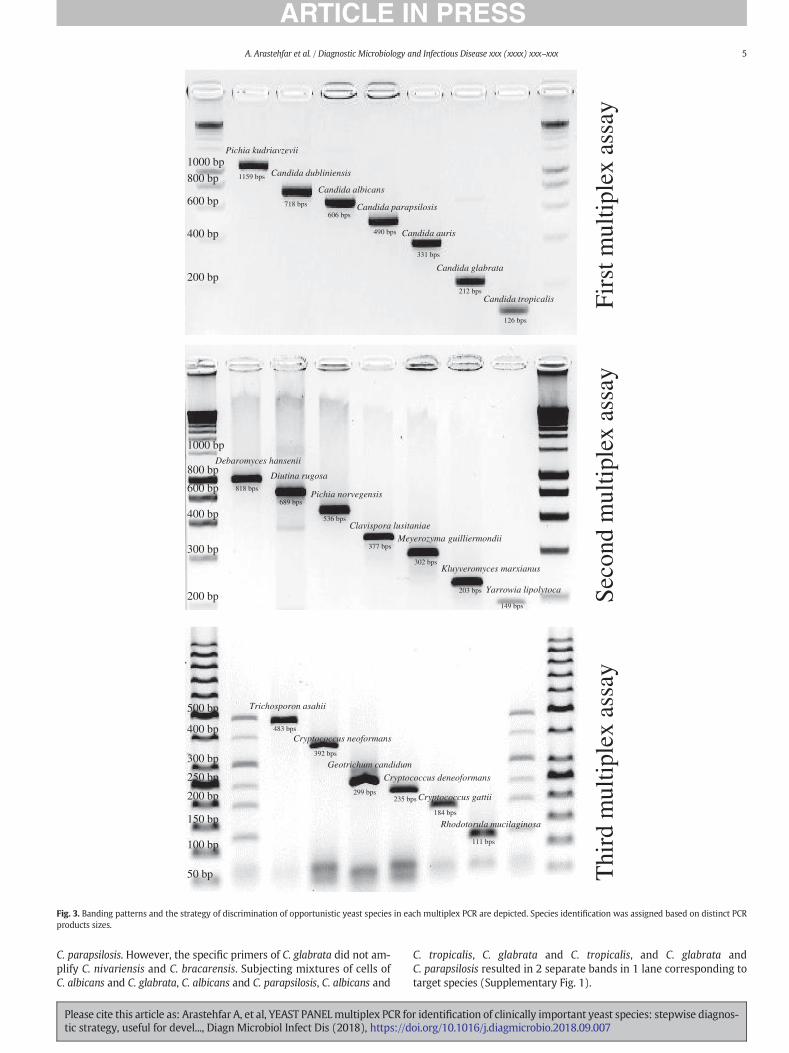
Pichia kudriavzevii
Candida albicans
Candida parapsilosis
Candida glabrata
Candida tropicalis
Candida dubliniensis
Candida auris
Debaromyces hansenii
Diutina rugosa
Pichia norvegensis
Clavispora lusitaniaeMeyerozyma guilliermondii
Kluyveromyces marxianus
Yarrowia lipolytoca
1000 bp
800 bp
600 bp
300 bp
400 bp
200 bp
1000 bp
800 bp
600 bp
400 bp
200 bp
Trichosporon asahii
Cryptococcus neoformans
Geotrichum candidum
Cryptococcus deneoformans
Cryptococcus gattii
Rhodotorula mucilaginosa
50 bp
100 bp
150 bp
200 bp
250 bp
300 bp
400 bp
500 bp
Firs
t mul
tiple
x as
say
Thi
rd m
ultip
lex
assa
ySe
cond
mul
tiple
x as
say
1159 bps
718 bps
606 bps
490 bps
331 bps
212 bps
126 bps
818 bps
689 bps
377 bps
536 bps
302 bps
203 bps
149 bps
483 bps
392 bps
299 bps235 bps
184 bps
111 bps
Fig. 3. Banding patterns and the strategy of discrimination of opportunistic yeast species in each multiplex PCR are depicted. Species identification was assigned based on distinct PCRproducts sizes.
5A. Arastehfar et al. / Diagnostic Microbiology and Infectious Disease xxx (xxxx) xxx–xxx
C. parapsilosis. However, the specific primers of C. glabrata did not am-plify C. nivariensis and C. bracarensis. Subjecting mixtures of cells ofC. albicans and C. glabrata, C. albicans and C. parapsilosis, C. albicans and
Please cite this article as: Arastehfar A, et al, YEAST PANELmultiplex PCR fotic strategy, useful for devel..., Diagn Microbiol Infect Dis (2018), https://d
C. tropicalis, C. glabrata and C. tropicalis, and C. glabrata andC. parapsilosis resulted in 2 separate bands in 1 lane corresponding totarget species (Supplementary Fig. 1).
r identification of clinically important yeast species: stepwise diagnos-oi.org/10.1016/j.diagmicrobio.2018.09.007
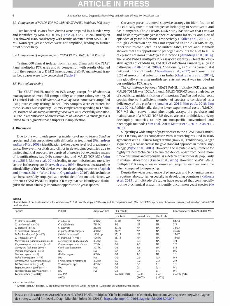
6 A. Arastehfar et al. / Diagnostic Microbiology and Infectious Disease xxx (xxxx) xxx–xxx
3.3. Comparison of MALDI-TOF MS with YEAST PANEL Multiplex PCR assay
Two hundred isolates from Austria were prepared in a blinded wayand identified by MALDI-TOF MS (Table 2). YEAST PANEL multiplexPCR showed 100% consistency with results obtained from MALDI-TOFMS. Nontarget yeast species were not amplified, leading to furtherproof of specificity.
3.4. Comparison of sequencing with YEAST PANEL Multiplex PCR assay
Testing 600 clinical isolates from Iran and China with the YEASTPanel multiplex PCR assay and its comparison with results obtainedfrom the sequencing of D1/D2 large subunit of rDNA and internal tran-scribed spacer were fully concordant (Table 3).
3.5. Pure colony testing
The YEAST PANEL multiplex PCR assay, except for Rhodotorulamucilaginosa, showed full compatibility with pure colony testing. Of12 clinical isolates of Rhodotorula mucilaginosa, none were amplifiedusing pure colony testing; hence, DNA samples were extracted forthese isolates. Subsequently, 12 DNA samples corresponding to 12 clin-ical isolates of Rhodotorula mucilaginosawere all successfully amplified.Failure in amplification of direct colonies of Rhodotorula mucilaginosa islinked to its pigments that hamper PCR amplification.
4. Discussion
Due to the worldwide growing incidence of non-albicans Candidaspecies and their association with difficulty in treatment (Richardsonand Lass-Flörl, 2008), identification to the species level is of great impor-tance. However, hospitals and clinics in developing countries due tolimited financial supports are deprived of precise but expensive meansof identifications, i.e., DNA sequencing and MALDI-TOF MS (Azimet al., 2015; Mathur et al., 2014), leading to poor infection andmortalitycontrol in these regions (Herwaldt et al., 1996). However, because of theaffordability of the PCR device even for developing countries (Ragheband Jimenez, 2014; World Health Organization, 2016), this techniquecan be successfully employed as a useful identification tool. Hence, wepresent a YEAST PANELmultiplex PCR assay that can identify and distin-guish the most clinically important opportunistic yeast species.
Table 2Clinical strains from Austria utilized for validation of YEAST Panel Multiplex PCR assay and its cproducts sizes.
Species PCR ID Amplicon size P
F
C. albicans (n=84) C. albicans 606 bp 8C. dubliniensis (n=3) C. dubliniensis 718 bp 3C. glabrata (n=33) C. glabrata 212 bp 3C. parapsilosis (n=26) C. parapsilosis complex 490 bp 2Pichia kudriavzevii (n=17) Pichia kudriavzevii 1159 bp 1C. tropicalis (n=15) C. tropicalis (n=15) 126 bp 1Meyerozyma guilliermondii (n=3) Meyerozyma guilliermondii 302 bp 0Kluyveromyces marxianus (n=2) Kluyveromyces marxianus 203 bp 0Clavispora lusitaniae (n=5) Clavispora lusitaniae 377 bp 0Diutina pararugosa (n=5) NA NA 0Diutina rugosa (n=1) Diutina rugosa 689 bp 0Pichia inconspicua (n=5) NA NA 0Cryptococcus neoformans (n=2) Cryptococcus neoformans 392 bp 0Trichosporon asahii (n=1) Trichosporon spp. 483 bp 0Stephanoascus ciferrii (n=1) NA NA 0Saccharomyces cerevisiae (n=1) NA NA 0Total number (n=204)a n= 192
(94%)n
NA = not amplified.a Among total 204 isolates, 12 are nontarget yeast species, while the rest of 192 isolates are
Please cite this article as: Arastehfar A, et al, YEAST PANELmultiplex PCR fotic strategy, useful for devel..., Diagn Microbiol Infect Dis (2018), https://d
Our assay presents a novel stepwise strategy for identification ofthe clinically most important yeasts belonging to Ascomycota andBasidiomycota. The ARTEMIS-DISK study has shown that Candidaand basidiomyceteous yeast species account for 95.8% and 4.2% ofepisodes of yeast infections, respectively (Pfaller et al., 2009). Al-though Geotrichum spp. was not reported in the ARTEMIS study,other studies conducted in the United States, France, and Denmarkshowed that this opportunistic pathogen accounts for 4.5% to 10.1%of episodes of non-Candida yeast infections (Arendrup et al., 2014).The YEAST PANEL multiplex PCR assay can identify 99.6% of the caus-ative agents of candidiasis, and 95% of infections caused by all yeastpathogens (Pfaller et al., 2009). Additionally, as C. auris caused fataloutbreak in 5 continents (Chowdhary et al., 2017) and constitutes5.2% of nosocomial infections in India (Chakrabarti et al., 2014),this globally emerging multidrug-resistant yeast was included inour multiplex PCR assay.
The consistency between YEAST PANEL multiplex PCR assay andMALDI-TOFMSwas 100%. AlthoughMALDI-TOFMS bears a high degreeof specificity, misidentification of important Candida and other yeastspecies due to insufficient number of spectra is considered as adeficiency of this platform (Jamal et al., 2014; Kim et al., 2016; Linget al., 2014). Additionally, despite lower experimental costs of MALDI-TOF MS than conventional phenotypic assays, the purchase andmaintenance of a MALDI-TOF MS device are cost-prohibitive, drivingdeveloping countries to rely on nonspecific conventional andphenotypic methods (Kim et al., 2016; Mathur et al., 2014; Xiao et al.,2016).
Subjecting a wide range of yeast species to the YEAST PANEL multi-plex PCR assay and its comparison with sequencing resulted in 100%agreementwith all clinical target strains (n=600). Traditionally, Sangersequencing is considered as the gold standard approach in medical my-cology (Pryce et al., 2003). However, the inevitable requirement forhighly trained technicians to run the device, apart from being moretime-consuming and expensive, is a deterrent factor for its popularityin routine laboratories (Criseo et al., 2015). However, YEAST PANELmultiplex PCR assay is less expensive and requires less hands-on timewhen compared to sequencing.
Despite thewidespread usage of phenotypic and biochemical assaysin routine laboratories, especially in developing countries (Kathuriaet al., 2015), a multitude of studies have revealed that commerciallyroutine biochemical assays misidentify uncommon yeast species (de
omparison with MALDI-TOFMS. Species identification was assigned based on distinct PCR
CR results Concordance with MALDI-TOF MS
irst tube Second tube Third tube
4/84 NA NA 84/84/3 NA NA 3/33/33 NA NA 33/336/26 NA NA 26/267/17 NA NA 17/175/15 NA NA 15/15/3 3/3 NA 3/3/2 2/2 NA 2/2/5 5/5 NA 5/5/5 0/5 0/5 0/5/1 1/1 NA 1/1/5 0/5 0/5 0/5/2 0/2 2/2 2/2/1 0/1 1/1 1/1/1 0/1 0/1 0/1/1 0/1 0/1 0/1=178 (100%) n=11
(100%)n=3(100%)
n=192 (94%)
among target species.
r identification of clinically important yeast species: stepwise diagnos-oi.org/10.1016/j.diagmicrobio.2018.09.007
Table 3Clinical strains from Iran and China utilized for validation of YEAST Panel Multiplex PCR assay and its comparison with Sanger sequencing. Species identification was assigned based ondistinct PCR products sizes. Identification for all of strains was performed on pure colonies, while for R. mucilaginosa, DNA samples extracted were used as PCR template.⁎a, ⁎⁎b
Species PCR ID Amplicon size PCR results Concordance with Sanger sequencing
First tube Second tube Third tube
C. albicans (n=156) C. albicans 606 bp 156/156 NA NA 156/156C. dubliniensis (n=8) C. dubliniensis 718 bp 8/8 NA NA 8/8C. glabrata (n=140) C. glabrata 212 bp 140/140 NA NA 140/140Pichia kudriavzevii (n=25) Pichia kudriavzevii 1159 bp 25/25 NA NA 25/25C. parapsilosis complexa (n=80) C. parapsilosis complex 490 bp 80/80 NA NA 80/80C. tropicalis (n=65) C. tropicalis 126 bp 65/65 NA NA 65/65Debaromyces hansenii (n=19) Debaromyces hansenii 818 bp 0/19 19/19 NA 19/19Meyerozyma guilliermondii (n=40) Meyerozyma guilliermondii 302 bp 0/40 40/40 NA 40/40Kluyveromyces marxianus (n=17) Kluyveromyces marxianus 203 bp 0/17 17/17 NA 17/17Yarrowia lipolytica (n=3) Yarrowia lipolytica 149 bp 0/3 3/3 NA 5/5Clavispora lusitaniae (n=1) Clavispora lusitaniae 377 bp 0/1 1/1 NA 1/1Pichia norvegensis (n=3) Pichia norvegensis 536 bp 0/3 3/3 NA 5/5Diutina rugosa (n=5) Diutina rugosa 689 bp 0/5 5/5 NA 5/5Cryptococcus neoformans (n=10) Cryptococcus neoformans 392 bp 0/10 0/10 10/10 10/10Geotrichum silvicola (n=9) Geotrichum spp. 299 bp 0/9 0/9 9/9b 9/9Trichosporon asahii (n=7) Trichosporon spp. 483 bp 0/7 0/7 7/7 7/7Rhodotorula mucilaginosa (n=12) Rhodotorula mucilaginosa 111 bp 0/12 0/12 12/12 12/12Total Number (n=600) n=474 (100%) n=88 (100%) n=38 (100%) n=600 (100%)
a Candida parapsilosis complex contained 2 strains of Candida orthopsilosis and 2 strains of Candida metapsilosis and were identified as Candida parapsilosis (490 bp).b Geotrichum silvicola is the synonym of Galactomyces candidum
7A. Arastehfar et al. / Diagnostic Microbiology and Infectious Disease xxx (xxxx) xxx–xxx
Almeida Júnior et al., 2014) and a broad variety of Candida species(Keçeli et al., 2016), especially M. guilliermondii and C. auris(Castanheira et al., 2013; Kathuria et al., 2015). Additionally, delayedand inappropriate identifications are associated with a higher mor-tality rate and hospitalization costs (Morrell et al., 2005). The turn-around time for identification, another inherent obstacle ofconventional biochemical assays (up to 48 h), was improved withthe YEAST PANEL multiplex PCR assay since identification wasachieved in 3–6 h.
Misidentification of C. zeylanoides asM. guilliermondii in the sec-ond multiplex PCR reaction was the major limitation of our study.However, literatures data revealed that the incidence ofC. zeylanoides is extremely low (0.01–0.04%) globally. In the sametube, Meyerozyma caribbica was misidentified as Meyerozymaguilliermondii, which can be explained by their close genetic simi-larity. Although Saccharomyces cerevisiae, C. nivariensis, andC. bracarensis have been implicated in human infections, theywere not included in our multiplex PCR assay (Enache-Angoulvant and Hennequin, 2005; Hou et al., 2017). Direct colonyand gel electrophoresis dependence and lack of identification ofclinically important molds were among the other limitations toour study. As Rhodotorula mucilaginosa contains red-colored pig-ments, DNA extraction and purification are imperative to obtainsuccessful PCR amplification and subsequently establish the iden-tity of this species.
5. Conclusion
Due to the shortages of systematic epidemiological data in devel-oping countries and the lack of financial supports for high-cost andprecise means of identification, such as Sanger sequencing andMALDI-TOF MS, these countries might face difficulties in the estab-lishment of empiric therapies, which cause a higher rate of mortality.However, because of the affordability of PCR devices, the YEASTPANEL multiplex PCR assay has the potential to be integrated inlarge-scale epidemiological studies. On the other hand, routine labo-ratories in developing countries can take the advantage of supple-mentation of biochemical assays with YEAST PANEL multiplex PCRassay.
Supplementary data to this article can be found online at https://doi.org/10.1016/j.diagmicrobio.2018.09.007.
Please cite this article as: Arastehfar A, et al, YEAST PANELmultiplex PCR fotic strategy, useful for devel..., Diagn Microbiol Infect Dis (2018), https://d
Acknowledgments
Not applicable.
Funding sources
This work was supported by the Major National R&D Projects of theNational Health Department (2018ZX10101003), EuropeanUnion’s Ho-rizon 2020 research and innovation program under the MarieSklodowska-Curie grant agreement no. 64209, National Natural ScienceFoundation of China (31770161), and Shanghai Science and TechnologyCommittee (grant numbers 14DZ2272900 and 14495800500).
References
de Almeida Júnior JN, Figueiredo DSY, Toubas D, Del Negro GMB, Motta AL, Rossi F, et al.Usefulness of matrix-assisted laser desorption ionisation-time-of-flight mass spec-trometry for identifying clinical Trichosporon isolates. Clin Microbiol Infect 2014;20(8):784–90. https://doi.org/10.1111/1469-0691.12502.
Arendrup MC, Boekhout T, Akova M, Meis JF, Cornely OA, Lortholary O, et al. ESCMID andECMM joint clinical guidelines for the diagnosis and management of rare invasiveyeast infections. Clin Microbiol Infect 2014;20(S3):76–98. https://doi.org/10.1111/1469-0691.12360.
Azim A, Baronia A, Marak RK, Gurjar M, Ahmed A. Invasive candidiasis in non neutropeniccritically ill—need for region-specific management guidelines. Indian J Crit Care Med2015;19(6):333. https://doi.org/10.4103/0972-5229.158273.
Cassagne C, Cella AL, Suchon P, Normand AC, Ranque S, Piarroux R. Evaluation of four pre-treatment procedures for MALDI-TOF MS yeast identification in the routine clinicallaboratory. Med Mycol 2013;51(4):371–7. https://doi.org/10.3109/13693786.2012.720720.
Castanheira M, Woosley LN, Diekema DJ, Jones RN, Pfaller MA. Candida guilliermondii andother species of Candidamisidentified as Candida famata: assessment by Vitek 2, DNAsequencing analysis, and matrix-assisted laser desorption ionization-time of flightmass spectrometry in two global antifungal surveillance program. J Clin Microbiol2013;51(1):117–24. https://doi.org/10.3109/13693786.2012.720720.
Chakrabarti A, Sood P, Rudramurthy SM, Chen S, Kaur H, Capoor M, et al. Incidence, char-acteristics and outcome of ICU-acquired candidemia in India. Intensive Care Med2014;41(2):285–95. https://doi.org/10.1007/s00134-014-3603-2.
Chowdhary A, Sharma C, Meis JF. Candida auris: a rapidly emerging cause of hospital-acquired multidrug-resistant fungal infections globally. PLoS Pathog 2017;13(5):1–10. https://doi.org/10.1371/journal.ppat.1006290.
Clark AE, Kaleta EJ, Arora A, Wolk DM. Matrix-assisted laser desorption ionization-time offlight mass spectrometry: a fundamental shift in the routine practice of clinical mi-crobiology. Clin Microbiol Rev 2013;26(3):547–603. https://doi.org/10.1128/CMR.00072-12.
Cleveland AA, Harrison LH, Farley MM, Hollick R, Stein B, Chiller TM, et al. Declining inci-dence of candidemia and the shifting epidemiology of Candida resistance in two USmetropolitan areas, 2008–2013: results from population-based surveillance. PLoSOne 2015;10(3):2008–13. https://doi.org/10.1371/journal.pone.0120452.
r identification of clinically important yeast species: stepwise diagnos-oi.org/10.1016/j.diagmicrobio.2018.09.007
8 A. Arastehfar et al. / Diagnostic Microbiology and Infectious Disease xxx (xxxx) xxx–xxx
Criseo G, Scordino F, Romeo O. Current methods for identifying clinically important cryp-tic Candida species. J Microbiol Methods 2015;111:50–6. https://doi.org/10.1016/j.mimet.2015.02.004.
Enache-Angoulvant A, Hennequin C. Invasive saccharomyces infection: a comprehensivereview. Clin Infect Dis 2005;41(11):1559–68. https://doi.org/10.1086/497832.
Herwaldt LA, McGowan JE, Metchock BG. Basic microbiologic support for hospital epide-miology. Infect Control Hosp Epidemiol 1996;17(5):298–303.
Hou X, Xiao M, Chen SCA, Wang H, Yu SY, Fan X, et al. Identification and antifungal sus-ceptibility profiles of Candida nivariensis and Candida bracarensis in a multi-centerChinese collection of yeasts. Front Microbiol 2017;8(JAN):1–8. https://doi.org/10.3389/fmicb.2017.00005.
Jamal WY, Ahmad S, Khan ZU, Rotimi VO. Comparative evaluation of two matrix-assistedlaser desorption/ionization time-of-flight mass spectrometry (MALDI-TOF MS) sys-tems for the identification of clinically significant yeasts. Int J Infect Dis 2014.https://doi.org/10.1016/j.ijid.2014.05.031.
Kathuria S, Singh PK, Sharma C, Prakash A, Masih A, Kumar A, et al. Multidrug-resistantCandida auris misidentified as Candida haemulonii: characterization by matrix-assisted laser desorption ionization-time of flight mass spectrometry and DNA se-quencing and its antifungal susceptibility profile variability by vitek 2, CLSI BrothMicrodilution, and Etest Method. J Clin Microbiol 2015;53(6):1823–30. https://doi.org/10.1128/JCM.00367-15.
Keçeli SA, Dündar D, Tamer GS. Comparison of Vitekmatrix-assisted laser desorption/ion-ization time-of-flight mass spectrometry versus conventional methods in Candidaidentification. Mycopathologia 2016;181(1–2):67–73. https://doi.org/10.1007/s11046-015-9944-8.
Kim T-H, Kweon OJ, Kim HR, Lee M-K. Identification of uncommon Candida species usingcommercial identification systems. J Microbiol Biotechnol 2016;26(12):2206–13.https://doi.org/10.4014/jmb.1609.09012.
Ling H, Yuan Z, Shen J, Wang Z, Xu Y. Accuracy of matrix-assisted laser desorption ionization-time of flight mass spectrometry for identification of clinical pathogenic fungi: a meta-analysis. J Clin Microbiol 2014;52(7):2573–82. https://doi.org/10.1128/JCM.00700-14.
Mathur P, Xess I, Misra M, Rajkumari N. Distribution of different yeasts isolates amongtrauma patients and comparison of accuracy in identification of yeasts by automatedmethod versus conventional methods for better use in low resource countries. IndianJ Med Microbiol 2014;32(4):391. https://doi.org/10.4103/0255-0857.142243.
Miceli MH, Díaz JA, Lee SA. Emerging opportunistic yeast infections. Lancet Infect Dis2011;11(2):142–51. https://doi.org/10.1016/S1473-3099(10)70218-8.
Morrell M, Fraser VJ, Kollef MH. Delaying the empiric treatment of Candida bloodstreaminfection until positive blood culture results are obtained: a potential risk factor forhospital mortality. Antimicrob Agents Chemother 2005;49(9):3640–5. https://doi.org/10.1128/AAC.49.9.3640–3645.2005.
Pappas PG, Alexander BD, Andes DR, Hadley S, Kauffman CA, Freifeld A, et al. Invasive fun-gal infections among organ transplant recipients: results of the Transplant-Associated
Please cite this article as: Arastehfar A, et al, YEAST PANELmultiplex PCR fotic strategy, useful for devel..., Diagn Microbiol Infect Dis (2018), https://d
Infection Surveillance Network (TRANSNET). Clin Infect Dis 2010;50(8):1101–11.https://doi.org/10.1086/651262.
Park BJ, Wannemuehler KA, Marston BJ, Govender N, Pappas PG, Chiller TM. Estimation ofthe current global burden of cryptococcal meningitis among persons living with HIV/AIDS. AIDS 2009;23(4):525–30. https://doi.org/10.1097/QAD.0b013e328322ffac.
Pfaller MA, Diekema DJ. Epidemiology of invasive candidiasis: a persistent public healthproblem. Clin Microbiol Rev 2007;20(1):133–63. https://doi.org/10.1128/CMR.00029-06.
Pfaller MA, Diekema DJ, Gibbs DL, Newell VA, Bijie H, Dzierzanowska D, et al. Results fromthe ARTEMIS DISK Global Antifungal Surveillance Study, 1997 to 2007: 10.5-yearanalysis of susceptibilities of noncandidal yeast species to fluconazole andvoriconazole determined by CLSI standardized disk diffusion testing. J Clin Microbiol2009;47(1):117–23. https://doi.org/10.1128/JCM.01747-08.
Posteraro B, De Carolis E, Vella A, Sanguinetti M. MALDI-TOF mass spectrometry in theclinical mycology laboratory: identification of fungi and beyond. Expert Rev Proteo-mics 2013;10(2):151–64. https://doi.org/10.1586/epr.13.8.
Posteraro B, Efremov L, Leoncini E, Amore R, Posteraro P, Ricciardi W, et al. Are the con-ventional commercial yeast identification methods still helpful in the era of new clin-ical microbiology diagnostics? A meta-analysis of their accuracy. J Clin Microbiol2015;53(8):2439–50. https://doi.org/10.1128/JCM.00802-15.
Pryce TM, Palladino S, Kay ID, Coombs GW. Rapid identification of fungi by sequencingthe ITSI and ITS2 regions using an automated capillary electrophoresis system. MedMycol 2003;41(5):369–81. [doi: IO.lO8OIl369378O3 1000 1600435].
Ragheb SM, Jimenez L. Polymerase chain reaction/rapid methods are gaining a foothold indeveloping countries. PDA J Pharm Sci Technol 2014;68(3):239–55. https://doi.org/10.5731/pdajpst.2014.00979.
Richardson M, Lass-Flörl C. Changing epidemiology of systemic fungal infections. ClinMicrobiol Infect 2008;14(Suppl. 4):5–24. https://doi.org/10.1111/j.1469-0691.2008.01978.x.
Stielow JB, Lévesque CA, Seifert KA, Meyer W, Irinyi L, Smits D, et al. One fungus, whichgenes? Development and assessment of universal primers for potential secondaryfungal DNA barcodes. Persoonia 2015;35(1):242–63. https://doi.org/10.3767/003158515X689135.
Vallabhaneni S, Mody RK, Walker T, Chiller T. The global burden of fungal diseases. InfectDis Clin N Am 2016;30(1):1–11. https://doi.org/10.1016/j.idc.2015.10.004.
World Health Organization (WHO). Establishment of PCR laboratory in developing coun-tries. Available from: http://apps.who.int/iris/bitstream/10665/249549/5/9789290225317-Eng.pdf?ua=1, 2016.
Xiao M, Fan X, Chen XX, Wang H, Zhang L, Xu ZP, et al. Misidentification of a rare species,Cryptococcus laurentii, by commonly used commercial biochemical methods andmatrix-assisted laser desorption ionization-time of flight mass spectrometry sys-tems: challenges for clinical mycology laboratories. J Clin Microbiol 2016;54(1):226–9. https://doi.org/10.1128/JCM.02830-15.
r identification of clinically important yeast species: stepwise diagnos-oi.org/10.1016/j.diagmicrobio.2018.09.007
Unequivocal identification of an underestimated opportunistic
yeast species, Cyberlindnera fabianii, and its close relatives using a dual-function PCR and literature review of published cases
Running Head: Identification of Cyberlindnera fabianii and literature review 1
Amir Arastehfar,a Wenjie Fang,b,c Abdullah M. S. Al-Hatmi,a,d Mohammad Hosein Afsarian,e Farnaz 2 Daneshnia,a Mina Bakhtiari,f Sara Khanjari Sadati,f Hamid Badali,g Sadegh Khodavaysi,h Ferry Hagena, 3 Wanqing Liao,b,c Weihua Pan,b,c* Kamiar Zomorodian,f* and Teun Boekhout,a, b, i4
a Westerdijk Fungal Biodiversity Institute, Utrecht, the Netherlands 5
b Department of Dermatology, Shanghai Changzheng Hospital, Second Military Medical University, Shanghai, 6 China 7
c Shanghai Key Laboratory of Molecular Medical Mycology, Shanghai Institute of Medical Mycology, Shanghai 8 Changzheng Hospital, Second Military Medical University, Shanghai, China 9
d Ministry of Health, Directorate General of Health Services, Ibri, Oman 10
e Departments of Parasitology & Mycology, Fasa University of Medical Sciences, Fasa, Iran 11
f Basic Sciences in Infectious Diseases Research Center, and Department of Medical Mycology and Parasitology, 12 School of Medicine, Shiraz University of Medical Sciences, Shiraz, Iran 13
g Department of Medical Mycology/Invasive Fungi Research Center (IFRC), School of Medicine, Mazandaran 14 University of Medical Sciences, Sari, Iran 15
h Department of Medical Parasitology and Mycology, School of Public Health, Tehran University of Medical 16 Sciences , Tehran, Iran 17
i Institute of Biodiversity and Ecosystem Dynamics, University of Amsterdam, Amsterdam, the Netherlands 18
* Correspondence:19
20
Prof. Weihua Pan, [email protected], 21
Prof. Kamiar Zomorodian, [email protected] 22
AA and WF have contributed equally to this work 23
Key words: Cy. fabianii, Cy. jadinii, Cy. mississipiensis, W. anomalus, biochemical assays, 24
multiplex PCR 25
Abstract 26
Cyberlindnera fabinaii is rare yeast opportunist, but has ability to cause septicemia, biofilm 27
production, and rapid acquirement of resistance to fluconazole and voriconazole, have made 28
its identification from closely related species important. The urge for reliable means of 29
identification remains unsolved for 28 years, as the biochemical assays misidentify this 30
species as Cyberlindnera jadinii or Wickerhamomyces anomalus. During six months, six 31
isolates of Cy. fabianii (central venous catheter n=2 and vaginal swabs n=4) and (vaginal 32
swabs n=1) of Cy. jadinii from Iranian patients were identified using MALDI-TOF MS and 33
Medical Mycology accepted in 29-Nov-2018, in Press

sequencing. In order to differentiate between Cy. fabianii, Cy. mississipiensis, Cy. jadinii, and 34
W. anomalus, we designed a dual-function multiplex PCR assay (conventional and qPCR). 35
Challenging our dual-function multiplex PCR assay with 30 most clinically important yeast 36
species, proved its specificity. Additionally, a literature review of published cases from 1990 37
to 2017 is reported. 38
39
40
41
1 Introduction 42
Candida species are associated with 10-15% causes of nosocomial candiduria (Sobel et al., 43
2011) and considered as the fourth common cause of nosocomial bloodstream infections 44
(Pfaller et al., 2018). Although C. albicans is still the most frequently isolated yeast pathogen, 45
the dynamic nature of epidemiology shows a shift towards a growing number of rare yeast 46
pathogens (Miceli et al., 2011). For instance, recently, C. auris, multidrug-resistant yeast has 47
emerged globally as a nosocomial pathogen and its systemic infections have increased 48
significantly (Mohsin et al., 2017). 49
Cyberlindnera fabianii, the teleomorph of C. fabianii, is an uncommon and rare ascomycetous 50
opportunistic yeast species with low virulence attributes (Jindal et al., 2015). However, it has 51
been reported causing invasive bloodstream infections (Bhally et al., 2006; Hamal et al., 2008; 52
Valenza et al., 2006). Another significant factor involved in Cy. fabianii virulence is its ability 53
to form antifungal resistant biofilms (Hamal et al., 2008). Previous studies reported that Cy. 54
fabianii developed resistance to different antifungals, including fluconazole, voriconazole, 55
caspofungin, and amphotericin B (Hamal et al., 2008; Jindal et al., 2015; Vágvölgyi et al., 56
2015). Infection with Cy. fabianii can lead to a 50% failure outcome with prophylactic and 57
therapeutic treatment with fluconazole (Vágvölgyi et al., 2015), and may cause fatal 58
septicemia (Taj-Aldeen et al., 2014; Valenza et al., 2006; Yun et al., 2013). 59
Cyberlindnera fabianii is usually misidentified as Cy. jadinii or Wickerhamomyces anomalus 60
especially when using commercially available biochemical assays (ID-32 C, API 20 C Aux, 61
Vitek2 YST Card, and Vitek2 Compact) (Baghdadi et al., 2015; Jindal et al., 2015; Lee et al., 62
2015; Svobodova et al., 2016a, 2016b; Valenza et al., 2006). However, using molecular 63
identification, it was found that the prevalence of Cy. fabainii was 10 times higher than W. 64
anomalus and approximately 37 higher than Cy. jadinii (Svobodova et al., 2016b). Because 65
susceptibility testing seems to differ between Cy. fabianii, Cy. jadinii, and W. anomalus 66
species (Svobodova et al., 2016b), a definitive molecular identification to species level is 67
important. Here, we described the first Iranian cases of Cy. fabianii from CVC and vagina, 68
performed a systematic review of the literature to identify clinical and mycological features 69
and outcomes of human Cy. fabainii infections and designed a dual-function multiplex PCR 70
that can be utilized in both conventional and real-time PCR and that targets Cy. fabianii, Cy. 71
jadinii, Cy. mississipiensis, and W. anomalus. 72
73
2 Materials and methods 74
75
2.1 Epidemiology and clinical profile 76
A clinical and epidemiology evaluation of global Cy. fabianii cases was performed published 77
cases. Cases from the literature were systematically searched from Pubmed and Google 78
scholar (searching date: 08-03-2018). The key words for our search were “fabianii” and 79
“infection”, without any limitation in language or date. All the ti tles and abstracts were 80
checked and full-text of related papers were downloaded for further evaluation. All cases have 81
been confirmed by culture, and appropriate diagnostic tools such as MALDI-TOF MS or DNA 82

sequencing for species identification, except for the first clinically isolated Cy. fabianii in 83
1990, as it was identified by API 20 C and appropriate complementary mating type. 84
85
2.2 Isolates and conditions 86
During six months (01-07-2017 to 31-12-2017), six isolates of Cy. fabianii (two from central 87
venous catheter and four from vaginal swabs) and one isolate of Cy. jadinii were collected 88
from Iranian patients in Shiraz, and Fasa. In order to check the specificity and sensitivity of 89
our multiplex PCR assay, a panel of 70 strains comprising of 30 clinically important yeast 90
species (40 target and 30 non-target species) were received from the collection of Westerdijk 91
Fungal Biodiversity Institute (formerly known as CBS) (Supplementary Table 1). Strains 92
were cultured on GYPA plates for 48 hours at 25 ⸰C. Central venous catheters and vaginal 93
swabs were streaked on the SDA and Chromagar media, followed by incubation at 37 ⸰C for 94
24-48 hours. Clinical isolates of Cy. fabianii (n=6) and Cy. jadinii (n=1) were included in this 95
study. Patients from who the isolates were collected were asked to sign the consent forms for 96
both participation in the study and publishing data derived from their isolates and medical 97
charts. 98
99
2.3 MALDI-TOF MS 100
The initial identification of clinical isolates were carried out by MALDI-TOF MS (Marklein 101
et al., 2009). Scores over two were considered as reliable identification. 102
103
2.4 Sequencing 104
The molecular identification of our strains was carried out using (ITS1 and ITS4) and (LR5 105
and LROR) primers targeting ITS and LSU of rDNA (Stielow et al., 2015). 106
107
2.5 DNA extraction, quality and quantity assessments 108
One full loop of pure colonies (10 µl) were subjected to the DNeasy Blood and Tissue kit and 109
DNA extraction was performed according to manufacturer’s protocol. In the last step of DNA 110
extraction, 70 µl of elution buffer (AE buffer) was added to the columns and they were 111
incubated at room temperature for 15 minutes followed by centrifugation at 10,000 rpm for 112
one minute. 113
NanoDrop™ 2000 (Thermo Fisher Scientific, Waltham, Massachusetts, USA) and QuBit 114
dsDNA BR Assay Kit (Thermo Fisher Scientific corporation, Waltham, Massachusetts, USA) 115
were used for quality and quantity assessments, respectively. DNA samples were adjusted at 116
one nanogram/µl and utilized as template for PCR reactions. 117
118
2.6 Primer design 119
In order to design specific primers for Cy. fabianii, Cy. mississipiensis, W. anomalus, and Cy. 120
jadinii, ITS1-5.8s-ITS2 rDNA loci were obtained from NCBI database 121
(https://blast.ncbi.nlm.nih.gov/Blast.cgi). One universal primer and five species-specific 122
primers were designed (Table 1). The primer properties were evaluated based on the IDT 123
Oligo analyzer on-line software (https://eu.idtdna.com/calc/analyzer) and primers were 124
manufactured by the IDT Company (Integrated DNA Technology, Leuven, Belgium). 125
126
2.7 Conventional PCR reactions 127
The multiplex PCR assay was utilized in both conventional and real-time PCR machines. For 128
the conventional PCR the reactions were adjusted at 50µl as follows, 39 µL MiliQ water 129
(Merck Millipore, Billerica, Massachusetts, United States), 5 µL 10X buffer, 1.5mM MgCl 2, 130
2.5 units of Taq enzyme (Bio Taq DNA Polymerase, Biolab), 0.2mM of mixed dNTP (dNTP 131
mix, 100Mm, Biolab), 5 picomoles of the PR-Ano, PR-Jad, PR-Missi and 10 pM of PF-132
Universal, PF-Fab and PR-Fab primers, and 1 µL of DNA template. 133
PCR (2720 Thermal Cycler, , Applied Biosystems, Waltham, Massachusetts, USA) used the 134
following program, pre-denaturation for 5 minutes at 94 ℃, 35 cycles of 94 ℃ for 30 seconds, 135
65 ℃ for 30 seconds, 72 ℃ for 30 seconds, and final extension at 72 ℃ for 8 minutes. PCR 136
products were run on 2% agarose gel for 45 minutes (8 Volt/cm), stained with GelRed 137
(BioTium Corporation, USA) and visualized using gel documentation with exposure time of 4 138
seconds (Gel Doc XR+, BioRad, California, USA). 139
140
2.8 Real-time PCR reactions 141
For real-time PCR (Applied Biosystems® 7500 fast, Thermo Fisher Scientific corporation, 142
Waltham, Massachusetts, USA) The following PCR conditions were used: 10 µl of Power Up 143
SYBR Green Master Mix (Thermo Fisher Scientific corporation, Waltham, Massachusetts, 144
USA) five pM of the PR-Fab, PF-Fab, PR-Ano, PR-Jad, PR-Missi and 10 pM of PF-Universal 145
primer, 1μl of DNA template and Mili-Q water (Merck Millipore, Darmstadt, Germany) to 146
reach the final volume of 20µl. The PCR program was as following: 50 ℃ for two minutes, 147
95 ℃ for 3 minutes, followed by 40 cycles of 95 ℃ for 15 seconds and 64 ℃ for 30 seconds. 148
Upon termination of the PCR program, the PCR products were undergone melting curve 149
analysis using continuous heating of 0.5 ℃/second increment from 95 ℃ for 15 seconds, 150
60 ℃ for one minute, 95 ℃ for 15 seconds and finally 60 ℃ for 15 seconds. Data analysis 151
was carried out using 7500 software version 2.3 (Thermo Fisher Scientific corporation, 152
Waltham, Massachusetts, USA). 153
154
2.9 Specificity and sensitivity testing 155
A panel of 70 CBS reference strains comprising 30 clinically-related yeast species were used 156
for the specificity testing (Supplementary Table 1). 157

In order to draw the standard curve, 10 log serial dilutions of DNA samples in duplicate 158
starting from 10,000 CFU/µl and ending at 10 CFU/µl were prepared for each target species. 159
Sensitivity, reproducibility, and the efficiency of the multiplex PCR were obtained from the 160
standard curves. 161
162
2.10 Antifungal susceptibility testing 163
Susceptibility testing was assayed for the six identified strains of Cy. fabianii and one isolate 164
of Cy. jadinii according to the Clinical and Laboratory Standards Institute (CLSI) M27-A3 165
and M27-S4 documents) (Rex et al., 2008). The following drugs were tested; itraconazole 166
(Janssen Research Foundation, Beerse, Belgium), voriconazole (Pfizer, Central Research, 167
Sandwich, United Kingdom), fluconazole (Pfizer, Groton, CT, USA), anidulafungin (Pfizer, 168
Central Research, Sandwich, United Kingdom), and amphotericin B (Sigma, St. Louis, MO, 169
USA) were obtained as reagent-grade powders from the respective manufacturers for 170
preparation of the CLSI microdilution trays. The minimal inhibitory concentration (MIC) 171
endpoints for amphotericin B and all other agents were determined by 100 and 50% inhibition, 172
respectively. MICs were compared with previously published breakpoints for amphotericin B 173
(≥ 2 µg /ml), itraconazole and voriconazole (≥ 1 µg/ml), fluconazole (≥ 8 µg /ml) and 174
caspofungin (≥ 1 µg/ml) (Pfaller and Diekema, 2012). Candida krusei ATCC 6258 and 175
Candida parapsilosis ATCC 22019 were used as quality control strains. 176
177
2.11 Utilized software 178
Genious (version 10.2.3) software was used to design the primers. SPSS (version 21, 179
International Business Machines Corporation, New York, U.S.A.) and Graph Pad Prism 180
(version 5, GraphPad Software, Inc. California, U.S.A.) were used for statistical analysis. 181
Mean ± standard deviation (SD) was used for data with normal distribution. 182
183
3 Results 184
185
3.1 Literature review of cases 186
Literature review identified 23 detailed cases (Tables 2 and 3). The global distribution of 187
infectious cases is shown in Figure 1. The majority of the cases clustered in Western Europe 188
(Figure 1.). Patient characteristics including demographic, clinical information, and 189
microbiological findings are presented in Tables 2 and 3. The majority of the cases (19/23; 190
83%) were published after 2010. Small differences in terms of gender were observed 191
(male/female =10/12). A high proportion of patients were neonates (<1 month, n=7), infant (> 192
1 month and <1 year, n=2), and children (> 1 year and <18 years, n=4). Fungemia is the most 193
common infection caused by Cy. fabianii (15/23, 65%), followed by candiduria (n=4), 194
prostatitis (n=1), endocarditis (n=1), meningitis (n=1) and peritonitis (n=1). Catheter 195
insertion was performed in eight cases and three of them showed growth of Cy. fabianii from 196
catheters. For the seven neonates cases, three of them were preterms born after 24, 25+3, 33+4 197
weeks gestation, respectively. Thirteen cases had a history of exposure to antibiotics before 198
the symptoms of infection. Post-surgery histories were observed among 12 cases. Five cases 199
were reported with cancer . Other risk factors included steroids exposure, dialysis, leukopenia, 200
mechanical ventilation, posterior urethral valve, and malnutrition. 201
Nineteen cases were not identified correctly, with 17 misidentified and two unidentified. Cy. 202
fabianii was most frequently misidentified as Cy. jadinii (n=10). The most frequently used 203
identification kits were API kits (n=9) or the VITEK system (n=4), but they failed in all 204
identification. However, subjecting misidentified cases to either MALDI-TOF or sequencing 205
resulted in accurate identification. 206
Antifungal susceptibility testing showed 16.7% (3/21), 12.5% (1/11), 8.3% (1/15), 12.5% 207
(2/19) resistant rates for fluconazole, itraconazole, voriconazole and amphotericin B. No 208
resistance to anidulafungin and flucytosine, were observed. The overall mortality for all 209
included cases was 33.4% (7/23). 210
As Cy. fabianii, Cy. jadinii, and W. anomalus are easily misidentified by phenotypic and 211
biochemical assays and in order to obtain a clear image about the epidemiology of Cy. 212
fabianii, papers that utilized MALDI-TOF and sequencing were investigated, as well 213
(Supplementary Table 2). 214
215
3.2 MALDI-TOF and Sanger sequencing results 216
From July 1st 2017 to December 31st, 2017, six isolates of Cy. fabianii and one isolate of Cy. 217
jadinii were isolated from central venous catheters and vaginal samples. Sanger sequencing 218
using D1/D2 LSU rDNA domains and MALDI-TOF MS were 100% in agreement with each 219
other. Cyberlindnera fabianii were isolated from central venous catheter (n=2) and vaginal 220
swabs (n=4) and the isolate of Cyberlindnera jadinii was isolated from vaginal swab. 221
222
3.3 Antifungal susceptibility testing 223
Supplementary table 3 summarize the MIC values of the tested antifungal drugs against six 224
clinical strains of Cy. fabiani and one isolate of Cy. jadinii. All strains of Cy. fabiani had low 225
MICs for anidulafungin, followed by itraconazole and voriconazole, whereas the least active 226
drug was fluconazole. Among the antifungal drugs, fluconazole exhibited the highest MIC 227
value that ranged from 1 to 8 μg/mL, followed by amphotericin B (MIC range 0.016-2 μg/ 228
mL), voriconazole (MIC range 0.016-1 μg/mL), and itraconazole (MIC range 0.063-1 μg/mL. 229
All Cy. fabianii and Cy. jadinii isolates were susceptible to anidulafungin (MIC=0.006 μg/ 230
mL). The MIC value of fluconazole against three isolates of Cy. fabianii (two from CVC and 231
one from vagina) was >3 log3 higher than that of the other isolates. 232
233
3.4 Conventional multiplex PCR results 234
Subjecting genomic DNA samples of Cy. fabianii (n=14), Cy. jadinii (n=11), W. anomalus 235
(n=13), Cy. mississipiensis (n=2) to our multiplex PCR resulted in amplicons of 84bps, 236
176bps, 235bps, and 320bps, respectively (Figure 2A). All four target species could be 237
distinguished and identified unequivocally. Subjecting CBS 7232 (reference strain of Cy. 238

jadinii) to our multiplex PCR resulted in negative amplification. Further identification by 239
MALDI-TOF and sanger sequencing matched the identity with Pichia guilliermondii. As a 240
result, designation of CBS 7232 in the collection department of Westerdijk Institute was 241
corrected. Challenging Our conventional multiplex PCR with DNA samples of 30 clinically 242
important yeast species, revealed 100% specificity. 243
244
3.5 Multiplex qPCR 245
Primers utilized for the conventional multiplex PCR were used for identification using a 246
SYBR Green I-based qPCR assay. Distinctive melt temperatures of 77.62±0.19 ℃ , 247
80.48±0.2℃ , 81.38±0.23℃ , and 81.56±0.38℃ distinguished Cy. fabianii (n=14), W. 248
anomalus (n=13), Cy. jadinii (n=11), Cy. mississipiensis (n=2) (Figure 2B). However, the 249
melting Tm of Cy. jadinii showed an overlap with that of Cy. mississipiensis. As a result, the 250
specific primer of PR-Missi was taken out from the multiplex reaction to unequivocally 251
identify Cy. jadinii (as the former species clinically is not important). Challenging our 252
multiplex PCR with a specificity test set, resulted in minor cross-reaction at higher Ct values 253
of 25 and 30 with the DNA of Pichia kudriavzevii (Candida krusei) and Pichia guilliermondii 254
(Candida guilliermondii), respectively (Supplementary Figure 1). However, by adjusting the 255
threshold at a Ct of 22, three species of Cy. fabianii, W. anomalus, Cy. jadinii were reliably 256
identified (100 CFU) and cross-reaction with other clinically important yeast pathogens was 257
not observed. Subjecting one ng DNA from the mistakenly designated reference strain of CBS 258
7232 to our multiplex qPCR resulted in amplification at a Ct value of 30, while the rest of 259
DNA of Cy. jadinii were amplified at a Ct value of 14, which was in concordant with results 260
obtained from the conventional multiplex PCR, MALDI-TOF MS and Sanger sequencing. 261
Regarding the reproducibility, five runs in three consecutive days generated an average R2 262
value of 0.99 for all target species, which demonstrated the high reproducibility of the qPCR 263
conditions (Supplementary Figure 2). 264
265
3.6 Accession numbers 266
D1/D2 LSU and ITS rDNA sequences obtained for Cy. fabianii and Cy. jadinii with the 267
accession numbers of MH236225, MH236226, MH236227, MH236228, MH236229, 268
MH236230, MH236231, MH236232, MH236233, MH236234, MH236235, MH236236, and 269
MH236237 were deposited in the GenBank database 270
(https://www.ncbi.nlm.nih.gov/genbank/). 271
272
4 Discussion 273
274
Cyberlindnera fabianii rarely is involved in human infections and regularly presented in 275
fermented products (Nyanga et al., 2013) and alcoholic beverages (Jeyaram et al., 2008). Up 276
to now multitude of studies in various countries have proved its association with infection in 277
human (Table 3). In 2009, Pichia fabianii, P. mississipiensis and P. jadinii were transferred 278
into Cyberlindnera genus. (D.W. Minter, 2009). Herein, we reported the first Iranian cases of 279

Cy. fabianii isolated from central venous catheter and vaginal samples and provided a 280
literature review of cases caused by Cy. fabianii. Additionally, we designed the first multiplex 281
PCR using the same primers which can be used in both conventional and real-time PCR 282
platforms as a diagnostic tools. 283
Using MALDI-TOF MS and sequencing of ITS and LSU rDNA, we identified six isolates of 284
Cy. fabianii (n=4 from vagina and n=2 from CVC). Two patients (isolates from CVC) had 285
underlying conditions. One suffered from acute myeloid leukemia (AML), end-stage renal 286
diseases, hypertension, and diabetes mellitus, and the other one from accident leading to 287
severe trauma and subsequent surgery. Concordant with previous studies, AML, severe 288
trauma and surgical interventions are risk factors for development of invasive candidiasis 289
(Yapar, 2014). Investigating published case-reports revealed that the main risk factors 290
associated with the acquirement of Cy. fabianii infection are attributable to previous exposure 291
with antibiotic therapy (54%) followed by central venous catheter insertion, surgery, and 292
cancer each 34%, neutropenia, chemotherapy, renal failure and low birth weight each 13%. In 293
our study, four patients with vaginitis did not have any underlying diseases and they were all 294
immunocompetent. Literature data revealed that there have been four reported cases of 295
funguria with Cy. fabaianii, however, in all cases the patients suffered from underlying 296
conditions, (Jindal et al., 2015; Vágvölgyi et al., 2015). Although it has been thought that 297
infections pertained to Cy. fabianii are mainly linked to neonates (Hof et al., 2017b), adults 298
with the age of ≥17 years and the range of 17-87 years, constituted the highest strata of 299
infected individuals (43%) followed by neonates with the age of <1 month (30%), and infants 300
with the age of <1 year (8%). In concordance with these findings, all of our cases were >18 301
years, which is in agreement with the Cy. fabianii isolate (obtained from sputum) previously 302
reported by Karimi et al. from Iran, from a 69-year old man (Karimi et al., 2015). No 303
significant difference between males and females in terms of acquiring infection by Cy. 304
fabianii was observed, but females showed a slightly higher proportion of infection (1.18/1) 305
than males. However, in our cases the ratio of five female to one male was observed, which is 306
due to the fact that four isolates were obtained from vagina, while CVC-isolated cases equally 307
infected one male and one female with Cy. fabianii. Although, Cy. fabianii features a wide 308
range of clinical syndromes, ranging fungemia and funguria to meningitis, fungemia with the 309
highest proportion (65.2%), is considered as the most prevalent clinical complication caused 310
by this opportunistic yeast species. 311
In the current study, antifungal susceptibility pattern of tested isolates exhibited the highest 312
MICs for fluconazole, followed by amphotericin B, which is in agreement with previous 313
reports (Jindal et al., 2015), and even multidrug resistant ( fluconazole and voriconazole) Cy. 314
fabianii (Hamal et al., 2008). In our study, results showed that 100% of Cy. fabianii isolates 315
(n= 5) were susceptible to anidulafungin, which is in agreement with previous study (Lee et 316
al., 2015). It has been shown that Cy. fabianii rapidly within 48 hours after treatment with 317
fluconazole acquired resistance against fluconazole and further treatment with voriconazole 318
resulted in emergence of strains resistant to both fluconazole and voriconazole (Hamal et al., 319
2008). Besides, Cy. fabianii can produce biofilm, which is pertained to resistance to 320
antifungal drugs (Hamal et al., 2008). However, no significant differences were found in the 321
activities of tested drugs between Cy. fabianii and Cy. jadinii (P > .05). 322
For treatment of Cy. fabianii, fluconazole was the most widely utilized drug followed by 323
different formulation of amphotericin B, caspofungin, and flucytosine in 14, 10, 7, and 2 324
studies, respectively. Regarding the efficacy of antifungal drugs for clearance of infection, 325
studies suggest that a combination of fluconazole with liposomal AMB or caspofungin or 326
removal of CVC and urinary tract catheter (Vágvölgyi et al., 2015). In addition, treatment 327

with anidulafungin (Lee et al., 2015) led to successful outcome for all patients infected with 328
Cy. fabiani. In our cases, one of patients died despite of treatment with AMB. However, death 329
could not be linked to failure in treatment as the patient suffered from other serious 330
background diseases. Treatment of the second patient (with trauma and surgery) with AMB 331
resulted in successful outcome. Regarding the patients suffering from vaginitis (n=4) were all 332
successfully treated with topical clotrimazole and fluconazole. Although, studied cases 333
describing infection with Cy. fabianii showed the mortality rate of 30.4% (7/23), all of dead 334
individuals suffered from other severe health conditions, including hematological disorders, 335
co-infection with other yeast or bacterial agents, low birth weight, open heart surgery and loss 336
of skin barrier due to burning (Hill and Wittwer, 2010; Hof et al., 2017a; Jindal et al., 2015; 337
Katagiri et al., 2016; Taj-Aldeen et al., 2014; Yun et al., 2013). 338
In order to overcome the persisting challenge of misidentification of Cy. fabianii, we 339
developed a multiplex PCR. Our dual-purpose multiplex PCR showed 100% agreement with 340
sequencing and MALDI-TOF MS. A previous study described a singleplex PCR that could 341
only detect Cy. fabianii and utilized limited number of yeast species for specificity testing 342
(Vágvölgyi et al., 2015). However, our assay firstly tested a broad range of yeast species 343
proving its specificity and secondly is a multiplex PCR targeting Cy. fabianii, Cy. 344
mississipiensis , Cy. jadinii, and W. anomalus. Although, MALDI-TOF and sequencing can 345
definitively identify these three clinically important yeast species, these devices are usually 346
not available in routine clinical microbiology laboratories and they are either expensive or 347
require highly trained personnel to perform the experiments (Criseo et al., 2015). Currently, 348
PCR as an affordable an reproducible device has been exploited as an supplementary 349
identification tool even in routine laboratories (including developing countries) (World Health 350
Organization, 2016). Our multiplex PCR Compared to time-consuming biochemical assays 351
takes only 2-3.5 hours to report the identification results. Although, biochemical assays are 352
used frequently for identification, especially in developing countries (Kathuria et al., 2015), 353
they do not have enough discriminatory power and frequently misidentify Cy. fabianii as Cy. 354
jadinii or W. anomalus (Svobodova et al., 2016b, 2016a). Consequently, important worldwide 355
studies such as the ARTEMIS DISK global antifungal surveillance over a period of 10.5 years 356
did not identify a single isolate of Cy. fabianii, while they could identify 88 and 6 isolates of 357
W. anomalus and Cy. jadinii, respectively (Pfaller et al., 2010). In contrast, MALDI-TOF MS 358
correctly identified 163 and 222 isolates of Cy. fabianii that previously using API ID 32C kit 359
were misidentified as W. anomalus and Cy. jadinii (Svobodova et al., 2016b, 2016a). 360
Obtaining these erroneous results can lead to a concealed image of epidemiology for Cy. 361
fabianii (Svobodova et al., 2016a). Different epidemiological studies that used either MALDI-362
TOF MS or sequencing for identification, showed that Cy. fabianii, although with a low rate, 363
but consistently is isolated (Fernández-Ruiz et al., 2017; Pongrácz et al., 2015; Won et al., 364
2013b). From five epidemiological studies, Cy. fabianii, W. anomalus, and Cy. jadinii were 365
identified in five, two, and no cases, respectively (Fernández-Ruiz et al., 2017; Karimi et al., 366
2015; Pongrácz et al., 2015; Taj-Aldeen et al., 2014; Won et al., 2013a). In our study, we 367
found six isolates of Cy. fabianii, one isolate of Cy. jadinii, and no W. anomalus. 368
Due to the growing number of infections caused by non-Candida albicans yeast species, 369
establishing definitive breakpoints and the development of specific molecular means of 370
identification for these species can contribute to both successful treatment and a better 371
knowledge on the occurrence of uncommon yeast pathogens. 372

The limitation of our study was that we utilized only 40 reference strains of Cy. fabianii, 373
W.anomalus, Cy. jadinii, and Cy. mississipiensis and therefore further experiments in different 374
setups are required. 375
376
5 Funding 377
378
This work was supported by the Major National R&D Projects of the National Health 379
Department (2018ZX10101003), European Union’s Horizon 2020 research and innovation 380
program under the Marie Sklodowska-Curie grant agreement No 642095, National Natural 381
Science Foundation of China (31770161), Shanghai Science and Technology Committee 382
(grant numbers 14DZ2272900 and 14495800500) 383
384
6 Conflict of Interest Statement 385
386
The authors declare that the research was conducted in the absence of any commercial or 387
financial relationships that could be construed as a potential conflict of interest. 388
389
7 Author Contributions Statement 390
391
AA, FD, and WJF participated in primer design, PCR optimization, data collection and 392
drafted the manuscript. WHP, KZ and TB participated in designing this study and revising the 393
manuscript. AMH, WL, and FH actively participated in paper revision. HB, SK, MHA, MB, 394
and SKA participated collecting Iranian clinical isolates and associated clinical data. All 395
authors contributed to the revision of the final manuscript. 396
397
8 References 398
Baghdadi, J., Hemarajata, P., Humphries, R., and Kelesidis, T. (2015). First Report of 399
Ventriculoperitoneal Shunt Infection due to Cyberlindnera fabianii. Case Rep. Infect. Dis. 400
2015, 630816. doi:10.1155/2015/630816. 401
Bhally, H. S., Jain, S., Shields, C., Halsey, N., Cristofalo, E., and Merz, W. G. (2006). 402
Infection in a neonate caused by Pichia fabianii: Importance of molecular identification. 403
Med. Mycol. 44, 185–187. doi:10.1080/13693780500160793. 404
Criseo, G., Scordino, F., and Romeo, O. (2015). Current methods for identifying clinically 405
important cryptic Candida species. J. Microbiol. Methods 111, 50–56. 406
doi:10.1016/j.mimet.2015.02.004. 407
D.W. Minter (2009). Cyberlindnera, a replacement name for Lindnera Kurtzman et al., nom. 408
illegit. Mycotaxon 110, 473–476. Available at: 409
http://lacertilia.de/AS/Bibliografie/BIB_5285.pdf#page=15. 410
Dooley, D. P., Beckius, M. L., Mcallister, C. K., and Jeffery, B. S. (1990). Prostatitis Caused 411
by Hansenula fabianii Isolated Meningitis Caused by Blastoschizomyces capitatus. 412
ournal Infect. Dis. 161, 1040–1041. 413
Fernández-Ruiz, M., Guinea, J., Puig-Asensio, M., Zaragoza, O., Almirante, B., Cuenca-414
Estrella, M., et al. (2017). Fungemia due to rare opportunistic yeasts: Data from a 415
population-based surveillance in Spain. Med. Mycol. 55, 125–136. 416
doi:10.1093/mmy/myw055. 417
Flores-González, J. C., Guerrero-Lozano, I., Pérez-Guerrero, J. J., Hernández-González, A., 418
Galán-Sánchez, F., Quintero-Otero, S., et al. (2016). Primer caso de infección fúngica 419
invasora por Candida fabianii en un paciente pediátrico no neonatal. Rev. Iberoam. Micol. 420
33, 48–50. doi:10.1016/j.riam.2015.06.003. 421
Gabriel, F., Noel, T., and Accoceberry, I. (2012). Lindnera (Pichia) fabianii blood infection 422
after mesenteric ischemia. Med. Mycol. 50, 310–314. 423
doi:10.3109/13693786.2011.587455. 424
Hamal, P., Ostransky, J., Dendis, M., Horvath, R., Ruzicka, F., Buchta, V., et al. (2008). A 425
case of endocarditis caused by the yeast Pichia fabianii with biofilm production and 426
developed in vitro resistance to azoles in the course of antifungal treatment. Med. Mycol. 427
46, 601–605. doi:10.1080/13693780802078180. 428
Hill, H. R., and Wittwer, C. T. (2010). Pichia fabianii Fungemia in a Neonate. Pediatr. Infect. 429
Dis. J. 29, 191. 430
Hof, H., Amann, V., Tauber, C., and Paulun, A. (2017a). Peritonitis in a neonate due to 431
Cyberlindnera fabianii, an ascomycetic yeast. Infection 45, 921–924. 432
doi:10.1007/s15010-017-1062-8. 433
Hof, H., Amann, V., Tauber, C., and Paulun, A. (2017b). Peritonitis in a neonate due to 434
Cyberlindnera fabianii, an ascomycetic yeast. Infection 45, 921–924. 435
doi:10.1007/s15010-017-1062-8. 436
Jeyaram, K., Singh, W. M., Capece, A., and Romano, P. (2008). Molecular identification of 437
yeast species associated with “Hamei” - A traditional starter used for rice wine 438
production in Manipur, India. Int. J. Food Microbiol. 124, 115–125. 439
doi:10.1016/j.ijfoodmicro.2008.02.029. 440
Jindal, N., Arora, S., Dhuria, N., and Arora, D. (2015). Cyberlindnera ( Pichia ) fabianii 441
infection in a neutropenic child : importance of molecular identification. JMM Case 442
Reports 1, 2015–2017. doi:10.1099/jmmcr.0.000033. 443
Karimi, L., Mirhendi, H., Khodadadi, H., and Mohammadi, R. (2015). Molecular 444
identification of uncommon clinical yeast species in Iran. Curr. Med. Mycol. 1, 1–6. 445
doi:10.18869/acadpub.cmm.1.2.1. 446
Katagiri, S., Gotoh, M., Tone, K., Akahane, D., Ito, Y., Ohyashiki, K., et al. (2016). Fatal 447
Cyberlindnera fabianii fungemia in a patient with mixed phenotype acute leukemia after 448

umbilical cord blood transplantation. Int. J. Hematol. 103, 592–595. 449
doi:10.1007/s12185-016-1953-y. 450
Kathuria, S., Singh, P. K., Sharma, C., Prakash, A., Masih, A., Kumar, A., et al. (2015). 451
Multidrug-resistant Candida auris misidentified as Candida haemulonii: Characterization 452
by matrix-assisted laser desorption ionization-time of flight mass spectrometry and DNA 453
sequencing and its antifungal susceptibility profile variability by vitek 2, CL. J. Clin. 454
Microbiol. 53, 1823–1830. doi:10.1128/JCM.00367-15. 455
Kim, Y. J., Yang, J. J., and Lee, H. J. (2015). Possibility of Frequent Detection of Invasive 456
Cyberlindera fabianii Infection Using Molecular Method. Ann. Clin. Microbiol. 18, 133–457
134. doi:0.1099/jmmcr.0.000033.3. 458
Lee, J. I., Yu, S., Park, J. S., Joo, E., Shin, J. H., and Kwon, M. (2015). Successful Treatment 459
of Fungemia Caused by Cyberlindnera fabianii with Anidulafungin : A Case Report. 18, 460
94–97. 461
Marklein, G., Josten, M., Klanke, U., Müller, E., Horré, R., Maier, T., et al. (2009). Matrix-462
assisted laser desorption ionization-time of flight mass spectrometry for fast and reliable 463
identification of clinical yeast isolates. J. Clin. Microbiol. 47, 2912–2917. 464
doi:10.1128/JCM.00389-09. 465
Miceli, M. H., Díaz, J. A., and Lee, S. A. (2011). Emerging opportunistic yeast infections. 466
Lancet Infect. Dis. 11, 142–151. doi:10.1016/S1473-3099(10)70218-8. 467
Nyanga, L. K., Nout, M. J. R., Smid, E. J., Boekhout, T., and Zwietering, M. H. (2013). 468
Fermentation characteristics of yeasts isolated from traditionally fermented masau 469
(Ziziphus mauritiana) fruits. Int. J. Food Microbiol. 166, 426–432. 470
doi:10.1016/j.ijfoodmicro.2013.08.003. 471
Pfaller, M. A., and Diekema, D. J. (2012). Progress in antifungal susceptibility testing of 472
Candida spp. by use of Clinical and Laboratory Standards Institute broth microdilution 473
methods, 2010 to 2012. J. Clin. Microbiol. 50, 2846–2856. doi:10.1128/JCM.00937-12. 474
Pfaller, M. A., Diekema, D. J., Gibbs, D. L., Newell, V. A., Ellis, D., Tullio, V., et al. (2010). 475
Results from the artemis disk global antifungal surveillance study, 1997 to 2007: A 10.5-476
year analysis of susceptibilities of candida species to fluconazole and voriconazole as 477
determined by CLSI standardized disk diffusion. J. Clin. Microbiol. 48, 1366–1377. 478
doi:10.1128/JCM.02117-09. 479
Pfaller, M. A., Jones, R. N., Messer, S. A., Edmond, M. B., and Wenzel, R. P. (2018). 480
National Surveillance of Nosocomial Blood Stream Infection Due to Species of 481
<em>Candida</em> Other than <em>Candida albicans</em>: Frequency of Occurrence 482
and Antifungal Susceptibility in the SCOPE Program. Diagn. Microbiol. Infect. Dis. 30, 483
121–129. doi:10.1016/S0732-8893(97)00192-2. 484
Pongrácz, J., Juhász, E., Iván, M., and Kristóf, K. (2015). Significance of yeasts in 485
bloodstream infection: Epidemiology and predisposing factors of Candidaemia in adult 486
patients at a university hospital (2010–2014). Acta Microbiol. Immunol. Hung. 62, 317–487
329. doi:10.1556/030.62.2015.3.9. 488
Rex, J. H., Alexander, B. D., Andes, D., Arthington-Skaggs, B., Brown, S. D., Chaturvedi, V., 489
et al. (2008). Reference method for broth dilution antifungal susceptibility testing of 490
yeasts: approved standard - third edition. Clin. Lab. Stand. Inst., 1–25. 491
doi:10.1007/SpringerReference_5244. 492
Sobel, J. D., Fisher, J. F., Kauffman, C. A., and Newman, C. A. (2011). Candida urinary tract 493
infections - Epidemiology. Clin. Infect. Dis. 52, 433–436. doi:10.1093/cid/cir109. 494
Stielow, J. B., Lévesque, C. A., Seifert, K. A., Meyer, W., Irinyi, L., Smits, D., et al. (2015). 495
One fungus, which genes? Development and assessment of universal primers for 496
potential secondary fungal DNA barcodes. Persoonia - Mol. Phylogeny Evol. Fungi 35, 497
242–263. doi:10.3767/003158515X689135. 498
Svobodova, L., Bednarova, D., and Hamal, P. (2016a). [The prevalence of Candida 499
pelliculosa, Candida utilis, and Candida fabianii in the Olomouc University Hospital: 500
epidemiological study]. Epidemiol. Mikrobiol. Imunol. Cas. Spol. pro Epidemiol. a 501
Mikrobiol. Ces. Lek. Spol. J.E. Purkyne 65, 34–38. 502
Svobodova, L., Bednarova, D., Ruzicka, F., Chrenkova, V., Dobias, R., Mallatova, N., et al. 503
(2016b). High frequency of Candida fabianii among clinical isolates biochemically 504
identified as Candida pelliculosa and Candida utilis. Mycoses 59, 241–246. 505
doi:10.1111/myc.12454. 506
Taj-Aldeen, S. J., AbdulWahab, A., Kolecka, A., Deshmukh, A., Meis, J. F., and Boekhout, T. 507
(2014). Uncommon opportunistic yeast bloodstream infections from Qatar. Med. Mycol. 508
52, 552–556. doi:10.1093/mmycol/myu016. 509
Vágvölgyi, C., Mlinarić-Missoni, E., Kocsubé, S., Hatvani, L., Škarić, I., and Lukić-Grlić, A. 510
(2015). Cyberlindnera fabianii in the neonatal and paediatric intensive care unit: case 511
reports. JMM Case Reports 2, 1–7. doi:10.1099/jmmcr.0.000032. 512
Valenza, G., Valenza, R., Brederlau, J., Frosch, M., and Kurzai, O. (2006). Identification of 513
Candida fabianii as a cause of lethal septicaemia. Mycoses 49, 331–334. 514
doi:10.1111/j.1439-0507.2006.01240.x. 515
Won, E. J., Shin, J. H., Lee, K., Kim, M.-N., Lee, H. S., Park, Y.-J., et al. (2013a). Accuracy 516
of species-level identification of yeast isolates from blood cultures from 10 university 517
hospitals in South Korea by use of the matrix-assisted laser desorption ionization-time of 518
flight mass spectrometry-based Vitek MS system. J. Clin. Microbiol. 51, 3063–3065. 519
doi:10.1128/JCM.00945-13. 520
Won, E. J., Shin, J. H., Lee, K., Kim, M. N., Lee, H. S., Park, Y. J., et al. (2013b). Accuracy 521
of species-level identification of yeast isolates from blood cultures from 10 university 522
hospitals in south korea by use of the matrix-assisted laser desorption ionization-time of 523
flight mass spectrometry-based vitek MS system. J. Clin. Microbiol. 51, 3063–3065. 524
doi:10.1128/JCM.00945-13. 525
World Health Organization (2016). Establishment of PCR laboratory in developing countries. 526
Available at: http://apps.who.int/iris/bitstream/10665/249549/5/9789290225317-527
Eng.pdf?ua=1. 528
Wu, Y., Wang, J., Li, W., Jia, H., Che, J., Lu, J., et al. (2013). Pichia fabianii blood infection 529
in a premature infant in China: Case report. BMC Res. Notes 6, 2–5. doi:10.1186/1756-530
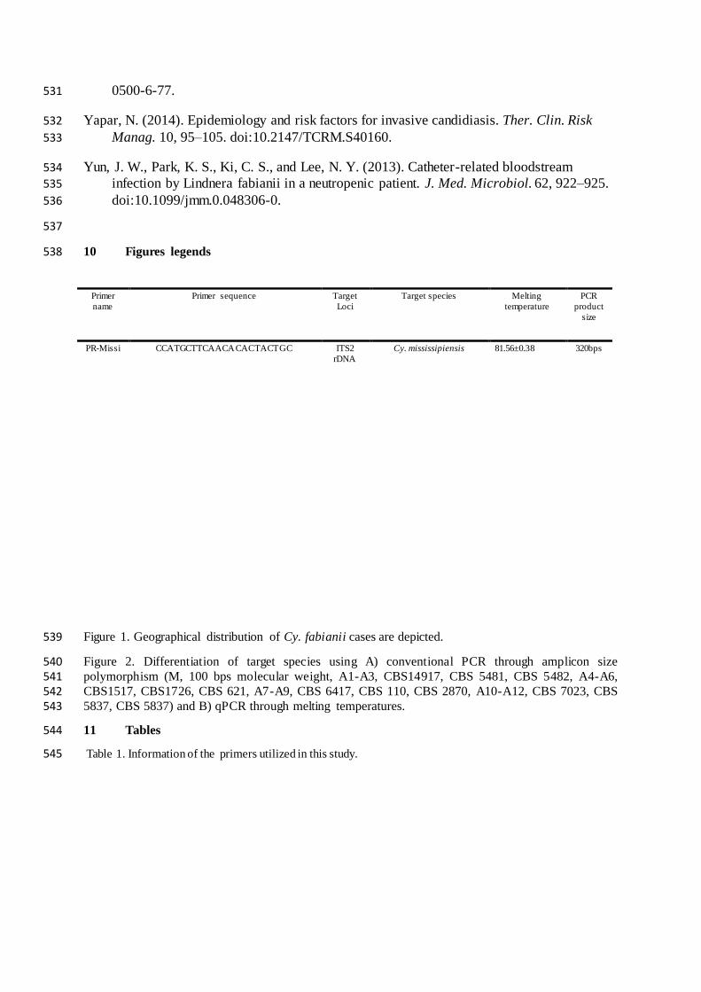
0500-6-77. 531
Yapar, N. (2014). Epidemiology and risk factors for invasive candidiasis. Ther. Clin. Risk 532
Manag. 10, 95–105. doi:10.2147/TCRM.S40160. 533
Yun, J. W., Park, K. S., Ki, C. S., and Lee, N. Y. (2013). Catheter-related bloodstream 534
infection by Lindnera fabianii in a neutropenic patient. J. Med. Microbiol. 62, 922–925. 535
doi:10.1099/jmm.0.048306-0. 536
537
10 Figures legends 538
Figure 1. Geographical distribution of Cy. fabianii cases are depicted. 539
Figure 2. Differentiation of target species using A) conventional PCR through amplicon size 540 polymorphism (M, 100 bps molecular weight, A1-A3, CBS14917, CBS 5481, CBS 5482, A4-A6, 541 CBS1517, CBS1726, CBS 621, A7-A9, CBS 6417, CBS 110, CBS 2870, A10-A12, CBS 7023, CBS 542 5837, CBS 5837) and B) qPCR through melting temperatures. 543
11 Tables 544
Table 1. Information of the primers utilized in this study. 545
Primer
name
Primer sequence Target
Loci
Target species Melting
temperature
PCR
product
size
PR-Missi CCATGCTTCAACACACTACTGC ITS2
rDNA
Cy. mississipiensis 81.56±0.38 320bps
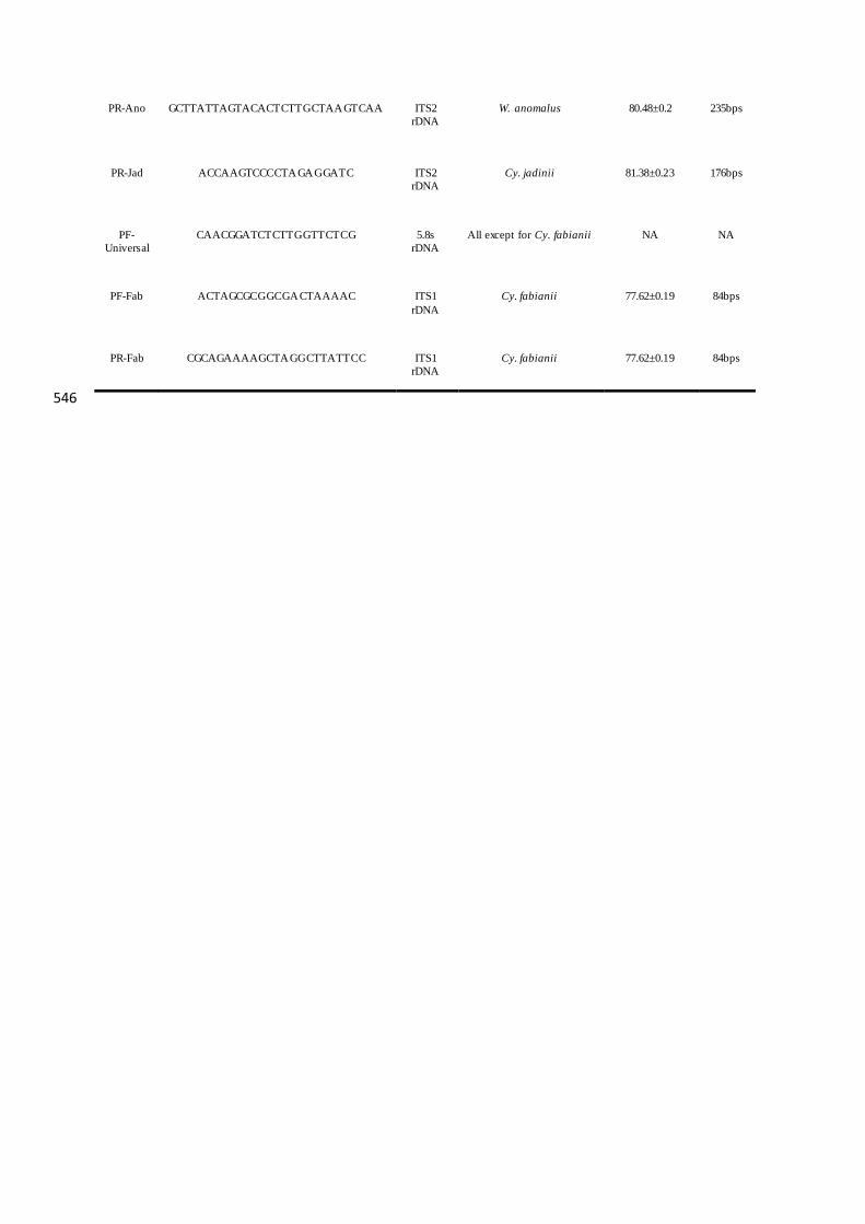
546
PR-Ano GCTTATTAGTACACTCTTGCTAAGTCAA ITS2
rDNA
W. anomalus 80.48±0.2 235bps
PR-Jad
ACCAAGTCCCCTAGAGGATC ITS2
rDNA
Cy. jadinii 81.38±0.23 176bps
PF-
Universal
CAACGGATCTCTTGGTTCTCG 5.8s
rDNA
All except for Cy. fabianii NA NA
PF-Fab ACTAGCGCGGCGACTAAAAC ITS1
rDNA
Cy. fabianii 77.62±0.19 84bps
PR-Fab CGCAGAAAAGCTAGGCTTATTCC ITS1
rDNA
Cy. fabianii 77.62±0.19 84bps

547
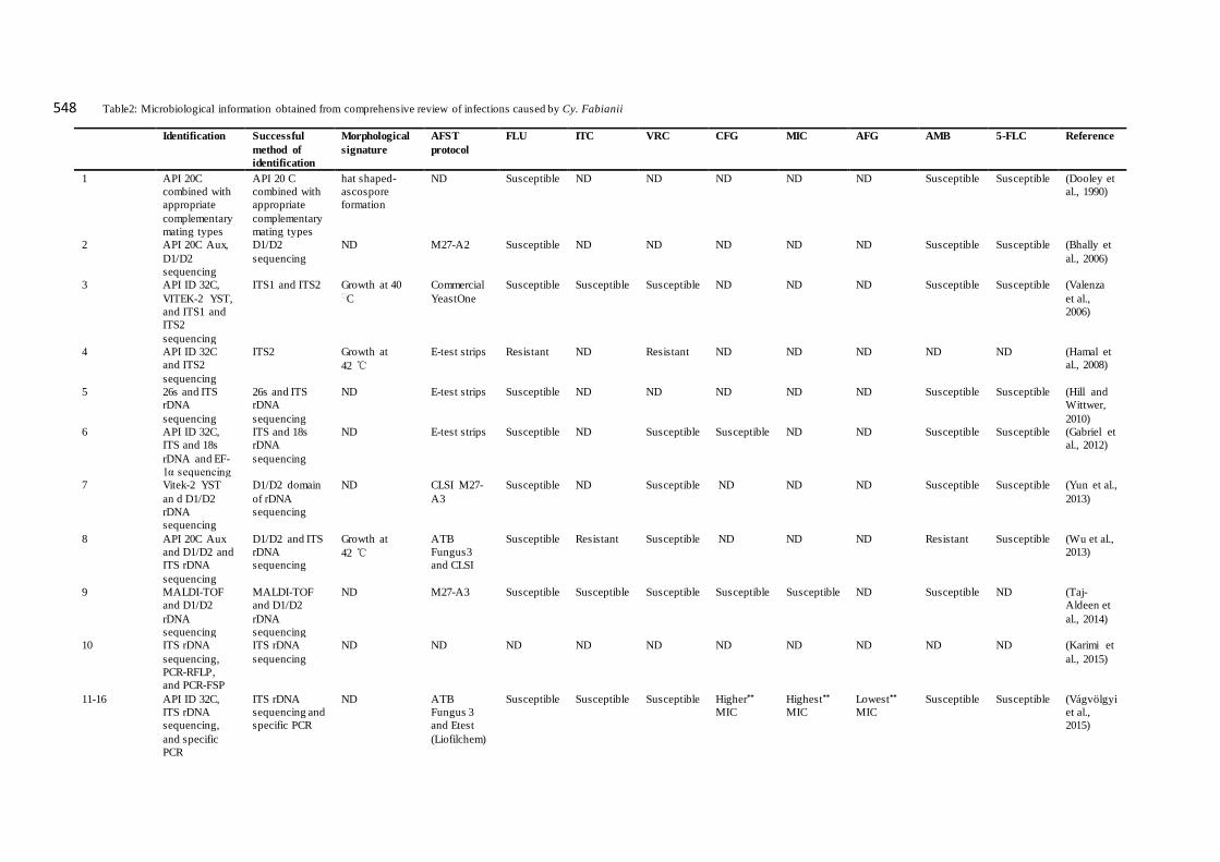
Table2: Microbiological information obtained from comprehensive review of infections caused by Cy. Fabianii 548
Identification Successful
method of
identification
Morphological
signature
AFST
protocol
FLU ITC VRC CFG MIC AFG AMB 5-FLC Reference
1 API 20C
combined with
appropriate
complementary
mating types
API 20 C
combined with
appropriate
complementary
mating types
hat shaped-
ascospore
formation
ND Susceptible ND ND ND ND ND Susceptible Susceptible (Dooley et
al., 1990)
2 API 20C Aux,
D1/D2
sequencing
D1/D2
sequencing
ND M27-A2 Susceptible ND ND ND ND ND Susceptible Susceptible (Bhally et
al., 2006)
3 API ID 32C,
VITEK-2 YST,
and ITS1 and
ITS2
sequencing
ITS1 and ITS2 Growth at 40 ⸰C
Commercial
YeastOne
Susceptible Susceptible Susceptible ND ND ND Susceptible Susceptible (Valenza
et al.,
2006)
4 API ID 32C
and ITS2
sequencing
ITS2 Growth at
42 ℃
E-test strips Resistant ND Resistant ND ND ND ND ND (Hamal et
al., 2008)
5 26s and ITS
rDNA
sequencing
26s and ITS
rDNA
sequencing
ND E-test strips Susceptible ND ND ND ND ND Susceptible Susceptible (Hill and
Wittwer,
2010)
6 API ID 32C,
ITS and 18s
rDNA and EF-
1α sequencing
ITS and 18s
rDNA
sequencing
ND E-test strips Susceptible ND Susceptible Susceptible ND ND Susceptible Susceptible (Gabriel et
al., 2012)
7 Vitek-2 YST
an d D1/D2
rDNA
sequencing
D1/D2 domain
of rDNA
sequencing
ND CLSI M27-
A3
Susceptible ND Susceptible ND ND ND Susceptible Susceptible (Yun et al.,
2013)
8 API 20C Aux
and D1/D2 and
ITS rDNA
sequencing
D1/D2 and ITS
rDNA
sequencing
Growth at
42 ℃
ATB
Fungus3
and CLSI
Susceptible Resistant Susceptible ND ND ND Resistant Susceptible (Wu et al.,
2013)
9 MALDI-TOF
and D1/D2
rDNA
sequencing
MALDI-TOF
and D1/D2
rDNA
sequencing
ND M27-A3 Susceptible Susceptible Susceptible Susceptible Susceptible ND Susceptible ND (Taj-
Aldeen et
al., 2014)
10 ITS rDNA
sequencing,
PCR-RFLP,
and PCR-FSP
ITS rDNA
sequencing
ND ND ND ND ND ND ND ND ND ND (Karimi et
al., 2015)
11-16 API ID 32C,
ITS rDNA
sequencing,
and specific
PCR
ITS rDNA
sequencing and
specific PCR
ND ATB
Fungus 3
and Etest
(Liofilchem)
Susceptible Susceptible Susceptible Higher**
MIC
Highest**
MIC
Lowest**
MIC
Susceptible Susceptible (Vágvölgyi
et al.,
2015)
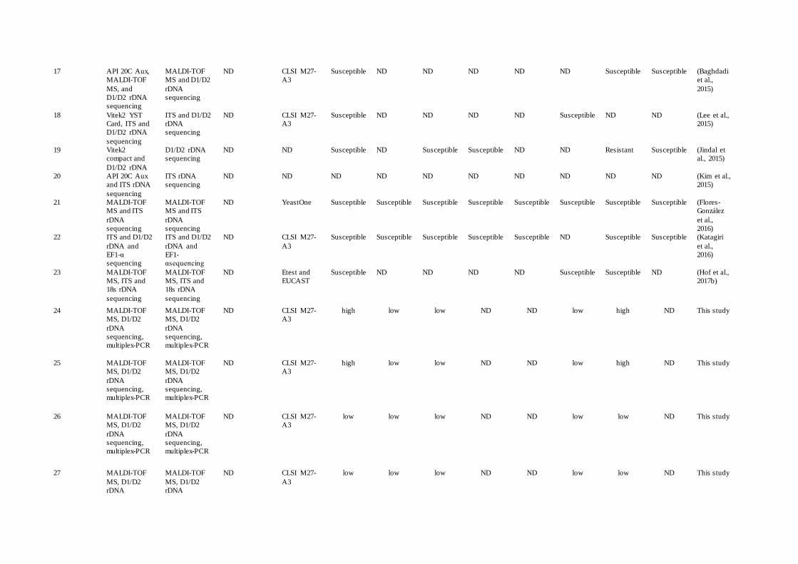
17 API 20C Aux,
MALDI-TOF
MS, and
D1/D2 rDNA
sequencing
MALDI-TOF
MS and D1/D2
rDNA
sequencing
ND CLSI M27-
A3
Susceptible ND ND ND ND ND Susceptible Susceptible (Baghdadi
et al.,
2015)
18 Vitek2 YST
Card, ITS and
D1/D2 rDNA
sequencing
ITS and D1/D2
rDNA
sequencing
ND CLSI M27-
A3
Susceptible ND ND ND ND Susceptible ND ND (Lee et al.,
2015)
19 Vitek2
compact and
D1/D2 rDNA
D1/D2 rDNA
sequencing
ND ND Susceptible ND Susceptible Susceptible ND ND Resistant Susceptible (Jindal et
al., 2015)
20 API 20C Aux
and ITS rDNA
sequencing
ITS rDNA
sequencing
ND ND ND ND ND ND ND ND ND ND (Kim et al.,
2015)
21 MALDI-TOF
MS and ITS
rDNA
sequencing
MALDI-TOF
MS and ITS
rDNA
sequencing
ND YeastOne Susceptible Susceptible Susceptible Susceptible Susceptible Susceptible Susceptible Susceptible (Flores-
González
et al.,
2016)
22 ITS and D1/D2
rDNA and
EF1-α
sequencing
ITS and D1/D2
rDNA and
EF1-
αsequencing
ND CLSI M27-
A3
Susceptible Susceptible Susceptible Susceptible Susceptible ND Susceptible Susceptible (Katagiri
et al.,
2016)
23 MALDI-TOF
MS, ITS and
18s rDNA
sequencing
MALDI-TOF
MS, ITS and
18s rDNA
sequencing
ND Etest and
EUCAST
Susceptible ND ND ND ND Susceptible Susceptible ND (Hof et al.,
2017b)
24 MALDI-TOF
MS, D1/D2
rDNA
sequencing,
multiplex-PCR
MALDI-TOF
MS, D1/D2
rDNA
sequencing,
multiplex-PCR
ND CLSI M27-
A3
high low low ND ND low high ND This study
25 MALDI-TOF
MS, D1/D2
rDNA
sequencing,
multiplex-PCR
MALDI-TOF
MS, D1/D2
rDNA
sequencing,
multiplex-PCR
ND CLSI M27-
A3
high low low ND ND low high ND This study
26 MALDI-TOF
MS, D1/D2
rDNA
sequencing,
multiplex-PCR
MALDI-TOF
MS, D1/D2
rDNA
sequencing,
multiplex-PCR
ND CLSI M27-
A3
low low low ND ND low low ND This study
27 MALDI-TOF
MS, D1/D2
rDNA
MALDI-TOF
MS, D1/D2
rDNA
ND CLSI M27-
A3
low low low ND ND low low ND This study
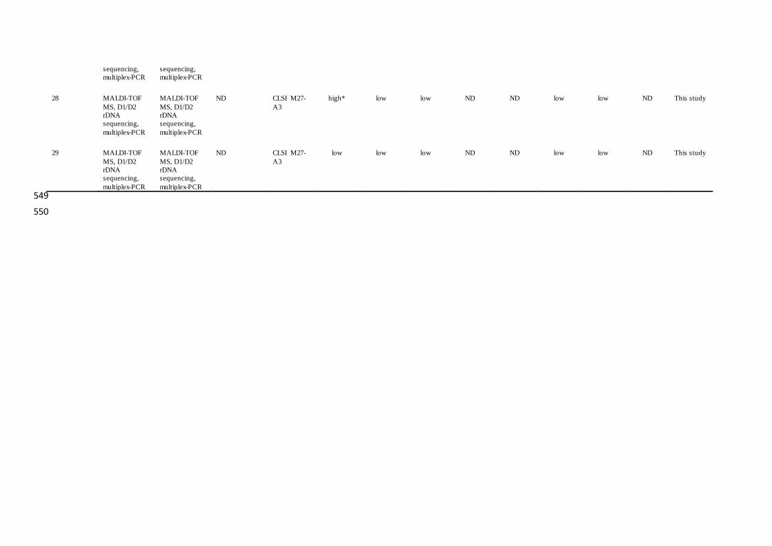
sequencing,
multiplex-PCR sequencing,
multiplex-PCR
28 MALDI-TOF
MS, D1/D2
rDNA
sequencing,
multiplex-PCR
MALDI-TOF
MS, D1/D2
rDNA
sequencing,
multiplex-PCR
ND CLSI M27-
A3
high* low low ND ND low low ND This study
29 MALDI-TOF
MS, D1/D2
rDNA
sequencing,
multiplex-PCR
MALDI-TOF
MS, D1/D2
rDNA
sequencing,
multiplex-PCR
ND CLSI M27-
A3
low low low ND ND low low ND This study
549
550

551

Table 3: Clinical information obtained from comprehensive review of infections caused by Cy. fabianii 552
No. Publication
year
Location Age/Sex Specimen type Type of
infection
Predisposing factors Treatment option Outcome reference
1 1990 USA 57 years/Male Urine Prostatitis Chronic lymphoytic leukemia, chronic urinary
tract infection
Amphotericin B Survived (Dooley et
al., 1990)
2 2006 USA 5 weeks /Female Blood
(catheter),
endotracheal
suction
Fungemia Born after 25+3
weeks gestation, mult iple
antibiotics exposure, steroids exposure, catheter
insertion
Amphotericin B, catheter
removal
Survived (Bhally et
al., 2006)
3 2006 Germany 46 years /Male Blood,
bronchoalveolar
lavage fluid
Fungemia Multiple antibiotics exposure, cholecystectomy Fluconazole→caspofungin Dead (multi-
organ failure)
(Valenza et
al., 2006)
4 2008 Czech
Republic
40 years /Male Blood, aortic
valve
Fungemia,
Endocarditis
congenital combined aortic incompetence of the
mitral valve, Ischemic stroke, decompressive
craniectomy
Fluconazole
→Voriconazole→Amphotericin
B
Survived with
neurological
sequelae
(Hamal et
al., 2008)
5 2010 France 11 days /Female Skin, mouth,
pleural fluid
aspirate, blood
Disseminated
Fungemia
Born after 24 weeks gestation, 700g b irth weight,
poor respiratory effort, APGAR scores were 5 and
7 at one and five minutes
L-Amphotericin B, flucytosine Dead (Hill and
Wittwer,
2010)
6 2012 France 53 years /Female Oropharyngeal
swab, urine,
stool, and blood
Disseminated
Fungemia
Mesenteric ischemia, renal fa ilu re (continuous
dialysis required), enterectomy, antibiotic
exposure
caspofungin → Fluconazole Survived (Gabriel et
al., 2012)
7 2013 South
Korea
47 years /Female Blood and
central venous
catheter
Fungemia Plas ma cell myeloma, autologous peripheral
blood stem cell transplantation, chemotherapy,
radiation therapy, respiratory failure, leukopenia,
catheter insertion
Amphotericin B → caspofungin Dead (multi-
organ failure,
septic shock and
lactic acidosis)
(Yun et al.,
2013)
8 2013 China <1 month
/Female
Blood Fungemia Born after 33+4 week of gestation, 1760g birth
weight, mild neonatal asphyxia, peripheral venous
hyperalimentation, respirator, antibiotic exposure;
mother: 33-years-old, hysteromyoma, gestational
hypertension.
Fluconazole Survived (Wu et al.,
2013)
9 2014 Qatar 19 years /Male Blood Fungemia Burn (55% of the body surface area) Fluconazole Dead (Taj-
Aldeen et
al., 2014)
10 2015 Iran 69 years /Male Sputum Suspected
infection
ND* ND ND (Karimi et
al., 2015)
11 2015 Croatia 3.5 years
/Female
Blood, stool Fungemia antibiotic exposure, chemotherapy, leukopenia,
antibacterial therapy
Fluconazole→L-Amphotericin
B
Survived (Vágvölgyi
et al., 2015)
12 2015 Croatia 1.5 months
/Male
Urine Funguria Posterior urethral valve, hydronephrosis, surgery,
antibiotic exposure, catheter insertion
Fluconazole, u rinary catheter
removal
Survived (Vágvölgyi
et al., 2015)
13 2015 Croatia <1 month
/Female
Urine,
nasopharyngeal
swab
Funguria Gastroschisis, surgery, mechanical ventilation,
parenteral nutrit ion, antibiotic exposure, catheter
insertion
Fluconazole, u rinary catheter
removal, central venous catheter
removal
Survived (Vágvölgyi
et al., 2015)
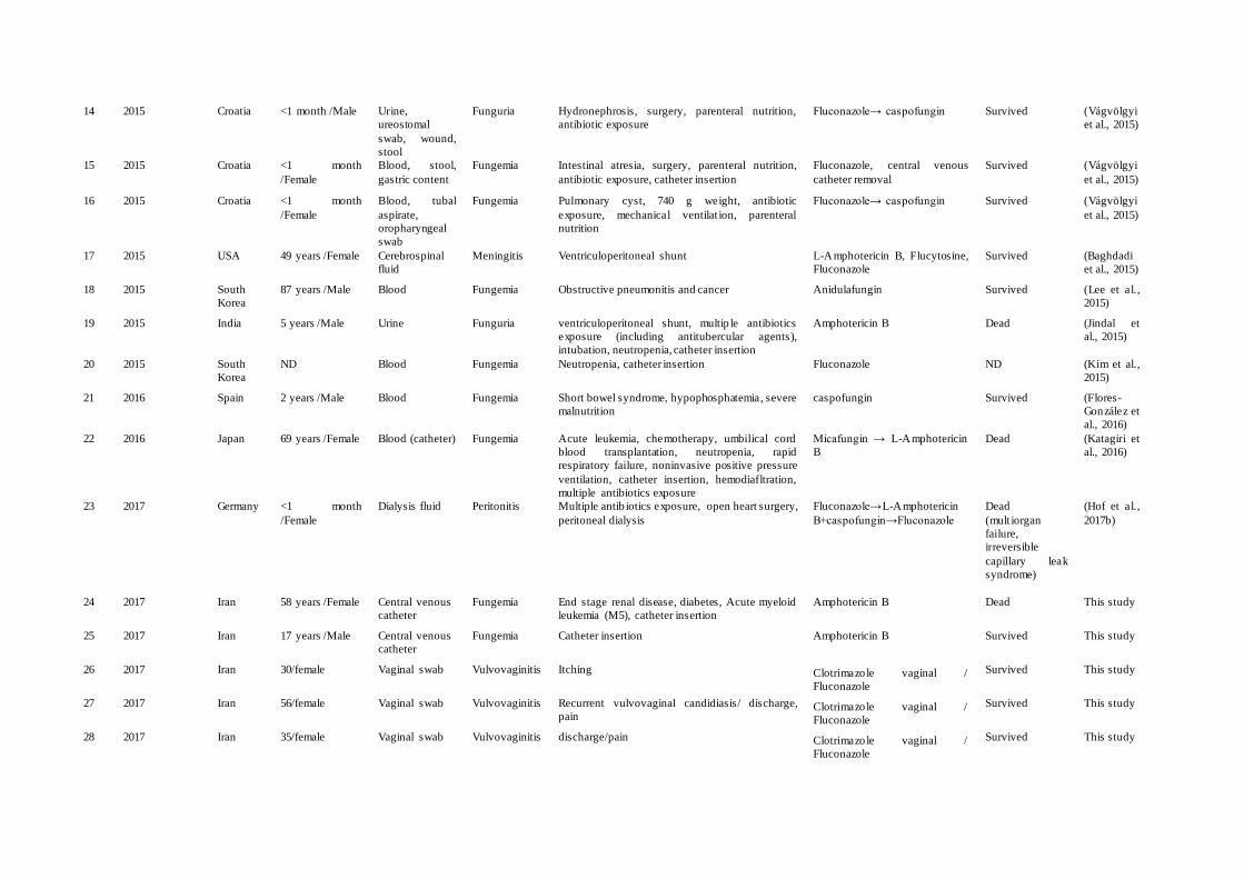
14 2015 Croatia <1 month /Male Urine,
ureostomal
swab, wound,
stool
Funguria Hydronephrosis, surgery, parenteral nutrition,
antibiotic exposure
Fluconazole→ caspofungin Survived (Vágvölgyi
et al., 2015)
15 2015 Croatia <1 month
/Female
Blood, stool,
gastric content
Fungemia Intestinal atresia, surgery, parenteral nutrition,
antibiotic exposure, catheter insertion
Fluconazole, central venous
catheter removal
Survived (Vágvölgyi
et al., 2015)
16 2015 Croatia <1 month
/Female
Blood, tubal
aspirate,
oropharyngeal
swab
Fungemia Pulmonary cyst, 740 g weight, antibiotic
exposure, mechanical ventilat ion, parenteral
nutrition
Fluconazole→ caspofungin Survived (Vágvölgyi
et al., 2015)
17 2015 USA 49 years /Female Cerebrospinal
fluid
Meningitis Ventriculoperitoneal shunt L-Amphotericin B, Flucytosine,
Fluconazole
Survived (Baghdadi
et al., 2015)
18 2015 South
Korea
87 years /Male Blood Fungemia Obstructive pneumonitis and cancer Anidulafungin Survived (Lee et al.,
2015)
19 2015 India 5 years /Male Urine Funguria ventriculoperitoneal shunt, multip le antibiotics
exposure (including antitubercular agents),
intubation, neutropenia, catheter insertion
Amphotericin B Dead (Jindal et
al., 2015)
20 2015 South
Korea
ND Blood Fungemia Neutropenia, catheter insertion Fluconazole ND (Kim et al.,
2015)
21 2016 Spain 2 years /Male Blood Fungemia Short bowel syndrome, hypophosphatemia, severe
malnutrition
caspofungin Survived (Flores-
González et
al., 2016)
22 2016 Japan 69 years /Female Blood (catheter) Fungemia Acute leukemia, chemotherapy, umbilical cord
blood transplantation, neutropenia, rapid
respiratory failure, noninvasive positive pressure ventilation, catheter insertion, hemodiafltration,
multiple antibiotics exposure
Micafungin → L-Amphotericin
B
Dead (Katagiri et
al., 2016)
23 2017 Germany <1 month
/Female
Dialysis fluid Peritonitis Multiple antib iotics exposure, open heart surgery, peritoneal dialysis
Fluconazole→L-Amphotericin
B+caspofungin→Fluconazole
Dead
(mult iorgan
failure,
irreversible
capillary leak
syndrome)
(Hof et al.,
2017b)
24 2017 Iran 58 years /Female Central venous
catheter
Fungemia End stage renal disease, diabetes, Acute myeloid
leukemia (M5), catheter insertion
Amphotericin B Dead This study
25 2017 Iran 17 years /Male Central venous
catheter
Fungemia Catheter insertion Amphotericin B Survived This study
26 2017 Iran 30/female Vaginal swab Vulvovaginitis Itching Clotrimazole vaginal /
Fluconazole
Survived This study
27 2017 Iran 56/female Vaginal swab Vulvovaginitis Recurrent vulvovaginal candidiasis/ discharge,
pain Clotrimazole vaginal /
Fluconazole
Survived This study
28 2017 Iran 35/female Vaginal swab Vulvovaginitis discharge/pain Clotrimazole vaginal /
Fluconazole
Survived This study

29 2017 Iran 60/female Vaginal swab Vulvovaginitis Recurrent vulvovaginal candidiasis/
discharge/pain/itching Clotrimazole vaginal /
Fluconazole
Survived This study
553
554
555

556

Supplementary files 557
Supplementary Figure 1. Late amplification of Pichia kudriavzevii and Pichia guilliermondii are 558 depicted. Adjusting Ct value of 22, distinguished target from non-target species. 559
Supplementary figure 2. Cumulative reproducibility ≥ 0.99 for five runs in three consecutive day for 560 target species. 561
Supplementary Figure 3. Tm distribution of target species of Cy. fabianii, W. anomalus, and Cy. jadinii. 562
Supplementary Table 1. Target and non-target species utilized for optimization and specificity testing 563 in this study. 564
Supplementary table 2. Epidemiological data that utilized MALDI-TOF MS or sequencing as a reliable means of 565 identification of Cy. fabianii, Cy. jadinii, and W. anomalus. 566
Supplementary table 3. Antifungal susceptibility pattern for Cy. fabianii and Cy. jadinii isolates in this 567 study. 568
569
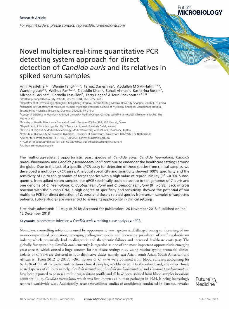
Research Article
For reprint orders, please contact: [email protected]
Novel multiplex real-time quantitative PCRdetecting system approach for directdetection of Candida auris and its relatives inspiked serum samplesAmir Arastehfar‡ ,1, Wenjie Fang‡ ,1,2,3, Farnaz Daneshnia1, Abdullah M S Al-Hatmi1,4,5,Wanqing Liao2,3, Weihua Pan*,2,3, Ziauddin Khan6, Suhail Ahmad6, Katharina Rosam7,Michaela Lackner7, Cornelia Lass-Florl7, Ferry Hagen1 & Teun Boekhout**,1,3,8
1Westerdijk Fungal Biodiversity Institute, Utrecht 3584, The Netherlands2Department of Dermatology, Shanghai Changzheng Hospital, Second Military Medical University, Shanghai 200003, PR China3Shanghai Key Laboratory of Molecular Medical Mycology, Shanghai Institute of Mycology, Shanghai Changzheng Hospital,Second Military Medical University, Shanghai 200003, PR China4Center of Expertise in Mycology Radboud University Medical Center, Canisius Wilhelmina Hospital, Nijmegen 6500HB, TheNetherlands5Ministry of Health, Directorate General of Health Services, PO Box 393, 100 Muscat, Oman6Department of Microbiology, Faculty of Medicine, Kuwait University, Safat, Kuwait7Division of Hygiene & Medical Microbiology, Medical University of Innsbruck, Innsbruck, Austria8Institute of Biodiversity & Ecosystem Dynamics, University of Amsterdam, Amsterdam 1012 WX, The Netherlands*Author for correspondence: Tel.: +86 8188 5494; [email protected]**Author for correspondence: Tel.: +31 62 929 0360; [email protected]‡Authors contributed equally
The multidrug-resistant opportunistic yeast species of Candida auris, Candida haemulonii, Candidaduobushaemulonii and Candida pseudohaemulonii continue to endanger the healthcare settings aroundthe globe. Due to the lack of a specific qPCR assay for detection of these species from clinical samples, wedeveloped a multiplex qPCR assay. Analytical specificity and sensitivity showed 100% specificity and thesensitivity of up to ten genomes of target species with a high value of reproducibility (R2 >0.99). Subse-quently, from spiked serum samples, our qPCR specifically could detect up to ten genomes of C. auris andone genome of C. haemulonii, C. duobushaemulonii and C. pseudohaemulonii (R2 >0.98). Lack of crossreaction with the human DNA, a high degree of specificity and sensitivity, showed the potential of ourmultiplex PCR for direct detection of C. auris and closely related species from serum samples of suspectedpatients. Future studies are warranted to assure its applicability in clinical settings.
First draft submitted: 11 August 2018; Accepted for publication: 20 November 2018; Published online:12 December 2018
Keywords: bloodstream infection • Candida auris • melting curve analysis • qPCR
Nowadays, controlling infections caused by opportunistic yeast species is challenged owing to increasing of im-munocompromised population, emerging pathogenic species and increasing prevalence of antifungal-resistantisolates, which potentially lead to diagnostic and therapeutic failures and increased healthcare costs [1–4]. Theglobally fast-spreading Candida auris currently is regarded as one of the most important opportunistic emergingyeast species, which caused a huge concern for healthcare settings [5–7]. Using routine typing protocols, clinicalisolates of C. auris are clustered in four distinctive clades namely, east Asian, south Asian, South American andAfrican [8]. From 2012 to 2017, >361 isolates of C. auris were obtained from blood cultures, accounting for67.48% of the all recovered isolates from clinical samples, worldwide [9]. On the other hand, the other closelyrelated species of C. auris namely, Candida haemulonii, Candida duobushaemuloni and Candida pseudohaemuloniihave been reported to possess a multidrug-resistant profile and all have been isolated from blood samples in variouscountries [10–12]. Candida haemulonii, which was first known as a human pathogen in 1984, is being increasinglyreported worldwide [6,10]. Additionally, recent surveillance studies of candidemia conducted in Panama, revealed
Future Microbiol. (Epub ahead of print) ISSN 1746-091310.2217/fmb-2018-0227 C© 2018 Weihua Pan
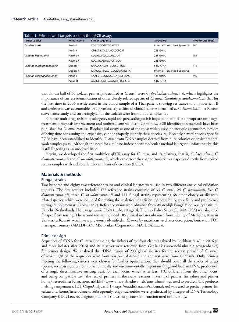
Research Article Arastehfar, Fang, Daneshnia et al.
Table 1. Primers and targets used in the qPCR assay.Target species Primer name Primer sequence Target loci Product size (bps)
Candida auris Auris-F CGGTGGCGTTGCATTCA Internal Transcribed Spacer 2 244
Auris-R CTGCTGCTAGAACACCTCGT 28S rDNA
Candida haemulonii Haemu-F CCGAGGACCGCAGCAAT 28S rDNA 181
Haemu-R CCCGTCCGAGCACTTCCA 28S rDNA
Candida duobushaemulonii Duobu-F GAACGCACATTGCGCCTTGG 5.8S rDNA 115
Duobu-R GTAGACTTCGCTGCGGATATGTTA Internal Transcribed Spacer 2
Candida pseudohaemulonii Pseud-F TAACCTGCGGAAGGATCATTAAG 18S rDNA 197
Pseud-R AATGTGCGTTCAAAGATTCGATG 5.8S rDNA
that almost half of 36 isolates primarily identified as C. auris were C. duobushaemulonii [13], which highlights theimportance of correct identification of other closely related species of C. auris. Candida pseudohaemulonii that forthe first time in 2006 was detected in the blood sample of a Thai patient showing resistance to amphotericin Band azoles [14], was accountable for approximately a third of clinical isolates identified as C. haemulonii in a Koreansurveillance study and surprisingly all of the isolates were from blood samples [10].
For these multidrug-resistant pathogens, rapid and precise diagnosis is important to initiate appropriate antifungaltreatment, prognosis improvement and outbreak control [15–17]. Up to now, >20 identification methods have beenpublished for C. auris [9,18–20]. Biochemical assays as one of the most widely used phenotypic approaches, besidesof being time consuming and expensive, cannot properly identify these species [21]. Recently, several species-specificPCRs have been established to identify C. auris from DNA samples derived from pure colonies or environmentalswab samples [18,19]. Although the need for a culture-independent molecular method is urgent, unfortunately, thisis still lingering as an unsolved issue.
Herein, we developed the first multiplex qPCR assay for C. auris, and its relatives, that is, C. haemulonii, C.duobushaemulonii and C. pseudohaemulonii, which can detect these opportunistic yeast species directly from spikedserum samples with a clinically relevant limit of detection (LOD).
Materials & methodsFungal strainsTwo hundred and eighty-two reference strains and clinical isolates were used in two different analytical validationtest sets. The first test set included 177 reference strains consisted of 33 C. auris, 25 C. haemulonii, five C.duobushaemulonii, three C. pseudohaemulonii and 111 fungal strains representing 68 other closely or distantlyrelated species, which were included for testing the analytical sensitivity, reproducibility, specificity and proficiencytesting (Supplementary Tables 1 & 2). Reference strains were obtained from Westerdijk Fungal Biodiversity Institute,Utrecht, Netherlands. Human genomic DNA (male, 10 ng/μl, Thermo Fisher Scientific, MA, USA) was also usedfor specificity testing. The second test set included 105 clinical isolates obtained from Faculty of Medicine, KuwaitUniversity, Kuwait, which were previously identified as C. auris by matrix-assisted laser desorption/ionization TOFmass spectrometry (MALDI-TOF MS; Bruker Corporation, MA, USA) [22,23].
Primer designSequences of rDNA for C. auris (including the isolates of the four clades analyzed by Lockhart et al. in 2016 [8]
and more isolates after 2016) and its relatives were retrieved from GenBank (www.ncbi.nlm.nih.gov/genbank/)for primer design. We analyzed the rDNA region of 233 global isolates for the reverse primer of C. auris,of which 138 of the sequences were from our own database and the rest were from Genbank. Only primersmeeting the following criteria were chosen for further optimization: they should cover all the clades of targetspecies; no cross reaction with other clinically and environmentally important fungi and human DNA; productionof a single discriminative melting peak for each locus, which is at least 1◦C different from the other locus;and being compatible with the rest of primers in the same reaction in terms of primer Tm values and primerhomo/heterodimer formations. uMELT (www.dna.utah.edu/umelt/umelt.html) was used to predict PCR productsmelting temperature. IDT OligoAnalyzer 3.1 (https://eu.idtdna.com/calc/analyzer) was used to predict primer Tmvalues and homo/heterodimers. Subsequently, oligonucleotides were synthesized by Integrated DNA TechnologyCompany (IDT, Leuven, Belgium). Table 1 shows the primers information used in this study.
10.2217/fmb-2018-0227 Future Microbiol. (Epub ahead of print) future science group
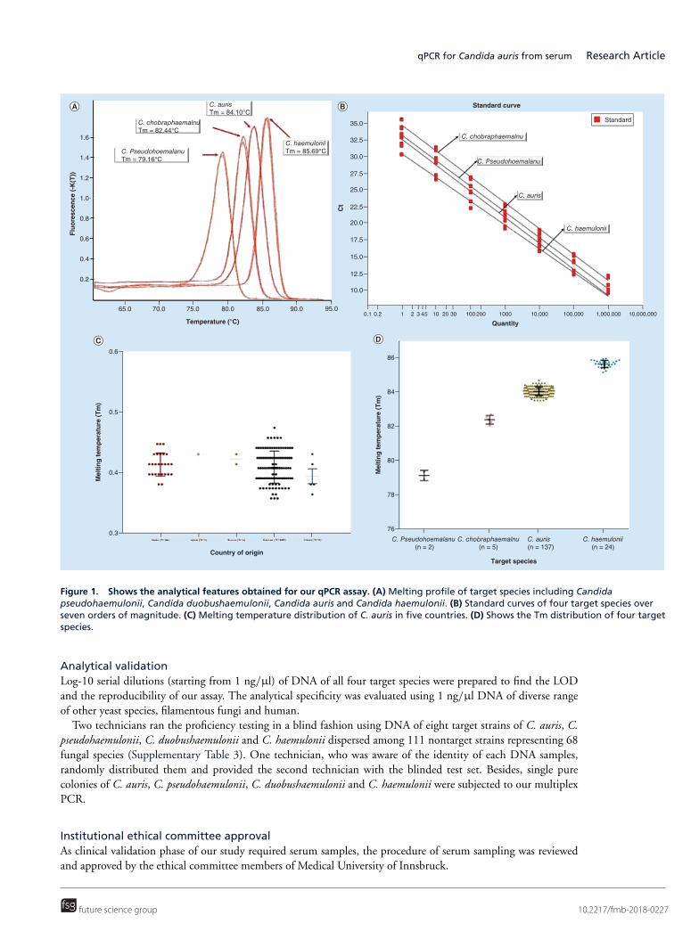
qPCR for Candida auris from serum Research Article
65.0 70.0 75.0 80.0 85.0
Temperature (°C)
90.0 95.0
0.2
0.4
0.6
0.8
Flu
ore
scen
ce (
-K(T
))
1.0
C. PseudohoemalanuTm = 79.16°C
C. haemuloniiTm = 85.69°C
C. aurisTm = 84.10°C
C. chobraphaemalnuTm = 82.44°C
1.2
1.4
1.6
0.1 0.2 1 2 3 45 10 20 100 1000 10,000 100,000 1,000,000 10,000,00020030
Quantity
10.0
12.5
15.0
17.5
20.0
22.5
25.0
27.5
30.0
32.5
35.0
Ct
C. Pseudohoemalanu
C. haemulonii
C. auris
Standard
Standard curve
C. chobraphaemalnu
Country of origin
Mel
tin
g t
emp
erat
ure
(T
m)
C. chobraphaemalnu(n = 5)
C. Pseudohoemalanu (n = 2)
C. haemulonii(n = 24)
C. auris (n = 137)
0.3
0.4
0.5
0.6
Target species
Mel
tin
g t
emp
erat
ure
(T
m)
76
78
80
82
84
86
Figure 1. Shows the analytical features obtained for our qPCR assay. (A) Melting profile of target species including Candidapseudohaemulonii, Candida duobushaemulonii, Candida auris and Candida haemulonii. (B) Standard curves of four target species overseven orders of magnitude. (C) Melting temperature distribution of C. auris in five countries. (D) Shows the Tm distribution of four targetspecies.
Analytical validationLog-10 serial dilutions (starting from 1 ng/μl) of DNA of all four target species were prepared to find the LODand the reproducibility of our assay. The analytical specificity was evaluated using 1 ng/μl DNA of diverse rangeof other yeast species, filamentous fungi and human.
Two technicians ran the proficiency testing in a blind fashion using DNA of eight target strains of C. auris, C.pseudohaemulonii, C. duobushaemulonii and C. haemulonii dispersed among 111 nontarget strains representing 68fungal species (Supplementary Table 3). One technician, who was aware of the identity of each DNA samples,randomly distributed them and provided the second technician with the blinded test set. Besides, single purecolonies of C. auris, C. pseudohaemulonii, C. duobushaemulonii and C. haemulonii were subjected to our multiplexPCR.
Institutional ethical committee approvalAs clinical validation phase of our study required serum samples, the procedure of serum sampling was reviewedand approved by the ethical committee members of Medical University of Innsbruck.
future science group 10.2217/fmb-2018-0227
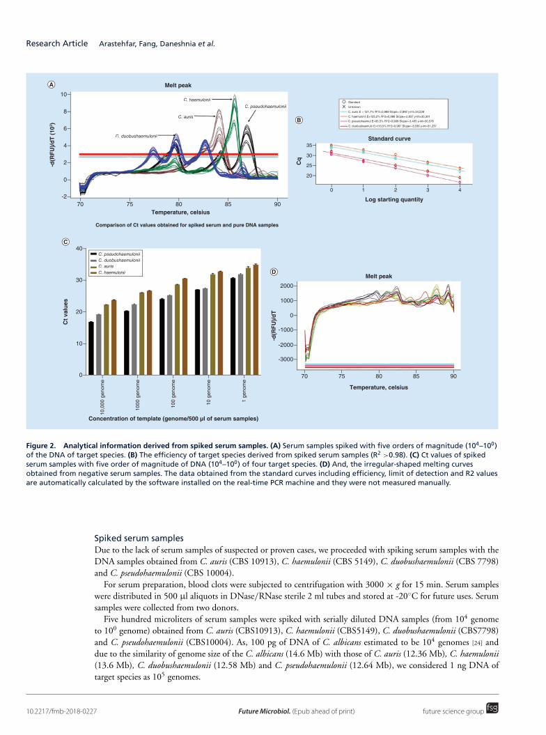
Research Article Arastehfar, Fang, Daneshnia et al.
70 75 80Temperature, celsius
Concentration of template (genome/500 µl of serum samples)
Temperature, celsius
-d(R
FU
)/d
T
-d(R
FU
)/d
T (
103 )
Melt peak
Melt peak
Standard curve
Comparison of Ct values obtained for spiked serum and pure DNA samples
85 90Log starting quantity
Cq
C. haemulonii
C. haemulonii
C. auris
C. auris
C. duobushaemulonii
C. duobushaemulonii
C. pseudohaemulonii
C. pseudohaemulonii
-2
0
2
4
6
8
10
0 1 2 3 4
20
25
30
35
10,0
00 g
enom
e
1000
gen
ome
100
geno
me
10 g
enom
e
1 ge
nom
e
0
10
20
30
40
70 75 80 85 90
-3000
-2000
-1000
0
1000
2000
Ct
valu
es
C. haemuloniiC. auris
C. haemulonii
C. auris
C. duobushaemulonii
C. pseudohaemuloStandard
Unknown
C. auris E = 121,7% R^2=0,989 Slope=-2,892 y-int=34,239
C. haemulonii E=125,2% R^2=0,986 Slope=-2,837 y-int=35,301
C. pseudohaemul E=95,5% R^2=0,999 Slope=-3,435 y-int=30,578
C. duobushaemulo E=113,5% R^2=0,987 Slope=-3,036 y-int=31,237
Figure 2. Analytical information derived from spiked serum samples. (A) Serum samples spiked with five orders of magnitude (104–100)of the DNA of target species. (B) The efficiency of target species derived from spiked serum samples (R2 >0.98). (C) Ct values of spikedserum samples with five order of magnitude of DNA (104–100) of four target species. (D) And, the irregular-shaped melting curvesobtained from negative serum samples. The data obtained from the standard curves including efficiency, limit of detection and R2 valuesare automatically calculated by the software installed on the real-time PCR machine and they were not measured manually.
Spiked serum samplesDue to the lack of serum samples of suspected or proven cases, we proceeded with spiking serum samples with theDNA samples obtained from C. auris (CBS 10913), C. haemulonii (CBS 5149), C. duobushaemulonii (CBS 7798)and C. pseudohaemulonii (CBS 10004).
For serum preparation, blood clots were subjected to centrifugation with 3000 × g for 15 min. Serum sampleswere distributed in 500 μl aliquots in DNase/RNase sterile 2 ml tubes and stored at -20◦C for future uses. Serumsamples were collected from two donors.
Five hundred microliters of serum samples were spiked with serially diluted DNA samples (from 104 genometo 100 genome) obtained from C. auris (CBS10913), C. haemulonii (CBS5149), C. duobushaemulonii (CBS7798)and C. pseudohaemulonii (CBS10004). As, 100 pg of DNA of C. albicans estimated to be 104 genomes [24] anddue to the similarity of genome size of the C. albicans (14.6 Mb) with those of C. auris (12.36 Mb), C. haemulonii(13.6 Mb), C. duobushaemulonii (12.58 Mb) and C. pseudohaemulonii (12.64 Mb), we considered 1 ng DNA oftarget species as 105 genomes.
10.2217/fmb-2018-0227 Future Microbiol. (Epub ahead of print) future science group
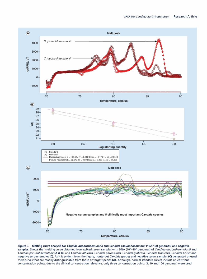
qPCR for Candida auris from serum Research Article
-1000
Temperature, celsius
Melt peak
-d(R
FU
)/d
T
70 90
0
1000
2000
3000
4000
75 80 85
C. pseudohaemulonii
C. duobushaemulonii
21222324252627
Log starting quantity
Cq
0.0 2.0
2829
0.5 1.0 1.5
StandardUnknownDuobushaemuloni E = 106.5%, R2 = 0.989 Slope = -3.176 y = int = 28,616Pseudo haemuloni E = 93.8%, R2 = 0.998 Slope = -3.480 y = int = 27,896
-2000
Temperature, celsius
Negative serum samples and 5 clinically most important Candida species
Melt peak
-d(R
FU
)/d
T
70 90
2000
-1000
0
1000
75 80 85
Figure 3. Melting curve analysis for Candida duobushaemulonii and Candida pseudohaemulonii (102–100 genomes) and negativesamples. Shows the melting curve obtained from spiked serum samples with DNA (102–100 genomes) of Candida duobushaemulonii andCandida pseudohaemulonii (A & B), and Candida albicans, Candida parapsilosis, Candida glabrata, Candida tropicalis, Candida krusei andnegative serum samples (C). As it is evident from the figure, nontarget Candida species and negative serum samples (C) generated unusualmelt curves that are readily distinguishable from those of target species (A). Although, normal standard curves include at least fourconcentration points, due to the clinical concentration relevance, only three concentration points (1, 10 and 100 genomes) were used.
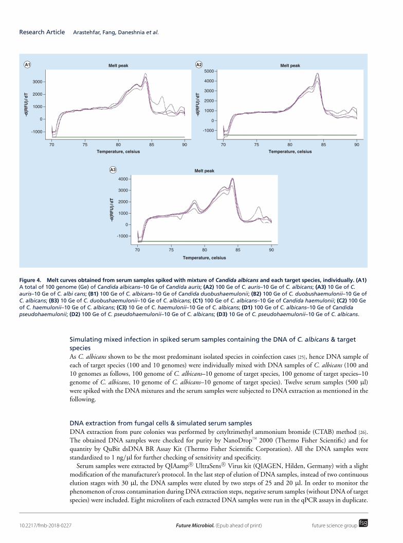
Research Article Arastehfar, Fang, Daneshnia et al.
-1000
Temperature, celsius
Melt peak
-d(R
FU
)/d
T
70
3000
2000
1000
0
75 80 85 90
Temperature, celsius
Melt peak
70 75 80 85 90
-1000
-d(R
FU
)/d
T
5000
2000
3000
4000
1000
0
-1000
Temperature, celsius
Melt peak
-d(R
FU
)/d
T
70
2000
3000
4000
1000
0
75 80 85 90
Figure 4. Melt curves obtained from serum samples spiked with mixture of Candida albicans and each target species, individually. (A1)A total of 100 genome (Ge) of Candida albicans–10 Ge of Candida auris; (A2) 100 Ge of C. auris–10 Ge of C. albicans; (A3) 10 Ge of C.auris–10 Ge of C. albi cans; (B1) 100 Ge of C. albicans–10 Ge of Candida duobushaemulonii; (B2) 100 Ge of C. duobushaemulonii–10 Ge ofC. albicans; (B3) 10 Ge of C. duobushaemulonii–10 Ge of C. albicans; (C1) 100 Ge of C. albicans–10 Ge of Candida haemulonii; (C2) 100 Geof C. haemulonii–10 Ge of C. albicans; (C3) 10 Ge of C. haemulonii–10 Ge of C. albicans; (D1) 100 Ge of C. albicans–10 Ge of Candidapseudohaemulonii; (D2) 100 Ge of C. pseudohaemulonii–10 Ge of C. albicans; (D3) 10 Ge of C. pseudohaemulonii–10 Ge of C. albicans.
Simulating mixed infection in spiked serum samples containing the DNA of C. albicans & targetspeciesAs C. albicans shown to be the most predominant isolated species in coinfection cases [25], hence DNA sample ofeach of target species (100 and 10 genomes) were individually mixed with DNA samples of C. albicans (100 and10 genomes as follows, 100 genome of C. albicans–10 genome of target species, 100 genome of target species–10genome of C. albicans, 10 genome of C. albicans–10 genome of target species). Twelve serum samples (500 μl)were spiked with the DNA mixtures and the serum samples were subjected to DNA extraction as mentioned in thefollowing.
DNA extraction from fungal cells & simulated serum samplesDNA extraction from pure colonies was performed by cetyltrimethyl ammonium bromide (CTAB) method [26].The obtained DNA samples were checked for purity by NanoDrop™ 2000 (Thermo Fisher Scientific) and forquantity by QuBit dsDNA BR Assay Kit (Thermo Fisher Scientific Corporation). All the DNA samples werestandardized to 1 ng/μl for further checking of sensitivity and specificity.
Serum samples were extracted by QIAamp R© UltraSens R© Virus kit (QIAGEN, Hilden, Germany) with a slightmodification of the manufacturer’s protocol. In the last step of elution of DNA samples, instead of two continuouselution stages with 30 μl, the DNA samples were eluted by two steps of 25 and 20 μl. In order to monitor thephenomenon of cross contamination during DNA extraction steps, negative serum samples (without DNA of targetspecies) were included. Eight microliters of each extracted DNA samples were run in the qPCR assays in duplicate.
10.2217/fmb-2018-0227 Future Microbiol. (Epub ahead of print) future science group
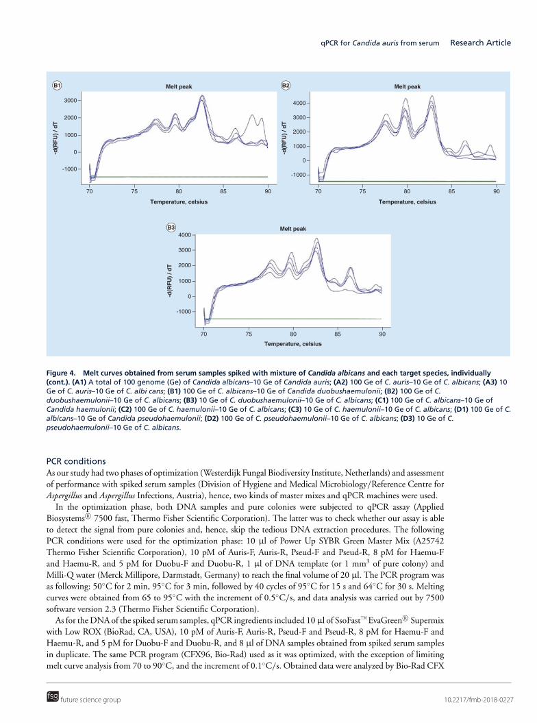
qPCR for Candida auris from serum Research Article
-1000
Temperature, celsius
Melt peak
-d(R
FU
) / d
T
70
3000
2000
1000
0
75 80 85 90
Temperature, celsius
Melt peak
70 75 80 85 90
-1000
-d(R
FU
) / d
T
2000
3000
4000
1000
0
-1000
Temperature, celsius
Melt peak
-d(R
FU
) / d
T
70
2000
3000
4000
1000
0
75 80 85 90
Figure 4. Melt curves obtained from serum samples spiked with mixture of Candida albicans and each target species, individually(cont.). (A1) A total of 100 genome (Ge) of Candida albicans–10 Ge of Candida auris; (A2) 100 Ge of C. auris–10 Ge of C. albicans; (A3) 10Ge of C. auris–10 Ge of C. albi cans; (B1) 100 Ge of C. albicans–10 Ge of Candida duobushaemulonii; (B2) 100 Ge of C.duobushaemulonii–10 Ge of C. albicans; (B3) 10 Ge of C. duobushaemulonii–10 Ge of C. albicans; (C1) 100 Ge of C. albicans–10 Ge ofCandida haemulonii; (C2) 100 Ge of C. haemulonii–10 Ge of C. albicans; (C3) 10 Ge of C. haemulonii–10 Ge of C. albicans; (D1) 100 Ge of C.albicans–10 Ge of Candida pseudohaemulonii; (D2) 100 Ge of C. pseudohaemulonii–10 Ge of C. albicans; (D3) 10 Ge of C.pseudohaemulonii–10 Ge of C. albicans.
PCR conditionsAs our study had two phases of optimization (Westerdijk Fungal Biodiversity Institute, Netherlands) and assessmentof performance with spiked serum samples (Division of Hygiene and Medical Microbiology/Reference Centre forAspergillus and Aspergillus Infections, Austria), hence, two kinds of master mixes and qPCR machines were used.
In the optimization phase, both DNA samples and pure colonies were subjected to qPCR assay (AppliedBiosystems R© 7500 fast, Thermo Fisher Scientific Corporation). The latter was to check whether our assay is ableto detect the signal from pure colonies and, hence, skip the tedious DNA extraction procedures. The followingPCR conditions were used for the optimization phase: 10 μl of Power Up SYBR Green Master Mix (A25742Thermo Fisher Scientific Corporation), 10 pM of Auris-F, Auris-R, Pseud-F and Pseud-R, 8 pM for Haemu-Fand Haemu-R, and 5 pM for Duobu-F and Duobu-R, 1 μl of DNA template (or 1 mm3 of pure colony) andMilli-Q water (Merck Millipore, Darmstadt, Germany) to reach the final volume of 20 μl. The PCR program wasas following: 50◦C for 2 min, 95◦C for 3 min, followed by 40 cycles of 95◦C for 15 s and 64◦C for 30 s. Meltingcurves were obtained from 65 to 95◦C with the increment of 0.5◦C/s, and data analysis was carried out by 7500software version 2.3 (Thermo Fisher Scientific Corporation).
As for the DNA of the spiked serum samples, qPCR ingredients included 10μl of SsoFast™ EvaGreen R© Supermixwith Low ROX (BioRad, CA, USA), 10 pM of Auris-F, Auris-R, Pseud-F and Pseud-R, 8 pM for Haemu-F andHaemu-R, and 5 pM for Duobu-F and Duobu-R, and 8 μl of DNA samples obtained from spiked serum samplesin duplicate. The same PCR program (CFX96, Bio-Rad) used as it was optimized, with the exception of limitingmelt curve analysis from 70 to 90◦C, and the increment of 0.1◦C/s. Obtained data were analyzed by Bio-Rad CFX
future science group 10.2217/fmb-2018-0227
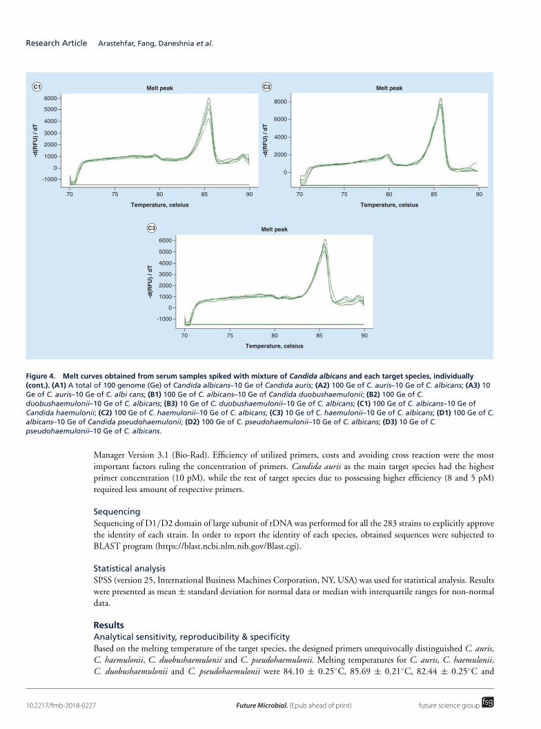
Research Article Arastehfar, Fang, Daneshnia et al.
-1000
Temperature, celsius
Melt peak
-d(R
FU
) / d
T
70
6000
2000
3000
4000
5000
1000
0
75 80 85 90
Temperature, celsius
Melt peak
70 75 80 85 90
-d(R
FU
) / d
T
4000
6000
8000
2000
0
-1000
Temperature, celsius
Melt peak
-d(R
FU
) / d
T
70
2000
3000
4000
5000
6000
1000
0
75 80 85 90
Figure 4. Melt curves obtained from serum samples spiked with mixture of Candida albicans and each target species, individually(cont.). (A1) A total of 100 genome (Ge) of Candida albicans–10 Ge of Candida auris; (A2) 100 Ge of C. auris–10 Ge of C. albicans; (A3) 10Ge of C. auris–10 Ge of C. albi cans; (B1) 100 Ge of C. albicans–10 Ge of Candida duobushaemulonii; (B2) 100 Ge of C.duobushaemulonii–10 Ge of C. albicans; (B3) 10 Ge of C. duobushaemulonii–10 Ge of C. albicans; (C1) 100 Ge of C. albicans–10 Ge ofCandida haemulonii; (C2) 100 Ge of C. haemulonii–10 Ge of C. albicans; (C3) 10 Ge of C. haemulonii–10 Ge of C. albicans; (D1) 100 Ge of C.albicans–10 Ge of Candida pseudohaemulonii; (D2) 100 Ge of C. pseudohaemulonii–10 Ge of C. albicans; (D3) 10 Ge of C.pseudohaemulonii–10 Ge of C. albicans.
Manager Version 3.1 (Bio-Rad). Efficiency of utilized primers, costs and avoiding cross reaction were the mostimportant factors ruling the concentration of primers. Candida auris as the main target species had the highestprimer concentration (10 pM), while the rest of target species due to possessing higher efficiency (8 and 5 pM)required less amount of respective primers.
SequencingSequencing of D1/D2 domain of large subunit of rDNA was performed for all the 283 strains to explicitly approvethe identity of each strain. In order to report the identity of each species, obtained sequences were subjected toBLAST program (https://blast.ncbi.nlm.nih.gov/Blast.cgi).
Statistical analysisSPSS (version 25, International Business Machines Corporation, NY, USA) was used for statistical analysis. Resultswere presented as mean ± standard deviation for normal data or median with interquartile ranges for non-normaldata.
ResultsAnalytical sensitivity, reproducibility & specificityBased on the melting temperature of the target species, the designed primers unequivocally distinguished C. auris,C. haemulonii, C. duobushaemulonii and C. pseudohaemulonii. Melting temperatures for C. auris, C. haemulonii,C. duobushaemulonii and C. pseudohaemulonii were 84.10 ± 0.25◦C, 85.69 ± 0.21◦C, 82.44 ± 0.25◦C and
10.2217/fmb-2018-0227 Future Microbiol. (Epub ahead of print) future science group

qPCR for Candida auris from serum Research Article
-2000
Temperature, celsius
Melt peak
-d(R
FU
) / d
T
70
2000
3000
1000
0
-1000
75 80 85 90
Temperature, celsius
70 75 80 85 90
-2000
Temperature, celsius
Melt peak
-d(R
FU
) / d
T
70
1000
2000
3000
0
-1000
75 80 85 90
Melt peak
-d(R
FU
) / d
T
2000
3000
4000
1000
0
-1000
Figure 4. Melt curves obtained from serum samples spiked with mixture of Candida albicans and each target species, individually(cont.). (A1) A total of 100 genome (Ge) of Candida albicans–10 Ge of Candida auris; (A2) 100 Ge of C. auris–10 Ge of C. albicans; (A3) 10Ge of C. auris–10 Ge of C. albi cans; (B1) 100 Ge of C. albicans–10 Ge of Candida duobushaemulonii; (B2) 100 Ge of C.duobushaemulonii–10 Ge of C. albicans; (B3) 10 Ge of C. duobushaemulonii–10 Ge of C. albicans; (C1) 100 Ge of C. albicans–10 Ge ofCandida haemulonii; (C2) 100 Ge of C. haemulonii–10 Ge of C. albicans; (C3) 10 Ge of C. haemulonii–10 Ge of C. albicans; (D1) 100 Ge of C.albicans–10 Ge of Candida pseudohaemulonii; (D2) 100 Ge of C. pseudohaemulonii–10 Ge of C. albicans; (D3) 10 Ge of C.pseudohaemulonii–10 Ge of C. albicans.
79.16 ± 0.18◦C, respectively (Figure 1A & D, Supplementary Table 4). No difference in melting temperature(p > 0.05) was shown among C. auris strains from different countries (Figure 1C). All the target strains werecorrectly identified except for CBS 12371, which was wrongly designated as C. pseudohaemulonii, while our assayin accordance to D1/D2 sequencing and MALDI-TOF MS identified this strain as C. haemulonii. When the DNAsamples obtained from pure cultures, the LOD for C. auris, C. pseudohaemulonii and C. duobushaemulonii was 10CFU/PCR with a Cq value of 32, and the LOD for C. haemulonii was 10 CFU/PCR with a Cq value of 29. Theaverage of R2 value for six consecutive runs of qPCR was 0.99, indicating a high degree of reproducibility of thisassay (Figure 1B). Specificity tests showed no cross reactivity with the other fungal species and genomic humanDNA. Subjecting 105 clinical isolates of C. auris from Kuwait to our qPCR gave the same results as those obtainedby MALDI-TOF MS and D1/D2 domain of ribosomal large subunit (LSU) rDNA sequencing, resulting in 100%agreement with these methods. Using blinded test set, our qPCR showed 100% accuracy and pure colonies as thepure DNA samples successfully identified target species.
Performance assessment with spiked serum samplesResults obtained from spiked serum samples revealed that our qPCR assay can reliably identify and detect tengenomes of C. auris (Cq = 31.75) and one genome of C. haemulonii (Cq = 34.82), C. pseudohaemulonii (Cq= 34.82) and C. duobushaemulonii (Cq = 30.61) (Figure 2A & C), and showed no cross reaction with negativeserum samples (Figure 2D). Spiked serum samples with C. auris and C. haemulonii showed an identical melt curveas what was obtained for the pure DNA of both species (Figure 2A). Unexpectedly C. duobushaemulonii and C.pseudohaemulonii generated unique triple peaks specific for each species (Figure 2A). Repeated experimentation of
future science group 10.2217/fmb-2018-0227

Research Article Arastehfar, Fang, Daneshnia et al.
spiking serum samples with C. pseudohaemulonii and C. duobushaemulonii provided the same results. However,in order to prove that these frequent melting peaks are unique for C. pseudohaemulonii and C. duobushaemulonii,500 μl of serum samples were spiked with three orders of magnitude (102 genomes to 100 genome) DNA of C.albicans, Candida parapsilosis, Candida glabrata, Candida tropicalis and Candida krusei (Figure 3A & C). As it isshown in the Figure 3, serum samples spiked with the DNA of aforementioned Candida species and negative serumsamples generated irregular-shaped melt curves, while melt peaks for C. pseudohaemulonii and C. duobushaemuloniiconsistently and efficiently resulted in the same melt curve patterns and turned out that those patterns are specificand distinctive fingerprints of these two species (Figure 3A & B). In order to prove that frequent melting peakswere not resulted from serum background, we utilized serum samples from two healthy individuals and bothserums resulted in identical and irregular melting peaks. Spiked serum samples with C. auris, C. haemulonii, C.pseudohaemulonii and C. duobushaemulonii showed acceptable reproducibility value (R2 ≥0.98). These observationsindicated that serum samples could be a suitable blood fraction for detection and identification of target specieswhen using our multiplex qPCR assay.
In simulated co-infection experiment, serum samples were spiked with DNA mixture of C. albicans and each targetspecies individually. Results from the qPCR of simulated co-infection test revealed that regardless of concentrationof DNA of C. albicans (ten-times more, ten-times less and equal to target species) target species were detected anddistinguished unequivocally (Figure 4).
DiscussionThe lack of reliable clinical diagnostic tools potentially led to underestimation and neglection of C. auris cases [17,27–
29]. When facing C. auris outbreaks, microbiologists mainly handle cultures, environmental swab or clinical samples(blood, sputum, urine). Using biochemical assays such as VITEK, API20C-AUX, AuxaColor 2, BD Phoenix,MicroScan etc. 90% of the C. auris isolates are misidentified as C. haemulonii (most commonly), Candida famata,Candida sake, Candida lusitaniae, Candida catenulata, Candida guilliermondii, C. parapsilosis, C. tropicalis, C.albicans, Rhodotorula glutinis and Saccharomyces cerevisiae [5].
Detecting C. auris directly from clinical or environmental swab samples in a culture-independent manner isessential for outbreak control. For environmental screening purposes, Leach et al. (2017) reported the ability ofsingle-plex probe-based qPCR for detection of C. auris from environmental swabs [18]. Although, in outbreaksenvironmental screening is an essential part of infection control, identification of patients suspected with blood-borne infections by C. auris and its relatives is of a great importance. Epidemiological studies revealed that C. auriscan represent up to 5.2% of candidemia cases in India [30], although with a lower prevalence, fatal bloodstreaminfection due to this pathogen is reported from other countries [9]. Moreover, it is estimated that delay of 1 h instarting immediate appropriate antimicrobial therapy for ICU patients will increase the mortality rate [31]. Thus,the urge for a rapid, specific, reliable and economic molecular means of identification from clinical samples is highlyrelevant. Herein for the first time, we developed a multiplex PCR assay that can detect C. auris and its relativesdirectly from serum samples in a culture-independent manner. Testing our qPCR with the serum samples spikedwith serially diluted DNA of target species, showed an exceptional LOD of one genome for C. haemulonii, C.pseudohaemulonii and C. duobushaemulonii, and ten genomes for C. auris. Moreover, serum due to its applicabilityfor downstream applications such as serology tests and its ease of DNA extraction procedure, has earned reputationfor being a suitable blood fraction for identification of fungemia [32]. It has been shown that for identification ofinvasive candidiasis, serum samples showed the same performance as whole blood samples when subjected to real-time PCR [33]. Additionally, serum has been regarded as a suitable matrix for validation of PCR-based assays meantto detect Candida species from infected patients [34–36]. The diagnostic strategy using our assay can dramaticallyshorten the clinical turn-around time (Supplementary Figure 1). Moreover, our qPCR assay utilizes the most basicand inexpensive qPCR chemistry, that is, SYBR GreenI and EVA Green, and offers the first qPCR assay with thepotential clinical utility for identification of C. auris and its relatives. Moreover, despite the fact that C. albicansaccounts for the majority of mixed infections, which could affect the specificity and sensitivity of PCR assays [25],our assay regardless of the DNA concentration of C. albicans (ten-times more, ten-times less and equal to targetspecies) readily identified C. auris, C. haemulonii, C. pseudohaemulonii and C. duobushaemulonii in serum samplesspiked the mixture DNA of target species and C. albicans. This observation reinforced the specificity of our qPCRassay in mixed infections.
For specificity testing, our assay was challenged with 111 reference strains representing 68 nontarget yeast andfilamentous fungal species, while in the published melting curve-based qPCR, only 19 nontarget species were
10.2217/fmb-2018-0227 Future Microbiol. (Epub ahead of print) future science group

qPCR for Candida auris from serum Research Article
tested [37]. Large difference in melting temperature between C. auris and its relatives is another advantage ofour qPCR, as PCR inhibitors, such as humic acid, melanin, hematin and collagen, are commonly found in theenvironmental and clinical samples and they can change the melting curve temperature causing melt temperatureshift followed by misidentification [38]. In our assay, the difference in average melting temperature between C. aurisand C. pseudohaemulonii, C. duobushaemulonii, C. haemulonii were 5, 1.7 and 1.6◦C, respectively. Additionally, theminimum difference in melting temperature between any two species was 1.1◦C. While in the published meltingcurve qPCR, small differences in melting temperature between C. auris and C. duobushaemulonii and C. haemuloniiwere observed (0.4 and 1◦C, respectively) [37]. On the other hand, despite the presence of heterogeneity in thesubmitted sequences of C. haemulonii complex, that is, C. haemulonii var. vulnera and C. haemulonii (C. haemuloniiGroup I), our assay successfully amplified various strains within this complex [39].
Although, a given developed diagnostic assays should be assessed with the relevant clinical materials obtainedfrom suspected patients, in our situation due to the lack of such clinical sources we had to spike serum sampleswith the DNA of target species. Hence, clinical performance and utility of our qPCR assay warrants furtherexperimentations in settings where suspected patients exist.
In summary, we developed a novel multiplex qPCR, which can discriminate C. auris, from C. haemulonii, C.duobushaemulonii and C. pseudohaemulonii in one multiplex qPCR reaction. The fact is that our multiplex assayhas been validated by using a comprehensive set of reference strains and clinical isolates, and proved to be rapid,specific and sensitive with spiked serum samples, indicating that it might have the applications for environmentalscreening and diagnostic purposes from clinical samples.
ConclusionCulture-independent and species-level identification of the multidrug-resistant opportunistic yeast species Candidaauris, C. haemulonii, C. pseudohaemulonii and C. duobushaemulonii is an important clinical practice. Involvedhealthcare settings of various countries can evaluate the specificity and sensitivity our multiplex qPCR assay in thelight of culture as the gold standard technique. If proved to be sensitive and specific, it will show implications inearly diagnosis followed by adminstartion of appropriate antifungal drugs. Moreover, its inexpensiveness, rapidityand sensitivity might highlight its applicability for environmental screening.
Summary points
• Based on the simple melt curve technique, our multiplex qPCR can distinguish and identify Candida auris, and itsrelatives, Candida haemulonii, Candida duobushaemulonii and Candida pseudohaemulonii.
• When DNAs obtained from pure colonies were used, the limit of detection for C. auris, C. haemulonii, C.pseudohaemulonii and C. duobushaemulonii was 10 CFU/PCR, with a high degree of reproducibility (R2 = 0.99).
• Specificity tests showed no cross reactivity with the other fungal species and genomic human DNA.
• Subjecting pure colonies of target strains showed the expected melting temperature, which indicated goodcompatibility of our assay when using pure colonies without DNA extraction.
• Results obtained from spiked serum samples revealed that our qPCR assay reliably identifies and detects tengenomes of C. auris (Ct = 31.75) and one genome of C. haemulonii (Ct = 34.82), C. pseudohaemulonii (Ct = 34.82)and C. duobushaemulonii (Ct = 30.61).
• Spiked serum samples with both C. auris and C. haemulonii showed acceptable reproducibility value (R2 ≥0.98%).
• Simulated co-infection test revealed that regardless of concentration of DNA of C. albicans, target species areidentified unequivocally.
• Having a high degree of sensitivity and reasonable difference in the melt profile of target species reveals thepotential utility of our qPCR assay for both serum samples derived from suspected patients and environmentalswab samples for infection control purposes.
Supplementary data
To view the supplementary data that accompany this paper please visit the journal website at: https://www.futuremedicine.com/d
oi/suppl/10.2217/fmb-2018-0227
future science group 10.2217/fmb-2018-0227
Research Article Arastehfar, Fang, Daneshnia et al.
Financial & competing interests disclosure
This work was supported by the National Health Department (2018ZX10101003), EU’s Horizon 2020 Research and Innovation
Program under the Marie Sklodowska-Curie grant (642095), National Natural Science Foundation of China (31770161), Second
Military Medical University (2017JZ47) and Shanghai Science and Technology Committee (17DZ2270900 and 14495800500). The
authors have no other relevant affiliations or financial involvement with any organization or entity with a financial interest in or
financial conflict with the subject matter or materials discussed in the manuscript apart from those disclosed.
No writing assistance was utilized in the production of this manuscript.
Open access
This work is licensed under the Attribution-NonCommercial-NoDerivatives 4.0 Unported License. To view a copy of this license,
visit http://creativecommons.org/licenses/by-nc-nd/4.0/
ReferencesPapers of special note have been highlighted as: •• of considerable interest
1. Pfaller MA, Diekema DJ, Gibbs DL et al. Results from the ARTEMIS DISK Global Antifungal Surveillance Study, 1997 to 2007:10.5-year analysis of susceptibilities of noncandidal yeast species to fluconazole and voriconazole determined by CLSI standardized diskdiffusion testing. J. Clin. Microbiol. 47(1), 117–123 (2009).
2. Miceli MH, Diaz JA, Lee SA. Emerging opportunistic yeast infections. Lancet. Infect. Dis. 11(2), 142–151 (2011).
3. Jeffery-Smith A, Taori SK, Schelenz S et al. Candida auris: a review of the literature. Clin. Microbiol. Rev 31(1), pii: e00029–00017(2017).
•• Misidentification through routinely used phenotypic and biochemical assays. Candida auris is easily misidentified as other yeastspecies when tested with the routinely used phenotypic and biochemical assays.
4. Fang W, Zhang L, Liu J et al. Tuberculosis/cryptococcosis co-infection in China between 1965 and 2016. Emerg. Microbes. Infect. 6(8),e73 (2017).
5. Chowdhary A, Sharma C, Meis JF. Candida auris: a rapidly emerging cause of hospital-acquired multidrug-resistant fungal infectionsglobally. PLoS Pathog. 13(5), e1006290 (2017).
•• Persistent colonizer in hospitals. Candida auris for a long period of time can stay in the hospitals and clinical settings.
6. Ben-Ami R, Berman J, Novikov A et al. Multidrug-resistant Candida haemulonii and C. auris, Tel Aviv, Israel. Emerg. Infect.Dis. 23(1), 195–203 (2017).
7. Ruiz-Gaitan A, Moret AM, Tasias-Pitarch M et al. An outbreak due to Candida auris with prolonged colonisation and candidaemia in atertiary care European hospital. Mycoses 61(7), 498–505 (2018).
8. Lockhart SR, Etienne KA, Vallabhaneni S et al. Simultaneous emergence of multidrug-resistant Candida auris on 3 continents confirmedby whole-genome sequencing and epidemiological analyses. Clin. Infect. Dis. 64(2), 134–140 (2017).
•• Multidrug resistance. Clinical isolates of C. auris can show multidrug-resistant nature to two or three classes of antifungalsposing a threat to its treatment.
9. Osei Sekyere J. Candida auris: a systematic review and meta-analysis of current updates on an emerging multidrug-resistant pathogen.Microbiologyopen doi:10.1002/mbo3.578 (2018) (Epub ahead of print).
•• Cause of fatal outbreaks. Once established in healthcare settings, it can be easily transferred to the other hospitals in differentcities, countries and even continents.
10. Kim MN, Shin JH, Sung H et al. Candida haemulonii and closely related species at 5 university hospitals in Korea: identification,antifungal susceptibility, and clinical features. Clin. Infect. Dis. 48(6), e57–e61 (2009).
11. Hou X, Xiao M, Chen SC et al. Identification and antifungal susceptibility profiles of Candida haemulonii species complex clinicalisolates from a multicenter study in China. J. Clin. Microbiol. 54(11), 2676–2680 (2016).
12. Khan ZU, Al-Sweih NA, Ahmad S et al. Outbreak of fungemia among neonates caused by Candida haemulonii resistant to amphotericinB, itraconazole, and fluconazole. J. Clin. Microbiol. 45(6), 2025–2027 (2007).
13. Ramos R, Caceres DH, Perez M et al. Emerging multidrug-resistant Candida duobushaemulonii infections in Panama hospitals:importance of laboratory surveillance and accurate identification. J. Clin. Microbiol. 56(7), (2018).
•• Wrong identification and overestimation in number of C. auris. By using reliable means of identification wrongly identified C.auris isolates were identified as Candida duobushaemulonii leading to revealing of the genuine epidemiology of this species.
14. Sugita T, Takashima M, Poonwan N, Mekha N. Candida pseudohaemulonii Sp. Nov., an amphotericin B-and azole-resistant yeast species,isolated from the blood of a patient from Thailand. Microbiol. Immunol. 50(6), 469–473 (2006).
•• Multidrug resistance nature of its siblings. Species within the Candida haemulonii complex, including Candida haemulonii,Candida pseudohaemulonii and Candida duobushaemulonii are multidrug-resistant emerging yeast species that are closelyrelated to C. auris. References 10–14
10.2217/fmb-2018-0227 Future Microbiol. (Epub ahead of print) future science group
qPCR for Candida auris from serum Research Article
15. Lepak AJ, Zhao M, Berkow EL, Lockhart SR, Andes DR. Pharmacodynamic optimization for treatment of invasive Candida aurisinfection. Antimicrob. Agents Chemother. 61(8), 1–5 (2017).
16. Fakhim H, Chowdhary A. In vitro interactions of echinocandins with triazoles against multidrug-resistant Candida auris. Antimicrob.Agents. Chemother. 61(11), pii: e01056-17 (2017).
17. Mizusawa M, Miller H, Green R et al. Can multidrug-resistant Candida auris be reliably identified in clinical microbiologylaboratories? J. Clin. Microbiol. 55(2), 638–640 (2017).
18. Leach L, Zhu Y, Chaturvedi S. Development and validation of a real-time PCR assay for rapid detection of Candida auris fromsurveillance samples. J. Clin. Microbiol. 56 2 pii: e01223-17 (2018).
19. Arastehfar A, Fang W, Badali H et al. Low-cost tetraplex PCR for the global spreading multi-drug resistant fungus, Candida auris and itsphylogenetic relatives. Front. Microbiol. 9, 1119 (2018).
20. Voelker R. New test identifies Candida auris. JAMA 319(21), 2164 (2018).
21. Snayd M, Dias F, Ryan RW, Clout D, Banach DB. Misidentification of Candida auris by RapID Yeast Plus, a commercial, biochemicalenzyme-based manual rapid identification system. J. Clin. Microbiol. 56(5), 1–2 (2018).
22. Vlek A, Kolecka A, Khayhan K et al. Interlaboratory comparison of sample preparation methods, database expansions, and cutoff valuesfor identification of yeasts by matrix-assisted laser desorption ionization-time of flight mass spectrometry using a yeast test panel. J. Clin.Microbiol. 52(8), 3023–3029 (2014).
23. Khan Z, Ahmad S, Benwan K et al. Invasive Candida auris infections in Kuwait hospitals: epidemiology, antifungal treatment andoutcome. Infection 46(5), 641–650 (2018).
•• High mortality rate. The rate of mortality for C. auris can go to up to 60%.
24. Loeffler J, Henke N, Hebart H et al. Quantification of fungal DNA by using fluorescence resonance energy transfer and the light cyclersystem. J. Clin. Microbiol. 38(2), 586–590 (2000).
25. Jensen J, Munoz P, Guinea J, Rodriguez-Creixems M, Pelaez T, Bouza E. Mixed fungemia: incidence, risk factors, and mortality in ageneral hospital. Clin. Infect. Dis. 44(12), e109–e114 (2007).
26. Diaz MR, Boekhout T, Kiesling T, Fell JW. Comparative analysis of the intergenic spacer regions and population structure of the speciescomplex of the pathogenic yeast Cryptococcus neoformans. FEMS Yeast Res. 5(12), 1129–1140 (2005).
27. Chowdhary A, Sharma C, Duggal S et al. New clonal strain of Candida auris, Delhi, India. Emerg. Infect. Dis. 19(10), 1670–1673 (2013).
28. Kathuria S, Singh PK, Sharma C et al. Multidrug-resistant Candida auris misidentified as Candida haemulonii: characterization bymatrix-assisted laser desorption ionization-time of flight mass spectrometry and DNA sequencing and its antifungal susceptibility profilevariability by Vitek 2, CLSI Broth Microdilution, and Etest Method. J. Clin. Microbiol. 53(6), 1823–1830 (2015).
29. Centers for Disease Control and Prevention. Clinical alert to US healthcare facilities – June 2016: global emergence of invasive infectionscaused by the multidrug-resistant yeast Candida auris. Centers for Disease Control and Prevention, Atlanta, GA, USA (2016).www.cdc.gov/fungal/diseases/candidiasis/candida-auris-alert.html
30. Chakrabarti A, Sood P, Rudramurthy SM et al. Incidence, characteristics and outcome of ICU-acquired candidemia in India. Intens.Care Med. 41(2), 285–295 (2015).
31. Kumar A, Roberts D, Wood KE et al. Duration of hypotension before initiation of effective antimicrobial therapy is the criticaldeterminant of survival in human septic shock. Crit. Care Med. 34(6), 1589–1596 (2006).
32. White PL, Mengoli C, Bretagne S et al. Evaluation of Aspergillus PCR protocols for testing serum specimens. J. Clin. Microbiol. 49(11),3842–3848 (2011).
33. Fortun J, Meije Y, Buitrago MJ et al. Clinical validation of a multiplex real-time PCR assay for detection of invasive candidiasis inintensive care unit patients. J. Antimicrob. Chemoth. 69(11), 3134–3141 (2014).
34. Dunyach C, Bertout S, Phelipeau C, Drakulovski P, Reynes J, Mallie M. Detection and identification of Candida spp. in human serumby LightCycler real-time polymerase chain reaction. Diagn. Micr. Infec. Dis. 60(3), 263–271 (2008).
35. Ahmad S, Khan Z, Mustafa AS, Khan ZU. Seminested PCR for diagnosis of candidemia: comparison with culture, antigen detection,and biochemical methods for species identification. J. Clin. Microbiol. 40(7), 2483–2489 (2002).
36. Mcmullan R, Metwally L, Coyle PV et al. A prospective clinical trial of a real-time polymerase chain reaction assay for the diagnosis ofcandidemia in nonneutropenic, critically ill adults. Clin. Infect. Dis. 46(6), 890–896 (2008).
37. Kordalewska M, Zhao Y, Lockhart SR, Chowdhary A, Berrio I, Perlin DS. Rapid and accurate molecular identification of the emergingmultidrug resistant pathogen Candida auris. J. Clin. Microbiol. 55(8), 2445–2452 (2017).
38. Mccord B, Opel K, Funes M, Zoppis S, Jantz LM. An investigation of the effect of DNA degradation and inhibition on PCRamplification of single source and mixed forensic samples. (2011). www.ncjrs.gov/pdff iles1/nij/grants/236692.pdf
39. Cendejas-Bueno E, Kolecka A, Alastruey-Izquierdo A et al. Reclassification of the Candida haemulonii complex as Candida haemulonii(C. haemulonii group I), C. duobushaemulonii sp. nov. (C. haemulonii group II), and C. haemulonii var. vulnera var. nov.: threemultiresistant human pathogenic yeasts. J. Clin. Microbiol. 50(11), 3641–3651 (2012).
future science group 10.2217/fmb-2018-0227

Research Article Arastehfar, Fang, Daneshnia et al.
10.2217/fmb-2018-0227 Future Microbiol. (Epub ahead of print) future science group