2015 ESC Guidelines for the management of infective ...
54
ESC GUIDELINES 2015 ESC Guidelines for the management of infective endocarditis The Task Force for the Management of Infective Endocarditis of the European Society of Cardiology (ESC) Endorsed by: European Association for Cardio-Thoracic Surgery (EACTS), the European Association of Nuclear Medicine (EANM) Authors/Task Force Members: Gilbert Habib * (Chairperson) (France), Patrizio Lancellotti * (co-Chairperson) (Belgium), Manuel J. Antunes (Portugal), Maria Grazia Bongiorni (Italy), Jean-Paul Casalta (France), Francesco Del Zotti (Italy), Raluca Dulgheru (Belgium), Gebrine El Khoury (Belgium), Paola Anna Erba a (Italy), Bernard Iung (France), Jose M. Miro b (Spain), Barbara J. Mulder (The Netherlands), Edyta Plonska-Gosciniak (Poland), Susanna Price (UK), Jolien Roos-Hesselink (The Netherlands), Ulrika Snygg-Martin (Sweden), Franck Thuny (France), Pilar Tornos Mas (Spain), Isidre Vilacosta (Spain), and Jose Luis Zamorano (Spain) Document Reviewers: Çetin Erol (CPG Review Coordinator) (Turkey), Petros Nihoyannopoulos (CPG Review Coordinator) (UK), Victor Aboyans (France), Stefan Agewall (Norway), George Athanassopoulos (Greece), Saide Aytekin (Turkey), Werner Benzer (Austria), He ´ ctor Bueno (Spain), Lidewij Broekhuizen (The Netherlands), Scipione Carerj (Italy), Bernard Cosyns (Belgium), Julie De Backer (Belgium), Michele De Bonis (Italy), Konstantinos Dimopoulos (UK), Erwan Donal (France), Heinz Drexel (Austria), Frank Arnold Flachskampf (Sweden), Roger Hall (UK), Sigrun Halvorsen (Norway), Bruno Hoen b (France), Paulus Kirchhof (UK/Germany), * Corresponding authors: Gilbert Habib, Service de Cardiologie, C.H.U. De La Timone, Bd Jean Moulin, 13005 Marseille, France, Tel: +33 4 91 38 75 88, Fax: +33 4 91 38 47 64, Email: [email protected] Patrizio Lancellotti, University of Lie `ge Hospital, GIGA Cardiovascular Sciences, Departments of Cardiology, Heart Valve Clinic, CHU Sart Tilman, Lie `ge, Belgium – GVM Care and Research, E.S. Health Science Foundation, Lugo (RA), Italy, Tel: +3243667196, Fax: +3243667194, Email: [email protected] ESC Committee for Practice Guidelines (CPG) and National Cardiac Societies document reviewers: listed in the Appendix ESC entities having participated in the development of this document: ESC Associations: Acute Cardiovascular Care Association (ACCA), European Association for Cardiovascular Prevention & Rehabilitation (EACPR), European Association of Cardiovascular Imaging (EACVI), European Heart Rhythm Association (EHRA), Heart Failure Association (HFA). ESC Councils: Council for Cardiology Practice (CCP), Council on Cardiovascular Nursing and Allied Professions (CCNAP), Council on Cardiovascular Primary Care (CCPC). ESC Working Groups: Cardiovascular Pharmacotherapy, Cardiovascular Surgery, Grown-up Congenital Heart Disease, Myocardial and Pericardial Diseases, Pulmonary Circulation and Right Ventricular Function, Thrombosis, Valvular Heart Disease. The content of these European Society of Cardiology (ESC) Guidelines has been published for personal and educational use only. No commercial use is authorized. No part of the ESC Guidelines may be translated or reproduced in any form without written permission from the ESC. Permission can be obtained upon submission of a written request to Oxford Uni- versity Press, the publisher of the European Heart Journal and the party authorized to handle such permissions on behalf of the ESC. Disclaimer. The ESC Guidelines represent the views of the ESC and were produced after careful consideration of the scientific and medical knowledge and the evidence available at the time of their publication. The ESC is not responsible in the event of any contradiction, discrepancy and/or ambiguity between the ESC Guidelines and any other official recom- mendations or guidelines issued by the relevant public health authorities, in particular in relation to good use of healthcare or therapeutic strategies. Health professionals are encour- aged to take the ESC Guidelines fully into account when exercising their clinical judgment, as well as in the determination and the implementation of preventive, diagnostic or therapeutic medical strategies; however, the ESC Guidelines do not override, in any way whatsoever, the individual responsibility of health professionals to make appropriate and accurate decisions in consideration of each patient’s health condition and in consultation with that patient and, where appropriate and/or necessary, the patient’s caregiver. Nor do the ESC Guidelines exempt health professionals from taking into full and careful consideration the relevant official updated recommendations or guidelines issued by the competent public health authorities, in order to manage each patient’s case in light of the scientifically accepted data pursuant to their respective ethical and professional obligations. It is also the health professional’s responsibility to verify the applicable rules and regulations relating to drugs and medical devices at the time of prescription. & The European Society of Cardiology 2015. All rights reserved. For permissions please email: [email protected]. European Heart Journal (2015) 36, 3075–3123 doi:10.1093/eurheartj/ehv319
-
Upload
khangminh22 -
Category
Documents
-
view
1 -
download
0
Transcript of 2015 ESC Guidelines for the management of infective ...

ESC GUIDELINES
2015 ESC Guidelines for the managementof infective endocarditisThe Task Force for the Management of Infective Endocarditis of theEuropean Society of Cardiology (ESC)
Endorsed by: European Association for Cardio-Thoracic Surgery(EACTS), the European Association of Nuclear Medicine (EANM)
Authors/Task Force Members: Gilbert Habib* (Chairperson) (France),Patrizio Lancellotti* (co-Chairperson) (Belgium), Manuel J. Antunes (Portugal),Maria Grazia Bongiorni (Italy), Jean-Paul Casalta (France), Francesco Del Zotti (Italy),Raluca Dulgheru (Belgium), Gebrine El Khoury (Belgium), Paola Anna Erbaa (Italy),Bernard Iung (France), Jose M. Mirob (Spain), Barbara J. Mulder (The Netherlands),Edyta Plonska-Gosciniak (Poland), Susanna Price (UK), Jolien Roos-Hesselink(The Netherlands), Ulrika Snygg-Martin (Sweden), Franck Thuny (France),Pilar Tornos Mas (Spain), Isidre Vilacosta (Spain), and Jose Luis Zamorano (Spain)
Document Reviewers: Çetin Erol (CPG Review Coordinator) (Turkey), Petros Nihoyannopoulos (CPG ReviewCoordinator) (UK), Victor Aboyans (France), Stefan Agewall (Norway), George Athanassopoulos (Greece),Saide Aytekin (Turkey), Werner Benzer (Austria), Hector Bueno (Spain), Lidewij Broekhuizen (The Netherlands),Scipione Carerj (Italy), Bernard Cosyns (Belgium), Julie De Backer (Belgium), Michele De Bonis (Italy),Konstantinos Dimopoulos (UK), Erwan Donal (France), Heinz Drexel (Austria), Frank Arnold Flachskampf (Sweden),Roger Hall (UK), Sigrun Halvorsen (Norway), Bruno Hoenb (France), Paulus Kirchhof (UK/Germany),
* Corresponding authors: Gilbert Habib, Service de Cardiologie, C.H.U. De La Timone, Bd Jean Moulin, 13005 Marseille, France, Tel: +33 4 91 38 75 88, Fax: +33 4 91 38 47 64,Email: [email protected]
Patrizio Lancellotti, University of Liege Hospital, GIGA Cardiovascular Sciences, Departments of Cardiology, Heart Valve Clinic, CHU Sart Tilman, Liege, Belgium – GVM Care andResearch, E.S. Health Science Foundation, Lugo (RA), Italy, Tel: +3243667196, Fax: +3243667194, Email: [email protected]
ESC Committee for Practice Guidelines (CPG) and National Cardiac Societies document reviewers: listed in the Appendix
ESC entities having participated in the development of this document:
ESC Associations: Acute Cardiovascular Care Association (ACCA), European Association for Cardiovascular Prevention & Rehabilitation (EACPR), European Association ofCardiovascular Imaging (EACVI), European Heart Rhythm Association (EHRA), Heart Failure Association (HFA).
ESC Councils: Council for Cardiology Practice (CCP), Council on Cardiovascular Nursing and Allied Professions (CCNAP), Council on Cardiovascular Primary Care (CCPC).
ESC Working Groups: Cardiovascular Pharmacotherapy, Cardiovascular Surgery, Grown-up Congenital Heart Disease, Myocardial and Pericardial Diseases, Pulmonary Circulationand Right Ventricular Function, Thrombosis, Valvular Heart Disease.
The content of these European Society of Cardiology (ESC) Guidelines has been published for personal and educational use only. No commercial use is authorized. No part of the ESCGuidelines may be translated or reproduced in any form without written permission from the ESC. Permission can be obtained upon submission of a written request to Oxford Uni-versity Press, the publisher of the European Heart Journal and the party authorized to handle such permissions on behalf of the ESC.
Disclaimer. The ESC Guidelines represent the views of the ESC and were produced after careful consideration of the scientific and medical knowledge and the evidence available atthe time of their publication. The ESC is not responsible in the event of any contradiction, discrepancy and/or ambiguity between the ESC Guidelines and any other official recom-mendations or guidelines issued by the relevant public health authorities, in particular in relation to good use of healthcare or therapeutic strategies. Health professionals are encour-aged to take the ESC Guidelines fully into account when exercising their clinical judgment, as well as in the determination and the implementation of preventive, diagnostic ortherapeutic medical strategies; however, the ESC Guidelines do not override, in any way whatsoever, the individual responsibility of health professionals to make appropriate andaccurate decisions in consideration of each patient’s health condition and in consultation with that patient and, where appropriate and/or necessary, the patient’s caregiver. Nordo the ESC Guidelines exempt health professionals from taking into full and careful consideration the relevant official updated recommendations or guidelines issued by the competentpublic health authorities, in order to manage each patient’s case in light of the scientifically accepted data pursuant to their respective ethical and professional obligations. It is also thehealth professional’s responsibility to verify the applicable rules and regulations relating to drugs and medical devices at the time of prescription.
& The European Society of Cardiology 2015. All rights reserved. For permissions please email: [email protected].
European Heart Journal (2015) 36, 3075–3123doi:10.1093/eurheartj/ehv319

Mitja Lainscak (Slovenia), Adelino F. Leite-Moreira (Portugal), Gregory Y.H. Lip (UK), Carlos A. Mestresc
(Spain/United Arab Emirates), Massimo F. Piepoli (Italy), Prakash P. Punjabi (UK), Claudio Rapezzi (Italy),Raphael Rosenhek (Austria), Kaat Siebens (Belgium), Juan Tamargo (Spain), and David M. Walker (UK)
The disclosure forms of all experts involved in the development of these guidelines are available on the ESC websitehttp://www.escardio.org/guidelines.
aRepresenting the European Association of Nuclear Medicine (EANM); bRepresenting the European Society of Clinical Microbiology and Infectious Diseases (ESCMID); andcRepresenting the European Association for Cardio-Thoracic Surgery (EACTS).
Online publish-ahead-of-print 29 August 2015
- - - - - - - - - - - - - - - - - - - - - - - - - - - - - - - - - - - - - - - - - - - - - - - - - - - - - - - - - - - - - - - - - - - - - - - - - - -- - - - - - - - - - - - - - - - - - - - - - - - - - - - - - - - - - - - - - - - - - - - - - - - - - - - - - - - - - - - - - - - - - - - - - - - - - -Keywords Endocarditis † Cardiac imaging † Valve disease † Echocardiography † Prognosis † Guidelines † Infection †
Nuclear imaging † Cardiac surgery † Cardiac device † Prosthetic heart valves † Congenital heart disease †
Pregnancy † Prophylaxis † Prevention
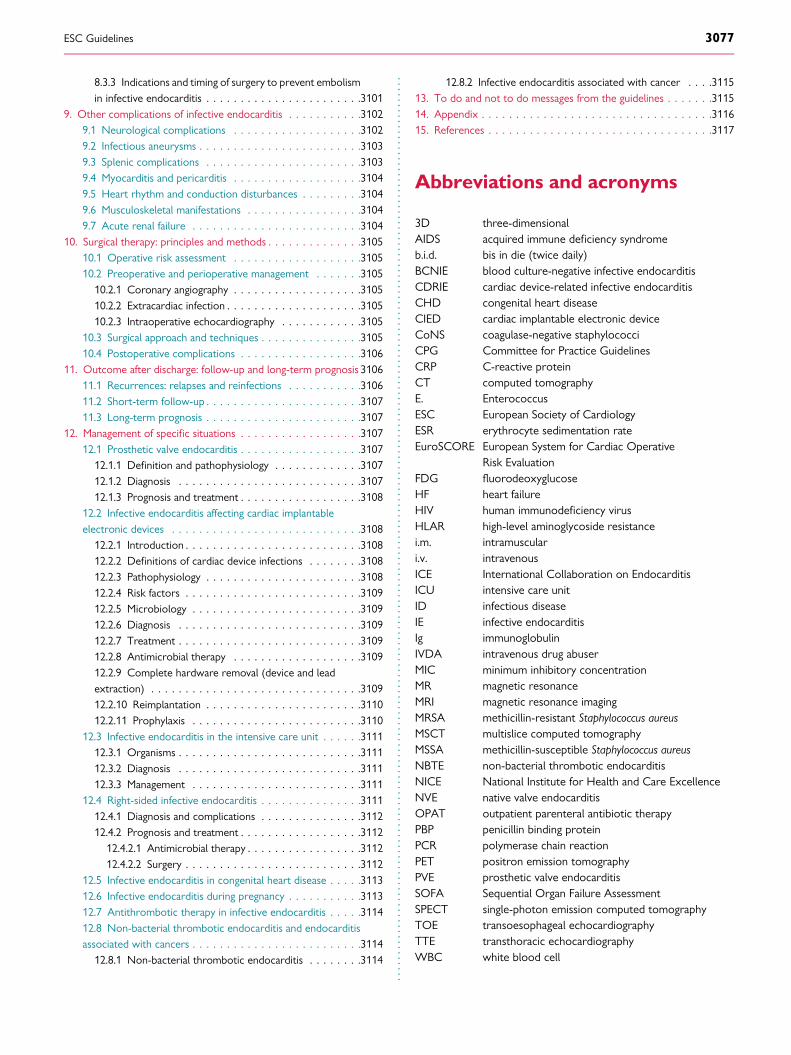
Table of ContentsAbbreviations and acronyms . . . . . . . . . . . . . . . . . . . . . . . .3077
1. Preamble . . . . . . . . . . . . . . . . . . . . . . . . . . . . . . . . . . .3078
2. Justification/scope of the problem . . . . . . . . . . . . . . . . . . .3079
3. Prevention . . . . . . . . . . . . . . . . . . . . . . . . . . . . . . . . . .3079
3.1 Rationale . . . . . . . . . . . . . . . . . . . . . . . . . . . . . . .3079
3.2 Population at risk . . . . . . . . . . . . . . . . . . . . . . . . . .3080
3.3 Situations and procedures at risk . . . . . . . . . . . . . . . .3081
3.3.1 Dental procedures . . . . . . . . . . . . . . . . . . . . . .3081
3.3.2 Other at-risk procedures . . . . . . . . . . . . . . . . . .3081
3.4 Prophylaxis for dental procedures . . . . . . . . . . . . . . .3081
3.5 Prophylaxis for non-dental procedures . . . . . . . . . . . .3082
3.5.1 Respiratory tract procedures . . . . . . . . . . . . . . . .3082
3.5.2 Gastrointestinal or genitourinary procedures . . . . .3082
3.5.3 Dermatological or musculoskeletal procedures . . . .3082
3.5.4 Body piercing and tattooing . . . . . . . . . . . . . . . .3082
3.5.5 Cardiac or vascular interventions . . . . . . . . . . . . .3082
3.5.6 Healthcare-associated infective endocarditis . . . . . .3082
4. The ‘Endocarditis Team’ . . . . . . . . . . . . . . . . . . . . . . . . .3083
5. Diagnosis . . . . . . . . . . . . . . . . . . . . . . . . . . . . . . . . . . .3084
5.1 Clinical features . . . . . . . . . . . . . . . . . . . . . . . . . . .3084
5.2 Laboratory findings . . . . . . . . . . . . . . . . . . . . . . . . .3084
5.3 Imaging techniques . . . . . . . . . . . . . . . . . . . . . . . . .3084
5.3.1 Echocardiography . . . . . . . . . . . . . . . . . . . . . . .3084
5.3.2 Multislice computed tomography . . . . . . . . . . . . .3086
5.3.3 Magnetic resonance imaging . . . . . . . . . . . . . . . .3087
5.3.4 Nuclear imaging . . . . . . . . . . . . . . . . . . . . . . . .3087
5.4 Microbiological diagnosis . . . . . . . . . . . . . . . . . . . . .3087
5.4.1 Blood culture–positive infective endocarditis . . . . .3087
5.4.2 Blood culture–negative infective endocarditis . . . . .3088
5.4.3 Histological diagnosis of infective endocarditis . . . .3088
5.4.4 Proposed strategy for a microbiological diagnostic
algorithm in suspected IE . . . . . . . . . . . . . . . . . . . . . .3088
5.5 Diagnostic criteria . . . . . . . . . . . . . . . . . . . . . . . . .3089
6. Prognostic assessment at admission . . . . . . . . . . . . . . . . . .3090
7. Antimicrobial therapy: principles and methods . . . . . . . . . . .3091
7.1 General principles . . . . . . . . . . . . . . . . . . . . . . . . .3091
7.2 Penicillin-susceptible oral streptococci and Streptococcus
bovis group . . . . . . . . . . . . . . . . . . . . . . . . . . . . . . . . .3092
7.3 Penicillin-resistant oral streptococci and Streptococcus bovis
group . . . . . . . . . . . . . . . . . . . . . . . . . . . . . . . . . . . .3092
7.4 Streptococcus pneumoniae, beta-haemolytic streptococci
(groups A, B, C, and G) . . . . . . . . . . . . . . . . . . . . . . . . .3092
7.5 Granulicatella and Abiotrophia (formerly nutritionally
variant streptococci) . . . . . . . . . . . . . . . . . . . . . . . . . . .3094
7.6 Staphylococcus aureus and coagulase-negative
staphylococci . . . . . . . . . . . . . . . . . . . . . . . . . . . . . . .3094
7.7 Methicillin-resistant and vancomycin-resistant
staphylococci . . . . . . . . . . . . . . . . . . . . . . . . . . . . . . .3094
7.8 Enterococcus spp. . . . . . . . . . . . . . . . . . . . . . . . . .3094
7.9 Gram-negative bacteria . . . . . . . . . . . . . . . . . . . . . .3096
7.9.1 HACEK-related species . . . . . . . . . . . . . . . . . . .3096
7.9.2 Non-HACEK species . . . . . . . . . . . . . . . . . . . . .3097
7.10 Blood culture–negative infective endocarditis . . . . . . .3097
7.11 Fungi . . . . . . . . . . . . . . . . . . . . . . . . . . . . . . . . .3097
7.12 Empirical therapy . . . . . . . . . . . . . . . . . . . . . . . . .3097
7.13 Outpatient parenteral antibiotic therapy for infective
endocarditis . . . . . . . . . . . . . . . . . . . . . . . . . . . . . . . .3098
8. Main complications of left-sided valve infective endocarditis and
their management . . . . . . . . . . . . . . . . . . . . . . . . . . . . . . .3099
8.1 Heart failure . . . . . . . . . . . . . . . . . . . . . . . . . . . . .3099
8.1.1 Heart failure in infective endocarditis . . . . . . . . . .3099
8.1.2 Indications and timing of surgery in the presence of
heart failure in infective endocarditis . . . . . . . . . . . . . . .3100
8.2 Uncontrolled infection . . . . . . . . . . . . . . . . . . . . . . .3100
8.2.1 Persisting infection . . . . . . . . . . . . . . . . . . . . . .3100
8.2.2 Perivalvular extension in infective endocarditis . . . .3100
8.2.3 Indications and timing of surgery in the
presence of uncontrolled infection in infective
endocarditis . . . . . . . . . . . . . . . . . . . . . . . . . . . . . .3101
8.2.3.1 Persistent infection . . . . . . . . . . . . . . . . . . .3101
8.2.3.2 Signs of locally uncontrolled infection . . . . . . .3101
8.2.3.3 Infection by microorganisms at low likelihood of
being controlled by antimicrobial therapy . . . . . . . . . .3101
8.3 Prevention of systemic embolism . . . . . . . . . . . . . . . .3101
8.3.1 Embolic events in infective endocarditis . . . . . . . . .3101
8.3.2 Predicting the risk of embolism . . . . . . . . . . . . . .3101
ESC Guidelines3076
8.3.3 Indications and timing of surgery to prevent embolism
in infective endocarditis . . . . . . . . . . . . . . . . . . . . . . .3101
9. Other complications of infective endocarditis . . . . . . . . . . .3102
9.1 Neurological complications . . . . . . . . . . . . . . . . . . .3102
9.2 Infectious aneurysms . . . . . . . . . . . . . . . . . . . . . . . .3103
9.3 Splenic complications . . . . . . . . . . . . . . . . . . . . . . .3103
9.4 Myocarditis and pericarditis . . . . . . . . . . . . . . . . . . .3104
9.5 Heart rhythm and conduction disturbances . . . . . . . . .3104
9.6 Musculoskeletal manifestations . . . . . . . . . . . . . . . . .3104
9.7 Acute renal failure . . . . . . . . . . . . . . . . . . . . . . . . .3104
10. Surgical therapy: principles and methods . . . . . . . . . . . . . .3105
10.1 Operative risk assessment . . . . . . . . . . . . . . . . . . .3105
10.2 Preoperative and perioperative management . . . . . . .3105
10.2.1 Coronary angiography . . . . . . . . . . . . . . . . . . .3105
10.2.2 Extracardiac infection . . . . . . . . . . . . . . . . . . . .3105
10.2.3 Intraoperative echocardiography . . . . . . . . . . . .3105
10.3 Surgical approach and techniques . . . . . . . . . . . . . . .3105
10.4 Postoperative complications . . . . . . . . . . . . . . . . . .3106
11. Outcome after discharge: follow-up and long-term prognosis 3106
11.1 Recurrences: relapses and reinfections . . . . . . . . . . .3106
11.2 Short-term follow-up . . . . . . . . . . . . . . . . . . . . . . .3107
11.3 Long-term prognosis . . . . . . . . . . . . . . . . . . . . . . .3107
12. Management of specific situations . . . . . . . . . . . . . . . . . .3107
12.1 Prosthetic valve endocarditis . . . . . . . . . . . . . . . . . .3107
12.1.1 Definition and pathophysiology . . . . . . . . . . . . .3107
12.1.2 Diagnosis . . . . . . . . . . . . . . . . . . . . . . . . . . .3107
12.1.3 Prognosis and treatment . . . . . . . . . . . . . . . . . .3108
12.2 Infective endocarditis affecting cardiac implantable
electronic devices . . . . . . . . . . . . . . . . . . . . . . . . . . . .3108
12.2.1 Introduction . . . . . . . . . . . . . . . . . . . . . . . . . .3108
12.2.2 Definitions of cardiac device infections . . . . . . . .3108
12.2.3 Pathophysiology . . . . . . . . . . . . . . . . . . . . . . .3108
12.2.4 Risk factors . . . . . . . . . . . . . . . . . . . . . . . . . .3109
12.2.5 Microbiology . . . . . . . . . . . . . . . . . . . . . . . . .3109
12.2.6 Diagnosis . . . . . . . . . . . . . . . . . . . . . . . . . . .3109
12.2.7 Treatment . . . . . . . . . . . . . . . . . . . . . . . . . . .3109
12.2.8 Antimicrobial therapy . . . . . . . . . . . . . . . . . . .3109
12.2.9 Complete hardware removal (device and lead
extraction) . . . . . . . . . . . . . . . . . . . . . . . . . . . . . . .3109
12.2.10 Reimplantation . . . . . . . . . . . . . . . . . . . . . . .3110
12.2.11 Prophylaxis . . . . . . . . . . . . . . . . . . . . . . . . .3110
12.3 Infective endocarditis in the intensive care unit . . . . . .3111
12.3.1 Organisms . . . . . . . . . . . . . . . . . . . . . . . . . . .3111
12.3.2 Diagnosis . . . . . . . . . . . . . . . . . . . . . . . . . . .3111
12.3.3 Management . . . . . . . . . . . . . . . . . . . . . . . . .3111
12.4 Right-sided infective endocarditis . . . . . . . . . . . . . . .3111
12.4.1 Diagnosis and complications . . . . . . . . . . . . . . .3112
12.4.2 Prognosis and treatment . . . . . . . . . . . . . . . . . .3112
12.4.2.1 Antimicrobial therapy . . . . . . . . . . . . . . . . .3112
12.4.2.2 Surgery . . . . . . . . . . . . . . . . . . . . . . . . . .3112
12.5 Infective endocarditis in congenital heart disease . . . . .3113
12.6 Infective endocarditis during pregnancy . . . . . . . . . . .3113
12.7 Antithrombotic therapy in infective endocarditis . . . . .3114
12.8 Non-bacterial thrombotic endocarditis and endocarditis
associated with cancers . . . . . . . . . . . . . . . . . . . . . . . . .3114
12.8.1 Non-bacterial thrombotic endocarditis . . . . . . . .3114
12.8.2 Infective endocarditis associated with cancer . . . .3115
13. To do and not to do messages from the guidelines . . . . . . .3115
14. Appendix . . . . . . . . . . . . . . . . . . . . . . . . . . . . . . . . . .3116
15. References . . . . . . . . . . . . . . . . . . . . . . . . . . . . . . . . .3117
Abbreviations and acronyms
3D three-dimensionalAIDS acquired immune deficiency syndromeb.i.d. bis in die (twice daily)BCNIE blood culture-negative infective endocarditisCDRIE cardiac device-related infective endocarditisCHD congenital heart diseaseCIED cardiac implantable electronic deviceCoNS coagulase-negative staphylococciCPG Committee for Practice GuidelinesCRP C-reactive proteinCT computed tomographyE. EnterococcusESC European Society of CardiologyESR erythrocyte sedimentation rateEuroSCORE European System for Cardiac Operative
Risk EvaluationFDG fluorodeoxyglucoseHF heart failureHIV human immunodeficiency virusHLAR high-level aminoglycoside resistancei.m. intramusculari.v. intravenousICE International Collaboration on EndocarditisICU intensive care unitID infectious diseaseIE infective endocarditisIg immunoglobulinIVDA intravenous drug abuserMIC minimum inhibitory concentrationMR magnetic resonanceMRI magnetic resonance imagingMRSA methicillin-resistant Staphylococcus aureusMSCT multislice computed tomographyMSSA methicillin-susceptible Staphylococcus aureusNBTE non-bacterial thrombotic endocarditisNICE National Institute for Health and Care ExcellenceNVE native valve endocarditisOPAT outpatient parenteral antibiotic therapyPBP penicillin binding proteinPCR polymerase chain reactionPET positron emission tomographyPVE prosthetic valve endocarditisSOFA Sequential Organ Failure AssessmentSPECT single-photon emission computed tomographyTOE transoesophageal echocardiographyTTE transthoracic echocardiographyWBC white blood cell
ESC Guidelines 3077

1. PreambleGuidelines summarize and evaluate all available evidence on a par-ticular issue at the time of the writing process, with the aim of assist-ing health professionals in selecting the best management strategiesfor an individual patient with a given condition, taking into accountthe impact on outcome, as well as the risk–benefit ratio of particu-lar diagnostic or therapeutic means. Guidelines and recommenda-tions should help health professionals to make decisions in theirdaily practice. However, the final decisions concerning an individualpatient must be made by the responsible health professional(s) inconsultation with the patient and caregiver as appropriate.
A great number of Guidelines have been issued in recent years bythe European Society of Cardiology (ESC) as well as by other soci-eties and organisations. Because of the impact on clinical practice,quality criteria for the development of guidelines have been estab-lished in order to make all decisions transparent to the user. The re-commendations for formulating and issuing ESC Guidelines can befound on the ESC website (http://www.escardio.org/Guidelines-&-Education/Clinical-Practice-Guidelines/Guidelines-development/Writing-ESC-Guidelines). ESC Guidelines represent the official pos-ition of the ESC on a given topic and are regularly updated.
Members of this Task Force were selected by the ESC to re-present professionals involved with the medical care of patientswith this pathology. Selected experts in the field undertook acomprehensive review of the published evidence for management(including diagnosis, treatment, prevention and rehabilitation) ofa given condition according to ESC Committee for PracticeGuidelines (CPG) policy. A critical evaluation of diagnostic andtherapeutic procedures was performed, including assessment ofthe risk–benefit ratio. Estimates of expected health outcomes forlarger populations were included, where data exist. The level ofevidence and the strength of the recommendation of particular
management options were weighed and graded according to prede-fined scales, as outlined in Tables 1 and 2.
The experts of the writing and reviewing panels provided declara-tions of interest forms for all relationships that might be perceived asreal or potential sources of conflicts of interest. These forms werecompiled into one file and can be found on the ESC website (http://www.escardio.org/guidelines). Any changes in declarations of inter-est that arise during the writing period must be notified to the ESCand updated. The Task Force received its entire financial supportfrom the ESC without any involvement from the healthcareindustry.
The ESC CPG supervises and coordinates the preparation of newGuidelines produced by task forces, expert groups or consensus pa-nels. The Committee is also responsible for the endorsement pro-cess of these Guidelines. The ESC Guidelines undergo extensivereview by the CPG and external experts. After appropriate revi-sions the Guidelines are approved by all the experts involved inthe Task Force. The finalized document is approved by the CPGfor publication in the European Heart Journal. The Guidelineswere developed after careful consideration of the scientific andmedical knowledge and the evidence available at the time oftheir dating.
The task of developing ESC Guidelines covers not only integra-tion of the most recent research, but also the creation of education-al tools and implementation programmes for the recommendations.To implement the guidelines, condensed pocket guidelines versions,summary slides, booklets with essential messages, summary cardsfor non-specialists, and an electronic version for digital applications(smartphones, etc.) are produced. These versions are abridged andthus, if needed, one should always refer to the full text version,which is freely available on the ESC website. The National Societiesof the ESC are encouraged to endorse, translate and implement allESC Guidelines. Implementation programmes are needed because it
Table 1 Classes of recommendations
Classes of recommendations
Suggested wording to use
Class I Evidence and/or general agreement that a given treatment or procedure is beneficial, useful,effective.
Is recommended/is indicated
Class II divergence of opinion about the Conflicting evidence and/or a
usefulness/efficacy of the given
favour of usefulness/efficacy.
Usefulness/efficacy is less well
treatment or procedure.
Class IIa Weight of evidence/opinion is in Should be considered
Class IIbestablished by evidence/opinion.
May be considered
Class III Evidence or general agreement that the given treatment or procedure is not useful/effective, and in some cases may be harmful.
Is not recommended
ESC Guidelines3078
has been shown that the outcome of disease may be favourably in-fluenced by the thorough application of clinical recommendations.
Surveys and registries are needed to verify that real-life daily prac-tice is in keeping with what is recommended in the guidelines, thuscompleting the loop between clinical research, writing of guidelines,disseminating them and implementing them into clinical practice.
Health professionals are encouraged to take the ESC Guidelinesfully into account when exercising their clinical judgment, as well asin the determination and the implementation of preventive, diagnos-tic or therapeutic medical strategies. However, the ESC Guidelinesdo not override in any way whatsoever the individual responsibilityof health professionals to make appropriate and accurate decisionsin consideration of each patient’s health condition and in consult-ation with that patient and the patient’s caregiver where appropriateand/or necessary. It is also the health professional’s responsibility toverify the rules and regulations applicable to drugs and devices at thetime of prescription.
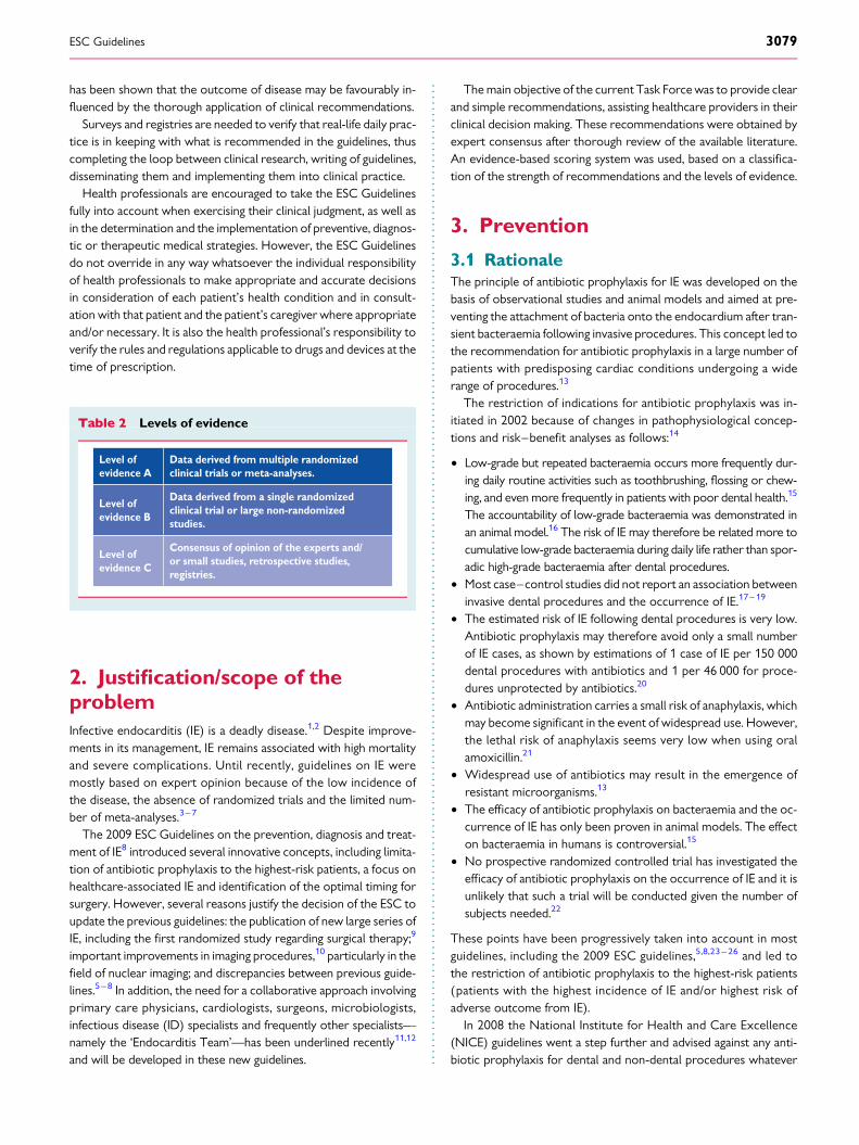
2. Justification/scope of theproblemInfective endocarditis (IE) is a deadly disease.1,2 Despite improve-ments in its management, IE remains associated with high mortalityand severe complications. Until recently, guidelines on IE weremostly based on expert opinion because of the low incidence ofthe disease, the absence of randomized trials and the limited num-ber of meta-analyses.3– 7
The 2009 ESC Guidelines on the prevention, diagnosis and treat-ment of IE8 introduced several innovative concepts, including limita-tion of antibiotic prophylaxis to the highest-risk patients, a focus onhealthcare-associated IE and identification of the optimal timing forsurgery. However, several reasons justify the decision of the ESC toupdate the previous guidelines: the publication of new large series ofIE, including the first randomized study regarding surgical therapy;9
important improvements in imaging procedures,10 particularly in thefield of nuclear imaging; and discrepancies between previous guide-lines.5– 8 In addition, the need for a collaborative approach involvingprimary care physicians, cardiologists, surgeons, microbiologists,infectious disease (ID) specialists and frequently other specialists—namely the ‘Endocarditis Team’—has been underlined recently11,12
and will be developed in these new guidelines.
The main objective of the current Task Force was to provide clearand simple recommendations, assisting healthcare providers in theirclinical decision making. These recommendations were obtained byexpert consensus after thorough review of the available literature.An evidence-based scoring system was used, based on a classifica-tion of the strength of recommendations and the levels of evidence.
3. Prevention
3.1 RationaleThe principle of antibiotic prophylaxis for IE was developed on thebasis of observational studies and animal models and aimed at pre-venting the attachment of bacteria onto the endocardium after tran-sient bacteraemia following invasive procedures. This concept led tothe recommendation for antibiotic prophylaxis in a large number ofpatients with predisposing cardiac conditions undergoing a widerange of procedures.13
The restriction of indications for antibiotic prophylaxis was in-itiated in 2002 because of changes in pathophysiological concep-tions and risk–benefit analyses as follows:14
† Low-grade but repeated bacteraemia occurs more frequently dur-ing daily routine activities such as toothbrushing, flossing or chew-ing, and even more frequently in patients with poor dental health.15
The accountability of low-grade bacteraemia was demonstrated inan animal model.16 The risk of IE may therefore be related more tocumulative low-grade bacteraemia during daily life rather than spor-adic high-grade bacteraemia after dental procedures.
† Most case–control studies did not report an association betweeninvasive dental procedures and the occurrence of IE.17– 19
† The estimated risk of IE following dental procedures is very low.Antibiotic prophylaxis may therefore avoid only a small numberof IE cases, as shown by estimations of 1 case of IE per 150 000dental procedures with antibiotics and 1 per 46 000 for proce-dures unprotected by antibiotics.20
† Antibiotic administration carries a small risk of anaphylaxis, whichmay become significant in the event of widespread use. However,the lethal risk of anaphylaxis seems very low when using oralamoxicillin.21
† Widespread use of antibiotics may result in the emergence ofresistant microorganisms.13
† The efficacy of antibiotic prophylaxis on bacteraemia and the oc-currence of IE has only been proven in animal models. The effecton bacteraemia in humans is controversial.15
† No prospective randomized controlled trial has investigated theefficacy of antibiotic prophylaxis on the occurrence of IE and it isunlikely that such a trial will be conducted given the number ofsubjects needed.22
These points have been progressively taken into account in mostguidelines, including the 2009 ESC guidelines,5,8,23 – 26 and led tothe restriction of antibiotic prophylaxis to the highest-risk patients(patients with the highest incidence of IE and/or highest risk ofadverse outcome from IE).
In 2008 the National Institute for Health and Care Excellence(NICE) guidelines went a step further and advised against any anti-biotic prophylaxis for dental and non-dental procedures whatever
Table 2 Levels of evidence
Level of evidence A
Data derived from multiple randomized clinical trials or meta-analyses.
Level of evidence B
Data derived from a single randomized clinical trial or large non-randomized studies.
Level of evidence C
Consensus of opinion of the experts and/or small studies, retrospective studies, registries.
ESC Guidelines 3079

the patient’s risk.27 The authors concluded there was an absence ofbenefit of antibiotic prophylaxis, which was also highly cost-ineffective. These conclusions have been challenged since estima-tions of the risks of IE are based on low levels of evidence due tomultiple extrapolations.28,29
Four epidemiological studies have analysed the incidence of IE fol-lowing restricted indications for antibiotic prophylaxis. The analysisof 2000–2010 national hospital discharge codes in the UK did notshow an increase in the incidence of streptococcal IE after the re-lease of NICE guidelines in 2008.30 The restriction of antibioticprophylaxis was seen in a 78% decrease in antibiotic prescriptionsbefore dental care. However, residual prescriptions raised concernsregarding a persisting use of antibiotic prophylaxis. A surveyperformed in 2012 in the UK showed that the majority of cardiolo-gists and cardiac surgeons felt that antibiotic prophylaxis wasnecessary in patients with valve prosthesis or prior IE.31 Recentlyan analysis of UK data collected from 2000 to 2013 showed a signifi-cant increase in the incidence of IE in both high-risk and lower-riskpatients in the UK starting in 2008.32 However, this temporal relation-ship should not be interpreted as a direct consequence of the NICEguidelines. These findings may be influenced by confounding factors,in particular changes in the number of patients at risk of hospitaliza-tions and healthcare-associated IE. Moreover, microbiological datawere not available. Thus we cannot know whether that increase isdue to the microbiological species covered by antibiotic prophylaxis.
A repeated prospective 1-year population-based French surveydid not show an increase in the incidence of IE, in particular strepto-coccal IE, between 1999 and 2008, whereas antibiotic prophylaxishad been restricted for native valve disease since 2002.33
Two studies from the USA did not find a negative impact of theabandonment of antibiotic prophylaxis in native valve disease in the2007 American Heart Association guidelines.34,35 A more recentanalysis on an administrative database found an increase in the inci-dence of IE hospitalizations between 2000 and 2011, with no signifi-cant change after the change of American guidelines in 2007.36 Theincrease in IE incidence was observed for all types of microorgan-isms, but was significant for streptococci after 2007.36 It was not sta-ted whether this was due to oral streptococci and if intermediate-or high-risk patients were involved.
The present guidelines maintain the principle of antibioticprophylaxis in high-risk patients for the following reasons:
† The remaining uncertainties regarding estimations of the risk ofIE, which play an important role in the rationale of NICEguidelines.
† The worse prognosis of IE in high-risk patients, in particular thosewith prosthetic IE.
† The fact that high-risk patients account for a much smaller num-ber than patients at intermediate risk, thereby reducing potentialharm due to adverse events of antibiotic prophylaxis.
3.2 Population at riskPatients with the highest risk of IE can be placed in three categories(Table 3):
(1) Patients with a prosthetic valve or with prosthetic material usedfor cardiac valve repair: these patients have a higher risk of IE, a
higher mortality from IE and more often develop complicationsof the disease than patients with native valves and an identicalpathogen.37 This also applies to transcatheter-implanted pros-theses and homografts.
(2) Patients with previous IE: they also have a greater risk of new IE,higher mortality and higher incidence of complications than pa-tients with a first episode of IE.38
(3) Patients with untreated cyanotic congenital heart disease(CHD) and those with CHD who have postoperative palliativeshunts, conduits or other prostheses.39,40 After surgical repairwith no residual defects, the Task Force recommends prophy-laxis for the first 6 months after the procedure until endothelia-lisation of the prosthetic material has occurred.
Although American Heart Association/American College ofCardiology guidelines recommend prophylaxis in cardiac transplantrecipients who develop cardiac valvulopathy, this is not supportedby strong evidence5,25,41 and is not recommended by the ESCTask Force.
Antibiotic prophylaxis is not recommended for patients atintermediate risk of IE, i.e. any other form of native valve disease(including the most commonly identified conditions: bicuspidaortic valve, mitral valve prolapse and calcific aortic stenosis).Nevertheless, both intermediate- and high-risk patients shouldbe advised of the importance of dental and cutaneous hygiene13
(Table 4). These measures of general hygiene apply to patientsand healthcare workers and should ideally be applied to the generalpopulation, as IE frequently occurs without known cardiac disease.
Table 3 Cardiac conditions at highest risk of infectiveendocarditis for which prophylaxis should beconsidered when a high-risk procedure is performed
Recommendations Classa Levelb
Antibiotic prophylaxis should be considered forpatients at highest risk for IE:(1) Patients with any prosthetic valve, including a
transcatheter valve, or those in whom anyprosthetic material was used for cardiac valverepair.
(2) Patients with a previous episode of IE.(3) Patients with CHD:
(a) Any type of cyanotic CHD.(b) Any type of CHD repaired with a
prosthetic material, whether placedsurgically or by percutaneous techniques,up to 6 months after the procedure orlifelong if residual shunt or valvularregurgitation remains.
IIa C
Antibiotic prophylaxis is not recommended inother forms of valvular or CHD.
III C
CHD ¼ congenital heart disease; IE ¼ infective endocarditis.aClass of recommendation.bLevel of evidence.cReference(s) supporting recommendations.
ESC Guidelines3080

3.3 Situations and procedures at risk3.3.1 Dental proceduresAt-risk procedures involve manipulation of the gingival or periapicalregion of the teeth or perforation of the oral mucosa (including scal-ing and root canal procedures) (Table 5).15,20 The use of dental im-plants raises concerns with regard to potential risk due to foreignmaterial at the interface between the buccal cavity and blood.Very few data are available.42 The opinion of the Task Force isthat there is no evidence to contraindicate implants in all patientsat risk. The indication should be discussed on a case-by-case basis.The patient should be informed of the uncertainties and the needfor close follow-up.
3.3.2 Other at-risk proceduresThere is no compelling evidence that bacteraemia resulting from re-spiratory tract procedures, gastrointestinal or genitourinary proce-dures, including vaginal and caesarean delivery, or dermatological ormusculoskeletal procedures causes IE (Table 5).
3.4 Prophylaxis for dental proceduresAntibiotic prophylaxis should only be considered for patients athighest risk for endocarditis, as described in Table 3, undergoing at-risk dental procedures listed in Table 5, and is not recommended inother situations. The main targets for antibiotic prophylaxis in thesepatients are oral streptococci. Table 6 summarizes the main regimensof antibiotic prophylaxis recommended before dental procedures.Fluoroquinolones and glycopeptides are not recommended due totheir unclear efficacy and the potential induction of resistance.
Table 5 Recommendations for prophylaxis ofinfective endocarditis in the highest-risk patientsaccording to the type of at-risk procedure
Recommendations Classa Levelb
A. Dental procedures
† Antibiotic prophylaxis should only beconsidered for dental procedures requiringmanipulation of the gingival or periapicalregion of the teeth or perforation of the oralmucosa
IIa C
† Antibiotic prophylaxis is not recommendedfor local anaesthetic injections in non-infectedtissues, treatment of superficial caries,removal of sutures, dental X-rays, placementor adjustment of removable prosthodontic ororthodontic appliances or braces or followingthe shedding of deciduous teeth or trauma tothe lips and oral mucosa
III C
Continued
Table 4 Non-specific prevention measures to befollowed in high-risk and intermediate-risk patients
These measures should ideally be applied to the generalpopulation and particularly reinforced in high-risk patients:
• Strict dental and cutaneous hygiene. Dental follow-up should be performed twice a year in high-risk patients and yearly in the others.
• Disinfection of wounds.
• Eradication or decrease of chronic bacterial carriage: skin, urine.
• Curative antibiotics for any focus of bacterial infection.
• No self-medication with antibiotics.
• Strict infection control measures for any at-risk procedure.
• Discourage piercing and tattooing.
• Limit the use of infusion catheters and invasive procedure when possible. Favour peripheral over central catheters, and systematic replacement of the peripheral catheter every 3–4 days. Strict adherence to care bundles for central and peripheral cannulae should be performed.
Table 5 Continued
Recommendations Classa Levelb
B. Respiratory tract proceduresc
† Antibiotic prophylaxis is not recommendedfor respiratory tract procedures, includingbronchoscopy or laryngoscopy, or transnasalor endotracheal intubation
III C
C. Gastrointestinal or urogenital procedures or TOEc
† Antibiotic prophylaxis is not recommendedfor gastroscopy, colonoscopy, cystoscopy,vaginal or caesarean delivery or TOE
III C
D. Skin and soft tissue proceduresc
† Antibiotic prophylaxis is not recommendedfor any procedure
III C
TOE ¼ transoesophageal echocardiography.aClass of recommendation.bLevel of evidence.cFor management when infections are present, please refer to Section 3.5.3.
Table 6 Recommended prophylaxis for high-riskdental procedures in high-risk patients
Situation Antibiotic
Single-dose 30–60 minutes before procedure
Adults Children
No allergy to penicillin or ampicillin
Amoxicillin or ampicillina
2 g orally or i.v. 50 mg/kg orally or i.v.
Allergy to penicillin or ampicillin
Clindamycin 600 mg orally or i.v.
20 mg/kg orally or i.v.
aAlternatively, cephalexin 2 g i.v. for adults or 50 mg/kg i.v. for children, cefazolin orceftriaxone 1 g i.v. for adults or 50 mg/kg i.v. for children.Cephalosporins should not be used in patients with anaphylaxis, angio-oedema, orurticaria after intake of penicillin or ampicillin due to cross-sensitivity.
ESC Guidelines 3081
Cephalosporins should not be used in patients with anaphylaxis,angio-oedema or urticaria after intake of penicillin or ampicillin dueto cross-sensitivity.
3.5 Prophylaxis for non-dentalproceduresSystematic antibiotic prophylaxis is not recommended for non-dental procedures. Antibiotic therapy is only needed when invasiveprocedures are performed in the context of infection.
3.5.1 Respiratory tract proceduresPatients listed in Table 3 who undergo an invasive respiratory tractprocedure to treat an established infection (i.e. drainage of an ab-scess) should receive an antibiotic regimen that contains an anti-staphylococcal drug.
3.5.2 Gastrointestinal or genitourinary proceduresIn the case of an established infection or if antibiotic therapy is in-dicated to prevent wound infection or sepsis associated with agastrointestinal or genitourinary tract procedure in patients de-scribed in Table 3, it is reasonable that the antibiotic regimen in-cludes an agent active against enterococci (i.e. ampicillin,amoxicillin or vancomycin; only in patients unable to tolerate beta-lactams). The use of intrauterine devices was regarded as contra-indicated, but this was based on low levels of evidence. Use of anintrauterine device is now considered acceptable, in particularwhen other contraceptive methods are not possible and in womenat low risk of genital infections.43
3.5.3 Dermatological or musculoskeletal proceduresFor patients described in Table 3 undergoing surgical proceduresinvolving infected skin (including oral abscesses), skin structure ormusculoskeletal tissue, it is reasonable that the therapeutic regimencontains an agent active against staphylococci and beta-haemolyticstreptococci.
3.5.4 Body piercing and tattooingThese growing societal trends are a cause for concern, particularlyfor individuals with CHD who are at increased susceptibility for theacquisition of IE. Case reports of IE after piercing and tattooing areincreasing, particularly when piercing involves the tongue,44 al-though publication bias may over- or underestimate the problem.Currently no data are available on the incidence of IE after such pro-cedures and the efficacy of antibiotics for prevention. Education ofpatients at risk of IE is paramount. They should be informed aboutthe hazards of piercing and tattooing and these procedures shouldbe discouraged not only in high-risk patients, but also in those withnative valve disease. If undertaken, procedures should be performedunder strictly sterile conditions, though antibiotic prophylaxis is notrecommended.
3.5.5 Cardiac or vascular interventionsIn patients undergoing implantation of a prosthetic valve, any type ofprosthetic graft or pacemakers, perioperative antibiotic prophylaxis
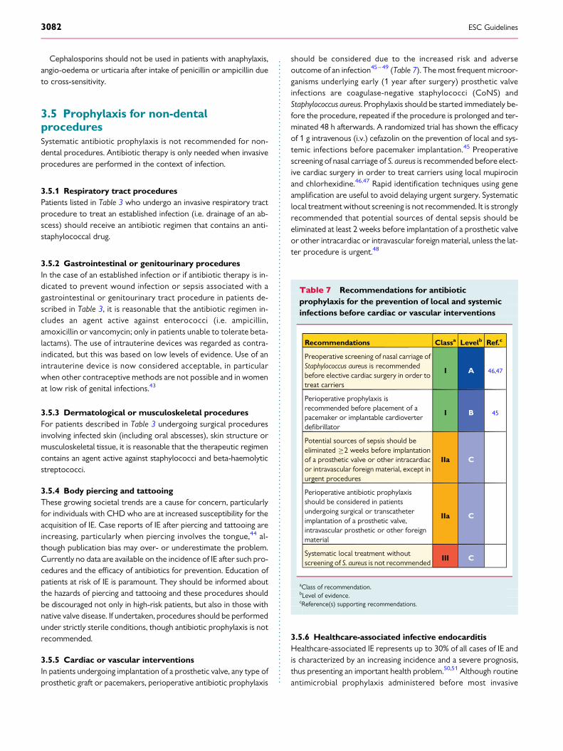
should be considered due to the increased risk and adverseoutcome of an infection45– 49 (Table 7). The most frequent microor-ganisms underlying early (1 year after surgery) prosthetic valveinfections are coagulase-negative staphylococci (CoNS) andStaphylococcus aureus. Prophylaxis should be started immediately be-fore the procedure, repeated if the procedure is prolonged and ter-minated 48 h afterwards. A randomized trial has shown the efficacyof 1 g intravenous (i.v.) cefazolin on the prevention of local and sys-temic infections before pacemaker implantation.45 Preoperativescreening of nasal carriage of S. aureus is recommended before elect-ive cardiac surgery in order to treat carriers using local mupirocinand chlorhexidine.46,47 Rapid identification techniques using geneamplification are useful to avoid delaying urgent surgery. Systematiclocal treatment without screening is not recommended. It is stronglyrecommended that potential sources of dental sepsis should beeliminated at least 2 weeks before implantation of a prosthetic valveor other intracardiac or intravascular foreign material, unless the lat-ter procedure is urgent.48
3.5.6 Healthcare-associated infective endocarditisHealthcare-associated IE represents up to 30% of all cases of IE andis characterized by an increasing incidence and a severe prognosis,thus presenting an important health problem.50,51 Although routineantimicrobial prophylaxis administered before most invasive
Table 7 Recommendations for antibioticprophylaxis for the prevention of local and systemicinfections before cardiac or vascular interventions
Recommendations Classa Levelb Ref.c
Preoperative screening of nasal carriage ofStaphylococcus aureus is recommendedbefore elective cardiac surgery in order totreat carriers
I A 46,47
Perioperative prophylaxis isrecommended before placement of apacemaker or implantable cardioverterdefibrillator
I B 45
Potential sources of sepsis should beeliminated ≥2 weeks before implantationof a prosthetic valve or other intracardiacor intravascular foreign material, except inurgent procedures
IIa C
Perioperative antibiotic prophylaxisshould be considered in patientsundergoing surgical or transcatheterimplantation of a prosthetic valve,intravascular prosthetic or other foreignmaterial
IIa C
Systematic local treatment withoutscreening of S. aureus is not recommended
III C
aClass of recommendation.bLevel of evidence.cReference(s) supporting recommendations.
ESC Guidelines3082

procedures is not recommended, aseptic measures during the inser-tion and manipulation of venous catheters and during any invasiveprocedures, including in outpatients, are mandatory to reduce therate of this healthcare-associated IE.52
In summary, these guidelines propose continuing to limit antibiot-ic prophylaxis to patients at high risk of IE undergoing thehighest-risk dental procedures. They highlight the importance ofhygiene measures, in particular oral and cutaneous hygiene. Epi-demiological changes are marked by an increase in IE due tostaphylococcus and of healthcare-associated IE, thereby high-lighting the importance of non-specific infection control mea-sures.51,53 This should concern not only high-risk patients, butshould also be part of routine care in all patients since IE occur-ring in patients without previously known heart disease now ac-counts for a substantial and increasing incidence. This meansthat although antibiotic prophylaxis should be restricted to thehighest-risk patients, preventive measures should be maintainedor extended to all patients with cardiac disease.
Although this section of the guidelines on IE prophylaxis isbased on weak evidence, they have been strengthened recentlyby epidemiological surveys, most of which did not show an in-creased incidence of IE due to oral streptococci.33 – 35 Their ap-plication by patients should follow a shared decision-makingprocess. Future challenges are to gain a better understandingof the mechanisms associated with valve infection, theadaptation of prophylaxis to the ongoing epidemiologicalchanges and the performance of specific prospective surveyson the incidence and characteristics of IE.
4. The ‘Endocarditis Team’IE is a disease that needs a collaborative approach for the followingreasons:
† First, IE is not a single disease, but rather may present with verydifferent aspects depending on the first organ involved, theunderlying cardiac disease (if any), the microorganism involved,the presence or absence of complications and the patient’s char-acteristics.8 No single practitioner will be able to manage andtreat a patient in whom the main clinical symptoms might be car-diac, rheumatological, infectious, neurological or other.
† Second, a very high level of expertise is needed from practitionersfrom several specialties, including cardiologists, cardiac surgeons,ID specialists, microbiologists, neurologists, neurosurgeons, ex-perts in CHD and others. Echocardiography is known to have amajor importance in the diagnosis and management of IE. How-ever, other imaging techniques, including magnetic resonance im-aging (MRI), multislice computed tomography (MSCT), andnuclear imaging, have also been shown to be useful for diagnosis,follow-up and decision making in patients with IE.10 Including all ofthese specialists in the team is becoming increasingly important.
† Finally, about half of the patients with IE undergo surgery duringthe hospital course.54 Early discussion with the surgical team isimportant and is considered mandatory in all cases of compli-cated IE [i.e. endocarditis with heart failure (HF), abscess or em-bolic or neurological complications].
Therefore the presence of an Endocarditis Team is crucial. Thismultidisciplinary approach has already been shown to be useful
in the management of valve disease11 (the ‘Heart Valve Clinic’),particularly in the selection of patients for transcatheter aortic valveimplantation procedures (‘Heart Team’ approach).55 In the field ofIE, the team approach adopted in France, including standardizedmedical therapy, surgical indications following guideline recommen-dations and 1 year of close follow-up, has been shown to significant-ly reduce the 1-year mortality, from 18.5% to 8.2%.12 Other authorshave recently reported similar results.56 Taking these reports to-gether, such a team approach has been recommended recently asclass IB in the 2014 American Heart Association/American Collegeof Cardiology guideline for the management of patients with valvularheart disease.25
The present Task Force on the management of IE of the ESCstrongly supports the management of patients with IE in refer-ence centres by a specialized team (the ‘Endocarditis Team’).The main characteristics of the Endocarditis Team and thereferring indications are summarized in Tables 8 and 9.
Table 8 Characteristics of the ‘Endocarditis Team’
When to refer a patient with IE to an ‘Endocarditis Team’in a reference centre 1. Patients with complicated IE (i.e. endocarditis with HF, abscess, or
embolic or neurological complication or CHD), should be referred early and managed in a reference centre with immediate surgical facilities.
2. Patients with non-complicated IE can be initially managed in a non-reference centre, but with regular communication with the reference centre, consultations with the multidisciplinary ‘Endocarditis Team’, and, when needed, with external visit to the reference centre.
Characteristics of the reference centre1. Immediate access to diagnostic procedures should be possible,
including TTE, TOE, multislice CT, MRI, and nuclear imaging.2. Immediate access to cardiac surgery should be possible during the
early stage of the disease, particularly in case of complicated IE (HF, abscess, large vegetation, neurological, and embolic complications).
3. Several specialists should be present on site (the ‘Endocarditis Team’), including at least cardiac surgeons, cardiologists, anaesthesiologists, ID specialists, microbiologists and, when available, specialists in valve diseases, CHD, pacemaker extraction, echocardiography and other cardiac imaging techniques, neurologists, and facilities forneurosurgery and interventional neuroradiology .
Role of the ‘Endocarditis Team’1. The ‘Endocarditis Team’ should have meetings on a regular basis in
order to discuss cases, take surgical decisions, and define the type offollow-up.
2. The ‘Endocarditis Team’ chooses the type, duration, and mode of follow up of antibiotic therapy, according to a standardized protocol, following the current guidelines.
3. The ‘Endocarditis Team’ should participate in national or international registries, publicly report the mortality and morbidity of their centre, and be involved in a quality improvement programme, as well as in a patient education programme.
4. The follow-up should be organized on an outpatient visit basis at a frequency depending on the patient’s clinical status (ideally at 1, 3, 6, and 12 months after hospital discharge, since the majority of events occur during this period57).
CHD ¼ Congenital heart disease; CT ¼ computed tomography; HF ¼ heartfailure; ID ¼ Infectious disease; IE ¼ infective endocarditis; MRI ¼ magneticresonance imaging; TOE ¼ transoesophageal echocardiography; TTE ¼transthoracic echocardiography.
ESC Guidelines 3083

5. Diagnosis
5.1 Clinical featuresThe diverse nature and evolving epidemiological profile of IE ensurethat it remains a diagnostic challenge. The clinical history of IE ishighly variable according to the causative microorganism, the pres-ence or absence of pre-existing cardiac disease, the presence or ab-sence of prosthetic valves or cardiac devices and the mode ofpresentation. Thus IE should be suspected in a variety of very differ-ent clinical situations. It may present as an acute, rapidly progressiveinfection, but also as a subacute or chronic disease with low-gradefever and non-specific symptoms that may mislead or confuse initialassessment. Patients may therefore present to a variety of specialistswho may consider a range of alternative diagnoses, including chronicinfection; rheumatological, neurological and autoimmune diseases;or malignancy. The early involvement of a cardiologist and an IDspecialist to guide management is highly recommended.
Up to 90% of patients present with fever, often associated with sys-temic symptoms of chills, poor appetite and weight loss. Heart mur-murs are found in up to 85% of patients. Up to 25% of patients haveembolic complications at the time of diagnosis. Therefore IE has to besuspected in any patient presenting with fever and embolic phenom-ena. Classic signs may still be seen in the developing world in subacuteforms of IE, although peripheral stigmata of IE are increasingly uncom-mon elsewhere, as patients generally present at an early stage of thedisease. However, vascular and immunological phenomena such assplinter haemorrhages, Roth spots and glomerulonephritis remaincommon. Emboli to the brain, lung or spleen occur in 30% of patientsand are often the presenting feature.58 In a febrile patient, diagnosticsuspicion may be strengthened by laboratory signs of infection, suchas elevated C-reactive protein (CRP) or erythrocyte sedimentationrate (ESR), leucocytosis, anaemia and microscopic haematuria.
However, these signs lack specificity and have not been integratedinto current diagnostic criteria. Atypical presentation is common inelderly or immunocompromised patients,59 in whom fever is lesscommon than in younger individuals. A high index of suspicion andlow threshold for investigation are therefore essential in these andother high-risk groups, such as those with CHD or prosthetic valves,to exclude IE or avoid delays in diagnosis.
5.2 Laboratory findingsIn addition to specialized microbiological and imaging investigations,a number of laboratory investigations and biomarkers have beenevaluated in sepsis/sepsis syndromes and endocarditis. The largenumber of proposed potential biomarkers reflects the complexpathophysiology of the disease process, involving pro- and anti-inflammatory processes, humoral and cellular reactions and bothcirculatory and end-organ abnormalities.60 However, owing to theirpoor positive predictive value for the diagnosis of sepsis and lack ofspecificity for endocarditis, these biomarkers have been excludedfrom being major diagnostic criteria and are only used to facilitaterisk stratification.
Sepsis severity may be indicated by the demonstration of a numberof laboratory investigations, including the degree of leucocytosis/leu-copoenia, the number of immature white cell forms, concentrationsof CRP and procalcitonin, ESR and markers of end-organ dysfunction(lactataemia, elevated bilirubin, thrombocytopaenia and changes inserum creatinine concentration); however, none are diagnostic forIE.61 Further, certain laboratory investigations are used in surgicalscoring systems relevant to risk stratification in patients with IE, in-cluding bilirubin, creatinine and platelet count [Sequential Organ Fail-ure Assessment (SOFA) score] and creatinine clearance [EuropeanSystem for Cardiac Operative Risk Evaluation (EuroSCORE) II]. Final-ly, the pattern of increase in inflammatory mediators or immunecomplexes may support, but not prove, the diagnosis of IE, includingthe finding of hypocomplementaemia in the presence of elevatedantineutrophil cytoplasmic antibody in endocarditis-associated vas-culitis or, where lead infection is suspected clinically, the laboratoryfinding of a normal procalcitonin and white cell count in the presenceof significantly elevated CRP and/or ESR.62
5.3 Imaging techniquesImaging, particularly echocardiography, plays a key role in both thediagnosis and management of IE. Echocardiography is also usefulfor the prognostic assessment of patients with IE, for its follow-upunder therapy and during and after surgery.63 Echocardiography isparticularly useful for initial assessment of the embolic risk and indecision making in IE. Transoesophageal echocardiography (TOE)plays a major role both before and during surgery (intraoperativeechocardiography). However, the evaluation of patients with IEis no longer limited to conventional echocardiography, butshould include several other imaging techniques such as MSCT,MRI, 18F-fluorodeoxyglucose (FDG) positron emission tomography(PET)/computed tomography (CT) or other functional imagingmodalities.10
5.3.1 EchocardiographyEchocardiography, either transthoracic echocardiography (TTE) orTOE, is the technique of choice for the diagnosis of IE, and plays a
Table 9 Recommendations for referring patients tothe reference centre
Recommendations Classa Levelb Ref.c
Patients with complicated IE should beevaluated and managed at an early stage ina reference centre, with immediatesurgical facilities and the presence of amultidisciplinary ‘Endocarditis Team’,including an ID specialist, a microbiologist,a cardiologist, imaging specialists, a cardiacsurgeon and, if needed, a specialist in CHD
IIa B 12,56
For patients with uncomplicated IEmanaged in a non-reference centre, earlyand regular communication with thereference centre and, when needed, visitsto the reference centre should be made
IIa B 12,56
CHD ¼ congenital heart disease; ID ¼ infectious disease; IE ¼ infectiveendocarditis.aClass of recommendation.bLevel of evidence.cReference(s) supporting recommendations.
ESC Guidelines3084

key role in the management and monitoring of these patients.64,65
Echocardiography must be performed as soon as IE is suspected.TOE must be performed in case of negative TTE when there is ahigh index of suspicion for IE, particularly when TTE is of suboptimalquality. TOE should also be performed in patients with positive TTEto rule out local complications. The indications of echocardiograph-ic examination for diagnosis and follow-up of patients with sus-pected IE are summarized in Table 10 and Figure 1. In patientswith S. aureus bacteraemia, echocardiography is justified in view ofthe frequency of IE in this setting, the virulence of this organismand its devastating effects once intracardiac infection is estab-lished.66,67 In these patients, TTE or TOE should be considered ac-cording to individual patient risk factors and the mode of acquisitionof S. aureus bacteraemia.66,67
Table 10 Role of echocardiography in infectiveendocarditis
Recommendations Classa Levelb Ref.c
A. Diagnosis
† TTE is recommended as thefirst-line imaging modality insuspected IE.
I B 64,65
† TOE is recommended in allpatients with clinical suspicionof IE and a negative ornon-diagnostic TTE.
I B64,
68–71
† TOE is recommended in patientswith clinical suspicion of IE,when a prosthetic heartvalve or an intracardiac device ispresent.
I B 64,71
† Repeat TTE and /or TOE within5–7 days is recommended in caseof initially negative examinationwhen clinical suspicion of IEremains high.
I C
† Echocardiography should beconsidered in Staphylococcusaureus bacteraemia.
IIa B 66,67
† TOE should be considered inpatients with suspected IE, evenin cases with positive TTE,except in isolated right-sidednative valve IE with goodquality TTE examination andunequivocal echocardiographicfindings.
IIa C
B. Follow-up under medical therapy
† Repeat TTE and/or TOE arerecommended as soon as anew complication of IE issuspected (new murmur,embolism, persisting fever, HF,abscess, atrioventricular block).
I B 64,72
Continued
Table 10 Continued
Recommendations Classa Levelb Ref.c
† Repeat TTE and/or TOE should beconsidered during follow-up ofuncomplicated IE, in order to detectnew silent complications andmonitor vegetation size. The timingand mode (TTE or TOE) of repeatexamination depend on the initialfindings, type of microorganism, andinitial response to therapy.
IIa B 64,72
C. Intraoperative echocardiography
† Intraoperative echocardiography isrecommended in all cases of IErequiring surgery.
I B 64,73
D. Following completion of therapy
† TTE is recommended at completionof antibiotic therapy for evaluationof cardiac and valve morphology andfunction.
I C
HF ¼ heart failure; IE ¼ infective endocarditis; TOE ¼ transoesophagealechocardiography; TTE ¼ transthoracic echocardiography.aClass of recommendation.bLevel of evidence.cReference(s) supporting recommendations.
Clinical suspicion of IE
TTE
Prosthetic valveIntracardiac device
If initial TOE is negative but high suspicion for IE remains, repeat TTE and/or TOE within 5–7 days
Non-diagnosisTTE
TOEa Stop
LowHigh
PositiveTTE
NegativeTTE
Clinical suspicionof IE
IE = infective endocarditis; TOE = transoesophageal echocardiography; TTE = transthoracic echocardiography. aTOE is not mandatory in isolated right-sided native valve IE with good quality TTE examination and unequivocal echocardiographic
Figure 1 Indications for echocardiography in suspected infect-ive endocarditis.
ESC Guidelines 3085

Three echocardiographic findings are major criteria in the diag-nosis of IE: vegetation, abscess or pseudoaneurysm and new dehis-cence of a prosthetic valve8,64,65 (see Table 11 for anatomical andechocardiographic definitions). Nowadays, the sensitivity for thediagnosis of vegetations in native and prosthetic valves is 70%and 50%, respectively, for TTE and 96% and 92%, respectively,for TOE.64,65 Specificity has been reported to be around 90%for both TTE and TOE. Identification of vegetations may be diffi-cult in the presence of pre-existing valvular lesions (mitral valveprolapse, degenerative calcified lesions), prosthetic valves, smallvegetations (, 2 – 3 mm), recent embolization and in non-vegetant IE. Diagnosis may be particularly challenging in IE affectingintracardiac devices, even with the use of TOE.
False diagnosis of IE may occur, and in some instances it may bedifficult to differentiate vegetations from thrombi, Lambl’s excres-cences, cusp prolapse, chordal rupture, valve fibroelastoma, de-generative or myxomatous valve disease, strands, systemic lupus(Libman – Sacks) lesions, primary antiphospholipid syndrome,rheumatoid lesions or marantic vegetations.74 Therefore the re-sults of the echocardiographic study must be interpreted with cau-tion, taking into account the patient’s clinical presentation and thelikelihood of IE.
The sensitivity of TTE for the diagnosis of abscesses is about 50%,compared with 90% for TOE. Specificity higher than 90% has beenreported for both TTE and TOE.64,65 Small abscesses may be diffi-cult to identify, particularly in the earliest stage of the disease, in thepostoperative period and in the presence of a prosthetic valve. IEmust always be suspected in patients with new periprosthetic regur-gitation, even in the absence of other echocardiographic findingsof IE.64
In cases with an initially negative examination, repeat TTE/TOEmust be performed 5–7 days later if the clinical level of suspicionis still high, or even earlier in the case of S. aureus infection.75 Otherimaging techniques should also be used in this situation (see section5.5). Finally, follow-up echocardiography to monitor complicationsand response to treatment is mandatory (Figure 1).
Real-time three-dimensional (3D) TOE allows the analysis of 3Dvolumes of cardiac structures in any possible plane. A recent studyhas shown that conventional TOE underestimates vegetation sizeand that 3D TOE is a feasible technique for the analysis of vegetationmorphology and size that may overcome the shortcomings of con-ventional TOE, leading to a better prediction of the embolic risk inIE.76 3D TOE is particularly useful in the assessment of perivalvularextension of the infection, prosthetic valve dehiscence and valveperforation.77 Although in clinical practice 3D TOE is increasinglyperformed along with conventional TOE in many centres, at present3D TOE should still be regarded as a supplement to standard echo-cardiography in most cases.
5.3.2 Multislice computed tomographyThe potential risks of vegetation embolization and/or haemo-dynamic decompensation during coronary angiography (when in-dicated) have led to proposals to consider MSCT coronaryangiography as an alternative technique for some patients withendocarditis.78
MSCT can be used to detect abscesses/pseudoaneurysms with adiagnostic accuracy similar to TOE, and is possibly superior in theprovision of information regarding the extent and consequences ofany perivalvular extension, including the anatomy of pseudoaneur-ysms, abscesses and fistulae.79 In aortic IE, CT may additionally beuseful to define the size, anatomy and calcification of the aorticvalve, root and ascending aorta, which may be used to inform sur-gical planning. In pulmonary/right-sided endocarditis, CT may re-veal concomitant pulmonary disease, including abscesses andinfarcts.
In the evaluation of prosthetic valve dysfunction, one recentstudy has suggested that MSCT may be equivalent or superiorto echocardiography for the demonstration of prostheses-relatedvegetations, abscesses, pseudoaneurysms and dehiscence.80 How-ever, large comparative studies between the two techniquesare missing, and echocardiography should always be performedfirst.
The higher sensitivity of MRI compared with CT for the detectionof cerebral lesions is well known and has been confirmed in the con-text of endocarditis. However, in the critically ill patient, CT may bemore feasible and practical and is an acceptable alternative whenMRI is not available. MSCT angiography allows complete
Table 11 Anatomical and echocardiographicdefinitions
Surgery/necropsy Echocardiography
Vegetation Infected mass attached to an endocardial structure or on implanted intracardiac material.
Oscillating or non-oscillating intracardiac mass on valve or other endocardial structures, or on implanted intracardiac material.
Abscess Perivalvular cavity with necrosis and purulent material not communicating with the cardiovascular lumen.
Thickened, non-homogeneous perivalvular area with echodense or echolucent appearance.
Pseudoaneurysm Perivalvular cavity communicating with the cardiovascular lumen.
Pulsatile perivalvular echo-free space, with colour-Dopplerdetected.
Perforation Interruption of endocardialtissue continuity.
Interruption of endocardial tissue continuity traversed by colour-Doppler
Fistula Communication betweentwo neighbouring cavitiesthrough a perforation.
Colour-Doppler communication between two neighbouring cavities through a perforation.
Valve aneurysm Saccular outpouching of valvular tissue.
Saccular bulging of valvular tissue.
Dehiscence of a prosthetic valve
Dehiscence of the prosthesis.
Paravalvular regurgitation by TTE/TOE, with or without rocking motion of the prosthesis.
TOE ¼ transoesophageal echocardiography; TTE ¼ transthoracicechocardiography.
ESC Guidelines3086

visualization of the intracranial vascular tree and carries a lower con-trast burden and risk of permanent neurological damage than con-ventional digital subtraction angiography, with a sensitivity of 90%and specificity of 86%.81 Where subarachnoid and/or intraparench-ymal haemorrhage is detected, other vascular imaging (i.e. angiog-raphy) is required to diagnose or exclude a mycotic aneurysm ifnot detected on CT.
Contrast-enhanced MSCT has a high sensitivity and specificityfor the diagnosis of splenic and other abscesses; however, the differ-entiation with infarction can be challenging. MSCT angiography pro-vides a rapid and comprehensive exploration of the systemic arterialbed. Detailed multiplanar and 3D contrast-enhanced angiographicreconstructions allow vascular mapping with identification and char-acterization of peripheral vascular complications of IE and theirfollow-up.82
5.3.3 Magnetic resonance imagingGiven its higher sensitivity than CT, MRI increases the likelihood ofdetecting cerebral consequences of IE. Different studies includingsystematic cerebral MRI during acute IE have consistently reportedfrequent lesions, in 60–80% of patients.83 Regardless of neurologicalsymptoms, most abnormalities are ischaemic lesions (in 50–80% ofpatients), with more frequent small ischaemic lesions than largerterritorial infarcts.84 Other lesions are found in ,10% of patientsand are parenchymal or subarachnoidal haemorrhages, abscessesor mycotic aneurysms.83– 86
Systematic cerebral MRI has an impact on the diagnosis of IEsince it adds one minor Duke criterion87 in patients who havecerebral lesions and no neurological symptoms. In one study, find-ings of cerebral MRI upgraded the diagnosis of IE in 25% of patientspresenting initially with non-definite IE, thereby leading to earlierdiagnosis.85
Cerebral microbleeds are detected only when using gradientecho T2* sequences and are found in 50–60% of patients.85 Micro-bleeds represent small areas of haemosiderin deposits and areconsidered as an indicator of small vessel disease. The lack ofconcordance between ischaemic lesions and microbleeds and thedifferences in their predictive factors suggest that microbleeds arenot of embolic origin.86,88 Therefore, although IE and the presenceof microbleeds are strongly linked, microbleeds should not beconsidered as a minor criterion in the Duke classification.87
Cerebral MRI is, in the majority of cases, abnormal in IE patientswith neurological symptoms.89 It has a higher sensitivity than CT inthe diagnosis of the culprit lesion, in particular with regards tostroke, transient ischaemic attack and encephalopathy. MRI mayalso detect additional cerebral lesions that are not related to clinicalsymptoms. Cerebral MRI has no impact on the diagnosis of IE in pa-tients with neurological symptoms, as they already have one minorDuke criterion, but MRI may impact the therapeutic strategy, par-ticularly the timing of surgery.89 In patients without neurologicalsymptoms, MRI shows cerebral lesions in at least half of the patients,most often ischaemic lesions.90 Systematic abdominal MRI detectslesions in one of three patients evaluated, most often affecting thespleen.91 Ischaemic lesions are most common, followed by ab-scesses and haemorrhagic lesions. Abdominal MRI findings haveno incremental impact on the diagnosis of IE when taking into ac-count the findings of cerebral MRI.
To summarize, cerebral MRI allows for a better lesion character-ization in patients with IE and neurological symptoms, whereas itsimpact on IE diagnosis is marked in patients with non-definite IEand without neurological symptoms.
5.3.4 Nuclear imagingWith the introduction of hybrid equipment for both conventionalnuclear medicine [e.g. single-photon emission CT (SPECT)/CT]and PET (i.e. PET/CT), nuclear molecular techniques are evolvingas an important supplementary method for patients with sus-pected IE and diagnostic difficulties. SPECT/CT imaging relies onthe use of autologous radiolabelled leucocytes (111In-oxine or99mTc-hexamethylpropyleneamine oxime) that accumulate in atime-dependent fashion in late images versus earlier images,92
whereas PET/CT is generally performed using a single acquisitiontime point (generally at 1 h) after administration of 18F-FDG, whichis actively incorporated in vivo by activated leucocytes, monocyte-macrophages and CD4+ T-lymphocytes accumulating at the sitesof infection.
Several reports have shown promising results for radiolabelledwhite blood cell (WBC) SPECT/CT and 18F-FDG PET/CT imagingin IE. The main added value of using these techniques is the reduc-tion in the rate of misdiagnosed IE, classified in the ‘Possible IE’ cat-egory using the Duke criteria, and the detection of peripheralembolic and metastatic infectious events.93 Limitations to the useof 18F-FDG PET/CT are represented by localization of septic emboliin the brain, due to the high physiological uptake of this tracer in thebrain cortex, and to the fact that at this site, metastatic infections aregenerally ,5 mm, the spatial resolution threshold of current PET/CT scanners.
Caution must be exercised when interpreting 18F-FDG PET/CTresults in patients who have recently undergone cardiac surgery,as a postoperative inflammatory response may result in non-specific18F-FDG uptake in the immediate postoperative period. Further-more, a number of pathological conditions can mimic the patternof focally increased 18F-FDG uptake that is typically observed inIE, such as active thrombi, soft atherosclerotic plaques, vasculitis,primary cardiac tumours, cardiac metastasis from a non-cardiac tu-mour, post-surgical inflammation and foreign body reactions.94
Radiolabelled WBC SPECT/CT is more specific for the detectionof IE and infectious foci than 18F-FDG PET/CT and should be pre-ferred in all situations that require enhanced specificity.95 Disadvan-tages of scintigraphy with radiolabelled WBC are the requirementof blood handling for radiopharmaceutical preparation, the durationof the procedure, which is more time consuming than PET/CT, and aslightly lower spatial resolution and photon detection efficiencycompared with PET/CT.
An additional promising role of 18F-FDG PET/CT may be seen inpatients with established IE, in whom it could be employed to moni-tor response to antimicrobial treatment. However, sufficient dataare not available at this time to make a general recommendation.
5.4 Microbiological diagnosis5.4.1 Blood culture–positive infective endocarditisPositive blood cultures remain the cornerstone of diagnosis and pro-vide live bacteria for both identification and susceptibility testing. At
ESC Guidelines 3087

least three sets are taken at 30-min intervals, each containing 10 mL ofblood, and should be incubated in both aerobic and anaerobic atmo-spheres. Sampling should be obtained from a peripheral vein ratherthan from a central venous catheter (because of the risk of contam-ination and misleading interpretation), using a meticulous sterile tech-nique. This is virtually always sufficient to identify the usual causativemicroorganisms. The need for culture before antibiotic administra-tion is self-evident. In IE, bacteraemia is almost constant and hastwo implications: (i) there is no rationale for delaying blood samplingwith peaks of fever and (ii) virtually all blood cultures are positive. As aresult, a single positive blood culture should be regarded cautiouslyfor establishing the diagnosis of IE. The microbiology laboratoryshould be aware of the clinical suspicion of IE at the time of blood cul-ture sampling. When a microorganism has been identified, blood cul-tures should be repeated after 48–72 h to check the effectiveness oftreatment. Automated machines perform continuous monitoring ofbacterial growth, which ensures quick provision of reports to physi-cians. When a positive blood culture bottle is identified, presumptiveidentification is based on Gram staining. This information is immedi-ately given to clinicians in order to adapt presumptive antibiotic ther-apy. Complete identification is routinely achieved within 2 days, butmay require longer for fastidious or atypical organisms. Since the de-lay between blood culture sampling and definitive identification of theorganism responsible for the bacteraemia and antibiotic susceptibilitytesting is long, many improvements have been proposed to speed upthe process of detection and identification. One of the most recentprocedures for rapid bacterial identification is based on peptide spec-tra obtained by matrix-assisted laser desorption ionizationtime-of-flight mass spectrometry. This technique has recently de-monstrated its usefulness in clinical microbiology; it also has the po-tential for direct identification of bacterial colonies in the bloodculture bottle supernatant.96
5.4.2 Blood culture–negative infective endocarditisBlood culture–negative IE (BCNIE) refers to IE in which no causa-tive microorganism can be grown using the usual blood culturemethods. BCNIE can occur in up to 31% of all cases of IE and oftenposes considerable diagnostic and therapeutic dilemmas. BCNIEmost commonly arises as a consequence of previous antibioticadministration, underlying the need for withdrawing antibioticsand repeating blood cultures in this situation. BCNIE can be causedby fungi or fastidious bacteria, notably obligatory intracellular bac-teria. Isolation of these microorganisms requires culturing them onspecialized media, and their growth is relatively slow. According tolocal epidemiology, systematic serological testing for Coxiella bur-netii, Bartonella spp., Aspergillus spp., Mycoplasma pneumonia, Brucel-la spp. and Legionella pneumophila should be proposed, followed byspecific polymerase chain reaction (PCR) assays for Tropherymawhipplei, Bartonella spp. and fungi (Candida spp., Aspergillus spp.)from the blood97 (Table 12). Most studies using blood PCRfor the diagnosis of BCNIE have highlighted the importance ofStreptococcus gallolyticus and Streptococcus mitis, enterococci,S. aureus, Escherichia coli and fastidious bacteria, the respectiveprevalence of which varies according to the status and conditionof the patient.98
When all microbiological assays are negative, the diagnosis ofnon-infectious endocarditis should systematically be consideredand assays for antinuclear antibodies as well as antiphospholipid syn-drome {anticardiolipin antibodies [immunoglobulin (Ig)G] andanti-b2-glycoprotein 1 antibodies [IgG and IgM]} should be per-formed. When all other tests are negative and the patient has a por-cine bioprosthesis together with markers of allergic response,anti-pork antibodies should be sought.99
5.4.3 Histological diagnosis of infective endocarditisPathological examination of resected valvular tissue or embolic frag-ments remains the gold standard for the diagnosis of IE. All tissuesamples that are excised during the course of the surgical removalof cardiac valves must be collected in a sterile container withoutfixative or culture medium. The entire sample should be taken tothe diagnostic microbiology laboratory for optimal recovery andidentification of microorganisms.
5.4.4 Proposed strategy for a microbiological diagnosticalgorithm in suspected IEA proposed diagnostic scheme is provided in Figure 2. When there isclinical suspicion of IE and blood cultures remain negative at 48 h,liaison with the microbiologist is necessary. A suggested strategy isthe use of a diagnostic kit including blood cultures and systematicserological testing for C. burnetii, Bartonella spp., Aspergillus spp.,L. pneumophila, Brucella spp., M. pneumonia, as well as rheumatoidfactor, the serological tests for antiphospholipid syndrome [anticar-diolipin (IgG) and anti-b2-glycoprotein 1 (IgG and IgM)], antinuclearantibodies and anti-pork antibodies. In addition, cardiac valvular ma-terials obtained at surgery have to be subjected to systematic cul-ture, histological examination and PCR aimed at documenting thepresence of fastidious organisms.
Table 12 Investigation of rare causes of blood culturenegative infective endocarditis
Pathogen Diagnostic procedures
Brucella spp. Blood cultures, serology, culture, immunohistology, and PCR of surgical material.
Coxiella burnetii Serology (IgG phase l >1:800), tissue culture, immunohistology, and PCR of surgical material.
Bartonella spp. Blood cultures, serology, culture, immunohistology, and PCR of surgical material.
Tropheryma whipplei
Histology and PCR of surgical material.
Mycoplasma spp. Serology, culture, immunohistology, and PCR of surgical material.
Legionella spp. Blood cultures, serology, culture, immunohistology, and PCR of surgical material.
Fungi Blood cultures, serology, PCR of surgical material.
Ig ¼ immunoglobulin; PCR ¼ polymerase chain reaction.
ESC Guidelines3088

5.5 Diagnostic criteriaBesides the pathological aspect obtained after valve surgery, in clinicalpractice the diagnosis of IE usually relies on the association betweenan infective syndrome and recent endocardial involvement. This is thecornerstone of the various criteria proposed to facilitate the difficultdiagnosis of this disease. Thus, in 2000, the modified Duke criteriawere recommended for diagnostic classification (Table 13). These cri-teria are based on clinical, echocardiographic and biological findings, aswell as the results of blood cultures and serologies.87 This classifica-tion has a sensitivity of approximately 80% overall when the criteriaare evaluated at the end of patient follow-up in epidemiological stud-ies.100 However, the modified Duke criteria show a lower diagnosticaccuracy for early diagnosis in clinical practice, especially in the case ofprosthetic valve endocarditis (PVE) and pacemaker or defibrillatorlead IE, for which echocardiography is normal or inconclusive in upto 30% of cases.101,102 Recent advances in imaging techniques have re-sulted in an improvement in identification of endocardial involvementsand extracardiac complications of IE.10,103 Thus recent works have de-monstrated that cardiac/whole-body CT scan, cerebral MRI, 18F-FDGPET/CT and radiolabelled leucocyte SPECT/CT might improve thedetection of silent vascular phenomena (embolic events or infectiousaneurysms) as well as endocardial lesions.79,80,83–85,93,94,104–108 Theaddition of the results of these imaging modalities may improve thesensitivity of the modified Duke criteria in difficult cases.
BCNIE = blood culture-negative infective endocarditis; IE = infective endocarditis; PCR = polymerase chain reaction.a microbiological laboratorybImmunological laboratory
Suspected IE
Blood cultures
Antibiotic resistanceand agar culture Agar culture
Blood PCRa Specific PCRa
Serologies
Coxiella burnetiiaBartonella henseleaa
Bartonella QuintanaLegionella PneumophilaBrucella sppMycoplasma sppAspergillus spp
Staphylococcus aureus, Tropheryma whipplei,Fungi, Escherichia coli, Streptococcus gallolyticusStreptococcus mitis, Enterococci
BCNIE
Antimicrobial susceptibility testing
Antimicrobialsusceptibility testing
Antinuclear antibodiesb
Anti phospholipid antibodiesb
Anti-Pork antibodiesb
Mass spectrometryOR
Routine identification
Microbiological identification
Identification by mass spectrometry
+ -
Figure 2 Microbiological diagnostic algorithm in culture-positive and culture-negative IE.
Table 13 Definition of infective endocarditisaccording to the modified Duke criteria (adapted fromLi et al.87)
Pathological criteria• Microorganisms demonstrated by culture or on histological examination of a vegetation, a vegetation that has embolized, or an intracardiac abscess specimen; or• Pathological lesions; vegetation or intracardiac abscess by histological examination showing active endocarditisClinical criteria• 2 major criteria; or• 1 major criterion and 3 minor criteria; or• 5 minor criteria
Possible IE
• 1 major criterion and 1 minor criterion; or• 3 minor criteria
Rejected IE
• Firm alternate diagnosis; or• Resolution of symptoms suggesting IE with antibiotic therapy for ≤4 days; or• No pathological evidence of IE at surgery or autopsy, with antibiotic therapy for ≤4 days; or• Does not meet criteria for possible IE, as above
ESC Guidelines 3089

Given the recent published data, the Task Force proposes theaddition of three further points in the diagnostic criteria (Table 14):
(1) The identification of paravalvular lesions by cardiac CT shouldbe considered a major criterion.
(2) In the setting of the suspicion of endocarditis on a prostheticvalve, abnormal activity around the site of implantation de-tected by 18F-FDG PET/CT (only if the prosthesis was im-planted for .3 months) or radiolabelled leucocyte SPECT/CT should be considered a major criterion.
(3) The identification of recent embolic events or infectious aneur-ysms by imaging only (silent events) should be considered a min-or criterion.
Figure 3 presents the proposed ESC diagnostic algorithm includingthe ESC 2015 modified diagnostic criteria. The diagnosis of IE is stillbased on the Duke criteria, with a major role of echocardiography
and blood cultures. When the diagnosis remains only ‘possible’ oreven ‘rejected’ but with a persisting high level of clinical suspicion,echocardiography and blood culture should be repeated and otherimaging techniques should be used, either for diagnosis of cardiacinvolvement (cardiac CT, 18F-FDG PET/CT or radiolabelled leuco-cyte SPECT/CT) or for imaging embolic events (cerebral MRI,whole-body CT and/or PET/CT). The results of these new investi-gations should then be integrated in the ESC 2015 modified diagnos-tic criteria.
Finally, 18F-FDG PET/CT and radiolabelled leucocyte SPECT/CThave proven their role in the diagnosis of cardiovascular electronicimplanted devices,108 but the data are not sufficient for them to beincluded in the diagnostic criteria of the specific topic of IE on pace-maker or defibrillator leads.
In summary, echocardiography (TTE and TOE), positive blood cul-tures and clinical features remain the cornerstone of IE diagnosis.When blood cultures are negative, further microbiological studiesare needed. The sensitivity of the Duke criteria can be improvedby new imaging modalities (MRI, CT, PET/CT) that allow the diag-nosis of embolic events and cardiac involvement when TTE/TOEfindings are negative or doubtful. These criteria are useful, butthey do not replace the clinical judgement of the Endocarditis Team.
6. Prognostic assessment atadmissionThe in-hospital mortality rate of patients with IE varies from 15% to30%.109 –114 Rapid identification of patients at highest risk of death
Table 14 Definitions of the terms used in theEuropean Society of Cardiology 2015 modified criteriafor the diagnosis of infective endocarditis
Major criteria
1. Blood cultures positive for IEa. Typical microorganisms consistent with IE from 2 separate blood cultures: • Viridans streptococci, Streptococcus gallolyticus (Streptococcus bovis), HACEK group, Staphylococcus aureus; or • Community-acquired enterococci, in the absence of a primary focus; orb. Microorganisms consistent with IE from persistently positive blood cultures: • ≥2 positive blood cultures of blood samples drawn >12 h apart; or • All of 3 or a majority of ≥4 separate cultures of blood (with and last samples drawn ≥1 h apart); orc. Single positive blood culture for Coxiella burnetii or phase I IgG antibody titre >1:800
2. Imaging positive for IEa. Echocardiogram positive for IE: • Vegetation; •Abscess, pseudoaneurysm, intracardiac • Valvular perforation or aneurysm; • New partial dehiscence of prosthetic valve.b. Abnormal activity around the site of prosthetic valve implantation detected by 18F-FDG PET/CT (only if the prosthesis was implanted for >3 months) or radiolabelled leukocytes SPECT/CT.
paravalvular lesions by cardiac CT.
Minor criteria
1. Predisposition such as predisposing heart condition, or injection drug use.2. Fever as temperature >38°C.3. Vascular phenomena (including those detected by imaging only): major arterial emboli, septic pulmonary infarcts, infectious (mycotic) aneurysm, intracranial haemorrhage, conjunctival haemorrhages, and Janeway’s lesions.4. Immunological phenomena: glomerulonephritis, Osler’s nodes, Roth’s spots, and rheumatoid factor.5. Microbiological evidence: positive blood culture but does not meet a major criterion as noted above or serological evidence of active infection with organism consistent with IE.
CT ¼ computed tomography; FDG ¼ fluorodeoxyglucose; HACEK ¼Haemophilus parainfluenzae, H. aphrophilus, H. paraphrophilus, H. influenzae,Actinobacillus actinomycetemcomitans, Cardiobacterium hominis, Eikenellacorrodens, Kingella kingae, and K. denitrificans; IE ¼ infective endocarditis; Ig ¼immunoglobulin; PET ¼ positron emission tomography; SPECT ¼ single photonemission computerized tomography. Adapted from Li et al.87
Clinical suspicion of IE
Modified Duke criteria (Li)
ESC 2015 modified diagnostic criteriab
Definite IE Possible/rejected IE buthigh suspicion
Nativevalve
Prostheticvalve
1 - Repeat echo (TTE + TOE)/microbiology2 - Imaging for embolic eventsa
3 - Cardiac CT
1 - Repeat echo (TTE + TOE)/microbiology2 - 18 F-FDG PET/CT or Leucocytes labeled SPECT/CT3 - Cardiac CT4 - Imaging for embolic eventsa
Rejected IELow suspicion
Definite IE Possible IE Rejected IE
CT = computed tomography; FDG = IE = infective endocarditis; PET = positron emission tomography; SPECT = single photon emission computerized tomography; TOE = transoesophageal echocardiography; TTE = transthoracic echocardiography.aMay include cerebral MRI, whole body CT, and/or PET/CT. bSee Table 14.
Figure 3 European Society of Cardiology 2015 algorithm fordiagnosis of infective endocarditis.
ESC Guidelines3090

may offer the opportunity to change the course of the disease (i.e.emergency or urgent surgery) and improve prognosis.115 Prognosisin IE is influenced by four main factors: patient characteristics, thepresence or absence of cardiac and non-cardiac complications,the infecting organism and the echocardiographic findings (Table 15).The risk of patients with left-sided IE has been formally assessed ac-cording to these variables.116,117 Patients with HF, periannular com-plications and/or S. aureus infection are at highest risk of death andneed for surgery in the active phase of the disease.117 When three ofthese factors are present, the risk reaches 79%.117 Therefore thesepatients with complicated IE should be referred early and managedin a reference centre with surgical facilities and preferably by anEndocarditis Team.118 A high degree of co-morbidity, diabetes, sep-tic shock, moderate-to-severe ischaemic stroke, brain haemorrhageor the need for haemodialysis are also predictors of poor in-hospitaloutcome.111 – 115,119 – 122 Persistence of positive blood cultures48–72 h after initiation of antibiotic treatment indicates a lack of in-fection control and is an independent risk factor for in-hospitalmortality.123
Nowadays, 40–50% of patients undergo cardiac surgery duringhospitalization.37,109 – 114 Surgical mortality in IE strongly dependson its indication. Among patients who need emergency or urgentsurgery, septic shock, persistent signs of infection and renal failure
are predictors of mortality.112,120,124 Predictably, patients with an in-dication for surgery who cannot proceed due to prohibitive surgicalrisk have the worst prognosis.125
In summary, prognostic assessment at admission can be performedusing simple clinical, microbiological and echocardiographic para-meters and should be used to select the best initial approach. Pa-tients with persistently positive blood cultures 48–72 h afterstarting antibiotics have a worse prognosis.
7. Antimicrobial therapy:principles and methods
7.1 General principlesSuccessful treatment of IE relies on microbial eradication by anti-microbial drugs. Surgery contributes by removing infected materialand draining abscesses. Host defences are of little help. This explainswhy bactericidal regimens are more effective than bacteriostatictherapy, both in animal experiments and in humans.126,127 Amino-glycosides synergize with cell-wall inhibitors (i.e. beta-lactams andglycopeptides) for bactericidal activity and are useful for shorteningthe duration of therapy (e.g. oral streptococci) and eradicating prob-lematic organisms (e.g. Enterococcus spp.).
One major hindrance to drug-induced killing is bacterial antibiotictolerance. Tolerant microbes are not resistant (i.e. they are still sus-ceptible to growth inhibition by the drug) but escape drug-inducedkilling and may resume growth after treatment discontinuation.Slow-growing and dormant microbes display phenotypic tolerancetowards most antimicrobials (except rifampin to some extent).They are present in vegetations and biofilms (e.g. in PVE) and justifythe need for prolonged therapy (6 weeks) to fully sterilize infectedheart valves. Some bacteria carry mutations rendering them tolerantduring both active growth and stationary (dormant) phases. Bacteri-cidal drug combinations are preferred to monotherapy against tol-erant organisms.
Drug treatment of PVE should last longer (at least 6 weeks) thanthat of native valve endocarditis (NVE) (2–6 weeks), but is other-wise similar, except for staphylococcal PVE, where the regimenshould include rifampin whenever the strain is susceptible.
In NVE needing valve replacement by a prosthesis during antibiot-ic therapy, the postoperative antibiotic regimen should be that re-commended for NVE, not for PVE. In both NVE and PVE, theduration of treatment is based on the first day of effective antibiotictherapy (negative blood culture in the case of initial positive bloodculture), not on the day of surgery. A new full course of treatmentshould only start if valve cultures are positive, with the choice ofantibiotic being based on the susceptibility of the latest recoveredbacterial isolate.
Finally, there are six important considerations in the currentrecommendations:
(1) The indications and pattern of use of aminoglycosides havechanged. They are no longer recommended in staphylococcalNVE because their clinical benefits have not been demon-strated, but they can increase renal toxicity;128 when they areindicated in other conditions, aminoglycosides should be givenin a single daily dose to reduce nephrotoxicity.129
Table 15 Predictors of poor outcome in patients withinfective endocarditis
Patient characteristics• Older age• Prosthetic valve IE• Diabetes mellitus• Comorbidity (e.g., frailty, immunosuppression, renal or pulmonary disease)
Clinical complications of IE • Heart failure• Renal failure• >Moderate area of ischaemic stroke• Brain haemorrhage• Septic shock
Microorganism• Staphylococcus aureus• Fungi• Non-HACEK Gram-negative bacilli
• Periannular complications• Severe left-sided valve regurgitation• Low left ventricular ejection fraction• Pulmonary hypertension• Large vegetations • Severe prosthetic valve dysfunction• Premature mitral valve closure and other signs of elevated diastolic pressures
HACEK ¼ Haemophilus parainfluenzae, H. aphrophilus, H. paraphrophilus,H. influenzae, Actinobacillus actinomycetemcomitans, Cardiobacterium hominis,Eikenella corrodens, Kingella kingae, and K. denitrificans; IE ¼ infectiveendocarditis.
ESC Guidelines 3091

(2) Rifampin should be used only in foreign body infections such asPVE after 3–5 days of effective antibiotic therapy, once thebacteraemia has been cleared. The rationale supporting thisrecommendation is based on the likely antagonistic effect ofthe antibiotic combinations with rifampin against planktonic/replicating bacteria,130 the synergy seen against dormant bac-teria within the biofilms and prevention of rifampin-resistantvariants.131
(3) Daptomycin and fosfomycin have been recommended fortreating staphylococcal endocarditis and netilmicin for treat-ing penicillin-susceptible oral and digestive streptococci, butthey are considered alternative therapies in these guidelinesbecause they are not available in all European countries.When daptomycin is indicated, it must be given at high doses(≥10 mg/kg once daily132) and combined with a second anti-biotic to increase activity and avoid the development ofresistance.133,134
(4) Only published antibiotic efficacy data from clinical trials andcohort studies in patients with endocarditis (or bacteraemia ifthere are no endocarditis data) have been considered in theseguidelines. Data from experimental endocarditis models havenot been taken into account in most cases.
(5) We are still using the Clinical and Laboratory Standards Instituteminimum inhibitory concentration (MIC) breakpoints instead ofthe European Committee on Antimicrobial Susceptibility Test-ing ones because most endocarditis data are derived from stud-ies using the former breakpoints.
(6) Although a consensus was obtained for the majority of antibiot-ic treatments, the optimal treatment of staphylococcal IE andthe empirical treatment are still debated.
7.2 Penicillin-susceptible oralstreptococci and Streptococcus bovis groupRecommended regimens against susceptible streptococci (penicil-lin MIC ≤0.125 mg/L) are summarized in Table 16.6,8,135,136 Thecure rate is expected to be .95%. In uncomplicated cases, short-term 2-week therapy can be administered by combining penicillinor ceftriaxone with gentamicin or netilmicin.137,138 Gentamicinand netilmicin can be given once daily in patients with IE due tosusceptible streptococci and normal renal function. Ceftriaxonealone or combined with gentamicin or netilmicin given once aday is particularly convenient for outpatient therapy.137 – 139 Ifdesensitization cannot be performed, patients allergic to beta-lactam should receive vancomycin. Teicoplanin has been proposedas an alternative,8 but requires loading doses (6 mg/kg/12 h for3 days) followed by 6–10 mg/kg/day. Loading is critical becausethe drug is highly bound (≥98%) to serum proteins and penetratesslowly into vegetations.140 However, only limited retrospectivestudies have assessed its efficacy in streptococcal141 and entero-coccal142 IE.
7.3 Penicillin-resistant oral streptococciand Streptococcus bovis groupPenicillin-resistant oral streptococci are classified as inter-mediate resistant (MIC 0.25 – 2 mg/L) and fully resistant
(MIC ≥4 mg/L). However, some guidelines consider an MIC.0.5 mg/L as fully resistant.6,8,135 Such resistant streptococci are in-creasing in number. Large strain collections have reported .30% ofintermediate- and fully resistant Streptococcus mitis and Streptococcusoralis.142,143 Conversely, .99% of digestive streptococci remainpenicillin susceptible.
Treatment guidelines for penicillin-resistant streptococcalIE rely on retrospectives series. Compiling four of them, 47 of60 patients (78%) were treated with penicillin or ceftriaxone,mostly combined with aminoglycosides, and some with eitherclindamycin or aminoglycosides alone.144 – 147 Most penicillinMICs were ≥1 mg/L. Fifty patients (83%) were cured and 10(17%) died. Death was not related to resistance, but to the pa-tients’ underlying conditions.146 Treatment outcomes weresimilar in PVE and NVE.145 Hence antibiotic therapy forpenicillin-resistant and penicillin-susceptible oral streptococci isqualitatively similar (Table 16). However, in penicillin-resistant cases,aminoglycoside treatment must be given for at least 2 weeks andshort-term therapy regimens are not recommended. Little ex-perience exists with highly resistant isolates (MIC ≥4 mg/L), butvancomycin might be preferred in such circumstances (combinedwith aminoglycosides). There is very limited experience withdaptomycin.
7.4 Streptococcus pneumoniae,beta-haemolytic streptococci(groups A, B, C, and G)IE due to S. pneumoniae has become rare since the introductionof antibiotics. It is associated with meningitis in up to 30% ofcases,149 which requires special consideration in cases withpenicillin resistance. Treatment of penicillin-susceptible strains(MIC ≤0.06 mg/L) is similar to that of oral streptococci (Table 16),except for the use of short-term 2-week therapy, which has notbeen formally investigated. The same holds true for penicillin inter-mediate (MIC 0.125–2 mg/L) or resistant strains (MIC ≥4 mg/L)without meningitis, although for resistant strains some authorsrecommend high doses of cephalosporins (e.g. cefotaxime or ceftri-axone) or vancomycin. In cases with meningitis, penicillin must beavoided because of its poor penetration of the cerebrospinal fluid,and should be replaced with ceftriaxone or cefotaxime alone or in as-sociation with vancomycin150 according to the antibiotic susceptibilitypattern.
IE due to group A, B, C, or G streptococci—including Streptococ-cus anginosus group (S. constellatus, S. anginosus, and S. intermedius)—is relatively rare.151 Group A streptococci are uniformly susceptibleto beta-lactams (MIC ≤0.12 mg/L), whereas other serogroupsmay display some degree of resistance. IE due to group B strepto-cocci was once associated with the peripartum period, but it nowoccurs in other adults, especially the elderly. Group B, C, and Gstreptococci and S. anginosus produce abscesses and thus may re-quire adjunctive surgery.151 Mortality from group B PVE is veryhigh and cardiac surgery is recommended.152 Antibiotic treatmentis similar to that of oral streptococci (Table 16), except that short-term therapy is not recommended. Gentamicin should be given for2 weeks.
ESC Guidelines3092

Table 16 Antibiotic treatment of infective endocarditis due to oral streptococci and Streptococcus bovis groupa
Antibiotic Dosage and route Duration(weeks)
Classb Levelc Ref.d Comments
Strains penicillin-susceptible (MIC ≤ 0.125 mg/L) oral and digestive streptococci
Standard treatment: 4-week duration
Penicillin Gor
Amoxicilline
or
Ceftriaxonef
12–18 million U/day i.v. either in 4–6 doses or continuously
100–200 mg/kg/day i.v. in 4–6 doses
2 g/day i.v. or i.m. in 1 dose
4 I B 6,8,135–139
Preferred in patients . 65 yearsor with impaired renal or VIII(vestibulocochlear) cranial nervefunctions.6-week therapy recommendedfor patients with PVE
4 I B
4 I B
Paediatric doses:g
Penicillin G 200,000 U/kg/day i.v. in 4–6 divided dosesAmoxicillin 300 mg/kg/day i.v. in 4–6 equally divided dosesCeftriaxone 100 mg/kg/day i.v. or i.m. in 1 dose
Standard treatment: 2-week duration
Penicillin GorAmoxicilline
or
Ceftriaxonef
combined with
Gentamicinh
or
Netilmicin
12–18 million U/day i.v. either in 4–6 doses or continuously
100–200 mg/kg/day i.v. in 4–6 doses
2 g/day i.v. or i.m. in 1 dose
3 mg/kg/day i.v. or i.m. in 1 dose
4–5 mg/kg/day i.v. in 1 dose
2 I B 6,8,127,135–138
Only recommended in patientswith non-complicated NVE withnormal renal function.
Netilmicin is not available in allEuropean countries.
2 I B
2 I B
2 I B
2 I B
Paediatric doses:g
Penicillin G, amoxicillin, and ceftriaxone as aboveGentamicin 3 mg/kg/day i.v. or i.m. in 1 dose or 3 equally divideddoses
In beta-lactam allergic patientsi
Vancomycinj 30 mg/kg/day i.v. in 2 doses 4 I C 6-week therapy recommendedfor patients with PVE
Paediatric doses:g
Vancomycin 40 mg/kg/day i.v. in 2 or 3 equally divided doses
Strains relatively resistant to penicillin (MIC 0.250–2 mg/l)k
Standard treatment
Penicillin GorAmoxicilline
orCeftriaxonef
combined with
Gentamicinh
24 million U/day i.v. either in 4–6 doses or continuously
200 mg/kg/day i.v. in 4–6 doses
2 g/day i.v. or i.m. in 1 dose
3 mg/kg/day i.v. or i.m. in 1 dose
4 I B 6,8,135,136
6-week therapy recommendedfor patients with PVE
4 I B
4 I B
2 I B
In beta-lactam allergic patientsi
Vancomycinj
withGentamicink
30 mg/kg/day i.v. in 2 doses
3 mg/kg/day i.v. or i.m. in 1 dose
Paediatric doses:g
As above
4 I C 6-week therapy recommendedfor patients with PVE
2 I C
Cmin ¼ minimum concentration; IE ¼ infective endocarditis; i.m. ¼ intramuscular; i.v. ¼ intravenous; MIC ¼ minimum inhibitory concentration; NVE ¼ native valveendocarditis; PVE ¼ prosthetic valve endocarditis; U ¼ units.aRefer to text for other streptococcal species; bClass of recommendation; cLevel of evidence; dReference(s) supporting recommendations; eOr ampicillin, same dosages asamoxicillin; fPreferred for outpatient therapy; gPaediatric doses should not exceed adult doses; hRenal function and serum gentamicin concentrations should be monitored once aweek. When given in a single daily dose, pre-dose (trough) concentrations should be , 1 mg/L and post-dose (peak; 1 hours after injection) serum concentrations should be�10–12 mg/L.148; iPenicillin desensitization can be attempted in stable patients; jSerum vancomycin concentrations should achieve 10–15 mg/L at pre-dose (trough) level,although some experts recommend to increase the dose of vancomycin to 45–60 mg/kg/day i.v. in 2 or 3 divided doses to reach serum trough vancomycin levels (Cmin) of 15–20 mg/L as in staphylococcal endocarditis. However, vancomycin dose should not exceed 2 g/d unless serum levels are monitored and can be adjusted to obtain a peak plasmaconcentration of 30–45 mg/mL 1 hour after completion of the i.v. infusion of the antibiotic; kPatients with penicillin-resistant strains (MIC . 2 mg/L) should be treated asenterococcal endocarditis (see Table 18).
ESC Guidelines 3093

7.5 Granulicatella and Abiotrophia(formerly nutritionally variantstreptococci)Granulicatella and Abiotrophia produce IE with a protracted course,which is associated with large vegetations (.10 mm), higher ratesof complications and valve replacement (around 50%),153,154
possibly due to delayed diagnosis and treatment. Antibiotic recom-mendations include penicillin G, ceftriaxone or vancomycin for6 weeks, combined with an aminoglycoside for at least the first2 weeks.153,154
7.6 Staphylococcus aureus andcoagulase-negative staphylococciStaphylococcus aureus is usually responsible for acute and destructiveIE, whereas CoNS produce more protracted valve infections(except S. lugdunensis155 and some cases of S. capitis).156,157 Table 17summarizes treatment recommendations for methicillin-susceptibleand methicillin-resistant S. aureus and CoNS in both native and pros-thetic valve IE. Of note, the addition of an aminoglycoside instaphylococcal native valve IE is no longer recommended becauseit increases renal toxicity.128,158 Short-term (2-week) and oral treat-ments have been proposed for uncomplicated right-sided nativevalve methicillin-susceptible S. aureus (MSSA) IE (see also section12.4.2), but these regimens cannot be applied to left-sided IE. Forpenicillin-allergic patients with MSSA IE, penicillin desensitizationcan be attempted in stable patients since vancomycin is inferior tobeta-lactams159 and should not be given. If beta-lactams cannot begiven, where available, daptomycin should be chosen and given incombination with another effective antistaphylococcal drug toincrease activity and avoid the development of resistance. Someexperts have recommended a combination of high doses ofcotrimoxazole plus clindamycin as an alternative for S. aureusIE.160 S. lugdunensis is always methicillin susceptible and can be trea-ted with cloxacillin.155
Staphylococcus aureus PVE carries a very high risk of mortality(.45%)161 and often requires early valve replacement. Other dif-ferences in comparison with NVE include the overall duration oftherapy, the use of aminoglycosides and the addition of rifampinafter 3–5 days of effective antibiotic therapy once the bacteraemiahas been cleared. The rationale supporting this recommendation isbased on the antagonistic effect of the antibiotic combinations withrifampin against planktonic/replicating bacteria and the synergyseen against dormant bacteria within the biofilm, as it has beendemonstrated in foreign body infection models and clinically inprosthetic orthopaedic and vascular infections. Although the le-vel of evidence is poor, adding rifampin to the treatment ofstaphylococcal PVE is standard practice, although treatmentmay be associated with microbial resistance, hepatotoxicity anddrug interactions.164
7.7 Methicillin-resistant andvancomycin-resistant staphylococciMethicillin-resistant S. aureus (MRSA) produces low-affinity penicil-lin binding protein 2a (PBP2a), which confers cross-resistance tomost beta-lactams. MRSA are usually resistant to multiple
antibiotics, leaving only vancomycin and daptomycin to treat severeinfections. However, vancomycin-intermediate S. aureus (MIC 4–8 mg/L) and hetero-vancomycin-intermediate S. aureus (MIC≤2 mg/L, but with subpopulations growing at higher concentra-tions) have emerged worldwide and are associated with IE treat-ment failures.165,166 Moreover, some highly vancomycin-resistantS. aureus strains have been isolated from infected patients in recentyears, requiring new approaches to treatment. In addition, a system-atic review and meta-analysis of studies published between 1996 and2011 in patients with MRSA bacteraemia with vancomycin-susceptible strains (MIC ≤2 mg/L)167 showed that a high vancomy-cin MIC (≥1.5 mg/L) was associated with higher mortality.Daptomycin is a lipopeptide antibiotic approved for S. aureusbacteraemia and right-sided IE.168 Cohort studies of S. aureusand CoNS IE132,168 – 170 have shown that daptomycin is at leastas effective as vancomycin, and in two cohort studies of MRSAbacteraemia with high vancomycin MICs (.1 mg/L),171,172 dapto-mycin was associated with better outcomes (including survival)compared with vancomycin. Importantly, daptomycin needs tobe administered in appropriate doses and combined with otherantibiotics to avoid further resistance in patients with IE.168,173
For this reason, daptomycin should be given at high doses(≥10 mg/kg), and most experts recommend it be combined withbeta-lactams133 or fosfomycin134 [beta-lactams (and probably fos-fomycin) increase membrane daptomycin binding by decreasingthe positive surface charge] for NVE and with gentamicin and ri-fampin for PVE.168,173,174
Other alternatives include fosfomycin plus imipenem,175 newerbeta-lactams with relatively good PBP2a affinity such as ceftaroline,176
quinupristin–dalfopristin with or without beta-lactams,177,178 beta-lactams plus oxazolidinones (linezolid),179 beta-lactams plus vanco-mycin180 and high doses of trimethoprim/sulfamethoxazole andclindamycin.160 Such cases warrant collaborative management withan ID specialist.
7.8 Enterococcus spp.Enterococcal IE is primarily caused by Enterococcus faecalis (90% ofcases) and, more rarely, by Enterococcus faecium (5% of cases) orother species.181 They pose two major problems. First, entero-cocci are highly resistant to antibiotic-induced killing, and eradica-tion requires prolonged administration (up to 6 weeks) ofsynergistic bactericidal combinations of two cell wall inhibitors(ampicillin plus ceftriaxone, which synergize by inhibiting comple-mentary PBPs) or one cell wall inhibitor with aminoglycosides(Table 18). Second, they may be resistant to multiple drugs, includ-ing aminoglycosides [high-level aminoglycoside resistance(HLAR)], beta-lactams (via PBP5 modification and sometimesbeta-lactamases) and vancomycin.182
Fully penicillin-susceptible strains (penicillin MIC ≤8 mg/L) aretreated with penicillin G or ampicillin (or amoxicillin) combinedwith gentamicin. Ampicillin (or amoxicillin) might be preferred sinceMICs are two to four times lower. Gentamicin resistance is frequentin both E. faecalis and E. faecium.182 An aminoglycoside MIC.500 mg/L (HLAR) is associated with the loss of bactericidal syner-gism with cell wall inhibitors, and aminoglycosides should not beused in such conditions. Streptomycin may remain active in suchcases and is a useful alternative.
ESC Guidelines3094

Table 17 Antibiotic treatment of infective endocarditis due to Staphylococcus spp.
Antibiotic Dosage and route Duration(weeks)
Classi Levelj Ref.k Comments
Native valves
Methicillin-susceptible staphylococci
(Flu)cloxacillin or oxacillin 12 g/day i.v. in 4–6 doses
4–6 I B
6,8,128,135,136,158
Gentamicin addition is not recommended because clinicalbenefit has not been demonstrated and there is increasedrenal toxicity
*for Stahylococcus aureus
Paediatric doses:g
200–300 mg/kg/day i.v. in 4–6 equally divideddoses
Alternative therapy*Cotrimoxazolea
with
Clindamycin
Sulfamethoxazole 4800 mg/day andTrimethoprim 960 mg/day (i.v. in 4–6 doses)
1800mg/day i.v. in 3 doses
1 i.v. + 5oral intake
IIb C
1 IIb C
Paediatric doses:g
Sulfamethoxazole 60 mg/kg/day andTrimethoprim 12 mg/kg/day (i.v. in 2 doses)Clindamycin 40 mg/kg/day (i.v. in 3 doses)
Penicillin-allergic patientsh or methicillin-resistant staphylococci
Vancomycinb ** 30–60 mg/kg/day i.v. in 2–3 doses4–6 I B
6,8,135,136
Cephalosporins (cefazolin 6 g/day or cefotaxime 6 g/dayi.v. in 3 doses) are recommended for penicillin-allergicpatients with non-anaphylactic reactions withmethicillin-susceptible endocarditis
Daptomycin is superior to vancomycin for MSSA andMRSA bacteraemia with vancomycin MIC . 1 mg/L
*for Stahylococcus aureus
Paediatric doses:g
40 mg/kg/day i.v. in 2–3 equally divided doses
Alternative therapy**:Daptomycinc,d 10 mg/kg/day i.v. once daily 4–6 IIa C
Paediatric doses:g
10 mg/kg/day i.v. once daily
Alternative therapy*Cotrimoxazolea
with
Clindamycin
Sulfamethoxazole 4800 mg/day andTrimethoprim 960 mg/day (i.v. in 4–6 doses)
1800mg/day IV in 3 doses
1 i.v. + 5oral intake
IIb C
1 IIb C
Prosthetic valves
Methicillin-susceptible staphylococci
(Flu)cloxacillinoroxacillinwithRifampine
and
Gentamicinf
12 g/day i.v. in 4–6 doses
900–1200 mg i.v. or orally in 2 or 3 divideddoses
3 mg/kg/day i.v. or i.m. in 1 or 2 doses
≥ 6 I B 6,8,135,136
Starting rifampin 3–5 days later than vancomycin andgentamicin has been suggested by some experts.
Gentamicin can be given in a single daily dose in order toreduce renal toxicity
≥ 6 I B
2 I B
Paediatric doses:g
Oxacillin and (flu)cloxacillin as aboveRifampin 20 mg/kg/day i.v. or orally in 3equally divided doses
Penicillin-allergic patientsh and methicillin-resistant staphylococci
Vancomycinb
withRifampine
and
Gentamicinf
30–60 mg/kg/day i.v. in 2–3 doses
900–1200 mg i.v. or orally in 2 or 3 divideddoses
3 mg/kg/day i.v. or i.m. in 1 or 2 doses
Paediatric dosing:g
As above
≥ 6 I B 6,8,135,136
Cephalosporins (cefazolin 6 g/day or cefotaxime 6 g/dayi.v. in 3 doses) are recommended for penicillin-allergicpatients with non-anaphylactic reactions withmethicillin-susceptible endocarditis.Starting rifampin 3–5 days later than vancomycin andgentamicin has been suggested by some experts.Gentamicin can be given in a single daily dose in order toreduce renal toxicity
≥ 6 I B
2 I B
AUC ¼ area under the curve; Cmin ¼ minimum concentration; IE ¼ infective endocarditis; MIC ¼ minimum inhibitory concentration; MRSA ¼ methicillin-resistantStaphylococcus aureus; MSSA ¼ methicillin-susceptible S. aureus; PVE ¼ prosthetic valve endocarditis.aRenal function, serum Cotrimoxazole concentrations should be monitored once/week (twice/week in patients with renal failure); bSerum trough vancomycin levels (Cmin)should be ≥20 mg/L. A vancomycin AUC/MIC .400 is recommended for MRSA infections; cMonitor plasma CPK levels at least once a week. Some experts recommend addingcloxacillin (2 g/4 h i.v.) or fosfomycin (2 g/6 h i.v.) to daptomycin in order to increase activity and avoid the development of daptomycin resistance; dDaptomycin and fosfomycinare not available in some European countries; eRifampin is believed to play a special role in prosthetic device infection because it helps eradicate bacteria attached to foreignmaterial.157 The sole use of rifampin is associated with a high frequency of microbial resistance and is not recommended. Rifampin increases the hepatic metabolism of warfarinand other drugs; fRenal function and serum gentamicin concentrations should be monitored once/week (twice/week in patients with renal failure); gPaediatric doses should notexceed adult doses; hPenicillin desensitization can be attempted in stable patients; iClass of recommendation; jLevel of evidence; kReference(s) supporting recommendations.** No clinical benefit of adding rifampicin or gentamicin
ESC Guidelines 3095

There have been two important advances in recent years. First isthe demonstration, in several cohort studies of E. faecalis IE includinghundreds of cases, that ampicillin plus ceftriaxone is as effective asampicillin plus gentamicin for non-HLAR E. faecalis IE. It is also safer,without any nephrotoxicity.183 – 185 In addition, this is the combin-ation of choice for treating HLAR E. faecalis IE. Second, the total dai-ly dose of gentamicin can be given in a single daily dose instead of thetwo or three divided doses recommended up to now, and the lengthof the treatment for non-HLAR E. faecalis IE may be safely shortenedfrom 4–6 weeks to 2 weeks, reducing the rates of nephrotoxicity tovery low levels.129,186,187
Beta-lactam and vancomycin resistance are mainly observed inE. faecium. Since dual resistance is rare, beta-lactam might be usedagainst vancomycin-resistant strains and vice versa. Varying resultshave been reported with quinupristin–dalfopristin (not active
against E. faecalis), linezolid, daptomycin (combined with ampicillin,ertapenem or ceftaroline) and tigecycline. Again, these situationsrequire the expertise of an ID specialist.
7.9 Gram-negative bacteria7.9.1 HACEK-related speciesHACEK Gram-negative bacilli are fastidious organisms and the la-boratory should be made aware that infection with these agents isunder consideration, as specialist investigations may be required(see also section 5). Because they grow slowly, standard MIC testsmay be difficult to interpret. Some HACEK-group bacilli producebeta-lactamases, and ampicillin is therefore no longer the first-lineoption. Conversely, they are susceptible to ceftriaxone, otherthird-generation cephalosporins and quinolones; the standardtreatment is ceftriaxone 2 g/day for 4 weeks in NVE and for 6
Table 18 Antibiotic treatment of infective endocarditis due to Enterococcus spp.
Antibiotic Dosage and route Duration,weeks
Classg Levelh Ref.i Comments
Beta-lactam and gentamicin-susceptible strains (for resistant isolates see a,b,c)
Amoxicillin*
with
Gentamicind
200 mg/kg/day i.v. in 4–6 doses
3 mg/kg/day i.v. or i.m. in 1 dose
4–6 I B6,8,
129,
135,
136,
186
6-week therapy recommended for patients with .3months symptoms or PVE
2–6** I B
Paediatric doses:e
Ampicillin 300 mg/kg/day i.v. in 4–6equally divided doses Gentamicin 3 mg/kg/day i.v. or i.m. in 3 equally divided doses
AmpicillinwithCeftriaxone
200 mg/kg/day i.v. in 4–6 doses
4 g/day i.v. or i.m. in 2 doses
6 I B 183–
185
This combination is active against Enterococcus faecalisstrains with and without HLAR, being thecombination of choice in patients with HLARE. faecalis endocarditis.
This combination is not active against E. faecium
6 I B
Paediatric doses:e
Amoxicillin as above Ceftriaxone 100 mg/kg/12 h i.v. or i.m.
Vancomycinf
withGentamicind
30 mg/kg/day i.v. in 2 doses
3 mg/kg/day i.v. or i.m. in 1 dose
Paediatric doses:e
Vancomycin 40 mg/kg/day i.v. in 2–3equally divided doses. Gentamicin asabove
6 I C
6 I C
HLAR: high-level aminoglycoside resistance; IE: infective endocarditis; MIC: minimum inhibitory concentration; PBP: penicillin binding protein; PVE: prosthetic valve endocarditis.aHigh-level resistance to gentamicin (MIC .500 mg/L): if susceptible to streptomycin, replace gentamicin with streptomycin 15 mg/kg/day in two equally divided doses.bBeta-lactam resistance: (i) if due to beta-lactamase production, replace ampicillin with ampicillin–sulbactam or amoxicillin with amoxicillin–clavulanate; (ii) if due to PBP5alteration, use vancomycin-based regimens.cMultiresistance to aminoglycosides, beta-lactams and vancomycin: suggested alternatives are (i) daptomycin 10 mg/kg/day plus ampicillin 200 mg/kg/day i.v. in four to six doses;(ii) linezolid 2 × 600 mg/day i.v. or orally for ≥8 weeks (IIa, C) (monitor haematological toxicity); (iii) quinupristin–dalfopristin 3 × 7.5 mg/kg/day for ≥8 weeks. Quinupristin–dalfopristin is not active against E. faecalis; (iv) for other combinations (daptomycin plus ertapenem or ceftaroline), consult infectious diseases specialists.dMonitor serum levels of aminoglycosides and renal function as indicated in Table 16.ePaediatric doses should not exceed adult doses.fMonitor serum vancomycin concentrations as stated in Table 16.gClass of recommendation.hLevel of evidence.iReference(s) supporting recommendations.*Or ampicillin, same dosages as amoxicillin.**Some experts recommend giving gentamicin for only 2 weeks (IIa, B).
ESC Guidelines3096
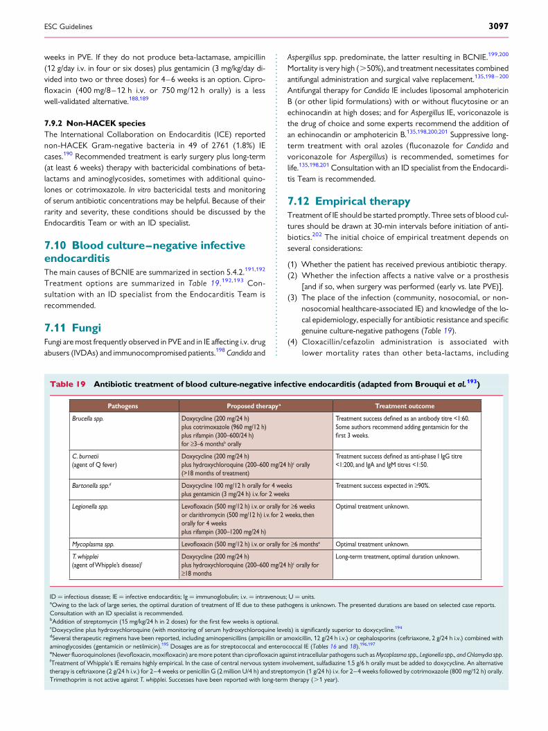
weeks in PVE. If they do not produce beta-lactamase, ampicillin(12 g/day i.v. in four or six doses) plus gentamicin (3 mg/kg/day di-vided into two or three doses) for 4–6 weeks is an option. Cipro-floxacin (400 mg/8–12 h i.v. or 750 mg/12 h orally) is a lesswell-validated alternative.188,189
7.9.2 Non-HACEK speciesThe International Collaboration on Endocarditis (ICE) reportednon-HACEK Gram-negative bacteria in 49 of 2761 (1.8%) IEcases.190 Recommended treatment is early surgery plus long-term(at least 6 weeks) therapy with bactericidal combinations of beta-lactams and aminoglycosides, sometimes with additional quino-lones or cotrimoxazole. In vitro bactericidal tests and monitoringof serum antibiotic concentrations may be helpful. Because of theirrarity and severity, these conditions should be discussed by theEndocarditis Team or with an ID specialist.
7.10 Blood culture–negative infectiveendocarditisThe main causes of BCNIE are summarized in section 5.4.2.191,192
Treatment options are summarized in Table 19.192,193 Con-sultation with an ID specialist from the Endocarditis Team isrecommended.
7.11 FungiFungi are most frequently observed in PVE and in IE affecting i.v. drugabusers (IVDAs) and immunocompromised patients.198 Candida and
Aspergillus spp. predominate, the latter resulting in BCNIE.199,200
Mortality is very high (.50%), and treatment necessitates combinedantifungal administration and surgical valve replacement.135,198 – 200
Antifungal therapy for Candida IE includes liposomal amphotericinB (or other lipid formulations) with or without flucytosine or anechinocandin at high doses; and for Aspergillus IE, voriconazole isthe drug of choice and some experts recommend the addition ofan echinocandin or amphotericin B.135,198,200,201 Suppressive long-term treatment with oral azoles (fluconazole for Candida andvoriconazole for Aspergillus) is recommended, sometimes forlife.135,198,201 Consultation with an ID specialist from the Endocardi-tis Team is recommended.
7.12 Empirical therapyTreatment of IE should be started promptly. Three sets of blood cul-tures should be drawn at 30-min intervals before initiation of anti-biotics.202 The initial choice of empirical treatment depends onseveral considerations:
(1) Whether the patient has received previous antibiotic therapy.(2) Whether the infection affects a native valve or a prosthesis
[and if so, when surgery was performed (early vs. late PVE)].(3) The place of the infection (community, nosocomial, or non-
nosocomial healthcare-associated IE) and knowledge of the lo-cal epidemiology, especially for antibiotic resistance and specificgenuine culture-negative pathogens (Table 19).
(4) Cloxacillin/cefazolin administration is associated withlower mortality rates than other beta-lactams, including
Table 19 Antibiotic treatment of blood culture-negative infective endocarditis (adapted from Brouqui et al.193)
Pathogens Proposed therapya Treatment outcome
Brucella spp. Doxycycline (200 mg/24 h)plus cotrimoxazole (960 mg/12 h) plus rifampin (300–600/24 h) for ≥3–6 monthsb orally
Treatment success as an antibody titre <1:60.Some authors recommend adding gentamicin for the
3 weeks.
C. burnetii(agent of Q fever)
Doxycycline (200 mg/24 h) plus hydroxychloroquine (200–600 mg/24 h)c orally(>18 months of treatment)
Treatment success as anti-phase I IgG titre<1:200, and IgA and IgM titres <1:50.
Bartonella spp.d Doxycycline 100 mg/12 h orally for 4 weeks plus gentamicin (3 mg/24 h) i.v. for 2 weeks
Treatment success expected in ≥90%.
Legionella spp. (500 mg/12 h) i.v. or orally for ≥6 weeksor clarithromycin (500 mg/12 h) i.v. for 2 weeks, thenorally for 4 weeks plus rifampin (300–1200 mg/24 h)
Optimal treatment unknown.
Mycoplasma spp. (500 mg/12 h) i.v. or orally for ≥6 monthse Optimal treatment unknown.
T. whipplei (agent of Whipple’s disease)f
Doxycycline (200 mg/24 h) plus hydroxychloroquine (200–600 mg/24 h)c orally for ≥18 months
Long-term treatment, optimal duration unknown.
ID ¼ infectious disease; IE ¼ infective endocarditis; Ig ¼ immunoglobulin; i.v. ¼ intravenous; U ¼ units.aOwing to the lack of large series, the optimal duration of treatment of IE due to these pathogens is unknown. The presented durations are based on selected case reports.Consultation with an ID specialist is recommended.bAddition of streptomycin (15 mg/kg/24 h in 2 doses) for the first few weeks is optional.cDoxycycline plus hydroxychloroquine (with monitoring of serum hydroxychloroquine levels) is significantly superior to doxycycline.194
dSeveral therapeutic regimens have been reported, including aminopenicillins (ampicillin or amoxicillin, 12 g/24 h i.v.) or cephalosporins (ceftriaxone, 2 g/24 h i.v.) combined withaminoglycosides (gentamicin or netilmicin).195 Dosages are as for streptococcal and enterococcal IE (Tables 16 and 18).196,197
eNewer fluoroquinolones (levofloxacin, moxifloxacin) are more potent than ciprofloxacin against intracellular pathogens such as Mycoplasma spp., Legionella spp., and Chlamydia spp.fTreatment of Whipple’s IE remains highly empirical. In the case of central nervous system involvement, sulfadiazine 1.5 g/6 h orally must be added to doxycycline. An alternativetherapy is ceftriaxone (2 g/24 h i.v.) for 2–4 weeks or penicillin G (2 million U/4 h) and streptomycin (1 g/24 h) i.v. for 2–4 weeks followed by cotrimoxazole (800 mg/12 h) orally.Trimethoprim is not active against T. whipplei. Successes have been reported with long-term therapy (.1 year).
ESC Guidelines 3097

amoxicillin/clavulanic acid or ampicillin/sulbactam,203 andvancomycin for empirically treating MSSA bacteraemia/endocarditis.159
Suggested regimens for empirical treatment in acute patientsare summarized in Table 20. NVE and late PVE regimens shouldcover staphylococci, streptococci and enterococci. Early PVE orhealthcare-associated IE regimens should cover methicillin-resistantstaphylococci, enterococci and, ideally, non-HACEK Gram-negativepathogens. Once the pathogen is identified (usually in ,48 h), theantibiotic treatment must be adapted to its antimicrobial susceptibil-ity pattern.
7.13 Outpatient parenteral antibiotictherapy for infective endocarditisOutpatient parenteral antibiotic therapy (OPAT) is used to consoli-date antimicrobial therapy once critical infection-related complica-tions are under control (e.g. perivalvular abscesses, acute HF, septicemboli and stroke).204 – 207 Two different phases may be identifiedduring the course of antibiotic therapy: (i) a first critical phase(the first 2 weeks of therapy), during which OPAT has a restrictedindication; and (ii) a second, continuation phase (beyond 2 weeks oftherapy), where OPAT may be feasible. Table 21 summarizes thesalient questions to address when considering OPAT for IE.205
Table 20 Proposed antibiotic regimens for initial empirical treatment of infective endocarditis in acute severely illpatients (before pathogen identification)a
Antibiotic Dosage and route Classb Levelc Comments
Community-acquired native valves or late prosthetic valves (≥12 months post surgery) endocarditis
Ampicillinwith(Flu)cloxacillin oroxacillinwithGentamicind
12 g/day i.v. in 4–6 doses
12 g/day i.v. in 4–6 doses
3 mg/kg/day i.v. or i.m. in 1dose
IIa C
Patients with BCNIE should be treated in consultation with an ID specialist.
Vancomycind
with
Gentamicind
30–60 mg/kg/day i.v. in 2–3doses
3 mg/kg/day i.v. or i.m. in 1dose
IIb C
For penicillin-allergic patients
Early PVE (<12 months post surgery) or nosocomial and non-nosocomial healthcare associated endocarditis
Vancomycind
withGentamicind
with
Rifampin
30 mg/kg/day i.v. in 2 doses
3 mg/kg/day i.v. or i.m. in 1dose
900–1200 mg i.v. or orallyin 2 or 3 divided doses
IIb C
Rifampin is only recommended for PVE and it should be started 3–5 days later thanvancomycin and gentamicin has been suggested by some experts. In healthcareassociated native valve endocarditis, some experts recommend in settings with aprevalence of MRSA infections .5% the combination of cloxacillin plusvancomycin until they have the final S. aureus identification
BCNIE ¼ blood culture-negative infective endocarditis; ID ¼ infectious disease; i.m. ¼ intramuscular; i.v. ¼ intravenous; PVE ¼ prosthetic valve endocarditis.aIf initial blood cultures are negative and there is no clinical response, consider BCNIE aetiology (see Section 7.10) and maybe surgery for molecular diagnosis and treatment, andextension of the antibiotic spectrum to blood culture-negative pathogens (doxycycline, quinolones) must be considered.bClass of recommendation.cLevel of evidence.dMonitoring of gentamicin or vancomycin dosages is as described in Tables 16 and 17.
Table 21 Criteria that determine suitability ofoutpatient parenteral antibiotic therapy for infectiveendocarditis (adapted from Andrews et al.205)
Phase of treatment
Guidelines for use
Critical phase(weeks 0–2)
• Complications occur during this phase• Preferred inpatient treatment during this phase• Consider OPAT if: oral streptococci or Streptococcus bovis,a native valve,b patient stable, no complications
Continuation phase(beyond week 2)
• Consider OPAT if medically stable• Do not consider OPAT if: HF, concerning echocardiographic features, neurological signs, or renal impairment
Essential for OPAT
• Educate patient and staff• Regular post-discharge evaluation (nurses 1/day, physicianc in charge 1 or 2/week)d
• Prefer physician-directed programme, not home- infusion model
HF ¼ heart failure; ID ¼ infectious disease; IE ¼ infective endocarditis; OPAT ¼outpatient parenteral antibiotic therapy; PVE ¼ prosthetic valve endocarditis.aFor other pathogens, consultation with an ID specialist is recommended.bFor patients with late PVE, consultation with an ID specialist is recommended.cPreferably from the Endocarditis Team.dGeneral physician can see the patient once a week, if needed.
ESC Guidelines3098

8. Main complications of left-sidedvalve infective endocarditis andtheir managementSurgical treatment is required in approximately half of the patientswith IE because of severe complications.54 Reasons to considerearly surgery in the active phase (i.e. while the patient is still receiv-ing antibiotic treatment) are to avoid progressive HF and irrevers-ible structural damage caused by severe infection and to preventsystemic embolism.6,54,115,208 –210 On the other hand, surgical ther-apy during the active phase of the disease is associated with signifi-cant risk. Surgery is justified in patients with high-risk features thatmake the possibility of cure with antibiotic treatment unlikely andwho do not have co-morbid conditions or complications thatmake the prospect of recovery remote. Age per se is not a contra-indication to surgery.211
Early consultation with a cardiac surgeon is recommended in or-der to determine the best therapeutic approach. Identification of pa-tients requiring early surgery is frequently difficult and is an
important objective of the ‘Heart Team’. Each case must be indivi-dualized and all factors associated with increased risk identified atthe time of diagnosis. Frequently the need for surgery will be deter-mined by a combination of several high-risk features.211
In some cases, surgery needs to be performed on an emergency(within 24 h) or urgent (within a few days, ,7 days) basis, irrespect-ive of the duration of antibiotic treatment. In other cases, surgerycan be postponed to allow 1 or 2 weeks of antibiotic treatment un-der careful clinical and echocardiographic observation before anelective surgical procedure is performed.63,115 The three main indi-cations for early surgery in IE are HF, uncontrolled infection andprevention of embolic events212– 216 (Table 22).
8.1 Heart failure8.1.1 Heart failure in infective endocarditisHF is the most frequent complication of IE and represents the mostcommon indication for surgery in IE.54 HF is observed in 42–60% ofcases of NVE and is more often present when IE affects the aorticrather than the mitral valve.111,208,212 HF is mainly caused by new
Table 22 Indications and timing of surgery in left-sided valve infective endocarditis (native valve endocarditis andprosthetic valve endocarditis)
Indications for surgery Timinga Classb Levelc Ref.d
1. Heart failure
Aortic or mitral NVE or PVE with severe acute regurgitation, obstruction or fistula causing refractorypulmonary oedema or cardiogenic shock
EmergencyI B
111,115,
213,216
Aortic or mitral NVE or PVE with severe regurgitation or obstruction causing symptoms of HF orechocardiographic signs of poor haemodynamic tolerance
UrgentI B
37,115,
209,216,
220,221
2. Uncontrolled infection
Locally uncontrolled infection (abscess, false aneurysm, fistula, enlarging vegetation) UrgentI B
37,209,
216
Infection caused by fungi or multiresistant organisms Urgent/elective
I C
Persisting positive blood cultures despite appropriate antibiotic therapy and adequate control ofseptic metastatic foci
UrgentIIa B 123
PVE caused by staphylococci or non-HACEK gram-negative bacteria Urgent/elective
IIa C
3. Prevention of embolism
Aortic or mitral NVE or PVE with persistent vegetations .10 mm after one or more embolicepisode despite appropriate antibiotic therapy
UrgentI B
9,58,72,
113,222
Aortic or mitral NVE with vegetations .10 mm, associated with severe valve stenosis orregurgitation, and low operative risk
UrgentIIa B 9
Aortic or mitral NVE or PVE with isolated very large vegetations (.30 mm) Urgent IIa B 113
Aortic or mitral NVE or PVE with isolated large vegetations (.15 mm) and no other indication forsurgerye
UrgentIIb C
HACEK ¼ Haemophilus parainfluenzae, Haemophilus aphrophilus, Haemophilus paraphrophilus, Haemophilus influenzae, Actinobacillus actinomycetemcomitans, Cardiobacterium hominis,Eikenella corrodens, Kingella kingae and Kingella denitrificans; HF ¼ heart failure; IE ¼ infective endocarditis; NVE ¼ native valve endocarditis; PVE ¼ prosthetic valve endocarditis.aEmergency surgery: surgery performed within 24 h; urgent surgery: within a few days; elective surgery: after at least 1–2 weeks of antibiotic therapy.bClass of recommendation.cLevel of evidence.dReference(s) supporting recommendations.eSurgery may be preferred if a procedure preserving the native valve is feasible.
ESC Guidelines 3099

or worsening severe aortic or mitral regurgitation, although intra-cardiac fistulae213 and, more rarely, valve obstruction may alsolead to HF.
Valvular regurgitation in native IE may occur as a result of mitralchordal rupture, leaflet rupture (flail leaflet), leaflet perforation orinterference of the vegetation mass with leaflet closure. A particularsituation is infection of the anterior mitral leaflet secondary to an in-fected regurgitant jet of a primary aortic IE.214 Resultant aneurysmformation on the atrial side of the mitral leaflet may later lead tomitral perforation.215
Clinical presentation of HF may include dyspnoea, pulmonary oe-dema and cardiogenic shock.111,120 Among the large ICE Prospect-ive Cohort Study patients with HF and IE, 66% were in New YorkHeart Association class III or IV.216 In addition to clinical findings,TTE is of crucial importance for initial evaluation and follow-up.64
Valve perforation, secondary mitral lesions and aneurysms arebest assessed using TOE.64,65,214 Echocardiography is also usefulto evaluate the haemodynamic consequences of valvular dysfunc-tion, measurement of pulmonary artery pressure, detection of peri-cardial effusion and assessment and monitoring of left ventricularsystolic function and left and right heart filling pressures.64 B-typenatriuretic peptide has potential use in the diagnosis and monitoringof HF in IE.217 Both elevated levels of cardiac troponins and B-typenatriuretic peptide are associated with adverse outcomes inIE.218,219 Moderate to severe HF is the most important predictorof in-hospital, 6-month and 1-year mortality.52,109,111,117,208
8.1.2 Indications and timing of surgery in the presence ofheart failure in infective endocarditis (Table 22)Identification of surgical candidates and timing of surgery decisionsshould preferably be made by the Endocarditis Team.118 The pres-ence of HF indicates surgery in the majority of patients with IE and isthe principal indication for urgent surgery.115,124 Surgery is indicatedin patients with HF caused by severe aortic or mitral regurgitation,intracardiac fistulae or valve obstruction caused by vegetations. Sur-gery is also indicated in patients with severe acute aortic or mitralregurgitation without clinical HF but with echocardiographic signsof elevated left ventricular end-diastolic pressure (e.g. prematureclosure of the mitral valve), high left atrial pressure or moderateto severe pulmonary hypertension. These rules apply in both NVEand PVE.37,220,221
Surgery must be performed on an emergency basis, irrespectiveof the status of infection, when patients are in persistent pul-monary oedema or cardiogenic shock despite medical therapy.63
Surgery must be performed on an urgent basis when HF is lesssevere. Urgent surgery should also be performed in patientswith severe aortic or mitral insufficiency with large vegetations,even without HF.9
In patients with well-tolerated (New York Heart Associationclass I or II) severe valvular regurgitation and no other reasons forsurgery, medical management with antibiotics under strict clinicaland echocardiographic observation is a good option, although earlysurgery may be an option in selected patients at low risk for surgery.Elective surgery should be considered depending on the toleranceof the valve lesion and according to the recommendations of theESC Guidelines on the management of valvular heart disease.55
In summary, HF is the most frequent and among the most severecomplications of IE. Unless severe co-morbidity exists, the pres-ence of HF is an indication for early surgery in NVE and PVE,even in patients with cardiogenic shock.
8.2 Uncontrolled infectionUncontrolled infection is one of the most feared complications of IEand is the second most frequent cause for surgery.54 Uncontrolledinfection is considered to be present when there is persisting infec-tion and when there are signs of locally uncontrolled infection. Infec-tion due to resistant or very virulent organisms often results inuncontrolled infection.
8.2.1 Persisting infectionThe definition of persisting infection is arbitrary and consists of feverand persisting positive cultures after 7–10 days of antibiotic treat-ment. Persisting fever is a frequent problem observed during treat-ment of IE. Usually, temperature normalizes within 7–10 days underspecific antibiotic therapy. Persisting fever may be related to severalfactors, including inadequate antibiotic therapy, resistant organisms,infected lines, locally uncontrolled infection, embolic complicationsor extracardiac site of infection and adverse reaction to antibiotics.3
Management of persisting fever includes replacement of i.v. lines, re-peat laboratory measurements, blood cultures, echocardiography,and the search for an intracardiac or extracardiac focus of infection.
8.2.2 Perivalvular extension in infective endocarditisPerivalvular extension of IE is the most frequent cause of uncon-trolled infection and is associated with a poor prognosis and highlikelihood of the need for surgery. Perivalvular complications in-clude abscess formation, pseudoaneurysms and fistulae (defined inTable 11).223,224
Perivalvular abscess is more common in aortic IE (10–40% inNVE)3,225 – 227 and is frequent in PVE (56–100%).3,6 In mitral IE, peri-valvular abscesses are usually located posteriorly or laterally.228 Inaortic IE, perivalvular extension occurs most frequently in themitral-aortic intervalvular fibrosa.229 Serial echocardiographic stud-ies have shown that abscess formation is a dynamic process, startingwith aortic root wall thickening and extending to the developmentof fistulae.229 In one study, the most important risk factors for peri-valvular complications were prosthetic valve, aortic location and in-fection with CoNS.230
Pseudoaneurysms and fistulae are severe complications of IE andare frequently associated with very severe valvular and perivalvulardamage.213,231 – 233 The frequency of fistula formation in IE has beenreported to be 1.6%, with S. aureus being the most commonly asso-ciated organism (46%).233
Despite high rates of surgery in this population (87%), hospitalmortality remains high (41%).213,233,234 Other complications dueto major extension of infection are less frequent and may includeventricular septal defect, third-degree atrio-ventricular block andacute coronary syndrome.223,224,234
Perivalvular extension should be suspected in cases with persist-ent unexplained fever or new atrio-ventricular block. Therefore anelectrocardiogram should be performed frequently during continu-ing treatment, particularly in aortic IE. TOE, MSCT and PET/CT103
are particularly useful for the diagnosis of perivalvular complications,
ESC Guidelines3100
while the sensitivity of TTE is ,50%225– 228 (see section 5). Indeed,perivalvular extension is frequently discovered on a systematic TOE.However, small abscesses can be missed, even using TOE, particu-larly those in a mitral location when there is co-existent annularcalcification.101
8.2.3 Indications and timing of surgery in the presence ofuncontrolled infection in infective endocarditis (Table 22)The results of surgery when the reason for the procedure is uncon-trolled infection are worse than when surgery is performed forother reasons.124,235
8.2.3.1 Persistent infectionIn some cases of IE, antibiotics alone are insufficient to eradicate theinfection. Surgery has been indicated when fever and positive bloodcultures persist for several days (7–10 days) despite an appropriateantibiotic regimen and when extracardiac abscesses (splenic, verte-bral, cerebral or renal) and other causes of fever have beenexcluded. However, the best timing for surgery in this difficult situ-ation is unclear. Recently it has been demonstrated that persistentblood cultures 48–72 h after initiation of antibiotics are an independ-ent risk factor for hospital mortality.123 These results suggest that sur-gery should be considered when blood cultures remain positive after3 days of antibiotic therapy, after the exclusion of other causes of per-sistent positive blood cultures (adapted antibiotic regimen).
8.2.3.2 Signs of locally uncontrolled infectionSigns of locally uncontrolled infection include increasing vegetationsize, abscess formation, false aneurysms, and the creation of fistu-lae.213,236,237 Persistent fever is also usually present and surgery isrecommended as soon as possible. Rarely when there are no otherreasons for surgery and fever is easily controlled with antibiotics,small abscesses or false aneurysms can be treated conservatively un-der close clinical and echocardiographic follow-up.
8.2.3.3 Infection by microorganisms at low likelihood of being controlledby antimicrobial therapySurgery is indicated in fungal IE,238,239 in cases of multiresistant or-ganisms (e.g. MRSA or vancomycin-resistant enterococci) or in therare infections caused by Gram-negative bacteria. Surgery shouldalso be considered in PVE caused by staphylococci or non-HACEKGram-negative bacteria. In NVE caused by S. aureus, surgery is indi-cated if a favourable early response to antibiotics is notachieved161,240,241 (Table 22). Finally, surgery should be performedin patients with PVE and S. aureus infection.
In summary, uncontrolled infection is most frequently related toperivalvular extension or ‘difficult-to-treat’ organisms. Unless se-vere co-morbidity exists, the presence of locally uncontrolled in-fection is an indication for early surgery in patients with IE.
8.3 Prevention of systemic embolism8.3.1 Embolic events in infective endocarditisEmbolic events are a frequent and life-threatening complication of IErelated to the migration of cardiac vegetations. The brain and spleenare the most frequent sites of embolism in left-sided IE, while pul-monary embolism is frequent in native right-sided and pacemakerlead IE. Stroke is a severe complication and is associated with
increased morbidity and mortality.105 Conversely, embolic eventsmay be totally silent in 20–50% of patients with IE, especially thoseaffecting the splenic or cerebral circulation, and can be diagnosed bynon-invasive imaging.83,85,242 Thus systematic abdominal and cere-bral CT scanning may be helpful. However, contrast media shouldbe used with caution in patients with renal impairment or haemo-dynamic instability because of the risk of worsening renal impair-ment in combination with antibiotic nephrotoxicity.
Overall, embolic risk is very high in IE, with embolic events occur-ring in 20–50% of patients.72,242 – 249 However, the risk of newevents (occurring after initiation of antibiotic therapy) is only 6–21%.72,115,243 A study from the ICE group250 demonstrated thatthe incidence of stroke in patients receiving appropriate antimicro-bial therapy was 4.8/1000 patient-days in the first week of therapy,falling to 1.7/1000 patient-days in the second week, and furtherthereafter.
8.3.2 Predicting the risk of embolismEchocardiography plays a key role in predicting embolicevents,72,115,246 –252 although prediction remains difficult in the indi-vidual patient. Several factors are associated with increased risk ofembolism, including the size and mobility of vegetations,72,242,246–253
the location of the vegetation on the mitral valve,72,246 – 249 the in-creasing or decreasing size of the vegetation under antibiotic ther-apy,72,253 particular microorganisms (S. aureus,72 S. bovis,254 Candidaspp.), previous embolism,72 multivalvular IE246 and biological mar-kers.255 Among these, the size and mobility of the vegetations arethe most potent independent predictors of a new embolic event.253
Patients with vegetations .10 mm in length are at higher risk of em-bolism,58,253 and this risk is even higher in patients with larger(.15 mm) and mobile vegetations, especially in staphylococcal IEaffecting the mitral valve.219 A recent study113 found that the riskof neurological complications was particularly high in patients withvery large (.30 mm length) vegetations.
Several factors should be taken into account when assessing em-bolic risk. In a recent study of 847 patients with IE, the 6-month in-cidence of new embolism was 8.5%.222 Six factors (age, diabetes,atrial fibrillation, previous embolism, vegetation length andS. aureus infection) were associated with an increased embolic riskand were used to create an ‘embolic risk calculator’.222
Whatever the risk factors observed in an individual patient, it mustbe re-emphasized that the risk of new embolism is highest during thefirst days following initiation of antibiotic therapy and rapidly de-creases thereafter, particularly beyond 2 weeks,58,72,243,250 althoughsome risk persists indefinitely while vegetations remain present, par-ticularly for very large vegetations.113 For this reason, the benefits ofsurgery to prevent embolism are greatest during the first 2 weeks ofantibiotic therapy, when embolic risk peaks.
8.3.3 Indications and timing of surgery to preventembolism in infective endocarditis (Table 22)Avoiding embolic events is difficult since the majority occur beforeadmission.222 The best means to reduce the risk of an embolic eventis the prompt institution of appropriate antibiotic therapy.38 Whilepromising,256,257 the addition of antiplatelet therapy did not reducethe risk of embolism in the only published randomized study.258
ESC Guidelines 3101

The exact role of early surgery in preventing embolic events re-mains controversial. In the Euro Heart Survey, vegetation size wasone of the reasons for surgery in 54% of patients with NVE and in25% of those with PVE,54 but was rarely the only reason. The valueof early surgery in an isolated large vegetation is controversial. A re-cent randomized trial demonstrated that early surgery in patientswith large vegetations significantly reduced the risk of death and em-bolic events compared with conventional therapy.9 However, thepatients studied were at low risk and there was no significant differ-ence in all-cause mortality at 6 months in the early surgery andconventional-treatment groups.
Finally, the decision to operate early for prevention of embolismmust take into account the presence of previous embolic events,other complications of IE, the size and mobility of the vegetation,the likelihood of conservative surgery and the duration of antibiotictherapy.115 The overall benefits of surgery should be weighedagainst the operative risk and must consider the clinical status andco-morbidity of the patient.
The main indications and timing of surgery to prevent embolismare given in Table 22. Surgery is indicated in patients with persistingvegetations .10 mm after one or more clinical or silent embolicevents despite appropriate antibiotic treatment.58 Surgery may beconsidered in patients with large (.15 mm) isolated vegetationson the aortic or mitral valve, although this decision is more difficultand must be very carefully individualized according to the probabil-ity of conservative surgery.58
Surgery undertaken for the prevention of embolism must be per-formed very early, during the first few days following initiation ofantibiotic therapy (urgent surgery), as the risk of embolism is highestat this time.58,72
In summary, embolism is very frequent in IE, complicating 20–50% of cases of IE, but falling to 6–21% after initiation of antibiot-ic therapy. The risk of embolism is highest during the first 2 weeksof antibiotic therapy and is clearly related to the size and mobilityof the vegetation, although other risk factors exist. The decisionto operate early to prevent embolism is always difficult and spe-cific for the individual patient. Governing factors include the sizeand mobility of the vegetation, previous embolism, type of micro-organism and duration of antibiotic therapy.
9. Other complications of infectiveendocarditis
9.1 Neurological complicationsSymptomatic neurological complications occur in 15–30% of patientswith IE and are mainly the consequence of embolism from vegeta-tions.110,113,259 Neurological manifestations occur before or at IE diag-nosis in a majority of cases, but new or recurrent events can also takeplace later in the course of IE. Clinical presentation is variable and mayinclude multiple symptoms or signs in the same patient, but focal signspredominate and ischaemic strokes are most commonly diagnosed.Transient ischaemic attack, intracerebral or subarachnoidal haemor-rhage, brain abscess, meningitis and toxic encephalopathy are alsoseen, and firm evidence supports that additional clinically silent cere-bral embolisms occur in 35–60% of IE patients.83,85,90 S. aureus IE ismore frequently associated with neurological complications compared
with IE caused by other bacteria. Vegetation length and mobility alsocorrelate with embolic tendency.88,242 Neurological complications areassociated with an excess mortality, as well as sequelae, particularly inthe case of stroke.113,259 Rapid diagnosis and initiation of appropriateantibiotics are of major importance to prevent a first or recurrentneurological complication.250 Early surgery in high-risk patients is thesecond mainstay of embolism prevention, while antithrombotic drugshave no role (see section 12.7).
Successful management of IE requires a combined medical andsurgical approach in a substantial proportion of patients. Followinga neurological event, the indication for cardiac surgery often remainsor is strengthened, but must be balanced with perioperative risk andpostoperative prognosis. Randomized studies are not possible andcohort studies suffer from bias that can only be partly compensatedfor by statistical methods.115,260 – 262 However, the risk of post-operative neurological deterioration is low after a silent cerebralemboli or transient ischaemic attack, and surgery is recommendedwithout delay if an indication remains.105 After an ischaemic stroke,cardiac surgery is not contraindicated unless the neurological progno-sis is judged too poor.263 Evidence regarding the optimal time intervalbetween stroke and cardiac surgery is conflicting, but recent data fa-vour early surgery.9,115 If cerebral haemorrhage has been excluded bycranial CT and neurological damage is not severe (i.e. coma), surgeryindicated for HF, uncontrolled infection, abscess or persistent highembolic risk should not be delayed and can be performed with alow neurological risk (3–6%) and good probability of completeneurological recovery.105,263 Conversely, in cases with intracranialhaemorrhage, neurological prognosis is worse and surgery shouldgenerally be postponed for at least 1 month,264,265 although one re-cent study has reported a relatively low risk of neurological deterior-ation in IE patients undergoing surgery within 2 weeks after anintracranial haemorrhage.266 The Task Force has thus decided toadapt the level of evidence to a class IIa. If urgent cardiac surgery isneeded, close cooperation with the neurosurgical team and theEndocarditis Team is mandatory. Table 23 and Figure 4 summarizethe recommended management of neurological complications in IE.
Cerebral imaging is mandatory for any suspicion of neurologicalcomplication of IE. CT scanning, with or without contrast agent, ismost often performed. The higher sensitivity of MRI, with or with-out contrast gadolinium enhancement, allows for better detectionand analysis of cerebral lesions in patients with neurological symp-toms, and this may have an impact on the timing of surgery89 (seesection 5). In patients without neurological symptoms, cerebralMRI often detects lesions that may change the therapeutic strategy;in particular, the indications and timing of surgery.85,90 Cerebral MRIoften detects microbleeds (round T2* hypointensities with a diam-eter ≤10 mm) in patients with IE. The lack of association with par-enchymal haemorrhage and the absence of postoperativeneurological complications in patients with microbleeds suggestthat microbleeds should not be interpreted as active bleeding andshould not lead to postponed surgery when this is indicated.89,90
In summary, symptomatic neurological events develop in 15–30%of all patients with IE and additional silent events are frequent.Stroke (ischaemic and haemorrhagic) is associated with excessmortality. Rapid diagnosis and initiation of appropriate antibioticsare of major importance to prevent a first or recurrent neurologicalcomplication. After a first neurological event, cardiac surgery, if
ESC Guidelines3102

indicated, is generally not contraindicated, except when extensivebrain damage or intracranial haemorrhage is present.
9.2 Infectious aneurysmsInfectious (mycotic) aneurysms result from septic arterial embolismto the intraluminal space or vasa vasorum or from subsequent spreadof infection through the intimal vessels. Infectious aneurysms are typ-ically thin walled and friable and, as such, exhibit a high tendency torupture and haemorrhage. No predictor of rupture has been identi-fied and, in contrast to non-infectious aneurysms, size does not ap-pear to be a reliable predictor of potential rupture.268,269
An intracranial location is most common and the reported fre-quency of 2–4% is probably an underestimation since some infec-tious aneurysms are clinically silent.267,270 Early detection andtreatment of infectious aneurysms is essential given the high morbid-ity and mortality rate secondary to rupture. Clinical presentation ishighly variable (i.e. focal neurological deficit, headache, confusion,seizures), so imaging should be systematically performed to detectintracranial infectious aneurysms in any case of IE with neurologicalsymptoms.268
Cerebral CT and MRI both reliably diagnose infectious aneurysmswith good sensitivity and specificity.271 However, conventional angi-ography remains the gold standard and should be performed whennon-invasive techniques are negative and suspicion remains.267
Owing to the lack of randomized trials, there is no widely acceptedstandard management for infectious aneurysms. Thus managementshould be provided by an Endocarditis Team and tailored to the indi-vidual patient. Some infectious aneurysms may resolve during anti-biotic treatment, while others require surgical or endovascularintervention depending on the occurrence of rupture and the loca-tion in the artery bed, as well as the clinical status of the patient.268,269
Regarding intracranial infectious aneurysms, ruptured aneurysmsmust be treated immediately by surgical or endovascular procedures.Unruptured infectious aneurysms should be followed by serial cere-bral imaging under antibiotic therapy. If the size of the aneurysm de-creases or resolves completely, surgical or endovascular interventionis usually unnecessary. However, if the size of the aneurysm increasesor remains unchanged, it is likely that the patient will require interven-tion. On the other hand, if the infectious aneurysm is voluminous andsymptomatic, neurosurgery or endovascular therapy is recom-mended.272 Finally, if early cardiac surgery is required, preoperativeendovascular intervention might be considered before the proced-ure, depending on associated cerebral lesions, the haemodynamic sta-tus of the patient and the risk of the procedure.
9.3 Splenic complicationsSplenic infarcts are common and very often asymptomatic. Persist-ent or recurrent fever, abdominal pain and bacteraemia suggest thepresence of complications (splenic abscess or rupture). Although
Neurological complication
• Heart failure• Uncontrolled infection• Abscess• High embolic risk
• Intracranial haemorrhage• Coma• Severe comorbilities• Stroke with severe damage
No
Yes
Yes
No
Consider surgery(see Table 22)
Conservative treatmentand monitoring
• Clincal assessment• Cerebral CT scan / MRI• TTE / TOE
CT = computed tomography; IE = infective endocarditis; MRI = magnetic resonance imaging; TOE = transoesophageal echocardiography; TTE = transthoracic echocardiography.
Figure 4 Therapeutic strategies for patients with infective endo-carditis and neurological complications.
Table 23 Management of neurologicalcomplications of infective endocarditis
Recommendations Classa Levelb Ref.c
After a silent embolism or transientischaemic attack, cardiac surgery, ifindicated, is recommended withoutdelay
I B105,
263
Neurosurgery or endovascular therapyis recommended for very large, enlargingor ruptured intracranial infectiousaneurysms
I C
Following intracranial haemorrhage,surgery should generally be postponedfor ≥1 month
IIa B 264–266
After a stroke, surgery indicated for HF,uncontrolled infection, abscess, orpersistent high embolic risk should beconsidered without any delay as long ascoma is absent and the presence ofcerebral haemorrhage has beenexcluded by cranial CT or MRI
IIa B 9,263
Intracranial infectious aneurysms shouldbe looked for in patients with IE andneurological symptoms. CT or MRangiography should be considered fordiagnosis. If non-invasive techniques arenegative and the suspicion ofintracranial aneurysm remains,conventional angiography should beconsidered
IIa B267,
268
CT ¼ computed tomography; HF ¼ heart failure; IE ¼ infective endocarditis;MR ¼ magnetic resonance; MRI ¼ magnetic resonance imaging.aClass of recommendation.bLevel of evidence.cReference(s) supporting recommendations.
ESC Guidelines 3103

splenic emboli are common, splenic abscesses are rare. Persistent orrecurrent fever and bacteraemia suggest the diagnosis. These pa-tients should be evaluated by abdominal CT, MRI or ultrasound. Re-cently PET has proved useful for the diagnosis of splenic metastasicinfection in patients with IE.273 Treatment consists of appropriateantibiotic regimens. Splenectomy may be considered for splenicrupture or large abscesses, which respond poorly to antibioticsalone, and should be performed before valvular surgery unless thelatter is urgent. Rarely, splenectomy and valvular surgery are per-formed during the same operative time. Percutaneous drainage isan alternative for high-risk surgical candidates.274,275
9.4 Myocarditis and pericarditisCardiac failure may be due to myocarditis, which is frequently asso-ciated with abscess formation or immune reaction. Ventricular arrhyth-mias may indicate myocardial involvement and imply a poor prognosis.Myocardial involvement is best assessed using TTE and cardiac MRI.
The inflammatory response, HF, periannular complications or in-fection itself can cause pericardial effusion, which could be a sign ofmore severe IE. Rarely, ruptured pseudoaneurysms or fistulae maycommunicate with the pericardium, with dramatic and often fatalconsequences. Purulent pericarditis is rare and may necessitate sur-gical drainage.276,277
9.5 Heart rhythm and conductiondisturbancesConduction disorders are uncommon complications of IE. Accord-ing to data from patient registries, their frequency is between 1%and 15% of cases and their presence is associated with worse prog-nosis and higher mortality.278
Conduction abnormalities (mainly first-, second-, and third-degree atrio-ventricular blocks, rarely bundle branch blocks) aredue to spread of the infection beyond the endocardium, from valvesto the conduction pathways, and are generally associated with peri-valvular complications. Complete atrio-ventricular block is most of-ten associated with involvement of the left-sided valves (aortic, 36%;mitral, 33%).278 This is because of the anatomical relationship withthe atrio-ventricular node, which is close to the non-coronary aorticcusp and the anterior mitral leaflet. In a study of patients with IE andcomplete atrio-ventricular block, pathology workup revealed thepresence of an infection, frequently accompanied by abscessesand fistulae, affecting the conduction pathways; in cases of paroxys-mal atrio-ventricular block, inflammation was observed at this level,which would explain the reversibility of the event.279
The occurrence of conduction abnormalities during electrocar-diographic monitoring in patients with endocarditis can thereforealert physicians to the appearance of perivalvular complications.
In the case of embolization of vegetation fragments into a coron-ary artery, the resulting myocardial ischaemia can be the substratefor the onset of tachyarrhythmias.280
Atrial fibrillation can be observed in patients with IE and may bepresent before IE or occur as a complication of IE. Atrial fibrillationhas been reported to be more frequent in the elderly and to be asso-ciated with a poor prognosis.281 More recently, in a large prospectiveseries of IE, atrial fibrillation was found to be associated with an
increased embolic risk, as were other factors (age, diabetes, previousembolism, vegetation length and S. aureus infection).222 Consequently,atrial fibrillation has the potential to increase the risk of both congest-ive HF and embolism in IE. However, there is no specific study on thissituation and no international consensus for the care of these patients.The management of anticoagulation therapy in these patients shouldbe taken on an individual basis by the Endocarditis Team.
9.6 Musculoskeletal manifestationsMusculoskeletal symptoms (arthralgia, myalgia, back pain) are fre-quent during IE.282,283 Rheumatological manifestations may be thefirst manifestations of IE and can delay its diagnosis, especiallywhen classic manifestations are less evident and a variety of anti-bodies (i.e. positive antineutrophil cytoplasmic antibody test) in-duced by infections284,285 are present. Arthralgia occurs in about10% of patients, while myalgia is present in 12–15%.282,286 Backpain is observed in about 13% of cases, and lumbar pain is themost common symptom in patients with IE and vertebral osteomye-litis.282,283,287,288 Peripheral arthritis occurs in about 14% ofcases.282 The prevalence of spondylodiscitis in patients with IE isabout 1.8–15%.282 Pyogenic vertebral osteomyelitis occurs in4.6–19% of IE patients with a high incidence of streptococcal andstaphylococcal bacteraemia.283,287 IE can complicate or be compli-cated by pyogenic osteomyelitis. The prevalence of IE in vertebralosteomyelitis is higher288,289 in the presence of Streptococcus viridansIE. CT, but preferably MRI, of the spine or whole-body 18F-FDG-PET/CT290 should be performed in IE patients with back or bonepain. Conversely, echocardiography should be performed inpatients with a definite diagnosis of pyogenic spondylodiscitis/osteomyelitis and underlying cardiac conditions predisposing to IE.
In definite spondylodiscitis and osteomyelitis, prolonged antibiot-ic therapy is generally required until no signs of inflammatory activityare detected by 18FDG PET/CT or MRI. Other musculoskeletalmanifestations are less common in IE and include sacroiliitis in about1% of cases, a condition mimicking polymyalgia rheumatica with painand morning stiffness of the shoulders and hips, proximal muscleweakness in about 0.9% of cases and cutaneous leucocytoclastic vas-culitis (purpuric skin lesions) in 3.6% of cases.282,289
9.7 Acute renal failureAcute renal failure is a common complication of IE and may worsenthe prognosis of IE. The onset of renal dysfunction is independentlyassociated with increased risk of in-hospital death291,292 and post-operative events.293
Acute renal dysfunction occurs in about 6–30% of pa-tients.291,292,294,295 Causes are often multifactorial:296,297 (i) immunecomplex and vasculitic glomerulonephritis; (ii) renal infarction,mostly due to septic emboli, occurring at any time during the courseof the disease; (iii) haemodynamic impairment in cases with HF orsevere sepsis or after cardiac surgery; (iv) antibiotic toxicity (acuteinterstitial nephritis), notably related to aminoglycosides, vancomy-cin (synergistic toxicity with aminoglycosides) and even high-dosepenicillin; and (v) nephrotoxicity of contrast agents used for imagingpurposes.
Haemodialysis may be required in some patients with advancedrenal failure and is associated with high mortality.295 Acute renal
ESC Guidelines3104
failure of a milder degree is often reversible.295 To mitigate this com-plication, antibiotic doses should be adjusted for creatinine clear-ance with careful monitoring of serum levels (aminoglycosides andvancomycin). Imaging with nephrotoxic contrast agents should beavoided when possible in patients with haemodynamic impairmentor previous renal insufficiency.
10. Surgical therapy: principlesand methods
10.1 Operative risk assessmentFew studies have evaluated the utility of operative risk scores in thesetting of IE. Although EuroSCORE II is frequently used,298 it was de-veloped and validated predominantly for coronary artery bypassgrafting and valve surgery. Risk scores specific to IE surgery havebeen developed: (i) from the Society of Thoracic Surgeons databaseusing 13 617 patients299 and (ii) an additional NVE risk score from asingle centre using 440 patients by De Feo et al.300 A study com-pared the prognostic utility of these contemporary risk scores formortality and morbidity after IE surgery in 146 patients.301 Here, al-though EuroSCORE II discriminated mortality and postoperativemorbidity (in particular, stroke), the Society of Thoracic Surgeonsendocarditis score and the De Feo et al. score300 performed betterat predicting operative mortality after surgery for active IE. How-ever, the relevance of these findings is limited by the small numberof patients involved. Similar to previous studies, preoperative use ofinotropes or an intra-aortic balloon pump, prior coronary artery by-pass surgery and renal failure requiring dialysis were independentpredictors of operative and long-term mortality.
Finally, although no single operative risk score is perfect, pre-operative assessment of operative risk is of utmost importance.Although the theoretical indications for surgery in IE are clear(Table 22), their practical application relies largely on the clinicalstatus of the patient, the patient’s co-morbidities and the patient’soperative risk.
10.2 Preoperative and perioperativemanagement10.2.1 Coronary angiographyCoronary angiography is recommended according to the ESCGuidelines on the management of valvular heart disease55 in men.40 years, in post-menopausal women and in patients with at leastone cardiovascular risk factor or a history of coronary artery dis-ease. Exceptions arise when there are aortic vegetations that maybe dislodged during catheterization or when emergency surgery isnecessary. In these situations, high-resolution CT may be used torule out significant coronary artery disease in haemodynamicallystable patients.55
10.2.2 Extracardiac infectionIf a primary focus of infection likely to be responsible for IE has beenidentified, it must be eradicated before cardiac surgical interventionunless valve surgery is urgent. In any case, it should be eradicatedbefore the end of antibiotic therapy.
10.2.3 Intraoperative echocardiographyIntraoperative TOE is most useful to determine the exact locationand extent of infection, guide surgery, assess the result and help inearly postoperative follow-up.73
10.3 Surgical approach and techniquesThe two primary objectives of surgery are total removal of infectedtissues and reconstruction of cardiac morphology, including repairor replacement of the affected valve(s).
Where infection is confined to the valve cusps or leaflets, anymethod to repair or replace the valve may be used. However, valverepair is favoured whenever possible, particularly when IE affects themitral or tricuspid valve without significant destruction.302 Perfora-tions in a single valve cusp or leaflet may be repaired with an untreat-ed or glutaraldehyde-treated autologous or bovine pericardialpatch. Isolated or multiple ruptured chordae may be replaced bypolytetrafluoroethylene neo-chordae.
More extensive destruction of a single leaflet or the presence ofan abscess is not necessarily a contraindication for valve repair.302
Rather, intraoperative assessment of the valve after debridementis of paramount importance in order to evaluate whether the re-maining tissue is of sufficient quality to achieve a durable repair.The need for a patch to achieve a competent valve, whether pericar-dial, tricuspid autograft or a flipped-over mitral patch, has not beenassociated with worse results in terms of recurrence of IE or mitralregurgitation when performed by experienced surgeons.303
To avoid paravalvular leaks in complex cases with locally uncon-trolled infection, total excision of infected and devitalized tissueshould be followed by valve replacement and repair of associateddefects to secure valve fixation.304
Mechanical and biological prostheses have similar operative mor-tality.305 Therefore the Task Force does not favour any specific valvesubstitute but recommends a tailored approach for each individualpatient and clinical situation. The use of foreign material should bekept to a minimum. Small abscesses can be closed directly, but largercavities should be allowed to drain into the pericardium orcirculation.
In mitral valve IE, successful valve repair can be achieved by ex-perienced teams in up to 80% of patients, but such results maynot be matched in non-specialist centres.306 Moreover, althoughsurgery may be deferred if control of the infection by antibiotic ther-apy appears evident in the absence of cardiac failure, early operationhas been associated in recent reports with a repair rate of 61–80%and improved in-hospital and long-term survival.209,210,302,303,307 Re-sidual mitral regurgitation should be assessed using intraoperativeTOE. Mitral subannular, annular or supraannular tissue defects arepreferably repaired with autologous or bovine pericardium, a pros-thetic valve then being secured to the reconstructed/reinforced an-nulus, if necessary. The choice of technique depends on the verticalextension of the lesion/tissue defect.308 –310 The use of mitral valvehomografts and pulmonary autografts (Ross II procedure) has beensuggested,311,312 but their application is limited by poor availabilityand difficulty of the surgical technique, and the results have notbeen consistent.
In aortic IE, replacement of the aortic valve using a mechanical orbiological prosthesis is the technique of choice. Nevertheless, in
ESC Guidelines 3105

centres with great expertise, aortic valve repair in IE can be achievedin up to 33% of patients. However, experience with aortic valve re-pair in this setting is still very limited and there is no evidence thatrepair is associated with improved outcomes compared with re-placement.313,314 Owing to their natural biocompatibility, the useof cryopreserved or sterilized homografts has been suggested to re-duce the risk of persistent or recurrent infection, especially in thepresence of annular abscesses.315,316 It is expert opinion and stand-ard strategy in many institutions that the use of a homograft is to befavoured over valve prostheses, particularly in the presence of rootabscess.316,317 However, mechanical prostheses and xenograftshave led to similar results in terms of persistent or recurrent infec-tion and survival if associated with complete debridement of annularabscesses.313,318 Homografts or stentless xenografts may be pre-ferred in PVE or in cases where there is extensive aortic root de-struction with aorto-ventricular discontinuity.315,319 The anteriormitral leaflet of the aortic homograft can be effectively used for re-construction of the outflow tract. A monoblock aorto-mitral homo-graft has been suggested as a surgical option for extensive bivalvularIE.320 In experienced hands, the Ross procedure may be used in chil-dren or adolescents to facilitate growth and in young adults for ex-tended durability.321,322
Cardiac transplantation may be considered in extreme caseswhere repeated operative procedures have failed to eradicate per-sistent or recurrent PVE.323
10.4 Postoperative complicationsPostoperative patient management should follow the usual recom-mendations after valvular surgery324 but should also take into ac-count the specificities of IE. Postoperative follow-up should beparticularly cautious given the in-hospital mortality of patients oper-ated on for acute IE on an emergency or urgent basis, which rangesfrom 10% to 20% in most series,1 and the increased risk of post-operative complications.
Among the most frequent complications are severe coagulopathyrequiring treatment with clotting factors, re-exploration of the chestfor bleeding or tamponade, acute renal failure requiring haemodialy-sis, stroke, low cardiac output syndrome, pneumonia and atrio-ventricular block following radical resection of an aortic root ab-scess with the need for pacemaker implantation.325 A preoperativeelectrocardiogram demonstrating left bundle branch block predictsthe need for a postoperative permanent pacemaker.23 When apatient does not survive surgery, the cause of death is oftenmultifactorial.325
11. Outcome after discharge:follow-up and long-term prognosisFollowing in-hospital treatment, the main complications include re-currence of infection, HF, need for valve surgery and death.57,326,327
11.1 Recurrences: relapses andreinfectionsThe actual risk of recurrence among survivors of IE varies between2% and 6%.57,326 –332 Two main types of recurrence are distinguish-able: relapse and reinfection. Although not systematically
differentiated in the literature, the term ‘relapse’ refers to a repeatepisode of IE caused by the same microorganism, while ‘reinfection’describes an infection caused by a different microorganism.38 Whenthe same species is isolated during a subsequent episode of IE, thereis often uncertainty as to whether the repeat infection is a relapse ofthe initial infection or a new infection (reinfection). In these cases,molecular methods including strain-typing techniques should beemployed.8,38 When these techniques or the identity of both iso-lates is unavailable, the timing of the second episode of IE may beused to distinguish relapse from reinfection. Thus, although variable,the time between episodes is usually shorter for relapse than for re-infection. Generally speaking, a recurrence caused by the same spe-cies within 6 months following the initial infection representsrelapse, whereas later events suggest reinfection.38 For these pur-poses, storage of IE isolates for at least 1 year is recommended.8,38
Factors associated with an increased rate of relapse are listed inTable 24. Relapses are most often due to insufficient duration of ori-ginal treatment, suboptimal choice of initial antibiotics or a persist-ent focus of infection. When the duration of therapy has beeninsufficient or the choice of antibiotic incorrect, relapse should betreated for a further 4–6 weeks depending on the causative micro-organism and its antibiotic susceptibility (remembering that resist-ance may develop in the meantime).
Patients with previous IE are at risk of reinfection,332 and prophy-lactic measures should be very strict. Reinfection is more frequent inIVDAs (especially in the year after the initial episode),332,333 inPVE,334 in patients undergoing chronic dialysis326,332 and in thosewith multiple risk factors for IE.8 Patients with reinfection are athigher risk of death and need for valve replacement.325,332 Paravalv-ular destruction is associated with a higher rate of recurrence and ahigher operative mortality.331 In a large series of surgically managedNVE (358 cases), 21% had paravalvular destruction, and freedomfrom recurrent PVE at 15 years was 78.9%.331
The type of valve implanted has no effect on the risk of recurrentIE.325,331 Aortic valve and root replacement with a prosthetic
Table 24 Factors associated with an increased rate ofrelapse
• Inadequate antibiotic treatment (agent, dose, duration)
• Resistant microorganisms, i.e. Brucella spp., Legionella spp., Chlamydia spp., Mycoplasma spp., Mycobacterium spp., Bartonella spp., Coxiella Burnetii, fungi
• Polymicrobial infection in an IVDA
• Empirical antimicrobial therapy for BCNIE
• Periannular extension
• Prosthetic valve IE
• Persistent metastatic foci of infection (abscesses)
• Resistance to conventional antibiotic regimens
• Positive valve culture
• Persistence of fever at the seventh postoperative day
• Chronic dialysis
BCNIE ¼ blood culture-negative infective endocarditis; IE ¼ infectiveendocarditis; IVDA ¼ intravenous drug abuser.
ESC Guidelines3106

conduit yields results similar to those for homograft rootreplacement.335,336
11.2 Short-term follow-upA first episode of IE should not be seen as an ending once the patienthas been discharged. Residual severe valve regurgitation may de-compensate left ventricular function, or valve deterioration mayprogress despite bacteriological cure, usually presenting with acuteHF. After completion of treatment, recommendations for surgeryfollow conventional guidelines.55 As a consequence of increasingrates of surgery during the active phase of infection, the needfor late valve surgery is low, ranging from 3% to 8% in recentseries.326– 328
Patients should be educated about the signs and symptoms of IEafter discharge. They should be aware that recurrence could occurin IE and that new onset of fever, chills or other signs of infectionmandate immediate evaluation, including procurement of blood cul-tures before empirical use of antibiotics. To monitor the develop-ment of secondary HF, an initial clinical evaluation and baselineTTE should be performed at the completion of antimicrobial ther-apy and repeated serially, particularly during the first year offollow-up.
Clinical follow-up should be done by the Endocarditis Team or bya Heart Valve Clinic specialist.11,337 Regular clinical and echocardio-graphic follow-up should be performed during the first year follow-ing completion of treatment.8,12 This Task Force also recommendsto take blood samples (i.e. white cell count, CRP, etc.), and bloodcultures systematically at the initial visit, and otherwise if there isclinical suspicion.
Good oral health maintenance, preventive dentistry and adviceabout skin hygiene, including tattoos and skin piercing, are manda-tory. Deficiencies in dental surveillance contribute to the continu-ous gradual increase in the incidence of IE.30,337 This increaseunderlines the need for repeating the principles of IE preventionat each follow-up visit.
11.3 Long-term prognosisIn recent series, the crude long-term survival rates after the comple-tion of treatment were estimated to be 80–90% at 1 year, 70–80%at 2 years and 60–70% at 5 years.57,326 – 332 The main predictorsof long-term mortality are older age, co-morbidities, recurrencesand HF, especially when cardiac surgery cannot beperformed.57,327,330
Compared with an age- and sex-matched general population, pa-tients surviving a first episode of IE have a significantly worse sur-vival.57 This excess mortality is especially high within the first fewyears after hospital discharge and can be explained by late complica-tions such as HF, higher risk of recurrences and higher patient vul-nerability.57,329 In fact, most recurrences and late cardiac surgeriesoccurred during this period of time.57,328,329
In summary, recurrences are rare following IE and may be asso-ciated with inadequate initial antibiotic therapy, resistant micro-organisms, persistent focus of infection, i.v. drug abuse andchronic dialysis. Patients with IE must be informed of the risk ofrecurrence and educated about how to diagnose and prevent anew episode of IE. The need for late valve surgery is low.
12. Management of specificsituations
12.1 Prosthetic valve endocarditisPVE is the most severe form of IE and occurs in 1–6% of patientswith valve prostheses,338 with an incidence of 0.3–1.2% per patient-year.216,233,339,340 PVE accounts for 10–30% of all cases of IE341 andaffects mechanical and bioprosthetic valves equally. PVE was ob-served in 16% of cases of IE in a French survey,122 in 26% of casesin the Euro Heart Survey54 and in 20% of 2670 patients with definiteIE in the ICE Prospective Cohort Study.340 PVE is still associatedwith difficulties in diagnosis, determination of the optimal therapeut-ic strategy and poor prognosis.
12.1.1 Definition and pathophysiologyEarly PVE is defined as IE occurring within 1 year of surgery and latePVE as IE occurring beyond 1 year, because of significant differences be-tween the microbiological profiles observed before and after this timepoint.3,342 However, this is an artificial distinction. What is important isnot the time from the valve replacement procedure to the onset of IE,but whether IE is acquired perioperatively and which microorganism isinvolved. A recent large, prospective, multicentre, international registryreported that 37% of PVE cases were associated with nosocomial infec-tion or non-nosocomial healthcare-associated infections in outpatientswith extensive healthcare contact.340
The pathogenesis of PVE differs according to both the type ofcontamination and the type of prosthetic valve. In cases with peri-operative contamination, the infection usually involves the junctionbetween the sewing ring and the annulus, leading to perivalvular ab-scess, dehiscence, pseudo-aneurysms and fistulae.339,343,344 In latePVE, additional mechanisms may exist. For example, in late biopros-thetic PVE, infection is frequently located on the leaflets of the pros-thesis, leading to vegetations, cusp rupture and perforation. PVE hasrecently been reported after transcatheter aortic bioprostheticvalve implantation, which should be managed in the same manneras other prosthetic valves.345,346 The risk of prosthetic valve im-plantation endocarditis increases with the use of orotracheal intub-ation and a self-expandable valve system.
The consequence of PVE is usually new prosthetic regurgitation. Lessfrequently, large vegetations may cause prosthetic valve obstruction,which can be diagnosed by TOE and sometimes by TTE or fluoroscopy.
12.1.2 DiagnosisDiagnosis is more difficult in PVE than in NVE. Clinical presentationis frequently atypical, particularly in the early postoperative period,in which fever and inflammatory syndromes are common in the ab-sence of IE. However, persistent fever should trigger the suspicionof PVE. As in NVE, diagnosis of PVE is based mainly on the results ofechocardiography and blood cultures. However, both are more fre-quently negative in PVE.100 Although TOE is mandatory in suspectedPVE (Figure 3), its diagnostic value is lower than in NVE. A negativeechocardiogram is frequently observed in PVE2 and does not ruleout the diagnosis, but identification of a new periprosthetic leak isa major criterion, in which case an additional imaging modality couldbe considered (such as CT or nuclear imaging).
ESC Guidelines 3107

In PVE, staphylococcal and fungal infections are more frequentand streptococcal infection less frequent than in NVE. Staphylo-cocci, fungi and Gram-negative bacilli are the main causes of earlyPVE, while the microbiology of late PVE mirrors that of NVE, withstaphylococci, oral streptococci, S. bovis and enterococci being themost frequent organisms, more likely due to community-acquiredinfections. Staphyloccoci and enteroccoci are the most commonagents in prosthetic valve implantation endocarditis.345,346
The Duke criteria have been shown to be helpful for the diagnosisof NVE, with a sensitivity of 70–80%,100,347 but are less useful in PVEbecause of their lower sensitivity in this setting.348,349 Recently, nucleartechniques, particularly 18F-FDG PET/CT, have been shown to be use-ful for the diagnosis of PVE.93 The addition of abnormal FDG uptake asa novel major criterion for PVE has thus been pointed out. An algo-rithm for evaluation of patients with suspected PVE, including echocar-diography and PET/CT has been suggested (see Figure 3).93
12.1.3 Prognosis and treatmentA very high in-hospital mortality rate of 20–40% has been reportedin PVE.338,341 As in NVE, prognostic assessment is of crucial im-portance in PVE, as it allows identification of high-risk subgroupsof patients in whom an aggressive strategy may be necessary.Several factors have been associated with poor prognosis inPVE,161,216,350–353 including older age, diabetes mellitus, healthcare-associated infections, staphylococcal or fungal infection, early PVE,HF, stroke and intracardiac abscess. Among these, complicatedPVE and staphylococcal infection are the most powerful markers.These patients need aggressive management, consisting of antibiotictherapy and early radical surgery.
Antimicrobial therapy for PVE is similar to that for NVE. An ex-ception is S. aureus PVE, which requires a more prolonged (≥6weeks) antibiotic regimen (particularly in association with aminogly-cosides) and frequent use of rifampin.
Surgery for PVE follows the general principles outlined for NVE.Radical debridement in these cases means removal of all infectedforeign material, including the original prosthesis, and any calciumremaining from previous surgery. Homografts, stentless xenograftsor autografts may be considered in aortic PVE, and homograft orxenograft root replacement is indicated for any abnormality of theaortic root that distorts the aortic sinuses. Alternatively, a valvedDacron conduit336 can be used.
The best therapeutic option in PVE is still debated.221,354–359 Al-though surgery is generally considered the best option when PVEcauses severe prosthetic dysfunction or HF,220 it was performed inonly 50% of patients with PVE in the Euro Heart Survey,54 a similarrate as for patients with NVE. Other groups have reported similardata.221,340 Early surgery was associated with lower in-hospital and1-year mortality in a large cohort of 4166 patients including both na-tive and prosthetic valve IE complicated by HF.216 Conversely, afteradjustment for differences in clinical characteristics and survivalbias, early valve replacement was not associated with lower mortalitycompared with medical therapy in a large international cohort.37
However, in these series, surgery was beneficial in the subgroup ofpatients with the greatest need for surgery, including valve regurgita-tion, vegetation and dehiscence or paravalvular abscess/fistula.37
Therefore a surgical strategy is recommended for PVE in high-risksubgroups identified by prognostic assessment, i.e. PVE complicated
by HF, severe prosthetic dysfunction, abscess or persistent fever(Table 22). Emergency surgery is indicated only in cases with refrac-tory congestive HF leading to pulmonary oedema or shock, as inNVE. Conversely, patients with uncomplicated non-staphylococcaland non-fungal late PVE can be managed conservatively.350,357,358
However, patients who are initially treated medically require closefollow-up because of the risk of late events.
In summary, PVE represents 20% of all cases of IE, with an increas-ing incidence. The diagnosis of PVE is more difficult than for NVE.Complicated PVE and staphylococcal PVE are associated with aworse prognosis if treated without surgery. These forms of PVEmust be managed aggressively. Patients with uncomplicated, non-staphylococcal late PVE can be managed conservatively withclose follow-up.
12.2 Infective endocarditis affectingcardiac implantable electronic devices12.2.1 IntroductionInfection of cardiac implantable electronic devices (CIEDs) is a severedisease associated with high mortality.360 The increased rates of CIEDimplantation coupled with increased implantation in older patientswith more co-morbidities have set the stage for higher rates ofCIED infection and the increasing frequency of IE in these patients.361
The reported incidence of permanent pacemaker infection varieswidely among studies.362,363 A population-based study found an inci-dence of CIED infection of 1.9 per 1000 device-years and a higherprobability of infection after implantable cardioverter defibrillatorscompared with permanent pacemakers.364 Both diagnosis and thera-peutic strategy are particularly difficult in these patients.365
12.2.2 Definitions of cardiac device infectionsA distinction should be made between local device infection andcardiac device-related IE (CDRIE). Local device infection is definedas an infection limited to the pocket of the cardiac device and is clin-ically suspected in the presence of local signs of inflammation at thegenerator pocket, including erythema, warmth, fluctuance, wounddehiscence, erosion, tenderness or purulent drainage.366 CDRIE isdefined as an infection extending to the electrode leads, cardiacvalve leaflets or endocardial surface. However, differentiating localdevice infection and CDRIE is frequently difficult. In one study,367
culture of intravascular lead segments was positive in 72% of 50 pa-tients with manifestations strictly limited to the implantation site.However, the possibility of intraoperative contamination of thelead tip cannot be excluded in these patients.
12.2.3 PathophysiologyThe pocket may become infected at the time of implantation, duringsubsequent surgical manipulation of the pocket or if the generatoror subcutaneous electrodes erode through the skin. Pocket infec-tion may track along the intravascular portion of the electrode toinvolve the intracardiac portion of the pacemaker or implantablecardioverter defibrillator. Alternatively, the pocket or intracardiacportion of the electrode may become infected as a result of haema-togenous seeding during a bacteraemia secondary to a distant in-fected focus. The consequence may be formation of vegetations,which can be found anywhere from the insertion vein to the super-ior vena cava, on the lead or on the tricuspid valve, as well as on the
ESC Guidelines3108

right atrial and ventricular endocardium. Septic pulmonary embol-ism is a very frequent complication of CDRIE.
12.2.4 Risk factorsSeveral factors have been associated with CIED infections.366,367 Patientfactors include renal failure, corticosteroid use, congestive HF, haema-toma formation, diabetes mellitus and anticoagulation use.368–370 Inaddition, procedural characteristics may also play an important role inthe development of CIED infection. The factors associated withan increased risk of infection include the type of intervention,371,372
device revisions, the site of intervention, the amount of indwellinghardware, the use of pre-procedural temporary pacing, failure to ad-minister perioperative antimicrobial prophylaxis,373 fever within the24 h before implantation and operator experience.374
12.2.5 MicrobiologyStaphylococci, and especially CoNS, account for 60–80% of cases inmost reported series.375,376 A variety of CoNS species have beendescribed.366,377 Methicillin resistance among staphylococci variesamong studies,376,378 but a low frequency of methicillin-resistantCoNS has been reported among individuals with no healthcarecontact, whereas a high rate of methicillin resistance in CoNSis associated with a healthcare environment source.379 Polymicro-bial infection sometimes involves more than one species ofCoNS.376,380,381 Corynebacterium spp., Propionibacterium acnes,Gram-negative bacilli and Candida spp. are rarely identified as patho-gens in CIED infection.366,376,377
12.2.6 DiagnosisClinical presentation is frequently misleading, with predominant re-spiratory and rheumatological symptoms as well as local signs of in-fection.382 CDRIE must be suspected in the presence of unexplainedfever in a patient with a CIED. Fever is frequently blunted, particu-larly in elderly patients. As in other forms of IE, echocardiographyand blood cultures are the cornerstones of diagnosis. S. aureus bac-teraemia might be the sole manifestation of device infection.
Echocardiography plays a key role in CDRIE and is helpful for thediagnosis of both lead vegetations and tricuspid involvement, quanti-fication of tricuspid regurgitation, sizing of vegetations and follow-upafter lead extraction. Several prognostic features may be better de-fined on TTE than on TOE, such as pericardial effusion, ventriculardysfunction and pulmonary vascular pressure estimations. TOE hassuperior sensitivity and specificity to TTE for diagnosis of lead-relatedendocarditis.381–385 TOE allows visualization of the lead in atypical lo-cations, such as the proximal superior vena cava, and of regions thatare difficult to visualize by TTE. In addition, the sensitivity of TOE forleft-sided involvement and for perivalvular extension of infection issuperior to that of TTE. Considering their complementary role, it isrecommended to perform both investigations in suspected CDRIE.
In the presence of infective material along the lead course notproviding typical vegetations of measurable size, both TTE andTOE may be falsely negative in CDRIE. Intracardiac echocardiog-raphy was recently found to be feasible and effective in cardiac de-vice patients386 and to have a superior sensitivity for the detectionof vegetations in cardiac devices.386 – 388
A normal echographic examination does not rule out CDRIE. Indifficult cases, other modalities such as radiolabelled leucocyte
scintigraphy389 and 18F-FDG PET/CT scanning108,390 have been de-scribed as additive tools in the diagnosis of CDRIE and related com-plications, including pulmonary septic embolism.
The Duke criteria are difficult to apply in these patients becauseof lower sensitivity.347 Modifications of the Duke criteria have beenproposed,382,391 including local signs of infection and pulmonaryembolism as major criteria.382
12.2.7 TreatmentCDRIE must be treated by prolonged antibiotic therapy associatedwith complete hardware removal.360,391
12.2.8 Antimicrobial therapyAntimicrobial therapy for CDRIE should be individualized and basedon culture and susceptibility results if possible (see section 7). Be-cause most CDRIE infections are secondary to staphylococcal speciesand, of those, up to 50% are methicillin-resistant,376,392 vancomycinshould be administered initially as empirical antibiotic coverage untilmicrobiological results are known. Daptomycin, approved for right-side IE and bacteraemia attributable to S. aureus,168 is a promising mol-ecule to treat CIED infection.393–395 Before hardware removal, butafter blood cultures, i.v. antibiotics should be initiated. There are noclinical trial data to define the optimal duration of antimicrobial ther-apy. The duration of therapy should be 4–6 weeks in most cases.362
At least 2 weeks of parenteral therapy is recommended after extrac-tion of an infected device for patients with bloodstream infection.Patients with sustained (.24 h) positive blood cultures despiteCIED removal and appropriate antimicrobial therapy should receiveparenteral therapy for at least 4 weeks.362,366
12.2.9 Complete hardware removal (device and leadextraction)In the case of definite CDRIE, medical therapy alone has been asso-ciated with high mortality and risk of recurrence.360,363,391 For thisreason, CIED removal is recommended in all cases of proven CDRIEand should also be considered when CDRIE is only suspected in thecase of occult infection without any apparent source other than thedevice.396
Complete removal of the system is the recommended treatmentfor patients with established CDRIE.363,391,396 Considering the in-herent risk of an open surgical procedure,380 transvenous lead ex-traction has become the preferred method. It is essential toremove all hardware to avoid the recurrence of infection.368,397 Inexperienced centres, procedural mortality rates have been shownto be between 0.1% and 0.6%.396,398 Long-term mortality variesamong subgroups, but rates are higher in systemic infections.399
Transvenous extractions are not without risk, and procedural com-plexity may vary significantly according to lead type and features.Typically ICD leads are more difficult to remove than coronary sinusleads, which are usually removed by simple manual traction.400 – 402
Transvenous lead extraction should be performed only in centrescommitted to a procedural volume allowing the maintenance ofskills of adequately trained teams and able to provide immediate car-diothoracic surgery backup in the event of emergency thoracotomyor sternotomy.396,403
Pulmonary embolism as a result of vegetation displacement dur-ing extraction occurs frequently, particularly when vegetations are
ESC Guidelines 3109

large.367,404 However, these episodes are frequently asymptomatic,and percutaneous extraction remains the recommended methodeven in cases of large vegetations,360,391,404 as overall risks areeven higher with surgical extraction.367,380
Some authors recommend surgery in patients with very large ve-getations.405 Until additional data are available, decisions regardingpercutaneous versus surgical removal of leads with vegetations.2 cm in diameter should be individualized.
Other indications for a surgical approach to lead removal includepatients who need a contemporary valve replacement or repair forIE or patients who have significant retained hardware after attemptsat percutaneous removal. However, mortality associated with surgi-cal removal is high in these frequently elderly patients with asso-ciated co-morbidities.380
12.2.10 ReimplantationThe first step before reimplantation is a re-evaluation of the indica-tion for CIED implantation.377,403 In a significant number of cases,reimplantation is not necessary.366,398 The device should be reim-planted on the contralateral side. There is no clear recommendationconcerning the optimal timing of reimplantation. Factors such aspersistent bacteraemia, persistent vegetation and pacemaker andimplantable cardioverter defibrillator dependency should be con-sidered and the decision adapted to the individual patient. Immedi-ate reimplantation should be avoided, owing to the risk of newinfection.366,377,398,403 Blood cultures should be negative for at least72 h before placement of a new device. In cases of evidence ofremnant valvular infection, implantation should be delayed for atleast 14 days.366,406
Temporary pacing is a risk factor for subsequent cardiac deviceinfection367 and should be avoided if possible. In pacing-dependentpatients, temporary use of active fixation leads connected to exter-nal devices is described as a ‘bridge’,407 permitting earlier mobiliza-tion with a reduced risk of pacing-related adverse events.408 – 410
12.2.11 ProphylaxisAlthough there are no large controlled studies on this topic, anti-biotic prophylaxis is recommended before implantation.367,368,373
A first-generation cephalosporin, such as cefazolin (6 g/day for24–36 h after the intervention), is usually used as prophylaxis andshould be parenterally administered 1 h before the procedure.
Vancomycin, teicoplanin and daptomicin may be considered in-stead of cefazolin in centres where oxacillin resistance amongstaphylococci is high, in high-risk patients or in patients with contra-indications to cephalosporins. They should always be started beforethe procedure according to the drug pharmacokinetics.
In summary, CDRIE is one of the most difficult forms of IE to diag-nose and must be suspected in the presence of frequently mislead-ing symptoms, particularly in elderly patients. Prognosis is poor,probably because of its frequent occurrence in elderly patientswith associated co-morbidities. In the majority of patients, CDRIEmust be treated by prolonged antibiotic therapy and device re-moval. Table 25 summarizes the main features concerning diag-nosis, treatment and prevention of CDRIE.
Table 25 Cardiac device-related infectiveendocarditis: diagnosis, treatment and prevention
Recommendations Classa Levelb Ref.c
A. Diagnosis
1. Three or more sets of bloodcultures are recommended beforeprompt initiation of antimicrobialtherapy for CIED infection
I C
2. Lead-tip culture is indicated whenthe CIED is explanted
I C
3. TOE is recommended in patientswith suspected CDRIE withpositive or negative bloodcultures, independent of theresults of TTE, to evaluatelead-related endocarditis and heartvalve infection
I C
4. Intracardiac echocardiography maybe considered in patients withsuspected CDRIE, positive bloodcultures and negative TTE and TOEresults
IIb C
5. Radiolabelled leucocytescintigraphy and 18F-FDG PET/CTscanning may be consideredadditive tools in patients withsuspected CDRIE, positive bloodcultures and negativeechocardiography
IIb C
B. Principles of treatment
1. Prolonged (i.e. before and afterextraction) antibiotic therapy andcomplete hardware (device andleads) removal are recommendedin definite CDRIE, as well asin presumably isolated pocketinfection
I C
2. Complete hardware removalshould be considered on thebasis of occult infectionwithout another apparent sourceof infection
IIa C
3. In patients with NVE or PVEand an intracardiac device withno evidence of associateddevice infection, completehardware extraction may beconsidered
IIb C
C. Mode of device removal
1. Percutaneous extraction isrecommended in most patientswith CDRIE, even those withvegetations .10 mm
I B382,
391,
405
Continued
ESC Guidelines3110
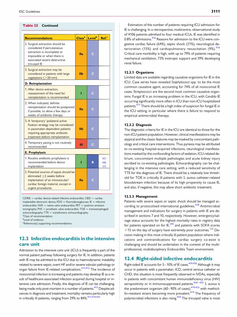
12.3 Infective endocarditis in the intensivecare unitAdmission to the intensive care unit (ICU) is frequently a part of thenormal patient pathway following surgery for IE. In addition, patientswith IE may be admitted to the ICU due to haemodynamic instabilityrelated to severe sepsis, overt HF and/or severe valvular pathology ororgan failure from IE-related complications.411,412 The incidence ofnosocomial infection is increasing and patients may develop IE as a re-sult of healthcare-associated infection acquired during hospital or in-tensive care admission. Finally, the diagnosis of IE can be challenging,being made only post-mortem in a number of patients.413 Despite ad-vances in diagnosis and treatment, mortality remains particularly highin critically ill patients, ranging from 29% to 84%.411,414,415
Estimation of the number of patients requiring ICU admission forIE is challenging. In a retrospective, multicentre, observational studyof 4106 patients admitted to four medical ICUs, IE was identified in0.8% of admissions.416 Reasons for admission to the ICU were con-gestive cardiac failure (64%), septic shock (21%), neurological de-terioration (15%) and cardiopulmonary resuscitation (9%).416
Critical care morbidity is high, with up to 79% of patients requiringmechanical ventilation, 73% inotropic support and 39% developingrenal failure.
12.3.1 OrganismsLimited data are available regarding causative organisms for IE in theICU. Case series have revealed Staphylococci spp. to be the mostcommon causative agent, accounting for 74% of all nosocomial IEcases. Streptococci are the second most common causative organ-isms. Fungal IE is an increasing problem in the ICU, with Candida IEoccurring significantly more often in ICU than non-ICU hospitalizedpatients.417 There should be a high index of suspicion for fungal IE inthe ICU setting, in particular where there is failure to respond toempirical antimicrobial therapy.
12.3.2 DiagnosisThe diagnostic criteria for IE in the ICU are identical to those for thenon-ICU patient population. However, clinical manifestations may beatypical and the classic features may be masked by concomitant path-ology and critical care interventions. Thus pyrexia may be attributedto co-existing hospital-acquired infections, neurological manifesta-tions masked by the confounding factors of sedation, ICU-related de-lirium, concomitant multiple pathologies and acute kidney injuryascribed to co-existing pathologies. Echocardiography can be chal-lenging in the intensive care setting, with a reduced sensitivity ofTTE for the diagnosis of IE. There should be a relatively low thresh-old for TOE in critically ill patients with S. aureus catheter-relatedbloodstream infection because of its high propensity to cause IE,and also, if negative, this may allow short antibiotic treatment.
12.3.3 ManagementPatients with severe sepsis or septic shock should be managed ac-cording to protocolised international guidelines.418 Antimicrobialmanagement and indications for surgery in patients with IE are de-scribed in sections 7 and 10, respectively. However, emergency/sal-vage status accounts for the highest mortality rates in registry datafor patients operated on for IE,299 and patients with SOFA scores.15 on the day of surgery have extremely poor outcomes.125 De-cision making in this most critically ill patient population where indi-cations and contraindications for cardiac surgery co-exist ischallenging and should be undertaken in the context of the multi-professional, multidisciplinary Endocarditis Team environment.
12.4 Right-sided infective endocarditisRight-sided IE accounts for 5–10% of IE cases.419,420 Although it mayoccur in patients with a pacemaker, ICD, central venous catheter orCHD, this situation is most frequently observed in IVDAs, especiallyin patients with concomitant human immunodeficiency virus (HIV)seropositivity or in immunosuppressed patients.420 – 422 S. aureus isthe predominant organism (60–90% of cases),419,423 with methicil-lin-resistant strains becoming more prevalent.414 The frequency ofpolymicrobial infections is also rising.424 The tricuspid valve is most
Table 25 Continued
Recommendations Classa Levelb Ref.c
2. Surgical extraction should beconsidered if percutaneousextraction is incomplete orimpossible or when there isassociated severe destructivetricuspid IE
IIa C
3. Surgical extraction may beconsidered in patients with largevegetations (.20 mm)
IIb C
D. Reimplantation
1. After device extraction,reassessment of the need forreimplantation is recommended
I C
2. When indicated, definitereimplantation should be postponedif possible, to allow a few days orweeks of antibiotic therapy
IIa C
3. A ‘temporary’ ipsilateral activefixation strategy may be consideredin pacemaker-dependent patientsrequiring appropriate antibiotictreatment before reimplantation
IIb C
4. Temporary pacing is not routinelyrecommended
III C
E. Prophylaxis
1. Routine antibiotic prophylaxis isrecommended before deviceimplantation
I B367,
368,
373
2. Potential sources of sepsis should beeliminated ≥2 weeks beforeimplantation of an intravascular/cardiac foreign material, except inurgent procedures
IIa C
CDRIE ¼ cardiac device-related infective endocarditis; CIED ¼ cardiacimplantable electronic device; FDG ¼ fluorodeoxyglucose; IE ¼ infectiveendocarditis; NVE ¼ native valve endocarditis; PET ¼ positron emissiontomography; PVE ¼ prosthetic valve endocarditis; TOE ¼ transoesophagealechocardiography; TTE ¼ transthoracic echocardiography.aClass of recommendation.bLevel of evidence.cReference(s) supporting recommendations.
ESC Guidelines 3111

frequently affected, but other valves—including left-sided—may alsobecome infected.425 In-hospital mortality is approximately 7%.426–429
12.4.1 Diagnosis and complicationsThe usual manifestations of right-sided IE are persistent fever, bacter-aemia and multiple septic pulmonary emboli, which may manifest aschest pain, cough or haemoptysis. When systemic emboli occur,paradoxical embolism or associated left-sided IE should be consid-ered. Isolated right HF is rare, but can be caused by pulmonary hyper-tension or severe right-sided valvular regurgitation or obstruction.425
Pulmonary hypertension can be secondary to left-sided IE.TTE usually allows assessment of tricuspid involvement because
of the anterior location of this valve and usual large vegeta-tions.430,431 Eustachian and pulmonary valves should always be as-sessed. TOE is more sensitive in the detection of pulmonaryvegetations432 and associated left-sided involvement.
12.4.2 Prognosis and treatmentVegetation length .20 mm and fungal aetiology were the main pre-dictors of death in a large retrospective cohort of right-sided IE inIVDAs.433 In HIV-infected patients, a CD4 count ,200 cells/mLhas a high prognostic value.420,421
12.4.2.1 Antimicrobial therapyThe choice of empiric antimicrobial therapy depends on the sus-pected microorganism, type of drug and solvent used by the addictand the infection location.424 In any case, S. aureus must always becovered. Initial treatment includes penicillinase-resistant penicillins,vancomycin or daptomycin, depending on the local prevalence ofMRSA,424 in combination with gentamicin. If the patient is a pentazo-cine addict, an antipseudomonas agent should be added.434 If anIVDA uses brown heroin dissolved in lemon juice, Candida spp.(not Candida albicans) should be considered and antifungal treat-ment added.435 Once the causative organisms have been isolated,therapy has to be adjusted.
Consistent data show that 2-week treatment may be sufficientand that the addition of an aminoglycoside may be unnecessary.436
Two-week treatment with oxacillin (or cloxacillin) without gentami-cin is effective for most patients with isolated tricuspid IE if all thefollowing criteria are fulfilled:
† MSSA,† Good response to treatment,† Absence of metastatic sites of infection or empyema,
† Absence of cardiac and extracardiac complications,† Absence of associated prosthetic valve or left-sided valve
infection,† ,20 mm vegetation, and† Absence of severe immunosuppression (,200 CD4 cells/mL)
with or without acquired immune deficiency syndrome (AIDS).
Because of limited bactericidal activity, poor penetration into vege-tations and increased drug clearance in IVDAs, glycopeptides(vancomycin) should not be used in a 2-week treatment. The stand-ard 4–6-week regimen must be used in the following situations:
† Slow clinical or microbiological response (. 96 h) to antibiotictherapy;426
† Right-sided IE complicated by right HF, vegetations .20 mm,acute respiratory failure, septic metastatic foci outside the lungs(including empyema) or extracardiac complications, e.g. acute re-nal failure;426
† Therapy with antibiotics other than penicillinase-resistantpenicillins;437
† IVDA with severe immunosuppression (CD4 count ,200 cells/mL) with or without AIDS;438 or
† Associated left-sided IE.
Alternatively, when conventional i.v. route therapy is not possible,right-sided S. aureus IE in IVDAs may also be treated with oral cipro-floxacin [750 mg bis in die (b.i.d.)] plus rifampicin (300 mg b.i.d.) pro-vided that the strain is fully susceptible to both drugs, the case isuncomplicated and patient adherence is monitored carefully.439
One randomized controlled study has demonstrated the non-inferiority of daptomycin compared with standard therapy in thetreatment of S. aureus infections, including right-sided IE.168 Whenusing daptomycin, most authors recommend using high doses(10 mg/kg/24 h) and combining it with cloxacillin or fosfomycin toavoid the development of resistance to this drug.174 Glycopeptides(e.g. vancomycin) or daptomycin are the agents of choice for MRSAinfections. Vancomycin may have a lower efficacy in infectionscaused by MRSA strains with a vancomycin MIC .1 mg/mL.171,172,440 In these cases, daptomycin would be the drug ofchoice. For organisms other than S. aureus, therapy in IVDAs doesnot differ from that in non-IVDAs.
12.4.2.2 SurgeryGiven the high recurrence rate of IE due to continued drugabuse, surgery should generally be avoided in IVDAs with right-
Table 26 Indications for surgical treatment of right-sided infective endocarditis
Recommendation Classa Levelb
Surgical treatment should be considered in the following scenarios:† Microorganisms difficult to eradicate (e.g. persistent fungi) or bacteraemia for . 7 days (e.g. S. aureus, P. aeruginosa) despite adequate
antimicrobial therapy or† Persistent tricuspid valve vegetations . 20 mm after recurrent pulmonary emboli with or without concomitant right heart failure or† Right HF secondary to severe tricuspid regurgitation with poor response to diuretic therapy
IIa C
HF ¼ heart failure.aClass of recommendation.bLevel of evidence.
ESC Guidelines3112

sided native IE, but it has to be considered in the following situations(Table 26):
† Right HF secondary to severe tricuspid regurgitation with poorresponse to diuretic therapy;
† IE caused by organisms that are difficult to eradicate (e.g. persist-ent fungi) or bacteraemia for at least 7 days (e.g. S. aureus, Pseudo-monas aeruginosa) despite adequate antimicrobial therapy;441 and
† Tricuspid valve vegetations .20 mm that persist after recurrentpulmonary emboli with or without concomitant right HF.426,433
Cardiac surgery in HIV-infected IVDAs with IE does not worsen theprognosis of either the IE or the HIV.
Recent nationwide data have shown that the three most frequentsurgical strategies for tricuspid valve IE are valvectomy, valve repairand valve replacement.429 Tricuspid valve replacement accountedfor the majority of cases, with most receiving a bioprosthetic valve.Some authors prefer valve repair (avoiding artificial material when-ever possible) over valve replacement, but the former did not im-prove outcomes over valve replacement or valvectomy.429
Valvectomy without prosthetic replacement can be done in ex-treme cases, but may be associated with severe postoperative rightHF, particularly in patients with pulmonary hypertension. In thesecases, the valve can be subsequently replaced once infection hasbeen cured and drug use discontinued. Pulmonary valve replace-ment should be avoided, but if judged necessary, use of a pulmonaryhomograft (or, if unavailable, a xenograft valve) is preferred.
In summary, right-sided IE is primarily a disease that affects IVDAsand patients with CHD. Diagnostic features include respiratorysymptoms and fever. S. aureus is responsible for most cases.TTE is of major value in these patients. Despite relatively low in-hospital mortality, right-sided IE has a high risk of recurrence inIVDAs and surgery is recommended only for intractable symp-toms, failure of medical therapy, recurrent septic emboli to thelungs or paradoxical emboli.
12.5 Infective endocarditis in congenitalheart diseaseThe population of children and adults with CHD is expanding, andthis is the major substrate for IE in younger patients. However, ourknowledge of IE in this setting is limited since systematic studies arefew and often retrospective and selection bias associated with stud-ies from highly specialized centres hampers universal application.
The reported incidence of IE in CHD is 15–140 times higher thanthat in the general population (the highest estimate originating froma highly specialized unit).442,443 The incidence is lower in children(0.04% per year) than in adults with CHD (0.1% per year).444,445
The reported proportion of CHD in patients with IE varies (prob-ably due to selection bias) by between 2% and 60%,446– 450 with aconsistent minor male dominance.443,451,452
Some simple lesions, such as secundum atrial septal defect andpulmonary valve disease, carry a low risk of IE, while others, suchas bicuspid aortic valve, carry higher risk. However, CHD often con-sists of multiple cardiac lesions, each contributing to the total risk ofIE. For example, the incidence of IE is considerably higher in patientswith a ventricular septal defect when there is associated aorticregurgitation.453
The distribution of causative organisms does not differ from thepattern found in acquired heart disease, with streptococci andstaphylococci being the most common strains.443,451,452
As in other groups, the diagnosis of IE is often made too late, high-lighting the need to consider the diagnosis of IE in any patient withCHD presenting with ongoing fever or other signs of ongoing infec-tion. Blood cultures should be taken before starting antibiotic treat-ment. The principal symptoms, complications and basis for diagnosisdo not differ from IE in general. However, right-sided IE is more fre-quent in CHD than in acquired cardiac disease. The superiority ofTOE over TTE has not been systematically studied in this setting.Nevertheless, complex anatomy and the presence of artificial mater-ial may reduce the rate of detection of vegetations and other featuresof IE, thus favouring the addition of TOE, particularly in the adultgroup.443 However, a negative study does not exclude the diagnosis.
Care of CHD patients with IE, from diagnosis to treatment, is bestprovided by specialized CHD centres with expertise in imaging, sur-gery and intensive care. Cardiac surgery is appropriate when med-ical therapy fails, when serious haemodynamic complications ariseand when there is a high risk of devastating septic embolism.
IE in CHD carries a mortality rate of 4–10%.443,451,452,454 This bet-ter prognosis compared with acquired heart disease may reflect thehigher proportion of right-heart IE or the better care in CHD centres.
Primary prevention is vital.455 The importance of good oral,dental and skin hygiene has already been emphasized, and antibioticprophylaxis is indicated in high-risk groups as defined insection 3. However, there is also an educational problem, especiallyin patients not followed in specialist CHD centres, and awareness ofthe risk of IE and the need for preventive measures are not satisfac-torily highlighted in the population with CHD.456 Cosmetic tattoo-ing and piercing, at least involving the tongue and mucousmembranes, should be discouraged in this group.
Surgical repair of CHD often reduces the risk of IE, providedthere is no residual lesion.447,457 However, in other cases when arti-ficial valve substitutes are implanted, the procedure may increasethe overall risk of IE. There are no scientific data justifying cardiacsurgery or percutaneous interventions (e.g. closure of a patent duc-tus arteriosus) with the sole purpose of eliminating the risk of IE.458
Cardiac repair as a secondary preventive measure to reduce the riskof recurrent IE has been described but not systematically studied.
In summary, IE in CHD is rare and more frequently affects the rightheart. Care of CHD patients with IE, from diagnosis to treatment, isbest provided by specialist CHD centres with expertise in imaging,surgery and intensive care. This applies to most patients with CHD.Complex anatomy makes echocardiographic assessment difficult.However, the diagnosis should be considered in all CHD patientswith ongoing infection or fever. Prognosis is better than in other formsof IE, with a mortality rate of <10%. Preventive measures and patienteducation are of particular importance in this population.
12.6 Infective endocarditis duringpregnancyA challenge for the physician during pregnancy in the cardiac patient isthe changing cardiovascular physiology, which can mimic cardiac dis-ease and confuse the clinical picture.459,460 The incidence of IE duringpregnancy has been reported to be 0.006%.196 The incidence of IE inpatients with cardiac disease is 0–1.2% and is higher in women with amechanical prosthetic valve.461–464 Therefore IE in pregnancy is ex-tremely rare and is either a complication of a pre-existing cardiac lesionor the result of i.v. drug abuse. Maternal mortality approaches 33%,with most deaths relating to HF or an embolic event, while foetal
ESC Guidelines 3113

mortality is reported to be about 29%.196 Close attention should bepaid to any pregnant woman with unexplained fever and a cardiacmurmur.
Rapid detection of IE and appropriate treatment is important in redu-cing the risk of both maternal and foetal mortality.196 Despite the highfoetal mortality, urgent surgery should be performed during pregnancyin women who present with HF due to acute regurgitation.
12.7 Antithrombotic therapy in infectiveendocarditisIndications for anticoagulant and antiplatelet therapy are the same inIE patients as in other patients, and evidence does not support theinitiation of medications interfering with the coagulation system asadjunctive therapy for IE.258 Thrombolytic therapy is generally con-traindicated and has sometimes resulted in severe intracranial haem-orrhage,465 but thrombectomy could be an alternative in selectedpatients with ischaemic stroke related to IE (see section 9.1).
The risk of intracranial haemorrhage may be increased in patientsalready on oral anticoagulants when IE is diagnosed, especially in pa-tients with S. aureus PVE.113,466 On the other hand, ongoing oral
anticoagulants during IE development may diminish early embolictendencies.467
The recommendations for management of anticoagulant therapy inIE patients are based on a low level of evidence, and decisions shouldbe made on an individual basis by the Endocarditis Team. The role ofbridging therapy with unfractionated or low molecular weight heparinhas not been studied in patients with IE, but may have reasonable ad-vantages in special situations (i.e. in unstable patients) before surgicaldecisions are made or to avoid drug interactions.
Evidence does not support initiation of antiplatelet therapy in pa-tients diagnosed with IE,258 despite promising results in experimen-tal studies.468 Some cohort studies indicate a possible reduction inthe rate of embolic complications257 or IE development in sub-groups of patients already on antiplatelet therapy,469 but the dataare contradictory.470,471
12.8 Non-bacterial thromboticendocarditis and endocarditis associatedwith cancers12.8.1 Non-bacterial thrombotic endocarditisNon-bacterial thrombotic endocarditis (NBTE) (i.e. marantic endo-carditis, Libman–Sacks endocarditis or verrucous endocarditis) ischaracterized by the presence of sterile vegetations consisting of fi-brin and platelet aggregates on cardiac valves. These vegetations areassociated with neither bacteraemia nor with destructive changes ofthe underlying valve.472 It is also quite relevant to differentiate trueNBTE versus patients with negative blood cultures due to previousantibiotic therapy.473
NBTE is a condition associated with numerous diseases such ascancer, connective tissue disorders (i.e. systemic lupus erythemato-sus patients possessing antiphospholipid antibodies, called Libman–Sacks endocarditis), autoimmune disorders, hypercoagulable states,septicaemia, severe burns or chronic diseases such as tuberculosis,uraemia or AIDS. It is a potentially life-threatening source ofthromboembolism, its main clinical manifestation.
It is essential to differentiate NBTE from IE. The same initial diag-nostic workup used for IE is recommended. The diagnosis of NBTEis difficult and relies on strong clinical suspicion in the context of adisease process known to be associated with NBTE, the presence ofa heart murmur, the presence of vegetations not responding to anti-biotic treatment and evidence of multiple systemic emboli.474
The presence of a new murmur or a change in a pre-existing mur-mur, although infrequent, in the setting of a predisposing diseaseshould alert the clinician to consider NBTE.
Valvular vegetations in NBTE are usually small, broad based andirregularly shaped. They have little inflammatory reaction at the siteof attachment, which make them more friable and detachable. Fol-lowing embolization, small remnants on affected valves (≤3 mm)may result in false-negative echocardiography results. TOE shouldbe ordered when there is a high suspicion of NTBE. Left-sided (mi-tral more than aortic) and bilateral vegetations are more consistentwith NTBE than with IE.475 When an early TOE examination is per-formed, the prognosis of NTBE is improved.476
Comprehensive haematological and coagulation studies should beperformed to search for a potential cause. Multiple blood culturesshould be undertaken to rule out IE, although negative blood cultures
Table 27 Recommendations for the use ofantithrombotic therapy
Recommendations Classa Levelb Ref.c
Interruption of antiplatelet therapy isrecommended in the presence of majorbleeding
I B 257
In intracranial haemorrhage, interruptionof all anticoagulation is recommended
I C
In ischaemic stroke without haemorrhage,replacement of oral anticoagulant(anti-vitamin K) therapy by unfractionatedor low molecular weight heparin for 1–2weeks should be considered under closemonitoringd
IIa C
In patients with intracranial haemorrhageand a mechanical valve, unfractionated orlow molecular weight heparin should bereinitiated as soon as possible followingmultidisciplinary discussion
IIa C
In the absence of stroke, replacement oforal anticoagulant therapy byunfractionated or low molecular weightheparin for 1–2 weeks should beconsidered in the case of Staphylococcusaureus IE under close monitoring
IIa C
Thrombolytic therapy is notrecommended in patients with IE
III C
IE ¼ infective endocarditis.aClass of recommendation.bLevel of evidence.cReference(s) supporting recommendations.dThere is very limited experience with new oral anticoagulant treatment in thefield of IE.
ESC Guidelines3114

can be observed in IE (i.e. previous antibiotic therapy, HACEK group,fungi, etc.). Immunological assays for antiphospholipid syndrome (i.e.lupus anticoagulant, anticardiolipin antibodies, and anti-b2-glycoprotein 1 antibodies; at least one must be positive for the diag-nosis of antiphospholipid syndrome on at least two occasions 12weeks apart) should be undertaken in patients presenting with recur-rent systemic emboli or known systemic lupus erythematous.477
NTBE is first managed by treating the underlying pathology.If there is no contraindication, these patients should be anti-coagulated with unfractioned or low molecular weight heparin orwarfarin, although there is little evidence to support this strategy.In NTBE, the use of direct thrombin or factor Xa inhibitors hasnot been evaluated. In antiphospholipid syndrome, lifelong anticoa-gulation is indicated. A trial comparing rivaroxaban (a factor Xa in-hibitor) and warfarin in patients with thrombotic antiphospholipidsyndrome is currently in progress.478 However, anticoagulation isassociated with a risk of haemorrhagic conversion of embolicevents. CT of the brain should be performed in patients withNBTE and cerebral attack before anticoagulation to rule out intra-cranial haemorrhage.
Surgical intervention, valve debridement and/or reconstructionare often not recommended unless the patient presents with recur-rent thromboembolism despite well-controlled anticoagulation.Other indications for valve surgery are the same as for IE. In thecontext of cancer, a multidisciplinary approach is recommended(Endocarditis Team).
12.8.2 Infective endocarditis associated with cancerIE may be a potential marker of occult cancers. In a large, Danish,nationwide, population-based cohort study, 997 cancers were iden-tified among 8445 IE patients with a median follow-up of 3.5 years.The risk of abdominal and haematological cancers was high soonafter IE diagnosis (within the first 3 months) and remained higherthan expected in the long-term follow-up (.12 months) for ab-dominal cancer.479
Several bacteria have been reported in association with coloniccancer, with the strongest and best-documented relationship withS. bovis infection, specifically the S. gallolyticus subspecies; S. bovis in-fection has been related to the presence of gastrointestinal neopla-sia, which in most cases is colonic adenoma or carcinoma.480
However, it is still a source of debate whether the association ofS. bovis/S. gallolyticus IE with colorectal tumours is merely a conse-quence of the gastrointestinal lesion or could trigger or promotecolorectal cancer.481
In the setting of S. bovis IE, there is a need for proper microbiologic-al classification. In case of S. bovis/S. gallolyticus IE, it is recommended torule out occult colon cancer during hospitalization. In the absence ofany tumour, scheduling an annual colonoscopy is highly suggested.482
As for other tests (i.e. faecal occult blood), the serology-baseddetection of colorectal cancer—serum IgG concentrations againstS. bovis antigens—is neither sensitive (not all colorectal tumoursare colonized by S. bovis) nor specific.483
FDG PET/CT is increasingly used in the diagnostic workup of IE. Itmay play an interesting role in detecting gastrointestinal pathologicalactivity and guide colonoscopy. However, negative PET/CT doesnot rule out significant colonic pathology. No study has examinedits clinical value for the detection of occult colorectal cancer in pa-tients with S. bovis/S. gallolyticus IE.
13. To do and not to do messagesfrom the guidelines
Recommendations Classa Levelb
1. Prophylaxis/prevention
Antibiotic prophylaxis should be considered forpatients at highest risk for IE:
a. Patients with any prosthetic valve, includingtranscatheter valve, or those in whom anyprosthetic material was used for cardiacvalve repair
b. Patients with a previous episode of IEc. Patients with congenital heart disease
(i.e. any type of cyanotic congenital heartdisease or any type of congenital heartdisease repaired with a prosthetic material)
IIa C
Antibiotic prophylaxis is not recommended inother forms of valvular or congenital heart disease
III C
Dental procedures
Antibiotic prophylaxis should only be consideredfor dental procedures requiring manipulation ofthe gingival or periapical region of the teeth orperforation of the oral mucosa
IIa C
Antibiotic prophylaxis is not recommended forlocal anaesthetic injections in non-infected tissues,treatment of superficial caries, removal of sutures,dental X-rays, placement or adjustment ofremovable prosthodontic or orthodonticappliances or braces, or following the shedding ofdeciduous teeth or trauma to the lips and oralmucosa
III C
Other procedures
Antibiotic prophylaxis is not recommended forrespiratory tract procedures, includingbronchoscopy or laryngoscopy, transnasal orendotracheal intubation, gastroscopy,colonoscopy, cystoscopy, vaginal or caesareandelivery, TOE or skin and soft tissue procedures
III C
2. Recommendations for referring patients to theReference Centre
Patients with complicated IE should be evaluatedand managed at an early stage in a referencecentre with immediate surgical facilities and thepresence of a multidisciplinary Endocarditis Team,including an ID specialist, a microbiologist, acardiologist, imaging specialists, a cardiac surgeonand, if needed, a specialist in CHD
IIa B
For patients with non-complicated IE managed ina non-reference centre, there should be early andregular communication with the reference centreand, when needed, visits to the reference centre,should be made
IIa B
3. Diagnosis
TTE is recommended as the first-line imagingmodality in suspected IE
I B
TOE is recommended in all patients with clinicalsuspicion of IE and a negative or non-diagnosticTTE
I B
ESC Guidelines 3115

14. AppendixESC Committee for Practice Guidelines (CPG): Jose Luis Za-morano (Chairperson) (Spain), Victor Aboyans (France), StephanAchenbach (Germany), Stefan Agewall (Norway), Lina Badimon(Spain), Gonzalo Baron-Esquivias (Spain), Helmut Baumgartner(Germany), Jeroen J. Bax (The Netherlands), Hector Bueno (Spain),Scipione Carerj (Italy), Veronica Dean (France), Çetin Erol (Turkey),Donna Fitzsimons (UK), Oliver Gaemperli (Switzerland), PaulusKirchhof (UK/Germany), Philippe Kolh (Belgium), Patrizio Lancellotti(Belgium), Gregory Y.H. Lip (UK), Petros Nihoyannopoulos (UK),Massimo F. Piepoli (Italy), Piotr Ponikowski (Poland), Marco Roffi(Switzerland), Adam Torbicki (Poland), Antonio Vaz Carneiro (Por-tugal), Stephan Windecker (Switzerland).
ESC National Cardiac Societies actively involved in the re-view process of the 2015 ESC Guidelines on the management of in-fective endocarditis:
Austria: Austrian Society of Cardiology, Bernhard Metzler;Azerbaijan: Azerbaijan Society of Cardiology, Tofig Jahangirov;Belarus: Belarusian Scientific Society of Cardiologists, SvetlanaSudzhaeva; Belgium: Belgian Society of Cardiology, Jean-Louis Va-noverschelde; Bosnia & Herzegovina: Association of Cardiolo-gists of Bosnia & Herzegovina, Amra Macic-Dzankovic; Bulgaria:Bulgarian Society of Cardiology, Temenuga Donova; Croatia: Cro-atian Cardiac Society, Bosko Skoric; Cyprus: Cyprus Society ofCardiology, Georgios C. Georgiou; Czech Republic: Czech Soci-ety of Cardiology, Katerina Linhartova; Denmark: Danish Societyof Cardiology, Niels Eske Bruun; Egypt: Egyptian Society of Cardi-ology, Hussein Rizk; Estonia: Estonian Society of Cardiology, SirjeKovask; Finland: Finnish Cardiac Society, Anu Turpeinen, FormerYugoslav Republic of Macedonia: Macedonian Society of Cardi-ology, Silvana Jovanova; France: French Society of Cardiology,Francois Delahaye; Georgia: Georgian Society of Cardiology, Shal-va Petriashvili; Germany: German Cardiac Society, ChristophK. Naber; Greece: Hellenic Cardiological Society, Georgios Haha-lis; Hungary: Hungarian Society of Cardiology, Albert Varga; Ice-land: Icelandic Society of Cardiology, Thordıs J. Hrafnkelsdottir;Israel: Israel Heart Society, Yaron Shapira; Italy: Italian Federation
Recommendations Classa Levelb
TOE is recommended in patients with clinicalsuspicion of IE when a prosthetic heart valve or anintracardiac device is present
I B
Repeat TTE and/or TOE within 5–7 days isrecommended in case of initially negativeexamination when clinical suspicion of IE remainshigh
I C
Repeat TTE and/or TOE are recommended assoon as a new complication of IE is suspected(new murmur, embolism, persisting fever, HF,abscess, atrioventricular block)
I B
Intra-operative echocardiography isrecommended in all cases of IE requiring surgery
I B
4. Treatment
Aortic or mitral NVE or PVE with severeregurgitation or obstruction causing symptoms ofHF or echocardiographic signs of poorhaemodynamic tolerance must by treated byurgent surgery
I B
Locally uncontrolled infection (abscess, falseaneurysm, fistula, enlarging vegetation) must bytreated by urgent surgery
I B
Infection caused by fungi or multiresistantorganisms must by treated by urgent surgery
I C
Aortic or mitral NVE or PVE with persistentvegetations .10 mm after ≥1 embolic episodesdespite appropriate antibiotic therapy must bytreated by urgent surgery
I B
5. Neurological complications
After a silent embolism or transient ischaemicattack, cardiac surgery, if indicated, isrecommended without delay
I B
Neurosurgery or endovascular therapy areindicated for very large, enlarging or rupturedintracranial infectious aneurysms
I C
Following intracranial haemorrhage, surgeryshould generally be postponed for ≥1 month
IIa B
6. Cardiac device-related infective endocarditis
Prolonged (i.e. before and after extraction)antibiotic therapy and complete hardware (deviceand leads) removal are recommended in definiteCDRIE, as well as in presumably isolated pocketinfection
I C
Percutaneous extraction is recommended in mostpatients with CDRIE, even those with vegetations.10 mm
I B
After device extraction, reassessment of the needfor reimplantation is recommended
I C
Temporary pacing is not routinely recommended III C
Routine antibiotic prophylaxis is recommendedbefore device implantation
I B
Recommendations Classa Levelb
7. Recommendations for the use of antithrombotictherapy
Interruption of antiplatelet therapy isrecommended in the presence of major bleeding
I B
In intracranial haemorrhage, interruption of allanticoagulation is recommended
I C
Thrombolytic therapy is not recommended inpatients with IE
III C
ESC Guidelines3116

of Cardiology, Enrico Cecchi; Kyrgyzstan: Kyrgyz Society of Car-diology, Alina Kerimkulova; Latvia: Latvian Society of Cardiology,Ginta Kamzola; Lithuania: Lithuanian Society of Cardiology, ReginaJonkaitiene; Luxembourg: Luxembourg Society of Cardiology,Kerstin Wagner; Malta: Maltese Cardiac Society, Daniela Cassar De-marco; Morocco: Moroccan Society of Cardiology, Jamila Zarzur;Norway: Norwegian Society of Cardiology, Svend Aakhus; Poland:Polish Cardiac Society, Janina Stepinska; Portugal: PortugueseSociety of Cardiology, Cristina Gavina; Romania: Romanian Societyof Cardiology, Dragos Vinereanu; Russia: Russian Society of
Cardiology, Filipp Paleev; Serbia: Cardiology Society of Serbia, Bilja-na Obrenovic-Kircanski; Slovakia: Slovak Society of Cardiology, Va-sil Hricak; Spain: Spanish Society of Cardiology, Alberto San Roman,Sweden: Swedish Society of Cardiology, Ulf Thilen; Switzerland:Swiss Society of Cardiology, Beat Kaufmann; The Netherlands:Netherlands Society of Cardiology, Berto J. Bouma; Tunisia: Tunis-ian Society of Cardiology and Cardio-Vascular Surgery, Hedi Baccar;Turkey: Turkish Society of Cardiology, Necla Ozer; United King-dom: British Cardiovascular Society, Chris P. Gale; Ukraine: Ukrain-ian Association of Cardiology, Elena Nesukay.
15. References1. Thuny F, Grisoli D, Collart F, Habib G, Raoult D. Management of infective endo-
carditis: challenges and perspectives. Lancet 2012;379:965–975.2. Habib G. Management of infective endocarditis. Heart 2006;92:124–130.3. Horstkotte D, Follath F, Gutschik E, Lengyel M, Oto A, Pavie A, Soler-Soler J,
Thiene G, von Graevenitz A, Priori SG, Garcia MA, Blanc JJ, Budaj A, Cowie M,Dean V, Deckers J, Fernandez BE, Lekakis J, Lindahl B, Mazzotta G, Morais J,Oto A, Smiseth OA, Lekakis J, Vahanian A, Delahaye F, Parkhomenko A,Filipatos G, Aldershvile J, Vardas P. Guidelines on prevention, diagnosis and treat-ment of infective endocarditis executive summary: the Task Force on InfectiveEndocarditis of the European Society of Cardiology. Eur Heart J 2004;25:267–276.
4. Naber CK, Erbel R, Baddour LM, Horstkotte D. New guidelines for infectiveendocarditis: a call for collaborative research. Int J Antimicrob Agents 2007;29:615–616.
5. Wilson W, Taubert KA, Gewitz M, Lockhart PB, Baddour LM, Levison M, Bolger A,Cabell CH, Takahashi M, Baltimore RS, Newburger JW, Strom BL, Tani LY,Gerber M, Bonow RO, Pallasch T, Shulman ST, Rowley AH, Burns JC, Ferrieri P,Gardner T, Goff D, Durack DT. Prevention of infective endocarditis: guidelinesfrom the American Heart Association: a guideline from the American Heart Asso-ciation Rheumatic Fever, Endocarditis, and Kawasaki Disease Committee, Councilon Cardiovascular Disease in the Young, and the Council on Clinical Cardiology,Council on Cardiovascular Surgery and Anesthesia, and the Quality of Care andOutcomes Research Interdisciplinary Working Group. Circulation 2007;116:1736–1754.
6. Baddour LM, Wilson WR, Bayer AS, Fowler VG Jr, Bolger AF, Levison ME,Ferrieri P, Gerber MA, Tani LY, Gewitz MH, Tong DC, Steckelberg JM,Baltimore RS, Shulman ST, Burns JC, Falace DA, Newburger JW, Pallasch TJ,Takahashi M, Taubert KA. Infective endocarditis: diagnosis, antimicrobial therapy,and management of complications: a statement for healthcare professionals fromthe Committee on Rheumatic Fever, Endocarditis, and Kawasaki Disease, Councilon Cardiovascular Disease in the Young, and the Councils on Clinical Cardiology,Stroke, and Cardiovascular Surgery and Anesthesia, American Heart Association:endorsed by the Infectious Diseases Society of America. Circulation 2005;111:e394–e434.
7. Nishimura RA, Carabello BA, Faxon DP, Freed MD, Lytle BW, O’Gara PT,O’Rourke RA, Shah PM. ACC/AHA 2008 guideline update on valvular heart dis-ease: focused update on infective endocarditis: a report of the American Collegeof Cardiology/American Heart Association Task Force on Practice Guidelines: en-dorsed by the Society of Cardiovascular Anesthesiologists, Society for Cardiovas-cular Angiography and Interventions, and Society of Thoracic Surgeons. Circulation2008;118:887–896.
8. Habib G, Hoen B, Tornos P, Thuny F, Prendergast B, Vilacosta I, Moreillon P,de Jesus AM, Thilen U, Lekakis J, Lengyel M, Muller L, Naber CK,Nihoyannopoulos P, Moritz A, Zamorano JL. Guidelines on the prevention, diag-nosis, and treatment of infective endocarditis (new version 2009): the TaskForce on the Prevention, Diagnosis, and Treatment of Infective Endocarditisof the European Society of Cardiology (ESC). Endorsed by the European Societyof Clinical Microbiology and Infectious Diseases (ESCMID) and the International
Society of Chemotherapy (ISC) for Infection and Cancer. Eur Heart J 2009;30:2369–2413.
9. Kang DH, Kim YJ, Kim SH, Sun BJ, Kim DH, Yun SC, Song JM, Choo SJ, Chung CH,Song JK, Lee JW, Sohn DW. Early surgery versus conventional treatment for infect-ive endocarditis. N Engl J Med 2012;366:2466–2473.
10. Bruun NE, Habib G, Thuny F, Sogaard P. Cardiac imaging in infectious endocardi-tis. Eur Heart J 2014;35:624–632.
11. Lancellotti P, Rosenhek R, Pibarot P, Iung B, Otto CM, Tornos P, Donal E,Prendergast B, Magne J, La Canna G, Pierard LA, Maurer G. ESC Working Groupon Valvular Heart Disease position paper—heart valve clinics: organization, struc-ture, and experiences. Eur Heart J 2013;34:1597–1606.
12. Botelho-Nevers E, Thuny F, Casalta JP, Richet H, Gouriet F, Collart F, Riberi A,Habib G, Raoult D. Dramatic reduction in infective endocarditis-related mortalitywith a management-based approach. Arch Intern Med 2009;169:1290–1298.
13. Duval X, Leport C. Prophylaxis of infective endocarditis: current tendencies, con-tinuing controversies. Lancet Infect Dis 2008;8:225–232.
14. Danchin N, Duval X, Leport C. Prophylaxis of infective endocarditis: French re-commendations 2002. Heart 2005;91:715–718.
15. Lockhart PB, Brennan MT, Sasser HC, Fox PC, Paster BJ, Bahrani-Mougeot FK.Bacteremia associated with toothbrushing and dental extraction. Circulation2008;117:3118–3125.
16. Veloso TR, Amiguet M, Rousson V, Giddey M, Vouillamoz J, Moreillon P,Entenza JM. Induction of experimental endocarditis by continuous low-grade bac-teremia mimicking spontaneous bacteremia in humans. Infect Immun 2011;79:2006–2011.
17. Van der Meer JT, Van Wijk W, Thompson J, Vandenbroucke JP, Valkenburg HA,Michel MF. Efficacy of antibiotic prophylaxis for prevention of native-valve endo-carditis. Lancet 1992;339:135–139.
18. Lacassin F, Hoen B, Leport C, Selton-Suty C, Delahaye F, Goulet V, Etienne J,Briancon S. Procedures associated with infective endocarditis in adults. A case con-trol study. Eur Heart J 1995;16:1968–1974.
19. Strom BL, Abrutyn E, Berlin JA, Kinman JL, Feldman RS, Stolley PD, Levison ME,Korzeniowski OM, Kaye D. Dental and cardiac risk factors for infectiveendocarditis. A population-based, case-control study. Ann Intern Med 1998;129:761–769.
20. Duval X, Alla F, Hoen B, Danielou F, Larrieu S, Delahaye F, Leport C, Briancon S.Estimated risk of endocarditis in adults with predisposing cardiac conditionsundergoing dental procedures with or without antibiotic prophylaxis. Clin InfectDis 2006;42:e102–e107.
21. Lee P, Shanson D. Results of a UK survey of fatal anaphylaxis after oral amoxicillin.J Antimicrob Chemother 2007;60:1172–1173.
22. Glenny AM, Oliver R, Roberts GJ, Hooper L, Worthington HV. Antibiotics for theprophylaxis of bacterial endocarditis in dentistry. Cochrane Database Syst Rev 2013;10:CD003813.
23. Gould FK, Elliott TS, Foweraker J, Fulford M, Perry JD, Roberts GJ, Sandoe JA,Watkin RW, Working Party of the British Society for Antimicrobial Chemother-apy. Guidelines for the prevention of endocarditis: report of the Working Party ofthe British Society for Antimicrobial Chemotherapy. J Antimicrob Chemother 2006;57:1035–1042.
The CME text ‘2015 ESC Guidelines for the Management of Infective Endocarditis is accredited by the European Board for Accreditation in Cardiology (EBAC). EBAC works accordingto the quality standards of the European Accreditation Council for Continuing Medical Education (EACCME), which is an institution of the European Union of Medical Specialists(UEMS). In compliance with EBAC/EACCME Guidelines, all authors participating in this programme have disclosed any potential conflicts of interest that might cause a bias in thearticle. The Organizing Committee is responsible for ensuring that all potential conflicts of interest relevant to the programme are declared to the participants prior to the CMEactivities.
CME questions for this article are available at: European Heart Journal http://www.oxforde-learning.com/eurheartj and European Society of Cardiology http://www.escardio.org/guidelines
ESC Guidelines 3117

24. Daly CG, Currie BJ, Jeyasingham MS, Moulds RF, Smith JA, Strathmore NF,Street AC, Goss AN. A change of heart: the new infective endocarditis prophy-laxis guidelines. Aust Dent J 2008;53:196–200.
25. Nishimura RA, Otto CM, Bonow RO, Carabello BA, Erwin JP III, Guyton RA,O’Gara PT, Ruiz CE, Skubas NJ, Sorajja P, Sundt TM III, Thomas JD. 2014 AHA/ACC guideline for the management of patients with valvular heart disease: execu-tive summary: a report of the American College of Cardiology/American HeartAssociation Task Force on Practice Guidelines. J Am Coll Cardiol 2014;63:2438–2488.
26. Naber C, Al Nawas B, Baumgartner H, Becker H, Block M, Erbel R, Ertl G,Fluckiger U, Franzen D, Gohlke-Barwolf C. Prophylaxe der infektiosen Endokar-ditis. Der Kardiologe 2007;1:243–250.
27. Prophylaxis against infective endocarditis: antimicrobial prophylaxis against infect-ive endocarditis in adults and children undergoing interventional procedures(CG64). National Institute for Health and Care Excellence (NICE). http://www.nice.org.uk/guidance/CG64.
28. Mohindra RK. A case of insufficient evidence equipoise: the NICE guidance onantibiotic prophylaxis for the prevention of infective endocarditis. J Med Ethics2010;36:567–570.
29. Chambers JB, Shanson D, Hall R, Pepper J, Venn G, McGurk M. Antibiotic prophy-laxis of endocarditis: the rest of the world and NICE. J R Soc Med 2011;104:138–140.
30. Thornhill M, Dayer M, Forde J, Corey G, Chu V, Couper D, Lockhart P. Impact ofthe NICE guideline recommending cessation of antibiotic prophylaxis for preven-tion of infective endocarditis: before and after study. BMJ 2011;342:d2392.
31. Dayer MJ, Chambers JB, Prendergast B, Sandoe JA, Thornhill MH. NICE guidanceon antibiotic prophylaxis to prevent infective endocarditis: a survey of clinicians’attitudes. QJM 2013;106:237–243.
32. Dayer MJ, Jones S, Prendergast B, Baddour LM, Lockhart PB, Thornhill MH. Inci-dence of infective endocarditis in England, 2000–13: a secular trend, interruptedtime-series analysis. Lancet 2015;385:1219–1228.
33. Duval X, Delahaye F, Alla F, Tattevin P, Obadia JF, Le MV, Doco-Lecompte T,Celard M, Poyart C, Strady C, Chirouze C, Bes M, Cambau E, Iung B,Selton-Suty C, Hoen B. Temporal trends in infective endocarditis in the contextof prophylaxis guideline modifications: three successive population-based sur-veys. J Am Coll Cardiol 2012;59:1968–1976.
34. Desimone DC, Tleyjeh IM, Correa de Sa DD, Anavekar NS, Lahr BD, Sohail MR,Steckelberg JM, Wilson WR, Baddour LM. Incidence of infective endocarditiscaused by viridans group streptococci before and after publication of the 2007American Heart Association’s endocarditis prevention guidelines. Circulation2012;126:60–64.
35. Pasquali SK, He X, Mohamad Z, McCrindle BW, Newburger JW, Li JS, Shah SS.Trends in endocarditis hospitalizations at US children’s hospitals: impact of the2007 American Heart Association Antibiotic Prophylaxis Guidelines. Am Heart J2012;163:894–899.
36. Pant S, Patel NJ, Deshmukh A, Golwala H, Patel N, Badheka A, Hirsch GA,Mehta JL. Trends in infective endocarditis incidence, microbiology, and valve re-placement in the United States from 2000 to 2011. J Am Coll Cardiol 2015;65:2070–2076.
37. Lalani T, Chu VH, Park LP, Cecchi E, Corey GR, Durante-Mangoni E, Fowler VGJr., Gordon D, Grossi P, Hannan M, Hoen B, Munoz P, Rizk H, Kanj SS,Selton-Suty C, Sexton DJ, Spelman D, Ravasio V, Tripodi MF, Wang A. In-hospitaland 1-year mortality in patients undergoing early surgery for prosthetic valveendocarditis. JAMA Intern Med 2013;173:1495–1504.
38. Chu VH, Sexton DJ, Cabell CH, Reller LB, Pappas PA, Singh RK, Fowler VG Jr.,Corey GR, Aksoy O, Woods CW. Repeat infective endocarditis: differentiatingrelapse from reinfection. Clin Infect Dis 2005;41:406–409.
39. Baumgartner H, Bonhoeffer P, De Groot NM, de Haan F, Deanfield JE, Galie N,Gatzoulis MA, Gohlke-Baerwolf C, Kaemmerer H, Kilner P, Meijboom F,Mulder BJ, Oechslin E, Oliver JM, Serraf A, Szatmari A, Thaulow E, Vouhe PR,Walma E. ESC Guidelines for the management of grown-up congenital heart dis-ease (new version 2010). Eur Heart J 2010;31:2915–2957.
40. Knirsch W, Nadal D. Infective endocarditis in congenital heart disease. Eur J Pediatr2011;170:1111–1127.
41. Sherman-Weber S, Axelrod P, Suh B, Rubin S, Beltramo D, Manacchio J,Furukawa S, Weber T, Eisen H, Samuel R. Infective endocarditis following ortho-topic heart transplantation: 10 cases and a review of the literature. Transpl InfectDis 2004;6:165–170.
42. Findler M, Chackartchi T, Regev E. Dental implants in patients at high risk for in-fective endocarditis: a preliminary study. Int J Oral Maxillofac Surg 2014;43:1282–1285.
43. Regitz-Zagrosek V, Blomstrom LC, Borghi C, Cifkova R, Ferreira R, Foidart JM,Gibbs JS, Gohlke-Baerwolf C, Gorenek B, Iung B, Kirby M, Maas AH, Morais J,Nihoyannopoulos P, Pieper PG, Presbitero P, Roos-Hesselink JW,Schaufelberger M, Seeland U, Torracca L. ESC Guidelines on the management
of cardiovascular diseases during pregnancy: the Task Force on the Managementof Cardiovascular Diseases during Pregnancy of the European Society of Cardi-ology (ESC). Eur Heart J 2011;32:3147–3197.
44. Yu CH, Minnema BJ, Gold WL. Bacterial infections complicating tongue piercing.Can J Infect Dis Med Microbiol 2010;21:e70–e74.
45. de Oliveira JC, Martinelli M, Nishioka SA, Varejao T, Uipe D, Pedrosa AA, Costa R,D’Avila A, Danik SB. Efficacy of antibiotic prophylaxis before the implantation ofpacemakers and cardioverter-defibrillators: results of a large, prospective, rando-mized, double-blinded, placebo-controlled trial. Circ Arrhythm Electrophysiol 2009;2:29–34.
46. van Rijen MM, Bode LG, Baak DA, Kluytmans JA, Vos MC. Reduced costs forStaphylococcus aureus carriers treated prophylactically with mupirocin and chlor-hexidine in cardiothoracic and orthopaedic surgery. PLoS One 2012;7:e43065.
47. Bode LG, Kluytmans JA, Wertheim HF, Bogaers D, Vandenbroucke-Grauls CM,Roosendaal R, Troelstra A, Box AT, Voss A, van der Tweel I, van Belkum A,Verbrugh HA, Vos MC. Preventing surgical-site infections in nasal carriers ofStaphylococcus aureus. N Engl J Med 2010;362:9–17.
48. Recommendations on the management of oral dental foci of infection. French So-ciety of Oral Surgery. http://www.societechirorale.com/documents/Recommandations/foyers_infectieux_argument-EN.pdf.
49. Goldmann DA, Hopkins CC, Karchmer AW, Abel RM, McEnany MT, Akins C,Buckley MJ, Moellering RC Jr. Cephalothin prophylaxis in cardiac valve surgery.A prospective, double-blind comparison of two-day and six-day regimens.J Thorac Cardiovasc Surg 1977;73:470–479.
50. Fernandez-Hidalgo N, Almirante B, Tornos P, Pigrau C, Sambola A, Igual A,Pahissa A. Contemporary epidemiology and prognosis of health care-associated in-fective endocarditis. Clin Infect Dis 2008;47:1287–1297.
51. Selton-Suty C, Celard M, Le MV, Doco-Lecompte T, Chirouze C, Iung B, Strady C,Revest M, Vandenesch F, Bouvet A, Delahaye F, Alla F, Duval X, Hoen B. Pre-eminence of Staphylococcus aureus in infective endocarditis: a 1-year population-based survey. Clin Infect Dis 2012;54:1230–1239.
52. Benito N, Miro JM, de Lazzari E, Cabell CH, del Rio A, Altclas J, Commerford P,Delahaye F, Dragulescu S, Giamarellou H, Habib G, Kamarulzaman A, Kumar AS,Nacinovich FM, Suter F, Tribouilloy C, Venugopal K, Moreno A, Fowler VG Jr.Health care-associated native valve endocarditis: importance of non-nosocomialacquisition. Ann Intern Med 2009;150:586–594.
53. Slipczuk L, Codolosa JN, Davila CD, Romero-Corral A, Yun J, Pressman GS,Figueredo VM. Infective endocarditis epidemiology over five decades: a systematicreview. PLoS One 2013;8:e82665.
54. Tornos P, Iung B, Permanyer-Miralda G, Baron G, Delahaye F, Gohlke-Barwolf C,Butchart EG, Ravaud P, Vahanian A. Infective endocarditis in Europe: lessons fromthe Euro heart survey. Heart 2005;91:571–575.
55. Vahanian A, Alfieri O, Andreotti F, Antunes MJ, Baron-Esquivias G, Baumgartner H,Borger MA, Carrel TP, De Bonis M, Evangelista A, Falk V, Iung B, Lancellotti P,Pierard L, Price S, Schafers HJ, Schuler G, Stepinska J, Swedberg K, Takkenberg J,von Oppell UO, Windecker S, Zamorano JL, Zembala M. Guidelines on the man-agement of valvular heart disease (version 2012). Eur Heart J 2012;33:2451–2496.
56. Chirillo F, Scotton P, Rocco F, Rigoli R, Borsatto F, Pedrocco A, De Leo A,Minniti G, Polesel E, Olivari Z. Impact of a multidisciplinary management strategyon the outcome of patients with native valve infective endocarditis. Am J Cardiol2013;112:1171–1176.
57. Thuny F, Giorgi R, Habachi R, Ansaldi S, Le Dolley Y, Casalta JP, Avierinos JF,Riberi A, Renard S, Collart F, Raoult D, Habib G. Excess mortality and morbidityin patients surviving infective endocarditis. Am Heart J 2012;164:94–101.
58. Thuny F, Di Salvo G, Belliard O, Avierinos JF, Pergola V, Rosenberg V, Casalta JP,Gouvernet J, Derumeaux G, Iarussi D, Ambrosi P, Calabro R, Riberi A, Collart F,Metras D, Lepidi H, Raoult D, Harle JR, Weiller PJ, Cohen A, Habib G. Risk of em-bolism and death in infective endocarditis: prognostic value of echocardiography: aprospective multicenter study. Circulation 2005;112:69–75.
59. Perez de Isla L, Zamorano J, Lennie V, Vazquez J, Ribera JM, Macaya C. Negativeblood culture infective endocarditis in the elderly: long-term follow-up. Gerontology2007;53:245–249.
60. Pierrakos C, Vincent JL. Sepsis biomarkers: a review. Crit Care 2010;14:R15.61. Yu CW, Juan LI, Hsu SC, Chen CK, Wu CW, Lee CC, Wu JY. Role of procalcitonin
in the diagnosis of infective endocarditis: a meta-analysis. Am J Emerg Med 2013;31:935–941.
62. Polewczyk A, Janion M, Podlaski R, Kutarski A. Clinical manifestations of lead-dependent infective endocarditis: analysis of 414 cases. Eur J Clin Microbiol InfectDis 2014;33:1601–1608.
63. Habib G, Avierinos JF, Thuny F. Aortic valve endocarditis: is there an optimal sur-gical timing? Curr Opin Cardiol 2007;22:77–83.
64. Habib G, Badano L, Tribouilloy C, Vilacosta I, Zamorano JL, Galderisi M, Voigt JU,Sicari R, Cosyns B, Fox K, Aakhus S. Recommendations for the practice of echocar-diography in infective endocarditis. Eur J Echocardiogr 2010;11:202–219.
ESC Guidelines3118

65. Mugge A, Daniel WG, Frank G, Lichtlen PR. Echocardiography in infective endocar-ditis: reassessment of prognostic implications of vegetation size determined by thetransthoracic and the transesophageal approach. J Am Coll Cardiol 1989;14:631–638.
66. Rasmussen RV, Host U, Arpi M, Hassager C, Johansen HK, Korup E,Schonheyder HC, Berning J, Gill S, Rosenvinge FS, Fowler VG Jr, Moller JE,Skov RL, Larsen CT, Hansen TF, Mard S, Smit J, Andersen PS, Bruun NE. Prevalenceof infective endocarditis in patients with Staphylococcus aureus bacteraemia: the va-lue of screening with echocardiography. Eur J Echocardiogr 2011;12:414–420.
67. Incani A, Hair C, Purnell P, O’Brien DP, Cheng AC, Appelbe A, Athan E. Staphylo-coccus aureus bacteraemia: evaluation of the role of transoesophageal echocardiog-raphy in identifying clinically unsuspected endocarditis. Eur J Clin Microbiol Infect Dis2013;32:1003–1008.
68. Daniel WG, Mugge A, Martin RP, Lindert O, Hausmann D, Nonnast-Daniel B,Laas J, Lichtlen PR. Improvement in the diagnosis of abscesses associated withendocarditis by transesophageal echocardiography. N Engl J Med 1991;324:795–800.
69. Sochowski RA, Chan KL. Implication of negative results on a monoplane transeso-phageal echocardiographic study in patients with suspected infective endocarditis.J Am Coll Cardiol 1993;21:216–221.
70. Karalis D, Chandrasekaran K, Wahl J, Ross J, Mintz G. Transesophageal echocardio-graphic recognition of mitral valve abnormalities associated with aortic valve endo-carditis. Am Heart J 1990;119:1209–1211.
71. Pedersen WR, Walker M, Olson JD, Gobel F, Lange HW, Daniel JA, Rogers J,Longe T, Kane M, Mooney MR. Value of transesophageal echocardiography as anadjunct to transthoracic echocardiography in evaluation of native and prostheticvalve endocarditis. Chest 1991;100:351–356.
72. Vilacosta I, Graupner C, San Roman JA, Sarria C, Ronderos R, Fernandez C,Mancini L, Sanz O, Sanmartin JV, Stoermann W. Risk of embolization after institu-tion of antibiotic therapy for infective endocarditis. J Am Coll Cardiol 2002;39:1489–1495.
73. Shapira Y, Weisenberg DE, Vaturi M, Sharoni E, Raanani E, Sahar G, Vidne BA,Battler A, Sagie A. The impact of intraoperative transesophageal echocardiographyin infective endocarditis. Isr Med Assoc J 2007;9:299–302.
74. Sanchez-Enrique C, Vilacosta I, Moreno HG, Delgado-Bolton R, Perez-Alonso P,Martinez A, Vivas D, Ferrera C, Olmos C. Infected marantic endocarditis with leu-kemoid reaction. Circ J 2014;78:2325–2327.
75. Eudailey K, Lewey J, Hahn RT, George I. Aggressive infective endocarditis and theimportance of early repeat echocardiographic imaging. J Thorac Cardiovasc Surg2014;147:e26–e28.
76. Berdejo J, Shibayama K, Harada K, Tanaka J, Mihara H, Gurudevan SV, Siegel RJ,Shiota T. Evaluation of vegetation size and its relationship with embolism in infect-ive endocarditis: a real-time 3-dimensional transesophageal echocardiographystudy. Circ Cardiovasc Imaging 2014;7:149–154.
77. Liu YW, Tsai WC, Lin CC, Hsu CH, Li WT, Lin LJ, Chen JH. Usefulness of real-timethree-dimensional echocardiography for diagnosis of infective endocarditis. ScandCardiovasc J 2009;43:318–323.
78. Hekimian G, Kim M, Passefort S, Duval X, Wolff M, Leport C, Leplat C, Steg G,Iung B, Vahanian A, Messika-Zeitoun D. Preoperative use and safety of coronaryangiography for acute aortic valve infective endocarditis. Heart 2010;96:696–700.
79. Feuchtner GM, Stolzmann P, Dichtl W, Schertler T, Bonatti J, Scheffel H, Mueller S,Plass A, Mueller L, Bartel T, Wolf F, Alkadhi H. Multislice computed tomography ininfective endocarditis: comparison with transesophageal echocardiography and in-traoperative findings. J Am Coll Cardiol 2009;53:436–444.
80. Fagman E, Perrotta S, Bech-Hanssen O, Flinck A, Lamm C, Olaison L, Svensson G.ECG-gated computed tomography: a new role for patients with suspected aorticprosthetic valve endocarditis. Eur Radiol 2012;22:2407–2414.
81. Goddard AJ, Tan G, Becker J. Computed tomography angiography for the detec-tion and characterization of intra-cranial aneurysms: current status. Clin Radiol 2005;60:1221–1236.
82. Huang JS, Ho AS, Ahmed A, Bhalla S, Menias CO. Borne identity: CT imaging ofvascular infections. Emerg Radiol 2011;18:335–343.
83. Snygg-Martin U, Gustafsson L, Rosengren L, Alsio A, Ackerholm P, Andersson R,Olaison L. Cerebrovascular complications in patients with left-sided infective endo-carditis are common: a prospective study using magnetic resonance imaging andneurochemical brain damage markers. Clin Infect Dis 2008;47:23–30.
84. Cooper HA, Thompson EC, Laureno R, Fuisz A, Mark AS, Lin M, Goldstein SA. Sub-clinical brain embolization in left-sided infective endocarditis: results from theevaluation by MRI of the brains of patients with left-sided intracardiac solid masses(EMBOLISM) pilot study. Circulation 2009;120:585–591.
85. Duval X, Iung B, Klein I, Brochet E, Thabut G, Arnoult F, Lepage L, Laissy JP,Wolff M, Leport C. Effect of early cerebral magnetic resonance imaging on clinicaldecisions in infective endocarditis: a prospective study. Ann Intern Med 2010;152:497–504, W175.
86. Okazaki S, Yoshioka D, Sakaguchi M, Sawa Y, Mochizuki H, Kitagawa K. Acute ische-mic brain lesions in infective endocarditis: incidence, related factors, and post-operative outcome. Cerebrovasc Dis 2013;35:155–162.
87. Li JS, Sexton DJ, Mick N, Nettles R, Fowler VG Jr, Ryan T, Bashore T, Corey GR.Proposed modifications to the Duke criteria for the diagnosis of infective endocar-ditis. Clin Infect Dis 2000;30:633–638.
88. Iung B, Tubiana S, Klein I, Messika-Zeitoun D, Brochet E, Lepage L, Al Attar N,Ruimy R, Leport C, Wolff M, Duval X. Determinants of cerebral lesions in endo-carditis on systematic cerebral magnetic resonance imaging: a prospective study.Stroke 2013;44:3056–3062.
89. Goulenok T, Klein I, Mazighi M, Messika-Zeitoun D, Alexandra JF, Mourvillier B,Laissy JP, Leport C, Iung B, Duval X. Infective endocarditis with symptomatic cere-bral complications: contribution of cerebral magnetic resonance imaging. Cerebro-vasc Dis 2013;35:327–336.
90. Hess A, Klein I, Iung B, Lavallee P, Ilic-Habensus E, Dornic Q, Arnoult F, Mimoun L,Wolff M, Duval X, Laissy JP. Brain MRI findings in neurologically asymptomatic pa-tients with infective endocarditis. AJNR Am J Neuroradiol 2013;34:1579–1584.
91. Iung B, Klein I, Mourvillier B, Olivot JM, Detaint D, Longuet P, Ruimy R, Fourchy D,Laurichesse JJ, Laissy JP, Escoubet B, Duval X. Respective effects of early cerebraland abdominal magnetic resonance imaging on clinical decisions in infective endo-carditis. Eur Heart J Cardiovasc Imaging 2012;13:703–710.
92. Palestro CJ, Brown ML, Forstrom LA, Greenspan BS, McAfee JG, Royal HD,Schauwecker DS, Seabold JE, Signore A. Society of Nuclear Medicine ProcedureGuideline for 99mTc-exametazime (HMPAO)-labeled leukocyte scintigraphy forsuspected infection/inflammation, version 3.0, 2004. HMPAO_v3 pdf 2004.
93. Saby L, Laas O, Habib G, Cammilleri S, Mancini J, Tessonnier L, Casalta JP,Gouriet F, Riberi A, Avierinos JF, Collart F, Mundler O, Raoult D, Thuny F. Positronemission tomography/computed tomography for diagnosis of prosthetic valveendocarditis: increased valvular 18F-fluorodeoxyglucose uptake as a novel majorcriterion. J Am Coll Cardiol 2013;61:2374–2382.
94. Erba PA, Conti U, Lazzeri E, Sollini M, Doria R, De Tommasi SM, Bandera F,Tascini C, Menichetti F, Dierckx RA, Signore A, Mariani G. Added value of99mTc-HMPAO-labeled leukocyte SPECT/CT in the characterization and manage-ment of patients with infectious endocarditis. J Nucl Med 2012;53:1235–1243.
95. Rouzet F, Chequer R, Benali K, Lepage L, Ghodbane W, Duval X, Iung B,Vahanian A, Le Guludec D, Hyafil F. Respective performance of 18F-FDG PETand radiolabeled leukocyte scintigraphy for the diagnosis of prosthetic valve endo-carditis. J Nucl Med 2014;55:1980–1985.
96. La Scola B, Raoult D. Direct identification of bacteria in positive blood culture bot-tles by matrix-assisted laser desorption ionisation time-of-flight mass spectrom-etry. PLoS One 2009;4:e8041.
97. Raoult D, Casalta JP, Richet H, Khan M, Bernit E, Rovery C, Branger S, Gouriet F,Imbert G, Bothello E, Collart F, Habib G. Contribution of systematic serologicaltesting in diagnosis of infective endocarditis. J Clin Microbiol 2005;43:5238–5242.
98. Fournier PE, Thuny F, Richet H, Lepidi H, Casalta JP, Arzouni JP, Maurin M,Celard M, Mainardi JL, Caus T, Collart F, Habib G, Raoult D. Comprehensive diag-nostic strategy for blood culture-negative endocarditis: a prospective study of 819new cases. Clin Infect Dis 2010;51:131–140.
99. Loyens M, Thuny F, Grisoli D, Fournier PE, Casalta JP, Vitte J, Habib G, Raoult D.Link between endocarditis on porcine bioprosthetic valves and allergy to pork. Int JCardiol 2013;167:600–602.
100. Habib G, Derumeaux G, Avierinos JF, Casalta JP, Jamal F, Volot F, Garcia M,Lefevre J, Biou F, Maximovitch-Rodaminoff A, Fournier PE, Ambrosi P, Velut JG,Cribier A, Harle JR, Weiller PJ, Raoult D, Luccioni R. Value and limitations of theDuke criteria for the diagnosis of infective endocarditis. J Am Coll Cardiol 1999;33:2023–2029.
101. Hill EE, Herijgers P, Claus P, Vanderschueren S, Peetermans WE, Herregods MC.Abscess in infective endocarditis: the value of transesophageal echocardiographyand outcome: a 5-year study. Am Heart J 2007;154:923–928.
102. Vieira ML, Grinberg M, Pomerantzeff PM, Andrade JL, Mansur AJ. Repeated echo-cardiographic examinations of patients with suspected infective endocarditis.Heart 2004;90:1020–1024.
103. Thuny F, Gaubert JY, Jacquier A, Tessonnier L, Cammilleri S, Raoult D, Habib G.Imaging investigations in infective endocarditis: current approach and perspec-tives. Arch Cardiovasc Dis 2013;106:52–62.
104. Gahide G, Bommart S, Demaria R, Sportouch C, Dambia H, Albat B,Vernhet-Kovacsik H. Preoperative evaluation in aortic endocarditis: findings oncardiac CT. AJR Am J Roentgenol 2010;194:574–578.
105. Thuny F, Avierinos JF, Tribouilloy C, Giorgi R, Casalta JP, Milandre L, Brahim A,Nadji G, Riberi A, Collart F, Renard S, Raoult D, Habib G. Impact of cerebrovas-cular complications on mortality and neurologic outcome during infective endo-carditis: a prospective multicentre study. Eur Heart J 2007;28:1155–1161.
106. Hyafil F, Rouzet F, Lepage L, Benali K, Raffoul R, Duval X, Hvass U, Iung B, Nataf P,Lebtahi R, Vahanian A, Le Guludec D. Role of radiolabelled leucocyte scintigraphy
ESC Guidelines 3119

in patients with a suspicion of prosthetic valve endocarditis and inconclusive echo-cardiography. Eur Heart J Cardiovasc Imaging 2013;14:586–594.
107. Bensimhon L, Lavergne T, Hugonnet F, Mainardi JL, Latremouille C, Maunoury C,Lepillier A, Le Heuzey JY, Faraggi M. Whole body [(18)F]fluorodeoxyglucose posi-tron emission tomography imaging for the diagnosis of pacemaker or implantablecardioverter defibrillator infection: a preliminary prospective study. Clin MicrobiolInfect 2011;17:836–844.
108. Sarrazin JF, Philippon F, Tessier M, Guimond J, Molin F, Champagne J, Nault I,Blier L, Nadeau M, Charbonneau L, Trottier M, O’Hara G. Usefulness offluorine-18 positron emission tomography/computed tomography for identifica-tion of cardiovascular implantable electronic device infections. J Am Coll Cardiol2012;59:1616–1625.
109. Leone S, Ravasio V, Durante-Mangoni E, Crapis M, Carosi G, Scotton PG,Barzaghi N, Falcone M, Chinello P, Pasticci MB, Grossi P, Utili R, Viale P,Rizzi M, Suter F. Epidemiology, characteristics, and outcome of infective endocar-ditis in Italy: the Italian Study on Endocarditis. Infection 2012;40:527–535.
110. Murdoch DR, Corey GR, Hoen B, Miro JM, Fowler VG Jr, Bayer AS,Karchmer AW, Olaison L, Pappas PA, Moreillon P, Chambers ST, Chu VH,Falco V, Holland DJ, Jones P, Klein JL, Raymond NJ, Read KM, Tripodi MF,Utili R, Wang A, Woods CW, Cabell CH. Clinical presentation, etiology, and out-come of infective endocarditis in the 21st century: the International Collaborationon Endocarditis-Prospective Cohort Study. Arch Intern Med 2009;169:463–473.
111. Nadji G, Rusinaru D, Remadi JP, Jeu A, Sorel C, Tribouilloy C. Heart failure in left-sided native valve infective endocarditis: characteristics, prognosis, and results ofsurgical treatment. Eur J Heart Fail 2009;11:668–675.
112. Olmos C, Vilacosta I, Fernandez C, Lopez J, Sarria C, Ferrera C, Revilla A, Silva J,Vivas D, Gonzalez I, San Roman JA. Contemporary epidemiology and prognosis ofseptic shock in infective endocarditis. Eur Heart J 2013;34:1999–2006.
113. Garcia-Cabrera E, Fernandez-Hidalgo N, Almirante B, Ivanova-Georgieva R,Noureddine M, Plata A, Lomas JM, Galvez-Acebal J, Hidalgo-Tenorio C,Ruiz-Morales J, Martinez-Marcos FJ, Reguera JM, Torre-Lima J, de Alarcon GA.Neurological complications of infective endocarditis: risk factors, outcome, andimpact of cardiac surgery: a multicenter observational study. Circulation 2013;127:2272–2284.
114. Delahaye F, Alla F, Beguinot I, Bruneval P, Doco-Lecompte T, Lacassin F,Selton-Suty C, Vandenesch F, Vernet V, Hoen B. In-hospital mortality of infectiveendocarditis: prognostic factors and evolution over an 8-year period. Scand J InfectDis 2007;39:849–857.
115. Thuny F, Beurtheret S, Mancini J, Gariboldi V, Casalta JP, Riberi A, Giorgi R,Gouriet F, Tafanelli L, Avierinos JF, Renard S, Collart F, Raoult D, Habib G. Thetiming of surgery influences mortality and morbidity in adults with severe compli-cated infective endocarditis: a propensity analysis. Eur Heart J 2011;32:2027–2033.
116. Chu VH, Cabell CH, Benjamin DK Jr, Kuniholm EF, Fowler VG Jr, Engemann J,Sexton DJ, Corey GR, Wang A. Early predictors of in-hospital death in infectiveendocarditis. Circulation 2004;109:1745–1749.
117. San Roman JA, Lopez J, Vilacosta I, Luaces M, Sarria C, Revilla A, Ronderos R,Stoermann W, Gomez I, Fernandez-Aviles F. Prognostic stratification of patientswith left-sided endocarditis determined at admission. Am J Med 2007;120:369–367.
118. Chambers J, Sandoe J, Ray S, Prendergast B, Taggart D, Westaby S, Arden C,Grothier L, Wilson J, Campbell B, Gohlke-Barwolf C, Mestres CA, Rosenhek R,Pibarot P, Otto C. The infective endocarditis team: recommendations from aninternational working group. Heart 2014;100:524–527.
119. Duval X, Alla F, Doco-Lecompte T, Le MV, Delahaye F, Mainardi JL, Plesiat P,Celard M, Hoen B, Leport C. Diabetes mellitus and infective endocarditis: the in-sulin factor in patient morbidity and mortality. Eur Heart J 2007;28:59–64.
120. Gelsomino S, Maessen JG, van der Veen F, Livi U, Renzulli A, Luca F, Carella R,Crudeli E, Rubino A, Rostagno C, Russo C, Borghetti V, Beghi C, De Bonis M,Gensini GF, Lorusso R. Emergency surgery for native mitral valve endocarditis:the impact of septic and cardiogenic shock. Ann Thorac Surg 2012;93:1469–1476.
121. Olmos C, Vilacosta I, Pozo E, Fernandez C, Sarria C, Lopez J, Ferrera C, Maroto L,Gonzalez I, Vivas D, Palacios J, San Roman JA. Prognostic implications of diabetesin patients with left-sided endocarditis: findings from a large cohort study. Medi-cine (Baltimore) 2014;93:114–119.
122. Hoen B, Alla F, Selton-Suty C, Beguinot I, Bouvet A, Briancon S, Casalta JP,Danchin N, Delahaye F, Etienne J, Le Moing V, Leport C, Mainardi JL, Ruimy R,Vandenesch F. Changing profile of infective endocarditis: results of a 1-year surveyin France. JAMA 2002;288:75–81.
123. Lopez J, Sevilla T, Vilacosta I, Sarria C, Revilla A, Ortiz C, Ferrera C, Olmos C,Gomez I, San Roman JA. Prognostic role of persistent positive blood cultures afterinitiation of antibiotic therapy in left-sided infective endocarditis. Eur Heart J 2013;34:1749–1754.
124. Revilla A, Lopez J, Vilacosta I, Villacorta E, Rollan MJ, Echevarria JR, Carrascal Y, DiStefano S, Fulquet E, Rodriguez E, Fiz L, San Roman JA. Clinical and prognostic
profile of patients with infective endocarditis who need urgent surgery. Eur HeartJ 2007;28:65–71.
125. Mirabel M, Sonneville R, Hajage D, Novy E, Tubach F, Vignon P, Perez P, Lavoue S,Kouatchet A, Pajot O, Mekontso-Dessap A, Tonnelier JM, Bollaert PE, Frat JP,Navellou JC, Hyvernat H, Hssain AA, Timsit JF, Megarbane B, Wolff M,Trouillet JL. Long-term outcomes and cardiac surgery in critically ill patientswith infective endocarditis. Eur Heart J 2014;35:1195–1204.
126. Durack DT, Pelletier LL, Petersdorf RG. Chemotherapy of experimental strepto-coccal endocarditis. II. Synergism between penicillin and streptomycin againstpenicillin-sensitive streptococci. J Clin Invest 1974;53:829–833.
127. Wilson WR, Geraci JE, Wilkowske CJ, Washington JA. Short-term intramusculartherapy with procaine penicillin plus streptomycin for infective endocarditis dueto viridans streptococci. Circulation 1978;57:1158–1161.
128. Cosgrove SE, Vigliani GA, Fowler VG Jr, Abrutyn E, Corey GR, Levine DP,Rupp ME, Chambers HF, Karchmer AW, Boucher HW. Initial low-dose gentami-cin for Staphylococcus aureus bacteremia and endocarditis is nephrotoxic. ClinInfect Dis 2009;48:713–721.
129. Dahl A, Rasmussen RV, Bundgaard H, Hassager C, Bruun LE, Lauridsen TK,Moser C, Sogaard P, Arpi M, Bruun NE. Enterococcus faecalis infective endocar-ditis: a pilot study of the relationship between duration of gentamicin treatmentand outcome. Circulation 2013;127:1810–1817.
130. Miro JM, Garcia-de-la-Maria C, Armero Y, Soy D, Moreno A, del Rio A, Almela M,Sarasa M, Mestres CA, Gatell JM, Jimenez de Anta MT, Marco F. Addition of gen-tamicin or rifampin does not enhance the effectiveness of daptomycin in treat-ment of experimental endocarditis due to methicillin-resistant Staphylococcusaureus. Antimicrob Agents Chemother 2009;53:4172–4177.
131. Garrigos C, Murillo O, Lora-Tamayo J, Verdaguer R, Tubau F, Cabellos C, Cabo J,Ariza J. Fosfomycin-daptomycin and other fosfomycin combinations as alternativetherapies in experimental foreign-body infection by methicillin-resistant Staphylo-coccus aureus. Antimicrob Agents Chemother 2013;57:606–610.
132. Kullar R, Casapao AM, Davis SL, Levine DP, Zhao JJ, Crank CW, Segreti J,Sakoulas G, Cosgrove SE, Rybak MJ. A multicentre evaluation of the effectivenessand safety of high-dose daptomycin for the treatment of infective endocarditis.J Antimicrob Chemother 2013;68:2921–2926.
133. Dhand A, Bayer AS, Pogliano J, Yang SJ, Bolaris M, Nizet V, Wang G, Sakoulas G.Use of antistaphylococcal beta-lactams to increase daptomycin activity in eradi-cating persistent bacteremia due to methicillin-resistant Staphylococcus aureus:role of enhanced daptomycin binding. Clin Infect Dis 2011;53:158–163.
134. Miro JM, Entenza JM, del Rio A, Velasco M, Castaneda X, Garcia de la Maria C,Giddey M, Armero Y, Pericas JM, Cervera C, Mestres CA, Almela M, Falces C,Marco F, Moreillon P, Moreno A. High-dose daptomycin plus fosfomycin is safeand effective in treating methicillin-susceptible and methicillin-resistant Staphylo-coccus aureus endocarditis. Antimicrob Agents Chemother 2012;56:4511–4515.
135. Gould FK, Denning DW, Elliott TS, Foweraker J, Perry JD, Prendergast BD,Sandoe JA, Spry MJ, Watkin RW, Working Party of the British Society for Anti-microbial Chemotherapy. Guidelines for the diagnosis and antibiotic treatmentof endocarditis in adults: a report of the Working Party of the British Societyfor Antimicrobial Chemotherapy. J Antimicrob Chemother 2012;67:269–289.
136. Westling K, Aufwerber E, Ekdahl C, Friman G, Gardlund B, Julander I, Olaison L,Olesund C, Rundstrom H, Snygg-Martin U, Thalme A, Werner M, Hogevik H.Swedish guidelines for diagnosis and treatment of infective endocarditis. Scand JInfect Dis 2007;39:929–946.
137. Francioli P, Ruch W, Stamboulian D. Treatment of streptococcal endocarditiswith a single daily dose of ceftriaxone and netilmicin for 14 days: a prospectivemulticenter study. Clin Infect Dis 1995;21:1406–1410.
138. Francioli P, Etienne J, Hoigne R, Thys JP, Gerber A. Treatment of streptococcalendocarditis with a single daily dose of ceftriaxone sodium for 4 weeks. Efficacyand outpatient treatment feasibility. JAMA 1992;267:264–267.
139. Sexton DJ, Tenenbaum MJ, Wilson WR, Steckelberg JM, Tice AD, Gilbert D,Dismukes W, Drew RH, Durack DT. Ceftriaxone once daily for four weeks com-pared with ceftriaxone plus gentamicin once daily for two weeks for treatment ofendocarditis due to penicillin-susceptible streptococci. Endocarditis TreatmentConsortium Group. Clin Infect Dis 1998;27:1470–1474.
140. Cremieux AC, Maziere B, Vallois JM, Ottaviani M, Azancot A, Raffoul H, Bouvet A,Pocidalo JJ, Carbon C. Evaluation of antibiotic diffusion into cardiac vegetations byquantitative autoradiography. J Infect Dis 1989;159:938–944.
141. Wilson AP, Gaya H. Treatment of endocarditis with teicoplanin: a retrospectiveanalysis of 104 cases. J Antimicrob Chemother 1996;38:507–521.
142. Venditti M, Tarasi A, Capone A, Galie M, Menichetti F, Martino P, Serra P. Teico-planin in the treatment of enterococcal endocarditis: clinical and microbiologicalstudy. J Antimicrob Chemother 1997;40:449–452.
143. Moet GJ, Dowzicky MJ, Jones RN. Tigecycline (GAR-936) activity against Strepto-coccus gallolyticus (bovis) and viridans group streptococci. Diagn Microbiol Infect Dis2007;57:333–336.
ESC Guidelines3120

144. Levy CS, Kogulan P, Gill VJ, Croxton MB, Kane JG, Lucey DR. Endocarditis causedby penicillin-resistant viridans streptococci: 2 cases and controversies in therapy.Clin Infect Dis 2001;33:577–579.
145. Knoll B, Tleyjeh IM, Steckelberg JM, Wilson WR, Baddour LM. Infective endocar-ditis due to penicillin-resistant viridans group streptococci. Clin Infect Dis 2007;44:1585–1592.
146. Hsu RB, Lin FY. Effect of penicillin resistance on presentation and outcome ofnonenterococcal streptococcal infective endocarditis. Cardiology 2006;105:234–239.
147. Shelburne SA III, Greenberg SB, Aslam S, Tweardy DJ. Successful ceftriaxone ther-apy of endocarditis due to penicillin non-susceptible viridans streptococci. J Infect2007;54:e99–e101.
148. Nicolau DP, Freeman CD, Belliveau PP, Nightingale CH, Ross JW, Quintiliani R.Experience with a once-daily aminoglycoside program administered to 2,184adult patients. Antimicrob Agents Chemother 1995;39:650–655.
149. Martinez E, Miro JM, Almirante B, Aguado JM, Fernandez-Viladrich P,Fernandez-Guerrero ML, Villanueva JL, Dronda F, Moreno-Torrico A,Montejo M, Llinares P, Gatell JM. Effect of penicillin resistance of Streptococcuspneumoniae on the presentation, prognosis, and treatment of pneumococcalendocarditis in adults. Clin Infect Dis 2002;35:130–139.
150. Friedland IR, McCracken GH Jr. Management of infections caused byantibiotic-resistant Streptococcus pneumoniae. N Engl J Med 1994;331:377–382.
151. Lefort A, Lortholary O, Casassus P, Selton-Suty C, Guillevin L, Mainardi JL. Com-parison between adult endocarditis due to beta-hemolytic streptococci (ser-ogroups A, B, C, and G) and Streptococcus milleri: a multicenter study in France.Arch Intern Med 2002;162:2450–2456.
152. Sambola A, Miro JM, Tornos MP, Almirante B, Moreno-Torrico A, Gurgui M,Martinez E, del Rio A, Azqueta M, Marco F, Gatell JM. Streptococcus agalactiae in-fective endocarditis: analysis of 30 cases and review of the literature, 1962–1998.Clin Infect Dis 2002;34:1576–1584.
153. Giuliano S, Caccese R, Carfagna P, Vena A, Falcone M, Venditti M. Endocarditiscaused by nutritionally variant streptococci: a case report and literature review.Infez Med 2012;20:67–74.
154. Adam EL, Siciliano RF, Gualandro DM, Calderaro D, Issa VS, Rossi F, Caramelli B,Mansur AJ, Strabelli TM. Case series of infective endocarditis caused by Granulica-tella species. Int J Infect Dis 2015;31:56–58.
155. Anguera I, del Rio A, Miro JM, Matinez-Lacasa X, Marco F, Guma JR, Quaglio G,Claramonte X, Moreno A, Mestres CA, Mauri E, Azqueta M, Benito N, Garcia-dela Maria C, Almela M, Jimenez-Exposito MJ, Sued O, de Lazzari E, Gatell JM.Staphylococcus lugdunensis infective endocarditis: description of 10 cases and ana-lysis of native valve, prosthetic valve, and pacemaker lead endocarditis clinical pro-files. Heart 2005;91:e10.
156. Cone LA, Sontz EM, Wilson JW, Mitruka SN. Staphylococcus capitis endocarditisdue to a transvenous endocardial pacemaker infection: case report and reviewof Staphylococcus capitis endocarditis. Int J Infect Dis 2005;9:335–339.
157. Sandoe JA, Kerr KG, Reynolds GW, Jain S. Staphylococcus capitis endocarditis: twocases and review of the literature. Heart 1999;82:e1.
158. Korzeniowski O, Sande MA. Combination antimicrobial therapy for Staphylococ-cus aureus endocarditis in patients addicted to parenteral drugs and in nonaddicts:a prospective study. Ann Intern Med 1982;97:496–503.
159. Apellaniz G, Valdes M, Perez R, Martin-Luengo F, Garcia A, Soria F, Gomez J.[Teicoplanin versus cloxacillin, cloxacillin-gentamycin and vancomycin in thetreatment of experimental endocarditis caused by methicillin-sensitive Staphylo-coccus aureus]. Enferm Infecc Microbiol Clin 1991;9:208–210.
160. Casalta JP, Zaratzian C, Hubert S, Thuny F, Gouriet F, Habib G, Grisoli D,Deharo JC, Raoult D. Treatment of Staphylococcus aureus endocarditis with highdoses of trimethoprim/sulfamethoxazole and clindamycin—preliminary report.Int J Antimicrob Agents 2013;42:190–191.
161. Chirouze C, Cabell CH, Fowler VG Jr, Khayat N, Olaison L, Miro JM, Habib G,Abrutyn E, Eykyn S, Corey GR, Selton-Suty C, Hoen B. Prognostic factors in 61cases of Staphylococcus aureus prosthetic valve infective endocarditis from theInternational Collaboration on Endocarditis merged database. Clin Infect Dis2004;38:1323–1327.
162. Zimmerli W, Widmer AF, Blatter M, Frei R, Ochsner PE. Role of rifampin for treat-ment of orthopedic implant-related staphylococcal infections: a randomized con-trolled trial. Foreign-Body Infection (FBI) Study Group. JAMA 1998;279:1537–1541.
163. O’Connor S, Andrew P, Batt M, Becquemin JP. A systematic review andmeta-analysis of treatments for aortic graft infection. J Vasc Surg 2006;44:38–45.
164. Riedel DJ, Weekes E, Forrest GN. Addition of rifampin to standard therapy fortreatment of native valve infective endocarditis caused by Staphylococcus aureus.Antimicrob Agents Chemother 2008;52:2463–2467.
165. Howden BP, Johnson PD, Ward PB, Stinear TP, Davies JK. Isolates with low-levelvancomycin resistance associated with persistent methicillin-resistant Staphylococ-cus aureus bacteremia. Antimicrob Agents Chemother 2006;50:3039–3047.
166. Bae IG, Federspiel JJ, Miro JM, Woods CW, Park L, Rybak MJ, Rude TH, Bradley S,Bukovski S, de la Maria CG, Kanj SS, Korman TM, Marco F, Murdoch DR, Plesiat P,Rodriguez-Creixems M, Reinbott P, Steed L, Tattevin P, Tripodi MF, Newton KL,Corey GR, Fowler VG Jr. Heterogeneous vancomycin-intermediate susceptibilityphenotype in bloodstream methicillin-resistant Staphylococcus aureus isolates froman international cohort of patients with infective endocarditis: prevalence, geno-type, and clinical significance. J Infect Dis 2009;200:1355–1366.
167. van Hal SJ, Lodise TP, Paterson DL. The clinical significance of vancomycin min-imum inhibitory concentration in Staphylococcus aureus infections: a systematic re-view and meta-analysis. Clin Infect Dis 2012;54:755–771.
168. Fowler VG Jr, Boucher HW, Corey GR, Abrutyn E, Karchmer AW, Rupp ME,Levine DP, Chambers HF, Tally FP, Vigliani GA, Cabell CH, Link AS, DeMeyer I,Filler SG, Zervos M, Cook P, Parsonnet J, Bernstein JM, Price CS, Forrest GN,Fatkenheuer G, Gareca M, Rehm SJ, Brodt HR, Tice A, Cosgrove SE. Daptomycinversus standard therapy for bacteremia and endocarditis caused by Staphylococ-cus aureus. N Engl J Med 2006;355:653–665.
169. Levine DP, Lamp KC. Daptomycin in the treatment of patients with infectiveendocarditis: experience from a registry. Am J Med 2007;120(Suppl 1):S28–S33.
170. Carugati M, Bayer AS, Miro JM, Park LP, Guimaraes AC, Skoutelis A, Fortes CQ,Durante-Mangoni E, Hannan MM, Nacinovich F, Fernandez-Hidalgo N, Grossi P,Tan RS, Holland T, Fowler VG Jr, Corey RG, Chu VH. High-dose daptomycin ther-apy for left-sided infective endocarditis: a prospective study from the InternationalCollaboration on Endocarditis. Antimicrob Agents Chemother 2013;57:6213–6222.
171. Moore CL, Osaki-Kiyan P, Haque NZ, Perri MB, Donabedian S, Zervos MJ. Dap-tomycin versus vancomycin for bloodstream infections due to methicillin-resistant Staphylococcus aureus with a high vancomycin minimum inhibitory con-centration: a case-control study. Clin Infect Dis 2012;54:51–58.
172. Murray KP, Zhao JJ, Davis SL, Kullar R, Kaye KS, Lephart P, Rybak MJ. Early use ofdaptomycin versus vancomycin for methicillin-resistant Staphylococcus aureus bac-teremia with vancomycin minimum inhibitory concentration .1 mg/L: a matchedcohort study. Clin Infect Dis 2013;56:1562–1569.
173. Gould IM, Miro JM, Rybak MJ. Daptomycin: the role of high-dose and combinationtherapy for Gram-positive infections. Int J Antimicrob Agents 2013;42:202–210.
174. Rose WE, Leonard SN, Sakoulas G, Kaatz GW, Zervos MJ, Sheth A, Carpenter CF,Rybak MJ. Daptomycin activity against Staphylococcus aureus following vancomycinexposure in an in vitro pharmacodynamic model with simulated endocardial ve-getations. Antimicrob Agents Chemother 2008;52:831–836.
175. del Rio A, Gasch O, Moreno A, Pena C, Cuquet J, Soy D, Mestres CA, Suarez C,Pare JC, Tubau F, Garcia de la Maria C, Marco F, Carratala J, Gatell JM, Gudiol F,Miro JM. Efficacy and safety of fosfomycin plus imipenem as rescue therapy forcomplicated bacteremia and endocarditis due to methicillin-resistant Staphylococ-cus aureus: a multicenter clinical trial. Clin Infect Dis 2014;59:1105–1112.
176. Tattevin P, Boutoille D, Vitrat V, Van Grunderbeeck N, Revest M, Dupont M,Alfandari S, Stahl JP. Salvage treatment of methicillin-resistant staphylococcalendocarditis with ceftaroline: a multicentre observational study. J Antimicrob Che-mother 2014;69:2010–2013.
177. Guignard B, Entenza JM, Moreillon P. Beta-lactams against methicillin-resistantStaphylococcus aureus. Curr Opin Pharmacol 2005;5:479–489.
178. Vouillamoz J, Entenza JM, Feger C, Glauser MP, Moreillon P. Quinupristin-dalfopristin combined with beta-lactams for treatment of experimental endocar-ditis due to Staphylococcus aureus constitutively resistant to macrolide-lincosamide-streptogramin B antibiotics. Antimicrob Agents Chemother 2000;44:1789–1795.
179. Jang HC, Kim SH, Kim KH, Kim CJ, Lee S, Song KH, Jeon JH, Park WB, Kim HB,Park SW, Kim NJ, Kim EC, Oh MD, Choe KW. Salvage treatment for persistentmethicillin-resistant Staphylococcus aureus bacteremia: efficacy of linezolid withor without carbapenem. Clin Infect Dis 2009;49:395–401.
180. Perichon B, Courvalin P. Synergism between beta-lactams and glycopeptidesagainst VanA-type methicillin-resistant Staphylococcus aureus and heterologousexpression of the vanA operon. Antimicrob Agents Chemother 2006;50:3622–3630.
181. Chirouze C, Athan E, Alla F, Chu VH, Ralph CG, Selton-Suty C, Erpelding ML,Miro JM, Olaison L, Hoen B. Enterococcal endocarditis in the beginning of the21st century: analysis from the International Collaboration on Endocarditis-Prospective Cohort Study. Clin Microbiol Infect 2013;19:1140–1147.
182. Reynolds R, Potz N, Colman M, Williams A, Livermore D, MacGowan A. Anti-microbial susceptibility of the pathogens of bacteraemia in the UK and Ireland2001–2002: the BSAC Bacteraemia Resistance Surveillance Programme.J Antimicrob Chemother 2004;53:1018–1032.
183. Gavalda J, Len O, Miro JM, Munoz P, Montejo M, Alarcon A, Torre-Cisneros J,Pena C, Martinez-Lacasa X, Sarria C, Bou G, Aguado JM, Navas E, Romeu J,Marco F, Torres C, Tornos P, Planes A, Falco V, Almirante B, Pahissa A. Brief com-munication: treatment of Enterococcus faecalis endocarditis with ampicillin plusceftriaxone. Ann Intern Med 2007;146:574–579.
ESC Guidelines 3121

184. Fernandez-Hidalgo N, Almirante B, Gavalda J, Gurgui M, Pena C, de Alarcon A,Ruiz J, Vilacosta I, Montejo M, Vallejo N, Lopez-Medrano F, Plata A, Lopez J,Hidalgo-Tenorio C, Galvez J, Saez C, Lomas JM, Falcone M, de la Torre J,Martinez-Lacasa X, Pahissa A. Ampicillin plus ceftriaxone is as effective as ampicil-lin plus gentamicin for treating Enterococcus faecalis infective endocarditis. Clin In-fect Dis 2013;56:1261–1268.
185. Pericas JM, Cervera C, del Rio A, Moreno A, Garcia de la Maria C, Almela M,Falces C, Ninot S, Castaneda X, Armero Y, Soy D, Gatell JM, Marco F,Mestres CA, Miro JM. Changes in the treatment of Enterococcus faecalis infectiveendocarditis in Spain in the last 15 years: from ampicillin plus gentamicin to ampi-cillin plus ceftriaxone. Clin Microbiol Infect 2014;20:O1075–O1083.
186. Olaison L, Schadewitz K. Enterococcal endocarditis in Sweden, 1995–1999: canshorter therapy with aminoglycosides be used? Clin Infect Dis 2002;34:159–166.
187. Miro JM, Pericas JM, del Rio A. A new era for treating Enterococcus faecalis endo-carditis: ampicillin plus short-course gentamicin or ampicillin plus ceftriaxone: thatis the question! Circulation 2013;127:1763–1766.
188. Das M, Badley AD, Cockerill FR, Steckelberg JM, Wilson WR. Infective endocar-ditis caused by HACEK microorganisms. Annu Rev Med 1997;48:25–33.
189. Paturel L, Casalta JP, Habib G, Nezri M, Raoult D. Actinobacillus actinomycetemco-mitans endocarditis. Clin Microbiol Infect 2004;10:98–118.
190. Morpeth S, Murdoch D, Cabell CH, Karchmer AW, Pappas P, Levine D,Nacinovich F, Tattevin P, Fernandez-Hidalgo N, Dickerman S, Bouza E, delRio A, Lejko-Zupanc T, de Oliveira RA, Iarussi D, Klein J, Chirouze C,Bedimo R, Corey GR, Fowler VG Jr. Non-HACEK Gram-negative bacillus endo-carditis. Ann Intern Med 2007;147:829–835.
191. Houpikian P, Raoult D. Blood culture-negative endocarditis in a reference center:etiologic diagnosis of 348 cases. Medicine (Baltimore) 2005;84:162–173.
192. Tattevin P, Watt G, Revest M, Arvieux C, Fournier PE. Update on blood culture-negative endocarditis. Med Mal Infect 2015;45:1–8.
193. Brouqui P, Raoult D. Endocarditis due to rare and fastidious bacteria. Clin MicrobiolRev 2001;14:177–207.
194. Ghigo E, Capo C, Aurouze M, Tung CH, Gorvel JP, Raoult D, Mege JL. Survival ofTropheryma whipplei, the agent of Whipple’s disease, requires phagosome acidifi-cation. Infect Immun 2002;70:1501–1506.
195. Rolain JM, Brouqui P, Koehler JE, Maguina C, Dolan MJ, Raoult D. Recommenda-tions for treatment of human infections caused by Bartonella species. AntimicrobAgents Chemother 2004;48:1921–1933.
196. Dajani AS, Taubert KA, Wilson W, Bolger AF, Bayer A, Ferrieri P, Gewitz MH,Shulman ST, Nouri S, Newburger JW, Hutto C, Pallasch TJ, Gage TW,Levison ME, Peter G, Zuccaro G Jr. Prevention of bacterial endocarditis. Recom-mendations by the American Heart Association. Circulation 1997;96:358–366.
197. Raoult D, Fournier PE, Vandenesch F, Mainardi JL, Eykyn SJ, Nash J, James E,Benoit-Lemercier C, Marrie TJ. Outcome and treatment of Bartonella endocardi-tis. Arch Intern Med 2003;163:226–230.
198. Tattevin P, Revest M, Lefort A, Michelet C, Lortholary O. Fungal endocarditis: cur-rent challenges. Int J Antimicrob Agents 2014;44:290–294.
199. Kalokhe AS, Rouphael N, El Chami MF, Workowski KA, Ganesh G, Jacob JT. As-pergillus endocarditis: a review of the literature. Int J Infect Dis 2010;14:e1040–e1047.
200. Smego RA Jr, Ahmad H. The role of fluconazole in the treatment of Candida endo-carditis: a meta-analysis. Medicine (Baltimore) 2011;90:237–249.
201. Lye DC, Hughes A, O’Brien D, Athan E. Candida glabrata prosthetic valve endo-carditis treated successfully with fluconazole plus caspofungin without surgery: acase report and literature review. Eur J Clin Microbiol Infect Dis 2005;24:753–755.
202. Lee A, Mirrett S, Reller LB, Weinstein MP. Detection of bloodstream infections inadults: how many blood cultures are needed? J Clin Microbiol 2007;45:3546–3548.
203. Paul M, Zemer-Wassercug N, Talker O, Lishtzinsky Y, Lev B, Samra Z, Leibovici L,Bishara J. Are all beta-lactams similarly effective in the treatment of methicillin-sensitive Staphylococcus aureus bacteraemia?. Clin Microbiol Infect 2011;17:1581–1586.
204. Tice AD, Rehm SJ, Dalovisio JR, Bradley JS, Martinelli LP, Graham DR, Gainer RB,Kunkel MJ, Yancey RW, Williams DN. Practice guidelines for outpatient paren-teral antimicrobial therapy. IDSA guidelines. Clin Infect Dis 2004;38:1651–1672.
205. Andrews MM, von Reyn CF. Patient selection criteria and management guidelinesfor outpatient parenteral antibiotic therapy for native valve infective endocarditis.Clin Infect Dis 2001;33:203–209.
206. Cervera C, del Rio A, Garcia L, Sala M, Almela M, Moreno A, Falces C,Mestres CA, Marco F, Robau M, Gatell JM, Miro JM. Efficacy and safety of out-patient parenteral antibiotic therapy for infective endocarditis: a ten-year pro-spective study. Enferm Infecc Microbiol Clin 2011;29:587–592.
207. Duncan CJ, Barr DA, Ho A, Sharp E, Semple L, Seaton RA. Risk factors for failureof outpatient parenteral antibiotic therapy (OPAT) in infective endocarditis.J Antimicrob Chemother 2013;68:1650–1654.
208. Hasbun R, Vikram HR, Barakat LA, Buenconsejo J, Quagliarello VJ. Complicatedleft-sided native valve endocarditis in adults: risk classification for mortality.JAMA 2003;289:1933–1940.
209. Aksoy O, Sexton DJ, Wang A, Pappas PA, Kourany W, Chu V, Fowler VG Jr,Woods CW, Engemann JJ, Corey GR, Harding T, Cabell CH. Early surgery in pa-tients with infective endocarditis: a propensity score analysis. Clin Infect Dis 2007;44:364–372.
210. Vikram HR, Buenconsejo J, Hasbun R, Quagliarello VJ. Impact of valve surgery on6-month mortality in adults with complicated, left-sided native valve endocarditis:a propensity analysis. JAMA 2003;290:3207–3214.
211. Di Salvo G, Thuny F, Rosenberg V, Pergola V, Belliard O, Derumeaux G, Cohen A,Iarussi D, Giorgi R, Casalta JP, Caso P, Habib G. Endocarditis in the elderly: clinical,echocardiographic, and prognostic features. Eur Heart J 2003;24:1576–1583.
212. Olmos C, Vilacosta I, Fernandez C, Sarria C, Lopez J, Del Trigo M, Ferrera C,Vivas D, Maroto L, Hernandez M, Rodriguez E, San Roman JA. Comparison of clin-ical features of left-sided infective endocarditis involving previously normal versuspreviously abnormal valves. Am J Cardiol 2014;114:278–283.
213. Anguera I, Miro JM, Vilacosta I, Almirante B, Anguita M, Munoz P, Roman JA, deAlarcon A, Ripoll T, Navas E, Gonzalez-Juanatey C, Cabell CH, Sarria C,Garcia-Bolao I, Farinas MC, Leta R, Rufi G, Miralles F, Pare C, Evangelista A,Fowler VG Jr, Mestres CA, de Lazzari E, Guma JR. Aorto-cavitary fistulous tractformation in infective endocarditis: clinical and echocardiographic features of 76cases and risk factors for mortality. Eur Heart J 2005;26:288–297.
214. Piper C, Hetzer R, Korfer R, Bergemann R, Horstkotte D. The importance of sec-ondary mitral valve involvement in primary aortic valve endocarditis; the mitralkissing vegetation. Eur Heart J 2002;23:79–86.
215. Vilacosta I, San Roman JA, Sarria C, Iturralde E, Graupner C, Batlle E, Peral V,Aragoncillo P, Stoermann W. Clinical, anatomic, and echocardiographic charac-teristics of aneurysms of the mitral valve. Am J Cardiol 1999;84:110–113, A9.
216. Kiefer T, Park L, Tribouilloy C, Cortes C, Casillo R, Chu V, Delahaye F,Durante-Mangoni E, Edathodu J, Falces C, Logar M, Miro JM, Naber C,Tripodi MF, Murdoch DR, Moreillon P, Utili R, Wang A. Association betweenvalvular surgery and mortality among patients with infective endocarditis compli-cated by heart failure. JAMA 2011;306:2239–2247.
217. Kahveci G, Bayrak F, Mutlu B, Bitigen A, Karaahmet T, Sonmez K, Izgi A,Degertekin M, Basaran Y. Prognostic value of N-terminal pro-B-type natriuretic pep-tide in patients with active infective endocarditis. Am J Cardiol 2007;99:1429–1433.
218. Purcell JB, Patel M, Khera A, De Lemos JA, Forbess LW, Baker S, Cabell CH,Peterson GE. Relation of troponin elevation to outcome in patients with infectiveendocarditis. Am J Cardiol 2008;101:1479–1481.
219. Shiue AB, Stancoven AB, Purcell JB, Pinkston K, Wang A, Khera A, De Lemos JA,Peterson GE. Relation of level of B-type natriuretic peptide with outcomes in pa-tients with infective endocarditis. Am J Cardiol 2010;106:1011–1015.
220. Lopez J, Sevilla T, Vilacosta I, Garcia H, Sarria C, Pozo E, Silva J, Revilla A,Varvaro G, del Palacio M, Gomez I, San Roman JA. Clinical significance of congest-ive heart failure in prosthetic valve endocarditis. A multicenter study with 257patients. Rev Esp Cardiol (Engl Ed) 2013;66:384–390.
221. Habib G, Tribouilloy C, Thuny F, Giorgi R, Brahim A, Amazouz M, Remadi JP,Nadji G, Casalta JP, Coviaux F, Avierinos JF, Lescure X, Riberi A, Weiller PJ,Metras D, Raoult D. Prosthetic valve endocarditis: who needs surgery? A multi-centre study of 104 cases. Heart 2005;91:954–959.
222. Hubert S, Thuny F, Resseguier N, Giorgi R, Tribouilloy C, Le Dolley Y, Casalta JP,Riberi A, Chevalier F, Rusinaru D, Malaquin D, Remadi JP, Ammar AB, Avierinos JF,Collart F, Raoult D, Habib G. Prediction of symptomatic embolism in infectiveendocarditis: construction and validation of a risk calculator in a multicenter co-hort. J Am Coll Cardiol 2013;62:1384–1392.
223. Anguera I, Miro JM, Evangelista A, Cabell CH, San Roman JA, Vilacosta I,Almirante B, Ripoll T, Farinas MC, Anguita M, Navas E, Gonzalez-Juanatey C,Garcia-Bolao I, Munoz P, de Alarcon A, Sarria C, Rufi G, Miralles F, Pare C,Fowler VG Jr, Mestres CA, de Lazzari E, Guma JR, Moreno A, Corey GR. Perian-nular complications in infective endocarditis involving native aortic valves. Am JCardiol 2006;98:1254–1260.
224. Anguera I, Miro JM, San Roman JA, de Alarcon A, Anguita M, Almirante B,Evangelista A, Cabell CH, Vilacosta I, Ripoll T, Munoz P, Navas E,Gonzalez-Juanatey C, Sarria C, Garcia-Bolao I, Farinas MC, Rufi G, Miralles F,Pare C, Fowler VG Jr, Mestres CA, de Lazzari E, Guma JR, del Rio A,Corey GR. Periannular complications in infective endocarditis involving prosthet-ic aortic valves. Am J Cardiol 2006;98:1261–1268.
225. Daniel W, Flaschkampf F. Infective endocarditis. In: Camm A, Luscher T, Serruys P,eds. The ESC textbook of cardiovascular medicine. Oxford: Blackwell, 2006.
226. Leung DY, Cranney GB, Hopkins AP, Walsh WF. Role of transoesophageal echo-cardiography in the diagnosis and management of aortic root abscess. Br Heart J1994;72:175–181.
ESC Guidelines3122

227. Graupner C, Vilacosta I, San Roman J, Ronderos R, Sarria C, Fernandez C,Mujica R, Sanz O, Sanmartin JV, Pinto AG. Periannular extension of infectiveendocarditis. J Am Coll Cardiol 2002;39:1204–1211.
228. Lengyel M. The impact of transesophageal echocardiography on the managementof prosthetic valve endocarditis: experience of 31 cases and review of the litera-ture. J Heart Valve Dis 1997;6:204–211.
229. Forteza A, Centeno J, Ospina V, Lunar IG, Sanchez V, Perez E, Lopez MJ, Cortina J.Outcomes in aortic and mitral valve replacement with intervalvular fibrous bodyreconstruction. Ann Thorac Surg 2015;99:838–845.
230. Chan KL. Early clinical course and long-term outcome of patients with infectiveendocarditis complicated by perivalvular abscess. CMAJ 2002;167:19–24.
231. Tingleff J, Egeblad H, Gotzsche CO, Baandrup U, Kristensen BO, Pilegaard H,Pettersson G. Perivalvular cavities in endocarditis: abscesses versus pseudoaneur-ysms? A transesophageal Doppler echocardiographic study in 118 patients withendocarditis. Am Heart J 1995;130:93–100.
232. Jenkins NP, Habib G, Prendergast BD. Aorto-cavitary fistulae in infective endocar-ditis: understanding a rare complication through collaboration. Eur Heart J 2005;26:213–214.
233. Bashore TM, Cabell C, Fowler V Jr. Update on infective endocarditis. Curr ProblCardiol 2006;31:274–352.
234. Manzano MC, Vilacosta I, San Roman JA, Aragoncillo P, Sarria C, Lopez D, Lopez J,Revilla A, Manchado R, Hernandez R, Rodriguez E. [Acute coronary syndrome ininfective endocarditis]. Rev Esp Cardiol 2007;60:24–31.
235. Manne MB, Shrestha NK, Lytle BW, Nowicki ER, Blackstone E, Gordon SM,Pettersson G, Fraser TG. Outcomes after surgical treatment of native and pros-thetic valve infective endocarditis. Ann Thorac Surg 2012;93:489–493.
236. Glazier JJ, Verwilghen J, Donaldson RM, Ross DN. Treatment of complicatedprosthetic aortic valve endocarditis with annular abscess formation by homograftaortic root replacement. J Am Coll Cardiol 1991;17:1177–1182.
237. Knosalla C, Weng Y, Yankah AC, Siniawski H, Hofmeister J, Hammerschmidt R,Loebe M, Hetzer R. Surgical treatment of active infective aortic valve endocarditiswith associated periannular abscess—11 year results. Eur Heart J 2000;21:490–497.
238. Ellis ME, Al Abdely H, Sandridge A, Greer W, Ventura W. Fungal endocarditis: evi-dence in the world literature, 1965–1995. Clin Infect Dis 2001;32:50–62.
239. Baddley JW, Benjamin DK Jr, Patel M, Miro J, Athan E, Barsic B, Bouza E, Clara L,Elliott T, Kanafani Z, Klein J, Lerakis S, Levine D, Spelman D, Rubinstein E,Tornos P, Morris AJ, Pappas P, Fowler VG Jr, Chu VH, Cabell C. Candida infectiveendocarditis. Eur J Clin Microbiol Infect Dis 2008;27:519–529.
240. Bishara J, Leibovici L, Gartman-Israel D, Sagie A, Kazakov A, Miroshnik E,Ashkenazi S, Pitlik S. Long-term outcome of infective endocarditis: the impactof early surgical intervention. Clin Infect Dis 2001;33:1636–1643.
241. Remadi JP, Habib G, Nadji G, Brahim A, Thuny F, Casalta JP, Peltier M,Tribouilloy C. Predictors of death and impact of surgery in Staphylococcus aureusinfective endocarditis. Ann Thorac Surg 2007;83:1295–1302.
242. Di SalvoG, Habib G, Pergola V, Avierinos JF, Philip E, Casalta JP, Vailloud JM,Derumeaux G, Gouvernet J, Ambrosi P, Lambert M, Ferracci A, Raoult D,Luccioni R. Echocardiography predicts embolic events in infective endocarditis.J Am Coll Cardiol 2001;37:1069–1076.
243. Steckelberg JM, Murphy JG, Ballard D, Bailey K, Tajik AJ, Taliercio CP, Giuliani ER,Wilson WR. Emboli in infective endocarditis: the prognostic value of echocardi-ography. Ann Intern Med 1991;114:635–640.
244. De Castro S, Magni G, Beni S, Cartoni D, Fiorelli M, Venditti M, Schwartz SL,Fedele F, Pandian NG. Role of transthoracic and transesophageal echocardiog-raphy in predicting embolic events in patients with active infective endocarditisinvolving native cardiac valves. Am J Cardiol 1997;80:1030–1034.
245. Heinle S, Wilderman N, Harrison JK, Waugh R, Bashore T, Nicely LM, Durack D,Kisslo J. Value of transthoracic echocardiography in predicting embolic events inactive infective endocarditis. Duke Endocarditis Service. Am J Cardiol 1994;74:799–801.
246. Rohmann S, Erbel R, Gorge G, Makowski T, Mohr-Kahaly S, Nixdorff U,Drexler M, Meyer J. Clinical relevance of vegetation localization by transoesopha-geal echocardiography in infective endocarditis. Eur Heart J 1992;13:446–452.
247. Erbel R, Liu F, Ge J, Rohmann S, Kupferwasser I. Identification of high-risk sub-groups in infective endocarditis and the role of echocardiography. Eur Heart J1995;16:588–602.
248. Sanfilippo AJ, Picard MH, Newell JB, Rosas E, Davidoff R, Thomas JD, Weyman AE.Echocardiographic assessment of patients with infectious endocarditis: predictionof risk for complications. J Am Coll Cardiol 1991;18:1191–1199.
249. Mugge A, Daniel WG, Frank G, Lichtlen PR. Echocardiography in infective endo-carditis: reassessment of prognostic implications of vegetation size determined bythe transthoracic and the transesophageal approach. J Am Coll Cardiol 1989;14:631–638.
250. Dickerman SA, Abrutyn E, Barsic B, Bouza E, Cecchi E, Moreno A,Doco-Lecompte T, Eisen DP, Fortes CQ, Fowler VG Jr, Lerakis S, Miro JM,Pappas P, Peterson GE, Rubinstein E, Sexton DJ, Suter F, Tornos P,
Verhagen DW, Cabell CH. The relationship between the initiation of antimicro-bial therapy and the incidence of stroke in infective endocarditis: an analysis fromthe ICE Prospective Cohort Study (ICE-PCS). Am Heart J 2007;154:1086–1094.
251. Cabell CH, Pond KK, Peterson GE, Durack DT, Corey GR, Anderson DJ, Ryan T,Lukes AS, Sexton DJ. The risk of stroke and death in patients with aortic and mitralvalve endocarditis. Am Heart J 2001;142:75–80.
252. Tischler MD, Vaitkus PT. The ability of vegetation size on echocardiography topredict clinical complications: a meta-analysis. J Am Soc Echocardiogr 1997;10:562–568.
253. Rohmann S, Erbel R, Darius H, Gorge G, Makowski T, Zotz R, Mohr-Kahaly S,Nixdorff U, Drexler M, Meyer J. Prediction of rapid versus prolonged healing ofinfective endocarditis by monitoring vegetation size. J Am Soc Echocardiogr 1991;4:465–474.
254. Pergola V, Di Salvo G, Habib G, Avierinos JF, Philip E, Vailloud JM, Thuny F,Casalta JP, Ambrosi P, Lambert M, Riberi A, Ferracci A, Mesana T, Metras D,Harle JR, Weiller PJ, Raoult D, Luccioni R. Comparison of clinical and echocardio-graphic characteristics of Streptococcus bovis endocarditis with that caused byother pathogens. Am J Cardiol 2001;88:871–875.
255. Durante ME, Adinolfi LE, Tripodi MF, Andreana A, Gambardella M, Ragone E,Precone DF, Utili R, Ruggiero G. Risk factors for "major" embolic events in hospi-talized patients with infective endocarditis. Am Heart J 2003;146:311–316.
256. Kupferwasser LI, Hafner G, Mohr-Kahaly S, Erbel R, Meyer J, Darius H. The pres-ence of infection-related antiphospholipid antibodies in infective endocarditis de-termines a major risk factor for embolic events. J Am Coll Cardiol 1999;33:1365–1371.
257. Anavekar NS, Tleyjeh IM, Anavekar NS, Mirzoyev Z, Steckelberg JM, Haddad C,Khandaker MH, Wilson WR, Chandrasekaran K, Baddour LM. Impact of priorantiplatelet therapy on risk of embolism in infective endocarditis. Clin Infect Dis2007;44:1180–1186.
258. Chan KL, Dumesnil JG, Cujec B, Sanfilippo AJ, Jue J, Turek MA, Robinson TI,Moher D. A randomized trial of aspirin on the risk of embolic events in patientswith infective endocarditis. J Am Coll Cardiol 2003;42:775–780.
259. Heiro M, Nikoskelainen J, Engblom E, Kotilainen E, Marttila R, Kotilainen P. Neuro-logic manifestations of infective endocarditis: a 17-year experience in a teachinghospital in Finland. Arch Intern Med 2000;160:2781–2787.
260. Tleyjeh IM, Steckelberg JM, Georgescu G, Ghomrawi HM, Hoskin TL, Enders FB,Mookadam F, Huskins WC, Wilson WR, Baddour LM. The association betweenthe timing of valve surgery and 6-month mortality in left-sided infective endocar-ditis. Heart 2008;94:892–896.
261. Barsic B, Dickerman S, Krajinovic V, Pappas P, Altclas J, Carosi G, Casabe JH,Chu VH, Delahaye F, Edathodu J, Fortes CQ, Olaison L, Pangercic A, Patel M,Rudez I, Tamin SS, Vincelj J, Bayer AS, Wang A. Influence of the timing of cardiacsurgery on the outcome of patients with infective endocarditis and stroke. Clin In-fect Dis 2013;56:209–217.
262. Bannay A, Hoen B, Duval X, Obadia JF, Selton-Suty C, Le MV, Tattevin P, Iung B,Delahaye F, Alla F. The impact of valve surgery on short- and long-term mortalityin left-sided infective endocarditis: do differences in methodological approachesexplain previous conflicting results? Eur Heart J 2011;32:2003–2015.
263. Ruttmann E, Willeit J, Ulmer H, Chevtchik O, Hofer D, Poewe W, Laufer G,Muller LC. Neurological outcome of septic cardioembolic stroke after infectiveendocarditis. Stroke 2006;37:2094–2099.
264. Yoshioka D, Sakaguchi T, Yamauchi T, Okazaki S, Miyagawa S, Nishi H,Yoshikawa Y, Fukushima S, Saito S, Sawa Y. Impact of early surgical treatmenton postoperative neurologic outcome for active infective endocarditis compli-cated by cerebral infarction. Ann Thorac Surg 2012;94:489–495.
265. Eishi K, Kawazoe K, Kuriyama Y, Kitoh Y, Kawashima Y, Omae T. Surgical manage-ment of infective endocarditis associated with cerebral complications. Multi-center retrospective study in Japan. J Thorac Cardiovasc Surg 1995;110:1745–1755.
266. Wilbring M, Irmscher L, Alexiou K, Matschke K, Tugtekin SM. The impact of pre-operative neurological events in patients suffering from native infective valveendocarditis. Interact Cardiovasc Thorac Surg 2014;18:740–747.
267. Hui FK, Bain M, Obuchowski NA, Gordon S, Spiotta AM, Moskowitz S, Toth G,Hussain S. Mycotic aneurysm detection rates with cerebral angiography in pa-tients with infective endocarditis. J Neurointerv Surg 2015;7:449–452.
268. Ducruet AF, Hickman ZL, Zacharia BE, Narula R, Grobelny BT, Gorski J,Connolly ES Jr. Intracranial infectious aneurysms: a comprehensive review. Neuro-surg Rev 2010;33:37–46.
269. Peters PJ, Harrison T, Lennox JL. A dangerous dilemma: management of infectiousintracranial aneurysms complicating endocarditis. Lancet Infect Dis 2006;6:742–748.
270. Corr P, Wright M, Handler LC. Endocarditis-related cerebral aneurysms: radio-logic changes with treatment. AJNR Am J Neuroradiol 1995;16:745–748.
271. White PM, Teasdale EM, Wardlaw JM, Easton V. Intracranial aneurysms: CT angi-ography and MR angiography for detection prospective blinded comparison in alarge patient cohort. Radiology 2001;219:739–749.
ESC Guidelines 3123

272. Gonzalez I, Sarria C, Lopez J, Vilacosta I, San Roman A, Olmos C, Saez C, Revilla A,Hernandez M, Caniego JL, Fernandez C. Symptomatic peripheral mycotic aneur-ysms due to infective endocarditis: a contemporary profile. Medicine (Baltimore)2014;93:42–52.
273. Bonfiglioli R, Nanni C, Morigi JJ, Graziosi M, Trapani F, Bartoletti M, Tumietto F,Ambrosini V, Ferretti A, Rubello D, Rapezzi C, Viale PL, Fanti S. 18F-FDG PET/CTdiagnosis of unexpected extracardiac septic embolisms in patients with suspectedcardiac endocarditis. Eur J Nucl Med Mol Imaging 2013;40:1190–1196.
274. Akhyari P, Mehrabi A, Adhiwana A, Kamiya H, Nimptsch K, Minol JP,Tochtermann U, Godehardt E, Weitz J, Lichtenberg A, Karck M, Ruhparwar A.Is simultaneous splenectomy an additive risk factor in surgical treatment for activeendocarditis? Langenbecks Arch Surg 2012;397:1261–1266.
275. Chou YH, Hsu CC, Tiu CM, Chang T. Splenic abscess: sonographic diagnosis andpercutaneous drainage or aspiration. Gastrointest Radiol 1992;17:262–266.
276. Katz LH, Pitlik S, Porat E, Biderman P, Bishara J. Pericarditis as a presenting sign ofinfective endocarditis: two case reports and review of the literature. Scand J InfectDis 2008;40:785–791.
277. Regueiro A, Falces C, Cervera C, del Rio A, Pare JC, Mestres CA, Castaneda X,Pericas JM, Azqueta M, Marco F, Ninot S, Almela M, Moreno A, Miro JM. Risk fac-tors for pericardial effusion in native valve infective endocarditis and its influenceon outcome. Am J Cardiol 2013;112:1646–1651.
278. DiNubile MJ, Calderwood SB, Steinhaus DM, Karchmer AW. Cardiac conductionabnormalities complicating native valve active infective endocarditis. Am J Cardiol1986;58:1213–1217.
279. Ryu HM, Bae MH, Lee SH, Lee JH, Lee JH, Kwon YS, Yang DH, Park HS, Cho Y,Chae SC, Jun JE, Park WH. Presence of conduction abnormalities as a predictor ofclinical outcomes in patients with infective endocarditis. Heart Vessels 2011;26:298–305.
280. Kitkungvan D, Denktas AE. Cardiac arrest and ventricular tachycardia from cor-onary embolism: an unusual presentation of infective endocarditis. Anadolu Kar-diyol Derg 2014;14:204–205.
281. Eisinger AJ. Atrial fibrillation in bacterial endocarditis. Br Heart J 1971;33:739–741.282. Gonzalez-Juanatey C, Gonzalez-Gay MA, Llorca J, Crespo F, Garcia-Porrua C,
Corredoira J, Vidan J, Gonzalez-Juanatey JR. Rheumatic manifestations of infectiveendocarditis in non-addicts. A 12-year study. Medicine (Baltimore) 2001;80:9–19.
283. Pigrau C, Almirante B, Flores X, Falco V, Rodriguez D, Gasser I, Villanueva C,Pahissa A. Spontaneous pyogenic vertebral osteomyelitis and endocarditis: inci-dence, risk factors, and outcome. Am J Med 2005;118:1287.
284. Bojalil R, Mazon-Gonzalez B, Carrillo-Cordova JR, Springall R, Amezcua-Guerra LM. Frequency and clinical significance of a variety of autoantibodies inpatients with definite infective endocarditis. J Clin Rheumatol 2012;18:67–70.
285. Ying CM, Yao DT, Ding HH, Yang CD. Infective endocarditis with antineutrophilcytoplasmic antibody: report of 13 cases and literature review. PLoS One 2014;9:e89777.
286. Nunes MC, Gelape CL, Ferrari TC. Profile of infective endocarditis at a tertiarycare center in Brazil during a seven-year period: prognostic factors and in-hospitaloutcome. Int J Infect Dis 2010;14:e394–e398.
287. Tamura K. Clinical characteristics of infective endocarditis with vertebral osteo-myelitis. J Infect Chemother 2010;16:260–265.
288. Koslow M, Kuperstein R, Eshed I, Perelman M, Maor E, Sidi Y. The unique clinicalfeatures and outcome of infectious endocarditis and vertebral osteomyelitis co-infection. Am J Med 2014;127:669.e9–669.e15.
289. Ojeda J, Lopez-Lopez L, Gonzalez A, Vila LM. Infective endocarditis initially pre-senting with a dermatomyositis-like syndrome. BMJ Case Rep 2014 Jan 10;2014. pii:bcr2013200865. doi:10.1136/bcr-2013-200865.
290. Vind SH, Hess S. Possible role of PET/CT in infective endocarditis. J Nucl Cardiol2010;17:516–519.
291. Ferraris L, Milazzo L, Ricaboni D, Mazzali C, Orlando G, Rizzardini G, Cicardi M,Raimondi F, Tocalli L, Cialfi A, Vanelli P, Galli M, Antona C, Antinori S. Profile ofinfective endocarditis observed from 2. BMC Infect Dis 2013;13:545.
292. Le V, Gill S. Serious complications after infective endocarditis. Dan Med Bull 2010;57:A4192.
293. Tamura K, Arai H, Yoshizaki T. Long-term outcome of active infective endocardi-tis with renal insufficiency in cardiac surgery. Ann Thorac Cardiovasc Surg 2012;18:216–221.
294. Conlon PJ, Jefferies F, Krigman HR, Corey GR, Sexton DJ, Abramson MA. Predic-tors of prognosis and risk of acute renal failure in bacterial endocarditis. Clin Ne-phrol 1998;49:96–101.
295. Majumdar A, Chowdhary S, Ferreira MA, Hammond LA, Howie AJ, Lipkin GW,Littler WA. Renal pathological findings in infective endocarditis. Nephrol Dial Trans-plant 2000;15:1782–1787.
296. Colen TW, Gunn M, Cook E, Dubinsky T. Radiologic manifestations of extra-cardiac complications of infective endocarditis. Eur Radiol 2008;18:2433–2445.
297. Mahr A, Batteux F, Tubiana S, Goulvestre C, Wolff M, Papo T, Vrtovsnik F, Klein I,Iung B, Duval X. Brief report: prevalence of antineutrophil cytoplasmic antibodiesin infective endocarditis. Arthritis Rheumatol 2014;66:1672–1677.
298. Nashef SA, Roques F, Sharples LD, Nilsson J, Smith C, Goldstone AR,Lockowandt U. EuroSCORE II. Eur J Cardiothorac Surg 2012;41:734–744.
299. Gaca JG, Sheng S, Daneshmand MA, O’Brien S, Rankin JS, Brennan JM, Hughes GC,Glower DD, Gammie JS, Smith PK. Outcomes for endocarditis surgery in NorthAmerica: a simplified risk scoring system. J Thorac Cardiovasc Surg 2011;141:98–106.
300. De Feo M, Cotrufo M, Carozza A, De Santo LS, Amendolara F, Giordano S, DellaRatta EE, Nappi G, Della CA. The need for a specific risk prediction system in na-tive valve infective endocarditis surgery. ScientificWorldJournal 2012;2012:307571.
301. Wang J, Liu H, Sun J, Xue H, Xie L, Yu S, Liang C, Han X, Guan Z, Wei L, Yuan C,Zhao X, Chen H. Varying correlation between 18F-fluorodeoxyglucose positronemission tomography and dynamic contrast-enhanced MRI in carotid atheroscler-osis: implications for plaque inflammation. Stroke 2014;45:1842–1845.
302. de Kerchove L, Vanoverschelde JL, Poncelet A, Glineur D, Rubay J, Zech F,Noirhomme P, El Khoury G. Reconstructive surgery in active mitral valve endo-carditis: feasibility, safety and durability. Eur J Cardiothorac Surg 2007;31:592–599.
303. de Kerchove L, Price J, Tamer S, Glineur D, Momeni M, Noirhomme P, ElKhoury G. Extending the scope of mitral valve repair in active endocarditis.J Thorac Cardiovasc Surg 2012;143(Suppl):S91–S95.
304. Meszaros K, Nujic S, Sodeck GH, Englberger L, Konig T, Schonhoff F, Reineke D,Roost-Krahenbuhl E, Schmidli J, Czerny M, Carrel TP. Long-term results after op-erations for active infective endocarditis in native and prosthetic valves. Ann ThoracSurg 2012;94:1204–1210.
305. Edwards MB, Ratnatunga CP, Dore CJ, Taylor KM. Thirty-day mortality and long-term survival following surgery for prosthetic endocarditis: a study from the UKheart valve registry. Eur J Cardiothorac Surg 1998;14:156–164.
306. Dreyfus G, Serraf A, Jebara VA, Deloche A, Chauvaud S, Couetil JP, Carpentier A.Valve repair in acute endocarditis. Ann Thorac Surg 1990;49:706–711.
307. Shang E, Forrest GN, Chizmar T, Chim J, Brown JM, Zhan M, Zoarski GH,Griffith BP, Gammie JS. Mitral valve infective endocarditis: benefit of early oper-ation and aggressive use of repair. Ann Thorac Surg 2009;87:1728–1733.
308. David TE, Regesta T, Gavra G, Armstrong S, Maganti MD. Surgical treatment ofparavalvular abscess: long-term results. Eur J Cardiothorac Surg 2007;31:43–48.
309. Nataf P, Jault F, Dorent R, Vaissier E, Bors V, Pavie A, Cabrol C, Gandjbakhch I.Extra-annular procedures in the surgical management of prosthetic valve endo-carditis. Eur Heart J 1995;16(Suppl B):99–102.
310. Vistarini N, d’Alessandro C, Aubert S, Jault F, Acar C, Pavie A, Gandjbakhch I. Sur-gery for infective endocarditis on mitral annulus calcification. J Heart Valve Dis2007;16:611–616.
311. Ali M, Iung B, Lansac E, Bruneval P, Acar C. Homograft replacement of the mitralvalve: eight-year results. J Thorac Cardiovasc Surg 2004;128:529–534.
312. Kabbani S, Jamil H, Nabhani F, Hamoud A, Katan K, Sabbagh N, Koudsi A,Kabbani L, Hamed G. Analysis of 92 mitral pulmonary autograft replacement(Ross II) operations. J Thorac Cardiovasc Surg 2007;134:902–908.
313. David TE. Aortic valve repair for active infective endocarditis. Eur J CardiothoracSurg 2012;42:127–128.
314. Mayer K, Aicher D, Feldner S, Kunihara T, Schafers HJ. Repair versus replacementof the aortic valve in active infective endocarditis. Eur J Cardiothorac Surg 2012;42:122–127.
315. Lopes S, Calvinho P, de Oliveira F, Antunes M. Allograft aortic root replacement incomplex prosthetic endocarditis. Eur J Cardiothorac Surg 2007;32:126–130.
316. Musci M, Weng Y, Hubler M, Amiri A, Pasic M, Kosky S, Stein J, Siniawski H,Hetzer R. Homograft aortic root replacement in native or prosthetic active infect-ive endocarditis: twenty-year single-center experience. J Thorac Cardiovasc Surg2010;139:665–673.
317. Klieverik LM, Yacoub MH, Edwards S, Bekkers JA, Roos-Hesselink JW,Kappetein AP, Takkenberg JJ, Bogers AJ. Surgical treatment of active native aorticvalve endocarditis with allografts and mechanical prostheses. Ann Thorac Surg2009;88:1814–1821.
318. Avierinos JF, Thuny F, Chalvignac V, Giorgi R, Tafanelli L, Casalta JP, Raoult D,Mesana T, Collart F, Metras D, Habib G, Riberi A. Surgical treatment of active aor-tic endocarditis: homografts are not the cornerstone of outcome. Ann Thorac Surg2007;84:1935–1942.
319. Takkenberg JJ, Klieverik LM, Bekkers JA, Kappetein AP, Roos JW, Eijkemans MJ,Bogers AJ. Allografts for aortic valve or root replacement: insights from an18-year single-center prospective follow-up study. Eur J Cardiothorac Surg 2007;31:851–859.
320. Obadia JF, Henaine R, Bergerot C, Ginon I, Nataf P, Chavanis N, Robin J,Andre-Fouet X, Ninet J, Raisky O. Monobloc aorto-mitral homograft or mechan-ical valve replacement: a new surgical option for extensive bivalvular endocarditis.J Thorac Cardiovasc Surg 2006;131:243–245.
ESC Guidelines3123a

321. Prat A, Fabre OH, Vincentelli A, Doisy V, Shaaban G. Ross operation and mitralhomograft for aortic and tricuspid valve endocarditis. Ann Thorac Surg 1998;65:1450–1452.
322. Schmidtke C, Dahmen G, Sievers HH. Subcoronary Ross procedure in patientswith active endocarditis. Ann Thorac Surg 2007;83:36–39.
323. Aymami M, Revest M, Piau C, Chabanne C, Le Gall F, Lelong B, Verhoye JP,Michelet C, Tattevin P, Flecher E. Heart transplantation as salvage treatment ofintractable infective endocarditis. Clin Microbiol Infect 2015;21:371.e1–371.e4.
324. Butchart EG, Gohlke-Barwolf C, Antunes MJ, Tornos P, De Caterina R,Cormier B, Prendergast B, Iung B, Bjornstad H, Leport C, Hall RJ, Vahanian A. Re-commendations for the management of patients after heart valve surgery. EurHeart J 26:2463–2471.
325. David TE, Gavra G, Feindel CM, Regesta T, Armstrong S, Maganti MD. Surgicaltreatment of active infective endocarditis: a continued challenge. J Thorac Cardio-vasc Surg 2007;133:144–149.
326. Heiro M, Helenius H, Hurme S, Savunen T, Metsarinne K, Engblom E,Nikoskelainen J, Kotilainen P. Long-term outcome of infective endocarditis: astudy on patients surviving over one year after the initial episode treated in a Finn-ish teaching hospital during 25 years. BMC Infect Dis 2008;8:49.
327. Martinez-Selles M, Munoz P, Estevez A, del Castillo R, Garcia-Fernandez MA,Rodriguez-Creixems M, Moreno M, Bouza E. Long-term outcome of infectiveendocarditis in non-intravenous drug users. Mayo Clin Proc 2008;83:1213–1217.
328. Fernandez-Hidalgo N, Almirante B, Tornos P, Gonzalez-Alujas MT, Planes AM,Galinanes M, Pahissa A. Immediate and long-term outcome of left-sided infectiveendocarditis. A 12-year prospective study from a contemporary cohort in a refer-ral hospital. Clin Microbiol Infect 2012;18:E522–E530.
329. Ternhag A, Cederstrom A, Torner A, Westling K. A nationwide cohort study ofmortality risk and long-term prognosis in infective endocarditis in Sweden. PLoSOne 2013;8:e67519.
330. Mokhles MM, Ciampichetti I, Head SJ, Takkenberg JJ, Bogers AJ. Survival of surgi-cally treated infective endocarditis: a comparison with the general Dutch popula-tion. Ann Thorac Surg 2011;91:1407–1412.
331. Fedoruk LM, Jamieson WR, Ling H, MacNab JS, Germann E, Karim SS,Lichtenstein SV. Predictors of recurrence and reoperation for prosthetic valveendocarditis after valve replacement surgery for native valve endocarditis.J Thorac Cardiovasc Surg 2009;137:326–333.
332. Alagna L, Park LP, Nicholson BP, Keiger AJ, Strahilevitz J, Morris A, Wray D,Gordon D, Delahaye F, Edathodu J, Miro JM, Fernandez-Hidalgo N,Nacinovich FM, Shahid R, Woods CW, Joyce MJ, Sexton DJ, Chu VH. Repeatendocarditis: analysis of risk factors based on the International Collaborationon Endocarditis – Prospective Cohort Study. Clin Microbiol Infect 2014;20:566–575.
333. Kaiser SP, Melby SJ, Zierer A, Schuessler RB, Moon MR, Moazami N, Pasque MK,Huddleston C, Damiano RJ Jr, Lawton JS. Long-term outcomes in valve replace-ment surgery for infective endocarditis. Ann Thorac Surg 2007;83:30–35.
334. Heiro M, Helenius H, Makila S, Hohenthal U, Savunen T, Engblom E,Nikoskelainen J, Kotilainen P. Infective endocarditis in a Finnish teaching hospital:a study on 326 episodes treated during 1980–2004. Heart 2006;92:1457–1462.
335. Sabik JF, Lytle BW, Blackstone EH, Marullo AG, Pettersson GB, Cosgrove DM.Aortic root replacement with cryopreserved allograft for prosthetic valve endo-carditis. Ann Thorac Surg 2002;74:650–659.
336. Hagl C, Galla JD, Lansman SL, Fink D, Bodian CA, Spielvogel D, Griepp RB. Re-placing the ascending aorta and aortic valve for acute prosthetic valve endocardi-tis: is using prosthetic material contraindicated? Ann Thorac Surg 2002;74:S1781–S1785.
337. Chambers JB, Ray S, Prendergast B, Taggart D, Westaby S, Grothier L, Arden C,Wilson J, Campbell B, Sandoe J, Gohlke-Barwolf C, Mestres CA, Rosenhek R,Otto C. Specialist valve clinics: recommendations from the British Heart Valve So-ciety working group on improving quality in the delivery of care for patients withheart valve disease. Heart 2013;99:1714–1716.
338. Vongpatanasin W, Hillis LD, Lange RA. Prosthetic heart valves. N Engl J Med 1996;335:407–416.
339. Moreillon P, Que YA. Infective endocarditis. Lancet 2004;363:139–149.340. Wang A, Athan E, Pappas PA, Fowler VG Jr, Olaison L, Pare C, Almirante B,
Munoz P, Rizzi M, Naber C, Logar M, Tattevin P, Iarussi DL, Selton-Suty C,Jones SB, Casabe J, Morris A, Corey GR, Cabell CH. Contemporary clinical profileand outcome of prosthetic valve endocarditis. JAMA 2007;297:1354–1361.
341. Habib G, Thuny F, Avierinos JF. Prosthetic valve endocarditis: current approachand therapeutic options. Prog Cardiovasc Dis 2008;50:274–281.
342. Lopez J, Revilla A, Vilacosta I, Villacorta E, Gonzalez-Juanatey C, Gomez I,Rollan MJ, San Roman JA. Definition, clinical profile, microbiological spectrum,and prognostic factors of early-onset prosthetic valve endocarditis. Eur Heart J2007;28:760–765.
343. Piper C, Korfer R, Horstkotte D. Prosthetic valve endocarditis. Heart 2001;85:590–593.
344. Mahesh B, Angelini G, Caputo M, Jin XY, Bryan A. Prosthetic valve endocarditis.Ann Thorac Surg 2005;80:1151–1158.
345. Amat-Santos IJ, Messika-Zeitoun D, Eltchaninoff H, Kapadia S, Lerakis S, Cheema A,Gutierrez-Ibanes E, Munoz-Garcia A, Pan M, Webb JG, Herrmann H, Kodali S,Nombela-Franco L, Tamburino C, Jilaihawi H, Masson JB, Sandoli dB,Ferreira MC, Correa LV, Mangione JA, Iung B, Durand E, Vahanian A, Tuzcu M,Hayek SS, Angulo-Llanos R, Gomez-Doblas JJ, Castillo JC, Dvir D, Leon MB,Garcia E, Cobiella J, Vilacosta I, Barbanti M, Makkar R, Barbosa RH, Urena M,Dumont E, Pibarot P, Lopez J, San Roman A, Rodes-Cabau J. Infective endocarditisfollowing transcatheter aortic valve implantation: results from a large multicenterregistry. Circulation 2015;131:1566–1574.
346. Pericas JM, Llopis J, Cervera C, Sacanella E, Falces C, Andrea R, Garcia de laMaria C, Ninot S, Vidal B, Almela M, Pare JC, Sabate M, Moreno A, Marco F,Mestres CA, Miro JM. Infective endocarditis in patients with an implanted trans-catheter aortic valve: Clinical characteristics and outcome of a new entity. J Infect2015;70:565–576.
347. Durack DT, Lukes AS, Bright DK. New criteria for diagnosis of infective endocar-ditis: utilization of specific echocardiographic findings. Duke Endocarditis Service.Am J Med 1994;96:200–209.
348. Lamas CC, Eykyn SJ. Suggested modifications to the Duke criteria for the clinicaldiagnosis of native valve and prosthetic valve endocarditis: analysis of 118 patho-logically proven cases. Clin Infect Dis 1997;25:713–719.
349. Perez-Vazquez A, Farinas MC, Garcia-Palomo JD, Bernal JM, Revuelta JM,Gonzalez-Macias J. Evaluation of the Duke criteria in 93 episodes of prosthetic valveendocarditis: could sensitivity be improved? Arch Intern Med 2000;160:1185–1191.
350. Tornos P, Almirante B, Olona M, Permanyer G, Gonzalez T, Carballo J, Pahissa A,Soler-Soler J. Clinical outcome and long-term prognosis of late prosthetic valveendocarditis: a 20-year experience. Clin Infect Dis 1997;24:381–386.
351. Akowuah EF, Davies W, Oliver S, Stephens J, Riaz I, Zadik P, Cooper G. Prostheticvalve endocarditis: early and late outcome following medical or surgical treat-ment. Heart 2003;89:269–272.
352. John MD, Hibberd PL, Karchmer AW, Sleeper LA, Calderwood SB. Staphylococcusaureus prosthetic valve endocarditis: optimal management and risk factors fordeath. Clin Infect Dis 1998;26:1302–1309.
353. Wolff M, Witchitz S, Chastang C, Regnier B, Vachon F. Prosthetic valve endocar-ditis in the ICU. Prognostic factors of overall survival in a series of 122 cases and con-sequences for treatment decision. Chest 1995;108:688–694.
354. Gordon SM, Serkey JM, Longworth DL, Lytle BW, Cosgrove DM III. Early onsetprosthetic valve endocarditis: the Cleveland Clinic experience 1992–1997. AnnThorac Surg 2000;69:1388–1392.
355. Sohail MR, Martin KR, Wilson WR, Baddour LM, Harmsen WS, Steckelberg JM.Medical versus surgical management of Staphylococcus aureus prosthetic valveendocarditis. Am J Med 2006;119:147–154.
356. Wang A, Pappas P, Anstrom KJ, Abrutyn E, Fowler VG Jr, Hoen B, Miro JM,Corey GR, Olaison L, Stafford JA, Mestres CA, Cabell CH. The use and effectof surgical therapy for prosthetic valve infective endocarditis: a propensity analysisof a multicenter, international cohort. Am Heart J 2005;150:1086–1091.
357. Truninger K, AttenhoferJost CH, Seifert B, Vogt PR, Follath F, Schaffner A, Jenni R.Long term follow up of prosthetic valve endocarditis: what characteristics identifypatients who were treated successfully with antibiotics alone? Heart 1999;82:714–720.
358. Hill EE, Herregods MC, Vanderschueren S, Claus P, Peetermans WE, Herijgers P.Management of prosthetic valve infective endocarditis. Am J Cardiol 2008;101:1174–1178.
359. Bonow RO, Carabello BA, Chatterjee K, de Leon AC Jr, Faxon DP, Freed MD,Gaasch WH, Lytle BW, Nishimura RA, O’Gara PT, O’Rourke RA, Otto CM,Shah PM, Shanewise JS. 2008 focused update incorporated into the ACC/AHA2006 guidelines for the management of patients with valvular heart disease: a re-port of the American College of Cardiology/American Heart Association TaskForce on Practice Guidelines (Writing Committee to revise the 1998 guidelinesfor the management of patients with valvular heart disease). Endorsed by the So-ciety of Cardiovascular Anesthesiologists, Society for Cardiovascular Angiog-raphy and Interventions, and Society of Thoracic Surgeons. J Am Coll Cardiol2008;52:e523–e661.
360. Rundstrom H, Kennergren C, Andersson R, Alestig K, Hogevik H. Pacemakerendocarditis during 18 years in Goteborg. Scand J Infect Dis 2004;36:674–679.
361. Greenspon AJ, Patel JD, Lau E, Ochoa JA, Frisch DR, Ho RT, Pavri BB, Kurtz SM.16-year trends in the infection burden for pacemakers and implantablecardioverter-defibrillators in the United States 1993 to 2008. J Am Coll Cardiol2011;58:1001–1006.
362. Baddour LM, Epstein AE, Erickson CC, Knight BP, Levison ME, Lockhart PB,Masoudi FA, Okum EJ, Wilson WR, Beerman LB, Bolger AF, Estes NA III,Gewitz M, Newburger JW, Schron EB, Taubert KA. Update on cardiovascular im-plantable electronic device infections and their management: a scientific state-ment from the American Heart Association. Circulation 2010;121:458–477.
ESC Guidelines 3123b

363. Baddour LM, Bettmann MA, Bolger AF, Epstein AE, Ferrieri P, Gerber MA,Gewitz MH, Jacobs AK, Levison ME, Newburger JW, Pallasch TJ, Wilson WR,Baltimore RS, Falace DA, Shulman ST, Tani LY, Taubert KA. Nonvalvular cardio-vascular device-related infections. Circulation 2003;108:2015–2031.
364. Uslan DZ, Sohail MR, St Sauver JL, Friedman PA, Hayes DL, Stoner SM,Wilson WR, Steckelberg JM, Baddour LM. Permanent pacemaker and implantablecardioverter defibrillator infection: a population-based study. Arch Intern Med2007;167:669–675.
365. Nof E, Epstein LM. Complications of cardiac implants: handling device infections.Eur Heart J 2013;34:229–236.
366. Sohail MR, Uslan DZ, Khan AH, Friedman PA, Hayes DL, Wilson WR,Steckelberg JM, Stoner S, Baddour LM. Management and outcome of permanentpacemaker and implantable cardioverter-defibrillator infections. J Am Coll Cardiol2007;49:1851–1859.
367. Klug D, Balde M, Pavin D, Hidden-Lucet F, Clementy J, Sadoul N, Rey JL, Lande G,Lazarus A, Victor J, Barnay C, Grandbastien B, Kacet S. Risk factors related to in-fections of implanted pacemakers and cardioverter-defibrillators: results of a largeprospective study. Circulation 2007;116:1349–1355.
368. Sohail MR, Uslan DZ, Khan AH, Friedman PA, Hayes DL, Wilson WR,Steckelberg JM, Stoner SM, Baddour LM. Risk factor analysis of permanent pace-maker infection. Clin Infect Dis 2007;45:166–173.
369. Bloom H, Heeke B, Leon A, Mera F, Delurgio D, Beshai J, Langberg J. Renal insuf-ficiency and the risk of infection from pacemaker or defibrillator surgery. PacingClin Electrophysiol 2006;29:142–145.
370. Lekkerkerker JC, van Nieuwkoop C, Trines SA, van der Bom JG, Bernards A, vande Velde ET, Bootsma M, Zeppenfeld K, Jukema JW, Borleffs JW, Schalij MJ, vanErven L. Risk factors and time delay associated with cardiac device infections: Lei-den device registry. Heart 2009;95:715–720.
371. Johansen J, Nielsen J, Arnsbo P, Moller M, Pedersen A, Mortensen P. Higher inci-dence of pacemaker infection after replacement than after implantation: experi-ences from 36,076 consecutive patients. 2006. p. 102–103.
372. Gould PA, Krahn AD. Complications associated with implantable cardioverter-defibrillator replacement in response to device advisories. JAMA 2006;295:1907–1911.
373. Da Costa A, Kirkorian G, Cucherat M, Delahaye F, Chevalier P, Cerisier A, Isaaz K,Touboul P. Antibiotic prophylaxis for permanent pacemaker implantation: ameta-analysis. Circulation 1998;97:1796–1801.
374. Al Khatib SM, Lucas FL, Jollis JG, Malenka DJ, Wennberg DE. The relation betweenpatients’ outcomes and the volume of cardioverter-defibrillator implantation pro-cedures performed by physicians treating Medicare beneficiaries. J Am Coll Cardiol2005;46:1536–1540.
375. Villamil CI, Rodriguez FM, Van den Eynde CA, Jose V, Canedo RC. Permanenttransvenous pacemaker infections: An analysis of 59 cases. Eur J Intern Med2007;18:484–488.
376. Bongiorni MG, Tascini C, Tagliaferri E, Di Cori A, Soldati E, Leonildi A,Zucchelli G, Ciullo I, Menichetti F. Microbiology of cardiac implantable electronicdevice infections. Europace 2012;14:1334–1339.
377. Tarakji KG, Chan EJ, Cantillon DJ, Doonan AL, Hu T, Schmitt S, Fraser TG, Kim A,Gordon SM, Wilkoff BL. Cardiac implantable electronic device infections: presen-tation, management, and patient outcomes. Heart Rhythm 2010;7:1043–1047.
378. Archer GL, Climo MW. Antimicrobial susceptibility of coagulase-negativestaphylococci. Antimicrob Agents Chemother 1994;38:2231–2237.
379. Abraham J, Mansour C, Veledar E, Khan B, Lerakis S. Staphylococcus aureus bacter-emia and endocarditis: the Grady Memorial Hospital experience with methicillin-sensitive S aureus and methicillin-resistant S aureus bacteremia. Am Heart J 2004;147:536–539.
380. del Rio A, Anguera I, Miro JM, Mont L, Fowler VG Jr, Azqueta M, Mestres CA. Sur-gical treatment of pacemaker and defibrillator lead endocarditis: the impact ofelectrode lead extraction on outcome. Chest 2003;124:1451–1459.
381. Cacoub P, Leprince P, Nataf P, Hausfater P, Dorent R, Wechsler B, Bors V, Pavie A,Piette JC, Gandjbakhch I. Pacemaker infective endocarditis. Am J Cardiol 1998;82:480–484.
382. Klug D, Lacroix D, Savoye C, Goullard L, Grandmougin D, Hennequin JL, Kacet S,Lekieffre J. Systemic infection related to endocarditis on pacemaker leads: clinicalpresentation and management. Circulation 1997;95:2098–2107.
383. Vilacosta I, Sarria C, San Roman JA, Jimenez J, Castillo JA, Iturralde E, Rollan MJ,Martinez EL. Usefulness of transesophageal echocardiography for diagnosis of in-fected transvenous permanent pacemakers. Circulation 1994;89:2684–2687.
384. Victor F, de Place C, Camus C, Le Breton H, Leclercq C, Pavin D, Mabo P,Daubert C. Pacemaker lead infection: echocardiographic features, management,and outcome. Heart 1999;81:82–87.
385. Golzio PG, Fanelli AL, Vinci M, Pelissero E, Morello M, Grosso MW, Gaita F. Leadvegetations in patients with local and systemic cardiac device infections: preva-lence, risk factors, and therapeutic effects. Europace 2013;15:89–100.
386. Bongiorni MG, Di Cori A, Soldati E, Zucchelli G, Arena G, Segreti L, De Lucia R,Marzilli M. Intracardiac echocardiography in patients with pacing and defibrillatingleads: a feasibility study. Echocardiography 2008;25:632–638.
387. Narducci ML, Pelargonio G, Russo E, Marinaccio L, Di Monaco A, Perna F,Bencardino G, Casella M, Di Biase L, Santangeli P, Palmieri R, Lauria C, AlMohani G, Di Clemente F, Tondo C, Pennestri F, Ierardi C, Rebuzzi AG,Crea F, Bellocci F, Natale A, Dello RA. Usefulness of intracardiac echocardiog-raphy for the diagnosis of cardiovascular implantable electronic device-relatedendocarditis. J Am Coll Cardiol 2013;61:1398–1405.
388. Dalal A, Asirvatham SJ, Chandrasekaran K, Seward JB, Tajik AJ. Intracardiac echo-cardiography in the detection of pacemaker lead endocarditis. J Am Soc Echocar-diogr 2002;15:1027–1028.
389. Erba PA, Sollini M, Conti U, Bandera F, Tascini C, De Tommasi SM, Zucchelli G,Doria R, Menichetti F, Bongiorni MG, Lazzeri E, Mariani G. Radiolabeled WBCscintigraphy in the diagnostic workup of patients with suspected device-relatedinfections. JACC Cardiovasc Imaging 2013;6:1075–1086.
390. Ploux S, Riviere A, Amraoui S, Whinnett Z, Barandon L, Lafitte S, Ritter P,Papaioannou G, Clementy J, Jais P, Bordenave L, Haissaguerre M, Bordachar P.Positron emission tomography in patients with suspected pacing system infec-tions may play a critical role in difficult cases. Heart Rhythm 2011;8:1478–1481.
391. Sohail MR, Uslan DZ, Khan AH, Friedman PA, Hayes DL, Wilson WR,Steckelberg JM, Jenkins SM, Baddour LM. Infective endocarditis complicating per-manent pacemaker and implantable cardioverter-defibrillator infection. Mayo ClinProc 2008;83:46–53.
392. Jan E, Camou F, Texier-Maugein J, Whinnett Z, Caubet O, Ploux S, Pellegrin JL,Ritter P, Metayer PL, Roudaut R, Haissaguerre M, Bordachar P. Microbiologiccharacteristics and in vitro susceptibility to antimicrobials in a large populationof patients with cardiovascular implantable electronic device infection.J Cardiovasc Electrophysiol 2012;23:375–381.
393. Tumbarello M, Pelargonio G, Trecarichi EM, Narducci ML, Fiori B, Bellocci F,Spanu T. High-dose daptomycin for cardiac implantable electronic device-relatedinfective endocarditis caused by staphylococcal small-colony variants. Clin InfectDis 2012;54:1516–1517.
394. Tascini C, Bongiorni MG, Di Cori A, Di Paolo A, Polidori M, Tagliaferri E,Fondelli S, Soldati E, Ciullo I, Leonildi A, Danesi R, Coluccia G, Menichetti F. Car-diovascular implantable electronic device endocarditis treated with daptomycinwith or without transvenous removal. Heart Lung 2012;41:e24–e30.
395. Durante-Mangoni E, Casillo R, Bernardo M, Caianiello C, Mattucci I, Pinto D,Agrusta F, Caprioli R, Albisinni R, Ragone E, Utili R. High-dose daptomycin for car-diac implantable electronic device-related infective endocarditis. Clin Infect Dis2012;54:347–354.
396. Wilkoff BL, Love CJ, Byrd CL, Bongiorni MG, Carrillo RG, Crossley GH III,Epstein LM, Friedman RA, Kennergren CE, Mitkowski P, Schaerf RH,Wazni OM. Transvenous lead extraction: Heart Rhythm Society expert consen-sus on facilities, training, indications, and patient management: this document wasendorsed by the American Heart Association (AHA). Heart Rhythm 2009;6:1085–1104.
397. Pichlmaier M, Knigina L, Kutschka I, Bara C, Oswald H, Klein G, Bisdas T,Haverich A. Complete removal as a routine treatment for any cardiovascular im-plantable electronic device-associated infection. J Thorac Cardiovasc Surg 2011;142:1482–1490.
398. Grammes JA, Schulze CM, Al Bataineh M, Yesenosky GA, Saari CS, Vrabel MJ,Horrow J, Chowdhury M, Fontaine JM, Kutalek SP. Percutaneous pacemakerand implantable cardioverter-defibrillator lead extraction in 100 patients with in-tracardiac vegetations defined by transesophageal echocardiogram. J Am Coll Car-diol 2010;55:886–894.
399. Maytin M, Jones SO, Epstein LM. Long-term mortality after transvenous lead ex-traction. Circ Arrhythm Electrophysiol 2012;5:252–257.
400. Di Cori A, Bongiorni MG, Zucchelli G, Segreti L, Viani S, Paperini L, Soldati E.Transvenous extraction performance of expanded polytetrafluoroethylene cov-ered ICD leads in comparison to traditional ICD leads in humans. Pacing Clin Elec-trophysiol 2010;33:1376–1381.
401. Di Cori A, Bongiorni MG, Zucchelli G, Segreti L, Viani S, De Lucia R, Paperini L,Soldati E. Large, single-center experience in transvenous coronary sinus lead ex-traction: procedural outcomes and predictors for mechanical dilatation. PacingClin Electrophysiol 2012;35:215–222.
402. Maytin M, Carrillo RG, Baltodano P, Schaerf RH, Bongiorni MG, Di Cori A,Curnis A, Cooper JM, Kennergren C, Epstein LM. Multicenter experience withtransvenous lead extraction of active fixation coronary sinus leads. Pacing ClinElectrophysiol 2012;35:641–647.
403. Deharo JC, Bongiorni MG, Rozkovec A, Bracke F, Defaye P, Fernandez-Lozano I,Golzio PG, Hansky B, Kennergren C, Manolis AS, Mitkowski P, Platou ES. Path-ways for training and accreditation for transvenous lead extraction: a EuropeanHeart Rhythm Association position paper. Europace 2012;14:124–134.
ESC Guidelines3123c
404. Meier-Ewert HK, Gray ME, John RM. Endocardial pacemaker or defibrillator leadswith infected vegetations: a single-center experience and consequences of trans-venous extraction. Am Heart J 2003;146:339–344.
405. Ruttmann E, Hangler HB, Kilo J, Hofer D, Muller LC, Hintringer F, Muller S,Laufer G, Antretter H. Transvenous pacemaker lead removal is safe and effectiveeven in large vegetations: an analysis of 53 cases of pacemaker lead endocarditis.Pacing Clin Electrophysiol 2006;29:231–236.
406. Gaynor SL, Zierer A, Lawton JS, Gleva MJ, Damiano RJ Jr., Moon MR. Laser assist-ance for extraction of chronically implanted endocardial leads: infectious versusnoninfectious indications. Pacing Clin Electrophysiol 2006;29:1352–1358.
407. Braun MU, Rauwolf T, Bock M, Kappert U, Boscheri A, Schnabel A, Strasser RH.Percutaneous lead implantation connected to an external device in stimulation-dependent patients with systemic infection—a prospective and controlled study.Pacing Clin Electrophysiol 2006;29:875–879.
408. Kornberger A, Schmid E, Kalender G, Stock UA, Doernberger V, Khalil M, Lisy M.Bridge to recovery or permanent system implantation: an eight-year single-centerexperience in transvenous semipermanent pacing. Pacing Clin Electrophysiol 2013;36:1096–1103.
409. Kawata H, Pretorius V, Phan H, Mulpuru S, Gadiyaram V, Patel J, Steltzner D,Krummen D, Feld G, Birgersdotter-Green U. Utility and safety of temporary pa-cing using active fixation leads and externalized re-usable permanent pacemakersafter lead extraction. Europace 2013;15:1287–1291.
410. Pecha S, Aydin MA, Yildirim Y, Sill B, Reiter B, Wilke I, Reichenspurner H,Treede H. Transcutaneous lead implantation connected to an externalized pace-maker in patients with implantable cardiac defibrillator/pacemaker infection andpacemaker dependency. Europace 2013;15:1205–1209.
411. Mourvillier B, Trouillet JL, Timsit JF, Baudot J, Chastre J, Regnier B, Gibert C,Wolff M. Infective endocarditis in the intensive care unit: clinical spectrum andprognostic factors in 228 consecutive patients. Intensive Care Med 2004;30:2046–2052.
412. Sonneville R, Mirabel M, Hajage D, Tubach F, Vignon P, Perez P, Lavoue S,Kouatchet A, Pajot O, Mekontso DA, Tonnelier JM, Bollaert PE, Frat JP,Navellou JC, Hyvernat H, Hssain AA, Tabah A, Trouillet JL, Wolff M. Neurologiccomplications and outcomes of infective endocarditis in critically ill patients: theENDOcardite en REAnimation prospective multicenter study. Crit Care Med2011;39:1474–1481.
413. Fernandez Guerrero ML, Alvarez B, Manzarbeitia F, Renedo G. Infective endocar-ditis at autopsy: a review of pathologic manifestations and clinical correlates. Medi-cine (Baltimore) 2012;91:152–164.
414. Saydain G, Singh J, Dalal B, Yoo W, Levine DP. Outcome of patients with injectiondrug use-associated endocarditis admitted to an intensive care unit. J Crit Care2010;25:248–253.
415. McDonald JR. Acute infective endocarditis. Infect Dis Clin North Am 2009;23:643–664.
416. Karth G, Koreny M, Binder T, Knapp S, Zauner C, Valentin A, Honninger R,Heinz G, Siostrzonek P. Complicated infective endocarditis necessitating ICU ad-mission: clinical course and prognosis. Crit Care 2002;6:149–154.
417. Glockner A, Cornely OA. [Invasive candidiasis in non-neutropenic adults:guideline-based management in the intensive care unit]. Anaesthetist 2013;62:1003–1009.
418. Dellinger RP, Levy MM, Rhodes A, Annane D, Gerlach H, Opal SM, Sevransky JE,Sprung CL, Douglas IS, Jaeschke R, Osborn TM, Nunnally ME, Townsend SR,Reinhart K, Kleinpell RM, Angus DC, Deutschman CS, Machado FR,Rubenfeld GD, Webb S, Beale RJ, Vincent JL, Moreno R. Surviving Sepsis Cam-paign: international guidelines for management of severe sepsis and septic shock,2012. Intensive Care Med 2013;39:165–228.
419. Frontera JA, Gradon JD. Right-side endocarditis in injection drug users: review ofproposed mechanisms of pathogenesis. Clin Infect Dis 2000;30:374–379.
420. Wilson LE, Thomas DL, Astemborski J, Freedman TL, Vlahov D. Prospective studyof infective endocarditis among injection drug users. J Infect Dis 2002;185:1761–1766.
421. Gebo KA, Burkey MD, Lucas GM, Moore RD, Wilson LE. Incidence of, risk factorsfor, clinical presentation, and 1-year outcomes of infective endocarditis in an ur-ban HIV cohort. J Acquir Immune Defic Syndr 2006;43:426–432.
422. Cooper HL, Brady JE, Ciccarone D, Tempalski B, Gostnell K, Friedman SR. Na-tionwide increase in the number of hospitalizations for illicit injection druguse-related infective endocarditis. Clin Infect Dis 2007;45:1200–1203.
423. Miro JM, del Rio A, Mestres CA. Infective endocarditis and cardiac surgery in intra-venous drug abusers and HIV-1 infected patients. Cardiol Clin 2003;21:167–184.
424. Sousa C, Botelho C, Rodrigues D, Azeredo J, Oliveira R. Infective endocarditis inintravenous drug abusers: an update. Eur J Clin Microbiol Infect Dis 2012;31:2905–2910.
425. Carozza A, De Santo LS, Romano G, Della CA, Ursomando F, Scardone M,Caianiello G, Cotrufo M. Infective endocarditis in intravenous drug abusers:
patterns of presentation and long-term outcomes of surgical treatment. J HeartValve Dis 2006;15:125–131.
426. Hecht SR, Berger M. Right-sided endocarditis in intravenous drug users. Prognosticfeatures in 102 episodes. Ann Intern Med 1992;117:560–566.
427. Moss R, Munt B. Injection drug use and right sided endocarditis. Heart 2003;89:577–581.
428. Gottardi R, Bialy J, Devyatko E, Tschernich H, Czerny M, Wolner E,Seitelberger R. Midterm follow-up of tricuspid valve reconstruction due to activeinfective endocarditis. Ann Thorac Surg 2007;84:1943–1948.
429. Gaca JG, Sheng S, Daneshmand M, Rankin JS, Williams ML, O’Brien SM,Gammie JS. Current outcomes for tricuspid valve infective endocarditis surgeryin North America. Ann Thorac Surg 2013;96:1374–1381.
430. San Roman JA, Vilacosta I, Lopez J, Revilla A, Arnold R, Sevilla T, Rollan MJ. Role oftransthoracic and transesophageal echocardiography in right-sided endocarditis:one echocardiographic modality does not fit all. J Am Soc Echocardiogr 2012;25:807–814.
431. San Roman JA, Vilacosta I, Zamorano JL, Almeria C, Sanchez-Harguindey L. Trans-esophageal echocardiography in right-sided endocarditis. J Am Coll Cardiol 1993;21:1226–1230.
432. Winslow T, Foster E, Adams JR, Schiller NB. Pulmonary valve endocarditis: im-proved diagnosis with biplane transesophageal echocardiography. J Am Soc Echo-cardiogr 1992;5:206–210.
433. Botsford KB, Weinstein RA, Nathan CR, Kabins SA. Selective survival in pentazo-cine and tripelennamine of Pseudomonas aeruginosa serotype O11 from drug ad-dicts. J Infect Dis 1985;151:209–216.
434. Martin-Davila P, Navas E, Fortun J, Moya JL, Cobo J, Pintado V, Quereda C,Jimenez-Mena M, Moreno S. Analysis of mortality and risk factors associatedwith native valve endocarditis in drug users: the importance of vegetation size.Am Heart J 2005;150:1099–1106.
435. Bisbe J, Miro JM, Latorre X, Moreno A, Mallolas J, Gatell JM, de la Bellacasa JP,Soriano E. Disseminated candidiasis in addicts who use brown heroin: report of83 cases and review. Clin Infect Dis 1992;15:910–923.
436. Ribera E, Gomez-Jimenez J, Cortes E, del Valle O, Planes A, Gonzalez-Alujas T,Almirante B, Ocana I, Pahissa A. Effectiveness of cloxacillin with and without gen-tamicin in short-term therapy for right-sided Staphylococcus aureus endocarditis. Arandomized, controlled trial. Ann Intern Med 1996;125:969–974.
437. Fortun J, Perez-Molina JA, Anon MT, Martinez-Beltran J, Loza E, Guerrero A.Right-sided endocarditis caused by Staphylococcus aureus in drug abusers. Antimi-crob Agents Chemother 1995;39:525–528.
438. Pulvirenti JJ, Kerns E, Benson C, Lisowski J, Demarais P, Weinstein RA. Infectiveendocarditis in injection drug users: importance of human immunodeficiencyvirus serostatus and degree of immunosuppression. Clin Infect Dis 1996;22:40–45.
439. Al Omari A, Cameron DW, Lee C, Corrales-Medina VF. Oral antibiotic therapyfor the treatment of infective endocarditis: a systematic review. BMC Infect Dis2014;14:140.
440. Sakoulas G, Moise-Broder PA, Schentag J, Forrest A, Moellering RC Jr.,Eliopoulos GM. Relationship of MIC and bactericidal activity to efficacy of vanco-mycin for treatment of methicillin-resistant Staphylococcus aureus bacteremia. J ClinMicrobiol 2004;42:2398–2402.
441. Akinosoglou K, Apostolakis E, Koutsogiannis N, Leivaditis V, Gogos CA. Right-sided infective endocarditis: surgical management. Eur J Cardiothorac Surg 2012;42:470–479.
442. Moller JH, Anderson RC. 1,000 consecutive children with a cardiac malformationwith 26- to 37-year follow-up. Am J Cardiol 1992;70:661–667.
443. Niwa K, Nakazawa M, Tateno S, Yoshinaga M, Terai M. Infective endocarditis incongenital heart disease: Japanese national collaboration study. Heart 2005;91:795–800.
444. Verheugt CL, Uiterwaal CS, van der Velde ET, Meijboom FJ, Pieper PG, Veen G,Stappers JL, Grobbee DE, Mulder BJ. Turning 18 with congenital heart disease:prediction of infective endocarditis based on a large population. Eur Heart J2011;32:1926–1934.
445. Rushani D, Kaufman JS, Ionescu-Ittu R, Mackie AS, Pilote L, Therrien J, Marelli AJ.Infective endocarditis in children with congenital heart disease: cumulative inci-dence and predictors. Circulation 2013;128:1412–1419.
446. Michel PL, Acar J. Native cardiac disease predisposing to infective endocarditis. EurHeart J 1995;16(Suppl B):2–6.
447. De Gevigney G, Pop C, Delahaye JP. The risk of infective endocarditis after cardiacsurgical and interventional procedures. Eur Heart J 1995;16(Suppl B):7–14.
448. Roder BL, Wandall DA, Espersen F, Frimodt-Moller N, Skinhoj P, Rosdahl VT.Neurologic manifestations in Staphylococcus aureus endocarditis: a review of 260bacteremic cases in nondrug addicts. Am J Med 1997;102:379–386.
449. Baek JE, Park SJ, Woo SB, Choi JY, Jung JW, Kim NK. Changes in patient character-istics of infective endocarditis with congenital heart disease: 25 years experiencein a single institution. Korean Circ J 2014;44:37–41.
ESC Guidelines 3123d

450. Webb R, Voss L, Roberts S, Hornung T, Rumball E, Lennon D. Infective endocar-ditis in New Zealand children 1994–2012. Pediatr Infect Dis J 2014;33:437–442.
451. Di Filippo S, Delahaye F, Semiond B, Celard M, Henaine R, Ninet J, Sassolas F,Bozio A. Current patterns of infective endocarditis in congenital heart disease.Heart 2006;92:1490–1495.
452. Li W, Somerville J. Infective endocarditis in the grown-up congenital heart(GUCH) population. Eur Heart J 1998;19:166–173.
453. Gabriel HM, Heger M, Innerhofer P, Zehetgruber M, Mundigler G, Wimmer M,Maurer G, Baumgartner H. Long-term outcome of patients with ventricular septaldefect considered not to require surgical closure during childhood. J Am Coll Car-diol 2002;39:1066–1071.
454. Yoshinaga M, Niwa K, Niwa A, Ishiwada N, Takahashi H, Echigo S, Nakazawa M.Risk factors for in-hospital mortality during infective endocarditis in patients withcongenital heart disease. Am J Cardiol 2008;101:114–118.
455. Warnes CA, Williams RG, Bashore TM, Child JS, Connolly HM, Dearani JA, delNido P, Fasules JW, Graham TP Jr, Hijazi ZM, Hunt SA, King ME, Landzberg MJ,Miner PD, Radford MJ, Walsh EP, Webb GD, Smith SC Jr, Jacobs AK,Adams CD, Anderson JL, Antman EM, Buller CE, Creager MA, Ettinger SM,Halperin JL, Hunt SA, Krumholz HM, Kushner FG, Lytle BW, Nishimura RA,Page RL, Riegel B, Tarkington LG, Yancy CW. ACC/AHA 2008 guidelines for themanagement of adults with congenital heart disease: a report of the American Col-lege of Cardiology/American Heart Association Task Force on Practice Guidelines(Writing Committee to Develop Guidelines on the Management of Adults WithCongenital Heart Disease). Developed in Collaboration with the American Societyof Echocardiography, Heart Rhythm Society, International Society for Adult Con-genital Heart Disease, Society for Cardiovascular Angiography and Interventions,and Society of Thoracic Surgeons. J Am Coll Cardiol 2008;52:e143–e263.
456. Moons P, De Volder E, Budts W, De Geest S, Elen J, Waeytens K, Gewillig M.What do adult patients with congenital heart disease know about their disease,treatment, and prevention of complications? A call for structured patient educa-tion. Heart 2001;86:74–80.
457. Gersony WM, Hayes CJ, Driscoll DJ, Keane JF, Kidd L, O’Fallon WM, Pieroni DR,Wolfe RR, Weidman WH. Bacterial endocarditis in patients with aortic stenosis,pulmonary stenosis, or ventricular septal defect. Circulation 1993;87:I121–I126.
458. Thilen U, Astrom-Olsson K. Does the risk of infective endarteritis justify routinepatent ductus arteriosus closure? Eur Heart J 1997;18:503–506.
459. Foley M. Cardiac disease. In: Dildy G, Belfort M, Saade G, Phelan J, Hankins G,Clark S, eds. Critical care obstetrics, 4th ed. Oxford: Blackwell, 2004:252–274.
460. Montoya ME, Karnath BM, Ahmad M. Endocarditis during pregnancy. South Med J2003;96:1156–1157.
461. Roos-Hesselink JW, Ruys TP, Stein JI, Thilen U, Webb GD, Niwa K, Kaemmerer H,Baumgartner H, Budts W, Maggioni AP, Tavazzi L, Taha N, Johnson MR, Hall R. Out-come of pregnancy in patients with structural or ischaemic heart disease: results of aregistry of the European Society of Cardiology. Eur Heart J 2013;34:657–665.
462. Morissens M, Viart P, Tecco L, Wauthy P, Michiels S, Dessy H, Malekzadeh MS,Verbeet T, Castro RJ. Does congenital heart disease severely jeopardise familylife and pregnancies? Obstetrical history of women with congenital heart diseasein a single tertiary centre. Cardiol Young 2013;23:41–46.
463. Aggarwal N, Suri V, Kaur H, Chopra S, Rohila M, Vijayvergiya R. Retrospective ana-lysis of outcome of pregnancy in women with congenital heart disease: single-centre experience from North India. Aust N Z J Obstet Gynaecol 2009;49:376–381.
464. Mazibuko B, Ramnarain H, Moodley J. An audit of pregnant women with prosthet-ic heart valves at a tertiary hospital in South Africa: a five-year experience. Cardi-ovasc J Afr 2012;23:216–221.
465. Ong E, Mechtouff L, Bernard E, Cho TH, Diallo LL, Nighoghossian N, Derex L.Thrombolysis for stroke caused by infective endocarditis: an illustrative caseand review of the literature. J Neurol 2013;260:1339–1342.
466. Tornos P, Almirante B, Mirabet S, Permanyer G, Pahissa A, Soler-Soler J. Infectiveendocarditis due to Staphylococcus aureus: deleterious effect of anticoagulant ther-apy. Arch Intern Med 1999;159:473–475.
467. Snygg-Martin U, Rasmussen RV, Hassager C, Bruun NE, Andersson R, Olaison L.Warfarin therapy and incidence of cerebrovascular complications in left-sided na-tive valve endocarditis. Eur J Clin Microbiol Infect Dis 2011;30:151–157.
468. Kupferwasser LI, Yeaman MR, Shapiro SM, Nast CC, Sullam PM, Filler SG,Bayer AS. Acetylsalicylic acid reduces vegetation bacterial density, hematogenousbacterial dissemination, and frequency of embolic events in experimentalStaphylococcus aureus endocarditis through antiplatelet and antibacterial effects.Circulation 1999;99:2791–2797.
469. Habib A, Irfan M, Baddour LM, Le KY, Anavekar NS, Lohse CM, Friedman PA,Hayes DL, Wilson WR, Steckelberg JM, Sohail MR. Impact of prior aspirin therapyon clinical manifestations of cardiovascular implantable electronic device infec-tions. Europace 2013;15:227–235.
470. Chan KL, Tam J, Dumesnil JG, Cujec B, Sanfilippo AJ, Jue J, Turek M, Robinson T,Williams K. Effect of long-term aspirin use on embolic events in infective endocar-ditis. Clin Infect Dis 2008;46:37–41.
471. Snygg-Martin U, Rasmussen RV, Hassager C, Bruun NE, Andersson R, Olaison L.The relationship between cerebrovascular complications and previously estab-lished use of antiplatelet therapy in left-sided infective endocarditis. Scand J InfectDis 2011;43:899–904.
472. Silbiger JJ. The valvulopathy of non-bacterial thrombotic endocarditis. J Heart ValveDis 2009;18:159–166.
473. Zamorano J, Sanz J, Almeria C, Rodrigo JL, Samedi M, Herrera D, Aubele A,Mataix L, Serra V, Moreno R, Sanchez-Harguindei L. Differences between endo-carditis with true negative blood cultures and those with previous antibiotic treat-ment. J Heart Valve Dis 2003;12:256–260.
474. Mazokopakis EE, Syros PK, Starakis IK. Nonbacterial thrombotic endocarditis(marantic endocarditis) in cancer patients. Cardiovasc Hematol Disord Drug Targets2010;10:84–86.
475. Dutta T, Karas MG, Segal AZ, Kizer JR. Yield of transesophageal echocardiographyfor nonbacterial thrombotic endocarditis and other cardiac sources of embolismin cancer patients with cerebral ischemia. Am J Cardiol 2006;97:894–898.
476. Zamorano J, de Isla LP, Moura L, Almeria C, Rodrigo JL, Aubele A, Macaya C. Im-pact of echocardiography in the short- and long-term prognosis of patients withinfective endocarditis and negative blood cultures. J Heart Valve Dis 2004;13:997–1004.
477. Lisnevskaia L, Murphy G, Isenberg D. Systemic lupus erythematosus. Lancet 2014;384:1878–1888.
478. Giles I, Khamashta M, D’Cruz D, Cohen H. A new dawn of anticoagulation for pa-tients with antiphospholipid syndrome? Lupus 2012;21:1263–1265.
479. Thomsen RW, Farkas DK, Friis S, Svaerke C, Ording AG, Norgaard M,Sorensen HT. Endocarditis and risk of cancer: a Danish nationwide cohort study.Am J Med 2013;126:58–67.
480. Gupta A, Madani R, Mukhtar H. Streptococcus bovis endocarditis, a silent sign forcolonic tumour. Colorectal Dis 2010;12:164–171.
481. Boleij A, van Gelder MM, Swinkels DW, Tjalsma H. Clinical Importance of Strepto-coccus gallolyticus infection among colorectal cancer patients: systematic reviewand meta-analysis. Clin Infect Dis 2011;53:870–878.
482. Ferrari A, Botrugno I, Bombelli E, Dominioni T, Cavazzi E, Dionigi P. Colonoscopyis mandatory after Streptococcus bovis endocarditis: a lesson still not learned. Casereport. World J Surg Oncol 2008;6:49.
483. Darjee R, Gibb AP. Serological investigation into the association between Strepto-coccus bovis and colonic cancer. J Clin Pathol 1993;46:1116–1119.
ESC Guidelines3123e