024707.pdf - Repositori Obert UdL
24
Document downloaded from: http://hdl.handle.net/10459.1/62954 The final publication is available at: https://doi.org/10.1016/j.ijfoodmicro.2016.09.003 Copyright cc-by-nc-nd, (c) Elsevier, 2016 Està subjecte a una llicència de Reconeixement-NoComercial- SenseObraDerivada 4.0 de Creative Commons
-
Upload
khangminh22 -
Category
Documents
-
view
0 -
download
0
Transcript of 024707.pdf - Repositori Obert UdL
Document downloaded from:
http://hdl.handle.net/10459.1/62954
The final publication is available at:
https://doi.org/10.1016/j.ijfoodmicro.2016.09.003
Copyright
cc-by-nc-nd, (c) Elsevier, 2016
Està subjecte a una llicència de Reconeixement-NoComercial-SenseObraDerivada 4.0 de Creative Commons
LaeA and VeA are involved in growth morphology, asexual development, and mycotoxin production in Alternaria alternata
N.Estiartea, C.B.Lawrenceb, V.Sanchisa, A.J.Ramosa, A.Crespo-Semperea
Highlights
• The loss of laeA and veA genes led to a drastic reduction of conidia production. • Deletion of laeA and veA genes strongly comprises mycotoxin production. • VeA could be linked to CmrA, a regulator of the melanin biosynthesis pathway.
Abstract
Alternaria alternata is a common filamentous fungus that contaminates various fruits, grains and vegetables causing important economic losses to farmers and the food industry. A. alternata is a mycotoxigenic mould, which may jeopardize human and animal health. Two of the most common A. alternata mycotoxins found in food and feed are alternariol and alternariol monomethyl ether. In this study we examined the role of LaeA and VeA, two regulatory proteins belonging to the velvet family, which have been described to be involved in several functions in many fungi including secondary metabolism. We found that deletion of laeA and veA genes, respectively, greatly reduced sporulation and strongly compromised mycotoxin production, both in vitro or during pathogenesis of tomato fruits. We have also studied how the loss of laeA and veA may affect expression of genes related to alternariol and alternariol monomethyl ether biosynthesis (pksJ and altR), and to melanin biosynthesis (cmrA, pksA).
Keywords: Alternaria alternate, Velvet complex, VeA, LaeA, AOH, AME,
1. Introduction
The Alternaria genus of ascomycete fungi comprises saprophytic and pathogenic species causing plant diseases in many field crops and post-harvest decay of various fruits, grains and vegetables. Besides their importance due to economic losses worldwide for farmers and the food industry, Alternaria is also a matter of concern due to its ability to produce a wide variety of different toxic secondary metabolites (mycotoxins). Among all Alternaria spp., A. alternata has been regarded as the most important mycotoxin-producing species (Barkai-Golan and Paster, 2008; EFSA, 2011). Although it is well documented that Alternaria mycotoxins are harmful for human and animals (Brugger et al., 2006; Liu et al., 1992; Pero et al., 1973; Pfeiffer et al., 2007; Pollock et al., 1982), currently there are no regulations on Alternaria toxins in food and feed in Europe or other regions of the world.
Since the discovery of aflatoxins produced by Aspergillus spp., many studies have focused on understanding the molecular mechanisms leading to mycotoxin biosynthesis. Although there have been significant advances in knowledge of the molecular regulation of some mycotoxins, still there is scarce information on the biosynthesis of Alternaria mycotoxins. Recently, Saha et al. (2012) have identified ten putative polyketide synthases (PKSs) in A. alternata, suggesting that one, PksJ, was supposedly responsible for catalyzing the first steps of the biosynthesis of alternariol (AOH) and alternariol monomethyl ether (AME), two of the most common Alternaria mycotoxins. Interestingly, they also found another gene, altR, that had homology to other fungal transcription factors, which was found to be involved in pksJ induction (Saha et al., 2012). A recently published report (Chooi et al., 2015) dealt with AOH and AME biosynthesis in the wheat pathogen Parastagonospora nodorum, which has been described to also produce AOH (Tan et al., 2009). In this latest study, a gene with close homology to pksJ was not found in the P. nodorum genome but, interestingly, it was reported that another protein, SnPKS19, was required for AOH biosynthesis and, additionally, SnPKS19 shared significant homology to PksI, a PKS that was also described by Saha et al. (2012) in the A. alternata genome. Hence, further investigation is required to verify the gene truly responsible of AOH synthesis in A. alternata.
The fungus Aspergillus nidulans is one of the best genetically characterized eukaryotic systems and has been quite useful for studying secondary metabolite biosynthesis mechanisms. For example, the heterotrimeric velvet complex was first characterized in this species (Bayram et al., 2008a). The velvet family proteins, LaeA, VeA and VelB, are fungal specific and have a marked functional plasticity in
different species, but they are structurally highly conserved among ascomycetes and basidiomycetes (Calvo, 2008; Ni and Yu, 2007). In several fungal species the velvet complex has been found to be involved in the regulation of diverse cellular processes, including control of asexual and sexual development, growth morphology, secondary metabolism and virulence (Bayram et al., 2008a).
Although there are just few studies regarding the function of VelB (Bayram et al., 2008a; Chang et al., 2013; Lan et al., 2014; López-Berges et al., 2013; Yang et al., 2013), several researchers have described the function of LaeA and VeA in different fungal species. The nuclear localized LaeA histone methyltransferase was first described in Aspergillus spp. as a global regulator of secondary metabolism (Bok and Keller, 2004). VeA was originally discovered in A. nidulans as an inhibitor of light-dependent conidiation (Käfer, 1965). It was later reported that the velvet complex and aspects of sexual and asexual development are strongly linked with secondary metabolite biosynthesis (Calvo et al., 2002; Chang et al., 2001; Kato et al., 2003). In an earlier study, Crespo-Sempere et al. (2013b) reported that darkness stimulated A. carbonarius secondary metabolite production, whereas light propitiated sexual or asexual development. This effect seemed to be regulated, in part, by the velvet complex and the environmental conditions triggered by light and darkness (Bayram et al., 2008a). The molecular mechanism proposed by Bayram et al. (2008a) elucidated that, in A. nidulans, VeA transport to the nucleus is inhibited by light while, in the dark, most VeA protein is found in the nuclei. During darkness, VeA migrates to the nucleus through interactions with other elements that involve an importin α, KapA, and the velvet-like protein B (VelB). In the nucleus, VeA interacts with LaeA and their union triggers secondary metabolism pathways in this species.
Although there is some information about the role of LaeA and VeA in different fungi, nothing has been reported about their function(s) in any Alternaria spp. Hence, this is the first work highlighting some processes involving LaeA and VeA in Alternaria. For this purpose we have deleted laeA and veA genes in A. alternata and analyzed how these deletions affect growth morphology, asexual development (sporulation and germination), mycotoxin production, virulence when infecting tomato fruit, and expression of genes related to the velvet complex in A. alternata (the melanin and the mycotoxin biosynthesis pathways).
2. Material and methods
2.1. Fungal strains, fruit material and growth conditions
The A. alternata strains used in this study were CBS 116.329 (isolated from apple), provided by Centraalbureau voor Schimmelcultures (CBS, Utrecht, The Netherlands), and ATCC 66981 (isolated from peanut), kindly provided by C. Lawrence (Virginia Bioinformatics Institute, Virginia, USA). Agrobacterium tumefaciens AGL-1 strain was kindly provided by L. Peña (Instituto Valenciano de Investigaciones Agrarias, Valencia, Spain). Both wild-type strains of A. alternata were used as controls. For each wild-type strain one knockout of LaeA and one knockout of VeA were used to carry out the experimental assays.
To prepare conidial suspensions, strains were grown on Potato Dextrose Agar plates (PDA; Biokar Diagnostics, France) in the dark at 26 °C for 14 days. Conidia were collected with a scalpel into a sterile solution of 0.005% (v/v) Tween 80 (J.T. Baker, Deventer, The Netherlands) and filtered through Miracloth (Calbiochem, USA). Conidial concentration was determined using a Thoma chamber. Fungal strains were stored as conidial suspensions at − 20 °C with 40% glycerol.
For the study of growth infection, wild-types, ΔveA, and ΔlaeA mutants were inoculated in tomato fruit (Solanum lycopersicum var. palladium). The tomato variety was selected taking into account its susceptibility to Alternaria spp. infection.
2.2. Construction of veA and laeA deletion strains
A Blastx algorithm with LaeA nucleotide sequence from A. flavus, A. nidulans and Cochliobolus heterostrophus (Accession numbers AY883016, AY394722 and JF826792) and VeA from A. flavus, A. nidulans and C. heterostrophus (Accession numbers DQ296645, AF109316 and JF826791) as queries was performed in the A. alternata genome, recently sequenced by Dang et al. (2015) using a 454
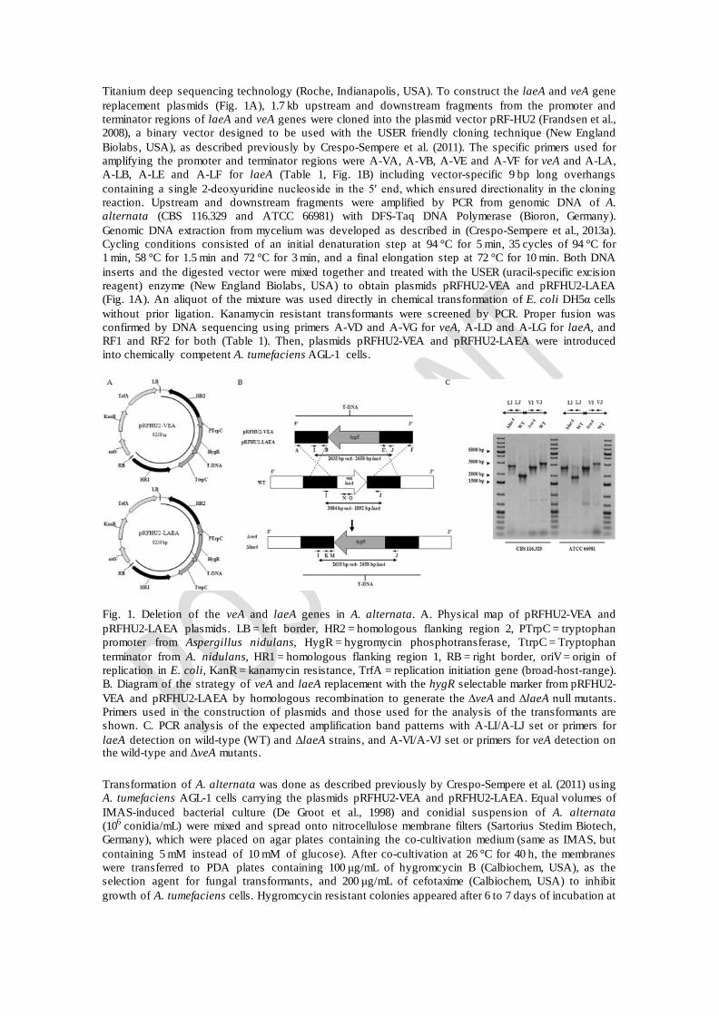
Titanium deep sequencing technology (Roche, Indianapolis, USA). To construct the laeA and veA gene replacement plasmids (Fig. 1A), 1.7 kb upstream and downstream fragments from the promoter and terminator regions of laeA and veA genes were cloned into the plasmid vector pRF-HU2 (Frandsen et al., 2008), a binary vector designed to be used with the USER friendly cloning technique (New England Biolabs, USA), as described previously by Crespo-Sempere et al. (2011). The specific primers used for amplifying the promoter and terminator regions were A-VA, A-VB, A-VE and A-VF for veA and A-LA, A-LB, A-LE and A-LF for laeA (Table 1, Fig. 1B) including vector-specific 9 bp long overhangs containing a single 2-deoxyuridine nucleoside in the 5′ end, which ensured directionality in the cloning reaction. Upstream and downstream fragments were amplified by PCR from genomic DNA of A. alternata (CBS 116.329 and ATCC 66981) with DFS-Taq DNA Polymerase (Bioron, Germany). Genomic DNA extraction from mycelium was developed as described in (Crespo-Sempere et al., 2013a). Cycling conditions consisted of an initial denaturation step at 94 °C for 5 min, 35 cycles of 94 °C for 1 min, 58 °C for 1.5 min and 72 °C for 3 min, and a final elongation step at 72 °C for 10 min. Both DNA inserts and the digested vector were mixed together and treated with the USER (uracil-specific excision reagent) enzyme (New England Biolabs, USA) to obtain plasmids pRFHU2-VEA and pRFHU2-LAEA (Fig. 1A). An aliquot of the mixture was used directly in chemical transformation of E. coli DH5α cells without prior ligation. Kanamycin resistant transformants were screened by PCR. Proper fusion was confirmed by DNA sequencing using primers A-VD and A-VG for veA, A-LD and A-LG for laeA, and RF1 and RF2 for both (Table 1). Then, plasmids pRFHU2-VEA and pRFHU2-LAEA were introduced into chemically competent A. tumefaciens AGL-1 cells.
Fig. 1. Deletion of the veA and laeA genes in A. alternata. A. Physical map of pRFHU2-VEA and pRFHU2-LAEA plasmids. LB = left border, HR2 = homologous flanking region 2, PTrpC = tryptophan promoter from Aspergillus nidulans, HygR = hygromycin phosphotransferase, TtrpC = Tryptophan terminator from A. nidulans, HR1 = homologous flanking region 1, RB = right border, oriV = origin of replication in E. coli, KanR = kanamycin resistance, TrfA = replication initiation gene (broad-host-range). B. Diagram of the strategy of veA and laeA replacement with the hygR selectable marker from pRFHU2-VEA and pRFHU2-LAEA by homologous recombination to generate the ΔveA and ΔlaeA null mutants. Primers used in the construction of plasmids and those used for the analysis of the transformants are shown. C. PCR analysis of the expected amplification band patterns with A-LI/A-LJ set or primers for laeA detection on wild-type (WT) and ΔlaeA strains, and A-VI/A-VJ set or primers for veA detection on the wild-type and ΔveA mutants.
Transformation of A. alternata was done as described previously by Crespo-Sempere et al. (2011) using A. tumefaciens AGL-1 cells carrying the plasmids pRFHU2-VEA and pRFHU2-LAEA. Equal volumes of IMAS-induced bacterial culture (De Groot et al., 1998) and conidial suspension of A. alternata (106 conidia/mL) were mixed and spread onto nitrocellulose membrane filters (Sartorius Stedim Biotech, Germany), which were placed on agar plates containing the co-cultivation medium (same as IMAS, but containing 5 mM instead of 10 mM of glucose). After co-cultivation at 26 °C for 40 h, the membranes were transferred to PDA plates containing 100 μg/mL of hygromcycin B (Calbiochem, USA), as the selection agent for fungal transformants, and 200 μg/mL of cefotaxime (Calbiochem, USA) to inhibit growth of A. tumefaciens cells. Hygromcycin resistant colonies appeared after 6 to 7 days of incubation at
26 °C. Fig. 1B shows in more detail the gene replacement strategy followed in order to disrupt veA and laeA.
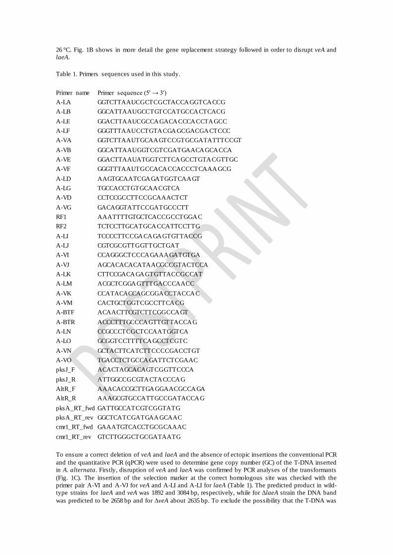
Table 1. Primers sequences used in this study.
Primer name Primer sequence (5′ → 3′) A-LA GGTCTTAAUCGCTCGCTACCAGGTCACCG A-LB GGCATTAAUGCCTGTCCATGCCACTCACG A-LE GGACTTAAUCGCCAGACACCCACCTAGCC A-LF GGGTTTAAUCCTGTACGAGCGACGACTCCC A-VA GGTCTTAAUTGCAAGTCCGTGCGATATTTCCGT A-VB GGCATTAAUGGTCGTCGATGAACAGCACCA A-VE GGACTTAAUATGGTCTTCAGCCTGTACGTTGC A-VF GGGTTTAAUTGCCACACCACCCTCAAAGCG A-LD AAGTGCAATCGAGATGGTCAAGT A-LG TGCCACCTGTGCAACGTCA A-VD CCTCCGCCTTCCGCAAACTCT A-VG GACAGGTATTCCGATGCCCTT RF1 AAATTTTGTGCTCACCGCCTGGAC RF2 TCTCCTTGCATGCACCATTCCTTG A-LI TCCCCTTCCGACAGAGTGTTACCG A-LJ CGTCGCGTTGGTTGCTGAT A-VI CCAGGGCTCCCAGAAAGATGTGA A-VJ AGCACACACATAACGCCGTACTCCA A-LK CTTCCGACAGAGTGTTACCGCCAT A-LM ACGCTCGGAGTTTGACCCAACC A-VK CCATACACCAGCGGACCTACCAC A-VM CACTGCTGGTCGCCTTCACG A-BTF ACAACTTCGTCTTCGGCCAGT A-BTR ACCCTTTGCCCAGTTGTTACCAG A-LN CCGCCCTCGCTCCAATGGTCA A-LO GCGGTCCTTTTCAGCCTCGTC A-VN GCTACTTCATCTTCCCCGACCTGT A-VO TGACCTCTGCCAGATTCTCGAAC pksJ_F ACACTAGCACAGTCGGTTCCCA pksJ_R ATTGGCCGCGTACTACCCAG AltR_F AAACACCGCTTGAGGAACGCCAGA AltR_R AAAGCGTGCCATTGCCGATACCAG pksA_RT_fwd GATTGCCATCGTCGGTATG pksA_RT_rev GGCTCATCGATGAAGCAAC cmr1_RT_fwd GAAATGTCACCTGCGCAAAC cmr1_RT_rev GTCTTGGGCTGCGATAATG
To ensure a correct deletion of veA and laeA and the absence of ectopic insertions the conventional PCR and the quantitative PCR (qPCR) were used to determine gene copy number (GC) of the T-DNA inserted in A. alternata. Firstly, disruption of veA and laeA was confirmed by PCR analyses of the transformants (Fig. 1C). The insertion of the selection marker at the correct homologous site was checked with the primer pair A-VI and A-VJ for veA and A-LI and A-LI for laeA (Table 1). The predicted product in wild-type strains for laeA and veA was 1892 and 3084 bp, respectively, while for ΔlaeA strain the DNA band was predicted to be 2658 bp and for ΔveA about 2635 bp. To exclude the possibility that the T-DNA was
integrated elsewhere in the genome, the qPCR was used instead of a Southern blot analysis. Hence, to determine the number of T-DNA molecules that had been integrated in the genome of each selected transformant, a qPCR analysis was carried out following an already demonstrated methodology described by several authors (Crespo-Sempere et al., 2013c; López-Pérez et al., 2015; Solomon et al., 2008) and firstly described in a filamentous fungus by De Preter et al. (2002). Two primer pairs, (Fig. 1B, Table 1), were designed within the T-DNA in the promoter region of the target genes, close to the selection marker, A-VK and A-VM for veA and A-LK and A-LM for laeA. qPCR reactions were performed in a final volume of 10 μL, containing 1X of SsoAdvanced™ SYBR® Green Supermix (BIO-RAD, USA), 250 nM of each primer and 1 μL of template DNA. All amplifications were performed on a CFX96 Touch™ Real-Time PCR Detection System (BIO-RAD, USA). The standard protocol included one cycle at 98 °C for 2 min, followed by 35 cycles at 98 °C for 5 s and 58 °C for 30 s. Reactions were done in triplicate for each knockout mutant candidate. qPCR efficiency (E) for each pair of primers was calculated from the slopes of the standard curve (Lee et al., 2006). The number of T-DNA copies that have been integrated in the genome of the transformant was calculated according to the Eq. (1), based on Pfaffl (2001) and Rasmussen (2001), which depends on E and the Crossing point (Cp) value of the transformant versus the wild-type strain, and normalized in comparison to a reference gene that is present with the same copy number in both wild-type strain and transformant.
The gene of β-tubulin (protein identification no 417073) was chosen as reference gene, using A-BTF and A-BTR primers (Table 1). All primers were designed using the OLIGO Primer Analysis Software V.7.
2.3. Phenotypic studies of ΔveA and ΔlaeA disrupted mutants
2.3.1. Mycelial growth, mycotoxin production and sporulation assessment
For growth assessment, mycotoxin production and sporulation quantification, PDA plates were inoculated centrally with 5 μL of conidia suspensions (105 conidia/mL) of the wild-type strain of A. alternata and the ΔveA and ΔlaeA knockout strains. Cultures were incubated at 26 °C under two different conditions, darkness or white light (Mazda, 23 W CFT/827, 1485 lm). The distance between the light source and the agar plates was 30 cm.
Mycelial growth was determined by measuring daily two perpendicular diameters of the growing colonies over four days. Mycotoxin production (AOH and AME) was quantified in five day old cultures. To this aim, three 5 mm agar plugs were removed from the inner, middle and outer part of the colonies. Plugs were weighed to ensure a standardization of the method and then AOH and AME extraction was carried out as described in Estiarte et al. (2016). For mycotoxin quantification, working standard solutions were used to perform a ten-point calibration curve for the mycotoxins (1500, 1250, 1000, 750, 500, 250, 100, 50, 25 and 10 ng/mL). The detection limit of the analysis was 10 ppb for AOH and 12 ppb for AME, based on a signal-to-noise ratio of 3:1. All solvents were HPLC grade and all chemicals were analytical grade.
Sporulation assessment was carried out by scraping the surface of 14 day old cultures with a scalpel. Conidia and mycelium were homogenized in a sterile solution of 0.005% (v/v) Tween 80. Conidial concentration was measured by using a Thoma counting chamber and results were expressed as conidia/mm2.
Assays were performed with independent biological triplicates and technical triplicates. All comparisons were analyzed by one way ANOVA followed by the Tukey's honestly significant different test (HSD), using Statgraphics Centurion Version XVI. Significance was defined as P < 0.05.
2.3.2. Germination and hyphal growth
Conidia were grown on a sterilized microscope slide in which a PDA drop had solidified. Once the medium was solid, it was inoculated with 25 μL of a conidial suspension (104 conidia/mL). Slides were
placed in Petri dishes that contained a sterilized filter paper previously moistened with 400 μL of water. Cultures were grown at the dark at 20 °C. Conidial germination and hyphal growth was monitored after 16 and 36 h with a light microscope Leica DM2000 coupled to a DFC290 HD digital camera (Leica Microsystems, Germany).
2.4. Gene expression analysis
PDA Petri dishes were inoculated with 100 μL of a conidial suspension (105 conidia/mL), homogeneously spread and incubated at 26 °C under two different conditions, light or darkness. After 5 days, mycelium was collected, frozen in liquid nitrogen and stored at − 80 °C before nucleic acid extraction. RNA was extracted as described in Estiarte et al. (2016). RNA concentration was spectrophotometrically measured and verified by ethidium-bromide staining of an agarose gel. Total RNA was treated with DNase (TURBO DNase, Ambion, USA) to remove contaminating genomic DNA. Single-strand cDNA was synthesized from 5 μg of total RNA using SuperScript III reverse transcription kit and an oligo(dT), according to the manufacturer's instructions (Invitrogen, USA).
Gene-specific primer sets, A-LN/A-LO and A-VN/A-VO, were designed for laeA and veA gene expression analysis, respectively (Table 1). To assess the involvement of deleted genes on mycotoxin biosynthesis, we studied gene expression of PksJ (protein identification n0 AAPT02903) and a putative transcriptional factor AltR (protein identification no AAPT02898), both claimed to have an essential role in AOH and AME biosynthesis in A. alternata (Saha et al., 2012), using the primer pairs pksJ_F/pksJ_R and altR_F/altR_F. Additionally, two primer pairs, pksA_RT_fwd/pksA_RT_rev and crm1_RT_fwd/cmr1_RT_rev (Fetzner et al., 2014), were used to analyze the involvement of LaeA and VeA in melanin metabolic pathways by studying gene expression of a PKS (PksA), required to melanin biosynthesis, and a putative transcription factor (CmrA) that controls the expression of at least three structural genes for melanin biosynthesis (Fetzner et al., 2014).
Real-time qPCR reactions were performed on a CFX96 Touch™ Real-Time PCR Detection System (BIO-RAD, USA) to monitor cDNA amplification. The primer pair A-BTF/A-BTR (Table 1) was designed within the β-tubulin gene to be used as a reference gene. Gene expression measures were derived from biological duplicates and technical triplicates. Gene expression ratios (R) were calculated using the formula described by Pfaffl et al. (2002) and detailed in Eq. (2).
2.5. Fungal growth infection on tomato fruits
To analyze Alternaria spp. artificial infection on tomatoes, fruit were previously surface-disinfected with 10% sodium hypochlorite for 1 min and rinsed with tap water for 10 min. Once dried, tomatoes were four-times injured with a sterilized awl. Inoculation was performed placing 5 μL of a conidial suspension (104 conidia/mL) in each wound. Control tomatoes were also injured but no conidial suspension was used. Tomatoes were stored into plastic bags at 20 °C and 70% RH for two weeks. Five tomatoes were considerate a single replicate and the assay was performed in quadruplicate.
Diameter lesion size was measured two weeks after the inoculation. For mycotoxin production assessment, plugs of 7 mm of diameter and 0.5 mm of thickness were removed where there was the fungal infection. Three plugs were taken from each tomato. All the plugs from the same replicate were put into a stomacher bag and mycotoxin extraction was performed as detailed in Estiarte et al. (2016). Previous to HPLC injection, samples were resuspended in 500 μL of the mobile phase solution (water-methanol, 50:50 v/v). HPLC conditions were the same as previously described.
3. Results
3.1. Identification of the LaeA and VeA orthologs in A. alternata
In order to identify LaeA and VeA homologs in A. alternata we interrogated the A. alternata genome sequences available at Alternaria Genomes Database using Blast alignment approaches (Dang et al., 2015). Blastx searches were performed using LaeA and VeA nucleotide sequences (coding and genomic) from various species including A. flavus (GenBank accession numbers AY883016 and DQ296645, respectively), A. nidulans (GenBank accession numbers AY394722 and AF109316, respectively) and Cochliobolus heterostrophus (GenBank accession numbers JF826792 and JF826791, respectively). Once LaeA and VeA homologs were identified from the A. alternata genome, their amino acid sequences (coding regions without introns) were used to analyze the similarity with LaeA and VeA from other fungal species. Several candidate homologs were found using a Blastx search against the NCBI database (Table 2). The hits with the highest identity percentage belonged to Stemphylium lycopersici, Pyrenophora tritici-repentis and Bipolaris maydis, also known as C. heterostrophus. All of them belong to the Dothideomycetes class and even share subclass (Pleosporomycetidae), order (Pleosporales) and family (Pleosporaceae). For LaeA, all the values for percentage identity were around 90% (E-values close to 0.0), which indicate that LaeA is a highly conserved protein in fungi including Alternaria. Alignment of LaeA and VeA with A. fumigatus, A. nidulans or A. niger showed lower percentage identity values. These results are not surprising as all Aspergillus spp. belong to the Eurotiomycetes class.
Table 2. Amino acid similarities between LaeA and VeA proteins from A. alternata and amino acid sequences deposited on the NCBI using the Blastx algorithm.
Prot ID AAT_PP02962 LaeA A. alternata Prot ID AAT_PP09942 VeA A. alternata
Organism Accession no E value Identity Organism Accession no E
value Identity
A. alternata BAP58880.1 0.0 99% A. alternata OAG17425.1 0.0 99% S. lycopersici KNG51967.1 0.0 88% B. maydis XP_014080488.1 1e-165 90%
P. tritici-repentis XP_001934837.1 0.0 94% P. tritici-repentis XP_001933979.1 0.0 82%
B. maydis AEP40318.1 0.0 92% S. lycopersici KNG51195.1 0.0 76% Neurospora crassa XP_011392800.1 3e-97 52% A. niger GAQ41759.1 5e-79 55%
A. nidulans XP_658411.1 3e-96 51% A. fumigatus XP_752619.1 8e-77 54%
3.2. Knockout of laeA and veA genes in A. alternata
Targeted gene disruption of laeA and veA was performed to investigate the role of both proteins in A. alternata. The first step of the gene deletion strategy was to construct the pRFHU2-LAEA and pRFHU2-VEA plasmids (Fig. 1A) using the USER friendly cloning technique. The resultant mixture was used to transform chemically competent E. coli DH5α cells. Positive transformants were selected as kanamycin-resistant colonies and screened by PCR. Afterwards, plasmids were introduced into chemically competent Agrobacterium tumefaciens cells (AGL-1). The following step was transformation of A. alternata by co-cultivation with A. tumefaciens. Positive transformant colonies were those able to grow on a hygromicin B medium.
The correct disruption of laeA and veA genes was verified by PCR using the primers A-LI and A-LJ for laeA and the primer pair A-VI and A-VJ for veA. Fig. 1C shows the expected amplification band patterns for wild-type and disrupted ∆ laeA and ∆ veA strains. When we used the primers for amplifying laeA and veA in the wild-type strain, the band fragments obtained were 1892 and 3084 bp, respectively, while for ΔlaeA strain the band was about 2658 bp and for ΔveA about 2635 bp. Thus, the hygromycin resistance marker had been integrated properly by homologous recombination replacing veA and laeA genes and excluding the possibility that the T-DNA had been integrated elsewhere in the genome (ectopic transformation). The number of T-DNA copies integrated in the genome was assessed by qPCR analysis, confirming that most of the mutants contained a single T-DNA integration (see supporting information; Table S1). Two ectopic transformants have been also included as an example of an ectopic transformation. The wild-type strains were used as controls and the β-tubulin gene was used as the reference. Efficiencies for laeA, veA and β-tubulin genes were 2.134, 2.34 and 2.115, respectively.
3.3. Involvement of LaeA and VeA in hyphal growth and conidiation
To determine in which fungal functions LaeA and VeA were involved, several experiments were performed. For this purpose, two different strains of A. alternata were used and the wild-type of each strain was used as the control. For each wild-type strain one knockout of LaeA and one knockout of VeA were used to carry out the assays.
Wild-type and disrupted laeA and veA strains were grown on PDA for 14 days, under light or dark conditions. As shown in Fig. 2, the CBS 116.329 wild-type colonies grew in a brown-grey uniform layer while ΔlaeA and ΔveA mutants were less-pigmented and grew as a white-grey velvet cover with a radial ring, which was clearly observed in ΔveA colonies, especially in those grown on the dark. Nevertheless, in the ATCC 66981 colonies, instead of being brown-grey pigmented (as observed in CBS 116.329), the green color was more predominant. Furthermore, while in CBS 116.329 a clear loss of pigmentation was observed compared to wild-type colonies, in ATCC 66981 there was a remarkable difference in color between wild-type and ΔveA, but not when it came to ΔlaeA colonies. No remarkable differences were found between colonies grown in the dark and colonies grown under light conditions and neither in the reverse plate appearance (data not shown).
Conidia and hyphal growth was assessed after 16 and 36 h of inoculation (Fig. 3). No significant differences were found between wild-type and ΔveA strains. In both strains there were large and small dark colored conidia. Approximately half of the conidia were obclavate to obpyriform and four-celled, while the other half were short and one-celled. Nevertheless, we did observe a difference in ΔlaeA spores, especially those of ATCC 66981. In general, ΔlaeA conidia were of reduced length and instead of having three or four cells, they were one or two-celled with a short beak. We did not observe differences on the hyphal growth on any of the analyzed strains.
To assess fungal growth, two perpendicular diameters of the colonies were measured over four days (Fig. 4). For ATCC 66981, results showed that wild-type was the strain that achieved the biggest diameter on the fourth day while ΔveA was the smallest. For CBS 116.329, (Fig. 4) ΔlaeA was the strain with the highest growth on the fourth day, while no significant differences were observed between the wild-type and the ΔveA transformant. There were no significant differences due to light conditions, regardless of the strain.
To investigate the involvement of LaeA and VeA in conidiation, conidial suspensions were prepared from 14 day old cultures. Results (Fig. 5) demonstrated that both proteins are linked in some way with conidial production as laeA and veA deletion resulted in a drastic reduction of conidial production for both strains. Both for ATCC 66981 or CBS 116.329, the wild-type colonies produced the highest number of conidia, but while the wild-type from the ATCC 66981 grown on the dark produced > 3.8·104 conidia/mm2, the wild-type from the CBS 116.329 produced about 2.9·104 conidia/mm2. In the case of ATCC 66981 cultivated in the light, the loss of LaeA and VeA resulted in a reduction of conidia of 84% for ΔlaeA and 92% for ΔveA. When colonies were cultivated in the darkness, the reduction was about 65% and 91% for ΔlaeA and ΔveA, respectively. The involvement was more remarkable when it came to CBS 116.329 transformants. In this case, the conidial concentration of ΔlaeA and ΔveA mutants was on or below the limit of quantification of the counting method. It is to be noted that for both strains and their corresponding transformants, the colonies produced more conidia under dark conditions though this difference was only statistically significant for ΔlaeA mutants from ATCC 66981 and for the wild-type colonies from CBS 116.329.
3.4. Requirement of LaeA and VeA in mycotoxin biosynthesis
In order to investigate whether LaeA or VeA are linked to secondary metabolism related to mycotoxin biosynthesis, mycotoxin extraction was performed in 5 day old cultures. Two different Alternaria mycotoxins were analyzed, AOH and AME (Fig. 6). Even though results were quite different for ATCC 66981 and CBS 116.329, in both cases and for all the transformants AOH was the mycotoxin produced in a higher amount.

Fig. 2. Front (top) colony view of the wild-type, ΔveA and ΔlaeA A. alternata strains inoculated on PDA plates incubated for 14 days at 26 °C under dark or light conditions.
For ATCC 66981, the colonies that produced the highest amount of mycotoxins were ΔlaeA mutants, followed by wild-type and then ΔveA mutants. In relation to AOH production, ΔlaeA mutants cultivated in light produced more than twice the amount compared to wild-type. This increase was smaller in colonies cultivated in the dark (~ 1.5 fold, Fig. 6). Results reveal that ΔveA mutant colonies compared to wild-type reduced AOH production by 79% and 76%, in the light or the dark, respectively. Similar patterns were observed for ΔlaeA mutants since AME production increment was about 2.8 and 3.3 fold for colonies grown on light and dark, respectively, compared to wild-type. Conversely, AME production
was completely inhibited for ΔveA mutants incubated in the light and inhibited about 82% when ATCC 66981 was grown in the darkness.
Results from experiments with CBS 116.329-derived transformants clearly indicated that LaeA and VeA are also associated with mycotoxin production. All ΔlaeA and ΔveA mutants exhibited reduced mycotoxin biosynthesis. Under light conditions wild-type produced the highest quantity of mycotoxins compared to mutant strains. Comparing the wild-type with ΔlaeA in light and dark, the reduction of mycotoxin production was about 94% and 91% for AOH and, 59% and 56% for AME. For ΔveA mutants, the decrease was drastic, as mycotoxin production was almost completely inhibited for both mycotoxins. It is noteworthy to mention that when comparing mycotoxin production in ATCC 66981 to CBS 116.329 (all wild-type and mutant strains), CBS 116.329 wild-type grown in light is by far the highest AOH producer, with 50 ng/mm2 being the highest amount observed, whereas for ATCC 66981, the highest production (AOH) was observed in the ΔlaeA mutant in light, (~ 11.5 ng/mm2).
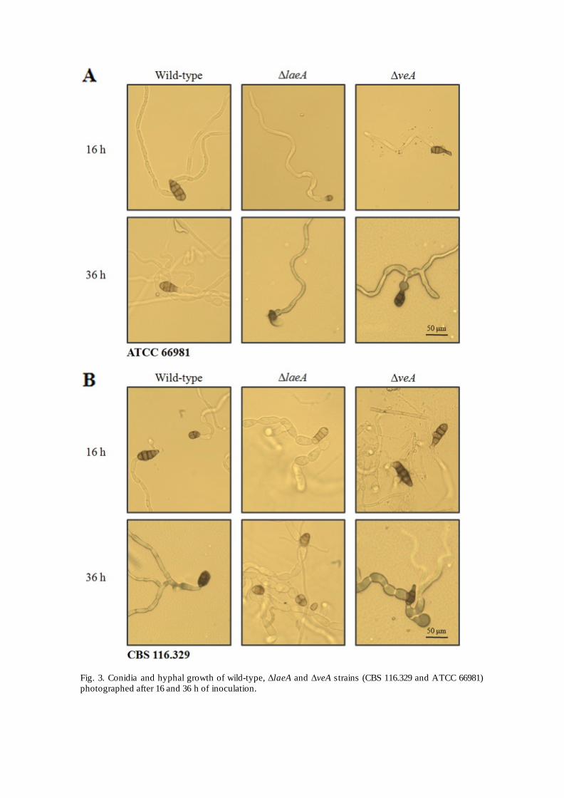
Fig. 3. Conidia and hyphal growth of wild-type, ΔlaeA and ΔveA strains (CBS 116.329 and ATCC 66981) photographed after 16 and 36 h of inoculation.

Fig. 4. Effect of veA and laeA deletion on the colony growth of A. alternata strains (CBS 116.329 and ATCC 66981). Colonies were grown on PDA plates on the darkness or under light conditions at 26 °C for 14 days. The colony diameter was measured on the fourth day. Error bars indicate standard errors. Different letters indicate significant differences among sample groups (ANOVA, P < 0.05).
Fig. 5. Conidia production per mm2 of colony in the wild-type, ΔlaeA and ΔveA strains of ATCC 66981 and CBS 116.329. Error bars indicate standard errors. Letters indicate homogeneous groups within the same day (ANOVA, P < 0.05).
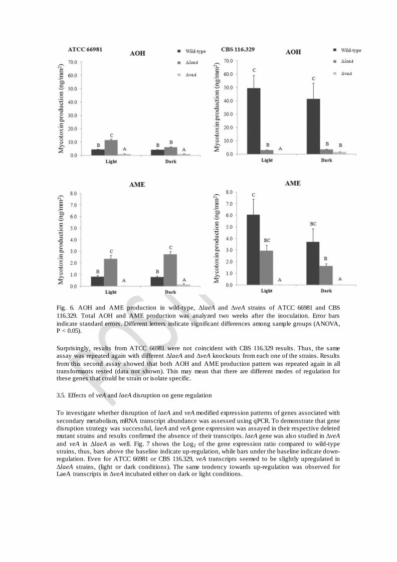
Fig. 6. AOH and AME production in wild-type, ΔlaeA and ΔveA strains of ATCC 66981 and CBS 116.329. Total AOH and AME production was analyzed two weeks after the inoculation. Error bars indicate standard errors. Different letters indicate significant differences among sample groups (ANOVA, P < 0.05).
Surprisingly, results from ATCC 66981 were not coincident with CBS 116.329 results. Thus, the same assay was repeated again with different ΔlaeA and ΔveA knockouts from each one of the strains. Results from this second assay showed that both AOH and AME production pattern was repeated again in all transformants tested (data not shown). This may mean that there are different modes of regulation for these genes that could be strain or isolate specific.
3.5. Effects of veA and laeA disruption on gene regulation
To investigate whether disruption of laeA and veA modified expression patterns of genes associated with secondary metabolism, mRNA transcript abundance was assessed using qPCR. To demonstrate that gene disruption strategy was successful, laeA and veA gene expression was assayed in their respective deleted mutant strains and results confirmed the absence of their transcripts. laeA gene was also studied in ΔveA and veA in ΔlaeA as well. Fig. 7 shows the Log2 of the gene expression ratio compared to wild-type strains, thus, bars above the baseline indicate up-regulation, while bars under the baseline indicate down-regulation. Even for ATCC 66981 or CBS 116.329, veA transcripts seemed to be slightly upregulated in ΔlaeA strains, (light or dark conditions). The same tendency towards up-regulation was observed for LaeA transcripts in ΔveA incubated either on dark or light conditions.
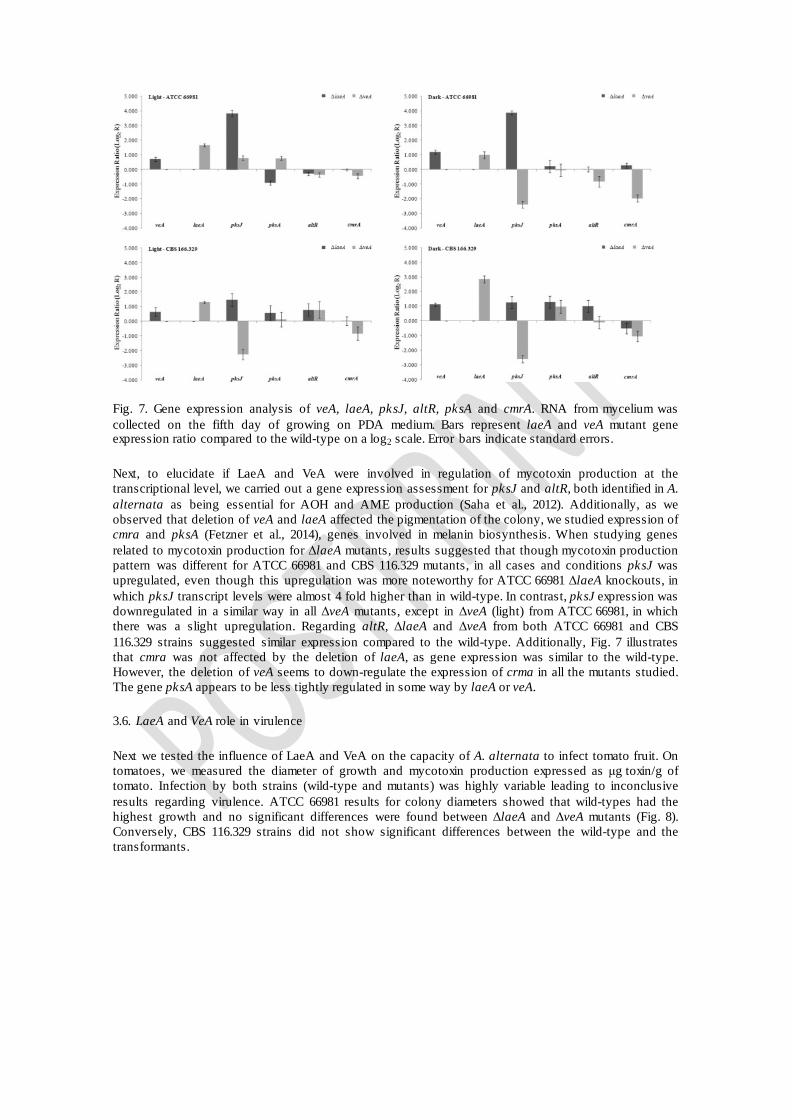
Fig. 7. Gene expression analysis of veA, laeA, pksJ, altR, pksA and cmrA. RNA from mycelium was collected on the fifth day of growing on PDA medium. Bars represent laeA and veA mutant gene expression ratio compared to the wild-type on a log2 scale. Error bars indicate standard errors.
Next, to elucidate if LaeA and VeA were involved in regulation of mycotoxin production at the transcriptional level, we carried out a gene expression assessment for pksJ and altR, both identified in A. alternata as being essential for AOH and AME production (Saha et al., 2012). Additionally, as we observed that deletion of veA and laeA affected the pigmentation of the colony, we studied expression of cmra and pksA (Fetzner et al., 2014), genes involved in melanin biosynthesis. When studying genes related to mycotoxin production for ΔlaeA mutants, results suggested that though mycotoxin production pattern was different for ATCC 66981 and CBS 116.329 mutants, in all cases and conditions pksJ was upregulated, even though this upregulation was more noteworthy for ATCC 66981 ΔlaeA knockouts, in which pksJ transcript levels were almost 4 fold higher than in wild-type. In contrast, pksJ expression was downregulated in a similar way in all ΔveA mutants, except in ΔveA (light) from ATCC 66981, in which there was a slight upregulation. Regarding altR, ΔlaeA and ΔveA from both ATCC 66981 and CBS 116.329 strains suggested similar expression compared to the wild-type. Additionally, Fig. 7 illustrates that cmra was not affected by the deletion of laeA, as gene expression was similar to the wild-type. However, the deletion of veA seems to down-regulate the expression of crma in all the mutants studied. The gene pksA appears to be less tightly regulated in some way by laeA or veA.
3.6. LaeA and VeA role in virulence
Next we tested the influence of LaeA and VeA on the capacity of A. alternata to infect tomato fruit. On tomatoes, we measured the diameter of growth and mycotoxin production expressed as μg toxin/g of tomato. Infection by both strains (wild-type and mutants) was highly variable leading to inconclusive results regarding virulence. ATCC 66981 results for colony diameters showed that wild-types had the highest growth and no significant differences were found between ΔlaeA and ΔveA mutants (Fig. 8). Conversely, CBS 116.329 strains did not show significant differences between the wild-type and the transformants.
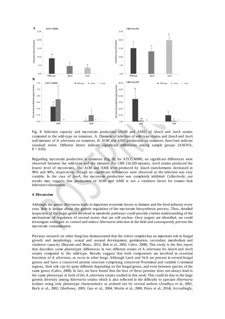
Fig. 8. Infection capacity and mycotoxin production (AOH and AME) of ΔlaeA and ΔveA strains compared to the wild-type on tomatoes. A. Diameter of infection of wild-type strains and ΔlaeA and ΔveA null mutants of A. alternata on tomatoes. B. AOH and AME production on tomatoes. Error bars indicate standard errors. Different letters indicate significant differences among sample groups (ANOVA, P < 0.05).
Regarding mycotoxin production in tomatoes (Fig. 8), for ATCC 66981, no significant differences were observed between the wild-type and the mutants. For CBS 116.329 mutants, ΔveA strains produced the lowest level of mycotoxins. The AOH and AME level produced by ΔlaeA transformants decreased in 96% and 90%, respectively, though no significant differences were observed as the infection was very variable. In the case of ΔveA, the mycotoxin production was completely inhibited. Collectively, our results may suggest that production of AOH and AME is not a virulence factor for tomato fruit infection/colonization.
4. Discussion
Although the genus Alternaria leads to important economic losses to farmers and the food industry every year, little is known about the genetic regulation of the mycotoxin biosynthesis process. Thus, detailed inspection of the target genes involved in metabolic pathways could provide a better understanding of the mechanisms of regulation of several routes that are still unclear. Once targets are identified, we could investigate strategies to control and reduce Alternaria infection in the field and consequently prevent the mycotoxin contamination.
Previous research on other fungi has demonstrated that the velvet complex has an important role in fungal growth and morphology, sexual and asexual development, germination, secondary metabolism and virulence capacity (Bayram and Braus, 2012; Bok et al., 2005; Calvo, 2008). This study is the first report that describes some phenotypic differences in two different strains of A. alternata for ΔlaeA and ΔveA strains compared to the wild-type. Results suggest that both components are involved in essential functions of A. alternata, as occur in other fungi. Although LaeA and VeA are present in several fungal genera and have a conserved protein structure comprising conserved N-terminal and variable C-terminal regions, their role can be quite different depending on the fungal genus, and even between species of the same genus (Calvo, 2008). In fact, we have found that the loss of these proteins does not always lead to the same phenotype in both of the A. alternata strains studied in this work. This could be due to the large genetic diversity among Alternaria strains which is also reflected in the difficulty to speciate Alternaria isolates using only phenotype characteristics as pointed out by several authors (Aradhya et al., 2001; Bock et al., 2002; Gherbawy, 2005; Guo et al., 2004; Morris et al., 2000; Pruss et al., 2014). Accordingly,
Pruss et al. (2014) in their study focused on clarifying the role of the white-collar 1 (WC-1) gene in A. alternata, also obtained contradictory results compared to previously published studies in A. alternata strains (Häggblom and Unestam, 1979; Söderhäll et al., 1978).
In the case of the ATCC 66981 strain, LaeA and VeA seemed to be relevant for colony growth under either light or darkness, especially VeA, as it has been observed in A. parasiticus and A. carbonarius (Calvo et al., 2004; Crespo-Sempere et al., 2013b). By contrast, for the CBS 116.329 strain, LaeA seemed to act as a negative regulator of the colony growth, while VeA did not have any significant effect compared to the wild-type. This LaeA pattern had also been observed by Crespo-Sempere et al. (2013b) in A. carbonarius, though in that case this increase was only observed when colonies where grown in light conditions.
Several studies postulate that the velvet complex is associated with sexual and asexual development (Bayram et al., 2008b; Calvo, 2008; Hee-Seo et al., 2002; Hoff et al., 2010; Jiang et al., 2011; Li et al., 2006; Yang et al., 2013). Results described in this work are consistent with these earlier studies, thus confirming that LaeA and VeA are involved in some aspects of asexual development in Alternaria. In A. nidulans, VeA acts positively regulating sexual structures, such as Hülle cells and cleistothecia, and negatively regulating asexual development (Hee-Seo et al., 2002). In Neurospora crassa, C. heterostrophus, Fusarium graminearum, Fusarium verticillioides and Botrytis cinerea, deletion of ve-1, Chvel1, FgVEA, FvVE1 and BcVEA genes, respectively, also led to a significant increase in conidial production (Bayram et al., 2008b; Jiang et al., 2011; Li et al., 2006; Wu et al., 2012; Yang et al., 2013). By contrast, in other fungi such as A. carbonarius, A. fumigatus, A. parasiticus, Fusarium fujikuroi, Fusarium oxysporum and Penicillium chrysogenum it has been described that VeA regulates sexual and asexual development in the opposite way, (Calvo et al., 2004; Crespo-Sempere et al., 2013b; Hoff et al., 2010; Krappmann et al., 2006; López-Berges et al., 2013; Wiemann et al., 2010). Our study provides results that suggest that VeA positively regulates asexual development in A. alternata since in both strains tested in this study, when veA was disrupted production of conidia drastically decreased. LaeA may also have a noteworthy role in sexual and asexual development. Our results indicate that LaeA positively regulates the asexual development in A. alternata as observed for VeA. However, this positive regulation could be less important if compared with VeA, as one of the strains (ATCC 66981) indicates that reduction of conidia is more severe when deleting veA. This positive asexual regulation has also been described in other fungi such as A. carbonarius, A. flavus, F. oxysporum, F. fujikuroi and P. chrysogenum (Crespo-Sempere et al., 2013b; Hoff et al., 2010; Kale et al., 2008; López-Berges et al., 2013; Wiemann et al., 2010; Wu et al., 2012), whereas in C. heterostrophus negative regulation has been reported (Wu et al., 2012). Nevertheless, in other studies performed with A. nidulans and A. fumigatus, laeA deletion hardly impaired conidial production, with knockout mutants showing almost the same conidiation level as the wild-type strains (Bok et al., 2005; Bok and Keller, 2004). In A. flavus it has been observed that in laeA knockout mutants conidial production was dependent on the media as in some media ∆ laeA produced higher conidiation level than the wild-type while on others production was lower (Chang et al., 2012).
Earlier studies suggest that in A. alternata there is an association between developmental structures and pigment biosynthesis pathways (Fetzner et al., 2014; Kawamura et al., 1999). Among the natural pigments the most common is melanin, biosynthesis of which contributes to the survival of the fungal spore by protecting against damaging UV light and it is an important virulence factor as well (Heinekamp et al., 2013; Liu and Nizet, 2009; Yin and Keller, 2011). In A. alternata, biosynthesis of melanin proceeds primarily by the 1,8-dihydroxinapthalene (DHN) pathway (Kimura and Tsuge, 1993). Its biosynthesis requires PksA, a 1,3,6,8-trihydroxynaphtalene (THN) reductase, a scytalone dehydratase (Bmr1) and a 1,3,8-THN reductase (Bmr2) (Eliahu et al., 2007; Fetzner et al., 2014). Some of these genes encoding enzymes required for melanin biosynthesis are regulated by CmrA, a transcriptional regulator that controls spore development as well (Eliahu et al., 2007; Tsuji et al., 2000). Thus, as we observed pigmentation differences in ΔveA and ΔlaeA mutants, we studied how the loss of two proteins of the velvet complex could affect cmra and pksA. While no important differences were observed in pksA gene expression in ΔveA and ΔlaeA mutants, cmrA was down-regulated in ΔveA mutants. Interestingly, higher down-regulation levels of cmra correlates with less pigmented colonies and with less sporulation. This may suggest that VeA could be also linked to melanin biosynthesis acting as a positive regulator of the pathway.
In A. alternata, LaeA and VeA are required not only for normal morphology and asexual development but also for the production of secondary metabolites. However, deletion of LaeA and VeA caused a
different strain-specific response. It was interesting that while both ΔlaeA and ΔveA transformants of the CBS 116.329 strain decreased their mycotoxin production, the ATCC 66981 ΔlaeA strain showed increased AOH and AME production. In the case of ΔveA mutants, mycotoxin levels decreased in all conditions.
We also studied the expression of two of the genes putatively involved in the biosynthesis pathway and regulation of AOH, pksJ and altR (Saha et al., 2012), which encode two proteins that supposedly play an important role in AOH production. Saha et al. (2012) observed that down-regulation of pksJ and altR caused a large decrease of AOH formation (Saha et al., 2012). In our study, while no remarkable findings were observed for altR, pksJ was overexpressed in all ΔlaeA transformants. This overexpression was remarkably higher when it came to ATCC 66981 ΔlaeA mutants. For ΔveA knockouts, pksJ expression was down-regulated almost in all the strains, which correlates with mycotoxin production results. In previous studies, Crespo-Sempere et al. (2013b) described a reduction of OTA production in A. carbonarius, both in ΔlaeA and ΔveA, linked with a down-regulation of the nonribosomal peptide synthetase (nrps) involved in OTA biosynthesis. In A. alternata this correlation has only been observed in ΔveA transformants. We are aware that there exists another recent theory dealing with the biosynthesis of AOH, in which is suggested that another PKS could be the responsible of the AOH production. This idea emerged from the recent study of Chooi et al. (2015) which reported that the key enzyme for the biosynthesis of AOH in P.nodorum was SnPKS19 and, at the same time, it was suggested that in A. alternata the responsible PKS could be PksI instead of PksJ, a PKS that was also described by Saha et al. (2012). Although it would have been interesting to assess the gene expression of pksI in the present work, the experimental assays of this study were previous to the publication of Chooi et al. (2015), so we just studied the expression of those genes already identified in A. alternata.
It may seem surprising that different results could be achieved in two strains of the same Alternaria species but, as it has been mentioned before, there is a large genetic variability among Alternaria strains even of the same species (Aradhya et al., 2001; Fetzner et al., 2014; Guo et al., 2004). It has been recently shown at the genomic level that individual strains of an Alternaria spp. may each possess a thousand or more predicted unique genes (Dang et al., 2015). Thus, other regulation mechanisms may control mycotoxin biosynthesis in a different way which could be related to overall gene content, small epigenetic modifications or instabilities of the genome, previously suggested by Pruss et al. (2014). Additionally, genetic diversity among fungal species is also influenced by different mechanisms: migration and gene flow, recombination, which occurs either through sexual reproduction or through a process of somatic hybridization and, genetic mutations. All these mechanisms may also contribute to the diversity of fungal populations (Burdon and Silk, 1997).
In conclusion, the research findings of this study have provided some evidence that both LaeA and VeA play an important role in morphology development, asexual differentiation and secondary metabolism in Alternaria. The loss of laeA and veA genes led to a drastic reduction of conidia production, suggesting that both velvet components could act as a positive regulator of asexual development. AOH and AME production is also altered in laeA and veA knockouts. However, this appeared to be isolate/strain specific. While deletion of veA gene seems to strongly inhibit mycotoxin production in both strains, deletion of laeA increased the mycotoxin production levels in one strain but decreased in the other. The genetic variability within Alternaria genus could be an explanation that may justify these differences. Hence, we believe that it is important and useful to perform these studies of target gene characterization using more than one strain of the same fungal species in order to take into consideration the variations that could be derived from the living microorganisms. We have also pointed out that VeA could be linked to CmrA, a regulator belonging to the melanin biosynthesis pathway and the formation of conidia, as the deletion of veA lead to a down-regulation of crma. All in all, the molecular mechanism of the velvet complex in Alternaria is still unclear and further studies are needed to fully understand the velvet system function in A. alternata.
The following are the supplementary data related to this article.
Table S1. Determination of the number of T-DNA copies integrated in the genome of A. alternata. Gene copy number (GC) quantification was conducted by qPCR according to Eq. (1). The genes laeA and veA were used as the target genes for ΔlaeA and ΔveA transformants, respectively. The β-tubulin gene was used as a reference gene. Wild type strains, ATCC 66981 and CBS 116.329 were used as controls. Efficiencies for laeA, veA and β-tubulin genes were 2.134, 2.34 and 2.115, respectively. All reactions were conducted in triplicate.
Acknowledgements
We are also grateful to the Catalonian Government (XaRTA-Reference Network on Food Technology) for their financial support. N. Estiarte thanks the Secretaria de Universitats i Recerca del Departament d'Economia i Coneixement of the Generalitat de Catalunya for the pre-doctoral grant (FI-DGR 2013).
References
M.K. Aradhya, H.M. Chan, D.E. ParfittGenetic variability in the pistachio late blight fungus, Alternaria alternata Mycol. Res., 105 (2001), pp. 300-306 R. Barkai-Golan, N. PasterMycotoxins in Fruits and Vegetables Ö. Bayram, G.H. BrausCoordination of secondary metabolism and development in fungi: the velvet family of regulatory proteins FEMS Microbiol. Rev., 36 (2012), pp. 1-24 Ö. Bayram, S. Krappmann, M. Ni, J.W. Bok, K. Helmstaedt, O. Valerius, S. Braus-Stromeyer, N.-J. Kwon, N.P. Keller, J.-H. Yu, G.H. BrausVelB/VeA/LaeA complex coordinates light signal with fungal development and secondary metabolism Science, 320 (2008), pp. 1504-1506 Ö. Bayram, S. Krappmann, S. Seiler, N. Vogt, G.H. BrausNeurospora crassa ve-1 affects asexual conidiation Fungal Genet. Biol., 45 (2008), pp. 127-138 C.H. Bock, P.H. Thrall, C.L. Brubaker, J.J. BurdonDetection of genetic variation in Alternaria brassicicola using AFLP fingerprinting Mycol. Res., 106 (2002), pp. 428-434 J.W. Bok, N.P. KellerLaeA, a regulator of secondary metabolism in Aspergillus spp Eukaryot. Cell, 3 (2004), pp. 527-535 J.W. Bok, S.A. Balajee, K.A. Marr, D. Andes, K.F. Nielsen, J.C. Frisvad, N.P. KellerLaeA, a regulator of morphogenetic fungal virulence factors Eukaryot. Cell, 4 (2005), pp. 1574-1582 E.-M. Brugger, J. Wagner, D.M. Schumacher, K. Koch, J. Podlech, M. Metzler, L. LehmannMutagenicity of the mycotoxin alternariol in cultured mammalian cells Toxicol. Lett., 164 (2006), pp. 221-230 J.J. Burdon, J. SilkSources and patterns of diversity in plant-pathogenic fungi Phytopathology, 87 (1997), pp. 664-669 A.M. CalvoThe VeA regulatory system and its role in morphological and chemical development in fungi Fungal Genet. Biol., 45 (2008), pp. 1053-1061 A.M. Calvo, R.A. Wilson, J.W. Bok, N.P. KellerRelationship between secondary metabolism and fungal development
Microbiol. Mol. Biol. Rev., 66 (2002), pp. 447-459 A.M. Calvo, J. Bok, W. Brooks, N.P. KellerveA is required for toxin and sclerotial production in Aspergillus parasiticus Appl. Environ. Microbiol., 70 (2004), pp. 4733-4739 P.K. Chang, J.W. Bennett, P.J. CottyAssociation of aflatoxin biosynthesis and sclerotial development in Aspergillus parasiticus Mycopathologia, 153 (2001), pp. 41-48 P.-K. Chang, L.L. Scharfenstein, K.C. Ehrlich, Q. Wei, D. Bhatnagar, B.F. IngberEffects of LaeA deletion on Aspergillus flavus conidial development and hydrophobicity may contribute to loss of aflatoxin production Fungal Biol., 116 (2012), pp. 298-307 P.-K. Chang, L.L. Scharfenstein, P. Li, K.C. EhrlichAspergillus flavus VelB acts distinctly from VeA in conidiation and may coordinate with FluG to modulate sclerotial production Fungal Genet. Biol., 58–59 (2013), pp. 71-79 Y.-H. Chooi, M.J. Muria-Gonzalez, O.L. Mead, P.S. SolomonSnPKS19 encodes the polyketide synthase for alternariol mycotoxin biosynthesis in the wheat pathogen Parastagonospora nodorum Appl. Environ. Microbiol., 81 (2015), pp. 5309-5317 A. Crespo-Sempere, M. López-Pérez, P.V. Martínez-Culebras, L. González-CandelasDevelopment of a green fluorescent tagged strain of Aspergillus carbonarius to monitor fungal colonization in grapes Int. J. Food Microbiol., 148 (2011), pp. 135-140 A. Crespo-Sempere, N. Estiarte, S. Marín, V. Sanchis, A.J. RamosPropidium monoazide combined with real-time quantitative PCR to quantify viable Alternaria spp. contamination in tomato products Int. J. Food Microbiol., 165 (2013), pp. 214-220 A. Crespo-Sempere, S. Marín, V. Sanchis, A.J. RamosVeA and LaeA transcriptional factors regulate ochratoxin A biosynthesis in Aspergillus carbonarius Int. J. Food Microbiol., 166 (2013), pp. 479-486 A. Crespo-Sempere, C. Selma-Lázaro, P.V. Martínez-Culebras, L. González-CandelasCharacterization and disruption of the cipC gene in the ochratoxigenic fungus Aspergillus carbonarius Food Res. Int., 54 (2013), pp. 697-705 H.X. Dang, B. Pryor, T. Peever, C.B. LawrenceThe Alternaria genomes database: a comprehensive resource for a fungal genus comprised of saprophytes, plant pathogens, and allergenic species BMC Genomics, 16 (2015), p. 239 M.J.A. De Groot, P. Bundock, P.J.J. Hooykaas, A.G.M. BeijersbergenAgrobacterium tumefaciens-mediated transformation of filamentous fungi Nat. Biotechnol., 16 (1998), pp. 839-842 K. De Preter, F. Speleman, V. Combaret, J. Lunec, G. Laureys, B.H.J. Eussen, N. Francotte, J. Board, A.D.J. Pearson, A. De Paepe, N. Van Roy, J. VandesompeleQuantification of MYCN, DDX1, and NAG gene copy number in neuroblastoma using a real-time quantitative PCR assay Mod. Pathol., 15 (2002), pp. 159-166 EFSAScientific opinion on the risks for animal and public health related to the presence of Alternaria toxins in feed and food EFSA J., 9 (2011), pp. 2407-2504 N. Eliahu, A. Igbaria, M.S. Rose, B.A. Horwitz, S. LevMelanin biosynthesis in the maize pathogen Cochliobolus heterostrophus depends on two mitogen-activated protein kinases, Chk1 and Mps1, and the transcription factor Cmr1 Eukaryot. Cell, 6 (2007), pp. 421-429
N. Estiarte, A. Crespo-Sempere, S. Marín, V. Sanchis, A.J. RamosEffect of 1-methylcyclopropene on the development of black mold disease and its potential effect on alternariol and alternariol monomethyl ether biosynthesis on tomatoes infected with Alternaria alternata Int. J. Food Microbiol., 236 (2016), pp. 74-82 R. Fetzner, K. Seither, M. Wenderoth, A. Herr, R. FischerAlternaria alternata transcription factor CmrA controls melanization and spore development Microbiology, 160 (2014), pp. 1845-1854 R. Frandsen, J. Andersson, M. Kristensen, H. GieseEfficient four fragment cloning for the construction of vectors for targeted gene replacement in filamentous fungi BMC Mol. Biol., 9 (2008), p. 70 Y.A.M.H. GherbawyGenetic variation among isolates of Alternaria spp. from select Egyptian crops Arch. Phytopathol. Plant Protect., 38 (2005), pp. 77-89 L.D. Guo, L. Xu, W.H. Zheng, K.D. HydeGenetic variation of Alternaria alternata, an endophytic fungus isolated from Pinus tabulaeformis as determined by random amplified microsatellites (RAMS) Fungal Divers., 16 (2004), pp. 53-65 P. Häggblom, T. UnestamBlue light inhibits mycotoxin production and increases total lipids and pigmentation in Alternaria alternata Appl. Environ. Microbiol., 38 (1979), pp. 1074-1077 K. Hee-Seo, H. Kyu-Yong, K. Kyung-Jin, H. Dong-Min, J. Kwang-Yeop, C. Keon-SangThe veA gene activates sexual development in Aspergillus nidulans Fungal Genet. Biol., 37 (2002), pp. 72-80 T. Heinekamp, A. Thywissen, J. Macheleidt, S. Keller, V. Valiante, A.A. BrakhageAspergillus fumigatus melanins: interference with the host endocytosis pathway and impact on virulence Front. Microbiol., 3 (2013), p. 440 B. Hoff, J. Kamerewerd, C. Sigl, R. Mitterbauer, I. Zadra, H. Kürnsteiner, U. KückTwo components of a velvet-like complex control hyphal morphogenesis, conidiophore development, and penicillin biosynthesis in Penicillium chrysogenum Eukaryot. Cell, 9 (2010), pp. 1236-1250 J. Jiang, X. Liu, Y. Yin, Z. MaInvolvement of a velvet protein FgVeA in the regulation of asexual development, lipid and secondary metabolisms and virulence in Fusarium graminearum PLoS One, 6 (2011), Article e28291 E. KäferOrigins of translocations in Aspergillus nidulans Genetics, 52 (1965), pp. 217-232 S.P. Kale, L. Milde, M.K. Trapp, J.C. Frisvad, N.P. Keller, J.W. BokRequirement of LaeA for secondary metabolism and sclerotial production in Aspergillus flavus Fungal Genet. Biol., 45 (2008), pp. 1422-1429 N. Kato, W. Brooks, A.M. CalvoThe expression of sterigmatocystin and penicillin genes in Aspergillus nidulans is controlled by veA, a gene required for sexual development Eukaryot. Cell, 2 (2003), pp. 1178-1186 C. Kawamura, T. Tsujimoto, T. TsugeTargeted disruption of a melanin biosynthesis gene affects conidial development and UV tolerance in the Japanese pear pathotype of Alternaria alternata Mol. Plant-Microbe Interact., 12 (1999), pp. 59-63 N. Kimura, T. TsugeGene cluster involved in melanin biosynthesis of the filamentous fungus Alternaria alternata J. Bacteriol., 175 (1993), pp. 4427-4435
S. Krappmann, C. Sasse, G.H. BrausGene targeting in Aspergillus fumigatus by homologous recombination is facilitated in a nonhomologous end-joining-deficient genetic background Eukaryot. Cell, 5 (2006), pp. 212-215 N. Lan, H. Zhang, C. Hu, W. Wang, A.M. Calvo, S.D. Harris, S. Chen, S. LiCoordinated and distinct functions of velvet proteins in Fusarium verticillioides Eukaryot. Cell, 13 (2014), pp. 909-918 C. Lee, J. Kim, S.G. Shin, S. HwangAbsolute and relative qPCR quantification of plasmid copy number in Escherichia coli J. Biotechnol., 123 (2006), pp. 273-280 S. Li, K. Myung, D. Guse, B. Donkin, R.H. Proctor, W.S. Grayburn, A.M. CalvoFvVE1 regulates filamentous growth, the ratio of microconidia to macroconidia and cell wall formation in Fusarium verticillioides Mol. Microbiol., 62 (2006), pp. 1418-1432 G.Y. Liu, V. NizetColor me bad: microbial pigments as virulence factors Trends Microbiol., 17 (2009), pp. 406-413 G.T. Liu, Y.Z. Qian, P. Zhang, W.H. Dong, Y.M. Qi, H.T. GuoEtiological role of Alternaria alternata in human esophageal cancer Chin. Med. J., 105 (1992), pp. 394-400 M.S. López-Berges, C. Hera, M. Sulyok, K. Schäfer, J. Capilla, J. Guarro, A. Di PietroThe velvet complex governs mycotoxin production and virulence of Fusarium oxysporum on plant and mammalian hosts Mol. Microbiol., 87 (2013), pp. 49-65 M. López-Pérez, A.-R. Ballester, L. González-CandelasIdentification and functional analysis of Penicillium digitatum genes putatively involved in virulence towards citrus fruit Mol. Plant Pathol., 16 (2015), pp. 262-275 P.F. Morris, M.S. Connolly, D.A. St ClairGenetic diversity of Alternaria alternata isolated from tomato in California assessed using RAPDs Mycol. Res., 104 (2000), pp. 286-292 M. Ni, J.-H. YuA novel regulator couples sporogenesis and trehalose biogenesis in Aspergillus nidulans PLoS One, 2 (2007), Article e970 R.W. Pero, H. Posner, M. Blois, D. Harvan, J.W. SpaldingToxicity of metabolites produced by the Alternaria Environ. Health Perspect., 4 (1973), pp. 87-94 M.W. PfafflA new mathematical model for relative quantification in real-time RT–PCR Nucleic Acids Res., 29 (2001), Article E45 M.W. Pfaffl, G.W. Horgan, L. DempfleRelative expression software tool (REST©) for group-wise comparison and statistical analysis of relative expression results in real-time PCR Nucleic Acids Res., 30 (2002), Article e36 E. Pfeiffer, N.H. Schebb, J. Podlech, M. MetzlerNovel oxidative in vitro metabolites of the mycotoxins alternariol and alternariol methyl ether Mol. Nutr. Food Res., 51 (2007), pp. 307-316 G.A. Pollock, C.E. Disabatino, R.C. Heimsch, D.R. HilblinkThe subchronic toxicity and teratogenicity of alternariol monomethyl ether produced by Alternaria solani Food Chem. Toxicol., 20 (1982), pp. 899-902
S. Pruss, R. Fetzner, K. Seither, A. Herr, E. Pfeiffer, M. Metzler, C.B. Lawrence, R. FischerRole of the Alternaria alternata blue-light receptor LreA (white-collar 1) in spore formation and secondary metabolism Appl. Environ. Microbiol., 80 (8) (2014), pp. 2582-25891 R. RasmussenQuantification on the LightCycler instrument S. Meuer, C. Wittwer, K. Nakagawara (Eds.), Rapid Cycle Real-time PCR: Methods and Applications, Springer, Heidelberg (2001) D. Saha, R. Fetzner, B. Burkhardt, J. Podlech, M. Metzler, H. Dang, C. Lawrence, R. FischerIdentification of a polyketide synthase required for alternariol (AOH) and alternariol-9-methyl ether (AME) formation in Alternaria alternata PLoS One, 7 (2012), Article e40564 K. Söderhäll, E. Svensson, T. UnestamLight inhibits the production of alternariol and alternariol monomethyl ether in Alternaria alternata Appl. Environ. Microbiol., 36 (1978), pp. 655-657 P.S. Solomon, S.V.S. Ipcho, J.K. Hane, K.C. Tan, R.P. OliverA quantitative PCR approach to determine gene copy number Fungal Genet. Rep., 55 (2008), pp. 5-8 K.-C. Tan, R.D. Trengove, G.L. Maker, R.P. Oliver, P.S. SolomonMetabolite profiling identifies the mycotoxin alternariol in the pathogen Stagonospora nodorum Metabolomics, 5 (2009), pp. 330-335 G. Tsuji, Y. Kenmochi, Y. Takano, J. Sweigard, L. Farrall, I. Furusawa, O. Horino, Y. KuboNovel fungal transcriptional activators, Cmr1p of Colletotrichum lagenarium and Pig1p of Magnaporthe grisea, contain Cys2His2 zinc finger and Zn(II)2Cys6 binuclear cluster DNA-binding motifs and regulate transcription of melanin biosynthesis genes in a developmentally specific manner Mol. Microbiol., 38 (2000), pp. 940-954 P. Wiemann, D.W. Brown, K. Kleigrewe, J.W. Bok, N.P. Keller, H.-U. Humpf, B. TudzynskiFfVel1 and FfLae1, components of a velvet-like complex in Fusarium fujikuroi, affect differentiation, secondary metabolism and virulence Mol. Microbiol., 77 (2010), pp. 972-994 D. Wu, S. Oide, N. Zhang, M.Y. Choi, B.G. TurgeonChLae1 and ChVel1 regulate T-toxin production, virulence, oxidative stress response, and development of the maize pathogen Cochliobolus heterostrophus PLoS Pathog., 8 (2012), Article e1002542 Q. Yang, Y. Chen, Z. MaInvolvement of BcVeA and BcVelB in regulating conidiation, pigmentation and virulence in Botrytis cinerea Fungal Genet. Biol., 50 (2013), pp. 63-71 W. Yin, N.P. KellerTranscriptional regulatory elements in fungal secondary metabolism J. Microbiol. (Seoul, Korea), 49 (2011), pp. 329-339
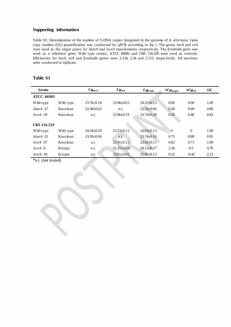
Supporting information Table S1: Determination of the number of T-DNA copies integrated in the genome of A. alternata. Gene copy number (GC) quantification was conducted by qPCR according to Eq 1. The genes laeA and veA were used as the target genes for ΔlaeA and ΔveA transformants, respectively. The β-tubulin gene was used as a reference gene. Wild type strains, ATCC 66981 and CBS 116.329 were used as controls. Efficiencies for laeA, veA and β-tubulin genes were 2.134, 2.34 and 2.115, respectively. All reactions were conducted in triplicate.
Table S1
Strain CplaeA CpveA Cpβ-tub ΔCptarget ΔCpref GC
ATCC 66981
Wild-type Wild type 23.76±0.16 23.06±0.03 24.22±0.13 0.00 0.00 1.00 ΔlaeA -27 Knockout 23.38±0.02 n.t. 23.53±0.06 0.38 0.69 0.80 ΔveA -29 Knockout n.t. 22.86±0.19 23.74±0.28 0.20 0.48 0.83 CBS 116.329 Wild-type Wild type 24.34±0.20 23.53±0.12 24.63±0.13 0 0 1.00 ΔlaeA -31 Knockout 23.59±0.06 n.t. 23.74±0.16 0.75 0.89 0.91 ΔveA -37 Knockout n.t. 22.91±0.15 23.92±0.11 0.62 0.71 1.00 ΔveA -6 Ectopic n.t. 21.25±0.03 24.13±0.27 2.28 0.5 4.78 ΔveA -18 Ectopic n.t. 23.01±0.05 25.05±0.13 0.52 -0.42 2.13 *n.t. (not tested)