0185 Breast Reconstructive Surgery - Aetna Better Health
35
Breast Reconstructive Surgery - Medical Clinical Policy Bulletins | Aetna (https://www.aetna.com/) Breast Reconstructive Surgery Last Review 04/15/2020 Effective: 11/13/1997 Next Review: 02/13/2020 R eview History D efinitions Additional Information C linical Policy Bulletin Notes Number: 0185 Policy *Please see amendment for Pennsylvania Medicaid at the end of this CPB. Aetna considers reconstructive breast surgery medically necessary after a medically necessary mastectomy or a medically necessary lumpectomy that results in a significantdeformity (i.e., mastectomy or lumpectomy for treatment of or prophylaxis for breast cancer and mastectomy or lumpectomyperformed forchronic,severe fibrocystic breast disease, also known as cystic mastitis, unresponsive to medical therapy). Medically necessary procedures include capsulectomy, capsulotomy, implantation of Food and Drug Administration (FDA)-approved internal breast prosthesis, mastopexy, insertion of breast prostheses, the use of tissue expanders, or reconstruction with a latissimus dorsi (LD) myocutaneous flap, Ruben’s flap, superficial inferior epigastric perforator (SIEP) flap, superior or inferior gluteal free flap,transverse upper gracilis (TUG) flap, transverse rectus abdominis myocutaneous (TRAM) flap, deep inferior epigastric perforator (DIEP) flap, superficial inferior epigastric artery (SIEA) flap,superior gluteal artery perforator (SGAP) flap,profunda artery perforator flap,or similarprocedures, including skin sparing techniques. Aetna considers the body lift perforator flap technique for breast reconstruction experimental and investigational because there is insufficientevidence to support the effectiveness of this approach. Proprietary 1/34
-
Upload
khangminh22 -
Category
Documents
-
view
1 -
download
0
Transcript of 0185 Breast Reconstructive Surgery - Aetna Better Health
Breast Reconstructive Surgery - Medical Clinical Policy Bulletins | Aetna
(https://www.aetna.com/)
Breast Reconstructive Surgery
Last Review
04/15/2020
Effective: 11/13/1997
Next Review: 02/13/2020
R eview History
Definitions
Addit ional Information
C linical Policy Bulletin
Notes
Number: 0185
Policy *Please see amendment for Pennsylvania Medicaid
at the end of this CPB.
Aetna considers reconstructive breast surgery medically necessary after
a medically necessary mastectomy or a medically necessary lumpectomy
that results in a significant deformity (i.e., mastectomy or lumpectomy for
treatment of or prophylaxis for breast cancer and mastectomy or
lumpectomy performed for chronic, severe fibrocystic breast disease, also
known as cystic mastitis, unresponsive to medical therapy). Medically
necessary procedures include capsulectomy, capsulotomy, implantation
of Food and Drug Administration (FDA)-approved internal breast
prosthesis, mastopexy, insertion of breast prostheses, the use of tissue
expanders, or reconstruction with a latissimus dorsi (LD) myocutaneous
flap, Ruben’s flap, superficial inferior epigastric perforator (SIEP) flap,
superior or inferior gluteal free flap, transverse upper gracilis (TUG) flap,
transverse rectus abdominis myocutaneous (TRAM) flap, deep inferior
epigastric perforator (DIEP) flap, superficial inferior epigastric artery
(SIEA) flap, superior gluteal artery perforator (SGAP) flap, profunda artery
perforator flap, or similar procedures, including skin sparing techniques.
Aetna considers the body lift perforator flap technique for breast
reconstruction experimental and investigational because there is
insufficient evidence to support the effectiveness of this approach.
Proprietary 1/34

Breast Reconstructive Surgery - Medical Clinical Policy Bulletins | Aetna
Aetna considers harvesting (via of lipectomy or liposuction) and grafting
of autologous fat as a replacement for implants for breast reconstruction,
or to fill defects after breast conservation surgery or other reconstructive
techniques medically necessary.
Aetna considers the use of the following acellular dermal matrices
medically necessary for breast reconstruction:
Alloderm (LifeCell Corp., Branchburg, NJ)
Alloderm-RTU (LifeCell Corp., Branchburg, NJ)
Cortiva (formerly known as AlloMax, NeoForm) (Davol, Inc.,
Warwick, RI)
DermACELL (Novadaq Technologies, Bonita Springs, FL)
DermaMatrix (Musculoskeletal Transplant Foundation/Synthes
CMF, West Chester, PA)
FlexHD (Musculoskeletal Transplant Foundation/Ethicon, Inc.,
Somerville, NJ)
Strattice (LifeCell Corp., Branchburg, NJ)
SurgiMend (TEI Biosciences, Boston, MA).
Aetna considers associated nipple and areolar reconstruction and
tattooing of the nipple area medically necessary. Reduction (or some
cases augmentation) mammoplasty and related reconstructive
procedures on the unaffected side for symmetry are also considered
medically necessary.
Aetna considers breast reconstructive surgery to correct
breast asymmetry cosmetic except for the following conditions:
Surgical correction of chest wall deformity causing functional
deficit in Poland syndrome when criteria are met in
C PB 0272 - Pectus Excavatum and Poland’s Syndrome: Surgical C
orrection (../200_299/0272.html)
; or
Repair of breast asymmetry due to a medically necessary
mastectomy or a medically necessary lumpectomy that results in a
significant deformity. Medically necessary procedures on the non-
diseased/unaffected/contralateral breast to produce a
symmetrical appearance may include areolar and nipple
Proprietary 2/34

Breast Reconstructive Surgery - Medical Clinical Policy Bulletins | Aetna
reconstruction, areolar and nipple tattooing, augmentation
mammoplasty, augmentation with implantation of FDA-approved
internal breast prosthesis when the unaffected breast is smaller
than the smallest available internal prosthesis, breast implant
removal and subsequent re-implantation when performed to
produce a symmetrical appearance, breast reduction by
mammoplasty or mastopexy, c apsulectomy, c apsulotomy, a nd
reconstructive surgery revisions to produce a symmetrical
appearance; or Prompt * repair of breast asymmetry due to trauma ( * Note: See
C PB 0031 - Cosmetic Surgery (../1_99/0031.html) for criteria related
to surgical repair of cosmetic disfigurement due to trauma).
Aetna considers Biodesign Nipple Reconstruction Cylinder experimental
and investigational becasue its effectiveness has not been established.
See also
CPB 0017 - Breast Reduction Surgery and Gynecomastia Surgery
(../1_99/0017.html)
, and CPB 0244 - Wound Care (../200_299/0244.html).
Breast reconstruction surgery rebuilds a breast's shape after a
mastectomy. The surgeon forms a breast mound by using an artificial
implant or autologous tissue from the abdomen, back or buttocks.
Implants are silicone sacs filled with saline (salt water) or silicone gel. The
type of reconstruction performed depends on body type, age, general
health status, type of cancer treatment or other reason for
reconstruction.
Breast reconstruction may require multiple surgeries, such as nipple and
areola reconstruction and tattoo pigmentation, revision surgery involving
the breast and/or donor site, and surgery on the opposite breast to correct
asymmetry.
Proprietary 3/34

Breast Reconstructive Surgery - Medical Clinical Policy Bulletins | Aetna
Breast reconstruction may involve insertion of tissue expanders or breast
implants, capsulotomy, capsulectomy or removal of breast implants.
Examples of breast reconstruction techniques include, but may not be
limited to, transverse rectus abdominis muscle (TRAM), deep inferior
epigastric perforator (DIEP), latissimus dorsi (LD), superficial inferior
epigastric artery (SIEA), transverse upper gracilis (TUG) and superior
gluteal artery perforator (SGAP) flap procedures. Procedure names are
related to the muscles or blood supplying vessels used and involve
surgically removing tissue, typically fat and muscle, from one area of the
body to create a breast mound. Pedicled flaps are positioned with their
vascular origin intact while free flaps require microsurgery to connect the
tiny blood vessels needed to supply the transplanted tissue.
Breast reconstruction using autologous tissue is most commonly
performed using the transverse rectus abdominis myocutaneous (TRAM)
flap. This flap has been in use for 20 years and has provided excellent
aesthetic results. However, a drawback of the TRAM flap is related to
donor site morbidity of the abdomen. The pedicle TRAM flap frequently
requires use of the entire rectus abdominis muscle,while the free TRAM
flap requires use of as little as a postage-stamp size portion of the
muscle. Abdominal complications resulting from a sacrifice of all or a
portion of the rectus abdominis muscle include a reduction in abdominal
strength (10 to 50 %), abdominal bulge (5 to 20 %), and hernia (less
than 5 %).
Perforator flaps have gained increasing attention with the realization that
the muscle component of the TRAM flap does not add to the quality of the
reconstruction and serves only as a carrier for the blood supply to the
flap. Thus, the concept of separating the flap (skin, fat, artery, and vein)
from the muscle was realized as a means of minimizing the morbidity
related to the abdominal wall and maintaining the aesthetic quality of the
reconstruction.
The deep inferior epigastric perforator (DIEP) flap was introduced in the
early 1990's and is identical to the free TRAM flap except that it contains
no muscle or fascia. Use of this flap has been popular in the Europe for a
number of years and is now gaining popularity in the United States. The
DIEP flap has been performed at Johns Hopkins for several years.
Candidates for this operation are similar to those for the free TRAM in
Proprietary 4/34

Breast Reconstructive Surgery - Medical Clinical Policy Bulletins | Aetna
that there must be adequate abdominal fat to create a new breast.
However, caution must be exercised in performing this technique in
women who require large volume reconstruction to prevent the
occurrence of fat necrosis or hardening of the new breast. The operation
can be performed immediately following mastectomy or on a delayed
basis. Performance of this operation is slightly more difficult than the free
TRAM flap because it requires meticulous dissection of the perforating
vessels from the muscle. Deep inferior epigastric perforator flaps tend to
have less robust blood flow than is typical with a standard TRAM
reconstruction, so careful patient selection is important. In patients who
are non-smokers, who require no more than 70 % of the TRAM flap skin
paddle to make a breast of adequate size, and who have at least 1
perforating vessel greater than 1-mm in diameter with a detectable pulse,
the incidence of flap complications reportedly is similar to that seen in
standard free TRAM flap reconstruction.
The superficial inferior epigastric artery (SIEA) flap uses the same
abdominal tissue as the DIEP flap but different blood supplying vessels.
Superior gluteal artery perforator (SGAP) flap or gluteal free flap
procedures use tissue from the buttock to create the breast shape. It is an
option for women who cannot or do not wish to use the abdominal sites
due to thinness, incisions, failed abdominal flap or other reasons. The
method is much like the free TRAM flap mentioned above. The skin, fat,
blood vessels SGAP flaps may be performed on women who are not
candidates for a TRAM flap or who have had a failed TRAM flap. Thin
women who may not have much tissue in the lower abdominal area often
have an adequate amount of tissue in the gluteal region. The inferior
gluteal artery perforator (IGAP) flap shares the same indications as the
superior gluteal flap, namely the inability to use the TRAM flap and an
abundance of soft tissue in the gluteal region.
The transverse upper gracilis (TUG) flap uses tissue from the upper
posterior thigh and lower buttock area and is an option for women with
insufficient lower abdominal fat for breast reconstruction.
The latissimus dorsi (LD) flap is tunneled through the axilla, leaving the
blood supplying vessels (the thoracodorsal artery and vein) intact. The LD
flap has less tissue volume and is usually used in combination with a
Proprietary 5/34

Breast Reconstructive Surgery - Medical Clinical Policy Bulletins | Aetna
saline or silicone implant.
Poland syndrome is an extremely rare developmental disorder that is
present at birth (congenital). It is characterized by absence (agenesis) or
under-development (hypoplasia) of certain muscles of the chest (e.g.,
pectoralis major, pectoralis minor, and/or other nearby muscles), and
abnormally short, webbed fingers (symbrachydactyly). Additional findings
may include underdevelopment or absence of 1 nipple (including the
darkened area around the nipple [areola]) and/or patchy hair growth
under the arm (axilla). In females, 1 breast may also be under-developed
(hypoplastic) or absent (amastia). In some cases, affected individuals
may also exhibit under-developed upper ribs and/or an abnormally short
arm with under-developed forearm bones (i.e., ulna and radius) on the
affected side. In most cases, physical abnormalities are confined to one
side of the body (unilateral). In approximately 75 % of the cases, the right
side of the body is affected. The range and severity of symptoms may
vary from case to case. The exact cause of Poland syndrome is not
known.
Autologous fat grafting (or lipomodeling) uses the patient's own fat cells to
replace volume after breast reconstruction, or to fill defects in the breast
following breast-conserving surgery (NICE, 2012). It can be used on its
own or as an adjunct to other reconstruction techniques. The procedure
aims to restore breast volume and contour without the morbidity of other
reconstruction techniques. With the patient under general or local
anesthesia, fat is harvested by aspiration with a syringe and cannula,
commonly from the abdomen, outer thigh or flank. The fat is usually
washed and centrifuged before being injected into the breast. Patients
subsequently undergo repeat treatments (typically 2 to 4 sessions) (NICE,
2012). Autologous fat grafting may be delayed for a variable period of
time after mastectomy. Most of the evidence for the use of autologous fat
grafting in breast reconstruction is as a technique to repair contour
defects and deformities. There is less information about the use of
autologous fat grafting for complete breast reconstruction.
Guidance from the National Institute for Health and Clinical Excellence
(NICE, 2012) states that current evidence on the efficacy of breast
reconstruction using lipomodelling after breast cancer treatment is
adequate and the evidence raises no major safety concerns. The
Proprietary 6/34
Breast Reconstructive Surgery - Medical Clinical Policy Bulletins | Aetna
guidance noted that there is a theoretical concern about any possible
influence of the procedure on recurrence of breast cancer in the long
term, although there is no evidence of this in published reports. The
guidance notes that a degree of fat resorption is common in the first 6
months and there have been concerns that it may make future
mammographic images more difficult to interpret.
A technology assessment on autologous fat injection for breast
reconstruction prepared for the Australian and New Zealand Horizon
Scanning Network (Humphreys, 2008) found that the technique has the
potential to improve some contour defects; however, the results appear to
be highly variable, with 2 case series finding that following autologous fat
injection between 21 % and 86.5 % of patients showed substantial
improvement at post-operative assessment. Patient satisfaction with the
procedure was not reported. The assessment stated that longer-term
follow-up is needed to determine how much of the injected fat survives
and how much is eventually re-absorbed by the body. There are also
important safety issues with the procedure, especially in association with
the lipo-necrotic lumps that can form in the breast from the injected fat.
Both case series reported this to occur in approximately 7 % of cases,
and there is concern that such lumps will impede future cancer detection.
Hyakusoku et al (2009) reported several cases of complications following
fat grafting to the breast. These investigators retrospectively reviewed 12
patients who had received autologous fat grafts to the breast and required
breast surgery and/or reconstruction to repair the damage presenting
between 2001 and 2007. All 12 patients (mean age of 39.3 years) had
received fat injections to the breast for augmentation mammaplasty for
cosmetic purposes. They presented with palpable indurations, 3 with
pain, 1 with infection, 1 with abnormal breast discharge, and 1 with
lymphadenopathy. Four cases had abnormalities on breast cancer
screening. All patients underwent mammography, computedtomography,
and magnetic resonance imaging to evaluate the injected fats. The
authors concluded that autologous fat grafting to the breast is not a
simple procedure and should be performed by well-trained and skilled
surgeons. Patients should be informed that it is associated with a risk of
calcification, multiple cyst formation, and indurations, and that breast
cancer screens will always detect abnormalities. Patients should also be
Proprietary 7/34

Breast Reconstructive Surgery - Medical Clinical Policy Bulletins | Aetna
followed-up over the long-term and imaging analyses (e.g.,
mammography,echography,computedtomography,andmagnetic
resonance imaging) should be performed.
The American Society of Plastic Surgeons (ASPS) fat grafting task force
(Gutowski, 2009) concluded that autologous fat grafting is a promising
and clinically relevant research topic. The current fat grafting literature is
limited primarily to case studies, leaving a tremendous need for high-
quality clinical studies.
Mizuno and Hyakusoku (2010) stated that recent technical advances in
fat grafting and the development of surgical devices such as liposuction
cannulae have made fat grafting a relatively safe and effective
procedure. However, guidelines issued by the ASPS in 2009 announced
that fat grafting to the breast is not a strongly recommended procedure,
as there are limited scientific data on the safety and efficacy of this
particular type of fat transfer. Recent progress by several groups has
revealed that multi-potent adult stem cells are present in human adipose
tissue. This cell population, termed adipose-derived stem cells (ADSC),
represents a promising approach to future cell-based therapies, such as
tissue engineering and regeneration. In fact, several reports have shown
that ADSC play a pivotal role in graft survival through both adipogenesis
and angiogenesis. Although tissue augmentation by fat grafting does
have several advantages in that it is a non-invasive procedure and results
in minimal scarring, it is essential that such a procedure be supported by
evidence-based medicine and that further research is conducted to
ensure that fat grafting is a safe and effective procedure.
Acellular dermal matrices are considered a standard-of-care as an
adjunct to breast reconstruction. The clinical literature on acellular dermal
matrix product in breast reconstruction primarily consists of single
institution case series focusing on surgical technique. Much of the early
literature focused on AlloDerm brand of acellular dermal matrix, since this
product was first to market, but more recent literature has
considered other acellular dermal matrix products. Recent literature has
provided comparisons of AlloDerm to certain other acellular dermal matrix
products, with the authors concluding that there is no significant
difference among products (see, e.g., Ibrahim, et al., 2013; Cheng, et al.,
Proprietary 8/34

Breast Reconstructive Surgery - Medical Clinical Policy Bulletins | Aetna
2012). While different acellular dermal matrix products are processed
differently, these appear to result in minor differences in performance in
breast reconstruction.
The Biodesign Nipple Reconstruction Cylinder is intended for implantation
to reinforce soft tissue where weakness exists in patients requiring soft
tissue repair or reinforcement in plastic and reconstructive surgery. It is
supplied sterile and is intended for 1-time use. There is a lack of
evidence regading the clincial value of this product in breast
reconstructive surgery.
Llewellyn-Bennett et al (2012) noted that latissimus dorsi (LD) flap
procedures comprise 50 % of breast reconstructions in the United
Kingdom. They are frequently complicated by seroma formation. In a
randomized study, these researchers investigated the effect of fibrin
sealant (Tisseel(®)) on total seroma volumes from the breast, axilla and
back (donor site) after LD breast reconstruction. Secondary outcomes
were specific back seroma volumes together with incidence and severity
of wound complications. Consecutive women undergoing implant-
assisted or extended autologous LD flap reconstruction were randomized
to either standard care or application of fibrin sealant to the donor-site
chest wall. All participants were blinded for the study duration but
assessors were only partially blinded. Non-parametric methods were
used for analysis. A total of 107 women were included (sealant = 54,
control = 53). Overall, back seroma volumes were high, with no
significant differences between control and sealant groups over 3 months.
Fibrin sealant failed to reduce in-situ back drainage volumes in the 10
days after surgery, and did not affect the rate or volume of seromas
following drain removal. The authors concluded that the findings of this
randomized study, which was powered for size effect, failed to show any
benefit from fibrin sealant in minimizing back seromas after LD
procedures.
Allen et al (2012) stated that the use of perforator flaps has allowed for
the transfer of large amounts of soft tissue with decreased morbidity. For
breast reconstruction, the DIEP flap, the superior and inferior gluteal
artery perforator flaps, and the transverse upper gracilis flap are all
options. These investigators presented an alternative source using
posterior thigh soft tissue based on profunda artery perforators, termed
Proprietary 9/34

Breast Reconstructive Surgery - Medical Clinical Policy Bulletins | Aetna
the profunda artery perforator flap. Pre-operative imaging helped identify
posterior thigh perforators from the profunda femoris artery. These are
marked, and an elliptical skin paddle, approximately 27 × 7 cm, is
designed 1 cm inferior to the gluteal crease. Dissection proceeded in a
supra-fascial plane until nearing the perforator, at which point sub-fascial
dissection was performed.The flap has a long pedicle (approximately 7
to 13 cm), which allowed more options when performing anastomosis at
the recipient site. The long elliptical shape of the flap allowed coning of
the tissue to form a more natural breast shape. All profunda artery
perforator flaps have been successful. The donor site was well-tolerated
and scars have been hidden within the gluteal crease. Long-term follow-
up is needed to evaluate for possible fat necrosis of the transferred
tissue. The authors presented a new technique for breast reconstruction
with a series of 27 flaps. They stated that this is an excellent option when
the abdomen is not available because of the long pedicle, muscle
preservation, ability to cone the tissue, and hidden scar.
Tanna et al (2013) presented the findings of the largest series of
microsurgical breast reconstructions following nipple-sparing
mastectomies. All patients undergoing nipple-sparing mastectomy with
microsurgical immediate breast reconstruction treated at New York
University Medical C enter (2007 to 2011) were identified. Patient
demographics, breast cancer history, intraoperative details,
complications, and revision operations were examined. Descriptive
statistical analysis, including t-test or regression analysis, was
performed. In 51 patients, 85 free flap breast reconstructions (n = 85)
were performed. The majority of flaps were performed for prophylactic
indications [n = 55 (64.7 %)], mostly through vertical i ncisions [n = 40
(47.0 %)]. Donor sites included abdominally based [n = 66 (77.6 %)],
profunda artery perforator [n = 12 (14.1 %)], transverse upper gracilis [n =
6 (7.0 %)], and superior gluteal artery perforator [n = 1 (1.2 %)] flaps. The
most common complications were mastectomy skin flap necrosis [n = 11
(12.7 %)] and nipple necrosis [n = 11 (12.7 %)]. There was no correlation
between mastectomy skin flap or nipple necrosis and choice of incision,
mastectomy specimen weight, body mass index, or age (p > 0.05).
However, smoking history was associated with nipple necrosis (p < 0.01).
The authors concluded that the findings of this series represented a high-
Proprietary 10/34

Breast Reconstructive Surgery - Medical Clinical Policy Bulletins | Aetna
volume experience with nipple-sparing mastectomy followed by
immediate microsurgical reconstruction. When appropriately executed, it
can deliver low complication rates.
Levine et al (2013) stated that recent evolutions of oncologic breast
surgery and reconstruction now allow surgeons to offer the appropriate
patients a single-stage, autologous tissue reconstruction with the least
donor-site morbidity. These investigators presented their series of buried
free flaps in nipple-sparing mastectomies as proof of concept, and
explored indications, techniques, and early outcomes from their series.
From 2001 to 2011, a total of 2,262 perforator-based free flaps for breast
reconstruction were reviewed from the authors' prospectively maintained
database. There were 338 free flaps performed on 215 patients following
nipple-sparing mastectomy, including 84 patients who underwent breast
reconstruction with 134 buried free flaps. Ductal carcinoma in-situ and
BRCA-positive were the most common diagnoses, in 26 patients (30.9 %)
each. The most common flaps used were the DIEP (77.6 %), transverse
upper gracilis (7.5 %), profunda artery perforator (7.5 %), and superficial
inferior epigastric artery flaps (3.7 %). An implantable Cook-Swartz
Doppler was used to monitor all buried flaps. Fat necrosis requiring
excision was present in 5.2 %of breast reconstructions, and there were 3
flap losses (2.2 %); 78 flaps (58.2 %) underwent minor revision for
improved cosmesis; 56 (41.8 %) needed no further surgery. The authors
concluded that nipple-sparing mastectomy with immediate autologous
breast reconstruction can successfully and safely be performed in a
single stage; however, the authors are not yet ready to offer this as their
standard of care.
Healy and Allen (2014) noted that it is over 20 years since the inaugural
DIEP flap breast reconstruction. These investigators reviewed the type of
flap utilized and indications in 2,850 microvascular breast reconstruction
over the subsequent 20 years in the senior author's practice (Robert J.
Allen). Data were extracted from a personal logbook of all microsurgical
free flap breast reconstructions performed between August 1992 and
August 2012. Indication for surgery; mastectomy pattern in primary
reconstruction; flap type, whether unilateral or bilateral; recipient vessels;
and adjunctive procedures were recorded. The DIEP was the most
commonly performed flap (66 %), followed by the superior gluteal artery
perforator flap (12 %), superficial inferior epigastric artery perforator flap
Proprietary 11/34

Breast Reconstructive Surgery - Medical Clinical Policy Bulletins | Aetna
(9 %), inferior gluteal artery perforator flap (6 %), profunda artery
perforator flap (3 %), and transverse upper gracilis flap (3 %). Primary
reconstruction accounted for 1,430 flaps (50 %), secondary 992 (35 %),
and tertiary 425 (15 %). As simultaneous bilateral reconstructions, 59 %
flaps were performed. With each flap, there typically ensues a period of
enthusiasm which translated into surge in flap numbers. However, each
flap has its own nuances and characteristics that influence patient and
physician choice. Of note, each newly introduced flap, either buttock or
thigh, results in a sharp decline in its predecessor. In this practice, the
DIEP flap has remained the first choice in autologous breast
reconstruction.
Weichman et al (2013) examined patients undergoing autologous
microsurgical breast reconstruction with and without the adjunct of
autologous fat grafting to clearly define utility and indications for use. A
retrospective review of all patients undergoing autologous breast
reconstruction with microvascular free flaps at a single institution between
November 2007 and October 2011 was conducted. Patients were divided
into 2 groups as follows: (i) those requiring postoperative fat grafting
and (ii) those not requiring fat grafting. Patient demographics,
indications for surgery, history of radiation therapy, patient body mass
index, mastectomy specimen weight, need for rib resection, flap weight,
and complications were analyzed in comparison. A total of 228 patients
underwent 374 microvascular free flaps for breast reconstruction. One
hundred (26.7 %) reconstructed breasts underwent post-operative fat
grafting, with an average of 1.12 operative sessions. Fat was most
commonly injected in the medial and superior medial poles of the breast
and the average volume injected was 147.8 ml per breast (22 to 564 ml).
The average ratio of fat injected to initial flap weight was 0.59 (0.07 to
1.39). Patients undergoing fat grafting were more likely to have had DIEP
and profunda artery perforator flaps as compared to muscle-sparing
transverse rectus abdominis myocutaneous. Patients additionally were
more likely to have a prophylactic indication 58 % (n = 58) versus 42 % (n
= 117) (p = 0.0087), rib resection 68 % ( n = 68) versus 54 % ( n = 148) (p
< 0.0153), and acute post-operative complications requiring operative
intervention 7 % ( n = 7) versus 2.1 % (n = 8) (p < 0.0480). Additionally,
patients undergoing autologous fat grafting had smaller body mass index,
mastectomy weight, and flap weight. The authors concluded that fat
Proprietary 12/34

Breast Reconstructive Surgery - Medical Clinical Policy Bulletins | Aetna
grafting is most commonly used in those breasts with rib harvest, DIEP
flap reconstructions, and those with acute post-operative complications.
It should be considered a powerful adjunct to improve aesthetic outcomes
in volume-deficientautologous breast reconstructions and additionally
optimize contour in volume-adequate breast reconstructions.
The “body lift” perforator flap technique allows for a double fat layer in
each breast when both breasts are being reconstructed. This is offered to
the thin patient with ample breasts in the setting of bilateral mastectomy
when volume preservation and projection are desired, yet the fat deposits
in the waist and tummy are minimal.A body lift incision design in the
waist gives both a tummy tuck effect and a lift of the buttocks in the donor
site. There is currently insufficientevidence to support the use of the
body lift perforator flap technique for breast reconstruction.
DellaCroce et al (2012) stated that for patients with a desire for
autogenous breast reconstruction and insufficient abdominal fat for
conventional abdominal flaps, secondary options such as gluteal
perforator flaps or latissimus flaps are usually considered. Patients who
also have insufficient soft tissue in the gluteal donor site and preference
to avoid an implant, present a vexing problem.These researchers
described an option that allows for incorporation of 4independent
perforator flaps for bilateral breast reconstruction when individual donor
sites are too thin to provide necessary volume. They presented their
experience with this technique in 25 patients with 100 individual flaps over
5 years. The “body lift” perforator flap technique, using a layered deep
inferior epigastric perforator/gluteal perforator flap combination for each
breast, was performed in this patient set with high success rates and
quality aesthetic outcomes over several years. Patient satisfaction was
high among the studied population. The authors concluded that the body
lift perforator flap breast reconstruction technique can be a reliable, safe,
but technically demanding solution for patients seeking autogenous
breast reconstruction with otherwise inadequate individual fatty donor
sites. This sophisticated procedure overcomes a limitation of autogenous
breast reconstruction for these patients that otherwise resulted in a breast
with poor projection and overall volume insufficiency. The harvest of
truncal fat with a circumferential body lift design gave the potential added
benefit of improved body contour as a complement to this powerful breast
reconstructive technique.
Proprietary 13/34

Breast Reconstructive Surgery - Medical Clinical Policy Bulletins | Aetna
Also, UpToDate reviews on “Principles of grafts and flaps for
reconstructive surgery” (Morris, 2013) and “Breast reconstruction in
women with breast cancer” (Nahabedian, 2013) do not mention the body
lift perforator flap technique as a management tool for breast
reconstruction.
S u rgiMend
Butterfield and colleagues (2013) noted that a 2010 nationwide survey of
plastic and reconstructive surgeons indicated that approximately 83 %
performed predominantly implant-based breast reconstruction, with
acellular dermal matrix (ADM) used by approximately 50 % of those
practitioners. Although the medical literature documents well over 2,000
cases of breast reconstruction with matrices, relatively few cases using
other than human cadaveric ADMs have been reported. This investigator
compared complications and costs using SurgiMend fetal bovine and
AlloDerm human cadaveric ADMs. A retrospective review of a single
surgeon's 5-year experience was performed for consecutive, non-
randomized immediate breast reconstructions with ADM from 2005 to
2010. A total of 281 patients had 440 implant-based reconstructions
using SurgiMend [222 patients (79.0 %)] or AlloDerm [59 patients (21.0
%)]. No significant differences in complication rates were observed
between SurgiMend and AlloDerm for hematoma, infection, major skin
necrosis, or breast implant removal. Seroma was the most prevalent
complication; the seroma rate for AlloDerm (15.7 %) was significantly
greater than that for SurgiMend (8.3 %). Using recent product costs for
equivalently sized AlloDerm and SurgiMend units, the cost of SurgiMend
was $1,024 less per breast than AlloDerm. The authors concluded that
SurgiMend fetal bovine and AlloDerm human cadaveric ADMs
demonstrated similar rates of major early complications in breast
reconstruction in this study. This similarity in complication rates between
SurgiMend and AlloDerm and the cost savings observed with the use of
SurgiMend were factors for the surgeon to consider in choosing a matrix
for breast reconstruction.
Ricci and associates (2016) compared the rates of complications between
2 commonly used products: AlloDerm (human cadaveric) and SurgiMend
(fetal bovine) ADMs. A retrospective review of a single center's 6-year
experience was performed for consecutive, immediatebreast
Proprietary 14/34

Breast Reconstructive Surgery - Medical Clinical Policy Bulletins | Aetna
reconstructions with ADM from 2009 to 2014. These researchers
compared demographics and surgical characteristics between patients
receiving AlloDerm versus SurgiMend. Multi-variate logistic regression
was used to determine any association between type of matrix and
surgical complications and to identify other clinical predictors for
complications. A total of 640 patients underwent 952 reconstructions
using AlloDerm [578 breasts (61 % )] or SurgiMend [374 breasts (39 %)].
The average follow-up was 587 days. Multi-variate analysis revealed
that type of matrix was not an independent risk factor for the development
of complications. However, smoking, age, radiotherapy, and initial tissue
expander fill volume were associated with increased risk of post-operative
complications. The authors concluded that both AlloDerm and SurgiMend
ADMs demonstrated similar rates of major complications when used in
immediate implant-based breast reconstruction. In contrast, pre-
operative radiation therapy, smoking, increasing age, and initial tissue
expander fill volume were independent risk factors for post-operative
complications. They stated that reconstructive surgeons should take
these findings into consideration when performing implant-based breast
reconstruction with a dermal matrix.
Ball and co-workers (2017) noted that ADM assisted implant-based
breast reconstruction (IBBR) has grown in popularity over traditional
submuscular techniques. Numerous human, bovine or porcine derived
ADMs are available with the type used varying considerably worldwide.
Yet, comparative evidence for the efficacy of different ADMs particularly
xenogenic is limited. In a retrospective study, these researchers
compared early outcomes of porcine (Strattice) and bovine (Surgimend)
ADMs in IBBR. Data were collected for patients undergoing ADM
assisted IBBR after prophylactic or therapeutic mastectomy in Cambridge
(October 2011 to March 2016). Patient demographics, adjuvant and
neoadjuvant therapies, operative details, post-operative management and
outcomes were analyzed. A total of 81 patients underwent IBBR w ith
ADM; 38 bilateral and 43 unilateral (n = 119 breasts). Strattice was used
in 30 breasts (25 %) and Surgimend in 89 (75 %). Analysis of patient
specific variables showed statistical significance only for higher
mastectomy weight in the Strattice group (367.1 ± 159.3 g versus 296.3 ±
133.4 g; P = 0.0379). Strattice was associated with higher rates of skin
erythema post-operatively (16.7 % versus 4.5 %; p = 0.044). Analyzed
per woman or per breast, there was no statistically significant difference
Proprietary 15/34

Breast Reconstructive Surgery - Medical Clinical Policy Bulletins | Aetna
in rates of hematoma, infection, wound dehiscence, skin necrosis or
seroma, although there was a trend towards more complications with
Strattice. The authors concluded that this study found significantly higher
rates of skin erythema and a trend towards higher complication rates with
Strattice in IBBR.
Mazari and associates (2018) stated that Strattice (porcine derivative)
and SurgiMend (bovine derivative) are the 2 most common ADMs used in
breast reconstruction in the United Kingdom. In a retrospective study,
these researchers compared clinical outcomes in immediate implant-
based breast reconstruction patients. The study, conducted across 3
hospitals, included all patients who underwent immediate implant-based
breast reconstruction using Strattice and SurgiMend. The primary
outcome measure was implant loss rate; secondary outcome measures
included ADM loss rate, seroma formation, and minor and major
complication rates; inter-group comparison was performed.A total of 82
patients (Strattice, n = 45; SurgiMend, n = 37) underwent 97 immediate
implant-based breast reconstructions (Strattice, n = 54; SurgiMend, n =
43). There were no differences between groups for age, co-morbidities,
specimen weight, or implant volume. Drains were used in all Strattice
and 36 (84 %) SurgiMend cases. The implant loss rate was higher for
Strattice (n = 10, 20 %) compared with SurgiMend (n = 3, 7 %); but failed
to reach statistical significance (Chi-square test, p = 0.077). The ADM
loss rate was significantly higher (Fisher's exact test, p = 0.014) in the
Strattice group (n = 7, 14 %), with no ADM loss with SurgiMend. The re-
operation rate was also significantly higher (Chi-square test, p = 0.002) in
the Strattice group (n = 17, 33 %, versus n = 3, 7 %). The incidence of
red breast was significantly higher (Chi-square test, p = 0.022) in the
SurgiMend group (n = 9, 21 %t, versus n = 3, 6 %); seroma, wound
problems, and infection rates were similar. The authors concluded that
clinical outcomes, including implant loss, ADM loss, and re-operation
rates, were significantly better when using SurgiMend in immediate
implant-based breast reconstruction compared withStrattice.
D e rm acell
Decellularized human skin has been used in a variety of medical
applications, primarily involving soft tissue reconstruction, wound healing,
and tendon augmentation. Theoretically, decellularization removes
Proprietary 16/34

Breast Reconstructive Surgery - Medical Clinical Policy Bulletins | Aetna
potentially immunogenic material and provides a clean scaffold for cellular
and vascular in growth. DermACELL acellular dermal matrix offers
advanced processing in order to attempt to decrease bio-intolerance and
complications in breast reconstruction and other procedures. There
are little published data on the use of DermACELL in breast
reconstruction.
Bullocks (2014) reported on 10 consecutive patients that presented for
breast reconstruction and were candidates for tissue expanders
underwent the procedure with the use of an acellular dermal matrix. The
patients underwent postoperative expansion/adjuvant cancer therapy,
then tissue expander exchange for permanent silicone breast prostheses.
Patients were followed through the postoperative course to assess
complication outcomes. Histologic evaluation of host integration into the
dermal matrix was also assessed. Of the ten patients included in the
study, eight completed reconstruction while two patients failed
reconstruction. The failures were related to chronic seromas and
infection. Histology analysis confirms rapid integration of mesenchymal
cells into the matrix compared to other acellular dermal matrices.
Vashi described the use of DermACELL acellular dermal matrix in two-
stage postmastectomy breast reconstruction. Ten consecutive breast
cancer patients were treated with mastectomies and immediate
reconstruction from August to November 2011. There were 8 bilateral and
1 unilateral mastectomies for a total of 17 breasts, with one exclusion for
chronic tobacco use. Reconstruction included the use of a new 6 × 16 cm
sterile, room temperature acellular dermal matrix patch (DermACELL)
soaked in a cefazolin bath. Results. Of the 17 breasts, 15 reconstructions
were completed; 14 of them with expander to implant sequence and
acellular dermal matrix. Histological analysis of biopsies obtained during
trimming of the matrix at the second stage appeared nonremarkable with
evidence of normal healing, cellularity, and vascular infiltration.
Zenn and Salzberg (2016) reported on their experience with Dermacell to
Alloderm-RTU.. The authors stated that retrospective study draws on the
experience of 2 expert surgeons with a history of long-standing use of the
Alloderm-RTU (LifeCell Corporation, Branchburg, NJ) product who
switched to the DermACELL acellular dermal matrix (LifeNet Health,
Virgina Beach, Va) product. The authors stated that the consecutive
Proprietary 17/34

Breast Reconstructive Surgery - Medical Clinical Policy Bulletins | Aetna
nature of these data over this change allowed comparison between the 2
products without the confounding effects of patient selection or change in
technique. The postoperative complications of seroma, infection, implant
loss, and unplanned return to the operating room were studied, and no
statistical differences were noted between these 2 products. The overall
complications rates were low,with implant loss and infection less than
2% in 249 cases. The authors recommended use of acellular dermal
matrix in breast reconstruction and product selection based on price and
availability.
Pittman et al (2017) compared the clinical outcomes between available
acellular dermal matrixes DermACELL and AlloDerm RTU. A
retrospective chart review was performed on 58 consecutive patients (100
breasts) reconstructed with either DermACELL(n=30 patients; 50 breasts)
or AlloDerm RTU (n=28 patients; 50 breasts). The mastectomies were
performed by three differentbreast surgeons. All reconstructions were
performed by the same Plastic surgeon (TAP). Statistical analysis was
performed by Fisher's exact test. The average age, body mass index
(BMI), percent having neo-djuvant/adjuvant chemotherapy or breast
irradiation, and numbers of therapeutic and prophylactic mastectomies
between the two groups was not statistically significant (p < 0.05).
Complications in both cohorts of patients were clinically recorded for 90
days post immediate reconstruction. The authors reported that, when
comparing outcomes, patients in the DermACELL group had significantly
less incidence of 'red breast' (0 % versus 26 %, p = 0.0001) and fewer
days before drain removal (15.8 versus 20.6, p = 0.017). No significant
difference was seen in terms of seroma, hematoma,delayed healing,
infection, flap necrosis, and explantation.
E x p ander-Implant B reast R econstruction
Chen and associates (2016) noted that immediate expander-implant
breast reconstruction (EIBR) with external beam radiation therapy (XRT)
is pursued by many breast cancer patients; however, there is still a lack of
consensus on the expected clinical outcomes. These researchers
performed a critical analysis of post-operative outcomes in EIBR patients
with XRT exposure through a retrospective review from January 2007 to
December 2013. Patients were stratified into 3 groups: (i) exposure to
pre-operative XRT (XRT-pre), (ii) post-operative XRT (XRT-post), or (iii)
Proprietary 18/34

Breast Reconstructive Surgery - Medical Clinical Policy Bulletins | Aetna
no XRT (control). A subset of XRT patients with bilateral E IBR w as
assessed using a matched-pair analysis with the patients serving as their
own controls. A total of 76 patients were included in the study. Major
complications were observed in 6 of 8, 26 of 38, and 14 of 30 patients in
the XRT-pre, XRT-post, and control gr oups, respectively, and were not
statistically different (p > 0.05). Failure rates of EIBR were 13.3 % in the
control group compared to 50.0 % in the XRT-pre group (p = 0.044) and
26.3 % in the XRT-post group (p > 0.05). In the matched-pair analysis, 16
of 26 irradiated breasts developed complications compared to only 7 of
26 contralateral non-irradiated breasts (p = 0.043). The authors detected
a significantly increased risk of complications in patients with pre-
mastectomy radiotherapy. Patients with this history of XRT should
strongly consider autologous reconstruction instead of EIBR to avoid the
high risk of developing complications and subsequently losing their
implant. They stated that increased complications in irradiated breasts
when compared to the contralateral non -irradiated breasts in bilateral
EIBR patients confirmed the detrimental role of XRT in the setting of
EIBR.
Nip p le R econstruction
Winocour and colleagues (2016) stated that many techniques have been
described for nipple reconstruction, with the principal limitation being
excessive loss of projection. The ideal reconstructed nipple provides
sustained projection, the fewest complications, and high levels of patient
satisfaction. A variety of materials are available for projection
augmentation, including autologous, allogeneic, and synthetic materials.
To date, there has been no systematic review to study the efficacy,
projection, and complication rates of different materials used in nipple
reconstruction. Medline, Embase, and PubMed databases were
searched, from inception to August of 2014, to identify literature reporting
on outcomes of autologous, allogeneic, and synthetic grafts in nipple
reconstruction. Retrospective and prospective studies with controlled and
uncontrolled conditions were included. Studies reporting the use of
autologous flap techniques without grafts and articles lacking post-
operative outcomes were excluded. Study quality was assessed using
the Newcastle-Ottawa Scale. A total of 31 studies met the inclusion
criteria. After evidence review, 1 study represented 2 of 9 stars on the
Newcastle-Ottawa Scale, 2 studies represented 3 stars, 6 studies
Proprietary 19/34
Breast Reconstructive Surgery - Medical Clinical Policy Bulletins | Aetna
represented 4 stars, 7 studies represented 5 stars, 11 studies
represented 6 stars, and 4 studies represented 7 stars. The authors
concluded that the findings of this review revealed heterogeneity in the
type of material used within each category and inconsistent methodology
used in outcomes assessment in nipple reconstruction. Overall, the
quality of evidence was low. Synthetic materials had higher complication
rates and allogeneic grafts had nipple projection comparable to that of
autologous grafts. They stated that further investigation with high-level
evidence is needed to determine the optimal material for nipple
reconstruction.
I m p act of Obesity on Outcomes in Breast Reconstruction
Myung and Heo (2017) noted that although many patients who undergo
reduction mammaplasty are obese, reports on whether obesity is a risk
factor for post-operative complications have been conflicting. In a
systematic review and meta-analysis, these investigators examined the
relationship between obesity and surgical complications after reduction
mammaplasty. PubMed, Medline, and Embase databases were searched
between 1998 and 2016 using the MeSH terms and keywords “reduction
mammoplasty (mammaplasty)”, “breast reduction”, “obesity”, “body
weight”, “body mass index” and “risk factor”. Among 26 studies that
reported surgical complication risk and patient body weight, 11 concluded
that obesity was not a risk factor and 15 reported that high BMI increases
surgical risk. On comparing obese and non-obese patients, these
researchers found that obese patients had a higher relative risk (RR) of
surgical complications (1.38, 95 % confidence interval [CI]: 1.13 to 1.69),
particularly skin and fat necrosis (2.01, 95 % CI: 1.54 to 2.63). The
pooled risk further increased with an increase in BMI, and it was 1.71 for
BMI greater than 35 kg/m2 and 2.05 for BMI greater than 40 kg/m2. The
authors concluded that the findings of this meta-analysis indicated that
the risk of surgical complications and tissue necrosis after reduction
mammaplasty was higher in obese patients than in non-obese patients
and that the risk gradually increased with an increase in the severity of
obesity. They stated that these findings could form a basis for pre-
operative patient education, surgical method selection, and determination
of the extent of post-operative care.
Proprietary 20/34

Breast Reconstructive Surgery - Medical Clinical Policy Bulletins | Aetna
The authors stated that this study had several dr awbacks. First, this
meta-analysis was based on observational studies, and thus, there was a
high risk of selection and publication bias. The calculation of RR would
have been more meaningful if the meta-analysis included studies with a
randomized control design and a prospective cohort. Second, potential
confounders were not considered. Study screening and exclusion of
other potential confounders was not possible owing to the nature of a
meta-analysis. The major potential c onfounders, according to previous
publications, were active smoking and resection weight or volume of the
operated breast. However, as mentioned earlier, active smoking had
been shown to negatively impact surgical outcomes in both in-vivo and
experimental studies, and since resection weight was relatively
proportional to a patient’s body weight, these variables unlikely influenced
the outcome of the present study. Third, the study did not examine the
risks of specific complication types or surgical methods. While analyzing
the rate of complications, rather than analyzing the incidences of specific
complication types, such as infection, hematoma, seroma, and wound
dehiscence, the data were grouped. Moreover, there were many surgical
options, including liposuction, round-type incision, vertical reduction, and
inverted T resection; however, the results of these different procedures
could not be reviewed owing to an insufficient number of reports. Data
heterogeneity in the meta-analysis was likely due to subtle differences in
the reporting pattern; the range of complications, symptoms, and follow-
up periods; and the limitation of pooled data. Nevertheless, to the
authors’ knowledge, this study was the 1st meta-analysis and systematic
review to evaluate the surgical ri sk of obesity in reduction mammaplasty,
and as such, it provided objective conclusions on this controversial topic.
Panayi and colleagues (2018) stated that increased rates of both breast
cancer and obesity have resulted in more obese women seeking breast
reconstruction. Studies reported that these women are at increased risk
for peri-operative complications. These investigators carried out a
systematic review t o examine the outcomes in obese women who
underwent breast reconstruction following mastectomy. Cochrane,
PubMed, and Embase electronic databases were screened and data
were extracted from included studies. The clinical outcomes evaluated
were surgical complications, medical complications, length of post-
operative hospital stay, re-operation rate, and patient satisfaction. A total
of 33 studies met the inclusion criteria and 29 provided enough data to be
Proprietary 21/34

Breast Reconstructive Surgery - Medical Clinical Policy Bulletins | Aetna
included in the meta-analysis (71,368 patients, 20,061 of whom were
obese). Obese women (BMI greater than 30 kg/m2) were 2.29 times
more likely to experience surgical complications (95 % CI: 2.19 to 2.39; p
< 0.00001), 2.89 times more likely to have medical complications (95 %
CI: 2.50 to 3.35; p < 0.00001), and had a 1.91 times higher risk of re-
operation (95 % CI: 1.75 to 2.07; p < 0.00001). The most common
complication, wound dehiscence, was 2.51 times more likely in obese
women (95 % C I: 1.80 to 3.52; p < 0.00001). Sensitivity analysis
confirmed that obese women were more likely to experience surgical
complications (RR 2.36, 95 % CI: 2.22 to 2.52; p < 0.00001). The authors
concluded that the findings of this study provided evidence that obesity
increased the risk of complications in both implant-based and autologous
reconstruction. Moreover, they stated that additional prospective and
observational studies are needed to determine if weight reduction before
reconstruction reduces the peri-operative risks associated with obesity.
CPT Codes / HCPCS Codes / ICD-10 Codes
Information in the [brackets] below has been added for clarification purposes. Codes requiring a 7th character are represented by "+":
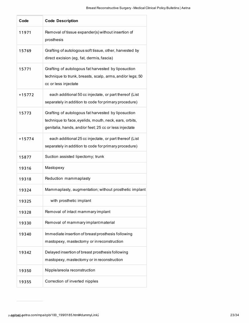
Code Code Description
CPT codes covered if selection criteria are met:
1 1 9 20 Tattooing, intradermal introduction of insoluble opaque
pigments to correct color defects of skin, including
micropigmentation; 6.0 sq cm or less
1 1 9 21 6.1 to 20.0 sq cm
+ 1 1 9 22 each additional 20.0 sq cm (List separately in
addition to code for primary procedure)
1 1 9 50 -
1 1 9 54
Subcutaneous injection of filling material (eg, collagen)
1 1 9 70 Replacement of tissue expander with permanent
prosthesis
aetnet.aetna.com/mpa/cpb/100_199/0185.html#dummyLink2 Proprietary 22/34
Breast Reconstructive Surgery - Medical Clinical Policy Bulletins | Aetna
Code Code Description
1 1 9 71 Removal of tissue expander(s) without insertion of
prosthesis
1 5 7 69 Grafting of autologous soft tissue, other, harvested by
direct excision (eg, fat, dermis, fascia)
1 5 7 71 Grafting of autologous fat harvested by liposuction
technique to trunk, breasts, scalp, arms, and/or legs; 50
cc or less injectate
+ 1 5 77 2 each additional 50 cc injectate, or part thereof (List
separately in addition to code for primary procedure)
1 5 7 73 Grafting of autologous fat harvested by liposuction
technique to face, eyelids, mouth, neck, ears, orbits,
genitalia, hands, and/or feet; 25 cc or less injectate
+ 1 5 77 4 each additional 25 cc injectate, or part thereof (List
separately in addition to code for primary procedure)
1 5 8 77 Suction assisted lipectomy; trunk
1 9 3 16 Mastopexy
1 9 3 18 Reduction mammaplasty
1 9 3 24 Mammaplasty, augmentation; without prosthetic implant
1 9 3 25 with prosthetic implant
1 9 3 28 Removal of intact mammary implant
1 9 3 30 Removal of mammary implant material
1 9 3 40 Immediate insertion of breast prosthesis following
mastopexy, mastectomy or in reconstruction
1 9 3 42 Delayed insertion of breast prosthesis following
mastopexy, mastectomy or in reconstruction
1 9 3 50 Nipple/areola reconstruction
1 9 3 55 Correction of inverted nipples
aetnet.aetna.com/mpa/cpb/100_199/0185.html#dummyLink2 Proprietary 23/34

Breast Reconstructive Surgery - Medical Clinical Policy Bulletins | Aetna
Code Code Description
1 9 3 57 Breast reconstruction, immediate or delayed, with tissue
expander, including subsequent expansion
1 9 3 61 Breast reconstruction with latissimus dorsi flap, without
prosthetic implant
1 9 3 64 Breast reconstruction with free flap
1 9 3 66 Breast reconstruction with other technique
1 9 3 67 Breast reconstruction with transverse rectus abdominus
myocutaneous flap (TRAM), single pedicle, including
closure of donor site;
1 9 3 68 with microvascular anastomosis (supercharging)
1 9 3 69 Breast reconstruction with transverse rectus abdominis
myocutaneous flap (TRAM), double pedicle, including
closure of donor site
1 9 3 70 Open periprosthetic capsulotomy, breast
1 9 3 71 Periprosthetic capsulectomy, breast
1 9 3 80 Revision of reconstructed breast
1 9 3 96 Preparation of moulage for custom breast implant
2 0 9 26 Tissue grafts, other (eg, paratenon, fat, dermis)
Other CPT codes related to the CPB:
1 9 1 20 -
1 9 1 26
Excision lesion of breast
1 9 3 00 -
1 9 3 07
Mastectomy procedures
2 1 7 40 -
2 1 7 43
Reconstructive repair of pectus excavatum or carinatum
HCPCS codes covered if selection criteria are met:
C 1 7 81 Mesh (implantable) [Cortiva]
C 1 7 89 Prosthesis, breast (implantable)
aetnet.aetna.com/mpa/cpb/100_199/0185.html#dummyLink2 Proprietary 24/34

Breast Reconstructive Surgery - Medical Clinical Policy Bulletins | Aetna
Code Code Description
C 9 3 58 Dermal substitute, native, non- denatured collagen, fetal
bovine origin (surgimend collagen matrix), per 0.5
square centimeters
C 9 3 60 Dermal substitute, native, non- denatured collagen,
neonatal bovine origin (SurgiMend Collagen Matrix), per
0.5 square centimeters
L 8 6 00 Implantable breast prosthesis, silicone or equal
Q4 1 1 6 Alloderm, per square centimeter
Q4 1 2 2 DermACELL, per sq cm
Q4 1 2 8 Flex HD, Allopatch HD, or Matrix HD, per square
centimeter
Q4 1 3 0 Strattice TM, per sq cm
S 2 0 66 Breast reconstruction with gluteal artery perforator
(GAP) flap, including harvesting of the flap,
microvascular transfer, closure of donor site and shaping
the flap into a breast, unilateral
S 2 0 67 Breast reconstruction of a single breast with "stacked"
deep inferior epigastric perforator (DIEP) flap(s) and/ or
gluteal artery perforator (GAP) flap(s), including
harvesting of the flap(s), microvascular transfer, closure
of donor site(s) and shaping the flap into a breast,
unilateral
S 2 0 68 Breast reconstruction with deep inferior epigastric
perforator (DIEP) flap or superficial inferior epigastric
artery (SIEA) flap, including harvesting of the flap,
microvascular transfer, closure of donor site and shaping
the flap into a breast, unilateral
Other HCPCS codes related to the CPB:
L 8 0 20 -
L 8 0 39
Breast prostheses
ICD-10 codes covered if selection criteria are met:
aetnet.aetna.com/mpa/cpb/100_199/0185.html#dummyLink2 Proprietary 25/34

Breast Reconstructive Surgery - Medical Clinical Policy Bulletins | Aetna
Code Code Description
C 5 0 .011 -
C 5 0 .929
Malignant neoplasm of breast
C 7 9 .81 Secondary malignant neoplasm of breast
D 0 5 .00 -
D 0 5 .92
Carcinoma in situ of breast
N6 0 .1 1 -
N6 0 .1 9
Diffuse cystic mastopathy [severe fibrocystic disease]
Z 8 5 .3 Personal history of malignantneoplasm of breast
Z 9 0 .10 -
Z 9 0 .13
Acquired absence of breast [following medically
necessary mastectomy or lumpectomy resulting in
significant deformity]
:
1. Kotwall CA. Breast cancer treatment and chemoprevention. Can
Fam Physician. 1999;45:1917-1924.
2. Polednak AP. Postmastectomy breast reconstruction in
Connecticut: Trends and predictors. Plast Reconstr Surg.
1999;104(3):669-673.
3. Brandberg Y, Malm M, Rutqvist LE, et al. A prospective
randomised study (named SVEA) of three methods of delayed
breast reconstruction. Study, design, patients' preoperative
problems and expectations. Scand J Plast Reconstr Surg Hand
Surg. 1999;33(2):209-216.
4. Delay E, Jorquera F, Pasi P, Gratadour AC. Autologous latissimus
breast reconstruction in association with the abdominal
advancement flap: A new refinement in breast reconstruction.
Ann Plast Surg. 1999;42(1):67-75.
5. Spear SL, Pennanen M, Barter J, Burke JB. Prophylactic
mastectomy, oophorectomy, hysterectomy, and immediate
transverse rectus abdominis muscle flap breast reconstruction in
Proprietary 26/34

Breast Reconstructive Surgery - Medical Clinical Policy Bulletins | Aetna
a BRCA- 2-positive patient. Plast Reconstr Surg. 1999;103(2):548-
553; discussion 554-555.
6. Yeh KA, Lyle G, Wei JP, Sherry R. Immediate breast reconstruction
in breast cancer: Morbidity and outcome. Am Surg.
1998;64(12):1195-1199.
7. Papp C, Wechselberger G, Schoeller T. Autologous breast
reconstruction after breast-conserving cancer surgery. Plast
Reconstr Surg. 1998;102(6):1932-1936; discussion 1937-1938.
8. Chavoin JP, Grolleau JL, Lanfrey E, Lavigne B. Breast
reconstruction after mastectomy for cancer. Rev Prat.
1998;48(1):67-70.
9. Delay E, Gounot N, Bouillot A, Zlatoff P, et al. Autologous
latissimus breast reconstruction: A 3-year clinical experience with
100 patients. Plast Reconstr Surg. 1998;102(5):1461-1478.
10. Bostwick J. Breast reconstruction after mastectomy and breast
implants. Current status in the USA. Ann Chir Plast Esthet.
1997;42(2):100-106.
11. Salmon RJ. Evolution of the surgery of cancer of the breast. Bull
Cancer. 1998;85(6):539-543.
12. Strozzo MD. An overview of surgical management of stage I and
stage II breast cancer for the primary care provider. Lippincotts
Prim Care Pract. 1998;2(2):160-169.
13. Hidalgo DA, Borgen PJ, Petrek JA, et al. Immediate reconstruction
after complete skin-sparing mastectomy with autologous tissue. J
Am Coll Surg. 1998;187(1):17-21.
14. Evans GR, Kroll SS. Choice of technique for reconstruction. Clin
Plast Surg. 1998;25(2):311-316.
15. Papp C, McCraw JB. Autogenous latissimus breast reconstruction.
Clin Plast Surg. 1998;25(2):261-266.
16. Kroll SS. Bilateral breast reconstruction. Clin Plast Surg.
1998;25(2):251-259.
17. Serletti JM, Moran SL. The combined use of the TRAM and
expanders/implants in breast reconstruction. Ann Plast Surg.
1998;40(5):510-514.
18. Bhatty M A, Berry RB. Nipple-areola reconstruction by tattooing
and nipple sharing. Br J Plast Surg. 1997;50(5):331-334.
19. Blondeel PN. One hundred free DIEP flap breast reconstructions:
A personal experience. Br J Plast Surg. 1999;52(2):104-111.
Proprietary 27/34

Breast Reconstructive Surgery - Medical Clinical Policy Bulletins | Aetna
20. Feller AM. Reconstruction of the female breast with free
transverse lower abdominal flap as perforator flap. Langenbecks
Arch Chir Suppl Kongressbd. 1998;115:971-972.
21. Hamdi M, Weiler-Mithoff EM, Webster MH. Deep inferior
epigastric perforator flap in breast reconstruction: Experience
with the first 50 flaps. Plast Reconstr Surg. 1999;103(1):86-95.
22. Blondeel PN, Boeckx WD. Refinements in free flap breast
reconstruction: The free bilateral deep inferior epigastric
perforator flap anastomosed to the internal mammary artery. Br
J Plast Surg. 1994;47(7):495-501.
23. National Organization for Rare Disorders, Inc. (NORD). Poland
syndrome. In: NORD Rare Disease Database. New Fairfield, CT:
NORD; 1996.
24. Blondeel PN, Demuynck M, Mete D, et al. Sensory nerve repair in
perforator flaps for autologous breast reconstruction:
Sensational or senseless? Br J Plast Surg. 1999;52(1):37-44.
25. Blondeel N, Vanderstraeten GG, Monstrey SJ, et al. The donor site
morbidity of free DIEP flaps and free TRAM flaps for breast
reconstruction. Br J Plast Surg. 1997;50(5):322-330.
26. Nahabedian MY, Dooley W, Singh N, et al. Contour abnormalities
of the abdomen after breast reconstruction with abdominal
flaps: The role of muscle preservation. Plast Reconstr Surg.
2002;109(1):91-101.
27. Yap LH, Whiten SC, Forster A, et al. The anatomical and
neurophysiological basis of the sensate free TRAM and DIEP
flaps. Br J Plast Surg. 2002;55(1):35-45.
28. Guzzetti T, Thione A. Successful breast reconstruction with a
perforator to deep inferior epigastric perforator flap. Ann Plast
Surg. 2001;46(6):641-643.
29. Keller A. The deep inferior epigastric perforator free flap for
breast reconstruction. Ann Plast Surg. 2001;46(5):474-480.
30. Kroll SS. Fat necrosis in free transverse rectus abdominis
myocutaneous and deep inferior epigastric perforator flaps. Plast
Reconstr Surg. 2000;106(3):576-583.
31. Rainsbury RM. Breast-sparing reconstruction with latissimus
dorsi miniflaps. Eur J Surg Oncol. 2002;28(8):891-895.
32. Sauven P; Association of Breast Surgery Family History
Guidelines Panel. Guidelines for the management of women at
Proprietary 28/34

Breast Reconstructive Surgery - Medical Clinical Policy Bulletins | Aetna
increased familial risk of breast cancer. Eur J Cancer.
2004;40(5):653-665.
33. Fischbacher C. Immediate versus delayed breast reconstruction.
STEER: Succint and Timely Evaluated Evidence Reviews. Bazian,
Ltd., eds. London, UK: Wessex Institute for Health Research and
Development, University of Southampton; 2002; 2(17):1-18.
34. Fischbacher C. Cosmetic breast augmentation. STEER: Succint
and TImely Evaluated Evidence Reviews. Bazian, Ltd., eds.
London, UK: Wessex Institute for Health Research and
Development, University of Southampton; 2003:3(1):1-12.
35. Edlich RF, Winters KL, Faulkner BC, et al. Advances in breast
reconstruction after mastectomy. J Long Term Eff Med Implants.
2005;15(2):197-207.
36. Fentiman IS, Hamed H. Breast reconstruction. Int J Clin Pract.
2006;60(4):471-474.
37. Javaid M, Song F, Leinster S, et al. Radiation effects on the
cosmetic outcomes of immediate and delayed autologous breast
reconstruction: An argument about timing. J Plast Reconstr
Aesthet Surg. 2006;59(1):16-26.
38. Chang DS, McGrath MH. Management of benign tumors of the
adolescent breast. Plast Reconstr Surg. 2007;120(1):13e-19e.
39. National Institute for Health and Clinical Excellence (NICE).
Laparoscopic mobilisation of the greater omentum for breast
reconstruction. Interventional Procedure Guidance 253. London,
UK: NICE; 2008.
40. Losken A, Hamdi M. Partial breast reconstruction: Current
perspectives. Plast Reconstr Surg. 2009;124(3):722-736.
41. Humphreys K. Autologous fat injection for breast reconstruction.
Horizon Scanning Report. Health Policy Advisory Committee on
Technology (HealthPACT), Australia and New Zealand Horizon
Scanning Network (ANZHSN); August 2008.
42. Hyakusoku H, Ogawa R, Ono S, et al. Complications after
autologous fat injection to the breast. Plast Reconstr Surg.
2009;123(1):360-370; discussion 371-372.
43. Gutowski KA; ASPS Fat Graft Task Force. Current applications and
safety of autologous fat grafts: A report of the ASPS fat graft task
force. Plast Reconstr Surg. 2009;124(1):272-280.
44. Mizuno H, Hyakusoku H. Fat grafting to the breast and adipose-
derived stem cells: Recent scientific consensus and controversy.
Proprietary 29/34

Breast Reconstructive Surgery - Medical Clinical Policy Bulletins | Aetna
Aesthet Surg J. 2010;30(3):381-387.
45. Cook Biotech Incorporated. Biodesign Nipple Reconstruction
Cylinder [website]. West Lafayette, IN: Cook Biotech Incoporated;
2009. Available at:
http://www.cookmedical.com/sur/content/mmedia/FP0054-
01C.pdf. Accessed January 24, 2012.
46. National Institute for Health and Clinical Excellence (NICE). Breast
reconstruction using lipomodelling after breast cancer
treatment. Interventional Procedure Guidance 417. London, UK:
NICE; January 2012.
47. Phillips BT, Bishawi M, Dagum AB, et al. A systematic review of
antibiotic use and infection in breast reconstruction: What is the
evidence? Plast Reconstr Surg. 2013;131(1):1-13.
48. Claro F Jr, Figueiredo JC, Zampar AG, et al. Applicability and safety
of autologous fat for reconstruction of the breast. Br J Surg.
2012;99(6):768-80.
49. Llewellyn-Bennett R, Greenwood R, Benson JR, et al. Randomized
clinical trial on the effect of fibrin sealant on latissimus dorsi
donor-site seroma formation after breast reconstruction. Br J
Surg. 2012;99(10):1381-1388.
50. Ibrahim AM, Ayeni OA, Hughes KB, et al. Acellular dermal
matrices in breast surgery: A comprehensive review. Ann Plast
Surg. 2013;70(6):732-738.
51. Cheng A, Saint-Cyr M. Comparison of different ADM materials in
breast surgery. Clin Plast Surg. 2012;39(2):167-175.
52. Allen RJ, Haddock NT, Ahn CY, Sadeghi A. Breast reconstruction
with the profunda artery perforator flap. Plast Reconstr Surg.
2012;129(1):16e-23e.
53. DellaCroce FJ, Sullivan SK, Trahan C, Jenkins CE. Body lift
perforator flap breast reconstruction: A review of 100 flaps in 25
cases. Plast Reconstr Surg. 2012;129(3):551-561.
54. Morris D. Principles of grafts and flaps for reconstructive surgery.
UpToDate [serial online]. Waltham, MA: UpToDate; reviewed
December 2013.
55. Nahabedian M. Breast reconstruction in women with breast
cancer. UpToDate [serial online]. Waltham, MA:
UpToDate; reviewed December 2013.
56. Tanna N, Broer PN, Weichman KE, et al. Microsurgical breast
reconstruction for nipple-sparing mastectomy. Plast Reconstr
Proprietary 30/34
Breast Reconstructive Surgery - Medical Clinical Policy Bulletins | Aetna
Surg. 2013;131(2):139e-147e.
57. Levine SM, Snider C, Gerald G, et al. Buried flap reconstruction
after nipple-sparing mastectomy: Advancing toward single-stage
breast reconstruction. Plast Reconstr Surg. 2013;132(4):489e-
497e.
58. Weichman KE, Broer PN, Tanna N, et al. The role of autologous
fat grafting in secondary microsurgical breast reconstruction.
Ann Plast Surg. 2013;71(1):24-30.
59. Healy C, Allen RJ Sr. The evolution of perforator flap breast
reconstruction: Twenty years after the first DIEP flap. J Reconstr
Microsurg. 2014;30(2):121-125.
60. Tsoi B, Ziolkowski NI, Thoma A, et al. Safety of tissue
expander/implant versus autologous abdominal tissue breast
reconstruction in postmastectomy breast cancer patients: A
systematic review and meta-analysis. Plast Reconstr Surg.
2014;133(2):234-249.
61. Valdatta L, Cattaneo AG, Pellegatta I, et al. Acellular dermal
matrices and radiotherapy in breast reconstruction: A systematic
review and meta-analysis of the literature. Plast Surg Int.
2014;2014:472604.
62. Agha RA, Fowler AJ, Herlin C, et al. Use of autologous fat grafting
for breast reconstruction: A systematic review with meta-analysis
of oncological outcomes. J Plast Reconstr Aesthet Surg.
2015;68(2):143-161.
63. Masia J, Bordoni D, Pons G, et al. Oncological safety of breast
cancer patients undergoing free-flap reconstruction and
lipofilling. Eur J Surg Oncol. 2015;41(5):612-616.
64. Letter from Jan Welch, Center for Devices and Radiological
Health, U.S. Food and Drug Administration (FDA), Silver Spring,
MD to Robert Beuhler, TEI Biosciences, Inc., Boston, MA, May 29,
2015.
65. U.S. Food and Drug Administration (FDA). MAUDE Adverse Event
Report: TEI Biosciences Inc. Surgimend Surgical Mesh, July 17,
2009.
66. Bullocks JM. DermACELL: A novel and biocompatible acellular
dermal matrix in tissue expander and implant-based breast
reconstruction. Eur J Plast Surg. 2014;37(10):529-538.
67. Vashi C. Clinical outcomes for breast cancer patients undergoing
mastectomy and reconstruction with use of DermACELL, a
Proprietary 31/34

Breast Reconstructive Surgery - Medical Clinical Policy Bulletins | Aetna
sterile, room temperature acellular dermal matrix. Plast Surg Int.
2014;2014:704323.
68. Rocco N, Rispoli C, Moja L, et al. Different types of implants for
reconstructive breast surgery. Cochrane Database Syst Rev.
2016;(5):CD010895.
69. Chen TA, Momeni A, Lee GK. Clinical outcomes in breast cancer
expander-implant reconstructive patients with radiation therapy.
J Plast Reconstr Aesthet Surg. 2016;69(1):14-22.
70. Winocour S, Saksena A, Oh C, et al. A systematic review of
comparison of autologous, allogeneic, and synthetic
augmentation grafts in nipple reconstruction. Plast Reconstr
Surg. 2016;137(1):14e-23e.
71. Zenn MR, Salzberg CA. A direct comparison of Alloderm-Ready to
Use (RTU) and DermACELL in immediate breast implant
reconstruction. Eplasty. 2016;16:e23.
72. Pittman TA, Fan KL, Knapp A, et al. Comparison of different
acellular dermal matrix (ADM) in breast reconstruction: The
50/50 Study. Plast Reconstr Surg. 2017;139(3):521-528.
73. Lee BT, Agarwal JP, Ascherman JA, et al. Evidence-based clinical
practice guideline: Autologous breast reconstruction with DIEP or
pedicled TRAM abdominal flaps. Plast Reconstr Surg.
2017;140(5):651e-664e
74. Cooke AL, Diaz-Abele J, Hayakawa T, et al. Radiation therapy
versus no radiation therapy to the neo-breast following skin-
sparing mastectomy and immediate autologous free flap
reconstruction for breast cancer: Patient-reported and surgical
outcomes at 1 year-A mastectomy reconstruction outcomes
consortium (MROC) substudy. Int J Radiat Oncol Biol Phys.
2017;99(1):165-172.
75. Olsen MA, Nickel KB, Fox IK, et al. Comparison of wound
complications after immediate, delayed, and secondary breast
reconstruction procedures. JAMA Surg. 2017;152(9):e172338.
76. Bennett KG, Qi J, Kim HM, Hamill JB, et al. Association of fat
grafting with patient-reported outcomes in postmastectomy
breast reconstruction. JAMA Surg. 2017;152(10):944-950.
77. Butterfield JL. 440 Consecutive immediate, implant-based, single-
surgeon breast reconstructions in 281 patients: A comparison of
early outcomes and costs between SurgiMend fetal bovine and
Proprietary 32/34

Breast Reconstructive Surgery - Medical Clinical Policy Bulletins | Aetna
AlloDerm human cadaveric A cellular dermal matrices. Plast
Reconstr Surg. 2013;131(5):940-951.
78. Ricci JA, Treiser MD, Tao R, et al. Predictors of complications and
comparison of outcomes using SurgiMend fetal bovine and
AlloDerm human cadaveric acellular dermal matrices in implant-
based breast reconstruction. Plast Reconstr Surg.
2016;138(4):583e-591e.
79. Ball JF, Sheena Y, Tarek Saleh DM, et al. A direct comparison of
porcine (Strattice™) and bovine (Surgimend™) acellular dermal
matrices in implant-based immediate breast reconstruction. J
Plast Reconstr Aesthet Surg. 2017;70(8):1076-1082
80. Myung Y, Heo CY. Relationship between obesity and surgical
complications after reduction mammaplasty: A systematic
literature review and meta-analysis. Aesthet Surg J.
2017;37(3):308-315.
81. Panayi AC, Agha RA, Sieber BA, Orgill DP. Impact of obesity on
outcomes in breast reconstruction: A systematic review and
meta-analysis. J Reconstr Microsurg. 2018;34(5):363-375.
82. Mazari FAK, Wattoo GM, Kazzazi NH, et al. The comparison of
Strattice and SurgiMend in acellular dermal matrix-assisted,
implant-based immediate breast reconstruction. Plast Reconstr
Surg. 2018;141(2):283-293.
Proprietary 33/34
Breast Reconstructive Surgery - Medical Clinical Policy Bulletins | Aetna
Copyright Aetna Inc. All rights reserved. Clinical Policy Bulletins are developed by Aetna to assist in administering plan benefits and
constitute neither offers of coverage nor medical advice. This Clinical Policy Bulletin contains only a partial, general description of plan or
program benefits and does not constitute a contract. Aetna does not provide health care services and, therefore, cannot guarantee any
results or outcomes. Participating providers are independent contractors in private practice and are neither employees nor agents of Aetna
or its affiliates. Treating providers are solely responsible for medical advice and treatment of members. This Clinical Policy Bulletin may be
updated and therefore is subject to change.
Copyright © 2001-2020 Aetna Inc.
Proprietary 34/34

AETNA BETTER HEALTH® OF PENNSYLVANIA
Amendment to Aetna Clinical Policy Bulletin Number: 0185
Breast Reconstructive Surgery
There are no amendments for Medicaid.
www.aetnabetterhealth.com/pennsylvania updated 04/15/2020
Proprietary