


Bahasa
Halaman
Hukum


Suppression of Autophagy Enhanced Growth Inhibitionand Apoptosis of Interferon-β in Human Glioma Cells
Yubin Li & Haiyan Zhu & Xian Zeng & Jiajun Fan &
Xiaolu Qian & Shaofei Wang & Ziyu Wang & Yun Sun &
Xiaodan Wang & Weiwu Wang & Dianwen Ju
Received: 14 November 2012 /Accepted: 3 January 2013 /Published online: 18 January 2013# Springer Science+Business Media New York 2013
Abstract Interferon-beta (IFN-β) is a cytokine with anti-viral, anti-proliferative, and immunomodulatory effects. Inthis study, we investigated the effects of IFN-β on theinduction of autophagy and the relationships among autoph-agy, growth inhibition, and apoptosis induced by IFN-β inhuman glioma cells. We found that IFN-β induced autopha-gosome formation and conversion of microtubule associatedprotein 1 light chain 3 (LC3) protein, whereas it inhibitedcell growth through caspase-dependent cell apoptosis. TheAkt/mTOR signaling pathway was involved in autophagyinduced by IFN-β. A dose- and time-dependent increase ofp-ERK 1/2 expression was also observed in human glioma
cells treated with IFN-β. Autophagy induced by IFN-β wassuppressed when p-ERK1/2 was impaired by treatment withU0126. We also demonstrated that suppression of autoph-agy significantly enhanced growth inhibition and cell apo-ptosis induced by IFN-β, whereas inhibition of caspase-dependent cell apoptosis impaired autophagy induced byIFN-β. Collectively, these findings indicated that autophagyinduced by IFN-β was associated with the Akt/mTOR andERK 1/2 signaling pathways, and inhibition of autophagycould enhance the growth inhibitory effects of IFN-β andincrease apoptosis in human glioma cells. Together, thesefindings support the possibility that autophagy inhibitorsmay improve IFN-β therapy for gliomas.
Keywords Autophagy . IFN-β . Glioma cells . Growthinhibition . Apoptosis
Introduction
Malignant gliomas are the most common and lethal primarybrain tumors in the central nervous system, and account for30 % of adult primary brain tumors [1]. Even though multi-modality treatments—including surgery, radiotherapy, andchemotherapy—have been developed to improve therapeu-tic efficacy, over the past 30 years, no significant improve-ments in the survival of patients suffering from this diseasehave been achieved [2]. Because malignant glioma cellsusually infiltrate deeply into normal tissues, and the com-plete removal of glioma is almost impossible, leading to ahigh incidence of tumor recurrence [3], the median survivaltime from diagnosis for those with a glioma is still only14.5 months [4]. Therefore, the development of novel ther-apeutic approaches is urgently needed.
Y.L. and H.Z. contributed equally to this work.
Y. Li :H. Zhu :X. Zeng : J. Fan : S. Wang : Z. Wang :Y. Sun :D. Ju (*)Department of Biosynthesis, School of Pharmacy,Fudan University, Shanghai 201203, Chinae-mail: [email protected]
Y. Li :W. WangKey Laboratory of Microbiological Engineering of AgriculturalEnvironment, Ministry of Agriculture, Department of Microbiology,College of Life Science, Nanjing Agricultural University, Nanjing,Jiangsu 210095, China
X. QianCollege of Veterinary Medicine, Nanjing Agricultural University,Nanjing, Jiangsu 210095, China
X. WangSchool of Life Science and Technology, Tongji University,Shanghai 200092, China
D. JuThe Key Laboratory of Smart Drug Delivery, Ministry of Education& The People’s Liberation Army, School of Pharmacy,Fudan University, Shanghai 201203, China
Mol Neurobiol (2013) 47:1000–1010DOI 10.1007/s12035-013-8403-0
IFN-β is a type I interferon that exhibits anti-viral, immu-nomodulating, and anti-tumor activities. It is widely usedalone or in combination with other anti-tumor agents, suchas nitrosoureas and temozolomide, in the treatment of ma-lignant gliomas [5]. Although IFN-β can inhibit growth orinduce apoptosis in human glioma cells [6], its therapeuticefficacy is still limited. Increasing evidence suggests thatmalignant glioma cells can be resistant to chemotherapy andradiotherapy because of resistance to apoptosis [7]. Studiesof multi-drug resistance in chronic lymphocytic leukemiaand breast cancer cells suggested that autophagy plays a keyrole in tumor resistance [8, 9].
In the present investigation, we hypothesized that autoph-agy could partly explain the resistance of glioma cells toIFN-β therapy, and that abrogation of autophagy induced byIFN-β may restore drug sensitivity and enhance the effectsof IFN-β on growth inhibition and apoptosis in humanglioma cells. Autophagy is an evolutionarily conserved pro-cess that is involved in aging, neurodegenerative diseases,and cancer [10]. It is characterized by the formation ofdouble membranes, and is often activated during cell star-vation and other stresses [11]. The triggering of autophagyis mostly associated with inhibition of the mammalian targetof rapamycin complex 1 (mTORC1), which causes theactivation of autophagy-related proteins (Atgs) and signal-ing pathways [12, 13]. Though activation of the autophagycan either protect cells or initiate type II programmed celldeath [14, 15], greater evidence supports the idea thatautophagy is cytoprotective, particularly in cancer therapy[16–18].
In this study, we first demonstrated IFN-β-inducedautophagy in U251MG and U87MG glioma cells. TheAkt/mTOR and MEK 1/2 signaling pathways were in-volved in autophagy induced by IFN-β. Suppression ofautophagy significantly enhanced growth inhibition andcell apoptosis induced by IFN-β, whereas inhibition ofcaspase-dependent cell apoptosis impaired autophagyinduced by IFN-β. This is the first report showing thatautophagy induced by IFN-β plays a critical cytoprotec-tive role in the inhibition of glioma cell growth and cellapoptosis. This finding may contribute to the applicationof autophagy inhibitors for the improvement of IFN-βtherapy for gliomas in clinic.
Materials and Methods
Cell Culture
The human glioma cell lines U251MG and U87MG wereobtained from the Cell Bank of the Shanghai Institutes forBiological Sciences, Chinese Academy of Science (Shang-hai, China). Both cells were cultured in Dulbecco's modified
eagle's medium (DMEM) (Invitrogen, San Diego, CA,USA) supplemented with 10 % heat-inactivated fetal bovineserum (Invitrogen, San Diego, CA, USA), 2 mM L-gluta-mine, 100 U/ml penicillin, and 100 μg/ml streptomycin at37 °C in air containing 5 % CO2.
Reagents and Antibodies
IFN-β was purchased from Bayer Sharing Pharma AG(Leverkusen, Germany). The specific activity of the pro-tein was 8×10⁶IU/ml. The PI3K inhibitor 3-methyladenine(3-MA) was obtained from EMDChemicals, Inc. (San Diego,CA, USA). The lysosomal inhibitor chloroquine (CQ) wasobtained from Sigma (St Louis, MO, USA). For experimentaluse, IFN-β, 3-MA, and CQ were prepared and diluted withDMEM. Cyto-ID Green dye was purchased from ENZO LifeSciences, Inc. (Farmingdale, NY, USA). Z-VAD-fmk wasprovided by Beyotime Institute of Biotechnology (Haimen,China), and the MEK1/2 inhibitor U0126 was obtained fromCell Signaling Technology (Danvers, MA, USA). The anti-bodies anti-LC3B, anti-caspase-3, anti-cleaved caspase-3,anti-phospho-Akt (Ser473), anti-Akt1, anti-phospho-mTOR(Ser2448), anti-mTOR, anti-phospho-p44/42 MAPK (Erk1/2) (Thr202/Tyr204), anti-p44/42 MAPK (Erk1/2), anti-β-actin, and anti-tubulin were obtained from Cell SignalingTechnology. Anti-p70 S6 Kinase Phospho (pS371) waspurchased from Epitomics (Burlingame, CA, USA). Thesecondary antibodies horseradish peroxidase (HRP)-con-jugated goat anti-mouse and anti-rabbit immunoglobulinG (IgG) were obtained from MR Biotech (Shanghai,China).
Cell Proliferation Assay
Cells were plated in 96-well flat bottom plates at a density of1×104 cells/ml. After 24 h of incubation, different concen-trations of IFN-β were added for additional time periods.The cell proliferation assay was performed using a 3-(4.5-dimethylthiazol-2-yl)-2,5-diphenyltetrazolium bromide(MTT) solution (0.5 mg/ml). The plates were incubated ina humidified incubator at 37 °C for 4 h, then the mediumwas removed and formazan dye was solubilized withDMSO; the optical density (OD) was measured at an absor-bance wavelength of 570 nm.
Western Blot Analysis
U251MG and U87MG cells were harvested, washed in coldphosphate-buffered saline (PBS) twice, and re-suspended inlysis buffer (Beyotime Biotechnology, China) for 30 min onice. The lysate was then centrifuged at 13,000×g for 5 min at4 °C. Protein concentrations were determined by the bicin-choninic acid (BCA) method. Equivalent amounts of protein
Mol Neurobiol (2013) 47:1000–1010 1001

were loaded onto gels and separated by SDS–PAGE, and thenelectro-transferred to polyvinylidene fluoride (PVDF) mem-branes. The membranes were blocked in TBST contain-ing 5 % nonfat milk and incubated with an appropriateantibody at 4 °C overnight. After incubation with thesecond antibody conjugated with horseradish peroxidase,membranes were visualized using an enhanced chemilu-minescent detection kit (Pierce, Rockford, IL, USA).Densitometric values of protein bands were quantifiedby the IQuantTL software (GE Healthcare, USA).
Transmission Electron Microscopy Analysis
U251MG and U87MG cells were fixed with 2 % glu-taraldehyde in DMEM medium for 15 min, and thenfixed in 2 % glutaraldehyde with 0.1 M Na cacodyla-te/HCl (pH7.4) for 30 min. After extensive wash in0.2 M Na cacodylate/HCl (pH7.4) for three times, cellswere fixed with 1 % OsO4–0.15 M Na cacodylate/HCl(pH7.4) for 30 min. The cells were then dehydrated inan increasing gradient of ethanol and polymerized at60 °C for 48 h [19]. Samples were prepared and ana-lyzed with a JEM 1230 transmission electron micro-scope (JEOL, USA, Inc.) at 60 kV. Micrographs weretaken at×5,000 or×10,000 magnification.
Confocal Immunofluorescence
U251MG and U87MG cells were plated in cell culturedishes with glass bottoms. After 24 h of incubation, cellswere treated with 2,000 IU/ml of IFN-β for another48 h. Positive controls were treated with the autophagyinducer rapamycin at 50 nM for 18 h. The cells weredisposed with Cyto-ID® Autophagy Detection Kitaccording to the manufacturer’s protocol [20]. Briefly,cells were washed twice with 1× assay buffer, and thentreated with Cyto-ID® Green dye and Hoechst 33342 at37 °C for 30 min. After incubation, cells were washedwith 1× assay buffer and immediately analyzed with anOlympus fluorescence microscope.
Flow Cytometry Analysis
Apoptosis was detected using the Annexin V–FITC/PI Ap-optosis Detection Kit (BD Biosciences, San Diego, CA,USA). U251MG and U87MG cells were treated withIFN-β and different autophagy inhibitors for 48 h, thenadherent and floating cells were harvested and washed withcold PBS, and re-suspended in 1× binding buffer at a con-centration of 1×10⁶ cells/ml. Cells were incubated withAnnexin V–fluorescein isothiocyanate in the dark for30 min, and then incubated with propidium iodide for2 min and immediately analyzed by flow cytometry
according to the manufacturer’s instructions. Analysis wasperformed using a FACSCalibur flow cytometer (Becton-Dickinson, Fullerton, CA, USA).
siRNA Transfection
U251MG and U87MG cells were seeded in 6-well plate or96-well plate at 1×10⁵/ml. After 24h of incubation, cellswere transfected with 50nM siRNA using X-tremeGENEsiRNA Transfection Reagent (Roche). Forty-eight hourspost-transfection cells were treated with control medium,IFN-β for 48h as indicated, and the relevant assays per-formed. The siRNA used were all synthesized by Ribobio(Guangzhou, China). The sequences were as follows:ATG5a: 5'-GTGAGATATGGTTTGAATA-3'; ATG5b: 5'-GCAACTCTGGATGGGATTG-3'; ATG5c: 5'-GGAACAT-CACAGTACATTT-3'.
Statistical Analysis
GraphPad Prism 5 was used for all statistical analyses. Alldata are presented as means ± standard deviations (SD). Allcomparisons were performed using the Student’s t test (two-tailed), and values of P <0.05 were considered statisticallysignificant.
Results
Autophagy Induced by IFN-β in Human Glioma Cells
To investigate the induction of autophagy by IFN-β, we firstexamined the morphology of U251MG and U87MG cellsafter exposure to 2,000 IU/ml of IFN-β for 48 h by trans-mission electron microscopy (TEM). As shown in Fig. 1a,cells treated with IFN-β presented numerous vacuolizationsand electron-dense inclusions. Upon magnification, theseinclusions were clearly double-layered membrane structuresthat appeared to be autophagosomes.
We also employed an autophagy detection kit to detectautophagy by fluorescence microscopy [20]. As shown inFig. 1b, green fluorescence was detected in the cytoplasm ofthe IFN-β-treated U251MG and U87MG cells, but not inthe negative control samples. Cells treated with rapamycinserved as the positive controls.
In addition, we examined the expression of themicrotubule-associated protein 1 light chain 3 (LC3-I) and its membrane-bound lipidated form LC3-II by Western blot analysis becauseLC3-II is expressed on the membranes of autophagosomes.The expression of LC3-II increased obviously in IFN-β-treated U251MG and U87MG cells (Fig. 1c), confirming theinduction of autophagy by IFN-β. We also determined thevalues of LC3-II/LC3-I by counting relative intensities of
1002 Mol Neurobiol (2013) 47:1000–1010

LC3-II and LC-I, as shown in Fig. 1d, and the values of LC3-II/LC3-I were significantly higher in IFN-β-treated gliomacells (P<0.05).
Collectively, our data indicate that IFN-β induced signif-icant autophagy in human glioma U251MG and U87MGcells.
Mol Neurobiol (2013) 47:1000–1010 1003

Suppression of Autophagy Enhanced Growth InhibitionInduced by IFN-β
To study the role of autophagy in the growth-inhibiting effectsof IFN-β on human glioma cells, we used two autophagy
inhibitors, 3-MA and CQ. 3-MA is a phosphatidylinositol 3-phosphate kinase inhibitor and decreases autophagosomicLC3 (LC3-II). CQ inhibits autophagy at a later stage byinhibiting fusion between the autophagosome and lysosome,which can significantly elevate the protein level of LC3-II [21,22]. Western blot analysis showed that 3-MA inhibitedautophagy induced by IFN-β in U251MG and U87MG cells,and decreased LC3-II formation. In contrast, the addition ofCQ to IFN-β significantly enhanced LC3-II protein levelswhen compared with IFN-β alone (Fig. 2a). Compared withIFN-β treatment alone, the combination of 2,000 IU/ml ofIFN-β with 1 mM of 3-MA or 5 μM of CQ significantlyenhanced growth inhibition in both U251MG and U87MGcells (Fig. 2b).
We also used siRNA transfection to inhibiting autophagyinduced by IFN-β through against the key autophagy regu-lator ATG5. To evaluate the impact of ATG5 silencing onautophagy induced by IFN-β, we compared IFN-β(2,000 IU/ml) induced autophagy in the face of nonsilencing
Fig. 2 Suppression of autophagy enhanced growth inhibition induced byIFN-β. a U251MG or U87MG cells were incubated with or without2,000 IU/ml of IFN-β in the presence or absence of the autophagy inhibitors3-MA or CQ for 48 h. The whole protein was extracted, and LC3 wasanalyzed byWestern blot.bU251MGorU87MGcellswere incubatedwithorwithout 2,000 IU/mlof IFN-β in thepresenceor absenceof the autophagyinhibitors 3-MA (1mM) or CQ (5μM) for 48 h. Cell growth inhibitionwasanalyzedbyMTT.Thedata arepresented as themeans±SDof four samples.*P<0.05 vs. IFN-β. cU251MGorU87MGcellswere cultured for 24 h and
transfected thenextdaywithsiRNAtargetingcore-autophagyproteinATG5.Forty-eighthourspost-transfection,controlmediumor2,000IU/mlofIFN-βwas added for another 48 h. Thewhole cell proteinwas extracted, andATG5and LC3were analyzed byWestern blot. dU251MG or U87MG cells werecultured for 24 h and transfected the next day with siRNA targeting core-autophagy proteins ATG5. Forty-eight hours post-transfection, control me-dium or 2,000 IU/ml of IFN-βwas added for another 48 h. The cell growthinhibitionwasanalyzedbyMTT.Thedatawerepresentedas themeans±SDof four samples. *P<0.05 vs. IFN-β
�Fig. 1 Autophagy induced by IFN-β in human glioma cells. a IFN-β-induced formation of autophagosomes. U251MG and U87MG cellswere either untreated or treated with 2,000 IU/ml of IFN-β for 48 h.Cell samples were prepared for transmission electron microscopyanalysis as described in “Materials and Methods”. A magnified viewof the electron photomicrograph shows a characteristic autophago-some. b U251MG or U87MG cells were treated with 2,000 IU/ml ofIFN-β for 48 h or with 500 nM of rapamycin for 12 h, and then thecells were stained with Cyto-ID® Green autophagy dye and analyzedby confocal microscopy. c IFN-β induced the accumulation of LC3-II.U251MG and U87MG cells were treated with 2,000 IU/ml of IFN-βfor 48 h. Cell lysates were analyzed by Western blot. β-Actin was usedas a protein-loading control. d Densitometric values of LC3-II andLC3-I were quantified using the IQuantTL software, and the data arepresented as the means ± SD of three samples. *P<0.05 vs. control
1004 Mol Neurobiol (2013) 47:1000–1010

scrambled control (SCR) or ATG5 siRNA transfection bymeans of LC3 conversion. Western blot analysis showedthat ATG5 protein levels were robust downregulated afterATG5 siRNA transfection compared with SCR siRNA trans-fection (Fig. 2c). Meanwhile, compared with SCR trans-fected cells treated by IFN-β, LC3-II protein levels werestrongly impaired after IFN-β treatment in ATG5 depletedglioma cells (Fig. 2c).
Compared with SCR transfected glioma cells, 2,000 IU/mlof IFN-β significantly enhanced growth inhibition inU251MG and U87MG cells transfected by ATG5 siRNA(Fig. 2d).
Suppression of Autophagy Enhanced IFN-β-InducedApoptosis
To elucidate apoptotic effects of inhibiting autophagy byIFN-β, we analyzed apoptosis using the Annexin V/PI assaywith flow cytometry. As shown in Fig. 3a, the proportion ofAnnexin V-positive U251MG and U87MG cells was mod-erately increased after treated with 2,000 IU/ml of IFN-β for96 h. Compared with cells treated with IFN-β, the propor-tion of Annexin V-positive cells was significantly higher incells treated with IFN-β combined with 3-MA and CQ(Fig. 3b).
We also assessed the expression of the autophagy-relatedprotein LC3 and the apoptosis-related protein caspase-3after inhibition of IFN-β-induced autophagy by 3-MA orCQ. We have previously found that the expression ofcleaved caspase-3 significantly increased in IFN-β-treatedU251MG and U87MG cells in a dose- and time-dependentmanner. In contrast, inhibition of caspase activity by Z-VAD-fmk impaired the apoptotic effects of IFN-β (datanot shown). The activation of caspases plays a central rolein cell apoptosis. Apoptosis is known to require activation ofexecutioner caspases, such as caspase-3, -6, and −7, andcleaved caspase-3 is therefore a marker of apoptosis [23].Compared with U251MG cells treated with IFN-β, morecaspase-3 was cleaved in cells treated with IFN-β combinedwith 3-MA or CQ (Fig. 3c).
To further study the relationships between autophagyinduced by IFN-β and apoptosis, we used the pan-caspaseinhibitor Z-VAD-fmk to inhibit caspase-dependent apopto-sis. As shown in Fig. 3d, cleaved caspase-3 notably de-creased in U251MG and U87MG cells treated with IFN-βin combination with 20 μM of Z-VAD-fmk when comparedwith IFN-β treatment alone. The level of LC3-II was alsoimpaired after inhibition of apoptosis, suggesting sup-pression of autophagy in U251MG and U87MG humanglioma cells. We also examined the autophagosome for-mation after exposure to 20 μM of Z-VAD-fmk with orwithout IFN-β (2,000 IU/ml) by the TEM. As shown inFig. 3e, cells treated with IFN-β presented numerous
vacuolizations, which were similar to the results inFig. 1a, whereas cells pretreated with Z-VAD-fmkshowed less double-layered membrane structures afterexposure to 2,000 IU/ml of IFN-β for 48 h. Meanwhile,we used an autophagy detection kit to detect autophagyafter cells’ exposure to IFN-β with Z-VAD-fmk. Com-pared with IFN-β-treated glioma cells, green fluores-cence was much less in the cytoplasm of IFN-β and Z-VAD-fmk-treated cells (Fig. 3f).
Taken together, these data indicate that inhibition ofautophagy enhances IFN-β-induced apoptosis, andautophagy may be a response to IFN-β-induced apopto-sis in human glioma cells.
Involvement of the Akt/mTOR Signaling Pathwayin Autophagy Induced by IFN-β in Human Glioma Cells
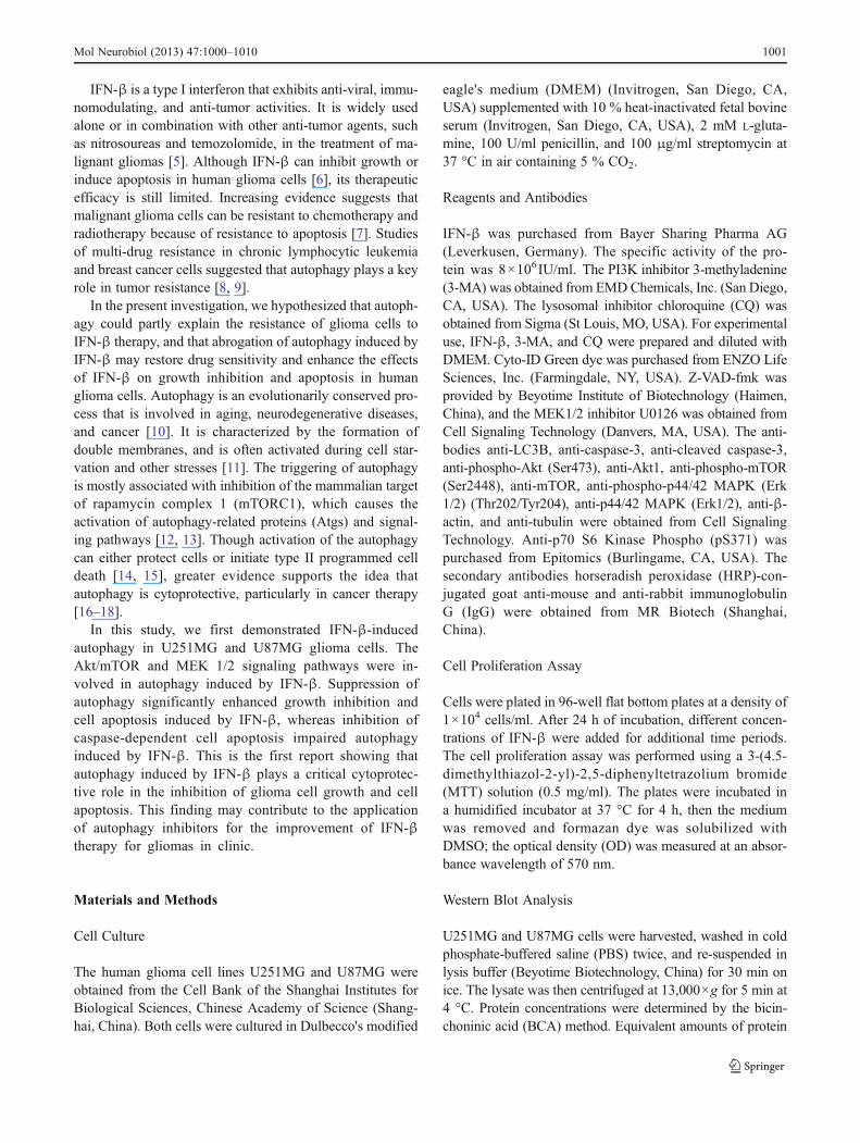
The Akt/mTOR signaling pathway is one of the majorpathways regulating autophagy in eukaryotic cells. Thispathway plays a variety of physiological roles, includingregulation of cell growth and cell survival [24, 25].AKT1 and mTOR signaling molecules are also majorregulators of autophagy [26, 27]. As shown in Fig. 4,treatment with IFN-β decreased phosphorylation (atSer473) of the Akt protein in U251MG cells in a dose-and time-dependent manner. Similar responses were ob-served in U87MG cells.
We also investigated the effects of IFN-β treatment onmTOR activity in glioma cells. Exposure of U251MG andU87MG cells to IFN-β resulted in diminished levels of thephosphorylated form of mTOR (Ser2448) in a dose- andtime-dependent manner. IFN-β treatment also decreasedphosphorylation of the mTOR targets p70 ribosomal proteinS6 kinase, revealing a potent inhibitory effect of IFN-βtreatment on the Akt/mTOR signaling pathway. The basal
�Fig. 3 Suppression of autophagy enhanced IFN-β-induced apoptosis. aU251MG or U87MG cells were incubated with or without 2,000 IU/ml ofIFN-β in the presence or absence of 3-MA (1 mM) or CQ (5 μM), cellswere stained with Annexin V/PI and analyzed by flow cytometry after96 h, and the percentage of Annexin V-positive cells is shown in the barcharts (b), *P<0.05 vs. control and #P<0.05 vs. IFN-β. c U251MG orU87MG cells were incubated with or without 2,000 IU/ml of IFN-β in thepresence or absence of the autophagy inhibitors 3-MA or CQ for 48 h. Thewhole protein was extracted, and LC3, caspase-3, and cleaved caspase-3were analyzed by Western blot. d U251MG or U87MG cells were incu-bated with or without 2,000 IU/ml of IFN-β in the presence or absence ofthe pan-caspase inhibitor Z-VAD-fmk for 48 h. Whole cell protein wasextracted, and LC3, caspase-3, and cleaved caspase-3 were analyzed byWestern blot. eU251MG or U87MG cells were incubated with or without2,000 IU/ml of IFN-β in the presence or absence of pan-caspase inhibitorZ-VAD-fmk for 48 h. Cell samples were prepared for transmission electronmicroscopy analysis as described in “Materials andMethods”. fU251MGor U87MG cells were incubated with or without 2,000 IU/ml of IFN-β inthe presence or absence of pan-caspase inhibitor Z-VAD-fmk for 48 h;cells were stained with Cyto-ID® Green autophagy dye and analyzed byconfocal microscopy
Mol Neurobiol (2013) 47:1000–1010 1005

1006 Mol Neurobiol (2013) 47:1000–1010

levels of AKT1 and MTOR phosphorylation decreasedalong the U251MG and U87MG cells treated by IFN-β ina dose- and time-dependent manner (Fig. 4). Because AKT1and MTOR act as major autophagy inhibitors, their de-creased activity leads to an increase in autophagy.
ERK 1/2 Signaling Pathway was Activated by AutophagyInduced by IFN-β in Human Glioma Cells
Extracellular signal-regulated kinase 1/2 (ERK1/2,MAPK1/3) activation has been previously reported to con-tribute to autophagy effects that promote cell survival [28,29]; furthermore, direct ERK activation by overexpressionof constitutively active MEK promotes autophagy even inthe absence of other stimuli [30]. In this study, we detectedan increase in ERK1/2 phosphorylation when U251MG andU87MG cells were incubated in 2,000 IU/ml of IFN-β for24 h, 48 h, 72 h, and 96 h or in 125 IU/ml, 250 IU/ml,500 IU/ml, 1,000 IU/ml, and 2,000 IU/ml of IFN-β for 48 h(Fig. 5a, b).
To further investigate the role of ERK1/2 in autophagyinduced by IFN-β, we used U0126, an inhibitor of bothMEK1 and MEK2, to block the phosphorylation of ERK1/2. U251MG and U87MG cells were pretreated with20 μMof U0126 for 2 h, and then incubated with 2,000 IU/mlof IFN-β for 48 h. Western blot analysis showed that phos-phorylation of ERK1/2 was inhibited by U0126. Inhibition ofphosphorylation of ERK1/2 significantly decreased the pro-tein level of LC3-II, indicating that inhibition of ERK 1/2signaling blocked autophagy induced by IFN-β (Fig. 5c).
Together, our experiments indicate that the ERK 1/2 sig-naling pathway is involved in autophagy induced by IFN-β.
Discussion
Previous studies demonstrated that IFN-β could reduceproliferative activity in numerous cell lines, includingglioma, lung cancer, pancreatic cancer, and prostatictumors [3, 31–33]. In this study, we studied the effectsof IFN-β on the induction of autophagy and the rela-tionships among autophagy, growth inhibition, and apo-ptosis induced by IFN-β in U251MG and U87MGhuman glioma cells. The major novel findings in thisstudy are that IFN-β induced significant autophagy inU251MG and U87MG human glioma cells, the Akt/m-TOR and ERK 1/2 signaling pathways were involved inthe autophagy induced by IFN-β, and the inhibition ofautophagy significantly enhanced growth inhibition andapoptosis induced by IFN-β.
IFN-β is a type-I interferon exhibiting anti-virus,anti-tumor, and immunomodulatory effects, and it iswidely used for treating multiple sclerosis [34]. It isalso one of the most commonly used cytokines for thetreatment for malignant gliomas [35], although IFN-βcan inhibit growth and induce apoptosis in human glio-ma cells when given alone or when combined withother anti-tumor agents. However, apoptosis-resistantglioma cells limit its therapeutic potential [6]. Increasingevidence shows that autophagy plays a key role in the
Fig. 4 The Akt/mTORsignaling pathway was involvedin autophagy induced by IFN-βin human glioma cells. a IFN-βinhibited the Akt/mTOR sig-naling pathway in a dose-dependent manner. U251MG orU87MG cells were treated withdifferent concentrations of IFN-β for 48 h. Changes in LC3,pAkt, pmTOR, and pP70S6Kwere examined by Western blot.b IFN-β inhibited the Akt/mTOR signaling pathway in atime-dependent manner.U251MG or U87MG cells weretreated with 2,000 IU/ml ofIFN-β from 24 h to 96 h. Wholecell protein was extracted, andchanges in LC3, pAkt, pmTOR,and pP70S6K were examinedby Western blot
Mol Neurobiol (2013) 47:1000–1010 1007

multi-drug resistance of tumor cells [36, 37]. In thepresent investigation, we hypothesized that autophagymay be the cause of glioma cell resistance to IFN-βtherapy, and that abrogation of autophagy induced byIFN-β may restore drug sensitivity. IFN-β has beenfound to induce autophagy in hepatitis C virus-infectedhepatocytes and pancreatic cancer cells [32, 38, 39].Previous investigations have demonstrated that activa-tion of autophagy can either be cytoprotective or medi-ate a type II form of programmed cell death [40, 41]. Inthis study, we first reported that autophagy was
activated in U251MG and U87MG cells after IFN-βtreatment. Some experimental procedures were undertak-en to verify the presence of autophagy, including auto-phagosome formation that was visualized on atransmission electron microscopy in IFN-β-treated cellsand fluorescent staining for LC3 protein by an autoph-agy detection kit. Moreover, autophagy induced byIFN-β in U251MG and U87MG cells affected the for-mation of LC3-II protein in a dose- and time-dependentmanner. Together, these data indicate that autophagy isinduced by IFN-β in human glioma cells.
Signaling through the PI3K/Akt/mTOR pathway con-trols proliferation and apoptosis of cancer cells [25],whereas mTOR, PI3K, and AMPK directly regulatecomponents of the autophagic machinery [42]. In thisstudy, we showed that constitutive phosphorylation ofAkt in U251MG and U87MG cells was decreased byIFN-β treatment in a dose- and time-dependent manner.We also noticed decreased phosphorylation of mTORand its target p70 ribosomal protein S6 kinase. BecauseAKT1 and MTOR act as major autophagy inhibitors,their reduced activity leads to an increase in autophagy.It has been shown that the activation of the MAPKsignaling pathway, which includes ERK 1/2, is involvedin the formation of autophagy [43]. As shown in Fig. 5,the appearance of the LC3-II protein was correlatedwith the activation of ERK 1/2 phosphorylation. Fur-thermore, the levels of the LC3-II protein in IFN-βtreated cells were lower after inhibition of ERK 1/2phosphorylation with the specific MEK 1/2 inhibitorU0126, suggesting that the MEK/ERK signaling path-way is involved in IFN-β-induced autophagy.
It has been demonstrated that activation of autophagycan either be cytoprotective or mediate a type II form ofprogrammed cell death in cancer chemotherapy, immu-notherapy, and radiotherapy [15]. To explore the role ofautophagy in the growth-inhibiting and apoptotic effectsof IFN-β, we used two autophagy inhibitors, the PI3Kinhibitor 3-MA and the autophagolysosome inhibitorCQ, to block IFN-β-induced autophagy. We found thatIFN-β-induced cell growth inhibition was significantlyenhanced by combined treatment with the autophagyinhibitors 3-MA and CQ, indicating that autophagyplayed a cytoprotective role in IFN-β-treated gliomacells. To further investigate the relationships betweenautophagy and apoptosis induced by IFN-β, we usedFCM to detect cell apoptosis. Our data showed thatinhibition of autophagy enhanced IFN-β-induced cellapoptosis, as reflected by an increased percentage ofAnnexin V-positive cells and caspase-3 cleavage, where-as 3-MA or CQ alone had little effect on glioma cells.These findings confirm the cytoprotective role ofautophagy induced by IFN-β. We also found that
Fig. 5 The ERK 1/2 signaling pathway was activated during autoph-agy induced by IFN-β. a IFN-β induced ERK 1/2 phosphorylation in adose-dependent manner. U251MG or U87MG cells were treated withdifferent concentrations of IFN-β for 48 h. Whole cell protein wasextracted, and Erk 1/2 and phospho-p44/42 MAPK (Erk 1/2) (Thr202/Tyr204) were analyzed by Western blot. b IFN-β induced ERK 1/2phosphorylation in a time-dependent manner. U251MG and U87MGcells were treated with or without 2,000 IU/ml of IFN-β from 24 h to96 h. Whole cell protein was extracted, and Erk 1/2 and phospho-p44/42 MAPK (Erk 1/2) (Thr202/Tyr204) were analyzed by Western blot. cInhibition of MEK 1/2 signaling blocked autophagy induced by IFN-β.U251MG or U87MG cells were incubated with or without 2,000 IU/mlof IFN-β in the presence or absence of the MAPK 1/3 signalingpathway inhibitor U0126 (20 μM) for 48 h. Whole protein wasextracted, conversion of LC3-I to LC3-II, Erk 1/2, and phospho-p44/42 MAPK (Erk 1/2) (Thr202/Tyr204) was determined by Western blot
1008 Mol Neurobiol (2013) 47:1000–1010

inhibition of caspase-dependent apoptosis using the pan-caspase inhibitor Z-VAD-fmk reduced the level of pro-tein LC3-II, indicating that autophagy induced afterIFN-β treatment occurs as a response to IFN-β-induced apoptosis.
In summary, we found that significant autophagy wasinduced in glioma cells treated by IFN-β, and this autoph-agy played a cytoprotective role. Inhibition of autophagy bythe autophagy inhibitors 3-MA and CQ can significantlyenhance growth inhibition and cell apoptosis in glioma cells.This study provided a new strategy to enhance the efficacyof IFN-β for cancer treatment and may encourage the de-velopment of an autophagy inhibitor to improve IFN-βtreatment for glioma.
Acknowledgments This work was supported by Shanghai Scienceand Technology Funds (11431920104, 09XD1421800), National Sci-ence and Technology Major Project for Drug Discovery of Ministry ofScience and Technology of China (2011ZX09102-001-27), and theNational Key Basic Research Program of China (2013CB932502).
Conflict of Interest None.
References
1. Natsume A, Ishii D, Wakabayashi T, Tsuno T, Hatano H, MizunoM, Yoshida J (2005) IFN-beta down-regulates the expression ofDNA repair gene MGMT and sensitizes resistant glioma cells totemozolomide. Cancer Res 65(17):7573–7579. doi:10.1158/0008-5472.CAN-05-0036
2. Stewart LA (2002) Chemotherapy in adult high-grade glioma: asystematic review and meta-analysis of individual patient datafrom 12 randomised trials. Lancet 359(9311):1011–1018
3. Okazaki T, Kageji T, Kuwayama K, Kitazato KT, Mure H, Hara K,Morigaki R, Mizobuchi Y, Matsuzaki K, Nagahiro S (2012) Up-regulation of endogenous PML induced by a combination ofinterferon-beta and temozolomide enhances p73/YAP-mediatedapoptosis in glioblastoma. Cancer Lett 323(2):199–207.doi:10.1016/j.canlet.2012.04.013
4. Auffinger B, Thaci B, Nigam P, Rincon E, Cheng Y, Lesniak MS(2012) New therapeutic approaches for malignant glioma: insearch of the Rosetta stone. F1000 Med Rep 4:18. doi:10.3410/M4-18
5. Natsume A, Wakabayashi T, Ishii D, Maruta H, Fujii M, ShimatoS, Ito M, Yoshida J (2008) A combination of IFN-beta and temo-zolomide in human glioma xenograft models: implication of p53-mediated MGMT downregulation. Cancer Chemother Pharmacol61(4):653–659. doi:10.1007/s00280-007-0520-x
6. Saito R, Mizuno M, Hatano M, Kumabe T, Yoshimoto T, Yoshida J(2004) Two different mechanisms of apoptosis resistance observedin interferon-beta induced apoptosis of human glioma cells. JNeurooncol 67(3):273–280
7. Yoshino A, Ogino A, Yachi K, Ohta T, Fukushima T, Watanabe T,Katayama Y, Okamoto Y, Naruse N, Sano E (2009) Effect of IFN-beta on human glioma cell lines with temozolomide resistance. IntJ Oncol 35(1):139–148
8. Mahoney E, Lucas DM, Gupta SV, Wagner AJ, Herman SE, SmithLL, Yeh YY, Andritsos L, Jones JA, Flynn JM, Blum KA, Zhang
X, Lehman A, Kong H, Gurcan M, Grever MR, Johnson AJ, ByrdJC (2012) ER stress and autophagy: new discoveries in the mech-anism of action and drug resistance of the cyclin-dependent kinaseinhibitor flavopiridol. Blood 120(6):1262–1273. doi:10.1182/blood-2011-12-400184
9. Zou Z, Yuan Z, Zhang Q, Long Z, Chen J, Tang Z, Zhu Y, Chen S,Xu J, Yan M, Wang J, Liu Q (2012) Aurora kinase A inhibition-induced autophagy triggers drug resistance in breast cancer cells.Autophagy 8(12):1798–810. doi:10.4161/auto.22110
10. Levine B, Kroemer G (2008) Autophagy in the pathogenesis ofdisease. Cell 132(1):27–42. doi:10.1016/j.cell.2007.12.018
11. Mizushima N (2007) Autophagy: process and function. Genes Dev21(22):2861–2873. doi:10.1101/gad.1599207
12. Corradetti MN, Guan KL (2006) Upstream of the mammaliantarget of rapamycin: do all roads pass through mTOR? Oncogene25(48):6347–6360. doi:10.1038/sj.onc.1209885
13. He C, Klionsky DJ (2009) Regulation mechanisms and signalingpathways of autophagy. Annu Rev Genet 43:67–93. doi:10.1146/annurev-genet-102808-114910
14. Shintani T, Klionsky DJ (2004) Autophagy in health and disease: adouble-edged sword. Science 306(5698):990–995. doi:10.1126/science.1099993
15. White E, DiPaola RS (2009) The double-edged sword of autoph-agy modulation in cancer. Clin Cancer Res 15(17):5308–5316.doi:10.1158/1078-0432.CCR-07-5023
16. Kroemer G, Levine B (2008) Autophagic cell death: the story of amisnomer. Nat Rev Mol Cell Biol 9(12):1004–1010. doi:10.1038/nrm2529
17. Madeo F, Tavernarakis N, Kroemer G (2010) Can autophagypromote longevity? Nat Cell Biol 12(9):842–846. doi:10.1038/ncb0910-842
18. Marino G, Madeo F, Kroemer G (2011) Autophagy for tissuehomeostasis and neuroprotection. Curr Opin Cell Biol 23(2):198–206. doi:10.1016/j.ceb.2010.10.001
19. Michallet AS, Mondiere P, Taillardet M, Leverrier Y, Genestier L,Defrance T (2011) Compromising the unfolded protein responseinduces autophagy-mediated cell death in multiple myeloma cells.PLoS One 6(10):e25820. doi:10.1371/journal.pone.0025820
20. Chan LL, Shen D, Wilkinson AR, Patton W, Lai N, Chan E,Kuksin D, Lin B, Qiu J (2012) A novel image-based cytometrymethod for autophagy detection in living cells. Autophagy 8(9):1371–1382. doi:10.4161/auto.21028
21. Seglen PO, Gordon PB (1982) 3-Methyladenine: specific inhibitorof autophagic/lysosomal protein degradation in isolated rat hepa-tocytes. Proc Natl Acad Sci U S A 79(6):1889–1892
22. Pliyev BK, Menshikov M (2012) Differential effects of the autoph-agy inhibitors 3-methyladenine and chloroquine on spontaneousand TNF-alpha-induced neutrophil apoptosis. Apoptosis 17(10):1050–1065. doi:10.1007/s10495-012-0738-x
23. Kumar S (2007) Caspase function in programmed cell death. CellDeath Differ 14(1):32–43. doi:10.1038/sj.cdd.4402060
24. Hanahan D, Weinberg RA (2000) The hallmarks of cancer. Cell100(1):57–70
25. Guertin DA, Sabatini DM (2007) Defining the role of mTOR incancer. Cancer Cell 12(1):9–22. doi:10.1016/j.ccr.2007.05.008
26. Takeuchi H, Kondo Y, Fujiwara K, Kanzawa T, Aoki H, Mills GB,Kondo S (2005) Synergistic augmentation of rapamycin-inducedautophagy in malignant glioma cells by phosphatidylinositol 3-kinase/protein kinase B inhibitors. Cancer Res 65(8):3336–3346.doi:10.1158/0008-5472.CAN-04-3640
27. Jung CH, Ro SH, Cao J, Otto NM, Kim DH (2010) mTORregulation of autophagy. FEBS Lett 584(7):1287–1295.doi:10.1016/j.febslet.2010.01.017
28. Ogier-Denis E, Pattingre S, El Benna J, Codogno P (2000) Erk1/2-dependent phosphorylation of Galpha-interacting protein stimu-lates its GTPase accelerating activity and autophagy in human
Mol Neurobiol (2013) 47:1000–1010 1009

colon cancer cells. J Biol Chem 275(50):39090–39095.doi:10.1074/jbc.M006198200
29. Pattingre S, Bauvy C, Codogno P (2003) Amino acids inter-fere with the ERK1/2-dependent control of macroautophagyby controlling the activation of Raf-1 in human colon cancerHT-29 cells. J Biol Chem 278(19):16667–16674. doi:10.1074/jbc.M210998200
30. Cagnol S, Chambard JC (2010) ERK and cell death: mechanismsof ERK-induced cell death—apoptosis, autophagy and senescence.FEBS J 277(1):2–21. doi:10.1111/j.1742-4658.2009.07366.x
31. Park MY, Kim DR, Jung HW, Yoon HI, Lee JH, Lee CT (2010)Genetic immunotherapy of lung cancer using conditionally repli-cating adenovirus and adenovirus-interferon-beta. Cancer GeneTher 17(5):356–364. doi:10.1038/cgt.2009.78
32. Vitale G, Zappavigna S, Marra M, Dicitore A, Meschini S,Condello M, Arancia G, Castiglioni S, Maroni P, Bendinelli P,Piccoletti R, van Koetsveld PM, Cavagnini F, Budillon A,Abbruzzese A, Hofland LJ, Caraglia M (2012) The PPAR-gamma agonist troglitazone antagonizes survival pathways in-duced by STAT-3 in recombinant interferon-beta treated pancreaticcancer cells. Biotechnol Adv 30(1):169–184. doi:10.1016/j.biotechadv.2011.08.001
33. Olson MV, Lee J, Zhang F, Wang A, Dong Z (2006) Induciblenitric oxide synthase activity is essential for inhibition of prostatictumor growth by interferon-beta gene therapy. Cancer Gene Ther13(7):676–685. doi:10.1038/sj.cgt.7700941
34. Paty DW, Li DK (1993) Interferon beta-1b is effective in relapsing-remitting multiple sclerosis. II. MRI analysis results of a multicen-ter, randomized, double-blind, placebo-controlled trial. UBC MS/MRI Study Group and the IFNB Multiple Sclerosis Study Group.Neurology 43(4):662–667
35. Ruotsalainen J, Martikainen M, Niittykoski M, Huhtala T,Aaltonen T, Heikkila J, Bell J, Vaha-Koskela M, Hinkkanen A
(2012) Interferon-beta sensitivity of tumor cells correlates withpoor response to VA7 virotherapy in mouse glioma models. MolTher 20(8):1529–1539. doi:10.1038/mt.2012.53
36. Paillas S, Causse A, Marzi L, de Medina P, Poirot M, Denis V,Vezzio-Vie N, Espert L, Arzouk H, Coquelle A, Martineau P, DelRio M, Pattingre S, Gongora C (2012) MAPK14/p38alpha confersirinotecan resistance to TP53-defective cells by inducing survivalautophagy. Autophagy 8(7):1098–112. doi:10.4161/auto.20268
37. Kong D, Ma S, Liang B, Yi H, Zhao Y, Xin R, Cui L, Jia L, Liu X(2012) The different regulatory effects of p53 status on multidrugresistance are determined by autophagy in ovarian cancer cells.B iomed Pharmacothe r 66(4) :271–278 . doi :10 .1016 /j.biopha.2011.12.002
38. Sun J, Desai MM, Soong L, Ou JH (2011) IFN-alpha/beta andautophagy: tug-of-war between HCV and the host. Autophagy 7(11):1394–1396. doi:10.4161/auto.7.11.17514
39. Desai MM, Gong B, Chan T, Davey RA, Soong L, Kolokoltsov AA,Sun J (2011) Differential, type I interferon-mediated autophagic traf-ficking of hepatitis C virus proteins in mouse liver. Gastroenterology141(2):674–685. doi:10.1053/j.gastro.2011.04.060, 685 e671-676
40. Liang C (2010) Negative regulation of autophagy. Cell DeathDiffer 17(12):1807–1815. doi:10.1038/cdd.2010.115
41. Lomonaco SL, Finniss S, Xiang C, Lee HK, Jiang W, Lemke N,Rempel SA, Mikkelsen T, Brodie C (2011) Cilengitide inducesautophagy-mediated cell death in glioma cells. Neuro Oncol 13(8):857–865. doi:10.1093/neuonc/nor073
42. Kondo Y, Kanzawa T, Sawaya R, Kondo S (2005) The role ofautophagy in cancer development and response to therapy. Nat RevCancer 5(9):726–734. doi:10.1038/nrc1692
43. Sivaprasad U, Basu A (2008) Inhibition of ERK attenuates autoph-agy and potentiates tumour necrosis factor-alpha-induced celldeath in MCF-7 cells. J Cell Mol Med 12(4):1265–1271.doi:10.1111/j.1582-4934.2008.00282.x
1010 Mol Neurobiol (2013) 47:1000–1010
Top Related
Copyright © 2022 FDOKUMEN

