


Bahasa
Halaman
Hukum

1 23
Swiss Journal of Geosciences ISSN 1661-8726Volume 106Number 2 Swiss J Geosci (2013) 106:265-278DOI 10.1007/s00015-013-0121-0
Specialization for amphibiosis inBrachyodus onoideus (Artiodactyla,Hippopotamoidea) from the Early Mioceneof France
Maeva J. Orliac, Pierre-Olivier Antoine,Anne-Lise Charruault, Sophie Hervet,Frédéric Prodeo & Francis Duranthon

1 23
Your article is protected by copyright and
all rights are held exclusively by Swiss
Geological Society. This e-offprint is for
personal use only and shall not be self-
archived in electronic repositories. If you wish
to self-archive your article, please use the
accepted manuscript version for posting on
your own website. You may further deposit
the accepted manuscript version in any
repository, provided it is only made publicly
available 12 months after official publication
or later and provided acknowledgement is
given to the original source of publication
and a link is inserted to the published article
on Springer's website. The link must be
accompanied by the following text: "The final
publication is available at link.springer.com”.
Specialization for amphibiosis in Brachyodus onoideus(Artiodactyla, Hippopotamoidea) from the Early Mioceneof France
Maeva J. Orliac • Pierre-Olivier Antoine • Anne-Lise Charruault •
Sophie Hervet • Frederic Prodeo • Francis Duranthon
Received: 28 October 2012 / Accepted: 25 February 2013 / Published online: 16 November 2013
� Swiss Geological Society 2013
Abstract A partial cranium of a very large anthracothere
was unearthed during a palaeontological excavation at
Saint-Antoine-de-Ficalba (Lot-et-Garonne, France; Early
Miocene, *18–17.0 Ma). The new material, referred to as
Brachyodus onoideus (Gervais, 1859), documents the cra-
nial features of this species, so far mainly known by dental
and postcranial remains. The preserved part of the skull
roughly coincides with the neurocranium and is remarkable
for the dorsally-protruding orbits, the importance of the
postorbital constriction, the small volume of the braincase,
and the gigantic size of the occipital condyle relative to the
other elements of the neurocranium. A very careful dis-
section of the left auditory region allowed extraction of the
left petrosal bone and provides the first description of a
petrosal for Brachyodus. The morphology of the petrosal is
strikingly similar to that of extant hippos with: (1) a ventral
basicapsular groove, (2) a sharp crista petrosa, (3) a wide
prefacial commissure fossa, (4) a reduced mastoid, and (5)
an hyperinflated tegmen tympani. Both the disposition of
the orifices of the head and the petrosal morphology sup-
port a specialization of Brachyodus onoideus to an
amphibious lifestyle and to potential underwater direc-
tional hearing.
Keywords Anthracotheriidae � Middle ear � Burdigalian �Underwater directional hearing
1 Introduction
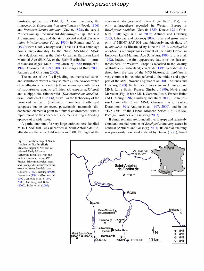
The renovation of the RN21 motorway between Ville-
neuve-sur-Lot and Agen (SW France) led to the discovery
by one of us (FP) of a new rich vertebrate locality west to
the Saint-Antoine-de-Ficalba village (0.712�E, 44.336�N;
Fig. 1). The fossiliferous exposure is a thin stri-
pe, *2.6 km-long (North–South) and 60/110 m-wide
(East–West). Fossil-yielding deposits consist of non-indu-
rated mica-rich sands and siltstones of fluvio-lacustrine
origin, known for decades to be late Early Miocene in age
(Burdigalian). Fossil remains were found scattered all over
the concerned surface; no significant accumulation, ana-
tomical connection, or bone bed was identified during
the excavation undertaken in July 2008. The vertebrate
assemblage of Saint-Antoine-de-Ficalba consists primarily
of an abundant chelonian fauna, widely dominated by the
Early Miocene land-dwelling tortoise Testudo promargi-
nata von Reinach, 1900 (complete shells), but also
including an aquatic/terrestrial testudinid (Ptychogaster
sp.; connected plates) and a strictly aquatic softshell tur-
tle (Trionyx sp.; connected carapace), both of poor
Editorial Handling: D. Becker & D. Marty.
M. J. Orliac (&) � P.-O. Antoine � A.-L. Charruault
Institut des Sciences de l’Evolution, UMR-CNRS 5554,
CC064, Universite Montpellier 2, Place Eugene-Bataillon,
34095 Montpellier, France
e-mail: [email protected]
M. J. Orliac
Department of African Zoology, Royal Museum for Central
Africa, Leuvensesteenweg 13, 30080 Tervuren, Belgium
S. Hervet
Association Paleovergne, 24 avenue de l’Abattoir,
03450 Ebreuil, France
F. Prodeo
Institut National de Recherches Archeologiques Preventives,
156 avenue Jean-Jaures, 33600 Pessac, France
F. Duranthon
Museum d’Histoire Naturelle/AMIS-UMR 5288 CNRS,
35 Allees Jules-Guesde, 31000 Toulouse, France
Swiss J Geosci (2013) 106:265–278
DOI 10.1007/s00015-013-0121-0
Author's personal copy
biostratigraphical use (Table 1). Among mammals, the
rhinocerotids Diaceratherium aurelianense (Nouel, 1866)
and Protaceratherium minutum (Cuvier, 1822), the cervid
Procervulus sp., the moschid Amphitragulus sp., the suid
Aureliachoerus sp., and the stem cricetid rodent Eucrice-
todon infralactorensis (Viret, 1930) in Roman and Viret
(1930) were notably recognised (Table 1). This assemblage
points unquestionably to the ‘base MN3-base MN4’
interval, documenting the Early Orleanian European Land
Mammal Age (ELMA), or the Early Burdigalian in terms
of standard stages (Mein 1989; Ginsburg 1990; Bruijn et al.
1992; Antoine et al. 1997, 2000; Ginsburg and Bulot 2000;
Antunes and Ginsburg 2003).
The nature of the fossil-yielding sediments (siltstones
and sandstones within a clayish matrix), the co-occurrence
of an alligatoroid crocodile (Diplocynodon sp.) with turtles
of strong/strict aquatic affinities (Ptychogaster/Trionyx)
and a hippo-like rhinocerotid (Diaceratherium aurelian-
ense; Bentaleb et al. 2006), as well as the taphonomy of the
preserved remains (chelonians: complete shells and
carapaces but no connected postcranials; mammals: dis-
connected elements) point to a fluvial environment, with a
rapid burial of the concerned specimens during a flooding
episode of a wide river.
A partial cranium of a very large anthracothere, labelled
MHNT SAF 001, was unearthed in Saint-Antoine-de-Fic-
alba during the same field season in 2008. Throughout the
concerned stratigraphical interval (*18–17.0 Ma), the
only anthracothere recorded in Western Europe is
Brachyodus onoideus (Gervais 1859; Dineur 1981; Gins-
burg 1990; Aguilar et al. 2003; Antunes and Ginsburg
2003; Lihoreau and Ducrocq 2007). Size and gross anat-
omy of MHNT SAF 001 unambiguously match those of
B. onoideus, as illustrated by Dineur (1981). Brachyodus
onoideus is a conspicuous element of the early Orleanian
European Land Mammal Age (Ginsburg 1990; Bruijn et al.
1992). Indeed, the first appearance datum of the ‘last an-
thracothere’ of Western Europe is recorded in the locality
of Bruttelen (Switzerland; von Studer 1895; Scherler 2011)
dated from the base of the MN3 biozone. B. onoideus is
very common in localities referred to the middle and upper
part of the MN3 biozone (Aguilar et al. 2003; Antunes and
Ginsburg 2003). Its last occurrences are in Artenay (base
MN4; Loire Basin, France; Ginsburg 1990), Navere and
Marsolan (Fig. 1; base MN4, Garonne Basin, France; Bulot
and Ginsburg 1996; Ginsburg and Bulot 2000), Bonrepos-
sur-Aussonnelle (lower MN4, Garonne Basin, France;
Duranthon 1991; Antoine et al. 1997, 2000), and in the
‘‘IVb unit’’ of the Lisbon Miocene Series (18–17.6 Ma,
Portugal; Antunes and Ginsburg 2003).
If dental remains are found all over Europe and relatively
abundant, cranial remains of Brachyodus are very scarce in
contrast (Antunes and Ginsburg 2003). Its cranial anatomy
was previously described in detail by Dineur (1981), based
Fig. 1 Location map of Saint-
Antoine-de-Ficalba (Early
Miocene, upper MN3) and of
selected Early Miocene
vertebrate localities from the
middle Garonne basin, SW
France. Biochronological ages
and Brachyodus occurrences are
extracted from Baudelot and
Collier (1978), Ginsburg (1990),
Duranthon (1991), (Bruijn et al.
1992), Antoine et al. (1997,
2000), Ginsburg and Bulot
(2000), Bulot et al. (2009)
266 M. J. Orliac et al.
Author's personal copy
on two partial crania from Chilleurs-aux-Bois, Loiret,
France (upper MN3; Antoine et al. 2000; Aguilar et al.
2003): S04600, a neurocranium broken just posterior to the
orbit nowadays curated in the Naturhistorisches Museum in
Basel, Switzerland (NMB), and a juvenile specimen without
number, consisting of a neurocranium lacking the occipital
(Pithiviers Museum, France; Dineur 1981: 22). From the
same locality, Dineur (1981) also mentioned other cranial
fragments from a private collection, documenting the gle-
noid and auditory areas (Faillie, FGE 13). We describe here
a partial cranium of B. onoideus from the Early Miocene of
Saint-Antoine-de-Ficalba, which completes our knowledge
of the cranial and intracranial anatomy of the ‘last anthra-
cothere’ of Western Europe. We further provide the first
description of a petrosal for Brachyodus and discuss the
aquatic affinities of B. onoideus.
2 Materials and methods
All the vertebrate remains from Saint-Antoine-de-Ficalba are
permanently stored in the collection of the Museum d’His-
toire Naturelle in Toulouse, France (MHNT). A very careful
dissection of the left auditory region allowed extraction of the
left petrosal bone of MHNT SAF 001. Independently, this
dissection allowed recognition of another isolated petrosal
within the ‘‘isolated petrosal collection’’ from Chilleurs-aux-
Bois stored in the NMB collections.
Descriptions of petrosals of ‘‘Anthracotheriidae’’ remain
scarce in the literature. Among this paraphyletic family, the
latter bones are known so far by an isolated specimen
referred to as Elomeryx armatus from the White River
Formation, South Dakota (AMNH-VP 579; O’Leary 2010)
and by two in situ specimens of Bothriogenys sp. from the
Jebel Qatrani Formation, Fayum, Egypt (DPC 20954, Early
Oligocene, *30 Ma; O’Leary et al. 2012) for which only
the dorsomedial surface has been described. Comparisons
of the petrosal within hippopotamoids are therefore limited
to these two taxa and to the living hippos Hippopotamus
amphibius and Choeropsis liberiensis.
In order to calculate the volume of the tegmen tympani
relative to volume of the petrosal (excluding the mastoid
region) following the protocol of O’Leary et al. (2012), we
scanned the extracted left petrosal of MNHT SAF 001 (CTscan
facility at the University Montpellier 2, RIO imaging—IBiZA
plateform; 1,200 slices, voxel size 35 lm). We reconstructed
the 3D model of the petrosal using the segmentation tools of
AVIZO 6.3 (Visualization Sciences Group) and calculated the
corresponding volumes by surface integration.
3 Systematic palaeontology
Artiodactyla Owen, 1848
Hippopotamoidea Gray, 1821 sensu Gentry and Hooker
(1988)
‘‘Anthracotheriidae’’ (paraphyletic)
Bothriodontinae Scott, 1940
Brachyodus Deperet, 1895
Brachyodus onoideus (Gervais, 1859)
Nomenclatural remark. The name Artiodactyla Owen,
1848 is used here to refer to the least inclusive crown clade
containing Camelus dromedarius Linnaeus, 1758, Hippo-
potamus amphibius Linnaeus, 1758, Sus scrofa Linnaeus,
1758, Bos taurus Linnaeus, 1758, following the definition
of O’Leary et al. (in press). Montgelard et al. (1997) pro-
posed the new name Cetartiodactyla for the combined
group of cetaceans and artiodactyls. Despite the subsequent
widespread use of this term, the topological change of
placing Cetacea several nodes within Artiodactyla was
actually never grounds to retire the original name Artio-
dactyla. We follow O’Leary et al. (in press) and consider
that the name Artiodactyla Owen, 1848 has priority over
Cetartiodactyla Montgelard et al., 1997 according to rules
of phylogenetic nomenclature.
Table 1 Vertebrate fauna of Saint-Antoine de Ficalba, SW France
(late Early Miocene, Early Orleanian, base MN3-base MN4)
Supra-familial rank Species Mammalian
Neogene zone
Chelonia Ptychogaster sp.
Testudo sp.
Ergilemys sp.
Trionyx sp.
Crocodylia Diplocynodon sp.
Serpentes indet.
Mammalia,
Perissodactyla
Diaceratherium
aurelianense
(MN3-base MN4)
Protaceratherium minutum (MN1-base MN4)
Plesiaceratherium sp.
(medium-sized)
(?MN3-MN5)
Mammalia,
Artiodactyla
Brachyodus onoideus (MN3-base MN4)
?Hyotherium sp. (MN1-MN8)
Aureliachoerus sp. (upper MN2-MN5)
Procervulus sp. (MN3-lower MN5)
Amphitragulus sp. (MN1-lower MN4)
Cainotherium sp. (MN1-MN5)
Mammalia,
Rodentia
Eucricetodon
infralactorensis
(MN3)
The inferred stratigraphical interval, based on mammalian biochro-
nology (Ginsburg 1990; Bruijn et al. 1992; Antoine et al. 1997, 2000;
Antunes and Ginsburg 2003), is indicated in terms of Mammal
Neogene biozones (MN; e.g., Mein 1989) for each biostratigraphi-
cally sound taxon
Cranium and petrosal of Brachyodus onoideus 267
Author's personal copy
3.1 Description and comparison
3.1.1 Cranium (Fig. 2)
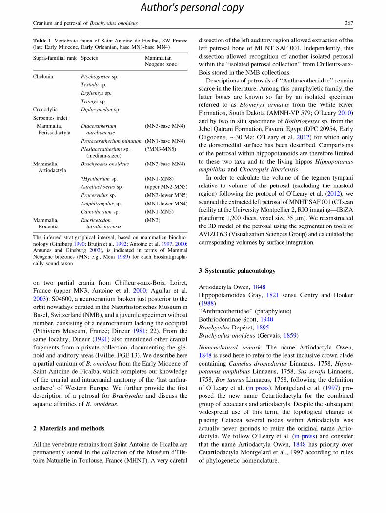
The specimen is crushed dorsoventrally and the basicra-
nium is slightly laterally shifted. The basicranium is
preserved from the basis of the pterygoid processes to the
foramen magnum. The ventral part of the cranium anterior
to the basisphenoid is missing and cannot be described. On
the dorsal surface, the cranium is broken anterior to the
orbits following a breakage line that most probably corre-
sponded laterally to the frontal/lacrymal suture.
In lateral view (Fig. 2e), despite of the dorsoventral
crushing of the specimen, it is possible to affirm that the
Fig. 2 Partial cranium of Brachyodus onoideus from Saint-Antoine-
de-Filcalba (Lot-et-Garonne, France, MNHT SAF 001) in ventral (a),
dorsal (b), anterior (c), posterior (d), and lateral (e) views. eam
external auditory meatus, fo foramen ovale, gf glaser fissure, gs
glenoid surface, hf hypoglos foramen, mlf medium lacerate foramen,
mp mastoid process, mt meatal tube, pap paroccipital process, pgp
postglenoid process, ptp postympanic process, smf stylomastoid
foramen, tb tympanic bulla. Scale bar 5 cm
268 M. J. Orliac et al.
Author's personal copy

cranium was very low, in the same way as the undeformed
specimen from Chilleurs-aux-Bois figured by Dineur
(1981, pl. 8: S04600). The lateral wall of the braincase is
mainly formed by the squamosal and parietal bones.
However, the bad preservation of the specimen and the
numerous fractures due to post-depositional processes
prevent us from locating the sutures with certainty. The
orbits are located in a high position and their dorsal mar-
gins protrude above the plane of the frontal (also visible in
anterior view, Fig. 2c), as in extant Hippopotamus. On the
left side of the cranium, the anatomic unit composed of the
glenoid surface, meatal tube, paroccipital process, and
occipital condyle is free from deformation. The postglenoid
process is massive and extends ventrally to the articular
surface. Posteriorly, the meatal tube is tightly imbricated
between the postglenoid and postympanic processes. The
external auditory meatus opens just below the temporal
crest, in quite a high position, corresponding to the mid--
height of the orbit. Considering this, the external ear
opening of the living animal must have also opened in a
high position, i.e. comparable to what is observed today in
living hippos. The paroccipital process is oriented ventrally
and slightly posteriorly, and is overhung by a blunt lateral
tuberosity. The height between the glenoid surface and the
temporal crest is similar to the total height of the large
occipital condyle. The glenoid surface is located at half
height of the occipital condyles. The latter are located far
posterior to the occiput perpendicular.
In dorsal view (Fig. 2b), the skull is slender and elon-
gated. The postorbital constriction is strong and the
braincase maximal width is less than the frontal width
anterior to the postorbital process. A strong sagittal crest
runs backward from the postorbital constriction to the
nuchal crest. The latter is a thin crest of bone, subhorizontal
and perpendicular to the sagittal crest medially, laterally
slanting ventrally and slightly anteriorly. The posterior-
most part of the zygomatic arches are preserved on both
sides of the cranium, broken at the level of the jugal/
squamosal suture. They are only slightly diverging from
the braincase, indicating that the arches were most proba-
bly not very salient laterally.
The ventral surface (Fig. 2a) of the cranium anterior to
the glenoid fossa is not preserved. The basisphenoid is
elongated and very narrow anteriorly; it widens posteriorly
to contact the basioccipital. The latter is of constant width
and bears two strong muscle insertions (for the Musculus
rectus capitis ventralis and the Musculus longus capitis).
The basisphenoid-basioccipital contact cannot be located
precisely due to the advanced individual age of the speci-
men (suture fused). The posterior-most extremities of the
pterygoids are preserved and they border the foramen ovale
anteriorly. The latter is partially overhung by the tympanic
bulla. In spite of its proximity with the medium lacerate
foramen, the foramen ovale appears to be still separate
from the latter by a bony crest, partially damaged on the
specimen. The Glazerian fissure is visible lateral to
the foramen ovale, on the right side of the neurocranium.
The glenoid surface is wide and it occupies most of the
ventral surface of the squamosal. On the left side of the
cranium, the posterior-most part of the zygomatic arch is
preserved; this structure was very light compared to the
heavily built mandibular joint. The glenoid surface extends
slightly anterior to the rostral part of the bulla. It is flat in
its anterior portion and strongly concave posteriorly due to
the presence of a strong postglenoid process. The latter is
blunt and it widens medially. While there is no clear evi-
dence of a postglenoid foramen located medial to the
glenoid surface, a small foramen is observed posterior to
the postglenoid process and interpreted as being the
postglenoid foramen. The bulla is large, globular, and
slightly elongated anteriorly. Both bullae are badly dam-
aged, but it is possible to observe the presence of a small
anterior process on the left one. The bulla is tightly
apposed to the glenoid fossa anterolaterally. The meatal
tube is a thick crest of bone intercalated between the
postglenoid process and the postympanic process. The
posterior part of the bulla is broken away and only the
medial and posterior margins of the jugular notch are
preserved. Laterally, between the bulla and the postym-
panic process, opens a wide stylomastoid foramen. The
paroccipital process is mainly oriented ventrally, tightly
apposed to the postympanic process of the squamosal; the
tip of the paroccipital process is broken so that their total
length cannot be determined.
The occipital condyles are large and massive, separated
from the paroccipital process by a wide condylar fossa. The
ventral surface of the occipital condyle bears a ‘‘hook-like’’
anterior extension, preserved on the left side of the skull.
The condylar foramen is a narrow slit located lateral to the
anterior extension of the occipital condyle. Its shape was
probably modified by post-mortem deformation.
In caudal view (Fig. 2d), the occiput of B. onoideus is
low and broad. The massive occipital condyles and wide
foramen magnum occupy most of the posterior surface of
the skull. There is a deep exoccipital fossa on the lateral
border of the occipital, extending dorsally and anteriorly to
the temporal crest. The foramen magnum is wider than
high; it is almost straight dorsally in outline. On the dorsal
side of the foramen magnum, the nuchal tubercles are blunt
and weakly developed. The articular surface is composed
of two convex parts forming an angle of approximately 80�and separated by a blunt oblique crest. The dorsal part of
the articular surface of the occipital condyle is widely
extended anterodorsally. The posterior surface of the
occipital is strongly waisted above the occipital condyles.
There are two wide nuchal fossae: one on each side of the
Cranium and petrosal of Brachyodus onoideus 269
Author's personal copy
salient nuchal tuberosity. The nuchal crest is a narrow crest
of bone. The external occipital protuberance is massive,
medially bordering the deep nuchal fossae.
3.1.2 Comparisons with other cranial material
of Brachyodus
The skull from Saint-Antoine-de-Ficalba perfectly matches
that of Chilleurs-aux-Bois (S04600), both for metrics and
morphology, which allows us to assign the cranium from
Saint-Antoine-de-Ficalba to Brachyodus onoideus without
any doubt (for metrics comparison, see Table 2). S04600 is
in a much better state of preservation than MHNT SAF 001.
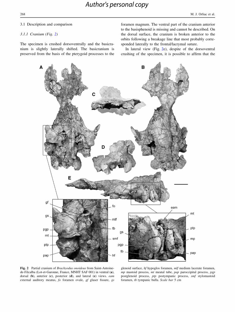
Accordingly, it brings some additional information to the
morphological description provided here. As already men-
tioned by Dineur (1981), the dorsal part of the squamosal is
perforated by several small foramina. Based on the juvenile
specimen curated in Pithiviers, Dineur (1981) mentioned
that these foramina communicate with the meatal duct, just
as the postglenoid foramen. It is worth noting that Dineur
(1981) mentions a foramen located medial to the glenoid
surface, also communicating with the meatal duct. This
foramen is illustrated in Fig. 3 with the other features of the
auditory region of the skull from Chilleurs. This foramen,
interpreted here as a secondary postglenoid foramen, cor-
responds to the medial postglenoid foramen of Geisler et al.
(2007). This character is of phylogenetic importance and
has been proposed as a synapomorphy shared by ‘‘anthra-
cotheres’’ and cetaceans. As mentioned in the description,
the specimen from Saint-Antoine-de-Ficalba does not show
any clear foramen medial to the glenoid fossa, suggesting
that the presence of this character might be variable, or
might depend on the individual age of the specimen.
S04600 also preserves both petrosals in situ, the ventral
surface of which is visible in the space let by the broken
bulla. This brings additional information about the in situ
location of the petrosal in the cranium.
According to Lihoreau and Ducrocq (2007), only one
species of Brachyodus is known from the Early Miocene
of Europe: B. onoideus (Gervais, 1859). In addition,
B. aequatorialis McInnes, 1951 occurs in the Early Mio-
cene of east Africa, while B. depereti (Fourtau, 1918) and
B. mogharensis Pickford, 1991 are identified in the late
Early Miocene of Egypt. Among the non-European species,
only B. aequatorialis is documented by good cranial
remains. The holotype of the species consists in a nearly
complete cranium, from the Early Miocene locality of
Table 2 Measurements of the
cranium of Brachyodus
onoideus from Chilleurs-aux-
Bois (S04600; Dineur 1981) and
corresponding values of the
specimen from Saint-Antoine-
de-Ficalba (MNHT SAF 001).
Dimensions in mm
Measurements B. onoideus Chilleurs-aux-Bois
(S04600) (Dineur 1981: 31)
B. onoideus Saint-Antoine-
de-Ficalba (MHNT SAF 001)
Maximal length of parietal bone 161 161
Minimal width at postorbital level 64 60.9 (slightly underestimated
due to deformation)
Maximal width measured at posterior
border of zygomatic arch
234 230 (estimation based on the
left side of the skull)
Cranial width at postorbital process 154 162 (estimation based on the
left side of the skull)
Minimal width between glenoid surfaces 85 81
Maximal width of the occipital at the
level of the exoccipital condyles
118 125 (estimation based on the
left side of the skull)
Cranial length posterior to posterior
lacerate foramen (foramen jugulare)
68 72
Fig. 3 Labelled detail view of the left auditory region of the cranium
of Brachyodus onoideus from Chilleurs-aux-Bois (Loiret, France,
NMB S04600). bo basioccipital, bs basisphenoid, fo foramen ovale, gf
glaser fissure, gs glenoid surface, hf hypoglos foramen, jn jugular
notch, mlf medium lacerate foramen, mp mastoid process, mt meatal
tube, pap paroccipital process, pb petrosal bone, pgf postglenoid
foramen, pgp postglenoid process, ptp postympanic process, smf
stylomastoid foramen, spgf secondary postglenoid foramen, tb
tympanic bulla. Scale bar 10 cm
270 M. J. Orliac et al.
Author's personal copy
Rusinga (Kenya; McInnes 1951). This specimen, figured by
McInnes (1951, pl. 1 and 2) in dorsal, lateral and ventral
views, has undergone very little distortion. As such, it gives
access to the proportions and relative position of the dif-
ferent elements of the cranium. The cranial features of this
species are by all aspects identical to the preserved part of
the cranium of B. onoideus. Interestingly, the anterior por-
tion of the snout (comprising the anteriormost part of the
nasals) is preserved in the specimen of B. aequatorialis,
contrary to what occurs in MHNT SAF 001. The nasals only
project over the nasal aperture on a little distance and do not
reach the anterior border of the premaxillae. A premaxilla
of B. onoideus from Chilleurs-aux-Bois figured by Dineur
(1981: pl. 3) shows that the nasal opening conversely
extended far back posteriorly in the latter species.
The cranial morphology of B. onoideus can be compared
to that of Elomeryx crispus, an ‘advanced bothriodontine’
ranging from the Late Eocene up to the early Late Oligo-
cene in Western and Southeastern Europe (MP18-26;
Lihoreau and Ducrocq 2007; Lihoreau et al. 2009). An
almost complete cranium of E. crispus was recently
described from the early Late Oligocene deposits of
Moissac-III, SW France (Fig. 1; Lihoreau et al. 2009).
E. crispus differs from B. onoideus in terms of cranial
morphology in having (1) orbits much larger compared to
the relative size of other elements of the cranium, (2) a
higher profile of the cranium with the auditory meatus
located at the level of/or slightly above the ventral and
margin of the orbit, (3) a weaker postorbital constriction,
and (4) a close distance between orbit and glenoid surface.
3.2 Petrosal bone (Figs. 4, 5)
The exact initial position of the petrosal in the MHNT SAF
001 cranium could not be precisely determined due to post-
mortem deformation. The left petrosal protruded dorsally
through the parietal bone so that parts of the parietal and
squamosal bones were removed to extract the left petrosal
from the endocranium. Due to the drastic reduction of the
mastoid part of the petrosal, the latter did not intercalate
between the squamosal and the occipital bones. Only a
small spike of the tegmen tympani (broken during the
dissection of the specimen) was intercalated between the
meatal tube and the basis of the postglenoid process of the
squamosal. Complementary information about the exact
position of the petrosal in the cranium is provided by the
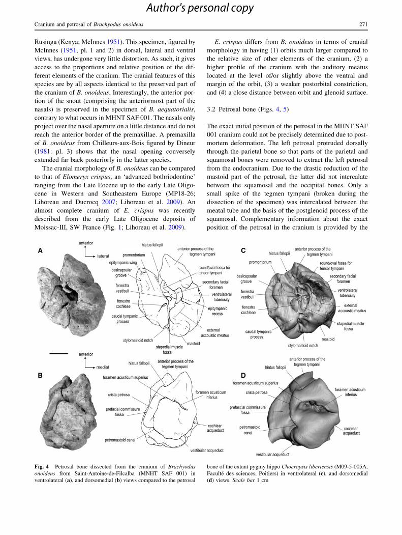
Fig. 4 Petrosal bone dissected from the cranium of Brachyodus
onoideus from Saint-Antoine-de-Filcalba (MNHT SAF 001) in
ventrolateral (a), and dorsomedial (b) views compared to the petrosal
bone of the extant pygmy hippo Choeropsis liberiensis (M09-5-005A,
Faculte des sciences, Poitiers) in ventrolateral (c), and dorsomedial
(d) views. Scale bar 1 cm
Cranium and petrosal of Brachyodus onoideus 271
Author's personal copy
ventral view of the cranium from Chilleurs-aux-Bois
(Fig. 2), showing the ventromedial part of the right and left
petrosals. The petrosal of B. onoideus is further docu-
mented by an isolated specimen from Chilleurs-aux-Bois
(NMB SO 355), only lacking the anterior part of the pars
cochlearis and part of the mastoid.
In ventrolateral view (Fig. 4a), the petrosal of B. onoi-
deus is blunt posteriorly and shows two anterior points of
equal size. The medial point corresponds to the anterior
extremity of the pars cochlearis and the lateral one is
formed by the gigantic tegmen tympani. The promontorium
is hemi-ellipsoidal, softly inflated and medially slanting. It
only extends anteriorly to half of the pars cochlearis. The
anterior development of the epitympanic wing is difficult to
determine: when present, it is completely fused to the
anterior end of the dorsomedial surface of the petrosal. The
fenestra cochleae is a small ellipsoid aperture directed
posteriorly. The fenestra vestibuli is slightly larger, located
beneath the fenestra cochleae, just like in living hippos
(Hippopotamus and Choeropsis; O’Leary 2010, Figure 42,
48). The two fenestrae are separated by a wide crista
interfenestralis. The promontorium is completely smooth
and no sulci are visible. The fossa for the tensor tympani is
shallow and teardrop-shaped, located lateral to the fenestra
vestibuli and only expanding slightly anterior to it; its floor
is broken and the secondary facial foramen canal is
exposed. The fossa for the tensor tympani excavates the
adjacent inflated tegmen tympani. The secondary facial
foramen opens lateral to the fenestra vestibule; its anterior
margin is broken, which corresponds to the anterior posi-
tion according to O’Leary (2010). The promontorium gives
rise anterolaterally to a long epitympanic wing merging to
the dorsomedial surface of the pars cochlearis anteriorly
and to a thin posteromedial flange medially, the external
margin of which is broken. The latter contacts a small
caudal tympanic process posteriorly. A distinct basicap-
sular groove runs anteroposteriorly along most of the
promontorium, until the level of the caudal tympanic
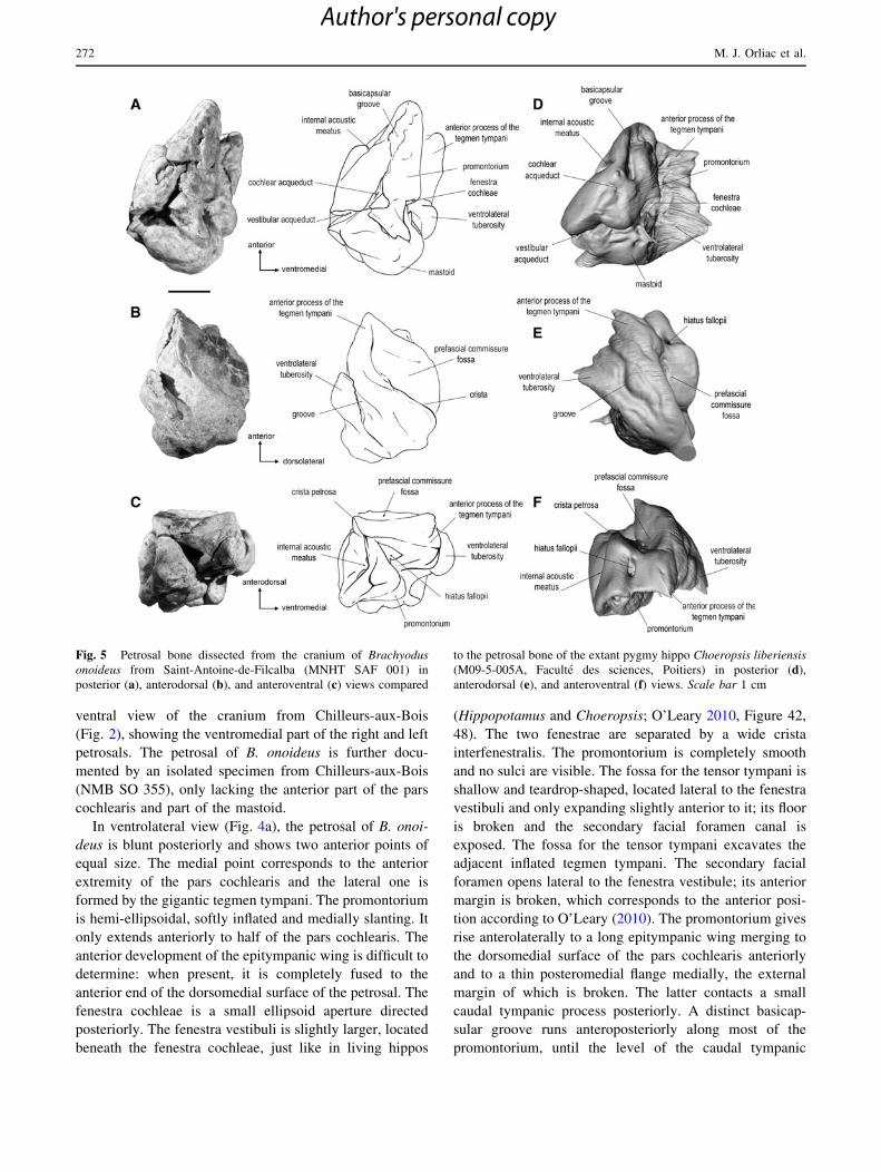
Fig. 5 Petrosal bone dissected from the cranium of Brachyodus
onoideus from Saint-Antoine-de-Filcalba (MNHT SAF 001) in
posterior (a), anterodorsal (b), and anteroventral (c) views compared
to the petrosal bone of the extant pygmy hippo Choeropsis liberiensis
(M09-5-005A, Faculte des sciences, Poitiers) in posterior (d),
anterodorsal (e), and anteroventral (f) views. Scale bar 1 cm
272 M. J. Orliac et al.
Author's personal copy

process (Fig. 4a). This ventral position of the basicapsular
groove is also observed in living hippopotamids (Fig. 4c).
The pars canalicularis is composed (1) of a reduced knob-
like mastoid, cut by a wide external auditory meatus, and
(2) of a gigantic and hyperinflated tegmen tympani, as wide
as the pars cochlearis, extending anteriorly to the anterior
edge of the petrosal. Its anterior process is a blunt spike
overhanging a wide hiatus fallopii, located rather ventrally.
The tegmen tympani is composed of two perpendicular/
orthogonal flat areas, the dorsal one of which is slightly
concave mediolaterally and also form a 90� angle with the
dorsomedial surface of the pars cochlearis. The lateral part
of the tegmen tympani shows a deep groove, almost closed
in a canal by a bony fold just anterior to the ventrolateral
tuberosity. This groove could correspond to the carotid
artery. Posterior to the ventrolateral tuberosity is a large
and deep depression corresponding to the petrosal contri-
bution to the external auditory meatus. Anterolateral to it is
the wide notch of the epitympanic recess. The facial sulcus
is short and separated from a shallow and poorly defined
stapedial muscle fossa by a small bony ridge. The stapedial
muscle fossa extends to the medial margin of the fenestra
cochleae. The stylomastoid notch (i.e., the petrosal con-
tribution to the stylomastoid foramen) is wide and flat,
located directly posterior to the fenestra cochleae. The
mastoid region is a swollen irregular knob, not extending
medially to the level of the fenestra cochleae. The tym-
panic bulla has a strong anterior contact with the tegmen
tympani and fuses to it in the part surrounding the fossa for
the tensor tympani. In ventrolateral view, the petrosal of B.
onoideus differs from that of Elomeryx armatus by several
features. The promontorium of E. armatus does not show a
basicapsular groove; the latter is visible on the dorsomedial
surface of the bone. The location of the epitympanic wing
is more lateral and the postero medial flange is more
developed in E. armatus. In general, the pars cochlearis of
E. armatus is dorsoventrally thinner than that of B. onoi-
deus. The development of the tegmen tympani of E. armatus
is greatly reduced compared to that of B. onoideus and so
does the ventrolateral tuberosity. However, relative size and
location of the fenestra cochleae and fenestra vestibuli are
similar in both taxa. The ventrolateral view of Bothriogenys
has not been figured by O’Leary et al. (2012).
The dorsomedial surface (Fig. 4b) of the petrosal ter-
minates anteriorly by a spike. The internal acoustic meatus
is a transverse slit instead of the large opening traditionally
present in Artiodactyla. The foramina acusticum superius
and inferius are wide holes of similar size, separated by a
very narrow crista transversa. The lateral edge of the
foramen acusticum superius is prolonged posteriorly by a
fissure lining a sharp crista petrosa (crista separating the
pars cochlearis in contact with the cerebellar part of the
brain and the tegmen tympani in contact with the cerebral
part of the brain). This fissure, directed toward the petro-
mastoid canal, is also observed in living hippopotamids
(Hippopotamus and Choeropsis; Fig. 4d; also visible in
O’Leary 2010: Figures. 44, 49). The petromastoid canal
lays in a shallow depression, that might be a relic of the
subarcuate fossa. Lateral to the crista petrosa, on the teg-
men tympani, is a wide and shallow concavity interpreted
here as the prefacial commissure fossa. The later is del-
imitated posteriorly by a sharp crest of bone. The position
of the prefacial commissure fossa of Brachyodus is similar
to that observed in Choeropsis, while this structure is
located more posterodorsally in Hippopotamus. The small
slit of the petromastoid canal is medially confluent with
that of the vestibular aqueduct. The latter occupies a
dorsomedial position. Just posterior to it, the mastoid
shows a deep foramen. In dorsomedial view, the caudal
tympanic process is visible on the posteromedial border; it
marks the position of the cochlear aqueduct.
The dorsomedial surface of the petrosals of B. onoideus
and E. armatus differs by the presence of a shallow but
wide subarcuate fossa in E. armatus while only a vestigial
concavity is observed in B. onoideus. Surprisingly, there is
no petromastoid canal in E. armatus, contrary to what
occurs in B. onoideus. In B. onoideus, the pars cochlearis
occupies most of the dorsomedial surface of the petrosal,
especially due to the very wide and flat portion of bone
extending from the internal auditory meatus to the fissure
surrounding running from the petromastoid canal to the
cochlear aqueduct. This part is much less developed in
E. armatus. According to the Figure 68 of O’Leary (2013),
the location of the vestibular aqueduct in the latter is more
medial than in B. onoideus. Despite these differences,
B. onoideus and E. armatus share the presence of a straight
and sharp crista petrosa. In both taxa, the vestibular
aqueduct lays in a deep slit joining the cochlear aqueduct.
The dorsomedial surface of the petrosal of Bothriogenys
has been figured by a drawing by O’Leary et al. (2012:
Fig. 3). From this drawing, it is possible to say that the
petrosals of B. onoideus and Bothriogenys share an anterior
opening of the hiatus fallopii and a hyperinflated tegmen
tympani. Unfortunately, the lateral and anterior extension
of the tegmen tympani cannot be observed in Bothriogenys
as it is partly hidden by the squamosal (O’Leary et al.,
2012: Figure 3). The angle between the pars cochlearis and
the tegmen tympani cannot be determined either. Besides,
on the tegmen tympani is a depression that could corre-
spond to the depression described here in B. onoideus as
the prefacial commissure fossa (i.e., lateral to the internal
acoustic meatus and not posterior to it). According to the
location of this fossa on the in situ specimen (O’Leary et al.
2012: Figure 3), it could well be in contact with the cere-
bral hemisphere while the rest of the dorsomedial surface
of the petrosal would rather contact the cerebellar part of
Cranium and petrosal of Brachyodus onoideus 273
Author's personal copy
the brain, as interpreted here in B. onoideus. This portion of
the tegmen tympani with the fossa is exposed in the
internal part of the cranium, while the rest of the tegmen
tympani contacts the squamosal, just like in B. onoideus.
Contrary to what is observed in B. onoideus and Bothrio-
genys, the petrosal of E. armatus has no hyperinflation of
the tegmen tympani, and no fossa on this structure.
Unfortunately, the fact that this specimen is isolated makes
it difficult to determine its position in situ.
The anterodorsal view (Fig. 5b) offers the best view of
the gigantic tegmen tympani. The latter structure represents
half of the petrosal total volume. It is parted in two by a
thin but sharp crest of bone, delimitating the lateral edge of
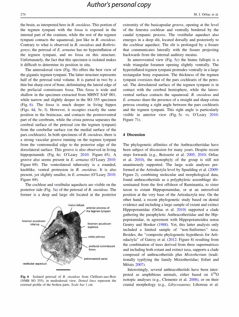
the prefacial commissure fossa. This fossa is wide and
shallow in the specimen extracted from MHNT SAF 001,
while narrow and slightly deeper in the SO 355 specimen
(Fig. 6). The fossa is much deeper in living hippos
(Figs. 4d, 5e, f). However, it occupies exactly the same
position in the braincase, and contacts the posteroventral
part of the cerebrum, while the crista petrosa separates the
cerebral surface of the petrosal (on the tegmen tympani)
from the cerebellar surface (on the medial surface of the
pars cochlearis). In both specimens of B. onoideus, there is
a strong vascular groove running on the tegmen tympani
from the ventromedial edge to the posterior edge of the
dorsolateral surface. This groove is also observed in living
hippopotamids (Fig. 4e; O’Leary 2010: Figure 45). A
groove also seems present in E. armatus (O’Leary 2010:
Figure 69). The ventrolateral tuberosity is a rounded,
knoblike, ventral protrusion in B. onoideus. It is also
present, yet slightly smaller, in E. armatus (O’Leary 2010:
Figure 69).
The cochlear and vestibular aqueducts are visible on the
posterior side (Fig. 5a) of the petrosal of B. onoideus. The
former is a deep and large slit located at the posterior
extremity of the basicapsular groove, opening at the level
of the fenestra cochleae and ventrally bordered by the
caudal tympanic process. The vestibular aqueduct also
emerges in a deep slit, located dorsally and posteriorly to
the cochlear aqueduct. The slit is prolonged by a fissure
that communicates laterally with the fissure projecting
backwards from the internal auditory meatus.
In anteroventral view (Fig. 5c) the hiatus fallopii is a
wide triangular foramen opening slightly ventrally. The
hyperinflated tegmen tympani protrudes ventrally in a large
rectangular bony expansion. The thickness of the tegmen
tympani oversizes that of the pars cochlearis of the petro-
sal. The dorsolateral surface of the tegmen tympani is in
contact with the cerebral hemisphere, while the latero-
ventral surface contacts the squamosal. B. onoideus and
E. armatus share the presence of a straight and sharp crista
petrosa creating a right angle between the pars cochlearis
and the tegmen tympani. This right angle is particularly
visible in anterior view (Fig. 5c vs. O’Leary 2010:
Figure 71).
4 Discussion
The phylogenetic affinities of the Anthracotheriidae have
been subject of discussion for many years. Despite recent
steps forwards (e.g., Boisserie et al. 2005, 2010; Orliac
et al. 2010), the monophyly of the group is still not
unanimously supported. The large scale analyses per-
formed at the Artiodactyla level by Spaulding et al. (2009:
Figure 2), combining molecular and morphological data,
found anthracotheriids as a polyphyletic assemblage dis-
seminated from the first offshoot of Ruminantia, to sister
taxon to extant Hippopotamidae, or at an unresolved
position at the very base of the Artiodactyla tree. On the
other hand, a recent phylogenetic study based on dental
evidence and including a large sample of extant and extinct
Hippopotamidae (Orliac et al. 2010) supported a clade
gathering the paraphyletic Anthracotheriidae and the Hip-
popotamidae, in agreement with Hippopotamoidea sensu
Gentry and Hooker (1988). Yet, this latter analysis only
included a limited sample of ‘‘non-Suiformes’’ taxa.
Besides, the ‘‘composite phylogenetic hypothesis for Arti-
odactyla’’ of Gatesy et al. (2012: Figure 8) resulting from
the combination of trees derived from three supermatrices
and including both extant and extinct taxa, supports a clade
composed of anthracotheriids plus Mixtotherium (tradi-
tionally typifying the family Mixtotheriidae; Erfurt and
Metais 2007).
Interestingly, several anthracotheriids have been inter-
preted as amphibious animals, either based on d18O
isotopic analyses (e.g., Clementz et al. 2008), or on their
cranial morphology (e.g., Libycosaurus; Lihoreau et al.
Fig. 6 Isolated petrosal of B. onoideus from Chilleurs-aux-Bois
(NMB SO 355), in mediodorsal view. Dotted lines represent the
external profile of the broken parts. Scale bar 1 cm
274 M. J. Orliac et al.
Author's personal copy

2003). The presence of an inflated tegmen tympani of the
petrosal bone, observed in both anthracotheres and living
hippos, as well as in cetaceans, has also been correlated
with directional underwater hearing and is another piece of
evidence of specialization for an amphibious lifestyle of
some ‘‘anthracotheriids’’ (e.g., Bothriogenys; O’Leary et al.
2012). Based on the results of Spaulding et al. (2009), this
shared character has however been interpreted as a con-
vergence and amphibiosis has been considered as
independently acquired in anthracotheres and hippos
(O’Leary et al. 2012). In the alternate hypothesis of
monophyletic Hippopotamoidea (sensu Gentry and Hooker
1988), as supported by a more exhaustive sample of fossil
hippopotamids and anthracotheres (Orliac et al. 2010),
clarifying the life style of ‘‘anthracotheriids’’ is of great
interest to reconstruct the evolutionary history of amphi-
biosis within hippopotamoids.
4.1 Semi-aquatic specialization of Brachyodus
onoideus
4.1.1 Disposition of the elements of the cranium
The peculiar disposition of the different elements of the
skull of living hippos allows them to stand in the water
with their head in an ‘‘amphibious position’’: eyes, ears,
and nostrils above water, and the rest of the body below
water. In this position, they are able to breathe and see
above the water surface, but also to communicate via
‘‘amphibious communication’’, a communication mode
defined by Barklow (2004). With their ears and nostrils
above water and mouth and throat below water, they pro-
duce and ear sounds in both air and underwater
simultaneously (Barklow 2004). The low-frequency fast-
travelling underwater sound produced by their throat
travels upstream and downstream along the river and is
heard by other hippos at least a mile away. This wave of
‘‘hippo calls’’ is interpreted as conveying information
about where the individuals are at a given time, avoiding
conflicts by indicating boundaries of the different territo-
ries (Barklow 2004). The peculiar morphology of the hippo
skull is therefore directly related to its semi-aquatic life
style.
Brachyodus has been interpreted as living in swamps or
close-by rivers, but their overall more cursorial postcranial
skeleton (e.g., more elongated metapodials) made them be
considered as ‘‘less aquatic-adapted’’ than living hippos
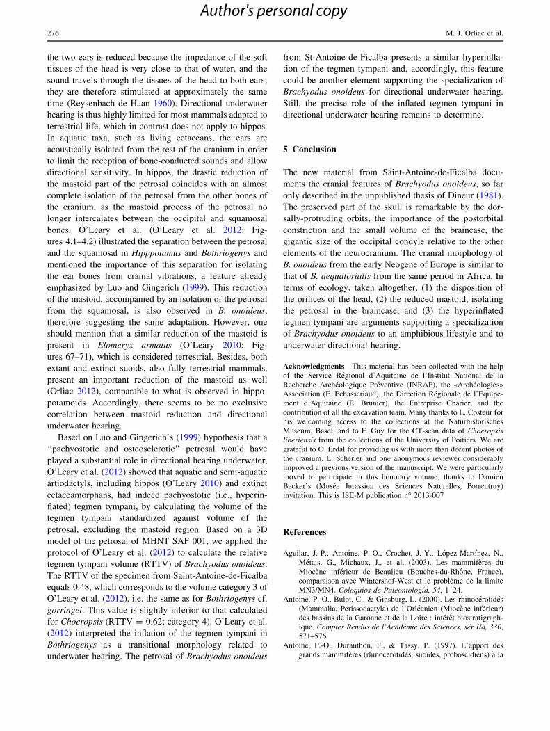
(Antunes and Ginsburg 2003). Yet, the location of the orbit
of Brachyodus, with the dorsal edge slightly protruding
above the level of the frontal, and the location of the
external auditory meatus, opening at half the height of the
orbit, is very close to the disposition of these elements
observed in the skull of Hippopotamus amphibius as
illustrated in Fig. 7. A premaxilla from Chilleurs-aux-Bois
figured by Dineur (Dineur 1981: pl. 3) shows that the
medial suture between premaxilla was high and vertical,
and that the nasal opening extended far back posteriorly,
which is compatible with a dorsal opening of the nostrils in
this taxon. The anterior part of the snout of B. aequatorialis
figured by McInnes (McInnes 1951: pl. 1) further indicates
that the anterior extension of the nasal was concurrently
very short in Brachyodus, another element compatible with
a high location of the nostrils. Taken together, the different
elements of the cranium known for B. onoideus (and B.
aequatorialis) allow proposing a reconstruction of the skull
as illustrated in Fig. 7b, and makes plausible the hypothesis
of an alignment of nostril-eye-ear, and therefore, an
‘‘amphibious position’’ of the head in this taxon.
4.1.2 Middle ear morphology and ability for underwater
directional hearing
The fact that Hippopotamus individuals can vocalize
underwater a waterborne sound implies that they are also
able to ear directionally underwater. In fact, Barklow
(2004) reported that H. amphibius responds to the direc-
tionality of underwater sounds. This latter ability implies a
specialization of the sound perception pathway. Indeed, for
terrestrial mammals, the direction of a sound is determined
(1) by the differences of acoustic stimuli received by the
two independent ears, and (2) by the interaural time dif-
ference (Reysenbach de Haan 1960; Nummela and
Thewissen 2008). In water, sound travels five times faster
than in air, reducing both interaural time and intensity
differences. Intensity difference of the stimuli received by
Fig. 7 Sketch of the left profile of the skull of Hippopotamus
amphibius illustrated in an ‘‘amphibious position’’ (a), and recon-
struction of the corresponding profile of Brachyodus onoideus (b).
The undulated line represents the water surface
Cranium and petrosal of Brachyodus onoideus 275
Author's personal copy
the two ears is reduced because the impedance of the soft
tissues of the head is very close to that of water, and the
sound travels through the tissues of the head to both ears;
they are therefore stimulated at approximately the same
time (Reysenbach de Haan 1960). Directional underwater
hearing is thus highly limited for most mammals adapted to
terrestrial life, which in contrast does not apply to hippos.
In aquatic taxa, such as living cetaceans, the ears are
acoustically isolated from the rest of the cranium in order
to limit the reception of bone-conducted sounds and allow
directional sensitivity. In hippos, the drastic reduction of
the mastoid part of the petrosal coincides with an almost
complete isolation of the petrosal from the other bones of
the cranium, as the mastoid process of the petrosal no
longer intercalates between the occipital and squamosal
bones. O’Leary et al. (O’Leary et al. 2012: Fig-
ures 4.1–4.2) illustrated the separation between the petrosal
and the squamosal in Hipppotamus and Bothriogenys and
mentioned the importance of this separation for isolating
the ear bones from cranial vibrations, a feature already
emphasized by Luo and Gingerich (1999). This reduction
of the mastoid, accompanied by an isolation of the petrosal
from the squamosal, is also observed in B. onoideus,
therefore suggesting the same adaptation. However, one
should mention that a similar reduction of the mastoid is
present in Elomeryx armatus (O’Leary 2010: Fig-
ures 67–71), which is considered terrestrial. Besides, both
extant and extinct suoids, also fully terrestrial mammals,
present an important reduction of the mastoid as well
(Orliac 2012), comparable to what is observed in hippo-
potamoids. Accordingly, there seems to be no exclusive
correlation between mastoid reduction and directional
underwater hearing.
Based on Luo and Gingerich’s (1999) hypothesis that a
‘‘pachyostotic and osteosclerotic’’ petrosal would have
played a substantial role in directional hearing underwater,
O’Leary et al. (2012) showed that aquatic and semi-aquatic
artiodactyls, including hippos (O’Leary 2010) and extinct
cetaceamorphans, had indeed pachyostotic (i.e., hyperin-
flated) tegmen tympani, by calculating the volume of the
tegmen tympani standardized against volume of the
petrosal, excluding the mastoid region. Based on a 3D
model of the petrosal of MHNT SAF 001, we applied the
protocol of O’Leary et al. (2012) to calculate the relative
tegmen tympani volume (RTTV) of Brachyodus onoideus.
The RTTV of the specimen from Saint-Antoine-de-Ficalba
equals 0.48, which corresponds to the volume category 3 of
O’Leary et al. (2012), i.e. the same as for Bothriogenys cf.
gorringei. This value is slightly inferior to that calculated
for Choeropsis (RTTV = 0.62; category 4). O’Leary et al.
(2012) interpreted the inflation of the tegmen tympani in
Bothriogenys as a transitional morphology related to
underwater hearing. The petrosal of Brachyodus onoideus
from St-Antoine-de-Ficalba presents a similar hyperinfla-
tion of the tegmen tympani and, accordingly, this feature
could be another element supporting the specialization of
Brachyodus onoideus for directional underwater hearing.
Still, the precise role of the inflated tegmen tympani in
directional underwater hearing remains to determine.
5 Conclusion
The new material from Saint-Antoine-de-Ficalba docu-
ments the cranial features of Brachyodus onoideus, so far
only described in the unpublished thesis of Dineur (1981).
The preserved part of the skull is remarkable by the dor-
sally-protruding orbits, the importance of the postorbital
constriction and the small volume of the braincase, the
gigantic size of the occipital condyle relative to the other
elements of the neurocranium. The cranial morphology of
B. onoideus from the early Neogene of Europe is similar to
that of B. aequatorialis from the same period in Africa. In
terms of ecology, taken altogether, (1) the disposition of
the orifices of the head, (2) the reduced mastoid, isolating
the petrosal in the braincase, and (3) the hyperinflated
tegmen tympani are arguments supporting a specialization
of Brachyodus onoideus to an amphibious lifestyle and to
underwater directional hearing.
Acknowledgments This material has been collected with the help
of the Service Regional d’Aquitaine de l’Institut National de la
Recherche Archeologique Preventive (INRAP), the «Archeologies»
Association (F. Echasseriaud), the Direction Regionale de l’Equipe-
ment d’Aquitaine (E. Brunier), the Entreprise Charier, and the
contribution of all the excavation team. Many thanks to L. Costeur for
his welcoming access to the collections at the Naturhistorisches
Museum, Basel, and to F. Guy for the CT-scan data of Choeropsis
liberiensis from the collections of the University of Poitiers. We are
grateful to O. Erdal for providing us with more than decent photos of
the cranium. L. Scherler and one anonymous reviewer considerably
improved a previous version of the manuscript. We were particularly
moved to participate in this honorary volume, thanks to Damien
Becker’s (Musee Jurassien des Sciences Naturelles, Porrentruy)
invitation. This is ISE-M publication n� 2013-007
References
Aguilar, J.-P., Antoine, P.-O., Crochet, J.-Y., Lopez-Martınez, N.,
Metais, G., Michaux, J., et al. (2003). Les mammiferes du
Miocene inferieur de Beaulieu (Bouches-du-Rhone, France),
comparaison avec Wintershof-West et le probleme de la limite
MN3/MN4. Coloquios de Paleontologıa, 54, 1–24.
Antoine, P.-O., Bulot, C., & Ginsburg, L. (2000). Les rhinocerotides
(Mammalia, Perissodactyla) de l’Orleanien (Miocene inferieur)
des bassins de la Garonne et de la Loire : interet biostratigraph-
ique. Comptes Rendus de l’Academie des Sciences, ser IIa, 330,
571–576.
Antoine, P.-O., Duranthon, F., & Tassy, P. (1997). L’apport des
grands mammiferes (rhinocerotides, suoıdes, proboscidiens) a la
276 M. J. Orliac et al.
Author's personal copy
connaissance des gisements du Miocene d’Aquitaine (France)
BioChro’M97. Memoires et Travaux de l’Institut de Montpellier,
Ecole pratique des hautes Etudes, 21, 581–590.
Antunes, M., & Ginsburg, L. (2003). The last Anthracothere
Brachyodus onoideus (Mammalia, Artiodactyla) from western-
most Europe and its extinction. Ciencias da Terra, 15, 161–172.
Barklow, W. E. (2004). Amphibious communication with sound in
hippos, Hippopotamus amphibius. Animal Behavior, 68,
1125–1132.
Baudelot, S., & Collier, A. (1978). Les faunes miocenes du Haut
Armagnac (Gers, France). 1. Les gisements. Bulletin de la
Societe d’Histoire Naturelle de Toulouse, 114, 194–206.
Bentaleb, I., Langlois, C., Martin, C., Iacumin, P., Carre, M., Antoine,
P.-O., et al. (2006). Rhinocerotid tooth enamel 18O/16O
variability between 23 and 12 Ma in southwestern France.
Comptes Rendus Geoscience, 338, 172–179.
Boisserie, J.-R., Lihoreau, F., & Brunet, M. (2005). Origins of
Hippopotamidae (Mammalia, Cetartiodactyla): towards resolu-
tion. Zoologica Scripta, 34, 119–143.
Boisserie, J.-R., Lihoreau, F., Orliac, M. J., Fisher, R., Weston, E., &
Ducrocq, S. (2010). Morphology and phylogenetic relationships
of the earliest known hippopotamids (Cetartiodactyla, Hippo-
potamidae, Kenyapotaminae). Zoological Journal of the Linnean
Society, 158, 325–366.
Bulot, C., Antoine, P.-O., & Duranthon, F. (2009). Rongeurs et
lagomorphes du Miocene inferieur de Beon 2 (MN4, Montreal-
du-Gers, SW France). Annales de Paleontologie, 95, 197–215.
Bulot, C., & Ginsburg, L. (1996). Precisions sur l’age des gisements a
mammiferes miocenes de Saint-Vincent de Lamonjoie (Lot et
Garonne) et de Navere (Gers). Bulletin du Museum national
d’Histoire Naturelle, 18, 615–628.
Clementz, M. T., Holroyd, P. A., & Koch, P. L. (2008). Identifying
aquatic habitats of herbivorous mammals through stable isotope
analysis. Palaios, 23, 574–585.
Cuvier, G. (1822). Recherches sur les ossemens fossiles. Paris:
Edmond d’Ocagne.
de Bruijn, H., Daams, R., Daxner-Hock, G., Fahlbusch, V., Ginsburg,
L., Mein, P., et al. (1992). Report of the RCMNS working group
on fossil mammals, Reisenburg 1990. Newsletters on Stratigra-
phy, 26, 65–118.
Deperet, C. (1895). Uber die Fauna von miozanen Wirbelthieren aus
der ersten Mediterranstufe von Eggenburg. Sitzsungsberichte der
Mathematisch-Naturwissen schaftlichen Classe der kaiserlichen
Akademie der Wissenschaften in Wien, 104, 1–22.
Dineur, H. (1981). Le genre Brachyodus, Anthracotheriidae (Artio-
dactyla, Mammalia) du Miocene inferieur d’Europe et d’Afrique.
Memoires des Sciences de la Terre, Universite Paris VI. These
3eme Cycle.
Duranthon, F. (1991). Biozonation des molasses continentales oligo-
miocenes de la region toulousaine par l’etude des mammiferes.
Apports a la connaissance du bassin d’Aquitaine. Comptes
Rendus de l’Academie des Sciences, Paris, 313, 965–970.
Erfurt J., & Metais G. (2007). Endemic European Paleogene
artiodactyls. In D. R Prothero, S. E Foss (Eds.), The Evolution
of Artiodactyls (pp 59–84). Baltimore: The Johns Hopkins
University Press.
Fourtau, R. (1918). Contributions a l’etude des vertebres miocenes de
l’Egypte. Egypt: Ministry of Finance Egypt Survey Department.
Gatesy, J., Geisler J. H., Chang J., Buell C., Berta A, Meredith R. W.
(2012). A phylogenetic blueprint for a modern whale. Molecular
Phylogenetics and Evolution. http://dx.doi.org/10.1016/j.
ympev.2012.10.012.
Geisler, J. H, Theodor, J. M., Uhen, M. D., Foss, S. E. (2007).
Phylogenetic relationships of cetaceans to terrestrial artiodactyls.
In D. R Prothero, S. E Foss (Eds.), The Evolution of Artiodactyls
(pp 19–31). Baltimore: The Johns Hopkins University Press.
Gentry, A. W., & Hooker, J. J. (1988). The phylogeny of the
Artiodactyla. In M. J. Benton (Ed.), The phylogeny and
classification of the Tetrapods (Vol. 2, pp. 235–272)., Mammals
(Systematics Association Special Volume) Oxford: Clarendon.
Gervais, P. (1859). Zoologie et Paleontologie francaises (animaux
vertebres). Nouvelles recherches sur les animaux vertebres dont
on trouve les ossements enfouis dans le sol de la France et sur
leur comparaison avec les especes propres aux autres regions du
globe. Paris: Arthus Bertrand.
Ginsburg, L. (1990). The faunas and stratigraphical subdivisions of the
Orleanian in the Loire Basin (France). In E. H. Lindsay, V.
Fahlbusch, & P. Mein (Eds.), European neogene mammal chro-
nology (pp. 157–176). New York, NATO ASI Series: Plenum Press.
Ginsburg, L., & Bulot, C. (2000). Le cadre stratigraphique du site de
Sansan. Bulletin du Museum National d’Histoire naturelle,
Paris, 183, 39–67.
Gray, J. E. (1821). On the natural arrangement of vertebrose animals.
London Medical Repository, 15, 296–310.
Lihoreau, F. (2003). Systematique et paleoecologie des Anthracothe-
riidae [Artiodactyla; Suiformes] du Mio-Pliocene de l’Ancien
Monde: implications paleobiogeographiques. Ph.D. dissertation,
Universite de Poitiers, France.
Lihoreau, F., Ducrocq, S. (2007). Family Anthracotheriidae. In D.
R Prothero, S. E Foss (Eds.), The Evolution of Artiodactyls (pp
89–105). Baltimore: The Johns Hopkins University Press.
Lihoreau, F., Ducrocq, S., Antoine, P.-O., Vianey-Liaud, M., Rafay,
S., Garcia, G., et al. (2009). First complete skull of Elomeryx
crispus (Gervais, 1849) and of Protaceraterium albigense
(Roman, 1912) from a new Oligocene locality near Moissac
(SW France). Journal of Vertebrate Paleontology, 29, 242–253.
Luo, Z. X., & Gingerich, P. D. (1999). Terrestrial Mesonychia to
aquatic Cetacea: transformation of the basicranium and evolu-
tion of hearing in whales. University of Michigan Papers on
Paleontology, 31, 1–98.
McInnes, D. G. (1951). Miocene anthracotheriidae from East Africa.
Fossil Mammals of Africa, 4, 1–24.
Mein, P. (1989). Updating of MN Zones. In E.H. Lindsay, V.
Fahlbusch, & P Mein (Eds.), European Mammal neogene
chronology, NATO ASI Series (pp. 73–90).
Montgelard, C., Catzeflis, F., & Douzery, E. (1997). Phylogenetic
relationships of artiodactyls and cetaceans as deduced from the
comparison of cytochrome b and 12S rRNA mitochondrial
sequences. Molecular Phylogenetics and Evolution, 14, 550–559.
Nouel, E. (1866). Memoire sur un nouveau rhinoceros fossile. Memo-
ires de la Societe Agricole d’Orleans, 8, 241–250 (1864–1866).
Nummela, S., & Thewissen, J. G. M. (2008). Hearing in aquatic
mammals. In S. Nummela & J. G. M. Thewissen (Eds.), Sensory
evolution on the threshold, adaptation in secondarily vertebrates
(pp. 211–226). Berkley and Los Angeles: University of Califor-
nia Press.
O’Leary, M. A. (2010). Anatomical and phylogenetic study of the
osteology of the petrosal of extant and extinct artiodactylans
(Mammalia) and relatives. Bulletin of the American Museum of
Natural History, 335, 1–206.
O’Leary, M., Orliac, M. J., Spaulding, M., Gatesy, J. (in press).
Artiodactyla. In P. D. Cantino, de Queiroz K., Gauthier J. A.
(Eds), Phylonyms: A Companion to the PhyloCode. Berkeley,
CA: University of California Press.
O’Leary, M. A., Patel, B. A., & Coleman, M. N. (2012). Endocranial
petrosal anatomy of Bothriogenys (Mammalia, Artiodactyla,
Anthracotheriidae) and petrosal volume and density comparisons
among aquatic and terrestrial artiodactyls and outgroups. Journal
of Paleontology, 86, 44–50.
Orliac, M. J. (2012). Osteology of the petrosal bone of Suoidea
(Artiodactyla, Mammalia). Journal of Systematic Palaeontolo-
gy,. doi:10.1080/14772019.2012.704409.
Cranium and petrosal of Brachyodus onoideus 277
Author's personal copy
Orliac, M. J., Boisserie, J.-R., MacLatchy, L., & Lihoreau, F. (2010).
Earliest Miocene hippopotamids constrain phylogenetic and
spaciotemporal settings of hippopotamid origin. Proceedings of
the National Academy of Science, USA, 107, 11871–11876.
Owen, R. (1848). On the Archetype and Homologies of the Vertebrate
Skeleton. Taylor, London: R. Taylor and J.
Pickford, M. (1991). Revision of the Neogene Anthracotheriidae of
Africa. In M. J. Salem (Ed.), The Geology of Libya (pp.
1491–1525). Amsterdam: Elsevier.
Reinach, A. von (1900). Schildkrotenreste im Mainzer Tertiarbecken
und in benachbarten, ungefahr gleichalterigen Ablagerungen.
Abhandlungen aus den Senckenberg Naturkunde Gesellschaft,
28, 1–153.
Reysenbach de Haan, F. W. (1960). Some aspects of mammalian
hearing under water. Proceedings of the Royal Society of
London, B, 152, 54–62.
Roman, F., & Viret, J. (1930). Le Miocene continental de l0Armagnac
et le gisement Burdigalien de La Romieu (Gers). Memoires de la
Societe Geologique de France, Nouvelle serie, 21, 1–67.
Scherler, L. (2011). Terrestrial paleoecosystems of large mammals
(Tapiridae, Anthracotheriidae, Suoidea) from the Early Oligo-
cene to the Early Miocene in the Swiss Molasse Basin:
biostratigraphy, biogeochemistry, paleobiogeography and paleo-
ecology. Unpublished PhD Thesis, University of Fribourg.
Scott, W. B. (1940). The mammalian fauna of the White River
Oligocene, Part IV: Artiodactyla. Philadelphia American Philo-
sophical Society Transactions, New Series, 28, 363–746.
Spaulding, M., O’Leary, M. A., & Gatesy, J. (2009). Relationships of
Cetacea (Artiodactyla) among mammals: increased taxon sam-
pling alters interpretations of key fossils and character evolution.
PLoS ONE, 4, e7062.
Studer, T. von (1895). Die Saugetierreste aus den marinen Molas-
seablagerungen von Bruttelen. Schweizerische palaontologische
Abhandlungen, 22, 1–47.
278 M. J. Orliac et al.
Author's personal copy
Top Related
Copyright © 2022 FDOKUMEN

