


Bahasa
Halaman
Hukum

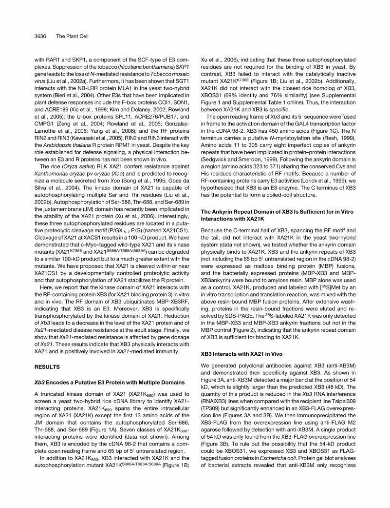
Rice XA21 Binding Protein 3 Is a Ubiquitin Ligase Requiredfor Full Xa21-Mediated Disease Resistance W OA
Yong-Sheng Wang,a,1 Li-Ya Pi,a,1 Xiuhua Chen,a,1 Pranjib K. Chakrabarty,a,2 Junda Jiang,b,3 Alfred Lopez De Leon,c,4
Guo-Zhen Liu,a,5 Liangcai Li,d Ulla Benny,a James Oard,b Pamela C. Ronald,c and Wen-Yuan Songa,6
a Department of Plant Pathology, University of Florida, Gainesville, Florida 32611b Department of Agronomy, Louisiana State University, Baton Rouge, Louisiana 70803c Department of Plant Pathology, University of California, Davis, California 95616d Department of Crop Science, North Carolina State University, Raleigh, North Carolina 27695
XA21 is a receptor-like kinase protein in rice (Oryza sativa) that confers gene-for-gene resistance to specific races of the
causal agent of bacterial blight disease, Xanthomonas oryzae pv oryzae. We identified XA21 binding protein 3 (XB3), an E3
ubiquitin ligase, as a substrate for the XA21 Ser and Thr kinase. The interaction between XB3 and the kinase domain of XA21
has been shown in yeast and in vitro, and the physical association between XB3 and XA21 in vivo has also been confirmed
by coimmunoprecipitation assays. XB3 contains an ankyrin repeat domain and a RING finger motif that is sufficient for its
interaction with the kinase domain of XA21 and for its E3 ubiquitin ligase activity, respectively. Transgenic plants with
reduced expression of the Xb3 gene are compromised in resistance to the avirulent race of X. oryzae pv oryzae.
Furthermore, reduced levels of Xb3 lead to decreased levels of the XA21 protein. These results indicate that Xb3 is
necessary for full accumulation of the XA21 protein and for Xa21-mediated resistance.
INTRODUCTION
Plant immunity is often governed by dominant resistance (R)
genes. Over the past decade, a large number of R genes have
been characterized from diverse plant species (Dangl and Jones,
2001; Staskawicz et al., 2001). The majority of these encode
proteins with a nucleotide binding site and leucine-rich repeats
(NB-LRRs). The NB-LRR class of R proteins can be divided into
two subgroups based on the N-terminal domains: coiled-coil
NB-LRRs carrying a coil-coil structure and TIR-NB-LRRs con-
taining the TIR domain that was originally identified in the
intracellular regions of the Drosophila melanogaster transmem-
brane receptor Toll and the mammalian interleukin 1 receptor (IL-
1R). In the evolutionarily conserved animal innate immunity
pathways, the TIR domains of Toll and IL-1R form similar protein
complexes, including the Ser and Thr kinase Pelle or IRAK
(Hoffmann and Reichhart, 2002). While it is still unclear whether
the TIR domain of the R proteins also recruits a Pelle-like kinase
in the defense response, a number of receptor-like kinases (RLKs)
containing a Pelle-related kinase domain have been implicated in
plant disease resistance (Song et al., 1995; Brueggeman et al.,
2002; Scheer and Ryan, 2002; Godiard et al., 2003; Sun et al.,
2004; Zipfel et al., 2004; Diener and Ausubel, 2005; Llorente
et al., 2005; Chen et al., 2006). These RLKs appear to be equiva-
lent to the receptor/kinase complexes in animal innate immunity.
Ubiquitin-mediated protein modification regulates many cel-
lular processes, including homeostasis, development, cell divi-
sion, growth, and hormone and stress responses (Smalle and
Vierstra, 2004). Ubiquitin is a small peptide that often directs its
conjugated target proteins for degradation. Ubiquitin is activated
by the ubiquitin-activating enzyme E1, transferred to the ubiq-
uitin-conjugating enzyme E2, and finally linked to a target sub-
strate by the ubiquitin ligase E3 (Smalle and Vierstra, 2004).
Characterization of a number of E3 proteins in both animal and
plant systems indicates that the zinc binding domain RING (for
Really Interesting New Gene) finger (RF) is essential for many
ubiquitin-mediated protein modification events (Joazeiro and
Weissman, 2000; Osterlund et al., 2000). Ubiquitinated proteins
can then be degraded by the 26S proteasome or can assume a
role in other proteolysis-independent processes (Ben-Neriah,
2002; Smalle and Vierstra, 2004). It has been suggested that
polyubiquitination of the RF-containing protein TRAF6 activates
the downstream protein kinase TAK1 in the IL-1–mediated innate
immunity pathway (Wang et al., 2001).
The ubiquitin-mediated protein modification system also plays
a role in plant defense mechanisms. For example, SGT1 and
RAR1 are required for the function of multiple R genes (Shirasu
et al., 1999; Austin et al., 2002; Azevedo et al., 2002; Liu et al.,
2002a; Tor et al., 2002; Tornero et al., 2002). SGT1 associates
1 These authors contributed equally to this work.2 Current address: Central Institute for Cotton Research, Post Bag 2,Shankarnagar PO, Nagpur 440010, India.3 Current address: Rice Experiment Station, California Cooperative RiceResearch Foundation, 955 Butte City Highway, Biggs, CA 95917.4 Current address: Novozymes Biotech, 1445 Drew Ave., Davis, CA95616.5 Current address: College of Life Sciences, Hebei Agricultural Univer-sity, Baoding, Hebei 071001, China.6 To whom correspondence should be addressed. E-mail [email protected]; fax 352-392-6532.The author responsible for distribution of materials integral to thefindings presented in this article in accordance with the policy describedin the Instructions for Authors (www.plantcell.org) is: Wen-Yuan Song([email protected]).W Online version contains Web-only data.OA Open Access articles can be viewed online without a subscription.www.plantcell.org/cgi/doi/10.1105/tpc.106.046730
The Plant Cell, Vol. 18, 3635–3646, December 2006, www.plantcell.org ª 2006 American Society of Plant Biologists
with RAR1 and SKP1, a component of the SCF-type of E3 com-
plexes. Suppression of the tobacco (Nicotiana benthamiana) SKP1
gene leads to the loss ofN-mediated resistance toTobaccomosaic
virus (Liu et al., 2002a). Furthermore, it has been shown that SGT1
interacts with the NB-LRR protein MLA1 in the yeast two-hybrid
system (Bieri et al., 2004). Other E3s that have been implicated in
plant defense responses include the F-box proteins COI1, SON1,
and ACRE189 (Xie et al., 1998; Kim and Delaney, 2002; Rowland
et al., 2005); the U-box proteins SPL11, ACRE276/PUB17, and
CMPG1 (Zeng et al., 2004; Rowland et al., 2005; Gonzalez-
Lamothe et al., 2006; Yang et al., 2006); and the RF proteins
RIN2 and RIN3 (Kawasaki et al., 2005). RIN2 and RIN3 interact with
the Arabidopsis thaliana R protein RPM1 in yeast. Despite the key
role established for defense signaling, a physical interaction be-
tween an E3 and R proteins has not been shown in vivo.
The rice (Oryza sativa) RLK XA21 confers resistance against
Xanthomonas oryzae pv oryzae (Xoo) and is predicted to recog-
nize a molecule secreted from Xoo (Song et al., 1995; Goes da
Silva et al., 2004). The kinase domain of XA21 is capable of
autophosphorylating multiple Ser and Thr residues (Liu et al.,
2002b). Autophosphorylation of Ser-686, Thr-688, and Ser-689 in
the juxtamembrane (JM) domain has recently been implicated in
the stability of the XA21 protein (Xu et al., 2006). Interestingly,
these three autophosphorylated residues are located in a puta-
tive proteolytic cleavage motif (P/GX 5-7 P/G) (named XA21CS1).
Cleavage of XA21 at XACS1 results in a 100-kD product. We have
demonstrated that c-Myc–tagged wild-type XA21 and its kinase
mutants (XA21K736E and XA21S686A/T688A/S689A) can be degraded
to a similar 100-kD product but to a much greater extent with the
mutants. We have proposed that XA21 is cleaved within or near
XA21CS1 by a developmentally controlled proteolytic activity
and that autophosphorylation of XA21 stabilizes the R protein.
Here, we report that the kinase domain of XA21 interacts with
the RF-containing protein XB3 (for XA21 binding protein 3) in vitro
and in vivo. The RF domain of XB3 ubiquitinates MBP-XB3RF,
indicating that XB3 is an E3. Moreover, XB3 is specifically
transphosphorylated by the kinase domain of XA21. Reduction
of Xb3 leads to a decrease in the level of the XA21 protein and of
Xa21-mediated disease resistance at the adult stage. Finally, we
show that Xa21-mediated resistance is affected by gene dosage
of Xa21. These results indicate that XB3 physically interacts with
XA21 and is positively involved in Xa21-mediated immunity.
RESULTS
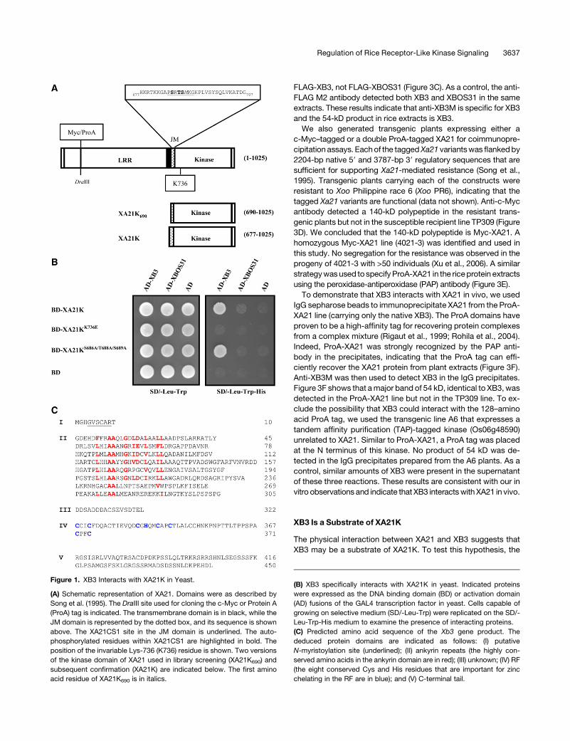
Xb3 Encodes a Putative E3 Protein with Multiple Domains
A truncated kinase domain of XA21 (XA21K690) was used to
screen a yeast two-hybrid rice cDNA library to identify XA21-
interacting proteins. XA21K690 spans the entire intracellular
region of XA21 (XA21K) except the first 13 amino acids of the
JM domain that contains the autophosphorylated Ser-686,
Thr-688, and Ser-689 (Figure 1A). Seven classes of XA21K690-
interacting proteins were identified (data not shown). Among
them, XB3 is encoded by the cDNA 98-2 that contains a com-
plete open reading frame and 65 bp of 59 untranslated region.
In addition to XA21K690, XB3 interacted with XA21K and the
autophosphorylation mutant XA21KS686A/T688A/S689A (Figure 1B;
Xu et al., 2006), indicating that these three autophosphorylated
residues are not required for the binding of XB3 in yeast. By
contrast, XB3 failed to interact with the catalytically inactive
mutant XA21KK736E (Figure 1B; Liu et al., 2002b). Additionally,
XA21K did not interact with the closest rice homolog of XB3,
XBOS31 (69% identity and 76% similarity) (see Supplemental
Figure 1 and Supplemental Table 1 online). Thus, the interaction
between XA21K and XB3 is specific.
The open reading frame of Xb3 and its 59 sequence were fused
in frame to the activation domain of the GAL4 transcription factor
in the cDNA 98-2. XB3 has 450 amino acids (Figure 1C). The N
terminus carries a putative N-myristoylation site (Resh, 1999).
Amino acids 11 to 305 carry eight imperfect copies of ankyrin
repeats that have been implicated in protein–protein interactions
(Sedgwick and Smerdon, 1999). Following the ankyrin domain is
a region (amino acids 323 to 371) sharing the conserved Cys and
His residues characteristic of RF motifs. Because a number of
RF-containing proteins carry E3 activities (Lorick et al., 1999), we
hypothesized that XB3 is an E3 enzyme. The C terminus of XB3
has the potential to form a coiled-coil structure.
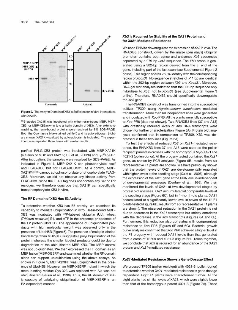
The Ankyrin Repeat Domain of XB3 Is Sufficient for in Vitro
Interactions with XA21K
Because the C-terminal half of XB3, spanning the RF motif and
the tail, did not interact with XA21K in the yeast two-hybrid
system (data not shown), we tested whether the ankyrin domain
physically binds to XA21K. XB3 and the ankyrin repeats of XB3
(not including the 65 bp 59 untranslated region in the cDNA 98-2)
were expressed as maltose binding protein (MBP) fusions,
and the bacterially expressed proteins (MBP-XB3 and MBP-
XB3ankyrin) were bound to amylose resin. MBP alone was used
as a control. XA21K, produced and labeled with [35S]Met by an
in vitro transcription and translation reaction, was mixed with the
above resin-bound MBP fusion proteins. After extensive wash-
ing, proteins in the resin-bound fractions were eluted and re-
solved by SDS-PAGE. The 35S-labeled XA21K was only detected
in the MBP-XB3 and MBP-XB3 ankyrin fractions but not in the
MBP control (Figure 2), indicating that the ankyrin repeat domain
of XB3 is sufficient for binding to XA21K.
XB3 Interacts with XA21 in Vivo
We generated polyclonal antibodies against XB3 (anti-XB3M)
and demonstrated their specificity against XB3. As shown in
Figure 3A, anti-XB3M detected a major band at the position of 54
kD, which is slightly larger than the predicted XB3 (48 kD). The
quantity of this product is reduced in the Xb3 RNA interference
(RNAiXB3) lines when compared with the recipient line Taipei309
(TP309) but significantly enhanced in an XB3-FLAG overexpres-
sion line (Figures 3A and 3B). We then immunoprecipitated the
XB3-FLAG from the overexpression line using anti-FLAG M2
agarose followed by detection with anti-XB3M. A single product
of 54 kD was only found from the XB3-FLAG overexpression line
(Figure 3B). To rule out the possibility that the 54-kD product
could be XBOS31, we expressed XB3 and XBOS31 as FLAG-
tagged fusion proteins in Eschericha coli. Protein gel blot analyses
of bacterial extracts revealed that anti-XB3M only recognizes
3636 The Plant Cell
FLAG-XB3, not FLAG-XBOS31 (Figure 3C). As a control, the anti-
FLAG M2 antibody detected both XB3 and XBOS31 in the same
extracts. These results indicate that anti-XB3M is specific for XB3
and the 54-kD product in rice extracts is XB3.
We also generated transgenic plants expressing either a
c-Myc–tagged or a double ProA-tagged XA21 for coimmunopre-
cipitation assays. Each of the tagged Xa21 variants was flanked by
2204-bp native 59 and 3787-bp 39 regulatory sequences that are
sufficient for supporting Xa21-mediated resistance (Song et al.,
1995). Transgenic plants carrying each of the constructs were
resistant to Xoo Philippine race 6 (Xoo PR6), indicating that the
tagged Xa21 variants are functional (data not shown). Anti-c-Myc
antibody detected a 140-kD polypeptide in the resistant trans-
genic plants but not in the susceptible recipient line TP309 (Figure
3D). We concluded that the 140-kD polypeptide is Myc-XA21. A
homozygous Myc-XA21 line (4021-3) was identified and used in
this study. No segregation for the resistance was observed in the
progeny of 4021-3 with >50 individuals (Xu et al., 2006). A similar
strategy was used to specify ProA-XA21 in the rice protein extracts
using the peroxidase-antiperoxidase (PAP) antibody (Figure 3E).
To demonstrate that XB3 interacts with XA21 in vivo, we used
IgG sepharose beads to immunoprecipitate XA21 from the ProA-
XA21 line (carrying only the native XB3). The ProA domains have
proven to be a high-affinity tag for recovering protein complexes
from a complex mixture (Rigaut et al., 1999; Rohila et al., 2004).
Indeed, ProA-XA21 was strongly recognized by the PAP anti-
body in the precipitates, indicating that the ProA tag can effi-
ciently recover the XA21 protein from plant extracts (Figure 3F).
Anti-XB3M was then used to detect XB3 in the IgG precipitates.
Figure 3F shows that a major band of 54 kD, identical to XB3, was
detected in the ProA-XA21 line but not in the TP309 line. To ex-
clude the possibility that XB3 could interact with the 128–amino
acid ProA tag, we used the transgenic line A6 that expresses a
tandem affinity purification (TAP)-tagged kinase (Os06g48590)
unrelated to XA21. Similar to ProA-XA21, a ProA tag was placed
at the N terminus of this kinase. No product of 54 kD was de-
tected in the IgG precipitates prepared from the A6 plants. As a
control, similar amounts of XB3 were present in the supernatant
of these three reactions. These results are consistent with our in
vitro observations and indicate that XB3 interacts with XA21 in vivo.
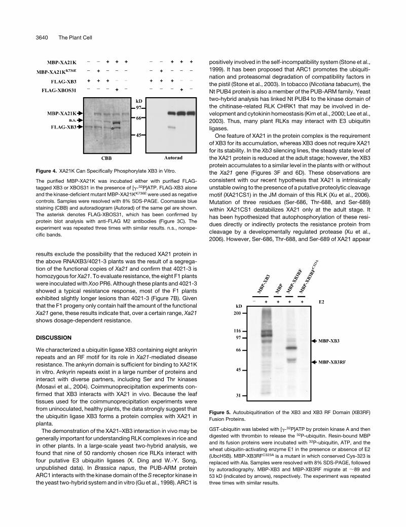
XB3 Is a Substrate of XA21K
The physical interaction between XA21 and XB3 suggests that
XB3 may be a substrate of XA21K. To test this hypothesis, the
Figure 1. XB3 Interacts with XA21K in Yeast.
(A) Schematic representation of XA21. Domains were as described by
Song et al. (1995). The DraIII site used for cloning the c-Myc or Protein A
(ProA) tag is indicated. The transmembrane domain is in black, while the
JM domain is represented by the dotted box, and its sequence is shown
above. The XA21CS1 site in the JM domain is underlined. The auto-
phosphorylated residues within XA21CS1 are highlighted in bold. The
position of the invariable Lys-736 (K736) residue is shown. Two versions
of the kinase domain of XA21 used in library screening (XA21K690) and
subsequent confirmation (XA21K) are indicated below. The first amino
acid residue of XA21K690 is in italics.
(B) XB3 specifically interacts with XA21K in yeast. Indicated proteins
were expressed as the DNA binding domain (BD) or activation domain
(AD) fusions of the GAL4 transcription factor in yeast. Cells capable of
growing on selective medium (SD/-Leu-Trp) were replicated on the SD/-
Leu-Trp-His medium to examine the presence of interacting proteins.
(C) Predicted amino acid sequence of the Xb3 gene product. The
deduced protein domains are indicated as follows: (I) putative
N-myristoylation site (underlined); (II) ankyrin repeats (the highly con-
served amino acids in the ankyrin domain are in red); (III) unknown; (IV) RF
(the eight conserved Cys and His residues that are important for zinc
chelating in the RF are in blue); and (V) C-terminal tail.
Regulation of Rice Receptor-Like Kinase Signaling 3637
purified FALG-XB3 protein was incubated with MBP-XA21K
(a fusion of MBP and XA21K; Liu et al., 2002b) and [g-32P]ATP.
After incubation, the samples were resolved by SDS-PAGE. As
indicated in Figure 4, MBP-XA21K can phosphorylate itself
and FLAG-XB3 but not FLAG-XBOS31. As a control, MBP-
XA21KK736E cannot autophosphorylate or phosphorylate FLAG-
XB3. Moreover, we did not observe any kinase activity from
FLAG-XB3. Since the FLAG tag does not contain any Ser or Thr
residues, we therefore conclude that XA21K can specifically
transphosphorylate XB3 in vitro.
The RF Domain of XB3 Has E3 Activity
To determine whether XB3 has E3 activity, we examined its
capability to mediate ubiquitination in vitro. Resin-bound MBP-
XB3 was incubated with 32P-labeled ubiquitin (Ub), wheat
(Triticum aestivum) E1, and ATP in the presence or absence of
the E2 protein UbcH5B. The appearance of ubiquitinated pro-
ducts with high molecular weight was observed only in the
presence of UbcH5B (Figure 5). The presence of multiple labeled
bands larger than MBP-XB3 suggests a polyubiquitination of the
protein, whereas the smaller labeled products could be due to
degradation of the ubiquitinated MBP-XB3. The MBP control
was not ubiquitinated. We then expressed the RF domain as an
MBP fusion (MBP-XB3RF) and examined whether the RF domain
alone can support ubiquitination using the above assays. As
shown in Figure 5, MBP-XB3RF was ubiquitinated in the pres-
ence of UbcH5B. However, an MBP-XB3RF mutant in which the
metal binding residue Cys-323 was replaced with Ala was not
ubiquitinated (Saurin et al., 1996). Thus, the RF domain of XB3
is capable of catalyzing ubiquitination of MBP-XB3RF in an
E2-dependent manner.
Xb3 Is Required for Stability of the XA21 Protein and
for Xa21-Mediated Resistance
We used RNAi to downregulate the expression of Xb3 in vivo. The
RNAiXB3 construct, driven by the maize (Zea mays) ubiquitin
promoter, contains both sense and antisense Xb3 sequences
separated by a 979-bp uidA sequence. The Xb3 probe is gen-
erated using a 302-bp region derived from the 39 end of the
gene, including part of the last exon (see Supplemental Figure 2
online). This region shares <50% identity with the corresponding
region of Xbos31. No sequence stretches of >11 bp are identical
within the 302-bp region between Xb3 and Xbos31. Moreover,
DNA gel blot analyses indicated that the 302-bp sequence only
hybridizes to Xb3, not to Xbos31 (see Supplemental Figure 3
online). Therefore, RNAiXB3 should specifically downregulate
the Xb3 gene.
The RNAiXB3 construct was transformed into the susceptible
cultivar TP309 using Agrobacterium tumefaciens–mediated
transformation. More than 60 independent lines were generated
and inoculated with Xoo PR6. All the plants were fully susceptible
to Xoo PR6 (data not shown). Two RNAiXB3 lines (37 and A13)
with drastically reduced levels of Xb3 RNA transcripts were
chosen for further characterization (Figure 6A). Protein blot ana-
lyses confirmed that in comparison to TP309, XB3 was de-
creased in these two lines (Figure 3A).
To test the effects of reduced Xb3 on Xa21-mediated resis-
tance, the RNAiXB3 lines 37 and A13 were used as the pollen
recipient parents in crosses with the homozygous Myc-XA21 line
4021-3 (pollen donor). All the progeny tested contained the Xa21
gene, as shown by PCR analyses (Figure 6B, results from six
representative F1 plants are shown). We have previously shown
that the protein levels of XA21 are developmentally regulated
with higher levels at the seedling stage (Xu et al., 2006), although
the expression of the Xa21 gene at the RNA level is independent
of developmental processes (Century et al., 1999). We then
monitored the levels of XA21 at two developmental stages by
protein blot analyses. XA21 accumulated at comparable levels at
the seedling stage (Figure 6C), but in 4-month-old plants, XA21
accumulated at a significantly lower level in seven of the 12 F1
plants tested (Figure 6D, results from six representative F1 plants
are shown). The observed reduction in the XA21 protein is not
due to decreases in the Xa21 transcripts but strictly correlates
with the decreases in the Xb3 transcripts (Figures 6A and 6E).
Furthermore, this reduction also correlates with compromised
resistance to Xoo PR6 (Figures 6F and 6G). Bacterial growth
curve analyses confirmed that Xoo PR6 achieved a higher level in
the F1 progeny with reduced XA21 levels than that generated
from a cross of TP309 and 4021-3 (Figure 6H). Taken together,
we conclude that Xb3 is required for an abundance of the XA21
protein and Xa21-mediated resistance.
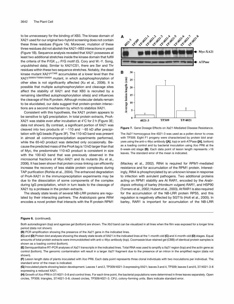
Xa21-Mediated Resistance Shows a Gene Dosage Effect
We crossed TP309 (pollen recipient) with 4021-3 (pollen donor)
to determine whether Xa21-mediated resistance is gene dosage
dependent. Eight F1 plants were characterized further. All the
eight plants had similar levels of XA21, which were slightly lower
than that of the homozygous parent 4021-3 (Figure 7A). These
Figure 2. The Ankyrin Domain of XB3 Is Sufficient for in Vitro Interactions
with XA21K.
35S-labeled XA21K was incubated with either resin-bound MBP, MBP-
XB3, or MBP-XB3ankyrin (the ankyrin domain of XB3). After extensive
washing, the resin-bound proteins were resolved by 8% SDS-PAGE.
Both the Coomassie blue–stained gel (left) and its autoradiogram (right)
are shown. XA21K visualized by autoradiogram is indicated. The exper-
iment was repeated three times with similar results.
3638 The Plant Cell
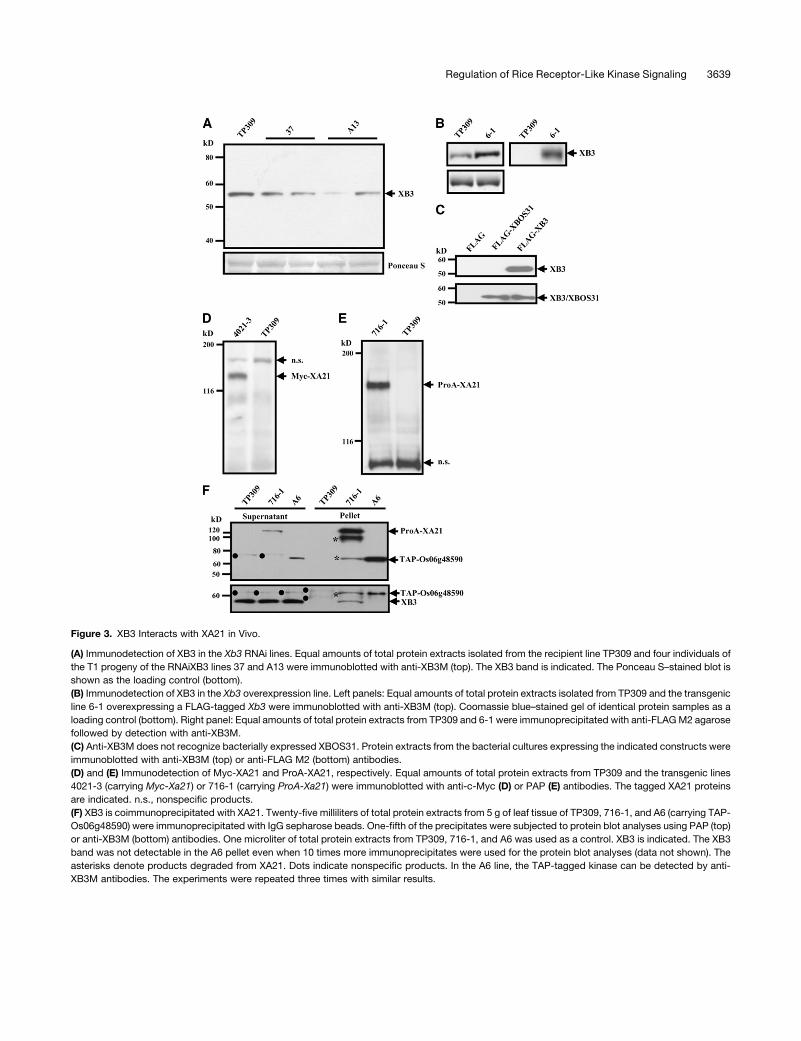
Figure 3. XB3 Interacts with XA21 in Vivo.
(A) Immunodetection of XB3 in the Xb3 RNAi lines. Equal amounts of total protein extracts isolated from the recipient line TP309 and four individuals of
the T1 progeny of the RNAiXB3 lines 37 and A13 were immunoblotted with anti-XB3M (top). The XB3 band is indicated. The Ponceau S–stained blot is
shown as the loading control (bottom).
(B) Immunodetection of XB3 in the Xb3 overexpression line. Left panels: Equal amounts of total protein extracts isolated from TP309 and the transgenic
line 6-1 overexpressing a FLAG-tagged Xb3 were immunoblotted with anti-XB3M (top). Coomassie blue–stained gel of identical protein samples as a
loading control (bottom). Right panel: Equal amounts of total protein extracts from TP309 and 6-1 were immunoprecipitated with anti-FLAG M2 agarose
followed by detection with anti-XB3M.
(C) Anti-XB3M does not recognize bacterially expressed XBOS31. Protein extracts from the bacterial cultures expressing the indicated constructs were
immunoblotted with anti-XB3M (top) or anti-FLAG M2 (bottom) antibodies.
(D) and (E) Immunodetection of Myc-XA21 and ProA-XA21, respectively. Equal amounts of total protein extracts from TP309 and the transgenic lines
4021-3 (carrying Myc-Xa21) or 716-1 (carrying ProA-Xa21) were immunoblotted with anti-c-Myc (D) or PAP (E) antibodies. The tagged XA21 proteins
are indicated. n.s., nonspecific products.
(F) XB3 is coimmunoprecipitated with XA21. Twenty-five milliliters of total protein extracts from 5 g of leaf tissue of TP309, 716-1, and A6 (carrying TAP-
Os06g48590) were immunoprecipitated with IgG sepharose beads. One-fifth of the precipitates were subjected to protein blot analyses using PAP (top)
or anti-XB3M (bottom) antibodies. One microliter of total protein extracts from TP309, 716-1, and A6 was used as a control. XB3 is indicated. The XB3
band was not detectable in the A6 pellet even when 10 times more immunoprecipitates were used for the protein blot analyses (data not shown). The
asterisks denote products degraded from XA21. Dots indicate nonspecific products. In the A6 line, the TAP-tagged kinase can be detected by anti-
XB3M antibodies. The experiments were repeated three times with similar results.
Regulation of Rice Receptor-Like Kinase Signaling 3639
results exclude the possibility that the reduced XA21 protein in
the above RNAiXB3/4021-3 plants was the result of a segrega-
tion of the functional copies of Xa21 and confirm that 4021-3 is
homozygous for Xa21. To evaluate resistance, the eight F1 plants
were inoculated with Xoo PR6. Although these plants and 4021-3
showed a typical resistance response, most of the F1 plants
exhibited slightly longer lesions than 4021-3 (Figure 7B). Given
that the F1 progeny only contain half the amount of the functional
Xa21 gene, these results indicate that, over a certain range, Xa21
shows dosage-dependent resistance.
DISCUSSION
We characterized a ubiquitin ligase XB3 containing eight ankyrin
repeats and an RF motif for its role in Xa21-mediated disease
resistance. The ankyrin domain is sufficient for binding to XA21K
in vitro. Ankyrin repeats exist in a large number of proteins and
interact with diverse partners, including Ser and Thr kinases
(Mosavi et al., 2004). Coimmunoprecipitation experiments con-
firmed that XB3 interacts with XA21 in vivo. Because the leaf
tissues used for the coimmunoprecipitation experiments were
from uninoculated, healthy plants, the data strongly suggest that
the ubiquitin ligase XB3 forms a protein complex with XA21 in
planta.
The demonstration of the XA21–XB3 interaction in vivo may be
generally important for understanding RLK complexes in rice and
in other plants. In a large-scale yeast two-hybrid analysis, we
found that nine of 50 randomly chosen rice RLKs interact with
four putative E3 ubiquitin ligases (X. Ding and W.-Y. Song,
unpublished data). In Brassica napus, the PUB-ARM protein
ARC1 interacts with the kinase domain of the S receptor kinase in
the yeast two-hybrid system and in vitro (Gu et al., 1998). ARC1 is
positively involved in the self-incompatibility system (Stone et al.,
1999). It has been proposed that ARC1 promotes the ubiquiti-
nation and proteasomal degradation of compatibility factors in
the pistil (Stone et al., 2003). In tobacco (Nicotiana tabacum), the
Nt PUB4 protein is also a member of the PUB-ARM family. Yeast
two-hybrid analysis has linked Nt PUB4 to the kinase domain of
the chitinase-related RLK CHRK1 that may be involved in de-
velopment and cytokinin homeostasis (Kim et al., 2000; Lee et al.,
2003). Thus, many plant RLKs may interact with E3 ubiquitin
ligases.
One feature of XA21 in the protein complex is the requirement
of XB3 for its accumulation, whereas XB3 does not require XA21
for its stability. In the Xb3 silencing lines, the steady state level of
the XA21 protein is reduced at the adult stage; however, the XB3
protein accumulates to a similar level in the plants with or without
the Xa21 gene (Figures 3F and 6D). These observations are
consistent with our recent hypothesis that XA21 is intrinsically
unstable owing to the presence of a putative proteolytic cleavage
motif (XA21CS1) in the JM domain of this RLK (Xu et al., 2006).
Mutation of three residues (Ser-686, Thr-688, and Ser-689)
within XA21CS1 destabilizes XA21 only at the adult stage. It
has been hypothesized that autophosphorylation of these resi-
dues directly or indirectly protects the resistance protein from
cleavage by a developmentally regulated protease (Xu et al.,
2006). However, Ser-686, Thr-688, and Ser-689 of XA21 appear
Figure 4. XA21K Can Specifically Phosphorylate XB3 in Vitro.
The purified MBP-XA21K was incubated either with purified FLAG-
tagged XB3 or XBOS31 in the presence of [g-32P]ATP. FLAG-XB3 alone
and the kinase-deficient mutant MBP-XA21KK736E were used as negative
controls. Samples were resolved with 8% SDS-PAGE. Coomassie blue
staining (CBB) and autoradiogram (Autorad) of the same gel are shown.
The asterisk denotes FLAG-XBOS31, which has been confirmed by
protein blot analysis with anti-FLAG M2 antibodies (Figure 3C). The
experiment was repeated three times with similar results. n.s., nonspe-
cific bands.
Figure 5. Autoubiquitination of the XB3 and XB3 RF Domain (XB3RF)
Fusion Proteins.
GST-ubiquitin was labeled with [g-32P]ATP by protein kinase A and then
digested with thrombin to release the 32P-ubiquitin. Resin-bound MBP
and its fusion proteins were incubated with 32P-ubiquitin, ATP, and the
wheat ubiquitin-activating enzyme E1 in the presence or absence of E2
(UbcH5B). MBP-XB3RFC323A is a mutant in which conserved Cys-323 is
replaced with Ala. Samples were resolved with 8% SDS-PAGE, followed
by autoradiography. MBP-XB3 and MBP-XB3RF migrate at ;89 and
53 kD (indicated by arrows), respectively. The experiment was repeated
three times with similar results.
3640 The Plant Cell
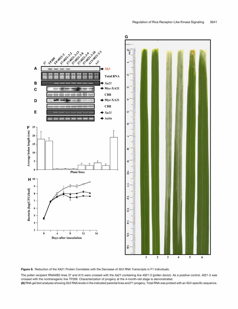
Figure 6. Reduction of the XA21 Protein Correlates with the Decrease of Xb3 RNA Transcripts in F1 Individuals.
The pollen recipient RNAiXB3 lines 37 and A13 were crossed with the Xa21-containing line 4021-3 (pollen donor). As a positive control, 4021-3 was
crossed with the nontransgenic line TP309. Characterization of progeny at the 4-month-old stage is demonstrated.
(A) RNA gel blot analyses showing Xb3 RNA levels in the indicated parental lines and F1 progeny. Total RNA was probed with an Xb3-specific sequence.
Regulation of Rice Receptor-Like Kinase Signaling 3641
to be unnecessary for the binding of XB3. The kinase domain of
XA21 used for our original two-hybrid screening does not contain
these three residues (Figure 1A). Moreover, mutation of these
three residues did not abolish the XA21–XB3 interactions in yeast
(Figure 1B). Sequence analysis revealed that XA21 possesses at
least two additional stretches inside the kinase domain that fulfill
the criteria of the P/GX 5-7 P/G motif (G. Cory and W.-Y. Song,
unpublished data). Similar to XA21CS1, there are Ser and Thr
residues within these two sequence stretches. Notably, the dead
kinase mutant XA21K736E accumulates at a lower level than the
XA21S686A/T688A/S689A mutant, in which autophosphorylation of
other sites is not significantly affected (Xu et al., 2006). It is
possible that multiple autophosphorylation and cleavage sites
affect the stability of XA21 and that XB3 is recruited by a
remaining identified autophosphorylation site(s) and influences
the cleavage of this R protein. Although molecular details remain
to be elucidated, our data suggest that protein–protein interac-
tions are a second mechanism by which to stabilize XA21.
Consistent with this hypothesis, the XA21 protein appears to
be sensitive to IgG precipitation. In total protein extracts, ProA-
XA21 was stable even after incubation at 48C for 2 h (Figure 3E;
data not shown). By contrast, a significant portion of XA21 was
cleaved into two products of ;110 and ;65 kD after precipi-
tation with IgG beads (Figure 3F). The 110-kD band was present
in almost all coimmunoprecipitation experiments performed,
while the 65-kD product was detected only occasionally. Be-
cause the predicted mass of the ProA tag is 13 kD larger than that
of Myc, the predominate 110-kD product is consistent in size
with the 100-kD band that was previously observed in the
microsomal fractions of Myc-XA21 and its mutants (Xu et al.,
2006). It has been shown that protein cross-linking can efficiently
increase the recovery of less stable protein complexes during
TAP purification (Rohila et al., 2004). The enhanced degradation
of ProA-XA21 in the immunoprecipitation experiments may be
due to the dissociation of some components of the complex
during IgG precipitation, which in turn leads to the cleavage of
XA21 by a protease in the protein extracts.
The steady state levels of several NB-LRR proteins are regu-
lated by their interacting partners. The Arabidopsis gene RIN4
encodes a novel protein that interacts with the R protein RPM1
(Mackey et al., 2002). RIN4 is required for RPM1-mediated
resistance and for accumulation of the RPM1 protein. Interest-
ingly, RIN4 is phosphorylated by an unknown kinase in response
to infection with avirulent pathogens. Two additional proteins
acting on RPM1 stability are At RAR1, encoded by the Arabi-
dopsis ortholog of barley (Hordeum vulgare) RAR1, and HSP90
(Tornero et al., 2002; Hubert et al., 2003). At RAR1 is also required
for the accumulation of the NB-LRR protein RPS5, and this
regulation is negatively affected by SGT1b (Holt et al., 2005). In
barley, RAR1 is important for accumulation of the NB-LRR
Figure 6. (continued).
Both autoradiogram (top) and agarose gel (bottom) are shown. The Xb3 band can be visualized in all lines when the film was exposed for a longer time
period (data not shown).
(B) PCR amplification showing the presence of the Xa21 gene in the indicated lines.
(C) and (D) Protein blot analyses showing the steady state levels of XA21 in the indicated lines at the 1-month-old (C) and 4-month-old (D) stages. Equal
amounts of total protein extracts were immunoblotted with anti-c-Myc antibody (top). Coomassie blue–stained gel (CBB) of identical protein samples is
shown as a loading control (bottom).
(E) Semiquantitative RT-PCR analyses of Xa21 transcripts in the indicated lines. Total RNA was used to amplify a Xa21 region (top) and the actin gene as
control (bottom). The genomic contamination will result in a larger Xa21 fragment due to the presence of an intron in the amplified region (data not
shown).
(F) Lesion length data of plants inoculated with Xoo PR6. Each data point represents three clonal individuals with two inoculations per individual. The
standard error of the mean is indicated.
(G) Inoculated plants showing lesion development. Leaves 1 and 2, TP309/4021-3 expressing XA21; leaves 3 and 4, TP309; leaves 5 and 6, 37/4021-3-8
expressing a reduced XA21.
(H) Growth of Xoo PR6 in 37/4021-3-8 and control lines. For each time point, the bacterial populations were determined in three leaves separately. Open
circles, TP309; triangles, 37/4021-3-8; closed circles, TP309/4021-3. CFU, colony-forming units. Bars indicate standard error.
Figure 7. Gene Dosage Effects on Xa21-Mediated Disease Resistance.
The Xa21 homozygous line 4021-3 was used as a pollen donor to cross
with TP309. Eight F1 progeny were characterized by protein blot anal-
yses using the anti-c-Myc antibody ([A], top) or anti-ATPase ([A], bottom)
as a loading control and by bacterial inoculation using Xoo PR6 at the
6-week-old stage (B). Each data point of lesion length represents >16
leaves. The standard error of the mean is indicated.
3642 The Plant Cell

proteins MLA1 and MLA6 (Bieri et al., 2004). Our results dem-
onstrate that the steady state accumulation of RLK XA21 is also
influenced by its binding protein. Therefore, the stability of R
proteins may generally rely on the presence of their partners, and
the measurement of the steady state levels of R proteins has
become a common tool to dissect R gene–mediated signaling
pathways.
XB3 can be phosphorylated by XA21K in vitro. Although this
finding remains to be confirmed in planta, the direct interaction
between XA21 and XB3 in vivo supports such an extrapolation.
In previous studies, we have found that XA21K is capable of
phosphorylating a 22–amino acid peptide derived from the
glutathione S-transferase (GST) vector when the peptide is fused
in frame with XA21K (Liu et al., 2002b). XA21K, however, cannot
phosphorylate the free GST protein that contains the same
peptide (G.-Z. Liu and W.-Y. Song, unpublished data), suggest-
ing that a physical connection between XA21 and the artificial
substrate is required for the phosphorylation. The two phos-
phorylated residues on the 22–amino acid peptide have been
mapped (Liu et al., 2002a). Sequence comparisons reveal that
the flanking sequences of these two residues share similarities
with those of other identified autophosphorylation sites inside
XA21K (G.-Z. Liu and W.-Y. Song, unpublished data). These
observations suggest that XA21-mediated transphosphorylation
requires the presence of a phosphorylation site and the physical
interaction of the substrate with the kinase. XB3 fulfills these
criteria. In line with this idea, XA21K was unable to phosphorylate
XBOS31, which does not interact with XA21K in yeast. Therefore,
protein–protein interactions may contribute to the specificity of
XA21 phosphorylation.
When Xb3 is reduced, resistance to Xoo PR6 is compromised
in the Xa21 lines. The compromised resistance can be attributed
to a decrease in the level of the XA21 protein. The dosage effects
have been suggested by several previous studies (Song et al.,
1995; Zhang et al., 1998; Xu et al., 2006). Additionally, the
heterozygous Xa21 plants generated by a cross between 4021-3
and TP309 are slightly less resistant than the homozygous plant
4021-3. Alternatively, but not mutually exclusive, the reduction
of Xb3 may directly contribute to the compromised resistance
as well. In this case, the XB3 level is a rate-limiting step for
Xa21-mediated resistance.
Based on our data, we propose that the physical interaction of
XB3 stabilizes the XA21 protein, thereby maintaining the level of
the R protein. Upon pathogen infection, the XA21-XB3 complex
is required for activation of XB3, presumably through transphos-
phorylation. The activated XB3 may ubiquitinate a third protein
and target it for degradation. In this scenario, the degraded
protein would be a negative regulator of the defense signaling.
Alternatively, XB3 may autoubiquitinate, triggering activation of a
downstream signaling protein(s). Support for the second model
comes from studies in animal innate immunity. Recognition of
IL-1 by IL-1R, for example, leads to the formation of a receptor/
kinase complex, including IL-1R, MyD88, and the Ser and Thr
kinase IRAK (Wu and Arron, 2003). Following this recognition
is the activation of TRAF6, an RF-containing ubiquitin ligase.
Polyubiquitination of TRAF6 has been suggested to activate the
downstream kinase TAK1 (Wang et al., 2001). Our model there-
fore predicts an interesting parallel between plant and animal
defense pathways mediated by a receptor kinase and a receptor/
kinase complex. Such a parallel has been recognized based on
the fact that a number of plant R proteins share the TIR domain
(Baker et al., 1997).
METHODS
Yeast Two-Hybrid Screening
To make the BD-XA21K690 construct for yeast two-hybrid screening,
XA21K690 was PCR amplified using the primer pair 59-AGGTCGACC-
CCGGGAATGAAAGGCCACCCATT-39/59-GGGGTACCGGATCCTCAAA-
ATTCAAGGCTCCCACCTTCAA-39 and cloned into the two-hybrid vector
pPC97 carrying the GAL4 BD domain (Chevray and Nathans, 1992; Chern
et al., 2001). All the PCR products in this study were confirmed by DNA
sequencing. A yeast two-hybrid library constructed from poly(A)þmRNA
harvested from 2-week-old seedlings of the rice (Oryza sativa) line TP309
(Yin et al., 1997) was kindly provided by R. Beachy. Yeast cells carrying
BD-XA21 were transformed with the rice library. Transformation effi-
ciency was estimated by plating an aliquot of transformation mixtures
onto SD/-Leu-Trp medium. Candidates selected onto SD/-Leu-Trp-His
medium were subjected to b-galactosidase assays as described in the
manufacturer’s procedure for the Matchmaker GAL4 system (Clontech).
Plasmids were recovered from the cells showing the Hisþ and Lacþ
phenotypes and sequenced to determine identity.
To make BD constructs for yeast two-hybrid analyses, XA21K,
XA21KK736E, and XA21KS686A/T688A/S689A were subcloned from their
GST versions of constructs into the pPC97 vector (Liu et al., 2002b; Xu
et al., 2006).
In Vitro Binding Assays
XA21K was PCR amplified with the primer pair 59-GGATCCGTCGACCA-
CAAGAGAACTAAAAAGGGAGC-39/59-GGATCCGTCGACCCCGGGCA-
GAAGTCGATCTGAAGTGTGGCA-39 and cloned into pET-28a (Novagen).
The resulting plasmid was subjected to the expression of XA21K and
labeling using the in vitro transcription and translation kit (Promega). Be-
cause the full-length XB3 was poorly expressed as an MBP fusion protein,
the cDNA encoding amino acids 1 to 428 of XB3 (missing the last 22 amino
acids) was cloned in frame into pMAL-c2X (New England Biolabs) to make
MBP-XB3. The ankyrin domain of Xb3 was PCR amplified with the primer
pair 59-GGATCCATGATATCCATGGGTCACGGTGTC-39/59-CGGGATC-
CGATATCAGATGCAGCAAAGCTCC-39 and cloned into pMAL-c2X. The35S-labeled XA21K was incubated with resin-bound MBP or MBP fusion
proteins in binding buffer (20 mM HEPES, pH 7.4, 1 mM EDTA, 5 mM
MgCl2, 1 mM DTT, 0.1% Triton X-100, 1 mg/mL BSA, and 13 complete
protease inhibitors [Roche]) for 120 min at 48C with gentle shaking. After
five washes using buffer (20 mM HEPES, pH 7.4, 1 mM EDTA, 5 mM
MgCl2, 1 mM DTT, and 0.1% Triton X-100), the bound proteins were
eluted with the same buffer supplemented with 10 mM maltose and
resolved by 8% SDS-PAGE. The gel was stained with Coomassie Brilliant
Blue, dried, and exposed to x-ray film.
Immunodetection
For the anti-XB3M antibodies, a region located in the middle of Xb3 (amino
acids 230 to 365) was PCR amplified with the primer pair 59-GGATC-
CATGATATCGAGAATTCCCTATTCTGTTGC-39/59-GGATCCGATATCTC-
ATGAGGGCGGTGTCAGGGTC-39 and cloned into the expression vectors
pGTK (Liu et al., 2002b) and pMAL-c2X, respectively. The fusion proteins
MBP-XB3M and GST-XB3M were expressed in the Escherichia coli strain
ER2566 and affinity purified (Liu et al., 2002b). MBP-XB3M was used to
immunize rabbits (Cocalico Biologicals). Antisera were subjected to
Regulation of Rice Receptor-Like Kinase Signaling 3643
affinity purification using GST-XB3M according to the method described
by Lin et al. (1996).
To create the FLAG-tagged XB3 and XBOS31 for bacterial expression,
the full-length coding regions of these two genes were PCR amplified
with the primer pairs 59-GTGTGATATCATGGGTCACGGTGTC-39/59-GGA-
TCCATGATATCGAGGATGATGCGGCGA-39 and 59-GTGTAGATCTCC-
GAATTCCTACCTATGGGGCACGGCCTGAG-39/59-GTGTGCGGCCGCC-
CCAAAGCTCTAGTGCTAGTAAGGTG-39 and cloned into pFLAG-N, an
expression vector derived from pFLAG-MAC (Sigma-Aldrich). The fusion
proteins FLAG-XB3 and FLAG-XBOS31 were expressed in the E. coli
strain ER2566 (Liu et al., 2002b).
Both the c-Myc and ProA tags were inserted in domain B of the Xa21
gene. The DNA sequences, encoding the 13–amino acid c-Myc and
128–amino acid double ProA epitope tags, were PCR amplified from the
plasmids pATGMyc and pUbi.nc1300.ntapintron.new and cloned into the
unique DraIII site of the plasmid pC822 to create pC822cMyc and
pC822ProA, respectively. The KpnI fragment containing Myc-Xa21 or
ProA-Xa21 was cloned into pCAMBIA1300 for rice transformation. The
construction of TAP-Os06g48590 was as described by Rohila et al.
(2006).
Total rice protein extracts were isolated by grinding leaf tissue in liquid
nitrogen and thawing in an equal volume of extraction buffer [50 mM Tris-
HCl, pH 7.5, 150 mM NaCl, 1 mM EDTA, 0.1% Triton X-100, 5% (v/v)
b-mercaptoethanol, 1 mM 4-(2-aminoethyl)-benzenesulfonyl fluoride
(Sigma-Aldrich), 2 mg/mL antipain (Sigma-Aldrich), 2 mg/mL leupeptin
(Sigma-Aldrich), and 2 mg/mL aprotinin (Sigma-Aldrich)]. Cell debris were
removed by centrifugation at 12,000g for 10 min at 48C. Protein concen-
tration was measured with Bio-Rad protein assays.
To coimmunoprecipitate ProA-XA21 and XB3, protein extracts were
prepared from 5 g of leaf tissue in 25 mL of ice-cold extraction buffer
(20 mM Tris-HCl, pH 8.0, 150 mM NaCl, 0.1% Triton X-100, 2.5 mM EDTA,
2 mM benzamidine [Sigma-Aldrich], 10 mM b-mercaptoethanol, 20 mM
NaF, 1 mM phenylmethylsulfonyl fluoride, 1% Protease Cocktail [Sigma-
Aldrich], 10 mM leupeptin, and 10% glycerol). After filtering through
double layers of Miracloth (Calbiochem) followed by centrifugation twice
at 13,000g for 10 min at 48C, the supernatant was mixed with 400 mL of
IgG Sepharose beads (Amersham Biosciences) and incubated at 48C for
1 h. The beads were then washed four times in 1 mL of protein extraction
buffer lacking protease inhibitors and twice in 0.4 mL of 5 mM ammonium
acetate, pH 5.0. The protein was eluted with 2 mL of 0.5 M HOAC, pH 3.4,
neutralized with one-tenth volume of 1 M Tris-HCl, pH 8.0, and concen-
trated by acetone precipitation.
Protein blot analyses were performed as described previously (Xu et al.,
2006).
Transphosphorylation Assays
Transphosphorylation of FLAG-XB3 by MBP-XA21K was essentially as
previously described (Liu et al., 2002b).
Ubiquitination Assays
pMBP-XB3RING was made as follows: the RF domain and C-terminal tail
of Xb3 were PCR amplified with the primer pair 59-GGATCCATGA-
TATCCGATGCATGCTCAGAG-39/59-GGATCCATGATATCGAGGATGAT-
GCGGCGA-39 and cloned into pMAL-c2X. To remove the C-terminal tail,
the resulting construct pMALXB3C was digested with PstI and self-
ligated. To create a stop codon after the RF domain, the self-ligated con-
struct was digested with HindIII, blunted by filling-in, and self-ligated
again. Site-directed mutagenesis was performed to create pMBP-
XB3RINGC323A as described previously (Liu et al., 2002b). The fusion
proteins MBP-XB3, MBP-XB3RING, and MBP-XB3RINGC323A were ex-
pressed in the E. coli strain ER2566 and affinity purified (Liu et al., 2002b).
Ubiquitination assays were performed as described previously (Nodzon
et al., 2004).
RNAiXB3 and Overexpression of the Xb3 Gene
The 39 end of Xb3 was PCR amplified with the following primer pairs:
59-GAATTCTCTAGACCGGGGCAGCATCTCA-39/59-ACTAGTGGATCC-
TTTCTGATACCAACGGA-39 and 59-GAATTCAGATCTCCGGGGCAGC-
ATCTCA-39/59-ACTAGTGATATCTTTCTGATACCAACGGA-39. The PCR
products were ligated to the uidA fragment spanning nucleotides 815 to
1793 in both antisense and sense orientations. The resulting construct
was then cloned into the overexpression vector pBHU-1, which contains
the hph gene, whose product confers resistance to hygromycin B, at the
site between the maize (Zea mays) ubiquitin promoter and the nos 39
terminator. Rice transformation was performed as described previously
(Xu et al., 2006).
A full-length Xb3 (lacking the stop codon) was PCR amplified with the
primer pair 59-GGATCCACTAGTATGGGTCACGGTGTCAG-39/59-GGA-
TCCTAGATCGTGCTCAGGCT-39 and in-frame fused to the FLAG tag
(containing a stop codon at the 39 end). The tagged Xb3 was cloned into
pCmHU-1, an overexpression vector derived from pBHU-1, for rice
transformation.
Characterization of Transgenic Plants
DNA and RNA gel blot analyses were performed using standard proce-
dures. Plant inoculation and growth curve analyses were performed as
described by Song et al. (1995), except that a bacterial suspension of OD
;1.0 was used.
Semiquantitative RT-PCR analyses were performed with primer pairs
59-CAGAAGTCGATCTGAAGTGTGGCA-39/59-GCACAAGAGAACTAAA-
AAGGGAGCCC-39 (for Xa21 transcripts) and 59-TGGCGCCCGAGGAG-
CACC-39/59-GTAACCCCTCTCAGTCAG-39 (for actin transcripts). Three
micrograms of total RNA were converted into cDNA using the SuperScript
First-Stand Synthesis system (Invitrogen) followed by PCR amplification
for 20, 25, 30, and 35 cycles. The amplified products were then resolved
by gel electrophoresis.
The transgenic lines 37 and A13 were used as the pollen recipient
parents to cross with pollen donor 4021-3. Eleven seeds were recovered
from the 37/4021-3 cross, whereas only one seed was obtained from the
A13/4021-3 cross. The nature of the F1 hybrids was confirmed by PCR
amplification of a 690-bp fragment spanning part of the Xa21 and 35S
promoters in the pCAMBIA1300-Myc-Xa21 construct using the primer
pair 59-ATTCATTAATGCAGCTGGCACGACA-39/59-GGTAATGGATGTA-
CACTGCAGAACGA-39.
Sequence Analyses
The PAUP (version 4.0b10) software package (Swofford, 2002) was used
to perform phylogenetic analyses. Neighbor joining was used to recon-
struct the phylogenetic relationships of these amino acids. Bootstrap
values were derived from 1000 replicates to quantify the relative support
for branches of the inferred phylogenetic tree. The identity and similarity
between proteins were calculated using the GAP tool within the Genetics
Computer Group program.
Accession Numbers
The rice cDNA accession numbers (KOME; http://cdna01.dna.affrc.go.
jp/cDNA/) for the Xbos genes are as follows: Xbos31, AK106014; Xbos32,
AK120632; Xbos33, AK065223; Xbos34, AK059792; and Xbos35,
AK067289. The GenBank accession number for Xbos36 is DQ088999.
The accession numbers for the XBAT genes were described previously
(Nodzon et al., 2004). The GenBank accession number for Xb3 cDNA
sequence is AF272860.
3644 The Plant Cell
Supplemental Data
The following materials are available in the online version of this article.
Supplemental Table 1. Comparisons of Amino Acid Sequences
between the XB3-Related Proteins from Rice and Arabidopsis.
Supplemental Figure 1. Phylogenetic Tree Based on the Predicted
Amino Acid Sequences of XB3 and XB3-Related Proteins from Rice
(XBOS) and Arabidopsis (XBAT).
Supplemental Figure 2. Schematic Representation of the Xb3 and
Xbos31 Genomic Gene Structures.
Supplemental Figure 3. DNA Gel Blot Analyses Showing Specificity
of the RNAiXB3 Probe.
ACKNOWLEDGMENTS
We thank R. Beachy for the rice yeast two-hybrid library; M. Fromm for
pUbi.nc1300.ntapintron.new; M. Boutry for anti-ATPase; O. da Costa e
Silva for pATGmyc; S. Dai, Y. Chen, Y. Xu, P. Tinjuangjun, Y.-R. Chen, A.
Snyder, and H. Xiao for technical assistance; Lisa Nodzon for cloning
Xbos36; M.-S. Shern for providing BD-XA21 for use in these studies;
and Fahong Yu for phylogenetic analyses. We also thank Jeff Chang,
J.B. Jones, H. Klee, Terry Davoli, and Margaret Joyner for critical
reading of the manuscript and their invaluable comments. This research
was supported primarily by the National Science Foundation under
Grant 0080155 to W.-Y.S. and by a grant from National Institutes of
Health to P.C.R.
Received August 18, 2006; revised October 4, 2006; accepted November
2, 2006; published December 15, 2006.
REFERENCES
Austin, M.J., Muskett, P., Kahn, K., Feys, B.J., Jones, J.D., and
Parker, J.E. (2002). Regulatory role of SGT1 in early R gene-mediated
plant defenses. Science 295, 2077–2080.
Azevedo, C., Sadanandom, A., Kitagawa, K., Freialdenhoven, A.,
Shirasu, K., and Schulze-Lefert, P. (2002). The RAR1 interactor
SGT1, an essential component of R gene-triggered disease resis-
tance. Science 295, 2073–2076.
Baker, B., Zambryski, P., Staskawicz, B., and Dinesh-Kumar, S.P.
(1997). Signaling in plant-microbe interactions. Science 276, 726–733.
Ben-Neriah, Y. (2002). Regulatory functions of ubiquitination in the
immune system. Nat. Immunol. 3, 20–26.
Bieri, S., Mauch, S., Shen, Q.H., Peart, J., Devoto, A., Casais, C.,
Ceron, F., Schulze, S., Steinbiss, H.H., Shirasu, K., and Schulze-
Lefert, P. (2004). RAR1 positively controls steady state levels of
barley MLA resistance proteins and enables sufficient MLA6 accu-
mulation for effective resistance. Plant Cell 16, 3480–3495.
Brueggeman, R., Rostoks, N., Kudrna, D., Kilian, A., Han, F., Chen,
J., Druka, A., Steffenson, B., and Kleinhofs, A. (2002). The barley
stem rust-resistance gene Rpg1 is a novel disease-resistance gene
with homology to receptor kinases. Proc. Natl. Acad. Sci. USA 99,
9328–9333.
Century, K.S., Lagman, R.A., Adkisson, M., Morlan, J., Tobias, R.,
Schwartz, K., Smith, A., Love, J., Ronald, P.C., and Whalen, M.C.
(1999). Developmental control of Xa21-mediated disease resistance in
rice. Plant J. 20, 231–236.
Chen, X., et al. (2006). A B-lectin receptor kinase gene conferring rice
blast resistance. Plant J. 46, 794–804.
Chern, M.S., Fitzgerald, H.A., Yadav, R.C., Canlas, P.E., Dong, X.,
and Ronald, P.C. (2001). Evidence for a disease-resistance pathway
in rice similar to the NPR1-mediated signaling pathway in Arabidopsis.
Plant J. 27, 101–113.
Chevray, P.M., and Nathans, D. (1992). Protein interaction cloning in
yeast: Identification of mammalian proteins that react with the leucine
zipper of Jun. Proc. Natl. Acad. Sci. USA 89, 5789–5793.
Dangl, J.L., and Jones, J.G. (2001). Plant pathogens and integrated
defence responses to infection. Nature 411, 826–833.
Diener, A.C., and Ausubel, F.M. (2005). RESISTANCE TO FUSARIUM
OXYSPORUM 1, a dominant Arabidopsis resistance gene, is not race
specific. Genetics 171, 305–321.
Godiard, L., Sauviac, L., Torii, K.U., Grenon, O., Mangin, B.,
Grimsley, N.H., and Marco, Y. (2003). ERECTA, an LRR receptor-
like kinase protein controlling development pleiotropically affects
resistance to bacterial wilt. Plant J. 36, 353–365.
Goes da Silva, F., Shen, Y., Dardick, C., Burdman, S., Yadav, R.,
Sharma, P., and Ronald, P.C. (2004). Components of a type I
secretion system and a sulfotransferase-like protein are required for
the XA21 receptor kinase mediated defense response. Mol. Plant
Microbe Interact. 17, 593–601.
Gonzalez-Lamothe, R., Tsitsigiannis, D.I., Ludwig, A.A., Panicot, M.,
Shirasu,K.,andJones,J.D. (2006).TheU-boxproteinCMPG1 isrequired
for efficient activation of defense mechanisms triggered by multiple
resistance genes in tobacco and tomato. Plant Cell 18, 1067–1083.
Gu, T., Mazzurco, M., Sulaman, W., Matias, D.D., and Goring, D.R.
(1998). Binding of an arm repeat protein to the kinase domain of the
S-locus receptor kinase. Proc. Natl. Acad. Sci. USA 95, 382–387.
Hoffmann, J.A., and Reichhart, J.M. (2002). Drosophila innate immu-
nity: An evolutionary perspective. Nat. Immunol. 3, 121–126.
Holt III, B.F., Belkhadir, Y., and Dangl, J.L. (2005). Antagonistic control
of disease resistance protein stability in the plant immune system.
Science 309, 929–932.
Hubert, D.A., Tornero, P., Belkhadir, Y., Krishna, P., Takahashi, A.,
Shirasu, K., and Dangl, J.L. (2003). Cytosolic HSP90 associates with
and modulates the Arabidopsis RPM1 disease resistance protein.
EMBO J. 22, 5679–5689.
Joazeiro, C.A.P., and Weissman, A.M. (2000). RING finger proteins:
Mediators of ubiquitin ligase activity. Cell 102, 549–552.
Kawasaki, T., Nam, J., Boyes, D.C., Holt III, B.F., Hubert, D.A., Wiig,
A., and Dangl, J.L. (2005). A duplicated pair of Arabidopsis RING-
finger E3 ligases contribute to the RPM1- and RPS2-mediated
hypersensitive response. Plant J. 44, 258–270.
Kim, H.S., and Delaney, T.P. (2002). Arabidopsis SON1 is an F-box
protein that regulates a novel induced defense response independent
of both salicylic acid and systemic acquired resistance. Plant Cell 14,
1469–1482.
Kim, Y.S., Lee, J.H., Yoon, G.M., Cho, H.S., Park, S.W., Suh, M.C.,
Choi, D., Ha, H.J., Liu, J.R., and Pai, H.S. (2000). CHRK1, a
chitinase-related receptor-like kinase in tobacco. Plant Physiol. 123,
905–915.
Lee, J.H., Takei, K., Sakakibara, H., Sun Cho, H., Kim, D.M., Kim,
Y.S., Min, S.R., Kim, W.T., Sohn, D.Y., Lim, Y.P., and Pai, H.S.
(2003). CHRK1, a chitinase-related receptor-like kinase, plays a role in
plant development and cytokinin homeostasis in tobacco. Plant Mol
Biol. 53, 877–890.
Lin, Y., Wang, Y., Zhu, J.K., and Yang, Z. (1996). Localization of a Rho
GTPase implies a role in tip growth and movement of the generative
cell in pollen tubes. Plant Cell 8, 293–303.
Liu, G.Z., Pi, L.-Y., Walker, J.C., Ronald, P.C., and Song, W.-Y.
(2002b). Biochemical characterization of the kinase domain of the rice
disease resistance receptor-like kinase XA21. J. Biol. Chem. 277,
20264–20269.
Regulation of Rice Receptor-Like Kinase Signaling 3645
Liu, Y., Schiff, M., Serino, G., Deng, X.W., and Dinesh-Kumar, S.P.
(2002a). Role of SCF ubiquitin-ligase and the COP9 signalosome in
the N gene-mediated resistance response to Tobacco mosaic virus.
Plant Cell 14, 1483–1496.
Llorente, F., Alonso-Blanco, C., Sanchez-Rodriguez, C., Jorda, L.,
and Molina, A. (2005). ERECTA receptor-like kinase and hetero-
trimeric G protein from Arabidopsis are required for resistance to
the necrotrophic fungus Plectosphaerella cucumerina. Plant J. 43,
165–180.
Lorick, K.L., Jensen, J.P., Fang, S., Ong, A.M., Hatakeyama, S., and
Weissman, A.M. (1999). RING fingers mediate ubiquitin-conjugating
enzyme (E2)-dependent ubiquitination. Proc. Natl. Acad. Sci. USA 96,
11364–11369.
Mackey, D., Holt III, B.F., Wiig, A., and Dangl, J.L. (2002). RIN4
interacts with Pseudomonas syringae Type III effector molecules and
is required for RPM1-mediated resistance in Arabidopsis. Cell 108,
743–754.
Mosavi, L.K., Cammett, T.J., Desrosiers, D.C., and Peng, Z.Y. (2004).
The ankyrin repeat as molecular architecture for protein recognition.
Protein Sci. 13, 1435–1448.
Nodzon, L.A., Xu, W.H., Wang, Y., Pi, L.-Y., Chakrabarty, P.K., and
Song, W.-Y. (2004). The ubiquitin ligase XBAT32 regulates lateral root
development in Arabidopsis. Plant J. 40, 996–1006.
Osterlund, M.T., Hardtke, C.S., Wei, N., and Deng, X.W. (2000).
Targeted destabilization of HY5 during light-regulated development of
Arabidopsis. Nature 405, 462–466.
Resh, M.D. (1999). Fatty acylation of proteins: New insights into
membrane targeting of myristoylated and palmitoylated proteins.
Biochim. Biophys. Acta 1451, 1–16.
Rigaut, G., Shevchenko, A., Rutz, B., Wilm, M., Mann, M., and
Seraphin, B. (1999). A generic protein purification method for protein
complex characterization and proteome exploration. Nat. Biotechnol.
17, 1030–1032.
Rohila, J.S., Chen, M., Cerny, R., and Fromm, M.E. (2004). Improved
tandem affinity purification tag and methods for isolation of protein
heterocomplexes from plants. Plant J. 38, 172–181.
Rohila, J.S., et al. (2006). Protein-protein interactions of TAP-tagged
protein kinases in rice. Plant J. 46, 1–13.
Rowland, O., Ludwig, A.A., Merrick, C.J., Baillieul, F., Tracy, F.E.,
Durrant, W.E., Fritz-Laylin, L., Nekrasov, V., Sjolander, K.,
Yoshioka, H., and Jones, J.D. (2005). Functional analysis of Avr9/
Cf-9 rapidly elicited genes identifies a protein kinase, ACIK1, that is
essential for full Cf-9-dependent disease resistance in tomato. Plant
Cell 17, 295–310.
Saurin, A.J., Borden, K.L., Boddy, M.N., and Freemont, P.S.
(1996). Does this have a family RING? Trends Biochem. Sci. 21,
208–214.
Scheer, J.M., and Ryan, C.A., Jr. (2002). The systemin receptor SR160
from Lycopersicon peruvianum is a member of the LRR receptor
kinase family. Proc. Natl. Acad. Sci. USA 99, 9585–9590.
Sedgwick, S.G., and Smerdon, S.J. (1999). The ankyrin repeat: A
diversity of interactions on a common structural framework. Trends
Biochem. Sci. 24, 311–316.
Shirasu, K., Lahaye, T., Tan, M.W., Zhou, F., Azevedo, C., and
Schulze-Lefert, P. (1999). A novel class of eukaryotic zinc-binding
proteins is required for disease resistance signaling in barley and
development in C. elegans. Cell 99, 355–366.
Smalle, J., and Vierstra, R.D. (2004). The ubiquitin 26S proteasome
proteolytic pathway. Annu. Rev. Plant Biol. 55, 555–590.
Song, W.-Y., Wang, G., Chen, L., Kim, H., Pi, L.-Y., Gardner, J.,
Wang, B., Holsten, T., Zhai, W., Zhu, L., Fauquet, C., and Ronald,
P.C. (1995). A receptor kinase-like protein encoded by the rice
disease resistance gene Xa21. Science 270, 661–667.
Staskawicz, B.J., Mudgett, M.B., Dangl, J.L., and Galan, J.E. (2001).
Common and contrasting themes of plant and animal diseases.
Science 292, 2285–2289.
Stone, S.L., Anderson, E.M., Mullen, R.T., and Goring, D.R. (2003).
ARC1 is an E3 ubiquitin ligase and promotes the ubiquitination of
proteins during the rejection of self-incompatible Brassica pollen.
Plant Cell 15, 885–898.
Stone, S.L., Arnoldo, M., and Goring, D.R. (1999). A breakdown of
Brassica self-incompatibility in ARC1 antisense transgenic plants.
Science 286, 1729–1731.
Sun, X., Cao, Y., Yang, Z., Xu, C., Li, X., Wang, S., and Zhang, Q.
(2004). Xa26, a gene conferring resistance to Xanthomonas oryzae pv.
oryzae in rice, encodes an LRR receptor kinase-like protein. Plant
J. 37, 517–527.
Swofford, D.L. (2002). PAUP: Phylogenetic Analysis Using Parsimony,
Version 4.0b10. (Sunderland, MA: Sinauer Associates).
Tor, M., Gordon, P., Cuzick, A., Eulgem, T., Sinapidou, E., Mert-Turk,
F., Can, C., Dangl, J.L., and Holub, E.B. (2002). Arabidopsis SGT1b
is required for defense signaling conferred by several downy mildew
resistance genes. Plant Cell 14, 993–1003.
Tornero, P., Merritt, P., Sadanandom, A., Shirasu, K., Innes, R.W.,
and Dangl, J.L. (2002). RAR1 and NDR1 contribute quantitatively to
disease resistance in Arabidopsis, and their relative contributions are
dependent on the R gene assayed. Plant Cell 14, 1005–1015.
Wang, C., Deng, L.M., Hong, M., Akkaraju, G.R., Inoue, J., and Chen,
Z.J. (2001). TAK1 is a ubiquitin-dependent kinase of MKK and IKK.
Nature 412, 346–351.
Wu, H., and Arron, J.R. (2003). TRAF6, a molecular bridge spanning
adaptive immunity, innate immunity and osteoimmunology. Bioessays
25, 1096–1105.
Xie, D.-X., Feys, B.F., James, S., Nieto-Rostro, M., and Turner, J.G.
(1998). COI1: An Arabidopsis gene required for jasmonate-regulated
defense and fertility. Science 280, 1091–1094.
Xu, W.H., Wang, Y., Liu, G., Chen, X., Tinjuangjun, P., Pi, L.Y., Zhang,
Y., and Song, W.Y. (2006). The autophosphorylated Ser686, Thr688
and Ser689 residues within a putative protease cleavage motif in the
juxtamembrane domain of XA21 are implicated in the stability control
of the rice receptor-like kinase. Plant J. 45, 740–751.
Yang,C.W.,Gonzalez-Lamothe, R.,Ewan,R.A., Rowland,O.,Yoshioka,
H., Shenton, M., Ye, H., O’Donnell, E., Jones, J.D., and Sadanandom,
A. (2006). The E3 ubiquitin ligase activity of Arabidopsis PLANT U-BOX17
and its functional tobacco homolog ACRE276 are required for cell death
and defense. Plant Cell 18, 1084–1098.
Yin, Y., Zhu, Q., Dai, S., Lamb, C., and Beachy, R.N. (1997). RF2a,
a bZIP transcriptional activator of the phloem-specific rice tungro
bacilliform virus promoter, functions in vascular development. EMBO
J. 16, 5247–5259.
Zeng, L.R., Qu, S., Bordeos, A., Yang, C., Baraoidan, M., Yan, H.,
Xie, Q., Nahm, B.H., Leung, H., and Wang, G.L. (2004). Spotted
leaf11, a negative regulator of plant cell death and defense, encodes a
U-box/armadillo repeat protein endowed with E3 ubiquitin ligase
activity. Plant Cell 16, 2795–2808.
Zhang, S.P., Song, W.-Y., Chen, L.-L., Ruan, D.L., Ronald, P.C.,
Beachy, R., and Fauquet, C. (1998). Genetic engineering of agro-
nomically important Indica rice varieties for resistance to bacterial
blight disease. Mol. Breed. 4, 551–558.
Zipfel, C., Robatzek, S., Navarro, L., Oakeley, E.J., Jones, J.D., Felix,
G., and Boller, T. (2004). Bacterial disease resistance in Arabidopsis
through flagellin perception. Nature 428, 764–767.
3646 The Plant Cell
Top Related
Copyright © 2022 FDOKUMEN

