


Bahasa
Halaman
Hukum

R E S T R I C T E D R E P L I C A T I O N O F L E N T I V I R U S E S
Visna Viruses I n d u c e a U n i q u e I n t e r f e r o n D u r i n g I n t e r a c t i o n Be t we e n
L y m p h o c y t e s a n d I n f e c t e d M a c r o p h a g e s
BY OPENDRA NARAYAN, *~ DARLENE SHEFFER,* JANICE E. CLEMENTS, *§ AND GIHAN TENNEKOON*
From the *Departments of Neurology, *Comparative Medicine, and ~Molecular Genetics, The Johns Hopkins School of Medicine, Baltimore, Maryland 21295
Lentiviruses are a subgroup of retroviruses that are so named because they cause diseases with long incubation periods, insidious onsets, and slowly progres- sive courses (1). The members of the virus group include visna virus of sheep, caprine arthritis encephalitis virus (CAEV) I of goats, equine infectious anemia virus of horses, and, tentatively, the retrovirus associated with acquired immune deficiency syndrome (AIDS) in humans (2, 3). These agents are host specific and cause persistent infections in various cells of the immune system (4-6). In cultured cells they cause productive lytic infections but in vivo they replicate continuously at a restricted, minimally productive level (7-9). The mechanism of this unique type of replication is poorly understood but it sets the pace for the slow onset of chronic disease.
The ruminant lentiviruses, visna and CAEV, are prototypes of the lentivirus group and cause chronic-active inflammation characterized by infiltration and proliferation of mononuclear cells in various organ systems. These include the central nervous system (CNS) (visna), the lungs (maedi), the synovia (arthritis), and mammary glands (mastitis) (4). Examination of virus-cell interactions in these infections has shown (10, 1 1) that infection is confined exclusively to cells of the macrophage lineage, extending from promonocytes in the bone marrow to specific populations o f tissue macrophages. No infection in lymphocytes has been observed either in inflamed tissues or in virus-inoculated cultures of mononuclear cells from blood (1 1, and Gendelman, H. E., O. Narayan, S. Kennedy-Stoskopf et al., manuscript submitted for publication). Levels of transcription and trans- lation of the viral genome become amplified during maturat ion ofpromonocytes to tissue macrophages but, in the latter cells, replication is restricted at some
This work was supported by grants NS12127 and RR00130 from the National Institutes of Health. Address correspondence to O. Narayan, The Johns Hopkins School of Medicine, Department of Neurology, A. Meyer Building, Rm. 6-181, Baltimore, MD 21205.
1Abbreviations used in this paper: BLV, bovine leukemia virus; CAEV-CO, caprine arthritis encephalitis virus strain CO; CDV, canine distemper virus; CNS, central nervous system; Con A, concanavalin A; DME, Dulbecco's modified Eagle's medium; GSM, goat synovial membrane; IFN, interferon; LV, lentivirus; LV-IFN, lentivirus-induced interferon; PBM, peripheral blood mononu- clear cells; PBM-MO, peripheral blood mononuclear cell-derived macrophage; PBS, phosphate- buffered saline; PFU, plaque-forming unit; PI-3, parainfluenza virus type 3; SCP, sheep choroid plexus fibroblasts; TrMO, SV40-transformed sheep alveolar macrophage; UV, ultraviolet; VSV, vesicular stomatitis virus.
1954 J. ExP. MEn. © The Rockefeller University Press - 0022-1007185/12/1954/16 $1.00 Volume 162 December 1985 1954-1969
on August 12, 2014
jem.rupress.org
Dow
nloaded from
Published December 1, 1985
NARAYAN ET AL. 1 9 5 5
point af ter t ranscript ion (11, 12). In situ hybridization and infectivity studies have shown that tissue macrophages of ten contain thousands o f copies of viral RNA but p roduce relatively few infectious particles (9, 11). This type of restr icted replication could not be r ep roduced in macrophage cultures inoculated in vitro (13). We the re fo re asked whether the restriction of replication in vivo may be media ted by an in te r fe ron (IFN) induced dur ing replication o f the virus.
We show in this r epor t that sheep and goat lentiviruses induce a unique IFN. Product ion o f the IFN requi red interact ion between lentivirus-infected macro- phages and lymphocytes in a cooperat ive mechanism previously undescribed for virus-induced IFN. We define the specificities in IFN induction o f the three participants, lentivirus, macrophage, and lymphocyte, and present a prel iminary character izat ion o f the IFN. An accompanying repor t (next article [ 14]) describes the biological effects o f IFN and its probable role in restricting virus replication and potent ia t ion of the inf lammatory disease in vivo.
Ma te r i a l s a n d M e t h o d s Viruses and Cell Cultures. Two strains of sheep/goat lentiviruses, Icelandic visna virus
strain 1514 and CAEV strain CO (CAEV-CO), were mainly used in these studies. Stock preparations of the two viruses were prepared in cultures of sheep choroid plexus fibroblasts (SCP) and goat synovial membrane cultures (GSM), respectively, as previously described (9, 15). Stocks of both cell types were obtained by explantation and subcuhiva- tion of choroid plexus (lamb) and synovial membranes (goat) from colostrum-deprived newborn animals. The viruses replicate productively in the respective cultures (visna virus in SCP and CAEV in GSM), achieving titers of ~5 x 10⁶ TCDs0/ml. Replication was accompanied by multinucleated giant cell formation. The viruses were also propagated in cultures of sheep alveolar macrophage cultures transformed (immortalized) by SV40 (TrMO) (13). Alveolar cells were obtained from a Corriedale lamb by broncoalveolar lavage and cultures were inoculated with SV40. Proliferating colonies of macrophages were then propagated in Dulbecco's modified Eagle's medium (DME) plus 20% heated (56°C for 30 rain) lamb serum and maintained in DME plus 2% lamb serum. These cells are trypsin dissociable and grow into density-dependent monolayers. In addition to the T antigen of SV40, the cells have nonspecific esterases and surface Fc receptors by which they readily phagocytize antibody-coated material (13). In addition, they express Ia antigens when treated with IFN induced by these lentiviruses (14). Inoculation of these cells with either of the two lentiviruses resulted in productive replication with infectivity titers of ~ 1 x 10⁵ plaque-forming units (PFU)/ml from day 3 through day 10, after which the cells degenerated.
Peripheral Blood Mononuclear Cells (PBM). Equal volumes of heparinized peripheral blood from sheep or goats and Ca++/Mg++-deficient Hanks' salt solution were mixed and layered onto Ficoll-Hypaque gradients (2.4 parts of 9% Ficoll [Sigma Chemical Co., St. Louis, MO] to 1 part of 33.9% Hypaque [Winthrop Laboratories, Sterling Drugs, Inc., New York]) and centrifuged at 600 g for 40 min. Mononuclear cell bands were collected by aspiration, washed by centrifugation at 500 g for 5 min in DME, and concentrations adjusted to 1 x 10⁶ cells/ml in medium. When used to produce PBM macrophages (PBM- MO), the cells were suspended in DME plus 20% lamb serum; when they were used as a source of lymphocytes they were suspended in RPMI 1640 plus 10% lamb serum (12).
Nonadherent Cells. Enriched populations of nonadherent cells were obtained by sus- pending the PBM in RPMI plus 10% lamb serum and incubating them in petri dishes coated with rabbit anti-goat IgG (Miles Laboratories, Inc., Elkhart, IN). Nonadherent cells were removed after two successive 2-h adsorption cycles at 37°C and passaged through nylon wool/glass bead columns as described previously (12). Eluates from these columns were pelleted and resuspended in RPMI plus 10% lamb serum.
PBM Macrophages (PBM-MO). In most cases macrophage cultures were developed in
on August 12, 2014
jem.rupress.org
Dow
nloaded from
Published December 1, 1985
1956 VISNA LENTIVIRUSES INDUCE I N T E R F E R O N
35-mm 2 tissue culture dishes that were seeded with 3 ml of fresh PBM cell suspension in DME plus 20% lamb serum. Monolayers of macrophages were obtained 7-10 d later. Culture dishes were then inverted, nonadherent cells washed away, and the macrophages inoculated with viruses.
Ovine parainfluenza type 3 (PI-3) virus was obtained from Dr. R. Cutlip, U. S. Department of Agriculture, Ames, IA. The virus was cultivated in GSM cell cultures in which it replicated to a titer of 1 x 10⁵ TCDs0/ml (16).
A stock preparation of bovine leukemia virus was kindly provided by Dr. Matthew Gonda, NC1-Frederick Cancer Research Facility, Frederick, MD. The Onderspoort strain of canine distemper virus (CDV) was obtained from Dr. Max Appel, Baker Institute, New York State College of Veterinary Medicine, Ithaca, NY, and the Edmonston strain of measles virus procured from the American Type Culture Collection, Rockville, MD. Both CDV and measles viruses were propagated in vero cell cultures as reported previously (17) with resulting infectivity titers of 5 × 10⁶ and 5 x 10⁵ PFU/ml, respectively. The New Jersey strain of vesicular stomatitis virus (VSV) was kindly provided by Dr. Paula Pitha, Oncology Center, Johns Hopkins Hospital. The virus was propagated in GSM cell cultures. The titer was 5 × 10⁵ PFU/ml.
IFN Assays. GSM cells or TrMO cells were cultivated in 96-well microtiter plates in DME plus 10% lamb serum. At confluence the cultures were rinsed with serum-free DME and inoculated with doubling dilutions of IFN test material in DME. After overnight incubation at 37°C, the inocula were removed, monolayers washed, and each culture was inoculated with 10 4 PFU of VSV and incubated for a further 24 h at 37°C. 24 h after control cultures were inoculated with VSV, they were completely lysed by the virus. The titer of IFN in various supernatant fluids was defined as the highest dilution of the fluid that protected these cell cultures against lysis by VSV.
Column Chromatography. Sephadex G50 (Pharmacia, Inc., Piscataway, NJ) was pre- swollen in phosphate-buffered saline (PBS), pH 7.4, degassed, and packed into a 60 x 1 cm column by gravity. The column was rinsed with 10 column volumes of DME and calibrated using known molecular weight marker standards: albumin, ovalbumin, and ribonuclease, which were detected at 280 nm. One ml of stock-induced lentivirus IFN (LV-IFN) supernatant fluid was then applied and eluted with DME plus 1% lamb serum. 2-ml fractions of eluate were collected in a fraction collector and assayed for IFN activity.
Two columns were packed with 1 ml of concanavalin A (Con A)-Sepharose and lens culinaris-Sepharose 4B, respectively, and washed with 10 ml of buffer (0.5 M NaC1, 0.02 M Tris, pH 7.4, 1 mM MgCI~, 1 mM CaCI~), and 1 ml of supernatant fluid containing IFN was applied to each column. This material was applied five times successively to the columns and the final effluent fluids stored for assay. The columns were then washed with 10 ml of buffer described above and any potentially bound IFN were eluted with 2 ml of 1 M a-methyl-n-mannoside. This material was dialyzed against Hanks' salt solution for 2 h and then assayed for IFN.
Animals. Blood from sheep and goats was obtained from animals in a herd that was free from infection with lentiviruses, at the Johns Hopkins Farm. Sheep and goats persistently infected with the lentiviruses were housed in special quarters at the medical school. Serum with virus-neutralizing antibodies was obtained from some of these animals (18).
Resul ts
Lack of IFN Production in Lentivirus-infected Cultures. Since IFN induced by viruses is usually produced by virus-infected cells, we inoculated five ovine cell cultures with visna and CAE viruses to determine whether any o f the infected cultures would produce IFN. Replicates of three 35-ram z tissue culture dishes containing monolayers o f pr imary PBM-MO, T r M O , SCP cells, GSM cells, and fresh PBM suspensions in Tef lon bottles (Cole-Parmer Ins t rument Co., Chicago, IL) were inoculated with each virus at a multiplicity o f infection (moi) of 2 and
on August 12, 2014
jem.rupress.org
Dow
nloaded from
Published December 1, 1985
NARAYAN ET AL.
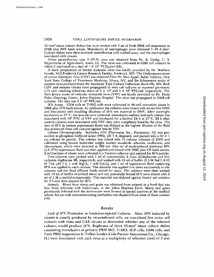
TABLE I Lack of lFN Production in Lentivirus-infected Cultures
1957
Stimuli Cultures
PBM-MO TrMO SCP GSM Fresh PBM
Visna virus . . . . . CAE virus . . . . . PI-3 virus 80 80 ND ND ND Con A - - ND ND 160
Monolayer cultures of macrophage (PBM-MO and TrMO) and nonmacrophage (SCP and GSM) cell types and suspensions of PBM cells in Teflon bottles were inoculated with viruses (moi = 2) and Con A (10 ug/ml) as indicated. 3 d later supernatant fluids from triplicate samples of each culture were examined for IFN in a VSV-GSM assay system. Numbers are reciprocals of the highest dilution of fluids that protected GSM cells against lysis by VSV and indicate the titer of IFN in the fluid. ( - ) Indicates no protection of GSM cells with undiluted fluids and therefore no IFN; ND, not done.
incubated at 37 °C. Samples of supernatant fluids were collected from each dish daily for 5 d and assayed for IFN within 2 d after storage at 4°C. For controls, replicates of PBM-MO and TrMO were inoculated with PI-3 virus at an moi of 2; a suspension of PBM was inoculated with Con A at a concentration of 10 ~g/ ml; and samples of supernatant fluids were examined for IFN content as de- scribed above.
Results of this experiment (Table I) showed that the lentiviruses did not induce IFN in any of the inoculated cultures. In contrast, both of the macrophage cultures infected with PI-3 virus and the PBM suspension treated with Con A, produced IFN. These results agree with well-established studies showing that retroviruses are poor inducers of IFN in the cell cultures they infect (19). However, as shown above, such cells were capable of producing IFN when appropriately stimulated.
Since neither LV-inoculated macrophages nor LV-inoculated PBM produced IFN, the experiment suggested that, if IFN is produced in vivo, it is unlikely that macrophages, the major cell type infected in the animal, would be the producers. It was possible that cells of another lineage, acting in consort with infected, mature macrophages, could produce IFN. To test this hypothesis, a monolayer of PBM-MO from a normal goat was inoculated with CAEV at an moi of 2 and incubated at 37°C in maintenance medium (DME plus 2% lamb serum). 3 d later the medium was removed and fresh PBM from the same animal were suspended in maintenance medium and added to the cultures of infected mac- rophages. Examination of the supernatant fluid from these cultures 48 h later showed that IFN with a titer of 1:160 had been produced. Thus, IFN production required interaction between PBM and infected, mature macrophages. This result suggested that the lentiviruses could induce IFN by a mechanism different from the classical pathway requiring that the infected cells be the IFN producer.
Cell Mixtures Required for LV-IFN Production. To dissect the virus-cell inter- actions required for LV-IFN production, we prepared monolayer cultures of PBM-MO, TrMO, and GSM cells, and infected some of these with CAEV. Freshly dispersed cultures of uninfected TrMO, SCP cells, GSM cells, and freshly prepared PBM were added to replicates of each of the monolayer cultures as
on August 12, 2014
jem.rupress.org
Dow
nloaded from
Published December 1, 1985
1958 VISNA LENTIVIRUSES INDUCE I N T E R F E R O N
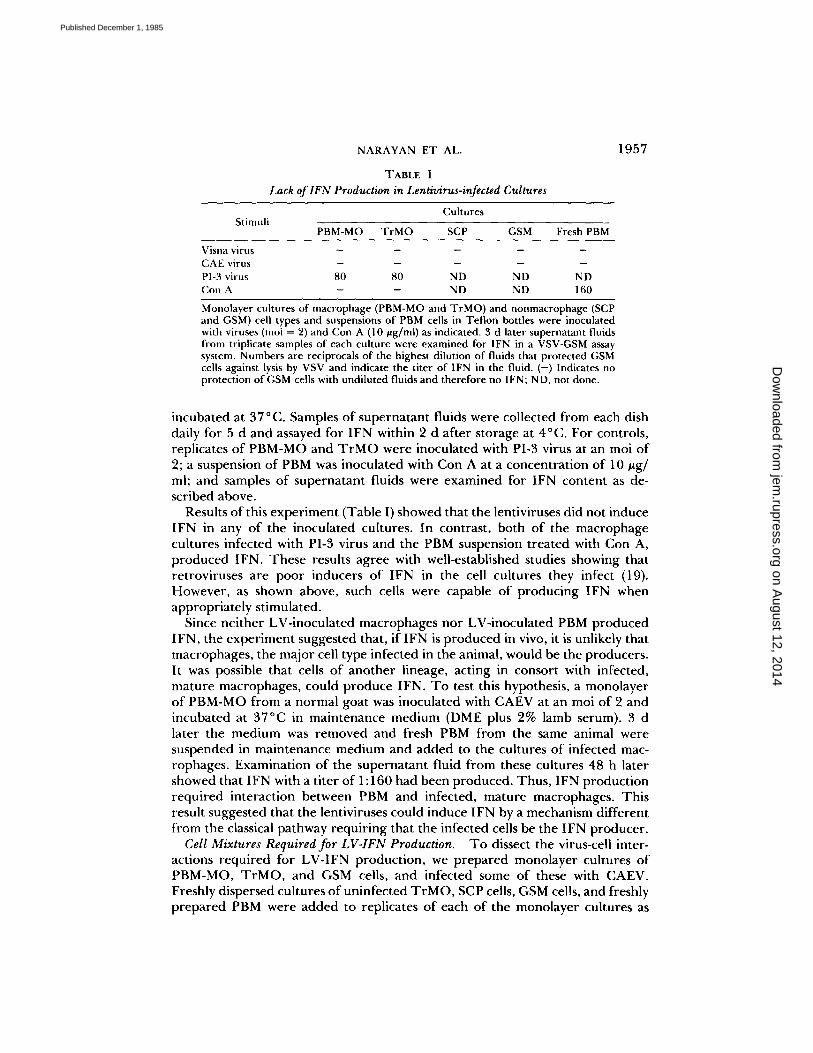
TABLE II Cell Mixtures Required for LV-IFN Production
Monolayers
Added cell suspensions
Nonadherent T r M O SCP GSM Fresh PBM
T cells
Normal PBM-MO . . . . . CAEV/PBM-MO - - - 320 320 C A E V / T r M O - - - 80 ND CAEV/GSM - - ND - -
Cell suspensions of macrophage and nonmacrophage cell types, PBM, and a nonadherent fraction of the PBM were added to monolayer cultures as indicated. The latter cultures included uninfected macrophages and macrophage and nonmacrophage (GSM) cell types that had been inoculated with CAEV ( m o i = 2) 3 d previously. Supernatant fluids were tested for IFN content 2 d later, IFN was produced only in cultures containing lymphocytes and infected macrophages.
illustrated in Table II. Supernatant fluids were examined for IFN content 48 h later.
The results were clear-cut and showed that IFN was produced only in cultures of infected macrophages to which PBM had been added. Virus-infected macro- phages were needed, since addition of PBM to normal macrophages did not stimulate IFN production. Similarly, infection in mature macrophages was essen- tial, since addition of PBM (which contained monocytes) to infected GSM cells did not result in IFN production.
To identify the PBM cell type that was responsible for IFN production, part of a batch of goat PBM containing 5 × 10⁶ cells/mi was fractionated and the nonadherent cells prepared by panning the PBM twice in succession on rabbit anti-goat IgG (Miles Laboratories, Inc.)-coated dishes followed by passage of the nonadherent cells through a nylon wool/glass bead column. As shown in Table II, the addition of nonadherent cells, separated from 5 ml of PBM, to the infected macrophage cultures resulted in production of as much IFN as did 5 ml of unfractionated PBM. The experiment was repeated four times, and examination of the supernatant fluids from the cultures showed IFN with a titer of 1:160 to 1:320. This indicated that the PBM cells which reacted with infected macro- phages to produce IFN were most probably T iymphocytes.
Lack of Requirement for Antiviral Immunity or Histocompatibility in Donors of Macrophages and Lymphocytes for LV-IFN Production. Addition of PBM to mac- rophage cultures infected with any strain of the sheep/goat lentiviruses resulted in IFN production. This included visna virus, four strains of CAEV, and four field strains of sheep viruses obtained in previous studies (10) (data not shown). We therefore used CAEV-CO virus to infect PBM-MO cultures from various animals and added fresh PBM from different sheep and goats to determine whether IFN production was immunologically specific or restricted by histocom- patibility. Monolayer cultures of macrophages were derived in 35-mm 2 tissue culture dishes and infected with CAEV at an moi of 2. Three days later 5 × 10⁶ PBM were added and supernatant fluids were examined for IFN content 2 d later.
The experiments are summarized in Table III, each result being an average of three trials. PBM-MO were derived from an immune, persistently infected
on August 12, 2014
jem.rupress.org
Dow
nloaded from
Published December 1, 1985
NARAYAN ET AL.
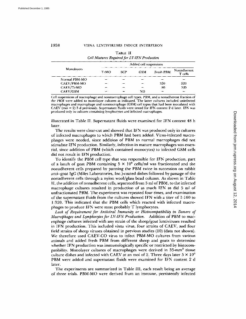
TABLE III Effect of Antiviral Immunity and Histocompatibility in Donors of PBM-MO and
Lymphocytes on LV-IFN Production
1959
Source of Source of Titer Exp. Virus PBM-MO lymphocytes of IFN
1 C A E V CAEV-infected, immune Autologous 320 goat
2 CAEV Uninfected goat Autotogous 160 3 CAEV Uninfected goat Uninfected sheep 160 4 CAEV TrMO (sheep) Uninfected goat 80 5 Bov. leukemia Uninfected goat Autologous -
Can. distemper Uninfected goat Autologous - Measles Uninfected goat Autologous -
6 CAEV Human Uninfected goat - CAEV Bovine Uninfected goat -
7 CAEV Uninfected goat Human - CAEV Uninfected goat Bovine -
In replicate cultures of experiments 1-7, the specific requirement for participation of lentiviruses, sheep/goat macrophages, and sheep/goat lymphocytes in IFN production are shown. (I and 2) IFN production was not dependent on immunity or prior infection in donors of cells. (3 and 4) Histocompatibility between donors of macrophages and lymphocytes was not required. (5) There was an absolute requirement for lentiviruses to initiate infection in macrophages. (6) Sheep/goat macrophages could not be substi- tuted with bovine and human macrophages. (7) Lymphocytes of human and bovine origin did not interact with lentivirus-infected goat macrophages to produce IFN.
goat (Table I I I , Exp. 1) and an uninfected goat (Exp. 2). Both macrophage cul tures were infected with CAEV and then supp lemented with autologous PBM 3 d later. Both sets o f cultures p roduced equivalent amoun t s o f IFN. The re fo re , p roduc t ion o f LV- IF N was not immunological ly specific. In Exps. 3 and 4, PBM- MO f rom the uninfected goat and T r M O f rom sheep were infected and supple- m en t ed with PBM f rom an uninfected sheep and an uninfected goat, respectively (i.e., infected goat macrophages were c o m p l e m e n t e d with sheep lymphocytes and vice versa). IFN titers in superna tan t fluids were equivalent. T h e r e f o r e , histocompatibil i ty be tween the donors o f macrophages and lymphocytes was not necessary for IFN product ion . In the next three exper iments , we evaluated the effects o f substi tution o f (a) lentivirus with o ther viruses, (b) sheep /goa t macro- phages with macrophages f rom o ther species, and (c) sheep /goa t lymphocytes with PBM f rom he tero logous species. In Tab l e I I I , Exp. 5, replicate cultures o f PBM-MO f rom the uninfected goat descr ibed above were inoculated with bovine leukemia virus (a re t rovi rus of cattle), canine d i s temper virus, and measles virus, at an moi of 2, and supp lemen ted with auto logous PBM. T h e latter two viruses were used because they are macrophage- t rop ic agents. In expe r imen t 6, PBM- MO cultures o f h u m a n and bovine origin were inoculated with CAEV and supp lemen ted with PBM f rom the uninfected goat. In expe r imen t 7, PBM-MO cultures f rom the uninfected goat were inoculated with CAEV and supplemented with PBM of h u m a n and bovine origin, respectively. None of the combinat ions in the lat ter th ree exper imen t s resul ted in p roduc t ion of IFN. Exp. 5 thus shows that viruses o the r than the lentiviruses could not induce IFN, probably because they failed to replicate in the m a c r o p h a g e cul tures (data not shown). Exp. 6
on August 12, 2014
jem.rupress.org
Dow
nloaded from
Published December 1, 1985
1960 VISNA LENTIVIRUSES INDUCE INTERFERON
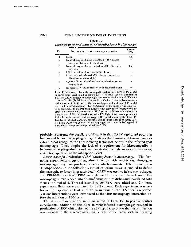
TABLE IV Determinants for Production of lFN-inducing Factor in Macrophages
Titer of Exp. Interventions in virus/macrophage system IFN
None 320 Neutralizing antibodies incubated with virus be-
fore inoculation of MO culture Neutralizing antibodies added to MO culture after 160
infection UV irradiation of infected MO culture UV-irradiated infected MO culture plus unirra-
diated supernatant fluid Lysate of infected MO culture in infectious super-
natant fluid Infected MO culture treated with dexamethasone
Fresh PBM obtained from the same goat used as the source of PBM-MO cultures were used in all experiments. (1) Positive control; addition of PBM to CAEV-infected macrophages resulted in production of IFN with a titer of 1:320. (2) Addition of neutralized CAEV to macrophage cultures did not result in infection of the macrophages, and addition of PBM did not result in production of IFN. (3) Addition of the specific virus-neutral- izing antibodies to macrophage cultures with established infection had no effect on subsequent production of IFN. (4 and 5) When infected macro- phages were killed by irradiation with UV light, infectious supernatant fluids from the culture did not trigger IFN production by the PBM. (6) Lysates of infected macrophages did not induce the PBM to produce IFN. (7) Pulse treatment of infected macrophages for I h with 100 ug/ml of dexamethazone prevented production of IFN.
p robab ly represents the coro l la ry o f Exp. 5 in that C A E V repl ica ted poor ly in h u m a n a nd bovine mac rophages . Exp. 7 shows that h u m a n and bovine lympho- cytes did no t r ecogn ize the I F N - i n d u c i n g fac tor (see below) in the infected goa t mac rophages . Thus , despi te the lack o f a r e q u i r e m e n t for h is tocompat ibi l i ty be tween m a c r o p h a g e d o n o r s and l y m p h o c y t e d o n o r s in the ov ine-capr ine species, res t r ic t ion a p p e a r e d at the interspecies level.
Determinants for Production of lFN-inducing Factor in Macrophages. T h e fore- go ing e xpe r i m e n t s suggest that , a f ter infect ion with lentiviruses, s h e e p / g o a t m a c r o p h a g e s may have p r o d u c e d a fac tor which s t imula ted I F N p r o d u c t i o n in T lymphocytes . In the fo l lowing series o f expe r imen t s we a t t e m p t e d to def ine the m a c r o p h a g e fac tor in g r ea t e r detail. C A E V was used to infect mac rophages , a nd P B M - M O a nd fresh PBM were de r ived f r o m an un in fec ted goat . T h e m a c r o p h a g e s were seeded into 3 5 - m m 2 tissue cu l tu re dishes and inocu la ted with virus at an moi o f 2. T h r e e d later, 5 x 10⁶ PBM were a d d e d and, 2 d later, s upe rna t a n t fluids were e x a m i n e d for I F N conten t . Each e x p e r i m e n t was per- f o r m e d in triplicate, at least, and the m e a n value o f the I F N ti ter is r epo r t ed . Var ious in te rven t ions were i n t r o d u c e d at the v i ru s -mac rophage in te rac t ion be- fo re the addi t ion o f PBM cells.
T h e var ious manipu la t ions are s u m m a r i z e d in T a b l e IV: In positive con t ro l exper imen t s , addi t ion o f the PBM to virus- infected m a c r o p h a g e s resul ted in p r o d u c t i o n o f I F N with a t i ter o f 1:320 (Exp. 1); to p rove that virus infect ion was essential in the mac rophages , C A E V was p r e incuba t ed with neut ra l iz ing
on August 12, 2014
jem.rupress.org
Dow
nloaded from
Published December 1, 1985
NARAYAN ET AL. 1961
antibodies for 1 h before addition to the PBM-MO cultures. The rest of the protocol was unchanged. The PBM-MO did not become infected and no IFN was produced after PBM were added (Exp. 2). In Exp. 3, virus-neutralizing antibodies were added to the PBM-MO culture on day 3 after infection, but 2 h before addition of PBM. The antibodies were maintained in the medium for the duration of the experiment. Before addition of the antibodies, the infectivity titer in the supernatant fluid of the infected PBM-MO was 5 × 10⁴ PFU/ml. No infectivity was detected in the fluid 2 h later when the fresh PBM were added. 2 d later, however, the culture produced IFN with a titer of 1:160 (Exp. 3, Table IV). Thus, virus-neutralizing antibodies had no effect on expression of the factor after infection in the PBM-MO had been initiated. To determine whether the factor was present on the surface of infected macrophages, we removed most of the supernatant fluids from the cultures and irradiated the ceils with ultraviolet (UV) light for 3 min immediately before addition of PBM. This treatment completely ablated the factor and resulted in no production of IFN (Exp. 4, Table IV). Replacing the supernatant fluids (which had been removed before irradiation of the cells) to the cultures of irradiated cells did not replenish the factor, and no IFN was produced when PBM were added. Since addition of supernatant fluids from infected macrophages to PBM failed tO produce the IFN in the previous experiments (Table I), this was not surprising. However, this experiment ruled out the possibility that the factor was present in supernatant fluids (unlike interleukin 1, which was present; data not shown) and suggested that the factor was strictly cell associated. To determine whether the factor was produced and stored intracellularly, we scraped the infected macrophages from tissue culture dishes into supernatant fluids and the suspensions were briefly sonicated before being added to PBM suspensions. No IFN was produced in these cultures. Thus, the factor was not stored intracellularly and the experiments suggest that living ceils may be needed for continuous production of the mac- rophage factor.
In previous unpublished experiments, we had observed that the steroid, dexamethasone (Sigma Chemical Co.), caused a slight increase in virus yields in infected macropbage cultures. We added the steroid to infected PBM-MO cultures to determine whether the IFN-inducing factor might be increased. In fact, dexamethasone completely abrogated production of the IFN-inducing fac- tor. Infected PBM-MO were pulsed with 100 #g/ml of dexamethasone for 1 b at 37°C, after which the cultures were washed three times with DME before addition of PBM. 2 d later supernatant fluids had infectivity titers of 5 x 10⁵ PFU/ml but no IFN. The mechanism for this inhibition is unclear.
These experiments thus showed that the IFN-inducing factor in macrophages was dependent on ientivirus infection but that synthesis of the product was independent of virus production. The factor was associated only with living macrophages and could be "neutralized" with dexamethasone.
Kinetics of Synthesis of LV-IFN. LV-IFN was produced only after sheep/goat lymphocytes had made contact with |entivirus-infected sheep/goat macrophages. In the following experiments we investigated the kinetics of IFN production by measuring the amount of IFN liberated into the supernatant fluids by cultured Iymphocytes that had been separated from infected PBM-MO cultures after
on August 12, 2014
jem.rupress.org
Dow
nloaded from
Published December 1, 1985
1962 V1SNA LENTIVIRUSES INDUCE INTERFERON
320 -
~ 16o-
80-
/Ore [h 4h 15 24h 48h
T i m e
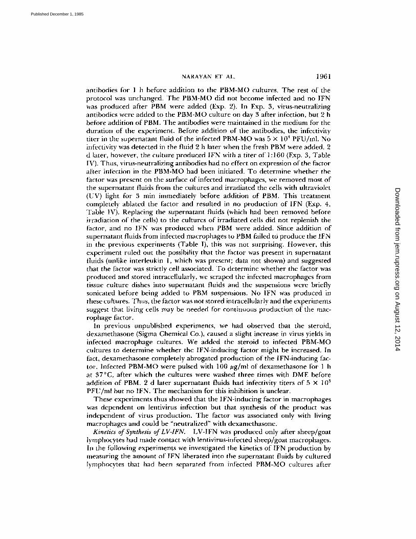
FIGURE 1. PBM were added to infected macrophage cultures in tissue culture dishes and, at times indicated, two dishes were removed for study. The supernatant fluids were tested for IFN and the nonadherent cells transferred to new tissue culture dishes and cultured for a further 24 h at 37°C. Supernatant fluids from these cultures were then tested for IFN. Open bars indicate the amount of IFN produced after the indicated period of cocuhivation of the two cell types, and the adjoining dark bars indicate amounts of IFN produced by separated lymphocytes 24 h later. IFN was first detected in cocultures after 7 h and peak levels were found at 48 h. Transferred lymphocytes began to produce IFN after a 1 h exposure to infected macrophages; maximum production required cocultivation for 7 h. After 24 h of cocuhivation, the transferred lymphocytes produced decreasing quantities of IFN.
various intervals of cocuhivation. The LV-IFN induction system described above using CAEV, PBM-MO, and PBM from an uninfected goat, was adopted in this experiment. 3 ml o f medium containing 5 x 10⁶ PBM was added to tissue culture dishes containing mature PBM-MO that had been infected 3 d previously with CAEV. At various intervals after cocuhivation, two dishes with cocultures were removed for study. A sample of supernatant fluid was collected from each dish and assayed for IFN. The nonadheren t cells were then transferred from each dish to a centrifuge tube in an excess of DME and sedimented by centrifugation. T h e cells were then resuspended in 3 ml of fresh RPMI plus 10% lamb serum, added to new tissue culture dishes (without macrophages), and incubated at 37 °C for 48 h. Supernatant fluids were then tested for IFN content. Fig. 1 shows the kinetics of synthesis of LV-IFN. No IFN was produced in individual cultures of infected PBM-MO or PBM (data not shown). In cocuhures, IFN was detected for the first time 7 h after PBM were added to infected PBM-MO. IFN titers increased to maximum levels by 48 h. After this there was no fur ther increase in the amount IFN in the supernatant fluids.
IFN was produced by the t ransferred lymphocytes but this required a minimum period of 1 h prior contact with infected PBM-MO. Longer contact of these cells with infected PBM-MO resulted in product ion of larger amounts of IFN after they were transferred. However, after 24 h of cocuhivation, the transferred lymphocytes began to produce decreasing amounts of IFN. Cell counts and viability studies on these cells, using trypan blue exclusion tests, showed that the decine in IFN product ion was not caused by a reduct ion in cell numbers or cell death.
These data suggest that the lymphocytes recognize the IFN-inducing factor in infected PBM-MO and begin to produce IFN after a minimum period of
on August 12, 2014
jem.rupress.org
Dow
nloaded from
Published December 1, 1985
NARAYAN ET AL. 1963
z
w
-~ 64O
320
c:a
4o
' ;" 20
' ' ! ' ' ! ' ' ' ] 2 Fr sk 4 5 Fr sh 7 8 9
PBM PBM PBM OAYS
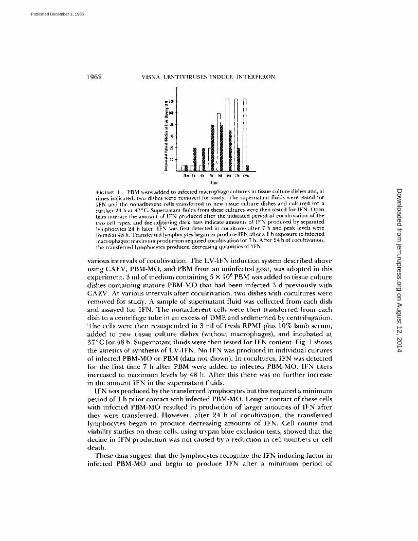
FIGURE 2. Fresh PBM were added to dishes of infected macrophages and supernatant fluids were assayed for IFN daily and discarded. The nonadherent cells were removed, washed by centrifugation, and added back to the original dishes in fresh medium. On days 3 and 6, new PBM were added to the dishes and the daily assay of supernatant fluids continued. New bursts of IFN 24 h after addition of new PBM are indicated.
sensitization. T h e sensitization is probably asynchronous, starting at 1 h and reaching maximal levels af ter 2 4 -4 8 h o f cocuhivation. T h e plateau in the IFN level af ter 48 h o f cocultivation suggests that no fu r the r IFN product ion was in progress. On the o the r hand, the decline in ability of lymphocytes to produce IFN af ter this per iod suggests that e i ther the lymphocytes were capable o f producing only a specific amount o f IFN, and became refractory after this, or that the IFN-inducing factor in infected PBM-MO were exhausted. Tw o exper- iments were pe r fo rmed to test this hypothesis: (a) "Spent" lymphocytes were t ransferred, f rom three dishes in which they had been cocultivated with infected PBM-MO for 3 d and had p roduced IFN, to three new dishes o f infected PBM- MO. No new IFN was produced. Addit ion o f 5 × 1 0 6 n e w PBM to each o f these latter dishes o f PBM-MO resulted in IFN product ion to peak levels 48 h later, with titers o f 1:160 to 1:320. This showed that the lymphocytes were not capable o f producing IFN af ter having done so once, and that spent lymphocytes did not interfere with the ability o f new lymphocytes to p roduce IFN.
(b) T o test whether the IFN-inducing factor was exhausted af ter a single round o f IFN product ion by lymphocytes, an exper iment was pe r fo rmed in duplicate in which three suspensions o f fresh PBM were added to a single dish o f infected macrophages at 3-d intervals. Supernatant fluids were measured for IFN content daily and the lymphocytes were removed, washed by centrifugation, and added back to the original dish in fresh medium. At two successive 3-d intervals, old PBM were replaced with new PBM and the daily determinat ions o f IFN contin- ued. Fig. 2 shows that new bursts o f IFN were p roduced 24 h af ter addition of new PBM. Fur the rmore , infected macrophages maintained their IFN-inducing capacity th rough more than one cycle o f IFN product ion. We concluded that the decline in IFN product ion was due to exhatistion in the lymphocyte popula- tion.
Preliminary Characterization of LV-IFN. Because o f its highly stable physical na ture (see below) LV-IFN was p roduced in a 200 ml batch that was aliquoted and stored frozen at - 7 0 ° C . Samples were then removed for different experi-
on August 12, 2014
jem.rupress.org
Dow
nloaded from
Published December 1, 1985
1964 VISNA LENTIVtRUSES INDUCE INTERFERON
60 tv-tfN
s4 . . . . . . . . I. ............. 5O
OVALBUMIN
40
30
2O
RIBONUCLEASE
l0
tO 30 50 70
ELUATE (nil)
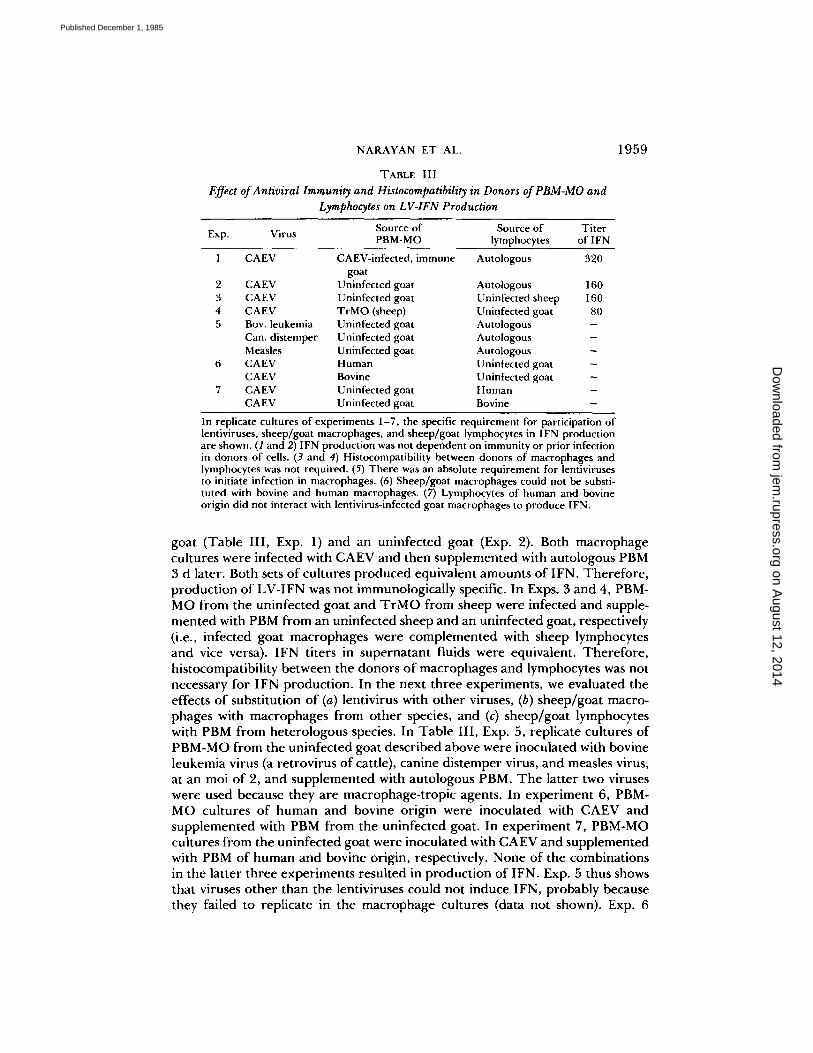
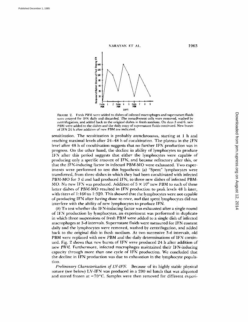
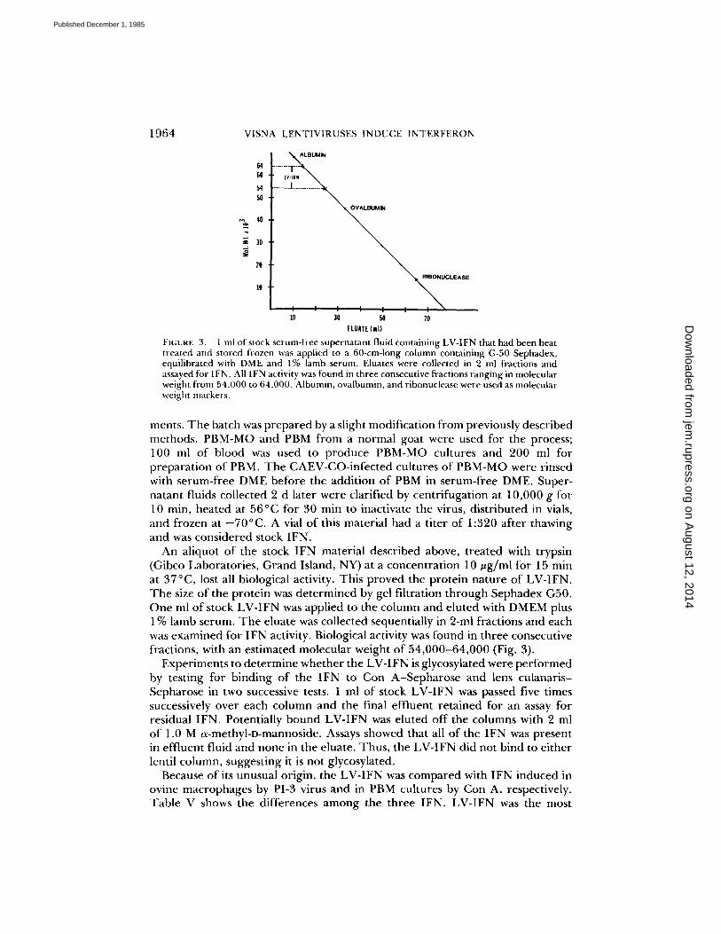
FICURE 3. 1 ml of stock serum-free supernatant fluid containing LV-IFN that had been heat treated and stored frozen was applied to a 60-cm-long column containing G-50 Sephadex, equilibrated with DME and 1% lamb serum. Eluates were collected in 2 ml fractions and assayed for IFN. All 1FN activity was found in three consecutive fractions ranging in molecular weight from 54,000 to 64,000. Albumin, ovalbumin, and ribonuelease were used as molecular weight markers.
merits. T h e batch was p repa red by a slight modif icat ion f rom previously described methods . PBM-MO and PBM f rom a normal goat were used for the process; 100 ml of blood was used to p roduce PBM-MO cultures and 200 ml for p repara t ion of PBM. T h e CAEV-CO-infec ted cultures of PBM-MO were rinsed with serum-free DME before the addit ion of PBM in serum-free DME. Super- na tant fluids collected 2 d later were clarified by centr i fugat ion at 10,000 g for 10 min, hea ted at 56°C for 30 min to inactivate the virus, dis t r ibuted in vials, and frozen at - 7 0 ° C . A vial o f this material had a t i ter o f 1:320 af ter thawing and was considered stock IFN.
An aliquot o f the stock IFN material described above, t rea ted with trypsin (Gibco Laborator ies , Grand Island, NY) at a concentra t ion 10 ug /ml for 15 min at 37°C, lost all biological activity. This p roved the prote in na ture of LV-IFN. T h e size of the prote in was de t e rmined by gel fi l tration th rough Sephadex G50. One ml of stock LV- IFN was applied to the column and eluted with DMEM plus 1% lamb serum. T h e eluate was collected sequentially in 2-ml fractions and each was examined for IFN activity. Biological activity was found in three consecutive fractions, with an est imated molecular weight o f 54 ,000 -64 ,000 (Fig. 3).
Exper iments to de t e rmine whe the r the LV-IFN is glycosylated were p e r f o r m e d by testing for binding o f the IFN to Con A - S e p h a r o s e and lens culanar is - Sepharose in two successive tests. 1 ml of stock LV- IFN was passed five times successively over each column and the final eff luent re ta ined for an assay for residual IFN. Potentially bound LV- IFN was eluted of f the columns with 2 ml o f 1.0 M 0~-methyl-I)-mannoside. Assays showed that all o f the IFN was present in eff luent fluid and none in the eluate. Thus , the LV-IFN did not bind to ei ther lentil column, suggesting it is not glycosylated.
Because of its unusual origin, the LV- IFN was c o m p a r e d with IFN induced in ovine macrophages by PI-3 virus and in PBM cultures by Con A, respectively. Tab le V shows the differences a m o n g the three IFN. LV-IFN was the most
on August 12, 2014
jem.rupress.org
Dow
nloaded from
Published December 1, 1985
NARAYAN ET AL. 1965
TABLE V
Comparative Properties of Different Ovine IFN
Supernatant Assay systems pH 2 56°C Freeze/
fluids VSV/GSM VSV/TrMO 24 h* 30 rain* thaw*
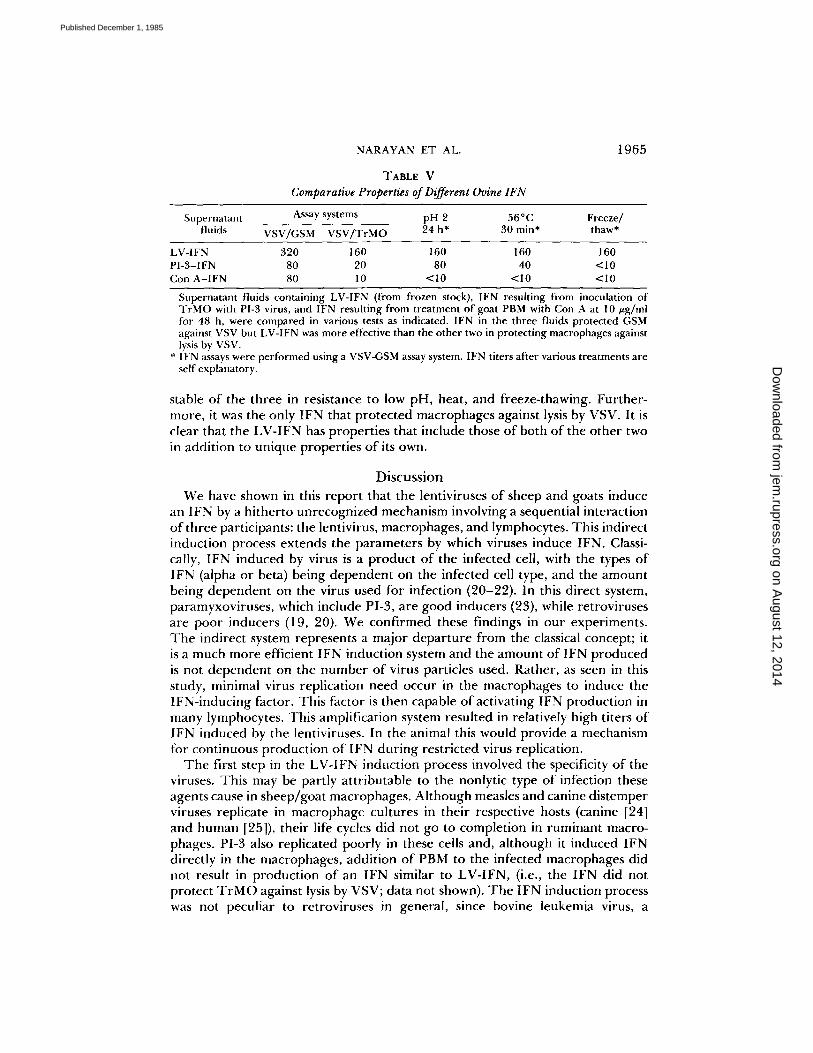
LV-IFN 320 160 160 160 160 PI-3-1FN 80 20 80 40 <10 Con A-IFN 80 10 <10 <10 <10
Supernatant fluids containing LV-IFN (from frozen stock), IFN resulting from inoculation of TrMO with PI-3 virus, and IFN resulting from treatment of goat PBM with Con A at 10/zg/ml for 48 h, were compared in various tests as indicated. IFN in the three fluids protected GSM against VSV but LV-IFN was more effective than the other two in protecting macrophages against lysis by VSV.
* IFN assays were performed using a VSV-GSM assay system. IFN titers after various treatments are self explanatory.
stable of the three in resistance to low pH, heat, and freeze-thawing. Further- more, it was the only IFN that protected macrophages against lysis by VSV. It is clear that the LV-IFN has properties that include those of both of the other two in addition to unique properties of its own.
Discussion We have shown in this report that the lentiviruses of sheep and goats induce
an IFN by a hitherto unrecognized mechanism involving a sequential interaction of three participants: the lentivirus, macrophages, and lymphocytes. This indirect induction process extends the parameters by which viruses induce IFN. Classi- cally, 1FN induced by virus is a product of the infected cell, with the types of IFN (alpha or beta) being dependent on the infected cell type, and the amount being dependent on the virus used for infection (20-22). In this direct system, paramyxoviruses, which include PI-3, are good inducers (23), while retroviruses are poor inducers (19, 20). We confirmed these findings in our experiments. The indirect system represents a major departure from the classical concept; it is a much more efficient IFN induction system and the amount of IFN produced is not dependent on the number of virus particles used. Rather, as seen in this study, minimal virus replication need occur in the macrophages to induce the IFN-inducing factor. This factor is then capable of activating IFN production in many lymphocytes. This amplification system resulted in relatively high titers of IFN induced by the lentiviruses. In the animal this would provide a mechanism for continuous production of IFN during restricted virus replication.
The first step in the LV-IFN induction process involved the specificity of the viruses. This may be partly attributable to the nonlytic type of infection these agents cause in sheep/goat macrophages. Although measles and canine distemper viruses replicate in macrophage cultures in their respective hosts (canine [24] and human [25]), their life cycles did not go to completion in ruminant macro- phages. PI-3 also replicated poorly in these cells and, although it induced IFN directly in the macrophages, addition of PBM to the infected macrophages did not result in production of an IFN similar to LV-IFN, (i.e., the IFN did not protect T rMO against iysis by VSV; data not shown). The IFN induction process was not peculiar to retroviruses in general, since bovine leukemia virus, a
on August 12, 2014
jem.rupress.org
Dow
nloaded from
Published December 1, 1985
1966 VISNA L E N T I V I R U S E S I N D U C E I N T E R F E R O N
retrovirus known to be extremely oncogenic in sheep (26), was incapable of inducing the IFN-inducing factor in sheep macrophages. The lentiviruses of ruminants therefore seemed uniquely endowed to induce this factor in species- specific macrophages.
The second specific step in the IFN induction process was the response of the macrophage. Although the lentiviruses replicated productively in several cell types of the natural host, including SCP fibroblasts and synovial membrane cells, only infected macrophages produced the 1FN-inducing factor. The fact that both alveolar and blood macrophages produced the factor suggests that infection in any macrophage from this species may suffice for this function. However, the nature of the factor remains to be characterized. The use of virus-neutralizing antibodies to study production of the factor clearly established that infection with lentiviruses was necessary but that infectious virus was not essential. The failure of UV-inactivated infected macrophages and lysates of infected macro- phages to supply the factor is compatible with a labile substance produced in small quantities by the infected macrophages. A similar observation was made in a previous study (10) in which fusion of SCP fibroblasts by certain field isolates of ientiviruses required continuous presence of macrophages. The fusion factor in macrophages was also cell associated and was produced in limited quantities. Whether the fusion factor and the IFN-inducing factor in the infected macro- phages are the same is unknown. It is possible that the IFN-inducing factor is a lysosomal enzyme or an enzyme-modified protein on the macrophage cell surface. Dexamethasone, which inhibits the IFN-inducing factor, may have a role in this process, given its ability to stabilize lysosomal membranes. Such hypothetical stabilization of iysosomes could prevent loss of enzymes and prevent production of the factor.
The third sequential step in the LV-IFN induction process involved the interaction of T lymphocytes with the infected macrophages. We have already discussed the suggestion of a sudden burst of IFN production by the lymphocytes after contact with the infected macrophages, together with the observation that the macrophage-to-lymphocyte signal was neither immunologically specific nor restricted by histocompatibility. However, restriction at the species level was at play, since neither bovine nor human lymphocytes recognized the signal to produce IFN. This requirement of participation between lymphocytes and mac- rophages for the production of LV-IFN has some resemblance to the interaction of similar cells in the production of mitogen-induced gamma IFN (27). However, this may be coincidental, since immunological parameters were not involved in the production of LV-IFN and the LV-IFN had physical properties distinct from gamma IFN.
One of the questions arising early in the study was the type of IFN induced by the lentiviruses. However, the lack of molecularly defined sheep/goat IFN and lack of typing sera made characterization of the LV-IFN difficult. The presump- tive classification is based on a comparison with the IFN induced by PI-3 virus and Con A, which are similar to classical IFN (21). Thus, the IFN induced by PI-3 virus infection of goat macrophages fits the general descriptions of human and murine alpha IFN, as determined by the macrophage cell source, the resistance of the IFN to low pH, and the relative heat stability. Similarly, the
on August 12, 2014
jem.rupress.org
Dow
nloaded from
Published December 1, 1985
NARAYAN ET AL. 1967
IFN resulting from Con A stimulation of PBM resembles gamma IFN, as determined by the mitogen inducer, the producer cells, and its lability to pH 2 and heat. In contrast, the LV-IFN had properties overlapping those of both the preceding two IFN, in addition to some unique properties. Its resistance to low pH and heat, molecular size, and nonglycosylated nature resemble alpha IFN, while synthesis by macrophage-dependent T lymphocytes resembles gamma IFN. This analogy is made stronger by the ability of LV-IFN to induce expression of Ia antigen in cultured macrophages (14), a phenomenon associated with gamma IFN (26). It is doubtful that LV-IFN represents a mixture of alpha and gamma IFN because it possesses the unique properties of resistance to freeze-thawing and the ability to protect macrophages from lysis with VSV.
The LV-IFN induction process is highly relevant to events in the infected animal. Since monocyte-macrophages are the main target cells in vivo for virus replication, and since the lesions in disease consist of infiltrations of lymphocytes and macrophages, conditions would be ideal for local synthesis of IFN. However, LV-IFN was not found in extracts of inflamed tissues. Nevertheless, as described in a companion study (14), the ability of LF-IFN to inhibit lentivirus replication in cultured macrophages, and to induce expression of Ia antigens of the major histocompatibility complex in macrophages, is consistent with findings in infected tissues where virus replication is restricted and Ia antigen expression in macro- phages is at a high level. LV-IFN may therefore be produced in inflamed tissues in quantities too small to be detected in the IFN biological assay but effective enough at the cellular level to restrict virus replication in macrophages and influence the cellular immune responses to the virus.
S u m m a r y Lentivirus infections are characterized by a persistent, restricted type of virus
replication in tissues. Using sheep and goat lentiviruses, whose target cells in vivo are macrophages, we explored virus-host cell interactions to determine whether an interferon (IFN) is produced during virus replication in vivo which causes restricted replication. We show that the lentiviruses were incapable of inducing IFN directly in any infected cell, including macrophages and lymphocytes. However, after infection with these viruses, sheep and goat macrophages ac- quired a factor that triggered IFN production by T iymphocytes. Only sheep/ goat lentiviruses were capable of inducing the factor and, although these viruses replicated productively in various cell cultures of the natural host animal, only infected macrophages developed the IFN-inducing factor. The factor was pro- duced continuously and was strictly cell associated, requiring direct contact with lymphocytes. The lymphocytes responded with a single, sudden release of IFN beginning 7 h after cocuhivation and reaching peak values at 48 h, after which they ceased production and became refractory. LFN production was not immu- nologically specific and did not require histocompatibility between donors of the two cell types. The IFN is a nonglycosylated protein of molecular weight 54,000- 64,000, and is stable to heat and acid treatments. These findings identify a unique IFN and a new method for virus induction of IFN. The novel two-stage process of induction provides a mechanism for local amplification and continuity of production of IFN in vivo. This is compatible with infection in the animal
on August 12, 2014
jem.rupress.org
Dow
nloaded from
Published December 1, 1985
1968 VISNA LENTIVIRUSES INDUCE INTERFERON
whose lentivirus-induced pathologic lesions consist o f accumulat ions of lympho- cytes and infected macrophages in target tissues.
We thank Dr. Richard T. Johnson and Dr. Diane E. Griffin for fruitful discussions and review of the manuscript, and Linda Kelly for preparing the manuscript.
Received for publication 22 July 1985.
R e f e r e n c e s
1. Haase, A. T. 1975. The slow infection caused by visna virus. 1975. Curr. Top. Microbiol. lmmunol. 72:101.
2. Gonda, M. A., F. Wong-Staal, R. C. Gallo, J. E. Clements, O. Narayan, and R. V. Gilden. 1985. Sequence homology and morphologic similarity of HTLV-III and visna virus, a pathogenic lentivirus. Science (Wash. DC). 227:173.
3. Rabson, A. B., and M. A. Martin. 1985. Molecular organization of the AIDS retrovirus. Cell. 40:477.
4. Narayan, O., and L. C. Cork. 1985. Lentiviral diseases of sheep and goats: chronic pneumonia, leukoencephalomyelitis and arthritis. Rev. Infect. Dis. 7:89.
5. Cheevers, W. R., and T. C. McGuire. 1985. Equine infectious anemia virus: immu- nopathogenesis and persistence. Rev. Infect. Dis. 7:84.
6. Popovic, M., M. G. Sargadharan, E. Read, and R. C. Gallo. 1984. Detection, isolation and continuous production of cytopathic retroviruses HTLV-III from patients with AIDS and pre-AIDS. Science (Wash. DC). 224:497.
7. Clements, J. E., O. Narayan, D. E. Griffin, and R. T. Johnson. 1979. The synthesis and structure of visna virus DNA. Virology. 93:377.
8. Haase, A. T., L. Stowring, J. D. Harris, B. Traynor, P. Ventura, R. Pelus, and M. Brahic. 1982. Visna virus DNA synthesis and the tempo of infection in vitro. Virology. 119:399.
9. Narayan, O., D. E. Griffin, and A. Silverstein. 1977. Slow virus infection: replication and mechanisms of persistence of visna virus in sheep. J. Infect. Dis. 135:800.
10. Narayan, O., J. S. Wolinsky, J. E. Clements, J. D. Strandberg, D. E. Griffin, and L. C. Cork. 1982. Slow virus replication: the role of macrophages in the persistence and expression of visna viruses in sheep and goats. J. Gen. Virol. 59:345.
11. Gendelman, H. E., O. Narayan, S. Molineaux, J. S. Clements, and Z. Ghotbi. 1985. Slow persistent replication of lentiviruses: role of tissue macrophages and macro- phage-precursors in bone marrow. Proc. Natl. Acad. Sci. USA: 82:in press.
12. Narayan, O., S. Kennedy-Stoskopf, D. Sheffer, D. E. Griffin, and J. E. Clements. 1983. Activation of caprine arthritis-encephalitis virus expression during maturation of monocytes to macrophages. Infect. lmmun. 41:67.
t 3. Gendelman, H. E., O. Narayan, S. Kennedy-Stoskopf, J. E. Clements, and G. H. Pezeshkpour. 1984. Slow virus-macrophage interactions: characterization of a trans- formed cell line of sheep alveolar macrophages that express a marker for susceptibility to ovine-caprine lentivirus infection. Lab. Invest. 51:547.
14. Kennedy, P. G. E., O. Narayan, J. Hopkins, Z. Ghotbi, H. E. Gendelman, and J. E. Clements. 1985. Persistent expression of Ia antigen and viral genome in visna-maedi virus-induced inflammatory cells: possible role of lentivirus-induced interferon. J. Exp. Med. 162:1970.
15. Narayan, O.,J. E. Clements, J. D. Strandberg, L. C. Cork, and D. E. Griffin. 1980. Biologic characterization of the virus causing leukoencephalitis and arthritis in goats. J. Gen. Virol. 41:343.
on August 12, 2014
jem.rupress.org
Dow
nloaded from
Published December 1, 1985
NARAYAN ET AL. 1969
16. Kennedy-Stoskopf, S., O. Narayan, and R. L. Hirsch. 1983. Immunosuppression in goats inoculated with parainfluenza type 3 virus. Am. J. Vet. Res. 44:2302.
17. Griffin, D. E., J. Mullinix, O. Narayan, and R. T. Johnson. 1974. Age dependence of virus expression: comparative pathogenesis of two rodent-adapted strains of measles virus in mice. Infect. Immun. 9:690.
18. Narayan, O., D. Sheffer, D. E. Griffin, J. E. Clements, and J. Hess. 1984. Lack of neutralizing antibody to caprine arthritis encephalitis lentivirus in persistently in- fected goats can be overcome with inactivated mycobacterium tuberculosis. J. Virol. 49:349.
19. Friedman, R. M., E. H. Chang, J. M. Ramseur, and M. W. Myers. 1975. Interferon- directed inhibition of chronic murine leukemia virus production in cell cultures: lack of effect on intracellular virus marker.J. Virol. 16:569.
20. Joklik, W. K. Interferons in Virology. B. N. Fields, editor. Raven Press, New York. 281-309.
21. Peska, S., and S. Baron. 1981. Definition and classification of interferons. Methods Enzymol. 78:3.
22. Preble, O. T., and R. M. Friedman. 1983. Interferon-induced alterations in cells: relevance to viral and non-viral disease. Lab. Invest. 49:4.
23. Marcus, P. I., C. Svitlik, and M.J. Sekellick. 1983. Interferon induction by viruses. X. A model for interferon induction by Newcastle disease virus.J. Gen. Virol. 64:2419.
24. Appel, M.J.G. , S. G. Mendelson, and W. W. P. Hall. 1984. Macrophage Fc receptors control infectivity and neutralization of canine distemper virus-antibody complexes. J. Virol. 51:643.
25. Joseph, B. S., P. W. Lampert, and M. B. A. Oldstone. 1975. Replication and persistence of measles virus in defined subpopulations of human leukocytes. J. Virol. 16:1638.
26. Kenyon, S. J., J. F. Ferrer, R. A. McFeely, and D. C. Graves. 1981. Induction of lymphosarcoma in sheep by ovine leukemia virus. J. Natl. Cancer Inst. 67:1157.
27. Kelly, V. E., W. Fiers, and T. B. Strom. 1984. Cloned human interferon gamma but not interferon beta on alpha induces expression of HLA-DR determinants by fetal monocytes and myeloid leukemia cells.J. Immunol. 132:240.
on August 12, 2014
jem.rupress.org
Dow
nloaded from
Published December 1, 1985
Top Related
Copyright © 2022 FDOKUMEN

