


Bahasa
Halaman
Hukum

Hindawi Publishing CorporationEvidence-Based Complementary and Alternative MedicineVolume 2011, Article ID 473953, 9 pagesdoi:10.1093/ecam/nep212
Original Article
REC-2006—A Fractionated Extract of Podophyllum hexandrumProtects Cellular DNA from Radiation-Induced Damage byReducing the Initial Damage and Enhancing Its Repair In Vivo
Pankaj Chaudhary,1, 2 Sandeep Kumar Shukla,1 and Rakesh Kumar Sharma1
1 Institute of Nuclear Medicine and Allied Sciences (INMAS), Delhi, India2 Brookhaven National Laboratory, Upton, NY 11973, USA
Correspondence should be addressed to Pankaj Chaudhary, [email protected]
Received 30 June 2009; Accepted 9 November 2009
Copyright © 2011 Pankaj Chaudhary et al. This is an open access article distributed under the Creative Commons AttributionLicense, which permits unrestricted use, distribution, and reproduction in any medium, provided the original work is properlycited.
Podophyllum hexandrum, a perennial herb commonly known as the Himalayan May Apple, is well known in Indian andChinese traditional systems of medicine. P. hexandrum has been widely used for the treatment of venereal warts, skin infections,bacterial and viral infections, and different cancers of the brain, lung and bladder. This study aimed at elucidating the effectof REC-2006, a bioactive fractionated extract from the rhizome of P. hexandrum, on the kinetics of induction and repair ofradiation-induced DNA damage in murine thymocytes in vivo. We evaluated its effect on non-specific radiation-induced DNAdamage by the alkaline halo assay in terms of relative nuclear spreading factor (RNSF) and gene-specific radiation-induced DNAdamage via semi-quantitative polymerase chain reaction. Whole body exposure of animals with gamma rays (10 Gy) caused asignificant amount of DNA damage in thymocytes (RNSF values 17.7± 0.47, 12.96± 1.64 and 3.3± 0.014) and a reduction in theamplification of β-globin gene to 0, 28 and 43% at 0, 15 and 60 min, respectively. Administrating REC-2006 at a radioprotectiveconcentration (15 mg kg−1 body weight) 1 h before irradiation resulted in time-dependent reduction of DNA damage evidentas a decrease in RNSF values 6.156± 0.576, 1.647± 0.534 and 0.496± 0.012, and an increase in β-globin gene amplification 36,95 and 99%, at 0, 15 and 60 min, respectively. REC-2006 scavenged radiation-induced hydroxyl radicals in a dose-dependentmanner stabilized DPPH free radicals and also inhibited superoxide anions. Various polyphenols and flavonoides present inREC-2006 might contribute to scavenging of radiation-induced free radicals, thereby preventing DNA damage and stimulating itsrepair.
1. Introduction
Radiation-induced free radicals oxidize cellular biomacro-molecules like DNA, proteins and lipids generating a varietyof cellular dysfunctions leading to cell death [1, 2]. Damagesto DNA, such as single- and double-strand breaks, basemodifications and adduct formation, are considered asbiologically significant cellular lesions [3–5]. Normally, cellsoperate diverse pathways to repair oxidative damage toDNA. Among them, the most important are direct repair ofan adduct, base-excision repair, nucleotide-excision repair,homologous recombination, non-homologous end-joining,DNA inter-strand cross-link repair and DNA mismatchrepair [6].
Several radioprotective agents, including amifostine,aminothiols, cysteamine, polyamines and DNA-binding lig-ands like Hoechst, protect DNA from radiation-induceddamage [7, 8]. However, therapeutic levels of most of theseagents entail severe side effects, such as nausea, vomiting,hypotension and neurotoxicity, thereby limiting their clinicaluse [9]. Consequently, newer and more effective agentsare being sought. Recent reports suggest that various plantextracts and natural products protect DNA from radiation-induced oxidative damage [10–17].
Podophyllum hexandrum (also known as Himalayan MayApple), a herb thriving at high altitudes in the Himalayashas been extensively used in Ayurvedic system of medicinefor the treatment of ailments like monocytoid leukemia,
2 Evidence-Based Complementary and Alternative Medicine
Hodgkin’s lymphoma, bacterial and viral infections, venerealwarts, rheumatoid artharalgia associated with limb numb-ness and pycnogenic infections of skin tissue [18–20]. Theroot and rhizome of P. hexandrum are reported to containa number of compounds with significant pharmacologicalproperties, for example, epipodophyllotoxin, podophyllo-toxone, 4-methylpodophyllotoxin, aryltetrahydronaphtha-lene lignans, flavonoids such as quercetin, quercetin-3-glycoside, 4-demethylpodophyllotoxin glycoside, podophyl-lotoxinglycoside, kaempferol and kaempferol-3-glucoside[21, 22].
An extract (code named as RP-1) from the rhizomeof P. hexandrum reportedly protects mice against a lethaldose of ionizing radiation [23]. Different mechanisms wereproposed to account for these radioprotective properties,including free radical scavenging, metal chelation and theelevation of antioxidant defense enzymes [24, 25]. A varietyof scenarios involve radiation exposures in the moderaterange (i.e., 1–10 Gy), including cancer therapy, plannedreactor maintenance and the explosion of a dirty bomb(radioactive dispersal device). With the likelihood that expo-sure to a moderate radiation dose will result in radiation-induced DNA damage entailing cell death, we undertook thisstudy to assess the effect of REC-2006 [25], a fractionatedextract of rhizome of P. hexandrum, on the induction andrepair kinetics of radiation (10 Gy)-induced gene-specificand non-specific DNA damage in murine thymocytes in vivo.
Intraperitoneal administration of REC-2006 to mice at15 mg kg−1 body weight conferred more than 90% protectionagainst whole-body irradiation (10 Gy) as compared to72% offered by 34.5 mg kg−1 body weight administrationof the parent plant extract RP-1. The radiosensitive natureof thymocytes provides an attractive system to study DNArepair [26, 27]. As we are interested to look at the earlykinetics of DNA repair within 1 h after irradiation, we choosemouse β-globin gene which is constitutively expressed intranscriptionally active or inactive domains thus acting as abiomarker of overall DNA repair [28, 29]. Similar approachhas also been used for investigating DNA damage repair[13, 30].
Observations of the effect of P. hexandrum upon therepair kinetics of gamma-radiation-induced DNA damagewill further delineate the mechanisms involved in overallradioprotective effects of this plant.
2. Methods
2.1. Chemicals. We obtained the following materials fromSigma Chemical Co. (St. Louis, MO, USA): agarose, lowmelting point agarose, aluminium trichloride, bromophe-nol blue, dimethyl sulfoxide (DMSO), di-sodium ethy-lene di-amine tetra acetic acid (Na2-EDTA), 2,2-diphenyl-1-picrylhydrazyl (DPPH), ethidium bromide, gallic acid,quercetin, sodium chloride (NaCl), sodium hydroxide(NaOH), sodium lauryl sarcosine, sucrose, triton-X-100,Tris—HCl, Tris base, trichloroacetic acid (TCA), thiobar-bituric acid (TBA) and xylene cyanol. Taq polymerase anddNTP mix were purchased from Qiagen, Chatsworth, CA.Primers were synthesized from The Center for Genomic
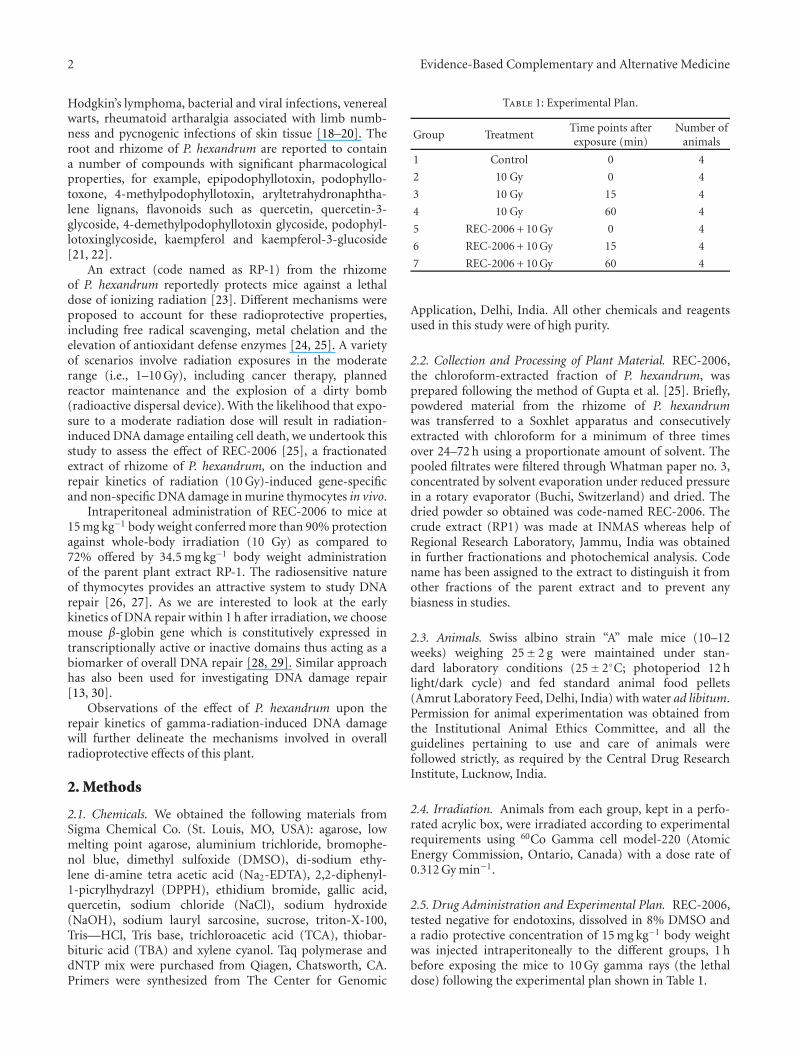
Table 1: Experimental Plan.
Group TreatmentTime points afterexposure (min)
Number ofanimals
1 Control 0 4
2 10 Gy 0 4
3 10 Gy 15 4
4 10 Gy 60 4
5 REC-2006 + 10 Gy 0 4
6 REC-2006 + 10 Gy 15 4
7 REC-2006 + 10 Gy 60 4
Application, Delhi, India. All other chemicals and reagentsused in this study were of high purity.
2.2. Collection and Processing of Plant Material. REC-2006,the chloroform-extracted fraction of P. hexandrum, wasprepared following the method of Gupta et al. [25]. Briefly,powdered material from the rhizome of P. hexandrumwas transferred to a Soxhlet apparatus and consecutivelyextracted with chloroform for a minimum of three timesover 24–72 h using a proportionate amount of solvent. Thepooled filtrates were filtered through Whatman paper no. 3,concentrated by solvent evaporation under reduced pressurein a rotary evaporator (Buchi, Switzerland) and dried. Thedried powder so obtained was code-named REC-2006. Thecrude extract (RP1) was made at INMAS whereas help ofRegional Research Laboratory, Jammu, India was obtainedin further fractionations and photochemical analysis. Codename has been assigned to the extract to distinguish it fromother fractions of the parent extract and to prevent anybiasness in studies.
2.3. Animals. Swiss albino strain “A” male mice (10–12weeks) weighing 25± 2 g were maintained under stan-dard laboratory conditions (25± 2◦C; photoperiod 12 hlight/dark cycle) and fed standard animal food pellets(Amrut Laboratory Feed, Delhi, India) with water ad libitum.Permission for animal experimentation was obtained fromthe Institutional Animal Ethics Committee, and all theguidelines pertaining to use and care of animals werefollowed strictly, as required by the Central Drug ResearchInstitute, Lucknow, India.
2.4. Irradiation. Animals from each group, kept in a perfo-rated acrylic box, were irradiated according to experimentalrequirements using 60Co Gamma cell model-220 (AtomicEnergy Commission, Ontario, Canada) with a dose rate of0.312 Gy min−1.
2.5. Drug Administration and Experimental Plan. REC-2006,tested negative for endotoxins, dissolved in 8% DMSO anda radio protective concentration of 15 mg kg−1 body weightwas injected intraperitoneally to the different groups, 1 hbefore exposing the mice to 10 Gy gamma rays (the lethaldose) following the experimental plan shown in Table 1.
Evidence-Based Complementary and Alternative Medicine 3
2.6. Isolation of Thymocytes. Mice were killed by cervicaldislocation, dissected, the abdominal cavity was perfusedwith 0.9% saline and the thymus was removed. All visibleclots were segregated carefully. We minced the thymic lobesfinely, and gently crushed them with the plunger of a syringe;the resultant cell suspension was passed through a 25-gaugeneedle to avoid cell aggregates. All procedures were carriedout on ice.
2.7. Alkaline Halo Assay. We detected DNA single-strandbreaks in individual cells using the alkaline halo assay [31]modified for murine thymocytes. After the different treat-ments, thymocytes were suspended (1.5× 10⁴ to 2.0× 10⁴
per 100 μl) in a 1.5% low-melting agarose solution inphosphate buffered saline, pH 7.4, and immediately pipettedon to slides coated with 1.0% normal agarose and spread uni-formly. After gelling was complete, the slides were immersedin the alkali buffer (0.1 M NaOH and 1 mM EDTA; pH 13)for 20 min at 4◦C, washed, and then stained with 10 μg ml−1
ethidium bromide for 5 min. The images were acquired on afluorescent microscope (Leica Q550 FW, Wetzlar, Germany)equipped with a 40× Neofluar objective lens, and analyzedusing Comet score software (Tritek Corporation, NorthernVirginia, USA). The intensely stained, intact chromatin massforms the central core of the halo, while the broken DNAfragments constitute its diffusely stained periphery. Levels ofDNA damage were expressed as the relative nuclear spreadingfactor (RNSF) values, calculated by subtracting the RNSFvalues of control cells from those of treated cells [13].
2.8. DNA Isolation from Thymocytes. The genomic DNAfrom murine thymocytes (5× 10⁶) was isolated usingthe DNeasy isolation kit (DNeasy Tissue kit, Qiagen,Chatsworth, USA) as per manufacturer’s instructions. DNAyields were estimated spectrophotometrically by measuringabsorbance at 260 and 280 nm.
2.9. Semi-Quantitative Polymerase Chain Reaction. Primerpairs used against mice β-globin 957 (5′-CGGGTGAGA-GATACATCCATCG-3′) and β-globin 5638 (5′-GATCCA-GAGAGCAACTTTCGACTA-3′) targeted the genomic β-globin gene cluster and yielded a product of 4681 bp[32]. The reaction mixture (20 μl) contained template DNA(20 ng), dNTP mix (200 μM each dNTP), 10 pM μl−1 primerpairs, 0.3 U Taq polymerase, Taq buffer containing MgCl2(1.5 mM) and sterile H2O. The polymerase chain reaction(PCR) cycling conditions used to amplify β-globin were pre-PCR incubation at 94◦C for 2 min followed by 30 cycles of94◦C for 15 s, 59◦C for 30 s and 68◦C for 4 min. At the end ofthe profile, we added a final extension of 4 min at 72◦C; thesamples were stored at 4◦C. An aliquot of each PCR productwas resolved by agarose-gel electrophoresis [33]. Briefly, 10 μlof the sample was mixed with 5 μl of loading dye (0.025%bromophenol blue, 0.25% xylene cyanol and 40% sucrose inwater) and loaded into the wells of a 1.2% agarose gel alongwith the 1 kb DNA ladder (Fermentas, MD, USA) to confirmthe size of the PCR-amplified product. Electrophoresis wascarried out for 2.5 h at 5 V cm−1 in the Tris acetate buffer in
the presence of 0.5 μg ml−1 ethidium bromide. The amplifiedproducts were quantified densitometrically using ImageQuant software (Molecular Dynamics, Sunnyvale, CA).
2.10. DPPH Radical Scavenging Activity. We measured the2,2-diphenyl-1-picrylhydrazyl (DPPH) radical-scavengingability of REC-2006 according to the method of Shimadaet al. [34]. A solution of DPPH in methanol mixed withREC-2006 was incubated for 15 min in the dark at 37◦C. Thedecrease in absorbance at 517 nm was measured against thatof methanol alone. The absorbance of DPPH alone was takenas 100% radical or 0% inhibition.
2.11. Hydroxyl Radical Scavenging. We quantified scaveng-ing of radiation (100 Gy) and Fenton reaction inducedhydroxyl radicals by REC-2006, using 2-deoxyribose as themarker substrate [35]. Briefly, we exposed to 100 μM FeSO4
or 100 Gy, 1 ml of reaction volume containing 5 mM 2-deoxyribose in the absence or presence of varied concen-trations of REC-2006. Thereafter, we added two volumes ofsolution containing 25% TCA and 1% TBA in 0.1 N NaOH,placed the mixture in a boiling water bath for 20 min, cooledit and measured the absorbance of the resulting pink-coloredchromogen at 532 nm.
2.12. Estimation of the Scavenging of Chemically GeneratedSuperoxide Anions. Following the method of Rao et al.,we estimated the superoxide anion scavenging ability ofREC-2006 [36]. Briefly, to a reaction mixture containing0.52 M sodium pyrophosphate (pH 8.3), 186 μM phenylmethane sulfate (PMS) and 300 μM nitro blue tetrazolium(NBT), we added various concentrations of REC-2006.The reaction was initiated by adding nicotinamide adeninedinucleotide reduced (NADH) (final concentration 780 μM)and incubating the solution for 90 s at 30◦C. The purple-colored chromogen that formed was measured spectropho-tometrically at 560 nm.
2.13. Total Antioxidant Capacity. The total antioxidantcapacity of REC-2006 was determined spectrophotometri-cally by quantifying the amount of phosphomolybdenumcomplex generated [37]. A 0.1 ml (100 μg) sample of REC-2006 was mixed with reagent solution (6 M sulfuric acid, 28mM sodium phosphate and 4 mM ammonium molybdate),and the mixture was incubated at 95◦C for 90 min. There-after, the samples were cooled to room temperature and theabsorbance was measured at 695 nm against a blank solution(typically, incubating 1 ml of reagent and the appropriatevolume of the same solvent (DMSO) used to prepare thesamples under the same conditions as the experimentalsamples). The total antioxidant capacity of REC-2006 wasexpressed as gallic acid equivalents using a standard curveprepared from a freshly prepared gallic acid solution.
2.14. Estimation of the Total Phenolic Content. A 100 μlaliquot of 10 mg ml−1 REC-2006 was mixed with 500 μlFolin-Cioccalteau reagent and 400 μl 7.5% sodium car-bonate [38]. Following incubation at 20◦C for 30 min,
4 Evidence-Based Complementary and Alternative Medicine
4.25
23.
809
5.31
26.
656
9.03
510
.347
13.4
29
17.2
16
19.8
51
25.5
2526
.645
27.6
0528
.021
31.3
7132
.715
43.4
88
35.3
6037
.163
38.5
9239
.295
40.0
3241
.024
42.1
33
44.9
3946
.379
47.5
63
52.4
91
55.1
57
59.3
0760
.384
61.9
6363
.104
65.1
7365
.432
67.9
04
24.3
520
500
1000
1500
A.U
.
0 10 20 30 40 50 60
(minutes)
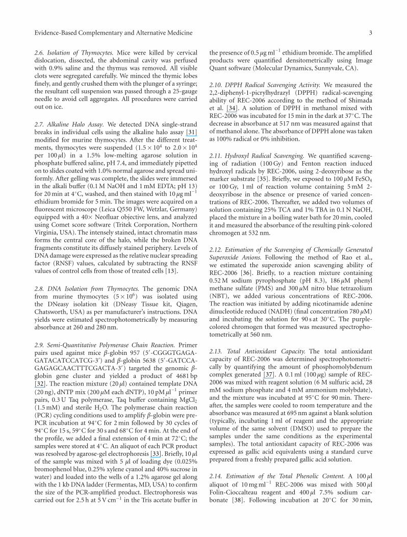
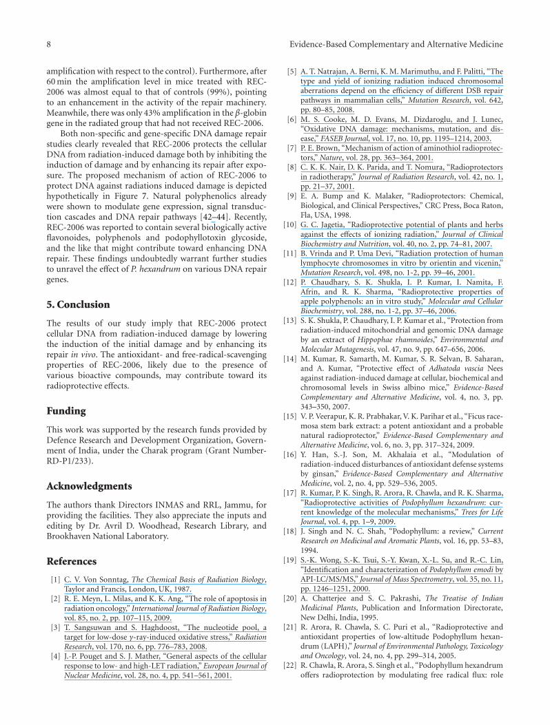
Figure 1: HPLC profile of REC-2006.
the absorbance was read at 765 nm. The total phenols inREC-2006 were expressed as gallic acid equivalents, esti-mated from a standard curve obtained from the absorbanceof fresh gallic acid equivalents.
2.15. Total Flavonoid Content. We obtained the totalflavonoid content of REC-2006 using the method of Zhisenet al. [39]. To 1 ml of diluted REC-2006 sample (500 μg),we added 4 ml of H2O, 0.3 ml of 5% NaNO2 followed,5 min later, by of 0.3 ml of 10% AlCl3 solution. One minutethereafter, 2 ml 1 M NaOH was added and the reactionmixture was immediately diluted with 2.4 ml of ddH2O. Thepink-colored chromogen was measured spectrophotometri-cally at 510 nm. The total flavonoid content (mg mg−1) wasexpressed as quercetin equivalents, using a standard curvefrom a fresh quercetin solution.
2.16. Data Analysis. The data are presented as the mean±standard deviation (SD) of three separate experiments, witheach experiment comprising three parallel measurements.We compared radiation and radiation + REC-2006 groups.The data were analyzed by one-way analysis of variance, andmultiple comparisons were made between different groupsby applying Bonferroni t-test. A probability of < 5% wasconsidered significant.
3. Results
3.1. Phytochemical Analysis. Figure 1 shows the HPLC profileof REC-2006. Demethylpodophyllotoxin, podophyllotoxinglycoside, epipodophyllotoxin and podophyllotoxin amongothers were identified by analyzing the fragmentationpatterns [25]. Chemical analysis and spectrophotometricdeterminations indicated that the total polyphenol contentof REC-2006 was 8 mg mg−1 of gallic acid equivalents whilethe total flavonoid content was 0.20 mg mg−1 of quercetinequivalents.
3.2. REC-2006 and Radiation-Induced DNA Damage
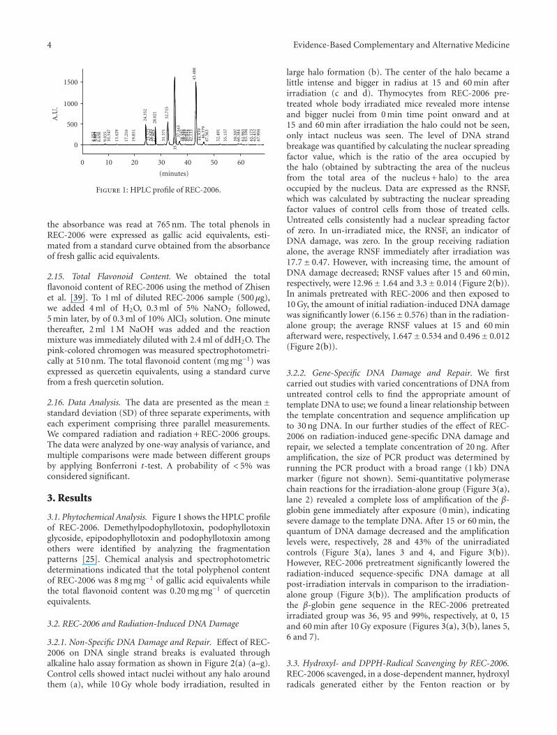
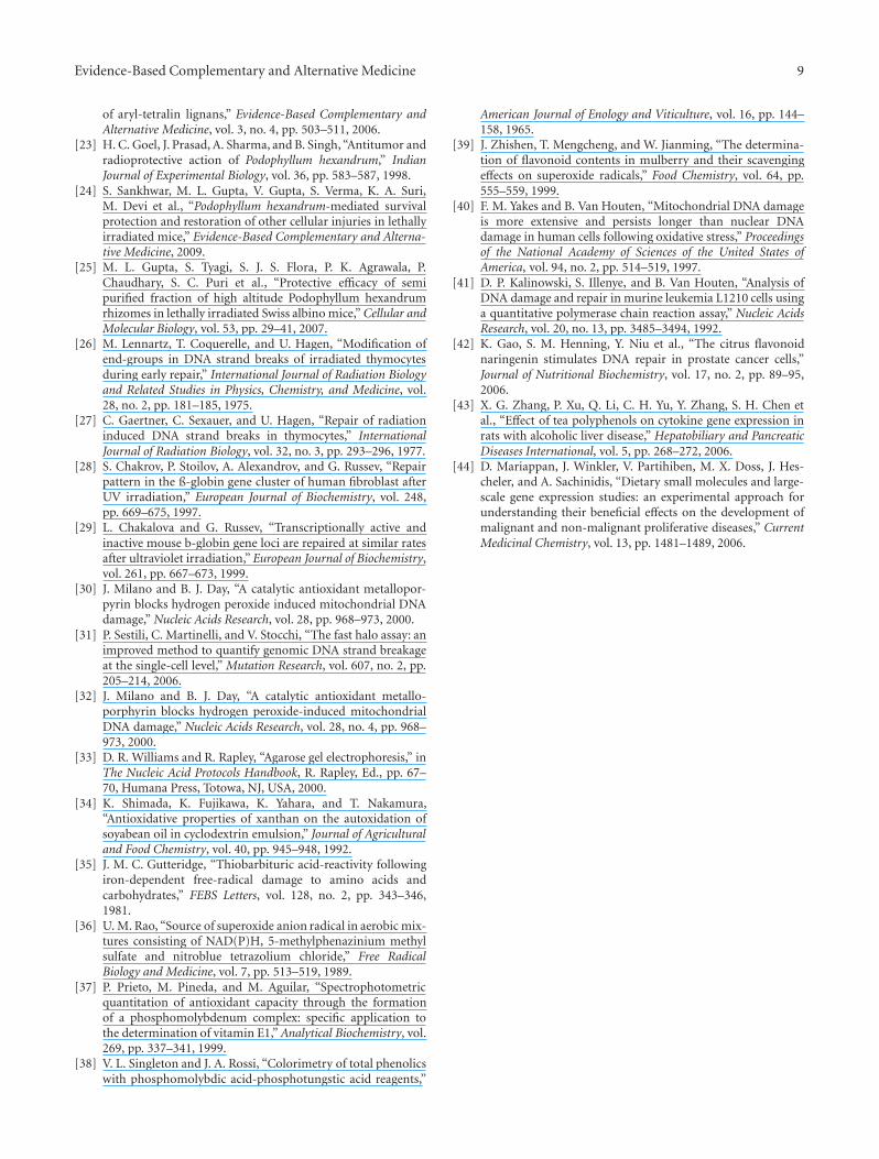
3.2.1. Non-Specific DNA Damage and Repair. Effect of REC-2006 on DNA single strand breaks is evaluated throughalkaline halo assay formation as shown in Figure 2(a) (a–g).Control cells showed intact nuclei without any halo aroundthem (a), while 10 Gy whole body irradiation, resulted in
large halo formation (b). The center of the halo became alittle intense and bigger in radius at 15 and 60 min afterirradiation (c and d). Thymocytes from REC-2006 pre-treated whole body irradiated mice revealed more intenseand bigger nuclei from 0 min time point onward and at15 and 60 min after irradiation the halo could not be seen,only intact nucleus was seen. The level of DNA strandbreakage was quantified by calculating the nuclear spreadingfactor value, which is the ratio of the area occupied bythe halo (obtained by subtracting the area of the nucleusfrom the total area of the nucleus + halo) to the areaoccupied by the nucleus. Data are expressed as the RNSF,which was calculated by subtracting the nuclear spreadingfactor values of control cells from those of treated cells.Untreated cells consistently had a nuclear spreading factorof zero. In un-irradiated mice, the RNSF, an indicator ofDNA damage, was zero. In the group receiving radiationalone, the average RNSF immediately after irradiation was17.7± 0.47. However, with increasing time, the amount ofDNA damage decreased; RNSF values after 15 and 60 min,respectively, were 12.96± 1.64 and 3.3± 0.014 (Figure 2(b)).In animals pretreated with REC-2006 and then exposed to10 Gy, the amount of initial radiation-induced DNA damagewas significantly lower (6.156± 0.576) than in the radiation-alone group; the average RNSF values at 15 and 60 minafterward were, respectively, 1.647± 0.534 and 0.496± 0.012(Figure 2(b)).
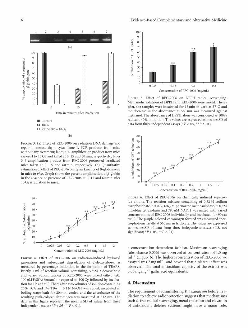
3.2.2. Gene-Specific DNA Damage and Repair. We firstcarried out studies with varied concentrations of DNA fromuntreated control cells to find the appropriate amount oftemplate DNA to use; we found a linear relationship betweenthe template concentration and sequence amplification upto 30 ng DNA. In our further studies of the effect of REC-2006 on radiation-induced gene-specific DNA damage andrepair, we selected a template concentration of 20 ng. Afteramplification, the size of PCR product was determined byrunning the PCR product with a broad range (1 kb) DNAmarker (figure not shown). Semi-quantitative polymerasechain reactions for the irradiation-alone group (Figure 3(a),lane 2) revealed a complete loss of amplification of the β-globin gene immediately after exposure (0 min), indicatingsevere damage to the template DNA. After 15 or 60 min, thequantum of DNA damage decreased and the amplificationlevels were, respectively, 28 and 43% of the unirradiatedcontrols (Figure 3(a), lanes 3 and 4, and Figure 3(b)).However, REC-2006 pretreatment significantly lowered theradiation-induced sequence-specific DNA damage at allpost-irradiation intervals in comparison to the irradiation-alone group (Figure 3(b)). The amplification products ofthe β-globin gene sequence in the REC-2006 pretreatedirradiated group was 36, 95 and 99%, respectively, at 0, 15and 60 min after 10 Gy exposure (Figures 3(a), 3(b), lanes 5,6 and 7).
3.3. Hydroxyl- and DPPH-Radical Scavenging by REC-2006.REC-2006 scavenged, in a dose-dependent manner, hydroxylradicals generated either by the Fenton reaction or by

Evidence-Based Complementary and Alternative Medicine 5
30 pixels
(a)
30 pixels
(b)
30 pixels
(c)
30 pixels
(d)
30 pixels
(e)
30 pixels
(f)
30 pixels
(g)
(a)
02
46
81012
14
1618
20
Ave
rage
RN
SFva
lue
0 15 60
Time in minutes after irradiation
10 GyREC-2006 + 10 Gy
(b)
Figure 2: (a) Effect of REC-2006 on 10 Gy-induced single strand breaks in mouse thymocytes. Mice were irradiated with or without REC-2006 treatment (8 mg kg−1 body weight i.p. 60 minutes before irradiation) and thymocytes from them were isolated at different intervals.DNA damage was studied employing the alkaline halo assay. (a) Cells without any treatment. (b–d) Cells from 10 Gy irradiated mice killedafter 0, 15 or 60 min, respectively. (e–g) Cells from REC-2006 pretreated irradiated mice killed after 0, 15 or 60 min, respectively. (b)Quantitative estimation of REC-2006 effect on radiation-induced DNA damage and repair. The level of DNA strand breakage was quantifiedby calculating the nuclear spreading factor value, which is the ratio of the area occupied by the halo (obtained by subtracting the area of thenucleus from the total area of the nucleus + halo) to the area occupied by the nucleus. Data are expressed as the relative nuclear spreadingfactor (RNSF), which was calculated by subtracting the nuclear spreading factor values of control cells from those of treated cells. Data arethe mean± SD for at least 100 cells for each observation in triplicate.
radiation (100 Gy) (Figure 4). Up to 0.05 mg ml−1 REC-2006 gradually inhibited the 2-deoxy-ribose degradationand the difference in inhibition was not very significantin Fenton-mediated and 100-Gy-induced hydroxyl radicals.Beyond 0.05 mg ml−1, a significant difference was observedin inhibition. Maximum inhibition was observed at aconcentration of 2.0 mg ml−1 for radiation (62.5%) andFenton reaction (69.2%)-mediated 2-deoxyribose degrada-tion. Increasing concentrations of REC-2006 significantlystabilized the DPPH radicals in a dose-dependent fashion
maximally at 0.2 mg ml−1 (Figure 5). The absorbance ofDPPH alone was taken as 100% radical or 0% inhibition.Beyond 0.2 mg ml−1, a plateau phase was observed (notshown in the figure).
3.4. Super Oxide Anions Scavenging Potential, Total Antiox-idant Capacity, Polyphenol and Flavonoid Contents of REC-2006. The superoxide anions generated by phenyl methanesulfate and NADH reduced NBT. REC-2006 inhibitedthe chemically generated superoxide anion formation in
6 Evidence-Based Complementary and Alternative Medicine
1 2 3 4 5 6 7
(a)
0
10
20
30
40
50
60
70
80
90
100
%am
plifi
cati
onof
ase
gmen
tof
β-g
lobi
nge
ne
0 15 60
Time in minutes after irradiation
Control10 GyREC-2006 + 10 Gy
(b)
Figure 3: (a) Effect of REC-2006 on radiation DNA damage andrepair in mouse thymocytes. Lane 1, PCR products from micewithout any treatment; lanes 2–4, amplification product from miceexposed to 10 Gy and killed at 0, 15 and 60 min, respectively; lanes5–7 amplification product from REC-2006 pretreated irradiatedmice taken at 0, 15 and 60 min, respectively. (b) Quantitativeestimation of effect of REC-2006 on repair kinetics of β-globin genein mice in vivo. Graph shows the percent amplification of β-globinin the absence or presence of REC-2006 at 0, 15 and 60 min after10 Gy irradiation to mice.
0
10
20
30
40
50
60
70
80
%in
hib
itio
nof
2-de
oxy
ribo
sede
grad
atio
n
0 0.025 0.05 0.1 0.2 0.5 1 1.5 2
Concentration of REC-2006 (mg/mL)
∗∗
∗∗
∗∗
∗∗∗∗
∗∗ ∗∗
Figure 4: Effect of REC-2006 on radiation-induced hydroxylgeneration and subsequent degradation of 2-deoxyribose, asmeasured by percentage inhibition in the formation of TBARS.Briefly, 1 ml of reaction volume containing, 5 mM 2-deoxyriboseand varied concentrations of REC-2006 were mixed either with100 μM FeSO4(Fenton) or exposed to 100 Gy followed by incuba-tion for 1 h at 37◦C. There after, two volumes of solution containing25% TCA and 1% TBA in 0.1 N NaOH was added, incubated inboiling water bath for 20 min, cooled and the absorbance of theresulting pink-colored chromogen was measured at 532 nm. Thedata in this figure represent the mean± SD of values from threeindependent assays (∗P < .05, ∗∗P < .01).
0
10
20
30
40
50
60
70
80
90
100
%in
hib
itio
nin
DP
PH
radi
cal
0.025 0.05 0.1 0.2
Concentration of REC-2006 (mg/mL)
∗∗
∗∗∗
Figure 5: Effect of REC-2006 on DPPH radical scavenging.Methanolic solutions of DPPH and REC-2006 were mixed. There-after, the samples were incubated for 15 min in dark at 37◦C andthe decrease in the absorbance at 560 nm was measured againstmethanol. The absorbance of DPPH alone was considered as 100%radical or 0% inhibition. The values are expressed as mean± SD ofdata from three independent assays (∗P < .05, ∗∗P < .01).
0
10
20
30
40
50
60
70
80%
inh
ibit
ion
ofN
BT
redu
ctio
n
0 0.025 0.05 0.1 0.2 0.5 1 1.5 2
Concentration of REC-2006 (mg/mL)
∗∗∗∗
∗∗
∗∗∗∗
∗∗∗∗
∗∗
Figure 6: Effect of REC-2006 on chemically induced superox-ide anions. The reaction mixture containing of 0.52 M sodiumpyrophosphate, pH 8.3, 186 μM phenazine methosulphate, 300 μMnitroblue tetrazolium and 780 μM NADH was mixed with variedconcentrations of REC-2006 individually and incubated for 90 s at30◦C. The purple-colored chromogen formed was measured spec-trophotometrically at 560 nm in triplicate. The values are expressedas mean± SD of data from three independent assays (NS, notsignificant; ∗P < .05, ∗∗P < .01).
a concentration-dependent fashion. Maximum scavenging(absorbance 0.056) was observed at concentration of 1.5 mgml−1 (Figure 6). The highest concentration of REC-2006 weassayed was 2 mg ml−1 and beyond that a plateau effect wasobserved. The total antioxidant capacity of the extract was0.06 mg mg−1 gallic acid equivalents.
4. Discussion
The requirement of administering P. hexandrum before irra-diation to achieve radioprotection suggests that mechanismssuch as free radical scavenging, metal chelation and elevationof antioxidant defense systems might have a major role.
Evidence-Based Complementary and Alternative Medicine 7
10 Gy gamma rays
Free radical formation
Lipidperoxidation
Proteinmodifications
DSB
SSB
Base damages
> DNA strand breaks (increase in RNSF) ↑> PCR amplification β-gene decrease ↓
Cell death and reduced survival
H◦H2O◦ + e◦ HO2
◦
OH◦
RO2◦
−1 hr REC-2006 (ip) +10 Gy gamma rays
Free radical scavenging
Termination of free radical chain elongation
Modulation of cellular homeostasis
Reduced damage to cellular macromolecules
> DNA strand breaks (reduction in RNSF) ↓> % amplification in β-globin gene ↑
Cell survival and radioprotection
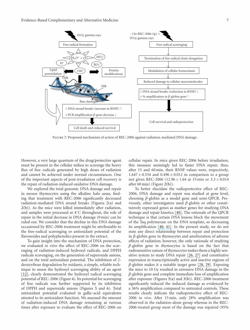
Figure 7: Proposed mechanism of action of REC-2006 against radiation-mediated DNA damage.
However, a very large quantum of the drug/protective agentmust be present in the cellular milieu to scavenge the heavyflux of free radicals generated by high doses of radiationand cannot be achieved under normal circumstances. Oneof the important aspects of post-irradiation cell recovery isthe repair of radiation-induced oxidative DNA damage.
We explored the total genomic DNA damage and repairin mouse thymocytes using the alkaline halo assay, find-ing that treatment with REC-2006 significantly decreasedradiation-mediated DNA strand breaks (Figures 2(a) and2(b)). As the mice were killed immediately after radiation,and samples were processed at 4◦C throughout, the role ofrepair in the initial decrease in DNA damage (0 min) can beruled out. We consider that the decline in this DNA damageoccasioned by REC-2006 treatment might be attributable tothe free-radical scavenging or antioxidant potential of theflavonoides and polyphenolics present in the extract.
To gain insight into the mechanism of DNA protection,we evaluated in vitro the effect of REC-2006 on the scav-enging of radiation-induced hydroxyl radicals, and DPPHradicals scavenging, on the generation of superoxide anions,and on the total antioxidant potential. The inhibition of 2-deoxyribose degradation by oxidants, a simple, reliable tech-nique to assess the hydroxyl scavenging ability of an agent[12], clearly demonstrated the hydroxyl radical scavengingpotential of REC-2006 (Figure 4). Its potential for scavengingof free radicals was further supported by its inhibitionof DPPH and superoxide anions (Figures 5 and 6). Totalantioxidant potential evaluated in gallic-acid equivalentsattested to its antioxidant function. We assessed the amountof radiation-induced DNA damage remaining at varioustimes after exposure to evaluate the effect of REC-2006 on
cellular repair. In mice given REC-2006 before irradiation,this measure seemingly led to faster DNA repair; thus,after 15 and 60 min, their RNSF values were, respectively,1.647± 0.534 and 0.496± 0.012 in comparison to a groupnot given REC-2006 (12.96± 1.64 at 15 min or 3.3± 0.014after 60 min) (Figure 2(b)).
To better elucidate the radioprotective effect of REC-2006, DNA damage and repair was studied at gene level,choosing β-globin as a model gene and semi-QPCR. Pre-viously, other investigators used β-globin or other consti-tutively expressed genes as marker genes for studying DNAdamage and repair kinetics [40]. The rationale of the QPCRtechnique is that certain DNA lesions block the movementof the Taq polymerase on the DNA template, so decreasingits amplification [40, 41]. In the present study, we do notstate any direct relationship between repair and protectionin β-globin gene in thymocytes and amelioration of adverseeffects of radiation; however, the only rationale of studyingβ-globin gene in thymocytes is based on the fact thatradiosensitive nature of thymocytes makes them a highly sen-sitive system to study DNA repair [26, 27] and constitutiveexpression in transcriptionally active and inactive regions ofβ-globin makes it a suitable target gene [28, 29]. Exposingthe mice to 10 Gy resulted in extensive DNA damage in theβ-globin gene and complete immediate loss of amplificationafter exposure (Figures 3(a) and 3(b)). REC-2006 treatmentsignificantly reduced the induced damage as evidenced bya 36% amplification compared to untreated controls. Theseresults clearly indicate the radioprotective effect of REC-2006 in vivo. After 15 min, only 28% amplification wasobserved in the radiation-alone group whereas in the REC-2006-treated group most of the damage was repaired (95%
8 Evidence-Based Complementary and Alternative Medicine
amplification with respect to the control). Furthermore, after60 min the amplification level in mice treated with REC-2006 was almost equal to that of controls (99%), pointingto an enhancement in the activity of the repair machinery.Meanwhile, there was only 43% amplification in the β-globingene in the radiated group that had not received REC-2006.
Both non-specific and gene-specific DNA damage repairstudies clearly revealed that REC-2006 protects the cellularDNA from radiation-induced damage both by inhibiting theinduction of damage and by enhancing its repair after expo-sure. The proposed mechanism of action of REC-2006 toprotect DNA against radiations induced damage is depictedhypothetically in Figure 7. Natural polyphenolics alreadywere shown to modulate gene expression, signal transduc-tion cascades and DNA repair pathways [42–44]. Recently,REC-2006 was reported to contain several biologically activeflavonoides, polyphenols and podophyllotoxin glycoside,and the like that might contribute toward enhancing DNArepair. These findings undoubtedly warrant further studiesto unravel the effect of P. hexandrum on various DNA repairgenes.
5. Conclusion
The results of our study imply that REC-2006 protectcellular DNA from radiation-induced damage by loweringthe induction of the initial damage and by enhancing itsrepair in vivo. The antioxidant- and free-radical-scavengingproperties of REC-2006, likely due to the presence ofvarious bioactive compounds, may contribute toward itsradioprotective effects.
Funding
This work was supported by the research funds provided byDefence Research and Development Organization, Govern-ment of India, under the Charak program (Grant Number-RD-P1/233).
Acknowledgments
The authors thank Directors INMAS and RRL, Jammu, forproviding the facilities. They also appreciate the inputs andediting by Dr. Avril D. Woodhead, Research Library, andBrookhaven National Laboratory.
References
[1] C. V. Von Sonntag, The Chemical Basis of Radiation Biology,Taylor and Francis, London, UK, 1987.
[2] R. E. Meyn, L. Milas, and K. K. Ang, “The role of apoptosis inradiation oncology,” International Journal of Radiation Biology,vol. 85, no. 2, pp. 107–115, 2009.
[3] T. Sangsuwan and S. Haghdoost, “The nucleotide pool, atarget for low-dose γ-ray-induced oxidative stress,” RadiationResearch, vol. 170, no. 6, pp. 776–783, 2008.
[4] J.-P. Pouget and S. J. Mather, “General aspects of the cellularresponse to low- and high-LET radiation,” European Journal ofNuclear Medicine, vol. 28, no. 4, pp. 541–561, 2001.
[5] A. T. Natrajan, A. Berni, K. M. Marimuthu, and F. Palitti, “Thetype and yield of ionizing radiation induced chromosomalaberrations depend on the efficiency of different DSB repairpathways in mammalian cells,” Mutation Research, vol. 642,pp. 80–85, 2008.
[6] M. S. Cooke, M. D. Evans, M. Dizdaroglu, and J. Lunec,“Oxidative DNA damage: mechanisms, mutation, and dis-ease,” FASEB Journal, vol. 17, no. 10, pp. 1195–1214, 2003.
[7] P. E. Brown, “Mechanism of action of aminothiol radioprotec-tors,” Nature, vol. 28, pp. 363–364, 2001.
[8] C. K. K. Nair, D. K. Parida, and T. Nomura, “Radioprotectorsin radiotherapy,” Journal of Radiation Research, vol. 42, no. 1,pp. 21–37, 2001.
[9] E. A. Bump and K. Malaker, “Radioprotectors: Chemical,Biological, and Clinical Perspectives,” CRC Press, Boca Raton,Fla, USA, 1998.
[10] G. C. Jagetia, “Radioprotective potential of plants and herbsagainst the effects of ionizing radiation,” Journal of ClinicalBiochemistry and Nutrition, vol. 40, no. 2, pp. 74–81, 2007.
[11] B. Vrinda and P. Uma Devi, “Radiation protection of humanlymphocyte chromosomes in vitro by orientin and vicenin,”Mutation Research, vol. 498, no. 1-2, pp. 39–46, 2001.
[12] P. Chaudhary, S. K. Shukla, I. P. Kumar, I. Namita, F.Afrin, and R. K. Sharma, “Radioprotective properties ofapple polyphenols: an in vitro study,” Molecular and CellularBiochemistry, vol. 288, no. 1-2, pp. 37–46, 2006.
[13] S. K. Shukla, P. Chaudhary, I. P. Kumar et al., “Protection fromradiation-induced mitochondrial and genomic DNA damageby an extract of Hippophae rhamnoides,” Environmental andMolecular Mutagenesis, vol. 47, no. 9, pp. 647–656, 2006.
[14] M. Kumar, R. Samarth, M. Kumar, S. R. Selvan, B. Saharan,and A. Kumar, “Protective effect of Adhatoda vascia Neesagainst radiation-induced damage at cellular, biochemical andchromosomal levels in Swiss albino mice,” Evidence-BasedComplementary and Alternative Medicine, vol. 4, no. 3, pp.343–350, 2007.
[15] V. P. Veerapur, K. R. Prabhakar, V. K. Parihar et al., “Ficus race-mosa stem bark extract: a potent antioxidant and a probablenatural radioprotector,” Evidence-Based Complementary andAlternative Medicine, vol. 6, no. 3, pp. 317–324, 2009.
[16] Y. Han, S.-J. Son, M. Akhalaia et al., “Modulation ofradiation-induced disturbances of antioxidant defense systemsby ginsan,” Evidence-Based Complementary and AlternativeMedicine, vol. 2, no. 4, pp. 529–536, 2005.
[17] R. Kumar, P. K. Singh, R. Arora, R. Chawla, and R. K. Sharma,“Radioprotective activities of Podophyllum hexandrum: cur-rent knowledge of the molecular mechanisms,” Trees for LifeJournal, vol. 4, pp. 1–9, 2009.
[18] J. Singh and N. C. Shah, “Podophyllum: a review,” CurrentResearch on Medicinal and Aromatic Plants, vol. 16, pp. 53–83,1994.
[19] S.-K. Wong, S.-K. Tsui, S.-Y. Kwan, X.-L. Su, and R.-C. Lin,“Identification and characterization of Podophyllum emodi byAPI-LC/MS/MS,” Journal of Mass Spectrometry, vol. 35, no. 11,pp. 1246–1251, 2000.
[20] A. Chatterjee and S. C. Pakrashi, The Treatise of IndianMedicinal Plants, Publication and Information Directorate,New Delhi, India, 1995.
[21] R. Arora, R. Chawla, S. C. Puri et al., “Radioprotective andantioxidant properties of low-altitude Podophyllum hexan-drum (LAPH),” Journal of Environmental Pathology, Toxicologyand Oncology, vol. 24, no. 4, pp. 299–314, 2005.
[22] R. Chawla, R. Arora, S. Singh et al., “Podophyllum hexandrumoffers radioprotection by modulating free radical flux: role
Evidence-Based Complementary and Alternative Medicine 9
of aryl-tetralin lignans,” Evidence-Based Complementary andAlternative Medicine, vol. 3, no. 4, pp. 503–511, 2006.
[23] H. C. Goel, J. Prasad, A. Sharma, and B. Singh, “Antitumor andradioprotective action of Podophyllum hexandrum,” IndianJournal of Experimental Biology, vol. 36, pp. 583–587, 1998.
[24] S. Sankhwar, M. L. Gupta, V. Gupta, S. Verma, K. A. Suri,M. Devi et al., “Podophyllum hexandrum-mediated survivalprotection and restoration of other cellular injuries in lethallyirradiated mice,” Evidence-Based Complementary and Alterna-tive Medicine, 2009.
[25] M. L. Gupta, S. Tyagi, S. J. S. Flora, P. K. Agrawala, P.Chaudhary, S. C. Puri et al., “Protective efficacy of semipurified fraction of high altitude Podophyllum hexandrumrhizomes in lethally irradiated Swiss albino mice,” Cellular andMolecular Biology, vol. 53, pp. 29–41, 2007.
[26] M. Lennartz, T. Coquerelle, and U. Hagen, “Modification ofend-groups in DNA strand breaks of irradiated thymocytesduring early repair,” International Journal of Radiation Biologyand Related Studies in Physics, Chemistry, and Medicine, vol.28, no. 2, pp. 181–185, 1975.
[27] C. Gaertner, C. Sexauer, and U. Hagen, “Repair of radiationinduced DNA strand breaks in thymocytes,” InternationalJournal of Radiation Biology, vol. 32, no. 3, pp. 293–296, 1977.
[28] S. Chakrov, P. Stoilov, A. Alexandrov, and G. Russev, “Repairpattern in the ß-globin gene cluster of human fibroblast afterUV irradiation,” European Journal of Biochemistry, vol. 248,pp. 669–675, 1997.
[29] L. Chakalova and G. Russev, “Transcriptionally active andinactive mouse b-globin gene loci are repaired at similar ratesafter ultraviolet irradiation,” European Journal of Biochemistry,vol. 261, pp. 667–673, 1999.
[30] J. Milano and B. J. Day, “A catalytic antioxidant metallopor-pyrin blocks hydrogen peroxide induced mitochondrial DNAdamage,” Nucleic Acids Research, vol. 28, pp. 968–973, 2000.
[31] P. Sestili, C. Martinelli, and V. Stocchi, “The fast halo assay: animproved method to quantify genomic DNA strand breakageat the single-cell level,” Mutation Research, vol. 607, no. 2, pp.205–214, 2006.
[32] J. Milano and B. J. Day, “A catalytic antioxidant metallo-porphyrin blocks hydrogen peroxide-induced mitochondrialDNA damage,” Nucleic Acids Research, vol. 28, no. 4, pp. 968–973, 2000.
[33] D. R. Williams and R. Rapley, “Agarose gel electrophoresis,” inThe Nucleic Acid Protocols Handbook, R. Rapley, Ed., pp. 67–70, Humana Press, Totowa, NJ, USA, 2000.
[34] K. Shimada, K. Fujikawa, K. Yahara, and T. Nakamura,“Antioxidative properties of xanthan on the autoxidation ofsoyabean oil in cyclodextrin emulsion,” Journal of Agriculturaland Food Chemistry, vol. 40, pp. 945–948, 1992.
[35] J. M. C. Gutteridge, “Thiobarbituric acid-reactivity followingiron-dependent free-radical damage to amino acids andcarbohydrates,” FEBS Letters, vol. 128, no. 2, pp. 343–346,1981.
[36] U. M. Rao, “Source of superoxide anion radical in aerobic mix-tures consisting of NAD(P)H, 5-methylphenazinium methylsulfate and nitroblue tetrazolium chloride,” Free RadicalBiology and Medicine, vol. 7, pp. 513–519, 1989.
[37] P. Prieto, M. Pineda, and M. Aguilar, “Spectrophotometricquantitation of antioxidant capacity through the formationof a phosphomolybdenum complex: specific application tothe determination of vitamin E1,” Analytical Biochemistry, vol.269, pp. 337–341, 1999.
[38] V. L. Singleton and J. A. Rossi, “Colorimetry of total phenolicswith phosphomolybdic acid-phosphotungstic acid reagents,”
American Journal of Enology and Viticulture, vol. 16, pp. 144–158, 1965.
[39] J. Zhishen, T. Mengcheng, and W. Jianming, “The determina-tion of flavonoid contents in mulberry and their scavengingeffects on superoxide radicals,” Food Chemistry, vol. 64, pp.555–559, 1999.
[40] F. M. Yakes and B. Van Houten, “Mitochondrial DNA damageis more extensive and persists longer than nuclear DNAdamage in human cells following oxidative stress,” Proceedingsof the National Academy of Sciences of the United States ofAmerica, vol. 94, no. 2, pp. 514–519, 1997.
[41] D. P. Kalinowski, S. Illenye, and B. Van Houten, “Analysis ofDNA damage and repair in murine leukemia L1210 cells usinga quantitative polymerase chain reaction assay,” Nucleic AcidsResearch, vol. 20, no. 13, pp. 3485–3494, 1992.
[42] K. Gao, S. M. Henning, Y. Niu et al., “The citrus flavonoidnaringenin stimulates DNA repair in prostate cancer cells,”Journal of Nutritional Biochemistry, vol. 17, no. 2, pp. 89–95,2006.
[43] X. G. Zhang, P. Xu, Q. Li, C. H. Yu, Y. Zhang, S. H. Chen etal., “Effect of tea polyphenols on cytokine gene expression inrats with alcoholic liver disease,” Hepatobiliary and PancreaticDiseases International, vol. 5, pp. 268–272, 2006.
[44] D. Mariappan, J. Winkler, V. Partihiben, M. X. Doss, J. Hes-cheler, and A. Sachinidis, “Dietary small molecules and large-scale gene expression studies: an experimental approach forunderstanding their beneficial effects on the development ofmalignant and non-malignant proliferative diseases,” CurrentMedicinal Chemistry, vol. 13, pp. 1481–1489, 2006.
Top Related
Copyright © 2022 FDOKUMEN

