


Bahasa
Halaman
Hukum


PULMONARY FUNCTIONS IN TYPE 2 DIABETIC
PATIENTS AND ITS CORRELATION WITH
FACTORS AFFECTING GLYCEMIC STATUS.
Dissertation submitted in
Partial fulfillment of the regulations required for the award of
M.D. DEGREE
In
PHYSIOLOGY– BRANCH V
THE TAMIL NADU DR. M.G.R. MEDICAL UNIVERSITY
CHENNAI
April – 2017
Dissertation submitted to
THE TAMILNADU DR.M.G.R MEDICAL UNIVERSITY
CHENNAI
PSG INSTITUTE OF MEDICAL SCIENCE & RESEARCH
PEELAMEDU, COIMBATORE – 4.
CERTIFICATE
This is to certify that the dissertation titled ‘Pulmonary functions in
type 2 diabetic patients and its correlation with factors affecting glycemic
status’ is an original work done by Dr. Bhavya.R.L. Post graduate student,
during the period of her post graduation in Physiology in our institution.
This work is done under the guidance of Dr.R.Nagashree, Professor and
HOD, Department of Physiology, PSG Institute of Medical sciences and
Research, Coimbatore.
Dr.R.Nagashree Guide, Professor and HOD Department of Physiology PSGIMS&R.
Dr.S.Ramalingam Dean PSGIMS&R

DECLARATION
I hereby declare that this dissertation entitled “Pulmonary functions
in type 2 diabetic patients and its correlation with factors affecting glycemic
status’ was prepared by me under the guidance and supervision of
Dr.R.Nagashree, Professor and HOD, Department of Physiology,
PSGIMS&R.
This dissertation is submitted to The Tamilnadu Dr. MGR Medical
University in fulfillment of the university regulations for the award of MD
Degree in Physiology.
BHAVYA.R.L.


ACKNOWLEDGEMENT
First of all, I express my thanks to Dr.S.Ramalingam, Dean, PSG
Institute of Medical Sciences and Research, for allowing me to do my
dissertation in PSGIMS&R.
I am very grateful to Dr.R.Nagashree M.D., Professor and Head,
Department of Physiology, PSG IMS&R for encouraging me with attention
and care and for guiding me in the study.
I also express my thanks to Dr.Umamaheshwari Professor,
Dr.G.V.LathaDevi Professor, Dr.P.Sathyavathi, Dr.V.Kannan, Dr.N.Shuba,
Dr.Deepalakshmi Associate Professors, Dr.Vijayabaskar Assistant Professor
Department of Physiology, PSG IMS&R for helping me during the study.
I am also extremely grateful to Dr. Ramanathan, MD., DM, Professor
and HOD, Department of Pulmonology, Dr.Senthil MD., DM, Professor and
HOD, Department of Endocrinology and all the Professors of Department of
Medicine for helping me during the study.
I also want to express my sincere thanks to all other colleagues of my
department, department staff as well as other department friends in
PSGIMS&R for their support and help that made this endeavor possible.
I express my sincere thanks to PSGIMS&R ethical and research
Committee for their approval and financial assistance. My hearty thanks to
my family and my friends for their encouragement and support throughout
the study period which enabled me to complete the work.

I also express my thanks to all those who supported me in this work.
Lastly, I pray and thank the Almighty and express my thanks to all
the volunteers involved in the study without whom this study would have
been not possible.

CONTENTS
S.NO TITLE PAGE NO
1 INTRODUCTION 1
2 AIMS AND OBJECTIVES 8
3 REVIEW OF LITERATURE 9
4 MATERIALS AND METHADOLOGY 40
5 RESULTS 51
6 DISCUSSION 68
7 CONCLUSION 77
8 BIBLIOGRAPHY 79
9 ANNEXURES 95
INTRODUCTION
DIABETES MELLITUS:
Diabetes mellitus (DM) is a major public health problem worldwide.
According to WHO, India will be a world diabetic capital in 2025. (1)
Diabetes mellitus (DM) is a metabolic syndrome, which affects multiple
organ systems in the body. There is a drastic increase in the incidence and
prevalence of DM in Asian Indians. (1)
In India the total number of people suffering from diabetes would be
around 87 million in 2030 according to an estimate by International Diabetes
Federation(IDF).(2)
In diabetes mellitus (DM) is there occurs disturbance of carbohydrate,
protein and fat metabolism. DM is due to defect in insulin secretion, insulin action
or both which is characterized by chronic hyperglycemia. (3)
The major risk factors for developing diabetes are increasing age,
physically inactive lifestyle and obesity.
Obesity predisposes to insulin resistance. Circulating levels of insulin may
be normal or increased in such people yet inadequate to control blood glucose
levels due to insulin resistance. Their glycemic status can be brought to control by
weight reduction and anti-glycemic drugs as insulin resistance can be improved
with weight reduction. (3)
A diabetic patient may have symptoms like visual blurring, polydipsia,
polyuria, polyphagia. The patient may also present with marked weight loss. In
severe uncontrolled diabetes the patient can go in for diabetic ketoacidosis.
Negligence, improper drug intake by the patient or lack of effective treatment will
ultimately end up in diabetic coma and death.
CLASSIFICATION OF DIABETES MELLITUS:
Diabetes can be broadly classified into type I and type II.
Type I diabetes is due to destruction of β-cell which leads to deficiency of
insulin. Type II results from insulin resistance with relative insulin
deficiency.(4, 5)
Type II diabetes is more common. A person diagnosed with type II
diabetes will have disorders of insulin action and insulin secretion. There occurs
progressive destruction of β-cells as disease duration increases (6)
Type-II DM does not produce classic symptoms as hyperglycemia. It
develops only gradually and hence difficult to diagnose in the early stage itself.
Diabetes is often diagnosed as a result of routine blood or urine investigations.
Therefore, by the time a person is diagnosed to be diabetic, would have developed
many functional as well as pathologic changes in the body.
Long term effects of diabetes include failure and dysfunction of various
organs which includes blood vessels, kidneys, heart and eyes. Patients are at high
risk for developing microvascular and macrovascular complications.

Micro-macro vascular pathologies can have detrimental effects on many
organs. The various micro vascular complications such as retinopathy,
nephropathy and neuropathy are well documented. However, the lung disorders
that occur due to diabetes have poor documentations with variable results. (7)
The involvement of lung remains asymptomatic until late stages of the
disease. The scleroproteins collagen and elastin, which forms the major tissue
proteins in bronchi, pulmonary vessels and interstium, are altered due to
hyperglycemia there by affecting the pulmonary functions.
In normoglycemic individuals there occurs enzymatic glycosylation of
proteins, which is essential for the proteins to become functionally active.
Enzymatic glycosyltion is actually a part of post-translational modification of
proteins, which is catalysed by enzymes. In some cases, this process is essential
for the proteins to function as enzymes.
Thus in this process there occurs addition of sugar or saccharide derivative
to the protein, in the presence of an enzyme. Whereas, in hyperglycemic
individuals there occurs non-enzymatic glycosylation of proteins.
Non-enzymatic glycosylation in other terms is called as glycation. Here
there is addition of sugar to protein in the absence of enzymes. Once the protein
becomes glycated, its function markedly reduces. Thus enzyme activity of these
proteins are reduced. The end-products of glycation are called as advanced
glycation end products (AGEs). These products on long term basis gets deposited
in the tissues especially collagen, elastin and lens crystallins. Once deposited, then

this process becomes irreversible. These proteins then cannot fold upon
themselves and they lose their normal functions. And their normal turnover is
dropped and they can’t be properly recycled. This can lead to too many pathologic
processes, which ultimately ends up in diseases. (8)
Non- enzymatic glycosylation (glycation) of elastin and collagen leads to
thickening of basement membrane and microangiopathy which in turn may
restrict lung volumes and capacities leading to development of restrictive pattern
of lung disease. (1) This can potentially incapacitate the patients and further worsen
their quality of life. Hence assessing the pulmonary status of these patients at the
outset is essential in retarding the progression of the disease, its management and
prognostication.
These alterations can be delayed by maintaining blood sugar within normal
range. These changes can also be seen as age advances but progression and
intensity of changes are more pronounced in patients with diabetes mellitus.
(9)
Pulmonary Function Tests (PFT) are widely used to provide a measure of
lung function for assessing and quantifying pulmonary impairment in various
clinical conditions and for monitoring response to therapy, effect of
environmental, occupational and drug exposures-associated with lung diseases.
SPIROMETRY:
Spirometry is the most common and most useful lung function test that has
extensive clinical implications. It is an easy test to perform and is widely available
at hospitals. Measurement of respiratory volumes and capacities is an essential
tool for determining how well the lung is functioning.
Assessment of mechanics of lung and chest wall, lung volume and
capacities will help to screen for any obstructive or restrictive lung
pathologies.PFT also will help to document the progression of lung disease and
effectiveness of any therapeutic intervention.
Spirometry has static as well as dynamic components.
Static – Includes lung volumes
Dynamic – Includes time
Subjects can be categorized to have normal pulmonary function or
obstructive and restrictive lung pathologies based on the dynamic PFTs.
Patients with an obstructive pattern as in conditions like asthma, bronchitis
will have decreased air flow where the rate at which air can be expelled from the
lungs is affected and this condition is characterized by decrease in FEV1 (forced
expiratory volume at the end of first second) , normal FVC (forced vital capacity)
and low FEV1 to FVC ratio. An FEV1/FVC ratio of ≤ 0.7 (70%) confirms
obstructive pathology (10).
In restrictive pattern of lung disease which occurs in conditions like
kyphosis, scoliosis, lung fibrosis will affect the lung expansion which can result in
reduced lung volumes or total lung capacity with a normal air flow. That is here
FEV1 and FVC values are reduced with a normal FEV1 /FVC ratio. (11)
The load- bearing elements also the structural proteins of the lung
parenchyma that is the connective tissue components (collagen and elastin)
undergo non-enzymatic cross- linking during aging and in diabetes.(12)
In diabetic individuals non enzymatic glycation of these proteins will result
in the formation of advanced glycated end-products that can lead to structural and
functional changes in the collagen-elastin fibre network. This can affect the
alveolar duct wall which in turn will affect the micromechanics of lung
parenchyma i.e. the elastic recoiling of the lung will be affected that in turn will
affect the rate at which the air is forced out.
The effect of diabetes on lung functions was proved by a study conducted
by Plopper et al. (13) The study was done in rats. In this study, diabetes was
induced in rats with the help of streptozotocin. Then on studying the histology of
pulmonary tissue it was found that, there were changes in the structure of granular
pneumocytes, scleroproteins present on the wall of the alveoli(14) and clara
cells.(15) It was postulated that these changes could be due to the side effects of the
drug itself. However, a study done by Kida et al. (14) showed that these were due to

the deficiency of insulin.The adverse effect of diabetes on lung functions were
demonstrated in the post-mortem studies done on the diabetic patients. It showed
thickening of basement membrane of capillaries of alveoli, which suggested
microangiopathy. (16) Microangiopathy was also reflected on the alveolar septal
capillaries as well as on the alveolar and pleural arterioles. (17, 18)
AIMS AND OBJECTIVES
AIM:
To assess the pulmonary functions of diabetic patients.
OBJECTIVES:
1. To assess and compare the pulmonary functions of diabetic patients with
age and BMI matched healthy individuals.
2. To correlate the lung functions of diabetic patients with their BMI (Body
mass index) and HbA1c (glycosylated hemoglobin).
JUSTIFICATION:
The pulmonary complications of Diabetes mellitus can have severe impact
on the quality of life of the affected individuals.
The studies related to the effect of diabetes on pulmonary functions are
relatively few and there is lack of adequate data on Indian population.
This study will focus on the pulmonary dysfunction, maximal forced
spirometric pulmonary function tests to be specific.

REVIEW OF LITERATURE
There is an alarming increase in the incidence and prevalence of DM in
Asian Indians. (1) In India the total number of people suffering from diabetes
would be around 87 million in 2030 according to an estimate by International
Diabetes Federation(IDF).(2)
A report by WHO predicts that the prevalence of diabetes among adults
around the world would increase up to 300 million in 2025. (1)
Developing countries had an approximate of 84 million diabetics in the
year 1995(19) .WHO suggests that in 2025 the percentage of diabetics would rise
up to 57.2 million in India (1) So approximately 75% of the population is prone for
diabetes in future. Urban areas have been reported to have high prevalence for
DM (20) .It has increased from 2.1% in early 1970, to 11.6% in 1996(21).
Though the rate of conversion of impaired glucose tolerance to full prone
DM is low, many people are subjected to the risk of developing the same. (22)
Diabetes mellitus, a metabolic syndrome characterized by hyperglycemia
occurs due to derangement in the metabolism of carbohydrate, protein and fat. (23)
Classification of diabetes is now based on:
1. Clinical stages
2. Etiological types
In 1997 the American Diabetes Association (ADA) proposed clinical
classification of diabetes(4) Later in 1999 the World Health Organization (WHO)
adopted this classification.(5)
Clinical stages of diabetes:
1. Stage of normal glucose tolerance
2. Stage of impaired glucose regulation (Impaired glucose tolerance/
Impaired fasting glucose).
3. Stage of diabetes mellitus
Not insulin requiring
Insulin requiring for control
Insulin requiring for survival
Etiological classification:
1) Type1:
Due to destruction of β-cell. This condition leads to absolute insulin
deficiency.
Causative factors:
Autoimmune conditions
Idiopathic

2) Type 2:
Due to insulin resistance.
This condition is characterized by relative deficiency of insulin.
Causative factor:
Defect in secretion of insulin. This defect in secretion can be either with
insulin resistance or without insulin resistance.
3) Gestational diabetes
4) Other types of diabetes:
β cell dysfunction due to genetic causes.
Defective action of insulin due to genetic defects.
Infections
Diseases of exocrine pancrease.
Endocrinopathies.
Immune mediated diabetes
Drug or chemical induced diabetes
Diabetes associated with genetic syndromes.
Criteria for diagnosis of diabetes (24)
1. Fasting plasma glucose - ≥ 126 mg/dl or ≥ 7.0 mmol/L
2. Two-hour plasma glucose - ≥ 200 mg/dl or ≥ 11.1 mmol/L
3. HbA1c - ≥ 6.5 %

Risk factors for diabetes (25)
Hereditary factors
Ethnicity
Positive history of previous gestational diabetes
Unhealthy dietary practices
Sedentary life style
Over weight, Obesity
Smoking
The commonest risk factor for type 2 diabetes is excess weight gain owing
to unhealthy dietary habits and physical inactivity.
Physical inactivity together with overweight and obesity poses the greatest
risk for the cause of global diabetes burden(26) But this association varies in
different set of population.(27) Diabetes develops in South-East Asians at a lower
range of BMI as when compared to Europeans.(28)
Common diets that pose the risk of type 2 diabetes are:
High consumption of saturated fats
Excess intake of total fat
Less consumption of dietary fiber(29,30,31)
Beverages containing free sugars(32)

The new WHO report is emphasizing the government to make sure that
every individual is able to take up healthy choices and that the system of health
care should be able to make the diagnosis, provide treatment and care for those
with diabetes. It encourages every individual to eat healthy, be active, physically
fit and prevent excessive weight gain. This was issued by WHO on world health
day 2016, which made a call for effective action on diabetes in both prevention as
well as treatment (33)
OBESITY:
Obesity is defined as weight about ≥ 20% of average weight per height
(34).A body mass index of more than 30mg/kg2 is considered as obese.
The different classes of BMI values which are used to categorize a person
as obese or normal is calculated by the formula:
Weight in kg / (Height in meter) (34)
BMI in kg/m2 CATEGORY
1. < 18.5 Underweight
2. 18.5 – 24.9 Normal
3. ≥ 25.0 – 29.9 Overweight
4. ≥ 30 Obese
5. 30 – 39.9 Obese class 1
6. 40 – 49.9 Obese class 2
7. ≥ 50 Obese class 3

OBESITY AND DIABETES:
Obesity is known to cause a low grade inflammation. The chronic
inflammatory process is characterized by rise in blood levels of pro-inflammatory
cytokines gain an access through blood stream to cause systemic inflammation .(35)
This can cause insulin resistance.
A recent study showed that treatment with salicylate can cause
improvement in insulin resistance. (35) Studies show that lifestyle changes and
certain therapies that reduce obesity can prevent diabetes. (36)
OBESITY AND LUNG FUNCTION:
Obesity can cause marked reduction in lung volumes and capacities. The
mechanism behind reduction in lung functions is attributed to the fact that in
obese individuals there is mechanical limitation to the abdominal and thoracic
movements owing to increased fat deposition in these areas. This can cause
reduction in the compliance of chest wall and air flow limitation leading to
reduced lung function.(37)
In obese individuals the increased abdominal obesity restricts the
downward movement of the diaphragm. This limits the lung expansion thereby
decreasing the total lung capacity. This is proved by the fact that weight reduction
helps in increasing the total capacity. (38, 39)

Fat deposition in sub-pleural spaces can possibly reduce the volume of
chest cavity which in turn can affect the lung volumes.(40)Metabolic rate in obese
individuals are high. Hence, there is an increase in consumption of oxygen and
production of carbon dioxide. Thus, minute ventilation is increased in such
individuals. As the compliance of the thoracic wall is decreased in these
individuals, the work of breathing is increased and the respiratory reserve volume
and vital capacity is reduced. (41)
Thus due to alteration in the ventilation-perfusion ratio the person may
develop hypoxia and hypercapnia leading to respiratory acidosis.
As discussed earlier increase in prevalence of diabetes is attributed to
following factors:(42)
Genetic predisposition
Unbalanced diet rich in carbohydrates and fats
Sedentary life style
Stress
One of the common reason for stress and sedentary life style are fast
urbanization. A report by WHO states that the annual cost for diabetic care in
2002 was Rs.12, 000 for those on insulin and Rs 2400 for those on oral
hypoglycemic. So almost 75.2 billion is needed for a standard treatment for an
estimate of 20% treated with insulin among the 17.3 million affected with
diabetes in India. (12)

A study conducted in The United States of America shows that about 6% of
women and 11% of men who are diabetic and between 45 to 65 years are reported
to have myocardial infarction. Compared to the non-diabetics the risk percentage
for MI in diabetic men and women are 4 and 2.5 times higher respectively.
There is high risk of atherosclerosis even in pre-diabetic individuals. Among
the type -2 diabetics around half of middle aged men and women are found to
have symptomatic CHD, the moment their disease is diagnosed (43). The
development of atherosclerosis is gradual which results in hyperinsulinemia and
hyperglycemia even before actual onset of type- II DM (44).
Hyperglycemia is the main cause of non-enzymatic glycosylation of the
scleroproteins. The study defines glycation as the formation of complexes
between amino acids and sugars, which is the reason for browning and hardening
of food on heating.
MAILLARD REACTION / ADVANCED GLYCATION:
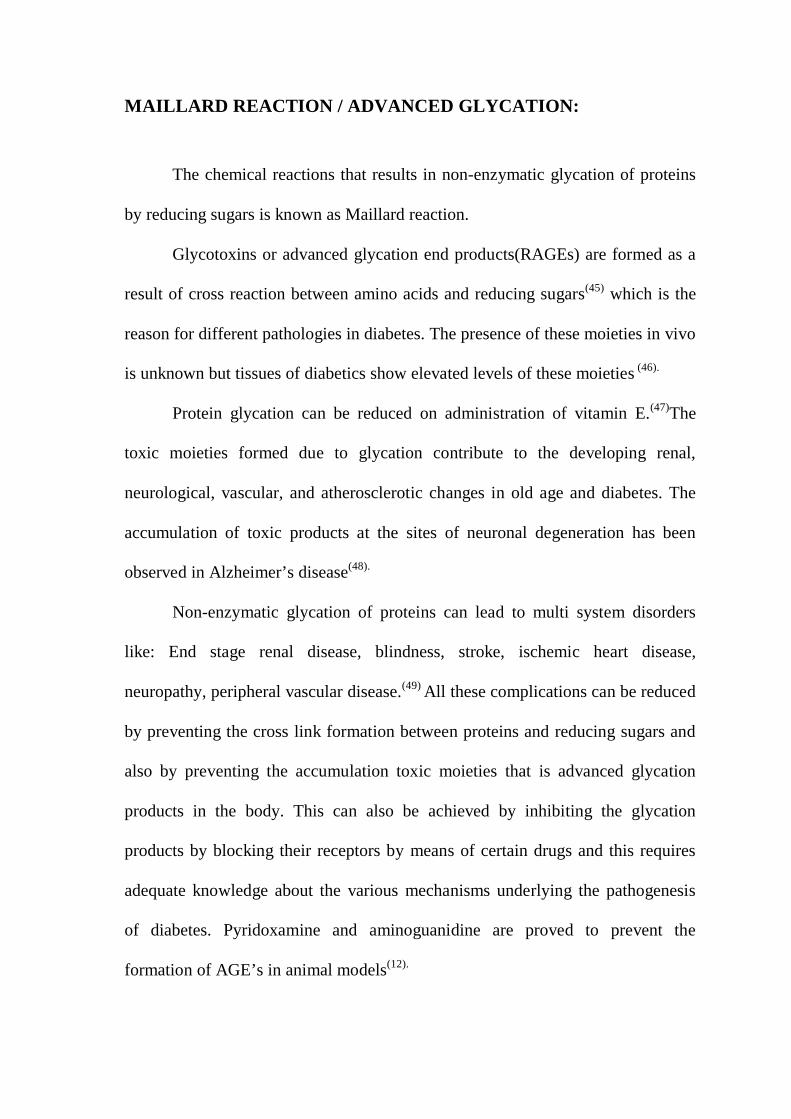
The chemical reactions that results in non-enzymatic glycation of proteins
by reducing sugars is known as Maillard reaction.
Glycotoxins or advanced glycation end products(RAGEs) are formed as a
result of cross reaction between amino acids and reducing sugars(45) which is the
reason for different pathologies in diabetes. The presence of these moieties in vivo
is unknown but tissues of diabetics show elevated levels of these moieties (46).
Protein glycation can be reduced on administration of vitamin E.(47)The
toxic moieties formed due to glycation contribute to the developing renal,
neurological, vascular, and atherosclerotic changes in old age and diabetes. The
accumulation of toxic products at the sites of neuronal degeneration has been
observed in Alzheimer’s disease(48).
Non-enzymatic glycation of proteins can lead to multi system disorders
like: End stage renal disease, blindness, stroke, ischemic heart disease,
neuropathy, peripheral vascular disease.(49) All these complications can be reduced
by preventing the cross link formation between proteins and reducing sugars and
also by preventing the accumulation toxic moieties that is advanced glycation
products in the body. This can also be achieved by inhibiting the glycation
products by blocking their receptors by means of certain drugs and this requires
adequate knowledge about the various mechanisms underlying the pathogenesis
of diabetes. Pyridoxamine and aminoguanidine are proved to prevent the
formation of AGE’s in animal models(12).
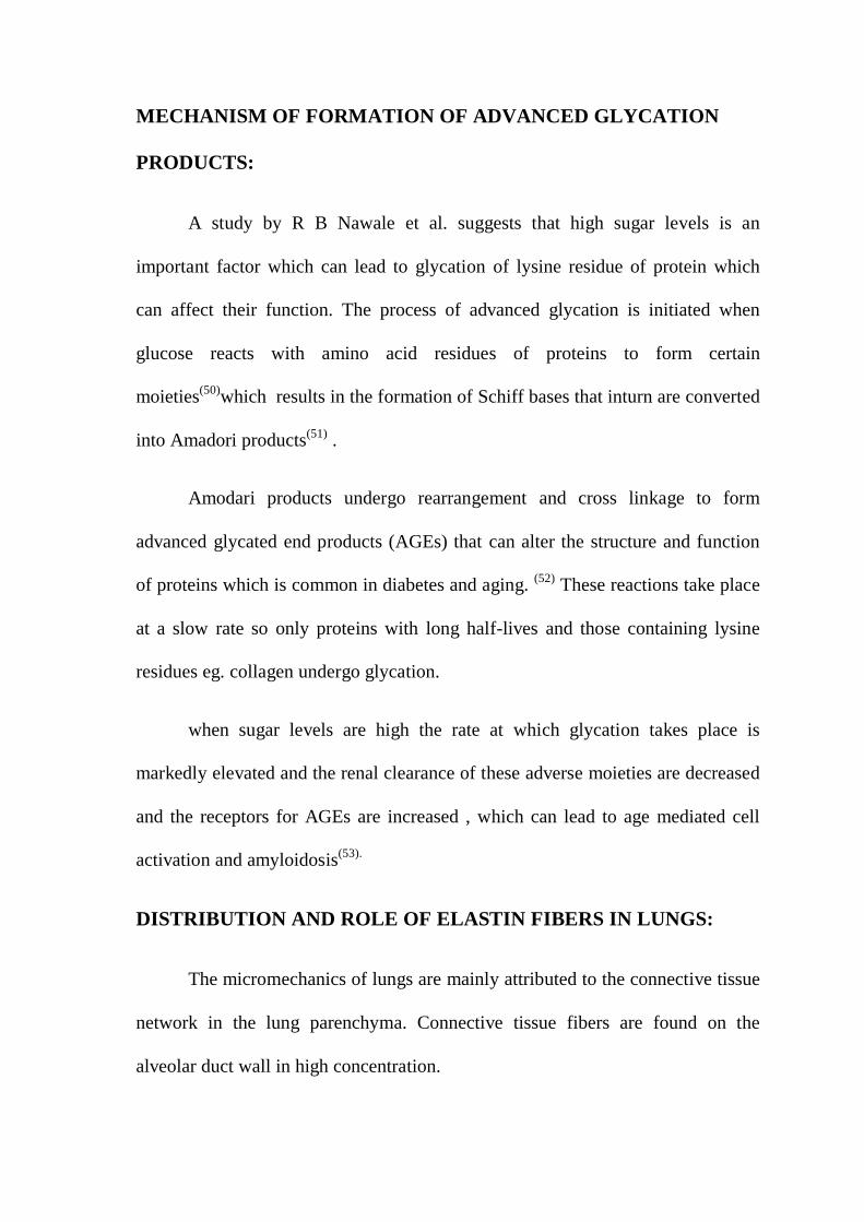
MECHANISM OF FORMATION OF ADVANCED GLYCATION
PRODUCTS:
A study by R B Nawale et al. suggests that high sugar levels is an
important factor which can lead to glycation of lysine residue of protein which
can affect their function. The process of advanced glycation is initiated when
glucose reacts with amino acid residues of proteins to form certain
moieties(50)which results in the formation of Schiff bases that inturn are converted
into Amadori products(51) .
Amodari products undergo rearrangement and cross linkage to form
advanced glycated end products (AGEs) that can alter the structure and function
of proteins which is common in diabetes and aging. (52) These reactions take place
at a slow rate so only proteins with long half-lives and those containing lysine
residues eg. collagen undergo glycation.
when sugar levels are high the rate at which glycation takes place is
markedly elevated and the renal clearance of these adverse moieties are decreased
and the receptors for AGEs are increased , which can lead to age mediated cell
activation and amyloidosis(53).
DISTRIBUTION AND ROLE OF ELASTIN FIBERS IN LUNGS:
The micromechanics of lungs are mainly attributed to the connective tissue
network in the lung parenchyma. Connective tissue fibers are found on the
alveolar duct wall in high concentration.

A ring like structure which is continuous is formed around the mouth of
each alveoli. The elastin fibres tend to pass deep into the septal wall of the alveoli.
Elastin fibers are broadly distributed in tissues like lung parenchyma,
pleura, certain ligaments and arteries. The elasticity of these fibers allows them to
stretch and can cause elastic recoiling as and when required.(54)
DISTRIBUTION AND ROLE OF COLLAGEN IN LUNGS:
The structural integrity of the lung tissue is mainly dependent on the
collagen fibers. Collagen especially type1 and type 2 is distributed widely in the
interstitium of the lung. Collagen fibers also act as a connecting bridge between
the visceral pleura and the alveolar ducts. Thus collagen plays an important role as
a load bearing element of alveolar duct and wall.(55) Collagen and elastin fibres are
found to be closely associated and connected to each other.(56)Thus collagen is
found to play an equal and important role in lung elasticity.(57)
Collagen helps in preventing over stretching of the lung matrix. The cross
linking between the collagen fibers, number of fibrils and its diameter determines
the stiffness of the collagen fibers that is enhanced cross linking and increased
diameter of the collagen fibers have the tendency to increase the stiffness of
normal collagen. (58, 59)Type 1 collagen is found to be stiffer than type 3 and both
of them play an important role in fiber stiffness.

ROLE OF INTERSTITIAL CELLS IN LUNG MECHANICS:
The smooth muscle cells located in the alveolar ducts, walls of blood
vessels as well as the myofibroblasts of the alveolar walls play important role as
contractile cells.(60)The viscoelastic properties of lung can be moderately modified
by the stress generated by the stimulation of these fibers.(61) But the main role of
these cells are in the active repair of the connective tissue.(62)
Surface tension forces acting on the lung tissues play an important role in
micromechanics of lungs.(54)Surfactant produced by the type 2 pneumocytes lines
the alveoli and airways which contributes to stability of alveoli and prevents it
from collapse when the lung volume goes down.(63)Studies show that the quantity
of surfactant produced and its composition depends on the pattern of stretching of
the lung parenchyma.(64)
LUNG AS A TARGET ORGAN IN DIABETES:
The discussion shows that apart from other complications of diabetes such
as retinopathy, nephropathy and neuropathy, lung also is a target organ in a person
with long standing diabetes. The non-enzymatic glycosylation of the
scleroproteins which are widely distributed in the chest wall and bronchial tree
and enhanced cross link formation between the collagen fibers are attributed to the
marked reduction in the mechanical function of lungs.(65)More over any change in
the quality and quantity of collagen can lead to restrictive impairment in lung
function.(66)As collagen is non enzymatically glycosylated in diabetic individuals
they exhibit resistance to digestion by collagenase and pepsin as compared to

normal non-diabetic healthy individuals.(67)Adding on to it, the reduction in the
normal turnover of collagen fibers can lead to reduction in the compliance of the
lung parenchyma. As compliance of lung decreases restrictive type of ventilatory
defect develops in the diabetic lung.(68) The mechanical function of lungs which is
determined by its elasticity can be tested by spirometric pulmonary function
tests.(69)
Restrictive pattern of lung function can be determined by low values of
spirometric pulmonary function tests. (10)
Lung volumes:
1. Forced vital capacity (FVC) – It is the maximum volume of air that can be
exhaled forcefully and rapidly with effort after a deep inhalation. The test
becomes significant only if the person can exhale forcefully for six seconds or
more.
Normal value is 80-120%
In diabetic individuals this value is reduced. This is attributed to increase in
formation of cross linkage between polypeptides of collagen embedded in the
connective tissue matrix of lung parenchyma.
2. Forced expiratory volume at first second (FEV1) – It is the volume of air that is
exhaled in the first second of maximal exhalation after a deep inspiration.
This is a useful tool to assess, how rapidly the lungs can be emptied.
Normal value is 80-120%.

In diabetic individuals this value is also decreased. This is also attributed to the
stiffening of the lung parenchyma due cross link formation between collagen
fibers as a result of enhanced glycosylation of collagen fibers due to
hyperglycemia.
3. FEV1 / FVC:
The best index of airflow limitation can be given by the ratio of FEV1 to FVC.
Normal value that is the absolute ratio should be within 5% of the predicted ratio.
Here FEV1 is expressed in terms of percentage of FVC.
In diabetic individuals with restrictive pattern of lung disease, this value is either
normal or increased that is ≥ 70%.
4. Peak expiratory flow rate (PEFR) – This flow rate is reached immediately by
the first bout of air as the person exhales. This helps to assess the following
parameters:
Helps to judge if the person is putting in maximum effort during the
procedure.
Quality of the test
Strength of muscles of expiration
Condition of large airways
In diabetic individuals due to modification of collagen and elastin ratio the
mechanical properties of lungs are altered which reflects as reduced compliance

and poor elastic recoil of lungs. All these factors will affect the peak expiratory
flow rate.
5. FEF25-75% :
This shows the forced expiratory flow in the middle half of forced vital
capacity that is the average flow from the point at which 25% of forced vital
capacity has been breathed out to the point at which 75% of forced vital capacity
has been breathed out. This indicates the patency of small airways.
In a person who is diabetic for a long period the lung may be subjected to
damage. This chronic status of lung disease will first be represented in the
smallest airways and in the flow volume loop this early damage will reflect
towards the end of the expiratory part of the loop. This may be due to poor elastic
recoil forces and poor muscular support of the respiratory system which is
essential for forced expiration.(70)
Certain studies have shown that activity of the enzyme lysyl oxidase is
increased in rats which developed diabetes that was experimentally induced. This
enzyme is involved in connective tissue formation. Thus in diabetic individuals
enhanced activity of these enzymes will lead to thickening of alveolar
interstitium.(71)

INVOLVEMENT OF RESPIRATORY MUSCLES AND
NEUROMUUCULAR FACTORS IN DIABETES:
Loss of force generating capacity of the muscles of expiration can result in
reduction of peak expiratory flow rate along with poor elastic recoiling of
lungs.(72,73) Similarly FEF25-75% is said to depend on both the neuromuscular
factors as well as the mechanical properties of the respiratory system as FEF25-
75% depicts nothing but the initial part of forced vital capacity.(74)
These factors show that the muscles of respiration are involved in diabetes.
This can be due to enhanced protein catabolism owing to high blood glucose
level. This can ultimately lead to poor strength of skeletal muscles. (75)
The defect in respiratory pump mechanism is also attributed to
development of diabetic polyneuropathy. As glycosylation causes thickening of
the basement membrane in almost all tissues, they can cause demyelination as
well as chromatolysis of axon as well as Schwann cells apart from
microangiopathy.(76)Thus it shows that thoracic nerves and phrenic nerve, which
are predominant nerve supply for the muscles of respiration including diaphragm
is affected in diabetic individuals.(76,77)
ROLE OF OXIDATIVE STRESS IN LUNG DYSFUNCTION:
High blood sugar level can lead to endothelial dysfunction. Increased blood
sugar level can increase the endothelial cell production of free radicals. This can
impede with the vessel dilatation.(78)This occurs due to oxidative stress. It is

proved as this process can be reversed by treatment with antioxidants or L-
arginine.
The mechanism behind this is that, the free radical production due to high
blood sugar results in the activation of protein kinase-C and nuclear factors. This
results in formation of AGEs within the cells. (79) Hyperglycemia can lead to
increase in production of superoxide anion. This leads to increase in the levels of
superoxide than nitric oxide within the endothelial cells, which ultimately results
in the production of nitrotyrosine and peroxinitrate. The major marker for
oxidative stress is nitrotyrosine. In patients with endothelial dysfunction and in
those with diabetes the nitrotyrosine level seems to be increased. (80,81)Thus it
shows that oxidative stress can lead to endothelial dysfunction in diabetic
subjects.
This process can affect the respiratory apparatus in diabetic patients. The
oxidative stress induced by hyperglycemia can lead to loss of integrity of
pulmonary capillary endothelium, which in turn can affect the gas exchange
process across the respiratory membrane. This can also affect the blood volume in
the lung capillaries, as acute increase in blood sugar can suppress vasodilatation
due to oxidative stress.(69)
Similarly markers of inflammation in the epithelial lining fluid of lungs can
be assessed by measuring the markers of inflammation in the exhaled breath
condensate. It was found that the concentration of leukotrine B4 was increased
four times in patients with chronic obstructive pulmonary disease with diabetes

than those without diabetes.(82) Studies suggests that oxidative stress induced by
high blood sugar level is attributed to non-enzymatic glycosylation of proteins
along with low plasma levels of ascorbate. The ideal measure for oxidative stress
is urinary F2 isoprostanes. This was found to be increased in diabetic subjects.
But studies have shown that supplementing these people with α-tocopherol tends
to decrease the levels of isoprastanes in urine.(78)Another study shows that 1250
mg of vitamin c, 680 units of α-tocopheral given for four weeks daily resulted in
reduction in albuminuria.(83)Another study showed that treating type1 diabetic
subjects with 1800 units of α-tocopheral for four months daily showed
improvement in blood flow to retina and creatinin clearance.(84)
GLYCOSYLATED HEMOGLOBIN AND ITS ASSOCIATION WITH
PULMONARY FUNCTION TESTS:
The rate of formation of glycation products is proportional to the
concentration of blood sugar.(85)So glycemic control must have some correlation
with the pulmonary function tests in diabetic individuals. Glycemic control in a
diabetic individual can be assessed by measuring the level of glycosylated
hemoglobin (HbA1c). HbA1c value serves as an indicator of control of blood
sugar over a short term period of one to three months.
A person with HbA1c <7% is said to be under control and those with
HbA1c more than 7% is said to have poor control of blood sugar.(86)If HbA1c is
more than 7% then the rate of glycation of tissue proteins will be on higher side.

Thus glycation of collagen and elastin can affect the lungs and will cause decrease
in the values of pulmonary function tests.
Increase in glycosylated hemoglobin can interfere with diffusion capacity
of the lungs due to poor affinity of glycosylated hemoglobin to carbonmonoxide.
REVIEW:
A study conducted by Sanjeev Verma et al.(87) suggests that non-enzymatic
glycosylation of collagen and elastin in lungs due to hyperglycemia can affect the
mechanical function of lungs which can manifest as altered lung volumes. The
reason behind it may be microangiopathy. As pulmonary interstitium, vessels,
major bronchi are rich in collagen, lung functions are affected in diabetes. Though
these changes are common in old age the severity of this condition is more
pronounced in diabetes. The alteration in scleroproteins is reversible to begin
with, so if blood glucose levels are maintained within normal range the
progression of this condition can be delayed.
In this study pulmonary functions were compared between type I, typeII diabetics
and normal controls. PFT was done using computerized Medspiror. Apart from
this a comparison was done between anthropometric variables like height, weight
as well as body surface area in diabetic individuals that includes both males and
females. The study found no significant difference between male and female
subjects.

A study by Sreeja et al.(88) found no significant difference in
anthropometric variables between male with diabetes and normal control. This
study was done to interpret the pulmonary function in diabetics on oral
hypoglycemic and those on insulin with normal subjects.PFT was done with
Pesomedicare smart Spirometer .The study found reduction in FEV1 and FVC%
in diabetics on both oral hypoglycemics as well as insulin therapy as compared
with the normal subjects.
The study shows that there is reduction in FEF 25-75%(forced expiratory flow
rate) in diabetics who were on oral hypoglycemics as compared to controls.
A study by Lange et al. was done to interpret the effect of plasma glucose
and diabetes on lung functions especially FVC and FEV1.Pulmonary function test
was carried out with Monaghan N 403 spirometer. The study showed reduction in
FVC and FEV1 values in both type I and type II diabetics but the value was
reduced more in diabetics on insulin compared to those not on insulin .(89)
A study by Schnapf et al. showed reduction in lung volumes as well as in
mobility of joints in type II diabetics(90)This proves that non enzymatic
glycosylation of connective tissue(collagen) occurs in people with high plasma
glucose(91).
One of the confounding factors that can affect the values of FEV1 and FVC is
obesity. Most of the type II diabetics are obese (92). But the study done by Lange P
et al. shows that pulmonary functions were reduced in type I diabetics as
compared to type II and BMI in type I diabetics is lower than that of the controls.

The study concludes that there is significant reduction in lung function in
diabetics treated on insulin than those on oral treatment and diet.
A study by Davis et al. (93) has suggested that chronic complications of type
II diabetes will include limitation of air flow and also reduction in lung volumes.
The study proved that vital capacity,FVC,FVE1and peak expiratory flow rates are
reduced in type II diabetics.
A study by Anasuma et al. (94). shows considerable reduction in the forced
vital capacity in Japanese diabetic patients in comparison to controls
A study by Ramirez et al. showed considerable difference in FVC in
diabetics on oral hypoglycemics and those on insulin treatment(95).
A study by Femognari et al. showed that diabetics suffer from restrictive
pattern of lung function due to reduction in FVC and FEV1 and normal
FEV1/FVC.(96)
Another study by Nakajima et al. (97). showed restrictive pattern and not
obstructive pattern of pulmonary function that may be associated with metabolic
syndrome. In this study metabolic factors and percentage of predicted forced vital
capacity (%PFVC) were compared. %PFVC is an indicator of lung compliance. It
was found to have correlation with metabolic abnormalities Abnormal lung
functions were caliberated based on the lower limit of normal (LLN) which was
according to American Thoracic society /European Respiratory Society
guidelines(98).

Restrictive pattern of lung disease in metabolic syndrome was correlated and
assosciated with C- reactive protein which is strong indicator of metabolic
syndrome. In this study confounding factors such as obesity was taken into
account since central obesity has impact on lung function. It results in restrictive
pattern of lung function as expansion of diaphragm is affected due to central
obesity. Hence waist circumference was also taken into consideration. Cut-off for
waist circumference was taken as ≥90 cm for men and ≥80 cm for women. Apart
from this other factors which correlate with metabolic syndrome such as lipid
profile especially triglyceride level >1.70mmol/L , high density
lipoprotein<1.05mmol/L , fasting blood glucose ≥ 6.11 mmol/L , blood pressure ≥
130/85mmHg were also taken into consideration.
PFT was done using Autospiro-507 in standing position. % PEFR obtained
by dividing observed FVC to that of predicted FVC. Air way resistance was
measured by taking FEV1/FVC ratio. The study shows association between
restrictive patterns of lung function and metabolic syndrome. (97)
A study done by Muhammad et al. (99) showed that diabetic subjects also
had associated increase in triglyceride levels as when compared to normal
subjects.PFT was done using Med Graphics profiler. In this study apart from
FVC,FEV1, its ratio and PEFR, slow vital capacity (SVC) was also taken into
account. The study showed statistically significant relation between diabetes and
hypertension with a P value <0.001 and also showed high triglyceride levels in
diabetics, which also had a P value of <0.001.The study shows a reduction in
FVC,FEV1, in diabetic subjects but no much difference in FEV1/FVC ratio and

maximum mid expiratory flow (MMEF).The major limitation of the study was a
small sample size, then the association between blood sugar control and lung
function was not correlated, then the diffusing lung capacity was not assessed due
economic issues.
A study conducted by Meo et al. (100,101) has showed a relation between
disease duration and lung function. The lung function was found to be reduced
with a dose effect relation of duration of diabetes. They found marked reduction
in FVC,FEV1 and PEF in diabetic subjects as when compared to normal subjects.
An Indian study showed impairment of diffusion capacity for carbon-
monoxide in diabetics especially Asian Indians. In this study subjects were
divided into three groups.
Group 1: Type 2 diabetics with microvascular complications
Group 2: Type 2 diabetics without any microvascular complication.
Group 3: Normal healthy individuals.
Patients with microangiopathies were selected based on the expert opinion.
Criteria for diabetic retinopathy:
Patients with non proliferative macular edema.

Criteria for diabetic nepropathy:
1. 24 hour urine test was done to detect loss of >300mg of albumin in urine.
2. Criatinine clearance was calculated.
Criteria for diabetic neuropathy:
1. Loss of >2 stretch reflexes in legs.
2. Decreased tactile sensations, kinesthesia, Pallesthesia
Criteria for microangiopathy:
1. History of myocardial infarction or cerebrovascular accidents.
2. Changes in ECG, ECHO along with clinical examination concluded
cardiovascular problems.
Criteria for Peripheral neuropathy:
1. Loss of at least two peripheral pulses.
2. Chronic ulcers of foot .
The study concludes that diffusing capacity of carbon monoxide was found to be
markedly reduced in group one individuals and the parameters like FVC,FEV1,
PEFR,MIP,MEP were comparable with other groups(102) .The results of
spirometry was interpreted based on the guidelines given by the American
thoracic society. The major limitation of this study was a small sample size.

Swathi et al. (103) showed that restrictive pattern of lung dysfunction is
attributed to connective tissue glycosylation, decrease in elastic recoil of lungs
and inflammatory changes. But the study has shown no association between lung
pathology and glycemic status or duration of disease. The study says that
pulmonary complication in diabetes has been poorly documented with conflicting
results. There occurs microangiopathy of pulmonary capillary network. However,
due to its large reserve, the loss of microvascular bed can be overcome without
developing any difficulty in breathing. Thus, the condition remains sub-clinical.
Many studies have suggested the following pulmonary changes in a
diabetic:
Decreased lung volume
Poor elastic recoil
Poor performance of respiratory muscle(104,105)
Chronic low grade inflammation
Reduction in diffusion capacity for carbonmonoxide(106)
Autonomic neuropathy of respiratory muscle(107)
Thus in a diabetics the lung function will deteriorate leading to loss of
pulmonary reserve. The study shows significant reduction in FVC and FEV1 but
no change in their ratio(103) Similar reports have been shown in other studies(108-
111).PFT was done using Helios 702 spirometer.The study found that except for
ratio of FEV1 to FVC all others were reduced and the study says that the
association between FVC , FEV1, duration of diabetes and glycosylated

hemoglobin is statistically insignificant as P value was >0.05(102).There is marked
reduction in the vital capacity in diabetics and they were found to have restrictive
of lung function (112,113)
Uchida et al. showed decreased diffusion capacity of lungs in diabetics
with a defect in perfusion on ventilation scintigrams(114).
A study conducted in western Australia by Davis, et al.(111) showed that
VC, FVC, FEV1, and PEFR were decreased in diabetic subjects that is between
1.1% and of 3.1% predicted values .Ehrlich et al. (115) showed that type 2 diabetics
are prone to develop asthma, fibrosis ,COPD,pneumonia
A study by Benbassat(116) showed no reduction in lung functions in
diabetics except that the ratio of residual volume to total lung capacity was
increased in non insulin dependent diabetes patients when compared to insulin
dependent diabetics. The study states that Dlco was not affected in those with
microangiopathic changes when it was corrected for alveolar volume.
A study by Fawn Yeh et al.(117,118) says that in Asian Indians with metabolic
syndrome or diabetes mellitus there is marked reduction in pulmonary functions.
The study suggests that these people develop reduced lung function even prior to
development of metabolic syndromes due to the adverse effects of obesity. The
reason behind it is attributed to inflammation which results due to obesity of this
arti. Diabetes care). The study showed that the percentage predicted values of
forced vital capacity and forced expiratory volume at 1st second both were lower
in participants with metabolic disorder with a p value of < 0.0001.

The study has divided the subjects into three groups :
Normal, those with metabolic syndrome, those with diabetes based on the
concentration of inflammatory markers. As the concentration of inflammatory
markers raised the percentage predicted values of pulmonary function tests
reduced. The mechanism behind reduced lung function and metabolic syndrome
might be attributed to obesity and inflammation resulting from it.(119)The
limitation of the study is that it has not provided any data for the inflammatory
markers.
A study conducted by Mahmoud M suggests that abundant connective
tissue in lungs can make it a target site for damage in diabetic patients. In this the
diabetic group was subdivided into two groups that is with those having HbA1c <
7(group II A) and the other group with HbA1c >7(group II B) and the group I
were controls. Then comparison between pulmonary functions of the control and
diabetic group was done which showed a significant difference in FEV1,
FEV1/FVC, FEF 25-75% with a p value <0.05. But the difference was not
significant for MVV. Comparison between group I and group IIA showed
reduction in all PFTs’ except FEF25-75 and MVV.Then comparison between
group I and II B, between group II A and II B shows significant reduction in all
parameters.Thus the study says that lung function is markedly reduced in diabetics
when compared to normoglycemics.The limitation of this study is that it has not
mentioned any criteria based how the diabetics were divided into those under
control based on HbA1c values.(120)

A study done by Yamini et al. (121)showed obstructive, restrictive as well as
mixed pattern of lung dysfunction in diabetic individuals. In this study
comparison was done between males and females also. This study includes 25
healthy and 25 diabetic subjects.
Of this:
1.All females showed FVC < 80% of predicted values but among males
only 48% showed FVC < 80%.
2.About 33% of women showed FEV1/FVC <70% where as 61% of males
showed < 70%.
3. Thus altogether 54% of males showed obstructive pattern as indicated by
FEV1/FVC < 70% and 31% showed restrictive pattern.
4. 67% of women had restrictive pattern.
5. Mixed pattern was seen in 33% women and one male.
Totally among diabetics 48% showed restrictive pattern and 28% showed
obstructive pattern 20% had mixed pattern. The limitation of the study is that it
did not mention about BMI which is an important confounding factor.
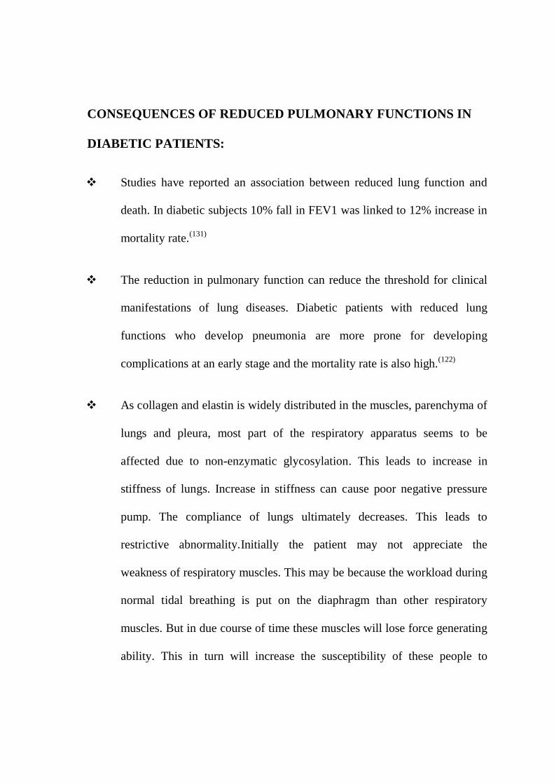
CONSEQUENCES OF REDUCED PULMONARY FUNCTIONS IN
DIABETIC PATIENTS:
Studies have reported an association between reduced lung function and
death. In diabetic subjects 10% fall in FEV1 was linked to 12% increase in
mortality rate.(131)
The reduction in pulmonary function can reduce the threshold for clinical
manifestations of lung diseases. Diabetic patients with reduced lung
functions who develop pneumonia are more prone for developing
complications at an early stage and the mortality rate is also high.(122)
As collagen and elastin is widely distributed in the muscles, parenchyma of
lungs and pleura, most part of the respiratory apparatus seems to be
affected due to non-enzymatic glycosylation. This leads to increase in
stiffness of lungs. Increase in stiffness can cause poor negative pressure
pump. The compliance of lungs ultimately decreases. This leads to
restrictive abnormality.Initially the patient may not appreciate the
weakness of respiratory muscles. This may be because the workload during
normal tidal breathing is put on the diaphragm than other respiratory
muscles. But in due course of time these muscles will lose force generating
ability. This in turn will increase the susceptibility of these people to

develop obstructive parameters. This depends on their sensitivity to various
infections and pollutions. (70)
As diabetic subjects are immunosuppresed, they develop defects in
neutrophil functions. The process of phagocytosis, chemotaxis and
bactericidal activities will be impaired in such subjects due to
hyperglycemia. Hence these people are prone for developing infections.
Apart from this the lung functions are deteriorated in diabetic subjects.
This will add on to the risk of hospitalization in these subjects due to
infections like pneumonia(123)Diabetic subjects with poor lung function are
prone to develop myocardial infarction.(124)
MECHANISM:
Thickening of basement membrane of alveoli and pulmonary capillaries
due to glycosylation can cause poor gas exchange across the respiratory
membrane. This can ultimately lead to poor oxygenation of blood. In a
person with already compromised coronary circulation, this type of
pulmonary dysfunction can lead on to myocardial ischemia.
A study by Tibbilin et al. showed that in those with reduced peak
expiratory flow rate, the proportion of mortality from coronary heart disease was
more among male population in Sweden and Goteberg.(124)
Apart from this when a diabetic person with poor pulmonary function is
taken for coronary artery bypass surgery, then his poor ventilatory function would
interfere with weaning from ventilation.

The person will be prone to develop dyspnea owing to poor mechanical
function of lungs, due to glycosylation of the scleroproteins.
DECREASED LUNG FUNCTION – A MARKER OF
CARDIOVASCULAR DISEASE:
It is a known fact that failure of ventricles can lead to engorgement of lung
vasculature and can cause edema of the interstitium. This will cause a drastic fall
in the compliance of the lungs and will alter the lung volumes.
This reduced lung function acts as a marker for heart disease and studies
have suggested that lung function should be carried out to predict the prognostic
significance of ventricular arrhythmia.
This is because the increased rate of death and myocardial infarction
which was associated with arrhythmia was confined to males with low
percentage predicted values of FEV1 and low value of FEV/VC ratio.

MATERIALS AND METHODOLOGY
SETTING:
PLACE OF STUDY:
This case-control study was done in the Department of respiratory medicine,
Department of Biochemistry at PSG Institute of Medical Sciences And
Research Coimbatore, Tamilnadu, India.
The study was approved by the institutional ethics committee.
STUDY DESIGN:
This is a case control study. Patients with diabetes mellitus were selected
for studying the abnormalities in pulmonary function and their pulmonary
function test findings were compared with age and body mass index (BMI)
matched controls.
The cases were those with uncontrolled blood sugar level within the age
group of 30 – 60 years. The cases were selected from the out-patient department
(OPD) of Endocrinology, provided they fulfilled the inclusion criteria. Then the
cases and controls were subjected to pulmonary function tests.

TIME FRAME:
The study was conducted for one year that is from 1st May 2015 to 31st
May 2016.
INCLUSION CRITERIA:
Thirty diabetic subjects, whose fasting blood glucose was ≥ 126 mg/dl, two
hour post prandial blood glucose was ≥ 200mg and HbA1c ≥ 6.5g%
according to current WHO recommendations for the diagnostic criteria for
diabetes and intermediate hyperglycemia(19) were enrolled in the study as
cases.
Participants were both men and women of age 30 – 60 years were included
in the study.
Thirty age and BMI matched normoglycemic healthy adults, with no
clinical evidence of chronic illness or medication were enrolled in the
control group.
EXCLUSION CRITERIA:
Patients with clinical evidence of any chronic respiratory illness like:
Asthma
Chronic obstructive pulmonary disease
Tuberculosis

Patients with history of:
Smoking
occupational diseases
Cardiac illness
Connective tissue diseases
Musculoskeletal disorders
Patients contraindicated for spirometry:
Absolute contraindications:
Severe acute airflow limitation depicted by FEV1 <50% of predicted.
History of myocardial infarction or cerebrovascular accident in last 3
months.
Uncontrolled hypertension: systolic blood pressure ≥200 mm Hg or
diastolic pressure ≥120 mmHg.
Known case of aortic aneurysm
Relative contraindications
Moderate airflow limitation: FEV1 <60% of predicted or <1.5 litres.
Inability to perform spirometry of acceptable quality.
Pregnancy, nursing mothers.

METHODOLOGY:
Informed written consent was obtained from all the participants before initiation
of the study.
Detailed history of every participant was taken- regarding:
Recent use of any anti diabetic medications.
History of their behavioral habits such as smoking. Then all the smokers
were excluded from the study.
History of any acute or chronic illness was elicited to exclude them from
the study provided they had a positive history.
LABORATORY COLLECTION:
BLOOD GLUCOSE ESTIMATION:
For fasting blood glucose measurement, about 2ml of blood sample was
collected from the individual subjects after overnight fasting by
venipuncture with the help of an evacuated tube system containing EDTA,
at 8 a.m. in the morning.
Then the subjects were asked to take their normal diet (breakfast) and
blood was collected for estimation of post prandial blood glucose two
hours after food intake.
HbA1c was also measured from the blood collected for post prandial blood
glucose estimation.

All blood samples were stored at 2 - 8º C after collection. Analysis of
fasting blood glucose and post prandial blood glucose was done using
COBAS INTEGRA 400/400 plus analyzer using glucose Hexokinase
Gen.3(GLUC3) method. HbA1c was analysed using Bio-Rad laboratories,
D-10 HbA1c version 220-0101 using Immuno-inhibition method.
BODY MASS INDEX:
Height and weight of all the participants were measured.
Standing height was measured by asking the subject to stand bare foot with
both the foot together against measuring instrument in centimeters.
Weight was measured with weighing machine in light weight garments
without foot wears in kg.
Then BMI was calculated for all the participants
BMI was calculated using formula:
Weight in kg / (Height in meter) ².

SPIROMETRY:
The instrument used for measuring lung function was Pesomedicare smart
spirometer. This is a personal computer based USB spirometer. This gives a
precise value for spirometric measurement.
The instrument works on modern electronics, which fulfills all the requirements
and guidelines of American Thoracic society (ATS) and European Respiratory
society (ERS).

Features of software installed:
GLI 2012, actual predicted.
This covers different age, multiple ethinic reference values for spirometry.
A set of predicted values for age, height and sex were derived using
prediction equations. New regression equations were derived based on
these values.
SQL Data base: This data base makes sure that the datas are stored with
high security. This uses applications that allow multiple user networks.
This instrument can be upgraded any time.
Back up facility is available, which is automated.
The result can be obtained in a printed format.
Linear flow sensor

This instrument makes use of complete linear flow sensor. The sensor is based on
pneumotachograph . This helps to carry out the spirometric tests with high
precision. The sensor are available with different orifice and negligible dead
space. The measurements are not affected by humidity.
The system uses standard USB 2.0 interphase.
Pre-test advice to the participants:
All the participants were asked to:
1. Avoid heavy meal prior to the test.
2. Avoid strenuous exercise before the test.
3. Avoid tight clothing during the procedure.
The apparatus was properly caliberated. The operating technique of the
instrument was based on its manual and the guidelines provided by The
American Thoracic Society.(20)
Then all the participants were given a prior explanation regarding the
maneuver. A demo was shown to them. The test was carried out by a well
trained personnel.
The test was performed in the sitting posture with the participants’ nose
closed with a soft nose clip.
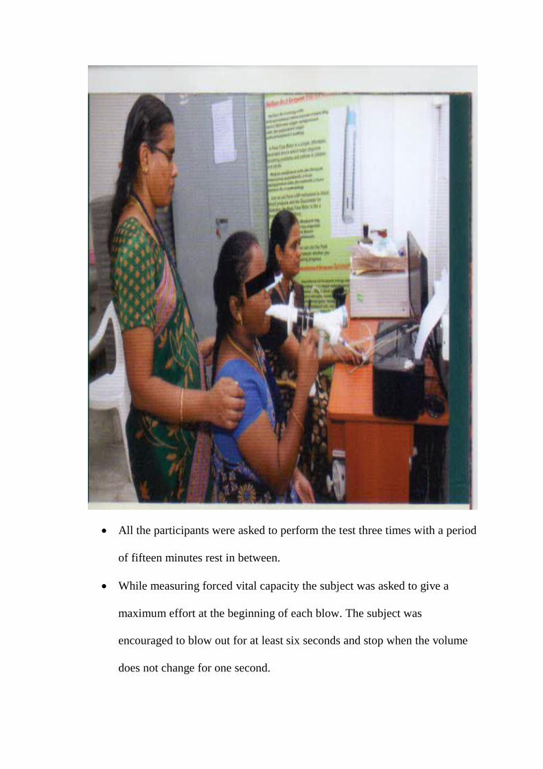
All the participants were asked to perform the test three times with a period
of fifteen minutes rest in between.
While measuring forced vital capacity the subject was asked to give a
maximum effort at the beginning of each blow. The subject was
encouraged to blow out for at least six seconds and stop when the volume
does not change for one second.

Then the difference between the two largest forced vital capacity and
forced expiratory volume at first second were analyzed and was found to
be within 200ml.
Best of the three measurements was taken into account.
The highest value of forced vital capacity and forced expiratory volume at
first second was used to calculate their ratio. The ratio expressed was as
percentage of FVC.
The parameters used for analysis were:
Forced vital capacity(FVC)
Forced expiratory volume at first second(FEV1)
FEV1/FVC
FEF25-75
Peak expiratory flow rate(PEFR)The percentage predicted values of the
above parameters were used for analysis purpose.
For the FEV1/FVC, absolute ratio was taken.
STATISTICAL ANALYSIS:
Statistical analysis was done using IBM SPSS statistics software
(Statistical package for the social science version 19).
Student 't' test was applied to compare the means of quantitative datas’
like FEV1/FVC, FVC, FEV1, FEF25-75, PEFR.

All values were expressed as mean ± SD (Standard deviation). The "p"
values were interpreted as :
(i) p > 0.05 was considered not significant.
(ii) p < 0.05 was considered statistically significant.
Correlation between pulmonary function test parameters like FEV1/FVC,
FVC, FEV1, FEF25-75, PEFR with HbA1c and Body-Mass Index among
the diabetics were analyzed by using Pearson correlation analysis.

RESULTS
Table 1:
Table 1 shows the physical parameters of the diabetic subjects as well as
the normal healthy controls.
Both the groups were matched for age and body mass index. That is there
was no statistical difference between the two groups as the “p” value was > 0.05.
The mean value of age for normal healthy controls was 42.63 ± 5.06 and
for diabetic individuals it was 44.23 ± 5.96. Both the groups were matched for
age. There was no statistical difference for the mean age for both the groups as the
“p” value was 0.267 which was statistically not significant.
The mean value of body mass index for normal healthy controls was 26.70
± 3.95 and for diabetic individuals it was 26.74 ± 4.18. Both the groups were
matched for the body mass index. There was no statistical difference for the mean
of body mass index for both the groups as the “p” value was 0.965 which was
statistically insignificant.

Table 2:
Table 2 shows the comparison of pulmonary function tests between
diabetic subjects and normal healthy controls.
The mean of FEV1/FVC ratio for normal healthy individuals was 82.92 ±
4.81 and for the diabetic subjects it was 83.67 ± 5.96. There was no statistical
difference between the two groups for the ratio of FEV1 to FVC as the “p” value
was 0.597.
The mean value of FVC for normal healthy controls was 99.90 ± 15.85 and
for diabetic subjects it was 81.83 ± 18.11. There was statistically significant
difference for the mean of forced vital capacity between the two groups as the “p”
value was 0.000.
The mean value of FEV1 for normal healthy controls was 91.53 ± 10.60
and for diabetic subjects it was 77.03 ± 13.81. The difference was statistically
significant as the “p” was 0.000.
The mean value of PEFR for controls was 95.77 ± 15.54 and for the
diabetic subjects it was 85.57 ± 18.69. There was statistically significant
difference between the mean values of PEFR between the two groups, as the “p”
value was 0.025.

The mean value of FEF25-75 % for normal healthy controls was 74.83 ±
20.36 and for diabetic individuals it was 66.90 ± 24.5. The difference was not
statistically significant as the “p” value was 0.179.
This study shows that the pulmonary function parameters like FVC, FEV1
and PEFR are reduced in participants with type 2 diabetes as when compared to
normal healthy participants.
The mean of both FEV1/FVC ratio and FEF25-75% between the normal
healthy controls and diabetic individuals was not statistically significant as their
“p” value was greater than 0.05.

Table 3 :
Table 3 shows comparison of pulmonary function tests among diabetic
subjects with HbA1c <7 and HbA1c > 7.
The 30 diabetic subjects were divided into two groups:
Group 1 = Diabetic subjects with HbA1c < 7 g%
Group 2 = Diabetic subjects with HbA1c > 7 g%
The mean of FEV1/FVC in diabetic subjects with HbA1c < 7 g% was 82.92
± 5.12 and in those with HbA1c > 7 g%, it was 84.10 ± 6.49. But the difference of
the mean values between the two groups was not statistically significant as the “p”
value was 0.611.
The mean value of FVC in diabetic individuals with HbA1c < 7 g% was
88.64 ± 21.50 and for those with HbA1c >7g% was 77.89 ± 15.07. This difference
was also not statistically significant as the “p” value was 0 .119.
The mean value of FEV1 for diabetic individuals with HbA1c < 7 g % was
80.91 ± 14.21 and for those with HbA1c >7g% was 74.79 ± 13.43. This also
showed no statistical difference between the two groups as the “p” value was
0.249.

The mean value of FEF25-75% for diabetic individuals with HbA1c < 7 g%
was 68.18 ± 22.63 and for those with HbA1c >7g%, it was66.16 ± 26.20. This was
also not statistically significant as the “p” value was 0.214.
The mean value of PEFR in diabetic subjects with HbA1c < 7g % was
91.45 ± 12.04 and for those with HbA1c > 7g%, it was 82.16 ± 21.19.
Thus the various parameters of pulmonary function tests showed no
significant difference between the diabetic subjects with HbA1c < 7 g% and
HbA1c > 7 g%.

Table 4 :
Table 4 shows the Pearson correlation of pulmonary function tests and
glycosylated hemoglobin among diabetic subjects.
Pearson correlation (r2) for FEV1/FVC and HbA1c was 0.307 among
diabetic subjects. This showed no correlation as the “p” value was 0.09.
Pearson correlation (r2) for FVC and HbA1c was -0.202 among diabetic
subjects. This showed no correlation as the “p” value was 0.284.
Pearson correlation (r2) for FEV1 and HbA1c was -0.011 among diabetic
subjects. This showed no correlation as the “p” value was 0.954.
Pearson correlation (r2) for FEF25-75 % and HbA1c was .136 among diabetic
subjects. This showed no correlation as the “p” value was 0.473.
Pearson correlation (r2) for PEFR and HbA1c was .023 among diabetic
subjects. This showed no correlation as the “p” value was 0.904.
There was a slight negative correlation between FVC, FEV1 and HbA1c.
But this was not statistically significant.
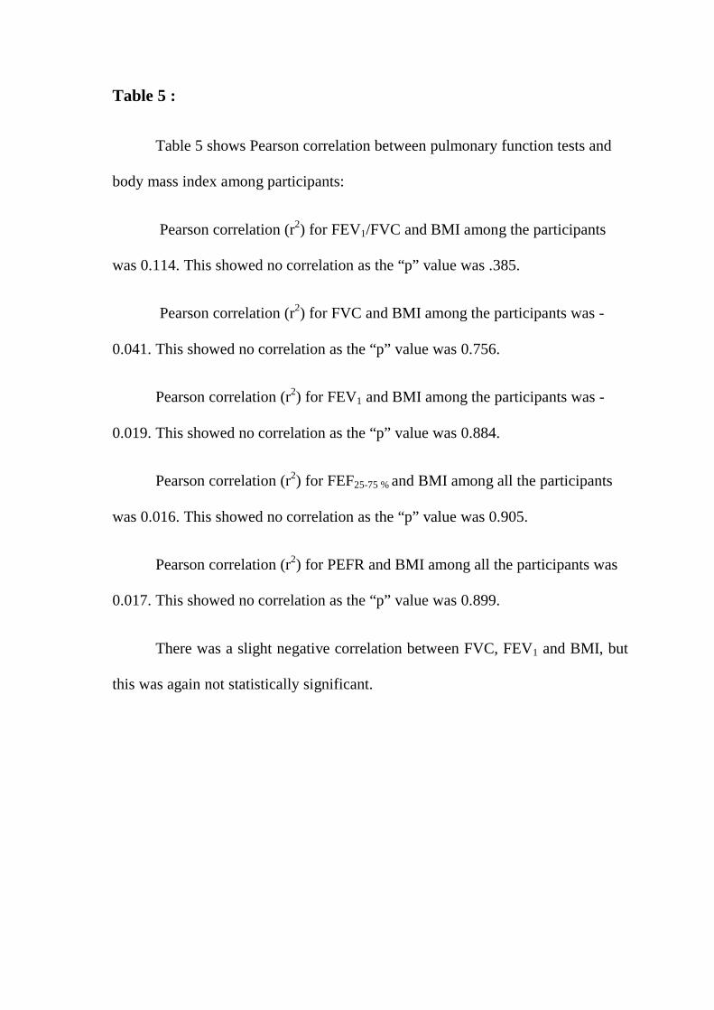
Table 5 :
Table 5 shows Pearson correlation between pulmonary function tests and
body mass index among participants:
Pearson correlation (r2) for FEV1/FVC and BMI among the participants
was 0.114. This showed no correlation as the “p” value was .385.
Pearson correlation (r2) for FVC and BMI among the participants was -
0.041. This showed no correlation as the “p” value was 0.756.
Pearson correlation (r2) for FEV1 and BMI among the participants was -
0.019. This showed no correlation as the “p” value was 0.884.
Pearson correlation (r2) for FEF25-75 % and BMI among all the participants
was 0.016. This showed no correlation as the “p” value was 0.905.
Pearson correlation (r2) for PEFR and BMI among all the participants was
0.017. This showed no correlation as the “p” value was 0.899.
There was a slight negative correlation between FVC, FEV1 and BMI, but
this was again not statistically significant.

Table 6 :
Table 6 shows the correlation between pulmonary function tests and body
mass index among diabetic subjects.
This was done to confirm whether the body mass index had any
correlation with pulmonary function tests among the diabetic individuals whose
lung function was already compromised.
Among diabetic subjects the Pearson correlation for FEV1/FVC and BMI
was 0.259 with a “p” value of 0.167. This correlation showed no significance.
Pearson correlation for FVC was -0.077 with a “p” value of 0.688.
Pearson correlation for FEV1 was -0.018 with a p value of 0.927.
Pearson correlation for FEF25-75 % was 0.106 with a “p” value of 0.578.
Pearson correlation for PEFR was 0.098 with a “p” value of 0.606.
This showed a slight negative correlation between FVC, FEV1 and BMI
among the diabetic subjects. But this correlation was not statistically significant.

Table 7 :
Table 7 shows comparison of pulmonary function tests between
participants with BMI < 25 and BMI > 25 among diabetic subjects:
Participants were categorized into two groups:
Group 1: BMI < 25
Group 2: BMI > 25
The mean values for the various parameters for both the groups were:
FEV1/FVC for those with BMI<25 among the diabetic individuals was
83.61 ± 5.23 and for those with BMI >25 was 83.70 ± 6.37. This was not
statistically significant as the “p” value was 0.971.
FVC for those with BMI < 25 among the diabetic individuals was 76.11 ±
12.11 and for those with BMI >25 was 84.28 ± 19.90. This was also not
statistically significant as the “p” value was 0.265.
FEV1 for those with BMI < 25 among the diabetic individuals was 74.33 ±
12.83 and for those with BMI >25 was 78.19 ± 14.35. This difference was not
statistically significant as the “p” value was 0.493.
FEF25-75 % for those with BMI < 25 among the diabetic individuals was
60.33 ± 17.93 and for those with BMI >25 was 69.71 ± 26.81. This showed no
statistical significance as the “p” value was 0.347.

PEFR for those with BMI < 25 among the diabetic individuals was 85.33 ±
18.40 and for those with BMI >25 was 85.66 ± 19.26. This also showed no
statistical significance as the “p” value was 0.965.
This study showed no significant difference in the mean values of pulmonary
function tests in both groups.

TABLE 1: PHYSICAL CHARACTERISTICS OF PARTICIPANTS
Parameters
Controls,
(n=60)
Mean ± SD
Patients with
Dm (n=60)
Mean ± SD
“P” VALUE Significance
Age (Years) 42.63 ± 5.06 44.23 ± 5.96 0.267 NS(> 0.05)
BMI (kg/m2) 26.70 ± 3.95 26.74 ± 4.18 0.965
NS(> 0.05)
DM: Diabetes mellitus
BMI: Body mass index
P < 0.05 – Significant
P > 0.05 – Not Significant (NS)

TABLE 2:
COMPARISON OF PULMONARY FUNTION TESTS BETWEEN
DIABETIC SUBJECTS AND NORMAL HEALTHY CONTROLS.
Parameters: Percentage predicted
values
Control subjects
Mean ± SD
Diabetic subjects
Mean ± SD “p”value Significance
FEV1/FVC 82.92±4.81 83.67±5.96 0.597 NS (>0.05)
FVC 99.90±15.85 81.83±18.11 0.000 HS (<0.05) *
FEV1 91.53±10.60 77.03±13.81 0.000 HS (<0.05) *
FEF25-75% 74.83±20.36 66.90±24.57 0.179 NS (>0.05)
PEFR 95.77±15.54 85.57±18.69 .025 HS (<0.05) *
FVC – Forced vital capacity
FEV1 – Forced expiratory volume at first second
FEF25-75% – Forced expiratory flow rate during first half of FVC
PEFR – Peak expiratory flow rate
P < 0.05 – Significant
P > 0.05 – Not Significant (NS)
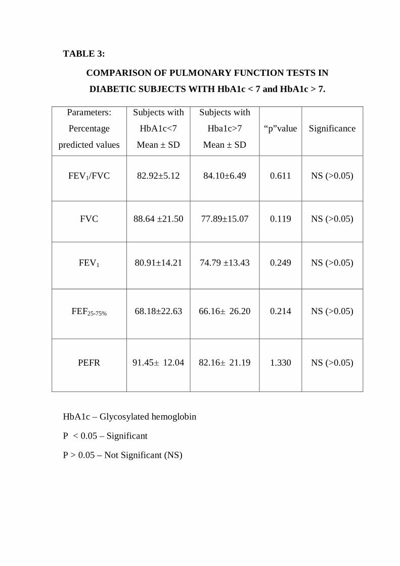
TABLE 3:
COMPARISON OF PULMONARY FUNCTION TESTS IN
DIABETIC SUBJECTS WITH HbA1c < 7 and HbA1c > 7.
Parameters:
Percentage
predicted values
Subjects with
HbA1c<7
Mean ± SD
Subjects with
Hba1c>7
Mean ± SD
“p”value Significance
FEV1/FVC 82.92±5.12 84.10±6.49 0.611 NS (>0.05)
FVC 88.64 ±21.50 77.89±15.07 0.119 NS (>0.05)
FEV1 80.91±14.21 74.79 ±13.43 0.249 NS (>0.05)
FEF25-75% 68.18±22.63 66.16±26.20 0.214 NS (>0.05)
PEFR 91.45±12.04 82.16±21.19 1.330 NS (>0.05)
HbA1c – Glycosylated hemoglobin
P < 0.05 – Significant
P > 0.05 – Not Significant (NS)

TABLE 4:
PEARSON CORRELATION OF PULMONARY FUNCTION TESTS
AND GLYCOSYLATED HEMOGLOBIN AMONG DIABETIC
SUBJECTS:
r2 – Pearson correlation
HbA1c – Glycosylated hemoglobin
P < 0.05 – Significant
P> 0.05 – Not Significant (NS)
PARAMETERS
% predicted
value
r2
(Pearson correlation) “p” VALUE SIGNIFICANCE
FEV1/FVC
with HbA1c 0.307 0.098 NS (>0.05)
FVC with
HbA1c -0.202 0.284 NS (>0.05)
FEV1 with
HbA1c -0.011 0.954 NS (>0.05)
FEF 25-75 with
HbA1c 0.136 0.473 NS (>0.05)
PEFR with
HbA1c 0.023 0.904 NS (>0.05)

TABLE : 5
PEARSON CORRELATION FOR BODY MASS AND INDEX
PULMONARY FUNCTION TESTS AMONG THE PARTICIPANTS:
P < 0.05 – Significant
P > 0.05 – Not Significant (NS)
Parameters
% predicted
value
r2
(PEARSON
CORRELATION)
“p” value Significance
FEV1/FVC
with BMI 0.114 0.385 NS (>0.05)
FVC with BMI -0.041 0.756 NS (>0.05)
FEV1 with
BMI -0.019 0.884 NS (>0.05)
FEF 25-75 with
BMI 0.016 0.905 NS (>0.05)
PEFR with
BMI 0.017 0.899 NS (>0.05)

TABLE : 6
PEARSON CORRELATION FOR BODY MASS INDEX AND
PULMONARY FUNCTION TESTS AMONG
DIABETIC INDIVIDUALS:
P < 0.05 – Significant
P > 0.05 – Not Significant (NS)
Parameters
% predicted
value
r2
(PEARSON
CORRELATION)
“p”value Significance
FEV1/FVC
with BMI 0.259 0.167 NS (>0.05)
FVC with BMI -0.077 0.688 NS (>0.05)
FEV1 with
BMI -0.018 0.927 NS (>0.05)
FEF 25-75 with
BMI 0.106 0.578 NS (>0.05)
PEFR with
BMI 0.098 0.606 NS (>0.05)

TABLE 7:
COMPARISON OF PULMONARY FUNCTION TESTS BETWEEN
PARTICIPANTS WITH BMI < 25 AND > 25 AMONG DIABETIC
SUBJECTS:
PARAMETERS:
Percentage
Predicted values
SUBJECTS
WITH
BMI<25
Mean ± SD
SUBJECTS
WITH
BMI>25
Mean ± SD
“p” VALUE SIGNIFICANCE
FEV1/FVC 83.61±5.23 83.70±6.37 .971 NS (>0.05)
FVC 76.11 ±12.11 84.28±19.90 .265 NS (>0.05)
FEV1 74.33±12.83 78.19±14.35 .493 NS (>0.05)
FEF25-75% 60.33±17.93 69.71±26.81 .347 NS (>0.05)
PEFR 85.33±18.40 85.66±19.26 .965 NS (>0.05)
P < 0.05 – Significant
P > 0.05 – Not Significant (NS)
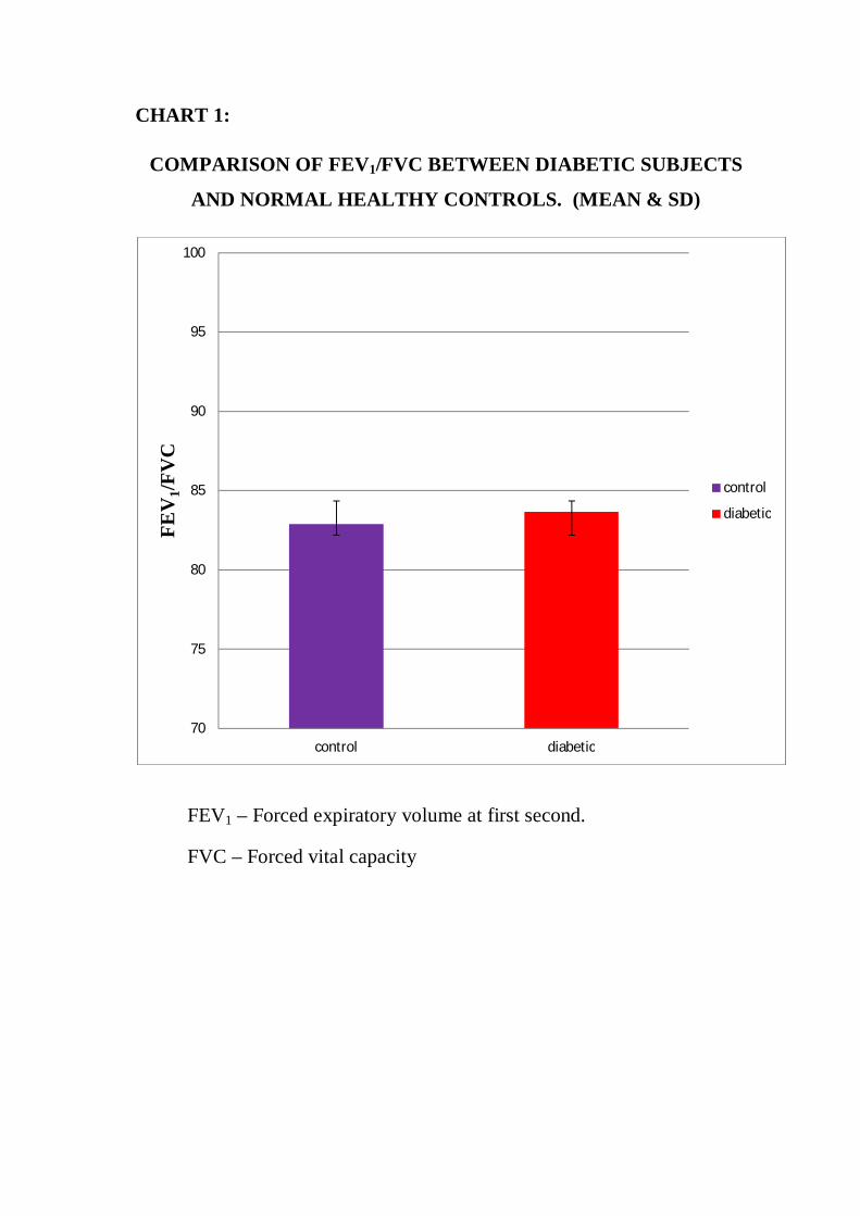
CHART 1:
COMPARISON OF FEV1/FVC BETWEEN DIABETIC SUBJECTS
AND NORMAL HEALTHY CONTROLS. (MEAN & SD)
FEV1 – Forced expiratory volume at first second.
FVC – Forced vital capacity
70
75
80
85
90
95
100
control diabetic
FEV
1/FV
C
control
diabetic
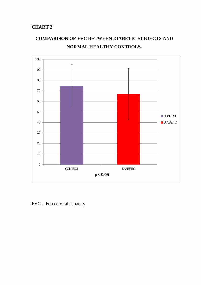
CHART 2:
COMPARISON OF FVC BETWEEN DIABETIC SUBJECTS AND
NORMAL HEALTHY CONTROLS.
FVC – Forced vital capacity
0
10
20
30
40
50
60
70
80
90
100
CONTROL DIABETIC
CONTROL
DIABETIC
p < 0.05

CHART 3 :
COMPARISON OF FEV1 BETWEEN DIABETIC SUBJECTS AND
NORMAL HEALTHY CONTROLS.
FEV1 – Forced expiratory volume at first second
0
10
20
30
40
50
60
70
80
90
100
CONTROL DIABETIC
CONTROL
DIABETIC
p < 0.05

CHART 4 :
COMPARISON OF FEF25-75%BETWEEN DIABETIC SUBJECTS
AND NORMAL HEALTHY CONTROLS.
FEF25-75% -Forced expiratory flow rate at 25-75% of FVC.
0
10
20
30
40
50
60
70
80
90
100
CONTROL DIABETIC
CONTROL
DIABETIC

CHART 5:
COMPARISON OF PEFR BETWEEN DIABETIC SUBJECTS AND
NORMAL HEALTHY CONTROLS.
PEFR – Peak expiratory flow rate
60
65
70
75
80
85
90
95
100
control diabetic
PEFR
control
diabetic
p < 0.05

DISCUSSION
Lung parenchyma which includes the respiratory bronchioles, alveolar
ducts, alveoli, blood vessels, interstitium are rich in connective tissue elements
such as collagen and elastin.
Though the bronchioles lack cartilages the lungs are able to maintain their
patency because of the elasticity of its parenchyma. The major determinant of the
elasticity of the lung parenchyma is elastin fibers. The stretching ability of the
elastin fibers are about 140% of their length in the relaxed state.(54)The elastin
fibers with few fibroblast are found in the alveoli of lungs which promotes normal
exhalation.
But collagen which is inelastic, contributes to the architecture and tensile strength
of the lung. Collagen can stretch only up to 2% of its resting length.
The elastic connective tissue surrounding the alveoli of lungs gets stretched
during inhalation. During the process of expiration these fibers recoil to squeeze
the air out. Thus expiration is a passive process where there is neither need of any
work to be done nor there is any energy expenditure. This property is due to the
normal elasticity of the lungs.
The interstium of lung parenchyma consists mainly of collagen fibers
which are of mainly type 1 and type 3. Collagen is responsible for the structural
framework of the alveolar wall that is, it maintains the architecture of alveolar
wall.

In diabetic individuals there occurs non-enzymatic glycosylation of
collagen and elastin, which in turn can affect the ventilator functions.
In this study the diabetic subjects and the controls were matched for age
and body mass index. So both the confounding factors were excluded .
The ventilatory functions in this study which was measured by Pesomedicare
smart spirometer showed the following data:
1. Forced vital capacity (FVC):
Forced vital capacity which is an important component of flow volume loop
depicts the total volume of air that is exhaled with maximum effort.
The normal percentage predicted value of FVC is 80-120%.
This study shows a marked reduction in forced vital capacity in individuals with
diabetes as when compared to normal healthy controls.
The reason attributable to this reduction may be due to:
Reduced compliance of lungs due to due to non-enzymatic glycosylation of
collagen and elastin.
The other reason must be thickening of the basal lamina of the epithelium in the
alveoli.
This result goes hand in hand with many other studies.
A cohort study conducted by Davis et al. (72) among the European
community shows similar decrease in forced vital capacity. The study states that
the reduced value of forced vital capacity is due to glycation of proteins in
diabetic individuals which will affect the compliance of lungs. Chronic exposure
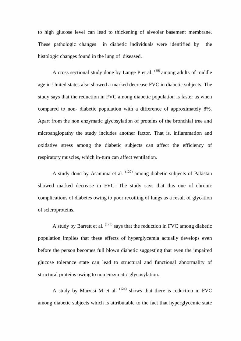
to high glucose level can lead to thickening of alveolar basement membrane.
These pathologic changes in diabetic individuals were identified by the
histologic changes found in the lung of diseased.
A cross sectional study done by Lange P et al. (89) among adults of middle
age in United states also showed a marked decrease FVC in diabetic subjects. The
study says that the reduction in FVC among diabetic population is faster as when
compared to non- diabetic population with a difference of approximately 8%.
Apart from the non enzymatic glycosylation of proteins of the bronchial tree and
microangiopathy the study includes another factor. That is, inflammation and
oxidative stress among the diabetic subjects can affect the efficiency of
respiratory muscles, which in-turn can affect ventilation.
A study done by Asanuma et al. (122) among diabetic subjects of Pakistan
showed marked decrease in FVC. The study says that this one of chronic
complications of diabetes owing to poor recoiling of lungs as a result of glycation
of scleroproteins.
A study by Barrett et al. (123) says that the reduction in FVC among diabetic
population implies that these effects of hyperglycemia actually develops even
before the person becomes full blown diabetic suggesting that even the impaired
glucose tolerance state can lead to structural and functional abnormality of
structural proteins owing to non enzymatic glycosylation.
A study by Marvisi M et al. (124) shows that there is reduction in FVC
among diabetic subjects which is attributable to the fact that hyperglycemic state

can lead to thickening of basement membrane of alveoli and pulmonary
capillaries. The study also suggests a decrease in the diffusing capacity of lungs as
the DLCO value was markedly reduced in diabetic subjects due to thickening of
basal lamina of pulmonary capillaries.
2. Forced expiratory volume at first second (FEV1):
It is the amount of air breathed out with maximum effort in the first second. The
normal percentage predicted value of FVC is 80-120%.
This study shows a marked reduction in FEV1 among diabetic population as when
compared to normal healthy controls.
The reason for marked reduction in FEV1 is attributed to:
Poor recoiling of lung tissues due to non-enzymatic glycosylation of the
scleroproteins collagen and elastin.
Thickening of basal lamina of alveolar epithelium.
This result correlates with the following studies.
A cohort study conducted by Davis et al. (72) and a study by Sanjeev sinha et
al.(102) suggests that the reduction of FEV1 among the diabetic subjects when
compared to normal healthy controls is due to poor elastic recoiling of the lungs
as the visco elastic nature of lung parenchyma is lost as a result of thickening of
collagen and loss of elastic nature of elastin owing to glycation.
3. Peak expiratory flow rate (PEFR):
The normal value of PEFR is 400-600ml/min.This study shows that in
diabetic subjects there is marked reduction in the PEFR values as compared to the

normal subjects. This implies that there is marked fall in the strength of the
muscles of expiration, which is reflected as fall in force generating capacity of
these muscles. This may be either due to increased protein catabolism or due to
poor neuromuscular function of respiratory muscles as a part of diabetic
neuropathy or change in composition of muscle fibers.
This result is similar to the study done by Kabitz HJ et al. (125) which states
that the reduction in peak expiratory flow rate among diabetic subjects is due to
poor efficiency of muscles of expiration , as the maximum velocity with which
air is forced out of lungs depends on the strength of muscles of expiration. This
may be due to enhanced protein catabolism.
A study done by Marin P et al. (125) also shows a marked reduction in peak
expiratory flow rate in diabetic subjects as when compared to normal healthy
controls. This is attributed to the fact that, in diabetic individuals’ peripheral
neuropathy can result in reduced neuromuscular function, which can affect the
muscles of respiration.
4. FEV1/ FVC :
In this study the mean of absolute ratio of FEV1 to FVC among diabetics
were found to be slightly more than that of the normal controls but was not
statistically significant.
The increase in the mean value of the ratio suggests that diabetic
individuals have a restrictive pattern of lung disease.
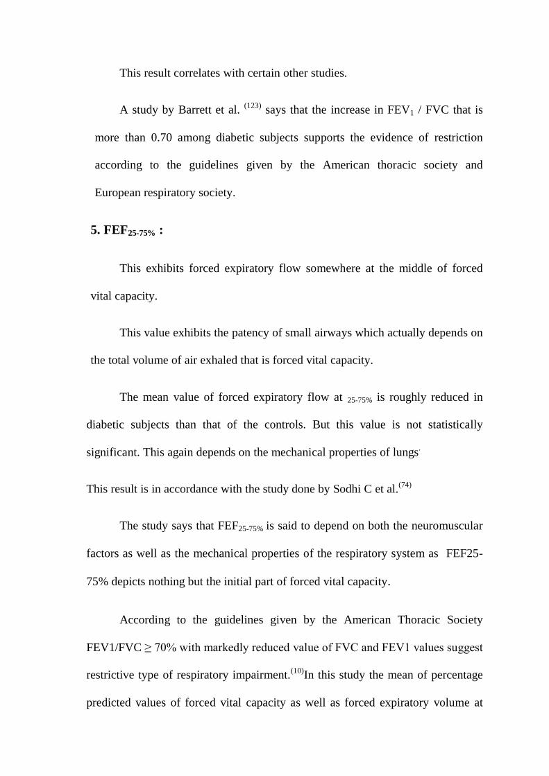
This result correlates with certain other studies.
A study by Barrett et al. (123) says that the increase in FEV1 / FVC that is
more than 0.70 among diabetic subjects supports the evidence of restriction
according to the guidelines given by the American thoracic society and
European respiratory society.
5. FEF25-75% :
This exhibits forced expiratory flow somewhere at the middle of forced
vital capacity.
This value exhibits the patency of small airways which actually depends on
the total volume of air exhaled that is forced vital capacity.
The mean value of forced expiratory flow at 25-75% is roughly reduced in
diabetic subjects than that of the controls. But this value is not statistically
significant. This again depends on the mechanical properties of lungs.
This result is in accordance with the study done by Sodhi C et al.(74)
The study says that FEF25-75% is said to depend on both the neuromuscular
factors as well as the mechanical properties of the respiratory system as FEF25-
75% depicts nothing but the initial part of forced vital capacity.
According to the guidelines given by the American Thoracic Society
FEV1/FVC ≥ 70% with markedly reduced value of FVC and FEV1 values suggest
restrictive type of respiratory impairment.(10)In this study the mean of percentage
predicted values of forced vital capacity as well as forced expiratory volume at

first second is markedly reduced in diabetic subjects when compared to normal
subjects. Hence the diabetic subjects in this study show a restrictive pattern of
ventilatory impairment.
Studies have shown low vital capacity and restrictive pattern of respiratory
impairment among type 2 diabetic subjects.(128,129)
6. Comparison of pulmonary functions in diabetic subjects with
HbA1c < 7g % and HbA1c > 7g %.
A study by Van den Borst et al. showed a restrictive pattern of lung
function among diabetic subjects which was statistically significant. But the
limitation of the study was that it did not take BMI, HbA1c and smoking into
consideration. (130)The present study has compared the pulmonary functions with
the glycemic control. A good indicator for glycemic control is HbA1c value.
The correlation of HbA1c of diabetic subjects with their pulmonary
function tests’ showed slightly negative correlation for FVC and FEV1.But this
was not statistically significant. The slight negative correlation shows that lung
function is slightly decreased in diabetic subjects as the lung parenchyma which is
rich in collagen and elastin is prone for non-enzymatic glycosylation due to high
blood glucose level. This can lead to stiffening of parenchyma, which will
ultimately affect the lung volumes and capacities.

The lung parenchyma changes are evident by electron microscopic studies
of lung tissue of diseased diabetic subjects. It showed thickening of capillary
endothelium and basement membrane.
The statictiscal insignificance may be due to the fact that, though HbA1c
levels are strong indicators of glycemic control, it depicts the glycemic control for
only a short duration of 1-3 months. And this short term control in no way can
explain the effect of long term high plasma glucose level on lung mechanics.
This correlates with the study done by Lange P et al. (110) This study was
done in The United nations among the middle aged population. The HbA1c values
showed a negative correlation with lung functions as high blood glucose will
affect the mechanical function of lungs.
7. Correlation of body mass index with pulmonary functions among
diabetic individuals:
The correlation between body mass index and pulmonary function tests’
among diabetic subjects showed a slight negative correlation for both forced vital
capacity and forced expiratory volume at first second. But this was not
statistically significant. This may be attributed to small sample size.
This result goes hand in hand with the study conducted by Jitendra Singh et
al.(86)which shows that there is no correlation between pulmonary function tests
and factors like body mass index and gender.

CONCLUSION
In diabetic patients following changes are noted in the lung:
Non-enzymatic glycosylation of collagen and elastin can lead to increased
cross link formation between polypeptides of collagen causing stiffening of
lung parenchyma as well as poor elastic recoiling of lung tissue. This
causes restrictive pattern of lung dysfunction in these subjects.
Microangiopathy not only affects the kidneys, nervous system, and eyes,
but also damages the alveolar basement membrane.
There is more chance for reduced strength of respiratory muscles, owing to
increased protein catabolism.
Next important complication in diabetes is thickening of basement
membrane of thoracic and phrenic nerves as a part of diabetic neuropathy,
which can cause demyelination, chromatolysis of their axons and Schwann
cells. This can also lead to decrease in strength of respiratory muscles.
Thus spirometry a non-invasive procedure must be added as a part of
investigation to monitor the effects of diabetes on lungs and also for early
detection of lung function to prevent any further complication due to poor
ventilatory function.

A good control of blood sugar may decrease the risk of mortality through
correction of ventilatory functions.
In a person with uncontrolled blood sugar level the chance for developing
respiratory infection is more. Hence it is advisable to immunize them against
influenza and pnuemoccocus.(135)
Studies say that pyridoxamine and aminoguanidine compounds have the
ability to prevent the formation of advanced glycation end products, in animal
models.(12)
Aminoguanidine is an AGE-inhibitor. It helps to prevent various signs of aging
process. But as it has the ability to prevent or slow down the proteins from cross
linking, it can be tried in diabetic subjects also.
Aminoguanidine tends to bind to the sugars and prevent the binding of
sugars to the lysine group of proteins. Thus aminoguanidine helps to maintain
and stabilize the glucose metabolism (136).
1. Stage 3 trial for aminoguanidine is going on at the Alteon Corporation in
the USA. The main function of aminoquanidine, carnosine, acetyl-α-
carnitine is to prevent cross link formation.
2. Another important development is ALT-711 (Thiazolium salt). This is
under 2nd stage of trial. This helps to break the existing links.

BIBLIOGRAPHY
1. King H, Aubert RE, and Herman WH. Global burden of diabetes
1995-2025. Prevalence, numerical estimates, and projections.
Diabetes care 1998;21:1414-31.
2. Anandhalakshmi S, Manikandan S, Ganeshkumar P et al. Alveolar
Gas Exchange and Pulmonary Functions in Type II Diabetes Mellitus.
J Clin Diagn Res. 2013 Sep; 7(9):1874-1877.
3. Bennette PH, Knowler WC. Definition, Diagnosis, and Classification
of Diabetes Mellitus and Glucose Homeostasis. Chapter 19 In. Khan
CR, Weir GC, King GL. Joslin’s Diabetes Mellitus 14th edition.
Boston: Joslin Diaetes Center; 2005:331-333.
4. Gavin JR III, Alberti KGMM, Davidson MB, et al. Report of the
Expert Committee on the Diagnosis and Classification of Diabetes
Mellitus. Diabetes care 1997;20: 1183-1197.
5. WHO consultation group. Definition, diagnosis and classification of
diabetes mellitus and its complications, 2nd ed. Part 1: Diagnosis and
classification of diabetes mellitus WHO/NCD/NCS/99. Geneva:
World Health Organisation,1999: 1-59.
6. Turner RC, Cull CA, Frighi V, et al. Glycemic control with diet,
sulphonylurea, metformin, or insulin in patients with type 2 diabetes:
progressive requirement for multiple therapies ( UKPDS 49). UK
Prospective Diabetes Study ( UKPDS 49)Group. JAMA 1999; 2005-
2012:281.
7. Shah SH, Sonawane P,Nahar P, et.al. Pulmonary function tests in
type 2 diabetes mellitus and their association with glycemic control

and duration of the disease. Chest Research Foundation, Lung
India.2013 Apr-Jun; 30(2):108-112.
8. Kamalov M, Harris PWR, Cooper GJS. What is the difference
between glycation and glycosylation? Chem. Commun. 2014;
50:4944-4946.
9. Verma S, Goni M, Kudyar RP. Assessment of Pulmonary Functions
in Patients with Diabetes Mellitus. JK Science. 2009 April-
June;11(2):71.
10. Timothy J, Barreiro DO, Perillo I. An approach to interpreting
spirometry. Am Fam Physician. 2004 March 1; 69(5): 1107-1115.
11. How to interpret spirometry results. Nursins times . net NICE (2010)
Management of chronoic obstructive pulmonary disease:
Management of adults in primary and secondary care(CG101).
NICE;October: 2011.
12. Nawale RB , Mourya VK ,Bhise SB. Non-enzymatic glycation of
proteins: A cause for complications in diabetes. Indian Journal of
Biochemistry & Biophysics.2006 December;Vol.43:337-344.
13. Plopper CG, Morishige WK: Alterations in granular (type ii)
pneumocyte ultrastructure bystreptozotocin-induced diabetes in the
rat. LabInvest 1978;38:143–148.
14. Kida K, Utsuyoma M, Takizawa T, ThurlbeckWM: Changes in lung
morphologic featuresand elasticity caused by streptozotocin-induced
diabetes mellitus in growing rats. AmRev Respir Dis 1983;128:125–
131.

15. Plopper CG, Morishige WK. Alterations in the ultrastructure of
nonciliated bronchiolar epi-thelial (Clara) cells by streptozotocin
induceddiabetes in rats. Am Rev Respir Dis 1979; 120:1137–1143.
16. Vracko R, Thorning D, Huang TW.Basal lamina of alveolar
epithelium and capillaries: Quantitative changes with aging and in
diabetes mellitus. Am Rev Respir 1979; 120:973–983.
17. Sandler M, Bunn AE, Steward RI. Pulmonary function in young
insulin dependent diabetic subjects. Chest 1986; 90:670–675.
18. Kodolova IM, Lysenko LV, Saltykov BB Changes in the lungs in
diabetesmellitus. Arkh Patol 1982; 44:35–40.
19. Kapur A, Shishoo S, Ahuja M M S. Consensus Guidelines –
Minimum Basic Care For Persons With Diabetes Mellitus . Indian J
Diab Dev Coun 17 2000;vol 20:2-12.
20. Ramachandran A , Snehalatha C , Kapur A. High prevalence of
diabetes and impaired glucose tolerance in India: National Urban
Diabetes Survey. Diabetologia(2001)Springer-Verlag 2001; 44: 1094
-1101.
21. Ramachandran A, Snehalatha C. NIDDM in India and Indians: Is it
increasing? Bull IDF 1995; 40:27-9.
22. Ramaiya KL, Kodali VRR, Albert KGMM . Epidemiology of
diabetes in Asians of the Indian sub-continent. Diabetes Metab Rev
1990;6:125-46.
23. Aparna. Pulmonary Function Tests in Type 2 Diabetics and Non-
Diabetic People -A Comparative Study. Journal of Clinical and
Diagnostic Research. JCDR. 2013;7(8):1606-1608.

24. Definition and diagnosis of diabetes and intermediate hyperglycemia.
Geneva: World Health Organization. 2006; Annexes 83.
25. WHO Diabetes Country Profile 2016. Fact Sheet No. 12.
26. Sing GM, Danaei G, Farzadfar F et al. The age specific quantitative
effects of metabolic risk factors on cardiovascular diseases and
diabetes: a pooled analysis. PLoS. One. 2013;8:(7)e65174.
27. Jeon CY, Murray MB. Diabetes mellitus increases the risk of active
tuberculosis: a systematic review of 13 observational studies. PLos.
Medicine. 2008;5:(7)e152
28. Global Burden of Metabolic Risk Factors for Chronic Diseases
Collaboration. Cardiovascular disease, chronic kidney disease and
diabetes mortality burden of cardiometabolic risk factors from 1980
to 2010: a comparative risk assessment. Lancet Diabetes
Endocrinology. 2014;2:(8)634-647.
29. NCD-Ris C. Worldwide trends in diabete since 1980: pooled analysis
of 709 population based measurement studies with over 7.2 million
participants. The Lancet (in press).
30. Finucane MM et al. National, regional, and global trends in
body-mass index since 1980: systematic analysis of health
examination surveys and epidemiological studies with 960
country-years and 9.1 million participants.TheLancet,2011,
377:557-67.
31. Ahmad O. Age standardization of rates: a new WHO standard
(Technical report). GPE discussion paper series: No 31. Geneva:
World Health Organization; 2001.
32. World Population Prospects: 2015revision.New York, United Nations
Population Division, 2013. 33. Global report on diabetes [Internet]. World Health Organization. 2016
[cited 27 May 2016]. Available from:l
http://www.who.int/diabetes/global-report/en/
34. Build study, 1979. Chicago: Society of Actuaries and Association of
Life Insurance Medical Directors of America,1980.
35. Lyon CJ, Law RE, Hsueh WA. Minireview: adiposity, inflammation
and atherogenesis. Endocrinology2003; 144:2195-2200.
36. Mokdad AH, Ford ES, Bowman BA et al. Prevalence of obesity,
diabetes and obesity –related health risk factors, 2001. JAMA 2003;
289: 76-79.
37. Soundariya K, Neelambikai N. Influence of Anthropometric Indices
on Pulmonary Function Tests in Young Individuals. World Journal
of Medical Sciences. 2013; 9 (3): 157-161.
38. Thomas PS, Milledge JS. Respiratory function in the morbidly obese
before and after weight loss.Thorax44.1989 May; 44(5): 382–386.
39. Womack CJ, Harris DL, Katzel LI et al. Weight loss, not aerobic
exercise, improves pulmonary function in older obese men. Journal of
Gerontology: Medical Sciences. 2000; 55 Aug;55(8):M453-7.
40. Salome CM, King GG, Berend N. Physiology of obesity and effects
on lung function. J Appl Physiol.2010;108: 206–211.
41. Zimmet P, Shaw J. Diabetes- A worldwide Problem. Chapter 31 In.
Khan CR, Weir GC, King GL. Joslin’s Diabetes Mellitus 14th edition.
Boston: Joslin Diaetes Center; 2005:535.
42. Raheja B S , Talim S. J Diab Assoc India :38, 1-11.

43. Uusitupa M, Siitonen O, Aro A & Pyorala K . Prevalance of coronary
heart disease and left ventricular failure and hypertension in middle
aged newly diagnosed type 2 (non insulin dependent)diabetic
subjects. Diabetologia 28:1985;22-27.
44. Veikko S ,Veikko S, Jeremy D K, Christopher N. Non–Insulin-
Dependent Diabetes Mellitus and Fasting Glucose and Insulin
Concentrations Are Associated With Arterial Stiffness Indexes .
AHA: circulation 91:1995 1432-1443.
45. Koschinsky T , He C , mitsuhashi T ,Bucala R . Orally absorbed
reactive glycation products (glycotoxins):An environmental risk
factor in diabetic nephropathy. Proc. Natl. Acad. Sci. USA: Vol. 94:
June 1997 ;pp. 6474 – 6479.
46. Kume S, Takeya M, Mori T. Immuno histochemical & ultra structural
detection of advanced glycation end products in atherosclerotic
lesions of human aorta with a novel specific monoclonal antibody.
Am J Pathol : 147:1995;654-667.
47. Ceriello A , Giugliano D , Quatraro A. Vitamin E reduction of
protein glycosylation in Diabetes: New prospect for prevention of
diabetic complication. American diabetes association:Diab care
14:1991;68-72.
48 Mark R E , Griffin S T & Graham D I . Aging-associated changes in
human brain. J Neuropathol. Exp. Neurol.56:1997;1269-1275.
49. Songer, T.J., and P.Z. Zimmet. Epidemiology of Type 11 diabetes:
An international perspective: Pharmacoeconomics 8(Supp 1) 1995:1-
11.

50. Makitha Z , Vlassara H , Cerami A. Immunochemical detectin of
advanced glycosylation end products in vivo. J Biol Chem 267 : 1992
; 5133-5138.
51. Bookchin R M , Gallop P M. Structure of hemoglobin A1 C : nature of
the N-terminal beta chain blocking group. Biochem. Biophys. Res.
Commun. 32(1) : 1968 ; 86-93.
52. Barret-Connor E, Orchard T J. Diabetes in America. NIH
publication:1985;85-1458.
53. Brownlee M, Cerami A , Vlassara H. Advanced glycosylation end
products in tissue and the biochemical basis of diabetic
complications. N Engl J Med 318 : 1988 ; 1315-1321.
54. Mercer RR, Crapo JD. Spatial distribution of collagen and elastin
fibers in the lungs. J Appl Physiology (1985). 1990
Aug;69(2):756-65.
55. Weibel E and Gil J. Structure-function relationships at the alveolar
level. In: Bioengineering Aspects of the Lung, edited by West JB.
New York: Dekker, 1977, vol. 3, p. 1–81. (Lung Biol. Health Sci.
Ser.)
56. Brown RE, Butler JP, Rogers RA, and Leith DE. Mechanical
connections between elastin and collagen. Connect Tissue Res 30:
295–308, 1994.
57. Yuan H, Kononov S, Cavalcante FS et al. Effects of collagenase
and elastase on the mechanical properties of lung tissue strips. J
Appl Physiol 2000; 89: 3–14.
58. Angele P, Abke J, Kujat R et al. Influence of different collagen
species on physico-chemical properties of cross linked collagen
matrices. Biomaterials 25: 2831–2841, 2004.

59. Silver FH, Freeman JW, and Seehra GP. Collagen self-assembly
and the development of tendon mechanical properties. J Biomech
36: 1529–1553, 2003.
60. Weibel E. Functional morphology of lung parenchyma. , chapter 8
In: Handbook of Physiology. The Respiratory System. Mechanics
of Breathing. Bethesda, MD: Am. Physiol. Soc, 1986, sect. 3, vol.
III, pt. 1, p. 89–112.
61. Dolhnikoff M, Morin J, and Ludwig MS. Human lung parenchyma
responds to contractile stimulation. Am J Respir Crit Care
Med.1998; 158: 1607–1612.
62. Suki B, Ito S, Stamenovic D et al. Biomechanics of the lung
parenchyma: critical roles of collagen and mechanical forces. Journal of Applied Physiology. 2005 May 1; 98(5):1892-1899.
63. Avery ME and Mead J. Surface properties in relation to atelectasis
and hyaline membrane disease. Am J Dis Child 97: 517–523, 1959.
64. Nicholas TE and Barr HA. Control of release of surfactant
phospholipids in the isolated perfused rat lung. J Appl
Physiol1981; 51: 90–98.
65. Sandler M. Is the lung a ‘target organ’ in diabetes mellitus? Arch
Intern Med. 1990; 150(7):1385–88. 66. Laurent GJ. Lung collagen More than scaffolding. Thorax 1986;
41:418428.
67. Sternberg M, Cohen-Forterre L, Peyroux J.Connective tissue in
diabetes mellitus: Biochemical alterations of the intercellular
matrix with special reference to proteoglycans, collagens and
basement membranes. Diabetes Me-tab 1985; 11:27–50.
68. Boulbou MS, Gourgoulianis KI, Klisiaris VK. Diabetes mellitus
and Lung Function. Med Princ Pract 2003;12:87–91.

69. Goldman MD. Lung Dysfunction in Diabetes. Diabetes Care. 2003
Jun; 26(6): 1915-1918.
70. Gajbhiye RN, Tambe AS. Glycosylated Hemoglobin Level and its
Correlation with Pulmonary Function Test in Type II Diabetics.
Sch. Acad. J. Biosci.2014; 2(8): 556-560.
71. Madia AM, Rozovski SJ, Kagan HM. Changes in lung lysyl
oxidase activity in streptozotocin-diabetes and in starvation.
Biochim Biophys Acta. 1979 Jul 18;585(4):481–07.
72. Davis WA, Knuiman M, Kendell P, et al. Glycaemic exposure is
associated with reduced pulmonary function in type 2 diabetes. The
Fremantile Diabetes Study. Diabetes Care 2004;27 :752-57.
73. Agarwal V, Gupta B, Dev P,et al. Deterioration of the lung
functions in type II diabetic subjects from northern India. Indian J
Physiol Pharmacol 2009; 53(2) 189-91.
74. Sodhi C, Singh S, Dandona PK. A study of the effect of yoga
training on the pulmonary functions in patients with bronchial
asthma. Indian J Physiol Pharmacol 2009; 53(2): 169-74.
75. Khan AM, Jahan N, Sultana N.FVC, FEV1 and FEV1/FVC% in
type-1 diabetic male and their relationships with HbA1c. J.
Bangladesh Soc. Physiol.7 (1) (2012) 23–28.
76. Subramaniam S. Textbook of Human Physiology. Chapter
“Physiological anatomy and respiratory movements.”
77. Sinha S, Guleria R, Misra A. Pulmonary functions in patients with
type 2 diabetes mellitus &correlation with anthropometry &
microvascular complications. Indian J Med Res 119, 2004
February ;pp 66-71.
78. Bloomgarden ZT. Inflammation and Insulin Resistance. Diabetes
Care 2003 May; 26(5): 1619-1623.

79. Collins AR, Meehan WP, Kintscher U, et al. Troglitazone inhibits
formation of early atherosclerotic lesions in diabetic and nondiabetic
low density lipoprotein receptor-deficient mice. Arterioscler Thromb
Vasc Biol 21:365–371, 2001.
80. McCaffrey TA, Fu C, Du B, et al .High-level expression of Egr-1
and Egr-1-inducible genes in mouse and human atherosclerosis. J
Clin Invest 105:653–662, 2000.
81. Jonassen AK, Brar BK, Mjos OD, et al.Insulin administered at
reoxygenation exerts a cardioprotective effect in myocytes by a
possible anti-apoptotic mechanism. J Mol Cell Cardiol 32:757– 764,
2000.
82. Becher G, Winsel K, Beck E, et al.Breath condensate as a method
of noninvasive assessment of inflammation mediators from the lower
airways. Pneumologie 1997; 52:456–459.
83. Jonassen AK, Sack MN, Mjos OD, Yellon DM.Myocardial
protection by insulin at reperfusion requires early administration
and is mediated via Akt and p70s6 kinase cell-survival signaling.
Circ Res. 2001; 89:1191–1198.
84. Makowski L, Boord JB, Maeda K, Parker RA, et al. Lack of
macrophage fatty-acid-binding protein aP2 protects mice deficient
in apolipoprotein E against atherosclerosis. Nat Med. 2001;7: 699–
705.
85. Lacinová K, Eckhardt A, Pataridis S. Glycation of proteins -their
analysis and physiological aspects. Transworld Research Network.
2010; 37/661 (2): 978-81.
86. Singh J, Gupta KK, Himanshu D. To study the effect of glycemic
control and duration of disease on pulmonary function tests and
diffusion capacity in type 2 diabetes mellitus. Int J Res Med Sci.
2015Jan; 3(1):224-228.
87. Verma S , Goni M , Kudiyar P R. Assessment of pulmonary
functions in patients with Diabetes Mellitus. Jk science : 11 (2):
2009
88. Sreeja C K , Samuel E , Kesavachandran C . Pulmonary function
in patients with diabetes mellitus. Ind J Physiol Pharmacol: 47(1) :
87-93.
89. Lange P , Groth S, Kastrup J. Diabetes mellitus, Plasma glucose
and lung function in a cross-sectional population study. Eur Respir
J : 2 :1989; 14-19.
90. Schnapf BM, Banks RA , Silverstein JH. Pulmonary function in
insulin-dependent diabetes mellitus with limited joint mobility.
Am Rev Respir Dis:130 :1984; 930-932.
91. Kohn RR, Schnider SL. – Glucosylation of human collagen.
Diabetes: 31: 1982; 47-51.
92. Ray CS, Sue DY , Bray G. Effects of obesity on respiratory
function. Am Rev Respir Dis:128: 1983;501-508.
93. Davis WA, Knuiman M, Kendall P. Glycemic exposure is
associated with reduced pulmonary function in type 2 diabetes: The
Fremantle Diabetes Study. Diabetes Care 2004;27:752-7.
94. Asanuma Y, Fujiya S, Ide H, Agishi Y. Characteristics of pulmonary
function in patients with diabetes mellitus. Diabetes Res Clin Pract
1985;1:95-101.
95. Ramirez C L , Nogare D A, Hsia C. Relationship between diabetes
control and pulmonary function in insulin-dependent diabetes
mellitus. The Am J Med:91(4) :1991;371-376.
96. Fimognari FL, Pasqualetti P, Moro L, et al. The association between
metabolic syndrome and restrictive ventilator dysfunction in older
persons. J Gerontology Med Sci 2007;62 A (7) : 760-65.

97. Nakajima K, Kubouchi Y, Muneyuki T,et al. A possible association
between suspected restrictive pattern as assessed by ordinary
pulmonary function test and the metabolic syndrome. J chest 2008;
134:712-718.
98. Pellegrino R, Viegi G, Brusasco V, et al.Interpretative strategies for
lung function tests. Eur Respire 2005; 26:948-968.
99. Irfan M, Jabbar A, Haque SA, et al. Pulmonary function in patients
with diabetes mellitus. Lung India 2011 Apr-Jun; 28(2): 89-92.
100. Meo SA, Al-Drees AM, Arif M, Al-Rubean K. Lung function in type
2 Saudi diabetic patients. Saudi Med J. 2006;27:338–43.
101. Meo SA, Al-Drees AM, Shah SF,et al. Lung function in type 1
Saudi diabetic patients. Saudi Med J. 2005;26:1728–33.
102. Sinha S, Guleria R, Misra A,et al. Pulmonary functions in patients
with type 2 diabetes mellitus and correlation with anthropometry and
microvascular complications. Indian J Med Res. 2004; 119: 66–71.
103. Shah H S, Sonawane P, Nahar P. Pulmonary function in type 2
diabetes mellitus and their association with glycemic control and
duration of the disease. Lung India 2013 Apr-June; 30(2) : 108-112.
104. Hamlin CR, Kohn RR, Luschin JH. Apparent accelerated aging of
human collagen in diabetes mellitus. Diabetes. 1975;24:902–4.
105. Fogarty AW, Jones S, Britton JR. Systemic inflammation and decline
in lung function in a general population: A prospective study.
2007;62:515–20.
106. Mori H, Okubo M, Okamura M, Yamane K, Kado S, Egusa G, et
al. Abnormalities of pulmonary function in patients with non
insulin dependent diabetes mellitus. Intern Med. 1992;31:189–93.
107. Williams JG, Morris AI, Hayter RC, Ogilvie CM. Respiratory
responses of diabetics to hypoxia, hypercapnia and exercise.
Thorax. 1984;39:529–34.

108. Davis WA, Knuiman M, Kendall P. Glycemic exposure is
associated with reduced pulmonary function in type 2 diabetes, the
fremantle diabetes study. Diabetes Care. 2004;27:752–7.
109. Asanuma Y, Fujiya S, Ide H, Agishi Y. Characteristics of
pulmonary function in patients with diabetes mellitus. Diabetes Res
Clin Pract. 1985;1:95–101.
110. Lange P, Groth S, Kastrup J, Mortensen J, Appleyard M, Nyboe J, et
al. Diabetes mellitus, plasma glucose and lung function in a cross
sectional population study. Eur Respir J. 1989;2:14–9.. Barrett-
Conor E, Frette C. NIDDM, impaired glucose tolerance, and
pulmonary function in older adults. Diabetes Care. 1996;19:1441–4.
111. Davis TM, Knuiman M, Kendall P, Vu H, Davis WA. Reduced
pulmonary function and its association in type 2 diabetes: The
fremantle diabetes study. Diabetes Res Clin Pract. 2000;50:153-9.
112. Engstrom GJ, Janzon L. Risk of developing diabetes is inversely
related to lung function: A population based cohort study. Diabet
Med. 2002;19:167–70.
113. Yeh HC, Punjabi NM, Wang NY, Pankow J, Duncan BB, Cox CE,
et al. Cross sectional and prospective study of lung function in adults
with diabetes mellitus. Diabetes. 2002;51:A242–3.
114. Uchida K, Takahashi K, Aoki R, Ashitaka T. Ventilation-perfusion
scintigram in diabetics. Ann Nucl Med. 1991;5:97–102.
115. Ehrlich SF, Quesenberry CP, Vanden Eeden SK, Shan J, Ferrara
A. Patients diagnosed with diabetes are at increased risk for
asthma, COPD, pulmonary fibrosis and pneumonia but not lung
cancer. Diabetes Care. 2010;33:55–6022.
116. Benbassat CA, Stern E, Kramer M, Lebzelter J, Blum I, Fink G.
Pulmonary function in patients with diabetes mellitus. Am J Med
Sci. 2001;322:127–32.

117. Ford ES, Mannino DM. National Healthand Nutrition
Examination Survey Epidemiologic Follow-up Study. Prospective
association between lung function andthe incidence of diabetes:
findings fromthe National Health and Nutrition Examination
Survey Epidemiologic Follow-upStudy. Diabetes Care
2004;27:2966–2970.
118. Yeh HC, Punjabi N M, Wang NY, Pankow JS et al.Vital capacityas
a predictor of incident type 2 diabetes:the Atherosclerosis Risk in
Communi-ties study. Diabetes Care 2005;28: 1472–1479.
119. Shore SA. Obesity, airway hyper responsiveness, and
inflammation. J Appl Physiol 2010;108:735–743.
120. El-Habashy MM, Agha MA, El-Basuni H. Impact of diabetes
mellitus and its control on pulmonary functions and
cardiopulmonary exercise tests. Egyptian Journal of Chest
Diseases and Tuberculosis. 2014 April;63( 2):471–476.
121. Singh Y, Kumari S. The Pulmonary Function Tests In Patients With
Diabetes Mellitus And Non-Diabetes Peopleof Ajmer,Rajasthan.
International Journal of Basic and Applied Medical Sciences. 2015
Sep- Dec;5(3):8-13.
122. Kornum JB, Thomsen RW, Riis A, et al. Diabetes, glycemic
control, and risk of hospitalization with pneumonia: A population
based case control study. Diabetes Care. 2008;31: 1541–5.
123. Ehrlich SF, Charles P, Quesenberry Jr. Patients Diagnosed With
Diabetes Are at Increased Risk for Asthma, Chronic Obstructive
Pulmonary Disease, Pulmonary Fibrosis, and Pneumonia but Not
Lung Cancer. Diabetes Care 2010 Jan; 33(1): 55-60.
124. Friedman GD, Klatsky AL, Siegelaub AB. Lung function and risk
of myocardial infarction and sudden cardiac death. N Engl J Med.
1976 May 13; 294(20):1071-5.

125. Asanuma Y, Fujiya S, Ide H, et al. Characteristics of pulmonary
function in patients with diabetes mellitus. Diabetes Res Clin Pract.
1985;1:95–101.
126. Barrett-Conor E, Frette C. NIDDM, impaired glucose tolerance,
and pulmonary function in older adults. Diabetes Care. 1
996;19:1441–4.
127. Marvisi M, Bartolini L, del Borrello P, et al. Pulmonary function in
non-insulin dependent diabetes mellitus. Respiration 2001 ; 68 :
268- 72.
128. Kabitz HJ, Walker SA, Walterspacher S. Diabetic polyneuropathy
is associated with respiratory muscle impairment in type 2
diabetes. Diabetologia. 2007 Sep.
129. Marin P, Andersson B, Krotkiewski M,et al. Muscle fiber
composition and capillary density in women and men with
NIDDM. Diabetes Care. 17:382–386.
130. Sodhi C, Singh S, Dandona PK. A study of the effect of yoga
training on the pulmonary functions in patients with bronchial
asthma.Indian J Physiol Pharmacol 2009; 53(2): 169-74.
131. Engstrom GJ, Janzon L. Risk of developing diabetes is inversely
related to lung function: A population based cohort study. Diabet
Med. 2002;19:167–70.
132. Yeh HC, Punjabi NM, Wang NY, Pankow J, Duncan BB, Cox CE,
et al. Cross sectional and prospective study of lung function in adults
with diabetes mellitus. Diabetes. 2002; 51:A242–3.
133. Borst BB, Gosker HR, Zeegers MP, et al.Pulmonary function in
Diabetes: A Metaanalysis. Chest. 2010; 138:393–406.

134. Knuiman MW, James AL, Diviniti ML,et al. Lung function,
respiratory symptoms, and mortality: Results from the buselton
health study. Ann Epidemiol. 1999;9: 297–306.
135. Kornum JB,Thomsen RW, Riis A. Diabetes, Glycemic Control,
and Risk of Hospitalization With Pneumonia:A population-based
case-control study.Diabetes Care. 2008 Aug; 31(8): 1541–1545.
136. Thornalley PJ. Use of aminoguanidine (Pimagedine) to prevent the
formation of advanced glycation endproducts. Arch Biochem
Biophys. 2003 Nov 1; 419(1):31-40

ANNEXURE – I
PSG Institute of Medical Science and Research, Coimbatore Institutional Human Ethics Committee INFORMED CONSENT FORMAT FOR RESEARCH PROJECTS FOR CASES I, Dr.R.L.Bhavya, am carrying out a study on the topic: Pulmonary Functions In Type II Diabetes Patients And its correlation with factors affecting glycemic status as part of my project being carried out under the aegis of the Department of Physiology (Applicable to students only): My research guide is: Prof.Nagashree.R. The justification for this study is: Though pulmonary dysfunction in TypeII Diabetes patients is well documented in literature, yet there is lack of adequate data on Indian population, more so from southern India. The present study will attempt to fill the paucity of knowledge in Indian scenario. The objectives of this study are:
Primary Objectives:
To assess the pulmonary functions of TypeII Diabetic patients and compare it with age and sex matched healthy individuals
Secondary Objectives: To identify the correlation between lung functions of Diabetic patients and factors like
• Age • Sex • BMI • HbA1c
Sample size: 30 TypeII Diabetic participants and 30 normal study volunteers. Study volunteers / participants are (specify population group & age group): both sex, age matched. Location: Endocrinology clinic PSGIMS & R
We request you to kindly cooperate with us in this study. We propose collect background information and other relevant details related to this study. We will be carrying out: Initial interview: collecting participants(controls) details through data collection tools. The controls and cases will undergo pulmonary function testing, using Pesomedicare smart spirometer. Data collected will be stored for a period of fifteen years. We will not use the data as part of another study.

Benefits from this study for the study volunteers?
To know pulmonary functions of the type II Diabetic patients. If they have abnormal PFT’s they are informed and send to
pulmonology or respiratory medicine department for further evaluation.
Risks involved by participating in this study: NO How the results will be used:
To my project study. If you are uncomfortable in answering any of our questions during the course of the interview, you have the right to withdraw from the interview / study at anytime. You have the freedom to withdraw from the study at any point of time. Kindly be assured that your refusal to participate or withdrawal at any stage, if you so decide, will not result in any form of compromise or discrimination in the services offered nor would it attract any penalty. You will continue to have access to the regular services offered to a patient. You will NOT be paid any remuneration for the time you spend with us for this interview / study. The information provided by you will be kept in strict confidence. Under no circumstances shall we reveal the identity of the respondent or their families to anyone. The information that we collect shall be used for approved research purposes only. You will be informed about any significant new findings - including adverse events, if any, – whether directly related to you or to other participants of this study, developed during the course of this research which may relate to your willingness to continue participation. Consent: The above information regarding the study, has been read by me/ read to me, and has been explained to me by the investigator/s. Having understood the same, I hereby give my consent to them to interview me. I am affixing my signature / left thumb impression to indicate my consent and willingness to participate in this study (i.e., willingly abide by the project requirements). Signature / Left thumb impression of the Study Volunteer / Legal Representative: Signature of the Interviewer with date: Witness: Contact details of PI: Phone: : +91-0422-2570170 Extension No.: 5809. PHONE 9715874362 [email protected]
Contact details of IHEC: Phone: +91-0422-2570170 Extension No.: 5818.

ANNEXURE – II PSG Institute of Medical Science and Research, Coimbatore Institutional Human Ethics Committee INFORMED CONSENT FORMAT FOR RESEARCH PROJECTS FOR CONTROLS I, Dr.R.L.Bhavya, am carrying out a study on the topic: Pulmonary Functions In TypeII Diabetic Patients And its correlation with factors affecting the glycemic status , as part of my project being carried out under the aegis of the Department of Physiology (Applicable to students only): My research guide is: Prof.Nagashree.R. The justification for this study is: Though pulmonary dysfunction in Diabetic patients is well documented in literature, yet there is lack of adequate data on Indian population, more so from southern India. The present study will attempt to fill the paucity of knowledge in Indian scenario. The objectives of this study are:
Primary Objectives:
To assess the pulmonary functions of TypeII Diabetic patients and compare it with age and sex matched healthy individuals
Secondary Objectives: To identify the correlation between lung functions of RA patients and factors like
• Age • sex • BMI • HbA1c
Sample size: 30 TypeII Diabetic participants and 30 normal study volunteers. Study volunteers / participants are (specify population group & age group): both sex, age matched. Location: Endocrinology clinic PSGIMS & R
We request you to kindly cooperate with us in this study. We propose collect background information and other relevant details related to this study. We will be carrying out: Initial interview : collecting participants(controls) details through data collection tools. The controls and cases will undergo pulmonary function testing, Pesomedicare smart spirometer. Data collected will be stored for a period of fifteen years. We will not use the data as part of another study.

Benefits from this study for the study volunteers?
To know pulmonary functions of the controls. If they have abnormal PFT’s they are informed and send to
pulmonology or respiratory medicine department for further evaluation.
Risks involved by participating in this study: NO How the results will be used:
To my project study. If you are uncomfortable in answering any of our questions during the course of the interview, you have the right to withdraw from the interview / study at anytime. You have the freedom to withdraw from the study at any point of time. Kindly be assured that your refusal to participate or withdrawal at any stage, if you so decide, will not result in any form of compromise or discrimination in the services offered nor would it attract any penalty. You will continue to have access to the regular services offered to a patient. You will NOT be paid any remuneration for the time you spend with us for this interview / study. The information provided by you will be kept in strict confidence. Under no circumstances shall we reveal the identity of the respondent or their families to anyone. The information that we collect shall be used for approved research purposes only. You will be informed about any significant new findings - including adverse events, if any, – whether directly related to you or to other participants of this study, developed during the course of this research which may relate to your willingness to continue participation. Consent: The above information regarding the study, has been read by me/ read to me, and has been explained to me by the investigator/s. Having understood the same, I hereby give my consent to them to interview me. I am affixing my signature / left thumb impression to indicate my consent and willingness to participate in this study (i.e., willingly abide by the project requirements). Signature / Left thumb impression of the Study Volunteer / Legal Representative: Signature of the Interviewer with date: Witness: Contact details of PI: Phone: +91-0422-2570170 Extension No.: 5809. PHONE9715874362 [email protected]
Contact details of IHEC: Phone: +91-0422-2570170 Extension No.: 5818.


ANNEXURE – IV
DATA COLLECTION TOOL
NAME :
AGE :
GENDER :
OCCUPATION :
HEIGHT :
WEIGHT :
BMI :
FAMILY HISTORY :
TREATMENT HISTORY :
ARE YOU ON TREATMENT FOR ANY OTHER CHRONIC DISEASE
(LIKE HYPERTENSION, ISCHEMIC HEART DISEASE MYCADIAL
INFARCTION)?
PERSONAL HISTORY (ALCOHOLIC / SMOKING / TOBACCO CHEWING):
SIGNATURE OF PARTICIPANT


Top Related
Copyright © 2022 FDOKUMEN

