


Bahasa
Halaman
Hukum

Pilon: An Integrated Tool for Comprehensive MicrobialVariant Detection and Genome Assembly ImprovementBruce J. Walker1*.
¤
, Thomas Abeel1,2., Terrance Shea1, Margaret Priest1, Amr Abouelliel1,
Sharadha Sakthikumar1, Christina A. Cuomo1, Qiandong Zeng1, Jennifer Wortman1, Sarah K. Young1,
Ashlee M. Earl1*
1 Broad Institute of MIT and Harvard, Cambridge, Massachusetts, United States of America, 2 VIB Department of Plant Systems Biology, Ghent University, Ghent, Belgium
Abstract
Advances in modern sequencing technologies allow us to generate sufficient data to analyze hundreds of bacterialgenomes from a single machine in a single day. This potential for sequencing massive numbers of genomes calls for fullyautomated methods to produce high-quality assemblies and variant calls. We introduce Pilon, a fully automated, all-in-onetool for correcting draft assemblies and calling sequence variants of multiple sizes, including very large insertions anddeletions. Pilon works with many types of sequence data, but is particularly strong when supplied with paired end datafrom two Illumina libraries with small e.g., 180 bp and large e.g., 3–5 Kb inserts. Pilon significantly improves draft genomeassemblies by correcting bases, fixing mis-assemblies and filling gaps. For both haploid and diploid genomes, Pilonproduces more contiguous genomes with fewer errors, enabling identification of more biologically relevant genes.Furthermore, Pilon identifies small variants with high accuracy as compared to state-of-the-art tools and is unique in itsability to accurately identify large sequence variants including duplications and resolve large insertions. Pilon is being usedto improve the assemblies of thousands of new genomes and to identify variants from thousands of clinically relevantbacterial strains. Pilon is freely available as open source software.
Citation: Walker BJ, Abeel T, Shea T, Priest M, Abouelliel A, et al. (2014) Pilon: An Integrated Tool for Comprehensive Microbial Variant Detection and GenomeAssembly Improvement. PLoS ONE 9(11): e112963. doi:10.1371/journal.pone.0112963
Editor: Junwen Wang, The University of Hong Kong, Hong Kong
Received August 25, 2014; Accepted October 16, 2014; Published November 19, 2014
Copyright: � 2014 Walker et al. This is an open-access article distributed under the terms of the Creative Commons Attribution License, which permitsunrestricted use, distribution, and reproduction in any medium, provided the original author and source are credited.
Data Availability: The authors confirm that all data underlying the findings are fully available without restriction. All sequence data files are available from theSequence Read Archive database (accession numbers SRX347313, SRX347312, SRX105400, SRX110130, SRX347317 and SRX347316).
Funding: This project has been funded in part with Federal funds from the National Institute of Allergy and Infectious Diseases, National Institutes of Health,Department of Health and Human Services, under Contract No.:HHSN272200900018C. This project has been also been funded in part with Federal funds from theNational Human Genome Research Institute, National Institutes of Health, Department of Health and Human Services, under grant U54HG003067. TA is apostdoctoral fellow of the Research Foundation-Flanders. The funders played no role collection, analysis, and interpretation of data; in the writing of themanuscript; and in the decision to submit the manuscript for publication.
Competing Interests: The authors have declared that no competing interests exist.
* Email: [email protected] (BJW); [email protected] (AME)
. These authors contributed equally to this work.
¤ Current address: Applied Minds, LLC, Boston, Massachusetts, United States of America
Introduction
Massively parallel sequencing technology has dramatically
reduced the cost of genome sequencing, making the generation
of large numbers of microbial genomes accessible to a wide range
of biological researchers. For example, a single Illumina
HiSeq2500 has the ability to generate the equivalent of 300
bacterial genomes of sequencing data in a single day using only
one flow cell. Comparisons of whole genome sequence data from
hundreds of microorganisms have provided unprecedented views
on all aspects of microbial diversity, and there is growing
recognition that ‘hundreds’ of genomes is the minimum scale
needed to address pressing questions related to microbial
evolution, diversity, pathogenicity and resistance to antimicrobial
drugs [1–4]. As such, the methods needed to analyze these large
volumes of data — including assembling and calling variants
relative to a reference — must be robust, accurate, scalable, and
able to operate without human intervention.
Several computational methods exist that make improvements
to the quality of draft assemblies by recognizing and correcting
errors involving (i) single bases and small insertion/deletion events
(indels) [5], (ii) gaps [6], (iii) read alignment discontinuities [7] or
by reconciling multiple de novo assemblies into an improved
consensus assembly [8]. However, no single tool performs
integrated assembly improvement of all error types. Computa-
tional tools for identifying sequence polymorphisms also exist
[9,10], but focus primarily on identifying variants in the human
genome [11], and particularly small events (SNPs and small indels)
or structural rearrangements (chromosomal rearrangements) [11].
Furthermore, many of these tools require multiple steps to identify
and subsequently filter variants to remove noise and false calls. In
addition, for tools able to identify variants that exceed the length of
the sequence reads (read-length) being evaluated, they generally
indicate the approximate chromosomal location and estimated size
of the predicted variant relative to the reference, but often do not
provide exact coordinates [11]. For insertions that are longer than
PLOS ONE | www.plosone.org 1 November 2014 | Volume 9 | Issue 11 | e112963
the read-length - particularly common in the microbial world -
current tools do not assemble and report the inserted sequence.
We introduce Pilon, an integrated software tool for compre-
hensive microbial genome assembly improvement and variant
detection, including detection of variants that exceed sequence
read-length. Conceptually, Pilon treats assembly improvement and
variant detection as the same process (Figure 1). Both start with an
input genome — either an existing draft assembly or a reference
assembly from another strain — and use evidence from read
alignments to identify specific differences from the input genome
supported by the sequencing data. Applying those changes to a
draft genome assembly yields an improved assembly, while
reporting the changes with respect to a reference genome yields
variant calls.
In genomic regions where read alignments are poor, Pilon is
capable of filling out and correcting sequence through an internal
local reassembly process. This capability allows Pilon to further
improve assemblies by filling gaps and correcting local mis-
assemblies, and it also enables Pilon to capture many large
insertion, deletion, and block substitution variants in their entirety.
These larger events are often completely missed or inaccurately
characterized by conventional variant calling tools that rely solely
on read alignments. Pilon has built-in heuristics to determine
which corrections and calls are of high confidence, so no separate
filtering criteria need be specified. This allows for the automated
processing of hundreds or thousands of data sets representing
different microbial species with minimal human intervention.
We benchmarked Pilon both as an assembly refinement tool
and variant caller. For assembly refinement, we used finished
reference genome sequences from Mycobacterium tuberculosis F11,
Streptococcus pneumoniae TIGR4 and Candida albicans SC5314
as benchmarks to evaluate the accuracy of Pilon in improving draft
assemblies. Pilon-improved assemblies were more contiguous and
complete than non-Pilon-improved assemblies and contained
improved sequences for genes implicated in pathogen-host
interaction and virulence. We also evaluated Pilon’s performance
against tools specializing in assembly base quality improvement
and gap filling, and, in each case, Pilon made more correct
improvements while making far fewer mistakes than the other
tools. For variant calling, we used read data from M. tuberculosisF11 to call polymorphisms against the finished M. tuberculosisH37Rv genome to evaluate Pilon’s ability to accurately call
polymorphisms. Pilon performed as well or better when compared
with two state-of-the-art variant detection tools in calling small
variants, and Pilon differentiated itself in its ability to identify
large-scale variants.
Results
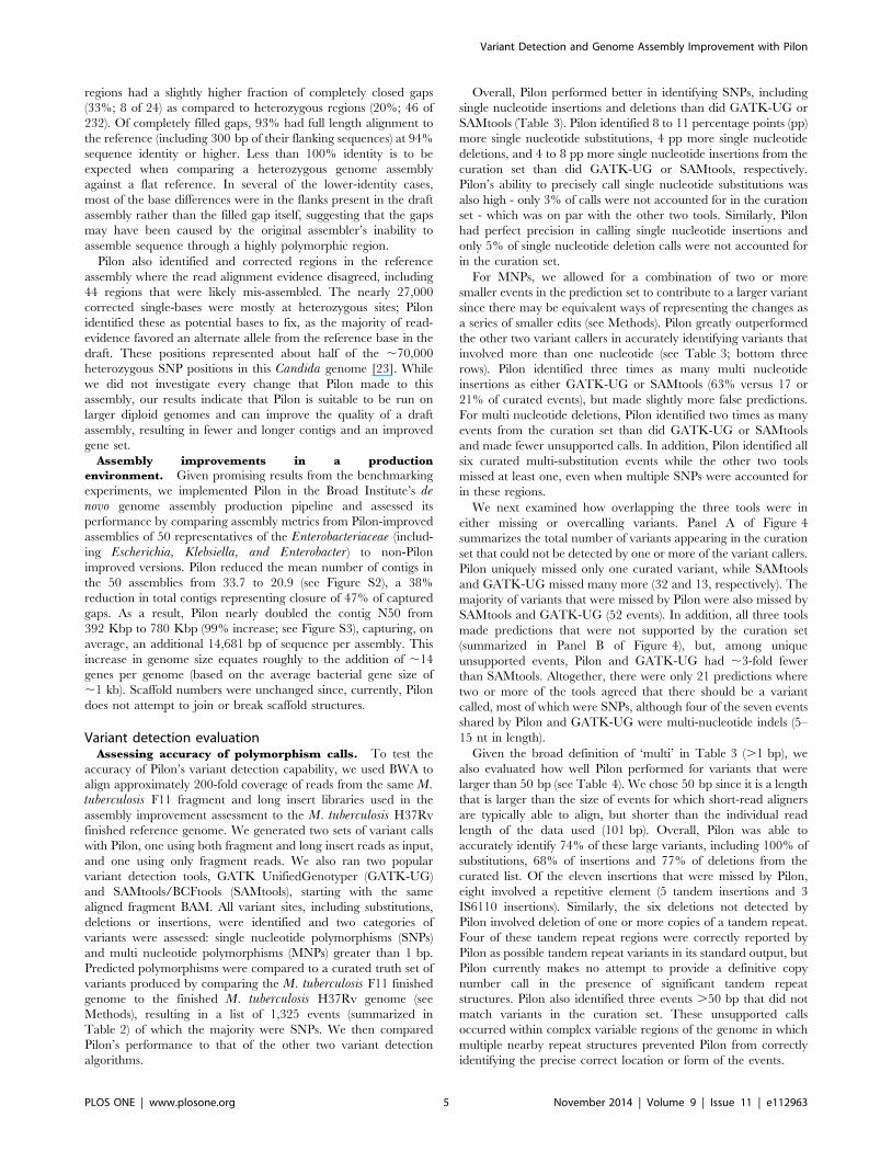
Assembly improvement evaluationAssessing accuracy on bacterial assemblies. To test the
accuracy of Pilon’s improvements on bacterial assemblies, we
sequenced and created draft assemblies for two bacterial strains
with finished references: S. pneumoniae TIGR4 and M. tubercu-losis F11 (see Methods). These strains were chosen because they
represent different GC content (40% and 66% GC content,
respectively) and both possess genomic features that are known to
confound assemblers, leading to mis-assembled and/or incomplete
genome sequences [12–14]. Sequence reads from both libraries
were aligned back to their respective draft assemblies using BWA[15], and Pilon was run with those alignments.
To assess the benefits of running Pilon, we compared the
original draft and Pilon-improved assemblies to each other and to
their respective finished genome sequence. Pilon made significant
improvements to the contiguity of both draft assemblies, increasing
the contig N50 size by 443 Kbp for S. pneumoniae TIGR4 (see
Table 1) and 196 Kbp for M. tuberculosis F11, even though the
F11 draft assembly had been generated with assistance from a
Figure 1. Simplified overview of the Pilon workflow for assembly improvement and variant detection. The left column depicts theconceptual steps of the Pilon process, and the center and right columns describe what Pilon does at each step while in assembly improvement andvariant detection modes, respectively. During the first step (top row), Pilon scans the read alignments for evidence where the sequencing datadisagree with the input genome and makes corrections to small errors and detects small variants. During the second step (second row), Pilon looksfor coverage and alignment discrepancies to identify potential mis-assemblies and larger variants. Finally (bottom row), Pilon uses reads and matepairs which are anchored to the flanks of discrepant regions and gaps in the input genome to reassemble the area, attempting to fill in the truesequence including large insertions. The resulting output is an improved assembly and/or a VCF file of variants.doi:10.1371/journal.pone.0112963.g001
Variant Detection and Genome Assembly Improvement with Pilon
PLOS ONE | www.plosone.org 2 November 2014 | Volume 9 | Issue 11 | e112963

close reference. In addition, Pilon assemblies were more complete,
with the M. tuberculosis F11 and S. pneumoniae TIGR4 Pilon-
improved assemblies containing an additional 11,516 bp and
9,608 bp, respectively.
Observed gains in genome coverage and contig N50 were
principally due to Pilon’s ability to recognize and fill (or partially
fill) by local assembly ‘‘captured gaps’’, i.e., missing sequence
between contigs within a scaffold. When run with default settings,
Pilon does not introduce ambiguous bases or additional Ns during
this process. Across the two draft assemblies, Pilon completely and
accurately filled 17 of the 44 captured gaps (39% closure rate)
including 8 gaps that represented more than 1 Kbp in sequence
length (see Table S1). None of Pilon’s gap closures were incorrect,
though one was judged to be ‘‘no worse’’: the sequence used to
bridge the gap was correct, but an error in the original assembly in
one of the gap flanks was not detected by Pilon. An additional 14
gaps (32% of total captured gaps) were partially filled by Pilon, and
13 (93%) of those extensions were error-free. The one partial fill
judged to be ‘‘Incorrect’’ involved a repetitive structure that Pilon
extended into flanking sequence belonging to a different copy of
the repeat.
We compared Pilon’s ability to close gaps in these assemblies
with two other tools commonly used for this purpose, IMAGE [16]
and GapFiller [17] (see Table S1b). Pilon’s overall gap closure rate
was only somewhat higher than that of the other tools, but its
accuracy was dramatically better. Across the two assemblies,
IMAGE closed 13 captured gaps (30% closure rate) but only two
of those closures were found to be correct by alignment with the
reference (15% precision). Similarly, GapFiller closed 16 of
captured gaps (36% closure rate) in the two assemblies, but only
four of its closures were correct (25% precision). In addition to
filling captured gaps, Pilon also corrected 43 single-bases and 4
small indels across both genomes, and all 47 changes were found
to be correct by alignment against the reference (100% accuracy;
see Table S2). By comparison, iCORN [18] made 47 single-base
changes and 2 single-base deletions, but only 35 of the 49 (71%) of
its changes were correct.
Optionally, Pilon can also make changes to genomic locations at
which it finds significant evidence for more than one alternative,
choosing the allele with the most support even where the evidence
is insufficient to make a confident call. When run with this option
on these assemblies, Pilon made 10 changes to ambiguous bases,
but only 3 were verified to be correct. This option is turned off by
default starting with Pilon version 1.8.
Pilon also detected and attempted to fix local mis-assemblies by
reassembling contig regions that were suspected to be incorrectly
assembled. Three of these regions were correctly fixed (see Table 1
and Table S1) and a fourth we classified as ‘‘No worse’’. For the
latter, Pilon correctly identified a repetitive region within the
original M. tuberculosis F11 draft assembly that contained extra
sequence with respect to the M. tuberculosis F11 reference.
However, Pilon’s change introduced a deletion with respect to the
reference, underscoring the difficulty of accurately assembling
repetitive regions with short read data [13].
For the M. tuberculosis F11 and S. pneumoniae TIGR4 Pilon
improved assemblies, there were 13 and 4 regions, respectively,
where Pilon detected a problem in the draft assembly, but was
unable to provide solutions. In each of these cases, Pilon flagged
the coordinates of the problematic region, and, in 10 of these
cases, it also reported the length of the detected tandem repeat
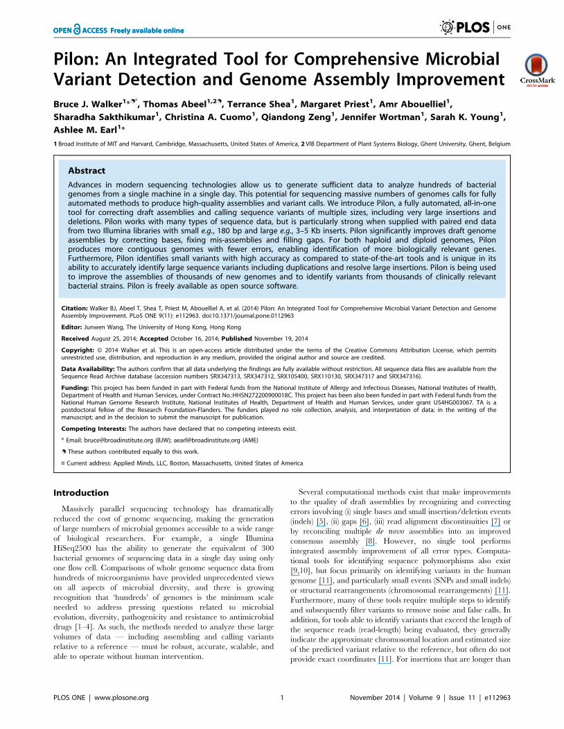
confounding resolution of the region. For example, Figure 2 shows
scaffold00001 coordinates 3159800–3159898 of the M. tubercu-losis F11 draft assembly, along with Pilon-generated genome
browser tracks representing some of the internal metrics it used to
identify this region as problematic. In this case, Pilon noted that it
was unable to resolve a 57 bp tandem repeat, which enabled an
experienced analyst to confirm the presence of a mis-assembly and
accurately narrow the bounds of the unresolvable region. Manual
comparison of the draft assembly with the reference revealed that
there should have been three full and one partial copies of the
57 bp repeat in tandem, whereas the draft assembly only
contained one full and one partial copy of the repeat.
Effect of assembly improvements on gene calls. To assess
the impact of Pilon-improvement on gene calls (i.e., functional
interpretation of the genome), we examined Pilon improvements
with respect to genes by investigating the regions that were
affected by Pilon modifications and the effect of these modifica-
tions on coding sequences. Thirty-two genes and seven intergenic
regions were impacted by Pilon changes to the M. tuberculosis F11
Pilon-improved assembly; of these, nearly all (95%; 37 of 39) were
correct improvements. Nearly half (13) of the genes that were
affected by a fix involved transposases that were completely or
partially filled with sequence that perfectly matched the reference
genome (see Table S3). One additional transposase had a single
base pair corrected with perfect match to the reference. A
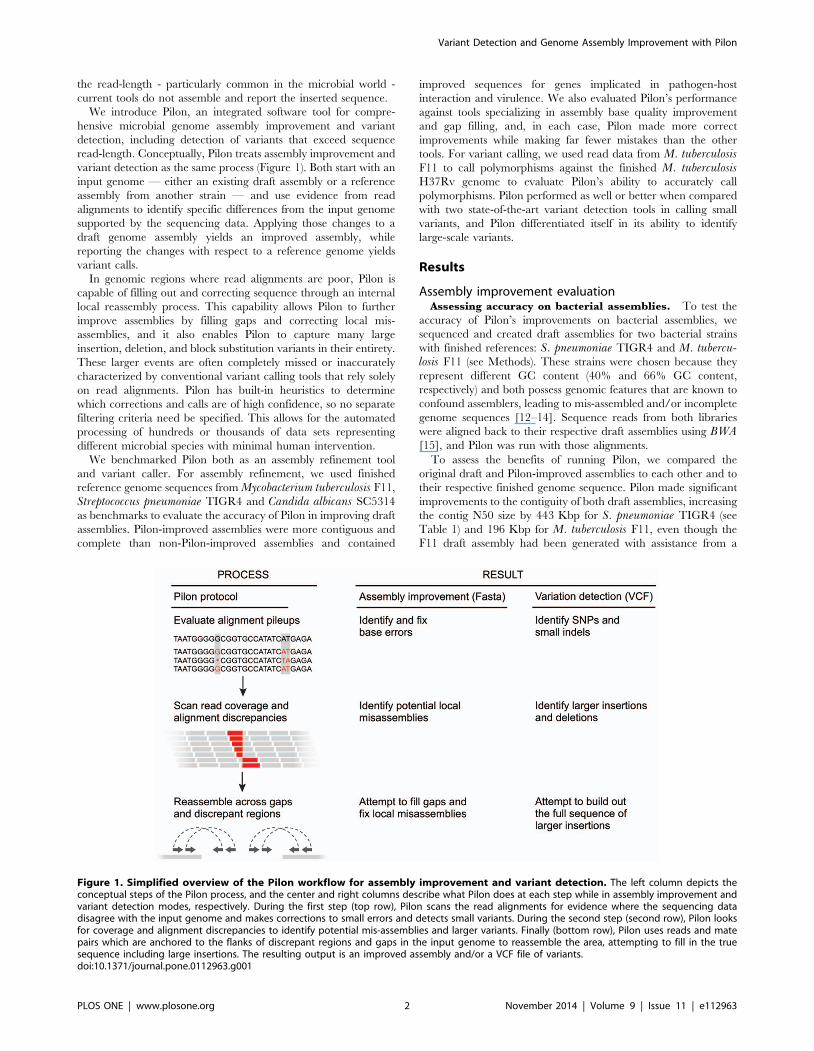
particularly complex 13 Kbp region in M. tuberculosis F11 is
highlighted in Figure 3. This region harbors three sets of
transposases in close proximity that were not captured in the
draft M. tuberculosis F11 assembly, but were accurately filled in by
Pilon. Two of the gaps were completely closed, and the third
transposase set was completely captured along with an additional
gene. However, due to Pilon’s conservative overlap requirement
for closure (95 bp), that gap was not closed despite a 42 bp overlap
in the extended flank sequences.
Of the remaining 19 genes, 6 were PE/PPE family protein
encoding genes. Five corrections were perfect and, in one case
(TBFG_11946), Pilon identified the problematic region, but could
not completely resolve the problem. However, the correction that
Table 1. Summary assembly statistics before and after Pilon improvement.
Genome M. tuberculosis F11 S. pneumoniae TIGR4 C. albicans SC5314
Contig N50 Increase 196 kb 443 kb 56 kb
Bases Added 11,516 9,608 33,804
Gaps Closed 9 9 54
Gaps Shrunk 7 7 102
Single-base Modifications 20 27 26,939
Mis-assembly fixes 3 1 44
In all cases the assemblies were more contiguous, contained more bases, and had fewer gaps and errors after Pilon improvement.doi:10.1371/journal.pone.0112963.t001
Variant Detection and Genome Assembly Improvement with Pilon
PLOS ONE | www.plosone.org 3 November 2014 | Volume 9 | Issue 11 | e112963
Pilon applied did not make the situation worse. Pilon also
identified and accurately corrected a mis-assembly (highlighted in
Figure S1) in which a gene had been truncated due to a collapsed
repeat in the draft assembly.
In S. pneumoniae TIGR4, 20 genes and 12 intergenic regions
were affected by fixes from Pilon. A majority (15 of 20) of the
improved genes were transposases, of which Pilon was able to
completely or partially fill 8 that matched completely and perfectly
with the reference; the remaining 7 were individual base pair
corrections. Pilon was also able to partially fill other genes
encoding repetitive cell wall surface proteins - including choline
binding protein A [19] and pneumococcal surface protein A [20] -
both implicated in adhesion and virulence in S. pneumoniae.
Assessing accuracy on the assembly of the larger,
polymorphic genome of C. albicans. To evaluate Pilon’s
ability to accurately improve assemblies of diploid genomes
containing a high level of heterozygosity, we ran Pilon on an
Illumina ALLPATHS-LG assembly of the SC5314 strain of C.albicans (Methods), for which there is a high quality reference
curated by the Candida Genome Database (www.candidagenome.
org). At 14.3 Mb, the C. albicans genome is 3- to 7-fold larger than
the bacterial genomes evaluated here. It consists of 8 chromosomes
that are present at diploid levels with an average of one SNP found
at every 330–390 bases, although large regions of most chromo-
somes display loss of heterozygosity [21,22].
Pilon was capable of improving the assembly and added .
33 Kb of sequence (see Table 1). While the increase in contig N50
was relatively small (56 Kb), Pilon completely or partially filled
61% (156 of 256) of the total captured gaps including in both
homo- and hetero-zygous regions of the genome. Homozygous
Figure 2. Example Pilon generated genome browser tracks. This region was flagged by Pilon as containing a possible local mis-assembly, butPilon was unable to determine a fix due to a tandem repeat sequence. The tracks shown here include: Pilon Features track indicating the extent of theregion flagged by Pilon as containing a potential mis-assembly, Valid Coverage track indicating the sequence coverage of valid read pair alignmentsexcluding the clipped portions of the alignments, Clipped Alignments track indicating the number of reads soft-clipped at each location, Pct BadAlignments track indicating the percentage of the total reads aligned to each location which are not part of Valid Coverage. These tracks are createdwith the ‘—tracks’ command-line option. Together, these tracks reveal the true bounds of the mis-assembly, and indicate that there are likely missingcopies of the tandem repeat in the draft assembly. In this case, manual analysis revealed the draft assembly was missing two of three full copies of a57-base tandem repeat.doi:10.1371/journal.pone.0112963.g002
Figure 3. Comparative view of a transposase-rich region of the M. tuberculosis F11 genome (coordinates 1,991,000 to 2,006,300)obtained from the draft (A) and Pilon-improved (B) assemblies. In the draft assembly, three regions containing transposases (shown in blue)remained unassembled resulting in gaps. In the Pilon-improved assembly, all three sets of transposases were successfully assembled. The Pilon-improved assembly also contained a hypothetical gene, TBFG_11790 (shown in red), missing from the draft assembly. Though TBFG_11790 was notfully closed in the Pilon-improved version, closer inspection revealed that there was a 42 bp overlap in assembled sequence at this site. By default,Pilon will not close gaps unless there is at least 95 bp overlapping sequence to minimize spurious joins.doi:10.1371/journal.pone.0112963.g003
Variant Detection and Genome Assembly Improvement with Pilon
PLOS ONE | www.plosone.org 4 November 2014 | Volume 9 | Issue 11 | e112963
regions had a slightly higher fraction of completely closed gaps
(33%; 8 of 24) as compared to heterozygous regions (20%; 46 of
232). Of completely filled gaps, 93% had full length alignment to
the reference (including 300 bp of their flanking sequences) at 94%
sequence identity or higher. Less than 100% identity is to be
expected when comparing a heterozygous genome assembly
against a flat reference. In several of the lower-identity cases,
most of the base differences were in the flanks present in the draft
assembly rather than the filled gap itself, suggesting that the gaps
may have been caused by the original assembler’s inability to
assemble sequence through a highly polymorphic region.
Pilon also identified and corrected regions in the reference
assembly where the read alignment evidence disagreed, including
44 regions that were likely mis-assembled. The nearly 27,000
corrected single-bases were mostly at heterozygous sites; Pilon
identified these as potential bases to fix, as the majority of read-
evidence favored an alternate allele from the reference base in the
draft. These positions represented about half of the ,70,000
heterozygous SNP positions in this Candida genome [23]. While
we did not investigate every change that Pilon made to this
assembly, our results indicate that Pilon is suitable to be run on
larger diploid genomes and can improve the quality of a draft
assembly, resulting in fewer and longer contigs and an improved
gene set.
Assembly improvements in a production
environment. Given promising results from the benchmarking
experiments, we implemented Pilon in the Broad Institute’s denovo genome assembly production pipeline and assessed its
performance by comparing assembly metrics from Pilon-improved
assemblies of 50 representatives of the Enterobacteriaceae (includ-
ing Escherichia, Klebsiella, and Enterobacter) to non-Pilon
improved versions. Pilon reduced the mean number of contigs in
the 50 assemblies from 33.7 to 20.9 (see Figure S2), a 38%
reduction in total contigs representing closure of 47% of captured
gaps. As a result, Pilon nearly doubled the contig N50 from
392 Kbp to 780 Kbp (99% increase; see Figure S3), capturing, on
average, an additional 14,681 bp of sequence per assembly. This
increase in genome size equates roughly to the addition of ,14
genes per genome (based on the average bacterial gene size of
,1 kb). Scaffold numbers were unchanged since, currently, Pilon
does not attempt to join or break scaffold structures.
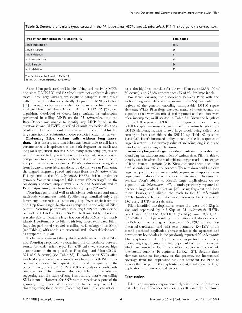
Variant detection evaluationAssessing accuracy of polymorphism calls. To test the
accuracy of Pilon’s variant detection capability, we used BWA to
align approximately 200-fold coverage of reads from the same M.tuberculosis F11 fragment and long insert libraries used in the
assembly improvement assessment to the M. tuberculosis H37Rv
finished reference genome. We generated two sets of variant calls
with Pilon, one using both fragment and long insert reads as input,
and one using only fragment reads. We also ran two popular
variant detection tools, GATK UnifiedGenotyper (GATK-UG)
and SAMtools/BCFtools (SAMtools), starting with the same
aligned fragment BAM. All variant sites, including substitutions,
deletions or insertions, were identified and two categories of
variants were assessed: single nucleotide polymorphisms (SNPs)
and multi nucleotide polymorphisms (MNPs) greater than 1 bp.
Predicted polymorphisms were compared to a curated truth set of
variants produced by comparing the M. tuberculosis F11 finished
genome to the finished M. tuberculosis H37Rv genome (see
Methods), resulting in a list of 1,325 events (summarized in
Table 2) of which the majority were SNPs. We then compared
Pilon’s performance to that of the other two variant detection
algorithms.
Overall, Pilon performed better in identifying SNPs, including
single nucleotide insertions and deletions than did GATK-UG or
SAMtools (Table 3). Pilon identified 8 to 11 percentage points (pp)
more single nucleotide substitutions, 4 pp more single nucleotide
deletions, and 4 to 8 pp more single nucleotide insertions from the
curation set than did GATK-UG or SAMtools, respectively.
Pilon’s ability to precisely call single nucleotide substitutions was
also high - only 3% of calls were not accounted for in the curation
set - which was on par with the other two tools. Similarly, Pilon
had perfect precision in calling single nucleotide insertions and
only 5% of single nucleotide deletion calls were not accounted for
in the curation set.
For MNPs, we allowed for a combination of two or more
smaller events in the prediction set to contribute to a larger variant
since there may be equivalent ways of representing the changes as
a series of smaller edits (see Methods). Pilon greatly outperformed
the other two variant callers in accurately identifying variants that
involved more than one nucleotide (see Table 3; bottom three
rows). Pilon identified three times as many multi nucleotide
insertions as either GATK-UG or SAMtools (63% versus 17 or
21% of curated events), but made slightly more false predictions.
For multi nucleotide deletions, Pilon identified two times as many
events from the curation set than did GATK-UG or SAMtools
and made fewer unsupported calls. In addition, Pilon identified all
six curated multi-substitution events while the other two tools
missed at least one, even when multiple SNPs were accounted for
in these regions.
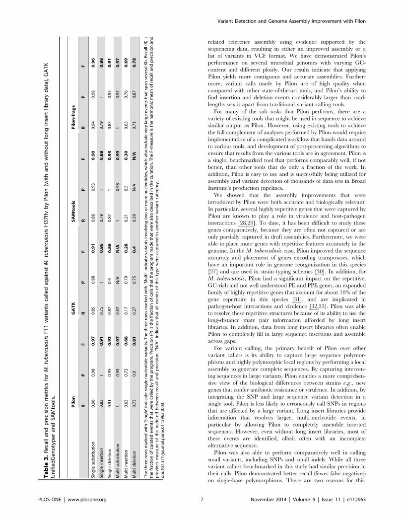
We next examined how overlapping the three tools were in
either missing or overcalling variants. Panel A of Figure 4
summarizes the total number of variants appearing in the curation
set that could not be detected by one or more of the variant callers.
Pilon uniquely missed only one curated variant, while SAMtools
and GATK-UG missed many more (32 and 13, respectively). The
majority of variants that were missed by Pilon were also missed by
SAMtools and GATK-UG (52 events). In addition, all three tools
made predictions that were not supported by the curation set
(summarized in Panel B of Figure 4), but, among unique
unsupported events, Pilon and GATK-UG had ,3-fold fewer
than SAMtools. Altogether, there were only 21 predictions where
two or more of the tools agreed that there should be a variant
called, most of which were SNPs, although four of the seven events
shared by Pilon and GATK-UG were multi-nucleotide indels (5–
15 nt in length).
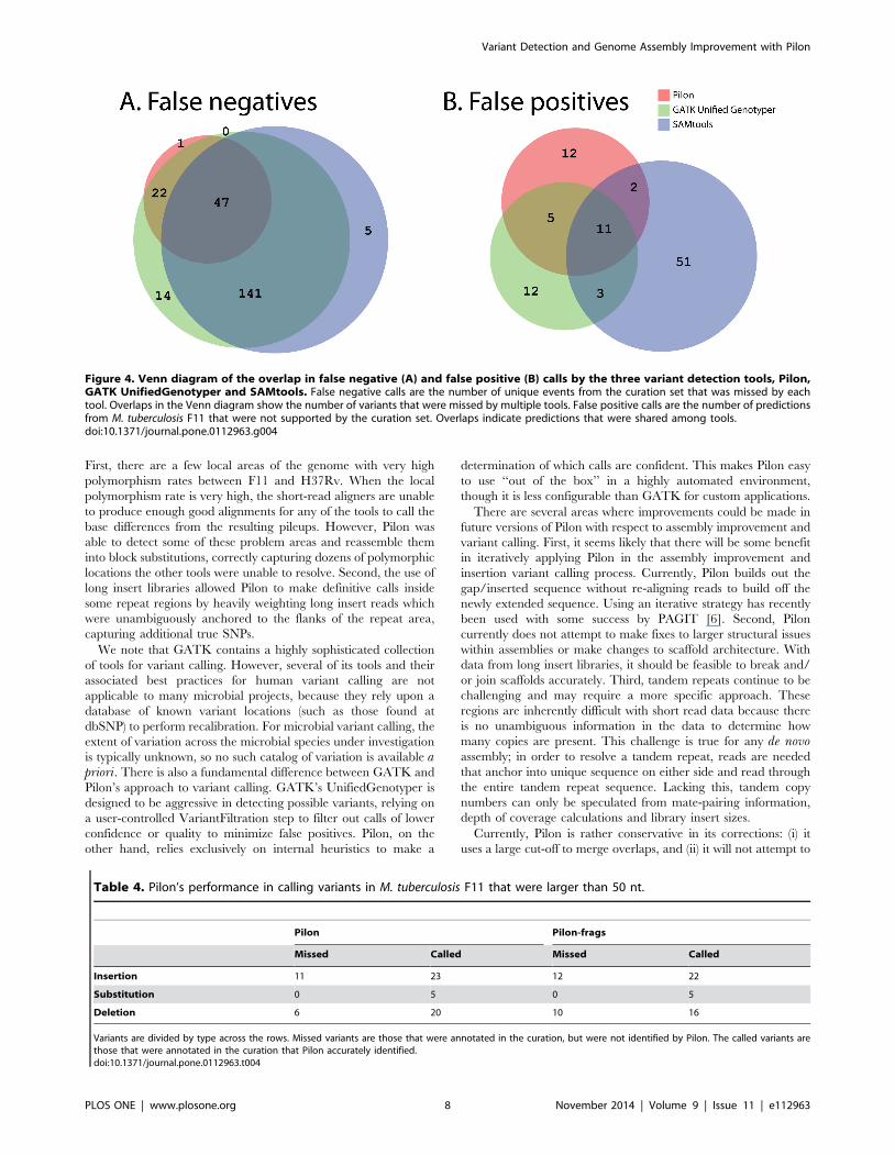
Given the broad definition of ‘multi’ in Table 3 (.1 bp), we
also evaluated how well Pilon performed for variants that were
larger than 50 bp (see Table 4). We chose 50 bp since it is a length
that is larger than the size of events for which short-read aligners
are typically able to align, but shorter than the individual read
length of the data used (101 bp). Overall, Pilon was able to
accurately identify 74% of these large variants, including 100% of
substitutions, 68% of insertions and 77% of deletions from the
curated list. Of the eleven insertions that were missed by Pilon,
eight involved a repetitive element (5 tandem insertions and 3
IS6110 insertions). Similarly, the six deletions not detected by
Pilon involved deletion of one or more copies of a tandem repeat.
Four of these tandem repeat regions were correctly reported by
Pilon as possible tandem repeat variants in its standard output, but
Pilon currently makes no attempt to provide a definitive copy
number call in the presence of significant tandem repeat
structures. Pilon also identified three events .50 bp that did not
match variants in the curation set. These unsupported calls
occurred within complex variable regions of the genome in which
multiple nearby repeat structures prevented Pilon from correctly
identifying the precise correct location or form of the events.
Variant Detection and Genome Assembly Improvement with Pilon
PLOS ONE | www.plosone.org 5 November 2014 | Volume 9 | Issue 11 | e112963
Since Pilon performed well in identifying and resolving MNPs
and since GATK-UG and SAMtools were not explicitly designed
to call these large variants, we sought to compare Pilon’s MNP
calls to that of methods specifically designed for MNP detection
[11]. Though neither was described for use on microbial data, we
evaluated how well BreakDancer [24] and CLEVER [25], two
algorithms developed to detect large variants in eukaryotes,
performed in calling MNPs on the M. tuberculosis test set.
BreakDancer was unable to identify any MNP found in the
curation set and CLEVER identified 21 multi nucleotide deletions,
of which only 1 corresponded to a variant in the curated list. No
large insertions or substitutions were predicted (data not shown).
Evaluating Pilon variant calls without long insert
data. It is unsurprising that Pilon was better able to call larger
variants since it is optimized to use both fragment (or small) and
long (or large) insert libraries. Since many sequencing projects do
not have access to long insert data and to also make a more direct
comparison to existing variant callers that are not optimized to
accept these data, we evaluated Pilon’s performance using data
from fragment insert libraries alone. To do this, we ran Pilon using
the aligned fragment paired end reads from the M. tuberculosisF11 genome to the M. tuberculosis H37Rv finished reference
genome. We then compared this output (‘‘Pilon-frags’’) to the
previously analyzed output from GATK and SAMtools and to
Pilon output using data from both library types (‘‘Pilon’’).
Pilon-frags performed well in identifying both single and multi
nucleotide variants (see Table 3). Pilon-frags identified only 2 pp
fewer single nucleotide substitutions, 4 pp fewer single insertions
and 4 pp fewer single deletions as compared to the original Pilon
output. Pilon-frag performance in calling SNPs was better or on
par with both GATK-UG and SAMtools. Remarkably, Pilon-frags
was also able to identify a large fraction of the MNPs, with nearly
identical performance to Pilon with long insert read data. Pilon-
frags also performed very well in calling variants larger than 50 bp
(see Table 4), with one less insertion call and 4 fewer deletions calls
as compared to Pilon.
To better understand the qualitative differences in what Pilon
and Pilon-frags reported, we examined the concordance between
results for each variant type. For SNP calls, we observed high
concordance in the outputs from Pilon-frags and Pilon (95.2%;
871 of 915 events) (see Table S5). Discordance in SNPs often
involved a position where a variant was found in both Pilon runs,
but was considered high quality in one and low quality in the
other. In fact, only 7 of 915 SNPs (0.8% of total) were confidently
predicted to differ between the two Pilon run conditions,
suggesting that the value of long insert library data when calling
SNPs is small. However, for SNPs within repetitive regions of the
genome, long insert data appeared to be very helpful in
disambiguating these events (Table S6). Small indel variant calls
were also highly concordant for the two Pilon runs (93.3%; 56 of
60 events), and 78.5% concordance (73 of 93) for large indels.
For larger variants, the discordance between Pilon with and
without long insert data was larger (see Table S5), particularly in
regions of the genome encoding transposable IS6110 repeat
elements. While Pilon-frags detected many of these events, the
sequences that were assembled and reported at these sites were
often incomplete, as illustrated in Table S7. Given the length of
the IS6110 repeat (,1.3 Kbp), the fragment pairs — only
,180 bp apart — were unable to span the entire length of the
IS6110 elements, leading to two large indels being called, one
coming in from each side of the IS6110 e.g. Table S7, position
1,541,957. Pilon’s improved ability to capture the full sequence of
larger insertions is the primary value of including long insert read
data for variant calling applications.
Assessing large-scale genome duplications. In addition to
identifying substitutions and indels of various sizes, Pilon is able to
identify areas in which the read evidence suggests additional copies
of large genomic regions (.10 Kbp) compared with the input
draft assembly or reference genome. These regions could indicate
large collapsed repeats in an assembly improvement application or
large genomic duplications in a variant detection application. To
evaluate Pilon’s ability to identify large duplications, we re-
sequenced M. tuberculosis T67, a strain previously reported to
harbor a large-scale duplication [26], using fragment and long
insert libraries, and aligned the reads to the M. tuberculosisH37Rv finished reference. Pilon was then run to detect variants in
T67 using H37Rv as a reference.
Pilon identified two duplication events that were .10 Kbp in
size and separated by ,3 Kbp at M. tuberculosis H37Rv
coordinates 3,494,063–3,551,070 (57 Kbp) and 3,554,192–
3,712,284 (158 Kbp) resulting in a combined duplication of
,215 Kbp. The left gene boundary (Rv3128c) of the first
predicted duplication and right gene boundary (Rv3427c) of the
second predicted duplication corresponded to the upstream and
downstream boundaries in the previously reported M. tuberculosisT67 duplication [26]. Upon closer inspection, the 3 Kbp
intervening region contained two copies of the IS6110 element,
which are routinely found in multiple copies within the M.tuberculosis genome (16 copies in H37Rv) [27]. Because these
elements occur so frequently in the genome, the incremental
coverage from the duplication was not sufficient for Pilon to
identify them as part of the duplication event, breaking a true large
duplication into two reported pieces.
Discussion
Pilon is an assembly improvement algorithm and variant caller
that identifies differences between a draft assembly or closely
Table 2. Summary of variant types curated in the M. tuberculosis H37Rv and M. tuberculosis F11 finished genome comparison.
Type of variation between F11 and H37RV Total found
Single substitution 1012
Single insertion 26
Single deletion 31
Multi substitution 13
Multi insertion 56
Multi deletion 47
The full list can be found in Table S8.doi:10.1371/journal.pone.0112963.t002
Variant Detection and Genome Assembly Improvement with Pilon
PLOS ONE | www.plosone.org 6 November 2014 | Volume 9 | Issue 11 | e112963
related reference assembly using evidence supported by the
sequencing data, resulting in either an improved assembly or a
list of variants in VCF format. We have demonstrated Pilon’s
performance on several microbial genomes with varying GC-
content and different ploidy. Our results indicate that applying
Pilon yields more contiguous and accurate assemblies. Further-
more, variant calls made by Pilon are of high quality when
compared with other state-of-the-art tools, and Pilon’s ability to
find insertion and deletion events considerably larger than read-
lengths sets it apart from traditional variant calling tools.
For many of the sub tasks that Pilon performs, there are a
variety of existing tools that might be used in sequence to achieve
similar output as Pilon. However, using existing tools to achieve
the full complement of analyses performed by Pilon would require
implementation of a complicated workflow that hands data around
to various tools, and development of post-processing algorithms to
ensure that results from the various tools are in agreement. Pilon is
a single, benchmarked tool that performs comparably well, if not
better, than other tools that do only a fraction of the work. In
addition, Pilon is easy to use and is successfully being utilized for
assembly and variant detection of thousands of data sets in Broad
Institute’s production pipelines.
We showed that the assembly improvements that were
introduced by Pilon were both accurate and biologically relevant.
In particular, several highly repetitive genes that were captured by
Pilon are known to play a role in virulence and host-pathogen
interactions [28,29]. To date, it has been difficult to study these
genes comparatively, because they are often not captured or are
only partially captured in draft assemblies. Furthermore, we were
able to place more genes with repetitive features accurately in the
genome. In the M. tuberculosis case, Pilon improved the sequence
accuracy and placement of genes encoding transposases, which
have an important role in genome reorganization in this species
[27] and are used in strain typing schemes [30]. In addition, for
M. tuberculosis, Pilon had a significant impact on the repetitive,
GC-rich and not well understood PE and PPE genes, an expanded
family of highly repetitive genes that account for about 10% of the
gene repertoire in this species [31], and are implicated in
pathogen-host interactions and virulence [32,33]. Pilon was able
to resolve these repetitive structures because of its ability to use the
long-distance mate pair information afforded by long insert
libraries. In addition, data from long insert libraries often enable
Pilon to completely fill in large sequence insertions and assemble
across gaps.
For variant calling, the primary benefit of Pilon over other
variant callers is its ability to capture large sequence polymor-
phisms and highly polymorphic local regions by performing a local
assembly to generate complete sequences. By capturing interven-
ing sequences in large variants, Pilon enables a more comprehen-
sive view of the biological differences between strains e.g., new
genes that confer antibiotic resistance or virulence. In addition, by
integrating the SNP and large sequence variant detection in a
single tool, Pilon is less likely to erroneously call SNPs in regions
that are affected by a large variant. Long insert libraries provide
information that resolves larger, multi-nucleotide events, in
particular by allowing Pilon to completely assemble inserted
sequences. However, even without long insert libraries, most of
these events are identified, albeit often with an incomplete
alternative sequence.
Pilon was also able to perform comparatively well in calling
small variants, including SNPs and small indels. While all three
variant callers benchmarked in this study had similar precision in
their calls, Pilon demonstrated better recall (fewer false negatives)
on single-base polymorphisms. There are two reasons for this.
Ta
ble
3.
Re
call
and
pre
cisi
on
me
tric
sfo
rM
.tu
ber
culo
sis
F11
vari
ants
calle
dag
ain
stM
.tu
ber
culo
sis
H3
7R
vb
yP
ilon
(wit
han
dw
ith
ou
tlo
ng
inse
rtlib
rary
dat
a),
GA
TK
Un
ifie
dG
en
oty
pe
ran
dSA
Mto
ols
.
Pil
on
GA
TK
SA
Mto
ols
Pil
on
-fra
gs
RP
FR
PF
RP
FR
PF
Sin
gle
sub
stit
uti
on
0.9
60
.98
0.9
70
.85
0.9
80
.91
0.8
80
.93
0.9
00
.94
0.9
80
.96
Sin
gle
inse
rtio
n0
.83
10
.91
0.7
51
0.8
60
.79
10
.88
0.7
91
0.8
8
Sin
gle
de
leti
on
0.9
10
.95
0.9
30
.87
0.9
0.8
60
.87
10
.93
0.8
70
.95
0.9
1
Mu
lti
sub
stit
uti
on
10
.95
0.9
70
.67
N/A
N/A
10
.98
0.9
91
0.9
50
.97
Mu
lti
inse
rtio
n0
.63
0.7
30
.68
0.1
70
.79
0.2
80
.21
0.5
0.3
00
.63
0.7
60
.69
Mu
lti
de
leti
on
0.7
30
.90
.81
0.2
70
.75
0.4
0.3
9N
/AN
/A0
.71
0.8
70
.78
Th
eth
ree
row
sm
arke
dw
ith
’Sin
gle
’in
dic
ate
sin
gle
nu
cle
oti
de
vari
ants
.Th
eth
ree
row
sm
arke
dw
ith
’Mu
lti’
ind
icat
eva
rian
tsin
volv
ing
two
or
mo
ren
ucl
eo
tid
es,
wh
ich
also
incl
ud
eve
ryla
rge
eve
nts
that
span
seve
ralK
b.R
eca
ll(R
)is
the
frac
tio
no
fcu
rate
de
ven
tsth
atw
ere
calle
db
yth
ep
rog
ram
.Pre
cisi
on
(P)
isth
efr
acti
on
of
calls
that
the
pro
gra
mm
ade
that
we
real
sod
esc
rib
ed
inth
ecu
rati
on
.Th
eF-
me
asu
reis
the
har
mo
nic
me
ano
fre
call
and
pre
cisi
on
and
pro
vid
es
me
asu
reo
fth
etr
ade
-off
be
twe
en
reca
llan
dp
reci
sio
n.
‘‘N/A
’’in
dic
ate
sth
atal
le
ven
tso
fth
isty
pe
we
reca
ptu
red
inan
oth
er
vari
ant
cate
go
ry.
do
i:10
.13
71
/jo
urn
al.p
on
e.0
11
29
63
.t0
03
Variant Detection and Genome Assembly Improvement with Pilon
PLOS ONE | www.plosone.org 7 November 2014 | Volume 9 | Issue 11 | e112963
First, there are a few local areas of the genome with very high
polymorphism rates between F11 and H37Rv. When the local
polymorphism rate is very high, the short-read aligners are unable
to produce enough good alignments for any of the tools to call the
base differences from the resulting pileups. However, Pilon was
able to detect some of these problem areas and reassemble them
into block substitutions, correctly capturing dozens of polymorphic
locations the other tools were unable to resolve. Second, the use of
long insert libraries allowed Pilon to make definitive calls inside
some repeat regions by heavily weighting long insert reads which
were unambiguously anchored to the flanks of the repeat area,
capturing additional true SNPs.
We note that GATK contains a highly sophisticated collection
of tools for variant calling. However, several of its tools and their
associated best practices for human variant calling are not
applicable to many microbial projects, because they rely upon a
database of known variant locations (such as those found at
dbSNP) to perform recalibration. For microbial variant calling, the
extent of variation across the microbial species under investigation
is typically unknown, so no such catalog of variation is available apriori. There is also a fundamental difference between GATK and
Pilon’s approach to variant calling. GATK’s UnifiedGenotyper is
designed to be aggressive in detecting possible variants, relying on
a user-controlled VariantFiltration step to filter out calls of lower
confidence or quality to minimize false positives. Pilon, on the
other hand, relies exclusively on internal heuristics to make a
determination of which calls are confident. This makes Pilon easy
to use ‘‘out of the box’’ in a highly automated environment,
though it is less configurable than GATK for custom applications.
There are several areas where improvements could be made in
future versions of Pilon with respect to assembly improvement and
variant calling. First, it seems likely that there will be some benefit
in iteratively applying Pilon in the assembly improvement and
insertion variant calling process. Currently, Pilon builds out the
gap/inserted sequence without re-aligning reads to build off the
newly extended sequence. Using an iterative strategy has recently
been used with some success by PAGIT [6]. Second, Pilon
currently does not attempt to make fixes to larger structural issues
within assemblies or make changes to scaffold architecture. With
data from long insert libraries, it should be feasible to break and/
or join scaffolds accurately. Third, tandem repeats continue to be
challenging and may require a more specific approach. These
regions are inherently difficult with short read data because there
is no unambiguous information in the data to determine how
many copies are present. This challenge is true for any de novoassembly; in order to resolve a tandem repeat, reads are needed
that anchor into unique sequence on either side and read through
the entire tandem repeat sequence. Lacking this, tandem copy
numbers can only be speculated from mate-pairing information,
depth of coverage calculations and library insert sizes.
Currently, Pilon is rather conservative in its corrections: (i) it
uses a large cut-off to merge overlaps, and (ii) it will not attempt to
Figure 4. Venn diagram of the overlap in false negative (A) and false positive (B) calls by the three variant detection tools, Pilon,GATK UnifiedGenotyper and SAMtools. False negative calls are the number of unique events from the curation set that was missed by eachtool. Overlaps in the Venn diagram show the number of variants that were missed by multiple tools. False positive calls are the number of predictionsfrom M. tuberculosis F11 that were not supported by the curation set. Overlaps indicate predictions that were shared among tools.doi:10.1371/journal.pone.0112963.g004
Table 4. Pilon’s performance in calling variants in M. tuberculosis F11 that were larger than 50 nt.
Pilon Pilon-frags
Missed Called Missed Called
Insertion 11 23 12 22
Substitution 0 5 0 5
Deletion 6 20 10 16
Variants are divided by type across the rows. Missed variants are those that were annotated in the curation, but were not identified by Pilon. The called variants arethose that were annotated in the curation that Pilon accurately identified.doi:10.1371/journal.pone.0112963.t004
Variant Detection and Genome Assembly Improvement with Pilon
PLOS ONE | www.plosone.org 8 November 2014 | Volume 9 | Issue 11 | e112963
resolve significant tandem repeats structures definitively. Notwith-
standing the challenges encountered with tandem repeats, Pilon
does an excellent job with other repetitive sequences and is able to
fix many genes of known repetitive gene families and is able to fill
in many transposable elements.
While we have evaluated Pilon’s assembly improvements on
both haploid and diploid genomes and obtained positive results for
both, we acknowledge that there is still significant opportunity for
future improvement in Pilon’s handling of diploid genomes. Pilon
could be enhanced to understand IUPAC ambiguity codes in its
input genome and generate them in its output, and Pilon’s
heuristics for identifying insertions and deletions in diploid
genomes could be improved, including its ability to recognize
and report heterozygous indels. Finally, the local reassembly
process could be improved to perform better in heterozygous
regions. Even so, our results indicate that in its current form, Pilon
is able to make valuable improvements to diploid genomes.
We have evaluated Pilon’s performance using microbial
genomes with finished references. However, there is no inherent
limitation on the size of genomes to which Pilon can be applied.
For example, we have used Pilon to improve assemblies of larger
genomes, including 16 strains of the Anopheles genus (,200 Mbp
diploid genome), but we were unable to verify the accuracy of
Pilon’s improvements since these genomes have not been finished.
Pilon runs within minutes on small microbial genomes and will
complete overnight on larger eukaryote genomes, such as
Anopheles, which is similar to the tools included in our
benchmarking.
Conclusion
Ultimately, Pilon has great utility and addresses an urgent need
for better and more efficient methods to deal with the thousands of
microbial genomes that are being produced. We have shown that
Pilon performs well as compared to the state-of-the-art for both
assembly improvement and variant detection, often outperforming
these tools. Pilon is also unique in its user-friendly integrated
approach to assembly improvement and is unique in its ability to
identify large variants accurately in microbial genomes. As a
recent addition to the production process for microbial genomes at
Broad Institute, Pilon has been used to automatically improve the
quality of over 8,000 prokaryote and eukaryote genomes prior to
their submission to Genbank, and it has been used to call variants
on over 6,000 genomes.
Material and Methods
Detailed algorithm descriptionInput requirements. Pilon requires an input genome in
FASTA format and one or more BAM files containing sequencing
reads aligned to the input genome. The BAM files must be sorted
in coordinate order and indexed. For Illumina data, these BAM
files are usually produced by an aligner such as BWA [15] or
Bowtie 2 [34]. It is recommended that single best hit or random
selection among equal best alignments is used as input into Pilon.
Pilon can use three types of BAM files:
1. Fragments: paired read data of short insert size, typically ,
1 Kbp. Reads should be in forward-reverse (FR) orientation;
2. Long inserts: paired read data of longer insert size, typically .
1 Kbp. Reads should be in forward-reverse (FR) orientation.
Sequencing of long insert libraries that are generated using the
standard Illumina mate-pair library preparation protocol
typically result in reverse-forward (RF) read orientation, so
they will need to be reversed in the BAM file.
3. Unpaired: unpaired sequencing read data.
To use Pilon with default arguments, read length should be 75
bases or longer and total sequence coverage should be 50x or
greater, though deeper total coverage of .100x is beneficial. Pilon
can also make use of longer reads, such as those from Sanger
capillary sequencing and circular-consensus or error-corrected
reads from Pacific Biosciences (PacBio) sequencing. However,
Pilon is not currently tuned to the error model of raw PacBio
reads, and their use may introduce false corrections.
Pilon makes extensive use of pairing information when it is
available, so paired libraries are highly recommended. Pilon is
capable of using paired libraries of any insert size, as it scans the
BAMs to compute statistics, including insert size distribution.
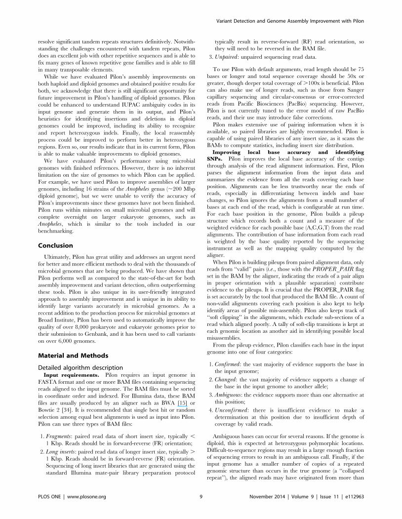
Improving local base accuracy and identifying
SNPs. Pilon improves the local base accuracy of the contigs
through analysis of the read alignment information. First, Pilon
parses the alignment information from the input data and
summarizes the evidence from all the reads covering each base
position. Alignments can be less trustworthy near the ends of
reads, especially in differentiating between indels and base
changes, so Pilon ignores the alignments from a small number of
bases at each end of the read, which is configurable at run time.
For each base position in the genome, Pilon builds a pileup
structure which records both a count and a measure of the
weighted evidence for each possible base (A,C,G,T) from the read
alignments. The contribution of base information from each read
is weighted by the base quality reported by the sequencing
instrument as well as the mapping quality computed by the
aligner.
When Pilon is building pileups from paired alignment data, only
reads from ‘‘valid’’ pairs (i.e., those with the PROPER_PAIR flag
set in the BAM by the aligner, indicating the reads of a pair align
in proper orientation with a plausible separation) contribute
evidence to the pileups. It is crucial that the PROPER_PAIR flag
is set accurately by the tool that produced the BAM file. A count of
non-valid alignments covering each position is also kept to help
identify areas of possible mis-assembly. Pilon also keeps track of
‘‘soft clipping’’ in the alignments, which exclude sub-sections of a
read which aligned poorly. A tally of soft-clip transitions is kept at
each genomic location as another aid in identifying possible local
misassemblies.
From the pileup evidence, Pilon classifies each base in the input
genome into one of four categories:
1. Confirmed: the vast majority of evidence supports the base in
the input genome;
2. Changed: the vast majority of evidence supports a change of
the base in the input genome to another allele;
3. Ambiguous: the evidence supports more than one alternative at
this position;
4. Unconfirmed: there is insufficient evidence to make a
determination at this position due to insufficient depth of
coverage by valid reads.
Ambiguous bases can occur for several reasons. If the genome is
diploid, this is expected at heterozygous polymorphic locations.
Difficult-to-sequence regions may result in a large enough fraction
of sequencing errors to result in an ambiguous call. Finally, if the
input genome has a smaller number of copies of a repeated
genomic structure than occurs in the true genome (a ‘‘collapsed
repeat’’), the aligned reads may have originated from more than
Variant Detection and Genome Assembly Improvement with Pilon
PLOS ONE | www.plosone.org 9 November 2014 | Volume 9 | Issue 11 | e112963
one instance of the repeat structure; where there are differences in
the true instances of the repeat, the alignments can show mixed
evidence.
Paired read information, especially information from long insert
libraries which span a longer distance, is extremely valuable in
helping resolve ambiguous locations due to collapsed repeats. Pairs
for which one read lands inside a repeat element, but the other
lands in unique anchoring sequence on the flanks of the repeat
help to resolve the true base content of the repeat structure. Data
from long inserts will typically have a higher alignment mapping
quality than short-range fragment pairs that lie completely within
the repeat because the fragment pairs may not be able to be placed
uniquely among the repeats. Since Pilon uses mapping quality to
weigh the evidence from each read, the long inserts can often pick
the correct haplotype variations of the repeat structure.
Pilon includes corrections to single-base errors in its output
genome, and optionally, it can also change ambiguous bases to the
allele with the preponderance of evidence.
Finding and fixing small indels. While recording the base-
by-base pileup evidence, Pilon also records the location and
content of indels present in the alignments. Indel alignments which
represent equivalent edits to the input genome may appear at
different coordinates in the alignments. For instance, if the input
genome has the sequence ACCCCT, but the read evidence
suggests one of the Cs should be deleted (ACCCT), each individual
read alignment might show a deletion at any of the four Ccoordinates. Pilon shifts alignment indels to their leftmost
equivalent edit in the input genome, so that the evidence from
all the equivalent edits is combined into evidence for a single event
at a one location.
Pilon makes an insertion or deletion call if a majority of the valid
reads support the change, though that threshold is lowered
somewhat for longer events, as it is typically more difficult for
aligners to identify longer indels in short read data. Called indels
from the input genome are fixed in Pilon’s output genome.
Fixing mis-assemblies, detecting large indels, and filling
gaps. Pilon is capable of reassembling local regions of the
genome when there is sufficient evidence from the alignments that
the contiguity of the input genome does not match the sequencing
data. For assembly improvement applications, this could be an
indication of a local mis-assembly. For variant calling applications,
this could be caused by insertions or deletions too large to be
reflected in the short read alignments.
Pilon tries to identify areas of potential local read alignment
discontiguity in the contigs of the input genome by employing four
heuristics: (i) a large percentage of reads containing a soft-clipped
alignment at a given base position, (ii) a large ratio of invalid pairs
to valid pairs spanning a location, (iii) areas of extremely low
coverage and (iv) rapid drops in alignment coverage over a
distance on the order of a read length. Once Pilon has identified
an area for local reassembly, it treats the suspicious region (which
may be a single base or a larger region) as untrusted, using
alignments to the trusted flanks on both sides to identify a
collection of reads that might contribute evidence for the true
sequence in the suspicious region.
Unpaired reads with partial alignments to the flanks are
included in the collection. For paired data, Pilon identifies pairs
in which one of the reads is anchored by proper alignment to one
of the flanks (e.g., with forward orientation on the left flank, or
reverse orientation on the right flank), but whose mate is either
unmapped or improperly mapped (e.g., to a remote location in the
genome). For fragment pairs, both reads of such pairs are included
in the collection; for long inserts, only the unanchored read is
included in the collection.
From the collected reads, Pilon builds a De Bruijn assembly
graph (default K = 47). For each k-mer in the reads, it uses the
same pileup structure to record the bases which follow that k-mer,
including weighting by base quality. Then, the pileups are
evaluated to determine the link(s) to the next k-mer(s); this results
in either a single base call, resulting in one forward link to the next
k-mer, or an ambiguous call, resulting in two links forward and a
branch in the assembly graph. This process automatically prunes
the assembly graph of most sequencing errors, as infrequent base
differences are unlikely to present enough evidence to affect the
forward links. A minimum coverage cutoff of five for each forward
link also prunes the assembly graph of many false links that could
appear because of sequencing errors.
Pilon then tries k-mers from the trusted flanks as starting points
to walk into the untrusted region from each side, building all
possible extensions with up to five branching points (25 possible
extensions). Tandem repeats with combined length .K cause
loops in the local assembly graph, and they are detected by noting
when the assembly walk reaches an already-incorporated k-mer.
Pilon currently does not attempt to determine the copy number of
such tandem repeats; instead, it will report the length of the repeat
structure encountered in its standard output, and it will not
attempt to close the two sides.
When no tandem repeat is detected, the resulting extensions
from each side are combinatorially matched for possible perfect
overlaps of sufficient length (2K+1) to be considered for closure. If
there is exactly one such closure and it differs from the input
genome, the assembled flank-to-flank sequence will replace the
corresponding sequence in the input genome. Since the default k-
mer size is 47, an overlap of 95 bases is required for closure.
If there are no closures or more than one possible closure, Pilon
will identify a consensus extension from each flank. If an optional
argument is set to allow opening of new gaps, Pilon will replace the
suspicious region with the consensus extensions from each flank
and create a gap between them; otherwise, it simply reports that it
was unable to find a solution. These reports identify areas that an
assembly analyst might wish to investigate manually.
Pilon also attempts to fill gaps between contigs in a scaffold
(‘‘captured gaps’’) in the input genome. In order to fill captured
gaps, Pilon employs the same local reassembly technique described
above, treating the gap itself as the ‘‘untrusted’’ region. If there is a
unique closure, the gap is filled; otherwise, consensus extensions
from each flank are used to reduce the size of the gap. Pilon does
not currently attempt to join or break scaffolds.
Large collapsed repeat (segmental duplication)
detection. Pilon includes heuristics that attempt to flag areas
indicative of large (.10 Kbp) collapsed repeats with respect to the
input genome. These are characteristically large contiguous areas
that appear to have double (or higher) read coverage compared to
the rest of the genomic element being analyzed. Long insert data
are excluded from this computation, as we have found long insert
coverage to be far more variable across some genomes. Pilon does
not attempt to fix these potentially collapsed regions, but it does
report them in its standard output for further investigation.
In variant calling applications, large segmental duplications in
the sequenced strain with respect to the reference have the same
signature as large collapsed repeats in a draft assembly; a
duplicated region of the genome will result in double the number
of reads covering that sequence. Pilon’s reporting of large
collapsed repeat regions can be used to identify candidate
segmental duplications.
Output files. Pilon generates a modified genome as a FASTA
file, including all single-base, small indel, gap filling, mis-assembly
and large-event corrections from the input genome. In the
Variant Detection and Genome Assembly Improvement with Pilon
PLOS ONE | www.plosone.org 10 November 2014 | Volume 9 | Issue 11 | e112963
assembly improvement case, this is the improved assembly
consensus. In variant detection mode, this is the reference
sequence which has been edited to represent the consensus of
the given sample more closely.
Pilon can optionally generate a Variant Call Format (VCF)
[http://vcftools.sourceforge.net/specs.html] file, which lists copi-
ous detailed information about the base and indel evidence at
every base position in the genome, including two scores regarding
variant quality: the QUAL column, and a depth-normalized call
quality (QD) field in the INFO column. For additional details on
the VCF format, we refer to the VCF specification referred above.
Changes generated by local reassembly, often triggered by larger
polymorphisms in variant calling applications, are included as
structural variant records (SVTYPE = INS and SVTYPE =
DEL). Pilon can also, optionally, generate a ‘‘changes’’ file which
lists the edits applied from input to output genome in tabular form,
including source and destination coordinates and source and
destination sequence. Finally, Pilon will optionally (with the —
tracks option) output a series of visualization tracks (‘‘bed’’ and
‘‘wig’’ files) suitable for viewing in genome browsers such as IGV
[35] and GenomeView [36]. Tracks include basic metrics across
the genome, such as sequence coverage and physical coverage, as
well as some of the calculated metrics Pilon uses in its heuristics for
finding potential areas of mis-assembly, such as percentage of valid
read pairs covering every location.
Pilon’s standard output also contains useful information,
including coverage levels, percentage of the input genome
confirmed, a summary of the changes made, as well as some
specifically flagged issues which were not corrected, such as
potentially large collapsed repeat regions, potential regions of mis-
assembly which were not able to be corrected, and detected
tandem repeats that were not resolved.
Data generationAll sequencing data used for these experiments were generated
from an Illumina HiSeq 2000 machine. For sequencing M.tuberculosis F11 and T67, two libraries were generated: one PCR-
free 180 bp insert paired fragment library [37] and large insert 3–
5 Kbp long insert library [38]. S. pneumoniae TIGR4 data also
consisted of two libraries: one robotically size selected 180 bp
insert paired fragment library [37] and a large insert 3–5 Kbp long
insert library [38]. The sequencing data for C. albicans SC5314
was generated from three libraries: a robotically size-selected
180 bp insert paired fragment library [37], a gel-cut 4 Kbp long
insert library [39], and a 40 Kb Fosill library [40]. Sequencing
data were submitted to the Sequence Read Archive with
identifiers: SRX347313, SRX347312, SRX105400,
SRX110130, SRX347317 and SRX347316.
Evaluation methodsAssembly improvement. All draft assemblies were generat-
ed using ALLPATHS-LG [41]. The draft assembly for Mycobac-terium tuberculosis F11 utilized 100x of the 180 bp insert fragment
library and 50x of the 3–5 Kb long insert library and was executed
using ALLPATHS-LG v45395 utilizing the ASSISTED_PATCH-
ING = 2.1 parameter and the M. tuberculosis H37RV reference
genome for assisting (GenBank accession: CP003248). The draft
assembly for S. pneumoniae TIGR4 was created using ALL-
PATHS-LG v45925 with default parameters and using 100x of the
180 bp insert fragment library and 50x of the 3–5 Kb long insert
library. The C. albicans SC5314 utilized 100x of the 180 bp insert
fragment library, 100x of the gel-cut 4 Kb long insert library and
50x of the Fosill library, and was assembled with ALLPATHS-LG
v39846 utilizing the ASSISTED_PATCHING and HAPLOI-
DIFY options with the C. albicans SC5314 reference sequence as a
reference for assisting.
We benchmarked Pilon’s ability to close gaps in the draft
bacterial assemblies against two tools built for this purpose,
IMAGE v2.4.1 [16] and GapFiller v1.10 [17]. The same sets of
sequencing reads used as input to Pilon were used for IMAGE
(fragment library only) and GapFiller (fragment and long insert
libraries). IMAGE was run in the manner implemented in the
PAGIT [6] example scripts: 6 iterations, one with a kmer size of
61, three with a kmer size of 49, and two with a kmer size of 41.
GapFiller was run for 10 iterations with a libraries.txt file
specifying a ratio r = 0.5 and library insert sizes computed by
Pilon from the aligned bams.
To evaluate the quality of Pilon’s single base and small indel
corrections to the draft assemblies, we also ran iCORN v0.97 [18],
the consensus sequence improvement tool in PAGIT, on the same
draft assemblies using the same sets of fragment reads. iCORN
was run in the manner implemented in the PAGIT example
scripts, only changing the library insert size mean and range
parameters. For TIGR4, we used a mean of 180 and a range of
120–300. For F11, we used a mean of 226 and a range of 100–
500, since the PCR-free library preparation resulted in a wider
range of insert sizes.
Fixes to the assemblies (Table S1) made by Pilon and the other
assembly improvement tools were assessed by extracting the
changed region of sequence in the output genome along with
300 bp flanks on each side. These extracted sequences were
aligned to their respective finished reference genomes with
BLASTN [42], and the accuracy of the changes was assessed by
manually inspecting the alignments for accuracy, judging each fix
as ‘‘Correct’’ or ‘‘Incorrect’’. For larger block changes which
resulted from local reassembly (gap filling and fixing of local mis-
assemblies), a third category of ‘‘No worse’’ was established for
situations in which: (i) the draft assembly contained a mis-assembly
in the changed region, (ii) Pilon made a change attempting to fix
the mis-assembly, and (iii) the fix was not entirely correct, but was
no worse than the original problem.
For the assembly improvement statistics, Bases added was
calculated by tallying bases added in locations where resulting fixes
resulted in a net gain of bases during gap filling and mis-assembly
correction processes, as reported in the Pilon standard output
indicated by the "fix gap" or "fix break" lines.
Variant calling. Variant calls were made using M. tubercu-losis H37Rv (GenBank accession: CP003248) as the reference and
the M. tuberculosis F11 aligned read and long insert fragments as
input data. From the sequenced fragment and long insert libraries,
a random subset of read pairs was selected from each library to
obtain an estimated 200x coverage of the M. tuberculosis H37Rv
reference genome. Each library’s reads were aligned to the M.tuberculosis H37Rv reference genome using BWA (version 0.5.9-
r16) to generate BAM files suitable for input to the variant calling
processes.
Pilon: Pilon was run with the —variant command line option,
specifying the M. tuberculosis H37Rv reference genome and the
above BAM file(s) as inputs. We evaluated two Pilon variant
calling sets, one generated using both fragment and long insert
library BAMs, and one using only the fragment library BAM.
GATK UnifiedGenotyper: Reads in the fragment library BAM
were realigned by applying the Genome Analysis Toolkit (GATK
version v3.2.2) RealignerTargetCreator and IndelRealigner tools
on the fragment library aligned BAM file. Variants were then
called from the realigned BAM file using UnifiedGenotyper run
with the following settings: -nt 32 -A AlleleBalance -ploidy 1
-pnrm EXACT_GENERAL_PLOIDY -glm BOTH —output_mode
Variant Detection and Genome Assembly Improvement with Pilon
PLOS ONE | www.plosone.org 11 November 2014 | Volume 9 | Issue 11 | e112963
EMIT_ALL_SITES. Low confidence variants were then filtered
using VariantFiltration (VF) run with the following settings: —
filterExpression "((DP-MQ0),10) || ((MQ0/(1.0*DP)).
=0.8) || (ABHom ,0.8) || (Dels .0.5)" —filterName
LowConfidence.
These VariantFiltration settings filtered out variant calls at
locations with less than 10 unambiguous read alignments, where
80% or more of the read depth had ambiguous mappings, where
fewer than 80% of the reads supported the alternate allele, or
more than half of the reads contained spanning deletions. This
filter expression was based on one previously used to call variants
from the European Escherichia coli O104:H4 outbreak [38],
adjusting depth and allele balance thresholds to yield the best
performance tradeoff between false negative and false positive
results on these data.
SAMtools/BCFtools: The same aligned fragment library BAM
file described above was used as input for variant calling using
SAMtool/BCFtools v0.1.19 according to recommendations found
on the SAMtools webpage (http://samtools.sourceforge.net/
mpileup.shtml). samtools mpileup was used to generate pileups
in bcf format, and variants were called using bcftools using the
-bcg option. Finally, variants were filtered using vcfutils.pl
varFilter -d 10 to filter out calls at locations where the aligned
coverage was less than 10 reads. We chose the minimum depth of
10 to be consistent with the filtering used for GATK Unified-
Genotyper.
CLEVER and BreakDancer: The aligned fragment and
combined fragment and long insert library described above were
used as input for CLEVER v2.0rc3 and BreakDancer 1.3.6.
clever —sorted —use_xa was used to generate calls for
CLEVER. bam2cfg.pl -g –h was used to generate the Break-
Dancer config file, which was then used with breakdancer-max.
Curating differences between F11 and
H37Rv. Differences between the finished M. tuberculosis F11
(GenBank accession: CP000717) and M. tuberculosis H37RV
(GenBank accession: CP003248) references were curated by
employing a banded Smith-Waterman algorithm to align syntenic
regions of the two genomes. Alignments were run, separately, for
each syntenic portion of the two sequences. When the alignment
diverged significantly, the program was run again to pick up at the
next syntenic block. The resulting alignments over syntenic regions
identified coordinates of small blocks of mismatches, typically only
a single-base long, but in some cases up to 289 bp. Areas where
there was a significant break in synteny or where the banded
Smith-Waterman alignment produced questionable results were
analyzed using either Nucmer [43], ClustalW [44] or Blast2 [45]
to verify the nature of the difference and to obtain more accurate
coordinates. In some cases, the alignments proved too difficult to
get accurate coordinates, but approximate definitions of these
differences were obtained. The resulting table of differences
between the two references (Table S8) has each difference
annotated with most likely coordinates, with two exceptions where
the variation between the strains was so high that it was impossible
to know whether each difference was captured individually. The
two highly variable regions corresponded to coordinates 1636857–
1639600 and 3928967–3949709, which, together, account for less
than 0.5% of the M. tuberculosis H37Rv genome. These regions
were excluded from all variant analyses.
Variant Assessment. The resulting variant calls were com-
pared to a manually curated set of differences between M.tuberculosis F11 and M. tuberculosis H37Rv as described above.
Based on this comparison, recall and precision were calculated
according to the strategy described in [46]. Briefly, recall is a
measure of completeness of calls against the curated truth set; false
negatives lower the recall score. Precision is a measure of the
accuracy of the calls made; false positives lower the precision score.
Specifically, recall = tp_c/(tp_c+fn) and precision = tp_p/(tp_p+fp), where tp_c is the number of true positive calls based on the
curation set, tp_p is the number of true positive calls from the set of
predicted variants, fp is the number of false calls from the set of
predicted variants, and fn is the number of missed calls from the
predicted variants based on the curation set. The F-measure is the
harmonic mean of the recall and precision rates, providing an
‘‘overall’’ metric that captures tradeoff between recall and precision.
True positives in the prediction set had at least one variant site
called in the curation set. For variants that affected more than a
single base in the curation set (i.e., multi nucleotide polymor-
phisms), we allowed for a combination of two or more smaller
events in the prediction set to be marked as correct, since tools
may call a densely polymorphic region as a block substitution
rather than a series of equivalent single-base changes. For
example, for the multi nucleotide substitution in the curation set,
ACCGT = . CCTGA, three SNP calls at the same location, A.
C, C.T and T.A, would be counted as a true positive. In
addition, predicted variants that affected more than 20 bases were
allowed to match only partially with the curation set because there
can be different ways to manually curate sites that vary among the
M. tuberculosis F11 and H37Rv finished reference genomes. In
particular, resolution of tandem repeats was challenging for both
prediction and curation of variants since it was difficult to
determine which copy of the repeat was inserted or deleted. In
these cases, we counted the variant as correct if a similar event was
predicted within 100 nucleotides.
Availability
Pilon is open source software available under the GNU General
Public License Version 2 (GPLv2). Pilon is written in the Scala
programming language, and it makes extensive use of the open
source Picard Java libraries (http://picard.sourceforge.net) for
parsing BAM and FASTA files. Pilon is compiled into a single Java
Archive (JAR) file which runs inside a 64-bit Java Virtual Machine
environment. Binary and source distributions can be obtained
from GitHub (http://github.com/broadinstitute/pilon/releases/).
Results in this paper were obtained with Pilon version 1.5. A
summary of all command-line options is available in Table S9.
Online documentation, as well as two example data sets to test
Pilon on the same data as was used in this manuscript, are
available from the web site http://broadinstitute.org/software/
pilon/. We provide the Streptococcus pneumoniae TIGR4 data set
as an assembly improvement example and the Mtb F11 data set as
a variant calling example.
Supporting Information
Figure S1 Muscle alignment of TB F11 geneTFBG_12611.
(PDF)
Figure S2 Contig count reduction in production.
(PDF)
Figure S3 Contig N50 increase in 50 production assem-blies.
(PDF)
Table S1 Assessment of gap filling and local reassemblyfixes. b: Comparison of assembly gap closures among Pilon,
IMAGE, and GapFiller.
(PDF)
Variant Detection and Genome Assembly Improvement with Pilon
PLOS ONE | www.plosone.org 12 November 2014 | Volume 9 | Issue 11 | e112963
Table S2 Assessment of base corrections by Pilon andiCORN.
(PDF)
Table S3 Detailed information regarding the genebased assessment of F11 assemblies.
(XLSX)
Table S4 Detailed information regarding the genebased assessment of TIGR4.
(XLSX)
Table S5 Summary of SNP, small in-dels, and large in-dels in M. tuberculosis F11 relative to H37Rv.
(PDF)
Table S6 Example SNPs only found with regular Pilon.
(PDF)
Table S7 Example IS6110 insertion element variation.
(PDF)
Table S8 Curation of Mtb F11.(XLSX)
Table S9 Pilon Command Line Arguments.(PDF)
Acknowledgments
We acknowledge the Broad Institute Genomics Platform for generating the
Illumina libraries and sequence used here, and the Genome Assembly and
Analysis Group and the A2E Group for their assistance in configuring and
maintaining Pilon within the Broad Institute’s production assembly
infrastructure. We also thank Gustavo Cerqueira, Christopher Desjardins,
Abigail McGuire, Jonathan Livny and, especially, Geraldine Van der
Auwera for their helpful comments.
Author Contributions
Conceived and designed the experiments: BJW TA CAC QZ JW SKY
AME. Performed the experiments: BJW TA TS SKY. Analyzed the data:
BJW TA TS MP AA SS CAC QZ JW SKY AME. Wrote the paper: BJW
TA TS MP AA SS CAC SKY AME.
References
1. Chewapreecha C, Harris SR, Croucher NJ, Turner C, Marttinen P, et al. (2014)
Dense genomic sampling identifies highways of pneumococcal recombination.
Nat Genet 46: 305–309. Available: http://www.ncbi.nlm.nih.gov/pubmed/
24509479. Accessed 21 March 2014.
2. Comas I, Coscolla M, Luo T, Borrell S, Holt KE, et al. (2013) Out-of-Africa
migration and Neolithic coexpansion of Mycobacterium tuberculosis with
modern humans. Nat Genet 45: 1176–1182. Available: http://www.ncbi.nlm.
nih.gov/pubmed/23995134. Accessed 19 March 2014.
3. Croucher NJ, Finkelstein J a, Pelton SI, Mitchell PK, Lee GM, et al. (2013)
Population genomics of post-vaccine changes in pneumococcal epidemiology.
Nat Genet 45: 656–663. Available: http://www.pubmedcentral.nih.gov/
articlerender.fcgi?artid=3725542&tool=pmcentrez&rendertype=abstract. Ac-
cessed 21 March 2014.
4. Grad YH, Kirkcaldy RD, Trees D, Dordel J, Harris SR, et al. (2014) Genomic
epidemiology of Neisseria gonorrhoeae with reduced susceptibility to cefixime in
the USA: a retrospective observational study. Lancet Infect Dis 14: 220–226.
Available: http://www.ncbi.nlm.nih.gov/pubmed/24462211. Accessed 21
March 2014.
5. Ronen R, Boucher C, Chitsaz H, Pevzner P (2012) SEQuel: improving the
accuracy of genome assemblies. Bioinformatics 28: i188–96. Available: http://
www.pubmedcentral.nih.gov/articlerender.fcgi?artid=3371851&tool=pmcentrez&
rendertype=abstract. Accessed 20 January 2014.
6. Swain MT, Tsai IJ, Assefa S a, Newbold C, Berriman M, et al. (2012) A post-
assembly genome-improvement toolkit (PAGIT) to obtain annotated genomes
from contigs. Nat Protoc 7: 1260–1284. Available: http://www.pubmedcentral.
nih.gov/articlerender.fcgi?artid=3648784&tool=pmcentrez&rendertype=abstract.
Accessed 24 January 2014.
7. Hunt M, Kikuchi T, Sanders M, Newbold C, Berriman M, et al. (2013) REAPR:
a universal tool for genome assembly evaluation. Genome Biol 14: R47.
Available: http://www.pubmedcentral.nih.gov/articlerender.fcgi?artid=3798757
&tool=pmcentrez&rendertype=abstract. Accessed 22 January 2014.
8. Vicedomini R, Vezzi F, Scalabrin S, Arvestad L, Policriti A (2013) GAM-NGS:
genomic assemblies merger for next generation sequencing. BMC Bioinfor-
matics 14 Suppl 7: S6. Available: http://www.pubmedcentral.nih.gov/
articlerender.fcgi?artid=3633056&tool=pmcentrez&rendertype=abstract. Ac-
cessed 28 January 2014.
9. Li H, Handsaker B, Wysoker A, Fennell T, Ruan J, et al. (2009) The Sequence
Alignment/Map format and SAMtools. Bioinformatics 25: 2078–2079.
Available: http://www.pubmedcentral.nih.gov/articlerender.fcgi?artid=2723002
&tool=pmcentrez&rendertype=abstract. Accessed 20 January 2014.
10. McKenna A, Hanna M, Banks E, Sivachenko A, Cibulskis K, et al. (2010) The
Genome Analysis Toolkit: a MapReduce framework for analyzing next-
generation DNA sequencing data. Genome Res 20: 1297–1303. Available:
http://www.pubmedcentral.nih.gov/articlerender.fcgi?artid=2928508&tool=
pmcentrez&rendertype=abstract. Accessed 21 January 2014.
11. Pabinger S, Dander A, Fischer M, Snajder R, Sperk M, et al. (2013) A survey of
tools for variant analysis of next-generation genome sequencing data. Brief
Bioinform 15: 256–278. Available: http://www.ncbi.nlm.nih.gov/pubmed/
23341494. Accessed 19 March 2014.
12. Cubillos-Ruiz A, Morales J, Zambrano MM (2008) Analysis of the genetic
variation in Mycobacterium tuberculosis strains by multiple genome alignments.
BMC Res Notes 1: 110. Available: http://www.pubmedcentral.nih.gov/
articlerender.fcgi?artid=2590607&tool=pmcentrez&rendertype=abstract. Ac-
cessed 28 January 2014.
13. El-Metwally S, Hamza T, Zakaria M, Helmy M (2013) Next-generation
sequence assembly: four stages of data processing and computational challenges.
PLoS Comput Biol 9: e1003345. Available: http://www.pubmedcentral.nih.
gov/articlerender.fcgi?artid=3861042&tool=pmcentrez&rendertype=abstract.
Accessed 21 January 2014.
14. Tettelin H, Nelson KE, Paulsen IT, Eisen J a, Read TD, et al. (2001) Complete
genome sequence of a virulent isolate of Streptococcus pneumoniae. Science
293: 498–506. Available: http://www.ncbi.nlm.nih.gov/pubmed/11463916.
Accessed 21 January 2014.
15. Li H, Durbin R (2009) Fast and accurate short read alignment with Burrows-
Wheeler transform. Bioinformatics 25: 1754–1760. Available: http://www.
pubmedcentral.nih.gov/articlerender.fcgi?artid=2705234&tool=pmcentrez&
rendertype=abstract. Accessed 20 January 2014.
16. Tsai IJ, Otto TD, Berriman M (2010) Improving draft assemblies by iterative
mapping and assembly of short reads to eliminate gaps. Genome Biol 11: R41.
Available: http://www.pubmedcentral.nih.gov/articlerender.fcgi?artid=2884544
&tool=pmcentrez&rendertype=abstract.
17. Nadalin F, Vezzi F, Policriti A (2012) GapFiller: a de novo assembly approach to
fill the gap within paired reads. BMC Bioinformatics 13 Suppl 14: S8. Available:
http://www.pubmedcentral.nih.gov/articlerender.fcgi?artid=3439727&tool=
pmcentrez&rendertype=abstract. Accessed 10 July 2014.
18. Otto TD, Sanders M, Berriman M, Newbold C (2010) Iterative Correction of
Reference Nucleotides (iCORN) using second generation sequencing technol-
ogy. Bioinformatics 26: 1704–1707. Available: http://www.pubmedcentral.nih.
gov/articlerender.fcgi?artid=2894513&tool=pmcentrez&rendertype=abstract.
Accessed 10 July 2014.
19. Luo R, Mann B, Lewis WS, Rowe A, Heath R, et al. (2005) Solution structure of
choline binding protein A, the major adhesin of Streptococcus pneumoniae.
EMBO J 24: 34–43. Available: http://www.pubmedcentral.nih.gov/
articlerender.fcgi?artid=544903&tool=pmcentrez&rendertype=abstract. Ac-
cessed 21 March 2014.
20. Tu AH, Fulgham RL, McCrory MA, Briles DE, Szalai AJ (1999) Pneumococcal
surface protein A inhibits complement activation by Streptococcus pneumoniae.
Infect Immun 67: 4720–4724. Available: http://www.pubmedcentral.nih.gov/
articlerender.fcgi?artid=96800&tool=pmcentrez&rendertype=abstract. Ac-
cessed 21 March 2014.
21. Butler G, Rasmussen MD, Lin MF, Santos M a S, Sakthikumar S, et al. (2009)
Evolution of pathogenicity and sexual reproduction in eight Candida genomes.
Nature 459: 657–662. Available: http://www.pubmedcentral.nih.gov/
articlerender.fcgi?artid=2834264&tool=pmcentrez&rendertype=abstract. Ac-
cessed 28 January 2014.
22. Jones T, Federspiel N a, Chibana H, Dungan J, Kalman S, et al. (2004) The
diploid genome sequence of Candida albicans. Proc Natl Acad Sci U S A 101:
7329–7334. Available: http://www.pubmedcentral.nih.gov/articlerender.
fcgi?artid=409918&tool=pmcentrez&rendertype=abstract.
23. Muzzey D, Schwartz K, Weissman JS, Sherlock G (2013) Assembly of a phased
diploid Candida albicans genome facilitates allele-specific measurements and
provides a simple model for repeat and indel structure. Genome Biol 14: R97.
Available: http://www.ncbi.nlm.nih.gov/pubmed/24025428. Accessed 2014
January 28.
24. Chen K, Wallis JW, McLellan MD, Larson DE, Kalicki JM, et al. (2009)
BreakDancer: an algorithm for high-resolution mapping of genomic structural
variation. Nat Methods 6: 677–681. Available: http://www.pubmedcentral.nih.
Variant Detection and Genome Assembly Improvement with Pilon
PLOS ONE | www.plosone.org 13 November 2014 | Volume 9 | Issue 11 | e112963
gov/articlerender.fcgi?artid=3661775&tool=pmcentrez&rendertype=abstract.
Accessed 2014 March 20.25. Marschall T, Costa IG, Canzar S, Bauer M, Klau GW, et al. (2012) CLEVER:
clique-enumerating variant finder. Bioinformatics 28: 2875–2882. Available:
http://www.ncbi.nlm.nih.gov/pubmed/23060616. Accessed 10 July 2014.26. Weiner B, Gomez J, Victor TC, Warren RM, Sloutsky A, et al. (2012)
Independent large scale duplications in multiple M. tuberculosis lineagesoverlapping the same genomic region. PLoS One 7: e26038. Available: http://
www.pubmedcentral.nih.gov/articlerender.fcgi?artid=3274525&tool=pmcentrez
&rendertype=abstract. Accessed 2012 August 24.27. Ioerger TR, Feng Y, Ganesula K, Chen X, Dobos KM, et al. (2010) Variation
among genome sequences of H37Rv strains of Mycobacterium tuberculosis frommultiple laboratories. J Bacteriol 192: 3645–3653. Available: http://www.
pubmedcentral.nih.gov/articlerender.fcgi?artid=2897344&tool=pmcentrez&rendertype=abstract. Accessed 2014 January 22.
28. Kohli S, Singh Y, Sharma K, Mittal A, Ehtesham NZ, et al. (2012) Comparative
genomic and proteomic analyses of PE/PPE multigene family of Mycobacteriumtuberculosis H37Rv and H37Ra reveal novel and interesting differences with
implications in virulence. Nucleic Acids Res 40: 7113–7122. Available: http://www.pubmedcentral.nih.gov/articlerender.fcgi?artid=3424577&tool=pmcentrez
&rendertype=abstract. Accessed 2014 March 21.
29. Vordermeier HM, Hewinson RG, Wilkinson RJ, Wilkinson K a, Gideon HP, etal. (2012) Conserved immune recognition hierarchy of mycobacterial PE/PPE
proteins during infection in natural hosts. PLoS One 7: e40890. Available:http://www.pubmedcentral.nih.gov/articlerender.fcgi?artid=3411574&tool=
pmcentrez&rendertype=abstract. Accessed 2014 March 21.30. Das S, Paramasivan CN, Lowrie DB, Prabhakar R, Narayanan PR (1995)
IS6110 restriction fragment length polymorphism typing of clinical isolates of
Mycobacterium tuberculosis from patients with pulmonary tuberculosis inMadras, south India. Tuber Lung Dis 76: 550–554. Available: http://www.ncbi.
nlm.nih.gov/pubmed/8593378. Accessed 2014 January 28.31. Karboul A, Mazza A, Gey van Pittius NC, Ho JL, Brousseau R, et al. (2008)
Frequent homologous recombination events in Mycobacterium tuberculosis PE/
PPE multigene families: potential role in antigenic variability. J Bacteriol 190:7838–7846. Available: http://www.pubmedcentral.nih.gov/articlerender.
fcgi?artid=2583619&tool=pmcentrez&rendertype=abstract. Accessed 2014 Jan-uary 28.
32. Ford C, Yusim K, Ioerger T, Feng S, Chase M, et al. (2012) Mycobacteriumtuberculosis—heterogeneity revealed through whole genome sequencing.
Tuberculosis (Edinb) 92: 194–201. Available: http://www.pubmedcentral.nih.
gov/articlerender.fcgi?artid=3323677&tool=pmcentrez&rendertype=abstract.Accessed 28 January 2014.
33. McEvoy CRE, Cloete R, Muller B, Schurch AC, van Helden PD, et al. (2012)Comparative analysis of Mycobacterium tuberculosis pe and ppe genes reveals
high sequence variation and an apparent absence of selective constraints. PLoS
One 7: e30593. Available: http://www.pubmedcentral.nih.gov/articlerender.fcgi?artid=3319526&tool=pmcentrez&rendertype=abstract. Accessed 2012 July
28.34. Langmead B, Salzberg SL (2012) Fast gapped-read alignment with Bowtie 2.
Nat Methods 9: 357–359. Available: http://www.pubmedcentral.nih.gov/articlerender.fcgi?artid=3322381&tool=pmcentrez&rendertype=abstract. Ac-
cessed 2014 January 20.
35. Thorvaldsdottir H, Robinson JT, Mesirov JP (2013) Integrative Genomics
Viewer (IGV): high-performance genomics data visualization and exploration.
Brief Bioinform 14: 178–192. Available: http://www.pubmedcentral.nih.gov/
articlerender.fcgi?artid=3603213&tool=pmcentrez&rendertype=abstract. Ac-
cessed 2014 January 20.
36. Abeel T, Van Parys T, Saeys Y, Galagan J, Van de Peer Y (2012) GenomeView:
a next-generation genome browser. Nucleic Acids Res 40: e12. Available:
http://www.pubmedcentral.nih.gov/articlerender.fcgi?artid=3258165&tool=
pmcentrez&rendertype=abstract. Accessed 2012 March 15.
37. Ross MG, Russ C, Costello M, Hollinger A, Lennon NJ, et al. (2013)
Characterizing and measuring bias in sequence data. Genome Biol 14: R51.
Available: http://www.ncbi.nlm.nih.gov/pubmed/23718773. Accessed 2014
January 22.
38. Grad YH, Lipsitch M, Feldgarden M, Arachchi HM, Cerqueira GC, et al.
(2012) Genomic epidemiology of the Escherichia coli O104:H4 outbreaks in
Europe, 2011. Proc Natl Acad Sci U S A 109: 3065–3070. Available: http://
www.pubmedcentral.nih.gov/articlerender.fcgi?artid=3286951&tool=pmcentrez
&rendertype=abstract. Accessed 2014 January 20.
39. Ribeiro FJ, Przybylski D, Yin S, Sharpe T, Gnerre S, et al. (2012) Finished
bacterial genomes from shotgun sequence data. Genome Res 22: 2270–2277.
Available: http://www.pubmedcentral.nih.gov/articlerender.fcgi?artid=3483556
&tool=pmcentrez&rendertype=abstract. Accessed 2014 January 23.
40. Williams LJS, Tabbaa DG, Li N, Berlin AM, Shea TP, et al. (2012) Paired-end
sequencing of Fosmid libraries by Illumina. Genome Res 22: 2241–2249.
Available: http://www.pubmedcentral.nih.gov/articlerender.fcgi?artid=3483553
&tool=pmcentrez&rendertype=abstract. Accessed 28 January 2014.
41. Gnerre S, Maccallum I, Przybylski D, Ribeiro FJ, Burton JN, et al. (2011) High-
quality draft assemblies of mammalian genomes from massively parallel
sequence data. Proc Natl Acad Sci U S A 108: 1513–1518. Available: http://
www.pubmedcentral.nih.gov/articlerender.fcgi?artid=3029755&tool=pmcentrez
&rendertype=abstract. Accessed 21 January 2014.
42. Altschul SF, Gish W, Miller W, Myers EW, Lipman DJ (1990) Basic local
alignment search tool. J Mol Biol 215: 403–410. Available: http://www.ncbi.
nlm.nih.gov/pubmed/2231712. Accessed 23 January 2014.
43. Delcher AL, Phillippy A, Carlton J, Salzberg SL (2002) Fast algorithms for large-
scale genome alignment and comparison. Nucleic Acids Res 30: 2478–2483.
Available: http://www.pubmedcentral.nih.gov/articlerender.fcgi?artid=117189
&tool=pmcentrez&rendertype=abstract.
44. Larkin M a, Blackshields G, Brown NP, Chenna R, McGettigan P a, et al. (2007)
Clustal W and Clustal X version 2.0. Bioinformatics 23: 2947–2948. Available:
http://www.ncbi.nlm.nih.gov/pubmed/17846036. Accessed 22 January 2014.
45. Tatusova TA, Madden TL (1999) BLAST 2 Sequences, a new tool for
comparing protein and nucleotide sequences. FEMS Microbiol Lett 174: 247–
250. Available: http://www.ncbi.nlm.nih.gov/pubmed/10339815. Accessed 28
January 2014.
46. Abeel T, Saeys Y, Rouze P, Van de Peer Y (2008) ProSOM: core promoter
prediction based on unsupervised clustering of DNA physical profiles.
Bioinformatics 24: i24–31. Available: http://www.pubmedcentral.nih.gov/
articlerender.fcgi?artid=2718650&tool=pmcentrez&rendertype=abstract. Ac-
cessed 8 July 2012.
Variant Detection and Genome Assembly Improvement with Pilon
PLOS ONE | www.plosone.org 14 November 2014 | Volume 9 | Issue 11 | e112963
Top Related
Copyright © 2022 FDOKUMEN

