
Bahasa
Halaman
Hukum

gels
Review
Phytochemical-Based Nano-Pharmacotherapeuticsfor Management of Burn Wound Healing
Abdul Qadir 1, Samreen Jahan 1, Mohd Aqil 1, Musarrat Husain Warsi 2,*, Nabil A. Alhakamy 3 ,Mohamed A. Alfaleh 3,4 , Nausheen Khan 5 and Athar Ali 6
�����������������
Citation: Qadir, A.; Jahan, S.; Aqil,
M.; Warsi, M.H.; Alhakamy, N.A.;
Alfaleh, M.A.; Khan, N.; Ali, A.
Phytochemical-Based
Nano-Pharmacotherapeutics for
Management of Burn Wound Healing.
Gels 2021, 7, 209. https://doi.org/
10.3390/gels7040209
Academic Editors: Filippo Rossi,
Chien-Chi Lin and Emanuele Mauri
Received: 28 September 2021
Accepted: 11 November 2021
Published: 13 November 2021
Publisher’s Note: MDPI stays neutral
with regard to jurisdictional claims in
published maps and institutional affil-
iations.
Copyright: © 2021 by the authors.
Licensee MDPI, Basel, Switzerland.
This article is an open access article
distributed under the terms and
conditions of the Creative Commons
Attribution (CC BY) license (https://
creativecommons.org/licenses/by/
4.0/).
1 Department of Pharmaceutics, School of Pharmaceutical Education & Research, Jamia Hamdard,New Delhi 110062, India; [email protected] (A.Q.); [email protected] (S.J.);[email protected] (M.A.)
2 Department of Pharmaceutics and Industrial Pharmacy, College of Pharmacy, Taif University, Al-Haweiah,Taif 21974, Saudi Arabia
3 Department of Pharmaceutics, Faculty of Pharmacy, King Abdulaziz University, Jeddah 21589, Saudi Arabia;[email protected] (N.A.A.); [email protected] (M.A.A.)
4 Vaccines and Immunotherapy Unit, King Fahd Medical Research Center, King Abdulaziz University,Jeddah 21589, Saudi Arabia
5 Department of Pharmacognosy and Phytochemistry, School of Pharmaceutical Education & Research,Jamia Hamdard, New Delhi 110062, India; [email protected]
6 Centre for Transgenic Plant Development, Department of Biotechnology, Jamia Hamdard,New Delhi 110062, India; [email protected]
* Correspondence: [email protected] or [email protected]
Abstract: Medicinal plants have been used since ancient times for their various therapeutic activitiesand are safer compared to modern medicines, especially when properly identifying and preparingthem and choosing an adequate dose administration. The phytochemical compounds present inplants are progressively yielding evidence in modern drug delivery systems by treating variousdiseases like cancers, coronary heart disease, diabetes, high blood pressure, inflammation, microbial,viral and parasitic infections, psychotic diseases, spasmodic conditions, ulcers, etc. The phytochemicalrequires a rational approach to deliver the compounds to enhance the efficacy and to improve patients’compatibility. Nanotechnology is emerging as one of the most promising strategies in disease control.Nano-formulations could target certain parts of the body and control drug release. Different studiesreport that phytochemical-loaded nano-formulations have been tested successfully both in vitro andin vivo for healing of skin wounds. The use of nano systems as drug carriers may reduce the toxicityand enhance the bioavailability of the incorporated drug. In this review, we focus on various nano-phytomedicines that have been used in treating skin burn wounds, and how both nanotechnologyand phytochemicals are effective for treating skin burns.
Keywords: burn; injury; phytochemical; nanotechnology; wound healing
1. Introduction
Skin is the largest visible and vulnerable organ of the human body. It protects ourbody from environmental changes and dehydration [1,2]. There are certain skin conditions,such as burns and other substantial loss of the outer layer of the skin (epidermis), whichacts as the barricade that prevents the skin from degeneration and microbial incursion andbalances the fluid levels of the body. In such conditions, both nutritional and electrolytesconstituents get demolished. Hence, skin wounds can drastically impact human health [3].Various diseases, such as eczema, herpes zoster, rosacea, and psoriasis, can cause harmto skin; however, burns are the major cause of skin damage [4]. According to the WorldHealth Organization (WHO), an estimated 180,000 deaths are caused by burns annually [5].A burn injury may result from hot and cold materials and vulnerability to chemicals andradiations. Burn wounds are of three types and classified by the profundity: (1) superficial
Gels 2021, 7, 209. https://doi.org/10.3390/gels7040209 https://www.mdpi.com/journal/gels
Gels 2021, 7, 209 2 of 19
(first degree), (2) partial thickness (second degree), and (3) full thickness (third degree) [6,7].Healing of burn wounds is a complicated process, and it proceeds through various phases,including inflammation, proliferation, and remodeling. These phases should occur inthe proper order and time sequence for better would healing as changes in any of thephases may cause a delay in the healing process [8,9]. After epidermal injury, plateletactivation leads to control of blood loss and results in clot formation, which is the first stepin wound healing mechanism [10]. Nanotechnology offers an excellent outlook to fast-trackpersistent wound healing by altering the different phases of healing with high payloadsof phytoconstituents [11]. Over the last two centuries, the use of plants extracts in woundhealing has increased due to the presence of active compounds in these extracts [12]. Herbalmedicines are widely accepted because of their efficacy and low level of adverse effects [13].For skin-related diseases and other disorders, utilization of herbal constituents is acceptedby 80% of population [14]. Using different plant extracts, various studies have beenperformed to observe the pharmacological action of constituents on various disease. From2011 to present day, the use of herbal medicines has increased from $18 million to $26 billion;it is also estimated that 50% of approved herbal drugs are provided worldwide [15,16].
Nanocarriers with herbal drugs have gathered significant recognition for their poten-tial and distinctive attributes in numerous domains of human activity [17].
The combination of nanotechnology with natural drugs would be a novel developmentfor enhancing the medicinal effect of these natural drugs [18]. To increase the acceptabilityof these compounds by patients and to prevent the need for repeated administration, thephytochemical needs an approach that can encourage the delivery of active componentsin a sustained release format. Novel drug delivery systems help achieve the requiredtherapeutic effects with reduced adverse events and enhance the bioavailability of herbalconstituents [19,20].
Comprehensive searches were done on Google Scholar and PubMed databases pertain-ing to herbal-based pharmaceuticals for burn wound, nano-drug delivery applications inburn healings, and past to present evolution of nano-phytomedicines for the managementof burn wound healing. We focused mainly on the last 15 years of works in this area,although some older references were also included to provide validity to the review.
2. Wound Healing Process
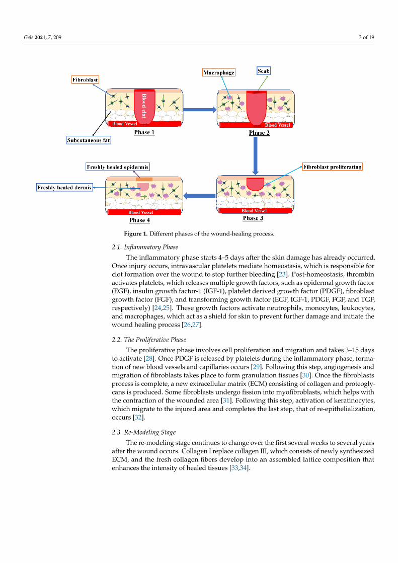
Would healing is a complicated process, and it progresses through various phases,including inflammation, proliferation, and remodeling. Involvement of fibroblasts, leuko-cytes, and monocytes in the healing process aid in reconstituting the destructed skin(Figure 1). Vitamins E and C play crucial roles in wound healing as these are key factors inthis process. Vitamin K prevents severe bleeding, carotenoids restore the skin epitheliallayer and tissues, and phytosterols have antimicrobial and anti-inflammatory effects [21].Would healing also involves the use of biochemical genetic reprogramming to reinstatethe skin health. Recent research has shown that the use of phytochemicals has activeconstituents that have the capability to induce wound healing with less side effects [22].
Gels 2021, 7, 209 3 of 19Gels 2021, 7, 209 3 of 19
Figure 1. Different phases of the wound-healing process.
2.1. Inflammatory Phase The inflammatory phase starts 4–5 days after the skin damage has already occurred.
Once injury occurs, intravascular platelets mediate homeostasis, which is responsible for clot formation over the wound to stop further bleeding [23]. Post-homeostasis, thrombin activates platelets, which releases multiple growth factors, such as epidermal growth fac-tor (EGF), insulin growth factor-1 (IGF-1), platelet derived growth factor (PDGF), fibro-blast growth factor (FGF), and transforming growth factor (EGF, IGF-1, PDGF, FGF, and TGF, respectively) [24,25]. These growth factors activate neutrophils, monocytes, leuko-cytes, and macrophages, which act as a shield for skin to prevent further damage and initiate the wound healing process [26,27].
2.2. The Proliferative Phase The proliferative phase involves cell proliferation and migration and takes 3–15 days
to activate [28]. Once PDGF is released by platelets during the inflammatory phase, for-mation of new blood vessels and capillaries occurs [29]. Following this step, angiogenesis and migration of fibroblasts takes place to form granulation tissues [30]. Once the fibro-blasts process is complete, a new extracellular matrix (ECM) consisting of collagen and proteoglycans is produced. Some fibroblasts undergo fission into myofibroblasts, which helps with the contraction of the wounded area [31]. Following this step, activation of keratinocytes, which migrate to the injured area and completes the last step, that of re-epithelialization, occurs [32].
2.3. Re-Modeling Stage The re-modeling stage continues to change over the first several weeks to several
years after the wound occurs. Collagen I replace collagen III, which consists of newly syn-thesized ECM, and the fresh collagen fibers develop into an assembled lattice composition that enhances the intensity of healed tissues [33,34].
3. The Impact of Antibiotics and Antioxidant Properties of Plants Since ancient times, various plants have been used for treatment of different diseases
and are currently in use worldwide. Due to their antimicrobial activities, natural constit-uents, such as aminoglycosides [35], beta-lactams [36], glycopeptides [37], quinolones [38],
Figure 1. Different phases of the wound-healing process.
2.1. Inflammatory Phase
The inflammatory phase starts 4–5 days after the skin damage has already occurred.Once injury occurs, intravascular platelets mediate homeostasis, which is responsible forclot formation over the wound to stop further bleeding [23]. Post-homeostasis, thrombinactivates platelets, which releases multiple growth factors, such as epidermal growth factor(EGF), insulin growth factor-1 (IGF-1), platelet derived growth factor (PDGF), fibroblastgrowth factor (FGF), and transforming growth factor (EGF, IGF-1, PDGF, FGF, and TGF,respectively) [24,25]. These growth factors activate neutrophils, monocytes, leukocytes,and macrophages, which act as a shield for skin to prevent further damage and initiate thewound healing process [26,27].
2.2. The Proliferative Phase
The proliferative phase involves cell proliferation and migration and takes 3–15 daysto activate [28]. Once PDGF is released by platelets during the inflammatory phase, forma-tion of new blood vessels and capillaries occurs [29]. Following this step, angiogenesis andmigration of fibroblasts takes place to form granulation tissues [30]. Once the fibroblastsprocess is complete, a new extracellular matrix (ECM) consisting of collagen and proteogly-cans is produced. Some fibroblasts undergo fission into myofibroblasts, which helps withthe contraction of the wounded area [31]. Following this step, activation of keratinocytes,which migrate to the injured area and completes the last step, that of re-epithelialization,occurs [32].
2.3. Re-Modeling Stage
The re-modeling stage continues to change over the first several weeks to several yearsafter the wound occurs. Collagen I replace collagen III, which consists of newly synthesizedECM, and the fresh collagen fibers develop into an assembled lattice composition thatenhances the intensity of healed tissues [33,34].
Gels 2021, 7, 209 4 of 19
3. The Impact of Antibiotics and Antioxidant Properties of Plants
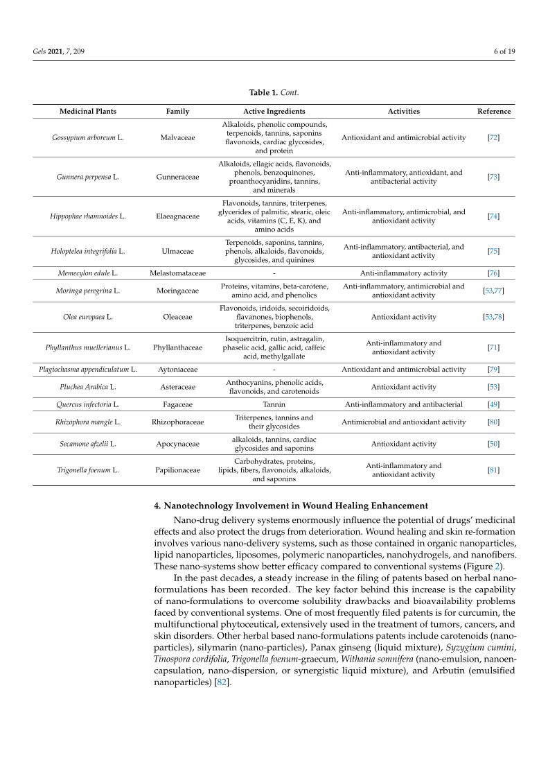
Since ancient times, various plants have been used for treatment of different dis-eases and are currently in use worldwide. Due to their antimicrobial activities, nat-ural constituents, such as aminoglycosides [35], beta-lactams [36], glycopeptides [37],quinolones [38], sulfonamides [39], and tetracyclines [40] have been utilized for woundhealing treatment. For wound therapy, the benefits of plant extracts or phytochemical havebeen recognized as has the existence of antioxidants in numerous plants extracts. The pres-ence of oxygen free radicals causes disruption in the wound healing process. Stress causedby oxidation slows down the healing process and causes additional damage to tissues.An antioxidant confers protection from oxygen free radicals by reducing their effects; thisprocess assists in the wound healing process. Active compounds possessing antimicrobialeffects play an important role as they neutralize free oxygen radicals and enhance thewound healing process [41,42]. Antioxidants have been used to hasten healing activityby extending antioxidant effects throughout the healing process. It seems the existenceof antioxidants is essential to facilitate recuperation from persistent skin injury; Table 1shows plants with both antimicrobial and antioxidant properties [43]. Natural metabolitesinfluence the wound healing process by introducing various growth factors, such as EGFand FGF, which affect cellular movement [44]. Animal studies have indicated that herbalcompounds encourage anti-inflammatory and antimicrobial activities for wound treatmentby promoting regeneration of skin cells and displacing connective tissues [45]. Table 1shows the names of natural compounds having antioxidant and antibiotic activities towardwound healing.
The efficacy of Centella asiatica has been studied broadly in animal models. This herbhelps heal the incision-induced injuries. In one study, it was concluded that the levelof antioxidants increases extensively in the presence of this herb, leading to improvedhealing activity [46]. Asiaticoside is extracted from Centella asiatica and produces a bettercapacity for injury healing process in both chronic and immediate healing as it has fibroblastproliferating activity [47].
Leaf extracts from Chromolaena odorata contain flavonoids and this herb has beenused widely in the wound healing activity due to the free radical approach, which hasshown a conclusive promotion in healing activity [48]. This extract was shown to causeimprovement in the fibroblast proliferation and keratinocyte and endothelial cell activity,and to stimulate keratinocyte migration [48].
Quercus infectoria Olivier possesses anti-inflammatory, anti-bacterial, and antioxidantproperties. The ethanolic extract of gallic acid, ellagic acid, and syringic acid form theactive constituents of tannins, which might be the reason for antioxidant effects resultingin enhanced healing activity. In this study, the incision wound animal model showedbetter healing activity after stimulation with antioxidants, which caused enhancement ofthe superoxide dismutase and catalase levels, both of which are influential antioxidantenzymes [49].
Wounds, burns, and internal and external ulcers can be treated by Buddleja globasa(common name: orange bell Buddleja). This herb is also used traditionally in Chile for thetreatment of ulcers and burns. This herb was tested for its capability to stimulate fibroblastgrowth and antioxidant activity in vitro. Testing was specific as the effect of the aqueoussolution of B. globasa on these two processes is considered as the first stage in the tissuerepair cycle [50]. It was proven that the damage caused by oxygen free radical causes adelay in the healing process; thus, an antioxidant was needed to reverse this delay [51].This process was achieved because of the presence of flavonoids and caffeic acid in theextract. Buddleja leaves have other applications in the skin layer formation that is part ofwound healing [52].
Gels 2021, 7, 209 5 of 19
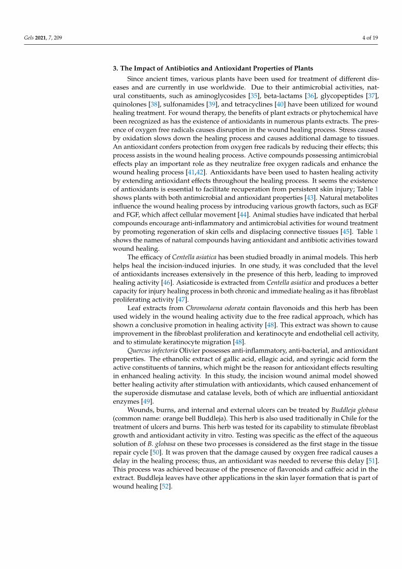
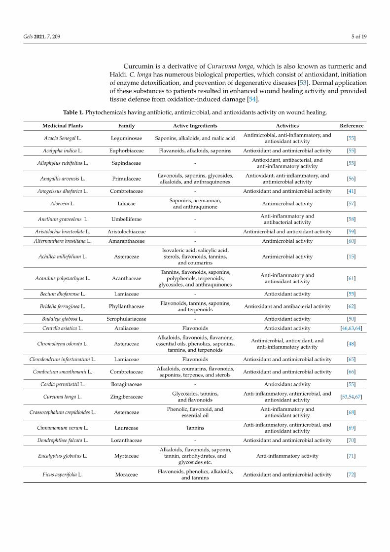
Curcumin is a derivative of Curucuma longa, which is also known as turmeric andHaldi. C. longa has numerous biological properties, which consist of antioxidant, initiationof enzyme detoxification, and prevention of degenerative diseases [53]. Dermal applicationof these substances to patients resulted in enhanced wound healing activity and providedtissue defense from oxidation-induced damage [54].
Table 1. Phytochemicals having antibiotic, antimicrobial, and antioxidants activity on wound healing.
Medicinal Plants Family Active Ingredients Activities Reference
Acacia Senegal L. Leguminosae Saponins, alkaloids, and malic acid Antimicrobial, anti-inflammatory, andantioxidant activity [55]
Acalypha indica L. Euphorbiaceae Flavanoids, alkaloids, saponins Antioxidant and antimicrobial activity [55]
Allophylus rubifolius L. Sapindaceae - Antioxidant, antibacterial, andanti-inflammatory activity [55]
Anagallis arvensis L. Primulaceae flavonoids, saponins, glycosides,alkaloids, and anthraquinones
Antioxidant, anti-inflammatory, andantimicrobial activity [56]
Anogeissus dhofarica L. Combretaceae - Antioxidant and antimicrobial activity [41]
Aloevera L. Liliacae Saponins, acemannan,and anthraquinone Antimicrobial activity [57]
Anethum graveolens L. Umbelliferae - Anti-inflammatory andantibacterial activity [58]
Aristolochia bracteolate L. Aristolochiaceae - Antimicrobial and antioxidant activity [59]
Alternanthera brasiliana L. Amaranthaceae - Antimicrobial activity [60]
Achillea millefolium L. AsteraceaeIsovaleric acid, salicylic acid,sterols, flavonoids, tannins,
and coumarinsAntimicrobial activity [15]
Acanthus polystachyus L. AcanthaceaeTannins, flavonoids, saponins,
polyphenols, terpenoids,glycosides, and anthraquinones
Anti-inflammatory andantioxidant activity [61]
Becium dhofarense L. Lamiaceae - Antioxidant activity [55]
Bridelia ferruginea L. Phyllanthaceae Flavonoids, tannins, saponins,and terpenoids Antioxidant and antibacterial activity [62]
Buddleja globosa L. Scrophulariaceae - Antioxidant activity [50]
Centella asiatica L. Araliaceae Flavonoids Antioxidant activity [46,63,64]
Chromolaena odorata L. AsteraceaeAlkaloids, flavonoids, flavanone,essential oils, phenolics, saponins,
tannins, and terpenoids
Antimicrobial, antioxidant, andanti-inflammatory activity [48]
Clerodendrum infortunatum L. Lamiaceae Flavonoids Antioxidant and antimicrobial activity [65]
Combretum smeathmanii L. Combretaceae Alkaloids, coumarins, flavonoids,saponins, terpenes, and sterols Antioxidant and antimicrobial activity [66]
Cordia perrottettii L. Boraginaceae - Antioxidant activity [55]
Curcuma longa L. Zingiberaceae Glycosides, tannins,and flavonoids
Anti-inflammatory, antimicrobial, andantioxidant activity [53,54,67]
Crassocephalum crepidioides L. Asteraceae Phenolic, flavonoid, andessential oil
Anti-inflammatory andantioxidant activity [68]
Cinnamomum verum L. Lauraceae Tannins Anti-inflammatory, antimicrobial, andantioxidant activity [69]
Dendrophthoe falcata L. Loranthaceae - Antioxidant and antimicrobial activity [70]
Eucalyptus globulus L. MyrtaceaeAlkaloids, flavonoids, saponin,
tannin, carbohydrates, andglycosides etc.
Anti-inflammatory activity [71]
Ficus asperifolia L. Moraceae Flavonoids, phenolics, alkaloids,and tannins Antioxidant and antimicrobial activity [72]
Gels 2021, 7, 209 6 of 19
Table 1. Cont.
Medicinal Plants Family Active Ingredients Activities Reference
Gossypium arboreum L. Malvaceae
Alkaloids, phenolic compounds,terpenoids, tannins, saponinsflavonoids, cardiac glycosides,
and protein
Antioxidant and antimicrobial activity [72]
Gunnera perpensa L. Gunneraceae
Alkaloids, ellagic acids, flavonoids,phenols, benzoquinones,
proanthocyanidins, tannins,and minerals
Anti-inflammatory, antioxidant, andantibacterial activity [73]
Hippophae rhamnoides L. Elaeagnaceae
Flavonoids, tannins, triterpenes,glycerides of palmitic, stearic, oleic
acids, vitamins (C, E, K), andamino acids
Anti-inflammatory, antimicrobial, andantioxidant activity [74]
Holoptelea integrifolia L. UlmaceaeTerpenoids, saponins, tannins,phenols, alkaloids, flavonoids,
glycosides, and quinines
Anti-inflammatory, antibacterial, andantioxidant activity [75]
Memecylon edule L. Melastomataceae - Anti-inflammatory activity [76]
Moringa peregrina L. Moringaceae Proteins, vitamins, beta-carotene,amino acid, and phenolics
Anti-inflammatory, antimicrobial andantioxidant activity [53,77]
Olea europaea L. OleaceaeFlavonoids, iridoids, secoiridoids,
flavanones, biophenols,triterpenes, benzoic acid
Antioxidant activity [53,78]
Phyllanthus muellerianus L. PhyllanthaceaeIsoquercitrin, rutin, astragalin,
phaselic acid, gallic acid, caffeicacid, methylgallate
Anti-inflammatory andantioxidant activity [71]
Plagiochasma appendiculatum L. Aytoniaceae - Antioxidant and antimicrobial activity [79]
Pluchea Arabica L. Asteraceae Anthocyanins, phenolic acids,flavonoids, and carotenoids Antioxidant activity [53]
Quercus infectoria L. Fagaceae Tannin Anti-inflammatory and antibacterial [49]
Rhizophora mangle L. Rhizophoraceae Triterpenes, tannins andtheir glycosides Antimicrobial and antioxidant activity [80]
Secamone afzelii L. Apocynaceae alkaloids, tannins, cardiacglycosides and saponins Antioxidant activity [50]
Trigonella foenum L. PapilionaceaeCarbohydrates, proteins,
lipids, fibers, flavonoids, alkaloids,and saponins
Anti-inflammatory andantioxidant activity [81]
4. Nanotechnology Involvement in Wound Healing Enhancement
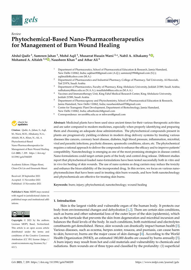
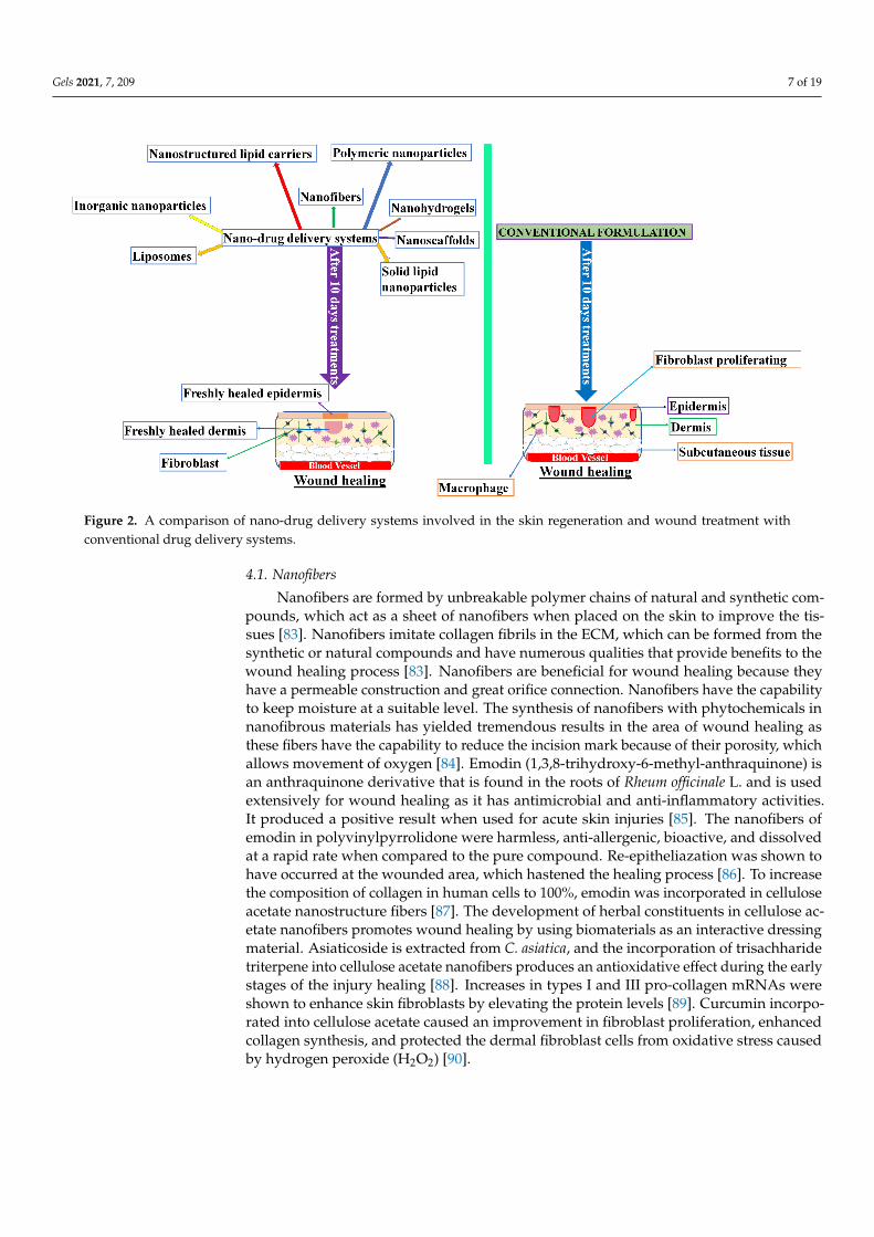
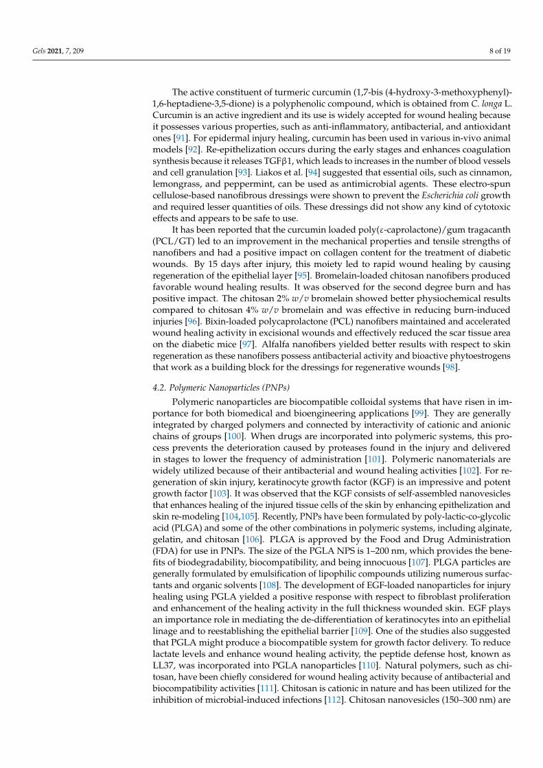
Nano-drug delivery systems enormously influence the potential of drugs’ medicinaleffects and also protect the drugs from deterioration. Wound healing and skin re-formationinvolves various nano-delivery systems, such as those contained in organic nanoparticles,lipid nanoparticles, liposomes, polymeric nanoparticles, nanohydrogels, and nanofibers.These nano-systems show better efficacy compared to conventional systems (Figure 2).
In the past decades, a steady increase in the filing of patents based on herbal nano-formulations has been recorded. The key factor behind this increase is the capabilityof nano-formulations to overcome solubility drawbacks and bioavailability problemsfaced by conventional systems. One of most frequently filed patents is for curcumin, themultifunctional phytoceutical, extensively used in the treatment of tumors, cancers, andskin disorders. Other herbal based nano-formulations patents include carotenoids (nano-particles), silymarin (nano-particles), Panax ginseng (liquid mixture), Syzygium cumini,Tinospora cordifolia, Trigonella foenum-graecum, Withania somnifera (nano-emulsion, nanoen-capsulation, nano-dispersion, or synergistic liquid mixture), and Arbutin (emulsifiednanoparticles) [82].
Gels 2021, 7, 209 7 of 19Gels 2021, 7, 209 7 of 19
Figure 2. A comparison of nano-drug delivery systems involved in the skin regeneration and wound treatment with conventional drug delivery systems.
In the past decades, a steady increase in the filing of patents based on herbal nano-formulations has been recorded. The key factor behind this increase is the capability of nano-formulations to overcome solubility drawbacks and bioavailability problems faced by conventional systems. One of most frequently filed patents is for curcumin, the multifunctional phytoceutical, extensively used in the treatment of tumors, cancers, and skin disorders. Other herbal based nano-formulations patents include carotenoids (nano-particles), silymarin (nano-particles), Panax ginseng (liquid mixture), Syzygium cumini, Tinospora cordifolia, Trigonella foenum-graecum, Withania somnifera (nano-emulsion, nanoencapsulation, nano-dispersion, or synergistic liquid mixture), and Arbutin (emulsified nanoparticles) [82].
4.1. Nanofibers Nanofibers are formed by unbreakable polymer chains of natural and synthetic
compounds, which act as a sheet of nanofibers when placed on the skin to improve the tissues [83]. Nanofibers imitate collagen fibrils in the ECM, which can be formed from the synthetic or natural compounds and have numerous qualities that provide benefits to the wound healing process [83]. Nanofibers are beneficial for wound healing because they have a permeable construction and great orifice connection. Nanofibers have the capability to keep moisture at a suitable level. The synthesis of nanofibers with phytochemicals in nanofibrous materials has yielded tremendous results in the area of wound healing as these fibers have the capability to reduce the incision mark because of their porosity, which allows movement of oxygen [84]. Emodin (1,3,8-trihydroxy-6-methyl-anthraquinone) is an anthraquinone derivative that is found in the roots of Rheum officinale L. and is used extensively for wound healing as it has antimicrobial and anti-inflammatory activities. It produced a positive result when used for acute skin injuries [85]. The nanofibers of emodin in polyvinylpyrrolidone were harmless, anti-allergenic, bioactive, and dissolved at a rapid rate when compared to the pure compound. Re-epitheliazation was shown to have occurred at the wounded area, which hastened the healing process [86]. To increase the composition of collagen in human cells to 100%, emodin was incorporated in cellulose acetate nanostructure fibers [87]. The development of herbal constituents in cellulose acetate nanofibers promotes wound healing by using
Figure 2. A comparison of nano-drug delivery systems involved in the skin regeneration and wound treatment withconventional drug delivery systems.
4.1. Nanofibers
Nanofibers are formed by unbreakable polymer chains of natural and synthetic com-pounds, which act as a sheet of nanofibers when placed on the skin to improve the tis-sues [83]. Nanofibers imitate collagen fibrils in the ECM, which can be formed from thesynthetic or natural compounds and have numerous qualities that provide benefits to thewound healing process [83]. Nanofibers are beneficial for wound healing because theyhave a permeable construction and great orifice connection. Nanofibers have the capabilityto keep moisture at a suitable level. The synthesis of nanofibers with phytochemicals innanofibrous materials has yielded tremendous results in the area of wound healing asthese fibers have the capability to reduce the incision mark because of their porosity, whichallows movement of oxygen [84]. Emodin (1,3,8-trihydroxy-6-methyl-anthraquinone) isan anthraquinone derivative that is found in the roots of Rheum officinale L. and is usedextensively for wound healing as it has antimicrobial and anti-inflammatory activities.It produced a positive result when used for acute skin injuries [85]. The nanofibers ofemodin in polyvinylpyrrolidone were harmless, anti-allergenic, bioactive, and dissolvedat a rapid rate when compared to the pure compound. Re-epitheliazation was shown tohave occurred at the wounded area, which hastened the healing process [86]. To increasethe composition of collagen in human cells to 100%, emodin was incorporated in celluloseacetate nanostructure fibers [87]. The development of herbal constituents in cellulose ac-etate nanofibers promotes wound healing by using biomaterials as an interactive dressingmaterial. Asiaticoside is extracted from C. asiatica, and the incorporation of trisachharidetriterpene into cellulose acetate nanofibers produces an antioxidative effect during the earlystages of the injury healing [88]. Increases in types I and III pro-collagen mRNAs wereshown to enhance skin fibroblasts by elevating the protein levels [89]. Curcumin incorpo-rated into cellulose acetate caused an improvement in fibroblast proliferation, enhancedcollagen synthesis, and protected the dermal fibroblast cells from oxidative stress causedby hydrogen peroxide (H2O2) [90].
Gels 2021, 7, 209 8 of 19
The active constituent of turmeric curcumin (1,7-bis (4-hydroxy-3-methoxyphenyl)-1,6-heptadiene-3,5-dione) is a polyphenolic compound, which is obtained from C. longa L.Curcumin is an active ingredient and its use is widely accepted for wound healing becauseit possesses various properties, such as anti-inflammatory, antibacterial, and antioxidantones [91]. For epidermal injury healing, curcumin has been used in various in-vivo animalmodels [92]. Re-epithelization occurs during the early stages and enhances coagulationsynthesis because it releases TGFβ1, which leads to increases in the number of blood vesselsand cell granulation [93]. Liakos et al. [94] suggested that essential oils, such as cinnamon,lemongrass, and peppermint, can be used as antimicrobial agents. These electro-spuncellulose-based nanofibrous dressings were shown to prevent the Escherichia coli growthand required lesser quantities of oils. These dressings did not show any kind of cytotoxiceffects and appears to be safe to use.
It has been reported that the curcumin loaded poly(ε-caprolactone)/gum tragacanth(PCL/GT) led to an improvement in the mechanical properties and tensile strengths ofnanofibers and had a positive impact on collagen content for the treatment of diabeticwounds. By 15 days after injury, this moiety led to rapid wound healing by causingregeneration of the epithelial layer [95]. Bromelain-loaded chitosan nanofibers producedfavorable wound healing results. It was observed for the second degree burn and haspositive impact. The chitosan 2% w/v bromelain showed better physiochemical resultscompared to chitosan 4% w/v bromelain and was effective in reducing burn-inducedinjuries [96]. Bixin-loaded polycaprolactone (PCL) nanofibers maintained and acceleratedwound healing activity in excisional wounds and effectively reduced the scar tissue areaon the diabetic mice [97]. Alfalfa nanofibers yielded better results with respect to skinregeneration as these nanofibers possess antibacterial activity and bioactive phytoestrogensthat work as a building block for the dressings for regenerative wounds [98].
4.2. Polymeric Nanoparticles (PNPs)
Polymeric nanoparticles are biocompatible colloidal systems that have risen in im-portance for both biomedical and bioengineering applications [99]. They are generallyintegrated by charged polymers and connected by interactivity of cationic and anionicchains of groups [100]. When drugs are incorporated into polymeric systems, this pro-cess prevents the deterioration caused by proteases found in the injury and deliveredin stages to lower the frequency of administration [101]. Polymeric nanomaterials arewidely utilized because of their antibacterial and wound healing activities [102]. For re-generation of skin injury, keratinocyte growth factor (KGF) is an impressive and potentgrowth factor [103]. It was observed that the KGF consists of self-assembled nanovesiclesthat enhances healing of the injured tissue cells of the skin by enhancing epithelization andskin re-modeling [104,105]. Recently, PNPs have been formulated by poly-lactic-co-glycolicacid (PLGA) and some of the other combinations in polymeric systems, including alginate,gelatin, and chitosan [106]. PLGA is approved by the Food and Drug Administration(FDA) for use in PNPs. The size of the PGLA NPS is 1–200 nm, which provides the bene-fits of biodegradability, biocompatibility, and being innocuous [107]. PLGA particles aregenerally formulated by emulsification of lipophilic compounds utilizing numerous surfac-tants and organic solvents [108]. The development of EGF-loaded nanoparticles for injuryhealing using PGLA yielded a positive response with respect to fibroblast proliferationand enhancement of the healing activity in the full thickness wounded skin. EGF playsan importance role in mediating the de-differentiation of keratinocytes into an epitheliallinage and to reestablishing the epithelial barrier [109]. One of the studies also suggestedthat PGLA might produce a biocompatible system for growth factor delivery. To reducelactate levels and enhance wound healing activity, the peptide defense host, known asLL37, was incorporated into PGLA nanoparticles [110]. Natural polymers, such as chi-tosan, have been chiefly considered for wound healing activity because of antibacterial andbiocompatibility activities [111]. Chitosan is cationic in nature and has been utilized for theinhibition of microbial-induced infections [112]. Chitosan nanovesicles (150–300 nm) are
Gels 2021, 7, 209 9 of 19
generally formulated utilizing the method of ionic gelation [113]. Nowadays, chitosan iswidely accepted in wound treatment, and it can also be utilized as a prophylactic agentto inhibit the infection development and enhance healing activity [114,115]. Studies haveshown that polylactic acid-loaded chitosan magnetic eugenol nanospheres had improvedprevention and development of biofilm compared to pure chitosan, whilst performingendothelial proliferation [116]. Most of the studies concerning this topic have reported thatnanovesicles containing chitosan and analogs might enhance healing activity by improvinginflammatory cell function and restoring fibroblasts and osteoblast functions [117]. In twodifferent studies, it was observed that chitosan-loaded nanovesicles improved the coagula-tion by binding to red blood cells (RBC) and ameliorating the function of inflammatorycells. In another study, the chitosan nanovesicles were used as the compounds in bandagesoutlined for the skin wound and, hence, enhanced healing activity in both humans andanimals [115,118].
4.3. Dendrimers
Dendrimers are nanoscale (1–10 nm) systems with homogeneous structures that aremonodispersed in polymer macromolecule that can be used for both therapeutic and diag-nostic purposes. Subunits of phenyl acetylene were used to develop dendrimers [119,120].In addition, functional groups present on the surface of dendrimers can operate as an-tibacterial agents. Dendrimers cause detachment of contaminated tissues and may ex-tend the phase of inflammation and slow injury diminution in addition to promotingre-epithelization and better wound healing activity [121]. The interaction between posi-tively and negatively charged groups present on dendrimers and on the bacterial cell wallwould lead to the bacterial structure disturbance [121]. In another study, silver-loadeddendrimer NSs were observed to show anti-inflammatory and anti-microbial activitiesin a synergistic manner. These properties were also shown to prevent inflammation andenhance healing activity [122].
4.4. Metallic Nanoparticles
Metal-based nanoparticles are widely utilized as they produce antibacterial, antimi-crobial, and anti-inflammatory effects. The chemical and physical structures of nanopar-ticles are important for determining the propensity of a nanoparticle to enter and/orbind to target cells with the capacity to interact with their biological machinery andelicit a response. The metal-based nanoparticles are widely accepted in medicine, andthe most acceptable metallic nanoparticles are silver- and gold-based nanostructures.Herbal plants are widely accepted in the development of metallic nanoparticles becauseof their low levels of side effects and more therapeutic effects as compared to the conven-tional dosage form [123]. Most of the herbal extracts, such as Cladophora fascicularis [124],Aerva lanata [125], Hippophae rhamnoides [126], Eucommia ulmoides [127], Black tea leaf [128],Averrhoa bilimbi [129], Salicornia brachiate [130], Abelmoschus esculentus [131], olive leaf [132],Ipomoea carnea [133], geranium [134], and Cissus arnotiana [135] have been incorporated intometallic nanoparticles
Silver nanoparticles are widely used as they possess antimicrobial, antibacterial, andanti-inflammatory properties [136]. The solubility and bioactivity of the silver particlesat the wounded area depend on the size of silver particles; the smaller the size is, thestronger the contact with the will skin be. Silver nanoparticle vesicle sizes range from1 to 100 nm. In one study, the silver–silver chloride nanoparticles combined with lowergrapheme oxide nanovesicles induced an escalation of the healing process because itgenerated a higher number of oxygen free radicals rather than free the silver ions. Apositive impact on the antibacterial activity on both Gram-negative and -positive bacteriahas been shown, and, hence, these particles can enhance wound healing activity as shownin in-vivo studies in mice [137]. ACTICOAT is an alternate form of silver antimicrobialbarrier wound dressing, which prevents the complication of prior agents. It slows down thebacterial activity, which leads to a reduction in inflammation and causes an improvement
Gels 2021, 7, 209 10 of 19
in the healing process [138]. The plant-based bio-prepared nanoparticles reveal potentialfor wound remedy and bacterial infection prevention [139]. Different methods for thepreparation of silver nanoparticles are used. Photochemical and chemical reduction are thetwo most widely used methods [140]. Different plant extracts have been incorporated intosilver nanoparticles for wound healing containing alkaloids, glycoside, corticosteroids andessential oils [141]. Cassia roxburghii prepared silver nanoparticles show the potential forwound healing enhancement as these particles have significant antibacterial and antifungalactivities [142].
The active constituent of Drosera binata is naphthoquinones, primarily plumbagin.D. binata silver nanoparticles show better antibacterial activity against Staphylococcus aureuswithout affecting human keratinocytes. It was also inconclusive as to whether it is D. binataextract or its pure form (3-chloroplumbagin) that would have effective results for antibioticsand, hence, enhance wound healing [143]. Extracts of grape pomace were also combinedwith silver nitrate, and grape-silver nanoparticle-stabilized liposomes were developed byCastangia et al. The resulting nano-formulation showed potential to offer a significantshield of keratinocytes and fibroblasts to combat oxidative stress, thus, avoiding celldamage and death [144]. The other highly acceptable nanoparticles in different applications,such as wound treatment, re-epithelization, and particularly drug delivery, include goldnanoparticles [145].
Their chemical stability and capability to absorb near-infrared (NIR) light combinedwith their positive impact and antibacterial activity will strengthen the wound healingprocess [146]. Gold nanoparticles have the potential to penetrate bacterial tissues and causealterations in the cell membrane, which causes inhibition of bacterial activity [147], andalso prevents bacteria from developing reactive oxygen species [148].
Gold nanoparticles are synthesized with collagen, gelatin, and chitosan to yield effec-tive injury recovery activity and also helps to achieve the biocompatibility [149]. Chitosan-loaded gold nanoparticles showed enhanced results in the healing process as these particlesincrease free radical scavenging and improve biocompatibility; in the model, these particlesenhance the formation of cells and lead to an improvement in hemostasis by increasingthe healing activity in comparison to pure chitosan [150]. The resulting metabolites fromIndigofera aspalathoides Vahl. (Papilionaceae), which is also known as Shivanarvembu, areextracted from plants and used for wound healing. The histopathology results demon-strate that the I. aspalathoides silver nanoparticles have a better effect on wound healing inmice. When treated with plant extract, the granulation tissue which possesses fibroblasts,collagen fibers, minimal edema, and newly developed blood vessels were noted [151]. Theother forms of metallic nanoparticles are gold and copper oxide nanovesicles that improvewound healing, which leads to fast injury healing and slows down the infection develop-ment. Both silver and gold nanoparticles are formed by incorporating Coleous forskohliiroot extracts. These particles exhibit antimicrobial activity and antioxidant activities andhave a positive effect on re-epithelization at the site of wound, which enhances connectivetissue formation and causes an increase in proliferation and remodeling rates of dermalcells [152]. The development of both titanium dioxide and copper oxide nanoparticles ofMoringa oleifera and Ficus religiosa leaf extracts, respectively, were shown to enhance woundhealing and decrease the removal wound site in rats [153].
4.5. Nanohydrogels
For wound treatment, nanohydrogels are considered to be effective carriers as theypossess three-dimensional polymeric networks. Due to their permeable network, they havethe capability to absorb the liquid, which helps the wound to keep hydrated and enhancethe wound healing process by keeping the proper oxygen level. Due to their effectiveness,compatibility, and showing beneficial results on skin revitalization, nanohydrogels havebecome widely accepted [154].
Gels 2021, 7, 209 11 of 19
To improve wound healing activity, the gellan cholesterol nanohydrogel is immersedin baicalin. The baicalin-loaded nanohydrogels manifest ideal efficacy for skin repairand also act as inflammation inhibitors when applied to an epidermal inflammation micemodel in in-vivo studies [155]. The freshly developed nanocrystal bacterial cellulosehydrogels instantly stick to fibroblasts, support human dermal fibroblast morphology,restrict the relocation of cells, enhance the proliferation of cells, and influence the nineexpressions of genes connected to healing of injury. These genes include interleukins 6and 10, granulocyte-macrophage colony-stimulating factor, matrix metalloproteinase 2(IL-6 and -10, GM-CSF, MMP-2, respectively), and TGF-β; hence, nanohydrogels play animportant role in skin regeneration [156].
4.6. Liposomes
Liposomes appear to be an important vehicle for topical delivery; they are harmlessand environmentally safe and possess high drug loading efficiency, long-term stability,biological acceptability with skin in addition to having the capability to incorporate bothhydrophobic and hydrophilic drugs in water and bilayer cavities [157]. Liposomes success-fully shield the injury site and build a humid habitat at the site of injury, which is beneficialfor the healing of the wounded skin. Taking all these characteristics into consideration,liposomes have become widely accepted in skin regeneration and injury treatment [158]. Astudy on propylene glycol liposome nanocarriers demonstrated numerous merits in com-parison to other nano-systems. This system showed the tendency to enhance the stability,retention, and permeation in the tissues of skin [159]. It surmised that propylene glycolameliorate the elasticity of vesicle containing bilayer of phospholipids. Hence, it improvedthe permeation into the skin. Moreover, the particles size of liposomes should be 150 nmfor better drug perforation into the skin layers [160]. Liposomes with silk fibroin hydrogelswere prepared to stabilize the basic fibroblast growth factor (bFGF) that maintained theactivity of proliferation of cells on wound fluids; it also enhances the healing process byinspiring angiogenesis [161]. Rabelo et al. assessed the gelatin-membrane consisting ofusnic acid-loaded liposomes and obtained encouraging results for wound healing. Theseresults showed that the membrane of liposomes prominently manages the second-gradeinfection on porcine model [162]. Furthermore, with improved collagen, accumulation oncellularized granulation tissue was discovered in the treated group of liposomal membrane,which when compared to one of the commercial products improved the granulation tissuematuration and repaired the scars [163]. Argan-liposomes and argan-hyalurosomes havebeen successfully developed by incorporating neem oil into them. These formulations wereextremely biocompatible and could protect skin cells from oxidative stress effectively withimproved efficacy of oil. Moreover, formulations stimulate wound closure substantiallymore effectively than oil dispersion [164]. The efficacy of mangiferin (employed in cureof skin lesions) was enhanced by modifying transferosomes with propylene glycol andglycerol. Improved deposition of mangiferin was observed in epidermal and dermal layerand fibroblasts were protected from oxidative stress and intensified their propagation [165].
4.7. Inorganic Nanoparticles
Inorganic nanoparticles are those derived from the inorganic materials and includecarbon-, metal-, and ceramic-based nanovesicles that accelerate tissue repair and re-modeling. These particles deliver assistance in the region of medicines, counting cancer,imaging, and drug delivery; however, their utilization in tissue regulation and skin re-modeling is new, it also provides adhesion in tissue and enhanced antimicrobial activity ininjury healing [166].
Gels 2021, 7, 209 12 of 19
4.8. Lipid Nanoparticles
Lipid nanoparticles were designed to overcome the stability limitation of liposomesdue to the lipid bilayer. Lipid nanovesicles consist of two types: (1) solid lipid nanoparticles(SLNs) and (2) nanostructured lipid carriers (NLCs). The preparation of lipid nanovesi-cles amid lipids molecules does not include the use of any potentially harmful bioticsolvents [167]. In a study, both SLN- and NLC-loaded rh-EGF (epidermal growth factor)for chronic injury treatment were formulated by the emulsification followed by an ultra-sonication method; however, the NLC process included no organic solvent and showedbetter entrapment efficiency. The results of both formulations show capabilities to en-hance cell proliferation when compared with free rh-EGF and considerably enhance thehealing activity for wound closure, re-establish the process of inflammation, and facilitatere-epithelization [168].
In another study, development of SLNs with the elastase inhibitor serpin A1 andantimicrobial peptide LL37 had a synergistic impact on injury healing. SLNs promotedthe closure of injury in cells of fibroblasts and keratinocytes. Moreover, it also led toimprovement in the activity of antibacterial against S. aureus and E. coli when comparedwith the LL37- and A1-treated groups [169].
5. Future Perspective and Conclusions
The main aim of this review article was to describe the advantages of using nano-systems for use in the wound healing process. The distinctive physiochemical properties ofnano-systems make them a perfect candidate for the application of wound healing process.The wound therapy process by nanotechnological systems demonstrates better therapeuticeffect compared to the conventional therapy for wound healing. Nanotechnological systemscan change one or more than one phase of wound healing during the process, as it possessesantibacterial, anti-inflammatory, and anti-proliferation activities. Worldwide, the researchhas been conducted on natural and herbal compounds due to their more therapeutic effectsand lesser side effects. There is a need for the development of improved systems for thedelivery of drugs at the target site with a dose that does not alter the existing treatment ofdisease. The herbal compounds have great potential and, hence, a better future, especiallywhen incorporated into the nanocarriers for chronic wound treatment as they have shownpromising results. Herbal medicine-based novel drug delivery systems have acknowledgedthe approaches in the field of pharmaceuticals, which will improve the health of the people.It is also concluded that the incorporation of herbal compound in the nano-vehicle willaggrandize the magnitude of the existing delivery system. Anyhow, various approacheshave been employed for the privileged application of nanocarriers in wound healingtherapy. The main concerns for the nano-vehicles are toxicity because they may causepossible side effects in the human body. Hence, this requires to be rectified at the startingpoint for further progression of wound healing therapies in clinical trials. In in vivo models,there is slighter comprehension regarding non-material mediated wound healing processesand this is one of the problems observed. The studies of non-material-wound healingprocesses are based on in-vitro studies or mainly depend on single aim bacteria. The in-vivowound healing application is required for the in-depth studies utilizing both Gram-positiveand -negative bacterial strains. Subsequently, the main focus should be on improving andenhancing target efficiency for more efficacious wound healing. Therefore, the investigatorsshould target producing a nanomaterial that is biocompatible and biodegradable and hasthe capability to correct all the phases of the wound healing process.
Author Contributions: Conceptualization, A.Q., M.A. and M.H.W.; methodology, S.J., A.Q.; software,A.A.; validation, A.A., N.A.A. and M.A.A.; formal analysis, A.Q., M.H.W. and N.K.; investigation,A.A., N.K.; resources, N.A.A., A.Q.; data curation, N.K., S.J.; writing—original draft preparation,A.Q.; writing—review and editing, M.H.W., M.A.A. and N.A.A.; visualization, M.H.W.; supervision,M.A.; project administration, M.A.A., N.A.A.; funding acquisition, M.A.A., N.A.A. All authors haveread and agreed to the published version of the manuscript.
Gels 2021, 7, 209 13 of 19
Funding: The Deanship of Scientific Research (DSR) at King Abdulaziz University, Jeddah, Saudi Arabiahas funded this project, under grant no. (FP-032-43).
Institutional Review Board Statement: Not applicable.
Informed Consent Statement: Not applicable.
Data Availability Statement: Not applicable.
Conflicts of Interest: The authors declare no conflict of interest.
References1. Wang, W.; Lu, K.J.; Yu, C.H.; Huang, Q.L.; Du, Y.Z. Nano-drug delivery systems in wound treatment and skin regeneration.
J. Nanobiotechnology 2019, 17, 82. [CrossRef]2. Pereira, R.F.; Carvalho, A.; Gil, M.H.; Mendes, A.; Bartolo, P. Influence of Aloe vera on water absorption and enzymatic in vitro
degradation of alginate hydrogel films. Carbohydr. Polym. 2013, 98, 311–320. [CrossRef]3. World Health Organization Burns—Key Facts. Available online: https://www.who.int/news-room/fact-sheets/detail/burns
(accessed on 4 January 2020).4. Souto, E.B.; Ribeiro, A.F.; Ferreira, M.I.; Teixeira, M.C.; Shimojo, A.A.; Soriano, J.L.; Naveros, B.C.; Durazzo, A.; Lucarini, M.;
Souto, S.B.; et al. New nanotechnologies for the treatment and repair of skin burns infections. Int. J. Mol. Sci. 2020, 21, 393.[CrossRef] [PubMed]
5. Deitch, E.A. The management of burns. N. Engl. J. Med. 1990, 323, 1249–1253. [PubMed]6. Wild, T.; Rahbarnia, A.; Kellner, M.; Sobotka, L.; Eberlein, T. Basics in nutrition and wound healing. Nutrition 2010, 26, 862–866.
[CrossRef] [PubMed]7. Hermans, M.H. Results of an internet survey on the treatment of partial thickness burns, full thickness burns, and donor sites.
J. Burn Care Res. 2007, 28, 835–847. [CrossRef] [PubMed]8. Roshangar, L.; Kheirjou, R.; Ranjkesh, R. Skin Burns: Review of Molecular Mechanisms and Therapeutic Approaches. Wounds
Compend. Clin. Res. Pract. 2019, 31, 308–315.9. Guo, S.A.; DiPietro, L.A. Factors affecting wound healing. J. Dent. Res. 2010, 89, 219–229. [CrossRef]10. Ghosh, P.K.; Gaba, A. Phyto-extracts in wound healing. J. Pharm. Pharm. Sci. 2013, 16, 760–820. [CrossRef]11. Sandhiya, V.; Ubaidulla, U. A review on herbal drug loaded into pharmaceutical carrier techniques and its evaluation process.
Future J. Pharm. Sci. 2020, 6, 1–16. [CrossRef]12. Bhatt, D.; Jethva, K.; Patel, S.; Zaveri, M. Novel drug delivery systems in herbals for cancer. World J. Pharm. Res. 2016, 5, 368–378.13. Ferreira, V.F.; Pinto, A.C. A fitoterapia no mundo atual. Química Nova 2010, 33, 1829. [CrossRef]14. Vickers, A.; Zollman, C. Herbal medicine. Br. Med. J. 1999, 319, 1050–1053. [CrossRef] [PubMed]15. Ali, S.I.; Gopalakrishnan, B.; Venkatesalu, V. Pharmacognosy, phytochemistry and pharmacological properties of Achillea
millefoliumh L.: A review. Phytother. Res. 2017, 31, 1140–1161. [CrossRef] [PubMed]16. Watkins, R.; Wu, L.; Zhang, C.; Davis, R.; Xu, B. Natural product-based nanomedicine: Recent advances and issues. Int. J.
Nanomed. 2015, 10, 6055–6074.17. Xu, R.; Luo, G.; Xia, H.; He, W.; Zhao, J.; Liu, B.; Tan, J.; Zhou, J.; Liu, D.; Wang, Y.; et al. Novel bilayer wound dressing composed
of silicone rubber with particular micropores enhanced wound re-epithelialization and contraction. Biomaterials 2014, 40, 1–11.[CrossRef] [PubMed]
18. Singh, R.P.; Singh, S.G.; Naik, H.; Jain, D.; Bisla, S. Herbal excipients in novel drug delivery system. Int. J. Compr. Pharm. 2011, 2, 1–7.19. Sungthongjeen, S.; Pitaksuteepong, T.; Somsiri, A.; Sriamornsak, P. Studies on pectins as potential hydrogel matrices for
controlled-release drug delivery. Drug Dev. Ind. Pharm. 1999, 25, 1271–1276. [CrossRef]20. Portou, M.; Baker, D.; Abraham, D.; Tsui, J. The innate immune system, toll-like receptors and dermal wound healing: A review.
Vasc. Pharmacol. 2015, 71, 31–36. [CrossRef]21. Andritoiu, C.V.; Andriescu, C.E.; Ibanescu, C.; Lungu, C.; Ivanescu, B.; Vlase, L.; Havarneanu, C.; Popa, M. Effects and
Characterization of Some Topical Ointments Based on Vegetal Extracts on Incision, Excision, and Thermal Wound Models.Molecules 2020, 25, 5356. [CrossRef]
22. Hajialyani, M.; Tewari, D.; Sobarzo-Sánchez, E.; Nabavi, S.M.; Farzaei, M.H.; Abdollahi, M. Natural product-based nanomedicinesfor wound healing purposes: Therapeutic targets and drug delivery systems. Int. J. Nanomed. 2018, 13, 5023–5043. [CrossRef]
23. Martin, P. Wound Healing–Aiming for Perfect Skin Regeneration. Science 1997, 276, 75–81. [CrossRef]24. Braund, R.; Hook, S.; Medlicott, N.J. The role of topical growth factors in chronic wounds. Curr. Drug Deliv. 2007, 4, 195–204.
[CrossRef] [PubMed]25. Gainza, G.; Villullas, S.; Pedraz, J.L.; Hernandez, R.M.; Igartua, M. Advances in drug delivery systems (DDSs) to release growth
factors for wound healing and skin regeneration. Nanomed. Nanotechnol. Biol. Med. 2015, 11, 1551–1573. [CrossRef] [PubMed]26. Kiritsy, C.P.; Lynch, S.E. Role of growth factors in cutaneous wound healing: A review. Crit. Rev. Oral Biol. Med. 1993, 4, 729–760.
[CrossRef] [PubMed]27. Eming, S.A.; Krieg, T.; Davidson, J.M. Inflammation in wound repair: Molecular and cellular mechanisms. J. Investig. Dermatol.
2007, 127, 514–525. [CrossRef]
Gels 2021, 7, 209 14 of 19
28. Singer, A.J.; Clark, R.A. Cutaneous wound healing. N. Engl. J. Med. 1999, 341, 738–746. [CrossRef]29. Velnar, T.; Bailey, T.; Smrkolj, V. The wound healing process: An overview of the cellular and molecular mechanisms.
J. Int. Med. Res. 2009, 37, 1528–1542. [CrossRef]30. Malinda, K.M.; Sidhu, G.S.; Banaudha, K.K.; Gaddipati, J.P.; Maheshwari, R.K.; Goldstein, A.L.; Kleinman, H.K. Thymosin α1
stimulates endothelial cell migration, angiogenesis, and wound healing. J. Immunol. 1998, 160, 1001–1006.31. Li, B.; Wang, J.H.C. Fibroblasts and myofibroblasts in wound healing: Force generation and measurement. J. Tissue Viability
2011, 20, 108–120. [CrossRef]32. Montesinos, M.C.; Gadangi, P.; Longaker, M.; Sung, J.; Levine, J.; Nilsen, D.; Reibman, J.; Li, M.; Jiang, C.-K.; Hirschhorn, R.;
et al. Wound Healing Is Accelerated by Agonists of Adenosine A2 (Gαs-linked) Receptors. J. Exp. Med. 1997, 186, 1615–1620.[CrossRef]
33. Ehrlich, H.P.; Keefer, K.A.; Myers, R.L.; Passaniti, A. Vanadate and the absence of myofibroblasts in wound contraction. Arch.Surg. 1999, 134, 494–501. [CrossRef] [PubMed]
34. Stadelmann, W.K.; Digenis, A.G.; Tobin, G.R. Physiology and healing dynamics of chronic cutaneous wounds. Am. J. Surg.1998, 176, 26S–38S. [CrossRef]
35. Pawar, H.V.; Tetteh, J.; Boateng, J.S. Preparation, optimisation and characterisation of novel wound healing film dressings loadedwith streptomycin and diclofenac. Colloids Surf. B Biointerfaces 2013, 102, 102–110. [CrossRef]
36. Sabitha, M.; Rajiv, S. Preparation and characterization of ampicillin-incorporated electrospun polyurethane scaffolds for woundhealing and infection control. Polym. Eng. Sci. 2014, 55, 541–548. [CrossRef]
37. Lan, Y.; Li, W.; Guo, R.; Zhang, Y.; Xue, W.; Zhang, Y. Preparation and characterisation of vancomycin-impregnated gelatinmicrospheres/silk fibroin scaffold. J. Biomater. Sci. Polym. Ed. 2013, 25, 75–87. [CrossRef] [PubMed]
38. Pásztor, N.; Rédai, E.; Szabó, Z.-I.; Sipos, E. Preparation and characterization of levofloxacin-loaded nanofibers as potentialwound dressings. Acta Med. Marisiensis 2017, 63, 66–69. [CrossRef]
39. Mohseni, M.; Shamloo, A.; Aghababaei, Z.; Vossoughi, M.; Moravvej, H. Antimicrobial wound dressing containing silversulfadiazine with high biocompatibility: In vitro study. Artif. Organs 2016, 40, 765–773. [CrossRef]
40. Adhirajan, N.; Shanmugasundaram, N.; Shanmuganathan, S.; Babu, M. Collagen-based wound dressing for doxycycline delivery:In-vivo evaluation in an infected excisional wound model in rats. J. Pharm. Pharmacol. 2009, 61, 1617–1623. [CrossRef]
41. Parihar, A.; Parihar, M.S.; Milner, S.; Bhat, S. Oxidative stress and anti-oxidative mobilization in burn injury. Burns 2008, 34, 6–17.[CrossRef]
42. Suntar, I.; Akkol, E.K.; Nahar, L.; Sarker, S.D. Wound healing and antioxidant properties: Do they coexist in plants? Free Radic.Antioxid. 2012, 2, 1–7. [CrossRef]
43. Blass, S.C.; Goost, H.; Tolba, R.H.; Stoffel-Wagner, B.; Kabir, K.; Burger, C.; Stehle, P.; Ellinger, S. Time to wound closure in traumapatients with disorders in wound healing is shortened by supplements containing antioxidant micronutrients and glutamine:A PRCT. Clin. Nutr. 2012, 31, 469–475. [CrossRef] [PubMed]
44. Schultz, G.S.; Sibbald, R.G.; Falanga, V.; Ayello, E.A.; Dowsett, C.; Harding, K.; Romanelli, M.; Stacey, M.C.S.; Teot, L.; Vanscheidt,W. Wound bed preparation: A systematic approach to wound management. Wound Repair Regen. 2003, 11, S1–S28. [CrossRef]
45. Tsala, D.E.; Amadou, D.; Habtemariam, S. Natural wound healing and bioactive natural products. Phytopharmacology2013, 4, 532–560.
46. Ruszymah, B.H.I.; Chowdhury, S.R.; Manan, N.A.B.A.; Fong, O.S.; Adenan, M.I.; Bin Saim, A. Aqueous extract of Centella asiaticapromotes corneal epithelium wound healing in vitro. J. Ethnopharmacol. 2012, 140, 333–338. [CrossRef] [PubMed]
47. Maquart, F.X.; Bellon, G.; Gillery, P.; Wegrowski, Y.; Borel, J.P. Stimulation of collagen synthesis in fibroblast cultures by a triterpeneextracted from Centella asiatica. Connect. Tissue Res. 1990, 24, 107–120. [CrossRef] [PubMed]
48. Thang, P.T.; Teik, L.S.; Yung, C.S. Anti-oxidant effects of the extracts from the leaves of Chromolaena odorata on human dermalfibroblasts and epidermal keratinocytes against hydrogen peroxide and hypoxanthine–xanthine oxidase induced damage. Burns2001, 27, 319–327. [CrossRef]
49. Umachigi, S.P.; Jayaveera, K.N.; Kumar, C.K.A.; Kumar, G.S.; Swamy, B.M.V.; Kumar, D.V.K. Studies on wound healing propertiesof Quercus infectoria. Trop. J. Pharm. Res. 2008, 7, 913–919. [CrossRef]
50. Mensah, A.Y.; Sampson, J.; Houghton, P.; Hylands, P.; Westbrook, J.; Dunn, M.; Hughes, M.; Cherry, G. Effects of Buddleja globosaleaf and its constituents relevant to wound healing. J. Ethnopharmacol. 2001, 77, 219–226. [CrossRef]
51. Shukla, A.; Rasik, A.M.; Dhawan, B.N. Asiaticoside-induced elevation of antioxidant levels in healing wounds. Phytother. Res. Int.J. Devoted Pharmacol. Toxicol. Eval. Nat. Prod. Deriv. 1999, 13, 50–54. [CrossRef]
52. Yamasaki, T.; Li, L.; Lau, B.H.S. Garlic compounds protect vascular endothelial cells from hydrogen peroxide-induced oxidantinjury. Phytother. Res. 1994, 8, 408–412. [CrossRef]
53. Ravindran, P.N.; Babu, K.N.; Sivaraman, K. (Eds.) Turmeric: The Genus Curcuma; CRC Press: Boca Raton, FL, USA, 2007.54. Gopinath, D.; Ahmed, M.; Gomathi, K.; Chitra, K.; Sehgal, P.; Jayakumar, R. Dermal wound healing processes with curcumin
incorporated collagen films. Biomaterials 2004, 25, 1911–1917. [CrossRef]55. Marwah, R.G.; Fatope, M.O.; Al Mahrooqi, R.; Varma, G.B.; Al Abadi, H.; Al-Burtamani, S.K.S. Antioxidant capacity of some
edible and wound healing plants in Oman. Food Chem. 2007, 101, 465–470. [CrossRef]56. Qadir, M.I. Medicinal and cosmetological importance of Aloe vera. Int. J. Nat. Ther. 2009, 2, 21–26.
Gels 2021, 7, 209 15 of 19
57. Pattanayak, S.; Sunita, P. Wound healing, anti-microbial and antioxidant potential of Dendrophthoe falcata (L.f) Ettingsh.J. Ethnopharmacol. 2008, 120, 241–247. [CrossRef]
58. Altameme, H.J.; Hameed, I.H.; Hamza, L.F. Anethum graveolens: Physicochemical properties, medicinal uses, antimicrobial effects,antioxidant effect, anti-inflammatory and analgesic effects: A review. Int. J. Pharm. Qual. Assur. 2017, 8, 88–91.
59. Shirwaikar, A.; Somashekar, A.; Udupa, A.; Udupa, S.; Somashekar, S. Wound healing studies of Aristolochia bracteolata Lam. withsupportive action of antioxidant enzymes. Phytomedicine 2003, 10, 558–562. [CrossRef] [PubMed]
60. Barua, C.C.; Talukdar, A.; Begum, S.A.; Sarma, D.K.; Fathak, D.C.; Barua, A.G.; Bora, R.S. Wound healing activity of methanolicextract of leaves of Alternanthera brasiliana Kuntz using in vivo and in vitro model. Indian J. Exp. Boil. 2009, 47, 1001–1005.
61. Demilew, W.; Adinew, G.M.; Asrade, S. Evaluation of the wound healing activity of the crude extract of leaves of Acanthuspolystachyus Delile (Acanthaceae). Evid.-Based Complement. Altern. Med. 2018, 2018, 1–9. [CrossRef]
62. Adetutu, A.; Morgan, W.A.; Corcoran, O. Antibacterial, antioxidant and fibroblast growth stimulation activity of crude extracts ofBridelia ferruginea leaf, a wound-healing plant of Nigeria. J. Ethnopharmacol. 2011, 133, 116–119. [CrossRef]
63. Shukla, A.; Rasik, A.; Jain, G.; Shankar, R.; Kulshrestha, D.; Dhawan, B. In vitro and in vivo wound healing activity of asiaticosideisolated from Centella asiatica. J. Ethnopharmacol. 1999, 65, 1–11. [CrossRef]
64. Chen, Y.J.; Dai, Y.S.; Chen, B.F.; Chang, A.; Chen, H.C.; Lin, Y.C.; Chang, K.H.; Lai, Y.L.; Chung, C.H.; Lai, Y.J. The effect oftetrandrine and extracts of Centella asiatica on acute radiation dermatitis in rats. Biol. Pharm. Bull. 1999, 22, 703–706. [CrossRef]
65. Gouthamchandra, K.; Mahmood, R.; Manjunatha, H. Free radical scavenging, antioxidant enzymes and wound healing activitiesof leaves extracts from Clerodendrum infortunatum L. Environ. Toxicol. Pharmacol. 2010, 30, 11–18. [CrossRef]
66. Agyare, C.; Asase, A.; Lechtenberg, M.; Niehues, M.; Deters, A.; Hensel, A. An ethnopharmacological survey and in vitroconfirmation of ethnopharmacological use of medicinal plants used for wound healing in Bosomtwi-Atwima-Kwanwoma area,Ghana. J. Ethnopharmacol. 2009, 125, 393–403. [CrossRef]
67. Koca, U.; Süntar, I.; Akkol, E.K.; Yılmazer, D.; Alper, M. Wound repair potential of Olea europaea L. leaf extracts revealed byin vivo experimental models and comparative evaluation of the extracts’ antioxidant activity. J. Med. Food 2011, 14, 140–146.[CrossRef] [PubMed]
68. Arawande, J.O.; Komolafe, E.A.; Imokhuede, B. Nutritional and phytochemical compositions of fireweed (Crassocephalumcrepidioides). J. Agric. Technol. 2013, 9, 439–449.
69. Mathew, S.; Abraham, T.E. In vitro antioxidant activity and scavenging effects of Cinnamomum verum leaf extract assayed bydifferent methodologies. Food Chem. Toxicol. 2006, 44, 198–206. [CrossRef]
70. Atun, S.; Handayani, S.; Rakhmawati, A.; Purnamaningsih, N.A.; Naila, B.I.; Lestari, A. Study of potential phenolic compoundsfrom stems of Dendrophthoe falcata (Loranthaceae) plant as antioxidant and antimicrobial agents. Orient. J. Chem. 2018, 34,2342–2349. [CrossRef]
71. Velmurugan, C.; Geetha, C.; Shajahan, S.; Vijayakumar, S.; Kumar, P.L. Wound healing potential of leaves of Eucalyptus citriodoralinrats. World J. Pharm. Sci. 2014, 2, 62–71.
72. Annan, K.; Houghton, P.J. Antibacterial, antioxidant and fibroblast growth stimulation of aqueous extracts of Ficus asperifolia Miq.and Gossypium arboreum L., wound-healing plants of Ghana. J. Ethnopharmacol. 2008, 119, 141–144. [CrossRef]
73. Steenkamp, V.; Mathivha, E.; Gouws, M.; van Rensburg, C.J. Studies on antibacterial, antioxidant and fibroblast growth stimulationof wound healing remedies from South Africa. J. Ethnopharmacol. 2004, 95, 353–357. [CrossRef] [PubMed]
74. Upadhyay, N.; Kumar, R.; Mandotra, S.; Meena, R.; Siddiqui, M.; Sawhney, R.; Gupta, A. Safety and healing efficacy of Seabuckthorn (Hippophae rhamnoides L.) seed oil on burn wounds in rats. Food Chem. Toxicol. 2009, 47, 1146–1153. [CrossRef][PubMed]
75. Reddy, B.S.; Reddy, R.K.K.; Naidu, V.; Madhusudhana, K.; Agwane, S.B.; Ramakrishna, S.; Diwan, P.V. Evaluation of antimicrobial,antioxidant and wound-healing potentials of Holoptelea integrifolia. J. Ethnopharmacol. 2008, 115, 249–256. [CrossRef] [PubMed]
76. Nualkaew, S.; Rattanamanee, K.; Thongpraditchote, S.; Wongkrajang, Y.; Nahrstedt, A. Anti-inflammatory, analgesic and woundhealing activities of the leaves of Memecylon edule Roxb. J. Ethnopharmacol. 2009, 121, 278–281. [CrossRef] [PubMed]
77. Muhammad, A.A.; Pauzi, N.A.S.; Arulselvan, P.; Abas, F.; Fakurazi, S. In vitro wound healing potential and identification ofbioactive compounds from Moringa oleifera Lam. BioMed Res. Int. 2013, 2013, 974580. [CrossRef]
78. Jain, S.; Shrivastava, S.; Nayak, S.; Sumbhate, S. Recent trends in Curcuma longa Linn. Pharmacogn. Rev. 2007, 1, 119–128.79. Singh, M.; Govindarajan, R.; Nath, V.; Rawat, A.K.S.; Mehrotra, S. Antimicrobial, wound healing and antioxidant activity of
Plagiochasma appendiculatum Lehm. et Lind. J. Ethnopharmacol. 2006, 107, 67–72. [CrossRef]80. Berenguer, B.; Sánchez, L.; Quílez, A.; López-Barreiro, M.; de Haro, O.; Gálvez, J.; Martín, M. Protective and antioxidant effects of
Rhizophora mangle L. against NSAID-induced gastric ulcers. J. Ethnopharmacol. 2006, 103, 194–200. [CrossRef]81. Taranalli, A.D.; Kuppast, I.J. Study of wound healing activity of seeds of Trigonella foenum graecum in rats. Indian J. Pharm. Sci.
1996, 58, 117.82. Jadhav, N.R.; Powar, T.; Shinde, S.; Nadaf, S. Herbal nanoparticles: A patent review. Asian J. Pharm. 2014, 8, 1–12. [CrossRef]83. Hromadka, M.; Collins, J.B.; Reed, C.; Han, L.; Kolappa, K.K.; Cairns, B.A.; Andrady, T.; van Aalst, J.A. Nanofiber applications for
burn care. J. Burn Care Res. 2008, 29, 695–703. [CrossRef]
Gels 2021, 7, 209 16 of 19
84. Cerchiara, T.; Abruzzo, A.; Palomino, R.A.Ñ.; Vitali, B.; De Rose, R.; Chidichimo, G.; Ceseracciu, L.; Athanassiou, A.; Saladini, B.;Dalena, F.; et al. Spanish Broom (Spartium junceum L.) fibers impregnated with vancomycin-loaded chitosan nanoparticles asnew antibacterial wound dressing: Preparation, characterization and antibacterial activity. Eur. J. Pharm. Sci. 2017, 99, 105–112.[CrossRef] [PubMed]
85. Tang, T.; Yin, L.; Yang, J.; Shan, G. Emodin, an anthraquinone derivative from Rheum officinale Baill, enhances cutaneous woundhealing in rats. Eur. J. Pharmacol. 2007, 567, 177–185. [CrossRef] [PubMed]
86. Dai, X.Y.; Nie, W.; Wang, Y.C.; Shen, Y.; Li, Y.; Gan, S.J. Electrospun emodin polyvinylpyrrolidone blended nanofibrous membrane:A novel medicated biomaterial for drug delivery and accelerated wound healing. J. Mater. Sci. Mater. Med. 2012, 23, 2709–2716.[CrossRef]
87. Panichpakdee, J.; Pavasant, P.; Supaphol, P. Electrospun cellulose acetate fiber mats containing emodin with potential for use aswound dressing. Chiang Mai J. Sci. 2016, 43, 1249–1259.
88. Suwantong, O.; Ruktanonchai, U.; Supaphol, P. In vitro biological evaluation of electrospun cellulose acetate fiber mats containingasiaticoside or curcumin. J. Biomed. Mater. Res. Part A 2010, 94, 1216–1225.
89. Panichpakdee, J.; Pavasant, P.; Supaphol, P. Electrospinning of asiaticoside/2-hydroxypropyl-β-cyclodextrin inclusion complex-loaded cellulose acetate fiber mats: Release characteristics and potential for use as wound dressing. Polym. Korea 2014, 38, 338–350.[CrossRef]
90. Suwantong, O.; Ruktanonchai, U.; Supaphol, P. Electrospun cellulose acetate fiber mats containing asiaticoside or Centella asiaticacrude extract and the release characteristics of asiaticoside. Polymer 2008, 49, 4239–4247. [CrossRef]
91. Momtazi, A.A.; Haftcheshmeh, S.M.; Esmaeili, S.-A.; Johnston, T.P.; Abdollahi, E.; Sahebkar, A. Curcumin: A natural modulatorof immune cells in systemic lupus erythematosus. Autoimmun. Rev. 2018, 17, 125–135. [CrossRef]
92. Prasad, R.; Kumar, D.; Kant, V.; Tandan, S.K.; Kumar, D. Curcumin enhanced cutaneous wound healing by modulating cytokinesand transforming growth factor in excision wound model in rats. Int. J. Curr. Microbiol. Appl. Sci. 2017, 6, 2263–2273. [CrossRef]
93. Sidhu, G.S.; Singh, A.K.; Thaloor, D.; Banaudha, K.K.; Patnaik, G.K.; Srimal, R.C.; Maheshwari, R.K. Enhancement of woundhealing by curcumin in animals. Wound Repair Regen. 1998, 6, 167–177. [CrossRef] [PubMed]
94. Liakos, I.; Rizzello, L.; Hajiali, H.; Brunetti, V.; Carzino, R.; Pompa, P.P.; Athanassiou, A.; Mele, E. Fibrous wound dressingsencapsulating essential oils as natural antimicrobial agents. J. Mater. Chem. B 2015, 3, 1583–1589. [CrossRef]
95. Huang, S.; Fu, X. Naturally derived materials-based cell and drug delivery systems in skin regeneration. J. Control. Release2010, 142, 149–159. [CrossRef]
96. Tokuda, M.; Yamane, M.; Thickett, S.C.; Minami, H.; Zetterlund, P.B. Synthesis of polymeric nanoparticles containing reducedgraphene oxide nanosheets stabilized by poly(ionic liquid) using miniemulsion polymerization. Soft Matter 2016, 12, 3955–3962.[CrossRef]
97. Yun, Y.H.; Goetz, D.J.; Yellen, P.; Chen, W. Hyaluronan microspheres for sustained gene delivery and site-specific targeting.Biomaterials 2003, 25, 147–157. [CrossRef]
98. Korrapati, P.S.; Karthikeyan, K.; Satish, A.; Krishnaswamy, V.R.; Venugopal, J.R.; Ramakrishna, S. Recent advancements innanotechnological strategies in selection, design and delivery of biomolecules for skin regeneration. Mater. Sci. Eng. C 2016, 67,747–765. [CrossRef]
99. Gardner, J.C.; Wu, H.; Noel, J.G.; Ramser, B.J.; Pitstick, L.; Saito, A.; Nikolaidis, N.M.; McCormack, F.X. Keratinocyte growthfactor supports pulmonary innate immune defense through maintenance of alveolar antimicrobial protein levels and macrophagefunction. Am. J. Physiol. Cell. Mol. Physiol. 2016, 310, L868–L879. [CrossRef] [PubMed]
100. Feng, Z.G.; Pang, S.F.; Guo, D.J.; Yang, Y.T.; Liu, B.; Wang, J.W.; Zheng, K.Q.; Lin, Y. Recombinant keratinocyte growth factor 1 intobacco potentially promotes wound healing in diabetic rats. BioMed Res. Int. 2014, 2014, 1–9. [CrossRef]
101. Koria, P.; Yagi, H.; Kitagawa, Y.; Megeed, Z.; Nahmias, Y.; Sheridan, R.; Yarmush, M.L. Self-assembling elastin-like peptidesgrowth factor chimeric nanoparticles for the treatment of chronic wounds. Proc. Natl. Acad. Sci. USA 2010, 108, 1034–1039.[CrossRef]
102. Ye, M.; Kim, S.; Park, K. Issues in long-term protein delivery using biodegradable microparticles. J. Control. Release 2010, 146,241–260. [CrossRef] [PubMed]
103. Chereddy, K.K.; Vandermeulen, G.; Préat, V. PLGA based drug delivery systems: Promising carriers for wound healing activity.Wound Repair Regen. 2016, 24, 223–236. [CrossRef]
104. Zhang, Y.; Wischke, C.; Mittal, S.; Mitra, A.; Schwendeman, S.P. Design of controlled release PLGA microspheres for hydrophobicfenretinide. Mol. Pharm. 2016, 13, 2622–2630.
105. Yüksel, E.; Karakeçili, A.; Demirtas, T.T.; Gümüsderelioglu, M. Preparation of bioactive and antimicrobial PLGA membranes bymagainin II/EGF functionalization. Int. J. Biol. Macromol. 2016, 86, 162–168.
106. Chereddy, K.K.; Her, C.-H.; Comune, M.; Moia, C.; Lopes, A.; Porporato, P.E.; Vanacker, J.; Lam, M.C.; Steinstraesser, L.; Sonveaux,P.; et al. PLGA nanoparticles loaded with host defense peptide LL37 promote wound healing. J. Control. Release 2014, 194, 138–147.[PubMed]
107. Matica, M.A.; Aachmann, F.L.; Tøndervik, A.; Sletta, H.; Ostafe, V. Chitosan as a wound dressing starting material: Antimicrobialproperties and mode of action. Int. J. Mol. Sci. 2019, 20, 5889. [CrossRef]
108. Dai, T.; Tegos, G.P.; Burkatovskaya, M.; Castano, A.P.; Hamblin, M.R. Chitosan acetate bandage as a topical antimicrobial dressingfor infected burns. Antimicrob. Agents Chemother. 2009, 53, 393–400. [CrossRef] [PubMed]
Gels 2021, 7, 209 17 of 19
109. Karimi, M.; Zangabad, P.S.; Ghasemi, A.; Amiri, M.; Bahrami, M.; Malekzad, H.; Asl, H.G.; Mahdieh, Z.; Bozorgomid, M.;Ghasemi, A.; et al. Temperature-responsive smart nanocarriers for delivery of therapeutic agents: Applications and recentadvances. ACS Appl. Mater. Interfaces 2016, 8, 21107–21133. [CrossRef] [PubMed]
110. Shrestha, A.; Hamblin, M.R.; Kishen, A. Characterization of a conjugate between Rose Bengal and chitosan for targeted antibiofilmand tissue stabilization effects as a potential treatment of infected dentin. Antimicrob. Agents Chemother. 2012, 56, 4876–4884.[CrossRef]
111. Dai, T.; Tanaka, M.; Huang, Y.; Hamblin, M.R. Chitosan preparations for wounds and burns: Antimicrobial and wound-healingeffects. Expert Rev. Anti-Infective Ther. 2011, 9, 857–879. [CrossRef]
112. Holban, A.M.; Grumezescu, V.; Grumezescu, A.M.; Vasile, B.; Truscă, R.; Cristescu, R.; Socol, G.; Iordache, F. Antimicrobialnanospheres thin coatings prepared by advanced pulsed laser technique. Beilstein J. Nanotechnol. 2014, 5, 872–880. [CrossRef]
113. Baxter, R.M.; Dai, T.; Kimball, J.; Wang, E.; Hamblin, M.R.; Wiesmann, W.P.; McCarthy, S.J.; Baker, S.M. Chitosan dressingpromotes healing in third degree burns in mice: Gene expression analysis shows biphasic effects for rapid tissue regenerationand decreased fibrotic signaling. J. Biomed. Mater. Res. Part A 2013, 101, 340–348. [CrossRef] [PubMed]
114. Karimi, M.; Avci, P.; Ahi, M.; Gazori, T.; Hamblin, M.R.; Naderi-Manesh, H. Evaluation of chitosan-tripolyphosphate nanoparticlesas a p-shRNA delivery vector: Formulation, optimization and cellular uptake study. J. Nanopharm. Drug Deliv. 2013, 1, 266–278.[CrossRef] [PubMed]
115. Abbasi, E.; Aval, S.F.; Akbarzadeh, A.; Milani, M.; Nasrabadi, H.T.; Joo, S.W.; Hanifehpour, Y.; Nejati-Koshki, K.; Pashaei-Asl, R.Dendrimers: Synthesis, applications, and properties. Nanoscale Res. Lett. 2014, 9, 247. [CrossRef]
116. Kalomiraki, M.; Thermos, K.; Chaniotakis, N.A. Dendrimers as tunable vectors of drug delivery systems and biomedical andocular applications. Int. J. Nanomed. 2016, 11, 1.
117. Nusbaum, A.G.; Gil, J.; Rippy, M.K.; Warne, B.; Valdes, J.; Claro, A.; Davis, S.C. Effective method to remove wound bacteria:Comparison of various debridement modalities in an in vivo porcine model. J. Surg. Res. 2012, 176, 701–707. [CrossRef] [PubMed]
118. Kumar, P.S.; Raj, N.M.; Praveen, G.; Chennazhi, K.P.; Nair, S.V.; Jayakumar, R. In vitro and in vivo evaluation of microporouschitosan hydrogel/nanofibrin composite bandage for skin tissue regeneration. Tissue Eng. Part A 2013, 19, 380–392. [CrossRef][PubMed]
119. El-Rafie, H.; El-Rafie, M.; Zahran, M. Green synthesis of silver nanoparticles using polysaccharides extracted from marine macroalgae. Carbohydr. Polym. 2013, 96, 403–410. [CrossRef]
120. Rajasekar, P.; Palanisamy, S.; Anjali, R.; Vinosha, M.; Thillaieswari, M.; Malaikozhundan, B.; Boomi, P.; Saravanan, M.; You, S.;Prabhu, N.M. Cladophora fascicularis Mediated Silver Nanoparticles: Assessment of Their Antibacterial Activity Against Aeromonashydrophila. J. Clust. Sci. 2019, 31, 673–683. [CrossRef]
121. Joseph, S.; Mathew, B. Microwave assisted facile green synthesis of silver and gold nanocatalysts using the leaf extract of Aervalanata. Spectrochim. Acta Part A Mol. Biomol. Spectrosc. 2015, 136, 1371–1379. [CrossRef]
122. Nasrollahzadeh, M.; Sajadi, S.M.; Maham, M. Green synthesis of palladium nanoparticles using Hippophae rhamnoides Linn leafextract and their catalytic activity for the Suzuki–Miyaura coupling in water. J. Mol. Catal. A Chem. 2015, 396, 297–303. [CrossRef]
123. Guo, M.; Li, W.; Yang, F.; Liu, H. Controllable biosynthesis of gold nanoparticles from a Eucommia ulmoides bark aqueous extract.Spectrochim. Acta Part A Mol. Biomol. Spectrosc. 2015, 142, 73–79. [CrossRef]
124. Begum, N.; Mondal, S.; Basu, S.; Laskar, R.A.; Mandal, D. Biogenic synthesis of Au and Ag nanoparticles using aqueous solutionsof Black Tea leaf extracts. Colloids Surf. B Biointerfaces 2009, 71, 113–118. [CrossRef]
125. Isaac, R.; Sakthivel, G.; Murthy, C. Green synthesis of gold and silver nanoparticles using Averrhoa bilimbi fruit extract.J. Nanotechnol. 2013, 2013, 906592. [CrossRef]
126. Ahmed, K.B.A.; Subramanian, S.; Sivasubramanian, A.; Veerappan, G.; Veerappan, A. Preparation of gold nanoparticles usingSalicornia brachiata plant extract and evaluation of catalytic and antibacterial activity. Spectrochim. Acta Part A Mol. Biomol.Spectrosc. 2014, 130, 54–58. [CrossRef] [PubMed]
127. Mollick, M.R.; Bhowmick, B.; Mondal, D.; Maity, D.; Rana, D.; Dash, S.K.; Chattopadhyay, S.; Roy, S.; Sarkar, J.; Acharya, K.; et al.Anticancer (in vitro) and antimicrobial effect of gold nanoparticles synthesized using Abelmoschus esculentus (L.) pulp extract viaa green route. RSC Adv. 2014, 4, 37838–37848. [CrossRef]
128. Khalil, M.M.H.; Ismail, E.H.; El-Magdoub, F. Biosynthesis of Au nanoparticles using olive leaf extract. Arab. J. Chem. 2012, 5,431–437. [CrossRef]
129. Abbasi, T.; Anuradha, J.; Ganaie, S.; Abbasi, S. Gainful utilization of the highly intransigent weed ipomoea in the synthesis ofgold nanoparticles. J. King Saud Univ.-Sci. 2015, 27, 15–22. [CrossRef]
130. Franco-Romano, M.; Gil, M.; Palacios-Santander, J.; Delgado-Jaén, J.; Naranjo-Rodríguez, I.; de Cisneros, J.H.-H.; Cubillana-Aguilera, L. Sonosynthesis of gold nanoparticles from a geranium leaf extract. Ultrason. Sonochemistry 2014, 21, 1570–1577.[CrossRef] [PubMed]
131. Rajeshkumar, S.; Menon, S.; Kumar, S.V.; Tambuwala, M.M.; Bakshi, H.A.; Mehta, M.; Satija, S.; Gupta, G.; Chellappan, D.K.;Thangavelu, L.; et al. Antibacterial and antioxidant potential of biosynthesized copper nanoparticles mediated through Cissusarnotiana plant extract. J. Photochem. Photobiol. B Biol. 2019, 197, 111531. [CrossRef] [PubMed]
132. Jain, P.; Pradeep, T. Potential of silver nanoparticle-coated polyurethane foam as an antibacterial water filter. Biotechnol. Bioeng.2005, 90, 59–63. [CrossRef] [PubMed]
Gels 2021, 7, 209 18 of 19
133. Zhou, Y.; Chen, R.; He, T.; Xu, K.; Du, D.; Zhao, N.; Cheng, X.; Yang, J.; Shi, H.; Lin, Y. Biomedical potential of ultrafine Ag/AgClnanoparticles coated on graphene with special reference to antimicrobial performances and burn wound healing. ACS Appl.Mater. Interfaces 2016, 8, 15067–15075. [CrossRef]
134. Dunn, K.; Edwards-Jones, V. The role of Acticoat™ with nanocrystalline silver in the management of burns. Burns 2004, 30, S1–S9.[CrossRef]
135. Thomas, R.; Soumya, K.R.; Mathew, J.; Radhakrishnan, E.K. Electrospun polycaprolactone membrane incorporated with biosynthe-sized silver nanoparticles as effective wound dressing material. Appl. Biochem. Biotechnol. 2015, 176, 2213–2224. [CrossRef]
136. Rivas, L.; Sanchez-Cortes, S.; Garcia-Ramos, J.; Morcillo, G. Growth of silver colloidal particles obtained by citrate reduction toincrease the Raman enhancement factor. Langmuir 2001, 17, 574–577. [CrossRef]
137. Srinivas Reddy, K.; Reddy, C.S.; Sanjeeva Kumar, A. Antimicrobial potential of Cassia roxburghii leaves. J. Pharm. Res. 2011, 4,4278–4279.
138. Balakumaran, M.; Ramachandran, R.; Balashanmugam, P.; Mukeshkumar, D.; Kalaichelvan, P. Mycosynthesis of silver and goldnanoparticles: Optimization, characterization and antimicrobial activity against human pathogens. Microbiol. Res. 2015, 182, 8–20.[CrossRef]
139. Krychowiak, M.; Grinholc, M.; Banasiuk, R.; Krauze-Baranowska, M.; Głód, D.; Kawiak, A.; Królicka, A. Combination of silvernanoparticles and Drosera binata extract as a possible alternative for antibiotic treatment of burn wound infections caused byresistant Staphylococcus aureus. PLoS ONE 2014, 9, e115727.
140. Naskar, A.; Bera, S.; Bhattacharya, R.; Roy, S.S.; Jana, S. Effect of bovine serum albumin immobilized Au–ZnO–graphenenanocomposite on human ovarian cancer cell. J. Alloy. Compd. 2018, 734, 66–74. [CrossRef]
141. Niska, K.; Zielinska, E.; Radomski, M.W.; Inkielewicz-Stepniak, I. Metal nanoparticles in dermatology and cosmetology:Interactions with human skin cells. Chem.-Biol. Interact. 2018, 295, 38–51. [CrossRef]
142. Rajendran, N.K.; Kumar, S.S.D.; Houreld, N.N.; Abrahamse, H. A review on nanoparticle based treatment for wound healing.J. Drug Deliv. Sci. Technol. 2018, 44, 421–430. [CrossRef]
143. Vijayakumar, V.; Samal, S.K.; Mohanty, S.; Nayak, S.K. Recent advancements in biopolymer and metal nanoparticle-basedmaterials in diabetic wound healing management. Int. J. Biol. Macromol. 2019, 122, 137–148. [CrossRef] [PubMed]
144. Castangia, I.; Marongiu, F.; Manca, M.L.; Pompei, R.; Angius, F.; Ardu, A.; Fadda, A.M.; Manconi, M.; Ennas, G. Combination ofgrape extract-silver nanoparticles and liposomes: A totally green approach. Eur. J. Pharm. Sci. 2017, 97, 62–69. [CrossRef]
145. Akturk, O.; Kismet, K.; Yasti, A.C.; Kuru, S.; Duymus, M.E.; Kaya, F.; Caydere, M.; Hucumenoglu, S.; Keskin, D. Collagen/goldnanoparticle nanocomposites: A potential skin wound healing biomaterial. J. Biomater. Appl. 2016, 31, 283–301. [CrossRef]
146. Volkova, N.; Yukhta, M.; Pavlovich, O.; Goltsev, A. Application of cryopreserved fibroblast culture with Au nanoparticles to treatburns. Nanoscale Res. Lett. 2016, 11, 22. [CrossRef] [PubMed]
147. Arunachalam, K.D.; Annamalai, S.K.; Arunachalam, A.M.; Kennedy, S. Green Synthesis of Crystalline Silver Nanoparticles UsingIndigofera aspalathoides-Medicinal Plant Extract for Wound Healing Applications. Asian J. Chem. 2013, 25, S311–S314.
148. Dhayalan, M.; Denison, M.I.J.; Ayyar, M.; Gandhi, N.N.; Krishnan, K.; Abdulhadi, B. Biogenic synthesis, characterization of goldand silver nanoparticles from Coleus forskohlii and their clinical importance. J. Photochem. Photobiol. B Biol. 2018, 183, 251–257.[CrossRef]
149. Sivaranjani, V.; Philominathan, P. Synthesize of Titanium dioxide nanoparticles using Moringa oleifera leaves and evaluation ofwound healing activity. Wound Med. 2016, 12, 1–5. [CrossRef]
150. Bhattacharya, M.; Malinen, M.M.; Laurén, P.; Lou, Y.-R.; Kuisma, S.W.; Kanninen, L.; Lille, M.; Corlu, A.; GuGuen-Guillouzo, C.;Ikkala, O.; et al. Nanofibrillar cellulose hydrogel promotes three-dimensional liver cell culture. J. Control. Release 2012, 164,291–298. [CrossRef] [PubMed]
151. Manconi, M.; Manca, M.L.; Caddeo, C.; Cencetti, C.; di Meo, C.; Zoratto, N.; Nacher, A.; Fadda, A.M.; Matricardi, P. Preparationof gellan-cholesterol nanohydrogels embedding baicalin and evaluation of their wound healing activity. Eur. J. Pharm. Biopharm.2018, 127, 244–249. [CrossRef]
152. Lokhande, G.; Carrow, J.K.; Thakur, T.; Xavier, J.R.; Parani, M.; Bayless, K.J.; Gaharwar, A.K. Nanoengineered injectable hydrogelsfor wound healing application. Acta Biomater. 2018, 70, 35–47. [CrossRef]
153. Chen, J.; Cheng, N.; Li, J.; Wang, Y.; Guo, J.-X.; Chen, Z.-P.; Cai, B.-C.; Yang, T. Influence of lipid composition on the phasetransition temperature of liposomes composed of both DPPC and HSPC. Drug Dev. Ind. Pharm. 2012, 39, 197–204. [CrossRef]
154. Manca, M.L.; Matricardi, P.; Cencetti, C.; Peris, J.E.; Melis, V.; Carbone, C.; Escribano, E.; Zaru, M.; Fadda, A.M.; Manconi, M.Combination of argan oil and phospholipids for the development of an effective liposome-like formulation able to improve skinhydration and allantoin dermal delivery. Int. J. Pharm. 2016, 505, 204–211. [CrossRef]
155. Zhao, Y.-Z.; Lu, C.-T.; Zhang, Y.; Xiao, J.; Zhao, Y.-P.; Tian, J.-L.; Xu, Y.-Y.; Feng, Z.-G.; Xu, C.-Y. Selection of high efficienttransdermal lipid vesicle for curcumin skin delivery. Int. J. Pharm. 2013, 454, 302–309. [CrossRef]
156. El Maghraby, G.M.; Barry, B.W.; Williams, A.C. Liposomes and skin: From drug delivery to model membranes. Eur. J. Pharm. Sci.2008, 34, 203–222. [CrossRef]
157. Xu, H.-L.; Chen, P.-P.; Zhuge, D.-L.; Zhu, Q.-Y.; Jin, B.-H.; Shen, B.-X.; Xiao, J.; Zhao, Y.-Z. Liposomes with Silk Fibroin HydrogelCore to Stabilize bFGF and Promote the Wound Healing of Mice with Deep Second-Degree Scald. Adv. Health Mater. 2017, 6,1700344. [CrossRef]
Gels 2021, 7, 209 19 of 19
158. Nunes, P.S.; Rabelo, A.S.; de Souza, J.C.C.; Santana, B.V.; da Silva, T.M.M.; Serafini, M.R.; Menezes, P.D.P.; Lima, B.D.S.; Cardoso,J.; Alves, J.C.S.; et al. Gelatin-based membrane containing usnic acid-loaded liposome improves dermal burn healing in a porcinemodel. Int. J. Pharm. 2016, 513, 473–482. [CrossRef] [PubMed]
159. Jahromi, M.A.M.; Zangabad, P.S.; Basri, S.M.M.; Zangabad, K.S.; Ghamarypour, A.; Aref, A.R.; Karimi, M.; Hamblin, M.R.Nanomedicine and advanced technologies for burns: Preventing infection and facilitating wound healing. Adv. Drug Deliv. Rev.2017, 123, 33–64. [CrossRef] [PubMed]
160. Urie, R.; Ghosh, D.; Ridha, I.; Rege, K. Inorganic Nanomaterials for Soft Tissue Repair and Regeneration. Annu. Rev. Biomed. Eng.2018, 20, 353–374. [CrossRef]
161. Almeida, A.; Souto, E. Solid lipid nanoparticles as a drug delivery system for peptides and proteins. Adv. Drug Deliv. Rev.2007, 59, 478–490. [CrossRef] [PubMed]
162. Gainza, G.; Pastor, M.; Aguirre, J.J.; Villullas, S.; Pedraz, J.L.; Hernandez, R.M.; Igartua, M. A novel strategy for the treatmentof chronic wounds based on the topical administration of rhEGF-loaded lipid nanoparticles: In vitro bioactivity and in vivoeffectiveness in healing-impaired db/db mice. J. Control. Release 2014, 185, 51–61. [CrossRef]
163. Fumakia, M.; Ho, E.A. Nanoparticles encapsulated with LL37 and serpin A1 promotes wound healing and synergisticallyenhances antibacterial activity. Mol. Pharm. 2016, 13, 2318–2331. [CrossRef] [PubMed]
164. Manca, M.; Manconi, M.; Meloni, M.; Marongiu, F.; Allaw, M.; Usach, I.; Peris, J.; Escribano-Ferrer, E.; Tuberoso, C.; Gutierrez, G.;et al. Nanotechnology for Natural Medicine: Formulation of Neem Oil Loaded Phospholipid Vesicles Modified with Argan Oil asa Strategy to Protect the Skin from Oxidative Stress and Promote Wound Healing. Antioxidants 2021, 10, 670. [CrossRef] [PubMed]
165. Allaw, M.; Pleguezuelos-Villa, M.; Manca, M.L.; Caddeo, C.; Aroffu, M.; Nacher, A.; Diez-Sales, O.; Saurí, A.R.; Ferrer, E.E.; Fadda,A.M.; et al. Innovative strategies to treat skin wounds with mangiferin: Fabrication of transfersomes modified with glycols andmucin. Nanomedicine 2020, 15, 1671–1685. [CrossRef]
166. Ranjbar-Mohammadi, M.; Rabbani, S.; Bahrami, H.; Joghataei, M.T.; Moayer, F. Antibacterial performance and in vivo diabeticwound healing of curcumin loaded gum tragacanth/poly(ε-caprolactone) electrospun nanofibers. Mater. Sci. Eng. C 2016, 69,1183–1191. [CrossRef] [PubMed]
167. Bayat, S.; Amiri, N.; Pishavar, E.; Kalalinia, F.; Movaffagh, J.; Hashemi, M. Bromelain-loaded chitosan nanofibers prepared byelectrospinning method for burn wound healing in animal models. Life Sci. 2019, 229, 57–66. [CrossRef]
168. García, A.D.P.; Cassini-Vieira, P.; Ribeiro, C.C.; Jensen, C.E.D.M.; Barcelos, L.S.; Cortes, M.E.; Sinisterra, R.D. Efficient cutaneouswound healing using bixin-loaded PCL nanofibers in diabetic mice. J. Biomed. Mater. Res. Part B Appl. Biomater. 2016, 105,1938–1949. [CrossRef] [PubMed]
169. Ahn, S.; Ardoña, H.A.M.; Campbell, P.H.; Gonzalez, G.M.; Parker, K.K. Alfalfa nanofibers for dermal wound healing. ACS Appl.Mater. Interfaces 2019, 11, 33535–33547.
Top Related
Copyright © 2022 FDOKUMEN

