


Bahasa
Halaman
Hukum

ORIGINAL ARTICLE
Molecular epidemiology of HIV in two highly endemic areasof northeastern South Africa
Benson Chuks Iweriebor • Lufuno Grace Mavhandu •
Tracy Masebe • David Rekosh • Marie-Louise Hammarskjold •
Jeffrey M. Mphahlele • Pascal Obong Bessong
Received: 18 June 2011 / Accepted: 19 November 2011 / Published online: 22 December 2011
� Springer-Verlag 2011
Abstract There is paucity of data on the genetic land-
scape of HIV-1 viruses circulating in the Limpopo Prov-
ince of northeastern South Africa. Here, we examine the
genetic diversity of viruses from Bela-Bela and Musina,
two towns with high HIV prevalence. Between June 2007
and March 2008, blood samples were collected from
antiretroviral-drug-naıve individuals. Viruses were ana-
lyzed for genetic subtypes and drug resistance mutations.
All of the viruses in these samples were shown by phylo-
genetic analysis based on gag p17, gag p24, reverse
transcriptase, protease and envelope C2-C3 gene regions to
belong to HIV-1 subtype C. Two of 44 reverse transcrip-
tase sequences (4.5%) contained N rather than the con-
sensus K at position 103. The K103N mutation is normally
associated with resistance to NNRTIs. No major mutations
were observed in the protease gene. However, several
polymorphisms and amino acid changes normally consid-
ered to be minor drug resistance mutations were observed
in the protease sequences. These results suggest that HIV-1
subtype C remains the predominant variant responsible for
the epidemic in northeastern South Africa and that the
prevalence of drug-resistant viruses among the naıve
population is low.
Introduction
Human immunodeficiency virus (HIV) presents a high
degree of genetic variability, permitting the classification
of isolates into types, groups, subtypes and recombinant
forms. Genetic variability is attributed to the error-prone
reverse transcriptase, which lacks proofreading functions
(30-50 exonuclease activity), the diploid nature of the viral
genome, the propensity for recombination, and the high
rate of viral replication [1–3]. Phylogenetic analysis of
different HIV type 1 strains from around the world has
revealed three groups, namely M (main group), O (outliers)
and N (non-M non-O viruses). Group M viruses are
responsible for the global HIV pandemic and can be further
subdivided into subtypes (A-D, F-H, J and K), sub-sub-
types (A1/A2/A3, F1/F2), circulating recombinant forms
(CRFs) and unique recombinant forms (URFs) [4–8]. The
global distribution of HIV-1 subtypes is very heteroge-
neous, with subtype B largely responsible for the epidemic
in the Americas and Europe. In the Central African region,
all of the subtypes seem to be represented, while subtype C
is driving the epidemic in India, China, Brazil, and in the
Southern African region. In addition, subtype C accounts
for more than 50% of infections globally [9].
South Africa has one of the highest numbers of HIV
infections, with an estimated 5 million cases [10]. At the
beginning of the epidemic in South Africa, HIV-1 subtype
B viruses were identified among homosexual men who
reported contacts in the United States [11]. Currently, HIV-
1 subtype C viruses are responsible for the epidemic in
South Africa [12, 13]. Subtypes A, D, and CRF01_AE and
B. C. Iweriebor � L. G. Mavhandu � T. Masebe �P. O. Bessong (&)
AIDS Virus Research Laboratory, Department of Microbiology,
University of Venda, PMB X5050, Thohoyandou 0950,
South Africa
e-mail: [email protected]
D. Rekosh � M.-L. Hammarskjold
Department of Microbiology, Myles H. Thaler Center for AIDS
and Human Retrovirus Research, University of Virginia,
Charlottesville, VA 22908, USA
J. M. Mphahlele
HIV and Hepatitis Research Unit, Department of Virology,
University of Limpopo Medunsa Campus, Pretoria, South Africa
123
Arch Virol (2012) 157:455–465
DOI 10.1007/s00705-011-1180-z
other recombinants viruses have also been identified,
although in relatively small numbers [14–16]. As a result of
viral diversity and redistribution of viral strains within
countries and worldwide, regular monitoring of the genetic
variants infecting individuals in a particular region is
important because of its implications for diagnosis, treat-
ment and prevention [17, 18].
The genetic diversity of HIV in several regions of South
Africa has been documented [12, 14, 19, 20]. However, only
two previous reports have provided genetic information
from a highly endemic region of northeastern South Africa
encompassing the Limpopo Province, using samples
collected in 2001 from Bela Bela in the Waterberg district
[21, 22]. The province is bordered to the north by Zimbabwe,
to the northwest by Botswana, and to the east by Mozam-
bique. Country reports indicate that in 2009 the prevalence
of HIV was 14.3%, 23% and 12.2% for Zimbabwe, Bots-
wana, and Mozambique, respectively [23]. The seropreva-
lence of HIV in Limpopo Province increased from 17.9% in
2000 to 21.4% in 2009 based on the national department of
health’s antenatal sentinel HIV and syphilis prevalence
survey [24]. Within South Africa, the province borders
Gauteng province to the south, Mpumalanga province to the
southeast, and North West province to the southwest, with
HIV prevalence of 29.8%, 34.7% and 30% respectively.
The present study was carried out to update the genetic
diversity profile of viruses in the Limpopo Province of
South Africa, looking at viruses from two highly endemic
areas. The diversity of viruses in one of two areas (Musina)
has not been examined previously.
Materials and methods
Ethical considerations
The study protocol was approved by the Health, Safety and
Research Ethics Committee of the University of Venda,
South Africa. Approval to use public health institutions
(clinics and hospitals) for the study was provided by the
Limpopo Provincial Department of Health. Permission was
also obtained from the authorities of the health establish-
ments from which study participants were recruited. All of
the study participants provided signed informed consent
before the collection of demographic data and blood sam-
ples. Explanations of the study were provided in the local
languages (Tshivenda and Sepedi) whenever there was a
need.
Study areas, study population, and sample collection
Two study areas, Musina and Bela-Bela, were chosen for
the study. Musina is the main border town between South
Africa and Zimbabwe and also serves as a transit point for
travelers and truckers going as far as Zambia and Malawi.
The HIV seroprevalence in Musina is 30% in pregnant
women according to the antenatal survey data of the
National Department of Health [10], suggesting a relatively
higher prevalence. No previous data are available on the
genetic diversity of HIV from the Musina area. Study
subjects from the Musina area were recruited from among
individuals attending the Nancefield Voluntary Counseling
and Testing (VCT) Center and the VCT service of the
Madimbo clinic. Bela Bela is a tourist town comprising an
informal settlement and an urban neighborhood. It is also a
stopping point for long-distance truck drivers to and from
Botswana, Zambia and Namibia. The seroprevalence of
HIV in pregnant women in Bela-Bela was 32% in 2004
[10]. Subjects for this study were recruited from the VCT
service of the HIV/AIDS Wellness Clinic run by the Bela-
Bela HIV/AIDS Prevention Group. All study subjects were
adults, with the exception of a 6-year-old boy.
Screening for HIV antibodies at the study sites was
carried out according to the algorithm recommended by the
South African Department of Health. The algorithm com-
prises the employment of a second rapid test of a different
test principle for initially reactive samples. Doubly reactive
individuals were considered HIV positive and were not
tested further. Although the study participants were
recruited from VCT services, the study questionnaire also
sought to capture previous use, if any, of antiretrovirals by
the self-reporting approach. None of the study participants
were on antiretroviral therapy. A total of 150 individuals
provided blood samples, 85 from the Musina area and 65
from Bela-Bela. The demographic data collected included
age, sex, probable place and year of infection, probable
route of infection, and WHO stage of disease. Recent
infections were not determined by laboratory procedures.
Samples were collected between June 2007 and May 2008.
Five ml of venous blood was collection from each indi-
vidual into EDTA vacutainer tubes. Plasma was subse-
quently extracted and stored in RNase- and DNAse-free
tubes at -80�C until RNA isolation.
Viral RNA purification and RT-PCR
Total RNA was purified from plasma using a QIAGEN Viral
RNA Mini Kit (QIAGEN GmbH, Germany) according to
the manufacturer’s instructions. Amplification of the poly-
merase gene was performed by a one-tube reverse trans-
criptase PCR followed by nested PCR as described
previously [21]. Briefly, a partial polymerase fragment of
1,400 bp was generated using the following primer pairs:
RT-RV, 50-TAT TTC AGC TAT CAA GTC TTT GAT
GGG TCA-30 and Pol1C 50-GAA GGA CAC CAA TTG
AAA GAC TGC AC-30, for the RT-PCR step and GagP1,
456 B. C. Iweriebor et al.
123
50-CAA GGG GAG GCC AGG GAA TTT-30, and Pol2R,
50-TGA TGG GTC ATA ATA TAC TCC ATG-30 for the
nested PCR. The RT-PCR reaction was carried out in 50 ll
of PCR mixture containing 5 ll of RNA, 5 ll of 10X buffer,
1 ll each of the RT-RV and Pol1C primers (10 pmol/ll),
0.5 ll of 10 mM dNTP mix, 0.25 ll each of Taq polymerase
enzyme (5 U/ll), AMV RT (22 U/ll), RNase inhibitor
(40 U/ll), 3 ll of MgCl2 (25 mM), and PCR-grade water to
make up the final volume. The thermal cycling conditions
for the RT-PCR step were as follows: 42�C for 60 min, then
an initial step of 95�C for 3 min, followed by 30 cycles of
94�C for 1 min, 58�C for 1 min and 68�C for 2 min, and a
final extension time of 10 min at 68�C.
The nested reactions were performed in 100 ll reaction
mixture with 5 ll of the first amplification product, 10 ll
of 10X buffer, 2 ll each of the second-round primers, 1 ll
of 10 mM dNTP mix, 0.5 ll of Taq polymerase enzyme
(5 U/ll), 6 ll of MgCl2, and water to make up the final
volume. The thermal cycling conditions were as for the
first round except for the RT step. A negative control was
included in all PCR reactions to detect contamination. PCR
products were examined for the expected band size after
1% gel electrophoresis followed by visualization under UV
transillumination.
The p17 and p24 regions of the gag gene were generated
as follows: cDNA was generated from 4 ll of RNA in a
reaction mixture containing 1 ll each of dNTP mix
(10 mM) and Gag D-rev primer (50-AAT TCC TCC TAT
CAT TTT TGG-30) and placed in a heating block at 65�C
for 5 min. Then, 4 ll of a master mixture of 2 ll of 5X
cDNA buffer, 0.5 ll each of 0.1 M DTT, RNaseOUT
(40 U/ll), Thermoscript (15 U/ll) and water of PCR grade
was added to give a final volume of 10 ll. This was then
incubated for 60 min at 50�C, and the reaction was ter-
minated by heating to 85�C for 5 min. RNA template was
removed with 1 ll (2 U/ll) E. coli RNase H and incubated
on a heating block for 20 min at 37�C. The first-round PCR
was performed using 1 ll each of Gag D-rev and Gag
D-forw (50-TCT CTA GCA GTG GCG CCC G-30) tar-
geting the entire gag region of 1220 bp in a reaction
mixture of 5 ll of 10X buffer, 6 ll of 25 mM MgCl2, 4 ll
of dNTP (200 lM), 0.125 ll of 5 U/ll Super-Therm Taq
Pol, and 5 ll of the cDNA, with water making the final
volume of 50 ll. The cycling conditions were as follows:
94�C for 2 min once and 94�C for 15 s, 55�C for 60 s and
72�C for 2 min for 35 cycles with a final extension time of
5 min at 72�C. The nested PCR was performed using 1 ll
(10 pmol/ll) each of Gag A-forw (50-CTC TCG AGC
CAG GAC TCG GCT T-30) and Gag C-rev (50-TCT TCT
AAT ACT GTA TCA GC-30) in a reaction mixture con-
taining 5 ll of 10X buffer, 2 ll of MgCl2 (25 mM), 4 ll of
dNTP (200 ll), 0.125 ll of 5 U/ll Super-Therm Taq Pol
and 5 ll of first-round product, with water to make a final
volume of 50 ll. The cycling conditions were as follows:
incubation at 94�C for 2 min, followed by 35 cycles of
94�C for 15 s, 57�C for 45 s and 72�C for 1 min and a final
extension time of 5 min at 72�C.
The C2-C3 region of the envelope gene was amplified
using the following primer pairs: ED31, 50-CCT CAGCCA
ATT ACA CAG GCC TGT CCA AAG-30, and ED33,
50-TTA ACA GTA GAA AAA ATT CCC CTC-30, for the
first-round PCR and Env Bf, 50-TAA CAC AAG CCT GCT
CAA AGG T-30, and Env Br, 50-AAT TTC TAG GTC
CCC TCC TGA-30, for the nested PCR. The first round was
carried out in a 50-ll reaction mixture consisting of 5 ll of
10X buffer, 2 ll of MgCl2 (25 mM), 4 ll of 10 mM dNTP
mix, 1 ll each of the first-round primers (10 pmol/ll),
0.125 ll of 5 U/ll Super-Therm Taq Polymerase, and 5 ll
of cDNA, which was generated in a manner like that of gag
except that the ED33 primer was used instead of gag D-rev,
with water to make up the final volume. The thermal
cycling conditions were as follows: an initial 94�C for
2 min followed by 35 cycles of 94�C for 10 s, 50�C for
45 s, and 72�C for 1 min with a final extension time of
7 min at 72�C. The nested product of about 500 bp was
generated using the second-round primers listed above. The
50-ll reaction mixture consisted of the following: 5 ll of
10X buffer, 4 ll of MgCl2 (25 mM), 4 ll of 10 mM dNTP
mix, 1 ll each of the forward and reverse primers, 0.175 ll
of 5 U/ll Super-Therm Taq Polymerase enzyme, and water
to make the final volume 50 ll. The cycling conditions
were as follows: an initial 94�C for 2 min followed by 35
cycles of 94�C for 15 sec, 57�C for 45 sec, and 72�C for
1 min, with a final extension time of 5 min at 72�C.
Sequencing and sequence compilation
Automated population-based sequencing was performed on
both strands of viral DNA with the PCR nested primers using
the dideoxynucleotide chain termination approach on an ABI
PRISM Genetic Analyzer (ABI Prism310, Applied Biosys-
tems, Foster City, CA). Forward and reverse nucleotide
sequences were assembled, edited and translated into pre-
dicted amino acids with the SeqMan Pro and Seqbuilder
programs included in the DNAStar software (DNASTAR,
INC, Madison, Wisconsin USA).
Phylogenetic analysis
HIV-1 subtype assignment was done by phylogenetic
analysis. Fifty-two gag p17, 47 p24, 43 RT, 29 PR and 22
C2-C3 env nucleotide sequences from 72 samples were
available for analysis in this study. Sequences of some gene
regions were not analyzed for all of the samples due either
to a lack of amplification or a bad sequence that could not
be reliably edited.
HIV subtypes in northeastern South Africa 457
123
The nucleotide sequences generated from the test isolates
and reference sequences obtained from the Los Alamos
sequence database (http://hiv-web.lanl.gov) representing
HIV-1 subtypes A-D, F-H, J and K were completely aligned
using ClustalX software. Mean genetic distances were cal-
culated according to the Kimura 2-parameter method
assuming a constant evolutionary rate among the sequences.
Evolutionary relatedness among the sequences was esti-
mated by the neighbor-joining method, and phylogenetic
trees were visualized with TreeView. Bootstrap re-sampling
(1000 datasets) of the multiple alignments was performed in
order to probe the statistical robustness of the tree.
Amino acid alignments
The predicted amino acid sequences of the gag p17, gag
p24, RT, PR and env C2-C3 gene regions were analyzed
for differences from the global consensus B and C
sequences. Consensus amino acid sequences for the dif-
ferent gene regions under investigation were created and
aligned with the global subtype B and C consensus
sequences using BioEdit in order to determine the amino
acid similarities or differences of test sequences to those of
the global subtypes B and C.
Prediction of co-receptor usage
The V3 loop is a major determinant for viral tropism and co-
receptor usage and also has a high degree of glycosylation.
The predicted amino acid sequences of the V3 loop of
the test isolates were submitted to webPSSM (http://indra.
mullins.microbiol.washington.edu/pssm/webpssm3.pl), an
online interactive program that predicts co-receptor usage
of viruses by calculating the net charge of the V3 loop.
Analysis of drug-resistance mutations
Analysis of drug-resistance-related mutations in the RT
and PR genes was performed using the Stanford HIV Drug
Resistance Interpretation Algorithm (http://www.hivd.
stanford.edu/hiv). This interactive program, based on the
subtype B consensus sequence, compares codons of query
sequences with resistance-encoding nucleotides contained
in the database.
Results
Demographic and clinical profiles of the study
population
The demographic characteristics of the study population
were as follows: the mean age was 26.5 years, (range
6-52 years). Ninety percent (135) of the samples were
obtained from women, and the main route of transmission
was reported to be heterosexual contact, except in one case
of perinatal transmission. The WHO clinical classification
indicated that 60 of these patients were at stage 1, 65 at
stage 2, 13 at stage 3 and 12 at stage 4. CD4 count and viral
load assessment were not performed or available at the
time of sample collection. None of the study participants
thought they were infected within the previous six months
at the time of sample collection.
Genetic subtypes, mean distances and immune pressure
analysis
Of the 150 samples collected, viral DNA was obtained from
101, and 82 of the DNA samples were successfully
sequenced (population-based sequencing) and edited reli-
ably in one or more gene regions (57 for gag p17, 47 for gag
p24, 35 for PR, 44 for RT, and 38 for env C2-C3). Two or
more sequences were obtained for 58 viruses, and only one
gene region was sequenced for 22 viruses. These sequences
were used for HIV genetic diversity analysis. About 30% of
the samples could not be amplified in any gene region.
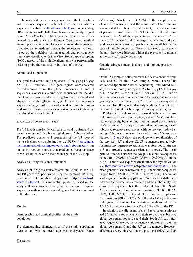
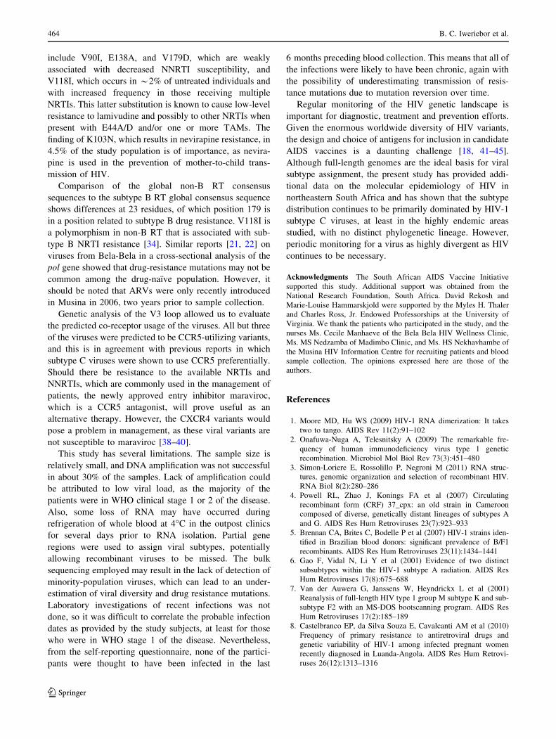
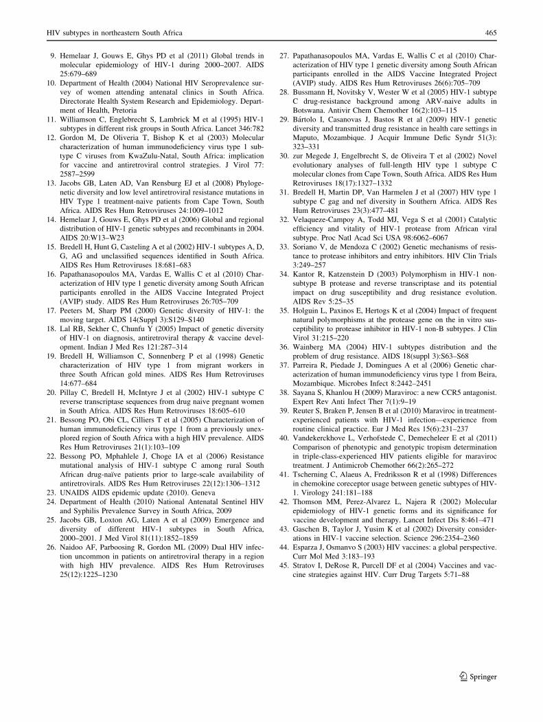
Phylogenetic analysis was performed on the gag p17, gag
p24, protease, reverse transcriptase, and env C2-V3 envelope
sequences. Neighbour-joining trees assigned the viruses to
HIV-1 subtype C, as they all clustered and intermingle with
subtype C reference sequences, with no monophyletic clus-
tering of the test sequences observed in any of the regions.
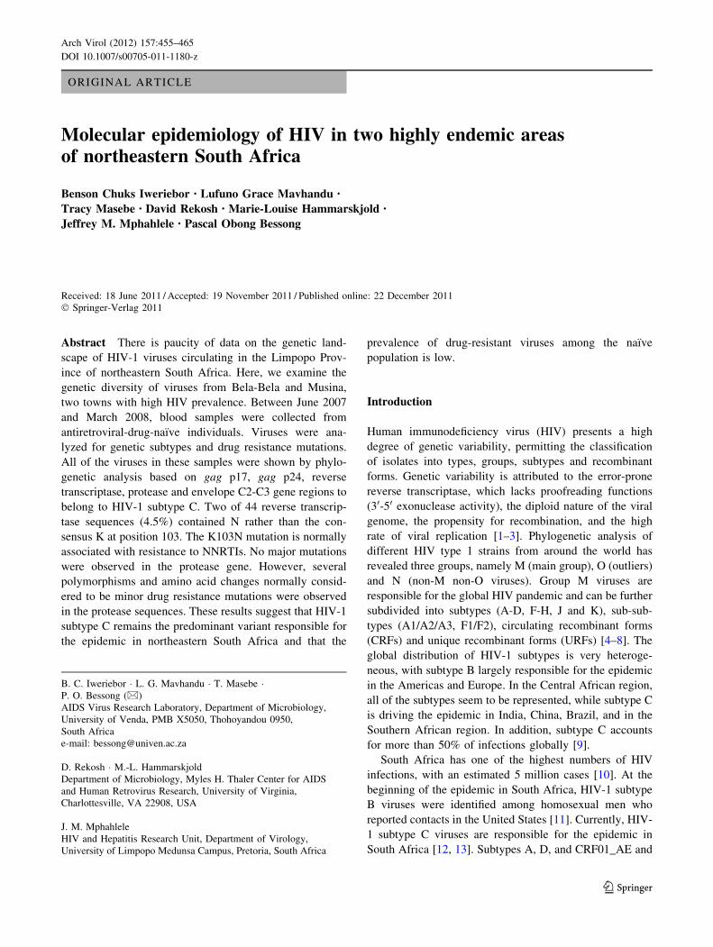
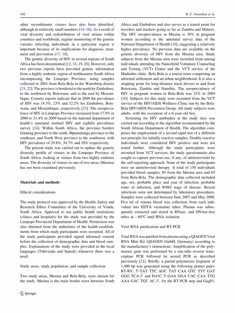
Figures 1, 2 and 3 show the phylogenetic relationships of
the gap p24, RT and env C2-C3 sequences, respectively.
A similar phylogenetic relationship was observed for the gag
p17 and protease sequences (data not shown). The mean
genetic distance between the gag p17 nucleotide sequences
ranged from 0.0053 to 0.2929 (0.53% to 29.29%). All of the
gag p17 amino acid sequences maintained the myristoylation
site (http://www.bioafrica.net/proteomics/index.html). The
mean genetic distance between the p24 nucleotide sequences
ranged from 0.0550 to 0.2510 (5.3% to 25.10%). The amino
acid alignments of the gag p17 and p24 showed no difference
between their consensus sequences and the global subtype C
consensus sequence, but they differed from the South
African vaccine strain at seven positions (E11G, K15A,
H27Q, I34L, M61I, K75R, and C111S) for the gag p17 and
four positions (I91V, N125S, V129I and R151K) in the gag
p24 region. Pairwise nucleotide distance analysis indicated a
3.4-9.6% divergence for the RT and 2.7-8.6% for the PR.
In addition, the alignment of the 44 reverse transcriptase
and 35 protease sequences with their respective subtype C
global consensus sequence and their South African refer-
ence sequence showed no sequence variation between the
global consensus C and the RT test sequences. However,
differences were observed at six positions (I60V, G123F,
458 B. C. Iweriebor et al.
123

V174A, K175Q, P277R and T286A) when compared to the
South African reference sequence. Also, the consensus of
the test sequences of the PR revealed one difference (T13I)
between the global subtype C consensus and three positions
(T19I, R20K and D35E) with regard to the South African
reference sequence (figures not shown).
0.1
07M7ZAAF067155 C
AY585266 CAY838582 C
07M17ZA07B8ZAA
07M8ZAU46016 C
AY838583 CU52953 C
07B15ZA07B12ZA
07M15ZA07B6ZA
07B14ZAAY838584 C
07B9ZA07B10ZA
07M11ZA07V7ZA
07M4ZA07B11ZA
07M25ZA07M18ZA
07V9ZA07V1ZA
07V4ZA07V12ZA
07M3ZA07B5ZA
1
07B13ZA07M19ZA
07V10ZA07V6ZA
07B36ZA07M2ZA
07B18ZA07M10ZA
07M16ZA07V8ZA
AY772699 C07M12ZA
07M13ZAAY838580 C
07B159ZA07B28ZA
07B26ZA07M24ZA
07B158ZA07B1ZA
07M22ZA07M9ZA
07M28ZA97
07V2ZA07V5ZA
AY838578 CAY838579 C
AY463217 CAY838586 C
07B2ZA
91
K03454 DAY371157 D
AY423387 BAF077336 F1
AF005494 F1AY371158 F2
AJ249236 F2AJ249235 K
AJ249239 K
84
AF190127 HAF190128 H
AF082394 JAF082395 J
AF004885 A1AF069670 A1
AF286238 A2AF286237 A2
96
AF084936 GAF061641 G
L20571 O
Fig. 1 Phylogeneticrelationships of gag p24nucleotide sequences fromnortheastern South Africa.Sequences were aligned with
reference HIV-1 subtype A-K
sequences, and a phylogenetic
tree was constructed by the
neighbor-joining method
implemented in ClustalX. Test
sequences (in boldface) cluster
and intermingle with subtype C
reference sequences. Bootstraps
values above 70% of 1000
replicates are indicated
HIV subtypes in northeastern South Africa 459
123
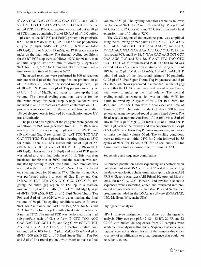
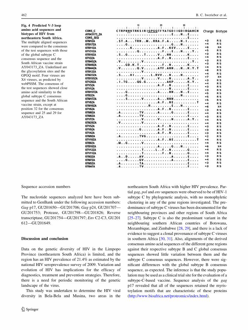
Comparison of the amino acid consensus sequence of
the V3 loop showed it was identical in all positions except
at Q32E to the global consensus sequences of subtypes C
and B. The mean genetic distance ranged from 0.0354 to
0.2630 (3.54-26.30%). Analysis of the V3 loop of the 38
env amino acid sequences showed that all of the isolates
had the GPGQ tetrapeptide, with the exception of
07M1ZA, 07M10ZA, and 07M23ZA, each of which had
amino acid substitutions in this motif. Co-receptor usage
prediction with webPSSM showed that all but 07B11ZA,
0.1
AY838568 CDQ222314 C
07B61ZADQ275647 C07B4ZADQ275648 C07B158ZA
07V29ZAAY463237 C
07B50ZA07B15ZA
07SH16ZA07B159ZA
DQ222316 C07M27ZA
DQ222313 CDQ222315 C
07M19ZAAY463228 C
07M16ZAAY463222 CDQ222310 C
07B1ZA07M5ZA
07B16ZA07B53ZA
DQ222312 C07M8ZA
07M22ZA07B10ZA
07M15ZA07V11ZA
AY463223 CAF067155 C
DQ275653 C07V13ZA
U52953 CU46016 C07B157ZA
07M18ZA07M45ZA
07B54ZA07H0601ZA
07M23ZA07V2ZA
07B18ZADQ222311 C
07M46ZA07V7ZA
07V8ZAAF110967 C
07V31ZA07M13ZA
DQ222317 C07M20ZA
07B13ZA07V12ZA
07B2ZA07V10ZA
AY228557 C07V32ZA07M26ZA07V1ZA
07V5ZAAF286226 BC
AF190127 HAF190128 H
K03455 BM17451 B
AF193276 ABAF385936 BF
K03454 DM27323 D
AF289548 CDAF077336 F1
AF005494 F1AJ249236 F2AJ249237 F2AJ249235 K
AJ249239 KAF004885 A1AF069670 A1
AF197340 AE
90
AF286237 A2
76
AF084936 GAF061641 G
AF063223 AG
98
AF082394 JAF082395 J
L20571 O
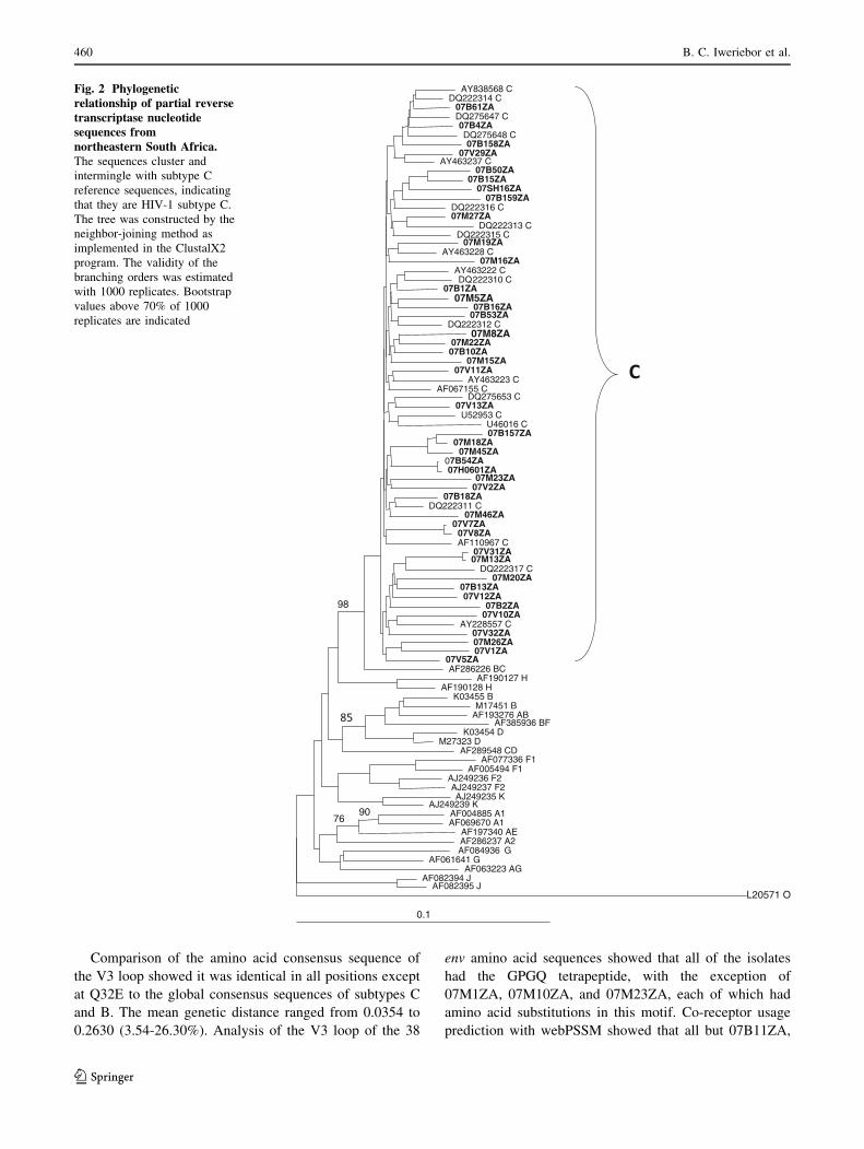
Fig. 2 Phylogeneticrelationship of partial reversetranscriptase nucleotidesequences fromnortheastern South Africa.The sequences cluster and
intermingle with subtype C
reference sequences, indicating
that they are HIV-1 subtype C.
The tree was constructed by the
neighbor-joining method as
implemented in the ClustalX2
program. The validity of the
branching orders was estimated
with 1000 replicates. Bootstrap
values above 70% of 1000
replicates are indicated
460 B. C. Iweriebor et al.
123
07B155ZA, and 07B10ZA (7.89%) were CCR5 viruses
with net charges ranging from ?2 to ?7. The usual
N-linked glycosylation sites at the start of the V3 loop were
maintained in all of the test sequences except 07M49ZA
and 07B155ZA (Fig. 4).
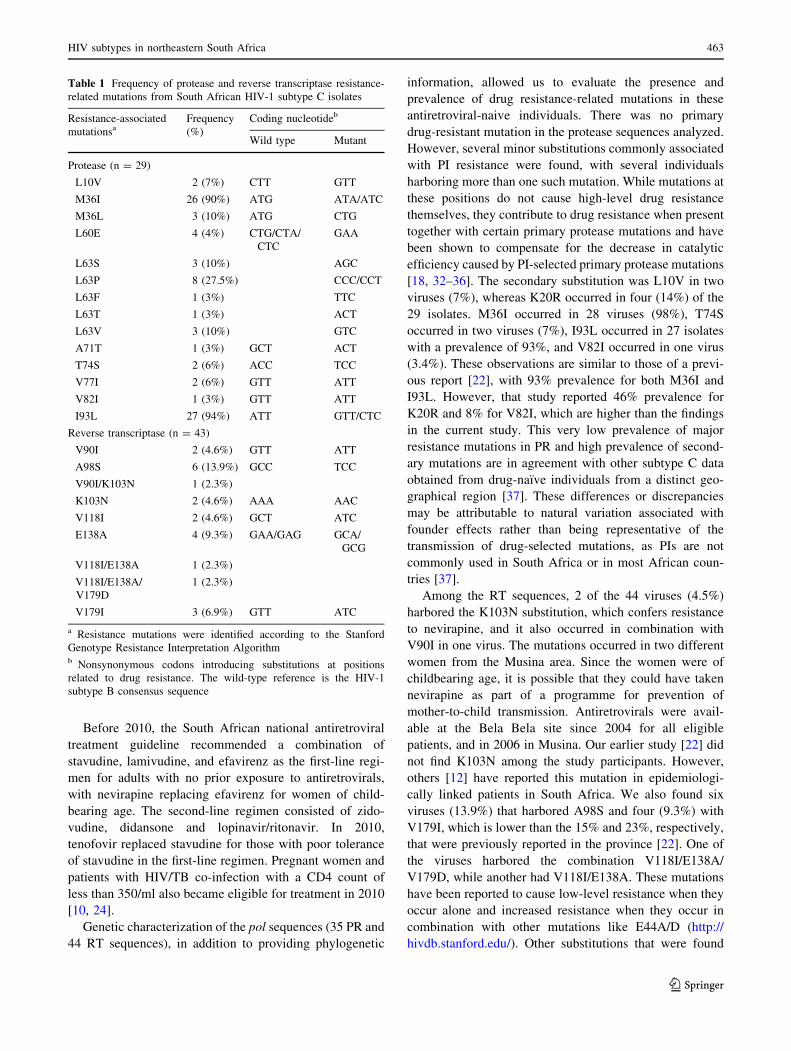
Genetic evidence for drug resistance in the analyzed
viruses
Nucleotide sequences were analyzed according to the
Stanford HIV genotypic resistance interpretation algorithm
in order to detect potential amino acid substitutions associ-
ated with resistance to PR and RT inhibitors. There were no
primary substitutions expected to confer resistance in the PR
sequences. However, substitutions in the protease that con-
tribute to PI resistance when present with other substitutions
were observed (Table 1). For the RT gene, the K103N
substitution, which confers primary resistance to NNRTIs
such as nevirapine was present in two of 44 sequences
(4.5%) (samples 07M5ZA and 07V2ZA). In addition, sec-
ondary mutations were found in many samples, and some
samples had combinations of substitutions (see Table 1).
0.1
07V12ZA07V11ZA
07V1ZA07V3ZA
07V5ZA07V9ZA07V4ZA
07V2ZA07V6ZA
DQ275653 CAY463223 C
07V8ZA07V10ZA
AY772699C07B11ZA
07B16ZAAY463237 C
07B2ZAAY463228 C07M10ZA
07B10ZA07M23ZA
DQ275648 CAY529672 C
DQ275647 C07M17ZA
07B155ZA07V7ZA
07B157ZA07V28ZA
07B12ZA07M44ZA
07M49ZAAY228557 C
U52953 CAY463235 C
07M1ZAAF067155 C
07V31ZA07M9ZA
07M5ZA07M2ZA07M8ZA
07M6ZA07M4ZA
07M7ZA07B17ZA
07B9ZA07M45ZA07M20ZA
U46016 C
91
AF077336 F1AF005494 F1
100
AY371158 F2AJ249236 F2
83
AJ249235 KAJ249239 K
94
AF084936 GAF061641 G
97
AF082394 JAF082395 J
AF004885 A1AF484509 A1
91
AF286238 A2AF286237 A2
K03454 DAY371157 D
K03455 BAY331295 B
99
AF190128 HAF190127 H
76
L20571 O
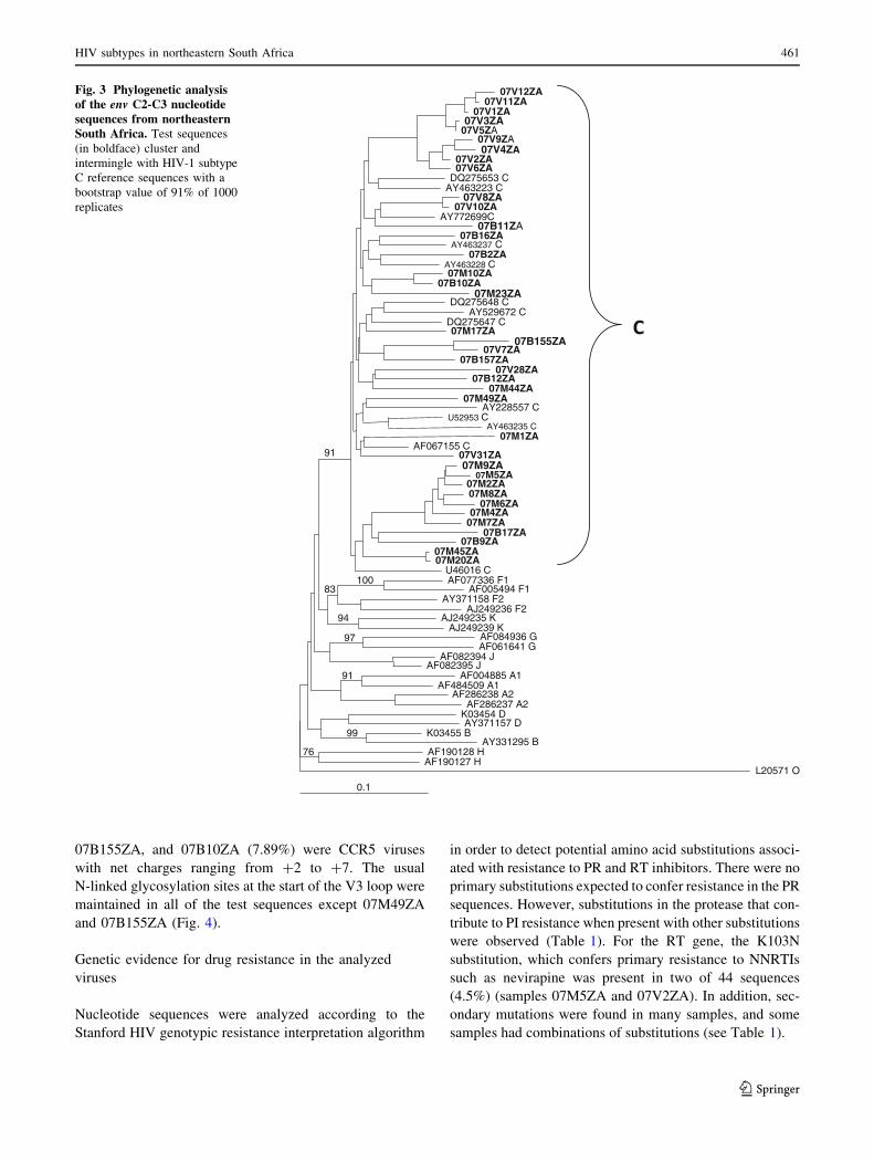
Fig. 3 Phylogenetic analysisof the env C2-C3 nucleotidesequences from northeasternSouth Africa. Test sequences
(in boldface) cluster and
intermingle with HIV-1 subtype
C reference sequences with a
bootstrap value of 91% of 1000
replicates
HIV subtypes in northeastern South Africa 461
123
Sequence accession numbers
The nucleotide sequences analyzed here have been sub-
mitted to GenBank under the following accession numbers:
Gag p17, GU201650—GU201706; Gag p24, GU201707—
GU201753; Protease, GU201798—GU201826; Reverse
transcriptase, GU201754—GU201797; Env C2-C3, GU201
612—GU201649.
Discussion and conclusion
Data on the genetic diversity of HIV in the Limpopo
Province (northeastern South Africa) is limited, and the
region has an HIV prevalence of 21.4% as estimated by the
national HIV seroprevalence survey of 2009. Variation and
evolution of HIV has implications for the efficacy of
diagnostics, treatment and prevention strategies. Therefore,
there is a need for periodic monitoring of the genetic
landscape of the virus.
This study was undertaken to determine the HIV viral
diversity in Bela-Bela and Musina, two areas in the
northeastern South Africa with higher HIV prevalence. Par-
tial gag, pol and env sequences were observed to be of HIV-1
subtype C by phylogenetic analysis, with no monophyletic
clustering in any of the gene regions investigated. The pre-
dominance of subtype C viruses has been documented for the
neighbouring provinces and other regions of South Africa
[25–27]. Subtype C is also the predominant variant in the
neighbouring southern African countries of Botswana,
Mozambique, and Zimbabwe [28, 29], and there is a lack of
evidence to suggest a clonal provenance of subtype C viruses
in southern Africa [30, 31]. Also, alignments of the derived
consensus amino acid sequences of the different gene regions
against their respective subtype B and C global consensus
sequences showed little variation between them and the
subtype C consensus sequences. However, there were sig-
nificant differences with the global subtype B consensus
sequence, as expected. The inference is that the study popu-
lation may be used as a clinical trial site for the evaluation of a
subtype-C-based vaccine. Sequence analysis of the gag
p17 revealed that all of the sequences retained the myris-
toylation motifs that are characteristic of these proteins
(http://www.bioafrica.net/proteomics/index.html).
Fig. 4 Predicted V-3 loopamino acid sequences andbiotypes of HIV fromnortheastern South Africa.The multiple aligned sequences
were compared to the consensus
of the test sequences with those
of the global subtype C
consensus sequence and the
South African vaccine strain
AY043173_ZA. Underlined are
the glycosylation sites and the
GPGQ motif. Four viruses are
X4 viruses, as predicted by
webPSSM. The consensus of
the test sequences showed close
amino acid similarity to the
global subtype C consensus
sequence and the South African
vaccine strain, except at
position 32 for the consensus
sequence and 25 and 29 for
AY043173_ZA
462 B. C. Iweriebor et al.
123
Before 2010, the South African national antiretroviral
treatment guideline recommended a combination of
stavudine, lamivudine, and efavirenz as the first-line regi-
men for adults with no prior exposure to antiretrovirals,
with nevirapine replacing efavirenz for women of child-
bearing age. The second-line regimen consisted of zido-
vudine, didansone and lopinavir/ritonavir. In 2010,
tenofovir replaced stavudine for those with poor tolerance
of stavudine in the first-line regimen. Pregnant women and
patients with HIV/TB co-infection with a CD4 count of
less than 350/ml also became eligible for treatment in 2010
[10, 24].
Genetic characterization of the pol sequences (35 PR and
44 RT sequences), in addition to providing phylogenetic
information, allowed us to evaluate the presence and
prevalence of drug resistance-related mutations in these
antiretroviral-naive individuals. There was no primary
drug-resistant mutation in the protease sequences analyzed.
However, several minor substitutions commonly associated
with PI resistance were found, with several individuals
harboring more than one such mutation. While mutations at
these positions do not cause high-level drug resistance
themselves, they contribute to drug resistance when present
together with certain primary protease mutations and have
been shown to compensate for the decrease in catalytic
efficiency caused by PI-selected primary protease mutations
[18, 32–36]. The secondary substitution was L10V in two
viruses (7%), whereas K20R occurred in four (14%) of the
29 isolates. M36I occurred in 28 viruses (98%), T74S
occurred in two viruses (7%), I93L occurred in 27 isolates
with a prevalence of 93%, and V82I occurred in one virus
(3.4%). These observations are similar to those of a previ-
ous report [22], with 93% prevalence for both M36I and
I93L. However, that study reported 46% prevalence for
K20R and 8% for V82I, which are higher than the findings
in the current study. This very low prevalence of major
resistance mutations in PR and high prevalence of second-
ary mutations are in agreement with other subtype C data
obtained from drug-naıve individuals from a distinct geo-
graphical region [37]. These differences or discrepancies
may be attributable to natural variation associated with
founder effects rather than being representative of the
transmission of drug-selected mutations, as PIs are not
commonly used in South Africa or in most African coun-
tries [37].
Among the RT sequences, 2 of the 44 viruses (4.5%)
harbored the K103N substitution, which confers resistance
to nevirapine, and it also occurred in combination with
V90I in one virus. The mutations occurred in two different
women from the Musina area. Since the women were of
childbearing age, it is possible that they could have taken
nevirapine as part of a programme for prevention of
mother-to-child transmission. Antiretrovirals were avail-
able at the Bela Bela site since 2004 for all eligible
patients, and in 2006 in Musina. Our earlier study [22] did
not find K103N among the study participants. However,
others [12] have reported this mutation in epidemiologi-
cally linked patients in South Africa. We also found six
viruses (13.9%) that harbored A98S and four (9.3%) with
V179I, which is lower than the 15% and 23%, respectively,
that were previously reported in the province [22]. One of
the viruses harbored the combination V118I/E138A/
V179D, while another had V118I/E138A. These mutations
have been reported to cause low-level resistance when they
occur alone and increased resistance when they occur in
combination with other mutations like E44A/D (http://
hivdb.stanford.edu/). Other substitutions that were found
Table 1 Frequency of protease and reverse transcriptase resistance-
related mutations from South African HIV-1 subtype C isolates
Resistance-associated
mutationsaFrequency
(%)
Coding nucleotideb
Wild type Mutant
Protease (n = 29)
L10V 2 (7%) CTT GTT
M36I 26 (90%) ATG ATA/ATC
M36L 3 (10%) ATG CTG
L60E 4 (4%) CTG/CTA/
CTC
GAA
L63S 3 (10%) AGC
L63P 8 (27.5%) CCC/CCT
L63F 1 (3%) TTC
L63T 1 (3%) ACT
L63V 3 (10%) GTC
A71T 1 (3%) GCT ACT
T74S 2 (6%) ACC TCC
V77I 2 (6%) GTT ATT
V82I 1 (3%) GTT ATT
I93L 27 (94%) ATT GTT/CTC
Reverse transcriptase (n = 43)
V90I 2 (4.6%) GTT ATT
A98S 6 (13.9%) GCC TCC
V90I/K103N 1 (2.3%)
K103N 2 (4.6%) AAA AAC
V118I 2 (4.6%) GCT ATC
E138A 4 (9.3%) GAA/GAG GCA/
GCG
V118I/E138A 1 (2.3%)
V118I/E138A/
V179D
1 (2.3%)
V179I 3 (6.9%) GTT ATC
a Resistance mutations were identified according to the Stanford
Genotype Resistance Interpretation Algorithmb Nonsynonymous codons introducing substitutions at positions
related to drug resistance. The wild-type reference is the HIV-1
subtype B consensus sequence
HIV subtypes in northeastern South Africa 463
123
include V90I, E138A, and V179D, which are weakly
associated with decreased NNRTI susceptibility, and
V118I, which occurs in *2% of untreated individuals and
with increased frequency in those receiving multiple
NRTIs. This latter substitution is known to cause low-level
resistance to lamivudine and possibly to other NRTIs when
present with E44A/D and/or one or more TAMs. The
finding of K103N, which results in nevirapine resistance, in
4.5% of the study population is of importance, as nevira-
pine is used in the prevention of mother-to-child trans-
mission of HIV.
Comparison of the global non-B RT consensus
sequences to the subtype B RT global consensus sequence
shows differences at 23 residues, of which position 179 is
in a position related to subtype B drug resistance. V118I is
a polymorphism in non-B RT that is associated with sub-
type B NRTI resistance [34]. Similar reports [21, 22] on
viruses from Bela-Bela in a cross-sectional analysis of the
pol gene showed that drug-resistance mutations may not be
common among the drug-naıve population. However, it
should be noted that ARVs were only recently introduced
in Musina in 2006, two years prior to sample collection.
Genetic analysis of the V3 loop allowed us to evaluate
the predicted co-receptor usage of the viruses. All but three
of the viruses were predicted to be CCR5-utilizing variants,
and this is in agreement with previous reports in which
subtype C viruses were shown to use CCR5 preferentially.
Should there be resistance to the available NRTIs and
NNRTIs, which are commonly used in the management of
patients, the newly approved entry inhibitor maraviroc,
which is a CCR5 antagonist, will prove useful as an
alternative therapy. However, the CXCR4 variants would
pose a problem in management, as these viral variants are
not susceptible to maraviroc [38–40].
This study has several limitations. The sample size is
relatively small, and DNA amplification was not successful
in about 30% of the samples. Lack of amplification could
be attributed to low viral load, as the majority of the
patients were in WHO clinical stage 1 or 2 of the disease.
Also, some loss of RNA may have occurred during
refrigeration of whole blood at 4�C in the outpost clinics
for several days prior to RNA isolation. Partial gene
regions were used to assign viral subtypes, potentially
allowing recombinant viruses to be missed. The bulk
sequencing employed may result in the lack of detection of
minority-population viruses, which can lead to an under-
estimation of viral diversity and drug resistance mutations.
Laboratory investigations of recent infections was not
done, so it was difficult to correlate the probable infection
dates as provided by the study subjects, at least for those
who were in WHO stage 1 of the disease. Nevertheless,
from the self-reporting questionnaire, none of the partici-
pants were thought to have been infected in the last
6 months preceding blood collection. This means that all of
the infections were likely to have been chronic, again with
the possibility of underestimating transmission of resis-
tance mutations due to mutation reversion over time.
Regular monitoring of the HIV genetic landscape is
important for diagnostic, treatment and prevention efforts.
Given the enormous worldwide diversity of HIV variants,
the design and choice of antigens for inclusion in candidate
AIDS vaccines is a daunting challenge [18, 41–45].
Although full-length genomes are the ideal basis for viral
subtype assignment, the present study has provided addi-
tional data on the molecular epidemiology of HIV in
northeastern South Africa and has shown that the subtype
distribution continues to be primarily dominated by HIV-1
subtype C viruses, at least in the highly endemic areas
studied, with no distinct phylogenetic lineage. However,
periodic monitoring for a virus as highly divergent as HIV
continues to be necessary.
Acknowledgments The South African AIDS Vaccine Initiative
supported this study. Additional support was obtained from the
National Research Foundation, South Africa. David Rekosh and
Marie-Louise Hammarskjold were supported by the Myles H. Thaler
and Charles Ross, Jr. Endowed Professorships at the University of
Virginia. We thank the patients who participated in the study, and the
nurses Ms. Cecile Manhaeve of the Bela Bela HIV Wellness Clinic,
Ms. MS Nedzamba of Madimbo Clinic, and Ms. HS Nekhavhambe of
the Musina HIV Information Centre for recruiting patients and blood
sample collection. The opinions expressed here are those of the
authors.
References
1. Moore MD, Hu WS (2009) HIV-1 RNA dimerization: It takes
two to tango. AIDS Rev 11(2):91–102
2. Onafuwa-Nuga A, Telesnitsky A (2009) The remarkable fre-
quency of human immunodeficiency virus type 1 genetic
recombination. Microbiol Mol Biol Rev 73(3):451–480
3. Simon-Loriere E, Rossolillo P, Negroni M (2011) RNA struc-
tures, genomic organization and selection of recombinant HIV.
RNA Biol 8(2):280–286
4. Powell RL, Zhao J, Konings FA et al (2007) Circulating
recombinant form (CRF) 37_cpx: an old strain in Cameroon
composed of diverse, genetically distant lineages of subtypes A
and G. AIDS Res Hum Retroviruses 23(7):923–933
5. Brennan CA, Brites C, Bodelle P et al (2007) HIV-1 strains iden-
tified in Brazilian blood donors: significant prevalence of B/F1
recombinants. AIDS Res Hum Retroviruses 23(11):1434–1441
6. Gao F, Vidal N, Li Y et al (2001) Evidence of two distinct
subsubtypes within the HIV-1 subtype A radiation. AIDS Res
Hum Retroviruses 17(8):675–688
7. Van der Auwera G, Janssens W, Heyndrickx L et al (2001)
Reanalysis of full-length HIV type 1 group M subtype K and sub-
subtype F2 with an MS-DOS bootscanning program. AIDS Res
Hum Retroviruses 17(2):185–189
8. Castelbranco EP, da Silva Souza E, Cavalcanti AM et al (2010)
Frequency of primary resistance to antiretroviral drugs and
genetic variability of HIV-1 among infected pregnant women
recently diagnosed in Luanda-Angola. AIDS Res Hum Retrovi-
ruses 26(12):1313–1316
464 B. C. Iweriebor et al.
123
9. Hemelaar J, Gouws E, Ghys PD et al (2011) Global trends in
molecular epidemiology of HIV-1 during 2000–2007. AIDS
25:679–689
10. Department of Health (2004) National HIV Seroprevalence sur-
vey of women attending antenatal clinics in South Africa.
Directorate Health System Research and Epidemiology. Depart-
ment of Health, Pretoria
11. Williamson C, Englebrecht S, Lambrick M et al (1995) HIV-1
subtypes in different risk groups in South Africa. Lancet 346:782
12. Gordon M, De Oliveria T, Bishop K et al (2003) Molecular
characterization of human immunodeficiency virus type 1 sub-
type C viruses from KwaZulu-Natal, South Africa: implication
for vaccine and antiretroviral control strategies. J Virol 77:
2587–2599
13. Jacobs GB, Laten AD, Van Rensburg EJ et al (2008) Phyloge-
netic diversity and low level antiretroviral resistance mutations in
HIV Type 1 treatment-naive patients from Cape Town, South
Africa. AIDS Res Hum Retroviruses 24:1009–1012
14. Hemelaar J, Gouws E, Ghys PD et al (2006) Global and regional
distribution of HIV-1 genetic subtypes and recombinants in 2004.
AIDS 20:W13–W23
15. Bredell H, Hunt G, Casteling A et al (2002) HIV-1 subtypes A, D,
G, AG and unclassified sequences identified in South Africa.
AIDS Res Hum Retroviruses 18:681–683
16. Papathanasopoulos MA, Vardas E, Wallis C et al (2010) Char-
acterization of HIV type 1 genetic diversity among South African
participants enrolled in the AIDS Vaccine Integrated Project
(AVIP) study. AIDS Res Hum Retroviruses 26:705–709
17. Peeters M, Sharp PM (2000) Genetic diversity of HIV-1: the
moving target. AIDS 14(Suppl 3):S129–S140
18. Lal RB, Sekher C, Chunfu Y (2005) Impact of genetic diversity
of HIV-1 on diagnosis, antiretroviral therapy & vaccine devel-
opment. Indian J Med Res 121:287–314
19. Bredell H, Williamson C, Sonnenberg P et al (1998) Genetic
characterization of HIV type 1 from migrant workers in
three South African gold mines. AIDS Res Hum Retroviruses
14:677–684
20. Pillay C, Bredell H, McIntyre J et al (2002) HIV-1 subtype C
reverse transcriptase sequences from drug naive pregnant women
in South Africa. AIDS Res Hum Retroviruses 18:605–610
21. Bessong PO, Obi CL, Cilliers T et al (2005) Characterization of
human immunodeficiency virus type 1 from a previously unex-
plored region of South Africa with a high HIV prevalence. AIDS
Res Hum Retroviruses 21(1):103–109
22. Bessong PO, Mphahlele J, Choge IA et al (2006) Resistance
mutational analysis of HIV-1 subtype C among rural South
African drug-naıve patients prior to large-scale availability of
antiretrovirals. AIDS Res Hum Retroviruses 22(12):1306–1312
23. UNAIDS AIDS epidemic update (2010). Geneva
24. Department of Health (2010) National Antenatal Sentinel HIV
and Syphilis Prevalence Survey in South Africa, 2009
25. Jacobs GB, Loxton AG, Laten A et al (2009) Emergence and
diversity of different HIV-1 subtypes in South Africa,
2000–2001. J Med Virol 81(11):1852–1859
26. Naidoo AF, Parboosing R, Gordon ML (2009) Dual HIV infec-
tion uncommon in patients on antiretroviral therapy in a region
with high HIV prevalence. AIDS Res Hum Retroviruses
25(12):1225–1230
27. Papathanasopoulos MA, Vardas E, Wallis C et al (2010) Char-
acterization of HIV type 1 genetic diversity among South African
participants enrolled in the AIDS Vaccine Integrated Project
(AVIP) study. AIDS Res Hum Retroviruses 26(6):705–709
28. Bussmann H, Novitsky V, Wester W et al (2005) HIV-1 subtype
C drug-resistance background among ARV-naive adults in
Botswana. Antivir Chem Chemother 16(2):103–115
29. Bartolo I, Casanovas J, Bastos R et al (2009) HIV-1 genetic
diversity and transmitted drug resistance in health care settings in
Maputo, Mozambique. J Acquir Immune Defic Syndr 51(3):
323–331
30. zur Megede J, Engelbrecht S, de Oliveira T et al (2002) Novel
evolutionary analyses of full-length HIV type 1 subtype C
molecular clones from Cape Town, South Africa. AIDS Res Hum
Retroviruses 18(17):1327–1332
31. Bredell H, Martin DP, Van Harmelen J et al (2007) HIV type 1
subtype C gag and nef diversity in Southern Africa. AIDS Res
Hum Retroviruses 23(3):477–481
32. Velaqueze-Campoy A, Todd MJ, Vega S et al (2001) Catalytic
efficiency and vitality of HIV-1 protease from African viral
subtype. Proc Natl Acad Sci USA 98:6062–6067
33. Soriano V, de Mendoza C (2002) Genetic mechanisms of resis-
tance to protease inhibitors and entry inhibitors. HIV Clin Trials
3:249–257
34. Kantor R, Katzenstein D (2003) Polymorphism in HIV-1 non-
subtype B protease and reverse transcriptase and its potential
impact on drug susceptibility and drug resistance evolution.
AIDS Rev 5:25–35
35. Holguin L, Paxinos E, Hertogs K et al (2004) Impact of frequent
natural polymorphisms at the protease gene on the in vitro sus-
ceptibility to protease inhibitor in HIV-1 non-B subtypes. J Clin
Virol 31:215–220
36. Wainberg MA (2004) HIV-1 subtypes distribution and the
problem of drug resistance. AIDS 18(suppl 3):S63–S68
37. Parreira R, Piedade J, Domingues A et al (2006) Genetic char-
acterization of human immunodeficiency virus type 1 from Beira,
Mozambique. Microbes Infect 8:2442–2451
38. Sayana S, Khanlou H (2009) Maraviroc: a new CCR5 antagonist.
Expert Rev Anti Infect Ther 7(1):9–19
39. Reuter S, Braken P, Jensen B et al (2010) Maraviroc in treatment-
experienced patients with HIV-1 infection—experience from
routine clinical practice. Eur J Med Res 15(6):231–237
40. Vandekerckhove L, Verhofstede C, Demecheleer E et al (2011)
Comparison of phenotypic and genotypic tropism determination
in triple-class-experienced HIV patients eligible for maraviroc
treatment. J Antimicrob Chemother 66(2):265–272
41. Tscherning C, Alaeus A, Fredriksson R et al (1998) Differences
in chemokine coreceptor usage between genetic subtypes of HIV-
1. Virology 241:181–188
42. Thomson MM, Perez-Alvarez L, Najera R (2002) Molecular
epidemiology of HIV-1 genetic forms and its significance for
vaccine development and therapy. Lancet Infect Dis 8:461–471
43. Gaschen B, Taylor J, Yusim K et al (2002) Diversity consider-
ations in HIV-1 vaccine selection. Science 296:2354–2360
44. Esparza J, Osmanvo S (2003) HIV vaccines: a global perspective.
Curr Mol Med 3:183–193
45. Stratov I, DeRose R, Purcell DF et al (2004) Vaccines and vac-
cine strategies against HIV. Curr Drug Targets 5:71–88
HIV subtypes in northeastern South Africa 465
123
Top Related
Copyright © 2022 FDOKUMEN

