


Bahasa
Halaman
Hukum

MINOES: a new approach to select a representativeensemble of structures in NMR studies of (partially)unfolded states. Application to Δ25-PYP
Mickaël Krzeminski, Gloria Fuentes†, Rolf Boelens and Alexandre M.J.J. Bonvin*
Bijvoet Center for Biomolecular Research, Science Faculty, Utrecht University, 3584 CH,
Utrecht, The Netherlands
†Current address: Structural Computational Biology, Centro Nacional de Investigaciones
Oncológicas (C.N.I.O.). Melchor Fernández Almagro, 3, E-28029 Madrid
* Corresponding author: Alexandre M.J.J. Bonvin
tel.: +31.(0)30.2532652
fax: +31.(0)30.2537623
Keywords: Partially unfolded states, PYP, ensemble selection, NMR, Monte-Carlo
MINimum Optimal Ensemble Selection
ABSTRACT In nature, some proteins partially unfold under specific environmentalconditions. These unfolded states typically consist of a large ensemble of conformations; theirproper description is therefore a challenging problem. NMR spectroscopy is particularly wellsuited for this task: information on conformational preferences can be derived for examplefrom chemical shifts or residual dipolar couplings. This information, which is measured as atime- and ensemble-average, can be used to model these states by generating large ensemblesof conformations. The challenge is then to select a minimum representative set ofconformations out of a large ensemble to represent the unfolded state.We have developed for this purpose an algorithm called MINOES (MINimum OptimalEnsemble Selection), that is based on an iterative selection procedure embedded in a Monte-Carlo process. MINOES aims at selecting an optimal ensemble of conformations that, onaverage, maximizes the agreement between back-calculated and experimental (NMR) data,without any a-priori assumption about the required ensemble size. This approach isdemonstrated by modeling the partially unfolded state of a deletion mutant of the PhotoactiveYellow Protein, Δ25-PYP, which has been previously characterized by NMR (Bernard et al.Structure 13, 953-962, 2005)
MINimum Optimal Ensemble Selection
INTRODUCTION
High resolution NMR spectroscopy is a powerful method that has become a routinefor the structural characterization of biomolecules. In particular the ability to study dynamicalproperties makes the technique complementary to X-ray crystallography. It normally relies ona dense network of distance restraints derived from nuclear Overhauser effects (NOEs)between nearby hydrogen atoms (Neuhaus and Williamson, 2000; Wüthrich, 1986) tocalculate the three dimensional (3D) structure of a protein in solution.
The preferred representation of an NMR protein structure in solution is an ensemble,in the order of 20 models, which explore regions of conformational space that satisfy at thesame time some physical parameters and experimentally-derived restraints. The most usedprocedures to select these structures from a large set of calculated ones are based on energy(selection of a sub-ensemble consisting of the lowest energy structures) and/or restraintsviolation criteria (selection of structures with no distance and/or dihedral angle violationabove a given threshold). Both approaches involve user-defined, arbitrary criteria for the cut-offs used and the number of structures selected; this together with possible differences inforce field parameters and in the treatment of the experimental restraints used in the processlead to the “structure selection problem” in the structural NMR field. This problem is evenmore acute when it comes to describing highly flexible molecules such as short peptides orpoorly defined (partially) unfolded states of proteins.
Chemical shifts have been long recognized as a potentially important structuralinformation source, due to their dependency and high sensitivity on multiple electronic andgeometric factors, and the high accuracy of their measurement (Wishart and Sykes, 1994a;Wüthrich, 1986). A large variety of conformational effects, including backbone torsionangles, side-chain orientations, hydrogen bonding, and the type and conformation ofneighboring residues, influence the chemical shift behavior in proteins (Le and Oldfield,1994; Spitzfaden et al., 1994). All these effects make the chemical shifts good reporters ofsecondary structure elements as implemented in several softwares (e.g. CSI (Wishart andSykes, 1994b), Talos (Cornilescu et al., 1999), Pecan (Eghbalnia et al., 2005)), as well as ofstructural changes observed upon different conditions (e.g. ligand binding (Zuiderweg,2002)). However, the multiple dependencies mentioned above make both the interpretationand accurate prediction of chemical shifts exceedingly difficult, particularly in large systems,and their use, so far, has been limited. Fortunately, significant computational progresses inchemical shift prediction have been made (Case, 1998; Meiler, 2003; Neal et al., 2003;Williamson and Asakura, 1997; Wishart and Case, 2001) opening new possibilities forincluding them on a regular basis in structural studies.
In this paper we investigate the use of chemical shifts as information source for theselection of a representative ensemble of NMR structures. For this, we chose a specific classof proteins containing a photoreceptor that partially unfolds upon illumination. Weconcentrate on the photoactive yellow protein (PYP), which has been extensively studied inour laboratory in the past (Bernard et al., 2005; Dux et al., 1998). The transition to thisexcited, partially unfolded state, which corresponds actually to the signaling state of theseproteins, has been described to follow a protein quake model (Ansari et al., 1985; Itoh andSasai, 2004). The transient and unstable nature of such a state makes the acquisition of NMRdata difficult. As a result, only sparse data can be typically obtained. Hence, characterizingthe partially unfolded state of such proteins becomes rather complicated, and a challenge tothe current structural methods. A protein that partially unfolds can usually be split into twoparts: a core, which maintains to a large extend a conformation similar to the native state, anda partially unfolded moiety.
MINimum Optimal Ensemble Selection
In a previous work (Fuentes et al., 2005), we have demonstrated that it is possible to model insilico partially unfolded states using native-like inter-residue restraints for those residues thatdo not show appreciable chemical shift changes upon partially unfolding. Here, assuming thata partially unfolded state corresponds to an intermediate between a fully folded and fullyunfolded state, we introduce a structure calculation protocol based on a gradual unfolding ofthe partially unfolded moiety by progressively decreasing the weight of the correspondingnative-like restraints in the structural calculation protocol. In addition, a radius of gyration(Rg) restraint (Kuszewski et al., 1999) derived from NMR diffusion measurements isintroduced for better characterization of the partially unfolded state. In that way a largeensemble of conformations can be generated covering the conformational space from thenative to the (partially) unfolded state. The challenge is then to select an ensemble ofstructures that best describes the experimental observables. In the case of partially unfolded proteins, the classical approach that selects the lowest energystructures and/or those having the minimum number of violations, is no longer suitable;because of the scarcity of the experimental data, those parts of the protein for which no oronly little experimental data are available will strongly depend on the forcefield andcalculation protocol used. The selection problem in the case of unfolded systems has beenaddressed previously by Forman-Kay’s group (Choy and Forman-Kay, 2001; Marsh et al.,2007) who developed an algorithm called ENSEMBLe. It makes uses of the availableexperimental data to define some kind of energy function in which averaged back-calculateddata and experimental data are compared; the latest version of the algorithm tries to select in aMonte Carlo process a user-defined number of structures that best represent the experimentaldata.
Here we propose an innovative selection method which extracts an optimal subset outof a large ensemble by maximizing the agreement between observables back-calculated fromthe generated models and the experimental data without making any assumption about theensemble size. While the approach is generic, we demonstrate it using protons Hα chemicalshifts for the selection process. We first validate our selection method using synthetic data forlysozyme and then apply it to model the partially unfolded state of a deletion mutant of thePhotoactive Yellow Protein (Δ25-PYP) (van der Horst et al., 2001), for which experimentalNMR data are available (Bernard et al., 2005); this allows us to compare the resultingstructures and validate our proposed selection method.
THEORY
In order to evaluate how a selected model or ensemble thereof will represent theexperimental data, one can calculate the "distance" between the available experimental dataand back-calculated data obtained from these models. This distance can be expressed in theform of a χ2 function like in the following formula:
2χ = iω2
ix − iy( )i=1
D
∑ i2σ
with:D = the number of available dataxi = the data value predicted from modelsyi = the experimental data value
MINimum Optimal Ensemble Selection
ωi = weight put on data i σi = experimental error associated with data i
Such a function has the advantage that it allows the combination of various kinds of data andtakes experimental errors into account. Also, each data point or set of points can be weightedseparately by adjusting the value of ωi.
In our selection algorithm, we make use of χ2 to determine from a pool of generatedstructures, the sub-ensemble that best fits the experimental data, without a-priori knowledgeof the optimum ensemble size. This is a combinatorial problem that would lead to anexplosion in computational time requirements if all possibilities were to be tested for a largeensemble. To avoid this problem, we use a recursive approach embedded in a Monte-Carloprocess. The selection algorithm consists of the following steps:
For N trials:
1. Randomly select a model from the ensemble.
2. Generate new ensembles bya. adding each of the non-selected structures in turnb. removing each of the selected structures in turnc. exchanging each of the selected structure by each of the non-selected ones
3. Select the ensemble from step 2 with the lowest χ2 . Compare the newly selectedensemble with the previously defined ensemble:
a. If 2newχ < 2oldχ , accept the new ensemble and start again from step 2.
b. If 2newχ ≥ 2oldχ , keep the old ensemble and start again from the step 2. If the
ensemble does not change after repeating this selection process twentytimes, proceed to step 4.
4. From the best ensemble determined so far, eliminate randomly a defined numberof selected models and repeat steps 2 to 4 until no ensemble with a better χ2 isfound after 20 consecutive runs.
To guarantee an unbiased distribution of selected structures in the new ensemble, each modelis given a probability of being chosen, that linearly decreases every time it is selected.Moreover, we make use of the tabu search method (Glover and Laguna, 1997). This heuristicmechanism allows to visit more possible ensembles by avoiding in the selection process to goback to a previous state. Hence, if a modification results in an ensemble that has already beenmet, it is not kept, but instead the second best one is selected, and so on.This selection method has been coded in C in the MINOES program, standing for MINimumOptimal Ensemble Selection.
MATERIAL AND METHODS
Generation of lysozyme reference ensembles for the validation of the method
The crystal structure hen egg-white lysozyme (PDB entry: 1AKI) was used to generateby molecular dynamic (MD) simulations models on which our algorithm could be validated.The reference ensemble for the native state in solution (RN) consists of 20 energy minimized
MINimum Optimal Ensemble Selection
MD snapshots taken from a 10 nanosecond (ns) MD simulation in explicit solvent, obtainedusing the Gromacs package (Lindahl, 2001) (for details of the simulation refer to (Fuentes etal., 2005)). We simulated an experimental-like 1H-15N HSQC spectrum by calculating withShiftX (Neal et al., 2003) HN and N chemical shifts for each of the extracted models and thenaveraging them over the reference ensemble.
The reference ensemble for the partially unfolded state (RU) consists of 20 energyminimized snapshots taken from the last 10 ns of a 20 ns stochastic dynamics simulation at600K, where position restraints where applied on the α-domain of the protein, while theremaining of the protein (β-domain, residues G4 to S36 and T89 to L129) was allowed to movefreely in order to sample a larger conformational space. Chemical shifts for this partiallyunfolded state ensemble were obtained as described above. 15N–1H chemical shift differencesfrom the simulated HSQC spectra of these two states were used to define native-like restraintsas described previously (Fuentes et al., 2005). In short, a native-like distance restraint isdefined only if the corresponding distance is shorter than 7.5 Å in at least half of structures ofthe native state NMR ensemble. In this case, the restraint is set to the average of all distancesbelow 7.5 Å, with a lower distance bound of 1.8 Å and an upper one equal to the average plusone standard deviation (core/core distance) or to the average plus the standard deviation plus2.0 Å (core/non-core or non-core/non-core). Distance restraints are only defined betweenresidues that are at least three positions apart in the sequence.
Additionally, the average radius of gyration (Rg) was calculated from the last 10 ns ofthe trajectory.
Experimental data for Δ25-PYP
The 1H15N-HSQC spectra of both the native (dark state, pG) and partially unfolded(light state, pB intermediate) states of Δ25-PYP have been described previously (Bernard etal., 2005). The chemical shift perturbation calculated from these two spectra allowed us todefine the core (61 residues out of 100, comprising by the segments: G29 to N43, K55 to F62,G82 to F96 and T103 to V125) and the unfolded part of the protein. This information was used todefine native-like restraints from the NMR pG ensemble (PDB entry: 1XFN)
NMR-based generation of a partially unfolded state ensemble
Structure ensembles were generated with CNS (Brunger, 1998) using protocolsderived from ARIA (Linge et al., 2001) as implemented in the RECOORD scripts(Nederveen, 2005). For each system, we performed first 13 structure calculation runs (withoutwater refinement), each of them producing 2000 structures. In all the runs, native-like Cα-Cβ
distance restraints were imposed for the folded (core region) moiety as described previously(Fuentes et al., 2005). In addition, considering that partial unfolding starts from the nativestate, native-like restraints were also applied to the unfolded part, with the difference thatdecreasing force constants (from 50 to 0 kcal mol-1 Å-2) were applied in the different runs (seeTable 1). This allows to slowly relax the non-native of the protein, from a fully folded state tothe fully unfolded one.
RunInitialweight
Finalweight
RunInitialweight
Finalweight
0 10 50 7 3 151 9 45 8 2 102 8 40 9 1 5
MINimum Optimal Ensemble Selection
3 7 35 10 0.2 14 6 30 11 0.1 0.55 5 25 12 0 06 4 20
Table 1: Force constants (kcal.mol-1.Å-2) for distance restraints usedduring the simulated annealing protocol for the various runs for thegeneration of the partially unfolded state ensemble.
The radius of gyration Rg was included as a restraint in such a way that the targetvalues imposed for the entire ensemble of structures calculated follow a log-normalprobability density function. Hence, for 0≥r , we have the following formula:
F(r) = 1r − Rg
eff ×1
σ 2π× e
− 12
ln r−Rgeff( )−µ
σ
⎛
⎝⎜⎜
⎞
⎠⎟⎟
2
Where effgR is the experimental value of the radius of gyration. We arbitrary set the values of
µ and σ to 0.2 and 0.4, respectively. By taking values from a log normal distribution, weallow for more extension than compaction of the structure. For each individual structurecalculation a different value of eff
gR is used, taken from the log-normal distribution. Rg was
set to 14.3 Å for the lysozyme (average from the last 10 ns of the simulation) and to 14.0 Åfor Δ25-PYP based on NMR diffusion experiments (Nico van Nuland, Universidad deGranada, Spain; personal communication).For each run, the 500 lowest energy structures obtained after simulated annealing weresubmitted to refinement in explicit solvent. From these refined models, only those with lessthan 5 distance restraint violations below 0.5 Å were kept for further analysis; their Hα
chemical shifts were calculated using ShiftX.
Analysis of models
The selected ensembles were compared with the reference one or with each other bycalculating the average pairwise positional root mean square deviations (RMSD). For this, thestructures were fitted on the backbone atoms of the defined core regions and the RMSD werecalculated on the entire backbone of the protein, using ProFit (Martin, A.C.R.,http://www.bioinf.org.uk/software/profit/).
Secondary structure analysis was performed with PROCHECK (Laskowski et al.,1993; Laskowski et al., 1996), which makes use of the Kabsch & Sander secondary structuredefinitions (Kabsch and Sander, 1983). A consensus secondary structure for each residue wasadopted when at least half of the structures of an NMR ensemble had the same classification.
RESULTS
Validation of the method with synthetic data
We validated our algorithm by performing the selection of a sub-ensemble that bestfits the synthetic lysozyme data. Since different methods and force fields were used to
MINimum Optimal Ensemble Selection
generate the reference and NMR-based ensembles, these might not per se overlap (containsimilar structures). Therefore we first tested whether we can recover the partially unfoldedreference ensemble (RU) from the combination of all models, i.e. the reference ensemble RU
plus all models generated using native-like distance restraints. The latter consists of 6465structures with less than 5 consistent violations (of the defined native-like restraints) below0.5 Å out of the 6500 (13x500) generated ones. Our algorithm successfully retrieved thereference ensemble of 20 structures out of the pools of 6465+20 models with a correlationcoefficient between back-calculated and reference Hα chemical shifts of 1.0 and a chi2 of 0.The next step was to consider only the ensemble of 6465 water-refined structures. The bestensemble found is composed of 20 structures, with a chi2 value of 5.64 and a correlationcoefficient of 0.83. The average total energy is –18820 ± 518 kcal.mol-1 and the averagepairwise RMSD between the selected ensemble and the reference one is 11.0 ± 4.0 Å. Forcomparison, averaging the chemical shifts over the entire ensemble (6465 structures) gives achi2 value of 12.00 and a correlation coefficient of 0.63, indicating that the selected ensembleis a better representation of the “experimental” data than the full ensemble. If, instead, weselect in a more traditional manner the 20 lowest energy structures (average total energy = -22172 ± 855 kcal.mol-1), with less than 5 consistent violations within the core, the chi2 valueincreases to 13.06 and the correlation coefficient drops to 0.58; the average pairwise RMSDfrom the reference ensemble increases to 13.2 ± 3.2 Å, This clearly indicates that ourchemical shift-based selection procedure is performing better than the energy based selection.
Despite the improvement of the chemical-shift based selection method, the highRMSD values and still rather low correlation coefficients indicate that there is only littleoverlap between the reference ensemble generated by MD simulation and the one calculatedusing native-like restraints in CNS. Differences in protocols and force fields used are mostlikely at the origin of this difference. We have however demonstrated that, if the referenceensemble is present in the selection pool of structures, our algorithm is able to correctlyretrieve it.
Application to the partially unfolded state of Δ25-PYP (pB).
The Photoactive Yellow Protein (PYP) is found in the bacterium calledEctothiorhodospira halophila. It is localized in the cell machinery that promotes theswimming away of the organism when exposed to intense blue light (Armitage, 1997;Armitage and Hellingwerf, 2003; Hellingwerf, 2002). This function has been found to rely ona chromophore embedded in the core of the protein, which absorbs light at 446 nm andtriggers a reorganization of the protein leading to partial unfolding. The latter statecorresponds to an active/signaling intermediate that triggers a cascade of events leading to theswimming away of the bacterium. The partially unfolded state of a deletion mutant of thisprotein, Δ25-PYP, in which the 25 first N-terminal residues have been deleted, shows a longerlifetime. This behavior makes it a better candidate for structural studies by NMR and asconsequence, the structure of both the native and partially unfolded states could bedetermined using classical NOE restraints (Bernard et al., 2005).
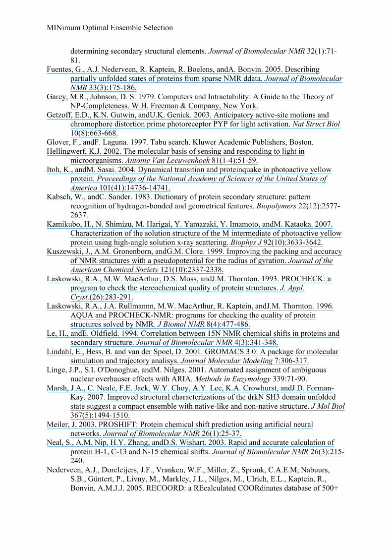
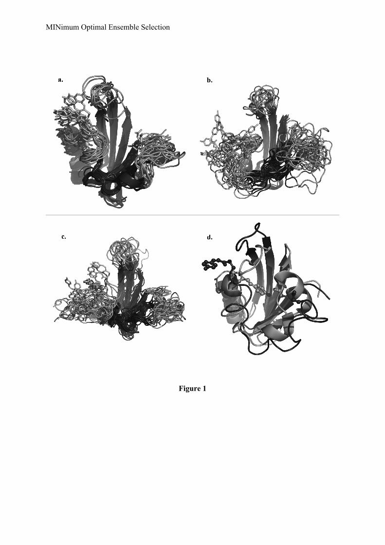
Following the protocol described above (see Material and Methods), we generated anensemble of 6494 water-refined structures with less than 5 violations within the core. RunningMINOES, we selected a set of 14 structures (Ntrial was set to 20) (Figure 1a). This finalensemble has an average internal energy of –18179 ± 675 kcal.mol-1, a chi2 value of 3.95 anda correlation coefficient between back-calculated and experimental Hα chemical shifts of 0.94(0.94 and 0.92 for core and partially unfolded moieties, respectively). The average pairwisebackbone RMSD between the structures is 2.4 ± 0.4 Å and the average radius of gyration is
MINimum Optimal Ensemble Selection
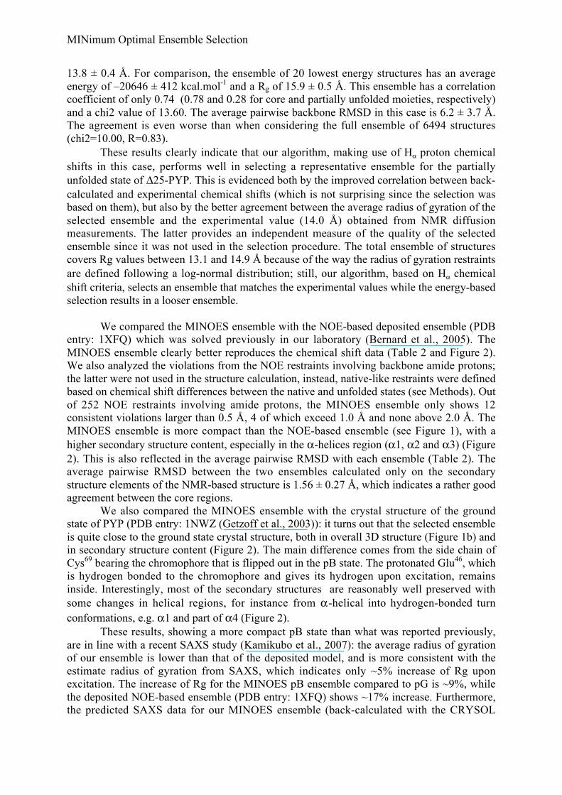
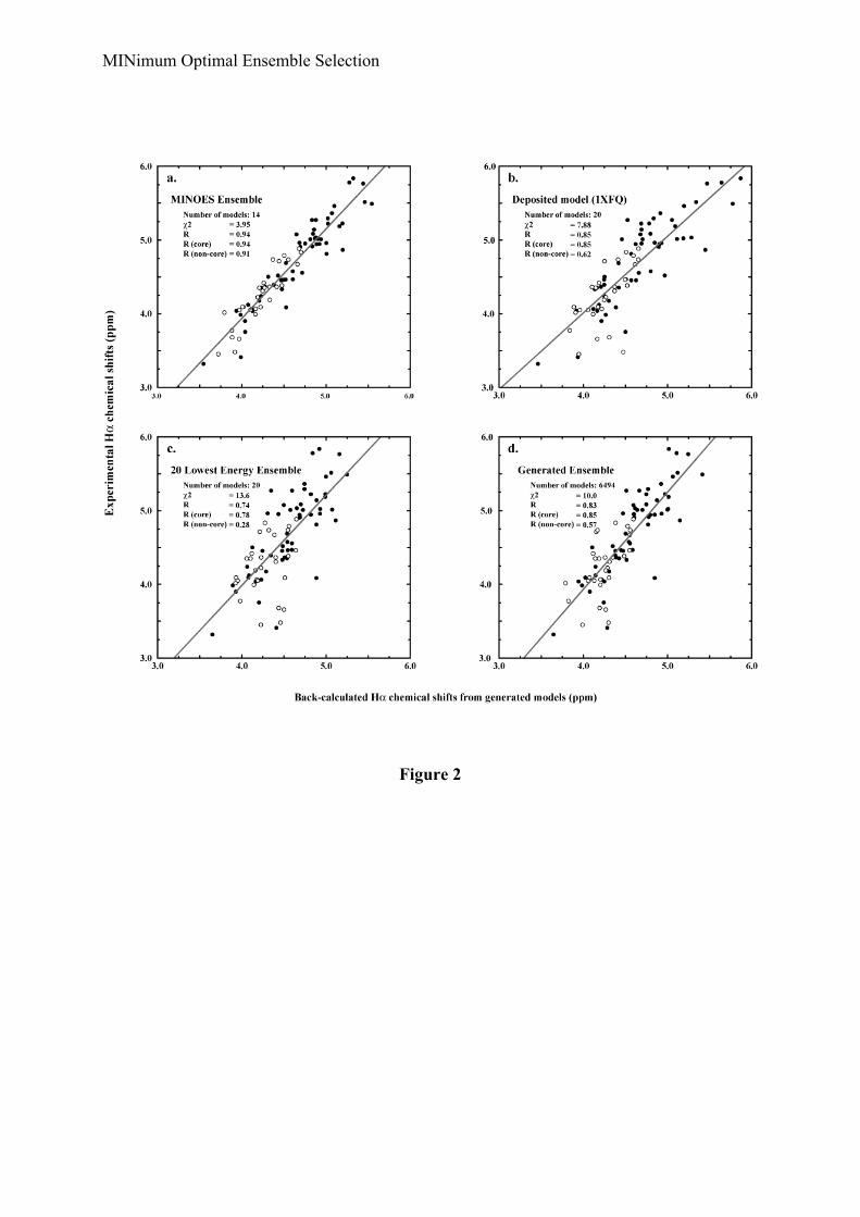
13.8 ± 0.4 Å. For comparison, the ensemble of 20 lowest energy structures has an averageenergy of –20646 ± 412 kcal.mol-1 and a Rg of 15.9 ± 0.5 Å. This ensemble has a correlationcoefficient of only 0.74 (0.78 and 0.28 for core and partially unfolded moieties, respectively)and a chi2 value of 13.60. The average pairwise backbone RMSD in this case is 6.2 ± 3.7 Å.The agreement is even worse than when considering the full ensemble of 6494 structures(chi2=10.00, R=0.83).
These results clearly indicate that our algorithm, making use of Hα proton chemicalshifts in this case, performs well in selecting a representative ensemble for the partiallyunfolded state of Δ25-PYP. This is evidenced both by the improved correlation between back-calculated and experimental chemical shifts (which is not surprising since the selection wasbased on them), but also by the better agreement between the average radius of gyration of theselected ensemble and the experimental value (14.0 Å) obtained from NMR diffusionmeasurements. The latter provides an independent measure of the quality of the selectedensemble since it was not used in the selection procedure. The total ensemble of structurescovers Rg values between 13.1 and 14.9 Å because of the way the radius of gyration restraintsare defined following a log-normal distribution; still, our algorithm, based on Hα chemicalshift criteria, selects an ensemble that matches the experimental values while the energy-basedselection results in a looser ensemble.
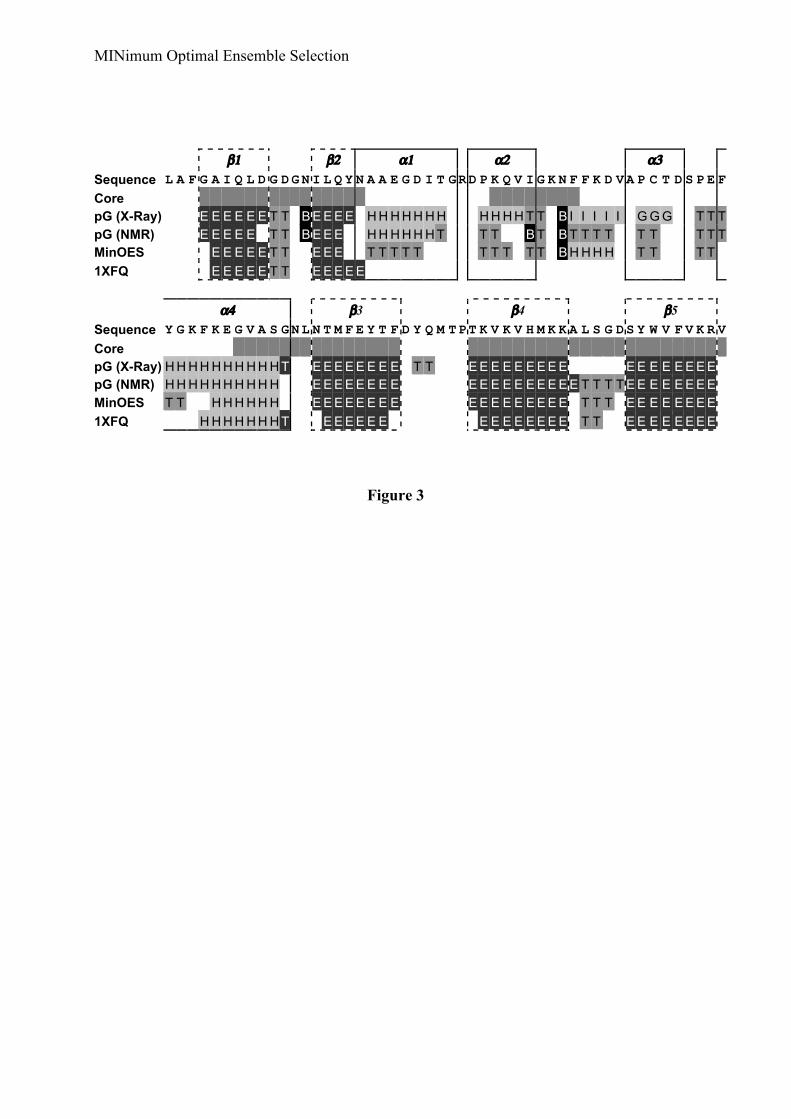
We compared the MINOES ensemble with the NOE-based deposited ensemble (PDBentry: 1XFQ) which was solved previously in our laboratory (Bernard et al., 2005). TheMINOES ensemble clearly better reproduces the chemical shift data (Table 2 and Figure 2).We also analyzed the violations from the NOE restraints involving backbone amide protons;the latter were not used in the structure calculation, instead, native-like restraints were definedbased on chemical shift differences between the native and unfolded states (see Methods). Outof 252 NOE restraints involving amide protons, the MINOES ensemble only shows 12consistent violations larger than 0.5 Å, 4 of which exceed 1.0 Å and none above 2.0 Å. TheMINOES ensemble is more compact than the NOE-based ensemble (see Figure 1), with ahigher secondary structure content, especially in the α-helices region (α1, α2 and α3) (Figure2). This is also reflected in the average pairwise RMSD with each ensemble (Table 2). Theaverage pairwise RMSD between the two ensembles calculated only on the secondarystructure elements of the NMR-based structure is 1.56 ± 0.27 Å, which indicates a rather goodagreement between the core regions.
We also compared the MINOES ensemble with the crystal structure of the groundstate of PYP (PDB entry: 1NWZ (Getzoff et al., 2003)): it turns out that the selected ensembleis quite close to the ground state crystal structure, both in overall 3D structure (Figure 1b) andin secondary structure content (Figure 2). The main difference comes from the side chain ofCys69 bearing the chromophore that is flipped out in the pB state. The protonated Glu46, whichis hydrogen bonded to the chromophore and gives its hydrogen upon excitation, remainsinside. Interestingly, most of the secondary structures are reasonably well preserved withsome changes in helical regions, for instance from α-helical into hydrogen-bonded turnconformations, e.g. α1 and part of α4 (Figure 2).
These results, showing a more compact pB state than what was reported previously,are in line with a recent SAXS study (Kamikubo et al., 2007): the average radius of gyrationof our ensemble is lower than that of the deposited model, and is more consistent with theestimate radius of gyration from SAXS, which indicates only ~5% increase of Rg uponexcitation. The increase of Rg for the MINOES pB ensemble compared to pG is ~9%, whilethe deposited NOE-based ensemble (PDB entry: 1XFQ) shows ~17% increase. Furthermore,the predicted SAXS data for our MINOES ensemble (back-calculated with the CRYSOL
MINimum Optimal Ensemble Selection
program (Svergun et al., 1995)) displays the experimentally observed bimodal profile (datanot shown).
Table 2 Comparison of excited state (pB) ensembles obtained by different approaches: NOE-based ensemble(deposited model), the 20 lowest energy structures obtained using native-like restraints and the ensembleselected by the MINOES algorithm.
R between Hα calculated vs. experimentalc
Number ofstructures Average Rg (Å)a RMSD (Å)b Full protein Core Non-core
Deposited model(1XFQ) 20 14.7 ± 0.3 4.3 ± 0.8 0.85 0.86 0.63
Lowest energystructures 20 15.9 ± 0.5 6.2 ± 3.7 0.74 0.78 0.28
MINOESensemble
14 13.7 ± 0.4 2.4 ± 0.4 0.94 0.94 0.92
All generatedstructures
6494 13.8 ± 0.4 4.3 ± 3.0 0.83 0.85 0.57
a) The experimental Rg value estimated from NMR diffusion experiments (Nico van Nuland, personalcommunication is 14.0 Å.
b) Average pairwise backbone RMSD (see Material and Methods)
c) Correlation coefficient between experimental and back-calculated Hz chemical shifts. The Hα chemical shiftswere back-calculated with SHIFTX (Neal et al., 2003).
Discussion
We have shown here that, in the case of partially unfolded states, selecting anensemble of structures based on the agreement between back-calculated data from theoreticalmodels and experimental data results in a better representation of the experimental data thansimple energy and violation criteria. The algorithm we have developed for this purpose findsin an efficient Monte Carlo process the optimum ensemble without any a-priori assumptionabout ensemble size.
The difficulty in such a selection method relies on the almost impossible task ofconsidering all possible sub-ensembles, since this is a combinatorial problem. For instance,assuming that we generated only 200 structures and want to determine which ensemble bestfits the experimental data, the number of possible combinations would be:
60200
0
1061.12 ×≈=∑=
n
k
knC
If we consider that a regular computer performs roughly 1012 calculations per second, thismeans we would need around 5x1040 years to achieve all required calculations, in other wordsabout 3x10
27 times the age of the Universe!
The complexity class of the problem addressed here is NP-Hard (Garey, 1979). Eachsub-ensemble can be interpreted as a unique state having a score indicating the agreementwith the experimental data. A simple way to represent these states is to consider all binarynumbers made of as many (0,1) as the size of the initial ensemble. For example, 01010 is thesub-ensemble composed with the structures 2 and 4 of an initial ensemble constituted of 5structures. Hence, the core of our algorithm tries to switch '0' and '1', in a stepwise manner.The main characteristics of such a process are that the final state is highly dependent on thestarting one and that there might be some states difficult to reach from which one could obtainthe best ensemble. To avoid these problems, we embedded our algorithm in a Monte-Carloprocess that performs a random selection of structures from the best sub-ensemble found sofar. In this way, we create the possibility to start from some states that would not be reachable
MINimum Optimal Ensemble Selection
otherwise. Another advantage of the current implementation is that we do not need to startfrom each structure in the initial pool, since we can assume that sub-ensembles generated withthis method could also be reached when starting from a different structure. An importantparameter in the selection of the structures is the fraction of structures that are removed orkept. Indeed, the size of the best ensemble (parameter not known) could be much smaller thanthe amount of structures in the currently selected sub-ensemble; in such a case the programwould never be able to reach the best ensemble. One possibility to overcome this problemmight be to start several selection runs with different sampling ratio. Another possibilitywould be to make use of a “funnel” effect, starting for example from a ratio of 0.5 (half ofstructures are removed randomly) and increasing this value up to 0.9 as the selection protocoladvances; thus, we would start each new selection process from less and less structures.
Another aspect of this selection process is that some structures could happen to beselected more often than others. To avoid this, we assigned to each structure a probability ofbeing selected that decreases every time the structure is chosen. This probability follows amere linear function that depends on the number of iterations. It is defined as:
pi Sn+1( ) = pi Sn( ) −1 Niterations
where pi(Sn+1) is the probability of selection structure i in the next Monte Carlo process if ithas already been selected in the current iteration. The probabilities are reinitialized every timea better ensemble is found or if the sampling ratio is changed.
Timing of the algorithm
For each run, the program tests the possibility of adding, removing or exchangingstructures, one by one, and finally keeps the combination that leads to the largest decrease inthe chi2. The computing time needed by the algorithm to find an optimal ensemble dependson several parameters, some of them a priori unknown, such as the number of Monte Carloselections, the size of the initial pool of structures, the number of available experimental data(chemical shifts, RDC, ...), etc. Benchmarking has indicated that the calculation time isproportional to the total number of structures in the selection pool and to the square of thenumber of data points. As an indication, in the case of Δ25PYP (6494 structures if theselection pools and 85 data points), the average run time was ~20 minutes on a 3.0 GHz XeonLinux PC (average over 20 runs starting from different structures and various sampling ratios(0.5, 0.3 and 0.1)).
Conclusions
We have described an innovative and efficient algorithm called MINOES that allows to selecta minimum representative set of conformations out of a large ensemble of structures and thiswithout a priori knowledge about the optimal ensemble size. MINOES aims at selecting anoptimal ensemble of conformations that, on average, maximizes the agreement between back-calculated and experimental (NMR) data. While the method was developed and demonstratedfor the selection of a representative partially unfolded state ensemble based on chemical shiftinformation, it is by far not limited to such a problem and/or type of data; the only conditionis that the data can be back-calculated from the generated models. MINOES is coded ingeneric manner and can accept in principle any kind of data. The current implementationassumes a linear averaging, but this can be easily modified. In the case of (partially) unfolded
MINimum Optimal Ensemble Selection
states, residual dipolar couplings should provide another very useful source of information(Bernado et al., 2005) that could be optimally used in our selection procedure.
Availability
The source code for MINOES is available for free from the authors upon request.
Acknowledgements
This work was supported by a “van Molecuul tot Cell” grant from the NetherlandsOrganization for Scientific Research (N.W.O.) and by a European Community, FP6 STREPgrant “UPMAN” (Contract no. LSHG-CT-2005-512052). A.M.J.J.B. is recipient of a VICIgrant from N.W.O.
References
Ansari, A., J. Berendzen, S.F. Bowne, H. Frauenfelder, I.E. Iben, T.B. Sauke, E.Shyamsunder, andR.D. Young. 1985. Protein states and proteinquakes. Proceedings ofthe National Academy of Sciences of the United States of America 82(15):5000-5004.
Armitage, J.P. 1997. Behavioural responses of bacteria to light and oxygen. Arch Microbiol168(4):249-261.
Armitage, J.P., andK.J. Hellingwerf. 2003. Light-induced behavioral responses (;phototaxis')in prokaryotes. Photosynth Res 76(1-3):145-155.
Bernado, P., L. Blanchard, P. Timmins, D. Marion, R.W. Ruigrok, andM. Blackledge. 2005.A structural model for unfolded proteins from residual dipolar couplings and small-angle x-ray scattering. Proc Natl Acad Sci U S A 102(47):17002-17007.
Bernard, C., K. Houben, N.M. Derix, D. Marks, M.A. van der Horst, K.J. Hellingwerf, R.Boelens, R. Kaptein, andN.A. van Nuland. 2005. The solution structure of a transientphotoreceptor intermediate: Delta25 photoactive yellow protein. Structure 13(7):953-962.
Brunger, A.T., Adams, P.D., Clore, G.M., DeLano, W.L., Gros, P., Grosse-Kunstleve, R.W.,Jiang, J.-S., Kuszewski, J., Nilges, N., Pannu, N.S., Read, R.J., Rice, L.M., Simonson,T., and Warren, G.L. 1998. Crystallography and NMR system (CNS): A new softwaresystem for macromolecular structure determination. Acta Crystallogr. D Biol. 54:905-921.
Case, D.A. 1998. The use of chemical shifts and their anisotropies in biomolecular structuredetermination. Current Opinion in Structural Biology 8(5):624-630.
Choy, W.Y., andJ.D. Forman-Kay. 2001. Calculation of ensembles of structures representingthe unfolded state of an SH3 domain. J Mol Biol 308(5):1011-1032.
Cornilescu, G., F. Delaglio, andA. Bax. 1999. Protein backbone angle restraints fromsearching a database for chemical shift and sequence homology. Journal ofBiomolecular NMR 13(3):289-302.
Dux, P., G. Rubinstenn, G.W. Vuister, R. Boelens, F.A. Mulder, K. Hard, W.D. Hoff, A.R.Kroon, W. Crielaard, K.J. Hellingwerf, andR. Kaptein. 1998. Solution structure andbackbone dynamics of the photoactive yellow protein. Biochemistry 37(37):12689-12699.
Eghbalnia, H.R., L. Wang, A. Bahrami, A. Assadi, andJ.L. Markley. 2005. Protein energeticconformational analysis from NMR chemical shifts (PECAN) and its use in
MINimum Optimal Ensemble Selection
determining secondary structural elements. Journal of Biomolecular NMR 32(1):71-81.
Fuentes, G., A.J. Nederveen, R. Kaptein, R. Boelens, andA. Bonvin. 2005. Describingpartially unfolded states of proteins from sparse NMR ddata. Journal of BiomolecularNMR 33(3):175-186.
Garey, M.R., Johnson, D. S. 1979. Computers and Intractability: A Guide to the Theory ofNP-Completeness. W.H. Freeman & Company, New York.
Getzoff, E.D., K.N. Gutwin, andU.K. Genick. 2003. Anticipatory active-site motions andchromophore distortion prime photoreceptor PYP for light activation. Nat Struct Biol10(8):663-668.
Glover, F., andF. Laguna. 1997. Tabu search. Kluwer Academic Publishers, Boston.Hellingwerf, K.J. 2002. The molecular basis of sensing and responding to light in
microorganisms. Antonie Van Leeuwenhoek 81(1-4):51-59.Itoh, K., andM. Sasai. 2004. Dynamical transition and proteinquake in photoactive yellow
protein. Proceedings of the National Academy of Sciences of the United States ofAmerica 101(41):14736-14741.
Kabsch, W., andC. Sander. 1983. Dictionary of protein secondary structure: patternrecognition of hydrogen-bonded and geometrical features. Biopolymers 22(12):2577-2637.
Kamikubo, H., N. Shimizu, M. Harigai, Y. Yamazaki, Y. Imamoto, andM. Kataoka. 2007.Characterization of the solution structure of the M intermediate of photoactive yellowprotein using high-angle solution x-ray scattering. Biophys J 92(10):3633-3642.
Kuszewski, J., A.M. Gronenborn, andG.M. Clore. 1999. Improving the packing and accuracyof NMR structures with a pseudopotential for the radius of gyration. Journal of theAmerican Chemical Society 121(10):2337-2338.
Laskowski, R.A., M.W. MacArthur, D.S. Moss, andJ.M. Thornton. 1993. PROCHECK: aprogram to check the stereochemical quality of protein structures. J. Appl.Cryst.(26):283-291.
Laskowski, R.A., J.A. Rullmannn, M.W. MacArthur, R. Kaptein, andJ.M. Thornton. 1996.AQUA and PROCHECK-NMR: programs for checking the quality of proteinstructures solved by NMR. J Biomol NMR 8(4):477-486.
Le, H., andE. Oldfield. 1994. Correlation between 15N NMR chemical shifts in proteins andsecondary structure. Journal of Biomolecular NMR 4(3):341-348.
Lindahl, E., Hess, B. and van der Spoel, D. 2001. GROMACS 3.0: A package for molecularsimulation and trajectory analisys. Journal Molecular Modeling 7:306-317.
Linge, J.P., S.I. O'Donoghue, andM. Nilges. 2001. Automated assignment of ambiguousnuclear overhauser effects with ARIA. Methods in Enzymology 339:71-90.
Marsh, J.A., C. Neale, F.E. Jack, W.Y. Choy, A.Y. Lee, K.A. Crowhurst, andJ.D. Forman-Kay. 2007. Improved structural characterizations of the drkN SH3 domain unfoldedstate suggest a compact ensemble with native-like and non-native structure. J Mol Biol367(5):1494-1510.
Meiler, J. 2003. PROSHIFT: Protein chemical shift prediction using artificial neuralnetworks. Journal of Biomolecular NMR 26(1):25-37.
Neal, S., A.M. Nip, H.Y. Zhang, andD.S. Wishart. 2003. Rapid and accurate calculation ofprotein H-1, C-13 and N-15 chemical shifts. Journal of Biomolecular NMR 26(3):215-240.
Nederveen, A.J., Doreleijers, J.F., Vranken, W.F., Miller, Z., Spronk, C.A.E.M, Nabuurs,S.B., Güntert, P., Livny, M., Markley, J.L., Nilges, M., Ulrich, E.L., Kaptein, R.,Bonvin, A.M.J.J. 2005. RECOORD: a REcalculated COORdinates database of 500+
MINimum Optimal Ensemble Selection
proteins from the PDB using restraint data from the BioMagResBank. Proteins:Struc.Funct. & Bioinformatics 59:662-672.
Neuhaus, D., andM.P. Williamson. 2000. The Nuclear Overhauser Effect in Structural andConformational Analysis. John Wiley & Sons.
Spitzfaden, C., W. Braun, G. Wider, H. Widmer, andK. Wuthrich. 1994. Determination of theNMR solution structure of the cyclophilin A-cyclosporin A complex. Journal ofBiomolecular NMR 4(4):463-482.
Svergun, D., C. Barberato, andM. Koch. 1995. CRYSOL - a program to evaluate X-raysolution scattering of biological macromolecules from atomic coordinates. J. Appl.Cryst. 28:768-773.
van der Horst, M.A., I.H. van Stokkum, W. Crielaard, andK.J. Hellingwerf. 2001. The role ofthe N-terminal domain of photoactive yellow protein in the transient partial unfoldingduring signalling state formation. FEBS Letters 497(1):26-30.
Williamson, M.P., andT. Asakura. 1997. Protein chemical shifts. Methods in MolecularBiology 60:53-69.
Wishart, D.S., andD.A. Case. 2001. Use of chemical shifts in macromolecular structuredetermination. Methods in Enzymology 338:3-34.
Wishart, D.S., andB.D. Sykes. 1994a. Chemical Shifts as a Tool for Structure Determination.Methods in Enzymology:363.
Wishart, D.S., andB.D. Sykes. 1994b. The 13C chemical-shift index: a simple method for theidentification of protein secondary structure using 13C chemical-shift data. Journal ofBiomolecular NMR 4(2):171-180.
Wüthrich, K. 1986. NMR of proteins and nucleic acids. . Wiley, New York.Zuiderweg, E.R. 2002. Mapping protein-protein interactions in solution by NMR
spectroscopy. Biochemistry 41(1):1-7.
MINimum Optimal Ensemble Selection
Figure Legends
Figure 1.
Cartoon representations of the pB state of Δ25-PYP for the a) MINOES ensemble selectedbased on Hα chemical shifts, b) (a), deposited (c) and lowest energy (d) ensembles. Thechromophore attached to Cys69 and Glu46, which serves as hydrogen donor upon excitation,are shown in sticks. The native-like (ground-state) core defined in this work is shown in darkgray. In (b) is shown a comparison between the X-ray ground state (dark color) at highresolution and one of our models (light color). The residues 69 and 46 are enlightened with astick and sphere representation.
Figure 2.
Comparison of average data of each ensemble with experimental data: a) MINOES ensemble,b) deposited model, c) 20 lowest energy structures and d) full ensemble generated withRECOORD (Nederveen, 2005). The trend line is drawn in grey. The equation of this latterand the determination coefficient R2 are shown on the left side.
Figure 3.
Secondary structure of Δ25-PYP calculated with PROCHECK (Laskowski et al., 1993;Laskowski et al., 1996) for a) the crystal structure of the ground state (pG) (1NWZ), b) thesolution NMR structure of the ground state (pG-1XFN), c) the partially unfolded state (pB)ensemble selected by MINOES and d) the NOE-based partially unfolded state ensemble (pB-1XFQ). H, G and I indicate alpha helices, 3/10-helices and pi-helices, respectively (all withthe same gray shading). T correspond to hydrogen bonded turns and B and E are residues inisolated beta-bridges and extended strands, respectively. Bends are not indicated for the sakeof clarity. The core defined in this work is indicated by filled grey boxes. The secondarystructure elements defined in the crystal structure are specified.
MINimum Optimal Ensemble Selection
Figure 1
MINimum Optimal Ensemble Selection
Figure 2
MINimum Optimal Ensemble Selection
β1 β2 α1 α2 α3
Sequence L A F G A I Q L D G D G N I L Q Y N A A E G D I T GR D P K Q V I G K N F F K D V A P C T D S P E FCore
pG (X-Ray) E E E E E E T T B E E E E H H H H H H H H H H H T T B I I I I I G G G T T TpG (NMR) E E E E E T T B E E E H H H H H H T T T B T B T T T T T T T T TMinOES E E E E E T T E E E T T T T T T T T T T B H H H H T T T T
1XFQ E E E E E T T E E E E E
α4 β3 β4 β5Sequence Y G K F K E G V A S G N L N T M F E Y T F D Y Q M TP T K V K V H M K K A L S G D S Y W V F V K R V
Core
pG (X-Ray) H H H H H H H H H H T E E E E E E E E T T E E E E E E E E E E E E E E E E EpG (NMR) H H H H H H H H H H E E E E E E E E E E E E E E E E E E T T T T E E E E E E E EMinOES T T H H H H H H E E E E E E E E E E E E E E E E E T T T E E E E E E E E
1XFQ H H H H H H H T E E E E E E E E E E E E E E T T E E E E E E E E
Figure 3
Top Related
Copyright © 2022 FDOKUMEN

