


Bahasa
Halaman
Hukum


© 2002 Blackwell Science Ltd International Endodontic Journal,
35
, 337–344, 2002 337
Blackwell Science Ltd
Measurement of strain on tooth roots during post removal with the Eggler post remover
T. V. Castrisos, J. E. A. Palamara & P. V. Abbott
School of Dental Science, University of Melbourne, Melbourne, Victoria, Australia
Abstract
Castrisos T, Palamara JEA, Abbott PV.
Measurement of
strain on tooth roots during post removal with the Eggler post
remover.
International Endodontic Journal
,
35
, 337–344, 2002.
Aim
The aim was to measure root surface strains inteeth when removing cast post/cores with the Eggler postremover.
Methodology
Two groups of 10 teeth each were tested:group 1 had 1 mm thickness of dentine coronally,and group 2 had 2 mm thickness of dentine. After rootfilling, 10 mm long cast post/cores were constructed andcemented with zinc phosphate cement, and strain gaugeswere applied to the roots. The post/cores were removedwith the Eggler post remover whilst strain measure-ments were being recorded. Posts were removed twice:initially along the long axis of the tooth and then at a 10
°
angle to the long axis. Comparisons between groups 1and 2 were analysed statistically with the Mann–Whitney
U
-test whilst strains within each group were analysed
with the Wilcoxon Signed Rank test at the 95% level ofconfidence.
Results
There was no significant difference in thestrains measured between groups 1 and 2, and no sig-nificant difference within each group when removingposts along the long axis of the tooth and at a 10
°
angle.Three teeth in group 1 and one tooth in group 2 fracturedwhen removing posts at the 10
°
angle. Three frac-tures were small slivers of dentine at the point wherethe Eggler’s repeller arms contacted the tooth mesiallyand distally, whilst one tooth (from group 1) fracturedobliquely.
Conclusions
Post removal with the Eggler device isa relatively safe procedure but care must be taken whenthere is a possibility of pulling the post out in a nonaxialdirection or when less than 1 mm of dentine surroundsthe apical end of the post.
Keywords:
post removal, posts, strain.
Received 11 December 2000; accepted 22 May 2001
Introduction
Several techniques are available for the removal of postsfrom roots and these include the use of ultrasonic vibra-tion and various post removal devices. Most of the liter-ature on post removal discusses the various methodsused in the form of clinical case presentations, althoughsome studies have investigated the efficiency of postremoval techniques. Buoncristiani
et al
. (1994) com-pared the efficiency of ultrasonic and sonic vibration toremove parallel-sided preformed posts and concludedthat ultrasonic vibration could remove them in less than10 min. Berbert
et al
. (1995) and Johnson
et al
. (1996)
have also examined the influence of ultrasonic vibrationon the forces required to remove posts. Berbert
et al
.(1995) used cast posts/cores and concluded that theforce required to remove them after application of ultra-sonics for 2 to 5 min was 30% less than the control groupwith no ultrasonic vibration. Johnson
et al
. (1996) con-cluded that 16 min of ultrasonic vibration could signific-antly reduce the amount of force required to removeparallel-sided preformed posts in extracted human pre-molars. Yoshida
et al
. (1997) examined the effect of usingone or two ultrasonic tips simultaneously placed on acast post and core and they reported that the use of twoultrasonic tips was significantly quicker. The use of theEggler post remover (Automaton-Ventriebs-Gesellschaft,Germany) and the Gonon (Thomas) post remover (ThomasExtracteur De Pivots; FFDM, Bourges, France) have beendescribed by Bando
et al
. (1985), Stamos & Gutmann
Correspondence: Dr. Paul V. Abbott, 5 Westley Avenue, Ivanhoe, Victoria 3079,Australia (fax: +61 39499 7156; e-mail: [email protected]).
IEJ_483.fm Page 337 Friday, March 22, 2002 11:16 AM

Strain during post removal
Castrisos et al.
International Endodontic Journal,
35
, 337–344, 2002 © 2002 Blackwell Science Ltd338
(1991) and Machtou
et al
. (1989), who all describedthem as highly efficient methods for removing posts.
A survey of the methods used by American endo-dontists to remove intraradicular posts found that postremoval devices such as the Gonon and Eggler postremovers were not commonly used (Stamos & Gutmann1993). The reasons given by the respondents to thissurvey for not using these devices were that they weretoo dangerous, could not be universally used, or did notwork. In contrast, a recent survey of endodontists inAustralia and New Zealand found that the Eggler postremover was the most frequently used post removaldevice, with 42% of the respondents using it regularly(Castrisos & Abbott 2001). Stamos & Gutmann (1991)stated that the advantages of using these types of postremovers were:
1
conservation of remaining tooth structure
2
reduced risk of root fracture
3
reduced risk of root perforation, and
4
reduced risk of torque forces being placed on the root.Although several articles have presented case reports
about the efficiency of various devices, the safety of thesedevices has not been established. Altshul
et al
. (1997)compared the prevalence of dentinal cracks in teeth withcast posts that had been removed by either ultrasonicvibration or the Gonon post removal system. There wasa statistically significant difference between the numberof dentinal cracks present following ultrasonic removalof posts compared with teeth where the post was notremoved. The dentinal cracks were seen more frequentlyat the cervical level following the use of ultrasonics and thetime taken to remove the posts with ultrasonic vibrationwas significantly longer than with the Gonon system.
As a result of the design and size of the Eggler postremover (Fig. 1), it is usually used to remove posts inanterior teeth. It is considered by most endodontists whouse it to be of greater benefit for the removal of cast posts/cores than ultrasonics (Castrisos & Abbott 2001), wherethe time required to remove the post is greater.
To date, no studies have reported the amount or natureof forces applied to teeth during post removal. However,several methods have been used in various studies todetermine the amount of force applied to teeth withvarious loads during other dental procedures. Thesemethods include two-dimensional and three-dimensionalphotoelastic stress analysis (Henry 1977, Mentink
et al
.1998), finite element stress analysis (Huysmans
et al
.1993) and strain gauge measurements. The latter havebeen used to assess forces applied when obturating rootcanals using lateral condensation (Saw & Messer 1995,Lertchirakarn
et al
. 1999), when cementing posts (Ross
et al
. 1991, Obermayr
et al
. 1991) and in measuring thereduction in stiffness of endodontically treated teeth(Reeh
et al
. 1989). The strain gauges used in these studieswere applied to the external surface of the root or crownto measure the strain on the tooth.
The aim of this study was to measure the surfacestrains caused by the application of an Eggler postremover parallel to the long axis of the tooth and to com-pare them with strains produced by loads applied at a 10
°
angle to the long axis.
Materials and methods
Twenty extracted single-rooted human teeth were obtainedfrom the clinics of the Royal Dental Hospital of Melbourne.At all times prior to and during the experiment, the teethwere kept moist by either storing them in phosphate bufferedsaline or by covering them with gauze moistened in phos-phate buffered saline. Radiographs were taken from thebuccal and mesial directions to ensure that only a single-root canal was present and all the teeth were examinedunder a microscope (
×
16 magnification) to ensure theywere free of fractures and cracks. The crowns of the teethwere removed with a diamond bur at a level 1 mm coronalto the labial cemento-enamel junction and the teeth werethen measured. The mesio-distal dimensions of the teethat the cemento-enamel junction ranged from 4.2 mm to5.9 mm and each tooth was checked to ensure that therepeller arms of the Eggler post remover could be restedon the root surface.
The root canals of each tooth were negotiated andinstrumented up to a size 40 Hedström file at a lengththat was 1 mm short of the apical foramen. The apicalthird of each canal was flared with the step-back
Figure 1 The Eggler Post Remover.
IEJ_483.fm Page 338 Friday, March 22, 2002 11:16 AM
Castrisos et al.
Strain during post removal
© 2002 Blackwell Science Ltd International Endodontic Journal,
35
, 337–344, 2002 339
technique and the coronal third was flared with sizes 2and 3 Gates Glidden burs. Whilst being instrumented,the canals were irrigated with 1% sodium hypochloritebetween each file size. They were then filled with gutta-percha and AH26 (Dentsply DeTrey GmbH, Konstanz,Germany) using lateral condensation; a post space wasprovided in each tooth to a length of 10 mm by removinggutta-percha with a heated plugger. A minimum of4 mm of gutta-percha remained in each tooth. Postspaces were further prepared with Parapost drills(Coltene Whaledent, Konstanz, Germany), ensuring thatat least 1 mm of dentine remained on both the mesial anddistal sides of the root canals at the apical extent of thepost holes. The dimensions of the post holes were verifiedradiographically.
The teeth were divided into two groups, with approxi-mately equal distribution of root lengths and diametersbased on visual inspection. The post hole preparationswere then finalized: in group 1, the post holes were flaredwith Gates Glidden burs to leave a thickness of 1 mm ofdentine coronally on the buccal, lingual, mesial and dis-tal sides; in group 2, post holes were prepared so as tomaintain 2 mm of dentine on each side of the root face.The final shape of each post was parallel-sided for theapical half with a tapered coronal half to accommodatethe thickness of coronal dentine required for each group.Direct patterns for cast posts/cores were formed usingParapost burn out points and GC Pattern Resin (GCCorporation, Tokyo, Japan). The core was cuboid-shapedwith dimensions of 2 mm
×
2 mm and a height of 3 mmto allow placement of the Eggler post remover. The post/cores were cast in a non-precious alloy and cementedwith zinc phosphate cement (SS White, Gloucester, UK)which was allowed to set for 1 week prior to removal ofthe posts with the Eggler post remover.
Strain gauges (EA-06–125BT-120, Micro-MeasurementsGroup Inc., Raleigh, NC, USA) were trimmed to fit theroot surface. Four strain gauges were placed vertically1.5 mm below the coronal root surface after each toothhad been lightly scraped on the buccal, lingual, mesialand distal surface to facilitate their placement. Pilotstudies indicated that this was the most appropriate sitefor placement of the strain gauges so that some extradentine could be removed for the second stage of theexperiment when the posts were removed at a 10
°
angleand also to allow clearance between the point of loadingand the wires of the strain gauge. The root surface wasetched with 37% orthophosphoric acid for 30 s, washedwith water for 15 s and dried with air. The strain gaugeswere cleaned with chloroform, primed with a catalystand attached to the root surface with cyanoacrylate
adhesive (M-Bond 200, Micro-Measurements GroupInc., Raleigh, NC, USA). Excess cyanoacrylate adhesivewas removed by applying pressure to the surface of thestrain gauges until adhesion occurred, leaving a thinlayer of cyanoacrylate that did not interfere with themeasurement of strain. The strain gauges, solder con-tacts and roots were covered with silicone (Dow Corning3140 RTV Coating, Dow Corning Corp., Midland, MI,USA) to protect the strain gauges from moisture. Thestrain gauges were then connected to a data acquisitionboard (AT-MID-16E-2, National Instruments Corpora-tion, Austin, TX, USA) and the data was stored on a com-puter using process control software (LabVIEW 4.0,National Instruments Corporation, Austin, TX, USA).Four separate channels were used to allow simultaneousmeasurements from each of the strain gauges and themeasurements were made continuously, whilst remov-ing the posts with the Eggler post remover. Throughoutthe experiment, one tooth was used as the experimentaltooth, and a second tooth, which had been prepared inthe same manner, served as a compensator for apparentstrain that may arise from temperature fluctuationsand electrical heating.
In order to remove the post, the forceps of the Egglerdevice were placed over the buccal and lingual surfacesof the core and tightened with the large inner wheel(Fig. 1). The repeller arms were then lowered by tighten-ing the small thumbscrew at the top of the device. As therepeller arms were lowered, they contacted the mesialand distal tooth structure and the forceps moved awayfrom the tooth as the post was removed (Figs 2,3).
Strain measurements were initially recorded whenthe cast post/cores were removed from the teeth in a
Figure 2 Application of the Eggler Post Remover to the core section of a cast post/core with strain gauges attached to the external root surfaces.
IEJ_483.fm Page 339 Friday, March 22, 2002 11:16 AM

Strain during post removal
Castrisos et al.
International Endodontic Journal,
35
, 337–344, 2002 © 2002 Blackwell Science Ltd340
direction that was parallel to the long axis of each toothand then again when removed at an angle of 10
°
to thelong axis. After the initial removal of the posts, anyretained luting agent was cleaned from the posts andfrom within the post spaces by scraping it off with a probeand an excavator. The teeth were examined under mag-nification (16
×
) to ensure complete removal of cementand for any evidence of fractures or cracks. The postswere recemented after a minimum period of 24 h and theteeth were kept moist in phosphate buffered saline duringthis time. The height of the mesial dentine of each toothwas reduced so that when the Eggler post remover wasapplied to the cores, it was at a 10
°
angle to the longaxis of each tooth. The 10
°
angle was measured witha protractor following application of the Eggler postremover. Then the posts/cores were removed from theteeth again and strain gauge measurements wererecorded. The teeth were examined once again undermagnification to determine whether there were any frac-tures or cracks.
Statistical analysis
Strain measurements were recorded continuously onthe buccal, lingual, mesial and distal surfaces of the toothwhilst each post was being removed. The maximumstrains recorded when removing the post along the longaxis and at a 10
°
angle to the long axis within the same groupwere compared using the Wilcoxon Signed Rank test.Comparisons were made between group 1 and group 2for removal of the post along the long axis and at the 10
°
angle to the long axis using the Mann–Whitney
U
-test. Allcomparisons were made with a 95% level of confidence todetermine statistical significance and data were analysedusing the SPSS statistical software program (
SPSS forWindows
6.1.31995, SPSS Inc, Chicago, IL, USA).
Results
All posts were removed in less than 2 min with the Egglerpost remover. Strain measurements could not be recordedin two teeth in group 1 when removing the post at 10
°
to thelong axis of the tooth because the repeller arms did notcontact the root surface when attempting to remove the post.
Strain is the change in length divided by the originallength of the object and it has no unit of measurement. Inthis study, the amount of deformation was reported as‘
µ
strain’ (where 10 000
µ
strain = 0.01 strain = 1% deforma-tion). A positive value indicated tension, whilst a negativevalue indicated compression. Tables 1 and 2 list the ranges,means and standard deviations for the greatest strainsduring post removal, as well as the character of the strain(tension or compression). The greatest strain on any surfacefor each tooth was identified and the strains on the otherthree surfaces were measured at that same point in time.
Root strain during post removal parallel to the long axis
The magnitude of strains measured on the buccal andlingual root surfaces was lower than those measured on
Figure 3 The Eggler Post Remover in use. Note the repeller arms have splayed as they are pushed against the mesial and distal surfaces of the root face which has caused the left repeller arm to almost slide off the tooth.
Location of strain gauge
Removed parallel to the long axis (n = 10)
Removed at 10° to the long axis (n = 8)
Range Mean SD Range Mean SD
Buccal –324 to 375 37 277 –806 to 750 –65 531Lingual –910 to 268 –133 395 –560 to 804 –63 396Distal –1498 to –214 –618 425 –560 to –164 –409 146Mesial –2307 to –268 –995 672 –5406 to –560 –1889 2004
Strain measurements do not have a unit of measurement but are expressed as ‘µstrain’ where 1 µstrain represents a change in dimension of 1 part in 1 million.Positive values indicate tension, whilst negative values indicate compression.
Table 1 Strain measurements for group 1 (1 mm thickness of dentine)
IEJ_483.fm Page 340 Friday, March 22, 2002 11:16 AM

Castrisos et al.
Strain during post removal
© 2002 Blackwell Science Ltd International Endodontic Journal,
35
, 337–344, 2002 341
the mesial and distal surfaces where the repeller arms ofthe Eggler post remover contacted the tooth. When postswere removed along the long axis of the teeth, the strainmeasurements were similar on the mesial and distal sur-faces for most teeth, although there were large discrep-ancies between strain measurements in three teeth fromgroup 1 and four teeth from group 2. All strain measure-ments in group 1 indicated compressive stresses on themesial and distal surfaces, whilst seven out of 10 teethhad tensile stresses on the buccal and lingual surfaces.Compressive stresses were present in nine out of the 10teeth on the mesial and distal surfaces in group 2. It wasnoted that the post was difficult to remove from the tooththat had tensile stresses and there was some rotation ofthe Eggler post remover during use on this tooth. Tensilestresses were present on the buccal or lingual surfacesof two of the 10 teeth in group 2. The Mann–Whitney
U
-test indicated that the strain measurements of group 1were not statistically different to those of group 2(Table 3) at the 95% level of confidence.
Root strain during post removal at 10
°°°°
to the long axis
The magnitude of the strains measured on the buccaland lingual surfaces were lower than those measured onthe mesial and distal surfaces and the highest strainswere recorded on the mesial surface. These strains werehigher in seven out of eight teeth in group 1 and in sevenout of 10 teeth in group 2. All teeth in both groups hadcompressive stresses on the mesial and distal surfaces.The Mann–Whitney
U
-test indicated that there wasno significant difference in the strain measurements
between groups 1 and 2 (Table 3) at the 95% level ofconfidence.
Comparison of strains within each group
Comparisons were made within the same group usingthe Wilcoxon Signed Rank test when removing the postparallel to the long axis and at 10
°
to the long axis of theroot. There were no significant differences between thestrain measurements on the buccal, lingual, mesial anddistal surfaces in both groups (Table 4) with a 95% levelof confidence.
Root fractures during post removal
Fractures of the root occurred in four teeth – three ofthese were from group 1 and one from group 2. All ofthese fractures occurred when the posts were being
Location of strain gauge
Removed parallel to the long axis (n = 10)
Removed at 10° to the long axis (n = 10)
Range Mean SD Range Mean SD
Buccal –378 to 268 –75 201 –560 to 482 –102 531Lingual –560 to 0 –276 161 –560 to 0 –294 198Distal –1260 to 214 –731 455 –1447 to –269 –633 413Mesial –1553 to –749 –1034 264 –2147 to –856 –1218 404
Strain measurements do not have a unit of measurement but are expressed as ‘µstrain’ where 1 µstrain represents a change in dimension of 1 part in 1 million.Positive values indicate tension, whilst negative values indicate compression.
Table 2 Strain measurements for group 2 (2 mm thickness of dentine)
Group 1 versus group 2
P-values
Buccal Lingual Distal Mesial
Post removed parallel to the long axis 0.35 0.25 0.44 0.58Post removed at 10° to the long axis 0.96 0.20 0.41 0.57
Table 3 Statistical analysis: Comparisons between groups 1 and 2 using the Mann–Whitney U-test at the 95% level of confidence
Table 4 Statistical analysis: comparisons within group 1 and within group 2 between removal of the posts parallel to the long axis and at a 10° angle to the long axis using the Wilcoxon Signed Rank Test at the 95% level of confidence
Group Surface P-value
1 Buccal 0.35Lingual 1.00Distal 0.29Mesial 0.73
2 Buccal 0.81Lingual 1.0Distal 0.41Mesial 0.12
IEJ_483.fm Page 341 Friday, March 22, 2002 11:16 AM
Strain during post removal
Castrisos et al.
International Endodontic Journal,
35
, 337–344, 2002 © 2002 Blackwell Science Ltd342
removed at an angle of 10
°
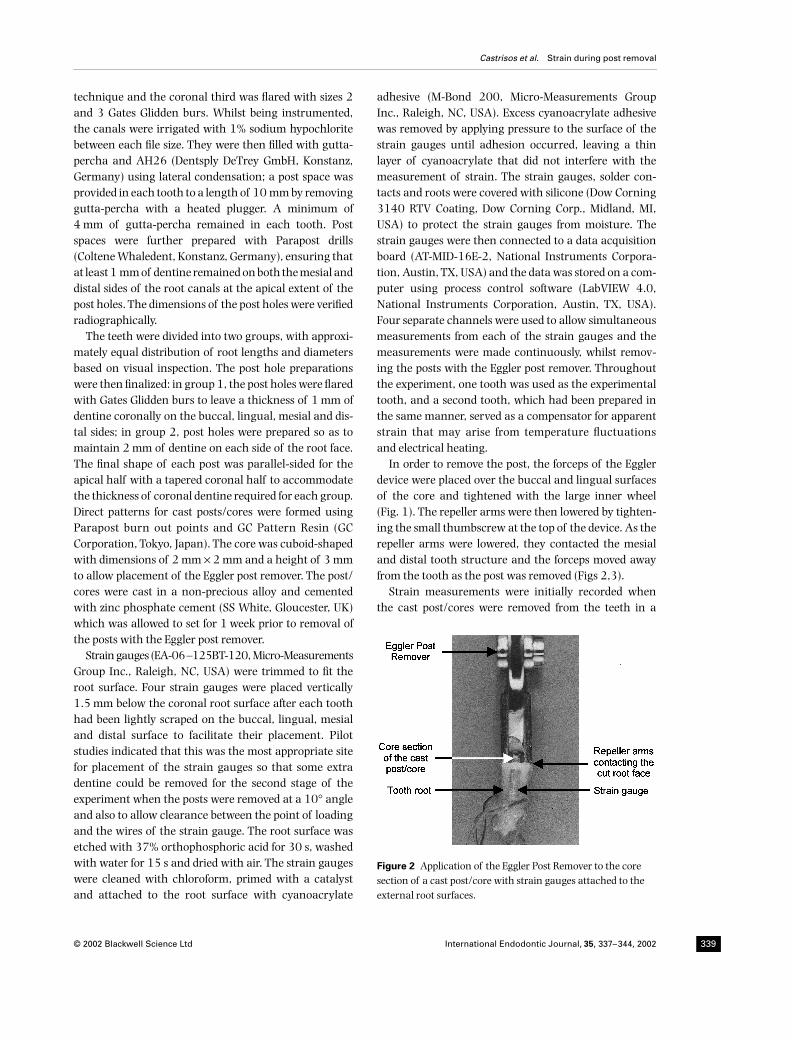
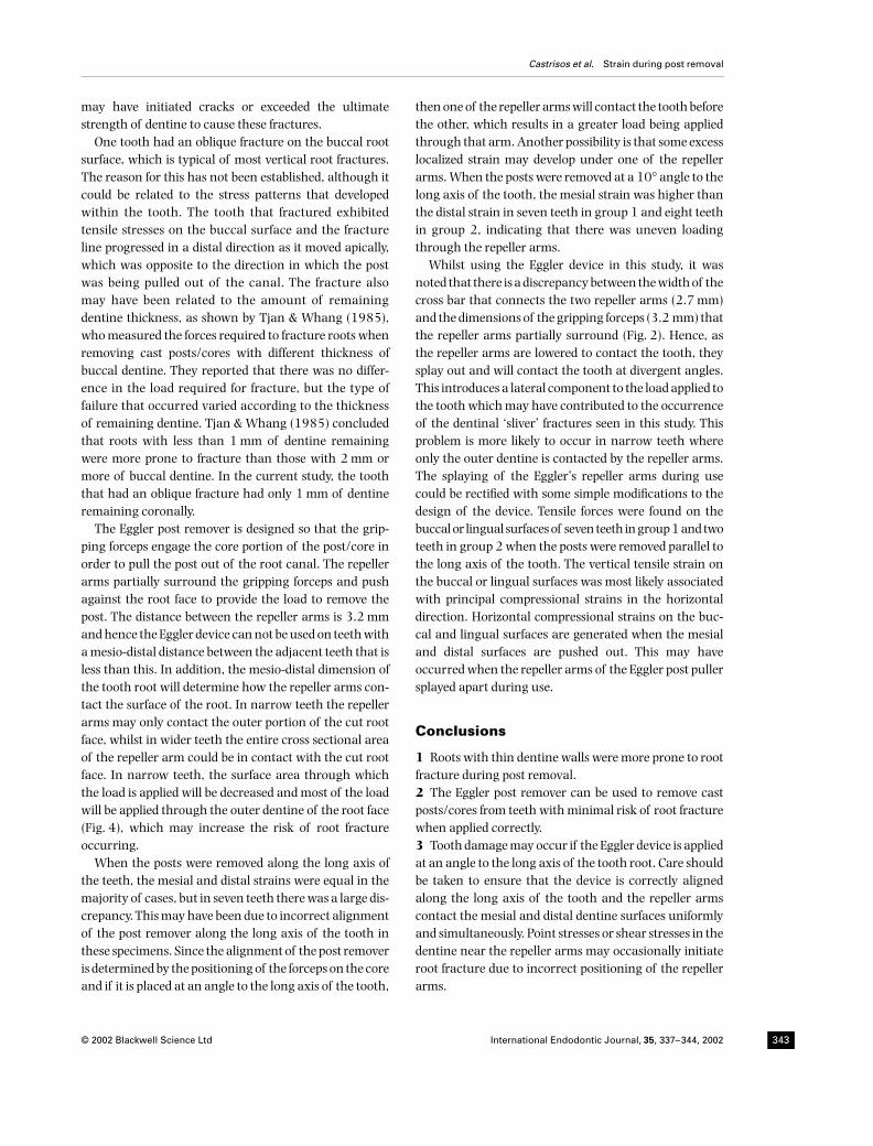
to the long axis of the root(Table 5). The fractures in three of these teeth (two fromgroup 1; one from group 2) were small slivers of dentinewhich resulted in loss of 1–2 mm of dentine on the outersurface of the root and all of these fractures occurred atthe point where the repeller arms of the Eggler postremover contacted the tooth. The other tooth from group1 had an oblique root fracture on the buccal surfacewhich extended at least 6 mm in a disto-apical direction(Fig. 4).
Discussion
The greatest strains measured were on the mesial anddistal root surfaces where the repeller arms of the Egglerpost remover contacted the root. There was considerable
variation in strain from a tensile value of 214
µ
strain toa compressive strain of 5406
µ
strain. The strains meas-ured at the root surface were below the slightly greaterthan 1% strain required to exceed the compressive elasticlimit where fracture may occur (Waters 1980).
Posts were cemented and then removed twice for eachtooth – first along the long axis of the root (0
°
) and thenat a 10
°
angle to the long axis. Post removal was per-formed in this order because there were concerns thatroot fractures may occur when the posts were removed at10
°
to the long axis of the tooth and this was confirmedwith four teeth during the second stage of the experi-ment. The teeth were examined under a microscope(
×
16) for evidence of any fractures or cracks prior to rece-menting the post, but none were seen. However, it is rec-ognized that multiple cementation and removal of eachpost, and the order in which the posts were removed,could possibly have had deleterious effects on the toothroots but this could not be determined in the currentstudy. The same teeth were used at 0
°
and then at 10
°
inan attempt to provide a more accurate comparison of thestrains produced by loading the roots at the two differentangles.
Four teeth (20% of cases) suffered damage when theEggler post puller was not used correctly – that is, at anangle of 10
°
to the long axis of the tooth root. Only one ofthese was a root fracture that clinically would have ren-dered the tooth unrestorable and necessitated extraction.The other three teeth had small (1–2 mm) sliver fracturesof dentine where the repeller arms of the Eggler postremover contacted the teeth. These findings emphasizethe need to be careful to ensure that the Eggler postremover is applied parallel to the long axis of the toothbefore applying any force.
There were several factors that may have contributedto these fractures. First, there was an uneven applicationof load by the repeller arms during post removal, asdemonstrated by the strain measurements in the teeththat fractured where there was a difference betweenthe distal and mesial surfaces with compressive
µ
strainvalues ranging from 490 up to 3481. Secondly, it wasobserved that the distance between the two repeller armsincreased as the load applied to the root surface wasincreased (Fig. 3). This action introduces lateral shearforces on the mesial and distal root surfaces, which mayincrease the risk of having small fractures as was seen atthe site of application of the repeller arms. Fractures mayoccur since dentine is weaker under shear or tensileloading than under compression (Waters 1980). In theregion where the repeller arms contacted the cut rootfaces, it is possible that point, tensile or shear stresses
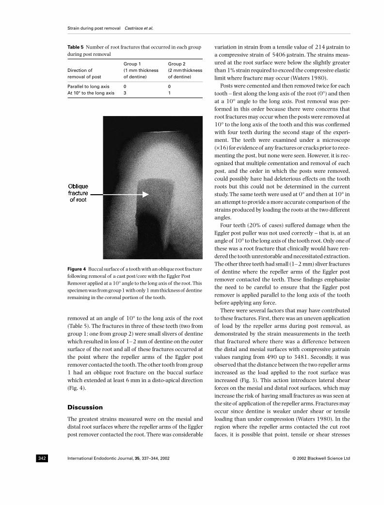
Table 5 Number of root fractures that occurred in each group during post removal
Direction of removal of post
Group 1 (1 mm thickness of dentine)
Group 2 (2 mm thickness of dentine)
Parallel to long axis 0 0At 10° to the long axis 3 1
Figure 4 Buccal surface of a tooth with an oblique root fracture following removal of a cast post/core with the Eggler Post Remover applied at a 10° angle to the long axis of the root. This specimen was from group 1 with only 1 mm thickness of dentine remaining in the coronal portion of the tooth.
IEJ_483.fm Page 342 Friday, March 22, 2002 11:16 AM
Castrisos et al.
Strain during post removal
© 2002 Blackwell Science Ltd International Endodontic Journal,
35
, 337–344, 2002 343
may have initiated cracks or exceeded the ultimatestrength of dentine to cause these fractures.
One tooth had an oblique fracture on the buccal rootsurface, which is typical of most vertical root fractures.The reason for this has not been established, although itcould be related to the stress patterns that developedwithin the tooth. The tooth that fractured exhibitedtensile stresses on the buccal surface and the fractureline progressed in a distal direction as it moved apically,which was opposite to the direction in which the postwas being pulled out of the canal. The fracture alsomay have been related to the amount of remainingdentine thickness, as shown by Tjan & Whang (1985),who measured the forces required to fracture roots whenremoving cast posts/cores with different thickness ofbuccal dentine. They reported that there was no differ-ence in the load required for fracture, but the type offailure that occurred varied according to the thicknessof remaining dentine. Tjan & Whang (1985) concludedthat roots with less than 1 mm of dentine remainingwere more prone to fracture than those with 2 mm ormore of buccal dentine. In the current study, the tooththat had an oblique fracture had only 1 mm of dentineremaining coronally.
The Eggler post remover is designed so that the grip-ping forceps engage the core portion of the post/core inorder to pull the post out of the root canal. The repellerarms partially surround the gripping forceps and pushagainst the root face to provide the load to remove thepost. The distance between the repeller arms is 3.2 mmand hence the Eggler device can not be used on teeth witha mesio-distal distance between the adjacent teeth that isless than this. In addition, the mesio-distal dimension ofthe tooth root will determine how the repeller arms con-tact the surface of the root. In narrow teeth the repellerarms may only contact the outer portion of the cut rootface, whilst in wider teeth the entire cross sectional areaof the repeller arm could be in contact with the cut rootface. In narrow teeth, the surface area through whichthe load is applied will be decreased and most of the loadwill be applied through the outer dentine of the root face(Fig. 4), which may increase the risk of root fractureoccurring.
When the posts were removed along the long axis ofthe teeth, the mesial and distal strains were equal in themajority of cases, but in seven teeth there was a large dis-crepancy. This may have been due to incorrect alignmentof the post remover along the long axis of the tooth inthese specimens. Since the alignment of the post removeris determined by the positioning of the forceps on the coreand if it is placed at an angle to the long axis of the tooth,
then one of the repeller arms will contact the tooth beforethe other, which results in a greater load being appliedthrough that arm. Another possibility is that some excesslocalized strain may develop under one of the repellerarms. When the posts were removed at a 10
°
angle to thelong axis of the tooth, the mesial strain was higher thanthe distal strain in seven teeth in group 1 and eight teethin group 2, indicating that there was uneven loadingthrough the repeller arms.
Whilst using the Eggler device in this study, it wasnoted that there is a discrepancy between the width of thecross bar that connects the two repeller arms (2.7 mm)and the dimensions of the gripping forceps (3.2 mm) thatthe repeller arms partially surround (Fig. 2). Hence, asthe repeller arms are lowered to contact the tooth, theysplay out and will contact the tooth at divergent angles.This introduces a lateral component to the load applied tothe tooth which may have contributed to the occurrenceof the dentinal ‘sliver’ fractures seen in this study. Thisproblem is more likely to occur in narrow teeth whereonly the outer dentine is contacted by the repeller arms.The splaying of the Eggler’s repeller arms during usecould be rectified with some simple modifications to thedesign of the device. Tensile forces were found on thebuccal or lingual surfaces of seven teeth in group 1 and twoteeth in group 2 when the posts were removed parallel tothe long axis of the tooth. The vertical tensile strain onthe buccal or lingual surfaces was most likely associatedwith principal compressional strains in the horizontaldirection. Horizontal compressional strains on the buc-cal and lingual surfaces are generated when the mesialand distal surfaces are pushed out. This may haveoccurred when the repeller arms of the Eggler post pullersplayed apart during use.
Conclusions
1
Roots with thin dentine walls were more prone to rootfracture during post removal.
2
The Eggler post remover can be used to remove castposts/cores from teeth with minimal risk of root fracturewhen applied correctly.
3
Tooth damage may occur if the Eggler device is appliedat an angle to the long axis of the tooth root. Care shouldbe taken to ensure that the device is correctly alignedalong the long axis of the tooth and the repeller armscontact the mesial and distal dentine surfaces uniformlyand simultaneously. Point stresses or shear stresses in thedentine near the repeller arms may occasionally initiateroot fracture due to incorrect positioning of the repellerarms.
IEJ_483.fm Page 343 Friday, March 22, 2002 11:16 AM

Strain during post removal
Castrisos et al.
International Endodontic Journal,
35
, 337–344, 2002 © 2002 Blackwell Science Ltd344
Acknowledgements
This study was supported by grants from the AustralianSociety of Endodontology Inc. and the School of DentalScience, University of Melbourne, Australia.
The authors thank Catherine Smith, from the Stat-istical Consulting Centre at the University of Melbournefor her assistance with the statistical analysis.
References
Altshul JH, Marshall G, Morgan LA, Baumgartner JC (1997)Comparison of dentinal crack incidence and of post removaltime resulting from post removal by ultrasonic or mechanicalforce
.
Journal of Endodontics
23
, 683–6.Bando E, Kawashima T, Tui IT, Kubo Y, Nakano M (1985)
Removing dowels in difficult teeth.
Journal of Prosthetic Den-tistry
54
, 34–6.Berbert A, Filho MT, Ueno AH, Bramante CM, Ishiikiriama A
(1995) The influence of ultrasound in removing intraradicularposts.
International Endodontic Journal
28
, 100–2.Buoncristiani J, Seto BG, Caputo AA (1994) Evaluation of ultra-
sonic and sonic instruments for intraradicular post removal.
Journal of Endodontics
20
, 486–9.Castrisos TV, Abbott PV (2002) A survey of methods used for
post removal in specialist endodontic practice.
InternationalEndodontic Journal
35
, 172–80.Henry PJ (1977) Photoelastic analysis of post core restorations.
Australian Dental Journal
22
, 157–9.Huysmans MC, Peters MC, Van der Varst PG, Plasschaert AJ
(1993) Failure behaviours of fatigue tested post and cores.
International Endodontic Journal
26
, 294–300.Johnson WT, Leary JM, Boyer DB (1996) Effect of ultrasonic
vibration on post removal in extracted human premolarteeth.
Journal of Endodontics
22
, 487–8.
Lertchirakarn V, Palamara JEA, Messer HH (1999) Load andstrain during lateral condensation and vertical root fracture.
Journal of Endodontics
25
, 99–104.Machtou P, Safarti P, Cohen AG (1989) Post removal prior to
retreatment.
Journal of Endodontics
15
, 552–4.Mentink AG, Creugers NH, Hoffenbrouwers PM, Meeuwissen R
(1998) Qualitative assessment of stress distribution duringinsertion of endodontic posts in photoelastic material.
Journalof Dentistry
26
, 125–31.Obermayr G, Walton RE, Leary JM, Krell KV (1991) Vertical root
fracture and relative deformation during obturation and postcementation.
Journal of Prosthetic Dentistry
66
, 181–7.Reeh ES, Douglas WH, Messer HH (1989) Stiffness of endodon-
tically treated teeth related to restoration technique.
Journal ofDental Research
68
, 1540–4.Ross RS, Nicholls JI, Harrington GW (1991) A comparison of
strains generated during placement of five endodontic posts.
Journal of Endodontics
17
, 450–6.Saw LH, Messer HH (1995) Root strains associated with different
obturation techniques.
Journal of Endodontics
21
, 314–20.Stamos DE, Gutmann JL (1991) Revisiting the post puller.
Journalof Endodontics
17
, 466–8.Stamos DE, Gutmann JL (1993) Survey of endodontic retreat-
ment methods used to remove intraradicular posts.
Journal ofEndodontics
19
, 366–9.Tjan AHL, Whang SB (1985) Resistance to root fracture of
dowel channels with various thickness of buccal dentinewalls.
Journal of Prosthetic Dentistry
53
, 496–500.Waters NE (1980) Some mechanical and physical properties of
teeth. In: Vincent D, ed.
The Mechanical Properties of BiologicalMaterials
. Cambridge, UK: Cambridge University Press, 63–134.
Yoshida T, Gomyo S, Itoh T, Shibata T, Sekine I (1997) An experi-mental study of the removal of cemented dowel retained castposts/cores by ultrasonic vibration.
Journal of Endodontics
23
,239–41.
IEJ_483.fm Page 344 Friday, March 22, 2002 11:16 AM
Copyright © 2022 FDOKUMEN

