


Bahasa
Halaman
Hukum


Eye Movements, Strabismus, Amblyopia, and Neuro-Ophthalmology
LHON Gene Therapy Vector Prevents Visual Loss and OpticNeuropathy Induced by G11778A Mutant MitochondrialDNA: Biodistribution and Toxicology Profile
Rajeshwari Koilkonda,1 Hong Yu,1 Venu Talla,1 Vittorio Porciatti,1 William J. Feuer,1
William W. Hauswirth,2 Vince Chiodo,2 Kirsten E. Erger,3 Sanford L. Boye,2 Alfred S. Lewin,4
Thomas J. Conlon,3 Lauren Renner,5 Martha Neuringer,5 Carol Detrisac,6 and John Guy1
1Bascom Palmer Eye Institute, University of Miami, Miller School of Medicine, Miami, Florida, United States2Department of Ophthalmology, University of Florida, College of Medicine, Gainesville, Florida, United States3Department of Pediatrics, University of Florida, College of Medicine, Gainesville, Florida, United States4Department of Molecular Genetics and Microbiology, University of Florida, College of Medicine, Gainesville, Florida, United States5Oregon National Primate Research Center, Oregon Health and Science University, Beaverton, Oregon, United States6Charles River Pathology Associates-Illinois, Chicago, Illinois, United States
Correspondence: John Guy, Neuro-Ophthalmology Service, BascomPalmer Eye Institute, McKnightBuilding Room 404, 1638 NW 10thAvenue, Miami, FL 33136, USA;[email protected].
Submitted: August 3, 2014Accepted: October 13, 2014
Citation: Koilkonda R, Yu H, Talla V, etal. LHON gene therapy vector pre-vents visual loss and optic neuropathyinduced by G11778A mutant mito-chondrial DNA: biodistribution andtoxicology profile. Invest Ophthalmol
Vis Sci. 2014;55:7739–7753.DOI:10.1167/iovs.14-15388
PURPOSE. To demonstrate safety and efficacy of allotopic human ND4 for treatment of a Leber’shereditary optic neuropathy (LHON) mouse model harboring the G11778A mitochondrialmutation.
METHODS. We induced LHON in mice by intravitreal injection of mutant (G11778A) humanND4 DNA, responsible for most cases of LHON, that was directed to mitochondria using anAAV2 vector to which we appended a mitochondrial targeting sequence to the VP2 capsid.We then attempted rescue of visual loss using our test article (ScAAV2-P1ND4v2) containing asynthetic nuclear encoded G11778G ND4 gene that was allotopically expressed. Control miceeither were uninjected or received AAV2-GFP or AAV2-mCherry. We performed RT-PCR andconfocal microscopy at 2 weeks post injection. Pattern electroretinograms (PERGs), spectral-domain optical coherence tomography (SD-OCT), histology, and transmission electronmicroscopy (TEM) were performed. For toxicology and biodistribution studies, the test articlewas administered intravitreally to rats and rhesus macaques at different doses.
RESULTS. Mutant and wild-type ND4 were efficiently expressed in the mitochondria of retinalganglion cells (RGCs). Visual function assessed by serial PERGs and retinal structure by serialSD-OCT showed a significant rescue by the test article. Histology and ultrastructural analysisconfirmed that loss of RGCs and demise of axons was prevented by ScAAV2-P1ND4v2. Ratand nonhuman primate biodistribution studies showed that vector spread outside theinjected eye into spleen and lymph nodes was minimal. Histopathology of tissues and organsincluding the eyes was comparable to that of uninfected and saline-injected eyes.
CONCLUSIONS. Allotopically expressed wild-type ND4 prevents the phenotype induced byG11778A mitochondrial DNA with a toxicology profile acceptable for testing in a phase Iclinical trial.
Keywords: LHON, mitochondria, gene therapy
The field of human mitochondrial genetics was born aquarter-century ago, and the list of neurodegenerative
disorders associated with mutated mitochondrial DNA keepsgrowing. While many different experimental approaches havebeen proposed, development of a clinically effective therapyhas been elusive.1 Leber’s hereditary optic neuropathy (LHON)is a maternally inherited disease, first described in 1871,resulting in acute bilateral and permanent loss of central visionusually in the second and third decades of life. In approxi-mately 95% of patients, it is caused by three pathogenic pointmutations in respiratory chain subunits of nicotinamideadenine dinucleotide-ubiquinone-oxidoreductase that is com-plex 1. They include a G to A transition at nucleotide 3460 inND1 or at nucleotide 11778 in ND4 or a T to C transition innucleotide 14484 in ND6 of the approximately 16-kb mito-
chondrial genome. In most LHON patients with visual loss, thepathogenic mitochondrial DNA (mtDNA) mutations are homo-plasmic, that is, lacking normal mtDNA. However, approxi-mately 14% of the patients may have the mutation in theheteroplasmic condition, that is, with both normal and mutatedmtDNA.2,3 Of the three mutations, the G11778A resulting inarginine to histidine substitution at amino acid 340 of ND4(R340H) is responsible for half of the cases. Spontaneous visualimprovement is rare and often incomplete.2,4–7
A potential target for therapeutic intervention is tointroduce a normal copy of the defective ND4 allele intoretinal ganglion cells (RGCs), the cell type exclusively affectedin LHON. Replacing a defective gene has been the basis formore than 1500 gene therapy clinical trials worldwide, but onlytwo involving mitochondrial disease (ClinicalTrials.gov No.
Copyright 2014 The Association for Research in Vision and Ophthalmology, Inc.
www.iovs.org j ISSN: 1552-5783 7739
NCT01267422, located in China, and No. NCT02064569,located in France). One of the major drawbacks with treatingmtDNA diseases by gene therapy has been a lack of availabilityof practical methods.8,9 To address this question, we and othergroups have investigated an alternative approach, ‘‘allotopicexpression,’’ in which a nuclear version of the mitochondrialgene (in this case ND4) was expressed in the nucleus,translated on cytoplasmic polyribosomes, and then importedinto the mitochondria with the addition of an N-terminalmitochondrial targeting sequence.10–12 Previously, our groupreported that allotopic expression of the normal ND4 genecorrected the defective adenosine triphosphate (ATP) synthe-sis in LHON cells with homoplasmic G11778A mtDNAmutation.13 We next delivered a nuclear-encoded version ofmutant human ND4 (R340H) allele into the mouse visualsystem and were successful in generating a bona fide mouseLHON model. This mouse model not only exhibited disruptionof mitochondrial architecture and increased generation ofreactive oxygen species (ROS), but also had swelling of opticnerve head, following which RGCs showed apoptosis with aprogressive demise including degeneration of axons compris-ing the optic nerve, the key hallmarks of LHON pathology.14
As viruses have been shown to enter mitochondria15 and thenormal AAV vector has been shown to deliver some of its DNAto mitochondria,16 we created an LHON mouse model by adifferent approach. We efficiently redirected the AAV virion tomitochondria by adding a mitochondrial targeting sequence(MTS) to the viral capsid surrounding its DNA core. Thiscytochrome oxidase subunit 8 (COX8) presequence was fusedin frame to the N-terminus of GFP in the AAV2 VP2 capsid openreading frame. The mutant G11778A human ND4 gene in themitochondrial genetic code driven by the human mitochondrialheavy strand promoter (HSP) was packaged into this MTS-AAVand delivered to the visual system of mice. This approachresulted in transcription and translation of mutant human ND4in mitochondria where it induced visual loss and opticatrophy.17,18 We had previously shown that wild-type ND4packaged into the MTS-AAV prevented visual loss and opticneuropathy induced by the mutant allotopic R340H ND4 allele.
In this study, we investigated if our proposed test article,ScAAV2-P1ND4v2, containing the human wild-type ND4subunit gene of complex I that is expressed allotopicallywould prevent the LHON pathology of rodents generated bythe MTS-AAV approach delivering the mutant ND4 allele tomurine RGCs and their axons in the optic nerve. This scenariowould be most analogous to the treatment of LHON patients inwhom disease is induced by the mutant G11778A mtDNA. Inthis set of experiments, the ratio of mutant ND4 relative to testarticle (ScAAV-P1ND4v2) is 4:1, thus 16-fold less than the 4:1ratio of ScAAV-P1ND4v2 to mutant allotopic R340H ND4described in our previous study of allotopic rescue of diseasecaused by an allotopic mutant R340H ND4. With mutant ND4DNA in excess, we show that ScAAV2-P1ND4v2 preventsvisual loss and optic neuropathy induced by mutant G11778ADNA. We also performed additional safety experiments todemonstrate that ScAAV2-P1ND4v2 does not spread outsidethe eye or cause any pathology in normal rodents and innonhuman primates at the highest doses planned for thehuman phase I clinical trial.
MATERIALS AND METHODS
Construction of the Viral Vector
The construct used in this study was designed to meet theneeds for future human good manufacturing practices (GMP)vector usage. It was derived from the original fusion geneconstruct containing human nuclear encoded ND4 with a MTS
(ATP1) and a FLAG epitope tag, P1ND4FLAG-UF22-WRPE,which was previously described in detail.19 For this study, theconstruct was modified to remove the FLAG tag. For thispurpose, the XbaI digested fragment of P1ND4FLAG-UF22-WRPE was first cloned into the XbaI site of pBluescript II(Stratagene, La Jolla, CA, USA), resulting in plasmid pBS-P1ND4
(XbaI). Self-complementary AAV vector plasmid, Sc-smCBA-P1ND4, was created by cutting Sc-trs-SB-smCBA-hGFP withBamHI and NotI and ligating into the NotI and BamHIfragment of pBS-P1ND4 (XbaI). This Sc-AAV vector plasmidcontained a promoter element, the 953 bp, and a truncatedchimeric CMV/CBA promoter (smCBA) and had modificationsin one of the AAV inverted terminal repeats that resulted ingeneration of self-complementary AAV genomes. These mod-ifications were described in detail by McCarty and col-leagues.20 The resulting self-complementary plasmidcontaining smCBA driving P1ND4-FLAG was named Sc-smCBA-P1ND4. For the removal of extra polyadenylationsignal and FLAG tag, Sc-smCBA-P1ND4 was cut with SphIand SalI and ligated with a synthetic fragment of DNA withSphI and SalI ends. The SphI/SalI P1ND4 fragment is asynthetic fragment of 1092 bases encompassing a SphI site tothe end of P1ND4 cDNA with the 30 end flanked by a SalI site.This synthetic fragment was manufactured by Genscript(Piscataway, NJ, USA). The resulting construct from the ligationno longer contained the SV40 PolyA and 30 FLAG epitope tag.The bGH polyA was already present in the vector plasmidprogenitor and remains in the new construct. This newconstruct was named Sc-smCBA-P1ND4v2.
For inducing LHON disease in mice, we used AAVcontaining the human mitochondrial mutant ND4 (G11778A)that was driven by the human mitochondrial HSP, ScAAV2-HSP-
ND4 (G11778A) FLAG. The human mutant ND4 contained astop codon AGA fused in frame downstream of the FLAGepitope. To the HSP-ND4 (G11778A) FLAG construct, weadded a second gene mitochondrial encoded (m) Cherry. Inbrief, nuclear encoded Cherry was cloned into pCDNA3 andwas used as a template to generate a mitochondrial encodedgene with a substitution of A for C at 559 nt by site-directedmutagenesis (Quikchange II XL site-directed mutagenesis kit;Stratagene). After confirmation by DNA sequencing, theresultant cassette was cloned into the mutant HSP-mutantND4FLAG backbone to give HSP-ND4FLAG(G11778A)þm-
Cherry.For delivery, we packaged both the allotopic wild-type and
mutant ND4 constructs into self-complementary AAV2 vectorswith triple tyrosine (Y) to phenylalanine (F) modifications atpositions 444, 500, and 730 (Y444FþY500FþY730F) in the VP3capsid. The AAV containing mutant ND4 was directed towardthe mitochondria, using a MTS appended to the open readingframe of VP2. This MTS had a GFP sequence in frame, and theplasmid was designated COX8GFP VP2. For controls, GFP waspackaged into self-complementary (sc) AAV2.
Animals
All animal procedures were performed in accordance with theNational Institutes of Health Guide for the Care and Use ofLaboratory Animals and the ARVO Statement for the Use ofAnimals in Ophthalmic and Vision Research.
Intraocular Injections (Mice and Rats)
For the intraocular injections of recombinant adeno-associatedvirus (rAAV), DBA/1J mice were sedated by inhalation with1.5% to 2% isoflurane. A local anesthetic (proparacainehydrochloride) was applied topically to the cornea, and a 32-
LHON Gene Therapy Vector Prevents Visual Loss IOVS j December 2014 j Vol. 55 j No. 12 j 7740

gauge needle attached to a Hamilton syringe (HamiltonCompany, Reno, NV, USA) was inserted through the pars planaunder the dissecting microscope (Carl Zeiss Microscopy,Thornwood, NY, USA). One microliter of ScAAV2-(Y444,500,730F)-HSP-ND4 (G11778A)þmCherry (COX8-GFP)(2.19 3 1012 particles/mL) (n¼ 20) was injected into the rightand left eyes. Seventy-two hours after these injections, 1 lL ofthe test article, ScAAV2-(Y444,500,730F)-P1ND4v2 (5.02 3
1011 particles/mL), was injected into 10 mice in both eyes (testgroup). The remaining 10 mice received 1 lL ScAAV2-GFP (4 3
1011 particles/mL) in both eyes (mock-treated group) or justScAAV2-mCherry for the no-disease control group (n ¼ 6).Another group of 10 mice were injected with the test article; 6mice remained uninjected controls. Spraque-Dawley ratsreceived intravitreal saline or P1ND4v2 at doses of 6.56 3
106 (n¼ 20), 6.56 3 107 (n¼ 20), or 1.97 3 108 viral genomes(VG) (n ¼ 20) or saline (n ¼ 20).
Electrophysiology (Mice)
Pattern electroretinograms (PERGs) were obtained from mice(n ¼ 24) at 2 months, 6 months, and 1 year after the viralinjections. In brief, mice were weighed and anesthetized usingintraperitoneal injections (0.5–0.7 mL/kg) of a mixture ofketamine (42.8 mg/mL) and xylazine (8.6 mg/mL) and wererestrained using a bite bar and a nose holder that allowedunobstructed vision. The animals were kept at a constant bodytemperature of 378C with a feedback-controlled heating pad(TCAT-2LV; Physitemp Instruments, Inc., Clifton, NJ, USA).Under these conditions, the eyes of mice were wide open andin a stable position, with undilated pupils pointing laterally andupward. The PERG electrode, with a diameter of 0.25 mm andmade of silver wire configured to a semicircular loop of a 2-mmradius, was placed on the corneal surface by means of amicromanipulator and positioned to encircle the pupil withoutlimiting the field of view. Reference and ground electrodeswere stainless steel needles inserted under the skin of the scalpand tail, respectively. After setting the mice on the stage andbefore recording, a small drop of balanced salt solution wastopically applied to the cornea to prevent drying. A visualstimulus of contrast-reversing (1 Hz, 2 reversals/s) horizontalbars was generated by a programmable graphic card (VSG;Cambridge Research Systems, Rochester, UK) on a cathode raytube display (Multiscan 500; Sony, Boston, MA, USA) whosecenter was aligned with the projection of the pupil. Eyes werenot refracted for the viewing distance because the mouse eyehas a large depth of focus due to the pinhole pupil. At theviewing distance of 15 cm, the stimulus field covered an area of69.48 3 63.48. Patterns had a fixed mean luminance of 50 cd/m2. Retinal signals were amplified (10,000-fold) and band-passfiltered (1–30 Hz). Three consecutive responses to 600contrast reversals each were recorded. The responses weresuperimposed to check for consistency and then averaged(1800 sweeps). Averaged PERGs were automatically analyzedto evaluate the major positive and negative waves usingcommercially available software (SigmaPlot; Systat Software,Inc., San Jose, CA, USA). The waveform of averaged PERGs tohigh-contrast (1.0) gratings of low spatial frequency (0.05 cyc/deg) consisted of a major positive peak at around 90 to 120 ms(defined as P100) followed by a slower negative wave with abroad trough at around 200 to 300 ms. The maximal voltage inthe expected time window for P100 (50–200 ms) and theminimal voltage in the expected time window for N250 (201–350 ms) were identified automatically using a simple macrowritten in SigmaPlot language (version 11.2). Statistical analysiswas evaluated by t-test, and P < 0.05 was consideredstatistically significant.
Imaging (Mice)
In vivo high-resolution three-dimensional (3D) imaging of themouse retina (n¼ 26) was performed at 4 months, 8 months,and 12 months post AAV injection using spectral-domainoptical coherence tomography (SD-OCT; Bioptigen, Inc.,Durham, NC, USA). Briefly, the mice were anesthetized withan intraperitoneal injection of ketamine (80 mg/kg) andxylazine (5 mg/kg). Both pupils were dilated with a topicallyapplied drop of tropicamide (1%). To preserve corneal clarity,we applied a drop of Systane Ultra (Alcon Laboratories, Inc.,Fort Worth, TX, USA) lubricating eye drops to the cornea.Mice were secured on a custom stage, which allowed freerotation, to align the eye for imaging of the optic nerve head(ONH). Rectangular volume scans centered on the ONH wereacquired in both eyes with the Bioptigen SD-OCT system. Foranalysis of SD-OCT images, all data sets were checked toensure that there was consistent image quality across the scan.Eyes with images showing shadowing due to the mediaopacities were excluded from analysis, as this could affectthickness measurements. A custom-made segmentation algo-rithm (Matlab; The Mathworks, Inc., Natick, MA, USA) wasused to detect retinal boundaries and measure the distancefrom the nerve fiber layer (NFL) to the inner boundary of theinner nuclear layer (INL) for acquired images. The result ofsegmentation was used to calculate 3D thickness maps of thecombined RGCþIPL layer. Statistical analysis was performedusing Student’s t-test; P < 0.05 was used as the cutoff forstatistical significance.
For the detection of mitochondrial mCherry expression inmouse eyes, confocal scanning laser ophthalmoscopy (CSLO;Heidelberg Engineering, Bonn, Germany) imaging was per-formed 4 months following either inoculation with ScAAV2-HSP-ND4(G11778A)þmCherry along with test article ScAAV2-P1ND4v2 (n¼ 10) or in mock-treated mice receiving Sc-AAV2-GFP (n ¼ 10) or age-matched uninjected control (n ¼ 2)ScAAV2-GFP injections. The built-in 488 laser was used for theexcitation of both GFP and mCherry, and band-pass filters of460 to 490 and 542 to 800 nm were used for the detection. Forthis purpose, the animals were anesthetized with ketamine/xylazine, pupils were dilated with phenylephrine/atropine,and corneal hydration was maintained with balanced saltsolution. Retinal images were obtained using a 308 field of viewand real-time averaging of 50 images. The CSLO was focused onthe inner retina by imaging of the NFL at 488 nm through theCSLO polarization filter (red-free imaging).
Retina Cryosections and Immunolabeling (Mice)
For expression studies, mice were humanely killed 2 weeksafter the viral injections (n ¼ 5), and the eyes were removedand fixed with 4% paraformaldehyde in phosphate-bufferedsaline solution (PBS) for 1 hour. The eyes were thentransferred to 0.4% paraformaldehyde overnight. To make flatmounts, the cornea and crystalline lens were removed, andthe entire retina was carefully dissected from the eye cup.Four radial cuts were made from the edge to the equator ofthe retina to make it flat. After three washes in PBS, retinaswere permeabilized with 0.5% Triton X-100 (Dow ChemicalCorporation, Midland, MI, USA) in PBS for 1 hour followed byincubation with a blocking solution of 0.5% Triton X-100 and10% goat serum for 1 hour. The flat-mounted retinas werethen rinsed in PBS and incubated with a mixture of eithermouse monoclonal anti-human ND4 antibody (Abnova, TaipeiCity, Taiwan) or mouse monoclonal anti-FLAG antibody(Sigma-Aldrich Corp., St. Louis, MO, USA), monoclonal ratThy1.2 antibody (1:200; Abcam, Cambridge, MA, USA), orpolyclonal rabbit anti-porin antibody (1:500; Abcam) over-night at 48C. Primary antibody solution is contained in 10%
LHON Gene Therapy Vector Prevents Visual Loss IOVS j December 2014 j Vol. 55 j No. 12 j 7741

goat serum and 0.2% Triton X-100 in PBS (pH, 7.4). After threewashes with PBS, retinas were reacted with the secondaryantibody, goat anti-mouse Cy3 (Jackson ImmunoresearchLaboratories, Inc., West Grove, PA, USA), goat anti-mouseFITC (Jackson Immunoresearch Laboratories, Inc.), goat anti-rat Cy2 (Jackson Immunoresearch Laboratories, Inc.), or goatanti-rabbit Cy5 (Jackson Immunoresearch Laboratories, Inc.),and 4 0,6-diamidino-2-phenylindole (DAPI; 2 lg/mL) (SantaCruz Biotechnology, Inc., Santa Cruz, CA, USA) in 1:500dilution contained in 10% goat serum and 0.2% Triton X-100and incubated at 48C overnight. Retinal tissues were finallywashed three times in PBS. Whole mounts were then placedon glass slides (RGC layer facing up), cover slipped, andobserved for fluorescence with a confocal microscope (TCSSP5; Leica, Wetzlar, Germany). For cryosections, the retinaswere incubated overnight in 30% sucrose and then embeddedin optimal cutting temperature embedding compound (SakuraFinetek, Torrance, CA, USA). Retinal sections, 8 lm inthickness, were then mounted with fluorescent mountingmedium (Vectashield; Vector Laboratories, Burlingame, CA,USA) and examined for fluorescence by confocal microscopy.
Histology and Ultrastructure (Mice)
For histology and electron microscopic analysis, mice receivingthe dual viral injections were euthanized after 1 year (n ¼ 5animals in each group). Briefly, eyes and optic nerves ofinfected mice were dissected out after euthanasia andperfusion with fixative (4% paraformaldehyde, 2.5% glutaral-dehyde) and were further processed by immersion fixation in2.5% glutaraldehyde, postfixed in 1% osmium tetroxide, 0.1 Msodium cacodylate-HCl buffer (pH 7.4), 7% sucrose in the cold,and then dehydrated through an ethanol series to propyleneoxide, infiltrated, and embedded in epoxy resin that waspolymerized at 608C overnight. Semithin longitudinal sections(0.5 lm) of the retina were stained with toluidine blue for lightmicroscopic examination, and images were taken at 4003magnification. For quantification, cells in the RGC layer werecounted. Ultrathin sections (90 nm) were placed on coppergrids for examination with a Hitachi H-7600 unit (Hitachi,Tokyo, Japan) operating at 80 kV.
RT-PCR (Mice)
Total RNA was isolated from the retina, optic nerves, brain, liver,and pancreas at 2 weeks post injections using Trizol reagent(Invitrogen, Carlsbad, CA, USA) according to the manufacturer’sdirections (n ¼ 2). First-strand cDNA was reverse transcribedfrom 1 lg total RNA in a final volume of 20 lL using reversetranscriptase and random hexamers from the iScript cDNAsynthesis kit (Bio-Rad, Hercules, CA, USA). Polymerase chainreaction was performed using primers hND4-FP-5 0
TGCTGAAGCTGGGCGGCTACGGC 3 0 and hND4-RP-50AACCGCCTCTCCCCGCGCGTT30 with Taq DNA polymerase(Invitrogen) in a DNA Master cycler gradient (Eppendorf,Hauppauge, NY, USA) according to a standard protocol asfollows: 1 cycle of 958C for 3 minutes; 23 cycles of 948C for 45seconds, annealing for 45 seconds, and 728C for 1 minute; a finalextension at 728C for 10 minutes; and holding at 48C. Theamount of cDNA used for each PCR was 20 ng in a 25-lLreaction volume. The PCR products were analyzed by electro-phoresis through 1% agarose gel and visualized by ethidiumbromide staining. 18S rRNA was used as an internal control.
Toxicology and Biodistribution in Rats andNonhuman Primates
A good laboratory practice (GLP)–compliant 1- and 3-monthdose-based toxicity study of ScAAV2-P1ND4v2 vector delivered
by intravitreal injection into the eyes of normal rodents wasperformed. The control group (group 1), low-dose group(group 2), medium-dose group (group 3), and high-dose group(group 4) each included 20 rats (10 males, 10 females). Half ofeach group were euthanized and necropsied at 1 month (30days) and half at 3 months (90 6 3 days) after injection,respectively.
We also performed a 3- to 4-month safety and biodistribu-tion study of P1ND4v2 vector delivered by intravitrealinjection into eight normal rhesus macaques. Vector titeredat 2.46 3 1011 vg/mL was diluted 1:1 with sterile water to 1.233 1011 vg/mL, and 200 lL was vitreally injected for a total doseof 2.46 3 1010 vg (n¼5); another group received a higher doseof 2.02 3 1011 vg (n ¼ 3). Two animals were euthanized at 3months (90 days) and one at 4 months (126 days). A fourthanimal received bilateral intravitreal injections of balanced saltsaline (volume¼ 200 lL) and was euthanized at 3 months (90days). Two animals received bilateral injections. In these, thefifth and sixth animals, a total dose of 2.46 3 1010 vg wasinjected into one eye; the second eye was injected 1 monthlater, and euthanasia was performed 3 months after the lastinjection. Three animals that received a higher dose of the testarticle were euthanized at 3 months (~90 days).
A neutralizing antibody assay was performed on serumsamples obtained from the rhesus macaques injected with thetest article synthesized at the vector core facility of theUniversity of Florida or at the University of North Carolina.The neutralizing antibody assay was performed according tothe modified protocol from Boye et al.21 Briefly, a self-complementary AAV2(Y444,500,703F)-smCBA-mCherry vec-tor was preincubated with heat-inactivated nonhuman pri-mate (NHP) serum samples at various dilutions, beginningwith a 1:5 dilution, followed by serial 1:4 dilutions and endingat 1:20,480. The serum-vector mixtures were used to infectARPE-19 cells at multiplicity of infection of 1000 vectorgenomes/cell. Three days post infection, cell viability wasconfirmed by microscopy and transduction scored by flowcytometry. The expression of mCherry was quantified bymultiplying the percentage of cells positive for mCherry bythe mean fluorescence intensity. The neutralizing antibody(NAb) titer was reported as the highest serum dilution thatinhibited self-complementary AAV2(Y444,500,703F)-smCBA-mCherry transduction > 50%.
In both species, tissue samples were collected in 10%neutral buffered formalin for histopathological analysis, andparallel samples were flash frozen in liquid nitrogen forassessment of vector biodistribution. The injected eye withoptic nerve was fixed in Davidson’s (rats) or 4% paraformal-dehyde (monkeys). Histology and histopathology examinationof prepared slides were performed under contract agreementat Charles River Pathology Associates-Illinois. Histopathologyanalysis was conducted by a board-certified veterinary pathol-ogist (CD) who was masked to study treatments and animalassignment. In addition, genomic DNA (gDNA) was extractedfrom frozen tissue or blood collected. All other frozen organswere segregated by treatment. The following organs wereextracted: skeletal muscle quadriceps, diaphragm, heart,cerebrum, lung, spleen, liver, kidney, pancreas, gonads,jejunum, parotid gland, mesenteric lymph node, and unin-jected eye with corresponding optic nerve. DNA wasquantified and analyzed by real-time PCR. Typically, 1.0 lg ofeach DNA sample was assayed in triplicate. Tissues from theday 30 high-dose rat groups were assayed first. If any tissuefrom at least one animal in this group was positive, that tissuewas analyzed in all the day 90 high-dose animals. The algorithmwas continued until the tissue was found to be negative. Forprimates, the presence of vector DNA was assessed.
LHON Gene Therapy Vector Prevents Visual Loss IOVS j December 2014 j Vol. 55 j No. 12 j 7742
Antigen-Specific Response LymphocyteProliferation Assay
Anti-AAV2-triple tyrosine (Y) to phenylalanine (F) capsidmutant antigen-specific lymphocyte proliferation responseswere assessed as previously described.22 After isolation andpurification from blood, lymphocytes were cultured at 1 3 105
cells per well of 96-well plates. Lymphocytes were separatedinto four groups with three control and six primate samplecultures per group: unstimulated (as negative control) orstimulated with AAV2trpmut (5000, 500, and 50 particles/cell.After 5 days of incubation the stimulation index (SI), defined asthe mean counts per minute of [3H]thymidine from stimulatedcells divided by the mean counts per minute of [3H]thymidinefrom unstimulated cells was calculated. Stimulation indexvalues greater than 2.0 to 3.0 are considered significant. The
viability of each lymphocyte culture was confirmed by positivecontrols with mitogen-induced proliferation in response tophytohemagglutinin (PHA) and recall antigen-induced prolifer-ation to Candida albicans.
Statistical Analysis
One-way analysis of variance followed by Student’s t-test wasused to compare PERGs and SD-OCT data. The mouse groupscompared were age-matched mice uninjected or injected withScAAV2-mCherry (no-disease control) and mice either inocu-lated with ScAAV2-HSP-ND4(G11778A)þmCherry along withthe test article, ScAAV2-P1ND4v2, or mock treated withScAAV2-GFP. Quantification of cells in the RGC layer and opticnerve axons between the mock-treated and test article–treatedmouse groups was determined using unpaired Student’s t-test.
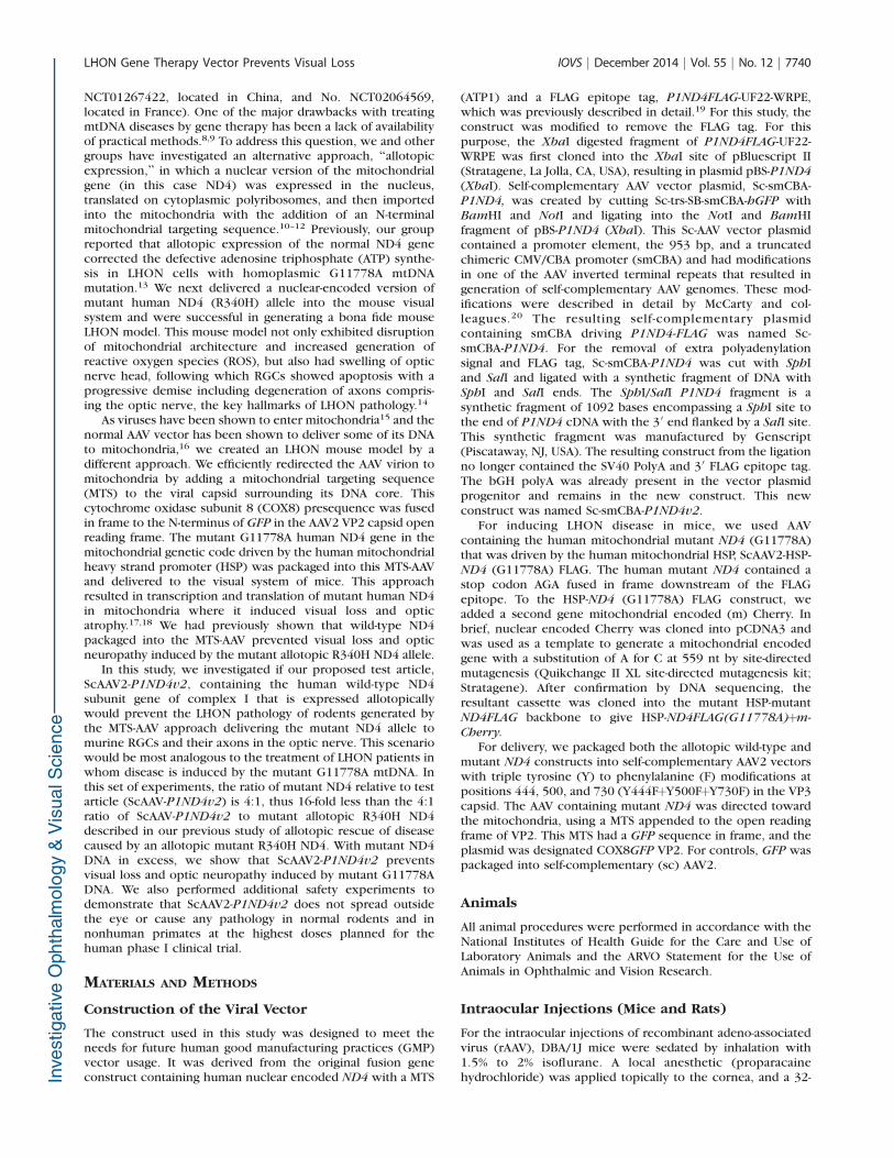
FIGURE 1. Plasmid maps and expression of mutant ND4. Plasmid maps of (a) the mutant mitochondrial ND4þmCherry and (b) test article,P1ND4v2, are shown. Four months after dual viral injections, confocal laser scanning ophthalmoscopy shows mCherry fluorescence (arrowheads)in ScAAV2-GFP- and ScAAV2-HSP-ND4(G11778A)þmCherry-injected eyes (c, d) and in test article, ScAAV2-P1ND4v2-, along with ScAAV2-HSP-
ND4(G11778A)þmCherry-injected eyes (e, f). mCherry fluorescence was absent in uninjected control (g). Confocal microscopy of retinal flatmounts performed 4 months after injection of ScAAV2-HSP-ND4(G11778A)þmCherry shows immunofluorescence of FLAG (h), Thy1.2 (i), porin(j), and merged image of FLAG, porin, and DAPI (k). A 3D rendered view of retinal longitudinal sections shows perinuclear mCherry expression (l)in RGCs (m), mitochondrial porin (n), and a merged image of mCherry, Thy1.2, porin, and DAPI (o). iTR, inverted terminal repeat; ATP1pre, ATP 1leader sequence; HSP, mitochondrial heavy strand promoter; ONL, outer nuclear layer; INL, inner nuclear layer; RGC, retinal ganglion cell layer.Scale bars: 25 lm.
LHON Gene Therapy Vector Prevents Visual Loss IOVS j December 2014 j Vol. 55 j No. 12 j 7743

All the results were expressed as mean 6 standard error (SE),and P values less than 0.05 were considered statisticallysignificant.
RESULTS
Mutant and Wild-Type ScAAV2-P1ND4v2 EfficientlyTransduces RGCs
For rescue experiments, mice (n ¼ 20) were injected in thevitreous with self-complementary AAV2 containing the mutantG11778A ND4 (ScAAV2-HSP-ND4G11778AFlag) and a secondgene, mCherry, in the mitochondrial genetic code (Fig. 1a) forvisualization of transduced organelles. Both genes were drivenby a single human HSP. This construct was packaged inmitochondrially targeted AAV virions and injected into botheyes of mice to induce LHON, followed 72 hours later byinjections of either ScAAV-GFP (n ¼ 10) or the test articleScAAV2-P1ND4v2 (Fig. 1b) (n ¼ 10); control mice remaineduninjected (n¼ 2). Four months later, mCherry was visualizedin the retinas of mock-treated (Figs. 1c, 1d) and test article–treated (Figs. 1e, 1f) live mice and absent in uninjected controlmice (Fig. 1g). Confocal microscopy revealed ND4FLAG (Fig.
1h) in RGCs labeled by Thy1.2 (Fig. 1i) colocalized tomitochondria labeled by VDAC/porin (Fig. 1j) in the mergedimage (Fig. 1k). Red fluorescence (Fig. 1l) in RGC (Fig. 1m)mitochondria (Fig. 1n) confirmed mCherry also expressed inRGC mitochondria (Fig. 1o). Confocal images showed expres-sion of human wild-type ND4 that colocalized to RGCmitochondria (Supplementary Figs. S1a–S1e) absent in nega-tive control (Supplementary Fig. S1f). Semiquantitative real-time PCR showed P1ND4v2 transcripts exclusively in theocular tissues and absent in nonocular tissues such as liver,pancreas, and heart (Supplementary Fig. S1g).
Test Article (ScAAV2-P1ND4v2) Preserves VisualFunction in LHON Mice—PERG Analysis
The PERG, a sensitive measure of visual loss and RGCfunction, probed the effects of the test article at 2, 6, and 12months after AAV injections. ANOVA with random effects toaccount for correlated measurements made on the sameanimals over time showed a highly significant difference inPERG amplitudes of age-matched uninjected controls, mock-treated, and test article–treated mouse groups (P ¼ 0.0001)(Supplementary Table S1); however, there was no statisti-
FIGURE 2. Rescue of visual function. PERG analysis of uninjected age-matched control mice and mice injected with ScAAV2-HSP-ND4(G11778A)þmCherry and mock treated with ScAAV2-GFP or test article, ScAAV2-P1ND4v2, performed at 2 months (2m) 6 months (6m),or 12 months (12m) post injection. Bar plots of PERG amplitudes (a) and latencies (b) are shown (mean 6 SE). Representative PERG waveforms areshown at (c) 2 months, (d) 6 months, and (e) 12 months post injection, n¼ 24, *P¼ 0.01 to 0.05, **P¼ 0.001 to 0.01, ***P < 0.001. m PI, monthspost injection.
LHON Gene Therapy Vector Prevents Visual Loss IOVS j December 2014 j Vol. 55 j No. 12 j 7744

cally significant difference when the latencies were com-pared (P ¼ 0.064) (Supplementary Table S2). At 2 monthspost injection, the average PERG amplitudes of test article–and mock-treated mice were significantly reduced comparedto those of age-matched uninjected mice (17.8 6 2.14, 14.946 1.3, and 25.85 6 2.7 lV; mean 6 SE) (P < 0.05).However, while the mock-treated mice continued to showsignificantly reduced amplitude at 6 months, which wors-ened at 12 months following mutant ND4 injection (P <0.05), the test article–treated mice showed rescue of PERGamplitudes, and the data were comparable to those for theage-matched uninjected mice (P > 0.05) (SupplementaryTable S1). In addition, the average PERG amplitudes andlatencies were significantly rescued in test article–treatedmice (16 6 1.68 lV; 117.5 6 4.64 ms) compared to themock-treated mouse group at 12 months post injection (11.9
6 1.14 lV; 136.9 6 6.13 ms) (P < 0.05) (SupplementaryTables S1, S2). Bar graphs show mean PERG amplitudes (Fig.2a) and latencies (Fig. 2b). Representative waveforms at 2(Fig. 2c), 6 (Fig. 2d), and 12 months (Fig. 2e) illustrate thatloss of amplitude and delay in latency were suppressed bytreatment.
Test Article (ScAAV2-P1ND4v2) Preserves the RGC
and Inner Plexiform Layers in LHON Mice—SD-
OCT Analysis
As measured by SD-OCT, the RGC layer and inner plexiformlayers appeared normal in mock-treated eyes of live mice at 4(Figs. 3a, 3b) and 8 months post injection (Figs. 3c, 3d), but at12 months these layers atrophied (Figs. 3e, 3f). In mice
FIGURE 3. In vivo optical coherence tomography (OCT) imaging. Serial OCT imaging of a mock-treated mouse eye shows the nerve fiber layer tothe inner boundary of the inner nuclear layer demarcated by yellow lines (a) and a corresponding 3D thickness map (b) at 4 months PI, a two-dimensional image (2D) (c) and 3D map (d) at 8 months PI, and a 2D (e) and 3D map (f) at 12 months PI. Also shown are 2D (g) and 3D maps (h) ofa rescued mouse eye injected with the test article at 4 months PI, 2D (i) and 3D maps (j) at 8 months PI, and a 2D image (k) and 3D map (l) at 12months PI (m). The bar plot shows average thickness measurements from the nerve fiber layer to the inner boundary of the inner nuclear layer (asmarked in the 2D images) at 8 and 12 months PI (mean 6 SE); n ¼ 26, *P ¼ 0.01 to 0.05, ***P < 0.001. m PI, months post injection.
LHON Gene Therapy Vector Prevents Visual Loss IOVS j December 2014 j Vol. 55 j No. 12 j 7745
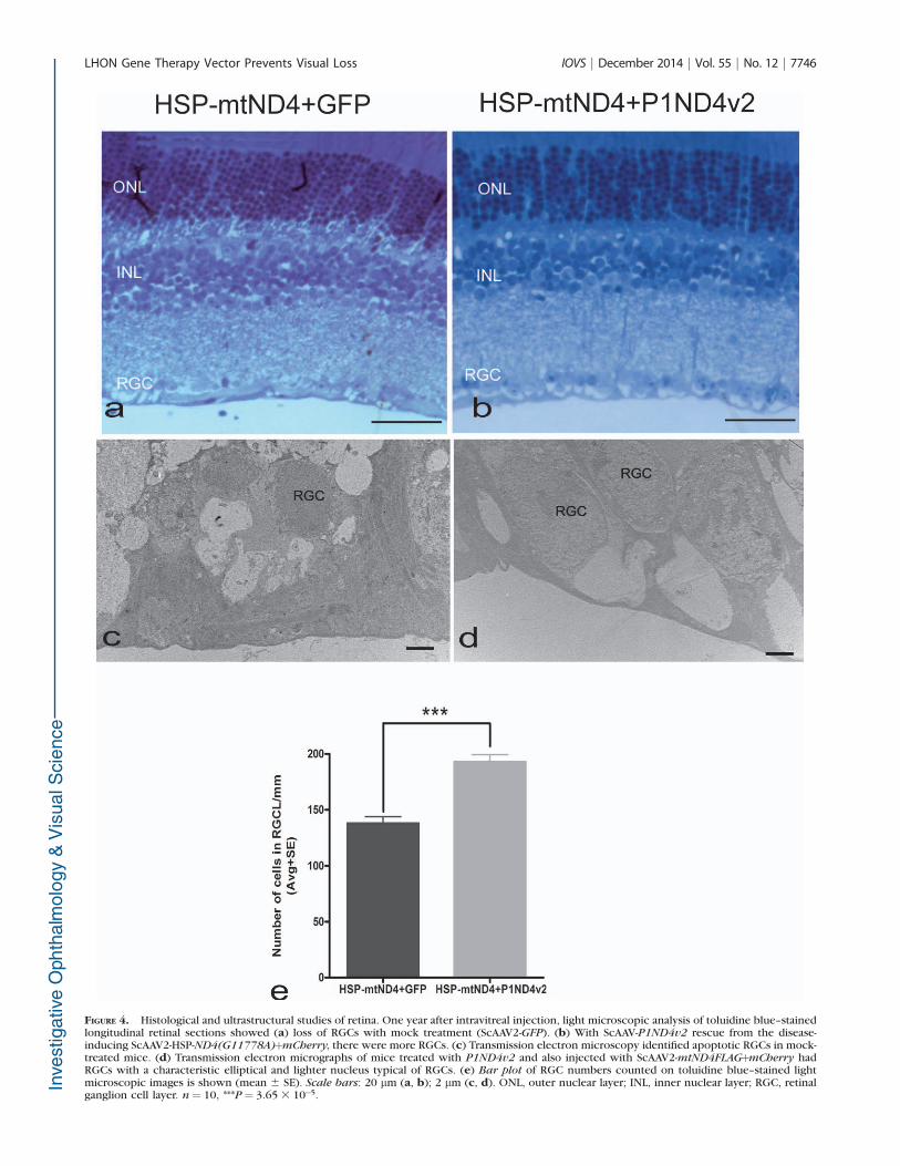
FIGURE 4. Histological and ultrastructural studies of retina. One year after intravitreal injection, light microscopic analysis of toluidine blue–stainedlongitudinal retinal sections showed (a) loss of RGCs with mock treatment (ScAAV2-GFP). (b) With ScAAV-P1ND4v2 rescue from the disease-inducing ScAAV2-HSP-ND4(G11778A)þmCherry, there were more RGCs. (c) Transmission electron microscopy identified apoptotic RGCs in mock-treated mice. (d) Transmission electron micrographs of mice treated with P1ND4v2 and also injected with ScAAV2-mtND4FLAGþmCherry hadRGCs with a characteristic elliptical and lighter nucleus typical of RGCs. (e) Bar plot of RGC numbers counted on toluidine blue–stained lightmicroscopic images is shown (mean 6 SE). Scale bars: 20 lm (a, b); 2 lm (c, d). ONL, outer nuclear layer; INL, inner nuclear layer; RGC, retinalganglion cell layer. n¼ 10, ***P¼ 3.65 3 10�5.
LHON Gene Therapy Vector Prevents Visual Loss IOVS j December 2014 j Vol. 55 j No. 12 j 7746
protected with the test article, these retinal layers werepreserved at 4 (Figs. 3g, 3h), 8 (Figs. 3i, 3j), and 12 months(Figs. 3k, 3l). ANOVA with random effects to account forcorrelated measurements made on the animals over timeshowed highly significant difference among the mCherry-injected no-disease control, disease-induced mock-treatedcontrol, and test article–treated mice (P < 0.0001). Comparedto the mCherry-injected no-disease control, the mean thick-ness of either mock-treated or test article–treated control didnot show a significant difference at 8 months post injection (P> 0.05); however, at 12 months, both the treated groupsshowed significant difference compared to the no-diseasemCherry control group (P < 0.05) (Supplementary Table S3).The mean thickness of the inner retina of the mock-treatedand test article–treated mice did not show significantdifference at 4 months (54.07 6 0.72 and 56.31 6 0.79 lm,mean 6 SE) and 8 months post injection (60 6 0.72 and 61.546 1.5 lm, mean 6 SE) (P > 0.05). However, at 12 monthspost injection, the test article–treated mice showed asignificant rescue of inner retina thickness compared to themock-treated mice (57.4 6 0.81 and 53.2 6 1.19 lm, mean 6SE) (P < 0.05) (Supplementary Table S3). Bar graphs showmean inner retina thickness at 8 and 12 months post injection(Fig. 3m).
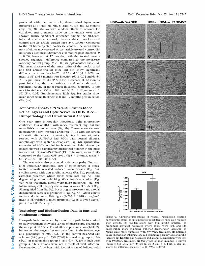
Test Article (ScAAV2-P1ND4v2) Rescues InnerRetinal Layers and Optic Nerves in LHON Mice—Histopathology and Ultrastructural Analysis
One year after intraocular injections, light microscopyconfirmed loss of RGCs with mock treatment (Fig. 4a) butmore RGCs in rescued eyes (Fig. 4b). Transmission electronmicrographs (TEM) revealed apoptotic RGCs with condensedchromatin after mock treatment (Fig. 4c). In contrast, micerescued with P1ND4v2 had RGCs with normal ellipticalmorphology with lighter nucleoplasm (Fig. 4d). Quantitativeevaluation of RGCs on toluidine blue–stained light microscopeimages showed a significantly greater cell number in the miceinjected with ScAAV2-P1ND4v2 (193 6 6.6/mm, mean 6 SE)compared to the ScAAV-GFP group (138 6 5.9/mm, mean 6
SE), P ¼ 8.8 3 10�6 (Fig. 4e).The test article also prevented optic neuropathy. One year
after intraocular injections, TEM of optic nerves of mock-treated animals revealed reduced axon density (Fig. 5a),swollen axons with thin myelin lamellae (Fig. 5b), prominentastroglial processes where axons were lost (Fig. 5c), anddegenerating axons exhibiting Wallerian degeneration (Fig.5d). With treatment, axons were more numerous (Fig. 5e).Inflammatory cell phagocytosis of myelin was still evident (Fig.5f, magnified from Fig. 5e), but astroglial processes and axonaldegeneration were less prominent (Figs. 5g, 5h). Axon countsfor treated mice were 50% higher (0.203 6 0.018 axons/lm2,mean 6 SE) relative to mock treatment (0.138 6 0.013 axons/lm2), P ¼ 0.00758 (Fig. 5i).
Toxicology and Biodistribution Data in Rats andNonhuman Primates
Histopathologic assessment by a veterinary pathologist maskedto study treatment showed a variety of microscopic changes inthe rat eye at 30 (Table 1) and 90 days post injection (Table 2),but not in other organs. Lesions were found in the injected eyeat a percentage of 30% (6/20) in the control balanced saltsolution (BSS) group 1, 35% (7/20) in low-dose group 2, 10%(4/20) in medium-dose group 3, and 40% (8/20) in high-dosegroup 4. Thus, lesions were not a result of viral infection.Degeneration of the lens with identification of balloon cells
FIGURE 5. Ultrastructural studies of rescue. Transmission electronmicrographs of the (a) optic nerves of mock-treated mice with reducedaxon density, (b) swollen axons with thin myelin lamellae, (c)prominent astroglial processes where axons were lost, and (d)degenerating axons exhibiting Wallerian degeneration (arrows). (e)Axons were more numerous with P1ND4v2 treatment. (f) Enlargedimage showing an inflammatory cell exhibiting phagocytosis of myelin(arrow). (g, h) Astroglial processes and axonal degeneration were lesswith P1ND4v2 treatment. (i) Bar graph of axon numbers is shown(mean 6 SE). Scale bar: 25 lm (a, e); 2 lm (b–d, f–h). g, glia; ax,axons; IC, inflammatory cell. n¼ 10, **P¼ 0.00758.
LHON Gene Therapy Vector Prevents Visual Loss IOVS j December 2014 j Vol. 55 j No. 12 j 7747

TABLE 1. Assessment of Histopathology (Rat 30 Day)
30 Days
Male Female
Group 1 Group 2 Group 3 Group 4 Group 1 Group 2 Group 3 Group 4
0 vg
6.56 3 106
vg, 0.13
6.56 3 107
vg, 1.03
1.97 3 108
vg, 3.03 0 vg
6.56 3 106
vg, 0.13
6.56 3 107
vg, 1.03
1.97 3 108
vg, 3.03
Number of animals 5 5 5 5 5 5 5 5
Brain, cerebrum
Examined 5 5 5 5 5 5 5 5
No visible lesions 5 5 5 5 5 5 5 5
Eye
Submitted/examined 5 5 5 5 5 5 5 5
Degeneration; retina 0 3 0 1 0 0 0 1
Minimal 0 1 0 0 0 0 0 0
Mild 0 1 0 1 0 0 0 1
Moderate 0 1 0 0 0 0 0 0
Gland, salivary, parotid
Examined 5 5 5 5 5 5 5 4
No visible lesions 5 5 5 5 5 5 5 4
Not examined, not
present in section
0 0 0 0 0 0 0 1
Vacuolation 0 0 0 0 0 0 0 0
Minimal 0 0 0 0 0 0 0 0
Infiltration, mononuclear cell 0 0 0 0 0 0 0 0
Minimal 0 0 0 0 0 0 0 0
Heart
Examined 5 5 5 5 5 5 5 5
No visible lesions 5 5 4 4 5 5 5 5
Cardiomyopathy 0 0 1 1 0 0 0 0
Minimal 0 0 1 1 0 0 0 0
Kidney
Examined 5 5 5 5 5 5 5 5
No visible lesions 5 5 4 5 5 5 5 5
Not examined;
insufficient tissue
available for evaluation 0 0 0 0 0 0 0 0
Ectasia; tubular 0 0 1 0 0 0 0 0
Minimal 2 1 3 3 0 0 0 0
Infiltration, mononuclear cell 0 0 0 0 0 0 0 0
Minimal 0 0 0 0 0 0 0 0
Liver
Examined 5 5 5 5 5 5 5 5
No visible lesions 5 5 5 5 5 5 5 4
Infiltration, mixed cell 0 0 0 0 0 0 0 0
Minimal 0 0 0 0 0 0 0 0
Lung
Examined 5 5 5 5 5 5 5 5
No visible lesions 5 5 5 5 5 5 5 5
Not examined, not
present in wet tissues 0 0 0 0 0 0 0 0
Hemorrhage, acute 0 0 0 0 0 0 0 0
Mild 0 0 0 0 0 0 0 0
Lymph node, mesenteric
Examined 3 4 5 5 3 5 5 3
No visible lesions 3 4 5 5 3 5 5 3
Not examined, not
present in section 2 1 0 0 2 0 0 2
LHON Gene Therapy Vector Prevents Visual Loss IOVS j December 2014 j Vol. 55 j No. 12 j 7748

(consistent with cataract) was old enough to have occurredduring the in-life period. Detachment of the retina in one group2 male (animal 171) also occurred during the in-life period, asthere were associated changes in the retinal pigment epithelialcells that are seen with longer-standing separation of the retina.Degeneration of the retina included all rosette formation ordisruption of the retinal layers. There was no inflammatoryreaction in any animal with a ruptured lens or a lifted(detached) epithelial layer of the cornea—these two findingsmay be procurement artifacts.
Additional studies showed that intravitreal administration ofP1ND4v2 to rhesus macaques at doses of 2.46 3 1010 vg (n¼5)23 or 2.02 3 1011 vg (n ¼ 3) resulted in no vector-inducedgross or microscopic abnormalities in the eye or any otherbody tissues at 3 to 4 months post injection (SupplementaryTable S4). Splitting of the outer plexiform layer of the retina
seen in some injected and uninjected control eyes was anartifact of postmortem fixation, as it was not evident on OCTimaging performed in life. Biodistribution studies of all majororgans of primates and rats were essentially negative (�100copies/lg genomic DNA), although a few viral genomes weredetected in lymph nodes and the spleen (SupplementaryTables S5, S6). At the higher dose, moderate levels (<1000 vg)of vector genomes were present in the optic nerves of twoP1ND4v2 primate-injected eyes.
Neutralizing AAV Antibodies and Immune
Response Assays in Nonhuman Primates
Neutralizing antibodies were evaluated in two groups ofprimates injected with the test article synthesized from eitherthe University of Florida or the University of North Carolina. In
TABLE 1. Continued
30 Days
Male Female
Group 1 Group 2 Group 3 Group 4 Group 1 Group 2 Group 3 Group 4
0 vg
6.56 3 106
vg, 0.13
6.56 3 107
vg, 1.03
1.97 3 108
vg, 3.03 0 vg
6.56 3 106
vg, 0.13
6.56 3 107
vg, 1.03
1.97 3 108
vg, 3.03
Muscle, diaphragm
Examined 5 5 5 5 5 5 5 5
No visible lesions 5 5 5 5 5 5 5 5
Muscle, quadriceps
Examined 5 5 5 5 5 5 5 5
No visible lesions 4 4 5 5 4 5 5 5
Infiltration, mononuclear cell 1 1 0 0 1 0 0 0
Minimal 1 1 0 0 1 0 0 0
Mild 0 0 0 0 0 0 0 0
Degeneration, myofiber 0 0 0 0 0 0 0 0
Mild 0 0 0 0 0 0 0 0
Optic nerve
Examined 2 5 5 5 4 5 4 5
No visible lesions 2 5 5 5 4 5 4 5
Not examined, not found
at necropsy 2 0 0 0 0 0 0 0
Not examined, not
present in section 2 0 0 0 1 0 1 0
Ovary
Examined - - - - 5 5 5 5
No visible lesions - - - - 5 5 5 5
Not examined, not
present in section - - - - 1 0 0 0
Pancreas
Examined 5 5 5 5 5 5 5 5
No visible lesions 5 5 5 5 5 5 5 5
Small intestine, jejunum
Submitted/Examined 5 5 5 5 5 5 5 5
No visible lesions 5 5 5 5 5 5 5 5
Abnormal appearance 0 0 0 0 0 0 0 0
Spleen
Examined 5 5 5 5 5 5 5 5
No visible lesions 5 5 5 5 5 5 5 5
Testis
Examined 5 5 5 5 - - - -
No visible lesions 5 5 5 5 - - - -
LHON Gene Therapy Vector Prevents Visual Loss IOVS j December 2014 j Vol. 55 j No. 12 j 7749

TABLE 2. Assessment of Histopathology (Rat 90 Day)
90 Days
Male Female
Group 1 Group 2 Group 3 Group 4 Group 1 Group 2 Group 3 Group 4
0 vg
6.56 3 106
vg, 0.13
6.56 3 107
vg, 1.03
1.97 3 108
vg, 3.03 0 vg
6.56 3 106
vg, 0.13
6.56 3 107
vg, 1.03
1.97 3 108
vg, 3.03
Number of animals 5 5 5 5 5 5 5 5
Brain, cerebrum
Examined 5 5 5 5 5 5 5 5
No visible lesions 5 5 5 5 5 5 5 5
Eye
Submitted/Examined 5 5 5 5 5 5 5 5
No visible lesions 2 3 2 2 5 3 4 5
Degeneration; retina 1 0 1 0 0 1 0 0
Minimal 1 0 1 0 0 1 0 0
Gland, salivary, parotid
Examined 5 5 5 5 5 5 5 4
No visible lesions 5 4 5 4 5 5 5 4
Not examined, not
present in section 0 0 0 0 0 0 0 1
Vacuolation 0 1 0 1 0 0 0 0
Minimal 0 1 0 1 0 0 0 0
Infiltration, mononuclear cell 0 1 0 1 0 0 0 0
Minimal 0 1 0 0 0 0 0 0
Heart
Examined 5 5 5 5 5 5 5 5
No visible lesions 5 5 5 5 5 5 5 4
Cardiomyopathy 0 0 0 0 0 0 0 1
Minimal 0 0 0 0 0 0 0 1
Kidney
Examined 5 5 5 4 3 5 5 5
No visible lesions 3 4 2 1 2 4 5 5
Not examined;
insufficient tissue
available for evaluation 0 0 0 1 2 0 0 0
Ectasia; tubular 2 1 3 3 1 0 0 0
Minimal 2 1 3 3 1 0 0 0
Infiltration, mononuclear cell 0 0 0 0 0 1 0 0
Minimal 0 0 0 0 0 1 0 0
Liver
Examined 5 5 5 5 5 5 5 5
No visible lesions 5 5 5 5 5 5 5 4
Infiltration, mixed cell 0 0 0 0 0 0 0 1
Minimal 0 0 0 0 0 0 0 1
Lung
Examined 5 4 5 5 5 5 5 5
No visible lesions 4 4 5 4 5 5 5 5
Not examined, not
present in wet tissues 0 1 0 0 0 1 0 0
Hemorrhage, acute 1 0 0 1 0 0 0 0
Mild
Lymph node, mesenteric
Examined 5 5 5 5 4 5 5 5
No visible lesions 5 5 5 5 4 5 5 5
Not examined, not
present in section 0 0 0 0 1 0 0 0
LHON Gene Therapy Vector Prevents Visual Loss IOVS j December 2014 j Vol. 55 j No. 12 j 7750

both the primate groups, the NAb was low to absent prior to
injection of the test article. In the first group of primates
receiving the test article from the University of Florida, titers of
NAbs rose after a single intravitreal injection of scAAV-
P1ND4v2 (Supplementary Table S7). In one rhesus macaque
(animal 6), NAb titers did not further increase after an injection
in the second eye. In another animal (animal 5), titers did
increase following the second injection. No significant levels of
NAbs were detected in the animal (animal 4) injected with BSS.
Interestingly, the primates that received the test article
obtained from the University of North Carolina did not show
any rise in antibody titers following injections (Supplementary
Table S7). The antigen-specific lymphocyte proliferation
response to AAV2-triple tyrosine (Y) to phenylalanine (F)
capsid modifications (Y444FþY500FþY730F) was monitored at
baseline and 1 and 3 months post injection on three primate
samples. None of the three primates (rhesus 7, rhesus 8, and
rhesus 9) showed a significant rise in SI at any time point(Supplementary Table S8).
DISCUSSION
Our results here demonstrated that intravitreal injection ofScAAV2-P1ND4v2 was well tolerated in normal rodents andnonhuman primates and in a mouse model of LHON caused bymutated G11778A ND4 DNA, where it rescued the hallmarkvisual loss and optic neuropathy characteristic of LHON.Currently, there is no effective therapy for LHON7 or any otherdisease caused by mutated mtDNA.24 However, extensiveresearch advances have been made over the past years, andone such emerging and exciting approach is termed allotopicexpression. Guy et al.13 were the first group to use thisapproach with a human ND4 gene to rescue the defects ofoxidative phosphorylation in G11778A LHON cells. It wasthrough allotopic expression of the mutant human ND4
TABLE 2. Continued
90 Days
Male Female
Group 1 Group 2 Group 3 Group 4 Group 1 Group 2 Group 3 Group 4
0 vg
6.56 3 106
vg, 0.13
6.56 3 107
vg, 1.03
1.97 3 108
vg, 3.03 0 vg
6.56 3 106
vg, 0.13
6.56 3 107
vg, 1.03
1.97 3 108
vg, 3.03
Muscle, diaphragm
Examined 5 5 5 5 5 5 5 5
No visible lesions 5 5 5 5 5 5 5 5
Muscle, quadriceps
Examined 5 5 5 5 5 5 5 5
No visible lesions 4 5 5 4 4 5 5 5
Infiltration, mononuclear cell 1 0 0 1 1 0 0 0
Minimal 1 0 0 0 1 0 0 0
Mild 0 0 0 1 0 0 0 0
Degeneration, myofiber 0 0 0 1 0 0 0 0
Mild 0 0 0 1 0 0 0 0
Optic nerve
Examined 3 1 3 5 3 2 5 3
No visible lesions 3 1 3 5 3 2 5 3
Not examined, not
present in section 2 4 2 0 2 3 0 2
Ovary
Examined - - - - 4 5 5 5
No visible lesions - - - - 4 5 5 5
Not examined, not
present in section - - - - 1 0 0 0
Pancreas
Examined 5 5 5 5 5 5 5 5
No visible lesions 5 5 5 5 5 5 5 5
Small intestine, jejunum
Submitted/examined 5 5 5 5 5 5 5 5
No visible lesions 5 5 5 5 5 5 5 5
Abnormal appearance 0 0 0 0 0 0 0 0
Spleen
Examined 5 5 5 5 5 5 5 5
No visible lesions 5 5 5 5 5 5 5 5
Testis
Examined 5 5 5 5 - - - -
No visible lesions 5 5 5 5 - - - -
LHON Gene Therapy Vector Prevents Visual Loss IOVS j December 2014 j Vol. 55 j No. 12 j 7751

(R340H) that we generated the first bona fide animal model ofLHON.14 Ellouze et al.25 reproduced the LHON rodent modelusing the electroporation method of allotopic ND4 plasmiddelivery with the R340H mutation and then showed transientrescue of visual and RGC loss using wild-type P1ND4 plasmid.
Recently, we showed that AAV-mediated delivery ofP1ND4v2 rescued the LHON pathology caused by a secondallotopic AAV containing the defective R340H ND4 allele.23 Inthat study we also showed that the wild-type human ND4(containing a FLAG epitope tag) protein was imported into themitochondria and integrated into the 45-subunit complex Iwhere it prevented defective ATP synthesis, suppressing visualloss, reducing apoptosis of RGCs, and preventing demise ofaxons in the optic nerve. Rescue was feasible only when weused a highly efficient self-complementary backbone andpackaging with a triple tyrosine to phenylalanine modifiedAAV vector for gene delivery of wild-type ND4. Moreover, inthat study we used a 4:1 ratio of the wild-type scAAV-P1ND4Flag to the allotopic mutant R340H ND4 (ssAAV2-MT-NDFLAG).23
To evaluate the potency of the test article in the currentstudy we used a 4:1 ratio of mutant G11778A ND4 relative tothe test article P1ND4v2, thus 16-fold less than in ourpreviously published rescue study. Therefore, the currentexperiments are most comparable to the human scenariowhere an excess of mutant ND4 DNA is responsible for thephenotype to be rescued.17,23 This MTS-AAV approach resultedin expression of mutant human ND4 that was translated insidemitochondria, where it resulted in visual loss and optic atrophyin rodents that mimicked the human LHON condition.17 Weperformed the test article injections following a 72-hour periodof mutant ND4 injections to minimize the immune responsesthat could occur within 1 to 2 weeks following the first AAVinjection, which could be functionally deleterious and mightresult in transient expression of these transgenes.26 Intravitrealdelivery of our test article allotopically resulted in ND4expression in RGC mitochondria and rescue of visual functionand ameliorated the demise of optic nerve axons consistentwith the results obtained using a tagged ND4 vector.23 Notethat the AAV2 vectors carrying wild-type human ND4 (testarticle) and the mutant human G11778A ND4 mtDNA used inthe current study were packaged in the ScAAV2 vectors withtriple tyrosine to phenylalanine capsid modifications. Thus,these studies support the potency of the test article in theLHON mouse model.
Using a comprehensive series of analyses, we also found thetest article scAAV-P1ND4v2 to be safe in two different animalspecies. Our previous analysis of its ocular safety in threenonhuman primates included the absence of structural orfunctional abnormalities on OCT and multifocal electroretino-grams, and ocular histopathology was extended to additionalanimals here.23 Here we further evaluated the safety ofintravitreal scAAV2-P1ND4v2 in additional animals and addi-tional tissues and organs of normal rats and nonhumanprimates. In-life studies found no vector-related toxicity orserious adverse ocular reactions except for mild delayedtransient vitreous inflammation in one of the nine injectedprimate eyes and transient autofluorescence in another eye.23
Splitting of the outer retina layers was an artifact ofpostmortem fixation, as it was not seen on OCT. Again, ourresults confirm that no local or systemic toxicity was detectedin any of the mild-, moderate-, or high-dose groups of rats ormoderate- or high-dose groups of nonhuman primates. As themoderate dose was safe in rhesus macaques, we felt it was notnecessary to test the lower dose in these animals. The final titerof the sscAAV-P1ND4v2 GMP vector was not high enough todose nonhuman primates, as these eyes can tolerate approx-
imately 200 lL intravitreal injection without significantlyraising intraocular pressure that could damage the optic nerve.
The spread of AAV vector was limited to the injected eyewith no indication of viral spread outside the treated eye of ratsand nonhuman primates, although a few viral genomes weredetected in lymph nodes and spleen. Animals that receivedbilateral injections had no increase in adverse ocular reactionsor systemic abnormalities when challenged with the secondintraocular injection a month after the first, despite thegeneration of a humoral-mediated response of NAbs againstAAV that has been shown to limit expression after a previousintravitreal injection. We feel that the dosages tested in thecurrent preclinical studies support the safety and efficacy ofthe test article for use in human studies at least at the low andmoderate doses tested here. The high levels of NAbs detectedin our nonhuman primate studies following intravitrealinjection did not pose a safety concern with injection intothe second eye, but would likely limit expression of thetransgene P1ND4v2 in the fellow eye.27
Introduction of ScAAV2-P1ND4v2 in G11778A LHONpatients could preserve residual visual function and preventfurther loss of RGCs and optic nerve axons. The RGC layerexclusively affected in LHON patients can be targeted byoptimizing the vector serotype, AAV2, and by choosing theroute of vector delivery, intravitreal injection, as we did in ourstudies.28,29 Self-complementary vectors that contain positiveand negative strands and tyrosine to phenylalanine modifica-tions in the capsid proteins increase the speed and efficiencyof transgene expression30–34 that is relevant to treating LHONpatients who have bilateral simultaneous or sequential onset ofacute visual loss. While oxidative injury, RGC apoptosis, andaxonal loss might already be partially irreversible at this time,gene therapy with a normal human ND4 would be highlyrelevant to treatment of the symptomatic or presymptomaticeye before significant RGC loss and optic atrophy. We suspectthat after safety testing in blind LHON eyes, we will be able totarget the asymptomatic fellow eye in those with acuteunilateral visual loss before it too goes blind, typically within6 months. If this is successful, these patients may not have toexperience bilateral blindness. Taken together, our studies ofsafety, efficacy, and biodistribution presented here are a proofof concept for using this test article for our phase I genetherapy clinical trial (NCT02161380) to treat LHON patientswith optic neuropathy harboring the G11778A mutation inND4.
Acknowledgments
Supported by National Eye Institute Grants R24EY018600,R01EY01714, and R01EY123555 (JG), National Institutes of Health(NIH) Grant P30EY014801 (VP), NIH Grant P51OD011092(Oregon National Primate Research Center Core Grant), andunrestricted grants to the Bascom Palmer Eye Institute and theUniversity of Florida Department of Ophthalmology from Researchto Prevent Blindness.
Disclosure: R. Koilkonda, None; H. Yu, None; V. Talla, None; V.Porciatti, None; W.J. Feuer, None; W.W. Hauswirth, AGTC (I),P; V. Chiodo, None; K.E. Erger, None; S.L. Boye, P; A.S. Lewin,None; T.J. Conlon, None; L. Renner, None; M. Neuringer, None;C. Detrisac, None; J. Guy, None
References
1. Pfeffer G, Majamaa K, Turnbull DM, et al. Treatment formitochondrial disorders. Cochrane Database Syst Rev. 2012;4:CD004426.
2. Lam BL, Feuer WJ, Abukhalil F, et al. Leber hereditary opticneuropathy gene therapy clinical trial recruitment: year 1.Arch Ophthalmol. 2010;128:1129–1135.
LHON Gene Therapy Vector Prevents Visual Loss IOVS j December 2014 j Vol. 55 j No. 12 j 7752

3. Smith KH, Johns DR, Heher KL, et al. Heteroplasmy in Leber’shereditary optic neuropathy. Arch Ophthalmol. 1993;111:1486–1490.
4. Leber T. Uber hereditare und congenital-angelegete Sehner-verleiden. Graefes Archiv fur klinsche und experimentelle
Ophthalmologie. 1871;7:249–291.
5. Riordan-Eva P, Sanders MD, Govan GG, et al. The clinicalfeatures of Leber’s hereditary optic neuropathy defined by thepresence of a pathogenic mitochondrial DNA mutation. Brain.1995;118(pt 2):319–337.
6. Harding AE, Sweeney MG, Govan GG, et al. Pedigree analysisin Leber hereditary optic neuropathy families with a patho-genic mtDNA mutation. Am J Hum Genet. 1995;57:77–86.
7. Newman NJ, Biousse V, David R, et al. Prophylaxis for secondeye involvement in leber hereditary optic neuropathy: anopen-labeled, nonrandomized multicenter trial of topicalbrimonidine purite. Am J Ophthalmol. 2005;140:407–415.
8. Keeney PM, Quigley CK, Dunham LD, et al. Mitochondrialgene therapy augments mitochondrial physiology in aParkinson’s disease cell model. Hum Gene Ther. 2009;20:897–907.
9. Tachibana M, Sparman M, Sritanaudomchai H, et al. Mitochon-drial gene replacement in primate offspring and embryonicstem cells. Nature. 2009;461:367–372.
10. Glick B, Schatz G. Import of proteins into mitochondria. Annu
Rev Genet. 1991;25:21–44.
11. Manfredi G, Fu J, Ojaimi J, et al. Rescue of a deficiency in ATPsynthesis by transfer of MTATP6, a mitochondrial DNA-encoded gene, to the nucleus. Nat Genet. 2002;30:394–399.
12. Neupert W. Protein import into mitochondria. Annu Rev
Biochem. 1997;66:863–917.
13. Guy J, Qi X, Pallotti F, et al. Rescue of a mitochondrialdeficiency causing Leber Hereditary Optic Neuropathy. Ann
Neurol. 2002;52:534–542.
14. Qi X, Sun L, Lewin AS, et al. The mutant human ND4 subunitof complex I induces optic neuropathy in the mouse. Invest
Ophthalmol Vis Sci. 2007;48:1–10.
15. Maul GG, Rovera G, Vorbrodt A, et al. Membrane fusion as amechanism of simian virus 40 entry into different cellularcompartments. J Virol. 1978;28:936–944.
16. Kaeppel C, Beattie SG, Fronza R, et al. A largely random AAVintegration profile after LPLD gene therapy. Nat Med. 2013;19:889–891.
17. Yu H, Ozdemir SS, Koilkonda RD, et al. Mutant NADHdehydrogenase subunit 4 gene delivery to mitochondria bytargeting sequence-modified adeno-associated virus inducesvisual loss and optic atrophy in mice. Mol Vis. 2012;18:1668–1683.
18. Yu H, Koilkonda RD, Chou TH, et al. Gene delivery tomitochondria by targeting modified adenoassociated virussuppresses Leber’s hereditary optic neuropathy in a mousemodel. Proc Natl Acad Sci U S A. 2012;109:E1238–E1247.
19. Koilkonda RD, Chou TH, Porciatti V, et al. Induction of rapidand highly efficient expression of the human ND4 complex Isubunit in the mouse visual system by self-complementaryadeno-associated virus. Arch Ophthalmol. 2010;128:876–883.
20. McCarty DM, Fu H, Monahan PE, et al. Adeno-associated virusterminal repeat (TR) mutant generates self-complementaryvectors to overcome the rate-limiting step to transduction invivo. Gene Ther. 2003;10:2112–2118.
21. Boye SE, Alexander JJ, Boye SL, et al. The human rhodopsinkinase promoter in an AAV5 vector confers rod- and cone-specific expression in the primate retina. Hum Gene Ther.2012;23:1101–1115.
22. Hauswirth WW, Aleman TS, Kaushal S, et al. Treatment ofleber congenital amaurosis due to RPE65 mutations by ocularsubretinal injection of adeno-associated virus gene vector:short-term results of a phase I trial. Hum Gene Ther. 2008;19:979–990.
23. Koilkonda RD, Yu H, Chou TH, et al. Safety and effects of thevector for the Leber hereditary optic neuropathy gene therapyclinical trial. JAMA Ophthalmol. 2014;132:409–420.
24. DiMauro S, Mancuso M. Mitochondrial diseases: therapeuticapproaches. Biosci Rep. 2007;27:125–137.
25. Ellouze S, Augustin S, Bouaita A, et al. Optimized allotopicexpression of the human mitochondrial ND4 preventsblindness in a rat model of mitochondrial dysfunction. Am J
Hum Genet. 2008;83:373–387.
26. Zhang YC, Powers M, Wasserfall C, et al. Immunity to adeno-associated virus serotype 2 delivered transgenes imparted bygenetic predisposition to autoimmunity. Gene Ther. 2004;11:233–240.
27. Pang JJ, Lauramore A, Deng WT, et al. Comparative analysis ofin vivo and in vitro AAV vector transduction in the neonatalmouse retina: effects of serotype and site of administration.Vision Res. 2008;48:377–385.
28. Zaiss AK, Muruve DA. Immunity to adeno-associated virusvectors in animals and humans: a continued challenge. Gene
Ther. 2008;15:808–816.
29. Peden CS, Burger C, Muzyczka N, et al. Circulating anti-wild-type adeno-associated virus type 2 (AAV2) antibodies inhibitrecombinant AAV2 (rAAV2)-mediated, but not rAAV5-mediat-ed, gene transfer in the brain. J Virol. 2004;78:6344–6359.
30. Zhong L, Li B, Mah CS, et al. Next generation of adeno-associated virus 2 vectors: point mutations in tyrosines lead tohigh-efficiency transduction at lower doses. Proc Natl Acad
Sci U S A. 2008;105:7827–7832.
31. Mingozzi F, Maus MV, Hui DJ, et al. CD8(þ) T-cell responses toadeno-associated virus capsid in humans. Nat Med. 2007;13:419–422.
32. Zhong L, Li B, Jayandharan G, et al. Tyrosine-phosphorylationof AAV2 vectors and its consequences on viral intracellulartrafficking and transgene expression. Virology. 2008;381:194–202.
33. Pien GC, Basner-Tschakarjan E, Hui DJ, et al. Capsid antigenpresentation flags human hepatocytes for destruction aftertransduction by adeno-associated viral vectors. J Clin Invest.2009;119:1688–1695.
34. Markusic DM, Herzog RW, Aslanidi GV, et al. High-efficiencytransduction and correction of murine hemophilia B usingAAV2 vectors devoid of multiple surface-exposed tyrosines.Mol Ther. 2010;18:2048–2056.
LHON Gene Therapy Vector Prevents Visual Loss IOVS j December 2014 j Vol. 55 j No. 12 j 7753
Top Related
Copyright © 2022 FDOKUMEN

