


Bahasa
Halaman
Hukum


Inhibition of p38α MAPK disrupts the pathological loop ofproinflammatory factor production in the MyelodysplasticSyndrome bone marrow microenvironment
Tony Navas*,1, Li Zhou*,2, Myka Estes*,3, Edwin Haghnazari1, Aaron N. Nguyen1, YongkaiMo2, Perry Pahanish2, Tushar Bhagat2, Srikanth Gajavelli2, Mani Mohindru2, SumanKambhampati4, Tim Cao1, Linda S. Higgins1, Leonidas C Platanias5, Alan List#,3, and AmitVerma#,21 Scios Inc, Fremont, CA2 Albert Einstein College of Medicine, Bronx, NY3 H Lee Moffitt Cancer Center, Tampa, FL4 Kansas City VA Medical Center, KA5 Robert Lurie Cancer Center, Northwestern University, Chicago, IL
AbstractMyelodysplastic syndromes (MDS) are common causes of ineffective hematopoiesis and cytopeniasin the elderly. Various myelosuppressive and proinflammatory cytokines have been implicated inthe high rates of apoptosis and hematopoietic suppression seen in MDS. We have previously shownthat p38 MAPK is overactivated in MDS hematopoietic progenitors, which led to current clinicalstudies of the selective p38α inhibitor, SCIO-469, in this disease. We now demonstrate that themyelosuppressive cytokines TNFα and IL-1β are secreted by bone marrow (BM) cells in a p38MAPK-dependent manner. Their secretion is stimulated by paracrine interactions between BMstromal and mononuclear cells and cytokine induction correlates with CD34+ stem cell apoptosis inan inflammation-simulated in vitro bone marrow microenvironment. Treatment with SCIO-469inhibits TNF secretion in primary MDS bone marrow cells and protects cytogenetically normalprogenitors from apoptosis ex vivo. Furthermore, p38 inhibition diminishes the expression ofTNFα- or IL-1β-induced proinflammatory chemokines in BM stromal cells. These data indicate thatp38 inhibition has anti-inflammatory effects on the bone marrow microenvironment thatcomplements its cytoprotective effect on progenitor survival. These findings support clinicalinvestigation of p38α as a potential therapeutic target in MDS and other related diseases characterizedby inflammatory bone marrow failure.
IntroductionThe myelodysplastic syndromes (MDS) comprise a hematologically and biologically diversegroup of stem cell malignancies characterized by ineffective hematopoiesis that leads torefractory cytopenias with increased risk of transformation to acute myeloid leukemia (AML)[1,2]. MDS is characterized by a clonal expansion of abnormal hematopoietic stem cells within
#Co Corresponding Authors: Amit Verma, MD 1300 Morris Park Ave, Bronx, NY 10461 Phone 718 430 8761 Fax 718 430 [email protected] OR Alan F. List, M.D. Chief, Malignant Hematology Division H. Lee Moffitt Cancer & Research Institute 12902Magnolia Drive Tampa, FL 33612 813-745-6086 Phone 813-745-3727 Fax [email protected].*Co First Authors
NIH Public AccessAuthor ManuscriptLeuk Lymphoma. Author manuscript; available in PMC 2009 September 1.
Published in final edited form as:Leuk Lymphoma. 2008 October ; 49(10): 1963–1975. doi:10.1080/10428190802322919.
NIH
-PA Author Manuscript
NIH
-PA Author Manuscript
NIH
-PA Author Manuscript

a bone marrow (BM) microenvironment with aberrant homeostasis associated with increasedextracellular matrix (ECM) degradation and excess production of inflammatory cytokines[1-3]. Cytokines play an important role in the regulation of hematopoiesis and a fine balancebetween the actions of stimulatory hematopoietic growth factors and myelosuppressive factorsis required for optimal production of cells of different hematopoietic lineages. Since functionalhematopoietic failure is the cause of cytopenias in MDS, cytokine dysregulation has beentargeted as one contributory mechanism potentially amenable to therapeutic intervention [3].
The overproduction of varied proinflammatory cytokines has been implicated in thepathobiology of MDS. Tumor necrosis factor α (TNFα) is a classic pro-apoptotic cytokine thatis known to promote progenitor apoptosis in MDS [4,5]. High plasma concentrations ofTNFα have been observed in the peripheral blood [6] and bone marrow [7] of MDS patients,and a higher expression of TNF receptors and TNF mRNA have also been reported in MDSbone marrow mononuclear cells [8,9]. Interferon γ (IFNγ) has been implicated strongly inaplastic anemia [10-12] and the hematopoietic failure of Fanconi anemia [13], and in certainMDS subtypes, bone marrow mononuclear cells display increased levels of IFNγ mRNAtranscripts compared to healthy controls [14]. Vascular Endothelial Growth Factor (VEGF) isan angiogenic cytokine implicated in the generation of inflammatory cytokines through itsparacrine effects [15]. VEGF also dramatically affects the differentiation of multiplehematopoietic lineages in vivo [16] and has been shown to support the self-renewal ofcytogenetically abnormal clones in the bone marrow [15]. Myelomonocytic precursors in MDSdisplay increased cellular VEGF and higher expression of high affinity VEGFR-1 receptor,implicating an autocrine stimulatory loop [17]. Similarly, increased production of IL-1β aredemonstrable in MDS bone marrow mononuclear cells [8], whereas the spontaneousproduction of IL-1β in AML blast cells has been implicated in the pathogenesis of leukemiatransformation [18,19]. IL-1β is a proinflammatory cytokine that has variable regulatory effectson hematopoiesis [20]. At physiological concentrations, IL-1β acts as a hematopoietic growthfactor that induces other colony stimulating factors (CSF), such as granulocyte-macrophageCSF (GM-CSF) and IL-3 [21]. At higher concentrations, as in chronic inflammatory bonemarrow states, IL-1β leads to the suppression of hematopoiesis through the induction ofTNFα and PGE2, a potent suppressor of myeloid stem cell proliferation [20]. In addition tothese cytokines, high levels of Interleukin-6 (IL-6), Fibroblast Growth Factor (FGF),Hepatocyte Growth Factor (HGF) and Transforming Growth Factor β (TGF-β) are alsodemonstrable [17]. Collectively, these data indicate that many different cytokines may havepathogenetic roles in the ineffective hematopoiesis of MDS regulated through paracrine andautocrine interactions.
MDS bone marrow stromal cells and infiltrating mononuclear cells have been implicated inthe production of pathogenetic cytokines. Stromal cells are an important source of cytokineproduction and play a role in the pathogenesis of multiple myeloma, myelofibrosis, and manyother hematologic diseases [22-24]. It remains unclear whether stromal cells in MDS areintrinsically defective [25-28] or are simply reactive bystanders [7,29,30]. The bone marrowmicroenvironment includes macrophages and lymphocytes that are potent producers ofTNFα and IFNγ, cytokines implicated in the increased apoptosis seen in aplastic anemia, abone marrow failure disease with phenotypic overlap with MDS [8,31]. Lymphocytepopulations are commonly clonally expanded in MDS, supporting the notion that host immunecells may play a role in the pathogenesis of the disease in select individuals [32-35]. In fact,recent findings have shown that clonally expanded CD8+ lymphocytes in MDS cases withtrisomy of chromosome 8 display specificity for WT-1, a protein encoded on this chromosomeand overexpressed in this MDS subtype [34,35]. These clonal lymphocyte populations directlysuppress hematopoiesis by progenitors containing the trisomy 8 abnormality, providingevidence for involvement of immune mechanisms in the pathogenesis of ineffectivehematopoiesis [34,35]. Even though studies suggest that both stromal cells and infiltrating
Navas et al. Page 2
Leuk Lymphoma. Author manuscript; available in PMC 2009 September 1.
NIH
-PA Author Manuscript
NIH
-PA Author Manuscript
NIH
-PA Author Manuscript

immune effectors may interact with the MDS clone to create an adverse cytokine milieufostering ineffective hematopoiesis, the molecular mechanisms involved in cytokinegeneration are not known. Signaling pathways involved in the generation of proinflammatorycytokines in MDS would be attractive targets for therapeutic intervention with perhaps greaterdisease specificity.
One important regulatory pathway is the p38 mitogen-activated protein (MAP) kinase signalingpathway. The p38 MAPK is a serine/threonine kinase, originally discovered as a stress-activated kinase that is involved in transducing inflammatory cytokine signals and incontrolling cell growth and differentiation [36-38]. Our recent data have shown that p38 MAPKis activated in lower risk MDS bone marrows and that increased p38 activation correlates withincreased apoptosis of normal progenitors [39]. Pharmocological inhibition of p38 kinaseactivity or downregulation of p38 expression by siRNAs leads to stimulation of hematopoiesisin MDS progenitors. Additionally, we have shown that treatment with SCIO-469, a potent andselective inhibitor of p38α, increases erythroid and myeloid colony formation from MDShematopoietic progenitors in a dose-dependent fashion [39]. Constitutive activation of p38MAPK in MDS bone marrow could arise from chronic stimulation by proinflammatorycytokines present in the MDS microenvironment. In this report, we show that elaboration ofmany of these cytokines from bone marrow cells is regulated by p38α. Inhibition of p38αactivity by SCIO-469 not only leads to the reduction in the production of these cytokines, butalso to the inhibition of their effects on the secondary induction of other proinflammatoryfactors that may contribute to the pathobiology disease.
Materials and MethodsReagents
Human IL-1β, TNFα, IL-12, IL-18, stem cell factor (SCF), thrombopoietin (Tpo), Flt3-ligand(FL) and TGF-β were purchased from R&D Systems (Minneapolis, MN). Fluorochrome-conjugated antibodies CD45-FITC, CD34-PerCP, CD3-Pacific Blue, CD19-APCCy7, CD56-PECy7, CD14-APC, IL-1β-PE, TNFα-PE, phospho-p38-PE, and their correspondingfluorochrome-conjugated isotype IgG control antibodies were from BD Biosciences (San Jose,CA). Lipopolysaccharide (LPS) was obtained from Sigma (St. Louis, MO). Brefeldin A (GolgiPlug) was obtained from BD Biosciences.
The p38α MAPK inhibitor SCIO-469 was synthesized by Medicinal Chemistry (Scios Inc.;Mountain View, CA). SCIO-469 has an IC50 of 9 nM for inhibition of p38α based on directenzymatic assays, about 10-fold selectivity for p38α over p38β, and at least 2000-foldselectivity for p38α over a panel of 20 other kinases, including other MAPKs. No significantaffinity was detected in a panel of 70 enzymes and receptors. In a cell based assay for inhibitionof LPS-induced TNFα secretion in whole human blood, an IC50 of 1.3 μM is observed.[39]
BMMNC and BMSC Cell CulturesPrimary human bone marrow mononuclear cells (BMMNC) were obtained from MDS patientsafter IRB-approved informed consent from the institutional review boards of Dallas VeteransAffairs Medical Center, University of Texas Southwestern Medical Center, Albert EinsteinCollege of Medicine and the University of South Florida. BMMNC were isolated by Ficoll-Paque density centrifugation. Whole blood was diluted 1:1 with Iscove's Modified DulbeccoMedium (IMDM, Cambrex; Walkersville, MD) containing 2% FBS and 10 mL of dilutedsample was layered onto 15 mL Ficoll-Paque (Stem Cell Technologies; Vancouver, B.C.,Canada) in a 50 mL conical tube at room temperature. The tube was centrifuged at 400 g for30 min. The top plasma layer was discarded while the whitish mononuclear layer wastransferred to a 17 × 100 mm polystyrene tube. Cells were washed with 10 mL of IMDM +
Navas et al. Page 3
Leuk Lymphoma. Author manuscript; available in PMC 2009 September 1.
NIH
-PA Author Manuscript
NIH
-PA Author Manuscript
NIH
-PA Author Manuscript

2% FBS twice and resuspended in 1 mL IMDM + 2% FBS. Normal BMMNC were obtainedcryopreserved from Cambrex (Atlanta, GA) and maintained in IMDM + 15% FBS containing50 ng/mL each of SCF, Tpo, and FL.
Non-irradiated bone marrow stromal cells (BMSC) from normal donors were obtained fromCambrex and maintained in Myelocult H5100 medium supplemented with 10−6 Mhydrocortisone (Stem Cell Technologies). BMSC from MDS patients were derived fromadherent layers that grew after two weeks in cell cultures of MDS BMMNC in IMDM + 10%FBS containing the hematopoietic stem cell cytokine panel. These cells were subsequentlymaintained in Myelocult H5100 medium.
Bone marrow sera from 3 MDS patients (after IRB approved informed consent) were obtainedafter centrifugation of bone marrow aspirates. 100uL of these were added to 2.4ml ofmethylcellulose containing cytokines and 10,000 normal bone marrow derived CD34+ cells.This was plated in duplicate and BFU-Erythroid and CFU-GM colonies were counted after 14days of culture as described before [40]
ELISAConcentration of TNFα in cell culture supernatants was assayed using ELISA kits fromBioSource International (Camarillo, CA).
Multicolor Flow CytometryBMMNC were washed in FBS buffer (PBS containing 1% FBS and 0.09% sodium azide, BDBiosciences) and then stained with fluorochrome-conjugated receptor antibodies for 30 min atRT. Cells were washed twice in FBS buffer and then simultaneously fixed and permeabilizedin Cytofix/Cytoperm solution (BD Biosciences) for 20 min at 4°C. Cells were then washedtwice in 1X staining solution (Cytoperm/Cytowash, BD Biosciences) and intracellularlystained with either TNFα-PE or IL-1β-PE for 30 min at RT. Cells were washed twice in stainingsolution and resuspended in 1% paraformaldehyde solution in PBS. Cells were analyzed bymulticolor flow analysis using the BD LSR II flow cytometer and the FACSDiva softwareprogram (BD Biosciences).
Apoptosis AssayDetection of apoptotic cells was performed by staining with Annexin V-PE and 7-AminoActinomycin D (7-AAD) (BD Pharmingen; San Diego, CA). BMMNC samples were co-stained with anti-CD34-PE Cy7 and CD45-APC Cy7 to detect apoptosis of CD34+ progenitors(CD34+CD45− cell population). Samples were analyzed by multicolor flow cytometry usingthe BD LSR II flow cytometer and the FACSDiva software program. 7-AAD is a nucleic aciddye that is used to exclude nonviable cells in flow cytometric assays. Cells that were AnnexinV-PE positive and 7-AAD negative were considered early apoptotic.
Intracellular cytokine stainingTo enumerate TNF production by MDS bone marrow mononuclear cells, intracellular cytokinestaining was performed using the Cytofix/Cytoperm kit (BD Biosciences) according to themanufacturer's instructions [41]. Briefly, isolated BMMNC were incubated in the presence of10 μg/mL of immobilized anti-CD3 mAb (145-2C11, ATCC; Manassas, VA) plus 2 μg/mL ofanti-CD28 antibody (PV-1, ATCC) in the presence of monensin for 6 h at 37°C. Addition ofanti-CD28 antibody providing costimulation and monensin blocking secretion of the cytokinesduring the incubation allows sensitive assessment of cytokine producing cells. After thestimulation, the cells were stained with CD14-PE antibody. The cells were then fixed,
Navas et al. Page 4
Leuk Lymphoma. Author manuscript; available in PMC 2009 September 1.
NIH
-PA Author Manuscript
NIH
-PA Author Manuscript
NIH
-PA Author Manuscript

permeabilized and stained for intracellular TNF using APC-labeled antibody according to theprotocol of BD Biosciences.
cDNA Microarray AnalysisDetails of microarray and data analysis have been described previously[42] The data wasnormalized using the maNorm function in marray package of Bioconductor version 1.5.8.Differential expression values were expressed as the ratio of the median of background-subtracted fluorescence intensity of the experimental RNA to the median of background-subtracted fluorescence intensity of the control RNA. The total BMSC RNA was extractedfrom cells using the RNeasy kit (Qiagen; Valencia, CA). Arrays were probed in quadruplicatefor a total of 16 hybridizations: control versus TNF□ (24 hours), TNFα̣ versus SCIO-469 +TNFα (24 hours), control versus IL-1β (24 hours), IL-1β versus SCIO-469 + IL-1β (24 hours).
Fluorescent In situ hybridizationPrimary MDS bone marrow aspirate cells were treated in the presence and absence of SCIO-469(500uM) for 48 hours and then cytospun on slides. Fluorescence in situ hybridization analysis(FISH) was performed on methanol-acetic acid fixed interphase nuclei using the manufacturer'sprotocol (Vysis, IL. USA) with slight modifications. Slides were denatured in 70% formamide/2X SSC at 72°C for 5 minutes and dehydrated in a cold ethanol series. Probes against EGR1on chromosome 5q31 locus and centromeric controls (D5S23, D5S721, Vysis, IL) were usedto detect cells with chromosome 5q deletion. Probes were mixed with appropriate volumes ofbuffer/distilled water and denatured at 72° C for 5 minutes. Probe mixtures were applied todenatured chromosomes and placed in a moist chamber at 37° C overnight. Post-hybridizationwashes for all the probes were in a 0.4X SSC/0.3% NP-40 solution at 73° C for 2 minutes, andthen in 2X SSC/0.1% NP-40 solution at room temperature. Air-dried slides were thencounterstained with DAPI. FISH images were captured (Zeiss Axioplan II Imaging, Germany),enhanced and stored using the computerized image analysis system (Metasystems, MA. USA).
Results1. Inflammatory bone marrow mononuclear cells secrete TNFα and IL-1β in a p38 MAPK-dependent manner
We have previously shown that p38 MAPK is highly activated in a majority of BM cells fromMDS patients [39]. This activation was noted in stromal cells, infiltrating mononuclear cellsas well as hematopoietic progenitors [39]. As TNFα and IL1-β are proinflammatory cytokinesthat are found to be overexpressed in MDS patients [3], we wanted to determine the role ofp38 MAPK in the generation of these cytokines in the bone marrow. Since the specific inducerof proinflammatory cytokine expression in MDS is still largely unknown, we initially usedLPS as a primary inducer of inflammation in primary human bone marrow mononuclear cells(BMMNC). Both basal and LPS-induced TNFα production was detected by ELISA fromsupernatants of normal BMMNC cultures after 24 hours (Fig. 1A). Treatment with the specificp38 MAPK inhibitor SCIO-469, currently being used in clinical trials in MDS, potentlyinhibited the secretion of TNFα from both basal or LPS-induced cell cultures with an IC50 of50 nM (Fig. 1A). IL-1β is another proinflammatory cytokine implicated in MDS pathogenesis.LPS is known to highly induce both TNFα and IL-1β expression in peripheral blood CD14+monocytes even after only 3 hours of in vivo or in vitro stimulation (37). Thus, we comparedthe LPS-induced expression of both TNFα and IL-1β in adherent CD14+ monocyte/macrophage cells isolated from the BM of a different normal donor (Fig. 1B). Intracellularflow cytometry revealed that IL-1β was induced earlier (after 4 hours of LPS stimulation) whencompared to TNFα in BM-derived CD14+ cells and was dose-dependently inhibited bySCIO-469 (Fig 1C,D). Further analysis showed (Fig. 1C) that IL-1β expression was inducedin BM CD14+ monocytes as well as in CD34+ progenitor cells but not in BM-derived CD56
Navas et al. Page 5
Leuk Lymphoma. Author manuscript; available in PMC 2009 September 1.
NIH
-PA Author Manuscript
NIH
-PA Author Manuscript
NIH
-PA Author Manuscript

+ NK cells, CD3+ T cells or CD19+ B cells. SCIO-469 effectively reduced the intracellularIL-1β expression in both CD14+ and CD34+ populations (Fig. 1C). While TNFα may not havebeen highly induced early in BM CD14+ cells (after 4 hours of LPS stimulation) we wantedto determine if IL-1β, which is induced and secreted early under these conditions, could havere-stimulated the same BM cells to secrete TNFα at the later time point. IL-1β stimulatedTNFα expression specifically in BM CD14+ monocytes and CD3+ T cells after 24 hours (Fig.1D). TNFα expression in these cells as well as in CD56+ NK and CD19+ B cells was inhibitedby SCIO-469 in a dose-dependent manner, with TNFα inhibition reaching below basal levelsin different cell types (Fig 1E). Thus p38 MAPK activation was required in the generation ofboth IL-1β and TNFα from primary bone marrow cells.
2. Secretion of TNF requires p38 MAPK-dependent interactions between stromal andmononuclear cells
The bone marrow microenvironment and stroma are hypothesized to play an important role inthe pathogenesis of MDS though it is not clear if they are intrinsically defective or are just afacilitator of the disease process. We first determined the role of BM stroma in stimulating BMmononuclear cells to secrete TNFα. Primary bone marrow stromal cells (BMSC) were isolatedfrom healthy donors and grown to form an adherent cell line and were cocultured with bonemarrow mononuclear cells (BMMNC) isolated from a different normal donor. BMSC wereable to strongly induced TNFα secretion from BMMNC in vitro. This process was p38 MAPKdependent as TNFα production was specifically inhibited by SCIO-469 (Fig. 2A). We furtherdetermined the functional differences between stromal cells derived from MDS and healthycontrols in stimulating BMMNCs. BM stromal cells were isolated from two different low riskMDS patients whose BM cells have been found to have activated p38 in our previous report[39]. These stromal cells were cocultured with BMMNC isolated from a normal donor. BMSCfrom MDS patients were capable of inducing normal BMMNC to secrete TNFα at levels similarto those induced by normal BMSC (Fig. 2B), suggesting that they may not be inherentlytransformed in the disease. Since the bone marrow in MDS comprises many different cell typesincluding stroma and BMMNCs, we next wanted to determine the cumulative role of p38MAPK inhibiton in primary total MDS bone marrow aspirates ex vivo. Fresh total bone marrowaspirates from 3 patients with MDS (containing mononuclear cells as well as stromal cells)were cultured ex vivo in the presence and absence of SCIO-469 in an attempt to mimic invivo bone marrow microenvironment in MDS. Intracellular flow cytometry revealed thatsignificant levels of TNFα was secreted by CD14+ cells in MDS BM aspirates and this wasinhibited in the presence of SCIO-469 (Fig. 2C). This observation coupled with earlier resultssuggest that selective inhibition of p38α by SCIO-469 inhibits the production of TNFα in totalBM aspirates isolated from MDS patients by disrupting the BMSC-BMMNC paracrineinteractions (Fig. 2) as well as directly inhibiting cytokine generation by the mononuclear cells(Fig. 1).
3. CD34+ stem cell apoptosis induced by inflammatory bone marrow cells correlates withTNF secretion
MDS is characterized by increased apoptosis of hematopoietic progenitors in the bone marrowthat leads to ineffective hematopoiesis and low peripheral blood counts. The exact etiology ofapoptosis is still unknown. We next wanted to determine the role of TNF production by thebone marrow microenvironment in this process. Primary BM-derived mononuclear cells werecultured in the presence of LPS to simulate an inflammatory BM microenvironment andapoptosis was measured in the CD34+ bone marrow stem cells. Bone marrow MNCs displayedincreased CD34+ apoptosis after 48 hours of exposure to LPS (Fig. 3A). The increase in CD34+ apoptosis was effectively inhibited by treatment with increasing concentrations of SCIO-469(Fig. 3A and 3B). The percentage of viable CD34+ cells inversely correlated with the levelsof TNFα secreted in the cell cultures (Fig. 3C). Treatment with SCIO-469 proportionately
Navas et al. Page 6
Leuk Lymphoma. Author manuscript; available in PMC 2009 September 1.
NIH
-PA Author Manuscript
NIH
-PA Author Manuscript
NIH
-PA Author Manuscript

reduced the levels of TNFα and correspondingly increased the proportion of viable CD34+progenitors in primary bone marrow cells.
4. TNF and Il-1β can stimulate proinflammatory chemokines in a p38-dependent fashion inthe bone marrow
Recent data has implicated chemokines in the migration and activation of proinflammatoryleukocytes to the bone marrow in various hematologic diseases [43,44]. Since TNF andIL-1β are important in MDS pathophysiology, we wanted to determine if these two cytokinescould also regulate the propagation of inflammation in the bone marrow by influencingchemokine production by the stroma. To also examine whether such mechanism could beregulated by p38, we stimulated BMSC with TNFα or IL-1β for 24 hours in the presence orabsence of SCIO-469 and analyzed the gene expression profile in these cells by microarrayanalysis. We found a number of chemokines that were strongly induced by IL-1β and TNFαand were strongly inhibited by SCIO-469 (Table 1). These include chemokines such as CCL2(monocyte chemoattractant protein-1, MCP-1), CCL7 (MCP-3), CXCL10 (IP-10), CXCL6(granulocyte chemotactic protein-2), CXCL3 (Gro-gamma), and CXCL1 (Gro-alpha). Mostof these chemokines have been recently implicated in promoting adhesion of leukocytes to BMstromal cells [45] and our data implicate p38 MAPK in another inflammatory pathway in thebone marrow microenvironment.
5. SCIO-469 can protect normal stem cell clones from apoptosis in MDSTo demonstrate the collective effect of all these proinflammatory cytokines in inhibitinghematopoiesis, normal CD34+ stem cells were incubated with fresh BM-derived sera from 3MDS patients and assessed for erythroid and myeloid colony formation (Fig. 4A). Wedetermined that MDS sera was able to suppress both Blast Forming Unit-Erythroid (BFU-E)and Colony Forming Unit-Granulocytic-Monocytic (CFU-GM) colonies when compared tothe effects of normal BM serum derived from healthy controls. Inhibition of p38 with anotherspecific p38α inhibitor SD-282 [46] resulted in increases of both BFU-E and CFU-GM colonynumbers similar to those found in normal controls. These results suggest that inhibitory factorspresent in MDS sera can activate p38 MAPK in normal CD34+ cells resulting in decreasedhematopoiesis.
Since both normal and malignant stem cell clones are found to coexist in MDS bone marrows,it is very important to determine which of these are targets of proinflammatory cytokines inthe marrow and are rescued by p38 inhibition. To evaluate this, we treated bone marrow MNCsfrom two MDS patients with chromosome 5q deletion with SCIO-469 in vitro. Cells harboringthe 5q deletion belonged to the abnormal clone and were found to comprise 65-68% of thebone marrow mononuclear cells by FISH analysis. Treatment with p38 inhibitor for 48 hrs ledto a decrease in the percentage of cells with 5q- deletion in both cases (Fig. 4B). Since we haveshown earlier that similar treatment with SCIO-469 increases the number of total viable MDSbone marrow progenitor cells [39], we believe this clonogenic data suggests that p38 inhibitionmay be rescuing the cytogenetically normal cells from apoptosis.
DiscussionPro-inflammatory cytokines are considered potent paracrine mediators of ineffectivehematopoiesis in MDS. Our studies show that SCIO-469, a selective inhibitor of p38α MAPK,effectively inhibits the production of several proinflammatory proteins implicated in thepathobiology of MDS. These cytokines include the proinflammatory and myelosuppresivecytokines such as IL-1β (Fig. 2 and 3) and TNFα (Fig. 3-5) as well as inflammatory chemokinesthat recruit and activate cytokine-secreting inflammatory cells to the local site of inflammationin the BM [3]. TNFα can directly induce CD34+ apoptosis through the activation of p38 MAPK
Navas et al. Page 7
Leuk Lymphoma. Author manuscript; available in PMC 2009 September 1.
NIH
-PA Author Manuscript
NIH
-PA Author Manuscript
NIH
-PA Author Manuscript

[40,47]. IL 1β, which is secreted by BM macrophages and proliferating myeloblasts, has beenlinked to more aggressive biologic behavior of leukemia [18,19]. Moreover, TNFα andIL-1β levels have been correlated with the cause of anemia by suppressing the growth of matureerythroid colony forming units (CFU-E) and by inhibiting the effects of erythroipoietin (Epo)on red blood cell development [48,49]. In addition to its direct effects, IL-1β also induces theproduction of TNFα and Prostaglandin E2 (PGE2), both potent suppressors of the myeloidstem cell development [20]. We have shown that IL-1β-induced TNFα expression is regulatedby p38 MAPK and inhibited by SCIO-469 in primary BM monocytes and T cells. TNFα hasalso been shown to induce IL-1β through the activation of NFκB [50], and TNFα-inducedNFκB activation, in turn, has been shown to be regulated by p38 MAPK [51]. In addition toregulating transcription, p38 MAPK has also been shown to regulate post transcriptionalmodification of TNFα and IL-1β, through message stabilization involving MapKapk-2 [52].Thus our data is consistent with previous reports of crosstalk between various inflammatorycytokine signaling pathways and demonstrates the central role of p38 MAPK activation in thisnetwork in primary human hematopoietic cells.
TNFα and IL-1β induce the secretion of a number of inflammatory chemokines by the bonemarrow stroma in a p38-dependent manner. These chemokines serve as chemoattractants forleukocytes, particularly monocytes, T cells and granulocytes, to the local sites of inflammation,which could lead to the amplification of the inflammatory signal found in chronic inflammation[45]. Recent data have shown that chemokines and their receptors are involved in regulatinghematopoietic stem cell homeostasis [44] and in the pathogenesis of various bone marrowdiseases [43]. Collectively, our data implicate p38 MAPK in multiple autocrine and paracrinecytokine loops that are activated in MDS bone marrows.
In addition to MDS, stromal cell-mediated production of cytokines has also been implicatedin the pathogenesis of other hematologic diseases such as multiple myeloma [22,23] andidiopathic myelofibrosis [24]. It is still unclear if stromal cells in MDS have a primary defect[25-28] or are just innocent bystanders [7,29,30]. Our data demonstrate that primary stromalcells from 2 MDS patients are functionally similar to those from healthy volunteers, withregards to the levels of TNF production. Our data also demonstrate that interactions betweenhematopoietic progenitors and stromal cells are very important mediators of cytokineproduction in the bone marrow. Thus agents that disrupt inflammatory stromal cell interactionswith abnormal hematopoietic stem cell clones can be potential therapeutic targets in certainsubsets of MDS.
MDS is a clonal hematologic malignancy, and at the low grade stage of the disease, both normaland cytogenetically abnormal hematopoietic clones are found to exist in the marrow [53].Previous reports have shown that abnormal MDS progenitor clones have higher levels of antiapoptotic proteins such as bcl-2, are resistant to apoptosis, and behave similarly to leukemiccells [54]. We have previously shown that p38 inhibition can expand total numbers of CD34+ cells from MDS patients [39]. In the present study, we show direct evidence that treatmentwith p38 inhibitors can reduce the numbers of stem cell clones with 5q chromosomal deletion.Taken together, these data imply that p38 inhibition rescues the normal stem cell clones inMDS. This observation has important implications for translation research efforts withSCIO-469 in MDS. A new immunomodulatory drug, lenalidomide (Revlimid), has shownremarkable clinical efficacy in MDS subsets with deletion of chromosome 5q [55,56].Treatment with this drug leads to reductions in the abnormal stem cell clones in 47% of patients,but the exact mechanism of action is unknown. It is known that lenalidomide leads to alterationsin functions of immune and NK cells and can alter the Th1/Th2 cytokine balance [3,57,58].Thus our similar in vitro observations with p38 inhibition in two patient samples with 5q- MDSmay reiterate the role of cytokine dysregulation in the pathogenesis of this clinically importantsubset of MDS.
Navas et al. Page 8
Leuk Lymphoma. Author manuscript; available in PMC 2009 September 1.
NIH
-PA Author Manuscript
NIH
-PA Author Manuscript
NIH
-PA Author Manuscript

Altogether, our results demonstrate that in addition to its direct anti-apoptotic effects on CD34+ stem cells, SCIO-469 also inhibits the expression of various proinflammatory factors in thebone marrow and disrupts the inflammatory loop that leads to the pleiotropic production ofsuch factors. SCIO-469 is presently being used in a Phase I/II clinical trial in low grade casesof MDS. Early results have shown some efficacy in this disease [59]. Due to the multiplecytokine pathways implicated in MDS pathogenesis, strategies to selectively inhibit individualcytokines and their receptors have not yielded much success in this disease [60]. Our datademonstrates that p38 MAPK may represent a common signaling pathway used by multiplecytokine pathways in MDS and thus may be an attractive therapeutic target in this disease.
AcknowledgementsSCIOS: We would like to thank Bruce Koppelmann, Jing Ying Ma, Heather Maecker, Gilbert O'Young, Yu-WangLiu, and Ann M. Kapoun for their contributions to this project.
Supported by NIH 1R01HL082946-01, Community Foundation of Southeastern Michigan JP Mccarthy fund award,NIH RO1 AG029138 and Immunology and Immunooncology Training Program T32 CA009173
Reference1. Greenberg P, Cox C, LeBeau MM, Fenaux P, Morel P, Sanz G, Sanz M, Vallespi T, Hamblin T, Oscier
D. International scoring system for evaluating prognosis in myelodysplastic syndromes. Blood1997;89:2079–88. [PubMed: 9058730]and others
2. Heaney ML, Golde DW. Myelodysplasia. N Engl J Med 1999;340:1649–60. [PubMed: 10341278]3. Verma A, List AF. Cytokine targets in the treatment of myelodysplastic syndromes. Curr Hematol Rep
2005;4:429–35. [PubMed: 16232378]4. Allampallam K, Shetty V, Mundle S, Dutt D, Kravitz H, Reddy PL, Alvi S, Galili N, Saberwal GS,
Anthwal S. Biological significance of proliferation, apoptosis, cytokines, and monocyte/macrophagecells in bone marrow biopsies of 145 patients with myelodysplastic syndrome. Int J Hematol2002;75:289–97. [PubMed: 11999358]and others
5. Claessens YE, Park S, Dubart-Kupperschmitt A, Mariot V, Garrido C, Chretien S, Dreyfus F, LacombeC, Mayeux P, Fontenay M. Rescue of early stage myelodysplastic syndrome-deriving erythroidprecursors by the ectopic expression of a dominant negative form of FADD. Blood. 2005
6. Zorat F, Shetty V, Dutt D, Lisak L, Nascimben F, Allampallam K, Dar S, York A, Gezer S, VenugopalP. The clinical and biological effects of thalidomide in patients with myelodysplastic syndromes. BrJ Haematol 2001;115:881–94. [PubMed: 11843822]and others
7. Deeg HJ, Beckham C, Loken MR, Bryant E, Lesnikova M, Shulman HM, Gooley T. Negativeregulators of hemopoiesis and stroma function in patients with myelodysplastic syndrome. LeukLymphoma 2000;37:405–14. [PubMed: 10752992]
8. Allampallam K, Shetty V, Hussaini S, Mazzoran L, Zorat F, Huang R, Raza A. Measurement of mRNAexpression for a variety of cytokines and its receptors in bone marrows of patients with myelodysplasticsyndromes. Anticancer Res 1999;19:5323–8. [PubMed: 10697556]
9. Mundle SD, Reza S, Ali A, Mativi Y, Shetty V, Venugopal P, Gregory SA, Raza A. Correlation oftumor necrosis factor alpha (TNF alpha) with high Caspase 3-like activity in myelodysplasticsyndromes. Cancer Lett 1999;140:201–7. [PubMed: 10403560]
10. Dufour C, Corcione A, Svahn J, Haupt R, Battilana N, Pistoia V. Interferon gamma and tumournecrosis factor alpha are overexpressed in bone marrow T lymphocytes from paediatric patients withaplastic anaemia. Br J Haematol 2001;115:1023–31. [PubMed: 11843845]
11. Welsh JP, Rutherford TR, Flynn J, Foukaneli T, Gordon-Smith EC, Gibson FM. In vitro effects ofinterferon-gamma and tumor necrosis factor-alpha on CD34+ bone marrow progenitor cells fromaplastic anemia patients and normal donors. Hematol J 2004;5:39–46. [PubMed: 14745429]
12. Koike M, Ishiyama T, Tomoyasu S, Tsuruoka N. Spontaneous cytokine overproduction by peripheralblood mononuclear cells from patients with myelodysplastic syndromes and aplastic anemia. LeukRes 1995;19:639–44. [PubMed: 7564474]
Navas et al. Page 9
Leuk Lymphoma. Author manuscript; available in PMC 2009 September 1.
NIH
-PA Author Manuscript
NIH
-PA Author Manuscript
NIH
-PA Author Manuscript
13. Dufour C, Corcione A, Svahn J, Haupt R, Poggi V, Beka'ssy AN, Scime R, Pistorio A, Pistoia V.TNF-alpha and IFN-gamma are overexpressed in the bone marrow of Fanconi anemia patients andTNF-alpha suppresses erythropoiesis in vitro. Blood 2003;102:2053–9. [PubMed: 12750172]
14. Kitagawa M, Saito I, Kuwata T, Yoshida S, Yamaguchi S, Takahashi M, Tanizawa T, Kamiyama R,Hirokawa K. Overexpression of tumor necrosis factor (TNF)-alpha and interferon (IFN)-gamma bybone marrow cells from patients with myelodysplastic syndromes. Leukemia 1997;11:2049–54.[PubMed: 9447819]
15. Bellamy WT, Richter L, Sirjani D, Roxas C, Glinsmann-Gibson B, Frutiger Y, Grogan TM, List AF.Vascular endothelial cell growth factor is an autocrine promoter of abnormal localized immaturemyeloid precursors and leukemia progenitor formation in myelodysplastic syndromes. Blood2001;97:1427–34. [PubMed: 11222390]
16. Gabrilovich D, Ishida T, Oyama T, Ran S, Kravtsov V, Nadaf S, Carbone DP. Vascular endothelialgrowth factor inhibits the development of dendritic cells and dramatically affects the differentiationof multiple hematopoietic lineages in vivo. Blood 1998;92:4150–66. [PubMed: 9834220]
17. Aguayo A, Kantarjian H, Manshouri T, Gidel C, Estey E, Thomas D, Koller C, Estrov Z, O'Brien S,Keating M. Angiogenesis in acute and chronic leukemias and myelodysplastic syndromes. Blood2000;96:2240–5. [PubMed: 10979972]and others
18. Kurzrock R, Kantarjian H, Wetzler M, Estrov Z, Estey E, Troutman-Worden K, Gutterman JU, TalpazM. Ubiquitous expression of cytokines in diverse leukemias of lymphoid and myeloid lineage. ExpHematol 1993;21:80–5. [PubMed: 8417962]
19. Griffin JD, Rambaldi A, Vellenga E, Young DC, Ostapovicz D, Cannistra SA. Secretion ofinterleukin-1 by acute myeloblastic leukemia cells in vitro induces endothelial cells to secrete colonystimulating factors. Blood 1987;70:1218–21. [PubMed: 3498521]
20. Dinarello CA. Biologic basis for interleukin-1 in disease. Blood 1996;87:2095–147. [PubMed:8630372]
21. Bagby GC Jr. Interleukin-1 and hematopoiesis. Blood Rev 1989;3:152–61. [PubMed: 2529007]22. Grigorieva I, Thomas X, Epstein J. The bone marrow stromal environment is a major factor in
myeloma cell resistance to dexamethasone. Exp Hematol 1998;26:597–603. [PubMed: 9657134]23. Yasui H, Hideshima T, Richardson PG, Anderson KC. Novel therapeutic strategies targeting growth
factor signalling cascades in multiple myeloma. Br J Haematol 2006;132:385–97. [PubMed:16412014]
24. Tefferi A. Myelofibrosis with myeloid metaplasia. N Engl J Med 2000;342:1255–65. [PubMed:10781623]
25. Flores-Figueroa E, Arana-Trejo RM, Gutierrez-Espindola G, Perez-Cabrera A, Mayani H.Mesenchymal stem cells in myelodysplastic syndromes: phenotypic and cytogeneticcharacterization. Leuk Res 2005;29:215–24. [PubMed: 15607371]
26. Narendran A, Hawkins LM, Ganjavi H, Vanek W, Gee MF, Barlow JW, Johnson G, Malkin D,Freedman MH. Characterization of bone marrow stromal abnormalities in a patient withconstitutional trisomy 8 mosaicism and myelodysplastic syndrome. Pediatr Hematol Oncol2004;21:209–21. [PubMed: 15202160]
27. Tauro S, Hepburn MD, Bowen DT, Pippard MJ. Assessment of stromal function, and its potentialcontribution to deregulation of hematopoiesis in the myelodysplastic syndromes. Haematologica2001;86:1038–45. [PubMed: 11602409]
28. Aizawa S, Nakano M, Iwase O, Yaguchi M, Hiramoto M, Hoshi H, Nabeshima R, Shima D, HandaH, Toyama K. Bone marrow stroma from refractory anemia of myelodysplastic syndrome is defectivein its ability to support normal CD34-positive cell proliferation and differentiation in vitro. Leuk Res1999;23:239–46. [PubMed: 10071075]
29. Deeg HJ. Marrow stroma in MDS: culprit or bystander? Leuk Res 2002;26:687–8. [PubMed:12008087]
30. Aizawa S, Hiramoto M, Hoshi H, Toyama K, Shima D, Handa H. Establishment of stromal cell linefrom an MDS RA patient which induced an apoptotic change in hematopoietic and leukemic cellsin vitro. Exp Hematol 2000;28:148–55. [PubMed: 10706070]
31. Young NS, Maciejewski J. The pathophysiology of acquired aplastic anemia. N Engl J Med1997;336:1365–72. [PubMed: 9134878]
Navas et al. Page 10
Leuk Lymphoma. Author manuscript; available in PMC 2009 September 1.
NIH
-PA Author Manuscript
NIH
-PA Author Manuscript
NIH
-PA Author Manuscript

32. Kook H, Zeng W, Guibin C, Kirby M, Young NS, Maciejewski JP. Increased cytotoxic T cells witheffector phenotype in aplastic anemia and myelodysplasia. Exp Hematol 2001;29:1270–7. [PubMed:11698122]
33. Selleri C, Maciejewski JP, Catalano L, Ricci P, Andretta C, Luciano L, Rotoli B. Effects ofcyclosporine on hematopoietic and immune functions in patients with hypoplastic myelodysplasia:in vitro and in vivo studies. Cancer 2002;95:1911–22. [PubMed: 12404285]
34. Sloand EM, Mainwaring L, Fuhrer M, Ramkissoon S, Risitano AM, Keyvanafar K, Lu J, Basu A,Barrett AJ, Young NS. Preferential suppression of trisomy 8 versus normal hematopoietic cell growthby autologous lymphocytes in patients with trisomy 8 myelodysplastic syndrome. Blood. 2005
35. Sloand EM, Pfannes L, Chen G, Shah S, Solomou EE, Barrett J, Young NS. CD34 cells from patientswith trisomy 8 myelodysplastic syndrome (MDS) express early apoptotic markers but avoidprogrammed cell death by up-regulation of antiapoptotic proteins. Blood 2007;109:2399–405.[PubMed: 17090657]
36. Johnson GL, Lapadat R. Mitogen-activated protein kinase pathways mediated by ERK, JNK, and p38protein kinases. Science 2002;298:1911–2. [PubMed: 12471242]
37. Kumar S, Boehm J, Lee JC. p38 MAP kinases: key signalling molecules as therapeutic targets forinflammatory diseases. Nat Rev Drug Discov 2003;2:717–26. [PubMed: 12951578]
38. Platanias LC. Map kinase signaling pathways and hematologic malignancies. Blood 2003;101:4667–79. [PubMed: 12623839]
39. Navas TA, Mohindru M, Estes M, Ma JY, Sokol L, Pahanish P, Parmar S, Haghnazari E, Zhou L,Collins R. Inhibition of overactivated p38 MAPK can restore hematopoiesis in myelodysplasticsyndrome progenitors. Blood 2006;108:4170–7. [PubMed: 16940419]and others
40. Verma A, Deb DK, Sassano A, Kambhampati S, Wickrema A, Uddin S, Mohindru M, Van BesienK, Platanias LC. Cutting edge: activation of the p38 mitogen-activated protein kinase signalingpathway mediates cytokine-induced hemopoietic suppression in aplastic anemia. J Immunol2002;168:5984–8. [PubMed: 12055203]
41. Mohindru M, Kang B, Kim BS. Initial capsid-specific CD4(+) T cell responses protect againstTheiler's murine encephalomyelitisvirus-induced demyelinating disease. Eur J Immunol2006;36:2106–15. [PubMed: 16761311]
42. Lim MY, Wang H, Kapoun AM, O'Connell M, O'Young G, Brauer HA, Luedtke GR, ChakravartyS, Dugar S, Schreiner GS. p38 Inhibition attenuates the pro-inflammatory response to C-reactiveprotein by human peripheral blood mononuclear cells. J Mol Cell Cardiol 2004;37:1111–4. [PubMed:15572041]and others
43. Haneline LS, Broxmeyer HE, Cooper S, Hangoc G, Carreau M, Buchwald M, Clapp DW. Multipleinhibitory cytokines induce deregulated progenitor growth and apoptosis in hematopoietic cells fromFac−/− mice. Blood 1998;91:4092–8. [PubMed: 9596654]
44. Devine SM, Flomenberg N, Vesole DH, Liesveld J, Weisdorf D, Badel K, Calandra G, DiPersio JF.Rapid mobilization of CD34+ cells following administration of the CXCR4 antagonist AMD3100to patients with multiple myeloma and non-Hodgkin's lymphoma. J Clin Oncol 2004;22:1095–102.[PubMed: 15020611]
45. List AF. Vascular endothelial growth factor signaling pathway as an emerging target in hematologicmalignancies. Oncologist 2001;6(Suppl 5):24–31. [PubMed: 11700389]
46. Nath P, Leung SY, Williams A, Noble A, Chakravarty SD, Luedtke GR, Medicherla S, Higgins LS,Protter A, Chung KF. Importance of p38 mitogen-activated protein kinase pathway in allergic airwayremodelling and bronchial hyperresponsiveness. Eur J Pharmacol 2006;544:160–7. [PubMed:16843456]
47. Verma A, Deb DK, Sassano A, Uddin S, Varga J, Wickrema A, Platanias LC. Activation of the p38mitogen-activated protein kinase mediates the suppressive effects of type I interferons andtransforming growth factor-beta on normal hematopoiesis. J Biol Chem 2002;277:7726–35.[PubMed: 11773065]
48. Means RT Jr. Dessypris EN, Krantz SB. Inhibition of human erythroid colony-forming units byinterleukin-1 is mediated by gamma interferon. J Cell Physiol 1992;150:59–64. [PubMed: 1730787]
49. Jelkmann W, Wolff M, Fandrey J. Modulation of the production of erythropoietin by cytokines: invitro studies and their clinical implications. Contrib Nephrol 1990;87:68–77. [PubMed: 2128766]
Navas et al. Page 11
Leuk Lymphoma. Author manuscript; available in PMC 2009 September 1.
NIH
-PA Author Manuscript
NIH
-PA Author Manuscript
NIH
-PA Author Manuscript
50. Werner SL, Barken D, Hoffmann A. Stimulus specificity of gene expression programs determinedby temporal control of IKK activity. Science 2005;309:1857–61. [PubMed: 16166517]
51. Brinkman BM, Telliez JB, Schievella AR, Lin LL, Goldfeld AE. Engagement of tumor necrosis factor(TNF) receptor 1 leads to ATF-2- and p38 mitogen-activated protein kinase-dependent TNF-alphagene expression. J Biol Chem 1999;274:30882–6. [PubMed: 10521481]
52. Kotlyarov A, Neininger A, Schubert C, Eckert R, Birchmeier C, Volk HD, Gaestel M. MAPKAPkinase 2 is essential for LPS-induced TNF-alpha biosynthesis. Nat Cell Biol 1999;1:94–7. [PubMed:10559880]
53. Legare RD, Gilliland DG. Myelodysplastic syndrome. Curr Opin Hematol 1995;2:283–92. [PubMed:9372009]
54. Greenberg PL. Apoptosis and its role in the myelodysplastic syndromes: implications for diseasenatural history and treatment. Leuk Res 1998;22:1123–36. [PubMed: 9922076]
55. List A, Dewald G, Bennett J, Giagounidis A, Raza A, Feldman E, Powell B, Greenberg P, ThomasD, Stone R. Lenalidomide in the myelodysplastic syndrome with chromosome 5q deletion. N EnglJ Med 2006;355:1456–65. [PubMed: 17021321]and others
56. List A, Gewald G, Bennett J, Giagounadis A, Raza A, Feldman E, Powell B, Greenberg P, Faleck H,Zeldis J. Results of the MDS-002 and -003 international phase II studies evaluating lenalidomide(CC-5013; Revlimid*) in the treatment of transfusion-dependent patients with myelodysplasticsyndrome (MDS). Haematologica 2005;90:307a. [PubMed: 15749662]and others
57. Tai YT, Li XF, Catley L, Coffey R, Breitkreutz I, Bae J, Song W, Podar K, Hideshima T, ChauhanD. Immunomodulatory drug lenalidomide (CC-5013, IMiD3) augments anti-CD40 SGN-40-inducedcytotoxicity in human multiple myeloma: clinical implications. Cancer Res 2005;65:11712–20.[PubMed: 16357183]and others
58. Anderson KC. Lenalidomide and thalidomide: mechanisms of action--similarities and differences.Semin Hematol 2005;42:S3–8. [PubMed: 16344099]
59. Sokol L, Cripe L, Kantarjian H, Sekeres M, Parmar S, Greenberg P, Goldberg S, Bhushan V, ShammoJ, Hohl R. Phase I/II, Randomized, MultiCenter MultiCenter, Dose, Dose-Ascension Study of thep38 MAPK inhibitor Ascension Study of the p38 MAPK inhibitor Scio Scio-469 in Patients withMyelodysplastic Syndromes (MDS) 469 in Patients with Myelodysplastic Syndromes (MDS).American Society of Hematology 2006;108:Poster 2657.and others
60. Raza A, Candoni A, Khan U, Lisak L, Tahir S, Silvestri F, Billmeier J, Alvi MI, Mumtaz M, GezerS. Remicade as TNF suppressor in patients with myelodysplastic syndromes. Leuk Lymphoma2004;45:2099–104. [PubMed: 15370256]and others
Navas et al. Page 12
Leuk Lymphoma. Author manuscript; available in PMC 2009 September 1.
NIH
-PA Author Manuscript
NIH
-PA Author Manuscript
NIH
-PA Author Manuscript
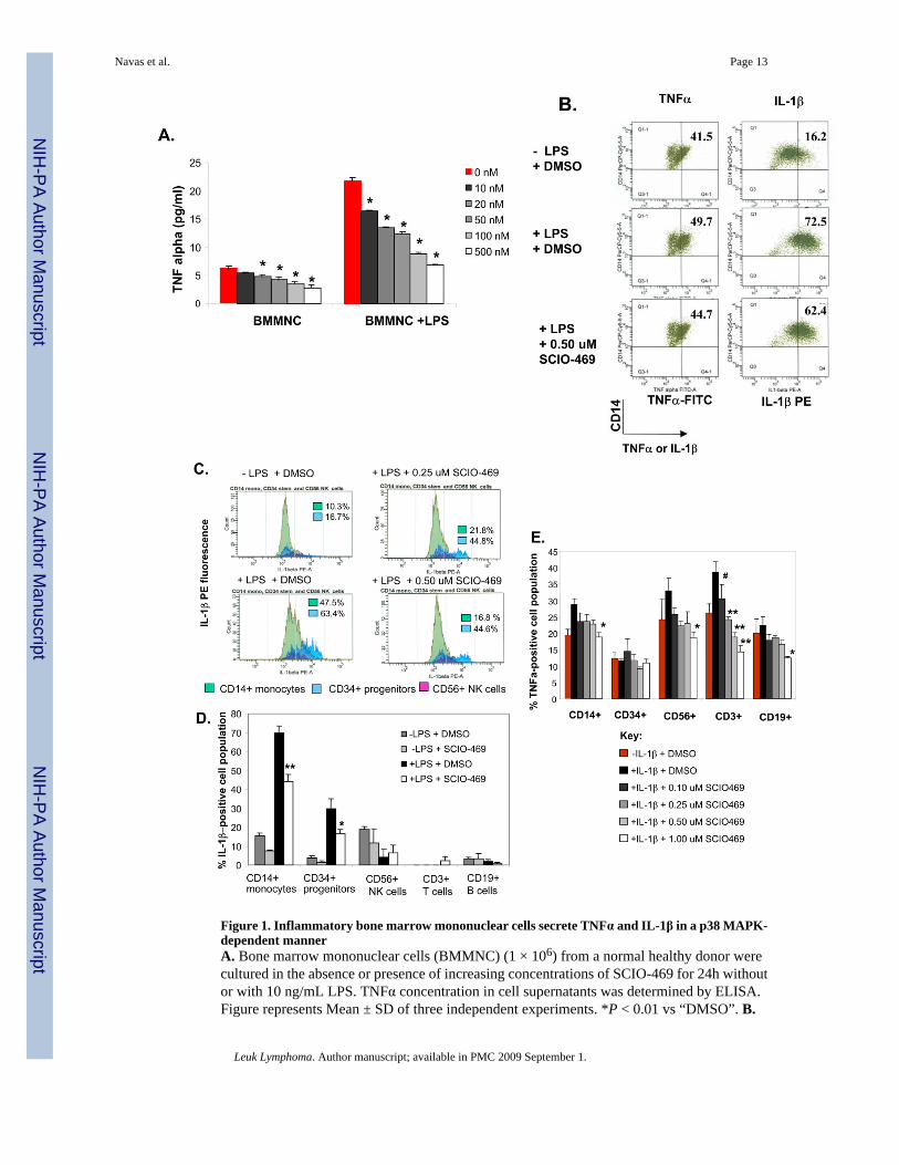
Figure 1. Inflammatory bone marrow mononuclear cells secrete TNFα and IL-1β in a p38 MAPK-dependent mannerA. Bone marrow mononuclear cells (BMMNC) (1 × 10⁶) from a normal healthy donor werecultured in the absence or presence of increasing concentrations of SCIO-469 for 24h withoutor with 10 ng/mL LPS. TNFα concentration in cell supernatants was determined by ELISA.Figure represents Mean ± SD of three independent experiments. *P < 0.01 vs “DMSO”. B.
Navas et al. Page 13
Leuk Lymphoma. Author manuscript; available in PMC 2009 September 1.
NIH
-PA Author Manuscript
NIH
-PA Author Manuscript
NIH
-PA Author Manuscript
Primary BM-derived CD14+ cells from a normal donor were incubated in IMDM + 10% FBSin the presence or absence of 20 ng/mL LPS and SCIO-469 for 4h. Brefeldin A (golgi plug)was added to a final concentration of 2 ug/mL during the last hour of incubation. Cells wereharvested, washed with FBS staining buffer and labeled with anti-CD14-PerCP Cy5.5 followedby intracellular staining with anti-IL-1β–PE and anti-TNFα–FITC. Figure shows percentdouble-stained CD14+ TNFα+ (left) and CD14+ IL-1β+ (right) in the same cell population.C. BMMNC from a normal donor (1 × 10⁶) were incubated in the presence or absence of 10ng/mL LPS for 4h. Brefeldin A (golgi plug) was added to a final concentration of 2 ug/mLduring the last hour of incubation. Cells were harvested, washed and labeled with differentfluorochrome-conjugated antibodies to CD14 (monocytes), CD56 (NK cells) and CD34(progenitor cells) followed by intracellular staining with PE-conjugated anti-IL-1β. Figureshows the relative IL-1β expression for each of the specific BM populations: CD14+ cells(green), CD34+ cells (light blue), CD56+ cells (violet). D BMMNC from a different normaldonor (1 × 10⁶) were treated with or without 0.5 μM SCIO-469 and incubated in the presenceor absence of 10 ng/mL LPS for 4h. Brefeldin A (golgi plug) was added to a final concentrationof 2 ug/mL during the last hour of incubation. Cells were harvested, washed and labeled withdifferent fluorochrome-conjugated antibodies to CD45 (leukocytes), CD14, CD3 (T cells),CD19 (B cells), CD56 and CD34 followed by intracellular staining with PE-conjugated anti-IL-1β. Figure shows the relative IL-1β expression for each of the specific BM population.Results are expressed as Mean ± SD of three independent experiments. **P < 0.001 or *P <0.01 vs “+ LPS − SCIO-469”. E. BMMNC (1 × 10⁶) were incubated without or with increasingconcentrations of SCIO-469 and in the presence or absence of 50 ng/mL IL-1β for 24h.Brefeldin A was added to a final concentration of 2 ug/ml during the last 2 hours of incubation.Cells were harvested, washed, labeled and then fixed with different fluorochrome-conjugatedantibodies to CD45, (leukocytes), CD14 (monocytes), CD3 (T cells), CD19 (B cells), CD56(NK cells) and CD34 (progenitor cells) followed by intracellular staining with PE-conjugatedanti-TNFα. Figure shows the relative TNFα expression for each of the specific BM populations.Results are expressed as mean +/− S.D. of three independent experiments. **P < 0.001 or*P < 0.01 or #P < 0.05 vs “+ IL-1β − SCIO-469”.
Navas et al. Page 14
Leuk Lymphoma. Author manuscript; available in PMC 2009 September 1.
NIH
-PA Author Manuscript
NIH
-PA Author Manuscript
NIH
-PA Author Manuscript

Figure 2. Secretion of TNF requires p38 MAPK-dependent interactions between stromal andmononuclear cellsA. BMSC and BMMNC from normal donors were either cultured alone or cocultured togetherfor 72h in the presence and absence of 0.5 μM SCIO-469. TNFα concentration in cellsupernatants was determined by ELISA. Figure represents Mean ± SD of three independentexperiments. B. Similar co-culture experiments were conducted as in (A) but using BMSCderived from either normal healthy control or from low risk MDS patients. Figure representsMean ± SD of three independent experiments. C. Total bone marrow aspirates isolated fromthree different MDS patients were assessed for intracellular TNFα production by using theCytofix/Cytoperm kit according to the manufacturer's instructions. Briefly, cells wereincubated in the presence of 10 μg/mL of immobilized anti-CD3 mAb plus 2 μg/mL of anti-CD28 antibody with or without 0.5 μM SCIO-469 in the presence of monensin for 6h at 37°C. Addition of anti-CD28 antibody providing costimulation and monensin blocking secretionof the cytokines during the incubation allows sensitive assessment of cytokine-producing cells.After the stimulation, cells were stained with anti-CD14-PE and TNFα-APC before analyzingby flow cytometry. CD14+ cells with intracellular TNF were expressed as a percentage of totalCD14+ cells.
Navas et al. Page 15
Leuk Lymphoma. Author manuscript; available in PMC 2009 September 1.
NIH
-PA Author Manuscript
NIH
-PA Author Manuscript
NIH
-PA Author Manuscript
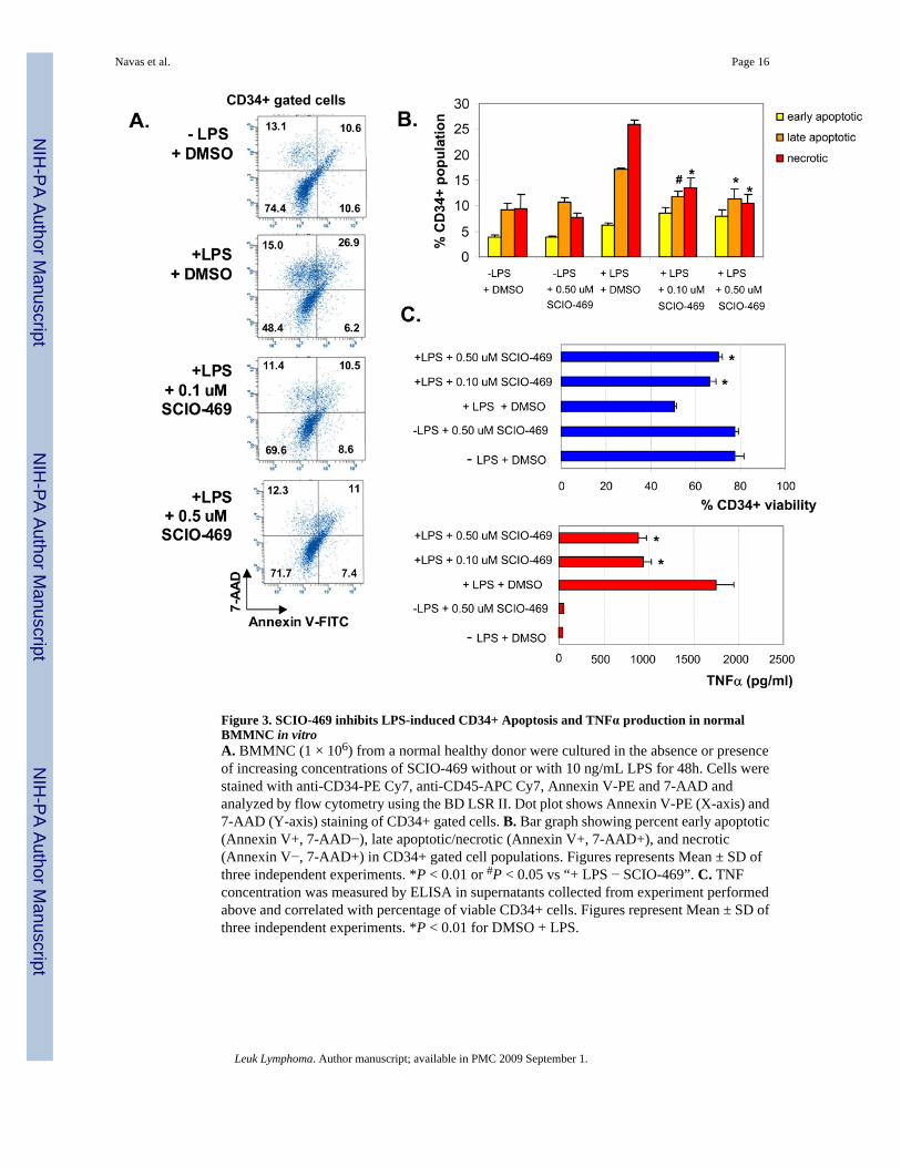
Figure 3. SCIO-469 inhibits LPS-induced CD34+ Apoptosis and TNFα production in normalBMMNC in vitroA. BMMNC (1 × 10⁶) from a normal healthy donor were cultured in the absence or presenceof increasing concentrations of SCIO-469 without or with 10 ng/mL LPS for 48h. Cells werestained with anti-CD34-PE Cy7, anti-CD45-APC Cy7, Annexin V-PE and 7-AAD andanalyzed by flow cytometry using the BD LSR II. Dot plot shows Annexin V-PE (X-axis) and7-AAD (Y-axis) staining of CD34+ gated cells. B. Bar graph showing percent early apoptotic(Annexin V+, 7-AAD−), late apoptotic/necrotic (Annexin V+, 7-AAD+), and necrotic(Annexin V−, 7-AAD+) in CD34+ gated cell populations. Figures represents Mean ± SD ofthree independent experiments. *P < 0.01 or #P < 0.05 vs “+ LPS − SCIO-469”. C. TNFconcentration was measured by ELISA in supernatants collected from experiment performedabove and correlated with percentage of viable CD34+ cells. Figures represent Mean ± SD ofthree independent experiments. *P < 0.01 for DMSO + LPS.
Navas et al. Page 16
Leuk Lymphoma. Author manuscript; available in PMC 2009 September 1.
NIH
-PA Author Manuscript
NIH
-PA Author Manuscript
NIH
-PA Author Manuscript

Figure 4. SCIO-469 can protect normal stem cell clones from apoptosis in MDSA. Normal CD34+ cells obtained from healthy volunteer were grown in methylcelluose in thepresence 100 μL of primary bone marrow sera obtained from bone marrows of 3 MDS patients.These experiments were done in the presence or absence of 100 nM SD-282. Erythroid andMyeloid colonies were counted after 14 days. Mean ± SD of 3 independent experiments areshown. B. BMMNC from 2 patients with MDS with chromosome 5q deletion were culturedin the presence or absence of 0.5 μM SCIO-469 for 48h. Cells were fixed onto slides pre- andpost-treatment and used for fluorescent in situ hybridization using EGR-1 probe (5q31-RED)to detect the number of abnormal clones. A 5p15 centromeric (GREEN) probe was used asinternal control. 200 cells per slide were counted and result expressed as percentage.
Navas et al. Page 17
Leuk Lymphoma. Author manuscript; available in PMC 2009 September 1.
NIH
-PA Author Manuscript
NIH
-PA Author Manuscript
NIH
-PA Author Manuscript

NIH
-PA Author Manuscript
NIH
-PA Author Manuscript
NIH
-PA Author Manuscript
Navas et al. Page 18
Table 1Gene Microarray Analysis of Chemokines Induced by IL-1β and inhibited by SCIO-469 in BMSC
Symbol Other Name Name IL-1β (24hr) * SCIO-469 + IL1β(24hr) **
CXCL1 GRO Chemokine (CXC) ligand 1 125.9 −1.6
CCL2 MCP-1 Chemokine (CC) ligand 2 9.9 −1.4
CXCL6 GCP2 Chemokine (CXC) ligand 6 138.8 −1.6
CXCL3 GROγ Chemokine (CXC) ligand 3 40.6 1.3
CCL7 MCP3 Chemokine (CC) ligand 7 6.2 −1.5
CXCL10 IP10 Chemokine (CXC) ligand 10 1 1
CXCL11 ITAC Chemokine (CXC) ligand 11 1 0
CXCL16 SR-PSOX Chemokine (CXC) ligand 16 9.3 −4.2
*Fold change of gene expression over control unstimultaed BMSCs
**Fold change of gene expression over IL-1beta stimulated BMSCs
Leuk Lymphoma. Author manuscript; available in PMC 2009 September 1.
Top Related
Copyright © 2022 FDOKUMEN

