


Bahasa
Halaman
Hukum

Genomic Instabilityand Aging-like Phenotypein the Absence of Mammalian SIRT6Raul Mostoslavsky,1,5,10 Katrin F. Chua,1,5,10,12 David B. Lombard,1,3,5,10 Wendy W. Pang,1,5 Miriam R. Fischer,1,5
Lionel Gellon,4 Pingfang Liu,4 Gustavo Mostoslavsky,5 Sonia Franco,1,5 Michael M. Murphy,1,5 Kevin D. Mills,1,5,11
Parin Patel,1,5 Joyce T. Hsu,1,5 Andrew L. Hong,1,5 Ethan Ford,7 Hwei-Ling Cheng,1,5 Caitlin Kennedy,1,5
Nomeli Nunez,8,13 Roderick Bronson,6 David Frendewey,2 Wojtek Auerbach,2 David Valenzuela,2
Margaret Karow,2 Michael O. Hottiger,9 Stephen Hursting,8,13 J. Carl Barrett,8,14 Leonard Guarente,7
Richard Mulligan,5 Bruce Demple,4 George D. Yancopoulos,2 and Frederick W. Alt1,5,*1Howard Hughes Medical Institute, The Children’s Hospital, CBR Institute for Biomedical Research, Harvard University Medical School,Boston, MA 02115, USA2Regeneron Pharmaceuticals, Inc., 777 Old Saw Mill River Road, Tarrytown, NY 10591, USA3Department of Pathology, Brigham and Women’s Hospital, Boston, MA 02115, USA4Department of Genetics and Complex Diseases, Harvard School of Public Health, Boston, MA 02115, USA5Department of Genetics6Department of PathologyHarvard Medical School, Boston, MA 02115, USA7Department of Biology, Massachusetts Institute of Technology, Cambridge, MA 02139, USA8Center for Cancer Research, National Cancer Institute, Bethesda, MD, USA9 Institute of Veterinary Biochemistry and Molecular Biology, University of Zurich, 8057 Zurich, Switzerland10These authors contributed equally to this work.11Present address: The Jackson Laboratory, 600 Main Street, Bar Harbor, ME 04609, USA.12Present address: Department of Medicine, Division of Endocrinology, Stanford University School of Medicine, and Geriatric ResearchEducation and Clinical Center, VA Palo Alto Health Care System, Palo Alto, CA 94304, USA.13Present address: Department of Human Ecology, College of Natural Sciences, Austin, TX 78712, USA.14Present address: Novartis Institute for Biomedical Research, Cambridge, MA 02139, USA.*Contact: [email protected] 10.1016/j.cell.2005.11.044
SUMMARY
The Sir2 histone deacetylase functions asa chromatin silencer to regulate recombina-tion, genomic stability, and aging in bud-ding yeast. Seven mammalian Sir2 homo-logs have been identified (SIRT1–SIRT7),and it has been speculated that some mayhave similar functions to Sir2. Here, wedemonstrate that SIRT6 is a nuclear, chro-matin-associated protein that promotes re-sistance to DNA damage and suppressesgenomic instability in mouse cells, in asso-ciation with a role in base excision repair(BER). SIRT6-deficient mice are small andat 2–3 weeks of age develop abnormalitiesthat include profound lymphopenia, lossof subcutaneous fat, lordokyphosis, andsevere metabolic defects, eventually dyingat about 4 weeks. We conclude that onefunction of SIRT6 is to promote normalDNA repair, and that SIRT6 loss leads to ab-
normalities in mice that overlap with aging-associated degenerative processes.
INTRODUCTION
The silent information regulator (SIR) genes are nonessential
genes required in trans for transcriptional repression of sev-
eral genomic loci in the budding yeast S. cerevisiae (Klar
et al., 1981; Rine and Herskowitz, 1987). Sir2 is unique
among these factors in belonging to a large family of closely
related proteins present both in prokaryotic and eukaryotic
species (reviewed in Dutnall and Pillus [2001]). Sir2 functions
in a complex with other Sir proteins to repress transcription
at the silent mating type loci and subtelomeric sequences.
In addition, Sir2 is part of another multicomponent complex
in the nucleolus, where it represses recombination and tran-
scription of rDNA repeats (Gotta et al., 1997; Gottlieb and
Esposito, 1989; Straight et al., 1999). Sir2 exerts its effects
on transcriptional and recombinational silencing via modula-
tion of chromatin structure (Fritze et al., 1997) by catalyzing
NAD-dependent deacetylation of histones (Imai et al.,
2000; Smith et al., 2000).
S. cerevisiae Sir2 promotes longevity in yeast mother cells
(Kaeberlein et al., 1999). Cells lacking Sir2 have a reduced
replicative life span, and cells with extra copies display
Cell 124, 315–329, January 27, 2006 ª2006 Elsevier Inc. 315
a longer life span. Sir2 extends yeast life span by inhibiting
recombination in the rDNA repeats, since the recombina-
tional excision and subsequent accumulation of extrachro-
mosomal rDNA circles promotes senescence (Kaeberlein
et al., 1999; Sinclair and Guarente, 1997). Increased Sir2 ac-
tivity in C. elegans and Drosophila also extends life span (Tis-
senbaum and Guarente, 2001; Wood et al., 2004); however,
the mechanism of life span extension seems to differ from
that in yeast. Thus, Sir2-driven increased longevity in C. ele-
gans is dependent on the Daf-16 transcription factor, a criti-
cal mediator in the insulin-like signaling pathway, which pro-
motes formation of long-lived dauer larvae under unfavorable
environmental conditions and increases stress resistance
and longevity in adults (Kenyon, 2001). Sir2 family members
also were suggested to mediate effects of caloric restriction
(CR; Sohal and Weindruch, 1996), a dietary regimen capable
of extending life span in diverse organisms from yeast to
mammals (Cohen et al., 2004; Howitz et al., 2003; Lin
et al., 2004; Rogina and Helfand, 2004; Wood et al.,
2004). CR likely decreases production of reactive oxygen
species (ROS), a potential source of persistent DNA damage
proposed to contribute to aging (Balaban et al., 2005).
The evolutionary conservation of a role for Sir2 in regulat-
ing life span has fueled speculation that such a role also may
be conserved in mammals (Blander and Guarente, 2004;
North and Verdin, 2004). There are seven mammalian Sir2
family members, designated SIRT1–SIRT7 (Frye, 2000),
with SIRT1 being the most highly related to S. cerevisiae
Sir2. However, unlike yeast Sir2, which deacetylates his-
tones exclusively, SIRT1 possesses a large list of substrates,
including p53 and forkhead transcription factors, which
modulate cellular resistance to oxidative and genotoxic
stress (Blander and Guarente, 2004). In addition, SIRT1,
like Sir2, has been shown to modify chromatin and silence
transcription of integrated reporter genes via histone deace-
tylation (Vaquero et al., 2004). However, potential roles of
SIRT1 in life span regulation have not yet been documented.
SIRT2 and SIRT3 possess deacetylase activity but are cyto-
plasmic proteins with functions that diverge from those of
ySir2 (Blander and Guarente, 2004). Little information is
available about the functions of the other mammalian SIRTs.
Mammals employ various pathways to repair specific
types of DNA damage. Mutant mice defective in individual
DNA repair pathways frequently show increased genomic in-
stability and hypersensitivity to specific DNA-damaging
agents. Double-strand breaks (DSBs), such as those in-
duced by ionizing radiation (IR), are repaired by nonhomolo-
gous end-joining (NHEJ) or homologous recombination
(HR). Single-stranded DNA lesions are repaired via nucleo-
tide excision repair (NER) or base excision repair (BER), de-
pending on the type of lesion (Hoeijmakers, 2001). In this
context, bulky photodimer single-strand lesions induced by
UV irradiation are primarily repaired by NER, whereas more
simple types of single-stranded lesions usually are repaired
by BER. The BER pathway may repair up to one million nu-
cleotides per cell per day (Holmquist, 1998) and repairs sin-
gle-strand breaks (SSBs) that arise spontaneously, such as
those from endogenous alkylation, oxidation, and deamina-
316 Cell 124, 315–329, January 27, 2006 ª2006 Elsevier Inc.
tion events, as well as small, nonhelix-distorting DNA lesions
induced by chemical mutagens such as the alkylating agent
methyl-methane sulphonate (MMS; (Lindahl and Barnes,
2000; Sancar et al., 2004). Defects in NHEJ, HR, and NER
have been shown to cause aging-like phenotypes in mice
and humans. However, there have been no mouse models
to study potential effects of BER with respect to aging; since
mutations of BER components either are lethal or confer no
obvious phenotype (Lombard et al., 2005).
In this study, we have focused on elucidating the functions
of the mammalian SIRT6 protein. We find that SIRT6 is
a chromatin-associated protein that is required for normal
BER maintenance of genomic integrity in cells. Moreover,
SIRT6 deficiency in mice leads to the development of an
acute degenerative aging-like phenotype.
RESULTS
SIRT6 Is a Chromatin-Associated Protein Expressed
in Most Tissues
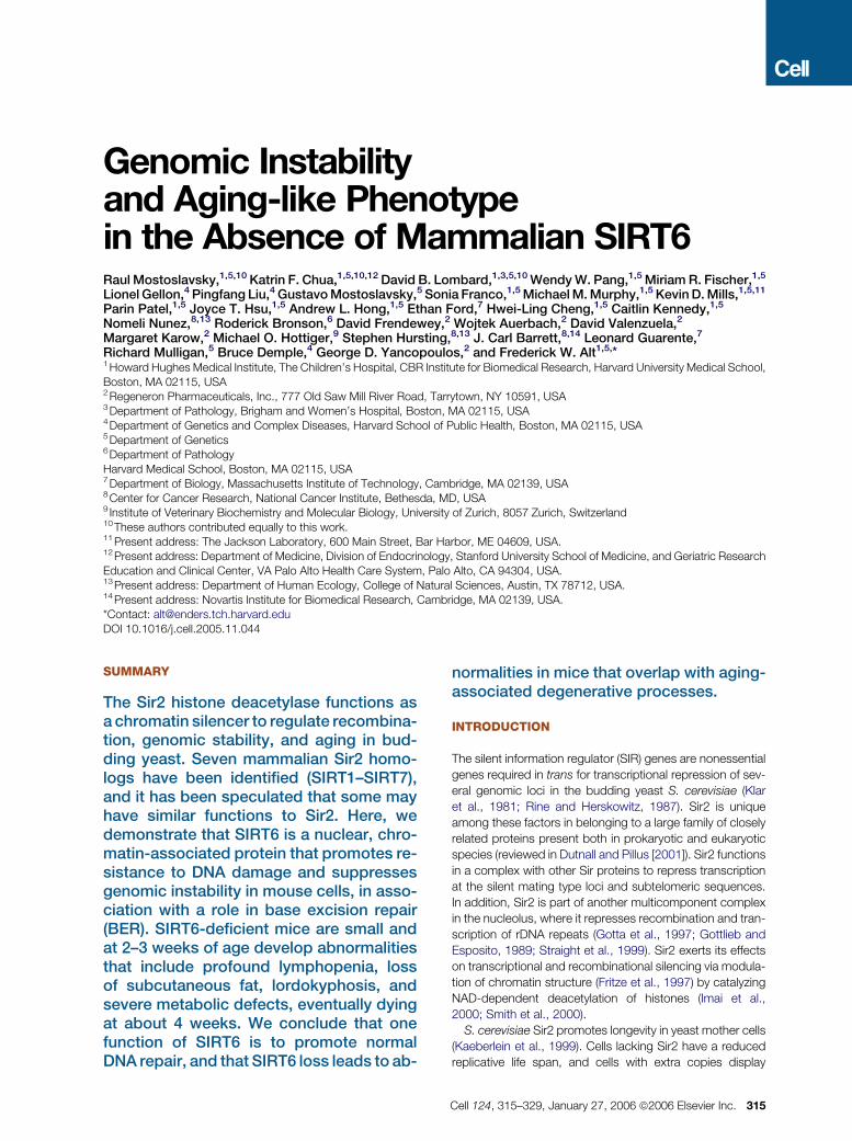
To gain insight into SIRT6 function, we characterized subcel-
lular and tissue distribution patterns of SIRT6 expression.
SIRT6 protein was predominantly nuclear, as assessed by
immunostaining mouse embryonic stem (ES) cells with an
anti-SIRT6 antibody and human HT1080 cells into which
we retrovirally transduced a FLAG-tagged SIRT6 protein
(Figures 1A and 1B). Analysis of SIRT6 RNA and protein re-
vealed SIRT6 expression in most mouse tissues (Figures 1C
and 1D), with particularly high protein levels in thymus, skel-
etal muscle, and brain. Overall, these findings are consistent
with those of other recent studies (Liszt et al., 2005; Michi-
shita et al., 2005).
To probe the possibility that SIRT6 might function in the
context of chromatin, we asked whether it is associated
with a chromatin-enriched biochemical fraction. Isolated nu-
clei were separated into nucleoplasmic and chromatin/
nuclear matrix subfractions, and SIRT6 protein detected by
Western analysis. Endogenous SIRT6 protein in mouse ES
cells, and retrovirally expressed FLAG-SIRT6 protein in
HT1080 cells cofractionated with histones, almost exclu-
sively within the chromatin/nuclear matrix subfraction (Fig-
ures 1E and 1F). As a control, the SIRT1 protein was present
in both the nucleoplasm and the chromatin/nuclear matrix
subfractions in these cells, as expected (Vaquero et al.,
2004; reviewed in Guarente and Picard [2005]). We con-
clude that SIRT6 is preferentially associated with chromatin
within the nucleus.
Generation of SIRT6-Deficient Cells and Mice
To gain insight into SIRT6 function we generated SIRT6-
deficient embryonic stem (ES) cells by replacing SIRT6 with
a LacZ gene introduced in frame into the exon 1 (see Figure
S1A in the Supplemental Data available with this article on-
line). Appropriately targeted ES cells were confirmed by
Southern blotting (Figure S1A) and used to generate mice
heterozygous for the SIRT6-inactivating mutation (referred
to as SIRT6+/�). SIRT6+/� were interbred to obtain
Figure 1. SIRT6 Is a Nuclear Protein Expressed in Many Different Tissues
(A) HT1080 cells were infected with a retrovirus encoding an N-terminal FLAG-SIRT6 fusion. SIRT6 protein was visualized with an antibody directed against
the epitope tag, and cells were counterstained with To-pro-3 to visualize nuclei.
(B) A rabbit antiserum raised against the C terminus of SIRT6 protein was used to stain embryonic stem (ES) cells.
(C) One microgramofRNA fromthe indicated tissueswas used inRT-PCRreactionswithspecificprimers recognizingeitherSIRT6oracontrol transcript (b actin).
(D) Protein lysate from the indicated tissues was probed with SIRT6 antiserum or GAPDH as control.
(E) Subcellular fractions were prepared from HT1080 cells infected with a retrovirus encoding an N-terminal FLAG-SIRT6 fusion. Western analysis of the
fractions was carried out to detect FLAG-SIRT6 and histone H3, SIRT1, or SIRT5, a cytoplasmic protein (not shown), as controls.
(F) Indicated subcellular fractions were prepared from mouse ES cells and probed with anti-SIRT6, anti-H4, or anti-SIRT1 antibodies.
homozygous mutant (SIRT6�/�) mice and SIRT6�/� mouse
embryonic fibroblasts (MEFs). We also generated SIRT6�/�
ES cells via the high G418-selection method (Mortensen
et al., 1992 and data not shown). We confirmed absence
of SIRT6 RNA and protein in SIRT6�/� cells by RT-PCR
and Western blotting, respectively (Figure S1B). We also an-
alyzed SIRT6+/� mice for bGal staining and confirmed ex-
pression of lacZ from the SIRT6 promoter in most tissues,
both in adult mice and in embryos during development
(Figure S1C and data not shown).
SIRT6-Deficient MEFs and ES Cells Show
Impaired Proliferation and Increased Sensitivity
to DNA-Damage Agents
To gain insight into the cellular effects of SIRT6 deficiency,
we compared proliferation rates of SIRT6�/� and wild-type
Cell 124, 315–329, January 27, 2006 ª2006 Elsevier Inc. 317
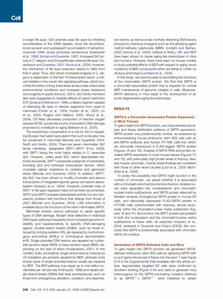
Figure 2. Impaired Proliferation and IR Sensitivity of SIRT6-Deficient MEFs and ES Cells
(A) Left: SIRT6-deficient MEFs or littermate controls were seeded at low density and the cell numbers quantified every other day as shown. Right: SIRT6-
deficient ES cells or late-passage MEFs were labeled with BrdU and subjected to flow cytometry to determine the fraction of cells in S phase.
(B and C) SIRT6-deficient MEFs (B) or ES cells (C) were subjected to indicated doses of IR and the number of cells (B) or colonies (C) present 1 week later
quantified. XRCC4-deficient MEFs (B) or ES cells (C) were used as positive controls.
(D) SIRT6 deficiency is not associated with UV sensitivity. SIRT6-deficient MEFs were subjected to indicated doses of UV and the cell numbers quantified
1 week later. XPF-deficient MEFs were used as a positive control.
(E) SIRT6-deficient MEFs or littermate controls were infected with an empty retrovirus (pBABE, B), a retrovirus encoding FLAG-tagged SIRT6 (BFS6), or
a retrovirus encoding a catalytic mutant FLAG-tagged SIRT6 (BFS6HA). The cells were subjected to irradiation and sensitivity determined as above.
In all panels, error bars indicate the standard error of the mean.
control MEFs in culture (Figure 2A). Multiple independent
SIRT6�/� MEF lines grew more slowly than wild-type con-
trols. Analysis of BrdU incorporation revealed that exponen-
tially growing SIRT6�/� MEF cultures had a smaller fraction
of S phase cells than control wt cultures (Figure 2A). Simi-
larly, exponentially growing SIRT6�/� ES cell cultures had
a reduced fraction of BrdU-incorporating cells compared
to controls (Figure 2A). Thus, absence of SIRT6 reduces
the proliferative rate of multiple cell types.
318 Cell 124, 315–329, January 27, 2006 ª2006 Elsevier Inc.
To test for SIRT6 roles in DNA repair, we compared the
sensitivity of SIRT6�/�MEFs and ES cells to IR and UV dam-
age. Notably, SIRT6�/�MEFs and ES cells both exhibited in-
creased sensitivity to IR (Figures 2B and 2C), albeit less than
cells deficient in XRCC4, a core NHEJ factor. In contrast,
SIRT6�/� MEFs exhibited normal UV sensitivity, indicating
that SIRT6 deficiency confers hypersensitivity only to specific
types of DNA damage (Figure 2D). To confirm that increased
IR-sensitivity of the SIRT6�/� cells was a consequence of
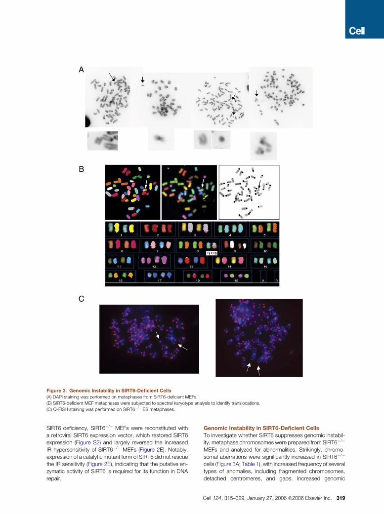
Figure 3. Genomic Instability in SIRT6-Deficient Cells
(A) DAPI staining was performed on metaphases from SIRT6-deficient MEFs.
(B) SIRT6-deficient MEF metaphases were subjected to spectral karyotype analysis to identify translocations.
(C) Q-FISH staining was performed on SIRT6�/� ES metaphases.
SIRT6 deficiency, SIRT6�/� MEFs were reconstituted with
a retroviral SIRT6 expression vector, which restored SIRT6
expression (Figure S2) and largely reversed the increased
IR hypersensitivity of SIRT6�/� MEFs (Figure 2E). Notably,
expression of a catalytic mutant form of SIRT6 did not rescue
the IR sensitivity (Figure 2E), indicating that the putative en-
zymatic activity of SIRT6 is required for its function in DNA
repair.
Genomic Instability in SIRT6-Deficient Cells
To investigate whether SIRT6 suppresses genomic instabil-
ity, metaphase chromosomes were prepared from SIRT6�/�
MEFs and analyzed for abnormalities. Strikingly, chromo-
somal aberrations were significantly increased in SIRT6�/�
cells (Figure 3A; Table 1), with increased frequency of several
types of anomalies, including fragmented chromosomes,
detached centromeres, and gaps. Increased genomic
Cell 124, 315–329, January 27, 2006 ª2006 Elsevier Inc. 319

Table 1. Increased Genomic Instability in SIRT6-Deficient Cells
(A) MEFs
DAPI SKY
wt KO wt KO
Total Metaphases 96 96 24 24
Fragments/breaks 4 23 Fragments 0 2
Gaps 5 18 Translocations 0 6
Detached centromeres 2 5 Dicentric 0 1
Other aberrations 0 7
Abnormal metaphases (%) 8 (7%) 36 (38%) 0 (0%) 9 (34%)
p < 0.0001 p < 0.01
(B) ES Cells—Q-FISH
wt XRCC4�/� KO1 KO2
Metaphases 50 50 50 50
Translocations 0 1 1 2
Breaks/fragments 5 10 14 13
Other aberrations 0 1 1 0
Total metaphases with abnormalities (%) 4 (8%) 11 (22%) 13 (28%) 12 (26%)
(A) MEFs were prepared from 13.5-day-old embryos, and metaphases were prepared from passage 1–2 cells. Cells were either stainedwith DAPI, or SKY was performed. Results include four independent experiments.(B) ES cells of the indicated genotypes were grown, and metaphases prepared as described in Experimental Procedures. Q-FISH wasperformed, and aberrations scored as indicated.
instability of SIRT6�/� MEFs was evident in both early and
late passage cultures (data not shown). Spectral karyotype
analysis (SKY) revealed chromosomal translocations in
SIRT6�/� MEFs (Figure 3B; Table 1). Karyotype analysis
of the SIRT6�/� ES cells also revealed increased genomic
instability (Figure 3C; Table 1). Together, these results indi-
cate that SIRT6 plays a general role in maintaining genome
integrity.
Intact Cell Cycle Checkpoints and DSB Repair
in SIRT6-Deficient Cells
Genomic instability can result from either direct defects in
DNA repair or faulty cell cycle checkpoints. We tested
whether SIRT6 is required for normal cell cycle checkpoints
in response to IR. Cells were irradiated with a range of doses
and collected following BrdU labeling or staining with anti-
phospho H3, to assess the G1/S and the G2/M checkpoints,
respectively. In this analysis, SIRT6�/� and wt cultures were
indistinguishable in exhibiting dose-dependent decreases in
S phase and mitotic fractions, indicating that the G1/S and
G2/M cell cycle checkpoints are not affected by absence
of SIRT6 (Figure 4A).
We then assessed integrity of DNA DSB repair pathways
in SIRT6�/� cells. To test NHEJ, an extrachromosomal plas-
mid-based recombination assay was used to quantify repair
of DNA DSBs introduced by the RAG endonuclease into an
320 Cell 124, 315–329, January 27, 2006 ª2006 Elsevier Inc.
immunoglobulin V(D)J recombination substrate (Hesse et al.,
1987; Taccioli et al., 1993). Based on this semiquantitative
NHEJ assay, SIRT6�/� MEFs repaired RAG-generated
DSBs within the range of wt cells (Figure 4B). This conclusion
was confirmed by our finding of normal lymphocyte develop-
ment in young SIRT6�/� mice (see below). To test for chro-
mosomal DSB repair, we performed pulse field gel electro-
phoresis (PFGE) following exposure of SIRT6�/� or control
cells to high doses of IR for various lengths of time and found
that SIRT6�/� cells showed similar DSB repair to wt cells
(Figure 4C). Further, we analyzed formation and clearance
of gH2AX foci, an early marker of the DSB response (Thiriet
and Hayes, 2005) and again observed no difference be-
tween wt and SIRT6�/� cells (Figure 4D). Together, these
observations suggest that the increased DNA damage sen-
sitivity of SIRT6�/� cells is unlikely to result from impaired
checkpoints or DSB repair pathways.
SIRT6 Deficiency Compromises the BER Pathway
We next tested whether SIRT6 deficiency affects repair of
DNA lesions other than DSBs. Because NER is the main
pathway responsible for repairing UV generated bulky ad-
ducts and SIRT6�/� MEFs showed normal sensitivity to UV
irradiation (Figure 2D), we focused on BER. Monofunctional
alkylating agents such as MMS and ROS generation due to
oxidative agents such as H2O2 generate lesions that are
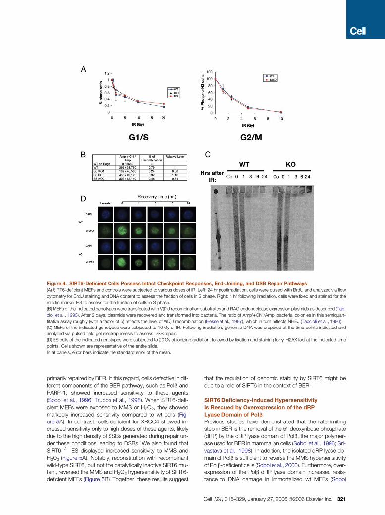
Figure 4. SIRT6-Deficient Cells Possess Intact Checkpoint Responses, End-Joining, and DSB Repair Pathways
(A) SIRT6-deficient MEFs and controls were subjected to various doses of IR. Left: 24 hr postirradiation, cells were pulsed with BrdU and analyzed via flow
cytometry for BrdU staining and DNA content to assess the fraction of cells in S phase. Right: 1 hr following irradiation, cells were fixed and stained for the
mitotic marker H3 to assess for the fraction of cells in S phase.
(B) MEFs of the indicated genotypes were transfected with V(D)J recombination substrates and RAG endonuclease expression plasmids as described (Tac-
cioli et al., 1993). After 2 days, plasmids were recovered and transformed into bacteria. The ratio of Ampr+Chlr/Ampr bacterial colonies in this semiquan-
titative assay roughly (with a factor of 5) reflects the level of V(D)J recombination (Hesse et al., 1987), which in turn reflects NHEJ (Taccioli et al., 1993).
(C) MEFs of the indicated genotypes were subjected to 10 Gy of IR. Following irradiation, genomic DNA was prepared at the time points indicated and
analyzed via pulsed field gel electrophoresis to assess DSB repair.
(D) ES cells of the indicated genotypes were subjected to 20 Gy of ionizing radiation, followed by fixation and staining for g-H2AX foci at the indicated time
points. Cells shown are representative of the entire slide.
In all panels, error bars indicate the standard error of the mean.
primarily repaired by BER. In this regard, cells defective in dif-
ferent components of the BER pathway, such as Polb and
PARP-1, showed increased sensitivity to these agents
(Sobol et al., 1996; Trucco et al., 1998). When SIRT6-defi-
cient MEFs were exposed to MMS or H2O2, they showed
markedly increased sensitivity compared to wt cells (Fig-
ure 5A). In contrast, cells deficient for XRCC4 showed in-
creased sensitivity only to high doses of these agents, likely
due to the high density of SSBs generated during repair un-
der these conditions leading to DSBs. We also found that
SIRT6�/� ES displayed increased sensitivity to MMS and
H2O2 (Figure 5A). Notably, reconstitution with recombinant
wild-type SIRT6, but not the catalytically inactive SIRT6 mu-
tant, reversed the MMS and H2O2 hypersensitivity of SIRT6-
deficient MEFs (Figure 5B). Together, these results suggest
that the regulation of genomic stability by SIRT6 might be
due to a role of SIRT6 in the context of BER.
SIRT6 Deficiency-Induced Hypersensitivity
Is Rescued by Overexpression of the dRP
Lyase Domain of Polb
Previous studies have demonstrated that the rate-limiting
step in BER is the removal of the 50-deoxyribose phosphate
(dRP) by the dRP lyase domain of Polb, the major polymer-
ase used for BER in mammalian cells (Sobol et al., 1996; Sri-
vastava et al., 1998). In addition, the isolated dRP lyase do-
main of Polb is sufficient to reverse the MMS hypersensitivity
of Polb-deficient cells (Sobol et al., 2000). Furthermore, over-
expression of the Polb dRP lyase domain increased resis-
tance to DNA damage in immortalized wt MEFs (Sobol
Cell 124, 315–329, January 27, 2006 ª2006 Elsevier Inc. 321
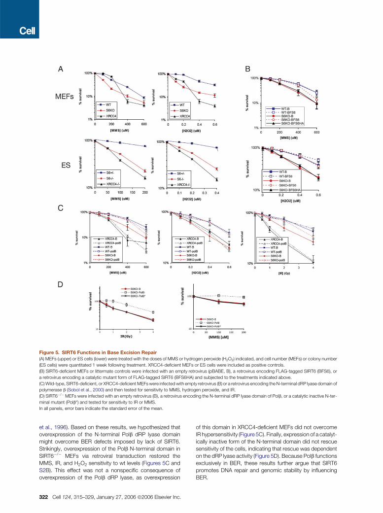
Figure 5. SIRT6 Functions in Base Excision Repair
(A) MEFs (upper) or ES cells (lower) were treated with the doses of MMS or hydrogen peroxide (H2O2) indicated, and cell number (MEFs) or colony number
(ES cells) were quantitated 1 week following treatment. XRCC4-deficient MEFs or ES cells were included as positive controls.
(B) SIRT6-deficient MEFs or littermate controls were infected with an empty retrovirus (pBABE, B), a retrovirus encoding FLAG-tagged SIRT6 (BFS6), or
a retrovirus encoding a catalytic mutant form of FLAG-tagged SIRT6 (BFS6HA) and subjected to the treatments indicated above.
(C) Wild-type, SIRT6-deficient, or XRCC4-deficient MEFs were infected with empty retrovirus (B) or a retrovirus encoding the N-terminal dRP lyase domain of
polymerase b (Sobol et al., 2000) and then tested for sensitivity to MMS, hydrogen peroxide, and IR.
(D) SIRT6�/�MEFs were infected with an empty retrovirus (B), a retrovirus encoding the N-terminal dRP lyase domain of Polb, or a catalytic inactive N-ter-
minal mutant (Polb*) and tested for sensitivity to IR or MMS.
In all panels, error bars indicate the standard error of the mean.
et al., 1996). Based on these results, we hypothesized that
overexpression of the N-terminal Polb dRP lyase domain
might overcome BER defects imposed by lack of SIRT6.
Strikingly, overexpression of the Polb N-terminal domain in
SIRT6�/� MEFs via retroviral transduction restored the
MMS, IR, and H2O2 sensitivity to wt levels (Figures 5C and
S2B). This effect was not a nonspecific consequence of
overexpression of the Polb dRP lyase, as overexpression
322 Cell 124, 315–329, January 27, 2006 ª2006 Elsevier Inc.
of this domain in XRCC4-deficient MEFs did not overcome
IR hypersensitivity (Figure 5C). Finally, expression of a catalyt-
ically inactive form of the N-terminal domain did not rescue
sensitivity of the cells, indicating that rescue was dependent
on the dRP lyase activity (Figure 5D). Because Polb functions
exclusively in BER, these results further argue that SIRT6
promotes DNA repair and genomic stability by influencing
BER.
Normal Expression of BER Factors and Efficient
Formation of XRCC1 and PAR Foci
in SIRT6-Deficient Cells
To test whether SIRT6 affects expression of any of the core
BER factors, protein levels of Polb, XRCC1, LigaseIII, Ape1,
and PARP-1 were assessed in SIRT6�/� and control MEFs.
None of these showed differences between wt and KO cells
(Figure S3). In addition, we tested whether SIRT6 colocalizes
with BER factors. In this regard, XRCC1 forms foci upon
MMS and H2O2 treatment that colocalize with sites of poly-
ADP ribose (PAR) formation (El-Khamisy et al., 2003). In con-
trast to XRCC1, SIRT6 showed diffuse nuclear staining fol-
lowing exposure of cells to either MMS or H2O2 (Figure S4A
and data not shown). In addition, both XRCC1 and PAR foci
formed normally in SIRT6-deficient cells (Figure S4B). To-
gether, these results suggest that SIRT6 might function in
BER through a mechanism that does not directly involve reg-
ulation of BER factors.
SIRT6-Deficient Mice Develop a Progeroid
Degenerative Syndrome
To analyze the role of SIRT6 in vivo, we generated and ana-
lyzed SIRT6�/� mice (Figure 6A). SIRT6�/� mice were born
at Mendelian frequency (SIRT6�/� 48 [22%], n = 223) and
showed no abnormalities at birth (data not shown). The
mice developed normally for the first two weeks, with no ap-
parent histologic abnormalities, though reduced body size
became apparent early after birth (Figure 6A). Strikingly, at
�3 weeks of age, the mice underwent several acute degen-
erative processes and failed to thrive, invariably dying at
around postnatal day (P) 24 (Figures 6A and 6B). Occasional
SIRT6-deficient mice that died prior to day 20 also exhibited
the same deterioration. Defects observed in these mice in-
clude acute loss of subcutaneous fat, lordokyphosis, a colitis
consisting of erosion of the superficial colonic epithelium,
and a severe lymphopenia associated with increased lym-
phocyte apoptosis (Figures 6C–6G). Indeed, flow cytometric
analyses revealed a 50-fold reduction in the number of
CD4+–CD8+ double-positive (DP) cells in the thymus and
a 10 fold-reduction in the number of splenic lymphocytes
and progenitor B cells in the bone marrow (data not shown).
In addition, SIRT6�/� mice exhibited osteopenia, with 30%
reduction in bone mineral density (Figure 6H), a finding
consistent with the lordokyphosis. Since bones are still de-
veloping in mice at this age, we cannot distinguish between
developmental versus degenerative defects in bone mineral-
ization. Food intake of the SIRT6�/�mice was normal, as as-
sessed by the amount of milk in the digestive track (data not
shown), excluding malnutrition as a cause for the observed
phenotypes.
The profound decrease in lymphocyte numbers in
SIRT6�/� mice could be due either to a cell-intrinsic effect
of SIRT6 deficiency or to systemic, non-cell-autonomous
defects. To distinguish between these possibilities, donor
bone marrow cells from 12-day-old SIRT6�/� -mice or wt lit-
termates, were mixed with wild-type competitor cells and
transplanted into lethally irradiated hosts. In these experi-
ments, SIRT6-deficient cells contributed to repopulation of
the lymphocyte compartment as efficiently as normal com-
petitor cells and were equally represented in these mice
even 5 months after transplant (Figure 7A and data not
shown). These results indicate that the lymphocyte defect
of SIRT6�/�mice is not cell intrinsic and suggest that disap-
pearance of the lymphoid cells reflects the response of these
cells to systemic alterations caused by lack of SIRT6. In this
context, thymocytes from SIRT6�/�mice were examined for
hypersensitivity to DNA-damaging agents. Notably, thymo-
cytes from 21-day-old mice demonstrated an increased
sensitivity to IR, MMS, and H2O2 (Figure S5), but thymocytes
from 12-day-old mice exhibited normal sensitivity (data not
shown). Similar to the bone marrow transplant experiments,
these results indicate that the lymphocyte defects are not cell
intrinsic but rather the response of these cells to systemic
defects.
Severe Metabolic Defects in the Absence of SIRT6
We assayed SIRT6�/� mice for systemic defects that might
explain the observed phenotypes. Thymocytes are ex-
tremely sensitive to glucocorticoid-induced apoptosis
(Wyllie, 1980). However, SIRT6-deficient mice exhibited nor-
mal serum glucocorticoid levels (data not shown). Insulin-like
growth factor IGF-1 confers upon lymphocytes resistance to
apoptosis (Pifer et al., 2003), and age-associated lympho-
cyte decline is associated with low levels of IGF-1 (Taub
and Longo, 2005). Strikingly, serum IGF-1 levels were se-
verely reduced in SIRT6�/� mice (Figure 7B), with lower
levels of serum IGF-1 than found in mice with a liver-specific
deletion of the IGF-1 gene (Yakar et al., 2001). Notably,
IGF-1 levels were low, even in 12-day-old SIRT6�/� mice,
before any of the other phenotypes were evident. Serum glu-
cose, although normal in 12-day-old animals, decreased
sharply afterwards and by day 24, reached the limit of detec-
tion (Figure 7C). Thus, SIRT6 function is required for proper
glucose homeostasis and maintenance of normal IGF-1
levels.
DISCUSSION
SIRT6 Influences DNA Repair and Suppresses
Degenerative Pathologies
The yeast SIR2 protein is a chromatin regulator that silences
DNA recombination and, thereby, regulates life span. We
show that SIRT6 is associated with chromatin and that cells
deficient for this factor display defective BER and elevated
levels of spontaneous genomic instability. Moreover, SIRT6
deficiency in mice leads to aging-like degenerative pro-
cesses. In mammals, regulation of genomic stability has
been linked to both tumor suppression and aging (Lombard
et al., 2005). While etiologies of SIRT6-deficient mouse pa-
thologies remain to be determined, some may reflect func-
tions of SIRT6 in BER and genome stabilization. Overall,
our findings that SIRT6 regulates genomic stability on the
cellular level and aging-associated pathologies at the organ-
ismal level demonstrate that the SIRT6 protein has certain
functions predicted for the mammalian SIRT proteins based
on analogies to yeast Sir2 (Blander and Guarente, 2004;
Cell 124, 315–329, January 27, 2006 ª2006 Elsevier Inc. 323
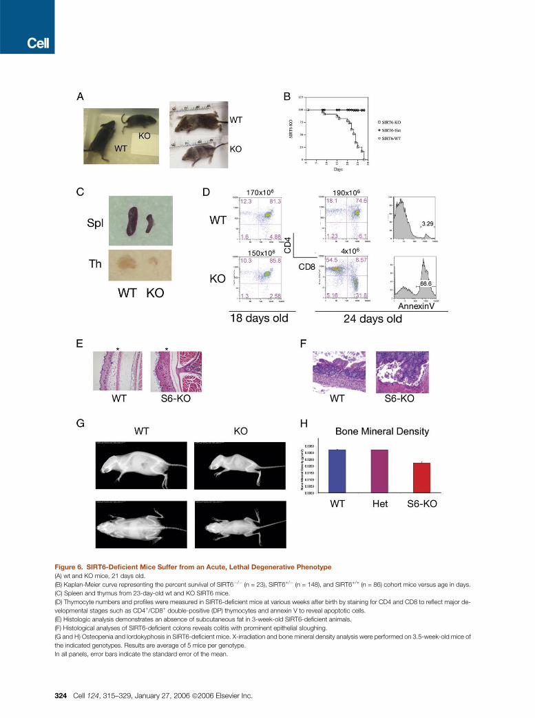
Figure 6. SIRT6-Deficient Mice Suffer from an Acute, Lethal Degenerative Phenotype
(A) wt and KO mice, 21 days old.
(B) Kaplan-Meier curve representing the percent survival of SIRT6�/� (n = 23), SIRT6+/� (n = 148), and SIRT6+/+ (n = 86) cohort mice versus age in days.
(C) Spleen and thymus from 23-day-old wt and KO SIRT6 mice.
(D) Thymocyte numbers and profiles were measured in SIRT6-deficient mice at various weeks after birth by staining for CD4 and CD8 to reflect major de-
velopmental stages such as CD4+/CD8+ double-positive (DP) thymocytes and annexin V to reveal apoptotic cells.
(E) Histologic analysis demonstrates an absence of subcutaneous fat in 3-week-old SIRT6-deficient animals.
(F) Histological analyses of SIRT6-deficient colons reveals colitis with prominent epithelial sloughing.
(G and H) Osteopenia and lordokyphosis in SIRT6-deficient mice. X-irradiation and bone mineral density analysis were performed on 3.5-week-old mice of
the indicated genotypes. Results are average of 5 mice per genotype.
In all panels, error bars indicate the standard error of the mean.
324 Cell 124, 315–329, January 27, 2006 ª2006 Elsevier Inc.
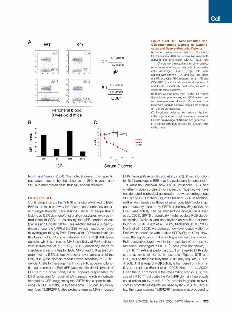
Figure 7. SIRT6�/� Mice Exhibited Non-
Cell-Autonomous Defects in Lympho-
cytes and Severe Metabolic Defects
(A) Bone marrow was purified from 12-day-old
SIRT6-deficient (KO) and control (wt) mice both
carrying the alloantigen CD45.2 [5.2], and
1 � 106 cells were injected into lethally irradiated
mice, together with equal amounts of competitor
cells (alloantigen CD45.1 [5.1]). Cells were
stained with either 5.1-PE and IgM-FITC (top),
5.1-PE and CD8-FITC (bottom), or 5.1-PE and
CD4-FITC (data not shown) to distinguish B
and T cells, respectively. FACS analysis from 8-
week-old mice is shown.
(B) Blood was collected from 19-day-old mice of
the indicated phenotypes, and IGF-1 levels in se-
rum was measured. Liver-IGF-1-deficient mice
(LID) were used as controls. Results are average
of 10 mice per genotype.
(C) Blood was collected from mice of the indi-
cated age, and serum glucose was measured.
Results are average of 10 mice per genotype.
In all panels, error bars indicate the standard error
of the mean.
North and Verdin, 2004). We note, however, that specific
pathways affected by the absence of Sir2 in yeast and
SIRT6 in mammalian cells, thus far, appear different.
SIRT6 and BER
Our findings indicate that SIRT6 is functionally linked to BER.
BER is the main pathway for repair of spontaneously occur-
ring single-stranded DNA lesions. Repair of single-strand
lesions by BER via monofunctional glycosylases involves in-
troduction of SSBs at lesions by the APE1 endonuclease
(Barnes and Lindahl, 2004). This reaction leaves a 50-deoxy-
ribose phosphate (dRP) at the SSB, which must be removed
following gap-filling by Polb. Removal of dRP is rate limiting in
this branch of BER and is catalyzed by the Polb dRP lyase
domain, which can rescue MMS sensitivity of Polb-deficient
cells (Srivastava et al., 1998). SIRT6 deficiency leads to
spectrum of sensitivities to H2O2, MMS, and IR that are con-
sistent with a BER defect. Moreover, overexpression of the
Polb dRP lyase domain rescues hypersensitivity of SIRT6-
deficient cells to these agents. Thus, SIRT6 appears to func-
tion upstream of the Polb dRP lyase reaction in this branch of
BER. On the other hand, SIRT6 appears dispensable for
DSB repair and for repair of UV damage which is normally
handled by NER, suggesting that SIRT6 has a specific influ-
ence on BER. Notably, a trypanosome T. brucei Sir2 family
member, TbSIR2RP1, also protects against MMS-induced
DNA damage (Garcia-Salcedo et al., 2003). Thus, a function
for Sir2 homologs in BER may be evolutionarily conserved.
It remains unknown how SIRT6 influences BER and
whether it does so directly or indirectly. Thus far, we have
not detected a physical association between endogenous
SIRT6 and BER factors (Figures S6A and S6B). In addition,
neither Polb levels nor those of other core BER factors ap-
pear markedly affected by SIRT6 deficiency (Figure S3). As
Polb lyase activity can be inhibited via acetylation (Hasan
et al., 2002), SIRT6 theoretically might regulate Polb via de-
acetylation. While in vitro deacetylase activity had not been
found for SIRT6 (Liszt et al., 2005; Michishita et al., 2005;
North et al., 2003), we detected low-level deacetylation of
Polb when incubated with purified SIRT6 (Figure S7A). How-
ever, the significance of this finding is unclear; since in vivo
Polb acetylation levels, within the resolution of our assays,
remained unchanged in SIRT6�/� cells (data not shown).
SIRT6�/� extracts performed BER on a naked DNA sub-
strate at levels similar to wt extracts (Figures S7B and
S7C), raising the possibility that SIRT6 may regulate BER in-
directly. In this regard, Polb activity is hampered on chroma-
tinized templates (Beard et al., 2003; Nilsen et al., 2002).
Given that dRP removal is the rate-limiting step in BER, res-
cue of SIRT6�/� cells with the Polb dRP domain theoretically
could reflect ability of this 8 kDa protein fragment to over-
come chromatin restraints imposed by lack of SIRT6. Nota-
bly, the trypanosomal TbSIR2RP1 protein was proposed to
Cell 124, 315–329, January 27, 2006 ª2006 Elsevier Inc. 325
promote chromatin accessibility following MMS-induced
DNA damage by catalyzing histone ADP ribosylation and de-
acetylation (Garcia-Salcedo et al., 2003). While SIRT6 has an
ADP-ribosylation activity in vitro (Liszt et al., 2005; Figure S8),
it remains unclear whether the activity occurs under physio-
logic conditions. Based on existing information, it is tempting
to speculate that SIRT6 may promote BER by creating ac-
cessibility for BER factors via modification of histones or
other chromatin-related factors.
SIRT6 Deficiency and Aging-Related Degeneration
ROS have been implicated in the pathogenesis of aging
(Finkel and Holbrook, 2000). As oxidative DNA damage is re-
paired primarily by BER, impairment of this process might
cause aging-related phenotypes. In this regard, nuclear
BER and Polb activity has been reported to decrease with
age, and this decline can be prevented by CR (Cabelof
et al., 2002; Cabelof et al., 2003). As targeted disruption of
BER genes often leads to early lethality due to critical func-
tions in development or to no phenotype, likely due to func-
tional redundancy (Hasty et al., 2003), the SIRT6-deficient
mouse potentially could provide a useful model of defective,
presumably hypomorphic, BER. In this context, there are
a number of similarities between the progeroid phenotypes
of SIRT6-deficient mice and those of mice with NER defi-
ciencies (de Boer et al., 2002; Murai et al., 2001). XPA/CS
and XPA/TTD mice are normal at birth but show decreased
size shortly after and die around day 22, with kyphosis, lack
of subcutaneous fat, and cachexia, all degenerative pheno-
types reminiscent of those of SIRT6-deficient mice. How-
ever, the XPA/CS and XPA/TTD animals differ in certain re-
spects from SIRT6-deficient mice; most notably, they lack
lymphocyte depletion. Conversely, the XPA/CS deficient
mice develop severe ataxia due to depletion of cerebellar
cells, a phenotype not observed in the SIRT6-deficient
mice. However, in both cases, affected organs are normal
at birth and show elevated apoptosis after a period of weeks.
In this regard, there may be differential requirements for the
BER and NER pathways in certain tissues, or some pheno-
types could be unrelated to the repair defects, particularly in
the case of SIRT6 deficiency.
Severe Metabolic Defects in the Absence of SIRT6:
The Insulin-Signaling Connection
Our bone marrow transplantation experiments indicate that
lymphocyte attrition in SIRT6-deficient mice is not a cell-
autonomous defect. During normal aging, depletion of lym-
phocytes has been linked to a diminished response to circu-
lating factors (Allman and Miller, 2005; Stephan et al., 1997).
Furthermore, circulating growth factors, like IGF-1, can pro-
tect lymphocytes against corticosteroid-induced apoptosis
(Pifer et al., 2003). In this context, thymic atrophy has been
correlated with pituitary gland deficiency and low levels of cir-
culating neuroendocrine hormones (Taub and Longo, 2005).
In addition, housing conditions dramatically affect lympho-
cyte homeostasis (Dorshkind et al., 2003). Thus, lymphocyte
homeostasis is extremely sensitive to changes in systemic
factors, as well as to levels of stress. In this regard, SIRT6-
326 Cell 124, 315–329, January 27, 2006 ª2006 Elsevier Inc.
deficient mice exhibited a severe reduction in circulating
IGF-1 levels, a defect that could explain some of their pheno-
types, and serum glucose was reduced to levels that may
lead to their demise. This dramatic imbalance in glucose me-
tabolism clearly indicates that SIRT6 plays an important role
in regulating organismal homeostasis.
Perturbations in IGF-1 and insulin signaling have been
linked to alterations in the rate of aging in multiple organisms
(Kenyon, 2005). In C. elegans, Sir2 regulates life span via the
Daf-16 transcription factor, a critical mediator in the insulin-
like signaling pathway (Tissenbaum and Guarente, 2001). Al-
though decreased IGF-1 signaling has been correlated with
increased life span in worms, flies, and mice (Kenyon, 2005),
several mouse models of premature aging in fact are associ-
ated with lower serum IGF-1 or insulin levels, such as ATM
(Peretz et al., 2001) and Klotho-deficient mice (Mori et al.,
2000). In addition, reduced serum IGF-1 is observed in el-
derly humans (reviewed in Lombardi et al. [2005]). Thus,
the role of IGF-1 in life span regulation is complex. In theory,
SIRT6 might play a role in insulin signaling, similar to Sir2 fac-
tors in other lower organisms. However, as in the premature
aging mouse models described above, it remains unclear
whether the altered serum IGF-1/insulin levels of SIRT-6-
deficient mice directly contribute to aging-like phenotypes
or, alternatively, reflect compensatory alterations. In this re-
gard, it will be of interest to determine whether SIRT6 is
involved in regulating the IGF-1 response to CR (Bartke,
2005). Finally, it is notable that mice in which the apoptotic
function of cytochrome C was disrupted exhibited quite sim-
ilar phenotypes to those of SIRT6-deficient mice and also in-
volved defects in the insulin signaling pathway (Hao et al.,
2005).
Perspective
We show that mammalian SIRT6 protein plays a key role in
DNA repair and maintenance of genomic stability in cells.
Moreover, we show that SIRT6 is necessary to maintain or-
ganismal health and to prevent the development of several
progeroid pathologies. Further studies of SIRT6-deficient
cells may elucidate molecular mechanisms that regulate
the BER pathway, while SIRT6-deficient mice may provide
a model for deciphering elements that influence the develop-
ment of progeroid symptoms and allow evaluation of poten-
tial roles of defective BER in such degenerative processes. In
these contexts, it will be of particular importance to elucidate
the relationship, if any, between the metabolic and DNA re-
pair defects of the SIRT6-deficient mice.
EXPERIMENTAL PROCEDURES
Chromatin Fractionation
Cell fractionation was performed as previously described (Mendez and
Stillman, 2000).
Western and Immunostaining Analysis
Western analysis was carried out as previously described (Cheng et al.,
2003). The antibodies used and their sources were as follows: anti-mouse
SIRT6 (Liszt et al., 2005), anti-DNA polymerase b (NeoMarkers), anti-
a-tubulin (Sigma). For immunostaining analysis, cells were grown in
coverslips and fixed in 1% paraformaldehyde as described (Bassing et al.,
2002). The LacZ staining of embryos was performed as described (Whit-
ing et al., 1991).
Construction of the Targeting Vector and Generation
of SIRT6-Deficient Mice
The SIRT6 KO targeting vector was constructed by replacing exons 1 to
6 with a LacZ gene inserted in frame after the first 21 bp of exon1. The
construct was generated using the VelociGene recombination method
(Valenzuela et al., 2003). Chimeric mice were generated by injection of
targeted ES clones into C57BL6/J blastocysts. Male chimeras were
mated with 129SvJ females to generate F1 heterozygous mice, which
were interbred to generate homozygous KO mice.
Generation of MEFs, Metaphase Analysis,
and DNA-Damage Assays
MEFs were generated from 13.5-day-old embryos by using standard
methods. Metaphases were prepared as previously described (Zhu
et al., 2002). Q-FISH was performed as previously described (Chua
et al., 2005), using a Cy3-labeled PNA telomeric probe (Cy3-(TTAGGG)3).
To assay for sensitivity to DNA damage, 5 � 104 MEFs were plated into
6-well plates and 12 hr later either g irradiated, UV irradiated, or treated
with H2O2 or MMS for 24 hr at the doses indicated.
DNA Repair Assay and gH2AX Analysis
The DNA repair assay was performed as described (Wong et al., 2000).
For gH2AX analysis, the indicated cells were cultured on slides, irradiated
with 20 Gy, followed by fixation in 4% paraformaldehyde at the indicated
time points. gH2AX immunostaining was performed as described
(Bassing et al., 2002).
Extrachromosomal VDJ Recombination Assay in MEF Cells
V(D)J recombination of plasmid substrates was performed as described
(Hesse et al., 1987; Taccioli et al., 1993).
Cell Cycle Analysis
BrdU incorporation was assayed with anti-BrdU antibodies (BD Pharmin-
gen) according to the manufacturer’s instructions. Briefly, 5 � 105 cells
were irradiated with the indicated doses of IR, and 24 hr later were pulsed
with BrdU for 4 hr, harvested, stained with FITC-conjugated anti-BrdU
antibodies and propidium iodide, and cell cycle profiles analyzed by
flow cytometry. For G2/M analysis, cells were irradiated with the indicated
doses of IR, and 1 hr later harvested and stained with anti-phospho H3
antibodies (Upstate), a specific mitotic marker (Wei et al., 1998).
Retroviral Infection
Reconstitution of MEFs was performed as previously described (Cheng
et al., 2003). The different SIRT6 cDNAs (wt and mutant) and the dRP ly-
ase 8 kDa Polb cDNAs (wt and mutant) were amplified by PCR and cloned
into the pBabe-puro vector. MEFs were infected by incubation with virus
and 2 mg/ml polybrene and 48 hr later, selected in 2.5 mg/ml puromycin.
Cells were allowed to recover from selection for 48 hr and then plated for
the different experiments.
Histological Analysis
Mouse tissue was fixed in Bouin’s fixative, embedded in paraffin, sec-
tioned at 6 mm, and hematoxylin/eosin staining was performed by stan-
dard methods.
IGF-1 and Glucose Measurements
IGF-1 was measured with commercially available radioimmunoassay
(RIA) kits from Linco Research, Inc. and Diagnostic Systems Laboratories,
Inc. Serum glucose was measured from tail blood using the Elite gluco-
meter (Elite) kit, following manufacturer’s instructions.
Bone Mineral Density Analyses
Whole-body bone mineral density was measured by dual energy X-ray
absorptiometry (DXA) using a GE Lunar Piximus II densitometer.
Bone Marrow Transplantations (BMT)
Competitive repopulation studies were performed as previously de-
scribed (Mostoslavsky et al., 2005). Briefly, CD45.1 recipient mice were
lethally irradiated with two doses of 7 Gy, 3 hr apart, 1 day before BMT.
1 � 106 whole marrow cells from CD45.1 mice were mixed with 1 �106 whole marrow cells purified from either CD45.2 control or SIRT6�/�
mice (CD45.2 as well), and injected retroorbitally. Peripheral blood was
obtained every 4 weeks and stained as indicated. Following gating on
B or T cells, respectively, cells were gated on CD45.1-PE, and the relative
chimerism calculated. All antibodies were from BD Biosciences.
Supplemental Data
Supplemental Data include eight figures and can be found with this article
online at http://www.cell.com/cgi/content/full/124/2/315/DC1/.
ACKNOWLEDGMENTS
We thank Abigail Land-Bracha, Constanza Lorente, Hanno Hock, Scott
Snapper, Shridar Ganesan, Yuko Fujiwara, Nicole Stokes, Aimee Wil-
liams, Tiffany Borjeson, Patricia Dunning, Samuel Wilson, and Stefano
Casola for helpful comments, reagents, and technical assistance. This
work was supported by an Ellison Senior Scholar Award (to F.W.A.),
NIH grants (to F.W.A. and B.D.), a Long-Term Fellowship of the Human
Frontier Science Program and a Senior Post-doctoral Fellowship from
The Leukemia and Lymphoma Society (to R.M.), a Pfizer Post-doctoral
Fellowship in Immunology/Rheumatology (to K.F.C.), an EMBO Long
Term Fellowship (to S.F.), and an NIA/NIH KO8 award (to D.B.L).
F.W.A. is an investigator of the Howard Hughes Medical Institute. Some
of the authors are employees and shareholders of Regeneron and have
financial interests related to this work.
Received: May 31, 2005
Revised: September 19, 2005
Accepted: November 3, 2005
Published: January 26, 2006
REFERENCES
Allman, D., and Miller, J.P. (2005). The aging of early B-cell precursors.
Immunol. Rev. 205, 18–29.
Balaban, R.S., Nemoto, S., and Finkel, T. (2005). Mitochondria, oxidants,
and aging. Cell 120, 483–495.
Barnes, D.E., and Lindahl, T. (2004). Repair and genetic consequences of
endogenous DNA base damage in mammalian cells. Annu. Rev. Genet.
38, 445–476.
Bartke, A. (2005). Minireview: role of the growth hormone/insulin-like
growth factor system in mammalian aging. Endocrinology 146, 3718–
3723.
Bassing, C.H., Chua, K.F., Sekiguchi, J., Suh, H., Whitlow, S.R., Fleming,
J.C., Monroe, B.C., Ciccone, D.N., Yan, C., Vlasakova, K., et al. (2002).
Increased ionizing radiation sensitivity and genomic instability in the ab-
sence of histone H2AX. Proc. Natl. Acad. Sci. USA 99, 8173–8178.
Beard, B.C., Wilson, S.H., and Smerdon, M.J. (2003). Suppressed cata-
lytic activity of base excision repair enzymes on rotationally positioned
uracil in nucleosomes. Proc. Natl. Acad. Sci. USA 100, 7465–7470.
Blander, G., and Guarente, L. (2004). The sir2 family of protein deacety-
lases. Annu. Rev. Biochem. 73, 417–435.
Cabelof, D.C., Raffoul, J.J., Yanamadala, S., Ganir, C., Guo, Z., and Hey-
dari, A.R. (2002). Attenuation of DNA polymerase beta-dependent base
excision repair and increased DMS-induced mutagenicity in aged mice.
Mutat. Res. 500, 135–145.
Cell 124, 315–329, January 27, 2006 ª2006 Elsevier Inc. 327
Cabelof, D.C., Yanamadala, S., Raffoul, J.J., Guo, Z., Soofi, A., and Hey-
dari, A.R. (2003). Caloric restriction promotes genomic stability by induc-
tion of base excision repair and reversal of its age-related decline. DNA
Repair (Amst.) 2, 295–307.
Cheng, H., Mostoslavsky, R., Saito, S.I., Manis, J.P., Gu, Y., Patel, P.,
Bronson, R., Appella, E., Alt, F.W., and Chua, K.F. (2003). Developmental
defects and p53 hyperacetylation in Sir2 homolog (SIRT1)-deficient mice.
Proc. Natl. Acad. Sci. USA 100, 10794–10799.
Chua, K.F., Mostoslavsky, R., Lombard, D.B., Pang, W.W., Saito, S.,
Franco, S., Kaushal, D., Cheng, H.L., Fischer, M.R., Stokes, N., et al.
(2005). Mammalian SIRT1 limits replicative life span in response to chronic
genotoxic stress. Cell Metab. 2, 67–76.
Cohen, H.Y., Miller, C., Bitterman, K.J., Wall, N.R., Hekking, B., Kessler,
B., Howitz, K.T., Gorospe, M., de Cabo, R., and Sinclair, D.A. (2004). Cal-
orie restriction promotes mammalian cell survival by inducing the SIRT1
deacetylase. Science 305, 390–392.
de Boer, J., Andressoo, J.O., de Wit, J., Huijmans, J., Beems, R.B., van
Steeg, H., Weeda, G., van der Horst, G.T., van Leeuwen, W., Themmen,
A.P., et al. (2002). Premature aging in mice deficient in DNA repair and
transcription. Science 296, 1276–1279.
Dorshkind, K., Welniak, L., Gault, R.A., Hixon, J., Montecino-Rodriguez,
E., Horseman, N.D., Gertner, J.M., and Murphy, W.J. (2003). Effects of
housing on the thymic deficiency in dwarf mice and its reversal by growth
hormone administration. Clin. Immunol. 109, 197–202.
Dutnall, R.N., and Pillus, L. (2001). Deciphering NAD-dependent deace-
tylases. Cell 105, 161–164.
El-Khamisy, S.F., Masutani, M., Suzuki, H., and Caldecott, K.W. (2003). A
requirement for PARP-1 for the assembly or stability of XRCC1 nuclear
foci at sites of oxidative DNA damage. Nucleic Acids Res. 31, 5526–5533.
Finkel, T., and Holbrook, N.J. (2000). Oxidants, oxidative stress and the
biology of ageing. Nature 408, 239–247.
Fritze, C.E., Verschueren, K., Strich, R., and Easton Esposito, R. (1997).
Direct evidence for SIR2 modulation of chromatin structure in yeast rDNA.
EMBO J. 16, 6495–6509.
Frye, R.A. (2000). Phylogenetic classification of prokaryotic and eukary-
otic Sir2-like proteins. Biochem. Biophys. Res. Commun. 273, 793–798.
Garcia-Salcedo, J.A., Gijon, P., Nolan, D.P., Tebabi, P., and Pays, E.
(2003). A chromosomal SIR2 homologue with both histone NAD-depen-
dent ADP-ribosyltransferase and deacetylase activities is involved in DNA
repair in Trypanosoma brucei. EMBO J. 22, 5851–5862.
Gotta, M., Strahl-Bolsinger, S., Renauld, H., Laroche, T., Kennedy, B.K.,
Grunstein, M., and Gasser, S.M. (1997). Localization of Sir2p: the nucle-
olus as a compartment for silent information regulators. EMBO J. 16,
3243–3255.
Gottlieb, S., and Esposito, R.E. (1989). A new role for a yeast transcrip-
tional silencer gene, SIR2, in regulation of recombination in ribosomal
DNA. Cell 56, 771–776.
Guarente, L., and Picard, F. (2005). Calorie restriction—the SIR2 connec-
tion. Cell 120, 473–482.
Hao, Z., Duncan, G.S., Chang, C.C., Elia, A., Fang, M., Wakeham, A.,
Okada, H., Calzascia, T., Jang, Y., You-Ten, A., et al. (2005). Specific ab-
lation of the apoptotic functions of cytochrome C reveals a differential re-
quirement for cytochrome C and Apaf-1 in apoptosis. Cell 121, 579–591.
Hasan, S., El-Andaloussi, N., Hardeland, U., Hassa, P.O., Burki, C., Im-
hof, R., Schar, P., and Hottiger, M.O. (2002). Acetylation regulates the
DNA end-trimming activity of DNA polymerase beta. Mol. Cell 10,
1213–1222.
Hasty, P., Campisi, J., Hoeijmakers, J., van Steeg, H., and Vijg, J. (2003).
Aging and genome maintenance: lessons from the mouse? Science 299,
1355–1359.
Hesse, J.E., Lieber, M.R., Gellert, M., and Mizuuchi, K. (1987). Extrachro-
mosomal DNA substrates in pre-B cells undergo inversion or deletion at
immunoglobulin V-(D)-J joining signals. Cell 49, 775–783.
328 Cell 124, 315–329, January 27, 2006 ª2006 Elsevier Inc.
Hoeijmakers, J.H. (2001). Genome maintenance mechanisms for pre-
venting cancer. Nature 411, 366–374.
Holmquist, G.P. (1998). Endogenous lesions, S-phase-independent
spontaneous mutations, and evolutionary strategies for base excision re-
pair. Mutat. Res. 400, 59–68.
Howitz, K.T., Bitterman, K.J., Cohen, H.Y., Lamming, D.W., Lavu, S.,
Wood, J.G., Zipkin, R.E., Chung, P., Kisielewski, A., Zhang, L.L., et al.
(2003). Small molecule activators of sirtuins extend Saccharomyces
cerevisiae life span. Nature 425, 191–196.
Imai, S., Armstrong, C.M., Kaeberlein, M., and Guarente, L. (2000). Tran-
scriptional silencing and longevity protein Sir2 is an NAD-dependent his-
tone deacetylase. Nature 403, 795–800.
Kaeberlein, M., McVey, M., and Guarente, L. (1999). The SIR2/3/4 com-
plex and SIR2 alone promote longevity in Saccharomyces cerevisiae by
two different mechanisms. Genes Dev. 13, 2570–2580.
Kenyon, C. (2001). A conserved regulatory system for aging. Cell 105,
165–168.
Kenyon, C. (2005). The plasticity of aging: Insights from long-lived mu-
tants. Cell 120, 449–460.
Klar, A.J., Strathern, J.N., Broach, J.R., and Hicks, J.B. (1981). Regula-
tion of transcription in expressed and unexpressed mating type cassettes
of yeast. Nature 289, 239–244.
Lin, S.J., Ford, E., Haigis, M., Liszt, G., and Guarente, L. (2004). Calorie
restriction extends yeast life span by lowering the level of NADH. Genes
Dev. 18, 12–16.
Lindahl, T., and Barnes, D.E. (2000). Repair of endogenous DNA dam-
age. Cold Spring Harb. Symp. Quant. Biol. 65, 127–133.
Liszt, G., Ford, E., Kurtev, M., and Guarente, L. (2005). Mouse Sir2 homo-
log SIRT6 is a nuclear ADP-ribosyltransferase. J. Biol. Chem. 280,
21313–21320.
Lombard, D.B., Chua, K.F., Mostoslavsky, R., Franco, S., Gostissa, M.,
and Alt, F.W. (2005). DNA repair, genome stability, and aging. Cell 120,
497–512.
Lombardi, G., Di Somma, C., Rota, F., and Colao, A. (2005). Associated
hormonal decline in aging: is there a role for GH therapy in aging men?
J. Endocrinol. Invest. 28, 99–108.
Mendez, J., and Stillman, B. (2000). Chromatin association of human or-
igin recognition complex, cdc6, and minichromosome maintenance pro-
teins during the cell cycle: assembly of prereplication complexes in late
mitosis. Mol. Cell. Biol. 20, 8602–8612.
Michishita, E., Park, J.Y., Burneskis, J.M., Barrett, J.C., and Horikawa, I.
(2005). Evolutionarily conserved and nonconserved cellular localizations
and functions of human SIRT proteins. Mol. Biol. Cell 16, 4623–4635.
Mori, K., Yahata, K., Mukoyama, M., Suganami, T., Makino, H., Nagae,
T., Masuzaki, H., Ogawa, Y., Sugawara, A., Nabeshima, Y., and Nakao,
K. (2000). Disruption of klotho gene causes an abnormal energy homeo-
stasis in mice. Biochem. Biophys. Res. Commun. 278, 665–670.
Mortensen, R.M., Conner, D.A., Chao, S., Geisterfer-Lowrance, A.A.,
and Seidman, J.G. (1992). Production of homozygous mutant ES cells
with a single targeting construct. Mol. Cell. Biol. 12, 2391–2395.
Mostoslavsky, G., Kotton, D.N., Fabian, A.J., Gray, J.T., Lee, J.-S., and
Mulligan, R.C. (2005). Efficiency of transduction of highly purified murine
hematopoietic stem cells by lentiviral and oncoretroviral vectors under
conditions of minimal in vitro manipulation. Mol. Ther. 11, 932–940.
Murai, M., Enokido, Y., Inamura, N., Yoshino, M., Nakatsu, Y., van der
Horst, G.T., Hoeijmakers, J.H., Tanaka, K., and Hatanaka, H. (2001).
Early postnatal ataxia and abnormal cerebellar development in mice lack-
ing Xeroderma pigmentosum Group A and Cockayne syndrome Group B
DNA repair genes. Proc. Natl. Acad. Sci. USA 98, 13379–13384.
Nilsen, H., Lindahl, T., and Verreault, A. (2002). DNA base excision repair
of uracil residues in reconstituted nucleosome core particles. EMBO J.
21, 5943–5952.
North, B.J., Marshall, B.L., Borra, M.T., Denu, J.M., and Verdin, E. (2003).
The human Sir2 ortholog, SIRT2, is an NAD(+)-dependent tubulin deace-
tylase. Mol. Cell 11, 437–444.
North, B.J., and Verdin, E. (2004). Sirtuins: Sir2-related NAD-dependent
protein deacetylases. Genome Biol. 5, 224.
Peretz, S., Jensen, R., Baserga, R., and Glazer, P.M. (2001). ATM-
dependent expression of the insulin-like growth factor-I receptor in a path-
way regulating radiation response. Proc. Natl. Acad. Sci. USA 98, 1676–
1681.
Pifer, J., Stephan, R.P., Lill-Elghanian, D.A., Le, P.T., and Witte, P.L.
(2003). Role of stromal cells and their products in protecting young and
aged B-lineage precursors from dexamethasone-induced apoptosis.
Mech. Ageing Dev. 124, 207–218.
Rine, J., and Herskowitz, I. (1987). Four genes responsible for a position
effect on expression from HML and HMR in Saccharomyces cerevisiae.
Genetics 116, 9–22.
Rogina, B., and Helfand, S.L. (2004). Sir2 mediates longevity in the fly
through a pathway related to calorie restriction. Proc. Natl. Acad. Sci.
USA 101, 15998–16003.
Sancar, A., Lindsey-Boltz, L.A., Unsal-Kacmaz, K., and Linn, S. (2004).
Molecular mechanisms of mammalian DNA repair and the DNA damage
checkpoints. Annu. Rev. Biochem. 73, 39–85.
Sinclair, D.A., and Guarente, L. (1997). Extrachromosomal rDNA circles—
a cause of aging in yeast. Cell 91, 1033–1042.
Smith, J.S., Brachmann, C.B., Celic, I., Kenna, M.A., Muhammad, S.,
Starai, V.J., Avalos, J.L., Escalante-Semerena, J.C., Grubmeyer, C., Wol-
berger, C., and Boeke, J.D. (2000). A phylogenetically conserved NAD+-
dependent protein deacetylase activity in the Sir2 protein family. Proc.
Natl. Acad. Sci. USA 97, 6658–6663.
Sobol, R.W., Horton, J.K., Kuhn, R., Gu, H., Singhal, R.K., Prasad, R.,
Rajewsky, K., and Wilson, S.H. (1996). Requirement of mammalian
DNA polymerase-beta in base-excision repair. Nature 379, 183–186.
Sobol, R.W., Prasad, R., Evenski, A., Baker, A., Yang, X.P., Horton, J.K.,
and Wilson, S.H. (2000). The lyase activity of the DNA repair protein beta-
polymerase protects from DNA-damage-induced cytotoxicity. Nature
405, 807–810.
Sohal, R.S., and Weindruch, R. (1996). Oxidative stress, caloric restric-
tion, and aging. Science 273, 59–63.
Srivastava, D.K., Berg, B.J., Prasad, R., Molina, J.T., Beard, W.A., Tom-
kinson, A.E., and Wilson, S.H. (1998). Mammalian abasic site base exci-
sion repair. Identification of the reaction sequence and rate-determining
steps. J. Biol. Chem. 273, 21203–21209.
Stephan, R.P., Lill-Elghanian, D.A., and Witte, P.L. (1997). Development
of B cells in aged mice: decline in the ability of pro-B cells to respond to
IL-7 but not to other growth factors. J. Immunol. 158, 1598–1609.
Straight, A.F., Shou, W., Dowd, G.J., Turck, C.W., Deshaies, R.J., John-
son, A.D., and Moazed, D. (1999). Net1, a Sir2-associated nucleolar pro-
tein required for rDNA silencing and nucleolar integrity. Cell 97, 245–256.
Taccioli, G.E., Rathbun, G., Oltz, E., Stamato, T., Jeggo, P.A., and Alt,
F.W. (1993). Impairment of V(D)J recombination in double-strand break
repair mutants. Science 260, 207–210.
Taub, D.D., and Longo, D.L. (2005). Insights into thymic aging and regen-
eration. Immunol. Rev. 205, 72–93.
Thiriet, C., and Hayes, J.J. (2005). Chromatin in need of a fix: Phosphor-
ylation of H2AX connects chromatin to DNA repair. Mol. Cell 18, 617–622.
Tissenbaum, H.A., and Guarente, L. (2001). Increased dosage of a sir-2
gene extends life span in Caenorhabditis elegans. Nature 410, 227–230.
Trucco, C., Oliver, F.J., de Murcia, G., and Menissier-de Murcia, J.
(1998). DNA repair defect in poly(ADP-ribose) polymerase-deficient cell
lines. Nucleic Acids Res. 26, 2644–2649.
Valenzuela, D.M., Murphy, A.J., Frendewey, D., Gale, N.W., Economides,
A.N., Auerbach, W., Poueymirou, W.T., Adams, N.C., Rojas, J., Yasen-
chak, J., et al. (2003). High-throughput engineering of the mouse genome
coupled with high-resolution expression analysis. Nat. Biotechnol. 21,
652–659.
Vaquero, A., Scher, M., Lee, D., Erdjument-Bromage, H., Tempst, P.,
and Reinberg, D. (2004). Human SirT1 interacts with histone H1 and pro-
motes formation of facultative heterochromatin. Mol. Cell 16, 93–105.
Wei, Y., Mizzen, C.A., Cook, R.G., Gorovsky, M.A., and Allis, C.D. (1998).
Phosphorylation of histone H3 at serine 10 is correlated with chromo-
some condensation during mitosis and meiosis in Tetrahymena. Proc.
Natl. Acad. Sci. USA 95, 7480–7484.
Whiting, J., Marshall, H., Cook, M., Krumlauf, R., Rigby, P.W., Stott, D.,
and Allemann, R.K. (1991). Multiple spatially specific enhancers are re-
quired to reconstruct the pattern of Hox-2.6 gene expression. Genes
Dev. 5, 2048–2059.
Wong, K.K., Chang, S., Weiler, S.R., Ganesan, S., Chaudhuri, J., Zhu, C.,
Artandi, S.E., Rudolph, K.L., Gottlieb, G.J., Chin, L., et al. (2000). Telo-
mere dysfunction impairs DNA repair and enhances sensitivity to ionizing
radiation. Nat. Genet. 26, 85–88.
Wood, J.G., Rogina, B., Lavu, S., Howitz, K., Helfand, S.L., Tatar, M., and
Sinclair, D. (2004). Sirtuin activators mimic caloric restriction and delay
ageing in metazoans. Nature 430, 686–689.
Wyllie, A.H. (1980). Glucocorticoid-induced thymocyte apoptosis is asso-
ciated with endogenous endonuclease activation. Nature 284, 555–556.
Yakar, S., Liu, J.L., Fernandez, A.M., Wu, Y., Schally, A.V., Frystyk, J.,
Chernausek, S.D., Mejia, W., and Le Roith, D. (2001). Liver-specific
igf-1 gene deletion leads to muscle insulin insensitivity. Diabetes 50,
1110–1118.
Zhu, C., Mills, K.D., Ferguson, D.O., Lee, C., Manis, J., Fleming, J., Gao,
Y., Morton, C.C., and Alt, F.W. (2002). Unrepaired DNA breaks in p53-
deficient cells lead to oncogenic gene amplification subsequent to trans-
locations. Cell 109, 811–821.
Cell 124, 315–329, January 27, 2006 ª2006 Elsevier Inc. 329
Top Related
Copyright © 2022 FDOKUMEN

