


![[Posterior cortical atrophy]](https://static.fdokumen.com/doc/165x107/6331b9d14e01430403005392/posterior-cortical-atrophy.jpg)


Bahasa
Halaman
Hukum


Research Article
Generation of human cortical neurons from a new immortalfetal neural stem cell line
E. Caccia,1, A. Villab,2, M. Parmarc, M. Cavallaroa, N. Mandahld, O. Lindvalle,A. Martinez-Serranob, Z. Kokaiaa,!aLaboratory of Neural Stem Cell Biology, Section of Restorative Neurology,Lund Strategic Research Center for Stem Cell Biology and Cell Therapy, BMC B10, Klinikgatan 26, University Hospital, SE-221 84 Lund, SwedenbLaboratory of Human Neural Stem Cell Research, Center of Molecular Biology Severo Ochoa, Lab CX-450, Autonomous University of Madrid,28049 Madrid, SpaincDivision of Neurobiology, Wallenberg Neuroscience Center, Lund University, BMC A11, SE-221 84 Lund, SwedendDepartment of Laboratory Medicine, Section of Clinical Genetics, University Hospital, SE-221 85 Lund, SwedeneLaboratory of Neurogenesis and Cell Therapy, Section of Restorative Neurology, Wallenberg Neuroscience Center,Lund Strategic Research Center for Stem Cell Biology and Cell Therapy, University Hospital, SE-221 84 Lund, Sweden
A R T I C L E I N F O R M A T I O N A B S T R A C T
Article Chronology:Received 21 July 2006Revised version received30 October 2006Accepted 1 November 2006Available online 7 November 2006
Isolation and expansion of neural stem cells (NSCs) of human origin are crucial forsuccessful development of cell therapy approaches in neurodegenerative diseases. Differentepigenetic and genetic immortalization strategies have been established for long-termmaintenance and expansion of these cells in vitro. Here we report the generation of a new,clonal NSC (hc-NSC) line, derived from human fetal cortical tissue, based on v-mycimmortalization. Using immunocytochemistry, we show that these cells retain thecharacteristics of NSCs after more than 50 passages. Under proliferation conditions, whensupplemented with epidermal and basic fibroblast growth factors, the hc-NSCs expressedneural stem/progenitor cell markers like nestin, vimentin and Sox2. When growth factorswere withdrawn, proliferation and expression of v-myc and telomerase were dramaticallyreduced, and the hc-NSCs differentiated into glia and neurons (mostly glutamatergic andGABAergic, as well as tyrosine hydroxylase-positive, presumably dopaminergic neurons).RT-PCR analysis showed that the hc-NSCs retained expression of Pax6, Emx2 andNeurogenin2, which are genes associated with regionalization and cell commitment incortical precursors during brain development. Our data indicate that this hc-NSC line couldbe useful for exploring the potential of human NSCs to replace dead or damaged corticalcells in animal models of acute and chronic neurodegenerative diseases. Taking advantageof its clonality and homogeneity, this cell line will also be a valuable experimental tool tostudy the regulatory role of intrinsic and extrinsic factors in human NSC biology.
© 2006 Elsevier Inc. All rights reserved.
Keywords:Neural stem cellsCerebral cortexFetalImmortalizationv-mycDifferentiationProliferationTelomeraseGrowth factor
E X P E R I M E N T A L C E L L R E S E A R C H 3 1 3 ( 2 0 0 7 ) 5 8 8 – 6 0 1
! Corresponding author. Fax: +46 46 222 05 60.E-mail address: [email protected] (Z. Kokaia).
1 Present address: Department of Cell and Developmental Biology, La Sapienza University, 00185 Rome, Italy.2 Present address: CISA-INIA , Ctra de Algete a El Casar Km 8, 100 28130 Valdeolmos Madrid, Spain.
0014-4827/$ – see front matter © 2006 Elsevier Inc. All rights reserved.doi:10.1016/j.yexcr.2006.11.001
ava i l ab l e a t www.sc i enced i r ec t . com
www.e l sev i e r. com/ loca te /yexc r
Introduction
Recent progress in stem cell research suggests that neuronssuitable for intracerebral transplantation can be generated fromin vitro expanded stem cells isolated from different sources, andthat stem cell-based therapy could be a future strategy for thetreatment of neurodegenerative disorders (for review see [1–3].Multipotent neural stem cells (NSCs) have been identified andisolated from mammalian central nervous system [4–6] includ-ing embryonic and adult human brain [7–15]. However, humanNSC lines which could be useful in clinical setting are difficult toestablish in vitro and, several different approaches have beendeveloped toovercome thisproblem.Oneof themisbasedon theepigenetic propagation of NSCs, when they are grown as free-floating aggregates, so-called neurospheres, and are kept pro-liferating by adding growth factors such as epidermal growthfactor (EGF), basic fibroblastgrowth factor (bFGF) and/or leukemiainhibitory factor (LIF) [7,9,10]. An alternative approach combinesgenetic and epigenetic immortalization strategies: cells aretransduced with an immortalizing gene (the best one being v-myc) by means of replication deficient retroviral vectors, andtheir proliferation is also supported with growth factors. Severallines of evidence indicate that transcription factors, such as Mycfamily members and their target genes, could be importantlyinvolved in the maintenance of “stemness” of cells. It has beendescribed that Myc-dependent mechanisms are involved in thecontrol of the pluripotency and self-renewing capability ofmurine embryonic stem cells [16] and neural progenitor cells[17,18]. Several human NSC lines have been produced fromwhole human fetal brain or telecephalic region using the DNAsequence of the v-myc gene isolated from the avian myelocyticleukemia virus [13,14,19]. Recently, a new stem cell line wasgenerated from human fetal cortical tissue by a conditionalimmortalizing approach [20]. The immortalizing transgeneencodes a fusion protein comprising a growth promoting gene,c-myc, and a tamoxifen-regulated hormone receptor. This stemcell line gave rise to !III-tubulin- and MAP2 immunoreactivecells in vitro, but the specific neuronal phenotypes acquired bythese cells after in vitro differentiation or in vivo transplantationwere not been investigated. The same fusion protein was notuseful on human forebrain and midbrain progenitors, whilst v-myc readily immortalized human NSCs from both regions(Villa et al., unpublished).
Identification of appropriate sources of human NSCs forisolation and expansion remains a crucial step in the effort todevelop cell transplantation strategies for neurological disor-ders. Characterization of the self-renewal capacity of these cellsas well as the assessment of their ability to generate differentneuronal phenotypes should first be performed in vitro. Culturesystems also allow for testing the cellular properties of theisolated lines, and for characterizing the different factors andconditions which could affect the fate of the expanded cells. Inthe next step, the cells should be tested in vivo in animalmodels. After intracerebral transplantation, the cells mightbehave differently as compared to the in vitro conditionsdepending on implantation site and local environment.
In this paper, we describe the generation of human somaticNSC lines, isolated from 8 to 9weeks old fetal cortical tissue, andimmortalized by a v-myc-based strategy. One of these clonal cell
lines (named hc-NSC-F7b) has been maintained in culture formore than 50 passages, with an estimated population doublingtime of about 24–28 h, and shown self-renewal capacity withoutany alteration in proliferation rate. This cell line also has thecapacity to generate different types of neurons with propertiescharacteristic of those in adult human cerebral cortex.
Materials and methods
Isolation and immortalization of human fetal cortical neuralstem cells
Human cells were derived from 8 to 9 weeks old fetal cortex.Brain tissue from aborted human fetuses was obtained fromLund University Hospital and Malmö Academic Hospital afterinformed consent of the women seeking abortion and inaccordance with EU directives, NECTAR recommendations,and ethical guidelines set by the Lund/Malmö Ethical Commit-tee. The cells were either immortalized by a genetic approachbased on v-myc expression or expanded by treatment withbFGF and EGF. Immortalizationwas performed by infecting thecells with a retroviral vector coding for p110gag-myc aspreviously described [14]. Briefly, dissected tissue was washedtwice in HBSS (without Ca/Mg), re-suspended in HBSS contain-ing 0.4 mg/ml of papain (Roche) and 0.05% w/v DNase (Sigma),and then incubated for 15 min at 37°C. Digested tissue wastriturated by using fire-polished Pasteur pipettes. After 5 minof centrifugation at 1000 rpm, the cell pellet was resuspendedin HBSS containing DNAse, and centrifuged again. Cells werethen plated at 100,000 cells/cm2 in poly-L-lysine-coated plasticflasks (10 "g/ml in PBS) in Dulbecco's Modified Eagle Medium/F12 (DMEM/F12; Gibco) containing EGF and bFGF (20 ng/mleach), 1% Albumax (Gibco), 0.6% glucose (Sigma) and N2supplement (Gibco).
The infection with moloney murine leukemia virus(MMLV)-derived, replication defective, VSV-G pseudo-typedretroviral vector particles (multiplicity of infection of 1) wasperformed at a cell confluence of 30–50%. After infection, thecells were selected by culturing them for 1 month in culturemedium containing 100 "g/ml neomycin.
Non-immortalized cortical cell cultures were obtained bydissecting human fetal cortex from 8 weeks old fetuses. Piecesof tissue were mechanically triturated and dissociated cellswere plated at 100,000/cm2 on both 10 "g/ml polyornithine and5 "g/ml laminin coated flasks and expanded in completeculture medium as described below.
Monolayer cell cultures
Immortalized human cortical neural stem cells (hc-NSCs) wereexpanded in plastic flasks (Nunc) coated with 10 "g/ml poly-ornithin and 5mg/ml laminin (Gibco). The cellsweremaintainedunder proliferative conditions at 37°C in a 5% CO2 atmosphere incomplete culture medium (DMEM/F12 with N2 supplement(100!; Gibco), 1% Albumax (Gibco), penicillin/streptomycin(P/S;100!; Gibco)), with addition of growth factors, humanrecombinant EGF and bFGF (R&D; 20 ng/ml each). Cultures werepassaged twice a week (splits 1:6–1:7) by trypsinization beforethey reached confluence.
589E X P E R I M E N T A L C E L L R E S E A R C H 3 1 3 ( 2 0 0 7 ) 5 8 8 – 6 0 1

Single-cell sorting
The polyclonal hc-NSC line was successfully expanded in vitrofor up to 14 passages after selectionwith neomycin (100 "g/ml,G-418 Sulfate; Gibco). In order to generate clonal cell lines,single cell sortingwas performed. First, cells were stainedwith7-amino actinomycin (7-ADDH; Sigma-Aldrich) to excludedead cells and then they were sorted according to their sideand forward scatter. Individual cells were deposited by asingle-cell depositor coupled to a FACSDiva cell sorter (BectonDickinson) into wells of 96-well plates containing 100 "l ofcomplete culture medium. After 24 h, the presence of singlecells in each well was confirmed under an inverted phasecontrast microscope. Two or 3 weeks later, colonies weregenerated from the single cells plated. Eleven clones wereselected for further expansion and characterization.
Cell proliferation
The ability of EGF and bFGF to support cell proliferation wasevaluated by growing clonal cells in culture medium withdifferent compositions. First, cells were plated at 15,000/cm2 incomplete culture medium. As soon as cells had attached (afterabout 4 h), “complete culture medium” was removed andreplaced with either fresh “complete culture medium” or with“basal medium” (same as “complete culture medium” butwithout growth factors) with addition of either 20 ng/ml ofbFGF or 20 ng/ml of EGF. Cell proliferation was evaluated after48 h by quantifying the number of dividing (Ki67+) cells aspercentage of total (Hoechst+) cell number.
Cell differentiation
Several protocols were used to differentiate the hc-NSClines. For all protocols, cells were first plated with completemedium at 20,000 cells/cm2 in 10 "g/ml poly-ornithin and5 "g/ml laminin (Gibco) coated 4-well chamber slides (Nunc)or T25 flasks (Nunc). In a first set of experiments, at 24 hafter plating, complete medium was removed and substi-tuted with basal medium containing 1% fetal bovine serum(FBS) and then, every 3 days, 2/3 of the medium wasreplaced with fresh medium. Cells were differentiatedunder these conditions for 4, 5 and 7 days. In a second setof experiments, complete medium was replaced with basalmedium containing either 20 ng/ml of EGF, or 20 ng/ml ofbFGF, or 20 ng/ml EGF and 5 "M forskolin, or 20 ng/ml ofbFGF and 5 "M forskolin. Cells were differentiated underthese conditions for 7 days (old medium was replaced with2/3 fresh medium every 3 days). In a third set of experi-ments, cultures which were pre-differentiated for 7 days inthe presence of EGF together with forskolin, were alsodifferentiated for an additional 2 and 3 weeks. During thisperiod, half of the medium was basal medium and the otherhalf was Neuro Basal Medium (Gibco) containing B27supplement (Gibco) and 1 mM glutamine (Gibco).
Immunocytochemistry
Differentiated cultures were fixed with 4% paraformalde-hyde (PFA) in phosphate-buffered saline (PBS) for 15 min.
For #-aminobutyric acid (GABA) and glutamate detection,cells were fixed with PBS containing 4% PFA and 0.25%glutaraldehyde (Sigma). After fixation, cultures were washedwithpotassium-PBS (KPBS) (pH7.4) and then incubated inKPBScontaining 5% of the appropriate normal serum and 0.025%Triton X-100 (pre-incubation solution) for 1 h at roomtemperature. Subsequently, cultureswere incubatedovernightat 4°C in pre-incubation solution containing primary anti-bodies listed below. Slideswere washed 3 timeswith KPBS andincubated for 2 h at room temperature in the appropriateincubating solution containing 10 "g/ml Hoechst 33342 (Mole-cular Probes, Eugene, OR), and the appropriate biotinylated(Vector, Burlingame, CA) and/or Cy3-conjugated (JacksonImmunoresearch) secondary antibodies at dilution 1:200.
After rinsing with KPBS, slides were incubated for 2 h withAlexa 488-conjugated streptavidin (Molecular Probes) at dilu-tion 1:200, rinsed, and coverslipped with PVA-DABCO mount-ing medium (Sigma).
The following primary antibodies were used: monoclonalmouse anti-vimentin (Dako; 1:50); polyclonal rabbit anti-nestin (provided by Dr. R.G. McKay, NIH, Bethesda, MA1:1000); monoclonal mouse anti-sox2 (R&D Systems; 1:50);monoclonal mouse anti-!III-tubulin (Sigma; 1:330); monoclo-nal mouse anti-human Tau (Zymed; 1:100); polyclonal rabbitanti-neuron-specific enolase (NSE; Chemicon; 1:100); polyclo-nal rabbit anti-glial fibrillary acid protein (GFAP; Dako; 1:500);monoclonal mouse anti-GFAP (Sigma; 1:200); polyclonal rabbitanti-NG2 (Chemicon; 1:150); polyclonal rabbit anti-GABA(Sigma; 1:2000); polyclonal rabbit anti-glutamate (Chemicon;1:100); polyclonal goat anti-calretinin (Chemicon; 1:2000);polyclonal rabbit anti-Pax6 (Chemicon; 1:500); polyclonalgoat anti-neurogenin 2 (Ngn2; Santa Cruz; 1:200); monoclonalmouse anti-tyrosine hydroxylase (TH; Chemicon; 1:200); poly-clonal rabbit anti-vesicular monoamine transporter 2 (V-MAT;Chemicon; 1:500); monoclonal mouse anti-Ki67 (Novocastra;1:100). Specificity of immunostaining was assessed by omis-sion of primary antibodies during overnight incubation.
RNA preparation and reverse transcription polymerase chainreaction (RT-PCR) analysis
Total RNA was isolated from both proliferating and differ-entiated hc-NSC lines by using RNA extraction kit RNAqu-eous-4CPR (Ambion, Austin, TX) according to manufacturer'sinstructions. Possible DNA contamination was removed bytwo subsequent RNase-free DNase I treatments (Ambion).Total cellular RNA (1 "g) was reverse transcribed withSuperScript II Reverse Transcriptase (Invitrogen, La Jolla,CA). One-twentieth of the cDNA was amplified in 20 "l ofPCR mix containing 1 U RedTaqDNA Polymerase providedtogether with 10!PCR buffer and containing 11 mM MgCl2(Sigma), 0.2 mM dNTP mixture, and 0.5 mM for each primer.To control for DNA contamination, reverse transcriptase wasomitted at the cDNA synthesis step, and the sample wasanalyzed by PCR. PCR amplification of a fragment ofglyceraldehydes-3-phosphate dehydrogenase (GAPDH) wasperformed as an internal control for each sample. Total RNAwas also collected from non-immortalized fetal cortical cellcultures expanded for 3 passages, and amplified as describedabove.
590 E X P E R I M E N T A L C E L L R E S E A R C H 3 1 3 ( 2 0 0 7 ) 5 8 8 – 6 0 1

All PCR programs were initiated with 4 min at 95°C. Thesequence and product size of primers are presented in thetable below.
Genes Primers Product(bp)
GAPDH[21]
F:5"-CCACAGTCCATGCCATCAC,R:5"-TCCACCACCCTGTTGCTGTA
400
Nestin[14]
F:5"-GGCAGCGTTGGAACAGAGGTTGGAR:5"-CTCTAAACTGGAGTGGTCAGGGCT
781
v-myc[14]
F:5"-CCTTTGTTGATTTCGCCAATR:5"-AGTTCTCCTCCTCCTCCTCG
273
hTERT[14]
F:5"-GAGGAGGAGGACACAGACCCR:5"-CAGGATCTCCTCACGCAGAC
289
Pax6 F:5"-AACACACTTGAGCCATCACCR:5"-TGTCTCGGATTTCCCAAGCA
681
Emx2 F:5"-AACTCCAGCCCCATAAATCCR:5GTTGCGAATCTGAGCCTTCT
549
Ngn2 F:5"-ATCCTTCATTCAGACGGGCTR:5-TTCTAACCTGCCCCTCTAAC
564
EGFR[22]
F:5"-CAGTCGTCAGCCTGAACATAACATCCR:5"-AGGTTGCACTTGTCCACGCATTCCC
300
FGFR[23]
F:5"-CGCTCTAGAGCAGAACTGGGATGTGGGGCTGR:5"-CTCGGATCCAGGGCTTCCAGAACGGTC
832/976/1089
Cytogenetic analysis
To explore whether the hc-NSC-7Fb line exhibited any grosschromosomal abnormalities after multiple expansions, kar-yotyping was performed. Briefly, cells were plated on chamberslides and arrested in metaphase using Colcemid (0.02 "g/mlfor 3 h). In situ preparations were made after hypotonic shockand fixation in methanol:acetic acid (3:1) G-banding wasobtained with Wright's stain. Twenty-five cells in metaphasewere analyzed.
Quantification and statistical analysis
Cell counting in cultures was performed using a computerizedsetup for stereology driven by the Computer Assisted Stereo-logical Toolbox (C.A.S.T.-GRID) software (Olympus, Denmark)with a 40! objective. A CCD IRIS color video camera displayedthe acquired images from the epifluorescence microscope(Olympus BX-61) live on a monitor screen. Counting frameareas and stepping distances were chosen in order to sampleapproximately 100–200 cells per well. Each well was evaluatedseparately, counting both total cell number based on Hoechststaining, and cells immunoreactive for different markers.One-way ANOVA followed by Fisher's post-hoc test were usedto assess differences between groups. Data are expressed asmeans±SEM and differences were considered significant atp<0.05.
Results
Generation of a human cortical NSC line (hc-NSC), subcloneisolation, and screening of basic neural properties
Cortical tissue from human fetal brain was mechanicallyand enzymatically dissociated to a single cell suspension,plated, and transduced with v-myc retrovirus. This hetero-
geneous cell culture was further expanded in the presenceof neomycin, thus allowing for the transduced cells togenerate polyclonal cell lines. In order to isolate clonal celllines, we used preparative cell sorting and deposited singlecells in each well of 96-well plates. We never observed morethan one cell in each well, though many wells did notcontain any cells (probably due to death of the single cells).Fifty out of 140 plated individual cells generated healthycolonies after 2–3 weeks (Fig. 1A). All colonies werepassaged at least twice and based on their proliferationproperties, 11 clones were selected for further analysis.When grown with bFGF and EGF on a polyornithine/laminin-coated surface these cells showed a flattened,mono- or bipolar, but mostly multipolar morphology, withuniform distribution on the surface of the flasks (Fig. 1B).The cells had to be passaged every 3–4 days when plated atsplit 1:6–1:7. Cytogenetic analysis of metaphases in 25 cellsfrom hc-NSC-F7b clone did not reveal any numerical orstructural chromosomal abnormality (Fig. 1C).
As a next step, we screened these clones for their capacityto generate neurons and astrocytes. Differentiation for 7 daysin the presence of 1% FBS, and consecutive staining for theneuronal marker !III-tubulin and the astrocytic markerGFAP, revealed that these markers were expressed indifferent cell populations without any co-labeling. Eightclones were capable of generating both neurons and glia(Table 1). However, there were clear differences between theclones in the number of !III-tubulin+ cells, as well as in theoccurrence of downregulation of v-myc expression afterdifferentiation.
At 48 h after plating in the presence of EGF and bFGF,these clonal cell lines were positive for NSC markers, such astranscription factor Sox2 (Figs. 1D–E) and the cytoskeletalproteins nestin and vimentin (Figs. 1F–K). Virtually all cellswere immunoreactive for these markers, reflecting theirstem cell status. Consequently, we did not detect any !III-tubulin+ cells but observed that 18% of the cells were GFAP+(Figs. 1L and M).
Capacity of hc-NSC lines to downregulate v-myc andtelomerase expression
The p110gag-myc protein (encoded in the genome of the avianmielocytomatosis virus) has been the most successful immor-talizing protein described for NSCs. In spite of being a fusionprotein involving coding exons 2–3 of chicken c-myc, it is nottransforming the cells, just immortalizing them, as describedin many instances [13,14,19]. In addition to its growth-promoting ability, v-myc enhances tert expression and telo-merase activity [24,25].
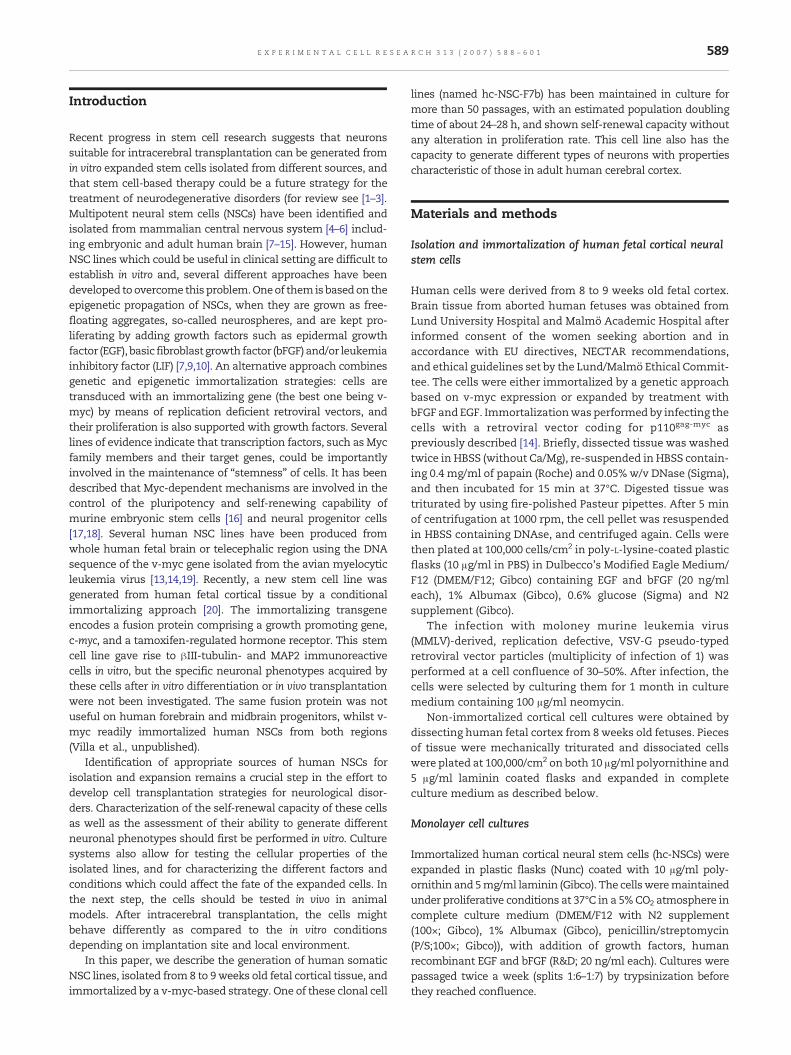
Despite the fact that no harmful actions have beendescribed for v-myc on NSCs, there is a concern about theproperties of the generated progeny. Here we studied both v-myc and telomerase expression after differentiation of thecells. Notably, telomerase has been reported to immortalizehuman NSCs [26]. Four out of 8 studied clones (Table 1)showed a clear decrease of v-myc expression when differ-entiated for 4 or 7 days in medium containing 1% serum (Fig.2). Since v-myc drives telomerase expression, the mRNA fortelomerase (tert) which was expressed in dividing cultures (in
591E X P E R I M E N T A L C E L L R E S E A R C H 3 1 3 ( 2 0 0 7 ) 5 8 8 – 6 0 1

contrast to non-immortal cells) was accordingly found to bedown-regulated after differentiation. Taken together, we showthat v-myc and TERT expression is down-regulated in parallelwith differentiation, yielding mature human neural cells(neurons and glia; see below).
Effects of growth factors on proliferation of hc-NSC lines
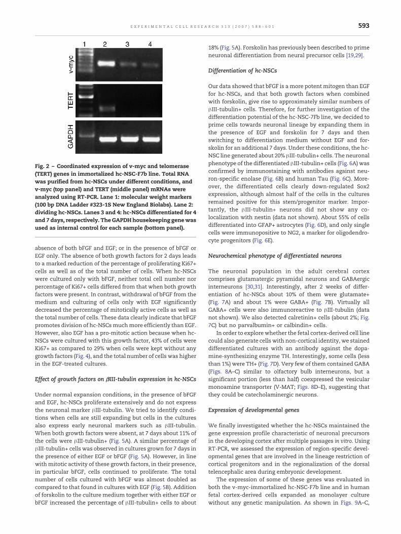
Previous studies have demonstrated that even after geneticimmortalization, NSC lines require support of growth factorsto survive and maintain mitotic activity [13,14,19,27]. The hc-NSCs were expanded in the presence of bFGF and EGF. Underthese conditions, at 2 days after plating, about 80% of hc-NSCswere positive for Ki67 (Figs. 3A, B and D), a marker formitotically active cells [28]. The specific receptors for bothgrowth factors were expressed by the hc-NSCs as demon-strated by RT-PCR (Fig. 3C), and interestingly also in non-immortalized cell cultures derived from 8 weeks old humanfetal cortex, expanded for only 3 passages. At 5 days after EGFand bFGF had been withdrawn and 1% FBS added to theculture medium, the number of Ki67+ cells had dropped to20%, and at 7 days was further decreased to 10% (Fig. 3D).These data clearly show that growth factors are needed for theexpansion of the hc-NSC lines. Similar results have beenobtained when analyzing these parameters in anotherimmortalized forebrain cell line called hNS1 [14].
To further evaluate the contribution of bFGF and EGF insupporting themitotic activity of hc-NSCs, we cultured the hc-NSC-F7b clone for 2 days under either of 4 different conditions(Fig. 4): In the presence (normal expansion condition) or
Fig. 1 – Phenotypic and genetic properties of immortalized human cortical neural stem cells. (A) Example of a clonal cellculture originating from a single cell of the polyclonal hc-NSC line. (B) hc-NSCs grown as adherent culture for 14 passages inthe presence of EGF and bFGF. (C) Karyotype analysis showing a normal chromosomal complement in the hc-NSC-F7b line(n=25 cells in metaphase; after more than 40 passages). Neither numerical nor structural chromosomal abnormalities weredetected. (D–E) Sox2 staining (D) and Hoechst counterstaining (E) of dividing hc-NSC-F7b line. (F–H) Photomicrographs ofcultured hc-NSCs showing nestin (F) and vimentin (G) immunoreactivity separately or as merged image (H). (I–K) Confocalimages of cultured hc-NSCs showing nestin (I) and vimentin (J) immunoreactivity separately or as merged image (K). (L–M)GFAP staining (L) and Hoechst counterstaining (M) of dividing hc-NSC-F7b line. Arrows in panels B, F and G depictmultipolar cells. Scale bar=50 !m.
Table 1 – Properties of clones isolated from polyclonalhc-NSC line
Clonename
!III-tubulin+cells
GFAP+cells
Ki67+cells
Downregulationof v-myc
F9B +++ ++ N.D. NoD10B +++ ++ N.D. NoF7B +++ ++ + YesE6C ++ ++ + YesD11C ++ ++ + YesG5B + ++ + YesG7C + ++ N.D. N.D.C11A + ++ N.D. N.D.
Clones were plated for 7 days without growth factors in thepresence of 1%FBS. Semi-quantitative assessment of number of!III-tubulin, GFAP, and Ki67 immunoreactive cells and occurrenceof v-myc mRNA (RT-PCR) downregulation. +—low number; ++—moderate number; +++—high number; N.D.—not determined.
592 E X P E R I M E N T A L C E L L R E S E A R C H 3 1 3 ( 2 0 0 7 ) 5 8 8 – 6 0 1
absence of both bFGF and EGF; or in the presence of bFGF orEGF only. The absence of both growth factors for 2 days leadsto a marked reduction of the percentage of proliferating Ki67+cells as well as of the total number of cells. When hc-NSCswere cultured only with bFGF, neither total cell number norpercentage of Ki67+ cells differed from that when both growthfactors were present. In contrast, withdrawal of bFGF from themedium and culturing of cells only with EGF significantlydecreased the percentage of mitotically active cells as well asthe total number of cells. These data clearly indicate that bFGFpromotes division of hc-NSCsmuchmore efficiently than EGF.However, also EGF has a pro-mitotic action because when hc-NSCs were cultured with this growth factor, 43% of cells wereKi67+ as compared to 29% when cells were kept without anygrowth factors (Fig. 4), and the total number of cells was higherin the EGF-treated cultures.
Effect of growth factors on !III-tubulin expression in hc-NSCs
Under normal expansion conditions, in the presence of bFGFand EGF, hc-NSCs proliferate extensively and do not expressthe neuronal marker !III-tubulin. We tried to identify condi-tions when cells are still expanding but cells in the culturesalso express early neuronal markers such as !III-tubulin.When both growth factors were absent, at 7 days about 11% ofthe cells were !III-tubulin+ (Fig. 5A). A similar percentage of!III-tubulin+ cells was observed in cultures grown for 7 days inthe presence of either EGF or bFGF (Fig. 5A). However, in linewithmitotic activity of these growth factors, in their presence,in particular bFGF, cells continued to proliferate. The totalnumber of cells cultured with bFGF was almost doubled ascompared to that found in cultures with EGF (Fig. 5B). Additionof forskolin to the culture medium together with either EGF orbFGF increased the percentage of !III-tubulin+ cells to about
18% (Fig. 5A). Forskolin has previously been described to primeneuronal differentiation from neural precursor cells [19,29].
Differentiation of hc-NSCs
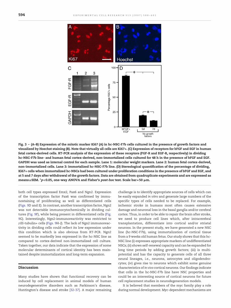
Our data showed that bFGF is amore potentmitogen than EGFfor hc-NSCs, and that both growth factors when combinedwith forskolin, give rise to approximately similar numbers of!III-tubulin+ cells. Therefore, for further investigation of thedifferentiation potential of the hc-NSC-7Fb line, we decided toprime cells towards neuronal lineage by expanding them inthe presence of EGF and forskolin for 7 days and thenswitching to differentiation medium without EGF and for-skolin for an additional 7 days. Under these conditions, the hc-NSC line generated about 20% !III-tubulin+ cells. The neuronalphenotype of the differentiated !III-tubulin+ cells (Fig. 6A) wasconfirmed by immunostaining with antibodies against neu-ron-specific enolase (Fig. 6B) and human Tau (Fig. 6C). More-over, the differentiated cells clearly down-regulated Sox2expression, although almost half of the cells in the culturesremained positive for this stem/progenitor marker. Impor-tantly, the !III-tubulin+ neurons did not show any co-localization with nestin (data not shown). About 55% of cellsdifferentiated into GFAP+ astrocytes (Fig. 6D), and only singlecells were immunopositive to NG2, a marker for oligodendro-cyte progenitors (Fig. 6E).
Neurochemical phenotype of differentiated neurons
The neuronal population in the adult cerebral cortexcomprises glutamatergic pyramidal neurons and GABAergicinterneurons [30,31]. Interestingly, after 2 weeks of differ-entiation of hc-NSCs about 10% of them were glutamate+(Fig. 7A) and about 1% were GABA+ (Fig. 7B). Virtually allGABA+ cells were also immunoreactive to !III-tubulin (datanot shown). We also detected calretinin+ cells (about 2%; Fig.7C) but no parvalbumin+ or calbindin+ cells.
In order to explorewhether the fetal cortex-derived cell linecould also generate cells with non-cortical identity, we staineddifferentiated cultures with an antibody against the dopa-mine-synthesizing enzyme TH. Interestingly, some cells (lessthan 1%) were TH+ (Fig. 7D). Very few of them contained GABA(Figs. 8A–C) similar to olfactory bulb interneurons, but asignificant portion (less than half) coexpressed the vesicularmonoamine transporter (V-MAT; Figs. 8D–E), suggesting thatthey could be catecholaminergic neurons.
Expression of developmental genes
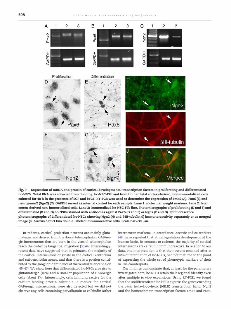
We finally investigated whether the hc-NSCs maintained thegene expression profile characteristic of neuronal precursorsin the developing cortex after multiple passages in vitro. UsingRT-PCR, we assessed the expression of region-specific devel-opmental genes that are involved in the lineage restriction ofcortical progenitors and in the regionalization of the dorsaltelencephalic area during embryonic development.
The expression of some of these genes was evaluated inboth the v-myc-immortalized hc-NSC-F7b line and in humanfetal cortex-derived cells expanded as monolayer culturewithout any genetic manipulation. As shown in Figs. 9A–C,
Fig. 2 – Coordinated expression of v-myc and telomerase(TERT) genes in immortalized hc-NSC-F7b line. Total RNAwas purified from hc-NSCs under different conditions, andv-myc (top panel) and TERT (middle panel) mRNAs wereanalyzed using RT-PCR. Lane 1: molecular weight markers(100 bp DNA Ladder #323-1S New England Biolabs). Lane 2:dividing hc-NSCs. Lanes 3 and 4: hc-NSCs differentiated for 4and 7 days, respectively. TheGAPDHhousekeeping genewasused as internal control for each sample (bottom panel).
593E X P E R I M E N T A L C E L L R E S E A R C H 3 1 3 ( 2 0 0 7 ) 5 8 8 – 6 0 1
both cell types expressed Emx2, Pax6 and Ngn2. Expressionof the transcription factor Pax6 was confirmed by immu-nostaining of proliferating as well as differentiated cells(Figs. 9D and E). In contrast, another transcription factor,Ngn2was not detectable immunocytochemically in dividing cul-tures (Fig. 9F), while being present in differentiated cells (Fig.9G). Interestingly, Ngn2-immunoreactivity was restricted to!III-tubulin+ cells (Figs. 9H–J). The lack of Ngn2 immunoreac-tivity in dividing cells could reflect its low expression underthis condition which is also obvious from RT-PCR. Ngn2seemed to be markedly less expressed in the hc-NSC line ascompared to cortex-derived non-immortalized cell culture.Taken together, our data indicate that the expression of somemolecular determinants of cortical identity has been main-tained despite immortalization and long-term expansion.
Discussion
Many studies have shown that functional recovery can beinduced by cell replacement in animal models of humanneurodegenerative disorders such as Parkinson's disease,Huntington's disease and stroke [32–37]. A major remaining
challenge is to identify appropriate sources of cells which canbe easily expanded in vitro and generate large numbers of thespecific types of cells needed to be replaced. For example,ischemic stroke in humans most often causes extensivedamage and neuronal loss in the basal ganglia and/or cerebralcortex. Thus, in order to be able to repair the brain after stroke,we need to produce cell lines which, after intracerebraltransplantation, differentiate into cortical and/or striatalneurons. In the present study, we have generated a new NSCline (hc-NSC-F7b), using immortalization of cortical tissuefrom a 9weeks old human fetus. Our study shows that this hc-NSC line (i) expresses appropriate markers of undifferentiatedNSCs; (ii) shows self-renewal capacity and can be expanded forlong time periods by adding growth factors; (iii) is multi-potential and has the capacity to generate cells of all threeneural lineages, i.e., neurons, astrocytes and oligodendro-cytes; (iv) gives rise to neurons which exhibit some genuinecharacteristics of in vivo cortical neurons. Our findings indicatethat cells in the hc-NSC-F7b line have NSC properties andcould be an interesting source of cortical neurons for futurecell replacement studies in neurodegeneration models.
It is believed that members of the myc family play a roleduring normal development. Myc-dependentmechanisms are
Fig. 3 – (A–B) Expression of the mitotic marker Ki67 (A) in hc-NSC-F7b cells cultured in the presence of growth factors andvisualized by Hoechst staining (B). Note that virtually all cells are Ki67+. (C) Expression of receptors for bFGF and EGF in humanfetal cortex-derived cells. RT-PCR analysis of the expression of these receptors (FGF-R and EGF-R, respectively) in dividinghc-NSC-F7b line- and human fetal cortex-derived, non-immortalized cells cultured for 48 h in the presence of bFGF and EGF.GAPDH was used as internal control for each sample. Lane 1: molecular weight markers. Lane 2: human fetal cortex-derived,non-immortalized cells. Lane 3: immortalized hc-NSC-F7b line. (D) Stereological quantification of the percentage of dividing,Ki67+ cells when immortalized hc-NSCs had been cultured under proliferation conditions in the presence of bFGF and EGF, andat 5 and 7 days after withdrawal of the growth factors. Data are obtained from quadruplicate experiments and are expressed asmeans±SEM. *p<0.05, one-way ANOVA and Fisher's post-hoc test. Scale bar=50 !m.
594 E X P E R I M E N T A L C E L L R E S E A R C H 3 1 3 ( 2 0 0 7 ) 5 8 8 – 6 0 1

involved in the control of pluripotency and self-renewingcapacity of murine embryonic stem cells [16], and in theexpansion of neural progenitor cell populations and inhibitionof differentiation [17,18]. Several studies have demonstratedthat the avian myelocytic leukemia retroviral v-myc gene, theortholog of cellular c-myc, could be successfully used toimmortalize human neural precursor cells [13,14,19]. Herewe used retroviral v-myc transduction to generate a humanfetal cortex-derived NSC polyclonal line, and then selectedsingle cell-derived clones with defined properties: expressionof neural markers, high expansion rate, decreased prolifera-tion upon removal of growth factors and capacity to differ-entiate into neurons.
A major concern regarding the genetic immortalizationstrategy, as used in the present study, is the safety of thegenerated cell lines. Could v-myc act as a transforming
oncogene like the mammalian c-myc counterpart? However,such an action has never been demonstrated experimentally[38]. By unknown reasons, v-myc works perfectly well forimmortalization of mammalian NSCs without transformingthem. In the present study we addressed the issue oftransformation of the hc-NSC line in three different ways:Firstly, we performed a chromosomal analysis and found thatthis hc-NSC line retains a normal karyotype even after morethan 40 passages in culture. No numerical or structuralchromosomal abnormalities were detected. Indirectly, thisfinding also speaks in favor of good telomere preservation asreported previously [25].
Secondly, we explored whether the immortalized hc-NSCsdown-regulate v-myc expression after switching from prolif-erative to differentiation culture conditions by removal ofgrowth factors. V-myc stimulates the expression of growthfactor-regulated genes and strongly promotes cellular prolif-eration. Moreover, its constitutive expression interferes withthe terminal differentiation of the majority of studied celltypes [16,17,39]. Our PCR analysis showed that the hc-NSC-F7bline down-regulates v-myc expression after growth factordeprivation. Cross-talk betweenmyc and telomerase has been
Fig. 5 – Percentage of "III-tubulin+ cells (A) and total numberof cells (B) in the immortalized hc-NSC cultures under variousdifferentiation conditions. Cells were cultured for 7 dayswithout growth factors and with FBS; or with EGF and bFGFonly; or with EGF and forskolin or bFGF and forskolin. Datawere obtained from quadruplicate experiments and areexpressed as means±SEM. *p<0.05, one-way ANOVA andFisher's post-hoc test.
Fig. 4 – Percentage of proliferating, Ki67+ cells (A) and totalnumber of cells (B) in the immortalized hc-NSC cultures.NSCswere platedwith orwithout bFGF and EGF, or with bFGFor EGF only. Data are obtained from quadruplicateexperiments and are expressed as mean±SEM. *p<0.05,one-way ANOVA and Fisher's post-hoc test.
595E X P E R I M E N T A L C E L L R E S E A R C H 3 1 3 ( 2 0 0 7 ) 5 8 8 – 6 0 1
demonstrated [24,25], and retroviral over-expression ofhuman TERT has been tried for immortalization of neuronalprogenitor cells derived from the human fetal spinal cord [26].Similar to previous observations in another cell line [25], ourdata indicate that when hc-NSCs differentiate, there is aparallel downregulation of v-myc and TERT expression.
Thirdly, we studied the growth factor-dependence of hc-NSCs for their in vitroproliferation. It has been shown that evenif genetically immortalized, human fetal cell lines needbFGForbFGF and EGF for their expansion [13,14,19,27]. Our findingsindicate that in medium containing 1% serum but withoutgrowth factors, cells stop dividing and differentiate intoneurons and glia. The percentage of cells with mitotic activity(10%) agrees with previous in vitro findings and more recent invivo data, showing that 11% of c-myc immortalized humancortical cells are Ki67+ at 5 weeks after transplantation [20].
Taken together our findings provide further evidence (i)that v-myc and telomerase are important for the expansion of
hc-NSCs; (ii) that v-myc controls expression of telomerase;and (iii) that after growth factor removal, these genes aredown-regulated in a coordinated way. These data are inaccordancewith the concept of “conditional immortalization”,i.e., that the cells are immortal as long asmitogens are presentin the culture medium. In the absence of mitogenes, the cellsresume their standard behavior, differentiating into neuronsand glia.
Our data show that bFGF has a more potent mitogenicactivity than EGF on the hc-NSC line. The PCR analysisindicates that, in the presence of their respective ligands,both bFGF and EGF receptors are expressed in the hc-NSCcultures. Importantly, expression of both receptors was alsodetected in non-immortalized human fetal cortical cultureswhich had been passaged only three times. These findingsargue against the hypothesis that expression of FGF and EGFreceptors is induced by the v-myc immortalization of the hc-NSC line. Previous reports have demonstrated that in vitro, FGF
Fig. 6 – Immunocytochemical demonstration of neuronal and glial markers in differentiated hc-NSC-F7b line. Cells were platedand differentiated for 7 days with EGF and forskolin and another 7 days only with B27. Cells immunoreactive for the immatureneuronal marker "III-tubulin (A) co-express another neuronal marker, neural specific enolase (NSE; B). Cells positive to"III-tubulin and NSE were visualized after double-fluorescent immunostaining by using Cy3-conjugated secondary antibody(A) andAlexa 488-conjugated streptavidin. After differentiation, the hc-NSCs also express themature neuronalmarker hTau (C)and astrocyte marker GFAP (D). Few cells in the differentiated hc-NSC cultures are positive to the oligodendrocyte marker NG2(E). Arrows depict examples of immunoreactive cells. Scale bar=30 !m.
596 E X P E R I M E N T A L C E L L R E S E A R C H 3 1 3 ( 2 0 0 7 ) 5 8 8 – 6 0 1

can induce responsiveness to EGF in primary cells from fetalrodent striatum [40] and mesencephalon [41]. Moreover,recent data indicate the existence of a single type of stemcell responsive to both FGF and EGF in expanded neurospherecultures of human fetal cortex from the first trimester [42]. It isnot clear from our study whether the same hc-NSCs expressFGF and EGF receptors, and if FGF increases responsiveness toEGF in these cells. However, the observation that the hc-NSCline could be expanded in the presence of EGF only indicatesthat this factor has sufficient, though low mitogenic activity.
Experimental evidence suggests that c-myc protein has pro-apoptotic activity which can be triggered by a range of insultsincluding growth factor deprivation [43]. To our knowledge,
such an action has not been reported for v-myc. Forskolin haspreviously been used in combination with growth factors toprime neuronal differentiation of human fetal mesencephalicneural progenitor cells [29]. Here we found that culturesdifferentiated for 7 days in the presence of FGF and forskolin orEGF and forskolin showed no significant differences in termsof percentage of !III-tubulin+ cells out of total cell number. It iswell known that differentiation requires cells to stop divisionand exit from the cell cycle. Because FGF supported celldivision stronger than EGF, we decided to use a protocol basedon 7 days of priming in the presence of EGF and forskolin forthe further characterization of the phenotype of cells differ-entiated from the hc-NSC line.
Fig. 7 – Immunocytochemical demonstration of specific neuronal phenotypes in differentiated hc-NSC-F7b line. Cells wereplated for 7 days with EGF and forskolin, and another 7 days only with B27. Photomicrographs illustrate cellsimmunoreactive for glutamate (A), GABA (B), calretinin (C), and tyrosine hydroxylase (TH, D). Arrows depict examples ofimmunoreactive cells. Scale bar=30 !m.
Fig. 8 – Characterization of phenotype of TH-positive cells generated from hc-NSC-F7b clone. Epifluorescence images ofdifferentiated hc-NSCs showing TH (A), GABA (B) and merged image (C), or TH (D), V-MAT (E) and merged image (F). Arrowsdepict TH+/GABA+ (A–C), and TH+/V-MAT+ (D–F) double-stained neurons; arrowheads indicate TH+/GABA- (A and C) andTH+/V-MAT- (D and F) neurons. Scale bar=30 !m.
597E X P E R I M E N T A L C E L L R E S E A R C H 3 1 3 ( 2 0 0 7 ) 5 8 8 – 6 0 1
In rodents, cortical projection neurons are mainly gluta-matergic and derived from the dorsal telencephalon. GABAer-gic interneurons that are born in the ventral telencephalonreach the cortex by tangential migration [30,44]. Interestingly,recent data have suggested that in primates, the majority ofthe cortical interneurons originate in the cortical ventricularand subventricular zones, and that there is a portion contri-buted by the ganglionic eminence of the ventral telencephalon[45–47]. We show here that differentiated hc-NSCs give rise toglutamatergic (10%) and a smaller population of GABAergiccells (about 1%). Interestingly, cells immunoreactive for thecalcium-binding protein calretinin, a marker for corticalGABAergic interneurons, were also detected but we did notobserve any cells containing parvalbumin or calbindin (other
interneuron markers). In accordance, Zecevic and co-workers[48] have reported that at mid-gestation development of thehuman brain, in contrast to rodents, the majority of corticalinterneurons are calretinin immunoreactive. In relation to ourdata, one interpretation is that the neurons obtained after invitro differentiation of hc-NSCs, had not matured to the pointof expressing the whole set of phenotypic markers of theirin vivo counterparts.
Our findings demonstrate that, at least for the parametersinvestigated here, hc-NSCs retain their regional identity evenafter multiple in vitro expansions. Using RT-PCR, we foundthat the undifferentiated hc-NSCs express the genes encodingthe basic helix–loop–helix (bHLH) transcription factor Ngn2and the homeodomain transcription factors Emx2 and Pax6.
Fig. 9 – Expression of mRNA and protein of cortical developmental transcription factors in proliferating and differentiatedhc-NSCs. Total RNA was collected from dividing, hc-NSC-F7b and from human fetal cortex-derived, non-immortalized cellscultured for 48 h in the presence of EGF and bFGF. RT-PCR was used to determine the expression of Emx2 (A), Pax6 (B) andneurogenin2 (Ngn2) (C). GAPDH served as internal control for each sample. Lane 1: molecular weight markers. Lane 2: fetalcortex-derived non-immortalized cells. Lane 3: immortalized hc-NSC-F7b line. Photomicrographs of proliferating (D and F) anddifferentiated (E and G) hc-NSCs stained with antibodies against Pax6 (D and E) or Ngn2 (F and G). Epifluorescencephotomicrographs of differentiated hc-NSCs showing Ngn2 (H) and "III-tubulin (I) immunoreactivity separately or as mergedimage (J). Arrows depict two double-labeled immunoreactive cells. Scale bar=30 !m.
598 E X P E R I M E N T A L C E L L R E S E A R C H 3 1 3 ( 2 0 0 7 ) 5 8 8 – 6 0 1

These genes have been proposed to be involved in theregionalization of the developing neocortex, and in the controlof different properties of cortical progenitors such as theirneuronal commitment, phenotype specification, and migra-tion [49–55]. Cells immunoreactive to Pax6 but not to Ngn2were detected in dividing cultures, while both transcriptionfactors were observed in differentiated cells which alsoexpressed the neuronal marker !III-tubulin. Although Ngn2was found with PCR in proliferating cells, the lack of Ngn2protein immunocytochemistry most likely reflects less impor-tance of this transcription factor during proliferation.
The PCR amplification as performed here was not quanti-tative, but it was obvious that Ngn2 mRNA was expressed athigher levels in non-immortalized cortical cultures as com-pared to the v-myc immortalized hc-NSC line. The results ofthe short-term culture expansion procedure applied to non-immortal cells (3 passages) provides evidence for the existenceof a heterogenous cell population comprising NSCs andcommitted progenitors. In contrast, the monoclonal hc-NSCline is more likely to consist of a relatively homogenouspopulation of NSCs. Nieto and co-workers [51] have proposedthat the expression of ngn-2 is restricted to committedneuronal progenitors. If this is the case, the differences inNgn2 expression observed here could be related to differentcellular composition of immortalized and non-immortalizedcell cultures. This interpretation is also supported by thefinding that Ngn2 was clearly expressed in !III-tubulin+neurons after 2 weeks of differentiation.
The gene expression profile and the ability to generateglutamatergic and GABAergic neurons demonstrate that thehc-NSCs maintain their regional identity as cortical progeni-tors. However, our data suggest that this cell line is alsocapable of generating neurons which are not normally presentin the adult cerebral cortex in vivo. Induction of TH geneexpression in neurons derived from human fetal cerebralcortex, as shown here, has been reported previously [56,57]. Inour experiments, not all V-MAT+ cells were also TH+. TH isinvolved in the synthesis of catecholamines (dopamine,adrenaline and noradrenaline) and V-MAT is the transporterof monoamines (dopamine, noradrenaline, histamine andserotonin) in the synaptic vesicles. Our findings provide someevidence that both catecholaminergic (TH+/V-MAT+) andserotoninergic (TH-/V-MAT+) cell types can be generatedafter differentiation of hc-NSC (for review see [58]). Interest-ingly a small fraction of the TH+ cells co-expressed GABA.Such cells have previously been described as a population ofinterneurons in the adult human brain [59,60].
In conclusion, our demonstration that the new human cellline generated here can produce cells in vitro expressingmarkers characteristic of some cortical neuron types repre-sents a promising first step towards a cell replacementstrategy following cortical injury such as stroke. It is nowimportant to determine how the differentiation of these hc-NSCs after intracerebral transplantation is influenced by theexternal cues present in the intact and pathological brain. Formaximum generation of several types of cortical neurons, itmay be necessary to predifferentiate the cell line in vitro.However, the ability of the cortical tissue environment to drivethe NSCs towards the specific cortical neuronal phenotypesshould also be explored.We also have to address if the grafted
new neurons can survive in large numbers and becomemorphologically and functionally integrated into existingneural circuitries. So far, there is very little evidence forreconstruction of cortical circuitries and, in particular, theformation of efferent connections from the cortical graft to thehost after cell transplantation in the injured brain. Moreover,whether NSCs can give rise to cells with in vivo electrophysio-logical properties specific for cortical neurons is unknown.Although we obtained no evidence for transformation of thehc-NSC line in culture, we must analyze in detail the risks fortumor formation in vivo. Finally, it will be of major importanceto determine if intracortical implantation of hc-NSC-derivedcells can induce significant functional recovery in models ofneurodegenerative disorders where cortical damage plays amajor pathophysiological role.
Acknowledgments
We thank Linda Magnusson for excellent technical assistance.This work was supported by the Swedish Research Council, EUproject LSHBCT-2003-503005 (EUROSTEMCELL), and the KingGustav V and Queen Victoria, Söderberg, Crafoord, and KockFoundations. The Lund Stem Cell Center is supported by aCenter of Excellence grant in Life Sciences from the SwedishFoundation for Strategic Research. Work at A.M.S. group wassupported by grants SAF2001-0841 and SAF2004-03405 fromSpanish Ministry of Science and Technology, QLK3-CT-2001-02120 from European Union, and the institutional grant ofRamon Areces foundation to the CBMSO.
R E F E R E N C E S
[1] O. Lindvall, Z. Kokaia, A. Martinez-Serrano, Stem cell therapyfor human neurodegenerative disorders—How to make itwork, Nat. Med. 10 (2004) S42–S50 (Suppl).
[2] S. Goldman, Stem and progenitor cell-based therapy of thehuman central nervous system, Nat. Biotechnol. 23 (2005)862–871.
[3] O. Lindvall, Z. Kokaia, Stem cells for the treatment ofneurological disorders, Nature 441 (2006) 1094–1096.
[4] F. Doetsch, I. Caille, D.A. Lim, J.M. Garcia-Verdugo,A. Alvarez-Buylla, Subventricular zone astrocytes are neuralstem cells in the adult mammalian brain, Cell 97 (1999)703–716.
[5] A.A. Davis, S. Temple, A self-renewing multipotential stemcell in embryonic rat cerebral cortex, Nature 372 (1994)263–266.
[6] C.B. Johansson, S. Momma, D.L. Clarke, M. Risling, U. Lendahl,J. Frisen, Identification of a neural stem cell in the adultmammalian central nervous system, Cell 96 (1999) 25–34.
[7] M.K. Carpenter, X. Cui, Z.Y. Hu, J. Jackson, S. Sherman,A. Seiger, L.U. Wahlberg, In vitro expansion of a multipotentpopulation of human neural progenitor cells, Exp. Neurol. 158(1999) 265–278.
[8] C.N. Svendsen, M.A. Caldwell, J. Shen, M.G. Ter Borg,A.E. Rosser, P. Tyers, S. Karmiol, S.B. Dunnett, Long-termsurvival of human central nervous system progenitor cellstransplanted into a rat model of Parkinson's disease, Exp.Neurol. 148 (1997) 135–146.
[9] A.L. Vescovi, E.A. Parati, A. Gritti, P. Poulin, M. Ferrario, E.Wanke, P. Frolichsthal-Schoeller, L. Cova, M.Arcellana-Panlilio, A. Colombo, R. Galli, Isolation and cloning
599E X P E R I M E N T A L C E L L R E S E A R C H 3 1 3 ( 2 0 0 7 ) 5 8 8 – 6 0 1
of multipotential stem cells from the embryonic human CNSand establishment of transplantable human neural stem celllines by epigenetic stimulation, Exp. Neurol. 156 (1999) 71–83.
[10] T.D. Palmer, P.H. Schwartz, P. Taupin, B. Kaspar, S.A. Stein,F.H. Gage, Cell culture. Progenitor cells from human brainafter death, Nature 411 (2001) 42–43.
[11] S. Kelly, T.M. Bliss, A.K. Shah, G.H. Sun, M. Ma, W.C. Foo, J.Masel, M.A. Yenari, I.L. Weissman, N. Uchida, T. Palmer, G.K.Steinberg, Transplanted human fetal neural stem cellssurvive, migrate, and differentiate in ischemic rat cerebralcortex, Proc. Natl. Acad. Sci. U. S. A. 101 (2004) 11839–11844.
[12] N. Uchida, D.W. Buck, D. He, M.J. Reitsma, M. Masek, T.V.Phan, A.S. Tsukamoto, F.H. Gage, I.L. Weissman, Directisolation of human central nervous system stem cells, Proc.Natl. Acad. Sci. U. S. A. 97 (2000) 14720–14725.
[13] J.D. Flax, S. Aurora, C. Yang, C. Simonin, A.M. Wills, L.L.Billinghurst, M. Jendoubi, R.L. Sidman, J.H. Wolfe, S.U. Kim,E.Y. Snyder, Engraftable human neural stem cells respond todevelopmental cues, replace neurons, and express foreigngenes, Nat. Biotechnol. 16 (1998) 1033–1039.
[14] A. Villa, E.Y. Snyder, A. Vescovi, A. Martinez-Serrano,Establishment and properties of a growth factor-dependent,perpetual neural stem cell line from the human CNS, Exp.Neurol. 161 (2000) 67–84.
[15] A. Martinez-Serrano, F.J. Rubio, B. Navarro, C. Bueno, A. Villa,Human neural stem and progenitor cells: in vitro and in vivoproperties, and potential for gene therapy and cellreplacement in the CNS, Curr. Gene Ther. 1 (2001) 279–299.
[16] P. Cartwright, C. Mclean, A. Sheppard, D. Rivett, K. Jones, S.Dalton, LIF/STAT3 controls ES cell self-renewal andpluripotency by a Myc-dependent mechanism, Development132 (2005) 885–896.
[17] P.S. Knoepfler, P.F. Cheng, R.N. Eisenman, N-myc is essentialduring neurogenesis for the rapid expansion of progenitor cellpopulations and the inhibition of neuronal differentiation,Genes Dev. 16 (2002) 2699–2712.
[18] A.M. Kenney, H.R. Widlund, D.H. Rowitch, Hedgehog and PI-3kinase signaling converge on Nmyc1 to promote cell cycleprogression in cerebellar neuronal precursors, Development131 (2004) 217–228.
[19] D.W. Sah, J. Ray, F.H. Gage, Bipotent progenitor cell lines fromthe human CNS, Nat. Biotechnol. 15 (1997) 574–580.
[20] K. Pollock, P. Stroemer, S. Patel, L. Stevanato, A. Hope, E.Miljan, Z. Dong, H. Hodges, J. Price, J.D. Sinden, A conditionallyimmortal clonal stem cell line from human corticalneuroepithelium for the treatment of ischemic stroke, Exp.Neurol. 199 (2006) 143–155.
[21] S. Horiguchi, J. Takahashi, Y. Kishi, A. Morizane, Y. Okamoto,M. Koyanagi, M. Tsuji, K. Tashiro, T. Honjo, S. Fujii, N.Hashimoto, Neural precursor cells derived from humanembryonic brain retain regional specificity, J. Neurosci. Res.75 (2004) 817–824.
[22] M. Schuldiner, O. Yanuka, J. Itskovitz-Eldor, D.A. Melton, N.Benvenisty, Effects of eight growth factors on thedifferentiation of cells derived from human embryonic stemcells, Proc. Natl. Acad. Sci. U. S. A. 97 (2000) 11307–11312.
[23] K. Nibu, G. Li, K. Kaga, J.L. Rothstein, bFGF inducesdifferentiation and death of olfactory neuroblastoma cells,Biochem. Biophys. Res. Commun. 279 (2000) 172–180.
[24] S. Oh, Y.H. Song, U.J. Kim, J. Yim, T.K. Kim, In vivo and in vitroanalyses of Myc for differential promoter activities of thehuman telomerase (hTERT) gene in normal and tumor cells,Biochem. Biophys. Res. Commun. 263 (1999) 361–365.
[25] A. Villa, B. Navarro-Galve, C. Bueno, S. Franco, M.A. Blasco, A.Martinez-Serrano, Long-term molecular and cellular stabilityof human neural stem cell lines, Exp. Cell Res. 294 (2004)559–570.
[26] N.S. Roy, T. Nakano, H.M. Keyoung, M. Windrem, W.K.Rashbaum, M.L. Alonso, J. Kang, W. Peng, M.K. Carpenter,
J. Lin, M. Nedergaard, S.A. Goldman, Telomeraseimmortalization of neuronally restricted progenitor cellsderived from the human fetal spinal cord, Nat. Biotechnol. 22(2004) 297–305.
[27] D.L. Kitchens, E.Y. Snyder, D.I. Gottlieb, FGF and EGF aremitogens for immortalized neural progenitors, J. Neurobiol.25 (1994) 797–807.
[28] J. Gerdes, H. Lemke, H. Baisch, H.H. Wacker, U. Schwab, H.Stein, Cell cycle analysis of a cell proliferation-associatedhuman nuclear antigen defined by the monoclonal antibodyKi-67, J. Immunol. 133 (1984) 1710–1715.
[29] X. Wang, X. Li, K. Wang, H. Zhou, B. Xue, L. Li, Forskolincooperating with growth factor on generation ofdopaminergic neurons from human fetal mesencephalicneural progenitor cells, Neurosci. Lett. 362 (2004) 117–121.
[30] J.G. Parnavelas, The origin andmigration of cortical neurones:new vistas, Trends Neurosci. 23 (2000) 126–131.
[31] J.G. Parnavelas, S.A. Anderson, A.A. Lavdas, M. Grigoriou, V.Pachnis, J.L. Rubenstein, The contribution of the ganglioniceminence to the neuronal cell types of the cerebral cortex,Novartis Found. Symp. 228 (2000) 129–139(Discussion 139–147).
[32] P. Brundin, W.-M. Duan, H. Sauer, Functional effects ofmesencephalic dopamine neurons and adrenal chromaffincells grafted to the rodent striatum, in: S.B. Dunnett, A.Björklund (Eds.), Functional Neural Transplantation, RavenPress Ltd., New York, 1994, pp. 9–46.
[33] J.H. Kim, J.M. Auerbach, J.A. Rodriguez-Gomez, I. Velasco, D.Gavin, N. Lumelsky, S.H. Lee, J. Nguyen, R. Sanchez-Pernaute,K. Bankiewicz, R. Mckay, Dopamine neurons derived fromembryonic stem cells function in an animal model ofParkinson's disease, Nature 418 (2002) 50–56.
[34] O. Isacson, P. Brundin, F.H. Gage, A. Björklund, Neural graftingin a rat model of Huntington's disease: progressiveneurochemical changes after neostriatal ibotenate lesionsand striatal tissue grafting, Neuroscience 16 (1985) 799–817.
[35] J.L. Mcbride, S.P. Behrstock, E.Y. Chen, R.J. Jakel, I. Siegel, C.N.Svendsen, J.H. Kordower, Human neural stem cell transplantsimprove motor function in a rat model of Huntington'sdisease, J. Comp. Neurol. 475 (2004) 211–219.
[36] Z.G. Zhang, Q. Jiang, R. Zhang, L. Zhang, L. Wang, P. Arniego,K.L. Ho, M. Chopp, Magnetic resonance imaging andneurosphere therapy of stroke in rat, Ann. Neurol. 53 (2003)259–263.
[37] B. Mattsson, J.C. Sorensen, J. Zimmer, B.B. Johansson, Neuralgrafting to experimental neocortical infarcts improvesbehavioral outcome and reduces thalamic atrophy in ratshoused in enriched but not in standard environments, Stroke28 (1997) 1225–1231.
[38] B. Navarro, A. Villa, I. Liste, C. Bueno, A. Martínez-Serrano,Isolation, proliferation, survival and differentiation ofhuman neural stem/ precursor cells in vitro and in vivo. Afocus on their potential clinical applications, in: J.E.Bottenstein (Ed.), Neural Stem Cells: Development andtransplantation, Kluwer Academic Publisher, Dordrecht,2003, pp. 271–298.
[39] S. Schreiner, M. Birke, M.P. Garcia-Cuellar, O. Zilles, J. Greil,R.K. Slany, MLL-ENL causes a reversible and myc-dependentblock of myelomonocytic cell differentiation, Cancer Res. 61(2001) 6480–6486.
[40] F. Ciccolini, C.N. Svendsen, Fibroblast growth factor 2 (FGF-2)promotes acquisition of epidermal growth factor (EGF)responsiveness in mouse striatal precursor cells:identification of neural precursors responding to both EGFand FGF-2, J. Neurosci. 18 (1998) 7869–7880.
[41] J. Santa-Olalla, L. Covarrubias, Basic fibroblast growth factorpromotes epidermal growth factor responsiveness andsurvival of mesencephalic neural precursor cells, J. Neurobiol.40 (1999) 14–27.
600 E X P E R I M E N T A L C E L L R E S E A R C H 3 1 3 ( 2 0 0 7 ) 5 8 8 – 6 0 1

[42] T. Ostenfeld, C.N. Svendsen, Requirement for neurogenesis toproceed through the division of neuronal progenitorsfollowing differentiation of epidermal growth factor andfibroblast growth factor-2-responsive human neural stemcells, Stem Cells 22 (2004) 798–811.
[43] A.O. Hueber, G.I. Evan, Traps to catch unwary oncogenes,Trends Genet. 14 (1998) 364–367.
[44] O. Marin, J.L. Rubenstein, A long, remarkable journey:tangential migration in the telencephalon, Nat. Rev.,Neurosci. 2 (2001) 780–790.
[45] N. Zecevic, P. Rakic, Development of layer I neurons in theprimate cerebral cortex, J. Neurosci. 21 (2001) 5607–5619.
[46] S. Rakic, N. Zecevic, Emerging complexity of layer I in humancerebral cortex, Cereb. Cortex 13 (2003) 1072–1083.
[47] K. Letinic, R. Zoncu, P. Rakic, Origin of GABAergic neurons inthe human neocortex, Nature 417 (2002) 645–649.
[48] N. Zecevic, Y. Chen, R. Filipovic, Contributions of corticalsubventricular zone to the development of the humancerebral cortex, J. Comp. Neurol. 491 (2005) 109–122.
[49] K.M. Bishop, J.L. Rubenstein, D.D. O'leary, Distinct actions ofEmx1, Emx2, and Pax6 in regulating the specification ofareas in the developing neocortex, J. Neurosci. 22 (2002)7627–7638.
[50] N. Heins, P. Malatesta, F. Cecconi, M. Nakafuku, K.L. Tucker,M.A. Hack, P. Chapouton, Y.A. Barde, M. Gotz, Glial cellsgenerate neurons: the role of the transcription factor Pax6,Nat. Neurosci. 5 (2002) 308–315.
[51] M. Nieto, C. Schuurmans, O. Britz, F. Guillemot, Neural bHLHgenes control the neuronal versus glial fate decision incortical progenitors, Neuron 29 (2001) 401–413.
[52] P. Chapouton, C. Schuurmans, F. Guillemot, M. Gotz, Thetranscription factor neurogenin 2 restricts cell migrationfrom the cortex to the striatum, Development 128 (2001)5149–5159.
[53] C. Schuurmans, O. Armant, M. Nieto, J.M. Stenman, O. Britz, N.Klenin, C. Brown, L.M. Langevin, J. Seibt, H. Tang, J.M.Cunningham, R. Dyck, C. Walsh, K. Campbell, F. Polleux, F.Guillemot, Sequential phases of cortical specification involveNeurogenin-dependent and-independent pathways, EMBO J.23 (2004) 2892–2902.
[54] A. Simeone, D. Acampora, M. Gulisano, A. Stornaiuolo, E.Boncinelli, Nested expression domains of fourhomeobox genes in developing rostral brain, Nature 358(1992) 687–690.
[55] A. Simeone, M. Gulisano, D. Acampora, A. Stornaiuolo, M.Rambaldi, E. Boncinelli, Two vertebrate homeobox genesrelated to the Drosophila empty spiracles gene are expressedin the embryonic cerebral cortex, EMBO J. 11 (1992)2541–2550.
[56] J. Zhou, B. Pliego-Rivero, H.F. Bradford, G.M. Stern, E.R.Jauniaux, Induction of tyrosine hydroxylase gene expressionin human foetal cerebral cortex, Neurosci. Lett. 252 (1998)215–217.
[57] F.B. Pliego Rivero, W.J. Mccormack, E. Jauniaux, G.M. Stern,H.F. Bradford, Forskolin-induced expression of tyrosinehydroxylase in human foetal brain cortex, Brain Res. Dev.Brain Res. 114 (1999) 201–206.
[58] C. Verney, Phenotypic expression of monoamines and GABAin the early development of human telencephalon, transientor not transient, J. Chem. Neuroanat. 26 (2003) 283–292.
[59] M. Dubach, Telencephalic dopamine cells in monkeys,human, and rats, in: A. Reiner (Ed.), Phylogeny andDevelopment of Catecholamine Systems in the CNS ofVertebrates, Cambridge Univ. Press, Cambridge, 1994,pp. 273–292.
[60] S. Trottier, M. Geffard, B. Evrard, Co-localization of tyrosinehydroxylase and GABA immunoreactivities in human corticalneurons, Neurosci. Lett. 106 (1989) 76–82.
601E X P E R I M E N T A L C E L L R E S E A R C H 3 1 3 ( 2 0 0 7 ) 5 8 8 – 6 0 1
Top Related
Copyright © 2022 FDOKUMEN

