


Bahasa
Halaman
Hukum

Research Journal of Recent Sciences ________________________________________________ ISSN 2277-2502
Vol. 2(6), 1-10, June (2013) Res.J.Recent Sci.
International Science Congress Association 1
Framework for the Comparison of Classifiers for Medical Image
Segmentation with Transform and Moment based features
Maria Hameed, Muhammad Sharif, Mudassar Raza, Syed Waqas Haider, Muhammad Iqbal Department of Computer Sciences, COMSATS Institute of Information Technology, Wah Cantt., 47040, PAKISTAN
Available online at: www.isca.in Received 10th October 2012, revised 14th November 2012, accepted 7th February 2013
Abstract
The paper depicts and elaborates a new framework for the comparison of classifiers for medical image segmentation with
transform and moment based features. Medical images modalities such as Ultrasound (US) bladder, Ultrasound (US)
phantom, Computerized Tomography (CT) and Magnetic Resonance (MR) images are segmented using different
algorithms namely, k-Nearest Neighbor (kNN), Grow and learn (GAL) and Incremental Supervised Neural Networks
(ISNN). Segmentation is performed by applying feature extraction methods such as 2D Continuous Wavelet Transform
(2D-CWT), Moments of gray level histogram (MGH) and a combined version of both 2D-CWT and MGH, called Hybrid
features. With different iterations, the analysis results indicate that kNN performs better than GAL, and the performance of
GAL is better than that of the ISNN for image segmentation. During analysis a comparison has been drawn between the
performance of kNN, GAL and ISNN on the above three feature extraction schemes and also provides the qualitative and
quantitative analysis of three classifiers. Results indicate that the performance of 2D-CWT and that of Hybrid features is
consistently better than MGH features for all image modalities. The demonstrated frame work or the system is capable to
meet the demand for selecting best approach in order to meet the given time constraints and accuracy standards in medical
image segmentation.
Keywords: kNN, GAL, ISNN, 2D-CWT, MGH
Introduction
Automatic tissue segmentation of images is helpful for
radiologists, as it is used to facilitate doctors during diagnosis.
Segmentation of medical images means to classify and identify
the structure of interest in medical images. The overall objective
is the computer-aided identification of the area of interest to
help the doctors and radiologist during diagnosis and treatment
of specific disease. Feature extraction is used for extracting
sufficient and desired information from the image resulting by
different variations from its features. Peculiar features having
relevant information are chosen failing which culminates the
segmentation process not to be executed correctly/properly1-5
.
For extracting right features, there is need of efficient feature
extraction methods. In this paper three transform and moment
based segmentation techniques namely, 2D-CWT, MGH and
hybrid are analyzed with three different classifiers.
In the literature, there are several approaches for image
segmentation to be used for different applications, such as edge
detection based segmentation6, region growing based
segmentation method7, threshold based segmentation
8, level set
method based segmentation9, neural network based
segmentation techniques10
, Watershed algorithm based
segmentation3,5
, graph theory based segmentation1,11
, clustering
based segmentation12
, active counter model based
segmentation10,13
, Marcove random field model based
segmentation14
, deformable model based segmentation15
and
improved mean shift based segmentation16
. In the literature
there are different transform and texture features extraction
based segmentation approaches are found17-19
. Similarly 2D
continuous, discrete wavelet transforms and 2D discrete cosine
transform based feature extraction methods for segmentation are
represented by Wang et al. and Ghazali et al.20-21
. The main
problem with some of the above methods is that they need too
much computational resources and time for segmentation
process. Some of them require too many parameters for proper
performance yet these fail to meet the desired performance
level.
The main work in this paper is to find out the best combination
of classifiers with feature extraction schemes to achieve
efficient segmentation for medical images. Recently, grow and
learn (GAL) and incremental supervised neural network (ISNN)
are compared under two feature extraction methods (moment of
grey level histogram (MGH) and two dimension continuous
wavelet transform (2D-CWT)). Neural network and SVM based
classifiers22
are compared to check which classifier has better
performance. Similarly, different classifiers23-24
are compared
for checking performance results. In this paper KNN, GAL and
ISNN under MGH, 2D-CWT and hybrid are comparatively
analyzed to find out best combination of classifier and feature
extraction scheme.
Methodology
In the proposed work kNN, GAL and ISNN are compared with
each other as classifiers under MGH, 2D-CWT and hybrid
Research Journal of Recent Sciences ______________________________________________________________ ISSN 2277-2502
Vol. 2(6), 1-10, June (2013) Res. J. Recent Sci.
International Science Congress Association 2
feature extraction method. According to recent work ISNN
performs better than GAL but according to the proposed work
GAL results are better than ISNN by comparing their no of
nodes, computational time and performance. It can also be seen
from results that kNN is better classifier than GAL and GAL is
better than ISNN by comparing their computational load and
performance. The performance evaluation is given on the basis
of four modalities which are: US bladder image, US phantom
image, CT image and MRI. For accurate performance, the
results are taken on the basis of 11 images of MRI modality.
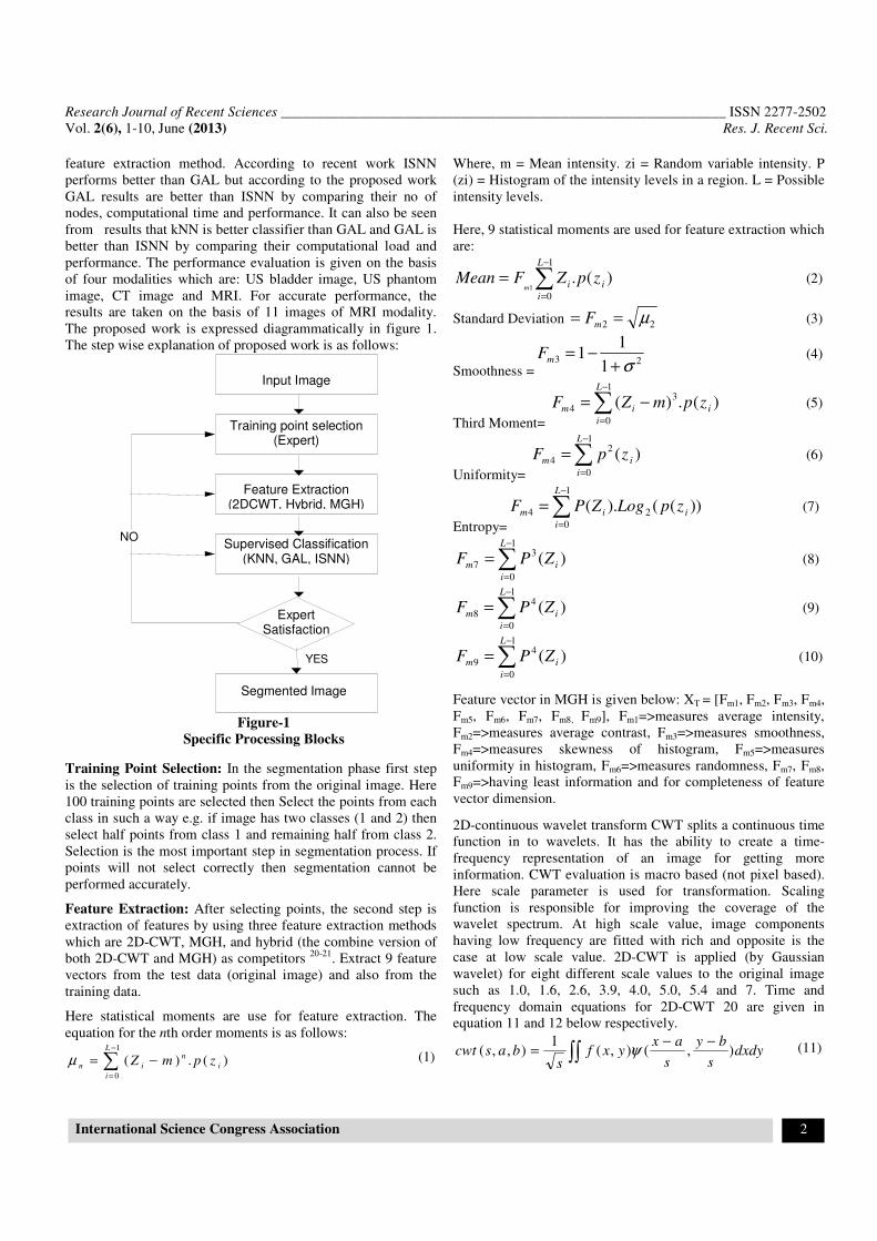
The proposed work is expressed diagrammatically in figure 1.
The step wise explanation of proposed work is as follows:
Figure-1
Specific Processing Blocks
Training Point Selection: In the segmentation phase first step
is the selection of training points from the original image. Here
100 training points are selected then Select the points from each
class in such a way e.g. if image has two classes (1 and 2) then
select half points from class 1 and remaining half from class 2.
Selection is the most important step in segmentation process. If
points will not select correctly then segmentation cannot be
performed accurately.
Feature Extraction: After selecting points, the second step is
extraction of features by using three feature extraction methods
which are 2D-CWT, MGH, and hybrid (the combine version of
both 2D-CWT and MGH) as competitors 20-21
. Extract 9 feature
vectors from the test data (original image) and also from the
training data.
Here statistical moments are use for feature extraction. The
equation for the nth order moments is as follows:
)(.)(1
0
∑−
=
−=L
i
i
n
in zpmZµ (1)
Where, m = Mean intensity. zi = Random variable intensity. P
(zi) = Histogram of the intensity levels in a region. L = Possible
intensity levels.
Here, 9 statistical moments are used for feature extraction which
are:
)(.1
01∑
−
=
=L
i
ii zpZFMeanm
(2)
Standard Deviation 22 µ== mF (3)
Smoothness =23
1
11
σ+−=mF (4)
Third Moment=
)(.)(1
0
3
4 ∑−
=
−=L
i
iim zpmZF (5)
Uniformity=
)(1
0
2
4 ∑−
=
=L
i
im zpF (6)
Entropy=
))(().(1
0
24 ∑−
=
=L
i
iim zpLogZPF (7)
)(1
0
3
7 ∑−
=
=L
i
im ZPF (8)
)(1
0
4
8 ∑−
=
=L
i
im ZPF (9)
)(1
0
4
9 ∑−
=
=L
i
im ZPF (10)
Feature vector in MGH is given below: XT = [Fm1, Fm2, Fm3, Fm4,
Fm5, Fm6, Fm7, Fm8, Fm9], Fm1=>measures average intensity,
Fm2=>measures average contrast, Fm3=>measures smoothness,
Fm4=>measures skewness of histogram, Fm5=>measures
uniformity in histogram, Fm6=>measures randomness, Fm7, Fm8,
Fm9=>having least information and for completeness of feature
vector dimension.
2D-continuous wavelet transform CWT splits a continuous time
function in to wavelets. It has the ability to create a time-
frequency representation of an image for getting more
information. CWT evaluation is macro based (not pixel based).
Here scale parameter is used for transformation. Scaling
function is responsible for improving the coverage of the
wavelet spectrum. At high scale value, image components
having low frequency are fitted with rich and opposite is the
case at low scale value. 2D-CWT is applied (by Gaussian
wavelet) for eight different scale values to the original image
such as 1.0, 1.6, 2.6, 3.9, 4.0, 5.0, 5.4 and 7. Time and
frequency domain equations for 2D-CWT 20 are given in
equation 11 and 12 below respectively.
dxdys
by
s
axyxf
sbascwt ),(),(
1),,(
−−= ∫∫ ψ (11)
Input Image
Training point selection (Expert)
Feature Extraction (2DCWT, Hybrid, MGH)
Supervised Classification (KNN, GAL, ISNN)
Expert Satisfaction
NO
Segmented Image
YES
Research Journal of Recent Sciences ______________________________________________________________ ISSN 2277-2502
Vol. 2(6), 1-10, June (2013) Res. J. Recent Sci.
International Science Congress Association 3
)2,1()2,1()2,1,( swswwwFswwscwt Φ= (12)
In above equations 11 and 12, ‘a’ and ‘b’ are translation
parameters and ‘s’ is a scale parameter for wavelet Ψ. Whereas,
‘x’ and ‘y’ are spatial domain coordinates and w1, w2 are
frequency domain coordinates. Hybrid features are formed by
combining both 2D-CWT and MGH features. Nine dimensional
hybrid features vector is formed by combining first five features
from 2D-CWT and remaining four features from MGH features.
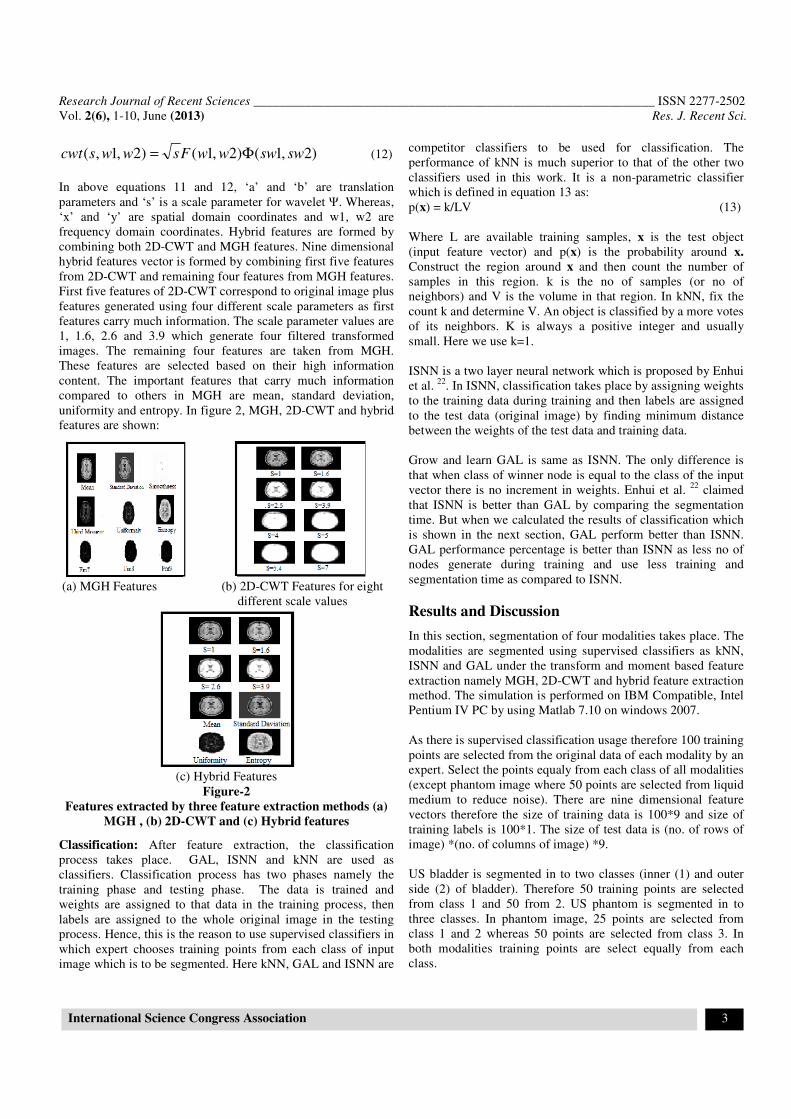
First five features of 2D-CWT correspond to original image plus
features generated using four different scale parameters as first
features carry much information. The scale parameter values are
1, 1.6, 2.6 and 3.9 which generate four filtered transformed
images. The remaining four features are taken from MGH.
These features are selected based on their high information
content. The important features that carry much information
compared to others in MGH are mean, standard deviation,
uniformity and entropy. In figure 2, MGH, 2D-CWT and hybrid
features are shown:
(a) MGH Features (b) 2D-CWT Features for eight
different scale values
(c) Hybrid Features
Figure-2
Features extracted by three feature extraction methods (a)
MGH , (b) 2D-CWT and (c) Hybrid features
Classification: After feature extraction, the classification
process takes place. GAL, ISNN and kNN are used as
classifiers. Classification process has two phases namely the
training phase and testing phase. The data is trained and
weights are assigned to that data in the training process, then
labels are assigned to the whole original image in the testing
process. Hence, this is the reason to use supervised classifiers in
which expert chooses training points from each class of input
image which is to be segmented. Here kNN, GAL and ISNN are
competitor classifiers to be used for classification. The
performance of kNN is much superior to that of the other two
classifiers used in this work. It is a non-parametric classifier
which is defined in equation 13 as:
p(x) = k/LV (13)
Where L are available training samples, x is the test object
(input feature vector) and p(x) is the probability around x.
Construct the region around x and then count the number of
samples in this region. k is the no of samples (or no of
neighbors) and V is the volume in that region. In kNN, fix the
count k and determine V. An object is classified by a more votes
of its neighbors. K is always a positive integer and usually
small. Here we use k=1.
ISNN is a two layer neural network which is proposed by Enhui
et al. 22
. In ISNN, classification takes place by assigning weights
to the training data during training and then labels are assigned
to the test data (original image) by finding minimum distance
between the weights of the test data and training data.
Grow and learn GAL is same as ISNN. The only difference is
that when class of winner node is equal to the class of the input
vector there is no increment in weights. Enhui et al. 22
claimed
that ISNN is better than GAL by comparing the segmentation
time. But when we calculated the results of classification which
is shown in the next section, GAL perform better than ISNN.
GAL performance percentage is better than ISNN as less no of
nodes generate during training and use less training and
segmentation time as compared to ISNN.
Results and Discussion
In this section, segmentation of four modalities takes place. The
modalities are segmented using supervised classifiers as kNN,
ISNN and GAL under the transform and moment based feature
extraction namely MGH, 2D-CWT and hybrid feature extraction
method. The simulation is performed on IBM Compatible, Intel
Pentium IV PC by using Matlab 7.10 on windows 2007.
As there is supervised classification usage therefore 100 training
points are selected from the original data of each modality by an
expert. Select the points equaly from each class of all modalities
(except phantom image where 50 points are selected from liquid
medium to reduce noise). There are nine dimensional feature
vectors therefore the size of training data is 100*9 and size of
training labels is 100*1. The size of test data is (no. of rows of
image) *(no. of columns of image) *9.
US bladder is segmented in to two classes (inner (1) and outer
side (2) of bladder). Therefore 50 training points are selected
from class 1 and 50 from 2. US phantom is segmented in to
three classes. In phantom image, 25 points are selected from
class 1 and 2 whereas 50 points are selected from class 3. In
both modalities training points are select equally from each
class.
Research Journal of Recent Sciences ______________________________________________________________ ISSN 2277-2502
Vol. 2(6), 1-10, June (2013) Res. J. Recent Sci.
International Science Congress Association 4
(a) (b)
(c) (d)
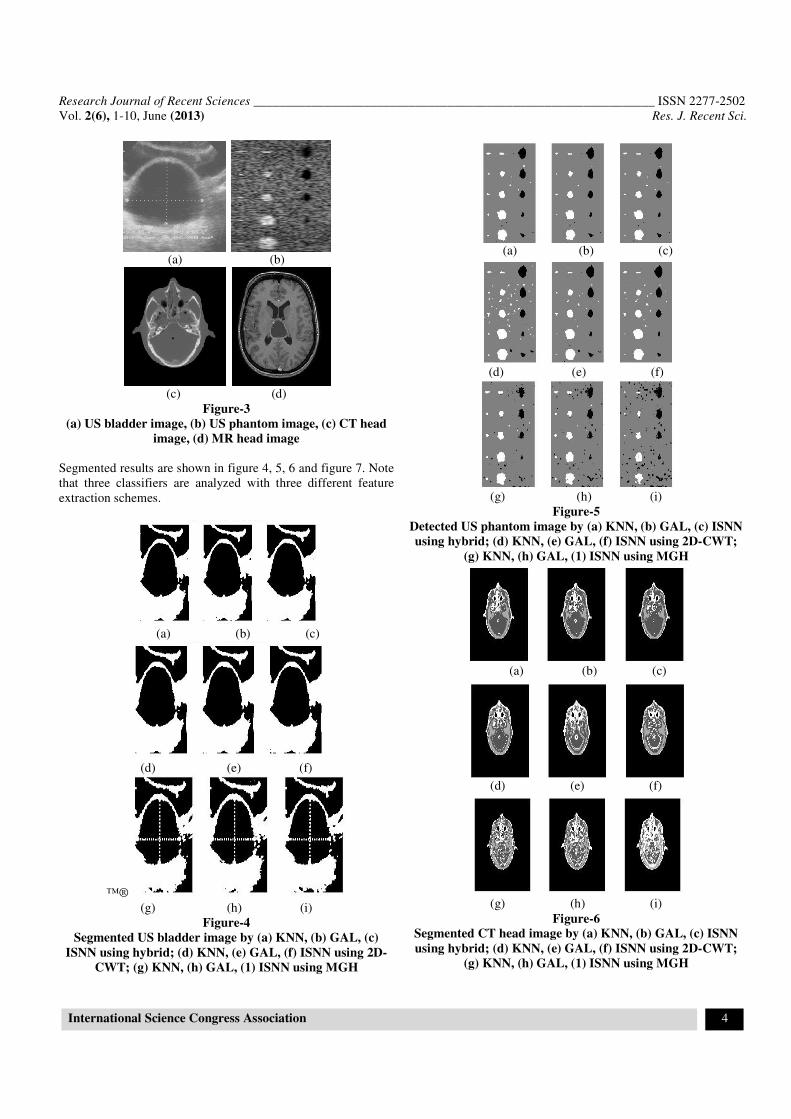
Figure-3
(a) US bladder image, (b) US phantom image, (c) CT head
image, (d) MR head image
Segmented results are shown in figure 4, 5, 6 and figure 7. Note
that three classifiers are analyzed with three different feature
extraction schemes.
(a) (b) (c)
(d) (e) (f)
™®
(g) (h) (i)
Figure-4
Segmented US bladder image by (a) KNN, (b) GAL, (c)
ISNN using hybrid; (d) KNN, (e) GAL, (f) ISNN using 2D-
CWT; (g) KNN, (h) GAL, (1) ISNN using MGH
(a) (b) (c)
(d) (e) (f)
(g) (h) (i)
Figure-5
Detected US phantom image by (a) KNN, (b) GAL, (c) ISNN
using hybrid; (d) KNN, (e) GAL, (f) ISNN using 2D-CWT;
(g) KNN, (h) GAL, (1) ISNN using MGH
(a) (b) (c)
(d) (e) (f)
(g) (h) (i)
Figure-6
Segmented CT head image by (a) KNN, (b) GAL, (c) ISNN
using hybrid; (d) KNN, (e) GAL, (f) ISNN using 2D-CWT;
(g) KNN, (h) GAL, (1) ISNN using MGH

Research Journal of Recent Sciences ______________________________________________________________ ISSN 2277-2502
Vol. 2(6), 1-10, June (2013) Res. J. Recent Sci.
International Science Congress Association 5
(a) (b) (c)
(d) (e) (f)
(g) (H) (i)
Figure-7
Segmented MR head image by (a) KNN, (b) GAL, (c) ISNN
using hybrid; (d) KNN, (e) GAL, (f) ISNN using 2D-CWT;
(g) KNN, (h) GAL, (1) ISNN using MGH
Comparison of GAL and ISNN under MGH, 2D-CWT and
Hybrid at different iterations: The test images are segmented
using GAL and ISNN at different iterations. In the existing
work, iteration no of GAL and ISNN were selected as 2000, and
10,000 respectively 22
. Therefore GAL and ISNN were not
performing efficiently as too much computational time were
consuming. Therefore after testing the results at different
iterations, the suggested and tested iteration would be 20, 50 and
100 for both GAL and ISNN because at these iterations, GAL
and ISNN are efficiently perform and use less training and
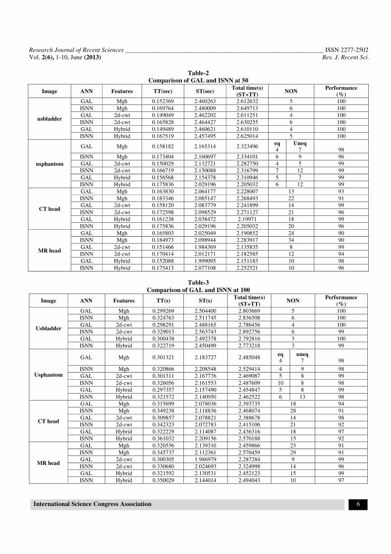
segmentation time. Table 1 , 2 and 3 shows the comparison of
GAL and ISNN at 20 , 50 and 100 iterations.
From the above tables it can be observed that the iteration no.
20, 50 and 100, results are more accurate. It is also observed
that, the GAL is better than ISNN using all selected iterations as
GAL generates less number of nodes than ISNN. Therefore
GAL has good performance and low computational time than
ISNN under different feature extraction schemes.
Performance analysis of KNN, GAL, ISNN under MGH,
2D-CWT, hybrid: The Performance results of segmentation for
all four modalities are evaluated on the basis of one image. In
the next section for identify more real performance of MR
image we evaluate performance on the base of 11 MR images.
Performance is the percentage comes by comparing training
labels with testing labels. Here we also compare each classifier
and feature extraction methods by comparing performance,
computational time (both training time and segmentation time).
Table-1
Comparison of GAL and ISNN at 20
Image ANN Features TT(sec) ST(sec) Total time(s)
(ST+TT) NON
Performance
(%)
usbladder
GAL Mgh 0.061746 2.495491 2.557237 6 100
ISNN Mgh 0.073545 2.502339 2.575884 5 100
GAL 2d-cwt 0.061578 2.448884 2.510462 3 100
ISNN 2d-cwt 0.073083 2.467418 2.540501 3 100
GAL Hybrid 0.061922 2.585885 2.647807 4 100
ISNN Hybrid 0.073207 2.629772 2.702979 5 100
usphantom
GAL Mgh 0.062149 2.210711 2.272860 eq 5
Uneq 8
98
ISNN Mgh 0.072470 2.136215 2.208685 6 8 96
GAL 2d-cwt 0.062934 2.238110 2.301044 5 6 99
ISNN 2d-cwt 0.073083 2.293207 2.366290 6 12 99
GAL Hybrid 0.062992 2.184066 2.247058 5 9 99
ISNN Hybrid 0.073174 2.177684 2.257058 6 11 99
CT head
GAL Mgh 0.068176 2.132186 2.200362 19 93
ISNN Mgh 0.092177 2.177600 2.269777 21 93
GAL 2d-cwt 0.064881 2.041324 2.106205 18 99
ISNN 2d-cwt 0.078795 2.062013 2.140808 19 96
GAL Hybrid 0.064251 2.028647 2.092898 15 97
ISNN Hybrid 0.076433 2.086171 2.162604 20 97
M head
GAL Mgh 0.065476 2.086582 2.152058 23 93
ISNN Mgh 0.079302 2.122091 2.201393 29 91
GAL 2d-cwt 0.063322 2.006598 2.069920 15 99
ISNN 2d-cwt 0.073378 1.993947 2.067325 15 97
GAL Hybrid 0.063661 2.024473 2.088134 9 98
ISNN Hybrid 0.075210 2.042749 2.117959 10 96
Research Journal of Recent Sciences ______________________________________________________________ ISSN 2277-2502
Vol. 2(6), 1-10, June (2013) Res. J. Recent Sci.
International Science Congress Association 6
Table-2
Comparison of GAL and ISNN at 50
Image ANN Features TT(sec) ST(sec) Total time(s)
(ST+TT) NON
Performance
(%)
usbladder
GAL Mgh 0.152369 2.460263 2.612632 5 100
ISNN Mgh 0.169764 2.480009 2.649713 6 100
GAL 2d-cwt 0.149049 2.462202 2.611251 4 100
ISNN 2d-cwt 0.165828 2.464427 2.630255 6 100
GAL Hybrid 0.149489 2.460621 2.610110 4 100
ISNN Hybrid 0.167519 2.457495 2.625014 5 100
usphantom
GAL Mgh 0.158182 2.165314 2.323496 eq 4
Uneq 7
98
ISNN Mgh 0.173404 2.160697 2.334101 6 9 96
GAL 2d-cwt 0.150029 2.132721 2.282750 4 5 99
ISNN 2d-cwt 0.166719 2.150088 2.316799 7 12 99
GAL Hybrid 0.156568 2.154378 2.310946 5 7 99
ISNN Hybrid 0.175836 2.029196 2.205032 6 12 99
CT head
GAL Mgh 0.163830 2.064177 2.228007 13 93
ISNN Mgh 0.183346 2.085147 2.268493 22 91
GAL 2d-cwt 0.158120 2.083779 2.241899 14 99
ISNN 2d-cwt 0.172598 2.098529 2.271127 21 96
GAL Hybrid 0.161238 2.038472 2.19971 18 99
ISNN Hybrid 0.175836 2.029196 2.205032 20 96
MR head
GAL Mgh 0.165803 2.025049 2.190852 24 90
ISNN Mgh 0.184973 2.098944 2.283917 34 90
GAL 2d-cwt 0.151466 1.984369 2.135835 8 99
ISNN 2d-cwt 0.170414 2.012171 2.182585 12 94
GAL Hybrid 0.152088 1.999095 2.151183 10 98
ISNN Hybrid 0.175413 2.077108 2.252521 10 96
Table-3
Comparison of GAL and ISNN at 100
Image ANN Features TT(s) ST(s) Total time(s)
(ST+TT) NON
Performance
(%)
Usbladder
GAL Mgh 0.299269 2.504400 2.803669 5 100
ISNN Mgh 0.324763 2.511745 2.836508 6 100
GAL 2d-cwt 0.298291 2.488165 2.786456 4 100
ISNN 2d-cwt 0.329013 2.563743 2.892756 6 99
GAL Hybrid 0.300438 2.492378 2.792816 3 100
ISNN Hybrid 0.322719 2.450499 2.773218 3 99
Usphantom
GAL Mgh 0.301321 2.183727 2.485048 eq 4
uneq 7
98
ISNN Mgh 0.320866 2.208548 2.529414 4 9 98
GAL 2d-cwt 0.301311 2.167776 2.469087 5 8 99
ISNN 2d-cwt 0.326056 2.161553 2.487609 10 8 98
GAL Hybrid 0.297357 2.157490 2.454847 5 8 99
ISNN Hybrid 0.321572 2.140950 2.462522 6 13 98
CT head
GAL Mgh 0.315699 2.078036 2.393735 18 94
ISNN Mgh 0.349238 2.118836 2.468074 28 91
GAL 2d-cwt 0.309857 2.078821 2.388678 14 98
ISNN 2d-cwt 0.342323 2.072783 2.415106 21 92
GAL Hybrid 0.322229 2.114087 2.436316 18 97
ISNN Hybrid 0.361032 2.209156 2.570188 15 92
MR head
GAL Mgh 0.320556 2.139310 2.459866 23 91
ISNN Mgh 0.345737 2.112361 2.570459 29 91
GAL 2d-cwt 0.300305 1.986979 2.287284 9 99
ISNN 2d-cwt 0.330680 2.024693 2.324998 14 96
GAL Hybrid 0.321592 2.130531 2.452123 15 99
ISNN Hybrid 0.350029 2.144014 2.494043 10 97
Research Journal of Recent Sciences ______________________________________________________________ ISSN 2277-2502
Vol. 2(6), 1-10, June (2013) Res. J. Recent Sci.
International Science Congress Association 7
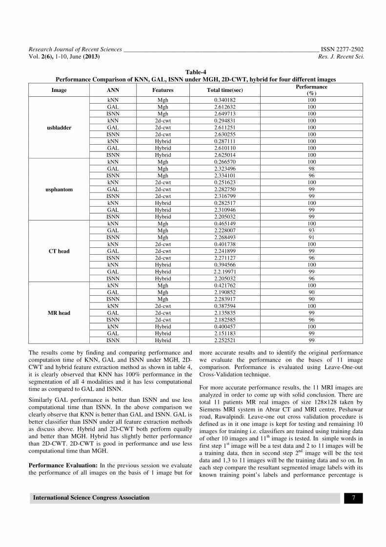
Table-4
Performance Comparison of KNN, GAL, ISNN under MGH, 2D-CWT, hybrid for four different images
Image ANN Features Total time(sec) Performance
(%)
usbladder
kNN Mgh 0.340182 100
GAL Mgh 2.612632 100
ISNN Mgh 2.649713 100
kNN 2d-cwt 0.294831 100
GAL 2d-cwt 2.611251 100
ISNN 2d-cwt 2.630255 100
kNN Hybrid 0.287111 100
GAL Hybrid 2.610110 100
ISNN Hybrid 2.625014 100
usphantom
kNN Mgh 0.266570 100
GAL Mgh 2.323496 98
ISNN Mgh 2.334101 96
kNN 2d-cwt 0.251623 100
GAL 2d-cwt 2.282750 99
ISNN 2d-cwt 2.316799 99
kNN Hybrid 0.282517 100
GAL Hybrid 2.310946 99
ISNN Hybrid 2.205032 99
CT head
kNN Mgh 0.465149 100
GAL Mgh 2.228007 93
ISNN Mgh 2.268493 91
kNN 2d-cwt 0.401738 100
GAL 2d-cwt 2.241899 99
ISNN 2d-cwt 2.271127 96
kNN Hybrid 0.394566 100
GAL Hybrid 2.2.19971 99
ISNN Hybrid 2.205032 96
MR head
kNN Mgh 0.421762 100
GAL Mgh 2.190852 90
ISNN Mgh 2.283917 90
kNN 2d-cwt 0.387594 100
GAL 2d-cwt 2.135835 99
ISNN 2d-cwt 2.182585 96
kNN Hybrid 0.400457 100
GAL Hybrid 2.151183 99
ISNN Hybrid 2.252521 99
The results come by finding and comparing performance and
computation time of KNN, GAL and ISNN under MGH, 2D-
CWT and hybrid feature extraction method as shown in table 4,
it is clearly observed that KNN has 100% performance in the
segmentation of all 4 modalities and it has less computational
time as compared to GAL and ISNN.
Similarly GAL performance is better than ISNN and use less
computational time than ISNN. In the above comparison we
clearly observe that KNN is better than GAL and ISNN. GAL is
better classifier than ISNN under all feature extraction methods
as discuss above. Hybrid and 2D-CWT both perform equally
and better than MGH. Hybrid has slightly better performance
than 2D-CWT. 2D-CWT is good in performance and use less
computational time than MGH.
Performance Evaluation: In the previous session we evaluate
the performance of all images on the basis of 1 image but for
more accurate results and to identify the original performance
we evaluate the performance on the bases of 11 image
comparison. Performance is evaluated using Leave-One-out
Cross-Validation technique.
For more accurate performance results, the 11 MRI images are
analyzed in order to come up with solid conclusion. There are
total 11 patients MR real images of size 128×128 taken by
Siemens MRI system in Abrar CT and MRI centre, Peshawar
road, Rawalpindi. Leave-one out cross validation procedure is
defined as in it one image is kept for testing and remaining 10
images for training i.e. classifiers are trained using training data
of other 10 images and 11th
image is tested. In simple words in
first step 1st image will be a test data and 2 to 11
images will be
a training data, then in second step 2nd
image will be the test
data and 1,3 to 11 images will be the training data and so on. In
each step compare the resultant segmented image labels with its
known training point’s labels and performance percentage is
Research Journal of Recent Sciences ______________________________________________________________ ISSN 2277-2502
Vol. 2(6), 1-10, June (2013) Res. J. Recent Sci.
International Science Congress Association 8
calculated for each image. Then for each image we would have
classifier’s performance.
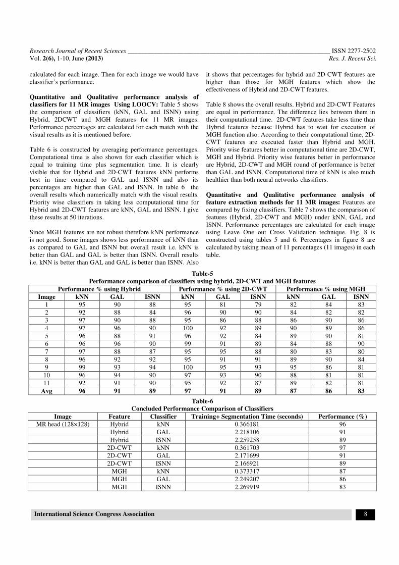
Quantitative and Qualitative performance analysis of
classifiers for 11 MR images Using LOOCV: Table 5 shows
the comparison of classifiers (kNN, GAL and ISNN) using
Hybrid, 2DCWT and MGH features for 11 MR images.
Performance percentages are calculated for each match with the
visual results as it is mentioned before.
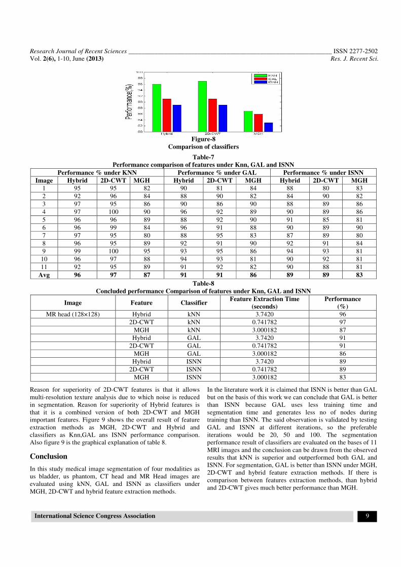
Table 6 is constructed by averaging performance percentages.
Computational time is also shown for each classifier which is
equal to training time plus segmentation time. It is clearly
visible that for Hybrid and 2D-CWT features kNN performs
best in time compared to GAL and ISNN and also its
percentages are higher than GAL and ISNN. In table 6 the
overall results which numerically match with the visual results.
Priority wise classifiers in taking less computational time for
Hybrid and 2D-CWT features are kNN, GAL and ISNN. I give
these results at 50 iterations.
Since MGH features are not robust therefore kNN performance
is not good. Some images shows less performance of kNN than
as compared to GAL and ISNN but overall result i.e. kNN is
better than GAL and GAL is better than ISNN. Overall results
i.e. kNN is better than GAL and GAL is better than ISNN. Also
it shows that percentages for hybrid and 2D-CWT features are
higher than those for MGH features which show the
effectiveness of Hybrid and 2D-CWT features.
Table 8 shows the overall results. Hybrid and 2D-CWT Features
are equal in performance. The difference lies between them in
their computational time. 2D-CWT features take less time than
Hybrid features because Hybrid has to wait for execution of
MGH function also. According to their computational time, 2D-
CWT features are executed faster than Hybrid and MGH.
Priority wise features better in computational time are 2D-CWT,
MGH and Hybrid. Priority wise features better in performance
are Hybrid, 2D-CWT and MGH round of performance is better
than GAL and ISNN. Computational time of kNN is also much
healthier than both neural networks classifiers.
Quantitative and Qualitative performance analysis of
feature extraction methods for 11 MR images: Features are
compared by fixing classifiers. Table 7 shows the comparison of
features (Hybrid, 2D-CWT and MGH) under kNN, GAL and
ISNN. Performance percentages are calculated for each image
using Leave One out Cross Validation technique. Fig. 8 is
constructed using tables 5 and 6. Percentages in figure 8 are
calculated by taking mean of 11 percentages (11 images) in each
table.
Table-5
Performance comparison of classifiers using hybrid, 2D-CWT and MGH features
Performance % using Hybrid Performance % using 2D-CWT Performance % using MGH
Image kNN GAL ISNN kNN GAL ISNN kNN GAL ISNN
1 95 90 88 95 81 79 82 84 83
2 92 88 84 96 90 90 84 82 82
3 97 90 88 95 86 88 86 90 86
4 97 96 90 100 92 89 90 89 86
5 96 88 91 96 92 84 89 90 81
6 96 96 90 99 91 89 84 88 90
7 97 88 87 95 95 88 80 83 80
8 96 92 92 95 91 91 89 90 84
9 99 93 94 100 95 93 95 86 81
10 96 94 90 97 93 90 88 81 81
11 92 91 90 95 92 87 89 82 81
Avg 96 91 89 97 91 89 87 86 83
Table-6
Concluded Performance Comparison of Classifiers
Image Feature Classifier Training+ Segmentation Time (seconds) Performance (%)
MR head (128×128) Hybrid kNN 0.366181 96
Hybrid GAL 2.218106 91
Hybrid ISNN 2.259258 89
2D-CWT kNN 0.361703 97
2D-CWT GAL 2.171699 91
2D-CWT ISNN 2.166921 89
MGH kNN 0.373317 87
MGH GAL 2.249207 86
MGH ISNN 2.269919 83
Research Journal of Recent Sciences ______________________________________________________________ ISSN 2277-2502
Vol. 2(6), 1-10, June (2013) Res. J. Recent Sci.
International Science Congress Association 9
Figure-8
Comparison of classifiers
Table-7
Performance comparison of features under Knn, GAL and ISNN
Performance % under KNN Performance % under GAL Performance % under ISNN
Image Hybrid 2D-CWT MGH Hybrid 2D-CWT MGH Hybrid 2D-CWT MGH
1 95 95 82 90 81 84 88 80 83
2 92 96 84 88 90 82 84 90 82
3 97 95 86 90 86 90 88 89 86
4 97 100 90 96 92 89 90 89 86
5 96 96 89 88 92 90 91 85 81
6 96 99 84 96 91 88 90 89 90
7 97 95 80 88 95 83 87 89 80
8 96 95 89 92 91 90 92 91 84
9 99 100 95 93 95 86 94 93 81
10 96 97 88 94 93 81 90 92 81
11 92 95 89 91 92 82 90 88 81
Avg 96 97 87 91 91 86 89 89 83
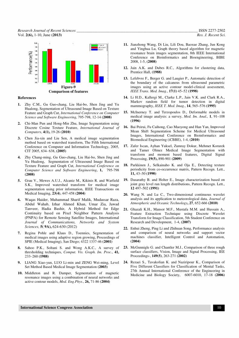
Table-8
Concluded performance Comparison of features under Knn, GAL and ISNN
Image Feature Classifier Feature Extraction Time
(seconds)
Performance
(%)
MR head (128×128) Hybrid kNN 3.7420 96
2D-CWT kNN 0.741782 97
MGH kNN 3.000182 87
Hybrid GAL 3.7420 91
2D-CWT GAL 0.741782 91
MGH GAL 3.000182 86
Hybrid ISNN 3.7420 89
2D-CWT ISNN 0.741782 89
MGH ISNN 3.000182 83
Reason for superiority of 2D-CWT features is that it allows
multi-resolution texture analysis due to which noise is reduced
in segmentation. Reason for superiority of Hybrid features is
that it is a combined version of both 2D-CWT and MGH
important features. Figure 9 shows the overall result of feature
extraction methods as MGH, 2D-CWT and Hybrid and
classifiers as Knn,GAL ans ISNN performance comparison.
Also figure 9 is the graphical explanation of table 8.
Conclusion
In this study medical image segmentation of four modalities as
us bladder, us phantom, CT head and MR Head images are
evaluated using kNN, GAL and ISNN as classifiers under
MGH, 2D-CWT and hybrid feature extraction methods.
In the literature work it is claimed that ISNN is better than GAL
but on the basis of this work we can conclude that GAL is better
than ISNN because GAL uses less training time and
segmentation time and generates less no of nodes during
training than ISNN. The said observation is validated by testing
GAL and ISNN at different iterations, so the preferable
iterations would be 20, 50 and 100. The segmentation
performance result of classifiers are evaluated on the bases of 11
MRI images and the conclusion can be drawn from the observed
results that kNN is superior and outperformed both GAL and
ISNN. For segmentation, GAL is better than ISNN under MGH,
2D-CWT and hybrid feature extraction methods. If there is
comparison between features extraction methods, than hybrid
and 2D-CWT gives much better performance than MGH.
Research Journal of Recent Sciences ______________________________________________________________ ISSN 2277-2502
Vol. 2(6), 1-10, June (2013) Res. J. Recent Sci.
International Science Congress Association 10
Figure-9
Comparison of features
References
1. Zhy C.M., Gu Guo-chang, Liu Hai-bo, Shen Jing and Yu
Hualong, Segmentation of Ultrasound Image Based on Texture
Feature and Graph Cut, International Conference on Computer
Science and Software Engineering, 795-798, 12-14 (2008)
2. Chi-Man Pun and Hong-Min Zhu, Image Segmentation using
Discrete Cosine Texture Feature, International Journal of
Computers, 4(1), 19-26 (2010)
3. Chen Jia-xin and Liu Sen, A medical image segmentation
method based on watershed transform, The Fifth International
Conference on Computer and Information Technology, 2005, CIT 2005, 634- 638, (2005)
4. Zhy Chang-ming, Gu Guo-chang, Liu Hai-bo, Shen Jing and
Yu Hualong, Segmentation of Ultrasound Image Based on
Texture Feature and Graph Cut, International Conference on
Computer Science and Software Engineering, 1, 795-798
(2008)
5. Grau V., Mewes A.U.J., Alcaniz M., Kikinis R. and Warfield
S.K., Improved watershed transform for medical image
segmentation using prior information, IEEE Transactions on
Medical Imaging, 23(4), 447-458 (2004)
6. Waqas Haider, Muhammad Sharif Malik, Mudassar Raza,
Abdul Wahab, Izhar Ahmed Khan, Umar Zia, Jawad
Tanveer, Hadia Bashir, A Hybrid Method for Edge
Continuity based on Pixel Neighbor Pattern Analysis
(PNPA) for Remote Sensing Satellite Images, International
Journal of Communications, Networks and System
Sciences, 5( 9A), 624-630 (2012)
7. Regina Pohle and Klaus D., Toennies, Segmentation of
medical images using adaptive region growing, Proceedings of SPIE (Medical Imaging), San Diego; 4322 1337-46 (2001)
8. Sahoo P.K., Soltani S. and Wong A.K.C., A survey of
thresholding techniques, Comput. Vis. Graph. Im. Proc., 41,
233–260 (1988)
9. LIANG Xiao-yun, LUO Li-min and ZENG Wei-ming, Level
Set Method Based Medical Image Segmentation (2003)
10. Middleton and R. Damper, Segmentation of magnetic
resonance images using a combination of neural networks and active contour models, Med. Eng.Phys., 26, 71-86 (2004)
11. Jianzhong Wang, Di Liu, Lili Dou, Baoxue Zhang, Jun Kong
and Yinghua Lu, Graph theory based algorithm for magnetic
resonance brain images segmentation, 8th IEEE International
Conference on Bioinformatics and Bioengineering, BIBE 2008, 1-5, (2008)
12. Jain A.K. and Dubes R.C., Algorithms for clustering data, Prentice Hall, (1988)
13. Lefebvre F., Berger G. and Laugier P., Automatic detection of
the boundary of the calcaneus from ultrasound parametric
images using an active contour model-clinical assessment, IEEE Trans. Med. Imag., 17(1) 45–52 (1998)
14. Li H.D., Kallergi M., Clarke L.P., Jain V.K. and Clark R.A.,
Markov random field for tumor detection in digital
mammography, IEEE T. Med. Imag., 14, 565–576 (1995)
15. McInerney T. and Terzopoulos D., Deformable models in
medical image analysis: a survey, Med. Im. Anal., 1, 91–108
(1996)
16. Bai Peirui, Fu Caihong, Cao Maoyong and Han Yan, Improved
Mean Shift Segmentation Scheme for Medical Ultrasound
Images, International Conference on Bioinformatics and
Biomedical Engineering (iCBBE), 1-4, (2010)
17. Zafer Iscan, Ayhan Yuksel, Zumray Dokur, Mehmet Korurek
and Tamer Olmez Medical Image Segmentation with
transform and moment based features, Digital Signal
Processing, 19(5), 890-901 (2009)
18. Parkkinen J., Selkainaho K. and Oja E., Detecting texture
periodicity from co-occurrence matrix, Pattern Recogn. Lett.,
11, 43–50 (1990)
19. Dasarathy B. and Holter E., Image characterization based on
joint gray level run length distributions, Pattern Recogn. Lett., 12 497–502 (1991)
20. Wang N. and Lu C., Two-dimensional continuous wavelet
analysis and its application to meteorological data, Journal of
Atmospheric and Oceanic Technology, 27, 652-666 (2010)
21. Ghazali K.H., Mansor M.F., Mustafa M.M. and Hussain A.,
Feature Extraction Technique using Discrete Wavelet
Transform for Image Classification, 5th Student Conference on Research and Development, 1-4, (2007)
22. Enhui Zheng, Ping Li and Zhihuan Song, Performance analysis
and comparison of neural networks and support vector
machines classifier, Intelligent Control and Automation,
(2004)
23. McGunnigle G. and Chantler M.J., Comparison of three rough
surface classifiers, Vision, Image and Signal Processing, IEE
Proceedings-, 149(5), 263-271 (2002)
24. Rezaei S., Tavakolian K. and Naziripour K., Comparison of
Five Different Classifiers for Classification of Mental Tasks,
27th Annual International Conference of the Engineering in Medicine and Biology Society, 6007-6010, 17-18 (2006)
Top Related
Copyright © 2022 FDOKUMEN

