


Bahasa
Halaman
Hukum

Electrical Brain Imaging Reveals the Expression and Timing of AlteredError Monitoring Functions in Major Depression
Kristien Aarts and Marie-Anne VanderhasseltGhent University
Georges OtteP.C. Dr. Guislain, Ghent, Belgium
Chris Baeken and Gilles PourtoisGhent University
Major depressive disorder (MDD) is characterized by disturbances in affect, motivation, and cognitivecontrol processes, including error detection. However, the expression and timing of the impairmentsduring error monitoring remain unclear in MDD. The behavior and event-related brain responses (ERPs)of 20 patients with MDD were compared with those of 20 healthy controls (HCs), while they performeda Go/noGo task. Errors during this task were associated with 2 ERP components, the error-relatednegativity (ERN/Ne) and the error positivity (Pe). Results show that the ERN/Ne-correct-relatednegativity (CRN) amplitude difference was significantly larger in MDD patients (after controlling forspeed), compared with HCs, although MDD patients exhibited overactive medial frontal cortex (MFC)activation. By comparison, the subsequent Pe component was smaller in MDD patients compared withHCs and this effect was accompanied by a reduced activation of ventral anterior cingulate cortex (ACC)regions. These results suggest that MDD has multiple cascade effects on early error monitoring brainmechanisms.
Keywords: major depression, action-monitoring, error-related negativity (ERN), error positivity (Pe)
A prefrontal-limbic network dysregulation seems to be relatedto the onset and maintenance of MDD (Mayberg, 1997) althoughthis network has also been associated with the detection of re-sponse errors (Bush, Luu, & Posner, 2000; Pourtois et al., 2010;Seifert, von Cramon, Imperati, Tittgemeyer, & Ullsperger, 2011).In light of this evidence, error-monitoring functions should thus bedeficient in MDD. Consistent with this view, Holmes and Pizza-galli (2008) showed that depression is associated with an increasedactivation within midline prefrontal regions. They observed in-creased activity in the rostral Anterior Cingulate Cortex (rACC)and the medial Prefrontal Cortex (PFC) �80 ms after error com-mission, as well as a disrupted connectivity between the rACC andthe left dorsolateral PFC. In healthy controls (HCs), increasedACC activity predicted the activity in the left dlPFC �472 ms after
error commission. A similar relationship was not found for MDDpatients.
Event-related potential (ERP) experiments looking at error-monitoring in MDD have focused on two components: the error-related negativity (ERN/Ne) and the error positivity (Pe; Falken-stein, Hoormann, Christ, & Hohnsbein, 2000). The ERN/Ne is anegative deflection peaking �0 ms–50 ms following an incorrectresponse with a maximum amplitude over fronto-central midlinesites (Falkenstein, Hohnsbein, Hoormann, & Blanke, 1991; Geh-ring, Goss, Coles, Meyer, & Donchin, 1993). The ERN/Ne com-ponent is followed by a large positivity, the Pe. This componentreaches its maximum amplitude over centro-parietal scalp record-ings along the midline �200 ms–400 ms posterror onset (Falken-stein et al., 1991; Overbeek, Nieuwenhuis, & Ridderinkhof, 2005).Unlike the ERN/Ne, the Pe is thought to reflect a conscious stageof error detection (Nieuwenhuis, Ridderinkhof, Blow, Band, &Kok, 2001). Alternatively, it could reflect an affective appraisal oferrors (Falkenstein et al., 2000), a P300-like orienting response(Ridderinkhof, Ramautar, & Wijnen, 2009), or the accumulation ofevidence that an error has been committed (Steinhauser & Yeung,2010).
Each of the two error-related ERP components was shown tovary with MDD. However, mixed results were obtained regardingthe nature and direction of these MDD-related changes. Althoughsome studies found a larger ERN/Ne in MDD patients comparedwith HCs (Chiu & Deldin, 2007; Holmes & Pizzagalli, 2008;Holmes & Pizzagalli, 2010), other studies reported similar (Schri-jvers, de Bruijn, et al., 2008; Schrijvers et al., 2009) or smallerERN/Ne amplitudes in MDD patients (Ruchsow et al., 2006;Ruchsow et al., 2004). Likewise, discrepant findings have been
Kristien Aarts and Marie-Anne Vanderhasselt, Department of Experi-mental Clinical and Health Psychology, Ghent University, Ghent, Bel-gium; Georges Otte, P.C. Dr. Guislain, Ghent, Belgium; Chris Baeken,Department of Psychiatry, University Hospital, Ghent University; GillesPourtois, Department of Experimental Clinical and Health Psychology,Ghent University.
This work is supported by grants from the European Research Council(Starting Grant #200758) and Ghent University (BOF Grant #05Z01708).MAV is a postdoctoral fellow of the Research Foundation Flanders (FWO)(FWO08/PDO/168). We thank Monica Dhar for her feedback and sugges-tions on this article.
Correspondence concerning this article should be addressed to KristienAarts, Department of Experimental Clinical and Health Psychology, GhentUniversity, Henri Dunantlaan 2, 9000 Ghent, Belgium. E-mail: [email protected]
Thi
sdo
cum
ent
isco
pyri
ghte
dby
the
Am
eric
anPs
ycho
logi
cal
Ass
ocia
tion
oron
eof
itsal
lied
publ
ishe
rs.
Thi
sar
ticle
isin
tend
edso
lely
for
the
pers
onal
use
ofth
ein
divi
dual
user
and
isno
tto
bedi
ssem
inat
edbr
oadl
y.
Journal of Abnormal Psychology © 2013 American Psychological Association2013, Vol. 122, No. 4, 939–950 0021-843X/13/$12.00 DOI: 10.1037/a0034616
939
tapraid5/z2l-abnpsy/z2l-abnpsy/z2l00413/z2l2681d13z xppws S�1 11/22/13 8:04 Art: 2012-0766APA NLM
reported regarding amplitude variation of the Pe component. Al-though Chiu and Deldin (2007), and Holmes and Pizzagalli (2008)reported similar Pe amplitudes for HCs and MDD patients, Schri-jvers, de Bruijn et al. (2008) and Schrijvers et al. (2009) reportedsmaller Pe amplitudes in MDD patients compared with HCs.
An explanation for these discrepant findings may be that theamplitude of the ERP signal might not be different between MDDand HCs when measured at a few electrode positions only, butrather that the expression of the global electric field would bedifferent in depression, consistent with a change in the underlyingneural generators. However, these topographical changes are usu-ally difficult to capture using standard peak measurements (Pour-tois, Delplanque, Michel, & Vuilleumier, 2008). Hence, the ques-tion arises whether when using alternative data analyses, we couldfind evidence for a change in prefrontal and anterior cingulatebrain areas giving rise to the ERN/Ne and Pe components as afunction of MDD.
The goal of this ERP study was to better characterize possiblechanges in early error monitoring brain processes (with a focus onthe ERN/Ne and Pe components) in MDD patients. Using 128-channel EEG, the electrophysiological responses to commissionerrors performed during a Go/noGo task were compared betweenMDD patients and HCs. Because previous ERP studies reportedmixed results regarding amplitude modulations of the ERN/Necomponent as a function of depression (Chiu & Deldin, 2007;Compton et al., 2008; Holmes & Pizzagalli, 2008; Holmes &Pizzagalli, 2010; Schrijvers, de Bruijn et al. 2008; Schrijvers et al.,2009), we did not formulate a clear directional prediction regard-ing a possible change of the amplitude of the ERN/Ne in MDD.However, given that MDD is typically conceived as an internal-izing disorder (Mineka, Watson, & Clark, 1998) and because theERN/Ne is thought to be reliably enhanced or overactive in thisdisorder, we surmised that MDD patients might show a largerERN/Ne than HCs (Olvet & Hajcak, 2008). Moreover, it waspredicted that this effect might be associated with altered activitiesin MFC regions, including the dACC (see also Holmes & Pizza-galli, 2008). Regarding effects of MDD on the subsequent Pecomponent, no hypothesis was formulated because previous ERPstudies reported mixed results for amplitude variations of thismidlatency error-related activity as a function of depression (Chiu& Deldin, 2007; Holmes & Pizzagalli, 2008; Holmes & Pizzagalli,2010; Schrijvers, de Bruijn et al., 2008; Schrijvers et al., 2009).
Method
Participants
Twenty-three nondepressed HCs (18 females; mean age: 39,SEM � 3.04) and 25 individuals meeting the Diagnostic andStatistical Manual of Mental Disorders criteria (DSM–IV–TR;American Psychiatric Association, 2000) for MDD (15 females;mean age: 38, SEM � 2.55) participated in this study. The data ofeight participants had to be excluded because they did not commitenough errors (i.e., � 6; two HCs and five MDD patients) or theraw EEG data were contaminated by many artifacts (i.e., more than20% precluding the possibility to compute reliable ERP wave-forms; one HC). The demographic and clinical data of these eightparticipants were comparable with the ones of the participantseventually included in the study, all p values � .10. In total, the
data of 20 HCs (17 females; mean age: 39, SEM � 3.43) and 20MDD patients (10 females; mean age: 37, SEM � 2.85) wereincluded in the analyses. Demographic and clinical characteristicsare presented in Table 1.
The MDD outpatients were recruited from a psychiatric clinic.All patients were selected by a psychiatrist using the Mini-International Neuropsychiatric Interview (MINI; Sheehan et al.,1998), a structured clinical interview, and they were all diagnosedwith unipolar major depression of the melancholic subtype (ICD-9-CM code 296.23 and 296.33) according to the DSM–IV–TR(American Psychiatric Association, 2000). Severity of depressionwas assessed with the 17-item Hamilton Depression Rating Scale(HDRS; Hamilton, 1960) and the 21-item Beck Depression Inven-tory (BDI-II; Beck, Steer, & Brown, 1996). A psychiatrist rateddepression symptoms and severity (HDRS). Moreover, the Ham-ilton Rating Scale for Depression (HAM-D; Hamilton, 1960) andthe MINI, were administered again 1 week before testing toexamine the severity of the current MDD episode (HAM-D: M �28.65; SEM � 1.17; see Table 1). Finally, levels of depressionwere again verified at testing, using the Beck Depression Inventory(BDI-II; Beck, Steer, & Brown, 1996) and the HAM-D (Hamilton,1960). These scores confirmed that all patients who were previ-ously diagnosed as clinically depressed, were still found to beclinically depressed at the day of testing (see Table 1). Exclusioncriteria were (a) other mood disorders than MDD (comorbid anx-iety disorders were allowed; specific phobia: n � 1; posttraumaticstress disorder: n � 1; social anxiety: n � 1); (b) the use ofantipsychotics, tricyclic antidepressants, and/or long lasting ben-zodiazepines; (c) a history of neurological disorder, includingepilepsy, head injury, and loss of consciousness; (d) a history ofelectroconvulsive therapy; (e) alcohol abuse during the past year;(f) a past or present substance dependence; (g) past or presentexperience of psychotic episodes; and (h) learning disorders. Dur-ing the test session, all MDD participants were medicated witheither Selective Serotonin Reuptake Inhibitors (SSRI) or SelectiveNoradrenalin Reuptake Inhibitors (SNRI). Nine out of 20 patients
Table 1Descriptive Statistics for Healthy Controls (HC) and DepressedPatients (MDD)
HC MDDpM (SEM) M (SEM)
N 20 20Age 38.95 (3.43) 36.90 (2.85) .65Sex 3M/17F 10M/10F �.05Education� 2.06 (.20) 1.69 (.22) .22HAM_D 0.24 (0.14) 28.12 (1.33) �.001BDI_II 1.59 (0.97) 33.24 (2.87) �.001MDD with comorbid anxiety n � 3Treatment resistance�� n � 9Age at onset (n � 17) 30.76 (3.20)Length of episode (months; n � 17) 7.35 (1.38)Number of episodes (n � 17) 2.76 (.35)
Note. Note that for the age of onset, length of episode and number ofepisodes, the data reported are for 17 MDD patients. These data for threeMDD patients could not be accessed and saved for confidentiality reasons.� Education: 0 � primary school; 1 � 3 years of high school; 2 � 6 yearsof high school and 3 � higher education. �� Treatment resistance � hadtaken antidepressant medication for at least 7 months prior to testing.
Thi
sdo
cum
ent
isco
pyri
ghte
dby
the
Am
eric
anPs
ycho
logi
cal
Ass
ocia
tion
oron
eof
itsal
lied
publ
ishe
rs.
Thi
sar
ticle
isin
tend
edso
lely
for
the
pers
onal
use
ofth
ein
divi
dual
user
and
isno
tto
bedi
ssem
inat
edbr
oadl
y.
940 AARTS, VANDERHASSELT, OTTE, BAEKEN, AND POURTOIS
T1
tapraid5/z2l-abnpsy/z2l-abnpsy/z2l00413/z2l2681d13z xppws S�1 11/22/13 8:04 Art: 2012-0766APA NLM
were taking antidepressant medication for a duration of at least 7months before testing and could, therefore, be considered as beingtreatment resistant. However, despite this prolonged pharmacolog-ical treatment, they still met the criteria for MDD. HCs wererecruited using advertisements in newspapers and were free of anymedication. HCs reported that they had never been diagnosed withMDD or another psychiatric disorder prior to the EEG testing. Thiswas also verified by the MINI that was administered at testing.
All participants were Dutch speakers, gave their written in-formed consent, and received a compensation of 20 Euros. Thestudy was approved by the medical ethics committee of the GhentUniversity hospital.
Stimuli and Task
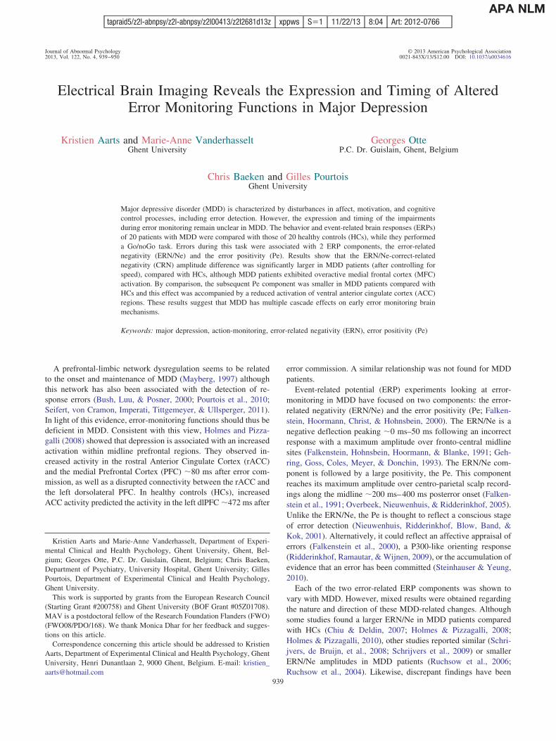
Participants performed a speeded Go/noGo task that was previ-ously used and validated (Figure 1; Vocat, Pourtois, & Vuil-leumier, 2008). Visual stimuli were shown on a 17-inch LCDscreen. They consisted of an arrow (11.4° � 0.05° visual angle ata 60 cm viewing distance) that was presented in the center of thescreen on a white background. Each trial started with a fixationcross that lasted for 1,000 ms. Then, a black arrow, oriented eitherup or down, was presented. After a variable interval (1,000 ms–2000 ms) the black arrow became either green or turquoise whileits orientation could either remain identical or shift in the oppositedirection. Participants were asked to perform a speeded color plusorientation discrimination task. When the black arrow turned greenand the orientation remained unchanged (two thirds of the trials),participants were instructed to press a predefined key on theresponse box as fast as possible with the index finger of theirdominant hand (Go trials). However, participants had to withholdresponding when either the arrow became green but changedorientation (one sixth of the trials), or when the arrow becameturquoise and kept its initial orientation (one sixth of the trials),enabling two types of noGo trials. For noGo trials, this color arrow
remained on the screen for a maximum duration of 1,000 ms.Instructions emphasized both speed and accuracy.
Given that the ERN/Ne amplitude varies according to the num-ber of errors (i.e., the ERN/Ne is larger when response errors arerare; see Gehring et al., 1993), it was important to avoid obviousgroup differences regarding error rate. Therefore, to ensure that thenumber of response errors was balanced between MDD patientsand HCs, a specific procedure was used to promote the occurrenceof fast RTs, and accordingly the commission of errors on noGotrials.
The experiment consisted of a practice block of 12 trials (fourGo, four noGo of each condition), three calibration blocks of 14trials (10 Go and two noGo of each type), and six test blocks of 60trials (40 Go trials and 20 noGo trials). Each calibration block wasfollowed by two test blocks. Trial presentation was randomizedwithin blocks. Stimulus presentation and response recording werecontrolled using E-prime software (V2.0., http://www.pstnet.com/products/e-prime/).
Analysis of Behavioral Data
RTs faster than 150 ms (Error: M � .79, SEM � .33; Hit: M �.33, SEM � .15) and slower than 800 ms (Error: M � 2.25, SEM �.91; Hit: M � 1.47, SEM � .36) were removed from the analyses.Next, RTs faster than M � 2.5 SD (Error: M � .14, SEM � .14; Hit:M � .01, SEM � .01) or slower than M � 2.5 SD (Error: M � 2.25,SEM � .43; Hit: M � 2.76, SEM � .16) were also excluded. Thenumber of outliers was not significantly different between HCs andMDD patients, all p � .10, except for RTs for Hits. MDDs reactedslower than 800 ms (M � 2.17, SEM � .65) more often than HCs(M � .77, SEM � .24) in the Hit condition. Color and orientationerrors were collapsed together (error condition) because there was nosignificant group difference regarding accuracy between these twoerror types, t � 1. A significant difference was observed in thenumber and reaction time (RT) speed between color and orientation
Figure 1. Stimuli and task. (A) On each trial, a black arrow was presented. After a variable interval (1000ms–2000 ms), the black arrow usually (two thirds, Go trials) became green and kept its initial orientation (eitherup or down). (B) On the remaining one third of the (noGo) trials, it became either turquoise and/or green but witha change in orientation (noGo trials).
Thi
sdo
cum
ent
isco
pyri
ghte
dby
the
Am
eric
anPs
ycho
logi
cal
Ass
ocia
tion
oron
eof
itsal
lied
publ
ishe
rs.
Thi
sar
ticle
isin
tend
edso
lely
for
the
pers
onal
use
ofth
ein
divi
dual
user
and
isno
tto
bedi
ssem
inat
edbr
oadl
y.
941ERROR-MONITORING IN MAJOR DEPRESSION
F1
COLOR
tapraid5/z2l-abnpsy/z2l-abnpsy/z2l00413/z2l2681d13z xppws S�1 11/22/13 8:04 Art: 2012-0766APA NLM
errors (accuracy: color errors: M � 10; SEM � 1.19; orientationerrors: M � 15; SEM � 1.57; t(39) � �5.95, p � .001; RT speed:color errors: M � 258 ms; SEM � 8.45; RT orientation errors: M �306; SEM � 11.94; t(37) � �4.95, p � .001). This result indicateda propensity to commit more false alarms with orientation changesthan color changes in this task. However, this effect was comparablefor MDD patients and HCs, F � 1. Fast and slow hits were col-lapsed and treated as a single condition (hit condition). MeanRTs for errors and hits as well as the number of errors and hitswere then computed and compared by means of 2 � 2 mixedanalyses of variance (ANOVAs), with group (HC vs. MDD) asbetween-subjects factor and accuracy (Error vs. Hit) as within-subject variable. Finally, the classical posterror slowing and pos-terror accuracy effects (Laming, 1979; Rabbitt, 1966) were calcu-lated to ascertain that in both groups errors were processedsimilarly as distinctive events, compared with hits.
EEG Recording
EEG was acquired at 512 Hz using a 128-channel BiosemiActive Two system (http://www.biosemi.com) referenced to theCommon Mode Sense (CMS) active electrode–Driven Right Leg(DRL) passive electrode. ERPs of interest were computed offlinefollowing a standard sequence of data transformations (Picton etal., 2000; (a) �500/�1,000 segmentation around the onset of theresponse; (b) preresponse interval baseline adjustment (from �500ms to response onset); (c) vertical ocular correction for blinks(Gratton, Coles, & Donchin, 1983), using the difference amplitudeof two electrodes attached above and below the left eye (nocorrection for horizontal eye movements was performed using thisprocedure; artifacts related to these horizontal eye movementswere removed manually during the artifact rejection step); (d)artifact rejection M � �87.25/�87.25, SEM � 2.24 amplitudescale (�V) across participants; no significant difference betweenHCs (M � 89.00, SEM � 2.98) and MDD patients (M � 85.50,SEM � 3.36), t � 1]; (e) averaging of trials, separately for eachgroup (HC vs. MDD) and experimental condition (errors vs. hits);and (f) 30 Hz low pass digital filtering of the individual averagedata.
Standard Peak Analyses
For each of the two error-related ERP deflections and for eachcondition, the area under the curve was calculated and analyzed(Picton et al., 2000). This was done during the 25 ms–55 msinterval post response onset at electrode FCz for the ERN/Ne amplitude,and during the 150 ms–210 ms interval postresponse onset atelectrode Cz for the Pe component. The selection of these twospecific scalp locations (and time windows) was based on thetopographic properties of the present dataset.
Statistical analyses were performed on the mean amplitude ofeach area using a 2 (accuracy: error vs. hit) � 2 (group) repeatedmeasures ANOVA, with the alpha cutoff set to p � .05.
Topographical Analyses
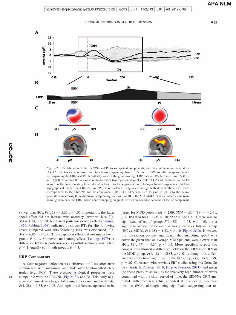
A complementing topographic mapping analysis of the ERPdata was performed (see Figure 2; Pourtois et al., 2008). Thisanalysis summarizes ERP data into a smaller number of dominant
field configurations, previously referred to as functional micro-states (Lehmann & Skrandies, 1980; Michel, Seeck, & Landis,1999). The rationale and basic principles of this temporal segmen-tation method have already been extensively described elsewhere(Michel et al., 1999; Murray, Brunet, & Michel, 2008; Pourtois etal., 2008). Following standard practice, a topographic patternanalysis was first performed on the grand-average ERP data from�55 ms until 379 ms after response onset (222 consecutive timeframes at 512 Hz sampling rate, encompassing the ERN/Ne and Pecomponents) using a standard K-means cluster method (Pascual-Marqui, 2002). The dominant scalp topographies (identified by theprevious analysis) were then fitted back to the ERP data of eachsubject using spatial fitting procedures to quantitatively determinetheir representation across subjects and conditions. GEV repre-sents the sum of the explained variance weighted by the GlobalField Power (GFP) at each moment in time. The resulting GEVvalues were entered in ANOVAs with two within-subject factors:accuracy (errors vs. hits) and map configuration (i.e., the dominantelectric field distributions identified by the spatial cluster analysis),as well as group (HC vs. MDD) as the between-subjects factor.These analyses were carried out using CARTOOL software (Ver-sion 3.34; developed by D. Brunet, Functional Brain MappingLaboratory, Geneva, Switzerland).
Source Localization Analyses
Finally, to estimate the neural generators underlying the domi-nant error-related electrical field configurations identified by theprevious analyses, a distributed linear inverse solution was used,namely standardized low-resolution brain electromagnetic tomog-raphy (sLORETA; Pascual-Marqui, 2002). SLORETA solutionsare computed within a three-shell spherical head model coregis-tered to the MNI152 template (Mazziotta et al., 2001). SLORETAestimates the three-dimensional (3D) intracerebral current densitydistribution in 6,239 voxels (5 mm resolution), each voxel con-taining an equivalent current dipole. This 3D solution space inwhich the inverse problem is solved, is restricted to the corticalgray matter. The head model for the inverse solution uses theelectric potential lead field computed with a boundary elementmethod applied to the MNI152 template (Fuchs, Kastner, Wagner,Hawes, & Ebersole, 2002). Scalp electrode coordinates on theMNI brain are derived from the international 5% system (Jurcak,Tsuzuki, & Dan, 2007). A direct comparison between the inversesolution results of MDD patients and HCs was performed sepa-rately for the ERN/Ne and Pe component, using unpaired t tests.To reveal group effects at the statistical level using a corrected p �.05 value, a stringent nonparametric randomization test (relying on5,000 iterations) was used. The calculation of all reconstructionparameters was based on the computed common average refer-ence.
Results
Behavior
Accuracy (errors vs. hits) and RT data are presented in Table 2.The number of errors was similar between MDD patients and HCs,t � 1. All participants were faster for errors than for hits,F(1, 38) � 43.21, p � .001, but overall, MDD patients reacted
Thi
sdo
cum
ent
isco
pyri
ghte
dby
the
Am
eric
anPs
ycho
logi
cal
Ass
ocia
tion
oron
eof
itsal
lied
publ
ishe
rs.
Thi
sar
ticle
isin
tend
edso
lely
for
the
pers
onal
use
ofth
ein
divi
dual
user
and
isno
tto
bedi
ssem
inat
edbr
oadl
y.
942 AARTS, VANDERHASSELT, OTTE, BAEKEN, AND POURTOIS
F2
T2
tapraid5/z2l-abnpsy/z2l-abnpsy/z2l00413/z2l2681d13z xppws S�1 11/22/13 8:04 Art: 2012-0766APA NLM
slower than HCs, F(1, 38) � 5.53, p � .05. Importantly, this latterspeed effect did not interact with accuracy (error vs. hit), F(1,38) � 1.13, p � .10. A classical posterror slowing effect (Laming,1979; Rabbitt, 1966), indicated by slower RTs for Hits followingerrors compared with Hits following Hits, was evidenced, F(1,38) � 6.96, p � .05. This adaptation effect did not interact withgroup, F � 1. Moreover, no Laming effect (Laming, 1979) ordifference between posterror versus posthit accuracy was noted,F � 1, equally so in both groups, F � 1.
ERP Components
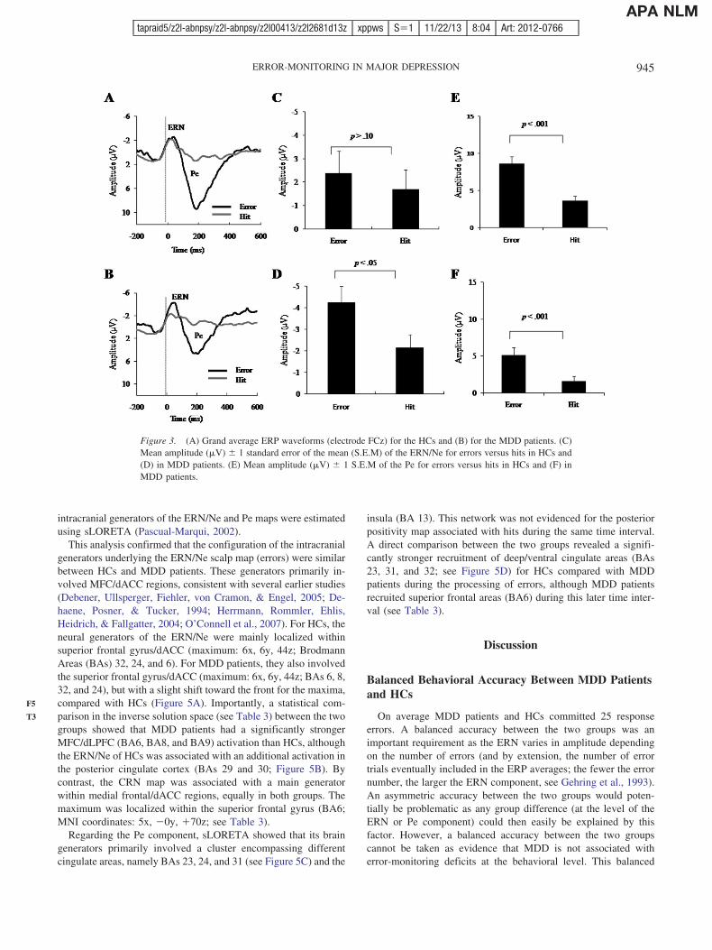
A clear negative deflection was observed �40 ms after errorcommission with maximum amplitude over fronto-central elec-trodes (e.g., FCz). These electrophysiological properties werecompatible with the ERN/Ne (Figure 3A and B). This early neg-ative component was larger following errors compared with hits,F(1, 38) � 5.33, p � .05. Although this difference appeared to be
larger for MDD patients (M � 2.09; SEM � .80; t(19) � �2.61,p � .05) than for HCs (M � .70; SEM � .90; t � 1), there was nosignificant effect of group, F(1, 38) � 1.53, p � .10, nor asignificant interaction between accuracy (error vs. hit) and group(HC vs. MDD), F(1, 38) � 1.33, p � .10 (Figure 3CD). However,this interaction became significant when including speed as acovariate given that on average MDD patients were slower thanHCs, F(1, 37) � 4.64, p � .05. More specifically, post hoccomparisons showed a difference between the ERN and CRN inthe MDD group, F(1, 18) � 10.83, p � .01, although this differ-ence was only trend significant in the HC group, F(1, 18) � 3.79,p � .07. Consistent with previous ERP studies using this Go/noGotask (Aarts & Pourtois, 2010; Dhar & Pourtois, 2011), and giventhe speed pressure as well as the relatively high number of errorscommitted within a short period of time, the ERN/Ne–CRN am-plitude difference was actually modest at this specific electrodeposition (FCz), although being significant, suggesting that re-
Figure 2. Identification of the ERN/Ne and Pe topographical components, and their intracerebral generators.(A) 128 electrodes were used and time-frames spanning from �55 ms to 379 ms after response onset,encompassing the ERN and Pe. A butterfly view of the grand-average ERP data of HCs (errors) from �500 msto �1,000 ms around the response is shown (with two representative electrodes, FCZ and Cz shown in black),as well as the corresponding time interval selected for the segmentation in topographical components. (B) Twotopographical maps, the ERN/Ne and Pe, were isolated using a clustering method. (C) These two mapscorresponded to the ERN/Ne and Pe component. (D) SLORETA was used to gain insight into the neuralgenerators underlying these dominant scalp configurations. For HCs, the MFC/dACC was estimated as the mainneural generator of the ERN, while nonoverlapping cingulate areas were found to account for the Pe component.
Thi
sdo
cum
ent
isco
pyri
ghte
dby
the
Am
eric
anPs
ycho
logi
cal
Ass
ocia
tion
oron
eof
itsal
lied
publ
ishe
rs.
Thi
sar
ticle
isin
tend
edso
lely
for
the
pers
onal
use
ofth
ein
divi
dual
user
and
isno
tto
bedi
ssem
inat
edbr
oadl
y.
943ERROR-MONITORING IN MAJOR DEPRESSION
F3
COLOR
tapraid5/z2l-abnpsy/z2l-abnpsy/z2l00413/z2l2681d13z xppws S�1 11/22/13 8:04 Art: 2012-0766APA NLM
sponse errors were discriminated from hits early on followingresponse onset, especially so for MDD patients.
The ERN/Ne was followed by a large positive component thatreached its maximum amplitude at central electrodes along themidline (i.e., Cz) and that was clearly modulated in size byaccuracy (errors vs. hits). This positive deflection was reliablylarger for errors compared with hits, F(1, 38) � 85.80, p � .001.These properties (latency, polarity, topography) were compatiblewith the generation of a genuine Pe component during early errordetection. This positive component was larger in HCs than inMDD patients, F(1, 38) � 6.70, p � .05, but this effect did notinteract with accuracy, F(1, 38) � 2.26, p � .10 (see Figure 3EF).An auxiliary analysis including speed as a covariate confirmed thisstatistical outcome (i.e., accuracy: F(1, 37) � 4.61, p � .05; group:F(1, 37) � 4.50, p � .05; accuracy x group: F(1, 37) � 1.54, p �.10).
Furthermore, to assess if MDD had a differential impact on theERN and Pe components, a 2 (accuracy: error vs. hit) � 2 (ERPcomponent: ERN vs. Pe) � 2 (group: HCs vs. MDDs) ANOVAwas carried out. This analysis showed significant effects of accu-racy, F(1, 38) � 16.60, p � .001, and of ERP component, F(1,38) � 201,38, p � .001. Whereas the interaction term betweenERP component, accuracy, and group remained nonsignificant,F � 1, a significant main effect of group was evidenced, F(1,38) � 5.30, p � .05.1 Complementary topographical and sourcelocalization analyses were used to assess if MDD, during each ofthese two consecutive moments, differentially influenced the neu-ral processing of these salient events in nonoverlapping corticalbrain areas compared with HCs.
Topographical Components
A solution with eight dominant maps explained 94% of thevariance. Next, an analysis was performed on the dominant mapsgenerated during the time interval corresponding to the ERN/Neand Pe, and their likely variations as a function of accuracy and/orgroup.
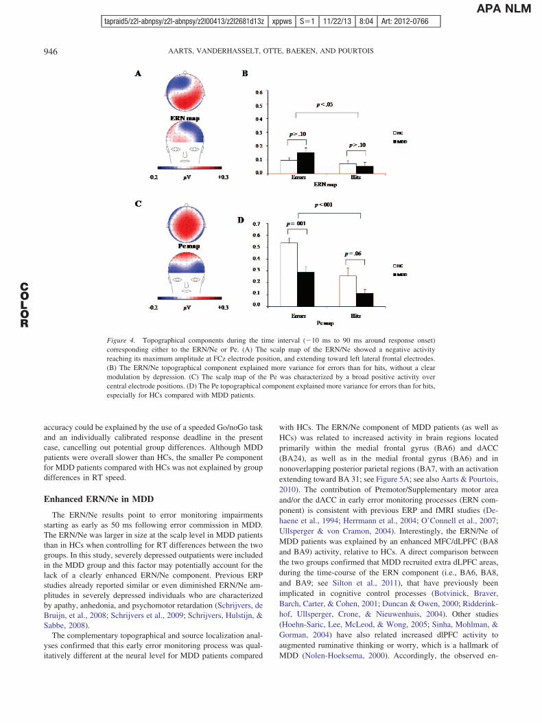
During the time interval corresponding the ERN/Ne versus CRNcomponent (starting �10 ms before response onset and ending�90 ms postresponse onset), a main change in the topographybetween errors and hits was evidenced. Whereas the topographyfor hits was characterized by a broad negative activity extending
toward prefrontal sites (CRN map), the scalp distribution forresponse errors was qualified by a negative activity circumscribedto a few precentral electrode positions, including FCz (Figure 4A;ERN/Ne map). This ERN topography showed a left lateralization,an observation which could be explained by the mono-manual (i.e.,right hand) stimulus-response mapping used with this Go/noGotask (Aarts & Pourtois, 2010; Gruendler, Ullsperger, & Huster,2011). This result suggests that beyond local amplitude variationsfound for the peak of the ERN/Ne component as measured atelectrode FCZ, errors are also associated with a change in thetopography of the global electric field compared with hits. Thisfinding therefore suggested that the brain network giving rise toresponse errors versus hits could be dissociated. These two dom-inant maps were fitted back to the individual ERP data to verifywhether this topography-related change during the ERN/CRN wassignificant (and different across the two groups) or not. The GEVvalues obtained for these two dominant maps after fitting weretherefore submitted to a 2 (map) � 2 (group) � 2 (accuracy)repeated measures ANOVA. This analysis revealed a significantinteraction between accuracy and map/scalp configuration, F(1,38) � 44.04, p � .001. Although the CRN map explained morevariance for hits than errors, t(39) � �8.06, p � .001, the ERN/Nemap had a symmetric profile, explaining more variance for errorsthan hits, t(39) � 2.66, p � .05. However, this interaction effectwas similar for MDD patients and HCs, F � 1 (Figure 4B).
Regarding the time interval corresponding to the Pe component(�145 ms–281 ms postresponse onset), a specific error-relatedtopography (Pe map, with a maximum amplitude at electrode CZ)was identified. By contrast, hits elicited a distinct posterior posi-tivity (see Figure 4C). Further analyses computed on the meanGEV values obtained for these two dominant maps confirmed asignificant interaction between accuracy and map, F(1, 38) �28.55, p � .001. Whereas the Pe map explained more variance forerrors than hits, t(39) � 5.39, p � .001, the other concurrent map(posterior positivity map) showed a symmetric effect, explainingmore variance for hits than errors, t(39) � �4.21, p � .001.Interestingly, this analysis also showed a significant interactionbetween map and group, F(1, 38) � 7.17, p � .01 (Figure 4D).This interaction was explained by the fact that the Pe map ex-plained more variance for errors committed by HCs than MDDpatients, t(38) � 3.67, p � .001. The same effect was evidenced,though much weaker, for hits, t(38) � 1.92, p � .06. However, theconcurrent posterior positivity map associated with hits was notsignificantly different between groups, both for errors, t(38) ��1.37, p � .10, and hits, t � 1, suggesting that MDD wasprimarily associated to an altered neural processing of errors, butnot hits.
Inverse Solutions
To gain insight into the configuration of the intracranial gener-ators underlying the global topographic-dependent changes, the
1 We also assessed whether the putative measurement error was similarfor the ERN and Pe components and for the two groups. For this purpose,we compared the standard deviation (interindividual variability) of theERN and Pe within each group as well as across the two groups. We alsocompared the standard deviation of the difference between the mean of theERN and Pe (see Masson & Loftus, 2003). These analyses showed com-parable measurement error for these two ERP components and two groups.
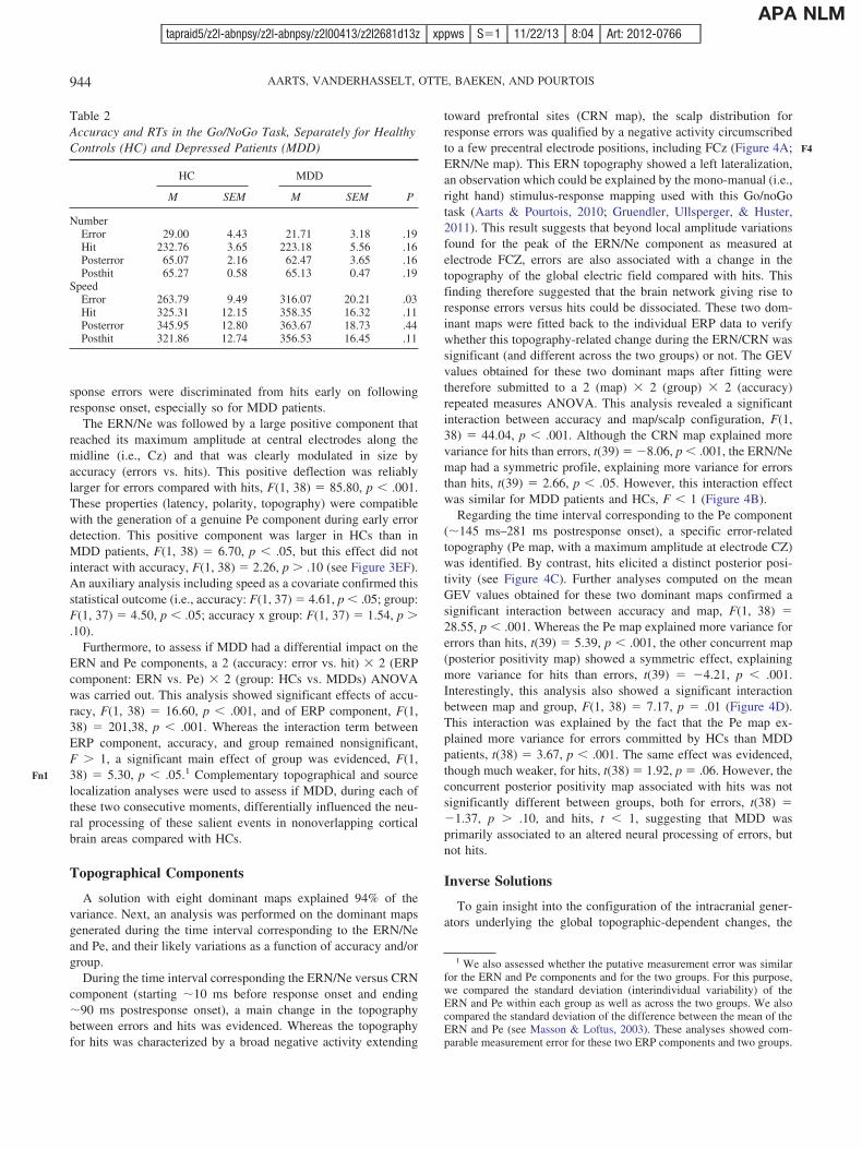
Table 2Accuracy and RTs in the Go/NoGo Task, Separately for HealthyControls (HC) and Depressed Patients (MDD)
HC MDD
PM SEM M SEM
NumberError 29.00 4.43 21.71 3.18 .19Hit 232.76 3.65 223.18 5.56 .16Posterror 65.07 2.16 62.47 3.65 .16Posthit 65.27 0.58 65.13 0.47 .19
SpeedError 263.79 9.49 316.07 20.21 .03Hit 325.31 12.15 358.35 16.32 .11Posterror 345.95 12.80 363.67 18.73 .44Posthit 321.86 12.74 356.53 16.45 .11
Thi
sdo
cum
ent
isco
pyri
ghte
dby
the
Am
eric
anPs
ycho
logi
cal
Ass
ocia
tion
oron
eof
itsal
lied
publ
ishe
rs.
Thi
sar
ticle
isin
tend
edso
lely
for
the
pers
onal
use
ofth
ein
divi
dual
user
and
isno
tto
bedi
ssem
inat
edbr
oadl
y.
944 AARTS, VANDERHASSELT, OTTE, BAEKEN, AND POURTOIS
Fn1
F4
tapraid5/z2l-abnpsy/z2l-abnpsy/z2l00413/z2l2681d13z xppws S�1 11/22/13 8:04 Art: 2012-0766APA NLM
intracranial generators of the ERN/Ne and Pe maps were estimatedusing sLORETA (Pascual-Marqui, 2002).
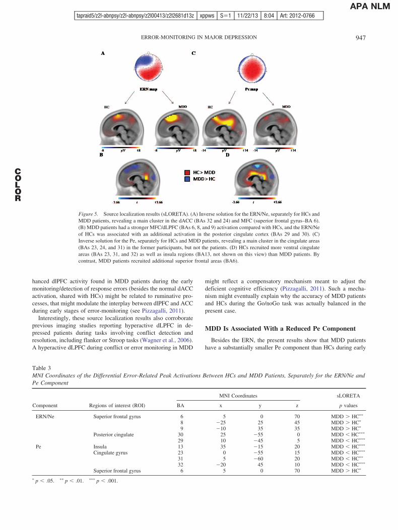
This analysis confirmed that the configuration of the intracranialgenerators underlying the ERN/Ne scalp map (errors) were similarbetween HCs and MDD patients. These generators primarily in-volved MFC/dACC regions, consistent with several earlier studies(Debener, Ullsperger, Fiehler, von Cramon, & Engel, 2005; De-haene, Posner, & Tucker, 1994; Herrmann, Rommler, Ehlis,Heidrich, & Fallgatter, 2004; O’Connell et al., 2007). For HCs, theneural generators of the ERN/Ne were mainly localized withinsuperior frontal gyrus/dACC (maximum: 6x, 6y, 44z; BrodmannAreas (BAs) 32, 24, and 6). For MDD patients, they also involvedthe superior frontal gyrus/dACC (maximum: 6x, 6y, 44z; BAs 6, 8,32, and 24), but with a slight shift toward the front for the maxima,compared with HCs (Figure 5A). Importantly, a statistical com-parison in the inverse solution space (see Table 3) between the twogroups showed that MDD patients had a significantly strongerMFC/dLPFC (BA6, BA8, and BA9) activation than HCs, althoughthe ERN/Ne of HCs was associated with an additional activation inthe posterior cingulate cortex (BAs 29 and 30; Figure 5B). Bycontrast, the CRN map was associated with a main generatorwithin medial frontal/dACC regions, equally in both groups. Themaximum was localized within the superior frontal gyrus (BA6;MNI coordinates: 5x, �0y, �70z; see Table 3).
Regarding the Pe component, sLORETA showed that its braingenerators primarily involved a cluster encompassing differentcingulate areas, namely BAs 23, 24, and 31 (see Figure 5C) and the
insula (BA 13). This network was not evidenced for the posteriorpositivity map associated with hits during the same time interval.A direct comparison between the two groups revealed a signifi-cantly stronger recruitment of deep/ventral cingulate areas (BAs23, 31, and 32; see Figure 5D) for HCs compared with MDDpatients during the processing of errors, although MDD patientsrecruited superior frontal areas (BA6) during this later time inter-val (see Table 3).
Discussion
Balanced Behavioral Accuracy Between MDD Patientsand HCs
On average MDD patients and HCs committed 25 responseerrors. A balanced accuracy between the two groups was animportant requirement as the ERN varies in amplitude dependingon the number of errors (and by extension, the number of errortrials eventually included in the ERP averages; the fewer the errornumber, the larger the ERN component, see Gehring et al., 1993).An asymmetric accuracy between the two groups would poten-tially be problematic as any group difference (at the level of theERN or Pe component) could then easily be explained by thisfactor. However, a balanced accuracy between the two groupscannot be taken as evidence that MDD is not associated witherror-monitoring deficits at the behavioral level. This balanced
Figure 3. (A) Grand average ERP waveforms (electrode FCz) for the HCs and (B) for the MDD patients. (C)Mean amplitude (�V) 1 standard error of the mean (S.E.M) of the ERN/Ne for errors versus hits in HCs and(D) in MDD patients. (E) Mean amplitude (�V) 1 S.E.M of the Pe for errors versus hits in HCs and (F) inMDD patients.
Thi
sdo
cum
ent
isco
pyri
ghte
dby
the
Am
eric
anPs
ycho
logi
cal
Ass
ocia
tion
oron
eof
itsal
lied
publ
ishe
rs.
Thi
sar
ticle
isin
tend
edso
lely
for
the
pers
onal
use
ofth
ein
divi
dual
user
and
isno
tto
bedi
ssem
inat
edbr
oadl
y.
945ERROR-MONITORING IN MAJOR DEPRESSION
F5
T3
tapraid5/z2l-abnpsy/z2l-abnpsy/z2l00413/z2l2681d13z xppws S�1 11/22/13 8:04 Art: 2012-0766APA NLM
accuracy could be explained by the use of a speeded Go/noGo taskand an individually calibrated response deadline in the presentcase, cancelling out potential group differences. Although MDDpatients were overall slower than HCs, the smaller Pe componentfor MDD patients compared with HCs was not explained by groupdifferences in RT speed.
Enhanced ERN/Ne in MDD
The ERN/Ne results point to error monitoring impairmentsstarting as early as 50 ms following error commission in MDD.The ERN/Ne was larger in size at the scalp level in MDD patientsthan in HCs when controlling for RT differences between the twogroups. In this study, severely depressed outpatients were includedin the MDD group and this factor may potentially account for thelack of a clearly enhanced ERN/Ne component. Previous ERPstudies already reported similar or even diminished ERN/Ne am-plitudes in severely depressed individuals who are characterizedby apathy, anhedonia, and psychomotor retardation (Schrijvers, deBruijn, et al., 2008; Schrijvers et al., 2009; Schrijvers, Hulstijn, &Sabbe, 2008).
The complementary topographical and source localization anal-yses confirmed that this early error monitoring process was qual-itatively different at the neural level for MDD patients compared
with HCs. The ERN/Ne component of MDD patients (as well asHCs) was related to increased activity in brain regions locatedprimarily within the medial frontal gyrus (BA6) and dACC(BA24), as well as in the medial frontal gyrus (BA6) and innonoverlapping posterior parietal regions (BA7, with an activationextending toward BA 31; see Figure 5A; see also Aarts & Pourtois,2010). The contribution of Premotor/Supplementary motor areaand/or the dACC in early error monitoring processes (ERN com-ponent) is consistent with previous ERP and fMRI studies (De-haene et al., 1994; Herrmann et al., 2004; O’Connell et al., 2007;Ullsperger & von Cramon, 2004). Interestingly, the ERN/Ne ofMDD patients was explained by an enhanced MFC/dLPFC (BA8and BA9) activity, relative to HCs. A direct comparison betweenthe two groups confirmed that MDD recruited extra dLPFC areas,during the time-course of the ERN component (i.e., BA6, BA8,and BA9; see Silton et al., 2011), that have previously beenimplicated in cognitive control processes (Botvinick, Braver,Barch, Carter, & Cohen, 2001; Duncan & Owen, 2000; Ridderink-hof, Ullsperger, Crone, & Nieuwenhuis, 2004). Other studies(Hoehn-Saric, Lee, McLeod, & Wong, 2005; Sinha, Mohlman, &Gorman, 2004) have also related increased dlPFC activity toaugmented ruminative thinking or worry, which is a hallmark ofMDD (Nolen-Hoeksema, 2000). Accordingly, the observed en-
Figure 4. Topographical components during the time interval (�10 ms to 90 ms around response onset)corresponding either to the ERN/Ne or Pe. (A) The scalp map of the ERN/Ne showed a negative activityreaching its maximum amplitude at FCz electrode position, and extending toward left lateral frontal electrodes.(B) The ERN/Ne topographical component explained more variance for errors than for hits, without a clearmodulation by depression. (C) The scalp map of the Pe was characterized by a broad positive activity overcentral electrode positions. (D) The Pe topographical component explained more variance for errors than for hits,especially for HCs compared with MDD patients.
Thi
sdo
cum
ent
isco
pyri
ghte
dby
the
Am
eric
anPs
ycho
logi
cal
Ass
ocia
tion
oron
eof
itsal
lied
publ
ishe
rs.
Thi
sar
ticle
isin
tend
edso
lely
for
the
pers
onal
use
ofth
ein
divi
dual
user
and
isno
tto
bedi
ssem
inat
edbr
oadl
y.
946 AARTS, VANDERHASSELT, OTTE, BAEKEN, AND POURTOIS
COLOR
tapraid5/z2l-abnpsy/z2l-abnpsy/z2l00413/z2l2681d13z xppws S�1 11/22/13 8:04 Art: 2012-0766APA NLM
hanced dlPFC activity found in MDD patients during the earlymonitoring/detection of response errors (besides the normal dACCactivation, shared with HCs) might be related to ruminative pro-cesses, that might modulate the interplay between dlPFC and ACCduring early stages of error-monitoring (see Pizzagalli, 2011).
Interestingly, these source localization results also corroborateprevious imaging studies reporting hyperactive dLPFC in de-pressed patients during tasks involving conflict detection andresolution, including flanker or Stroop tasks (Wagner et al., 2006).A hyperactive dLPFC during conflict or error monitoring in MDD
might reflect a compensatory mechanism meant to adjust thedeficient cognitive efficiency (Pizzagalli, 2011). Such a mecha-nism might eventually explain why the accuracy of MDD patientsand HCs during the Go/noGo task was actually balanced in thepresent case.
MDD Is Associated With a Reduced Pe Component
Besides the ERN, the present results show that MDD patientshave a substantially smaller Pe component than HCs during early
Figure 5. Source localization results (sLORETA). (A) Inverse solution for the ERN/Ne, separately for HCs andMDD patients, revealing a main cluster in the dACC (BAs 32 and 24) and MFC (superior frontal gyrus–BA 6).(B) MDD patients had a stronger MFC/dLPFC (BAs 6, 8, and 9) activation compared with HCs, and the ERN/Neof HCs was associated with an additional activation in the posterior cingulate cortex (BAs 29 and 30). (C)Inverse solution for the Pe, separately for HCs and MDD patients, revealing a main cluster in the cingulate areas(BAs 23, 24, and 31) in the former participants, but not the patients. (D) HCs recruited more ventral cingulateareas (BAs 23, 31, and 32) as well as insula regions (BA13, not shown on this view) than MDD patients. Bycontrast, MDD patients recruited additional superior frontal areas (BA6).
Table 3MNI Coordinates of the Differential Error-Related Peak Activations Between HCs and MDD Patients, Separately for the ERN/Ne andPe Component
Component Regions of interest (ROI)
MNI Coordinates sLORETA
BA x y z p values
ERN/Ne Superior frontal gyrus 6 5 0 70 MDD � HC��
8 �25 25 45 MDD � HC�
9 �10 35 35 MDD � HC�
Posterior cingulate 30 25 �55 0 MDD � HC���
29 10 �45 5 MDD � HC���
Pe Insula 13 35 �15 20 MDD � HC���
Cingulate gyrus 23 0 �55 15 MDD � HC���
31 5 �60 20 MDD � HC��
32 �20 45 10 MDD � HC���
Superior frontal gyrus 6 5 0 70 MDD � HC�
� p � .05. �� p � .01. ��� p � .001.
Thi
sdo
cum
ent
isco
pyri
ghte
dby
the
Am
eric
anPs
ycho
logi
cal
Ass
ocia
tion
oron
eof
itsal
lied
publ
ishe
rs.
Thi
sar
ticle
isin
tend
edso
lely
for
the
pers
onal
use
ofth
ein
divi
dual
user
and
isno
tto
bedi
ssem
inat
edbr
oadl
y.
947ERROR-MONITORING IN MAJOR DEPRESSION
COLOR
tapraid5/z2l-abnpsy/z2l-abnpsy/z2l00413/z2l2681d13z xppws S�1 11/22/13 8:04 Art: 2012-0766APA NLM
error monitoring. This decreased Pe component during error mon-itoring in MDD patients might be explained either by symptomseverity, which is stronger in MDD patients (the present study;Olvet, Klein, & Hajcak, 2010; Schrijvers, de Bruijn et al., 2008;Schrijvers et al., 2009) than in moderately depressed individuals(Chiu & Deldin, 2007; Compton et al., 2008; Holmes & Pizzagalli,2008).
Given the impaired motivation in MDD patients (DSM–IV–TR,APA, 2000), and the link between the Pe component and themotivational significance of an error (Overbeek et al., 2005), areduced Pe component may be explained in terms of a change inthe detection of an otherwise salient or behaviorally relevant event(i.e., unwanted response error). However, the posterror adjustmentfollowing errors (Rabbitt, 1966) and the total number of errors wascomparable in MDD patients relative to HCs. This suggests thatMDD patients were equally able to comply with the task demandscompared with HCs and that a mere change in levels of “intrinsic”motivation during the task across the two groups did not accountfor the present ERP results.
Finally, a blunted Pe component in MDD patients could stemfrom an exaggerated ruminative thinking style. In this view, theaccumulation of evidence leading to the conscious detection of aresponse error, as reflected by the Pe component (Steinhauser &Yeung, 2010), would be impaired because other intrusive thoughtsmay prevent its normal unfolding. This limited resource account isalso consistent with the idea that the Pe reflects a “bottom-up”attentional orienting process, similar to the P300 component (Rid-derinkhof et al., 2009). Presumably, if less “bottom-up” attentionis allocated to the monitoring of actions and errors (becauseattention resources are used by a concurrent mental process, e.g.,rumination), the monitoring and the conscious registration of theseerrors are probably less effective. Interestingly, previous studiesalready reported a decreased noGo P300 in depressed individuals(Ruchsow, Groen, & Kiefer, 2008).
More generally, the current ERP results, which are consistentwith earlier findings obtained with comparable clinical samples(Schrijvers et al., 2009), suggest that early stages of error detectionare different between MDDs and HCs at multiple levels throughmodulations in nonoverlapping medial frontal and ACC networks.We did not find evidence for a differential effect of MDD at thelevel of the Pe, using standard scalp measurements. However, thecomplementing topographical and source localization resultsshowed that these two consecutive stages of early error detection(ERN and Pe) were different in MDD patients compared with HCs,due to the reliable modulation of specific and different brainnetworks: Although the ACC was overactive and additional dlPFCsources underlying the ERN were found in MDD, the “normal”ventral ACC sources giving rise to the Pe component were sub-stantially reduced in MDD.
Presumably, these effects might reflect an inability or deficiencyto treat or regulate the emotional value of actions early on follow-ing response onset, at the level of the ERN (Aarts, De Houwer, &Pourtois, 2012; Aarts, De Houwer, & Pourtois, 2013). Such anearly deficient process could stem from abnormal prefrontal-basedexecutive functions or alternatively an exaggerated ruminativethinking style, which might in turn consume resources used oth-erwise to process later the motivational significance or salience ofresponse errors (Pe effect). Future studies are needed to establish
whether rumination (or another process) might account for theseabnormal early error monitoring processes seen in MDD.
Limitations
A few limitations should be noted. First, we could recruit 20MDD patients and 20 HCs, which corresponds to a modest samplesize. On the other hand, the complementary topographical andsource localization results clearly showed that the present studywas not underpowered, as we were able to reveal significantmodulatory effects of MDD at two different moments followingresponse error onset in nonoverlapping medial frontal and ACCregions.
Second, regular antidepressant drugs may have either amplifiedor obscured some of the group differences found during errorprocessing. However, these drugs have not yet been linked tosystematic alterations of the amplitude or morphology of error-related ERP components in previous ERP studies using HCs (deBruijn, Sabbe, Hulstijn, Ruigt, & Verkes, 2006; Stern et al., 2010).Nonetheless, additional ERP studies are needed in order to assesswhether systematic changes in early error monitoring brain pro-cesses seen in MDD patients (e.g., blunted Pe component) aremodified by antidepressant medication.
To conclude, the present study reveals that MDD is associatedwith altered early error monitoring processes at multiple levels(ERN and Pe components) through impairments in different MFCand dLPFC brain networks.
References
Aarts, K., De Houwer, J., & Pourtois, G. (2012). Evidence for the auto-matic evaluation of self-generated actions. Cognition, 124, 117–127.doi:10.1016/j.cognition.2012.05.009
Aarts, K., De Houwer, J., & Pourtois, G. (2013). Erroneous and correctactions have a different affective value: Evidence from ERPs. Emotion.doi:10.1037/a0032808
Aarts, K., & Pourtois, G. (2010). Anxiety not only increases, but also altersearly error-monitoring functions. Cognitive, Affective, & BehavioralNeuroscience, 10, 479–492. doi:10.3758/CABN.10.4.479
American Psychiatric Association. (2000). Diagnostic and statistical man-ual of mental disorders (text revision). Washington, DC: Author.
Beck, A. T., Steer, R. A., & Brown, G. K. (1996). Manual for the BeckDepression Inventory-II. San Antonio, TX: Psychological Corporation.
Botvinick, M. M., Braver, T. S., Barch, D. M., Carter, C. S., & Cohen, J. D.(2001). Conflict monitoring and cognitive control. Psychological Re-view, 108, 624–652. doi:10.1037/0033-295X.108.3.624
Bush, G., Luu, P., & Posner, M. I. (2000). Cognitive and emotionalinfluences in anterior cingulate cortex. Trends in Cognitive Sciences, 4,215–222. doi:10.1016/S1364-6613(00)01483-2
Chiu, P. H., & Deldin, P. J. (2007). Neural evidence for enhanced errordetection in major depressive disorder. The American Journal of Psy-chiatry, 164, 608–616. doi:10.1176/appi.ajp.164.4.608
Compton, R. J., Lin, M., Vargas, G., Carp, J., Fineman, S. L., & Quandt,L. C. (2008). Error detection and posterror behavior in depressed un-dergraduates. Emotion, 8, 58–67. doi:10.1037/1528-3542.8.1.58
Debener, S., Ullsperger, M., Fiehler, K., von Cramon, D. Y., & Engel,A. K. (2005). Monitoring error processing by means of simultaneousEEG/fMRI recordings II: Single-trial independent component analysisof the error-related negativity (ERN). Journal of Psychophysiology, 19,111.
de Bruijn, E. R. A., Sabbe, B. G. C., Hulstijn, W., Ruigt, G. S. F., &Verkes, R. J. (2006). Effects of antipsychotic and antidepressant drugs
Thi
sdo
cum
ent
isco
pyri
ghte
dby
the
Am
eric
anPs
ycho
logi
cal
Ass
ocia
tion
oron
eof
itsal
lied
publ
ishe
rs.
Thi
sar
ticle
isin
tend
edso
lely
for
the
pers
onal
use
ofth
ein
divi
dual
user
and
isno
tto
bedi
ssem
inat
edbr
oadl
y.
948 AARTS, VANDERHASSELT, OTTE, BAEKEN, AND POURTOIS
tapraid5/z2l-abnpsy/z2l-abnpsy/z2l00413/z2l2681d13z xppws S�1 11/22/13 8:04 Art: 2012-0766APA NLM
on action monitoring in healthy volunteers. Brain Research, 1105,122–129. doi:10.1016/j.brainres.2006.01.006
Dehaene, S., Posner, M. I., & Tucker, D. M. (1994). Localization of aneural system for error-detection and compensation. Psychological Sci-ence, 5, 303–305. doi:10.1111/j.1467-9280.1994.tb00630.x
Dhar, M., & Pourtois, G. (2011). Early error detection is generic, butsubsequent adaption to errors is not: Evidence from ERPs. Neuropsy-chologia, 49, 1236–1245. doi:10.1016/j.neuropsychologia.2011.01.006
Duncan, J., & Owen, A. M. (2000). Common regions of the human frontallobe recruited by diverse cognitive demands. Trends in Neurosciences,23, 475–483. doi:10.1016/S0166-2236(00)01633-7
Falkenstein, M., Hohnsbein, J., Hoormann, J., & Blanke, L. (1991). Effectsof crossmodal divided attention on late ERP components. II. Errorprocessing in choice reaction tasks. Electroencephalography and Clin-ical Neurophysiology, 78, 447–455. doi:10.1016/0013-4694(91)90062-9
Falkenstein, M., Hoormann, J., Christ, S., & Hohnsbein, J. (2000). ERPcomponents on reaction errors and their functional significance: Atutorial. Biological Psychology, 51, 87–107. doi:10.1016/S0301-0511(99)00031-9
Fuchs, M., Kastner, J., Wagner, M., Hawes, S., & Ebersole, J. S. (2002). Astandardized boundary element method volume conductor model. Clin-ical Neurophysiology, 113, 702–712. doi:10.1016/S1388-2457(02)00030-5
Gehring, W. J., Goss, B., Coles, M. G. H., Meyer, D. E., & Donchin, E.(1993). A neural system for error-detection and compensation. Psycho-logical Science, 4, 385–390. doi:10.1111/j.1467-9280.1993.tb00586.x
Gratton, G., Coles, M. G. H., & Donchin, E. (1983). A new method foroff-line removal of ocular artifact. Electroencephalography and ClinicalNeurophysiology, 55, 468–484. doi:10.1016/0013-4694(83)90135-9
Gruendler, T. O., Ullsperger, M., & Huster, R. J. (2011). Event-relatedpotential correlates of performance-monitoring in a lateralized time-estimation task. PLOS one, 6, e25591. doi:10.1371/journal.pone.0025591
Hamilton, M. (1960). A rating scale for depression. Journal of Neurology,Neurosurgery and Psychiatry, 23, 56–61. doi:10.1136/jnnp.23.1.56
Herrmann, M. J., Rommler, J., Ehlis, A. C., Heidrich, A., & Fallgatter, A. J.(2004). Source localization (LORETA) of the error-related negativity(ERN/Ne) and positivity (Pe). Cognitive Brain Research, 20, 294–299.doi:10.1016/j.cogbrainres.2004.02.013
Hoehn-Saric, R., Lee, J. S., McLeod, D. R., & Wong, D. F. (2005). Effectof worry on regional cerebral blood flow in nonanxious subjects. Psy-chiatry Research: Neuroimaging, 140, 259 –269. doi:10.1016/j.pscychresns.2005.05.013
Holmes, A. J., & Pizzagalli, D. A. (2008). Spatiotemporal dynamics oferror processing dysfunctions in major depressive disorder. Archives ofGeneral Psychiatry, 65, 179 –188. doi:10.1001/archgenpsychiatry.2007.19
Holmes, A. J., & Pizzagalli, D. A. (2010). Effects of task-relevant incen-tives on the electrophysiological correlates of error processing in majordepressive disorder. Cognitive, Affective & Behavioral Neuroscience,10, 119–128. doi:10.3758/CABN.10.1.119
Jurcak, V., Tsuzuki, D., & Dan, I. (2007). 10/20, 10/10, and 10/5 systemsrevisited: Their validity as relative head-surface-based positioning sys-tems. Neuroimage, 34, 1600–1611. doi:10.1016/j.neuroimage.2006.09.024
Laming, D. (1979). Autocorrelation of choice-reaction times. Acta Psycho-logica, 43, 381–412. doi:10.1016/0001-6918(79)90032-5
Lehmann, D., & Skrandies, W. (1980). Reference-free identification ofcomponents of checkerboard-evoked multichannel potential fields. Elec-troencephalography and Clinical Neurophysiology, 48, 609–621. doi:10.1016/0013-4694(80)90419-8
Masson, M. E. J., & Loftus, G. R. (2003). Using confidence intervals forgraphically based data interpretation. Canadian Journal of Experimental
Psychology/Revue canadienne de psychologie expérimentale, 57, 203–220.
Mayberg, H. S. (1997). Limbic-cortical dysregulation: A proposed modelof depression. Journal of Neuropsychiatry and Clinical Neurosciences,9, 471–481.
Mazziotta, J., Toga, A., Evans, A., Fox, P., Lancaster, J., Zilles, K., . . .Mazoyer, B. (2001). A probabilistic atlas and reference system for thehuman brain: International Consortium for Brain Mapping (ICBM).Philosophical Transactions of the Royal Society. Series B, BiologicalSciences, 356, 1293–1322. doi:10.1098/rstb.2001.0915
Michel, C. M., Seeck, M., & Landis, T. (1999). Spatiotemporal dynamicsof human cognition. News in Physiological Sciences, 14, 206–214.
Mineka, S., Watson, D., & Clark, L. A. (1998). Comorbidity of anxiety andunipolar mood disorders. Annual Review of Psychology, 49, 377–412.doi:10.1146/annurev.psych.49.1.377
Murray, M. M., Brunet, D., & Michel, C. M. (2008). Topographic ERPanalyses: A step-by-step tutorial review. Brain Topography, 20, 249–264. doi:10.1007/s10548-008-0054-5
Nieuwenhuis, S., Ridderinkhof, K. R., Blow, J., Band, G. P. H., & Kok, A.(2001). Error-related brain potentials are differentially related to aware-ness of response errors: Evidence from an antisaccade task. Psychophys-iology, 38, 752–760. doi:10.1111/1469-8986.3850752
Nolen-Hoeksema, S. (2000). The role of rumination in depressive disordersand mixed anxiety/depressive symptoms. Journal of Abnormal Psychol-ogy, 109, 504–511. doi:10.1037/0021-843X.109.3.504
O’Connell, R. G., Dockree, P. M., Bellgrove, M. A., Kelly, S. P., Hester,R., Garavan, H., . . . Foxe, J. J. (2007). The role of cingulate cortex inthe detection of errors with and without awareness: A high-densityelectrical mapping study. European Journal of Neuroscience, 25, 2571–2579. doi:10.1111/j.1460-9568.2007.05477.x
Olvet, D. M., & Hajcak, G. (2008). The error-related negativity (ERN) andpsychopathology: Toward an endophenotype. Clinical Psychology Re-view, 28, 1343–1354. doi:10.1016/j.cpr.2008.07.003
Olvet, D. M., Klein, D. N., & Hajcak, G. (2010). Depression symptomseverity and error-related brain activity. Psychiatry Research, 179, 30–37. doi:10.1016/j.psychres.2010.06.008
Overbeek, T. J. M., Nieuwenhuis, S., & Ridderinkhof, K. R. (2005).Dissociable components of error processing: On the functional signifi-cance of the Pe vis-a-vis the ERN/Ne. Journal of Psychophysiology, 19,319–329. doi:10.1027/0269-8803.19.4.319
Pascual-Marqui, R. D. (2002). Standardized low-resolution brain electro-magnetic tomography (sLORETA): Technical details. Methods andFindings in Experimental and Clinical Pharmacology, 24D, 5–12.
Picton, T. W., Bentin, S., Berg, P., Donchin, E., Hillyard, S. A., Johnson,R., . . . Taylor, M. J. (2000). Guidelines for using human event-relatedpotentials to study cognition: Recording standards and publication cri-teria. Psychophysiology, 37, 127–152. doi:10.1111/1469-8986.3720127
Pizzagalli, D. A. (2011). Frontocingulate dysfunction in depression: To-ward biomarkers of treatment response. Neuropsychopharmacology, 36,183–206. doi:10.1038/npp.2010.166
Pourtois, G., Delplanque, S., Michel, C., & Vuilleumier, P. (2008). Beyondconventional event-related brain potential (ERP): Exploring the time-course of visual emotion processing using topographic and principalcomponent analyses. Brain Topography, 20, 265–277. doi:10.1007/s10548-008-0053-6
Pourtois, G., Vocat, R., N=diaye, K., Spinelli, L., Seeck, M., & Vuil-leumier, P. (2010). Errors recruit both cognitive and emotional moni-toring systems: Simultaneous intracranial recordings in the dorsal ante-rior cingulate gyrus and amygdala combined with fMRI.Neuropsychologia, 48, 1144 –1159. doi:10.1016/j.neuropsychologia.2009.12.020
Rabbitt, P. M. (1966). Errors and error correction in choice-response tasks.Journal of Experimental Psychology, 71, 264 –272. doi:10.1037/h0022853
Thi
sdo
cum
ent
isco
pyri
ghte
dby
the
Am
eric
anPs
ycho
logi
cal
Ass
ocia
tion
oron
eof
itsal
lied
publ
ishe
rs.
Thi
sar
ticle
isin
tend
edso
lely
for
the
pers
onal
use
ofth
ein
divi
dual
user
and
isno
tto
bedi
ssem
inat
edbr
oadl
y.
949ERROR-MONITORING IN MAJOR DEPRESSION
tapraid5/z2l-abnpsy/z2l-abnpsy/z2l00413/z2l2681d13z xppws S�1 11/22/13 8:04 Art: 2012-0766APA NLM
Ridderinkhof, K. R., Ramautar, J. R., & Wijnen, J. G. (2009). To Pe or notto Pe: A P3-like ERP component reflecting the processing of responseerrors. Psychophysiology, 46, 531–538. doi:10.1111/j.1469-8986.2009.00790.x
Ridderinkhof, K. R., Ullsperger, M., Crone, E. A., & Nieuwenhuis, S.(2004). The role of medial frontal cortex in cognitive control. Science,306, 443–447. doi:10.1126/science.1100301
Ruchsow, M., Groen, G., & Kiefer, M. (2008). Electrophysiological evi-dence for reduced inhibitory control in depressed patients in partialremission: A Go/Nogo study. International Journal of Psychophysiol-ogy, 68, 209–218. doi:10.1016/j.ijpsycho.2008.01.010
Ruchsow, M., Hernberger, B., Beschoner, P., Gron, G., Spitzer, M., &Kiefer, M. (2006). Error processing in major depressive disorder: Evi-dence from event-related potentials. Journal of Psychiatric Research,40, 37–46. doi:10.1016/j.jpsychires.2005.02.002
Ruchsow, M., Hernberger, B., Wiesend, C., Gron, G., Spitzer, M., &Kiefer, M. (2004). The effect of erroneous responses on response mon-itoring in patients with major depressive disorder: A study with event-related potentials. Psychophysiology, 41, 833–840. doi:10.1111/j.1469-8986.2004.00237.x
Schrijvers, D., de Bruijn, E. R., Maas, Y., De Grave, C., Sabbe, B. G., &Hulstijn, W. (2008). Action monitoring in major depressive disorderwith psychomotor retardation. Cortex, 44, 569 –579. doi:10.1016/j.cortex.2007.08.014
Schrijvers, D., De Bruijn, E. R., Maas, Y. J., Vancoillie, P., Hulstijn, W.,& Sabbe, B. G. (2009). Action monitoring and depressive symptomreduction in major depressive disorder. International Journal of Psycho-physiology, 71, 218–224. doi:10.1016/j.ijpsycho.2008.09.005
Schrijvers, D., Hulstijn, W., & Sabbe, B. G. C. (2008). Psychomotorsymptoms in depression: A diagnostic, pathophysiological and therapeu-tic tool. Journal of Affective Disorders, 109, 1–20. doi:10.1016/j.jad.2007.10.019
Seifert, S., von Cramon, D. Y., Imperati, D., Tittgemeyer, T., & Ullsperger,M. (2011). Thalamocingulate interactions in performance monitoring.The Journal of Neuroscience, 31, 3375–3383. doi:10.1523/JNEUROSCI.6242-10.2011
Sheehan, D. V., Lecrubier, Y., Sheehan, K. H., Amorim, P., Janavs, J.,Weiller, E., . . . Dunbar, G. C. (1998). The Mini-International Neuro-psychiatric Interview (M.I.N.I.): The development and validation of astructured diagnostic psychiatric interview for DSM-IV and ICD-10. TheJournal of Clinical Psychiatry, 59 Suppl 20, 22–33; quiz 34–57.
Silton, R. L., Heller, W., Towers, D. N., Engels, A. S., Edgar, J. C.,Spielberg, J. M., . . . Miller, G. A. (2011). Depression and anxiousapprehension distinguish frontocingulate cortical activity during top-down attentional control. Journal of Abnormal Psychology, 120, 272–285. doi:10.1037/a0023204
Sinha, S., Mohlman, J., & Gorman, J. M. (2004). Neurobiology of gener-alized anxiety disorder. In R. G. Heimberg, C. L. Turk, & D. S. Mennin(Eds.), Generalized anxiety disorder: Advances in research and practice(pp. 187–216). New York, NY: Guilford Press.
Steinhauser, M., & Yeung, N. (2010). Decision processes in human per-formance monitoring. The Journal of Neuroscience, 30, 15643–15653.doi:10.1523/JNEUROSCI.1899-10.2010
Stern, E. R., Liu, Y., Gehring, W. J., Lister, J. J., Yin, G., Zhang, J., . . .Taylor, S. F. (2010). Chronic medication does not affect hyperactiveerror responses in obsessive-compulsive disorder. Psychophysiology, 47,913–920. doi:10.1111/j.1469-8986.2010.00988.x
Ullsperger, M., & von Cramon, D. Y. (2004). Neuroimaging of perfor-mance monitoring: Error detection and beyond. Cortex, 40, 593–604.doi:10.1016/S0010-9452(08)70155-2
Vocat, R., Pourtois, G., & Vuilleumier, P. (2008). Unavoidable errors: Aspatio-temporal analysis of time-course and neural sources of evokedpotentials associated with error processing in a speeded task. Neuropsy-chologia, 46, 2545–2555. doi:10.1016/j.neuropsychologia.2008.04.006
Wagner, G., Sinsela, E., Sobanskic, T., Köhlera, S., Marinoua, T., Ment-zelb, H., . . . Schlössera, R. G. M. (2006). Cortical inefficiency inpatients with unipolar depression: An event-related fMRI study with theStroop task. Biological Psychiatry, 59, 958–965. doi:10.1016/j.biopsych.2005.10.025
Received May 22, 2012Revision received August 19, 2013
Accepted August 28, 2013 �
Thi
sdo
cum
ent
isco
pyri
ghte
dby
the
Am
eric
anPs
ycho
logi
cal
Ass
ocia
tion
oron
eof
itsal
lied
publ
ishe
rs.
Thi
sar
ticle
isin
tend
edso
lely
for
the
pers
onal
use
ofth
ein
divi
dual
user
and
isno
tto
bedi
ssem
inat
edbr
oadl
y.
950 AARTS, VANDERHASSELT, OTTE, BAEKEN, AND POURTOIS
tapraid5/z2l-abnpsy/z2l-abnpsy/z2l00413/z2l2681d13z xppws S�1 11/22/13 8:04 Art: 2012-0766APA NLM
Top Related
Copyright © 2022 FDOKUMEN

