


Bahasa
Halaman
Hukum

39CliniCal MediCine insights: Cardiology 2014:8
Open Access: Full open access to this and thousands of other papers at http://www.la-press.com.
Clinical Medicine Insights: Cardiology
IntroductionHeart failure (HF) is the leading cause of death and hospitalization in industrialized countries, and covers an important part of the health care costs.1–3 In recent years, the introduction of new drugs and the use of devices has reduced mortality rates;4 however, the hospitalization rates continue to increase and the prognosis still remains unsatisfactory with high mortality and readmission rates.5–7 The growing acknowl-edge regarding metabolic abnormalities, hypercatabolic status, and cachexia in HF and their impact on outcomes have con-tributed to consider HF as a cardiac and whole body metabolic
disease, which may be the final pathway of the hemodynamic abnormalities and the neurohormonal and inflammatory acti-vation.8 Several macro and micronutrients deficiencies have been recognized in HF and lead to a disruption in energy as well as in anabolic metabolism. Among micronutrients, amino acids (AAs) act in both these metabolic pathways as intermediary for energy production and transfer and as main constituents of proteins.9 The aim of our study was to evaluate the effects of the administration of a mixture of 11 AAs on functional capacity expressed by the improvement in cardio-pulmonary stress test parameters and six minutes walking test
Effects of Oral Amino Acid Supplements on Functional Capacity in Patients with Chronic Heart Failure
Carlo lombardi, Valentina Carubelli, Valentina lazzarini, enrico Vizzardi, Filippo Quinzani, Federica guidetti, riccardo rovetta, savina nodari, Mihai gheorghiade and Marco MetraCardiology, Department of Medical and Surgical Specialties, Radiological Sciences, and Public Health, University and Civil Hospital of Brescia, Brescia, Italy.
AbstrAct: Amino acids (AAs) availability is reduced in patients with heart failure (HF) leading to abnormalities in cardiac and skeletal muscle metabo-lism, and eventually to a reduction in functional capacity and quality of life. In this study, we investigate the effects of oral supplementation with essential and semi-essential AAs for three months in patients with stable chronic HF. The primary endpoints were the effects of AA’s supplementation on exercise tolerance (evaluated by cardiopulmonary stress test and six minutes walking test (6MWT)), whether the secondary endpoints were change in quality of life (evaluated by Minnesota Living with Heart Failure Questionnaire—MLHFQ ) and N-terminal pro-brain natriuretic peptide (NT-proBNP) levels. We enrolled 13 patients with chronic stable HF on optimal therapy, symptomatic in New York Heart Association (NYHA) class II/III, with an ejection frac-tion (EF) ,45%. The mean age was 59 ± 14 years, and 11 (84.6%) patients were male. After three months, peak VO2 (baseline 14.8 ± 3.9 mL/minute/kg vs follow-up 16.8 ± 5.1 mL/minute/kg; P = 0.008) and VO2 at anaerobic threshold improved significantly (baseline 9.0 ± 3.8 mL/minute/kg vs follow-up 12.4 ± 3.9 mL/minute/kg; P = 0.002), as the 6MWT distance (baseline 439.1 ± 64.3 m vs follow-up 474.2 ± 89.0 m; P = 0.006). However, the quality of life did not change significantly (baseline 21 ± 14 vs follow-up 25 ± 13; P = 0.321). A non-significant trend in the reduction of NT-proBNP levels was observed (baseline 1502 ± 1900 ng/L vs follow-up 1040 ± 1345 ng/L; P = 0.052). AAs treatment resulted safe and was well tolerated by all patients. In our study, AAs supplementation in patients with chronic HF improved exercise tolerance but did not change quality of life.
Keywords: amino acid, heart failure, exercise capacity, nutrition, systolic dysfunction
CitAtiOn: lombardi et al. effects of oral amino acid supplements on Functional Capacity in Patients with Chronic heart Failure. Clinical Medicine Insights: Cardiology 2014:8 39–44 doi: 10.4137/CMC.s14016.
RECEivEd: January 2, 2014. RESubmittEd: February 11, 2014. ACCEPtEd FOR PubliCAtiOn: February 15, 2014.
ACAdEmiC EditOR: thomas e. Vanhecke, editor in Chief
tYPE: original research
Funding: Essential amminoacids were provided by Aminotrofic Inc, Professional Dietistic, Milan, Italy.
COmPEting intEREStS: Marco Metra received consulting incomes from Bayer, novartis, servier. Mihai ghoerghiade received consulting incomes abbott laboratories, astellas, astraZeneca, Bayer healthCare ag, Corthera, Cytokinetics, debioPharm s.a., errekappa terapeutici, glaxo- smithKline, ikaria, Johnson & Johnson, Medtronic, Merck, Novartis Pharma AG, Otsuka Pharmaceuticals, Palatin Technologies, Pericor Therapeutics, Protein Design Laboratories, Sanofi-Aventis, Sigma Tau, Solvay Phar-maceuticals, takeda Pharmaceutical, and trevena therapeutics.
COPYRigHt: © the authors, publisher and licensee libertas academica limited. this is an open-access article distributed under the terms of the Creative Commons CC-By-nC 3.0 license.
CORRESPOndEnCE: [email protected]
Lombardi et al
40 CliniCal MediCine insights: Cardiology 2014:8
(6MWT) distance. The secondary endpoints were to evaluate the effects on quality of life and NT-proBNP levels in a popu-lation of ambulatory patients with chronic HF.
MethodsPatients. We recruited 13 consecutive patients with
stable chronic HF because of dilated cardiomyopathy with-out coronary disease. All patients were symptomatic for HF for $6 months in NYHA class II or III, with an ejection fraction (EF) ,45% by echocardiography or radionuclide ventriculogra-phy (RVG), and able to perform a cardiopulmonary exercise test with a peak VO2 $10 mL/kg/minute. Patients were on optimal medical therapy, treated with angiotensin-converting-enzyme inhibitor (ACE) or angiotensin-receptor blocker and beta-block-ers at a stable dose for at least four weeks before entering in the study. Patients were excluded if they had ischemic-dilated car-diomyopathy, symptoms of myocardial ischemia, acute coronary syndromes, or a coronary revascularization procedure in the pre-vious three months; implantation of a cardiac resynchronization therapy (CRT) device in the prior six months or likely to receive implantation in the next three months; history of severe valvular disease (with the exception of functional mitral regurgitation); congenital heart disease, acute myocarditis, and hypertrophic or restrictive cardiomyopathy; cerebrovascular events or major sur-gery in the previous six months; and any concomitant disease that might adversely impair the exercise performance or the progno-sis of the patient. The investigation conformed to the principles outlined in the Declaration of Helsinki. The study received ethi-cal approval from the Department of Cardiology, University of Brescia, and all patients gave their written, informed consent to participate.
study protocol. A prospective, open study evaluated the efficacy of the administration of a mixture of essential and semi-essential AAs in patients with stable chronic HF. Patients were evaluated at baseline and after a three months follow-up. Each patient underwent clinical assessment, maxi-mal cardiopulmonary exercise test, 6MWT, transthoracic echocardiogram, RVG or cardiac magnetic resonance imaging (MRI), and Minnesota Living with Heart Failure Question-naire (MLHFQ ) before initiation of AAs administration and after three months. Each patient followed habitual daily diet. Cardiopulmonary stress test was performed with a cycloer-gometer with expiratory gas exchange and ECG monitor-ing, starting with a workload of 20 W with increments of 10 W/minute. All patients performed two preliminary cardio-pulmonary tests before the baseline evaluation to be familiar with the procedure with a variability less than 10% in peak VO2 between the screening tests. A maximal exercise was defined as reaching a respiratory exchange ratio (RER) $1.10. Peak VO2 was measured at the maximal exercise as the aver-age value in the last 30 seconds of exercise.
Patients received for three months a mixture of essential and semi-essential AAs (L-leucine, L-lysine, L-isoleucine, L-valine, L-threonine, L-cystine, L-hystidine, L-phenylalanine,
L-methionine, L-tyrosine, L-tryptophan; the composition is outlined in Table 1), thiamine, and pyridoxine (Aminotrofic, Professional Dietistic, Milan, Italy), at the dose of 4 g twice a day, diluted with water, at 10:00 AM and 4:00 PM.
The primary endpoint was to evaluate the effects of AAs supplementation on exercise tolerance at cardiopulmonary stress test and 6MWT. The secondary endpoints were to eval-uate the effects on quality of life and NT-proBNP levels.
echocardiographic analysis. Echocardiography exami-nations were performed by experienced operators in accor-dance with the recommendations of the European Society of echocardiography10 using a Vivid Seven (GE Health-care, UK) system operating at 3.4 MHz. Doppler tracings and two-dimensional images were obtained from paraster-nal long- and short-axes, apical, and subcostal views. Two-dimensional guided M-mode measurements of left ventricle’s (LV’s) internal dimensions, and septum and posterior wall thicknesses were made at the LV minor axis. Left ventricu-lar ejection fraction (LVEF) was measured using Simpson’s biplane method.
statistical analysis. Categorical data are presented as percentages, normally distributed continuous data as mean ± standard deviation (SD), and non-normally distributed vari-ables as median and interquartile range. Comparisons were made with paired Student’s t-test for continuous variables and chi-squared test for categorical variables as appropriate. For the statistical analysis, we used the SPSS version 19.0.1 (SPSS Inc., Chicago, IL), and a two-sided P-value #0.05 was con-sidered statistically significant.
resultscharacteristics of patients. Baseline characteristics of
the patients are outlined in Table 2. We enrolled 13 patients, and all patients reached the end of the study after a follow-up of three months. The mean age was 59 ± 14 years, and 11 (84.6%) patients were male. All patients were treated with
table 1. Nutritional composition of Aminotrofic®.
AminO ACid
total amino acids 4 g
l-leucine 1250 mg
l-lysine 650 mg
l-isoleucine 625 mg
l-Valine 625 mg
l-threonine 350 mg
l-Cystine 150 mg
l-hystidine 150 mg
l-Phenylalanine 100 mg
l-Methionine 50 mg
l-tyrosine 30 mg
l-triptophan 20 mg
Amino acid supplements chronic heart failure
41CliniCal MediCine insights: Cardiology 2014:8
ACE inhibitors and/or ARBs and beta-blockers at baseline, and the treatment did not change significantly during the follow-up period. Renal function was normal or mildly reduced at baseline (serum creatinine 1.0 ± 0.3 mg/dL, estimated glomerular filtration rate 88.8 ± 27.6 mL/minute/m2), and NT-proBNP levels were elevated 1502 ± 1900 ng/L in agreement with other studies in ambulatory patients with HF.11 Echocardiographic and functional capacity data at baseline and follow-up are outlined in Table 3. All patients had a moderate to severe LV dysfunction with an EF 29.0 ± 7.9% by echocardiography and dilated LV demonstrated with an end diastolic diameter (EDD) 7.3 ± 1.0 mm and end dia-stolic volume index (EDVI) 107.2 ± 34.6 mL. Data regarding LV function evaluated by echocardiography were consistent with the equilibrium RVG or cardiac MRI measurements, which estimated an EF of 30.1 ± 9.2% at baseline. The func-tional capacity, assessed with cardiopulmonary stress test,
was reduced at baseline with a peak VO2 of 14.8 ± 3.9 mL/minute/kg. During the treatment period, no adverse effects have been documented by either relevant medical history or laboratory values’ abnormalities. No patient died or has been rehospitalized during the study.
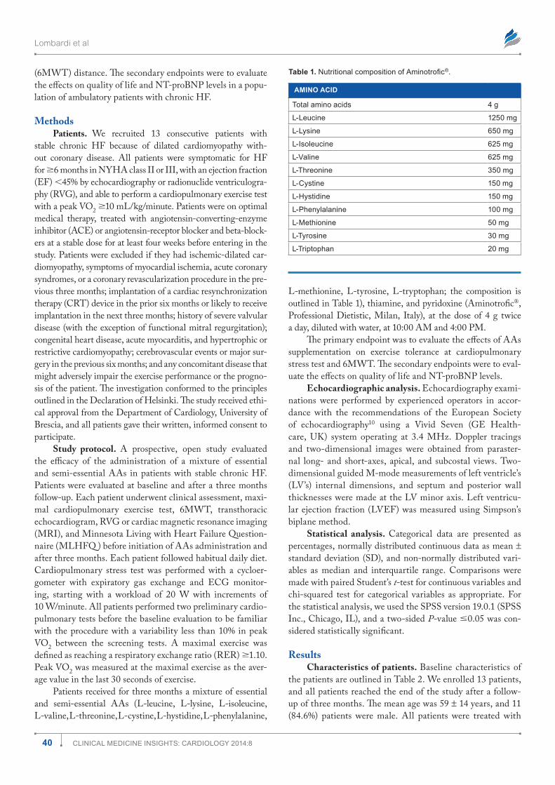
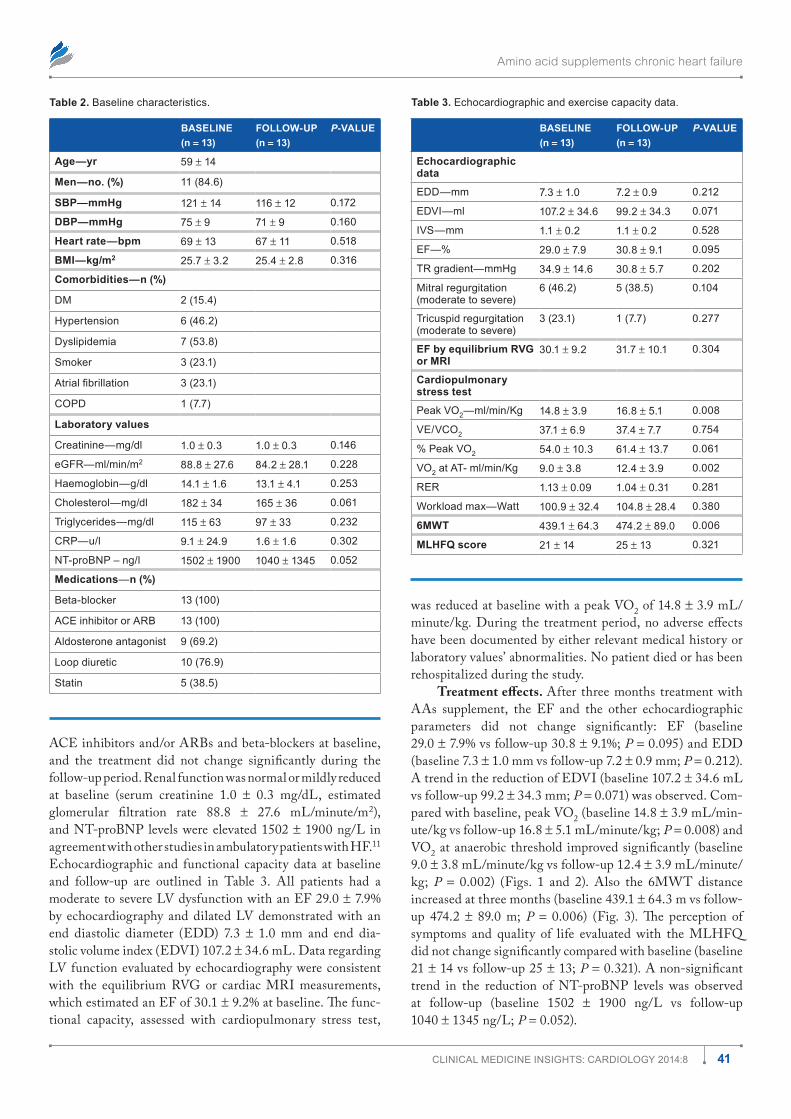
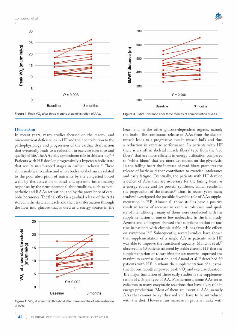
treatment effects. After three months treatment with AAs supplement, the EF and the other echocardiographic parameters did not change significantly: EF (baseline 29.0 ± 7.9% vs follow-up 30.8 ± 9.1%; P = 0.095) and EDD (baseline 7.3 ± 1.0 mm vs follow-up 7.2 ± 0.9 mm; P = 0.212). A trend in the reduction of EDVI (baseline 107.2 ± 34.6 mL vs follow-up 99.2 ± 34.3 mm; P = 0.071) was observed. Com-pared with baseline, peak VO2 (baseline 14.8 ± 3.9 mL/min-ute/kg vs follow-up 16.8 ± 5.1 mL/minute/kg; P = 0.008) and VO2 at anaerobic threshold improved significantly (baseline 9.0 ± 3.8 mL/minute/kg vs follow-up 12.4 ± 3.9 mL/minute/kg; P = 0.002) (Figs. 1 and 2). Also the 6MWT distance increased at three months (baseline 439.1 ± 64.3 m vs follow-up 474.2 ± 89.0 m; P = 0.006) (Fig. 3). The perception of symptoms and quality of life evaluated with the MLHFQ did not change significantly compared with baseline (baseline 21 ± 14 vs follow-up 25 ± 13; P = 0.321). A non-significant trend in the reduction of NT-proBNP levels was observed at follow-up (baseline 1502 ± 1900 ng/L vs follow-up 1040 ± 1345 ng/L; P = 0.052).
table 2. Baseline characteristics.
bASElinE(n = 13)
FOllOw-uP(n = 13)
p-vAluE
Age—yr 59 ± 14
men—no. (%) 11 (84.6)
SbP—mmHg 121 ± 14 116 ± 12 0.172
dbP—mmHg 75 ± 9 71 ± 9 0.160
Heart rate—bpm 69 ± 13 67 ± 11 0.518
bmi—kg/m2 25.7 ± 3.2 25.4 ± 2.8 0.316
Comorbidities—n (%)
dM 2 (15.4)
hypertension 6 (46.2)
dyslipidemia 7 (53.8)
smoker 3 (23.1)
Atrial fibrillation 3 (23.1)
CoPd 1 (7.7)
laboratory values
Creatinine—mg/dl 1.0 ± 0.3 1.0 ± 0.3 0.146
egFr—ml/min/m2 88.8 ± 27.6 84.2 ± 28.1 0.228
haemoglobin—g/dl 14.1 ± 1.6 13.1 ± 4.1 0.253
Cholesterol—mg/dl 182 ± 34 165 ± 36 0.061
triglycerides—mg/dl 115 ± 63 97 ± 33 0.232
CrP—u/l 9.1 ± 24.9 1.6 ± 1.6 0.302
nt-proBnP – ng/l 1502 ± 1900 1040 ± 1345 0.052
medications—n (%)
Beta-blocker 13 (100)
aCe inhibitor or arB 13 (100)
aldosterone antagonist 9 (69.2)
loop diuretic 10 (76.9)
statin 5 (38.5)
table 3. echocardiographic and exercise capacity data.
bASElinE(n = 13)
FOllOw-uP(n = 13)
p-vAluE
Echocardiographic data
edd—mm 7.3 ± 1.0 7.2 ± 0.9 0.212
edVi—ml 107.2 ± 34.6 99.2 ± 34.3 0.071
iVs—mm 1.1 ± 0.2 1.1 ± 0.2 0.528
eF—% 29.0 ± 7.9 30.8 ± 9.1 0.095
tr gradient—mmhg 34.9 ± 14.6 30.8 ± 5.7 0.202
Mitral regurgitation (moderate to severe)
6 (46.2) 5 (38.5) 0.104
tricuspid regurgitation (moderate to severe)
3 (23.1) 1 (7.7) 0.277
EF by equilibrium Rvg or mRi
30.1 ± 9.2 31.7 ± 10.1 0.304
Cardiopulmonary stress test
Peak Vo2—ml/min/Kg 14.8 ± 3.9 16.8 ± 5.1 0.008
Ve/VCo2 37.1 ± 6.9 37.4 ± 7.7 0.754
% Peak Vo2 54.0 ± 10.3 61.4 ± 13.7 0.061
Vo2 at at- ml/min/Kg 9.0 ± 3.8 12.4 ± 3.9 0.002
rer 1.13 ± 0.09 1.04 ± 0.31 0.281
Workload max—Watt 100.9 ± 32.4 104.8 ± 28.4 0.380
6mwt 439.1 ± 64.3 474.2 ± 89.0 0.006
mlHFQ score 21 ± 14 25 ± 13 0.321
Lombardi et al
42 CliniCal MediCine insights: Cardiology 2014:8
discussionIn recent years, many studies focused on the macro- and micronutrient deficiencies in HF and their contribution to the pathophysiology and progression of the cardiac dysfunction that eventually leads to a reduction in exercise tolerance and quality of life. The AAs play a prominent role in this setting.9,12 Patients with HF develop progressively a hypercatabolic state that results in advanced stages in cardiac cachexia.13 These abnormalities in cardiac and whole body metabolism are related to the poor absorption of nutrients by the congested bowel wall; by the activation of local and systemic inflammatory response; by the neurohormonal abnormalities, such as sym-pathetic and RAAs activation; and by the prevalence of cata-bolic hormones. The final effect is a gradual release of the AAs stored in the skeletal muscle and their transformation through the liver into glucose that is used as a energy source in the
heart and in the other glucose-dependent organs, namely the brain. The continuous release of AAs from the skeletal muscle leads to a progressive loss in muscle bulk and thus a reduction in exercise performance. In patients with HF there is a shift in skeletal muscle fibers’ type from the “red fibers” that are more efficient in energy utilization compared to “white fibers” that are more dependent on the glycolysis. In the failing heart the increase of read fibers promotes the release of lactic acid that contributes to exercise intolerance and early fatigue. Eventually, the patients with HF develop a deficit of AAs that are necessary for the failing heart as a energy source and for protein synthesis, which results in the progression of the disease.14 Thus, in recent years many studies investigated the possible favorable role of AAs supple-mentation in HF. Almost all those studies have a positive result in terms of increase in exercise tolerance and qual-ity of life, although many of them were conducted with the supplementation of one or few molecules. In the first study, Azuma and colleagues showed that supplementation of tau-rine in patients with chronic stable HF has favorable effects on symptoms.15,16 Subsequently, several studies have shown that supplementation of a single AA in patients with HF was able to improve the functional capacity. Mancini et al.17 observed in 60 patients affected by stable chronic HF that the supplementation of l-carnitine for six months improved the maximum exercise duration, and Anand et al.18 described 30 patients with HF in whom the supplementation of l-carni-tine for one month improved peak VO2 and exercise duration. The major limitation of these early studies is the supplemen-tation of a single type of AA. Furthermore, some AAs act as cofactors in many enzymatic reactions that have a key role in energy production. Most of them are essential AAs, namely AAs that cannot be synthesized and have to be introduced with the diet. However, an increase in protein intake with
0
Baseline 3 months
P = 0.008
5
10
15
Pea
k V
O2
(mL
/min
/kg
)
20
25
30
Figure 1. Peak Vo2 after three months of administration of aas.
0
Baseline 3 months
P = 0.002
5
10
15
VO
2 a
t an
aero
bic
th
resh
old
(m
L/m
in/k
g)
20
25
Figure 2. Vo2 at anaerobic threshold after three months of administration of aas.
200
Baseline 3 months
P = 0.006
300
400
500
6MW
T d
ista
nce
(m
)
600
700
Figure 3. 6MWt distance after three months of administration of aas.
Amino acid supplements chronic heart failure
43CliniCal MediCine insights: Cardiology 2014:8
diet is not enough to counteract the AAs deficit.19 In this view, mixed AAs supplementation should be encouraged. In our study, we treated 13 patients with chronic stable HF, in NYHA class II–III with a mixture of essential and semi-essential AAs supplementation at the dose of 4 g twice a day. After three months of follow-up, exercise tolerance, evalu-ated as peak VO2, VO2 at anaerobic threshold and 6MWT distance improved. Moreover, we observed a trend in the reduction of EDV and of NT-proBNP levels. AAs treatment resulted well tolerated, without reported side effects or abnor-malities in laboratory tests.
Our results are consistent with the previous studies. Aquilani et al.20 randomized in a double-blind fashion 95 patients with stable chronic HF in NYHA class II–III to essential AAs mixture (4 g twice daily) or placebo, and after one month of follow-up, observed a significant improvement in exercise capacity in the treatment with exercise maximal workload (P , 0.01), duration (P , 0.02) and peak VO2 (P , 0.02). Similar results were obtained in another open label, placebo-controlled trial, which enrolled 15 stable chronic HF patients with EF ,40%. In the group treated with essential AAs mixture, they observed an improvement in the 6MWT distance.21
In our study, the results of almost all the trials are prom-ising, finding a positive association between mixed AAs supplementation and improvement in functional capacity, and resulted well tolerated and safe. Different from previ-ous studies, we have analyzed a very heterogeneous mixture composed of 11 AAs. Our results encourage a wide range of micronutrient supplementation considering the serious depletion of macro- and micronutrients present in patients with HF.
Our study has some limitations, namely small sample size, the absence of a control group, and the short period of follow-up. Another major limitation is that in our study, we did not evaluate myocardial viability prior to AAs supplementation. This may be a confounding factor as muscle-depletion by itself and a more advanced stage of the disease are associated with a more pronounced exercise intolerance. Another limitation is the lack of plasma assay of a marker of muscle metabolism (eg, lactic acid) that could more accurately confirm the favor-able effects of the mixture of AAs on the functional capacity.
In our study, supplementation with essential and semi-essential AAs for three months in patients with sta-ble chronic HF improved exercise tolerance. However, it did not change symptoms’ perception and quality of life and NT-proBNP levels. AAs treatment resulted safe and was well tolerated by all patients. We conclude that AAs supplementation may be a useful non-pharmacologic treat-ment, associated with conventional therapy, in patients with HF. Larger studies are needed to confirm these data and to investigate the potential benefits of AAs supplementation on the patients’ outcomes.
Author contributionsConceived and designed the experiments: CL, VC, MM. Analyzed the data: VC, VL, EV, FQ , RR. Wrote the first draft of the manuscript: VC, CL, FG. Contributed to the writing of the manuscript: MM, SN. Agree with manuscript results and conclusions: CL, VC, VL, EV, FQ , FG, RR, SN, MG, MM. Jointly developed the structure and arguments for the paper: MM, CL, VC. Made critical revisions and approved final version: MG, MM. All authors reviewed and approved of the final manuscript.
diSClOSuRES And EtHiCSAs a requirement of publication the authors have provided signed confirmation of their compliance with ethical and legal obligations including but not limited to compliance with iCMJe authorship and competing interests guidelines, that the article is neither under consideration for publication nor published elsewhere, of their compliance with legal and ethical guidelines concerning human and animal research participants (if applicable), and that permission has been obtained for reproduction of any copy-righted material. this article was subject to blind, independent, expert peer review. the reviewers reported no competing interests.
references 1. Cleland JG, Swedberg K, Follath F, et al. The EuroHeart Failure survey
programme—a survey on the quality of care among patients with heart fail-ure in Europe. Part 1: patient characteristics and diagnosis. Eur Heart J. 2003;24:442–63.
2. McCullough PA, Philbin EF, Spertus JA, Kaatz S, Sandberg KR. Confirma-tion of a heart failure epidemic: findings from the Resource Utilization Among Congestive Heart Failure (REACH) study. J Am Coll Cardiol. 2002;39:60–9.
3. Rosamond W, Flegal K, Furie K, et al. Heart disease and stroke statistics—2008 update: a report from the American Heart Association Statistics Committee and Stroke Statistics Subcommittee. Circulation. 2008;117:e25–146.
4. Hunt SA, Abraham WT, Chin MH, et al. Focused update incorporated into the ACC/AHA 2005 guidelines for the diagnosis and management of heart failure in adults: a report of the American College of Cardiology Foundation/Ameri-can Heart Association Task Force on Practice Guidelines. J Am Coll Cardiol. 2009;53:e1–90.
5. Fang J, Mensah GA, Croft JB, Keenan NL. Heart failure-related hospitalization in the U.S., 1979 to 2004. J Am Coll Cardiol. 2008;52:428–34.
6. Ahmed A, Allman RM, Fonarow GC, et al. Incident heart failure hospital-ization and subsequent mortality in chronic heart failure: a propensity-matched study. J Card Fail. 2008;14:211–8.
7. Gheorghiade M, Pang PS. Acute heart failure syndromes. J Am Coll Cardiol. 2009;53:557–73.
8. Milo-Cotter O, Cotter-Davison B, Lombardi C, Sun H, Bettari L, et al. Neu-rohormonal activation in acute heart failure: results from VERITAS. Cardiology. 2011;119(2):96–105.
9. Taegtmeyer H, Harinstein ME, Gheorghiade M. More than bricks and mortar: comments on protein and amino acid metabolism in the heart. Am J Cardiol. 2008;101:3E–7E.
10. Lang RM, Bierig M, Devereux RB, et al. Recommendations for chamber quan-tification. Eur J Echocardiogr. 2006;7:79–108.
11. Januzzi JL Jr, Rehman SU, Mohammed AA, et al. Use of amino-terminal pro-B-type natriuretic peptide to guide outpatient therapy of patients with chronic left ventricular systolic dysfunction. J Am Coll Cardiol. 2011;58(18):1881–9.
12. Ardehali H, Sabbah HN, Burke MA, et al. Targeting myocardial substrate metabolism in heart failure: potential for new therapies. Eur J Heart Fail. 2012;14(2):120–9.
13. von Haehling S, Doehner W, Anker SD. Nutrition, metabolism, and the complex pathophysiology of cachexia in chronic heart failure. Cardiovasc Res. 2007;73:298–309.
14. Soukoulis V, Dihu JB, Sole M, et al. Micronutrient deficiencies an unmet need in heart failure. J Am Coll Cardiol. October 27, 2009;54(18):1660–73.
15. Azuma J, Hasegawa H, Sawamura A, et al. Therapy of congestive heart failure with orally administered taurine. Clin Ther. 1983;5:398–408.
16. Azuma J, Sawamura A, Awata N, et al. Therapeutic effect of taurine in congestive heart failure: a double-blind crossover trial. Clin Cardiol. 1985;8:276–82.
Lombardi et al
44 CliniCal MediCine insights: Cardiology 2014:8
17. Mancini M, Rengo F, Lingetti M, Sorrentino GP, Nolfe G. Controlled study on the therapeutic efficacy of propionyl-l-carnitine in patients with congestive heart failure. Arzneimittelforschung. 1992;42:1101–4.
18. Anand I, Chandrashekhan Y, De Giuli F, et al. Acute and chronic effects of propionyl-l-carnitine on the hemodynamics, exercise capacity, and hormones in patients with congestive heart failure. Cardiovasc Drugs Ther. 1998;12:291–9.
19. Aquilani R, Opasich C, Gualco A, et al. Adequate energy-protein intake is not enough to improve nutritional and metabolic status in muscle-depleted patients with chronic heart failure. Eur J Heart Fail. 2008;10(11):1127–35.
20. Aquilani R, Viglio S, Iadarola P, et al. Oral amino acid supplements improve exercise capacities in elderly patients with chronic heart failure. Am J Cardiol. 2008;101(suppl):104E–10E.
21. Scognamiglio R, Testa A, Aquilani R, Dioguardi FS, Pasini E. Impairment in walking capacity and myocardial function in the elderly: is there a role for non-pharmacologic therapy with nutritional amino acid supplements? Am J Cardiol. 2008;101(suppl):78E–81E.
Top Related
Copyright © 2022 FDOKUMEN

