


Bahasa
Halaman
Hukum

M A J O R A R T I C L E
Discovery and Validation of Biomarkers to GuideClinical Management of Pneumonia in AfricanChildren
Honglei Huang,1 Readon C. Ideh,4 Evelyn Gitau,3,5 Marie L. Thézénas,1 Muminatou Jallow,4 Bernard Ebruke,4
Osaretin Chimah,4 Claire Oluwalana,4 Henri Karanja,5 Grant Mackenzie,4 Richard A. Adegbola,4 Dominic Kwiatkowski,1
Benedikt M. Kessler,1 James A. Berkley,2,5 Stephen R. C. Howie,4 and Climent Casals-Pascual1
1Wellcome Trust Centre for Human Genetics and 2Centre for Clinical Vaccinology and Tropical Medicine, Nuffield Department of Medicine, University ofOxford, and 3Liverpool School of Tropical Medicine, United Kingdom; 4Child Survival Theme, Medical Research Council Unit, The Gambia; and 5KenyaMedical Research Institute, Centre for Geographical Medicine Research (Coast), Kilifi
Background. Pneumonia is the leading cause of death in children globally. Clinical algorithms remain suboptimal fordistinguishing severe pneumonia from other causes of respiratory distress such asmalaria or distinguishing bacterial pneu-monia and pneumonia from others causes, such as viruses. Molecular tools could improve diagnosis and management.
Methods. Weconductedamass spectrometry–basedproteomic study to identifyandvalidatemarkersof severity in390Gambian children with pneumonia (n = 204) and age-, sex-, and neighborhood-matched controls (n = 186). Independentvalidation was conducted in 293 Kenyan children with respiratory distress (238 with pneumonia, 41 with Plasmodium fal-ciparummalaria, and 14 with both). Predictive value was estimated by the area under the receiver operating characteristiccurve (AUC).
Results. Lipocalin 2 (Lpc-2)was the best protein biomarkerof severe pneumonia (AUC, 0.71 [95%confidence interval,.64–.79]) and highly predictive of bacteremia (78% [64%–92%]), pneumococcal bacteremia (84% [71%–98%]), and “prob-able bacterial etiology” (91% [84%–98%]). These results were validated inKenyan childrenwith severemalaria and respira-tory distress who also met the World Health Organization definition of pneumonia. The combination of Lpc-2 andhaptoglobin distinguished bacterial versusmalaria origin of respiratory distresswith high sensitivity and specificity inGam-bian children (AUC, 99% [95% confidence interval, 99%–100%]) and Kenyan children (82% [74%–91%]).
Conclusions. Lpc-2 and haptoglobin can help discriminate the etiology of clinically defined pneumonia and could beused to improve clinical management. These biomarkers should be further evaluated in prospective clinical studies.
Keywords. pneumonia; malaria; lipocalin-2/NGAL; respiratory infection; biomarkers.
Pneumonia is the leading cause of death in young chil-dren globally, accounting for almost 2 million deathsevery year, mainly in developing countries [1–4]. InThe Gambia, acute lower respiratory tract infection,
principally pneumonia, is a leading cause of death inyoung children [5, 6].
To reach and push beyond the United Nations’Millen-nium Development Goal 4 for child survival, the numberof deaths caused bypneumoniamust be reduced. Thiswillrequire acombinationof effectivepreventivemeasuresandimproved clinical management [7–10]. Previous studieshave shown that delayed referral is one of themost impor-tant risk factors fordeath inchildrenwithpneumonia [11].Therefore, current clinical algorithms to refer patientswith pneumonia to hospital are commonly based on diag-nostic sensitivity rather than specificity. Consequently,overreferral of pneumonia cases has been a significantproblem with the World Health Organization (WHO)case management strategy in some settings [12].
Received 6 September 2013; accepted 20 March 2014; electronically published2 April 2014.
A patent on the biomarkers described has been filed by the University of Oxfordthrough ISIS Ltd (PCT/GB2013/051768).
Correspondence: Climent Casals-Pascual, MD, DPhil, Wellcome Trust Centre forHuman Genetics, Roosevelt Drive, Oxford, OX3 7BN, UK ([email protected]).
Clinical Infectious Diseases 2014;58(12):1707–15© The Author 2014. Published by Oxford University Press on behalf of the InfectiousDiseasesSocietyofAmerica. This isanOpenAccessarticle distributedunder the termsof the Creative Commons Attribution License (http://creativecommons.org/licenses/by/3.0/), which permits unrestricted reuse, distribution, and reproduction in anymedium, provided the original work is properly cited.DOI: 10.1093/cid/ciu202
Diagnosis-Based Management of Acute Respiratory Infections in Africa • CID 2014:58 (15 June) • 1707
Importantly, clinical criteria do not distinguish bacterial causes ofpneumonia from other causes such as viruses that do not requireantibiotic treatment.
The analysis of biofluids (eg, plasma) using mass spectrome-try–based methods has been widely adopted for biomarker dis-covery [13–15]. Once identified and validated, proteins ofclinical value could be incorporated into rapid, point-of-care tests.
In African children, pneumonia is not the only cause of re-spiratory distress in children. In malaria endemic areas, Plasmo-dium falciparum infection may cause respiratory distress andthe overlap of these conditions frequently compromises the di-agnosis and management of these patients [16–18]. In acute pe-diatric admissions, the clinical syndrome of severe pneumoniaoverlapped in 39% of severe malaria cases [19].
We conducted a study to describe the plasma proteomic sig-nature in samples from Gambian children with severe pneumo-nia and nonsevere pneumonia and controls. We identified andvalidated biomarkers to (1) predict disease severity in childrenwith pneumonia, (2) predict blood culture positivity, (3) predictprobable bacterial etiology of children with pneumonia, (4)evaluate biomarker performance to discriminate respiratorydistress caused by pneumonia or by severe malaria, and (5) pro-vide an independent validation for the diagnostic performanceof these markers in Kenyan children.
METHODS
Study Sites and Populations StudiedThe study participants were infants and children from theGreater Banjul and Basse areas aged 2–59 months taking partin a case-control study of childhood pneumonia and childrenadmitted at the Kilifi District Hospital with a diagnosis oflower respiratory tract infection (Table 1). The sites, popula-tions studied, and clinical definitions are described in detail inthe Supplementary Methods and Supplementary Table 1.
Biomarker Discovery and Validation StudiesPlasma samples from Gambian infants and children aged 2–59months were used for mass spectrometry–based proteomicstudies (see Supplementary Methods for details). The concen-trations of selected proteins (C-reactive protein [CRP], vonWil-lebrand factor [vWF], lipocalin 2 [Lp-2] and haptoglobin) weremeasured with enzyme-linked immunosorbent assay (R&DSystems), according to the manufacturer’s instructions.
Data Management and Statistical AnalysesClinical data were collected on standardized forms and double en-tered. Univariate and multiple logistic regression models were fit-ted for all clinical variables using disease severity and probablebacterial etiology as dependent variables. The interaction of
independent variables was checked using the likelihood ratiotest. Data were analyzed with Stata 11 software (StataCorp).
Diagnostic Performance and Selection of Clinical Variables forMultivariate ModelsThe area under the receiver operating characteristic curve(AUC) was used to compare the sensitivity and specificity of se-lected markers. Cutoff values were chosen based on highest sen-sitivity and specificity to predict outcome using the roctab/detail function (Stata 11.0). When the diagnostic performancewas assessed for >1 variable, the estimates were derived from alogistic regression model using the selected markers or clinicalfeatures as independent variables and the condition to diagnoseas the dependent variable. These analyses were carried out usingthe lroc, lstat, and roctab/graph functions in Stata. The positiveand negative likelihood ratios have been calculated for the bio-marker concentrations with the highest sensitivity and specific-ity for predicting severe pneumonia.
Clinical variables and/or molecular markers were included inthemultivariatemodels if (1) the variable showeda statistically sig-nificant association (P < .05) in the univariate model and (2) thevariable had not been used as a criterion for a priori classification.For example, respiratory ratewas combinedwithmolecularmark-ers to compare nonsevere and severe pneumonia because this var-iable was not used as a criterion to separate the 2 conditions.Ordinal univariate logistic regressionmodels usingdisease severitycoded as 0–3 (from control [0] to very severe [3]) as the dependentvariable were used to evaluate the significance of the associationbetween biomarker concentration and clinical outcome.
Ethical ApprovalWritten informed consent was given by the parent or guardianof each participant. Joint Gambia Government/Medical Re-search Council (MRC) Ethical Committee approval wasobtained for both the pneumonia study (SCC/EC 1062) andthe severe malaria study (SCC/EC 630 and 670). The use ofthe archived plasma samples from Kenya was approved by theKenya Medical Research Institute (KEMRI) Ethics ReviewCommittee (SSC 2280).
RESULTS
Diagnostic Performance of Clinical Features and ProteinBiomarkers to Discriminate Severe Pneumonia and NonseverePneumoniaThe proteomic analysis identified 238 proteins in children withsevere pneumonia, 316 in children with nonsevere pneumonia,and 268 in healthy controls. The difference in protein numbersacross different groups and batches was not significantly different(analysis of variance, P = .35). We identified 23 differentially reg-ulated proteins (>1.5 fold) that were present in ≥2 of 3 batches
1708 • CID 2014:58 (15 June) • Huang et al
when severe and nonsevere cases were compared and 19 whennonsevere cases and controls were compared (SupplementaryFigure 2). Of these 42 proteins, only 8 (19%) indicated a progres-sion pattern, namely, increasing concentration from controls tomild to severe cases. Based on the number of peptides used toidentify these proteins and their biological relevance, Lpc-2,CRP, and vWF were selected for further validation (Table 2).
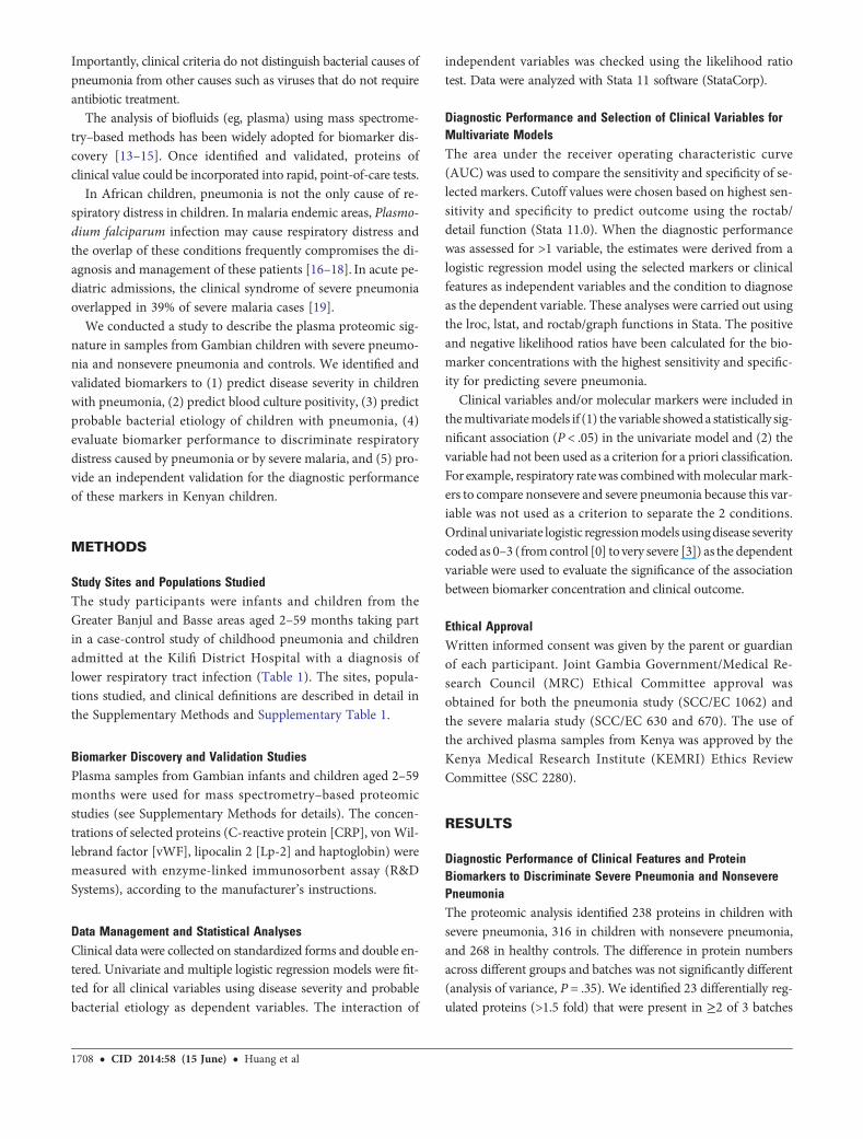
Lpc-2, CRP, and vWF levels were significantly higher in chil-dren with severe pneumonia than in those with nonseverepneumonia (Supplementary Figure 3). Lpc-2 was the best pre-dictor of severe pneumonia, with a sensitivity of 72.3% and aspecificity of 70.1% (AUC, 0.71 [95% confidence interval [CI],.64–.79]; Table 3). In children with Lpc-2 levels >118 ng/mL,the odds of having severe disease increased by nearly 3-fold(odds ratio [OR], 2.69 [95% CI, 1.08–6.69). A CRP concentra-tion >157 µg/mL was associated with increased disease severity(OR, 3.55 [95% CI, 1.41–8.93]), but despite its good sensitivityto predict disease severity (70.8%), its specificity was low(56.2%). Similarly, plasma vWF concentrations >648 mU/mLwere associated with a 5-fold increase in the odds of severepneumonia (OR, 5.26 [95% CI, 2.42–11.4]), with good sensitiv-ity (87.0%) but poor specificity (41.7%; Figure 1).
The best combination of clinical and molecular markers topredict severe pneumonia included respiratory rate, crackles,Lpc-2, and CRP. The addition of molecular markers to the clin-ical features had no impact on sensitivity but increased specif-icity from 68.0% to 82.0% (Figure 1), and the effect was superiorto radiological changes (end point consolidation), crackles, andpositive blood culture combined (Supplementary Figure 6). Thesensitivity of this combination of markers increased to 94.7%(95% CI, 88.4%–100%) in children enrolled during the dry sea-son, possibly owing to the absence of malaria cases (Supple-mentary Figure 4). Only 3 children of 204 (1.5%) identified ashaving pneumonia had a positive malaria slide. These childrenall had nonsevere pneumonia (2 enrolled during the dry seasonand 1 during the rainy season).
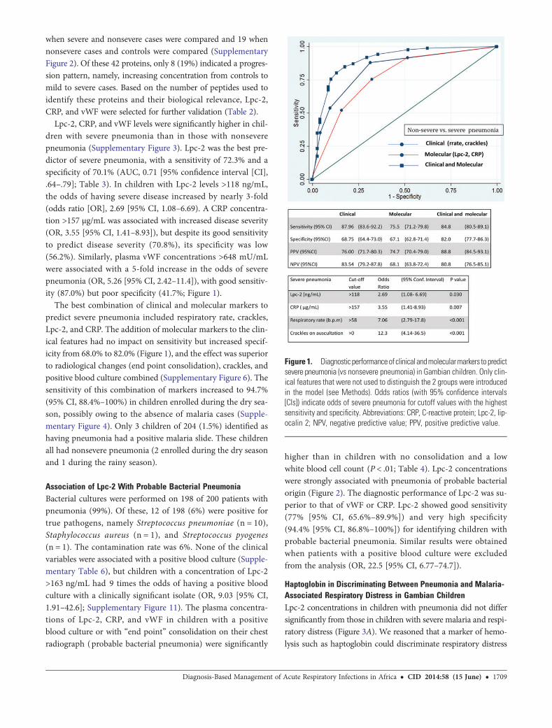
Association of Lpc-2 With Probable Bacterial PneumoniaBacterial cultures were performed on 198 of 200 patients withpneumonia (99%). Of these, 12 of 198 (6%) were positive fortrue pathogens, namely Streptococcus pneumoniae (n = 10),Staphylococcus aureus (n = 1), and Streptococcus pyogenes(n = 1). The contamination rate was 6%. None of the clinicalvariables were associated with a positive blood culture (Supple-mentary Table 6), but children with a concentration of Lpc-2>163 ng/mL had 9 times the odds of having a positive bloodculture with a clinically significant isolate (OR, 9.03 [95% CI,1.91–42.6]; Supplementary Figure 11). The plasma concentra-tions of Lpc-2, CRP, and vWF in children with a positiveblood culture or with “end point” consolidation on their chestradiograph (probable bacterial pneumonia) were significantly
higher than in children with no consolidation and a lowwhite blood cell count (P < .01; Table 4). Lpc-2 concentrationswere strongly associated with pneumonia of probable bacterialorigin (Figure 2). The diagnostic performance of Lpc-2 was su-perior to that of vWF or CRP. Lpc-2 showed good sensitivity(77% [95% CI, 65.6%–89.9%]) and very high specificity(94.4% [95% CI, 86.8%–100%]) for identifying children withprobable bacterial pneumonia. Similar results were obtainedwhen patients with a positive blood culture were excludedfrom the analysis (OR, 22.5 [95% CI, 6.77–74.7]).
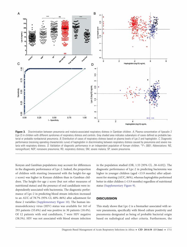
Haptoglobin in Discriminating Between Pneumonia and Malaria-Associated Respiratory Distress in Gambian ChildrenLpc-2 concentrations in children with pneumonia did not differsignificantly from those in children with severe malaria and respi-ratory distress (Figure 3A). We reasoned that a marker of hemo-lysis such as haptoglobin could discriminate respiratory distress
Figure 1. Diagnostic performanceof clinical andmolecularmarkers to predictsevere pneumonia (vs nonsevere pneumonia) in Gambian children. Only clin-ical features that were not used to distinguish the 2 groups were introducedin the model (see Methods). Odds ratios (with 95% confidence intervals[CIs]) indicate odds of severe pneumonia for cutoff values with the highestsensitivity and specificity. Abbreviations: CRP, C-reactive protein; Lpc-2, lip-ocalin 2; NPV, negative predictive value; PPV, positive predictive value.
Diagnosis-Based Management of Acute Respiratory Infections in Africa • CID 2014:58 (15 June) • 1709
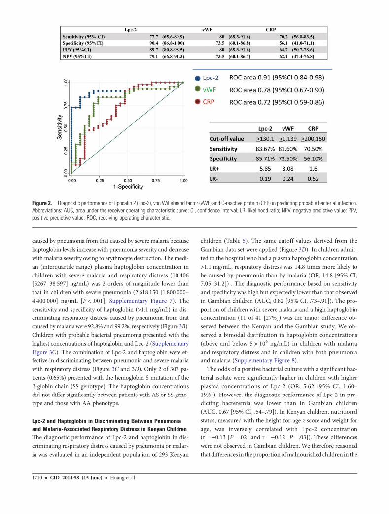
caused by pneumonia from that caused by severe malaria becausehaptoglobin levels increase with pneumonia severity and decreasewith malaria severity owing to erythrocyte destruction. The medi-an (interquartile range) plasma haptoglobin concentration inchildren with severe malaria and respiratory distress (10 406[5267–38 597] ng/mL) was 2 orders of magnitude lower thanthat in children with severe pneumonia (2 618 150 [1 800 000–4 400 000] ng/mL [P < .001]; Supplementary Figure 7). Thesensitivity and specificity of haptoglobin (>1.1 mg/mL) in dis-criminating respiratory distress caused by pneumonia from thatcaused bymalariawere 92.8% and 99.2%, respectively (Figure 3B).Children with probable bacterial pneumonia presented with thehighest concentrations of haptoglobin and Lpc-2 (SupplementaryFigure 3C). The combination of Lpc-2 and haptoglobin were ef-fective in discriminating between pneumonia and severe malariawith respiratory distress (Figure 3C and 3D). Only 2 of 307 pa-tients (0.65%) presented with the hemoglobin S mutation of theβ-globin chain (SS genotype). The haptoglobin concentrationsdid not differ significantly between patients with AS or SS geno-type and those with AA phenotype.
Lpc-2 and Haptoglobin in Discriminating Between Pneumoniaand Malaria-Associated Respiratory Distress in Kenyan ChildrenThe diagnostic performance of Lpc-2 and haptoglobin in dis-criminating respiratory distress caused by pneumonia or malar-ia was evaluated in an independent population of 293 Kenyan
children (Table 5). The same cutoff values derived from theGambian data set were applied (Figure 3D). In children admit-ted to the hospital who had a plasma haptoglobin concentration>1.1 mg/mL, respiratory distress was 14.8 times more likely tobe caused by pneumonia than by malaria (OR, 14.8 [95% CI,7.05–31.2]) . The diagnostic performance based on sensitivityand specificity was high but expectedly lower than that observedin Gambian children (AUC, 0.82 [95% CI, .73–.91]). The pro-portion of children with severe malaria and a high haptoglobinconcentration (11 of 41 [27%]) was the major difference ob-served between the Kenyan and the Gambian study. We ob-served a bimodal distribution in haptoglobin concentrations(above and below 5 × 10⁶ ng/mL) in children with malariaand respiratory distress and in children with both pneumoniaand malaria (Supplementary Figure 8).
The odds of a positive bacterial culture with a significant bac-terial isolate were significantly higher in children with higherplasma concentrations of Lpc-2 (OR, 5.62 [95% CI, 1.60–19.6]). However, the diagnostic performance of Lpc-2 in pre-dicting bacteremia was lower than in Gambian children(AUC, 0.67 [95% CI, .54–.79]). In Kenyan children, nutritionalstatus, measured with the height-for-age z score and weight forage, was inversely correlated with Lpc-2 concentration(r =−0.13 [P = .02] and r = −0.12 [P = .03]). These differenceswere not observed in Gambian children. We therefore reasonedthat differences in theproportionofmalnourished children in the
Figure 2. Diagnostic performance of lipocalin 2 (Lpc-2), von Willebrand factor (vWF) and C-reactive protein (CRP) in predicting probable bacterial infection.Abbreviations: AUC, area under the receiver operating characteristic curve; CI, confidence interval; LR, likelihood ratio; NPV, negative predictive value; PPV,positive predictive value; ROC, receiving operating characteristic.
1710 • CID 2014:58 (15 June) • Huang et al
Kenyan and Gambian populations may account for differencesin the diagnostic performance of Lpc-2. Indeed, the proportionof children with stunting (measured with the height-for-agez score) was higher in Kenyan children than in Gambian chil-dren. The height-for-age z score (but not other measures ofnutritional status) and the presence of oral candidiasis were in-dependently associated with bacteremia. The diagnostic perfor-mance of Lpc-2 in predicting blood stream infection increasedto an AUC of 78.7% (95% CI, 60%–86%) after adjustment forthese 2 variables (Supplementary Figure 10). The human im-munodeficiency virus (HIV) status was available for 163 of293 patients (55.6%) and was positive in 30 patients (18.4%).Of 12 patients with oral candidiasis, 7 were HIV negative(58.3%). HIV was not associated with blood stream infection
in the population studied (OR, 1.53 [95% CI, .38–6.03]). Thediagnostic performance of Lpc-2 in predicting bacteremia washigher in younger children (aged <13.9 months) after adjust-ment for stunting (AUC, 86%), whereas haptoglobin performedbetter in older children (>13.9 months) regardless of nutritionalstatus (Supplementary Figure 9).
DISCUSSION
This study shows that Lpc-2 is a biomarker associated with se-vere pneumonia, specifically with blood culture positivity andpneumonia designated as being of probable bacterial originbased on radiological and other criteria. Furthermore, the
Figure 3. Discrimination between pneumonia and malaria-associated respiratory distress in Gambian children. A, Plasma concentration of lipocalin 2(Lpc-2) in children with different syndromes of respiratory distress and controls. Gray shaded area indicates subanalysis of cases defined as probable bac-terial or probable nonbacterial pneumonia. B, Distribution of cases of respiratory distress based on plasma levels of Lpc-2 and haptoglobin. C, Diagnosticperformance (receiving operating characteristic curve) of haptoglobin in discriminating between respiratory distress caused by pneumonia and severe ma-laria with respiratory distress. D, Validation of diagnostic performance in an independent population of Kenyan children. *P < .0001; Abbreviations: NS,nonsignificant; NSP, nonsevere pneumonia; RD, respiratory distress; SM, severe malaria; SP, severe pneumonia.
Diagnosis-Based Management of Acute Respiratory Infections in Africa • CID 2014:58 (15 June) • 1711
combination of Lpc-2 with haptoglobin discriminates betweenpneumonia and malaria-associated respiratory distress.
To reduce the number of deaths caused by pneumonia, earlydiagnosis is critical for pneumonia cases due to bacterial infec-tion or likely to become severe, so appropriate treatment can beadministered promptly. However, the diagnosis of bacterialpneumonia is usually compromised by the lack of specificityof respiratory symptoms, which are commonly shared withother conditions that cause respiratory distress in children,many associated with high case fatality rates. In sub-SaharanAfrica P. falciparum malaria and bacterial blood stream infec-tions are frequent causes of respiratory distress in childrenand possibly pathogenically linked [20]. For a biomarker tobe helpful in this clinical context, its diagnostic performancehas to be superior to clinical examination, and its specificitymust be sufficient to guide clinical management.
The WHO definition of pneumonia severity used in ourstudy is an operational clinical algorithm rather than a “goldstandard.” This definition aims to reduce mortality by
improving referral practices and thus prioritizes sensitivityover specificity. The WHO has recently proposed a more specif-ic definition [21]. The starting hypothesis of our study was thatthe addition of molecular markers could increase the specificityof the clinical definition of severe pneumonia. In this context,we have reported 3 potential plasma biomarkers that alone orin association with clinical features could be used to improveclinical management by identifying pneumonia with high dis-ease severity or probable bacterial etiology.
Our results indicate that CRP and Lpc-2 can correctly distin-guish most patients with severe pneumonia from those withnonsevere pneumonia and probable bacterial from probablenonbacterial causes. The sensitivity of these markers combinedincreased to 94.7% when the analysis was performed exclusivelyin children with pneumonia recruited during the dry season.The specificity of this panel of markers decreased from 82%to 77% when the analysis was performed in children enrolledduring the rainy season (Supplementary Figure 4). Owing tothe high seasonality of malaria transmission in The Gambia
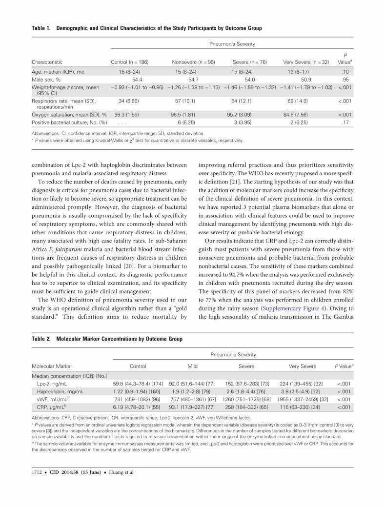
Table 1. Demographic and Clinical Characteristics of the Study Participants by Outcome Group
Characteristic
Pneumonia Severity
Control (n = 186) Nonsevere (n = 96) Severe (n = 76) Very Severe (n = 32)P
Valuea
Age, median (IQR), mo 15 (8–24) 15 (8–24) 15 (8–24) 12 (6–17) .10
Male sex, % 54.4 54.7 54.0 50.9 .95Weight-for-age z score, mean(95% CI)
−0.93 (−1.01 to −0.86) −1.26 (−1.38 to −1.13) −1.46 (−1.59 to −1.32) −1.41 (−1.79 to −1.03) <.001
Respiratory rate, mean (SD),respirations/min
34 (6.66) 57 (10.1) 64 (12.1) 69 (14.0) <.001
Oxygen saturation, mean (SD), % 98.3 (1.59) 96.5 (1.81) 95.2 (3.09) 84.6 (7.56) <.001
Positive bacterial culture, No. (%) . . . 6 (6.25) 3 (3.95) 2 (6.25) .17
Abbreviations: CI, confidence interval; IQR, interquartile range; SD, standard deviation.a P values were obtained using Kruskal-Wallis or χ2 test for quantitative or discrete variables, respectively.
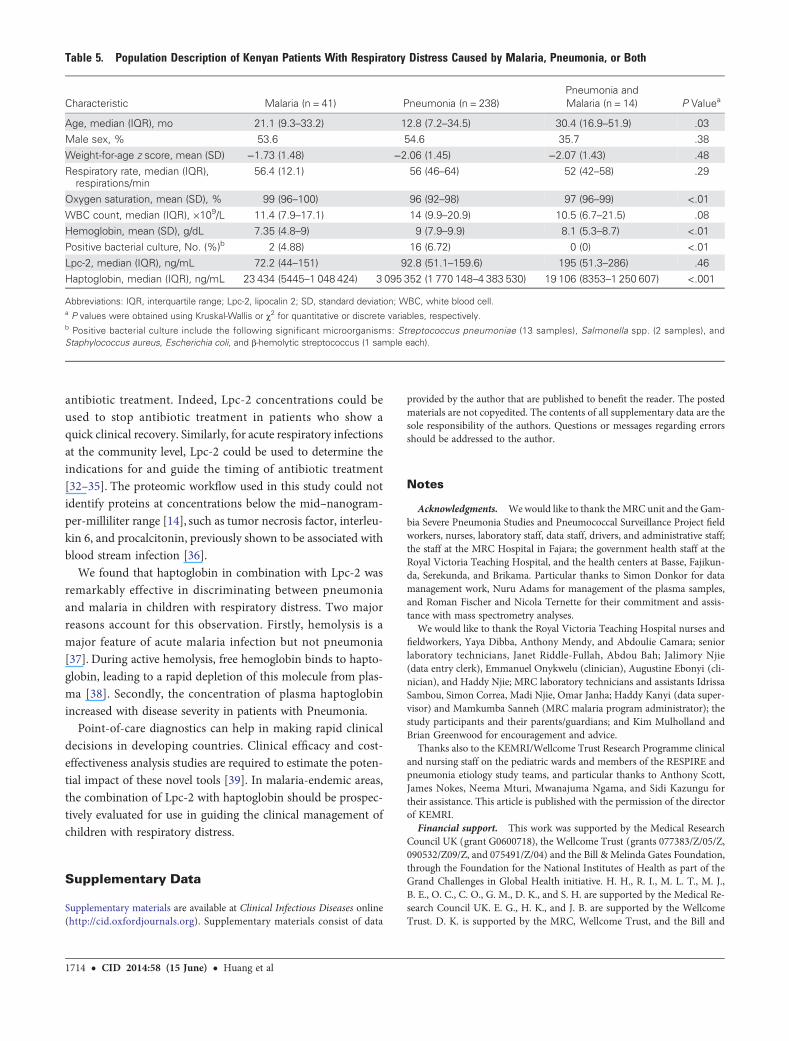
Table 2. Molecular Marker Concentrations by Outcome Group
Molecular Marker
Pneumonia Severity
Control Mild Severe Very Severe P Valuea
Median concentration (IQR) [No.]Lpc-2, ng/mL 59.8 (44.3–78.4) [174] 92.0 (51.6–144) [77] 152 (87.6–283) [73] 224 (139–455) [32] <.001
Haptoglobin, mg/mL 1.22 (0.8–1.94) [160] 1.9 (1.2–2.8) [79] 2.6 (1.8–4.4) [76] 3.8 (2.5–4.9) [32] <.001
vWF, mU/mLb 731 (459–1082) [96] 757 (480–1361) [67] 1260 (751–1725) [68] 1955 (1337–2459) [32] <.001CRP, µg/mLb 6.19 (4.78–20.1) [55] 93.1 (17.9–227) [77] 258 (184–332) [65] 116 (63–230) [24] <.001
Abbreviations: CRP, C-reactive protein; IQR, interquartile range; Lpc-2, lipocalin 2; vWF, von Willebrand factor.a P values are derived from an ordinal univariate logistic regression model wherein the dependent variable (disease severity) is coded as 0–3 (from control [0] to verysevere [3]) and the independent variables are the concentrations of the biomarkers. Differences in the number of samples tested for different biomarkers dependedon sample availability and the number of tests required to measure concentration within linear range of the enzyme-linked immunosorbent assay standard.b The sample volume available for enzyme immunoassay measurements was limited, and Lpc-2 and haptoglobin were prioritized over vWF or CRP. This accounts forthe discrepancies observed in the number of samples tested for CRP and vWF.
1712 • CID 2014:58 (15 June) • Huang et al

and the fact that both CRP and Lpc-2 have been shown to in-crease in children with malaria [22, 23], it is plausible that thisincrease in sensitivity and specificity is explained by the lownumber of malaria infections during the dry season. This in-crease in diagnostic specificity is likely to reduce but notcompletely remove the number of false-positive cases burden-ing busy health services. Similarly, our panel of biomarkers per-formed well against the more recent definition for severityproposed by the WHO in 2013 (Supplementary Figure 5).
Here we report that Lpc-2 is associated with blood culture pos-itivity. This is particularly important because clinical features didnot identify children with a positive bacterial blood culture. Wedid not confirm findings from some previous studies reportingan association between bacterial infection and the clinical signsof respiratory distress or high temperature (SupplementaryTable 6) [24–28].To our knowledge, we are also the first to reportthat Lpc-2 is associated with pneumonia of probable bacterial or-igin, defined by the presence of consolidation on the chest radio-graph or a positive blood culture. This was true for both primarybacterial and nonbacterial pneumonia definitions. The associa-tion of Lpc-2 with positive blood cultures and pneumonia ofprobable bacterial origin is biologically plausible and clinicallyimportant. We also observed that the association of Lpc-2 andbacteremia in Kenyan children, although significant, was partiallycompromised by the patient’s nutritional status. This observationmay be explained, at least in part, by the reduced ability of pa-tients with chronic malnutrition to generate an effective immuneresponse to infection. This limitation may be important in thedesign of prospective studies, and different cutoff values maybe required for populations with severe stunting.
The role of Lpc-2 in innate defense against bacterial infectionis well established. The transcription of the Lpc-2 gene has beenshown to be up-regulated in activated macrophages through Toll-like receptor 4 ligation and to interfere with bacterial iron uptake[29, 30]. More importantly, Lpc-2 transcription is increased by65-fold in the nasal mucosa of mice in response to S. pneumoniaeand Haemophilus influenzae colonization [31].
In resource-limited settings where laboratory facilities and ra-diology are rarely available to help diagnose bacterial pneumo-nia cases, molecular markers such as Lpc-2 could be developedinto a point-of-care diagnostic tool to target cases that require
Table 3. Diagnostic Performance of Clinical Features andMolecular Markers Associated With a Risk of Severe or VerySevere Pneumonia (vs Nonsevere Pneumonia)
Likelihood Ratioa
Clinical FeatureChildren,
No. AUC (95% CI) Positive Negative
Respiratory rateb 204 0.76 (.70–.83) . . . . . .
Cracklesb 204 0.72 (.66–.77) . . . . . .Heart rate 204 0.68 (.61–.75) . . . . . .
Pallor 204 0.57 (.52–.61) . . . . . .
Molecular markerLpc-2b 182 0.71 (.64–.79) 2.39 0.40
CRPb 166 0.68 (.60–.76) 1.66 0.53
vWFb 167 0.70 (.62–.78) 2.16 0.46
Abbreviations: AUC, area under the receiver operating characteristic curve; CI,confidence interval; CRP, C-reactive protein; Lpc-2, lipocalin 2; vWF, vonWillebrand factor.a The positive and negative likelihood ratios have been calculated for thebiomarker concentrations with the highest sensitivity and specificity forpredicting severe pneumonia (Lpc-2, 118 ng/mL; CRP, 157 mg/mL; andvWF, 648 mU/mL).b Variables independently associatedwith outcome in the multivariate models.
Table 4. Population Description of Patients With Pneumonia of Probable Bacterial or Probable Viral Etiology
Characteristic
Pneumonia
Probable Non-bacterial (n = 49) Probable Bacterial (n = 54) P Valuea
Age, mean (SD), mo 19 (10.5) 17.4 (11.6) .14
Male sex, % 50% 51% .90Weight-for-age z score, mean (SD) −1.03 (1.10) −1.37 (1.21) .24
Respiratory rate, mean (SD), respirations/min 57.0 (12.0) 62.6 (9.8) .007
Oxygen saturation, mean (SD), % 96.3 (2.64) 92.84 (7.09) <.01WBC count, mean (SD), ×109/L 10.1 (6.42) 23.7 (10.6) <.01
Hemoglobin, mean (SD), g/dL 10.3 (1.53) 8.65 (1.74) <.01
Molecular marker concentration, median (IQR) [No.]Lpc-2, ng/mL [42] 81.7 (45.7–109) [45] 282 (155–365) <.001
CRP, µg/mL [41] 175 (182–278) [47] 283 (142–350) <.001
vWF, mU/mL [34] 810 (647–1201) [45] 1514 (1205–2036) <.001
Abbreviations: CRP, C-reactive protein; IQR, interquartile range; Lpc-2, lipocalin 2; SD, standard deviation; vWF, von Willebrand factor; WBC, white blood cell.a P values were obtained using Mann-Whitney or χ2 test for quantitative or discrete variables, respectively.
Diagnosis-Based Management of Acute Respiratory Infections in Africa • CID 2014:58 (15 June) • 1713
antibiotic treatment. Indeed, Lpc-2 concentrations could beused to stop antibiotic treatment in patients who show aquick clinical recovery. Similarly, for acute respiratory infectionsat the community level, Lpc-2 could be used to determine theindications for and guide the timing of antibiotic treatment[32–35]. The proteomic workflow used in this study could notidentify proteins at concentrations below the mid–nanogram-per-milliliter range [14], such as tumor necrosis factor, interleu-kin 6, and procalcitonin, previously shown to be associated withblood stream infection [36].
We found that haptoglobin in combination with Lpc-2 wasremarkably effective in discriminating between pneumoniaand malaria in children with respiratory distress. Two majorreasons account for this observation. Firstly, hemolysis is amajor feature of acute malaria infection but not pneumonia[37]. During active hemolysis, free hemoglobin binds to hapto-globin, leading to a rapid depletion of this molecule from plas-ma [38]. Secondly, the concentration of plasma haptoglobinincreased with disease severity in patients with Pneumonia.
Point-of-care diagnostics can help in making rapid clinicaldecisions in developing countries. Clinical efficacy and cost-effectiveness analysis studies are required to estimate the poten-tial impact of these novel tools [39]. In malaria-endemic areas,the combination of Lpc-2 with haptoglobin should be prospec-tively evaluated for use in guiding the clinical management ofchildren with respiratory distress.
Supplementary Data
Supplementary materials are available at Clinical Infectious Diseases online(http://cid.oxfordjournals.org). Supplementary materials consist of data
provided by the author that are published to benefit the reader. The postedmaterials are not copyedited. The contents of all supplementary data are thesole responsibility of the authors. Questions or messages regarding errorsshould be addressed to the author.
Notes
Acknowledgments. Wewould like to thank theMRC unit and the Gam-bia Severe Pneumonia Studies and Pneumococcal Surveillance Project fieldworkers, nurses, laboratory staff, data staff, drivers, and administrative staff;the staff at the MRC Hospital in Fajara; the government health staff at theRoyal Victoria Teaching Hospital, and the health centers at Basse, Fajikun-da, Serekunda, and Brikama. Particular thanks to Simon Donkor for datamanagement work, Nuru Adams for management of the plasma samples,and Roman Fischer and Nicola Ternette for their commitment and assis-tance with mass spectrometry analyses.We would like to thank the Royal Victoria Teaching Hospital nurses and
fieldworkers, Yaya Dibba, Anthony Mendy, and Abdoulie Camara; seniorlaboratory technicians, Janet Riddle-Fullah, Abdou Bah; Jalimory Njie(data entry clerk), Emmanuel Onykwelu (clinician), Augustine Ebonyi (cli-nician), and Haddy Njie; MRC laboratory technicians and assistants IdrissaSambou, Simon Correa, Madi Njie, Omar Janha; Haddy Kanyi (data super-visor) and Mamkumba Sanneh (MRC malaria program administrator); thestudy participants and their parents/guardians; and Kim Mulholland andBrian Greenwood for encouragement and advice.Thanks also to the KEMRI/Wellcome Trust Research Programme clinical
and nursing staff on the pediatric wards and members of the RESPIRE andpneumonia etiology study teams, and particular thanks to Anthony Scott,James Nokes, Neema Mturi, Mwanajuma Ngama, and Sidi Kazungu fortheir assistance. This article is published with the permission of the directorof KEMRI.Financial support. This work was supported by the Medical Research
Council UK (grant G0600718), the Wellcome Trust (grants 077383/Z/05/Z,090532/Z09/Z, and 075491/Z/04) and the Bill & Melinda Gates Foundation,through the Foundation for the National Institutes of Health as part of theGrand Challenges in Global Health initiative. H. H., R. I., M. L. T., M. J.,B. E., O. C., C. O., G. M., D. K., and S. H. are supported by the Medical Re-search Council UK. E. G., H. K., and J. B. are supported by the WellcomeTrust. D. K. is supported by the MRC, Wellcome Trust, and the Bill and
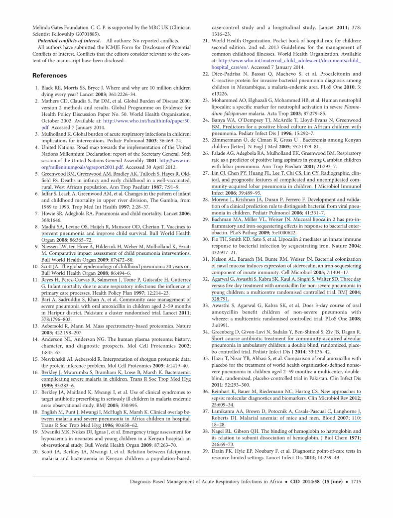
Table 5. Population Description of Kenyan Patients With Respiratory Distress Caused by Malaria, Pneumonia, or Both
Characteristic Malaria (n = 41) Pneumonia (n = 238)Pneumonia andMalaria (n = 14) P Valuea
Age, median (IQR), mo 21.1 (9.3–33.2) 12.8 (7.2–34.5) 30.4 (16.9–51.9) .03Male sex, % 53.6 54.6 35.7 .38
Weight-for-age z score, mean (SD) −1.73 (1.48) −2.06 (1.45) −2.07 (1.43) .48
Respiratory rate, median (IQR),respirations/min
56.4 (12.1) 56 (46–64) 52 (42–58) .29
Oxygen saturation, mean (SD), % 99 (96–100) 96 (92–98) 97 (96–99) <.01
WBC count, median (IQR), ×109/L 11.4 (7.9–17.1) 14 (9.9–20.9) 10.5 (6.7–21.5) .08Hemoglobin, mean (SD), g/dL 7.35 (4.8–9) 9 (7.9–9.9) 8.1 (5.3–8.7) <.01
Positive bacterial culture, No. (%)b 2 (4.88) 16 (6.72) 0 (0) <.01
Lpc-2, median (IQR), ng/mL 72.2 (44–151) 92.8 (51.1–159.6) 195 (51.3–286) .46Haptoglobin, median (IQR), ng/mL 23 434 (5445–1 048 424) 3 095 352 (1 770 148–4 383 530) 19 106 (8353–1 250 607) <.001
Abbreviations: IQR, interquartile range; Lpc-2, lipocalin 2; SD, standard deviation; WBC, white blood cell.a P values were obtained using Kruskal-Wallis or χ2 for quantitative or discrete variables, respectively.b Positive bacterial culture include the following significant microorganisms: Streptococcus pneumoniae (13 samples), Salmonella spp. (2 samples), andStaphylococcus aureus, Escherichia coli, and β-hemolytic streptococcus (1 sample each).
1714 • CID 2014:58 (15 June) • Huang et al
Melinda Gates Foundation. C. C. P. is supported by the MRC UK (ClinicianScientist Fellowship G0701885).Potential conflicts of interest. All authors: No reported conflicts.All authors have submitted the ICMJE Form for Disclosure of Potential
Conflicts of Interest. Conflicts that the editors consider relevant to the con-tent of the manuscript have been disclosed.
References
1. Black RE, Morris SS, Bryce J. Where and why are 10 million childrendying every year? Lancet 2003; 361:2226–34.
2. Mathers CD, Claudia S, Fat DM, et al. Global Burden of Disease 2000:version 2 methods and results. Global Programme on Evidence forHealth Policy Discussion Paper No. 50. World Health Organization,October 2002. Available at: http://www.who.int/healthinfo/paper50.pdf. Accessed 7 January 2014.
3. Mulholland K. Global burden of acute respiratory infections in children:implications for interventions. Pediatr Pulmonol 2003; 36:469–74.
4. United Nations. Road map towards the implementation of the UnitedNations Millennium Declaration: report of the Secretary General. 56thsession of the United Nations General Assembly. 2001. http://www.un.org/millenniumgoals/sgreport2001.pdf. Accessed 30 April 2012.
5. Greenwood BM, Greenwood AM, Bradley AK, Tulloch S, Hayes R, Old-field FS. Deaths in infancy and early childhood in a well-vaccinated,rural, West African population. Ann Trop Paediatr 1987; 7:91–9.
6. Jaffar S, Leach A, Greenwood AM, et al. Changes in the pattern of infantand childhood mortality in upper river division, The Gambia, from1989 to 1993. Trop Med Int Health 1997; 2:28–37.
7. Howie SR, Adegbola RA. Pneumonia and child mortality. Lancet 2006;368:1646.
8. Madhi SA, Levine OS, Hajjeh R, Mansoor OD, Cherian T. Vaccines toprevent pneumonia and improve child survival. Bull World HealthOrgan 2008; 86:365–72.
9. Niessen LW, ten Hove A, Hilderink H, Weber M, Mulholland K, EzzatiM. Comparative impact assessment of child pneumonia interventions.Bull World Health Organ 2009; 87:472–80.
10. Scott JA. The global epidemiology of childhood pneumonia 20 years on.Bull World Health Organ 2008; 86:494–6.
11. Reyes H, Perez-Cuevas R, Salmeron J, Tome P, Guiscafre H, GutierrezG. Infant mortality due to acute respiratory infections: the influence ofprimary care processes. Health Policy Plan 1997; 12:214–23.
12. Bari A, Sadruddin S, Khan A, et al. Community case management ofsevere pneumonia with oral amoxicillin in children aged 2–59 monthsin Haripur district, Pakistan: a cluster randomised trial. Lancet 2011;378:1796–803.
13. Aebersold R, Mann M. Mass spectrometry-based proteomics. Nature2003; 422:198–207.
14. Anderson NL, Anderson NG. The human plasma proteome: history,character, and diagnostic prospects. Mol Cell Proteomics 2002;1:845–67.
15. Nesvizhskii AI, Aebersold R. Interpretation of shotgun proteomic data:the protein inference problem. Mol Cell Proteomics 2005; 4:1419–40.
16. Berkley J, Mwarumba S, Bramham K, Lowe B, Marsh K. Bacteraemiacomplicating severe malaria in children. Trans R Soc Trop Med Hyg1999; 93:283–6.
17. Berkley JA, Maitland K, Mwangi I, et al. Use of clinical syndromes totarget antibiotic prescribing in seriously ill children in malaria endemicarea: observational study. BMJ 2005; 330:995.
18. English M, Punt J, Mwangi I, McHugh K, Marsh K. Clinical overlap be-tween malaria and severe pneumonia in Africa children in hospital.Trans R Soc Trop Med Hyg 1996; 90:658–62.
19. Mwaniki MK, Nokes DJ, Ignas J, et al. Emergency triage assessment forhypoxaemia in neonates and young children in a Kenyan hospital: anobservational study. Bull World Health Organ 2009; 87:263–70.
20. Scott JA, Berkley JA, Mwangi I, et al. Relation between falciparummalaria and bacteraemia in Kenyan children: a population-based,
case-control study and a longitudinal study. Lancet 2011; 378:1316–23.
21. World Health Organization. Pocket book of hospital care for children:second edition. 2nd ed. 2013 Guidelines for the management ofcommon childhood illnesses. World Health Organization. Availableat: http://www.who.int/maternal_child_adolescent/documents/child_hospital_care/en/. Accessed 7 January 2014.
22. Diez-Padrisa N, Bassat Q, Machevo S, et al. Procalcitonin andC-reactive protein for invasive bacterial pneumonia diagnosis amongchildren in Mozambique, a malaria-endemic area. PLoS One 2010; 5:e13226.
23. Mohammed AO, Elghazali G, Mohammed HB, et al. Human neutrophillipocalin: a specific marker for neutrophil activation in severe Plasmo-dium falciparum malaria. Acta Trop 2003; 87:279–85.
24. Banya WA, O’Dempsey TJ, McArdle T, Lloyd-Evans N, GreenwoodBM. Predictors for a positive blood culture in African children withpneumonia. Pediatr Infect Dis J 1996; 15:292–7.
25. Zimmermann O, de Ciman R, Gross U . Bacteremia among Kenyanchildren [letter]. N Engl J Med 2005; 352:1379–81.
26. Falade AG, Adegbola RA, Mulholland EK, Greenwood BM. Respiratoryrate as a predictor of positive lung aspirates in young Gambian childrenwith lobar pneumonia. Ann Trop Paediatr 2001; 21:293–7.
27. Lin CJ, Chen PY, Huang FL, Lee T, Chi CS, Lin CY. Radiographic, clin-ical, and prognostic features of complicated and uncomplicated com-munity-acquired lobar pneumonia in children. J Microbiol ImmunolInfect 2006; 39:489–95.
28. Moreno L, Krishnan JA, Duran P, Ferrero F. Development and valida-tion of a clinical prediction rule to distinguish bacterial from viral pneu-monia in children. Pediatr Pulmonol 2006; 41:331–7.
29. Bachman MA, Miller VL, Weiser JN. Mucosal lipocalin 2 has pro-in-flammatory and iron-sequestering effects in response to bacterial enter-obactin. PLoS Pathog 2009; 5:e1000622.
30. Flo TH, Smith KD, Sato S, et al. Lipocalin 2 mediates an innate immuneresponse to bacterial infection by sequestrating iron. Nature 2004;432:917–21.
31. Nelson AL, Barasch JM, Bunte RM, Weiser JN. Bacterial colonizationof nasal mucosa induces expression of siderocalin, an iron-sequesteringcomponent of innate immunity. Cell Microbiol 2005; 7:1404–17.
32. Agarwal G, Awasthi S, Kabra SK, Kaul A, Singhi S, Walter SD. Three dayversus five day treatment with amoxicillin for non-severe pneumonia inyoung children: a multicentre randomised controlled trial. BMJ 2004;328:791.
33. Awasthi S, Agarwal G, Kabra SK, et al. Does 3-day course of oralamoxycillin benefit children of non-severe pneumonia withwheeze: a multicentric randomised controlled trial. PLoS One 2008;3:e1991.
34. Greenberg D, Givon-Lavi N, Sadaka Y, Ben-Shimol S, Ziv JB, Dagan R.Short course antibiotic treatment for community-acquired alveolarpneumonia in ambulatory children: a double blind, randomized, place-bo controlled trial. Pediatr Infect Dis J 2014; 33:136–42.
35. Hazir T, Nisar YB, Abbasi S, et al. Comparison of oral amoxicillin withplacebo for the treatment of world health organization-defined nonse-vere pneumonia in children aged 2–59 months: a multicenter, double-blind, randomized, placebo-controlled trial in Pakistan. Clin Infect Dis2011; 52:293–300.
36. Reinhart K, Bauer M, Riedemann NC, Hartog CS. New approaches tosepsis: molecular diagnostics and biomarkers. Clin Microbiol Rev 2012;25:609–34.
37. Lamikanra AA, Brown D, Potocnik A, Casals-Pascual C, Langhorne J,Roberts DJ. Malarial anemia: of mice and men. Blood 2007; 110:18–28.
38. Nagel RL, Gibson QH. The binding of hemoglobin to haptoglobin andits relation to subunit dissociation of hemoglobin. J Biol Chem 1971;246:69–73.
39. Drain PK, Hyle EP, Noubary F, et al. Diagnostic point-of-care tests inresource-limited settings. Lancet Infect Dis 2014; 14:239–49.
Diagnosis-Based Management of Acute Respiratory Infections in Africa • CID 2014:58 (15 June) • 1715
Top Related
Copyright © 2022 FDOKUMEN

