


Bahasa
Halaman
Hukum

MOLECULAR AND CELLULAR BIOLOGY,0270-7306/99/$04.0010
Mar. 1999, p. 2155–2168 Vol. 19, No. 3
Copyright © 1999, American Society for Microbiology. All Rights Reserved.
Different Regulation of the p53 Core Domain Activities 39-to-59Exonuclease and Sequence-Specific DNA Binding
FRIEDEMANN JANUS,1 NILS ALBRECHTSEN,1 UWE KNIPPSCHILD,1 LISA WIESMULLER,1
FRANK GROSSE,2 AND WOLFGANG DEPPERT1*
Heinrich-Pette-Institut fur Experimentelle Virologie und Immunologie an der Universitat Hamburg,D-20251 Hamburg,1 and Institut fur Molekulare Biotechnologie, Jena,2 Germany
Received 27 March 1998/Returned for modification 7 May 1998/Accepted 8 December 1998
In this study we further characterized the 3*-5* exonuclease activity intrinsic to wild-type p53. We showedthat this activity, like sequence-specific DNA binding, is mediated by the p53 core domain. Truncation of theC-terminal 30 amino acids of the p53 molecule enhanced the p53 exonuclease activity by at least 10-fold,indicating that this activity, like sequence-specific DNA binding, is negatively regulated by the C-terminal basicregulatory domain of p53. However, treatments which activated sequence-specific DNA binding of p53, likebinding of the monoclonal antibody PAb421, which recognizes a C-terminal epitope on p53, or a higherphosphorylation status, strongly inhibited the p53 exonuclease activity. This suggests that at least on full-length p53, sequence-specific DNA binding and exonuclease activities are subject to different and seeminglyopposing regulatory mechanisms. Following up the recent discovery in our laboratory that p53 recognizes andbinds with high affinity to three-stranded DNA substrates mimicking early recombination intermediates (C.Dudenhoeffer, G. Rohaly, K. Will, W. Deppert, and L. Wiesmueller, Mol. Cell. Biol. 18:5332–5342), we askedwhether such substrates might be degraded by the p53 exonuclease. Addition of Mg21 ions to the binding assayindeed started the p53 exonuclease and promoted rapid degradation of the bound, but not of the unbound,substrate, indicating that specifically recognized targets can be subjected to exonucleolytic degradation by p53under defined conditions.
The best-studied molecular activity of the tumor suppressorp53 probably is that of a sequence-specific transactivator (3, 9,19, 28, 44, 69). p53 becomes activated as a transcription factorunder various cellular stress situations, including genotoxicstress. This leads to transcriptional upregulation of p53 targetgenes, which in turn mediate growth arrest or apoptosis (13,32, 41). In addition to its transactivator function, p53 exerts avariety of other biochemical activities involved in DNA dam-age recognition and repair, which characterize p53 as a supe-rior control element in maintaining the integrity of the cellgenome (2, 26, 37, 49). We recently reported that wild-type(wt) but not mutant p53 exerts a novel intrinsic 39-59 exonu-clease activity (48). Exonucleases are required in a variety ofprocesses contributing to genomic stability, such as proofread-ing, mismatch and nucleotide excision repair, and recombina-tion (31, 39). Therefore, we hypothesize that p53, through itsexonuclease activity, could be actively involved in such pro-cesses, thereby significantly expanding the role of p53 as a“guardian of the genome” (35).
In this study we further characterized the p53 exonucleaseactivity and specifically addressed the question of how thisactivity is related to the sequence-specific DNA binding activ-ity of p53. Our previous experiments had suggested that thep53 intrinsic exonuclease activity is exerted by the p53 coredomain (48), which also mediates sequence-specific DNAbinding by p53 (1, 4, 16, 51, 67). The localization of two suchdifferent activities to the same domain of the p53 moleculeposes the problem of how these activities are regulated, sinceone would expect that p53, which regulates transcription in a
sequence-specific manner, would not exert an exonuclease ac-tivity. The p53 core domain has a complex structure, as it iscomposed of two alpha-helical loop domains and a beta-sheetdomain, compacted via metal (zinc) binding (6). This is anunusual arrangement for a DNA binding domain, which so farhas been found only in a few sequence-specific DNA bindingproteins, including NFkB (47). Interestingly, a similar struc-tural arrangement has been found for the catalytic domain ofEscherichia coli exonuclease III, a multifunctional enzyme ex-hibiting 39 phosphatase, endonuclease, and 39-59 and 59-39 exo-nuclease activities (45). As a working hypothesis, we thereforeassumed that the composite structure of the p53 core domainmight allow the execution of different activities through con-formational alterations leading to slightly different arrange-ments of its various structural components.
The data presented in this report confirm that the centraldomain of wild-type, but not that of mutant, p53 exerts the p53intrinsic exonuclease activity. Like sequence-specific DNAbinding (22, 24), this exonuclease activity is negatively regu-lated by the C-terminal basic regulatory domain of p53. How-ever, treatments activating sequence-specific DNA binding offull-length p53 strongly inhibited its exonuclease activity, indi-cating that p53 exonuclease and sequence-specific DNA bind-ing are separate activities of the p53 core domain, regulated inopposing manners. As C-terminally truncated p53 had at leasta 10-fold higher specific exonuclease activity than full-lengthp53, we conclude that under appropriate conditions, wt p53acts as a bona fide exonuclease. Activation of the p53 exonu-clease activity by addition of Mg21 ions when p53 had boundto a potential in vivo substrate, a three-stranded DNA mim-icking a recombination intermediate with a single mismatch(8), resulted in rapid degradation of the bound, but not theunbound, substrate, which is indicative of the activation of thep53 exonuclease within a defined enzyme-substrate complex.
Our data are compatible with a model according to which
* Corresponding author. Mailing address: Heinrich-Pette-Institutfur Experimentelle Virologie und Immunologie an der UniversitatHamburg, Martinstr. 53, D-20251 Hamburg, Germany. Phone: 49-40-480 51-261. Fax: 49-40-480 51-117. E-mail: [email protected].
2155
on Decem
ber 3, 2014 by guesthttp://m
cb.asm.org/
Dow
nloaded from
p53 exerts two complementary functions in maintaining theintegrity of the genome. As its basal function in maintaininggenetic stability, p53 participates actively in repair processesthrough activities not related to sequence-specific DNA bind-ing, specifically through its exonuclease activity (48). At an-other level of control, cellular stress activates the functions ofp53 generally associated with its role as a guardian of thegenome, namely, growth arrest and apoptosis (34, 35).
(This work was conducted by F. Janus and N. Albrechsten inpartial fulfillment of the requirements for a Ph.D. degree at theUniversity of Hamburg, Hamburg, Germany.)
MATERIALS AND METHODS
Bacteria and cells. Recombinant baculovirus expressing full-length and frag-ments of murine wt p53 protein with coding information for a His6 tag at the Nterminus were kindly provided by P. Tegtmeyer (SUNY, Stony Brook) (67).Murine wt and MethA (7) p53 DNAs coding for amino acids 80 to 280 wereinserted into the pH6EX3 vector and expressed in DH5a bacteria. Simian virus40 (SV40) large-T-antigen (T-Ag) recombinant baculovirus was kindly providedby Ellen Fanning (Department of Molecular Biology, Vanderbilt University,Nashville, Tenn.). Human wt p53 and human oligomerization mutant 1262 re-combinant baculoviruses were kindly provided by John Jenkins (Cell Prolifera-tion Laboratory, Marie Curie Research Institute, Oxted, Surrey, United King-dom).
Protein purification. (i) Immunoaffinity chromatography. Protein was purifiedby PAb421 immunoaffinity chromatography as described previously (48).
(ii) Co21 metal affinity chromatography. About 109 High Five insect cells wereinfected with the corresponding recombinant baculovirus and harvested at 44 hpostinfection (hpi). Okadaic acid (200 nM) was added as indicated in Results at41 hpi. To purify histidine-tagged proteins by Co21 metal affinity column chro-matography (TALON; Clontech), cells were lysed by addition of ice-cold lysisbuffer (20 mM Tris-HCl [pH 8.0], 100 mM NaCl, 0.5% [wt/vol] Lubrol, 5 mMb-mercaptoethanol, 2 mM phenylmethylsulfonyl fluoride, 2 mM Na2S2O5, 50 mgof leupeptin, per ml, 1% [vol/vol] Trasylol) and rocked for 1 h. Crude lysate wascentrifuged at 200,000 3 g for 35 min and incubated with column material for 45min. The column material was washed with buffer A (20 mM Tris-HCl [pH 8.0],100 mM KCl, 2 mM b-mercaptoethanol) and with 10 mM imidazole in buffer A.Proteins were eluted with 300 mM imidazole in buffer A, and 1-ml fractions werecollected. The majority of the bound p53 proteins were recovered in fraction 2 athigh purity.
(iii) Ion-exchange and heparin sulfate affinity chromatographies. After metalaffinity chromatography, the p531-320 fragment was further purified by anion-exchange chromatography with UNO Q anion-exchange column or by heparinsulfate-Sepharose affinity chromatography with an AKTApurifier fast proteinliquid chromatography system (Pharmacia). The p531-320 fragment was purifiedvia Talon metal affinity chromatography as described above, and a .90% pure,exonuclease-active peak fraction was subjected to ion-exchange chromatography.For anion-exchange chromatography, a 1.3-ml UnoQ column (Bio-Rad) wasequilibrated with 5 column volumes of buffer A (20 mM Tris, 40 mM KCl, 0.1%mercaptoethanol, pH 8.0) and loaded with 100 mg of protein. For cation-ex-change chromatography 140 mg of the TALON-purified p531-320 fragment wasloaded onto a 1-ml HiTrap heparin-Sepharose column (Pharmacia) equilibratedwith buffer A. The columns were washed with 3 column volumes of buffer A, andthe protein was eluted with a linear salt gradient (40 to 800 mM KCl) at a flowrate of 2 ml/min. Fractions of 0.5 ml were collected. All fractions were tested forexonuclease activity, and peak fractions were analyzed by sodium dodecyl sul-fate-polyacrylamide gel electrophoresis (SDS-PAGE) and Coomassie blue stain-ing.
SDS-PAGE and Western immunoblotting. Protein fractions were denatured inSDS sample buffer, separated by SDS-PAGE, and transferred to a polyvinylidinedifluoride membrane. Proteins were detected with monoclonal antibodies asindicated in Results.
Exonuclease assays. The 39-59 exonuclease activity was measured by filterbinding assays as described previously (48). For inhibition experiments withPAb421 and PAb108, 5 ng to 10 mg of antibody and 150 ng of metal affinity-purified p53 protein were mixed, preincubated for 15 min at 4°C, and tested forexonuclease activity. Proteolytic digestion was done by addition of a dilutionseries of thermolysin from 4 ng (1:50) to 20 pg (1:10,000) to 150 ng of metalaffinity-purified p53 in exonuclease reaction buffer (25 mM Tris [pH 8.5], 10 mMMgCl2, 1 mM dithiothreitol) and incubation for 15 min at 37°C. 3H-labeled DNAwas added, and the filter binding assay was performed as described above.Exonuclease III (Boehringer, Mannheim, Germany) with the same exonucleo-lytic activity as 150 ng of p53 was used as a control.
Gel mobility shift assay. Synthetic p21 promoter oligonucleotides (10) wereend labeled by using T4 polynucleotide kinase and [g-32P]ATP, gel purified, andused as probes in binding reactions after annealing with unlabeled complemen-tary strand. p53 was purified by metal affinity chromatography or nuclear extrac-tion (29). 32P-labeled three-stranded substrates containing an A-G mismatch
were prepared as described previously (8). Binding reactions were carried out asdescribed previously (8, 22).
RESULTS
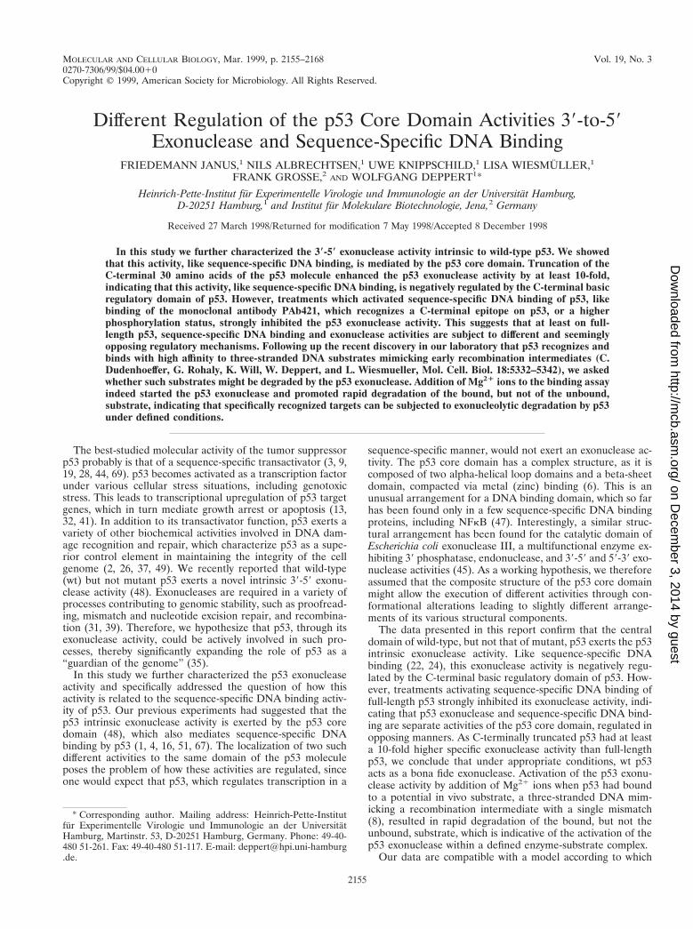
The p53 exonuclease activity is mediated by the p53 coredomain. In our previous experiments, a bacterial fragmentcomprising the p53 core domain (amino acids 80 to 280) wasrenatured after SDS-PAGE and was shown to exhibit 39-59exonuclease activity (48). To analyze the structural prerequi-sites of the p53 exonuclease activity on native p53, we analyzedvarious deletion fragments of mouse p53 (see Fig. 3). With theexception of the bacterial p5380-280 fragments, all of the p53fragments were expressed in insect cells infected with the re-spective recombinant baculoviruses. Full-length p53 and allp53 fragments were His tagged at their N termini, which al-lowed their easy purification via metal affinity chromatography(see Materials and Methods). Figure 1 shows the purified bac-ulovirus-expressed p53 proteins and their corresponding exo-nuclease profiles. It is evident that all fragments containing thep53 core domain, as well as the isolated p53 core domain itself,exhibited exonuclease activity, whereas those fragments notcontaining the core domain were exonuclease negative. Weconclude that the p53 core domain mediates exonuclease ac-tivity. This conclusion was further substantiated by comparingthe exonuclease activities of the isolated core domains (aminoacids 80 to 280) of wt p53 and MethA mutant p53 (7) (con-taining the mutations C132F, E168G, and M234I [11]), whichwere expressed in bacteria and purified by metal affinity chro-
FIG. 1. Exonuclease activities of p53 and p53 deletion mutants. wt p53 andp53 deletion mutants were expressed in High Five insect cells infected withrecombinant baculovirus and purified by metal affinity chromatography as de-scribed in Materials and Methods. Peak fractions of all proteins were analyzed bySDS-PAGE (A), and 150 ng of each protein was tested for exonuclease activityby the 3H filter retention assay (B). Exonuclease III was used as a control. aa,amino acids. Lane M, markers. Numbers on the left in panel A are molecularmasses in kilodaltons.
2156 JANUS ET AL. MOL. CELL. BIOL.
on Decem
ber 3, 2014 by guesthttp://m
cb.asm.org/
Dow
nloaded from
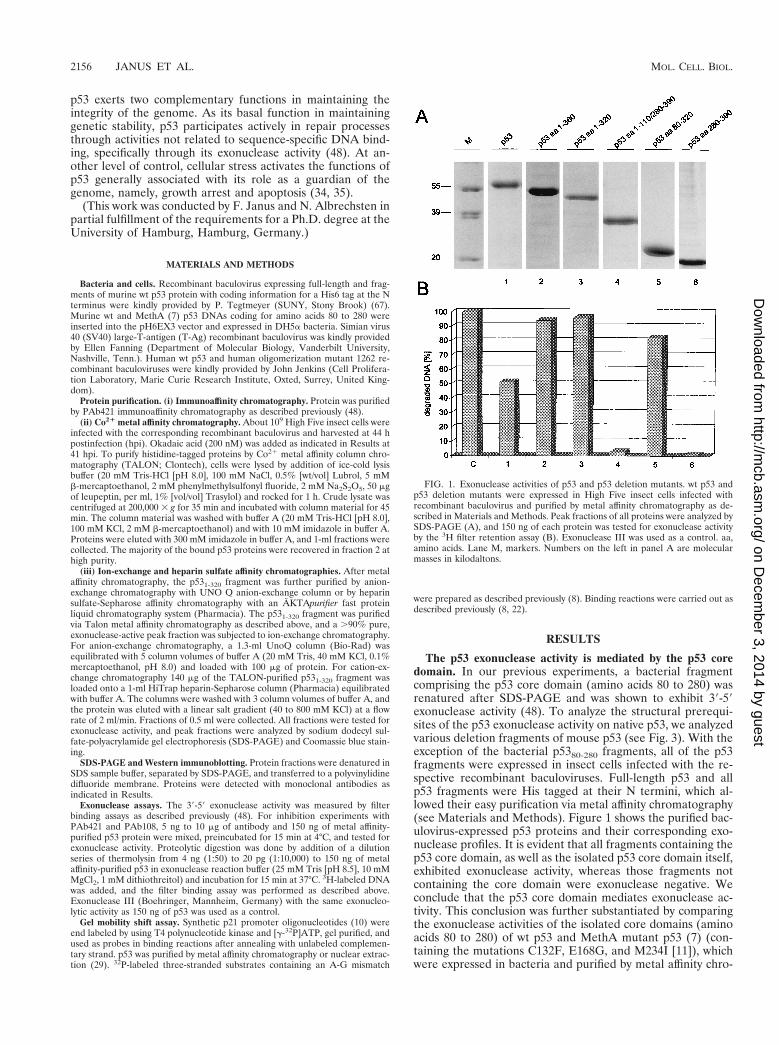
matography. Figure 2A and B show that these fragments wereobtained in high yields and reasonable purity. Whereas the wtp53 core fragment displayed exonuclease activity (Fig. 2C), theMethA mutant p53 core domain was devoid of any exonucle-ase activity (Fig. 2D). This provides further evidence that struc-tural alterations induced by mutations in the p53 core domainabolish not only sequence-specific DNA binding of p53 butalso its exonuclease activity, supporting our previous finding(48) that mutant p53 proteins have lost the p53 exonucleaseactivity. The results of the exonuclease-mapping experimentare summarized in Fig. 3.
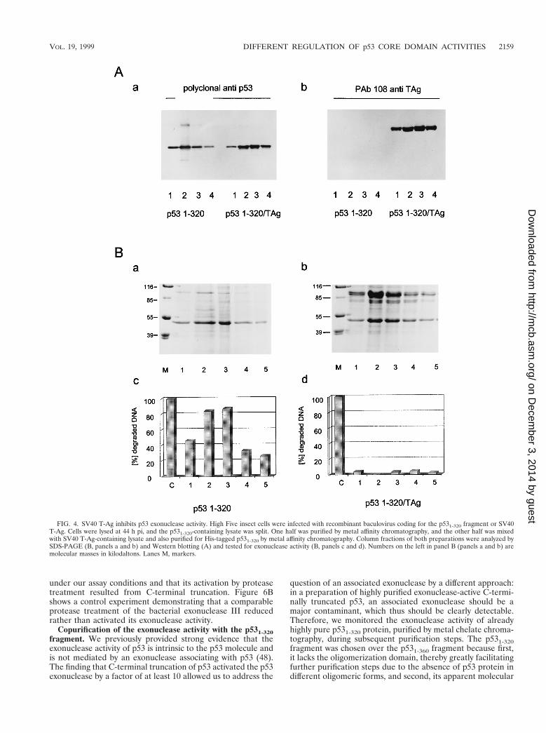
Binding of SV40 T-Ag to p53 eliminates its exonucleaseactivity. SV40 T-Ag targets p53 in SV40 lytic infection andcellular transformation by binding to the p53 core domain (54).This largely eliminates sequence-specific DNA binding of p53and its ability to transactivate p53 target genes (5, 27, 42). Itwas therefore of interest to analyze whether binding of T-Ag top53 would also affect the p53 exonuclease activity. If so, thiswould provide further and independent evidence that exonu-clease activity and sequence-specific DNA binding of p53 aremediated by the same catalytic domain. Furthermore, targetingof p53 exonuclease activity by SV40 T-Ag might provide hintsas to a possible in vivo relevance of this activity (see Discus-sion).
Insect cells were infected in parallel either with a recombi-nant baculovirus encoding SV40 T-Ag or with a virus encodingthe p531-320 fragment. The p531-320 fragment was chosen forcomplex formation with T-Ag, because this fragment, in con-trast to full-length p53, very effectively and quantitatively formsa complex with T-Ag in vitro (unpublished observation). After
lysis of the infected cells, the lysate containing the p531-320fragment was split, and half of it was mixed with the lysate ofcells expressing T-Ag to allow formation of the T-Ag/p531-320complex. The p531-320 fragment and the T-Ag/p531-320 com-plex were purified from the respective lysates by metal affinitychromatography via the His tag of the p531-320 fragment (fordetails, see Materials and Methods). Figure 4A shows theanalysis of the purified p531-320 fragment and of the T-Ag/p531-320 complex by Western blotting with a polyclonal anti-serum for p53 (panel a) or the T-Ag-specific monoclonal an-tibody PAb108 (panel b). This analysis demonstrates that sim-ilar amounts of free p531-320 fragment and of p531-320 fragmentcomplexed to T-Ag were recovered during purification andthat the complexed p531-320 fragment had bound a significantamount of T-Ag. Figure 4B shows the Coomassie blue-stainedpattern of fractions of the free p531-320 fragment (panel a) andof this fragment in complex with T-Ag after metal affinitychromatography and SDS-PAGE (panel b) and the corre-sponding exonuclease activities of the p531-320 fragment in therespective fractions (panels c and d). As is evident from Fig.4B, the free p531-320 fragment had exonuclease activity (panelc), whereas this fragment in complex with T-Ag was devoid ofexonuclease activity (panel d).
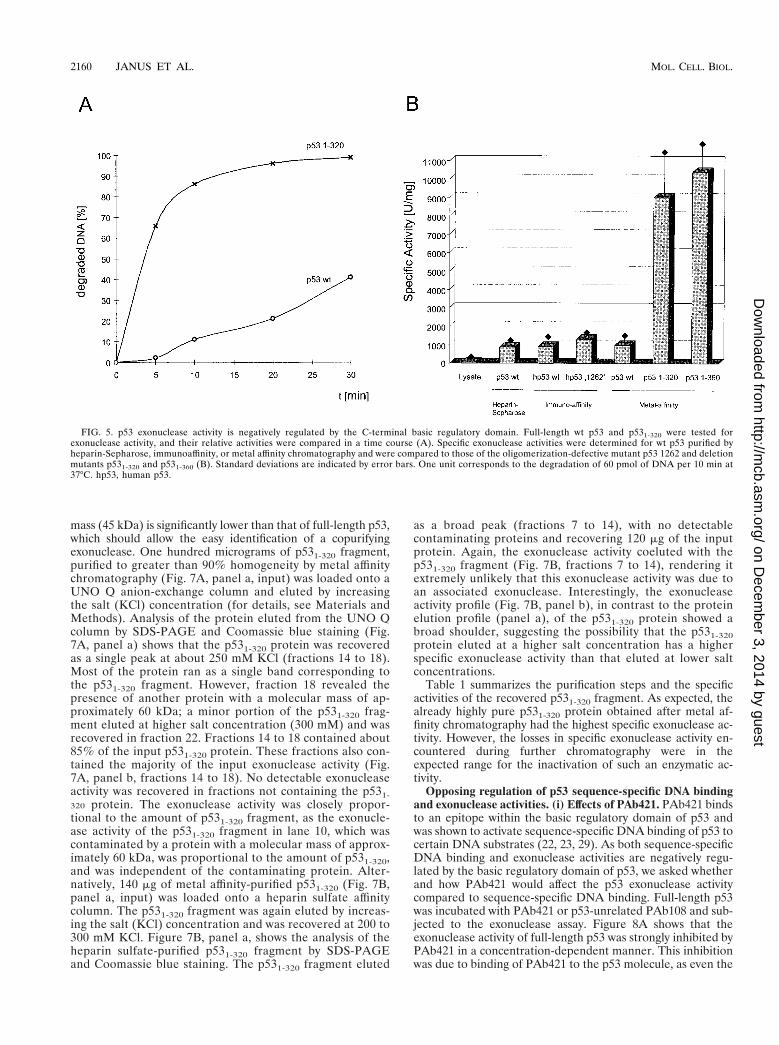
C-terminally truncated p53 exhibits a significantly higherexonuclease activity than full-length p53. During our analysesof p53 fragments for exonuclease activity, we noticed that theC-terminally truncated p53 fragments seemed to have a higherspecific activity than full-length p53. This was verified by com-paring the kinetics of substrate degradation for full-length p53and for the p531-320 fragment. Figure 5A shows that the
FIG. 2. wt but not mutant p53 core domain is necessary and sufficient for p53 exonuclease activity. wt (A and C) and MethA (B and D) p53 core fragments (aminoacids 80 to 280) were expressed in bacteria and purified by metal affinity chromatography. Column fractions of both proteins were analyzed by SDS-PAGE (A and B)and tested for exonuclease activity by the 3H filter retention assay (C and D). Exonuclease III (lane C) was used as a control. Numbers on the left in panels A and Bare molecular masses in kilodaltons.
VOL. 19, 1999 DIFFERENT REGULATION OF p53 CORE DOMAIN ACTIVITIES 2157
on Decem
ber 3, 2014 by guesthttp://m
cb.asm.org/
Dow
nloaded from
p531-320 fragment degraded the input substrate DNA at amuch higher catalytic rate than did full-length p53. Quantita-tive evaluation and determination of the specific exonucleaseactivities of various full-length p53 preparations and of theC-terminally truncated p531-320 and p531-360 fragments (Fig.5B) revealed that full-length p53, regardless of its mode ofpurification, had about a 10-times-lower specific exonucleaseactivity than the C-terminally truncated p531-320 and p531-360fragments. We conclude that the exonuclease activity of full-length p53 is negatively regulated by the C-terminal basic do-main, as the shortest truncation which strongly enhanced thep53 exonuclease was a deletion of the C-terminal 30 aminoacids (p531-360 fragment). Interestingly, and in contrast to thecase for sequence-specific DNA binding (16, 63), the oligomer-ization status of p53 did not influence the p53 intrinsic exonu-clease activity. This conclusion is based on the findings that theoligomerization-defective p531-320 fragment exhibited thesame, activated specific exonuclease activity as the oligomer-ization-competent p531-360 fragment and, conversely, that thefull-length oligomerization-defective p53, 1262 (60, 61), had anexonuclease activity similar to that of oligomerization-compe-tent wt p53.
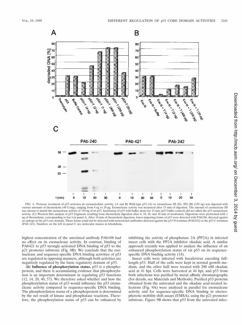
Protease treatment of the full-length p53 protein activatesexonuclease activity. It has been reported that the full-lengthp53 molecule is rather unstable but that C-terminal truncationenhances the stability of p53 (17). To test the possibility thatsuch an effect might be responsible for the observed higher
specific activity of the C-terminally truncated p53 fragments,we designed a control experiment which was based on thepreviously reported finding that limited proteolytic digestion ofp53 with thermolysin preferentially degrades the N- and C-terminal portions of the p53 molecule, whereas the p53 corefragment is more resistant to degradation (4). Constantamounts of purified wt p53 were incubated in parallel withconsecutively higher dilutions of thermolysin and subjected toexonuclease assays. If activation of the p53 exonuclease resultsfrom C-terminal truncation, then protease treatment at certainthermolysin-to-p53 ratios should lead to higher exonucleaseactivities. Figure 6A shows that incubation of p53 with ther-molysin at certain enzyme dilutions resulted in a significantactivation of the p53 exonuclease activity. Analysis of the p53fragments generated by thermolysin in a time course experi-ment (Fig. 6C) shows that thermolysin treatment rapidly gen-erated lower-migrating forms of p53; these were detected byPAb240, which reacts with an epitope in the p53 core domain(15, 59, 70), but not by PAb421, which reacts with a C-terminalepitope of p53 (18, 58, 65), or by PAb242, which reacts with anN-terminal epitope of p53 (36, 70). This strongly supports theinterpretation that the lower exonuclease activity of full-lengthp53 compared to C-terminally truncated p53 fragments resultsfrom negative regulation by the C-terminal basic regulatorydomain of p53. Note that incubation of full-length p53 withreaction buffer alone did not affect the p53 exonuclease activity(Fig. 6A), further demonstrating that this activity was stable
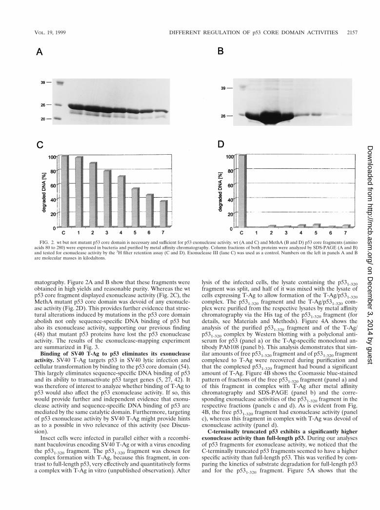
FIG. 3. Summary of p53 exonuclease activity mapping data. p53 fragments and their corresponding exonuclease activities are shown schematically. aa, aminoacids.
2158 JANUS ET AL. MOL. CELL. BIOL.
on Decem
ber 3, 2014 by guesthttp://m
cb.asm.org/
Dow
nloaded from
under our assay conditions and that its activation by proteasetreatment resulted from C-terminal truncation. Figure 6Bshows a control experiment demonstrating that a comparableprotease treatment of the bacterial exonuclease III reducedrather than activated its exonuclease activity.
Copurification of the exonuclease activity with the p531-320fragment. We previously provided strong evidence that theexonuclease activity of p53 is intrinsic to the p53 molecule andis not mediated by an exonuclease associating with p53 (48).The finding that C-terminal truncation of p53 activated the p53exonuclease by a factor of at least 10 allowed us to address the
question of an associated exonuclease by a different approach:in a preparation of highly purified exonuclease-active C-termi-nally truncated p53, an associated exonuclease should be amajor contaminant, which thus should be clearly detectable.Therefore, we monitored the exonuclease activity of alreadyhighly pure p531-320 protein, purified by metal chelate chroma-tography, during subsequent purification steps. The p531-320fragment was chosen over the p531-360 fragment because first,it lacks the oligomerization domain, thereby greatly facilitatingfurther purification steps due to the absence of p53 protein indifferent oligomeric forms, and second, its apparent molecular
FIG. 4. SV40 T-Ag inhibits p53 exonuclease activity. High Five insect cells were infected with recombinant baculovirus coding for the p531-320 fragment or SV40T-Ag. Cells were lysed at 44 h pi, and the p531-320-containing lysate was split. One half was purified by metal affinity chromatography, and the other half was mixedwith SV40 T-Ag-containing lysate and also purified for His-tagged p531-320 by metal affinity chromatography. Column fractions of both preparations were analyzed bySDS-PAGE (B, panels a and b) and Western blotting (A) and tested for exonuclease activity (B, panels c and d). Numbers on the left in panel B (panels a and b) aremolecular masses in kilodaltons. Lanes M, markers.
VOL. 19, 1999 DIFFERENT REGULATION OF p53 CORE DOMAIN ACTIVITIES 2159
on Decem
ber 3, 2014 by guesthttp://m
cb.asm.org/
Dow
nloaded from
mass (45 kDa) is significantly lower than that of full-length p53,which should allow the easy identification of a copurifyingexonuclease. One hundred micrograms of p531-320 fragment,purified to greater than 90% homogeneity by metal affinitychromatography (Fig. 7A, panel a, input) was loaded onto aUNO Q anion-exchange column and eluted by increasingthe salt (KCl) concentration (for details, see Materials andMethods). Analysis of the protein eluted from the UNO Qcolumn by SDS-PAGE and Coomassie blue staining (Fig.7A, panel a) shows that the p531-320 protein was recoveredas a single peak at about 250 mM KCl (fractions 14 to 18).Most of the protein ran as a single band corresponding tothe p531-320 fragment. However, fraction 18 revealed thepresence of another protein with a molecular mass of ap-proximately 60 kDa; a minor portion of the p531-320 frag-ment eluted at higher salt concentration (300 mM) and wasrecovered in fraction 22. Fractions 14 to 18 contained about85% of the input p531-320 protein. These fractions also con-tained the majority of the input exonuclease activity (Fig.7A, panel b, fractions 14 to 18). No detectable exonucleaseactivity was recovered in fractions not containing the p531-320 protein. The exonuclease activity was closely propor-tional to the amount of p531-320 fragment, as the exonucle-ase activity of the p531-320 fragment in lane 10, which wascontaminated by a protein with a molecular mass of approx-imately 60 kDa, was proportional to the amount of p531-320,and was independent of the contaminating protein. Alter-natively, 140 mg of metal affinity-purified p531-320 (Fig. 7B,panel a, input) was loaded onto a heparin sulfate affinitycolumn. The p531-320 fragment was again eluted by increas-ing the salt (KCl) concentration and was recovered at 200 to300 mM KCl. Figure 7B, panel a, shows the analysis of theheparin sulfate-purified p531-320 fragment by SDS-PAGEand Coomassie blue staining. The p531-320 fragment eluted
as a broad peak (fractions 7 to 14), with no detectablecontaminating proteins and recovering 120 mg of the inputprotein. Again, the exonuclease activity coeluted with thep531-320 fragment (Fig. 7B, fractions 7 to 14), rendering itextremely unlikely that this exonuclease activity was due toan associated exonuclease. Interestingly, the exonucleaseactivity profile (Fig. 7B, panel b), in contrast to the proteinelution profile (panel a), of the p531-320 protein showed abroad shoulder, suggesting the possibility that the p531-320protein eluted at a higher salt concentration has a higherspecific exonuclease activity than that eluted at lower saltconcentrations.
Table 1 summarizes the purification steps and the specificactivities of the recovered p531-320 fragment. As expected, thealready highly pure p531-320 protein obtained after metal af-finity chromatography had the highest specific exonuclease ac-tivity. However, the losses in specific exonuclease activity en-countered during further chromatography were in theexpected range for the inactivation of such an enzymatic ac-tivity.
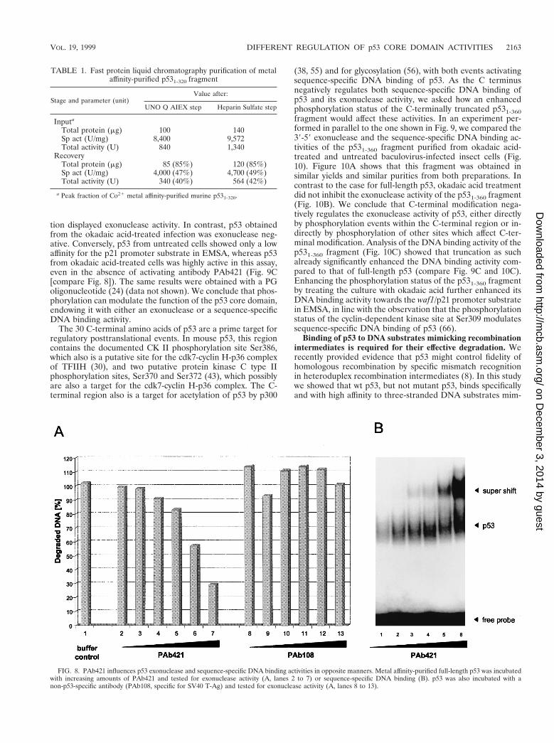
Opposing regulation of p53 sequence-specific DNA bindingand exonuclease activities. (i) Effects of PAb421. PAb421 bindsto an epitope within the basic regulatory domain of p53 andwas shown to activate sequence-specific DNA binding of p53 tocertain DNA substrates (22, 23, 29). As both sequence-specificDNA binding and exonuclease activities are negatively regu-lated by the basic regulatory domain of p53, we asked whetherand how PAb421 would affect the p53 exonuclease activitycompared to sequence-specific DNA binding. Full-length p53was incubated with PAb421 or p53-unrelated PAb108 and sub-jected to the exonuclease assay. Figure 8A shows that theexonuclease activity of full-length p53 was strongly inhibited byPAb421 in a concentration-dependent manner. This inhibitionwas due to binding of PAb421 to the p53 molecule, as even the
FIG. 5. p53 exonuclease activity is negatively regulated by the C-terminal basic regulatory domain. Full-length wt p53 and p531-320 were tested forexonuclease activity, and their relative activities were compared in a time course (A). Specific exonuclease activities were determined for wt p53 purified byheparin-Sepharose, immunoaffinity, or metal affinity chromatography and were compared to those of the oligomerization-defective mutant p53 1262 and deletionmutants p531-320 and p531-360 (B). Standard deviations are indicated by error bars. One unit corresponds to the degradation of 60 pmol of DNA per 10 min at37°C. hp53, human p53.
2160 JANUS ET AL. MOL. CELL. BIOL.
on Decem
ber 3, 2014 by guesthttp://m
cb.asm.org/
Dow
nloaded from
highest concentration of the unrelated antibody PAb108 hadno effect on its exonuclease activity. In contrast, binding ofPAb421 to p53 strongly activated DNA binding of p53 to thep21 promoter substrate (Fig. 8B). We conclude that the exo-nuclease and sequence-specific DNA binding activities of p53are regulated in opposing manners, although both activities arenegatively regulated by the basic regulatory domain of p53.
(ii) Influence of phosphorylation status. p53 is a phospho-protein, and there is accumulating evidence that phosphoryla-tion is an important determinant in regulating p53 functions(12, 14, 20, 40, 57). We therefore asked whether and how thephosphorylation status of p53 would influence the p53 exonu-clease activity compared to sequence-specific DNA binding.The phosphorylation status of a phosphoprotein is determinedby the net result of kinase and phosphatase reactions. There-fore, the phosphorylation status of p53 can be enhanced by
inhibiting the activity of phosphatase 2A (PP2A) in infectedinsect cells with the PP2A inhibitor okadaic acid. A similarapproach recently was applied to analyze the influence of anenhanced phosphorylation status of rat p53 on its sequence-specific DNA binding activity (14).
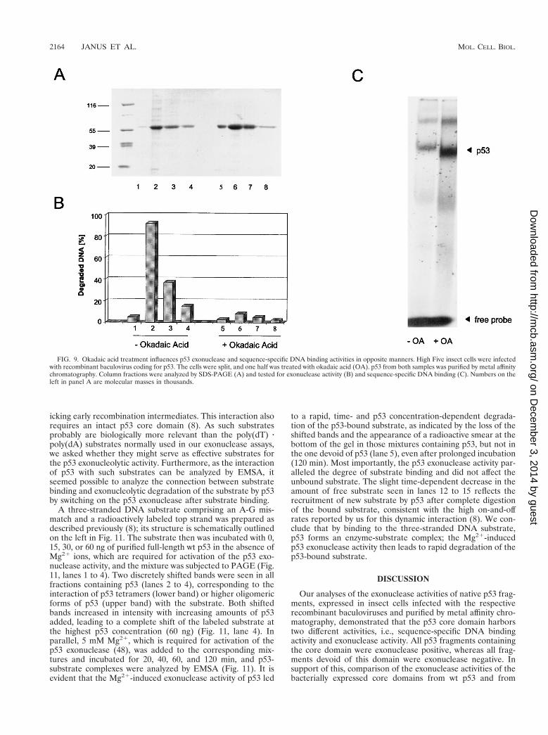
Insect cells were infected with baculovirus encoding full-length p53. Half of the cells were kept in normal growth me-dium, and the other half were treated with 200 nM okadaicacid at 41 hpi. Cells were harvested at 44 hpi, and p53 fromboth infections was purified by metal affinity chromatography(for details, see Materials and Methods). Purified p53 proteinsobtained from the untreated and the okadaic acid-treated in-fections (Fig. 9A) were analyzed in parallel for exonucleaseactivity and for sequence-specific DNA binding in electro-phoretic mobility shift assays (EMSA), using the p21 promotersubstrate. Figure 9B shows that p53 from the untreated infec-
FIG. 6. Protease treatment of p53 activates its exonucleolytic activity. (A and B) Wild-type p53 (A) or exonuclease III (Ex. III) (B) (150 ng) was digested withvarious amounts of thermolysin (40 U/mg), ranging from 4 ng to 20 pg. Exonuclease activity was measured after 15 min of digestion. The amount of exonuclease IIIwas chosen to match the exonuclease activity of 150 ng of wt p53. Incubation of p53 with buffer alone for 15 min (p53 buffer control) did not affect the p53 exonucleaseactivity. (C) Western blot analysis of p53 fragments resulting from thermolysin digestion after 0, 10, 20, and 30 min of incubation. Digestions were performed with 1ng of thermolysin, corresponding to bar 6 in panel A. After 10 min of thermolysin digestion, lower-migrating forms of p53 were detected with PAb240, directed againstan epitope in the p53 core domain. Those forms could not be detected with monoclonal antibodies directed against the p53 N terminus (PAb242) or the p53 C terminus(PAb 421). Numbers on the left in panel C are molecular masses in kilodaltons.
VOL. 19, 1999 DIFFERENT REGULATION OF p53 CORE DOMAIN ACTIVITIES 2161
on Decem
ber 3, 2014 by guesthttp://m
cb.asm.org/
Dow
nloaded from
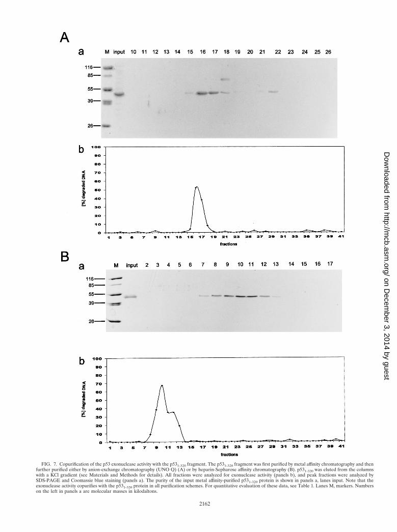
FIG. 7. Copurification of the p53 exonuclease activity with the p531-320 fragment. The p531-320 fragment was first purified by metal affinity chromatography and thenfurther purified either by anion-exchange chromatography (UNO Q) (A) or by heparin-Sepharose affinity chromatography (B). p531-320 was eluted from the columnswith a KCl gradient (see Materials and Methods for details). All fractions were analyzed for exonuclease activity (panels b), and peak fractions were analyzed bySDS-PAGE and Coomassie blue staining (panels a). The purity of the input metal affinity-purified p531-320 protein is shown in panels a, lanes input. Note that theexonuclease activity copurifies with the p531-320 protein in all purification schemes. For quantitative evaluation of these data, see Table 1. Lanes M, markers. Numberson the left in panels a are molecular masses in kilodaltons.
2162
on Decem
ber 3, 2014 by guesthttp://m
cb.asm.org/
Dow
nloaded from
tion displayed exonuclease activity. In contrast, p53 obtainedfrom the okadaic acid-treated infection was exonuclease neg-ative. Conversely, p53 from untreated cells showed only a lowaffinity for the p21 promoter substrate in EMSA, whereas p53from okadaic acid-treated cells was highly active in this assay,even in the absence of activating antibody PAb421 (Fig. 9C[compare Fig. 8]). The same results were obtained with a PGoligonucleotide (24) (data not shown). We conclude that phos-phorylation can modulate the function of the p53 core domain,endowing it with either an exonuclease or a sequence-specificDNA binding activity.
The 30 C-terminal amino acids of p53 are a prime target forregulatory posttranslational events. In mouse p53, this regioncontains the documented CK II phosphorylation site Ser386,which also is a putative site for the cdk7-cyclin H-p36 complexof TFIIH (30), and two putative protein kinase C type IIphosphorylation sites, Ser370 and Ser372 (43), which possiblyare also a target for the cdk7-cyclin H-p36 complex. The C-terminal region also is a target for acetylation of p53 by p300
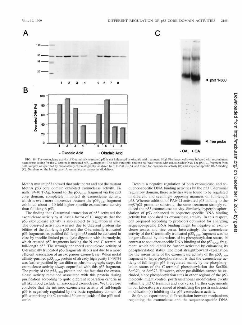
(38, 55) and for glycosylation (56), with both events activatingsequence-specific DNA binding of p53. As the C terminusnegatively regulates both sequence-specific DNA binding ofp53 and its exonuclease activity, we asked how an enhancedphosphorylation status of the C-terminally truncated p531-360fragment would affect these activities. In an experiment per-formed in parallel to the one shown in Fig. 9, we compared the39-59 exonuclease and the sequence-specific DNA binding ac-tivities of the p531-360 fragment purified from okadaic acid-treated and untreated baculovirus-infected insect cells (Fig.10). Figure 10A shows that this fragment was obtained insimilar yields and similar purities from both preparations. Incontrast to the case for full-length p53, okadaic acid treatmentdid not inhibit the exonuclease activity of the p531-360 fragment(Fig. 10B). We conclude that C-terminal modification nega-tively regulates the exonuclease activity of p53, either directlyby phosphorylation events within the C-terminal region or in-directly by phosphorylation of other sites which affect C-ter-minal modification. Analysis of the DNA binding activity of thep531-360 fragment (Fig. 10C) showed that truncation as suchalready significantly enhanced the DNA binding activity com-pared to that of full-length p53 (compare Fig. 9C and 10C).Enhancing the phosphorylation status of the p531-360 fragmentby treating the culture with okadaic acid further enhanced itsDNA binding activity towards the waf1/p21 promoter substratein EMSA, in line with the observation that the phosphorylationstatus of the cyclin-dependent kinase site at Ser309 modulatessequence-specific DNA binding of p53 (66).
Binding of p53 to DNA substrates mimicking recombinationintermediates is required for their effective degradation. Werecently provided evidence that p53 might control fidelity ofhomologous recombination by specific mismatch recognitionin heteroduplex recombination intermediates (8). In this studywe showed that wt p53, but not mutant p53, binds specificallyand with high affinity to three-stranded DNA substrates mim-
FIG. 8. PAb421 influences p53 exonuclease and sequence-specific DNA binding activities in opposite manners. Metal affinity-purified full-length p53 was incubatedwith increasing amounts of PAb421 and tested for exonuclease activity (A, lanes 2 to 7) or sequence-specific DNA binding (B). p53 was also incubated with anon-p53-specific antibody (PAb108, specific for SV40 T-Ag) and tested for exonuclease activity (A, lanes 8 to 13).
TABLE 1. Fast protein liquid chromatography purification of metalaffinity-purified p531-320 fragment
Stage and parameter (unit)Value after:
UNO Q AIEX step Heparin Sulfate step
Inputa
Total protein (mg) 100 140Sp act (U/mg) 8,400 9,572Total activity (U) 840 1,340
RecoveryTotal protein (mg) 85 (85%) 120 (85%)Sp act (U/mg) 4,000 (47%) 4,700 (49%)Total activity (U) 340 (40%) 564 (42%)
a Peak fraction of Co21 metal affinity-purified murine p531-320.
VOL. 19, 1999 DIFFERENT REGULATION OF p53 CORE DOMAIN ACTIVITIES 2163
on Decem
ber 3, 2014 by guesthttp://m
cb.asm.org/
Dow
nloaded from
icking early recombination intermediates. This interaction alsorequires an intact p53 core domain (8). As such substratesprobably are biologically more relevant than the poly(dT) zpoly(dA) substrates normally used in our exonuclease assays,we asked whether they might serve as effective substrates forthe p53 exonucleolytic activity. Furthermore, as the interactionof p53 with such substrates can be analyzed by EMSA, itseemed possible to analyze the connection between substratebinding and exonucleolytic degradation of the substrate by p53by switching on the p53 exonuclease after substrate binding.
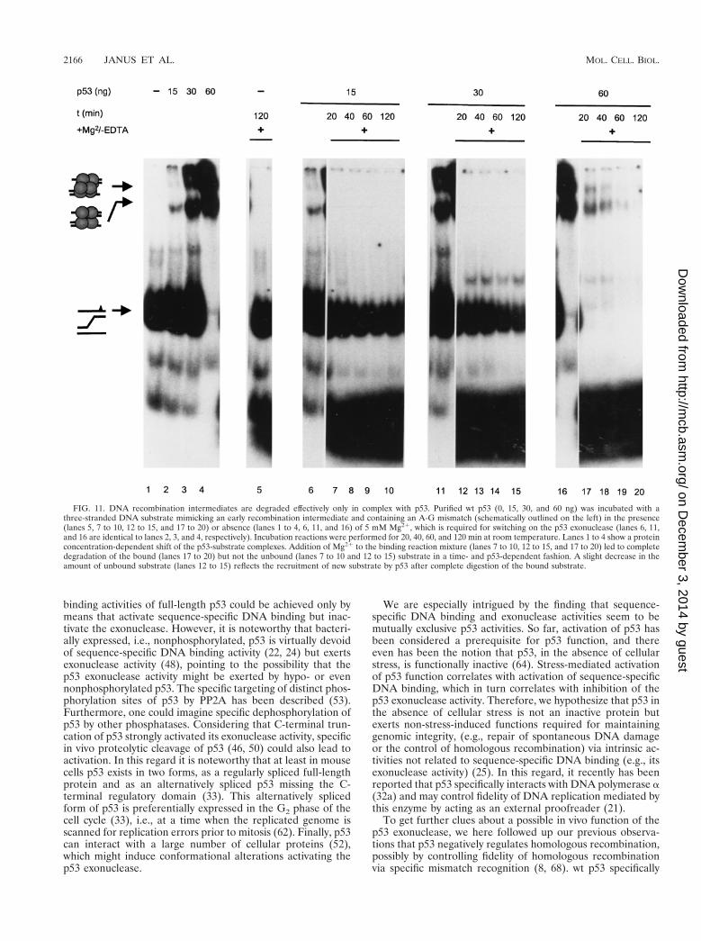
A three-stranded DNA substrate comprising an A-G mis-match and a radioactively labeled top strand was prepared asdescribed previously (8); its structure is schematically outlinedon the left in Fig. 11. The substrate then was incubated with 0,15, 30, or 60 ng of purified full-length wt p53 in the absence ofMg21 ions, which are required for activation of the p53 exo-nuclease activity, and the mixture was subjected to PAGE (Fig.11, lanes 1 to 4). Two discretely shifted bands were seen in allfractions containing p53 (lanes 2 to 4), corresponding to theinteraction of p53 tetramers (lower band) or higher oligomericforms of p53 (upper band) with the substrate. Both shiftedbands increased in intensity with increasing amounts of p53added, leading to a complete shift of the labeled substrate atthe highest p53 concentration (60 ng) (Fig. 11, lane 4). Inparallel, 5 mM Mg21, which is required for activation of thep53 exonuclease (48), was added to the corresponding mix-tures and incubated for 20, 40, 60, and 120 min, and p53-substrate complexes were analyzed by EMSA (Fig. 11). It isevident that the Mg21-induced exonuclease activity of p53 led
to a rapid, time- and p53 concentration-dependent degrada-tion of the p53-bound substrate, as indicated by the loss of theshifted bands and the appearance of a radioactive smear at thebottom of the gel in those mixtures containing p53, but not inthe one devoid of p53 (lane 5), even after prolonged incubation(120 min). Most importantly, the p53 exonuclease activity par-alleled the degree of substrate binding and did not affect theunbound substrate. The slight time-dependent decrease in theamount of free substrate seen in lanes 12 to 15 reflects therecruitment of new substrate by p53 after complete digestionof the bound substrate, consistent with the high on-and-offrates reported by us for this dynamic interaction (8). We con-clude that by binding to the three-stranded DNA substrate,p53 forms an enzyme-substrate complex; the Mg21-inducedp53 exonuclease activity then leads to rapid degradation of thep53-bound substrate.
DISCUSSION
Our analyses of the exonuclease activities of native p53 frag-ments, expressed in insect cells infected with the respectiverecombinant baculoviruses and purified by metal affinity chro-matography, demonstrated that the p53 core domain harborstwo different activities, i.e., sequence-specific DNA bindingactivity and exonuclease activity. All p53 fragments containingthe core domain were exonuclease positive, whereas all frag-ments devoid of this domain were exonuclease negative. Insupport of this, comparison of the exonuclease activities of thebacterially expressed core domains from wt p53 and from
FIG. 9. Okadaic acid treatment influences p53 exonuclease and sequence-specific DNA binding activities in opposite manners. High Five insect cells were infectedwith recombinant baculovirus coding for p53. The cells were split, and one half was treated with okadaic acid (OA). p53 from both samples was purified by metal affinitychromatography. Column fractions were analyzed by SDS-PAGE (A) and tested for exonuclease activity (B) and sequence-specific DNA binding (C). Numbers on theleft in panel A are molecular masses in thousands.
2164 JANUS ET AL. MOL. CELL. BIOL.
on Decem
ber 3, 2014 by guesthttp://m
cb.asm.org/
Dow
nloaded from
MethA mutant p53 showed that only the wt and not the mutantMethA p53 core domain exhibited exonuclease activity. Fi-nally, SV40 T-Ag, bound to the p531-320 fragment via the p53core domain, completely inhibited its exonuclease activity,which is even more impressive because the p531-320 fragmentexhibited about a 10-fold-higher specific exonuclease activitythan full-length p53.
The finding that C-terminal truncation of p53 activated theexonuclease activity by at least a factor of 10 suggests that thep53 exonuclease activity is also subject to regulation in vivo.The observed activation was not due to different protein sta-bilities of the full-length p53 and the C-terminally truncatedp53 fragments, as purified full-length p53 could be activated invitro by specific limited proteolytic digestion with thermolysin,which created p53 fragments lacking the N and C termini offull-length p53. The strongly enhanced exonuclease activity ofC-terminally truncated p53 fragments also is not due to a moreefficient association of an exogenous exonuclease. When metalaffinity-purified p531-320 protein of already high purity (.90%)was further purified by two different chromatography steps, theexonuclease activity always copurified with this p53 fragment.The purity of the p531-320 protein and the fact that the exonu-clease activity remained associated with this protein duringpurification according to quite different separation criteria inall likelihood exclude an associated exonuclease. We thereforeconclude that the intrinsic exonuclease activity of full-lengthp53 is negatively regulated by the basic regulatory domain ofp53 comprising the C-terminal 30 amino acids of the p53 mol-ecule.
Despite a negative regulation of both exonuclease and se-quence-specific DNA binding activities by the p53 C-terminalregulatory domain, these activities were found to be regulatedin different and seemingly opposing manners on full-lengthp53. Whereas addition of PAb421 activated p53 binding to thewaf1/p21 promoter substrate, the same treatment strongly re-duced the p53 exonuclease activity. Similarly, hyperphosphor-ylation of p53 enhanced its sequence-specific DNA bindingactivity but abolished its exonuclease activity. In this respect,p53 prepared according to protocols optimized for analyzingsequence-specific DNA binding might be negative in exonu-clease assays and vice versa. Interestingly, the exonucleaseactivity of the C-terminally truncated p531-360 fragment was nolonger affected by alterations of its phosphorylation status, incontrast to sequence-specific DNA binding of the p531-360 frag-ment, which could still be further activated by enhancing itsphosphorylation status. The most straightforward explanationfor the insensitivity of the exonuclease activity of the p531-360fragment to hyperphosphorylation is that the exonuclease ac-tivity of full-length p53 is regulated mainly by the phosphory-lation status of the C-terminal phosphorylation site Ser386,Ser370, or Ser372. However, other possibilities cannot be ex-cluded, since phosphorylation sites in other regions of the p53molecule might control posttranslational modification eventswithin the p53 C terminus and vice versa. Further experimentsin our laboratory are aimed at identifying the posttranslationalmodification(s) inhibiting the p53 exonuclease activity.
So far, an experimental differentiation between mechanismsregulating the exonuclease and the sequence-specific DNA
FIG. 10. The exonuclease activity of C-terminally truncated p53 is not influenced by okadaic acid treatment. High Five insect cells were infected with recombinantbaculovirus coding for the C-terminally truncated p531-360 fragment. The cells were split, and one half was treated with okadaic acid (OA). The p531-360 fragment fromboth samples was purified by metal affinity chromatography, analyzed by SDS-PAGE (A), and tested for exonuclease activity (B) and sequence-specific DNA binding(C). Numbers on the left in panel A are molecular masses in kilodaltons.
VOL. 19, 1999 DIFFERENT REGULATION OF p53 CORE DOMAIN ACTIVITIES 2165
on Decem
ber 3, 2014 by guesthttp://m
cb.asm.org/
Dow
nloaded from
binding activities of full-length p53 could be achieved only bymeans that activate sequence-specific DNA binding but inac-tivate the exonuclease. However, it is noteworthy that bacteri-ally expressed, i.e., nonphosphorylated, p53 is virtually devoidof sequence-specific DNA binding activity (22, 24) but exertsexonuclease activity (48), pointing to the possibility that thep53 exonuclease activity might be exerted by hypo- or evennonphosphorylated p53. The specific targeting of distinct phos-phorylation sites of p53 by PP2A has been described (53).Furthermore, one could imagine specific dephosphorylation ofp53 by other phosphatases. Considering that C-terminal trun-cation of p53 strongly activated its exonuclease activity, specificin vivo proteolytic cleavage of p53 (46, 50) could also lead toactivation. In this regard it is noteworthy that at least in mousecells p53 exists in two forms, as a regularly spliced full-lengthprotein and as an alternatively spliced p53 missing the C-terminal regulatory domain (33). This alternatively splicedform of p53 is preferentially expressed in the G2 phase of thecell cycle (33), i.e., at a time when the replicated genome isscanned for replication errors prior to mitosis (62). Finally, p53can interact with a large number of cellular proteins (52),which might induce conformational alterations activating thep53 exonuclease.
We are especially intrigued by the finding that sequence-specific DNA binding and exonuclease activities seem to bemutually exclusive p53 activities. So far, activation of p53 hasbeen considered a prerequisite for p53 function, and thereeven has been the notion that p53, in the absence of cellularstress, is functionally inactive (64). Stress-mediated activationof p53 function correlates with activation of sequence-specificDNA binding, which in turn correlates with inhibition of thep53 exonuclease activity. Therefore, we hypothesize that p53 inthe absence of cellular stress is not an inactive protein butexerts non-stress-induced functions required for maintaininggenomic integrity, (e.g., repair of spontaneous DNA damageor the control of homologous recombination) via intrinsic ac-tivities not related to sequence-specific DNA binding (e.g., itsexonuclease activity) (25). In this regard, it recently has beenreported that p53 specifically interacts with DNA polymerase a(32a) and may control fidelity of DNA replication mediated bythis enzyme by acting as an external proofreader (21).
To get further clues about a possible in vivo function of thep53 exonuclease, we here followed up our previous observa-tions that p53 negatively regulates homologous recombination,possibly by controlling fidelity of homologous recombinationvia specific mismatch recognition (8, 68). wt p53 specifically
FIG. 11. DNA recombination intermediates are degraded effectively only in complex with p53. Purified wt p53 (0, 15, 30, and 60 ng) was incubated with athree-stranded DNA substrate mimicking an early recombination intermediate and containing an A-G mismatch (schematically outlined on the left) in the presence(lanes 5, 7 to 10, 12 to 15, and 17 to 20) or absence (lanes 1 to 4, 6, 11, and 16) of 5 mM Mg21, which is required for switching on the p53 exonuclease (lanes 6, 11,and 16 are identical to lanes 2, 3, and 4, respectively). Incubation reactions were performed for 20, 40, 60, and 120 min at room temperature. Lanes 1 to 4 show a proteinconcentration-dependent shift of the p53-substrate complexes. Addition of Mg21 to the binding reaction mixture (lanes 7 to 10, 12 to 15, and 17 to 20) led to completedegradation of the bound (lanes 17 to 20) but not the unbound (lanes 7 to 10 and 12 to 15) substrate in a time- and p53-dependent fashion. A slight decrease in theamount of unbound substrate (lanes 12 to 15) reflects the recruitment of new substrate by p53 after complete digestion of the bound substrate.
2166 JANUS ET AL. MOL. CELL. BIOL.
on Decem
ber 3, 2014 by guesthttp://m
cb.asm.org/
Dow
nloaded from
binds to three-stranded DNA substrates mimicking early re-combination intermediates and rapidly and efficiently degradesthe bound substrate when the exonuclease activity of p53 isswitched on by addition of Mg21 as a cofactor. In addition tosuggesting that binding of such a substrate represents the for-mation of a relevant enzyme-substrate complex, this findingprovides further independent evidence for the intrinsic natureof the p53 exonuclease: as the exonuclease activity as well asbinding of the three-stranded DNA substrate requires an intactp53 core domain (8), it is extremely unlikely that an exogenousexonuclease will associate with the p53 core domain and stillallow binding of the substrate but, on the other hand, will be ina spatial orientation that allows degradation of the substrateattached to the same p53 molecule. This conclusion is drawnfrom our finding that specifically the bound, and not the un-bound, substrate was degraded upon addition of Mg21 ions asa cofactor, starting the p53 exonuclease activity. Regarding thebiological relevance of this interaction, it may be more than acoincidence that binding of SV40 T-Ag to p53 abolished thep53 exonuclease activity in vitro (this study) and enhanced thefrequency of recombination in SV40-infected cells by at least 1order of magnitude (68). However, more direct proof for an invivo involvement of the p53 exonuclease activity in recombi-nation or other repair events has to be obtained in order tosubstantiate our model of a dual role for p53 in maintaininggenomic integrity (25).
ACKNOWLEDGMENTS
This work was supported by Deutsche Krebshilfe grant 10-0858-De2, German Israeli Foundation (G.I.F.) grant 1044-207.03/96, DFGgrant Wi 1376/1-2, Boehringer Mannheim, and the Fonds der chemis-chen Industrie. F.J. was supported by Boehringer Ingelheim Fonds,Stuttgart, Germany. The Heinrich-Pette-Institut is financially sup-ported by Freie und Hansestadt Hamburg and Bundesministerium furGesundheit.
F.J. and N.A. contributed equally to this work.
REFERENCES
1. Bakalkin, G., G. Selivanova, T. Yakovleva, E. Kiseleva, E. Kashuba, K. P.Magnusson, L. Szekely, G. Klein, L. Terenius, and K. G. Wiman. 1995. p53binds single-stranded DNA ends through the C-terminal domain and inter-nal DNA segments via the middle domain. Nucleic Acids Res. 23:362–369.
2. Bakalkin, G., T. Yakovleva, G. Selivanova, K. P. Magnusson, L. Szekely, E.Kiseleva, G. Klein, L. Terenius, and K. G. Wiman. 1994. p53 binds single-stranded DNA ends and catalyzes DNA renaturation and strand transfer.Proc. Natl. Acad. Sci. USA 91:413–417.
3. Barak, Y., T. Juven, R. Haffner, and M. Oren. 1993. mdm2 expression isinduced by wild-type p53 activity. EMBO J. 12:461–468.
4. Bargonetti, J., J. J. Manfredi, X. Chen, D. R. Marshak, and C. Prives. 1993.A proteolytic fragment from the central region of p53 has marked sequence-specific DNA-binding activity when generated from wild-type but not fromoncogenic mutant p53 protein. Genes Dev. 7:2565–2574.
5. Bargonetti, J., I. Reynisdottir, P. N. Friedman, and C. Prives. 1992. Site-specific binding of wild-type p53 to cellular DNA is inhibited by SV40 Tantigen and mutant p53. Genes Dev. 6:1886–1898.
6. Cho, Y., S. Gorina, P. D. Jeffrey, and N. P. Pavletich. 1994. Crystal structureof a p53 tumor suppressor-DNA complex: understanding tumorigenic mu-tations. Science 265:346–355.
7. DeLeo, A. B., H. Shiku, T. Takahashi, M. John, and L. J. Old. 1977. Cellsurface antigens of chemically induced sarcomas of the mouse. I. Murineleukemia virus-related antigens and alloantigens on cultured fibroblasts andsarcoma cells: description of a unique antigen on BALB/c Meth A sarcoma.J. Exp. Med. 146:720–734.
8. Dudenhoeffer, C., G. Rohaly, K. Will, W. Deppert, and L. Wiesmueller. 1998.Specific mismatch recognition in heteroduplex intermediates by p53 suggestsa role in fidelity control of homologous recombination. Mol. Cell. Biol.18:5332–5342.
9. El-Deiry, W. S., J. W. Harper, P. M. O’Connor, V. E. Velculescu, C. E.Canman, J. Jackman, J. A. Pietenpol, M. Burrell, D. E. Hill, Y. Wang, K. G.Wiman, W. E. Mercer, M. B. Kastan, K. W. Kohn, S. J. Elledge, K. W.Kinzler, and B. Vogelstein. 1994. WAF1/CIP1 is induced in p53-mediatedG1 arrest and apoptosis. Cancer Res. 54:1169–1174.
10. El-Deiry, W. S., T. Tokino, V. E. Velculescu, D. B. Levy, R. Parsons, J. M.
Trent, D. Lin, W. E. Mercer, K. W. Kinzler, and B. Vogelstein. 1993. WAF1,a potential mediator of p53 tumor suppression. Cell 75:817–825.
11. Eliyahu, D., N. Goldfinger, O. Pinhasi-Kimhi, G. Shaulsky, Y. Skurnik, N.Arai, V. Rotter, and M. Oren. 1988. Meth A fibrosarcoma cells express twotransforming mutant p53 species. Oncogene 3:313–321.
12. Fiscella, M., S. J. Ullrich, N. Zambrano, M. T. Shields, D. Lin, S. P. Lees-Miller, C. W. Anderson, W. E. Mercer, and E. Appella. 1993. Mutation of theserine 15 phosphorylation site of human p53 reduces the ability of p53 toinhibit cell cycle progression. Oncogene 8:1519–1528.
13. Fritsche, M., C. Haessler, and G. Brandner. 1993. Induction of nuclearaccumulation of the tumor-suppressor protein p53 by DNA-damagingagents. Oncogene 8:307–318.
14. Fuchs, B., D. Hecker, and K. H. Scheidtmann. 1995. Phosphorylation studieson rat p53 using the baculovirus expression system—manipulation of thephosphorylation state with okadaic acid and influence on DNA binding. Eur.J. Biochem. 228:625–639.
15. Gannon, J. V., R. Greaves, R. Iggo, and D. P. Lane. 1990. Activating muta-tions in p53 produce a common conformational effect. A monoclonal anti-body specific for the mutant form. EMBO J. 9:1595–1602.
16. Halazonetis, T. D., and A. N. Kandil. 1993. Conformational shifts propagatefrom the oligomerization domain of p53 to its tetrameric DNA bindingdomain and restore DNA binding to select p53 mutants. EMBO J. 12:5057–5064.
17. Hansen, S., T. R. Hupp, and D. P. Lane. 1996. Allosteric regulation of thethermostability and DNA binding activity of human p53 by specific interact-ing proteins. J. Biol. Chem. 271:3917–3924.
18. Harlow, E., L. V. Crawford, D. C. Pim, and N. M. Williamson. 1981. Mono-clonal antibodies specific for simian virus 40 tumor antigens. J. Virol. 39:861–869.
19. Harper, J. W., G. R. Adami, M. Wei, K. Keyomarsi, and S. J. Elledge. 1993.The p21 Cdk-interacting protein Cip1 is a potent inhibitor of G1 cyclin-dependent kinases. Cell 75:805–816.
20. Hecker, D., G. Page, M. Lohrum, S. Weiland, and K. H. Scheidtmann. 1996.Complex regulation of the DNA-binding activity of p53 by phosphorylation:differential effects of individual phosphorylation sites on the interaction withdifferent binding motifs. Oncogene 12:953–961.
21. Huang, P. 1998. Excision of mismatched nucleotides from DNA: a potentialmechanism for enhancing DNA replication fidelity by the wild-type p53protein. Oncogene 17:261–270.
22. Hupp, T. R., and D. P. Lane. 1994. Allosteric activation of latent p53 tet-ramers. Curr. Biol. 4:865–875.
23. Hupp, T. R., D. W. Meek, C. A. Midgley, and D. P. Lane. 1993. Activation ofthe cryptic DNA binding function of mutant forms of p53. Nucleic AcidsRes. 21:3167–3174.
24. Hupp, T. R., D. W. Meek, C. A. Midgley, and D. P. Lane. 1992. Regulationof the specific DNA binding function of p53. Cell 71:875–886.
25. Janus, F., N. Albrechtsen, I. Dornreiter, L. Wiesmuller, F. Grosse, and W.Deppert. The dual role model for p53 in maintaining genomic integrity. Cell.Mol. Life Sci., in press.
26. Jayaraman, L., and C. Prives. 1995. Activation of p53 sequence-specificDNA binding by short single strands of DNA requires the p53 C-terminus.Cell 81:1021–1029.
27. Jiang, D., A. Srinivasan, G. Lozano, and P. D. Robbins. 1993. SV40 Tantigen abrogates p53-mediated transcriptional activity. Oncogene 8:2805–2812.
28. Kastan, M. B., Q. Zhan, W. D. El-Deiry, F. Carrier, T. Jacks, W. V. Walsh,B. S. Plunkett, B. Vogelstein, and A. J. Fornace, Jr. 1992. A mammalian cellcycle checkpoint pathway utilizing p53 and GADD45 is defective in ataxia-telangiectasia. Cell 71:587–597.
29. Kim, E., N. Albrechtsen, and W. Deppert. 1997. DNA-conformation is animportant determinant of sequence-specific DNA binding by tumor suppres-sor p53. Oncogene 15:857–869.
30. Ko, L. J., S. Y. Shieh, X. B. Chen, L. Jayaraman, K. Tamai, Y. Taya, C.Prives, and Z. Q. Pan. 1997. p53 is phosphorylated by Cdk7-cyclin H in ap36mat1-dependent manner. Mol. Cell. Biol. 17:7220–7229.
31. Kornberg, A., and T. Baker. 1991. DNA replication, 2nd ed. W. H. Freeman,San Francisco, Calif.
32. Kuerbitz, S. J., B. S. Plunkett, W. V. Walsh, and M. B. Kastan. 1992.Wild-type p53 is a cell cycle checkpoint determinant following irradiation.Proc. Natl. Acad. Sci. USA 89:7491–7495.
32a.Kuhn, C., F. Muller, C. Meller, H.-P. Nasheue, F. Janus, W. Deppert, and F.Grosse. Surface plasma resonance measurements reveal stable complex for-mation between p53 and DNA polymerase a. Oncogene, in press.
33. Kulesz-Martin, M. F., B. Lisafeld, H. Huang, N. D. Kisiel, and L. Lee. 1994.Endogenous p53 protein generated from wild-type alternatively spliced p53RNA in mouse epidermal cells. Mol. Cell. Biol. 14:1698–1708.
34. Lane, D. P. 1993. Cancer: a death in the life of p53. Nature 362:786–787.35. Lane, D. P. 1992. Cancer: p53, guardian of the genome. Nature 358:15–16.36. Lane, D. P., C. W. Stephen, C. A. Midgley, A. Sparks, T. R. Hupp, D. A.
Daniels, R. Greaves, A. Reid, B. Vojtesek, and S. M. Picksley. 1996. Epitopeanalysis of the murine p53 tumour suppressor protein. Oncogene 12:2461–2466.
VOL. 19, 1999 DIFFERENT REGULATION OF p53 CORE DOMAIN ACTIVITIES 2167
on Decem
ber 3, 2014 by guesthttp://m
cb.asm.org/
Dow
nloaded from

37. Lee, S., B. Elenbaas, A. Levine, and J. Griffith. 1995. p53 and its 14 kDaC-terminal domain recognize primary DNA damage in the form of insertion/deletion mismatches. Cell 81:1013–1020.
38. Lill, N. L., S. R. Grossman, D. Ginsberg, J. DeCaprio, and D. M. Livingston.1997. Binding and modulation of p53 by p300/CBP coactivators. Nature387:823–827.
39. Linn, S. M., R. S. Lloyd, and R. J. Roberts. 1993. Nucleases, 2nd ed. ColdSpring Harbor Laboratory, Cold Spring Harbor, N.Y.
40. Lohrum, M., and K. H. Scheidtmann. 1996. Differential effects of phosphor-ylation of rat p53 on transactivation of promoters derived from different p53responsive genes. Oncogene 13:2527–2539.
41. Marx, J. 1993. How p53 suppresses cell growth. Science 262:1644–1645.42. Mietz, J. A., T. Unger, J. M. Huibregtse, and P. M. Howley. 1992. The
transcriptional transactivation function of wild-type p53 is inhibited by SV40large T-antigen and by HPV-16 E6 oncoprotein. EMBO J. 11:5013–5020.
43. Milne, D. M., L. McKendrick, L. J. Jardine, E. Deacon, J. M. Lord, andD. W. Meek. 1996. Murine p53 is phosphorylated within the PAb421 epitopeby protein kinase C in vitro, but not in vivo, even after stimulation with thephorbol ester o-tetradecanoylphorbol 13-acetate. Oncogene 13:205–211.
44. Miyashita, T., and J. C. Reed. 1995. Tumor suppressor p53 is a directtranscriptional activator of the human bax gene. Cell 80:293–299.
45. Mol, C. D., C. F. Kuo, M. M. Thayer, R. P. Cunningham, and J. A. Tainer.1995. Structure and function of the multifunctional DNA-repair enzymeexonuclease III. Nature 374:381–386.
46. Molinari, M., A. L. Okorokov, and J. Milner. 1996. Interaction with damagedDNA induces selective proteolytic cleavage of p53 to yield 40 kDa and 35kDa fragments competent for sequence-specific DNA binding. Oncogene13:2077–2086.
47. Muller, C. W., and S. C. Harrison. 1995. The structure of the NF-kappa Bp560:DNA-complex: a starting point for analyzing the Rel family. FEBSLett. 369:113–117.
48. Mummenbrauer, T., F. Janus, B. Mueller, L. Wiesmueller, W. Deppert, andF. Grosse. 1996. p53 protein exhibits 39-to-59 exonuclease activity. Cell 85:1089–1099.
49. Oberosler, P., P. Hloch, U. Ramsperger, and H. Stahl. 1993. p53-catalyzedannealing of complementary single-stranded nucleic acids. EMBO J. 12:2389–2396.
50. Okorokov, A. L., F. Ponchel, and J. Milner. 1997. Induced N- and C-terminalcleavage of p53: a core fragment of p53, generated by interaction withdamaged DNA, promotes cleavage of the N-terminus of full-length p53,whereas ssDNA induces C-terminal cleavage of p53. EMBO J. 16:6008–6017.
51. Pavletich, N. P., K. A. Chambers, and C. O. Pabo. 1993. The DNA-bindingdomain of p53 contains the four conserved regions and the major mutationhot spots. Genes Dev. 7:2556–2564.
52. Pietenpol, J. A., and B. Vogelstein. 1993. Tumor suppressor genes: no roomat the p53 inn. Nature 365:17–18.
53. Scheidtmann, K. H., M. C. Mumby, K. Rundell, and G. Walter. 1991. De-phosphorylation of simian virus 40 large-T antigen and p53 protein by pro-tein phosphatase 2A: inhibition by small-t antigen. Mol. Cell. Biol. 11:1996–2003.
54. Schmieg, F. I., and D. T. Simmons. 1988. Characterization of the in vitro
interaction between SV40 T antigen and p53: mapping the p53 binding site.Virology 164:132–140.
55. Scolnick, D. M., N. H. Chehab, E. S. Stavridi, M. C. Lien, L. Caruso, E.Moran, S. L. Berger, and T. D. Halazonetis. 1997. CREB-binding proteinand p300/CBP-associated factor are transcriptional coactivators of the p53tumor suppressor protein. Cancer Res. 57:3693–3696.
56. Shaw, P., J. Freeman, R. Bovey, and R. Iggo. 1996. Regulation of specificDNA binding by p53: evidence for a role for O-glycosylation and chargedresidues at the carboxy-terminus. Oncogene 12:921–930.
57. Slingerland, J. M., J. R. Jenkins, and S. Benchimol. 1993. The transformingand suppressor functions of p53 alleles: Effects of mutations that disruptphosphorylation, oligomerization and nuclear translocation. EMBO J. 12:1029–1037.
58. Stephen, C. W., P. Helminen, and D. P. Lane. 1995. Characterization ofepitopes on human p53 using phage-displayed peptide libraries: insights intoantibody-peptide interactions. J. Mol. Biol. 248:58–78.
59. Stephen, C. W., and D. P. Lane. 1992. Mutant conformation of p53. Preciseepitope mapping using a filamentous phage epitope library. J. Mol. Biol.225:577–583.
60. Sturzbecher, H. W., R. Brain, C. Addison, K. Rudge, M. Remm, M.Grimaldi, E. Keenan, and J. R. Jenkins. 1992. A C-terminal a-helix plusbasic region motif is the major structural determinant of p53 tetramerization.Oncogene 7:1513–1523.
61. Tarunina, M., and J. R. Jenkins. 1993. Human p53 binds DNA as a proteinhomodimer but monomeric variants retain full transcription transactivationactivity. Oncogene 8:3165–3173.
62. Tavormina, P. A., Y. Wang, and D. J. Burke. 1997. Differential requirementsfor DNA replication in the activation of mitotic check points in Saccharo-myces cerevisiae. Mol. Cell. Biol. 17:3315–3322.
63. Unger, T., J. A. Mietz, M. Scheffner, C. L. Yee, and P. M. Howley. 1993.Functional domains of wild-type and mutant p53 proteins involved in tran-scriptional regulation, transdominant inhibition, and transformation sup-pression. Mol. Cell. Biol. 13:5186–5194.
64. Vogelstein, B., and K. W. Kinzler. 1992. p53 function and dysfunction. Cell70:523–526.
65. Wade-Evans, A., and J. R. Jenkins. 1985. Precise epitope mapping of themurine transformation-associated protein, p53. EMBO J. 4:699–706.
66. Wang, Y., and C. Prives. 1995. Increased and altered DNA binding of humanp53 by S and G2/M but not G1 cyclin-dependent kinases. Nature 376:88–91.
67. Wang, Y., M. Reed, P. Wang, J. E. Stenger, G. Mayr, M. E. Anderson, J. F.Schwedes, and P. Tegtmeyer. 1993. p53 domain: identification and charac-terization of two autonomous DNA-binding regions. Genes Dev. 7:2575–2586.
68. Wiesmuller, L., J. Cammenga, and W. Deppert. 1996. In vivo assay of p53function in homologous recombination between simian virus 40 chromo-somes. J. Virol. 70:737–744.
69. Wu, X., J. H. Bayle, D. Olson, and A. J. Levine. 1993. The p53-mdm-2autoregulatory feedback loop. Genes Dev. 7:1126–2232.
70. Yewdell, J. W., J. V. Gannon, and D. P. Lane. 1986. Monoclonal antibodyanalysis of p53 expression in normal and transformed cells. J. Virol. 59:444–452.
2168 JANUS ET AL. MOL. CELL. BIOL.
on Decem
ber 3, 2014 by guesthttp://m
cb.asm.org/
Dow
nloaded from
Top Related
Copyright © 2022 FDOKUMEN

