


Bahasa
Halaman
Hukum

Control of chromosome stability by the β-TrCP–REST–Mad2 axis
Daniele Guardavaccaro1, David Frescas1, N. Valerio Dorrello1, Angelo Peschiaroli1, Asha S.Multani3, Timothy Cardozo2, Anna Lasorella4, Antonio Iavarone4, Sandy Chang3, EvaHernando1, and Michele Pagano11Department of Pathology, NYU Cancer Institute, New York University School of Medicine, 550 FirstAvenue, MSB 599, New York, New York 10016, USA.
2Department of Pharmacology, NYU Cancer Institute, New York University School of Medicine, 550 FirstAvenue, MSB 599, New York, New York 10016, USA.
3Department of Cancer Genetics, The MD Anderson Cancer Center, 1515 Holcombe Boulevard, Houston,Texas 77030, USA.
4Institute for Cancer Genetics, Columbia University New York, New York 10032, USA.
AbstractREST/NRSF (repressor-element-1-silencing transcription factor/neuron-restrictive silencing factor)negatively regulates the transcription of genes containing RE1 sites1,2. REST is expressed in non-neuronal cells and stem/progenitor neuronal cells, in which it inhibits the expression of neuron-specific genes. Overexpression of REST is frequently found in human medulloblastomas andneuroblastomas3–7, in which it is thought to maintain the stem character of tumour cells. Neuralstem cells forced to express REST and c-Myc fail to differentiate and give rise to tumours in themouse cerebellum3. Expression of a splice variant of REST that lacks the carboxy terminus has beenassociated with neuronal tumours and small-cell lung carcinomas8–10, and a frameshift mutant(REST-FS), which is also truncated at the C terminus, has oncogenic properties11. Here we show,by using an unbiased screen, that REST is an interactor of the F-box protein β-TrCP. REST isdegraded by means of the ubiquitin ligase SCF β-TrCP during the G2 phase of the cell cycle to allowtranscriptional derepression of Mad2, an essential component of the spindle assembly checkpoint.The expression in cultured cells of a stable REST mutant, which is unable to bind β-TrCP, inhibitedMad2 expression and resulted in a phenotype analogous to that observed in Mad2+/− cells. Inparticular, we observed defects that were consistent with faulty activation of the spindle checkpoint,such as shortened mitosis, premature sister-chromatid separation, chromosome bridges and mis-segregation in anaphase, tetraploidy, and faster mitotic slippage in the presence of a spindle inhibitor.An indistinguishable phenotype was observed by expressing the oncogenic REST-FS mutant11,which does not bind β-TrCP. Thus, SCF β-TrCP-dependent degradation of REST during G2 permitsthe optimal activation of the spindle checkpoint, and consequently it is required for the fidelity ofmitosis. The high levels of REST or its truncated variants found in certain human tumours maycontribute to cellular transformation by promoting genomic instability.
© 2008 Nature PublishingGroupCorrespondence and requests for materials should be addressed to M.P. (E-mail: [email protected]).Author Contributions D.G. performed and planned all experiments (except chromosome analysis in Fig. 4b, which was performed byA.S.M. and S.C, and the β-TrCP immunopurifications, which were performed by N.V.D. and A.P.) and helped to write the manuscript.M.P. coordinated the study, oversaw the results and wrote the manuscript. D.F. contributed to time-lapse experiments. E.H. providedreagents and suggestions. T.C. developed the interaction models. A.L. and A.I. performed unpublished experiments to analyse stem celldifferentiation. All authors discussed the results and commented on the manuscript.Author Information Reprints and permissions information is available at www.nature.com/reprints.
NIH Public AccessAuthor ManuscriptNature. Author manuscript; available in PMC 2009 July 9.
Published in final edited form as:Nature. 2008 March 20; 452(7185): 365–369. doi:10.1038/nature06641.
NIH
-PA Author Manuscript
NIH
-PA Author Manuscript
NIH
-PA Author Manuscript
F-box proteins are the substrate-recognition subunits of SCF (SKP1–CUL1–F-box protein)ubiquitin ligases, providing specificity to ubiquitin conjugation reactions12,13. Mammalsexpress two paralogues of the F-box protein β-TrCP (β-TrCP1 and β-TrCP2) that arebiochemically indistinguishable; we shall therefore use β-TrCP to refer to both, unlessotherwise specified.
To identify substrates of the SCF β-TrCP ubiquitin ligase, we used an immunoaffinity/enzymaticassay followed by mass spectrometry analysis14,15. In two independent purifications, peptidescorresponding to REST were identified. The interaction between REST and β-TrCP suggestedthat SCF β-TrCP is the ubiquitin ligase targeting REST for degradation. To investigate thespecificity of this binding, we screened 16 F-box proteins as well as two related proteins, CDH1and CDC20. β-TrCP1 and β-TrCP2 were the only proteins that immunoprecipitated togetherwith endogenous REST (Fig. 1a and data not shown). Interaction between endogenous β-TrCP1and REST was also observed (Supplementary Fig. 1a).
Most proteins recognized by β-TrCP contain a DSGXXS degron in which the serine residuesare phosphorylated, allowing binding to β-TrCP16. REST has a similar motif at the C terminusin which the first serine residue is replaced by glutamic acid, in an analogous manner to otherknown β-TrCP substrates (Supplementary Fig. 2a). Supplementary Fig. 3 shows that thissequence fits with low energy into the three-dimensional structural space of the β-TrCPsubstrate-binding surface, similarly to a phospho-peptide corresponding to the degron of β-catenin, a well-characterized substrate of β-TrCP17.
We generated a number of human REST mutants (all with haemagglutinin epitope (HA) tags),in which Glu 1009 and/or Ser 1013 were mutated to Ala (Supplementary Fig. 2b), expressedthem in HEK-293T cells, and immunoprecipitated them with anti-HA resin. Whereas wild-type REST efficiently immunoprecipitated endogenous β-TrCP1, the REST(E1009A), REST(S1013A) and REST (E1009A/S1013A) mutants did not (Fig. 1b and Supplementary Fig. 1b),showing that Glu 1009 and Ser 1013 are required for binding to β-TrCP. Accordingly, incomparison with wild-type REST, the half-lives of REST mutants were increased inHEK-293T cells (Fig. 1c).
Because SCFβ-TrCP mediates the ubiquitination of several proteins in specific phases of thecell cycle12,15,18–20, we analysed the expression of REST during the cell cycle. When HeLacells were released from a G1/S block, REST protein levels decreased in G2, at a time whenthe levels of cyclin A and Emi1, which are both degraded in early mitosis, were still elevated(Fig. 1d). Similar oscillations in REST expression were observed with differentsynchronization methods and cell types, including HCT116, U-2OS and human diploidIMR-90 fibroblasts (Supplementary Fig. 4a, b and data not shown). The proteasome inhibitorMG132 prevented the disappearance of REST in HeLa and HCT116 cells arrested inprometaphase by a spindle poison (Supplementary Fig. 5a), showing that REST degradationis mediated by the proteasome and that this degradation persists during spindle checkpointactivation. Accordingly, in contrast with wild-type REST, REST(E1009A/S1013A) is stablein prometaphase cells (Supplementary Fig. 6a, b).
MG132-treated prometaphase cells accumulated phosphorylated REST (Supplementary Fig.5b). Moreover, REST, but not REST (E1009A/S1013A), immunopurified from prometaphasecells was ubiquitinated in vitro in the presence of β-TrCP (but not FBXW8) (Fig. 1e andSupplementary Fig. 5c). Finally, incubation with λ-phosphatase completely inhibited theubiquitination of wild-type REST (Fig. 1e). These findings indicate that REST phosphorylationis necessary for its ubiquitination.
To further test whether β-TrCP regulates the stability of REST, we used a double-strandedRNA (dsRNA) oligonucleotide that efficiently targets both β-TrCP1 and β-TrCP2 (refs 14,
Guardavaccaro et al. Page 2
Nature. Author manuscript; available in PMC 2009 July 9.
NIH
-PA Author Manuscript
NIH
-PA Author Manuscript
NIH
-PA Author Manuscript
15, 18) to decrease their expression in HeLa cells. β-TrCP knockdown inhibited the G2-specificdegradation of REST (Fig. 1f). Moreover, phosphorylated REST accumulated after β-TrCPsilencing (Supplementary Fig. 5b).
Together, the above results demonstrate that β-TrCP-mediated degradation of REST starts inG2, and this event requires the DEGXXS degron in the REST C terminus.
Because REST is a transcriptional repressor, we proposed that its degradation in G2 might benecessary to derepress genes involved in mitosis. We therefore analysed the expression ofproteins regulating mitosis and/or cell proliferation in U-2OS cells expressing either wild-typeREST or REST(E1009A/S1013A). Prometaphase U-2OS cells showed higher levels of REST(E1009A/S1013A) than wild-type REST(Fig. 2a) as a result of its stabilization (SupplementaryFig. 6a, b). Of the 37 proteins analysed, only the levels of Mad2 were altered in cells expressingREST(E1009A/S1013A) (Fig. 2a, and data not shown). Lower levels of Mad2 were alsoobserved when the stable REST mutant was expressed in IMR-90 fibroblasts (SupplementaryFig. 7) and NIH 3T3 cells (data not shown). Mad2 is a crucial component of the spindlecheckpoint, inhibiting the anaphase-promoting complex to prevent sister-chromatid separationuntil microtubules radiating from the spindle poles have been attached to all kinetochores21.Northern blot analysis showed downregulation of Mad2 mRNA in REST(E1009A/S1013A)-expressing cells (Fig. 2b). Analysis of the Mad2 genomic sequence showed several putativeRE1 sites. Chromatin immunoprecipitation analysis confirmed in vivo binding of endogenousREST to the Mad2 promoter (Fig. 2c). In addition, a human Mad2 genomic fragment containingan RE1 site (position 26–46 relative to the transcription start site), but not one containing adeletion in the RE1 site, conferred REST responsiveness to a luciferase reporter after thetransient transfection of U-2OS or a IMR-90 cells (Fig. 2d and Supplementary Fig. 8).Dominant-negative β-TrCP22, which stabilizes endogenous REST (data not shown), inhibitedthe activity of the Mad2 promoter-driven luciferase reporter (Fig. 2d). Importantly, depletionof REST in G2 IMR-90 cells induced an increase in Mad2 levels (Fig. 2e).
These results indicate that Mad2 is a direct and physiologically relevant transcriptional targetof REST. Consistent with this notion, Mad2 expression (both at the mRNA and protein level)is inversely proportional to REST protein levels during the progression of cells through G2(Supplementary Fig. 4b, c).
Deletion of a single Mad2 allele in mouse embryonic fibroblasts or human HCT116 cancercells results in a defective mitotic checkpoint 23. To study whether failure to degrade RESTin G2 also affects the spindle checkpoint, we analysed HCT116 cells (which have a relativelystable karyotype) expressing HA-tagged wild-type REST or HA-tagged REST(E1009A/S1013A). As expected, wild-type REST was degraded in G2, whereas REST(E1009A/S1013A) was stable in G2 (Supplementary Fig. 9). Cells expressing REST (E1009A/S1013A)showed a decreased percentage of metaphases (Fig. 3a). Because this effect might have beendue to a faster progression through metaphase, we analysed mitotic progression by time-lapsemicroscopy. The average time from nuclear envelope breakdown to anaphase onset wasdecreased in cells expressing REST (E1009A/S1013A) in comparison with control cells (Fig.3b, c and Supplementary Fig. 10). Moreover, expression of the stable REST mutant increasedthe number of lagging chromosomes and chromosome bridges in anaphase (Fig. 4a) and theappearance of tetraploidy (11/61 versus 0/61 in control cells), as scored in metaphase spreadsfrom two different experiments. More than 8% of cells expressing the stable REST mutant(6/71) displayed prematurely separated sister chromatids, in contrast with 1.4% (1/71) incontrol cells (Fig. 4b). Finally, cells expressing the stable REST mutant showed a decrease inthe mitotic index in the presence of a spindle poison, despite the fact that they entered intomitosis with normal kinetics (Fig. 4c and Supplementary Fig. 11), suggesting an increased rateof mitotic slippage and adaptation to the spindle checkpoint.
Guardavaccaro et al. Page 3
Nature. Author manuscript; available in PMC 2009 July 9.
NIH
-PA Author Manuscript
NIH
-PA Author Manuscript
NIH
-PA Author Manuscript
These phenotypes indicate that in cells expressing the stable REST mutant, anaphase proceedsfaster and in the absence of complete and accurate chromosome–microtubule attachment.Premature anaphase and chromosome aberrations are hallmarks of the defective spindlecheckpoint observed in Mad2+/− cells23.
We predicted that REST-FS, an oncogenic frame-shift mutant from a colon cancer cellline11, would be stabilized because it lacks the β-TrCP degron. Indeed, we found that REST-FS did not bind β-TrCP and was stable in G2 HCT116 cells (Supplementary Figs 1b and 12a).Expression of REST-FS in U-2OS cells caused a decrease in Mad2 mRNA and Mad2 protein(Supplementary Fig. 12b, c) and a decrease in the activity of a Mad2 promoter-driven luciferasereporter (Fig. 2d), similarly to the expression of REST(E1009A/S1013A). Finally, the mitoticphenotypes induced by the expression of REST-FS (Fig 3b, c and Fig 4) were indistinguishablefrom those caused by REST(E1009A/S1013A).
We show here that β-TrCP-mediated degradation of REST in G2 is necessary for the optimalexpression of Mad2. Failure to degrade REST produces a deficient spindle checkpoint andconsequent chromosome instability. REST is expressed in non-neuronal cells and stem/progenitor neural cells, in which it inhibits neuronal differentiation by blocking the expressionof neuron-specific genes2,24. The transition from embryonic stem cell to stem/progenitorneuronal cell requires the proteasome-mediated degradation of REST24. Our study suggeststhat REST proteolysis must be accurately controlled to avoid subjecting neuronal tissues tocancer risk. In fact, increased levels of REST resulting from overproduction and/or C-terminaltruncations, as observed in human neuronal tumours3–7,10, would both inhibit differentiationand generate chromosomal instability, two mechanisms that contribute to tumour development.Although REST has oncogenic properties in neuronal cells, the reduction of REST expressionin certain non-neuronal tumours suggests a tumour suppressor role for REST and a functionin the neuroendocrine phenotype in some of these lesions11,25. C-terminally truncated RESTvariants are also observed in non-neuronal tumours8–11. Our study suggests that these stableREST variants may contribute to cell transformation by promoting aneuploidy26 and geneticinstability in both neuronal and non-neuronal tissues.
METHODS SUMMARYBiochemical methods
Extract preparation, immunoprecipitation and immunoblotting were as describedpreviously14,15,18.
Transient transfections and lentivirus-mediated gene transferHEK-293T cells were transfected by using calcium phosphate. U-2OS cells were transfectedwith the use of FuGENE-6 reagent (Roche). For lentivirus-mediated gene transfer, HEK-293Tcells were co-transfected with pTRIP-PGK together with packaging vectors. At 48 h aftertransfection, virus-containing medium was collected and supplemented with 8 µg ml−1
Polybrene (Sigma). Cells were then infected with the viral supernatant for 6 h.
Transcription analysesRNA was extracted by using the RNeasy Kit (Qiagen). cDNA synthesis was performed withSuperscript III (Invitrogen). Quantitative real-time PCR analysis was performed in accordancewith standard procedures, using SYBR Green mix (Bio-Rad). Mad2 primer sequences werereported previously27. Control ARPP P0 primers sequences were 5′-GCACTGGAAGTCCAACTACTTC-3′ and 5′-TGAGGTCCTCCTTGGTGAACAC-3′.Northern blotting was performed as described28. 32P-labelled human full-length Mad2complementary DNA was used as a probe to detect Mad2 mRNA. For luciferase assays, U-2OS
Guardavaccaro et al. Page 4
Nature. Author manuscript; available in PMC 2009 July 9.
NIH
-PA Author Manuscript
NIH
-PA Author Manuscript
NIH
-PA Author Manuscript
cells were transfected in a 4:2:1 ratio with HA-tagged REST constructs, luciferase reporterplasmid (pGL3-Mad2)27 and pCMV-β-galactosidase. Luciferase activity was measured withthe Luciferase Reporter Assay System (Promega), and relative luciferase activities werenormalized to lacZ.
Chromatin immunoprecipitationsChromatin immunoprecipitations were conducted as described previously29. Mad2 primersequences were as reported previously27. Glyceraldehyde-3-phosphate dehydrogenase(GAPDH) primer sequences were 5′-TCCACCACCCTGTTGCTGTA-3′ and 5′-ACCACAGTCCATGCCATCAC-3′.
Full Methods and any associated references are available in the online version of the paper atwww.nature.com/nature.
METHODSCell culture, synchronization and drug treatments
U-2OS, HCT116, HEK-293T, HeLa and IMR-90 cells were grown, synchronized and treatedwith drugs as described previously14,15,18.
Purification of β-TrCP interactorsAn immunoaffinity/enzymatic assay that enriches for ubiquitinated substrates of F-boxproteins, followed by mass spectrometry analysis, was described previously14,15.
AntibodiesAnti-Mad2 and anti-REST antibodies were provided by H. Yu and by G. Mandel, respectively.Commercially available antibodies were as follows: anti-REST-C (Upstate 07-579), anti-Mad2(BD 610679), anti-Cul1 (Zymed 32-2400), anti-phospho-histone H3 (anti-pHH3; Upstate06-570), anti-M2 FLAG (Sigma F3165), anti-Cdk1 phosphorylated on Tyr 15 (pCdk1; SantaCruz sc-7989-R), anti-phospho MPM2 (Upstate 05-368), and anti-α-Tubulin (Zymed32-2500). All additional antibodies used here were described previously14,15,18.
VectorsHuman REST cDNA was provided by G. Mandel and N. Ballas and subcloned into pcDNA3.1.REST mutants were generated using the QuickChange Site-directed Mutagenesis kit(Stratagene). Enhanced green fluorescent protein-labelled histone H2B cloned into pEGFP-N1 was provided by I. Sanchez. For lentivirus production, both wild-type REST and RESTmutants were subcloned into the lentivirus vector pTRIP, which was provided by D. Levy.
Gene silencing by small interfering RNAsiRNA for β-TrCP was described previously14,15,18. A 21-nucleotide siRNA duplexcorresponding to a non-relevant gene (lacZ) was used as control.
Indirect immunofluorescenceCells were plated and cultured on chambered glass tissue-culture slides (BD Falcon) withcomplete medium. Cells were washed in PBS, fixed and permeabilized in 100% methanol at−20 °C for 10 min and then incubated with the primary antibodies for 1 h at 25 °C in 0.5%Tween 20 in PBS (0.5% TBST). Slides were washed three times in 0.5% TBST for 5 min andincubated with secondary antibodies diluted 1:1,000. 4,6-Diamidino-2-phenylindole(Molecular Probes) was included to reveal nuclei. Slides were washed in PBS and subsequently
Guardavaccaro et al. Page 5
Nature. Author manuscript; available in PMC 2009 July 9.
NIH
-PA Author Manuscript
NIH
-PA Author Manuscript
NIH
-PA Author Manuscript
mounted with Aqua Poly/Mount (Polysciences). Images were acquired with a Nikon EclipseE800 fluorescence deconvolution microscope.
Live cell imagingLive cell imaging was performed as described previously27.
Chromosome analysisChromosome analysis of cells in metaphase was performed as described23.
In vitro ubiquitination/degradation assayHA-tagged wild-type REST or HA-tagged REST(E1009/S1013A) were immunoprecipitatedwith anti-HA antibody from HeLa cells infected with lentiviruses expressing HA-tagged wild-type REST or HA-tagged REST(E1009/S1013A), treated with nocodazole for 15 h and withMG132 for the last 3 h. Immunoprecipitates were incubated with either λ phosphatase orphosphatase buffer for 1 h at 30 °C. In vitro ubiquitination/degradation assays were performedby adding to agarose beads 10 µl containing 50mM Tris-HCl pH 7.6, 5mM MgCl2, 0.6mMdithiothreitol, 2mM ATP, 1.5 ng µl−1 E1 (Boston Biochem), 10 ng µl−1 Ubc3, 10 ng µl−1 Ubc5,2.5 µg µl−1 ubiquitin (Sigma), 1 µM ubiquitin aldehyde and 2 µl of unlabelled in vitrotranscribed/translated β-TrCP1 or FBXW8. The reactions were then incubated at 30 °C for theindicated durations and analysed by immunoblotting with an anti-HA antibody.
Interaction modelsAll-atom models of all peptides were derived from the PDB structure of β-TrCP bound to β-catenin (PDB: 1p22) using a stochastic global optimization in internal coordinates with pseudo-brownian and collective probability-biased random moves as implemented in the ICM 3.0program (Molsoft; ICM software manual, Version 3.0, 2004). The energy function used tocalculate the interaction energy of phosphoserine and mutations with β-TrCP uses ECEPP/3force-field parameters31:
in which Ehb is an original ECEPP/3 energy function. The electrostatics term Eel was calculatedby using the boundary element method with ECEPP/3 atomic charges and an internal dielectricconstant of 4.0 (ref. 32). The same energy function was used to assess 54 crystallographicallyresolved phosphoserines containing only one contacting arginine and bound at proteininteraction sites from the Protein Data Bank. The mean and one standard deviation of the rangeof energy values is shown by the vertical green line in Supplementary Fig. 3.
Supplementary MaterialRefer to Web version on PubMed Central for supplementary material.
References1. Ballas N, Mandel G. The many faces of REST oversee epigenetic programming of neuronal genes.
Curr. Opin. Neurobiol 2005;15:500–506. [PubMed: 16150588]2. Ooi L, Wood IC. Chromatin crosstalk in development and disease: lessons from REST. Nature Rev.
Genet 2007;8:544–554. [PubMed: 17572692]3. Su X, et al. Abnormal expression of REST/NRSF and Myc in neural stem/progenitor cells causes
cerebellar tumors by blocking neuronal differentiation. Mol. Cell. Biol 2006;26:1666–1678. [PubMed:16478988]
Guardavaccaro et al. Page 6
Nature. Author manuscript; available in PMC 2009 July 9.
NIH
-PA Author Manuscript
NIH
-PA Author Manuscript
NIH
-PA Author Manuscript
4. Fuller GN, et al. Many human medulloblastoma tumors overexpress repressor element-1 silencingtranscription (REST)/neuron-restrictive silencer factor, which can be functionally countered by REST-VP16. Mol. Cancer Ther 2005;4:343–349. [PubMed: 15767543]
5. Higashino K, Narita T, Taga T, Ohta S, Takeuchi Y. Malignant rhabdoid tumor shows a unique neuraldifferentiation as distinct from neuroblastoma. Cancer Sci 2003;94:37–42. [PubMed: 12708472]
6. Lawinger P, et al. The neuronal repressor REST/NRSF is an essential regulator in medulloblastomacells. Nature Med 2000;6:826–831. [PubMed: 10888935]
7. Nishimura E, Sasaki K, Maruyama K, Tsukada T, Yamaguchi K. Decrease in neuron-restrictive silencerfactor (NRSF) mRNA levels during differentiation of cultured neuroblastoma cells. Neurosci. Lett1996;211:101–104. [PubMed: 8830854]
8. Gurrola-Diaz C, Lacroix J, Dihlmann S, Becker CM, von Knebel Doeberitz M. Reduced expressionof the neuron restrictive silencer factor permits transcription of glycine receptor α1 subunit in small-cell lung cancer cells. Oncogene 2003;22:5636–5645. [PubMed: 12944912]
9. Neumann SB, et al. Relaxation of glycine receptor and onconeural gene transcription control in NRSFdeficient small cell lung cancer cell lines. Brain Res. Mol. Brain Res 2004;120:173–181. [PubMed:14741407]
10. Coulson JM, Edgson JL, Woll PJ, Quinn JP. A splice variant of the neuron-restrictive silencer factorrepressor is expressed in small cell lung cancer: a potential role in derepression of neuroendocrinegenes and a useful clinical marker. Cancer Res 2000;60:1840–1844. [PubMed: 10766169]
11. Westbrook TF, et al. A genetic screen for candidate tumor suppressors identifies REST. Cell2005;121:837–848. [PubMed: 15960972]
12. Guardavaccaro D, Pagano M. Stabilizers and destabilizers controlling cell cycle oscillators. Mol. Cell2006;22:1–4. [PubMed: 16600864]
13. Jin J, et al. Systematic analysis and nomenclature of mammalian F-box proteins. Genes Dev2004;18:2573–2580. [PubMed: 15520277]
14. Dorrello NV, et al. S6K1- and βTRCP-mediated degradation of PDCD4 promotes protein translationand cell growth. Science 2006;314:467–471. [PubMed: 17053147]
15. Peschiaroli A, et al. SCFβTrCP-mediated degradation of Claspin regulates recovery from the DNAreplication checkpoint response. Mol. Cell 2006;23:319–329. [PubMed: 16885022]
16. Cardozo T, Pagano M. The SCF ubiquitin ligase: insights into a molecular machine. Nature Rev. Mol.Cell Biol 2004;5:739–751. [PubMed: 15340381]
17. Wu G, et al. Structure of a β-TrCP1-Skp1-β-catenin complex: destruction motif binding and lysinespecificity of the SCFβ-TrCP1 ubiquitin ligase. Mol. Cell 2003;11:1445–1456. [PubMed: 12820959]
18. Guardavaccaro D, et al. Control of meiotic and mitotic progression by the F box protein β-Trcp1 invivo. Dev. Cell 2003;4:799–812. [PubMed: 12791266]
19. Busino L, et al. Degradation of Cdc25A by β-TrCP during S phase and in response to DNA damage.Nature 2003;426:87–91. [PubMed: 14603323]
20. Watanabe N, et al. Ubiquitination of somatic Wee1 by SCFβTrcp is required for mitosis. Proc. NatlAcad. Sci. USA 2004;101:4419–4424. [PubMed: 15070733]
21. Nasmyth K. How do so few control so many? Cell 2005;120:739–746. [PubMed: 15797376]22. Latres E, Chiaur J, Pagano M. The human F box protein β-Trcp associates with the Cul1/Skp1 complex
and regulates the stability of β-catenin. Oncogene 1999;18:849–854. [PubMed: 10023660]23. Michel LS, et al. MAD2 haplo-insufficiency causes premature anaphase and chromosome instability
in mammalian cells. Nature 2001;409:355–359. [PubMed: 11201745]24. Ballas N, Grunseich C, Lu DD, Speh JC, Mandel G. REST and its corepressors mediate plasticity of
neuronal gene chromatin throughout neurogenesis. Cell 2005;121:645–657. [PubMed: 15907476]25. Majumder S. REST in good times and bad: roles in tumor suppressor and oncogenic activities. Cell
Cycle 2006;5:1929–1935. [PubMed: 16929174]26. Pellman D. Aneuploidy and cancer. Nature 2007;446:38–39. [PubMed: 17330036]27. Hernando E, et al. Rb inactivation promotes genomic instability by uncoupling cell cycle progression
from mitotic control. Nature 2004;430:797–802. [PubMed: 15306814]28. Kudo Y, et al. Role of F-box protein βTrcp1 in mammary gland development and tumorigenesis.
Mol. Cell. Biol 2004;24:8184–8194. [PubMed: 15340078]
Guardavaccaro et al. Page 7
Nature. Author manuscript; available in PMC 2009 July 9.
NIH
-PA Author Manuscript
NIH
-PA Author Manuscript
NIH
-PA Author Manuscript
29. Busino L, et al. SCFFbxl3 controls the oscillation of the circadian clock by directing the degradationof cryptochrome proteins. Science 2007;316:900–904. [PubMed: 17463251]
30. Amador V, Ge S, Santamaria P, Guardavaccaro D, Pagano M. APC/CCdc20 controls the ubiquitin-mediated degradation of p21 in prometaphase. Mol. Cell 2000;27:462–473. [PubMed: 17679094]
31. Nemethy G, et al. Energy parameters in polypeptides. 10. Improved geometric parameters andnonbonded interactions for use in the ECEPP/3 algorithm with application to proline-containingpeptides. J. Phys. Chem 1992;96:6472–6484.
32. Totrov M, Abagyan R. Rapid boundary element solvation electrostatics calculations in foldingsimulations: successful folding of a 23-residue peptide. Biopolymers 2001;60:124–133. [PubMed:11455546]
AcknowledgementsWe thank V. D’Angiolella, S. Ge, L. Gnatovskiy, J. Staveroski and N. E. Sherman for their contributions to this work;P. Jallepalli and J. Skaar for suggestions and/or critically reading the manuscript; S. Elledge and T. Westbrook forcommunicating results before publication; and the T. C. Hsu Molecular Cytogenics Core. M.P. is grateful to T. M.Thor for continuous support. D.G. is grateful to R. Dolce and L. Guardavaccaro. This work was supported by anEmerald Foundation grant to D.G., a fellowship from Provincia di Benevento to D.G., American–Italian CancerFoundation fellowships to D.G., N.V.D. and A.P., and grants from the National Institutes of Health to S.C. and M.P.
Guardavaccaro et al. Page 8
Nature. Author manuscript; available in PMC 2009 July 9.
NIH
-PA Author Manuscript
NIH
-PA Author Manuscript
NIH
-PA Author Manuscript
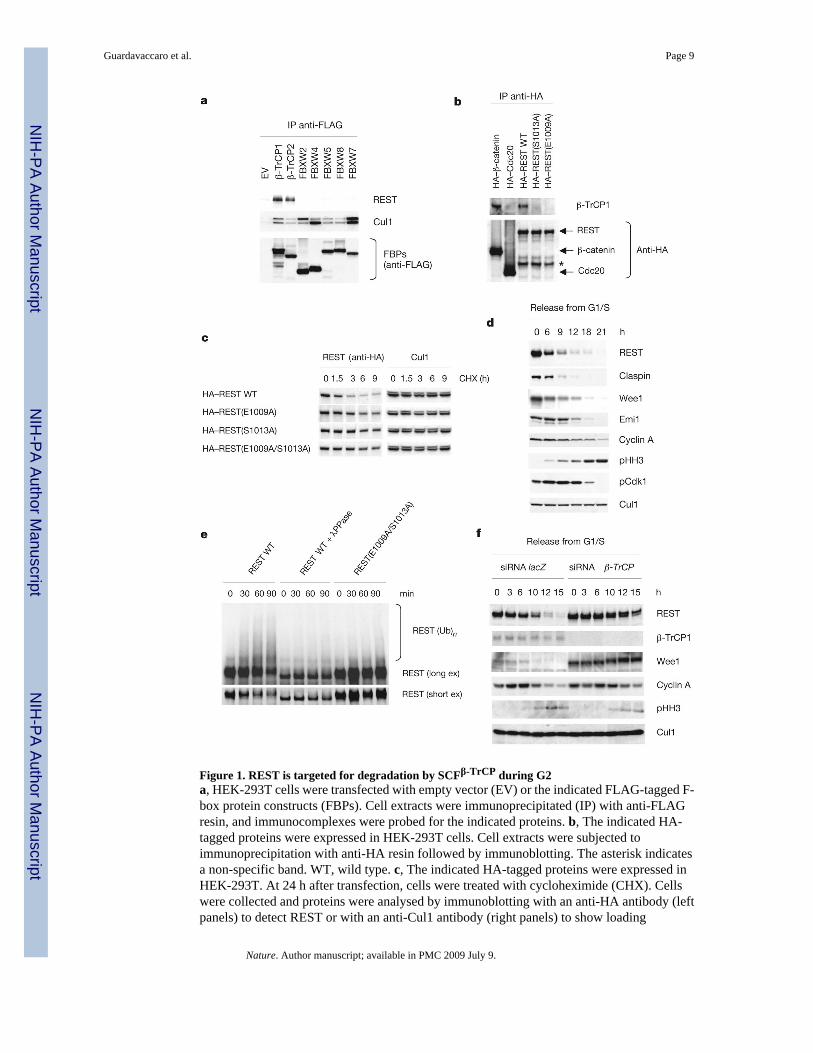
Figure 1. REST is targeted for degradation by SCFβ-TrCP during G2a, HEK-293T cells were transfected with empty vector (EV) or the indicated FLAG-tagged F-box protein constructs (FBPs). Cell extracts were immunoprecipitated (IP) with anti-FLAGresin, and immunocomplexes were probed for the indicated proteins. b, The indicated HA-tagged proteins were expressed in HEK-293T cells. Cell extracts were subjected toimmunoprecipitation with anti-HA resin followed by immunoblotting. The asterisk indicatesa non-specific band. WT, wild type. c, The indicated HA-tagged proteins were expressed inHEK-293T. At 24 h after transfection, cells were treated with cycloheximide (CHX). Cellswere collected and proteins were analysed by immunoblotting with an anti-HA antibody (leftpanels) to detect REST or with an anti-Cul1 antibody (right panels) to show loading
Guardavaccaro et al. Page 9
Nature. Author manuscript; available in PMC 2009 July 9.
NIH
-PA Author Manuscript
NIH
-PA Author Manuscript
NIH
-PA Author Manuscript
normalization. d, HeLa cells were synchronized by a double-thymidine block and released intonocodazole-containing medium. Cells were collected at the indicated times, lysed andimmunoblotted. e, β-TrCP-mediated REST ubiquitination is dependent on phosphorylation.HeLa cells were infected with lentiviruses expressing HA-tagged wild-type REST or HA-tagged REST (E1009/S1013A), synchronized in prometaphase and incubated with MG132during the last 3 h before lysis. Cell extracts were immunoprecipitated with anti-HA resin.Immunoprecipitates were incubated with either λ-protein phosphatase (λPPase) or enzymebuffer before ubiquitination/degradation assays (at 30 °C for the indicated times) in thepresence of in vitro transcribed/translated β-TrCP. The bracket marks a ladder of bandscorresponding to polyubiquitinated REST detected by immunoblotting. ex, exposure. f, HeLacells were transfected twice with short interfering RNA (siRNA) molecules to a non-relevantmRNA (lacZ) or to β-TrCP mRNA and then synchronized and analysed as in d.
Guardavaccaro et al. Page 10
Nature. Author manuscript; available in PMC 2009 July 9.
NIH
-PA Author Manuscript
NIH
-PA Author Manuscript
NIH
-PA Author Manuscript
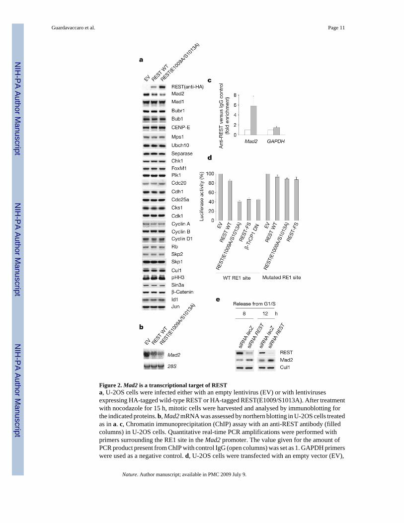
Figure 2. Mad2 is a transcriptional target of RESTa, U-2OS cells were infected either with an empty lentivirus (EV) or with lentivirusesexpressing HA-tagged wild-type REST or HA-tagged REST(E1009/S1013A). After treatmentwith nocodazole for 15 h, mitotic cells were harvested and analysed by immunoblotting forthe indicated proteins. b, Mad2 mRNA was assessed by northern blotting in U-2OS cells treatedas in a. c, Chromatin immunoprecipitation (ChIP) assay with an anti-REST antibody (filledcolumns) in U-2OS cells. Quantitative real-time PCR amplifications were performed withprimers surrounding the RE1 site in the Mad2 promoter. The value given for the amount ofPCR product present from ChIP with control IgG (open columns) was set as 1. GAPDH primerswere used as a negative control. d, U-2OS cells were transfected with an empty vector (EV),
Guardavaccaro et al. Page 11
Nature. Author manuscript; available in PMC 2009 July 9.
NIH
-PA Author Manuscript
NIH
-PA Author Manuscript
NIH
-PA Author Manuscript
HA-tagged REST proteins or a dominant-negative β-TrCP1 mutant (FLAG–β-TrCP1 DN)together with a luciferase reporter linked to a Mad2 genomic fragment containing either a wild-type (WT) or a mutated RE1 site. Prometaphase cells were collected and the relative luciferasesignal was quantified. The value given for luciferase activity in EV-transfected cells was setat 100%. e, IMR-90 cells were transfected twice with siRNA molecules to a non-relevantmRNA (lacZ) or to REST mRNA and synchronized in G2 by release from an aphidicolin blockfor the indicated durations30. Cells were then lysed and immunoblotted. Where present, errorbars represent s.d. (n = 3).
Guardavaccaro et al. Page 12
Nature. Author manuscript; available in PMC 2009 July 9.
NIH
-PA Author Manuscript
NIH
-PA Author Manuscript
NIH
-PA Author Manuscript
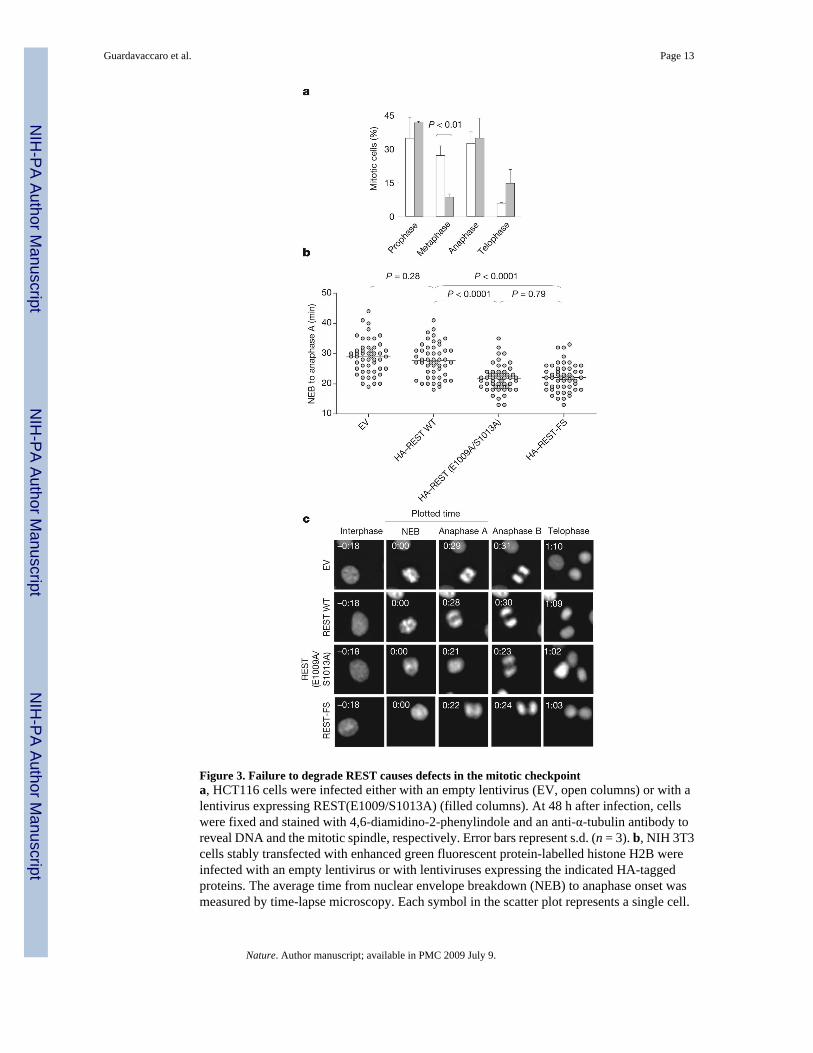
Figure 3. Failure to degrade REST causes defects in the mitotic checkpointa, HCT116 cells were infected either with an empty lentivirus (EV, open columns) or with alentivirus expressing REST(E1009/S1013A) (filled columns). At 48 h after infection, cellswere fixed and stained with 4,6-diamidino-2-phenylindole and an anti-α-tubulin antibody toreveal DNA and the mitotic spindle, respectively. Error bars represent s.d. (n = 3). b, NIH 3T3cells stably transfected with enhanced green fluorescent protein-labelled histone H2B wereinfected with an empty lentivirus or with lentiviruses expressing the indicated HA-taggedproteins. The average time from nuclear envelope breakdown (NEB) to anaphase onset wasmeasured by time-lapse microscopy. Each symbol in the scatter plot represents a single cell.
Guardavaccaro et al. Page 13
Nature. Author manuscript; available in PMC 2009 July 9.
NIH
-PA Author Manuscript
NIH
-PA Author Manuscript
NIH
-PA Author Manuscript

c, Representative fluorescence videomicroscopy series from b; numbers in the top left are times(h:min).
Guardavaccaro et al. Page 14
Nature. Author manuscript; available in PMC 2009 July 9.
NIH
-PA Author Manuscript
NIH
-PA Author Manuscript
NIH
-PA Author Manuscript
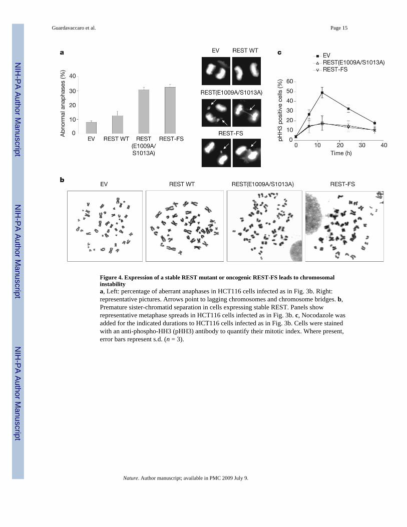
Figure 4. Expression of a stable REST mutant or oncogenic REST-FS leads to chromosomalinstabilitya, Left: percentage of aberrant anaphases in HCT116 cells infected as in Fig. 3b. Right:representative pictures. Arrows point to lagging chromosomes and chromosome bridges. b,Premature sister-chromatid separation in cells expressing stable REST. Panels showrepresentative metaphase spreads in HCT116 cells infected as in Fig. 3b. c, Nocodazole wasadded for the indicated durations to HCT116 cells infected as in Fig. 3b. Cells were stainedwith an anti-phospho-HH3 (pHH3) antibody to quantify their mitotic index. Where present,error bars represent s.d. (n = 3).
Guardavaccaro et al. Page 15
Nature. Author manuscript; available in PMC 2009 July 9.
NIH
-PA Author Manuscript
NIH
-PA Author Manuscript
NIH
-PA Author Manuscript
Copyright © 2022 FDOKUMEN

