


Bahasa
Halaman
Hukum

Clinical utility of multiplex ligation-dependent probe amplification technique in identification of aetiology of unexplained mental retardation: A study in 203 Indian patients
Vijay R. Boggula1, Anju Shukla1, Sumita Danda2, Sankar V. Hariharan3, Sheela Nampoothiri4, Rashmi Kumar5 & Shubha R. Phadke1
1Department of Medical Genetics, Sanjay Gandhi Post Graduate Institute of Medical Sciences, Lucknow, 2Department of Pediatrics, Christian Medical College & Hospital, Vellore, 3Department of Pediatrics, SAT Hospital, Medical College, Thiruvananthapuram, 4Department of Pediatric Genetics, Amrita Institute of Medical Sciences & Research Center, Cochin & 5Department of Pediatrics, King George’s Medical University, Lucknow, India
Received May 1, 2012
Background & objectives: Developmental delay (DD)/mental retardation also described as intellectual disability (ID), is seen in 1-3 per cent of general population. Diagnosis continues to be a challenge at clinical level. With the advancement of new molecular cytogenetic techniques such as cytogenetic microarray (CMA), multiplex ligation-dependent probe amplification (MLPA) techniques, many microdeletion/microduplication syndromes with DD/ID are now delineated. MLPA technique can probe 40-50 genomic regions in a single reaction and is being used for evaluation of cases with DD/ID. In this study we evaluated the clinical utility of MLPA techniques with different probe sets to identify the aetiology of unexplained mental retardation in patients with ID/DD.Methods: A total of 203 randomly selected DD/ID cases with/without malformations were studied. MLPA probe sets for subtelomeric regions (P070/P036) and common microdeletions/microduplications (P245-A2) and X-chromosome (P106) were used. Positive cases with MLPA technique were confirmed using either f uorescence in situ hybridization (FISH) or follow up confirmatory MLPA probe sets.Results: The overall detection rate was found to be 9.3 per cent (19 out of 203). The detection rates were 6.9 and 7.4 per cent for common microdeletion/microduplication and subtelomeric probe sets, respectively. No abnormality was detected with probe set for X-linked ID. The subtelomeric abnormalities detected included deletions of 1p36.33, 4p, 5p, 9p, 9q, 13q telomeric regions and duplication of 9pter. The deletions/duplications detected in non telomeric regions include regions for Prader Willi/Angelman regions, Williams syndrome, Smith Magenis syndrome and Velocardiofacial syndrome.Interpretation & conclusions: Our results show that the use of P245-A2 and P070/P036-E1 probes gives good diagnostic yield. Though MLPA cannot probe the whole genome like cytogenetic microarray, due to its ease and relative low cost it is an important technique for evaluation of cases with DD/ID.
Key words Common microdeletion/microduplication syndromes - developmental delay - intellectual disability - India - MLPA - subtelomeric abnormalities
Indian J Med Res 139, January 2014, pp 66-75
66
Developmental delay (DD) or intellectual disability (ID) is a common indication of referral for genetic counselling. The identification of correct aetiology is the prerequisite for genetic counselling. In spite of complete investigations, the genetic aetiology remains unidentified in 25 to 50 per cent of cases1. Molecular cytogenetic techniques have markedly increased the detection of chromosomal abnormalities. Traditional cytogenetics can detect the genomic imbalances more than 5 to 10 Mbp (megabase pair) size. New molecular cytogenetic techniques have led to the identification of many microdeletion/microduplication syndromes including the abnormalities of subtelomeric regions. Fluorescence in situ hybridization (FISH) was the first clinically used molecular cytogenetic technique2 and is still being widely used for evaluation of patients with suspected microdeletion syndrome. FISH can test one or a few regions at a time; therefore, clinical suspicion is needed before ordering the specific FISH based test3. This limitation was overcome by multiplex ligation-dependent probe amplification (MLPA) technique4. This technique has revolutionized the evaluation of DD/ID syndromes as it can test for 40-50 regions of the genome in a single reaction. Microarray based cytogenetic analysis is a better technique of higher resolution and simultaneous coverage of whole genome5, but high cost limits its widespread application in India.
In this study, randomly collected 203 Indian patients with ID/DD with/without malformations and dysmorphism were analyzed with MLPA technique. The utility/performance of MLPA probe sets for subtelomeric regions, common microdeletion/microduplication syndromes, and X chromosome was evaluated.
Material & Methods
This study was conducted in the department of Pediatrics, Chhatrapati Shahuji Maharaj Medical University (CSMMU), Lucknow, India, during 2009 to 2011. The study protocol was approved by the institutional ethics committee of CSMMU, Lucknow.
Two hundred and three patients with DD or ID with or without malformations/dysmorphism were selected after evaluation by clinical geneticists. The blood (2 ml)samples were collected after obtaining written consent from the parents. A three generation pedigree was drawn for each case. All cases were sporadic and there was no positive family history in any of the cases. Cases in which the aetiology was detected by karyotyping,
neuroimaging, metabolic screening and appropriate investigations based on clinical examination were excluded from this study. Molecular studies for fragile X syndrome were done in males without malformation or microcephaly and confirmed cases were excluded from the study. Inclusion criteria were presence of DD/ID with normal karyotype and with/without malformations or dysmorphic features. Only cases of DD/ID of unidentified aetiology were included. The average age was 5 yr (ranged from 4 months to 18 yr). There were 124 (61%) males and 79 (39%) females. The present results also include our published data on 65 cases in which only MLPA testing for subtelomeric regions was performed6. Samples obtained from different centers for this study were as follows; Sanjay Gandhi Postgraduate Institute of Medical Sciences (SGPGIMS), Lucknow and CSMMU, Lucknow (130), Amrita Institute of Medical Sciences & Research Center, Cochin (50), Christian Medical College, Vellore (18), Sree Avittom Thirunal Medical college, Thiruvananthapuram (5). Assessment of intelligent quotient (IQ) was performed in cooperative children older than 6 yr of age using Malin’s Intelligence Scale for Indian Children (an adaption of Wechsler Intelligence Scale for Children)7. For younger children developmental quotient was assessed.
Molecular cytogenetic methods: Genomic DNA was isolated from peripheral blood using QIAGEN genomic DNA isolation kit (Hilden, Germany) according to manufacturer’s instruction. MLPA was performed according to the manufacturer’s protocol (MRC- Holland, Amsterdam). All samples were processed for MLPA with probe sets for common microdeletion syndromes as well as for subtelomeric regions. Probe sets for subtelomeric region used were P036-E1 and P070. Abnormality identified by one probe set was confirmed using another. Probe set for common microdeletion syndrome used was P245-A2. This probe set covers common syndromic regions such as Prader-Willi/Angelman, DiGeorge/ Velocardiofacial, and 19 other syndromes. Deletions of 5pter, 4pter, Prader-Willi/Angelman syndrome region and Williams syndrome were confirmed by FISH analysis using probes for the respective regions. For other deletions and duplications detected by probe set P245-A2, confirmation was done by appropriate confirmatory probe sets namely; P372-A1, P373-A1, and P374-A1. Selection criteria for X-chromosome screening included males with normal MLPA profile using P245-A2 and P070/P036-E1 probe sets. Of the
BOGGULA et al: UTILITY OF MLPA TECHNIQUE IN UNEXPLAINED MENTAL RETARDATION 67
124 males, 89 were screened with X-linked ID probe set (MRX, P106). Probes used in these probe sets are for the genes which are known to cause syndromes with ID/DD.
Coffalyser stand alone alpha version (coffalyser.org) was used for analysis of peak values obtained from GENESCAN software (ABI 310 instrument, Applied Biosystems, USA). Coffalyser software has built-in setup for peak height normalization and reaction quality control calculations. All the statistical analyses were performed according to coffalyser software manual instructions supplied by the manufacturer. Mean cut-off for normalized peak height ratio of patient to the control sample was less than 0.65 in case of deletions and more than 1.40 in case of duplications.
Results
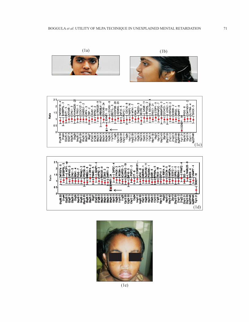
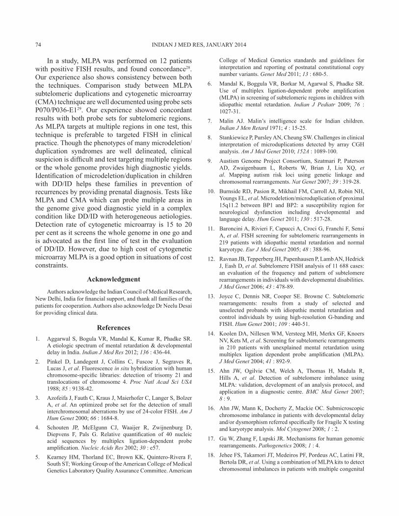
Two hundred and three cases were studied by both the probe sets; for subtelomeric regions and common microdeletion syndromes. Probe set P245-A2 detected 14 abnormalities with detection rate of 6.8 per cent (14/203). Abnormalities included deletions of 15q11.2, 22q11.2, 7q11.2, 1p36.33, 4p, 5p, 17p11.2 and duplication of 15q11.2 (Table). The presence of duplication/deletion values in 3 or 2 probes for a specific region indicates aberration. Results with MLPA confirmatory kits or FISH results were consistent with the abnormalities detected by probe set P245-A2 (Fig.1). MLPA for subtelomeric regions identified 15 duplications/deletions (7.4%, 15/203) (Table). The clinical features and IQ/DQ of cases with deletions/duplications are given in the Table. The probe sets for subtelomeric region identified abnormalities in 15 cases with deletions of 1pter, 4pter, 5pter, 9pter, 9qter, 13qter, deletion of Prader Willi/Angelman syndrome region and duplication of 9pter. This included four new cases in addition to 10 cases also detected by P245 probe set. There were 10 cases which were detected by both kits. Combining the results of subtelomeric and common microdeletion syndrome probe sets, the abnormalities were detected in 19 of the 203 cases giving the detection rate of 9.3 per cent. No deletions/duplications were found using P106 probe set for X-linked mental retardation genes. Of the 19 cases detected by MLPA probe sets, 15 were of ≤3 yr age and four were more than 3 yr. All patients with deletion/duplication had dysmorphic features (Table, Figs 1 and 2).
Discussion
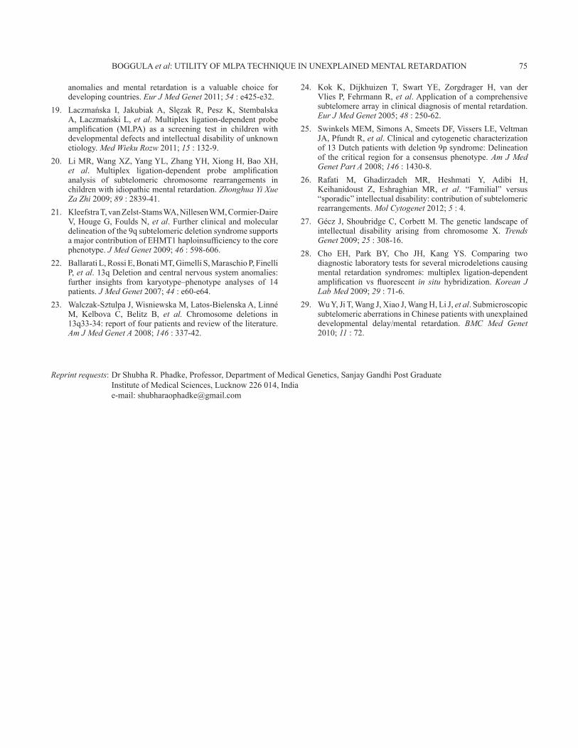
This study identified chromosomal aetiology in 9.3 per cent cases with DD/ID of unidentified aetiology. All the cases had dysmorphism but the clinical diagnoses of specific syndromes were not made at the time of sample collection. This may be mainly because the clinical features are subtle in many cases and clinical diagnosis may become obvious as the child grows. There is a great deal of variability of clinical presentation in some of the microdeletion syndromes and clinical suspicion of a specific syndrome is difficult. In case 1887, deletion of 15q11.2 region associated with Prader-Willi/Angelman syndrome was detected by MLPA and later confirmed by FISH. This test was performed at 18 months of age and the child had DD, strabismus and microcephaly. After follow up, at 3 yr of age she had developed characteristic features of Angelman syndrome. Same was the situation with the case number 799 who was under follow up for more than a year before the clinical features of Angelman syndrome became obvious (Fig 2e). The other two cases with deletion 15q11.2 had non specific dysmorphism. Case no. 2268 did not have obesity and her facial features were not characteristic but supportive of the diagnosis of Angelman syndrome. Hypotonia and joint laxity were pronounced in case number 885 supporting the diagnosis of Prader Willi syndrome. But the child did not have obesity (Body mass index of 17 kg/m2). The re-evaluation of clinical photographs of cases with 7q11.2 deletion revealed consistent features with the diagnosis of Williams syndrome. Both of them had prominent cheeks, thick lips but no cardiac anomaly. In these cases the clinical suspicion was possible, but was not suspected by the clinical geneticist. The syndromes of 22q 11.2 deletion, 17p11.2 deletion and terminal deletions of 1p, 4p and 5p are well delineated syndromes. Though some of the patients had clinical features consistent with the diagnosis, definitive clinical diagnosis of the particular microdeletion syndrome was not possible in many of them. Especially microduplication syndrome are difficult to suspect clinically because usually they have subtle phenotypes8. Case number 2329 was detected to have duplication of 15q11.2 region. He had microcephaly, facial dysmorphism and undescended testes but did not have obesity, seizures and autistic features described in syndrome9.
The contribution of subtelomeric deletions and duplications to unknown ID/DD cases has long been studied by FISH and reported to be ranging from 5 to 7 per cent10-13. Other studies using MLPA have reported
68 INDIAN J MED RES, JANUARY 2014
BOGGULA et al: UTILITY OF MLPA TECHNIQUE IN UNEXPLAINED MENTAL RETARDATION 69
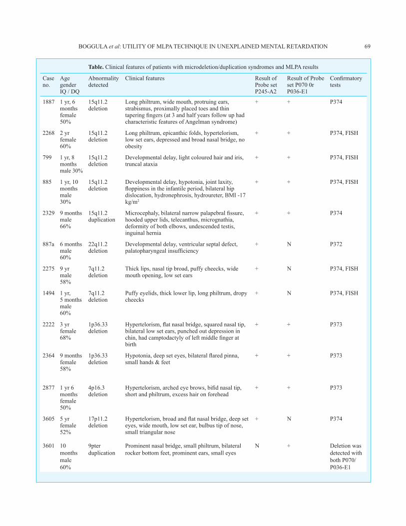
Table. Clinical features of patients with microdeletion/duplication syndromes and MLPA results
Case no.
Age gender IQ / DQ
Abnormality detected
Clinical features Result of Probe set P245-A2
Result of Probe set P070 0r P036-E1
Confirmatory tests
1887 1 yr, 6 monthsfemale50%
15q11.2 deletion
Long philtrum, wide mouth, protruing ears, strabismus, proximally placed toes and thin tapering fingers (at 3 and half years follow up had characteristic features of Angelman syndrome)
+ + P374
2268 2 yrfemale60%
15q11.2 deletion
Long philtrum, epicanthic folds, hypertelorism, low set ears, depressed and broad nasal bridge, no obesity
+ + P374, FISH
799 1 yr, 8 monthsmale 30%
15q11.2 deletion
Developmental delay, light coloured hair and iris, truncal ataxia
+ + P374, FISH
885 1 yr, 10 monthsmale 30%
15q11.2 deletion
Developmental delay, hypotonia, joint laxity, floppiness in the infantile period, bilateral hip dislocation, hydronephrosis, hydroureter, BMI -17 kg/m2
+ + P374, FISH
2329 9 monthsmale66%
15q11.2 duplication
Microcephaly, bilateral narrow palapebral fissure, hooded upper lids, telecanthus, micrognathia, deformity of both elbows, undescended testis, inguinal hernia
+ + P374
887a 6 monthsmale60%
22q11.2 deletion
Developmental delay, ventricular septal defect, palatopharyngeal insufficiency
+ N P372
2275 9 yrmale58%
7q11.2 deletion
Thick lips, nasal tip broad, puffy cheecks, wide mouth opening, low set ears
+ N P374, FISH
1494 1 yr, 5 monthsmale60%
7q11.2 deletion
Puffy eyelids, thick lower lip, long philtrum, dropy cheecks
+ N P374, FISH
2222 3 yrfemale68%
1p36.33 deletion
Hypertelorism, flat nasal bridge, squared nasal tip, bilateral low set ears, punched out depression in chin, had camptodactyly of left middle finger at birth
+ + P373
2364 9 monthsfemale58%
1p36.33 deletion
Hypotonia, deep set eyes, bilateral flared pinna, small hands & feet
+ + P373
2877 1 yr 6 monthsfemale50%
4p16.3 deletion
Hypertelorism, arched eye brows, bifid nasal tip, short and philtrum, excess hair on forehead
+ + P373
3605 5 yrfemale52%
17p11.2 deletion
Hypertelorism, broad and flat nasal bridge, deep set eyes, wide mouth, low set ear, bulbus tip of nose, small triangular nose
+ N P374
3601 10 months male 60%
9pter duplication
Prominent nasal bridge, small philtrum, bilateral rocker bottom feet, prominent ears, small eyes
N + Deletion was detected with both P070/P036-E1
detection rates similar to those by FISH14-16. These abberations have been classified under recurrent and non recurrent groups occuring in the human genome17. This study identified 1pter, 4pter which are recurrent abnormalities18-20. Phenotype of these disorders have been well delineated but clinical diagnosis is not always possible. Cases of 9pter, 9qter, 13qter deletions and duplications of 9p terminal are reported in the literatures21-24. Phenotype of 9pter deletion cases includes trigonocephaly and midface hypoplasia which were not present in our case25. The features described in deletion of 9qter includes non specific features like severe mental retardation (MR), hypotonia, seizures, microcephaly and specific features like flat face, hypertelorism, synophrys, anteverted nostrils, microglossia and heart defects. The case reported in this study (case no. 2137) had flat face, synophrys, brachycepahly and prognathism. Though it may be difficult to clinically diagnose this syndrome, there is similarity of facial phenotype of this case with those reported in the literature21. The dysmorphic features of 13qter deletion syndrome are non specific22,23 as
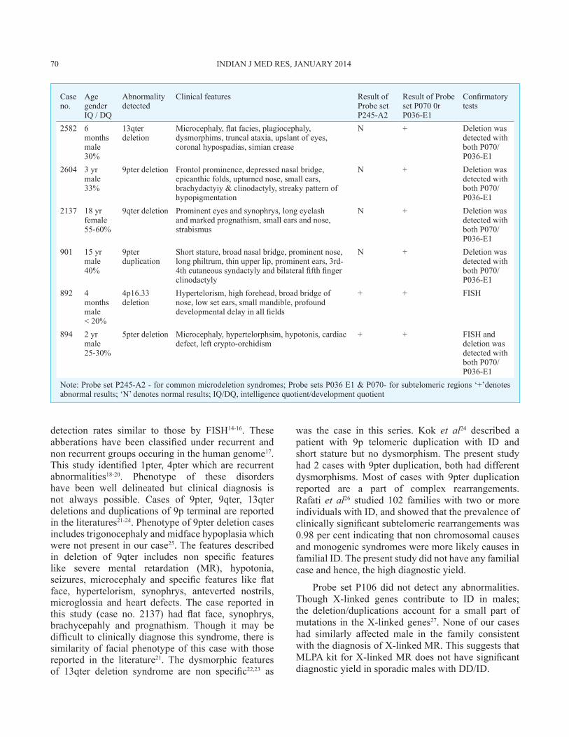
Case no.
Age gender IQ / DQ
Abnormality detected
Clinical features Result of Probe set P245-A2
Result of Probe set P070 0r P036-E1
Confirmatory tests
2582 6 monthsmale30%
13qter deletion
Microcephaly, fl at facies, plagiocephaly, dysmorphims, truncal ataxia, upslant of eyes, coronal hypospadias, simian crease
N + Deletion was detected with both P070/P036-E1
2604 3 yrmale33%
9pter deletion Frontol prominence, depressed nasal bridge, epicanthic folds, upturned nose, small ears, brachydactyiy & clinodactyly, streaky pattern of hypopigmentation
N + Deletion was detected with both P070/P036-E1
2137 18 yrfemale55-60%
9qter deletion Prominent eyes and synophrys, long eyelash and marked prognathism, small ears and nose, strabismus
N + Deletion was detected with both P070/P036-E1
901 15 yrmale40%
9pter duplication
Short stature, broad nasal bridge, prominent nose, long philtrum, thin upper lip, prominent ears, 3rd-4th cutaneous syndactyly and bilateral fifth finger clinodactyly
N + Deletion was detected with both P070/P036-E1
892 4 monthsmale< 20%
4p16.33 deletion
Hypertelorism, high forehead, broad bridge of nose, low set ears, small mandible, profound developmental delay in all fields
+ + FISH
894 2 yrmale25-30%
5pter deletion Microcephaly, hypertelorphsim, hypotonis, cardiac defect, left crypto-orchidism
+ + FISH anddeletion was detected with both P070/P036-E1
Note: Probe set P245-A2 - for common microdeletion syndromes; Probe sets P036 E1 & P070- for subtelomeric regions ‘+’denotes abnormal results; ‘N’ denotes normal results; IQ/DQ, intelligence quotient/development quotient
was the case in this series. Kok et al24 described a patient with 9p telomeric duplication with ID and short stature but no dysmorphism. The present study had 2 cases with 9pter duplication, both had different dysmorphisms. Most of cases with 9pter duplication reported are a part of complex rearrangements. Rafati et al26 studied 102 families with two or more individuals with ID, and showed that the prevalence of clinically significant subtelomeric rearrangements was 0.98 per cent indicating that non chromosomal causes and monogenic syndromes were more likely causes in familial ID. The present study did not have any familial case and hence, the high diagnostic yield.
Probe set P106 did not detect any abnormalities. Though X-linked genes contribute to ID in males; the deletion/duplications account for a small part of mutations in the X-linked genes27. None of our cases had similarly affected male in the family consistent with the diagnosis of X-linked MR. This suggests that MLPA kit for X-linked MR does not have significant diagnostic yield in sporadic males with DD/ID.
70 INDIAN J MED RES, JANUARY 2014
(1d)
BOGGULA et al: UTILITY OF MLPA TECHNIQUE IN UNEXPLAINED MENTAL RETARDATION 71
(1a) (1b)
(1e)
(1c)

(1f)
(1g)
(1h)
72 INDIAN J MED RES, JANUARY 2014

(2d)(2e)
(2f)
(2a)
(2b)
(2c)
Fig. 2: (a) Case no. 2222- Deletion 1pter. Note straight eybrows (b) Case no. 2364 - Deletion 1pter. Note deep seated eyes (c) Case no. 2604 –Deletion 9pter (d) Case no. 2582- Deletion of 13qter (e) Case no. 2268- Deletion 15q11.2 with features though not characteristic but supportive of Angelman syndrome.
Fig. 1: (a) & (b) Facial features of representative case no.2137 with 9q34.3 deletion, note flat face and prognathism. (c &d) MLPA P070 and P036-E1 probe sets results of case no. 2137 showing deletion of 9q34. (e) Facial features of case No.2877 with 4p16.33 deletion, note hypertelorism. (f) MLPA results of case 2877 using P373 confirmatory probe set showing deletion of 4p16.33. (g) FISH result of case 2275 showing single copy of 7q11.2 (pink signal). Green signals are controls (2 copies) (h) MLPA profile showing deletion at 7q11.29 using P245-A2 probe set (i) MLPA profile showing deletion at 7q11.29 using P374 confirmatory probe set.
(1i)
BOGGULA et al: UTILITY OF MLPA TECHNIQUE IN UNEXPLAINED MENTAL RETARDATION 73
In a study, MLPA was performed on 12 patients with positive FISH results, and found concordance28. Our experience also shows consistency between both the techniques. Comparison study between MLPA subtelomeric duplications and cytogenetic microarray (CMA) technique are well documented using probe sets P070/P036-E129. Our experience showed concordant results with both probe sets for subtelomeric regions. As MLPA targets at multiple regions in one test, this technique is preferable to targeted FISH in clinical practice. Though the phenotypes of many microdeletion/duplication syndromes are well delineated, clinical suspicion is difficult and test targeting multiple regions or the whole genome provides high diagnostic yields. Identification of microdeletion/duplication in children with DD/ID helps these families in prevention of recurrences by providing prenatal diagnosis. Tests like MLPA and CMA which can probe multiple areas in the genome give good diagnostic yield in a complex condition like DD/ID with heterogeneous aetiologies. Detection rate of cytogenetic microarray is 15 to 20 per cent as it screens the whole genome in one go and is advocated as the first line of test in the evaluation of DD/ID. However, due to high cost of cytogenetic microarray MLPA is a good option in situations of cost constraints.
Acknowledgment
Authors acknowledge the Indian Council of Medical Research, New Delhi, India for financial support, and thank all families of the patients for cooperation. Authors also acknowledge Dr Neelu Desai for providing clinical data.
ReferencesAggarwa1. l S, Bogula VR, Mandal K, Kumar R, Phadke SR. A etiologic spectrum of mental retardation & developmental delay in India. Indian J Med Res 2012; 136 : 436-44.Pinkel D, Landegent J, Collins C, Fuscoe J, Segraves R, 2. Lucas J, et al. Fluorescence in situ hybridization with human chromosome-specific libraries: detection of trisomy 21 and translocations of chromosome 4. Proc Natl Acad Sci USA 1988; 85 : 9138-42.Azofeifa J, Fauth C, Kraus J, Maierhofer C, Langer S, Bolzer 3. A, et al. An optimized probe set for the detection of small interchromosomal aberrations by use of 24-color FISH. Am J Hum Genet 2000; 66 : 1684-8.Schouten JP, McElgunn CJ, Waaijer R, Zwijnenburg D, 4. Diepvens F, Pals G. Relative quantification of 40 nucleic acid sequences by multiplex ligation-dependent probe amplification. Nucleic Acids Res 2002; 30 : e57.Kearney HM, Thorland EC, Brown KK, Quintero-Rivera F, 5. South ST; Working Group of the American College of Medical Genetics Laboratory Quality Assurance Committee. American
College of Medical Genetics standards and guidelines for interpretation and reporting of postnatal constitutional copy number variants. Genet Med 2011; 13 : 680-5.Mandal K, Boggula VR, Borkar M, Agarwal S, Phadke SR. 6. Use of multiplex ligation-dependent probe amplification (MLPA) in screening of subtelomeric regions in children with idiopathic mental retardation. Indian J Pediatr 2009; 76 : 1027-31.Malin AJ. Malin’s intelligence scale for Indian children. 7. Indian J Men Retard 1971; 4 : 15-25.Stankiewicz P, Pursley AN, Cheung SW. Challenges in clinical 8. interpretation of microduplications detected by array CGH analysis. Am J Med Genet 2010; 152A : 1089-100.Austism Genome Project Consortium, Szatmari P, Paterson 9. AD, Zwaigenbaum L, Roberts W, Brian J, Liu XQ, et al. Mapping autism risk loci using genetic linkage and chromosomal rearrangements. Nat Genet 2007; 39 : 319-28.Burnside RD, Pasion R, Mikhail FM, Carroll AJ, Robin NH, 10. Youngs EL, et al. Microdeletion/microduplication of proximal 15q11.2 between BP1 and BP2: a susceptibility region for neurological dysfunction including developmental and language delay. Hum Genet 2011; 130 : 517-28.Baroncini A, Rivieri F, Capucci A, Croci G, Franchi F, Sensi 11. A, et al. FISH screening for subtelomeric rearrangements in 219 patients with idiopathic mental retardation and normal karyotype. Eur J Med Genet 2005; 48 : 388-96.Ravnan JB, Tepperberg JH, Papenhausen P, Lamb AN, Hedrick 12. J, Eash D, et al. Subtelomere FISH analysis of 11 688 cases: an evaluation of the frequency and pattern of subtelomere rearrangements in individuals with developmental disabilities. J Med Genet 2006; 43 : 478-89.Joyce C, Dennis NR, Cooper SE. Browne C. Subtelomeric 13. rearrangements: results from a study of selected and unselected probands with idiopathic mental retardation and control individuals by using high-resolution G-banding and FISH. Hum Genet 2001; 109 : 440-51.Koolen DA, Nillesen WM, Versteeg MH, Merkx GF, Knoers 14. NV, Kets M, et al. Screening for subtelomeric rearrangements in 210 patients with unexplained mental retardation using multiplex ligation dependent probe amplification (MLPA). J Med Genet 2004; 41 : 892-9.Ahn JW, Ogilvie CM, Welch A, Thomas H, Madula R, 15. Hills A, et al. Detection of subtelomere imbalance using MLPA: validation, development of an analysis protocol, and application in a diagnostic centre. BMC Med Genet 2007; 8 : 9.Ahn JW, Mann K, Docherty Z, Mackie OC. Submicroscopic 16. chromosome imbalance in patients with developmental delay and/or dysmorphism referred specifically for Fragile X testing and karyotype analysis. Mol Cytogenet 2008; 1 : 2. Gu W, Zhang F, Lupski JR. Mechanisms for human genomic 17. rearrangements. Pathogenetics 2008; 1 : 4.Jehee FS, Takamori JT, Medeiros PF, Pordeus AC, Latini FR, 18. Bertola DR, et al. Using a combination of MLPA kits to detect chromosomal imbalances in patients with multiple congenital
74 INDIAN J MED RES, JANUARY 2014
anomalies and mental retardation is a valuable choice for developing countries. Eur J Med Genet 2011; 54 : e425-e32.Laczmańska I, Jakubiak A, Slęzak R, Pesz K, Stembalska 19. A, Laczmański L, et al. Multiplex ligation-dependent probe amplification (MLPA) as a screening test in children with developmental defects and intellectual disability of unknown etiology. Med Wieku Rozw 2011; 15 : 132-9. Li MR, Wang XZ, Yang YL, Zhang YH, Xiong H, Bao XH, 20. et al. Multiplex ligation-dependent probe amplification analysis of subtelomeric chromosome rearrangements in children with idiopathic mental retardation. Zhonghua Yi Xue Za Zhi 2009; 89 : 2839-41. Kleefstra T, van Zelst-Stams WA, Nillesen WM, Cormier-Daire 21. V, Houge G, Foulds N, et al. Further clinical and molecular delineation of the 9q subtelomeric deletion syndrome supports a major contribution of EHMT1 haploinsufficiency to the core phenotype. J Med Genet 2009; 46 : 598-606.Ballarati L, Rossi E, Bonati MT, Gimelli S, Maraschio P, Finelli 22. P, et al. 13q Deletion and central nervous system anomalies: further insights from karyotype–phenotype analyses of 14 patients. J Med Genet 2007; 44 : e60-e64.Walczak-Sztulpa J, Wisniewska M, Latos-Bielenska A, Linné 23. M, Kelbova C, Belitz B, et al. Chromosome deletions in 13q33-34: report of four patients and review of the literature. Am J Med Genet A 2008; 146 : 337-42.
Reprint requests: Dr Shubha R. Phadke, Professor, Department of Medical Genetics, Sanjay Gandhi Post Graduate Institute of Medical Sciences, Lucknow 226 014, India
e-mail: [email protected]
Kok K, Dijkhuizen T, Swart YE, Zorgdrager H, van der 24. Vlies P, Fehrmann R, et al. Application of a comprehensive subtelomere array in clinical diagnosis of mental retardation. Eur J Med Genet 2005; 48 : 250-62.Swinkels MEM, Simons A, Smeets DF, Vissers LE, Veltman 25. JA, Pfundt R, et al. Clinical and cytogenetic characterization of 13 Dutch patients with deletion 9p syndrome: Delineation of the critical region for a consensus phenotype. Am J Med Genet Part A 2008; 146 : 1430-8.Rafati M, Ghadirzadeh MR, Heshmati Y, Adibi H, 26. Keihanidoust Z, Eshraghian MR, et al. “Familial” versus “sporadic” intellectual disability: contribution of subtelomeric rearrangements. Mol Cytogenet 2012; 5 : 4. Gécz J, Shoubridge C, Corbett M. The genetic landscape of 27. intellectual disability arising from chromosome X. Trends Genet 2009; 25 : 308-16.Cho EH, Park BY, Cho JH, Kang YS. Comparing two 28. diagnostic laboratory tests for several microdeletions causing mental retardation syndromes: multiplex ligation-dependent amplification vs fluorescent in situ hybridization. Korean J Lab Med 2009; 29 : 71-6.Wu Y, Ji T, Wang J, Xiao J, Wang H, Li J, 29. et al. Submicroscopic subtelomeric aberrations in Chinese patients with unexplained developmental delay/mental retardation. BMC Med Genet 2010; 11 : 72.
BOGGULA et al: UTILITY OF MLPA TECHNIQUE IN UNEXPLAINED MENTAL RETARDATION 75
Top Related
Copyright © 2022 FDOKUMEN

