


Bahasa
Halaman
Hukum

Characterization of the recombinant human prostanoidDP receptor and identi®cation of L-644,698, a novel selectiveDP agonist
D. Hamish Wright, 1,2Kathleen M. Metters, 1Mark Abramovitz & 1Anthony W. Ford-Hutchinson
Department of Pharmacology & Therapeutics, McGill University, Montre al, Que bec, H3C 5C4 and 1Department of Biochemistry& Molecular Biology, Merck Frosst Centre for Therapeutic Research, Pointe Claire-Dorval, Que bec, Canada, H9R 4P8
1 A human embryonic kidney cell line [HEK 293(EBNA)] stably expressing the human recombinantprostaglandin D2 (PGD2) receptor (hDP) has been characterized with respect to radioligand binding andsignal transduction properties by use of prostanoids and prostanoid analogues. Radioligand bindingstudies included saturation analyses, the e�ects of nucleotide analogues, the initial rate of ligand-receptorassociation and equilibrium competition assays. In addition, adenosine 3':5'-cyclic monophosphate (cyclicAMP) generation in response to ligand challenge was also measured, as this is the predominant hDPsignalling pathway.
2 L-644,698 ((4-(3-(3-(3-hydroxyoctyl)-4-oxo-2-thiazolidinyl) propyl) benzoic acid) (racemate)) wasidenti®ed as a novel ligand having high a�nity for hDP with an inhibitor constant (Ki) of 0.9 nM. ThisKi value was comparable to the Ki values obtained in this study for ligands that have previously shownhigh a�nity for DP: PGD2 (0.6 nM), ZK 110841 (0.3 nM), BW245C (0.4 nM), and BW A868C (2.3 nM).
3 L-644,698 was found to be a full agonist with an EC50 value of 0.5 nM in generating cyclic AMPfollowing activation of hDP. L-644,698 is, therefore, comparable to those agonists with known e�cacy atthe DP receptor (EC50): PGD2 (0.5 nM), ZK 110841 (0.2 nM) and BW245C (0.3 nM).
4 L-644,698 displayed a high degree of selectivity for hDP when compared to the family of clonedhuman prostanoid receptors: EP1 (425,400 fold), EP2 (*300 fold), EP3-III (*4100 fold), EP4 (*10000fold), FP (425,400 fold), IP (425,400 fold) and TP (425,400 fold). L-644,698 is, therefore, one of themost selective DP agonists as yet described.
5 PGJ2 and D12-PGJ2, two endogenous metabolites of PGD2, were also tested in this system and shownto be e�ective agonists with Ki and EC50 values in the nanomolar range for both compounds. Inparticular, PGJ2 was equipotent to known DP speci®c agonists with a Ki value of 0.9 nM and an EC50
value of 1.2 nM.
Keywords: Prostanoid receptor; DP; recombinant; prostaglandin D2; prostaglandin J2; selective agonist
Introduction
Prostaglandins, prostacyclin (PGI2), and thromboxane A2
(TXA2) are collectively described as prostanoids. Originally itwas proposed (Kennedy et al., 1982; Coleman et al., 1984) thatindividual prostanoid receptors existed for each of the primary
bioactive prostanoids. This classi®cation described distinctreceptors for prostaglandin D2 (PGD2), PGE2, PGF2a, PGI2and TXA2 which were denoted DP, EP (EP1 and EP2) FP, IP
and TP, respectively. This was followed by a furthersubdivision of the EP class of receptors into four subtypes:EP1, EP2, EP3 and EP4 (for review, Coleman et al., 1994). Thecloning of the human (h) prostanoid receptors currently
includes TP (Hirata et al., 1991), FP (Abramovitz et al.,1994), IP (Boie et al., 1994), EP1 (Funk et al., 1993), EP2(Regan et al., 1994), EP3 (Adam et al., 1994), EP4 (An et al.,
1993; Bastien et al., 1994) and DP (Boie et al., 1995). Thesereceptors form a sub-family within the G-protein-coupledreceptor (GPCR) superfamily. During cloning, alternatively
spliced isoforms of both hTP (Raychowdhury et al., 1995) andhEP3 (Adam et al., 1994; Schmid et al., 1995; Kotani et al.,1995) were identi®ed.
The endogenous prostanoids demonstrate preference to-wards individual prostanoid receptors but, in general, there is amarked degree of cross-reactivity between these ligands and
the entire receptor family (Dong et al., 1986; Sheldrick et al.,
1988; Armstrong et al., 1989; Bunce et al., 1990). This hasdriven the development of selective compounds. Severalselective and e�cacious prostanoid agonists have been
identi®ed, including cicaprost (Dong et al., 1986; Armstronget al., 1989) at the IP receptor, GR63799X (Bunce et al., 1990)at the EP3 receptor and butaprost (Abramovitz et al.,
unpublished observations) at the EP2 receptor. To date, thegroup of synthetic DP agonists includes BW245C, ZK 110841,RS-93520, RS-93427, 572C85 and 192C86. BW245C is themost comprehensively studied of these synthetic DP agonists
(Town et al., 1983; Whittle et al., 1983; Woodward et al., 1990;Fernandes & Crankshaw, 1995; Rangachari et al., 1995) butalthough the e�cacy of BW245C at DP is well-established
(Town et al., 1983; Boie et al., 1995), the selectivity of thisligand versus the other prostanoid receptors is not completelyde®ned. Data suggest that BW245C has a�nity for at least one
other prostanoid receptor that couples to stimulation ofadenylate cyclase via the guanine nucleotide binding (G)protein Gs. Some ®ndings suggest that BW245C cross-reacts
with EP2 (Giles et al., 1989; Matsugi et al., 1995), while otherinvestigators have proposed IP (Trist et al., 1989). Anotherpotential limitation associated with BW245C concerns itsstability in aqueous solution, since it has been shown to
produce an epimer which is less biologically active than theparent compound (Brockwell et al., 1981).2Author for correspondence.
British Journal of Pharmacology (1998) 123, 1317 ± 1324 1998 Stockton Press All rights reserved 0007 ± 1188/98 $12.00
Studies of the DP receptor have been complicated by itsrelatively low abundance and narrow scope of distribution.Thus, it was the last known human prostanoid receptor to be
cloned (Boie et al., 1995). In this study, the radioligand bindingand signal transduction properties of recombinant hDP havebeen more completely studied, both with prostanoids and
prostanoid synthetic analogues of varying selectivity. Throughthese analyses and by use of the system of cloned recombinanthuman prostanoid receptors previously described (Abramovitzet al., unpublished observations), a novel selective DP agonist,
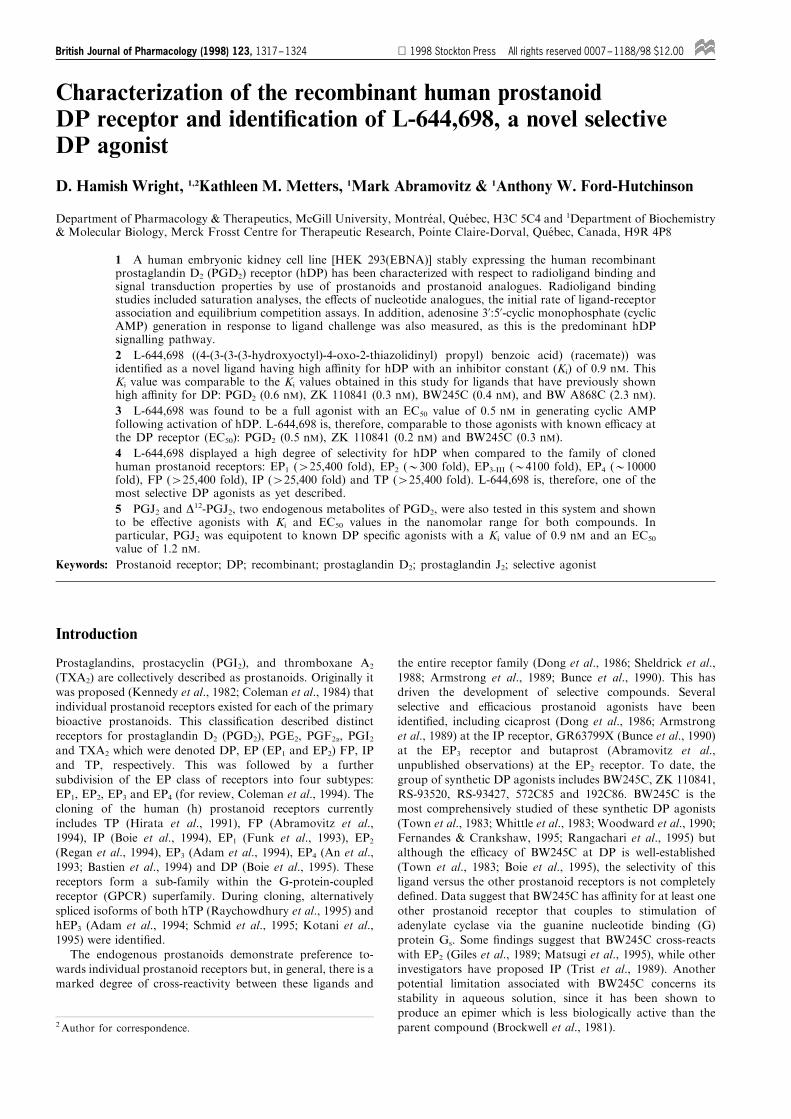
L-644,698 ((4 - (3- (3 - (3- hydroxyoctyl)-4-oxo-2-thiazolidinyl)propyl) benzoic acid) (racemate) (Figure 1), has beenidenti®ed.
Methods
pCEP4-hDP stable expression in HEK 293(EBNA)cells
Stable expression of the hDP receptor was achieved bytransfection of the pCEP4-hDP (Abramovitz et al., unpub-lished observations) plasmid into HEK 293(EBNA) cells
[maintained under selection with GENETICIN (G418)] bycationic-liposome mediated transfer with LipofectAMINEreagent (Felgner et al., 1987). Cells were maintained in culture
for 48 h post transfection and then grown in the presence of200 mg ml71 hygromycin B for 2 weeks, to select for resistantcolonies expressing the hDP receptor. Resistant colonies were
expanded and subsequently tested for hDP expression byradioligand binding. The clone with the highest level ofbinding activity was then used for signal transduction assays.
Cell culture and membrane preparation
HEK 293(EBNA) cells stably expressing hDP (hDP-HEK)
were maintained in culture in Dulbecco's modi®ed Eagle's
medium growth medium (Dulbecco's modi®ed Eagle'smedium containing 10% heat-inactivated foetal bovine serum,1 mM sodium pyruvate, 20 units ml71 penicillin G,
20 mg ml71 streptomycin sulphate, 250 mg ml71 GENETI-CIN and 200 mg ml71 hygromycin B). In order to preparemembranes from hDP-HEK cells (all procedures at 48C), theywere ®rst resuspended by Dounce homogenization (pestle B,10 strokes) in the presence of 2 mM phenylmethylsulphonyl-¯uoride. Cells were next disrupted by nitrogen-cavitation at800 psi for 30 min on ice. The resulting cell suspension was
subjected to two centrifugation steps: 10006gmax for 10 minfollowed by 100 0006gmax for 30 min. The resulting pellet wasresuspended to 1/10th the original volume in 10 mM HEPES/
KOH (pH 7.4) containing 1 mM EDTA (tetrasodium salt) byDounce homogenization (pestle A, 10 strokes), and aliquotswere stored at 7808C at a protein concentration of 8 ±
10 mg ml71.
[3H]-PGD2 binding to hDP-HEK membranes
Radioligand binding assays were performed in 0.2 ml of10 mM HEPES-KOH (pH 7.4) 1 mM EDTA containing(unless otherwise noted) 0.8 nM [3H]-PGD2 (115 Ci mmol
71)
and 10 mM MnCl2. Compounds were added in dimethylsulph-oxide (Me2SO) at 1% (v/v) of the ®nal incubation volume(vehicle concentration was constant throughout). The reaction
was initiated by the addition of 30 mg of hDP-HEK membraneprotein to all tubes and the samples were incubated at roomtemperature for 1 h. The reaction was terminated by rapid
®ltration at 48C in 3 ± 4 ml of 10 mM HEPES/KOH (pH 7.4)through a GF/C ®lter (Uni®lter) which had been presoaked inthe same bu�er. Each ®lter was dried for 1 ± 2 h at 558C andthe residual [3H]-PGD2 bound to the ®lter (33% e�ciency) was
determined in 50 ml per well of Ultima Gold scintillationcocktail. Non-speci®c binding was determined in the presenceof 1 mM PGD2.
Analysis of [3H]-PGD2 binding
Rates of association were calculated through a one-site curve-®t analysis, employing the equation Bt=Beq-Beq[e
(-Kobst)]; whereBt represents the radioligand bound speci®cally at time t, Beq
represents the radioligand bound speci®cally at equilibrium
and Kobs is the observed association rate which is thenexpressed as Kobs/[radioligand].
Speci®c binding saturation isotherms were deduced by
subtracting the nonspeci®c binding from the total binding,both measured experimentally. The saturation isotherms weretransformed by use of nonlinear, least-squares, regression
analysis adapted from the work of Feldman (1972) where theequation [(BMax6F)/(K1+F)]+[(BMax6F)/(K2+F)] representsthe radioligand speci®cally bound, BMax is the maximal
number of binding sites, K is the equilibrium dissociationconstant and F is the concentration of free radioligand. Theanalyses were performed by use of Accu®t Two-Site saturationsoftware (Beckman Instruments).
Sigmoidal curves from equilibrium competition assays wereanalysed by custom designed software which employs a non-linear least-squares ®tting routine based on the four parameter
logistic equation: y=(m17m2)(1+(m0/m3)em4)71+m2;where m1 and m2 represent the maximum and minimum ofthe curve, m3 represents the in¯ection point (IP), m4 represents
the slope of the curve at the in¯ection point, m0 represents theconcentration of the competing ligand and y represents the %[3H]-PGD2-speci®c binding. Ki values were calculated from theequation Ki=IP/1+[radioligand]/KD.
OH
O
S
N
O
OH
Figure 1 Structure of L-644,698 (racemate).
L-644,698, a novel DP receptor agonist1318 D. H. Wright et al
Chemical and metabolic stability of [3H]-PGD2 underthe experimental conditions
The stability of [3H]-PGD2 exposed to the incubationconditions for 2 h was veri®ed by reverse-phase highperformance liquid chromatography (r.p.-h.p.l.c.). Following
incubation of [3H]-PGD2 under standard conditions thereaction was terminated with the addition of 26r.p.-h.p.l.c. solvent (r.p.-h.p.l.c. solvent=66:15:18:1 (v/v)H2O:CH3CN:CH3OH:CH3COOH, adjusted to pH 5.6 with
10 N NaOH) to the samples. Samples were allowed to incubatefor 10 min at 308C to dissociate the receptor-bound [3H]-PGD2
and were then subjected to centrifugation at 100 0006g for
15 min at 48C. The resulting supernatant fractions, containingboth bound and unbound [3H]-PGD2, were then analysed byr.p.-h.p.l.c. with a NovaPak C18 column (0.39615 cm;
Waters). [3H]-PGD2 was eluted with a linear gradient from 0to 70% (v/v) acetonitrile developed over 35 min at a ¯ow rateof 1 ml min71 following an initial 3 min elution with r.p.-h.p.l.c. solvent. The 35 min gradient elution was followed by a
®nal 10 min wash with 90% (v/v) acetonitrile. The pro®le ofradioactivity was monitored with an on-line ¯ow-throughradioactivity detector (Berthold). Eluant fractions were
subsequently mixed with 5 ml of scintillation ¯uid for theprecise quantitation of the radioactivity recovered.
Cyclic AMP assays with hDP-HEK cells
hDP-HEK cells were harvested at 80% con¯uence by
resuspension in 10 ml of enzyme-free cell dissociation bu�erand washed in phosphate-bu�ered saline by centrifugation(3006gmax for 6 min at room temperature). Cells were thenwashed in 10 ml of Hank's balanced salt solution (HBSS) by
centrifugation under the same conditions as above andresuspended in HBSS at 46106 cells ml71. Cell viability wasdetermined to be 495%, by trypan blue exclusion. The
generation of adenosine 3':5'-cyclic monophosphate (cyclicAMP) was performed in a ®nal incubation volume of 0.2 ml ofHBSS containing 100 mM Ro 20-1724 to abrogate cyclic AMP
hydrolysis. Compounds were added in Me2SO kept constant at1% (v/v) of the ®nal incubation volume. Assays were initiatedby the addition of 26104 cells per reaction and samplesincubated for 10 min at 378C with shaking. Reactions were
halted by the incubation of samples in boiling water for 3 minand cyclic AMP was subsequently measured by use of a [125I]-cyclic AMP scintillation proximity assay.
Sigmoidal concentration-response curves were analysed bycustom designed software to determine EC50 values. Maximalstimulation was de®ned as the quantity of cyclic AMP
produced by incubation with 1 mM PGD2. Statistical analysisof the maximal response for each ligand as a percentage ofthis maximal stimulation was performed with SigmaStat
software, version 2.0 (Jandel Scienti®c). A one-way ANOVAfollowed by a Tukey test was used to determine full andpartial agonists.
Protein assays
Protein concentration was measured by the bicinchoninic acid
(BCA) protein assay kit (Pierce) with bovine serum albumin asthe standard.
Reagents
PGD2, PGE1, PGE2, PGF2a, U46619 (9,11-dideoxy-9a,11a-methanoepoxy-PGF2a), PGJ2, D12-PGJ2, 15-deoxy-D12,14-PGJ2
and Ro-20-1724 (4-(3 butoxy-4-methoxybenzyl)-2-imidazoli-dinone) were from Biomol Research Laboratories (PlymouthMeeting, PA, U.S.A.). 13,14-dihydro-15-keto-PGD2 was from
Cayman Chemical (Ann Arbor, MI, U.S.A.). BW245C (5-(6-carboxyhexyl)-1 - (3 - cyclohexyl - 3 - hydroxypropylhydantoin))and BW A868C ((+)-3-benzyl-5-(6-carboxyl)-1-(2-cyclohexyl-
2-hydroxyethylamino)-hydantoin) were generous gifts fromThe Wellcome Foundation Ltd (Beckenham, Kent, U.K.). ZK110841 ((5Z,13E)-(9R,11R,15S)-9b-chlor-15-cyclohexyl-11,15-dihydroxy-16,17,18,19,20-pentanor-5,13-prostadienoic acid)
was a generous gift from Dr D. Crankshaw from theDepartment of Obstetrics & Gynecology (McMaster Uni-versity, Hamilton, Ontario, Canada). L-644,698 was synthe-
sized at Merck Research Laboratories by Dr J.B. Bicking.Iloprost and [125I]-cyclic AMP scintillation proximity assay kitswere from Amersham (Oakville, ON, Canada). [3H]-PGD2 was
from Dupont NEN (Boston, MA, U.S.A.). GTPgS (Guano-sine-5'-O-(3-thiotriphosphate)), ATPgS (adenosine-5'-O-(3-thiotriphosphate)), GMP-PNP (guanylyl-imidodiphosphate)and AMP-PNP (adenylyl-imidodiphosphate) were from
Boehringer Mannheim Canada (Laval, QC, Canada). Bicinch-oninic acid (BCA) protein assays were from Pierce (Rockford,IL, U.S.A.). GENETICIN (G418) and LipofectAMINE were
from GIBCO/BRL (Burlington, ON, Canada). Enzyme-freecell dissociation bu�er was from Life-Technologies Inc(Gaithersburg, MD, U.S.A.). Hygromycin B was from
Calbiochem (La Jolla, CA, U.S.A.). Uni®lters and UltimaGold were from Packard (Meriden, CT, U.S.A.).
Results
Rate of association
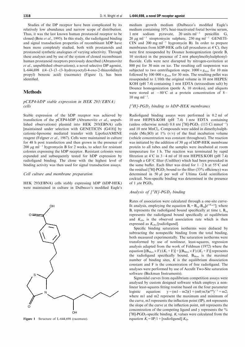
The rate of association of [3H]-PGD2 with hDP at roomtemperature is shown in Figure 2. The initial rate of
association was 0.15+0.01 min71 nM71. Equilibrium wasreached within 20 min. Once achieved, equilibrium bindingwas maintained throughout the 2 h time course. The stability
100
80
60
40
20
0
% m
axim
um [3 H
]-PG
D2
sp
ecifi
c bi
ndin
g
0 20 40 60 80 100 120 140
Incubation time (min)
Specific binding Total binding Nonspecific binding
Figure 2 Rates of association of [3H]-PGD2 binding to the humanDP receptor expressed on membranes from HEK 293(EBNA) cells.Radioligand membrane binding assays were carried out as previouslydescribed under Methods, with the following modi®cations: the ratesof total and nonspeci®c binding were assessed over a 2 h time courseby the sequential addition of HEK 293(EBNA) membranesexpressing the hDP receptor to separate incubation tubes. Mem-branes were added initially at the ®nal time point of 2 h andsuccessively at various other intervals until the initial time point of1 min. Speci®c binding was calculated as the di�erence betweennonspeci®c binding, measured in the presence of 1 mM PGD2, andtotal binding. Data points are the mean from three separateexperiments performed in duplicate; vertical lines show s.e.mean.
L-644,698, a novel DP receptor agonist 1319D. H. Wright et al
of [3H]-PGD2 under the assay conditions was veri®ed byreverse-phase high performance liquid chromatography (r.p.-h.p.l.c.). [3H]-PGD2 was incubated under the assay conditions
for 2 h, recovered from the incubation media and resolved byr.p.-h.p.l.c. as a single peak with the same retention time(24.5 min) as a control sample of [3H]-PGD2 analysed under
identical conditions (data not shown). Approximately 87% ofthe [3H]-PGD2 added to the incubation medium was recovered.[3H]-PGD2 is, therefore, chemically and metabolically stableduring a 2 h incubation under these experimental conditions as
determined by r.p.-h.p.l.c.The association rate of [3H]-PGD2 speci®c binding to the
hDP receptor was also investigated at 308C (data not shown).
Equilibrium binding was attained more rapidly than underconditions of room temperature, at a rate of 0.31+0.03 min71 nM71. However, it was not maintained, as
demonstrated by a time-dependent decrease in [3H]-PGD2
speci®c binding during the 2 h incubation to a value 75% ofthe maximum level obtained.
E�ects of nucleotide analogues
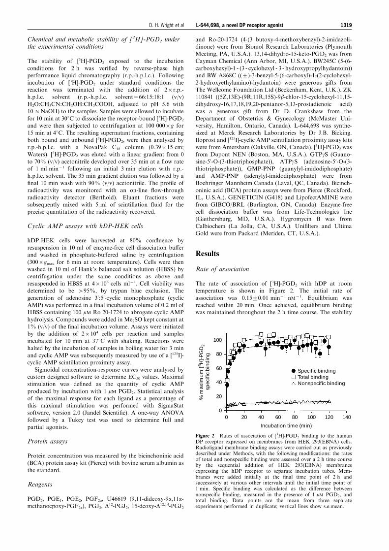
The e�ects of slowly-hydrolyzable nucleotide analogues on
[3H]-PGD2 speci®c binding to the hDP receptor were studied(Figure 3). [3H]-PGD2 speci®c binding was inhibited in aconcentration-dependent manner by GTPgS, ATPgS, GMP-
PNP and AMP-PNP. The IC50 values (mM) for thesecompounds were 0.01, 1.67, 1.51 and 347, corresponding tothe following rank order of nucleotide analogue potency at the
hDP receptor: GTPgS44ATPgS=GMP-PNP44AMP-PNP. The e�ects of GTPgS were studied further in conjunctionwith saturation analyses as described below.
Saturation analyses
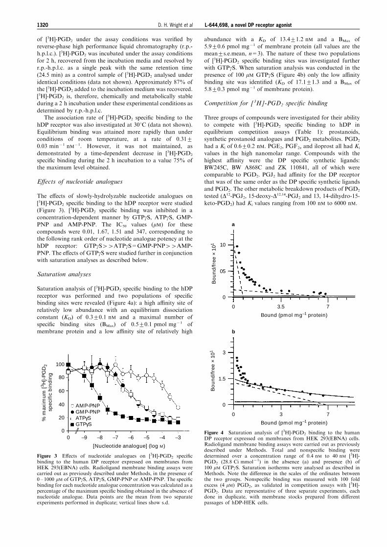
Saturation analysis of [3H]-PGD2 speci®c binding to the hDP
receptor was performed and two populations of speci®cbinding sites were revealed (Figure 4a): a high a�nity site ofrelatively low abundance with an equilibrium dissociation
constant (KD) of 0.3+0.1 nM and a maximal number ofspeci®c binding sites (BMax) of 0.5+0.1 pmol mg71 ofmembrane protein and a low a�nity site of relatively high
abundance with a KD of 13.4+1.2 nM and a BMax of5.9+0.6 pmol mg71 of membrane protein (all values are themean+s.e.mean, n=3). The nature of these two populations
of [3H]-PGD2 speci®c binding sites was investigated furtherwith GTPgS. When saturation analysis was conducted in thepresence of 100 mM GTPgS (Figure 4b) only the low a�nity
binding site was identi®ed (KD of 17.1+1.3 and a BMax of5.8+0.3 pmol mg71 of membrane protein).
Competition for [3H]-PGD2 speci®c binding
Three groups of compounds were investigated for their abilityto compete with [3H]-PGD2 speci®c binding to hDP in
equilibrium competition assays (Table 1): prostanoids,synthetic prostanoid analogues and PGD2 metabolites. PGD2
had a Ki of 0.6+0.2 nM. PGE2, PGF2a and iloprost all had Ki
values in the high nanomolar range. Compounds with thehighest a�nity were the DP speci®c synthetic ligands:BW245C, BW A868C and ZK 110841, all of which werecomparable to PGD2. PGJ2 had a�nity for the DP receptor
that was of the same order as the DP speci®c synthetic ligandsand PGD2. The other metabolic breakdown products of PGD2
tested (D12-PGJ2, 15-deoxy-D12,14-PGJ2 and 13, 14-dihydro-15-
keto-PGD2) had Ki values ranging from 100 nM to 6000 nM.
100
80
60
40
20
0
% m
axim
um
[3 H
]-P
GD
2
spec
ific
bin
din
g
0 –9 –8 –7 –6 –5 –4 –3
[Nucleotide analogue] (log M)
AMP-PNP GMP-PNP ATPγS GTPγS
Figure 3 E�ects of nucleotide analogues on [3H]-PGD2 speci®cbinding to the human DP receptor expressed on membranes fromHEK 293(EBNA) cells. Radioligand membrane binding assays werecarried out as previously described under Methods, in the presence of0 ± 1000 mM of GTPgS, ATPgS, GMP-PNP or AMP-PNP. The speci®cbinding for each nucleotide analogue concentration was calculated as apercentage of the maximum speci®c binding obtained in the absence ofnucleotide analogue. Data points are the mean from two separateexperiments performed in duplicate; vertical lines show s.d.
10
05
0
0 3.5 7
Bound (pmol mg–1 protein)
Bo
un
d/f
ree
× 10
1
a
b
0 3 7
Bound (pmol mg–1 protein)
3
1.5
0
Bo
un
d/f
ree
× 10
1
Figure 4 Saturation analysis of [3H]-PGD2 binding to the humanDP receptor expressed on membranes from HEK 293(EBNA) cells.Radioligand membrane binding assays were carried out as previouslydescribed under Methods. Total and nonspeci®c binding weredetermined over a concentration range of 0.4 nM to 40 nM [3H]-PGD2 (28.8 Ci mmol71) in the absence (a) and presence (b) of100 mM GTPgS. Saturation isotherms were analysed as described inMethods. Note the di�erence in the scales of the ordinates betweenthe two groups. Nonspeci®c binding was measured with 100 foldexcess (4 mM) PGD2, as validated in competition assays with [3H]-PGD2. Data are representative of three separate experiments, eachdone in duplicate, with membrane stocks prepared from di�erentpassages of hDP-HEK cells.
L-644,698, a novel DP receptor agonist1320 D. H. Wright et al
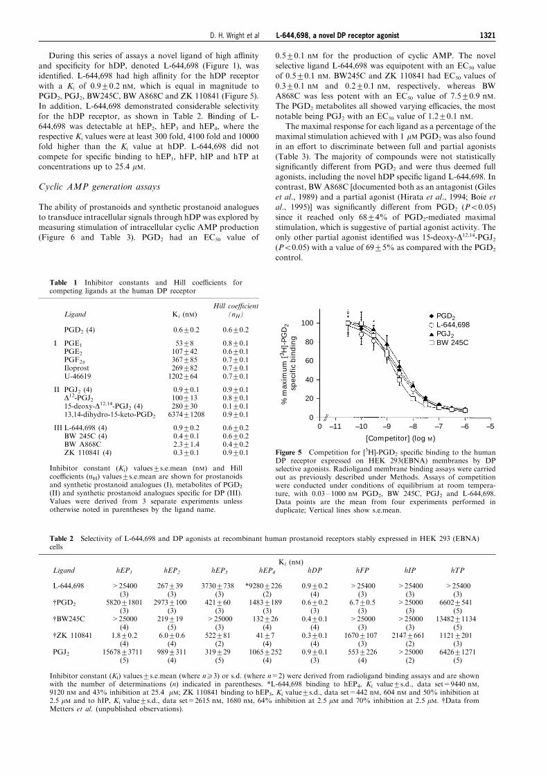
During this series of assays a novel ligand of high a�nityand speci®city for hDP, denoted L-644,698 (Figure 1), wasidenti®ed. L-644,698 had high a�nity for the hDP receptor
with a Ki of 0.9+0.2 nM, which is equal in magnitude toPGD2, PGJ2, BW245C, BW A868C and ZK 110841 (Figure 5).In addition, L-644,698 demonstrated considerable selectivity
for the hDP receptor, as shown in Table 2. Binding of L-644,698 was detectable at hEP2, hEP3 and hEP4, where therespective Ki values were at least 300 fold, 4100 fold and 10000fold higher than the Ki value at hDP. L-644,698 did not
compete for speci®c binding to hEP1, hFP, hIP and hTP atconcentrations up to 25.4 mM.
Cyclic AMP generation assays
The ability of prostanoids and synthetic prostanoid analogues
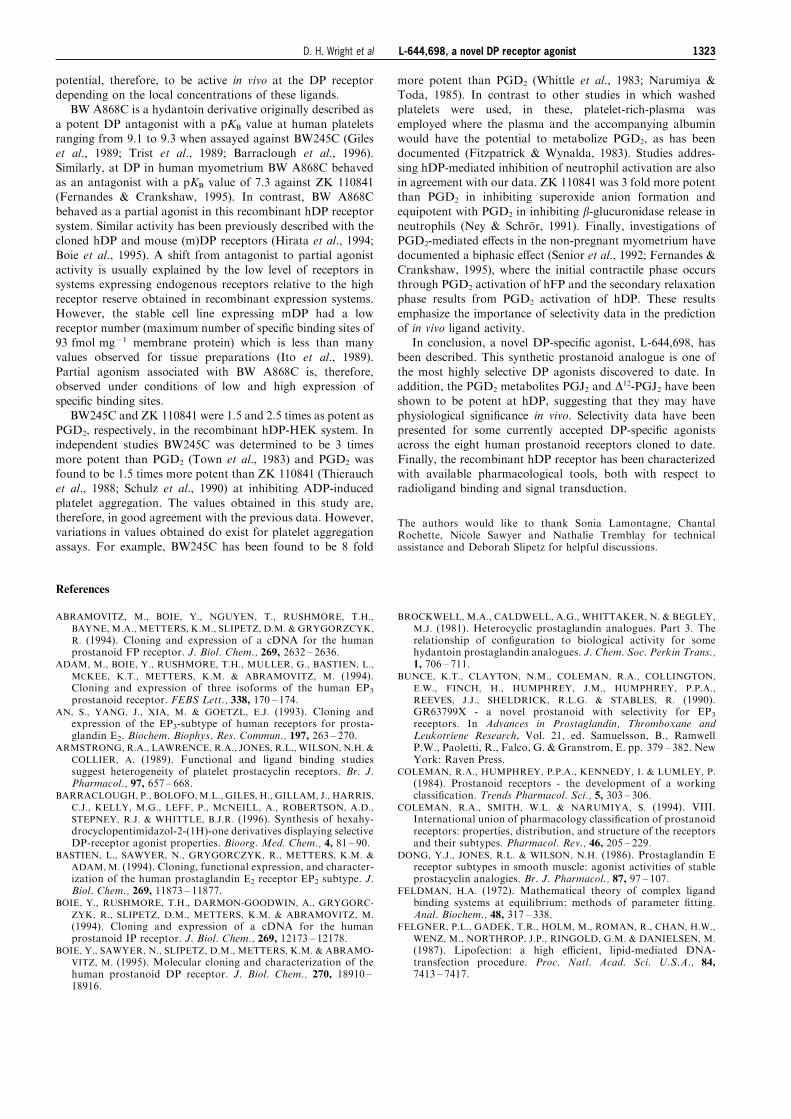
to transduce intracellular signals through hDP was explored bymeasuring stimulation of intracellular cyclic AMP production(Figure 6 and Table 3). PGD2 had an EC50 value of
0.5+0.1 nM for the production of cyclic AMP. The novelselective ligand L-644,698 was equipotent with an EC50 valueof 0.5+0.1 nM. BW245C and ZK 110841 had EC50 values of
0.3+0.1 nM and 0.2+0.1 nM, respectively, whereas BWA868C was less potent with an EC50 value of 7.5+0.9 nM.The PGD2 metabolites all showed varying e�cacies, the most
notable being PGJ2 with an EC50 value of 1.2+0.1 nM.The maximal response for each ligand as a percentage of the
maximal stimulation achieved with 1 mM PGD2 was also foundin an e�ort to discriminate between full and partial agonists
(Table 3). The majority of compounds were not statisticallysigni®cantly di�erent from PGD2 and were thus deemed fullagonists, including the novel hDP speci®c ligand L-644,698. In
contrast, BW A868C [documented both as an antagonist (Gileset al., 1989) and a partial agonist (Hirata et al., 1994; Boie etal., 1995)] was signi®cantly di�erent from PGD2 (P50.05)
since it reached only 68+4% of PGD2-mediated maximalstimulation, which is suggestive of partial agonist activity. Theonly other partial agonist identi®ed was 15-deoxy-D12,14-PGJ2(P50.05) with a value of 69+5% as compared with the PGD2
control.
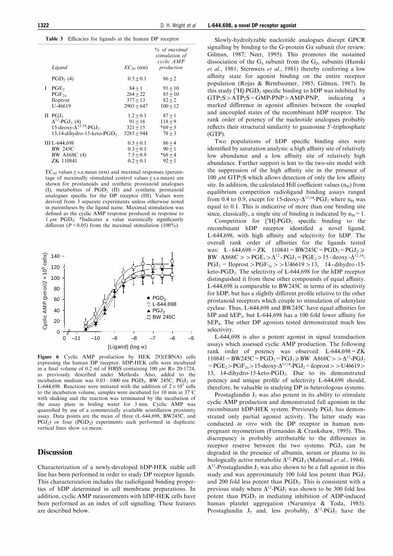
Table 1 Inhibitor constants and Hill coe�cients forcompeting ligands at the human DP receptor
Hill coe�cientLigand Ki (nM) (nH)
PGD2 (4) 0.6+0.2 0.6+0.2
I PGE1PGE2PGF2aIloprostU-46619
53+8107+42367+85269+821202+64
0.8+0.10.6+0.10.7+0.10.7+0.10.7+0.1
II PGJ2 (4)D12-PGJ215-deoxy-D12,14-PGJ2 (4)13,14-dihydro-15-keto-PGD2
0.9+0.1100+13280+306374+1208
0.9+0.10.8+0.10.1+0.10.9+0.1
III L-644,698 (4)BW 245C (4)BW A868CZK 110841 (4)
0.9+0.20.4+0.12.3+1.40.3+0.1
0.6+0.20.6+0.20.4+0.20.9+0.1
Inhibitor constant (Ki) values+s.e.mean (nM) and Hillcoe�cients (nH) values+s.e.mean are shown for prostanoidsand synthetic prostanoid analogues (I), metabolites of PGD2
(II) and synthetic prostanoid analogues speci®c for DP (III).Values were derived from 3 separate experiments unlessotherwise noted in parentheses by the ligand name.
Table 2 Selectivity of L-644,698 and DP agonists at recombinant human prostanoid receptors stably expressed in HEK 293 (EBNA)cells
Ki (nM)Ligand hEP1 hEP2 hEP3 hEP4 hDP hFP hIP hTP
L-644,698
{PGD2
{BW245C
{ZK 110841
PGJ2
>25400(3)
5820+1801(3)
>25000(4)
1.8+0.2(4)
15678+3711(5)
267+39(3)
2973+100(3)
219+19(5)
6.0+0.6(4)
989+311(4)
3730+738(3)
421+60(3)
>25000(3)
522+81(2)
319+29(5)
*9280+226(2)
1483+189(3)
132+26(4)
41+7(4)
1065+252(4)
0.9+0.2(4)
0.6+0.2(3)
0.4+0.1(4)
0.3+0.1(4)
0.9+0.1(3)
>25400(3)
6.7+0.5(3)
>25000(3)
1670+107(3)
553+226(4)
>25400(3)
>25000(3)
>25000(3)
2147+661(2)
>25000(2)
>25400(3)
6602+541(5)
13482+1134(5)
1121+201(3)
6426+1271(5)
Inhibitor constant (Ki) values+s.e.mean (where n53) or s.d. (where n=2) were derived from radioligand binding assays and are shownwith the number of determinations (n) indicated in parentheses. *L-644,698 binding to hEP4, Ki value+s.d., data set=9440 nM,9120 nM and 43% inhibition at 25.4 mM; ZK 110841 binding to hEP3, Ki value+s.d., data set=442 nM, 604 nM and 50% inhibition at2.5 mM and to hIP, Ki value+s.d., data set=2615 nM, 1680 nM, 64% inhibition at 2.5 mM and 70% inhibition at 2.5 mM. {Data fromMetters et al. (unpublished observations).
100
80
60
40
20
0
% m
axim
um
[3 H
]-P
GD
2
spec
ific
bin
din
g
0 –11 –10 –9 –8 –7 –6 –5
[Competitor] (log M)
PGD2 L-644,698 PGJ2 BW 245C
Figure 5 Competition for [3H]-PGD2 speci®c binding to the humanDP receptor expressed on HEK 293(EBNA) membranes by DPselective agonists. Radioligand membrane binding assays were carriedout as previously described under Methods. Assays of competitionwere conducted under conditions of equilibrium at room tempera-ture, with 0.03 ± 1000 nM PGD2, BW 245C, PGJ2 and L-644,698.Data points are the mean from four experiments performed induplicate; Vertical lines show s.e.mean.
L-644,698, a novel DP receptor agonist 1321D. H. Wright et al
Discussion
Characterization of a newly-developed hDP-HEK stable cellline has been performed in order to study DP receptor ligands.
This characterization includes the radioligand binding proper-ties of hDP determined in cell membrane preparations. Inaddition, cyclic AMP measurements with hDP-HEK cells havebeen performed as an index of cell signalling. These features
are described below.
Slowly-hydrolyzable nucleotide analogues disrupt GPCRsignalling by binding to the G-protein Ga subunit (for review:Gilman, 1987; Neer, 1995). This promotes the sustained
dissociation of the Ga subunit from the Gbg subunits (Hanskiet al., 1981; Sternweis et al., 1981) thereby conferring a lowa�nity state for agonist binding on the entire receptor
population (Rojas & Birmbaumer, 1985; Gilman, 1987). Inthis study [3H]-PGD2 speci®c binding to hDP was inhibited byGTPgS4ATPgS=GMP-PNP4AMP-PNP, indicating amarked di�erence in agonist a�nities between the coupled
and uncoupled states of the recombinant hDP receptor. Therank order of potency of the nucleotide analogues probablyre¯ects their structural similarity to guanosine 5'-triphosphate(GTP).
Two populations of hDP speci®c binding sites wereidenti®ed by saturation analysis: a high a�nity site of relatively
low abundance and a low a�nity site of relatively highabundance. Further support is lent to the two-site model withthe suppression of the high a�nity site in the presence of100 mM GTPgS which allows detection of only the low a�nity
site. In addition, the calculated Hill coe�cient values (nH) fromequilibrium competition radioligand binding assays rangedfrom 0.4 to 0.9, except for 15-deoxy-D12,14-PGJ2 where nH was
equal to 0.1. This is indicative of more than one binding sitesince, classically, a single site of binding is indicated by nH=1.
Competition for [3H]-PGD2 speci®c binding to the
recombinant hDP receptor identi®ed a novel ligand,L-644,698, with high a�nity and selectivity for hDP. Theoverall rank order of a�nities for the ligands tested
was: L - 644,698=ZK 110841=BW245C=PGD2=PGJ25BW A868C44PGE14D12 - PGJ2=PGE2415 - deoxy -D12,14-PGJ2= Iloprost4PGF2a44U46619413, 14 - dihydro -15-keto-PGD2. The selectivity of L-644,698 for the hDP receptor
distinguished it from these other compounds of equal a�nity.L-644,698 is comparable to BW245C in terms of its selectivityfor hDP, but has a slightly di�erent pro®le relative to the other
prostanoid receptors which couple to stimulation of adenylatecyclase. Thus, L-644,698 and BW245C have equal a�nities forhIP and hEP2, but L-644,698 has a 100 fold lower a�nity for
hEP4. The other DP agonists tested demonstrated much lessselectivity.
L-644,698 is also a potent agonist in signal transductionassays which assessed cyclic AMP production. The following
rank order of potency was observed: L-644,698=ZK110841=BW245C=PGD2=PGJ25BW A868C44D12-PGJ2=PGE24PGF2a415-deoxy-D12,14-PGJ2=iloprost44U46619413, 14-dihydro-15-keto-PGD2. Due to its demonstratedpotency and unique pro®le of selectivity L-644,698 should,therefore, be valuable in studying DP in heterologous systems.
Prostaglandin J2 was also potent in its ability to stimulatecyclic AMP production and demonstrated full agonism in therecombinant hDP-HEK system. Previously PGJ2 has demon-
strated only partial agonist activity. The latter study wasconducted in vitro with the DP receptor in human non-pregnant myometrium (Fernandes & Crankshaw, 1995). Thisdiscrepancy is probably attributable to the di�erences in
receptor reserve between the two systems. PGJ2 can bedegraded in the presence of albumin, serum or plasma to itsbiologically active metabolite D12-PGJ2 (Mahmud et al., 1984).
D12-Prostaglandin J2 was also shown to be a full agonist in thisstudy and was approximately 100 fold less potent than PGJ2and 200 fold less potent than PGD2. This is consistent with a
previous study where D12-PGJ2 was shown to be 300 fold lesspotent than PGD2 in mediating inhibition of ADP-inducedhuman platelet aggregation (Narumiya & Toda, 1985).Prostaglandin J2 and, less probably, D12-PGJ2 have the
Table 3 E�cacies for ligands at the human DP receptor
% of maximalstimulation ofcyclic AMP
Ligand EC50 (nM) production
PGD2 (4) 0.5+0.1 86+2
I PGE2PGF2aIloprostU-46619
84+1264+22377+132903+647
91+1085+1082+2100+12
II PGJ2D12-PGJ2 (4)15-deoxy-D12,14-PGJ213,14-dihydro-15-keto-PGD2
1.2+0.191+18321+155283+944
87+1118+9*69+578+3
III L-644,698BW 245CBW A868C (4)ZK 110841
0.5+0.10.3+0.17.5+0.90.2+0.1
86+490+1*68+492+1
EC50 values+s.e.mean (nM) and maximal responses (percen-tage of maximally stimulated control values+s.e.mean) areshown for prostanoids and synthetic prostanoid analogues(I), metabolites of PGD2 (II) and synthetic prostanoidanalogues speci®c for the DP receptor (III). Values werederived from 3 separate experiments unless otherwise notedin parentheses by the ligand name. Maximal stimulation wasde®ned as the cyclic AMP response produced in response to1 mM PGD2. *Indicates a value statistically signi®cantlydi�erent (P<0.05) from the maximal stimulation (100%).
140
120
100
80
60
40
20
0
Cyc
lic A
MP
(p
mo
l/2 ×
105
cells
)
0 –11 –10 –9 –8 –7 –6 –5
[Ligand] (log M)
PGD2 L-644,698 PGJ2 BW 245C
Figure 6 Cyclic AMP production by HEK 293(EBNA) cellsexpressing the human DP receptor. hDP-HEK cells were incubatedin a ®nal volume of 0.2 ml of HBSS containing 100 mM Ro 20-1724,as previously described under Methods. Also, added to theincubation medium was 0.03 ± 1000 nM PGD2, BW 245C, PGJ2 orL-644,698. Reactions were initiated with the addition of 26105 cellsto the incubation volume, samples were incubated for 10 min at 378Cwith shaking and the reaction was terminated by the incubation ofthe assay plate in boiling water for 3 min. Cyclic AMP wasquanti®ed by use of a commercially available scintillation proximityassay. Data points are the mean of three (L-644,698, BW245C, andPGJ2) or four (PGD2) experiments each performed in duplicate;vertical lines show s.e.mean.
L-644,698, a novel DP receptor agonist1322 D. H. Wright et al
potential, therefore, to be active in vivo at the DP receptordepending on the local concentrations of these ligands.
BW A868C is a hydantoin derivative originally described as
a potent DP antagonist with a pKB value at human plateletsranging from 9.1 to 9.3 when assayed against BW245C (Gileset al., 1989; Trist et al., 1989; Barraclough et al., 1996).
Similarly, at DP in human myometrium BW A868C behavedas an antagonist with a pKB value of 7.3 against ZK 110841(Fernandes & Crankshaw, 1995). In contrast, BW A868Cbehaved as a partial agonist in this recombinant hDP receptor
system. Similar activity has been previously described with thecloned hDP and mouse (m)DP receptors (Hirata et al., 1994;Boie et al., 1995). A shift from antagonist to partial agonist
activity is usually explained by the low level of receptors insystems expressing endogenous receptors relative to the highreceptor reserve obtained in recombinant expression systems.
However, the stable cell line expressing mDP had a lowreceptor number (maximum number of speci®c binding sites of93 fmol mg71 membrane protein) which is less than manyvalues observed for tissue preparations (Ito et al., 1989).
Partial agonism associated with BW A868C is, therefore,observed under conditions of low and high expression ofspeci®c binding sites.
BW245C and ZK 110841 were 1.5 and 2.5 times as potent asPGD2, respectively, in the recombinant hDP-HEK system. Inindependent studies BW245C was determined to be 3 times
more potent than PGD2 (Town et al., 1983) and PGD2 wasfound to be 1.5 times more potent than ZK 110841 (Thierauchet al., 1988; Schulz et al., 1990) at inhibiting ADP-induced
platelet aggregation. The values obtained in this study are,therefore, in good agreement with the previous data. However,variations in values obtained do exist for platelet aggregationassays. For example, BW245C has been found to be 8 fold
more potent than PGD2 (Whittle et al., 1983; Narumiya &Toda, 1985). In contrast to other studies in which washedplatelets were used, in these, platelet-rich-plasma was
employed where the plasma and the accompanying albuminwould have the potential to metabolize PGD2, as has beendocumented (Fitzpatrick & Wynalda, 1983). Studies addres-
sing hDP-mediated inhibition of neutrophil activation are alsoin agreement with our data. ZK 110841 was 3 fold more potentthan PGD2 in inhibiting superoxide anion formation andequipotent with PGD2 in inhibiting b-glucuronidase release inneutrophils (Ney & SchroÈ r, 1991). Finally, investigations ofPGD2-mediated e�ects in the non-pregnant myometrium havedocumented a biphasic e�ect (Senior et al., 1992; Fernandes &
Crankshaw, 1995), where the initial contractile phase occursthrough PGD2 activation of hFP and the secondary relaxationphase results from PGD2 activation of hDP. These results
emphasize the importance of selectivity data in the predictionof in vivo ligand activity.
In conclusion, a novel DP-speci®c agonist, L-644,698, hasbeen described. This synthetic prostanoid analogue is one of
the most highly selective DP agonists discovered to date. Inaddition, the PGD2 metabolites PGJ2 and D12-PGJ2 have beenshown to be potent at hDP, suggesting that they may have
physiological signi®cance in vivo. Selectivity data have beenpresented for some currently accepted DP-speci®c agonistsacross the eight human prostanoid receptors cloned to date.
Finally, the recombinant hDP receptor has been characterizedwith available pharmacological tools, both with respect toradioligand binding and signal transduction.
The authors would like to thank Sonia Lamontagne, ChantalRochette, Nicole Sawyer and Nathalie Tremblay for technicalassistance and Deborah Slipetz for helpful discussions.
References
ABRAMOVITZ, M., BOIE, Y., NGUYEN, T., RUSHMORE, T.H.,
BAYNE, M.A., METTERS, K.M., SLIPETZ, D.M. & GRYGORZCYK,
R. (1994). Cloning and expression of a cDNA for the humanprostanoid FP receptor. J. Biol. Chem., 269, 2632 ± 2636.
ADAM, M., BOIE, Y., RUSHMORE, T.H., MULLER, G., BASTIEN, L.,
MCKEE, K.T., METTERS, K.M. & ABRAMOVITZ, M. (1994).Cloning and expression of three isoforms of the human EP3prostanoid receptor. FEBS Lett., 338, 170 ± 174.
AN, S., YANG, J., XIA, M. & GOETZL, E.J. (1993). Cloning andexpression of the EP3-subtype of human receptors for prosta-glandin E2. Biochem. Biophys. Res. Commun., 197, 263 ± 270.
ARMSTRONG, R.A., LAWRENCE, R.A., JONES, R.L., WILSON, N.H. &
COLLIER, A. (1989). Functional and ligand binding studiessuggest heterogeneity of platelet prostacyclin receptors. Br. J.Pharmacol., 97, 657 ± 668.
BARRACLOUGH, P., BOLOFO, M.L., GILES, H., GILLAM, J., HARRIS,
C.J., KELLY, M.G., LEFF, P., MCNEILL, A., ROBERTSON, A.D.,
STEPNEY, R.J. & WHITTLE, B.J.R. (1996). Synthesis of hexahy-drocyclopentimidazol-2-(1H)-one derivatives displaying selectiveDP-receptor agonist properties. Bioorg. Med. Chem., 4, 81 ± 90.
BASTIEN, L., SAWYER, N., GRYGORCZYK, R., METTERS, K.M. &
ADAM, M. (1994). Cloning, functional expression, and character-ization of the human prostaglandin E2 receptor EP2 subtype. J.Biol. Chem., 269, 11873 ± 11877.
BOIE, Y., RUSHMORE, T.H., DARMON-GOODWIN, A., GRYGORC-
ZYK, R., SLIPETZ, D.M., METTERS, K.M. & ABRAMOVITZ, M.
(1994). Cloning and expression of a cDNA for the humanprostanoid IP receptor. J. Biol. Chem., 269, 12173 ± 12178.
BOIE, Y., SAWYER, N., SLIPETZ, D.M., METTERS, K.M. & ABRAMO-
VITZ, M. (1995). Molecular cloning and characterization of thehuman prostanoid DP receptor. J. Biol. Chem., 270, 18910 ±18916.
BROCKWELL, M.A., CALDWELL, A.G., WHITTAKER, N. & BEGLEY,
M.J. (1981). Heterocyclic prostaglandin analogues. Part 3. Therelationship of con®guration to biological activity for somehydantoin prostaglandin analogues. J. Chem. Soc. Perkin Trans.,1, 706 ± 711.
BUNCE, K.T., CLAYTON, N.M., COLEMAN, R.A., COLLINGTON,
E.W., FINCH, H., HUMPHREY, J.M., HUMPHREY, P.P.A.,
REEVES, J.J., SHELDRICK, R.L.G. & STABLES, R. (1990).GR63799X - a novel prostanoid with selectivity for EP3receptors. In Advances in Prostaglandin, Thromboxane andLeukotriene Research, Vol. 21, ed. Samuelsson, B., RamwellP.W., Paoletti, R., Falco, G. & Granstrom, E. pp. 379 ± 382. NewYork: Raven Press.
COLEMAN, R.A., HUMPHREY, P.P.A., KENNEDY, I. & LUMLEY, P.
(1984). Prostanoid receptors - the development of a workingclassi®cation. Trends Pharmacol. Sci., 5, 303 ± 306.
COLEMAN, R.A., SMITH, W.L. & NARUMIYA, S. (1994). VIII.International union of pharmacology classi®cation of prostanoidreceptors: properties, distribution, and structure of the receptorsand their subtypes. Pharmacol. Rev., 46, 205 ± 229.
DONG, Y.J., JONES, R.L. & WILSON, N.H. (1986). Prostaglandin Ereceptor subtypes in smooth muscle: agonist activities of stableprostacyclin analogies. Br. J. Pharmacol., 87, 97 ± 107.
FELDMAN, H.A. (1972). Mathematical theory of complex ligandbinding systems at equilibrium: methods of parameter ®tting.Anal. Biochem., 48, 317 ± 338.
FELGNER, P.L., GADEK, T.R., HOLM, M., ROMAN, R., CHAN, H.W.,
WENZ, M., NORTHROP, J.P., RINGOLD, G.M. & DANIELSEN, M.
(1987). Lipofection: a high e�cient, lipid-mediated DNA-transfection procedure. Proc. Natl. Acad. Sci. U.S.A., 84,7413 ± 7417.
L-644,698, a novel DP receptor agonist 1323D. H. Wright et al
FERNANDES, B. & CRANKSHAW, D. (1995). Functional character-ization of the prostanoid DP receptor in human myometrium.Eur. J. Pharmacol., 283, 73 ± 81.
FITZPATRICK, F.A. & WYNALDA, M.A. (1983). Albumin-catalyzedmetabolism of prostaglandin D2, identi®cation of productsformed in vitro. J. Biol. Chem., 258, 11713 ± 11718.
FUNK, C.D., FURCI, L., FITZGERALD, G.A., GRYGORCZYK, R.,
ROCHETTE, C., BAYNE, M.A., ABRAMOVITZ, M., ADAM, M. &
METTERS, K.M. (1993). Cloning and expression of a cDNA forthe human prostaglandin E receptor EP1 subtype. J. Biol. Chem.,268, 26767 ± 26772.
GILES, H., LEFF, P., BOLOFO, M.L., KELLY, M.G. & ROBERTSON,
A.D. (1989). The classi®cation of prostaglandin DP-receptors inplatelets and vasculature using BW A868C, a novel, selective andpotent competitive antagonist. Br. J. Pharmacol., 96, 291 ± 300.
GILMAN, A.G. (1987). G proteins: transducers of receptor-generatedsignals. Ann. Rev. Biochem., 56, 615 ± 649.
HANSKI, E., STERNWEIS, P.C., NORTHUP, J.K., DROMERICK, A.W.
& GILMAN, A.G. (1981). The regulatory component of adenylatecyclase. Puri®cation and properties of the turkey erythrocyteprotein. J. Biol. Chem., 256, 12911 ± 12919.
HIRATA, M., HAYAISHI, Y., USHIKUBI, F.L., YOKOTA, Y., KA-
GEYAMA, R., NAKANISHI, S. & NARUMIYA, S. (1991). Cloningand expression of cDNA for a human thromboxane A2 receptor.Nature, 349, 617 ± 620.
HIRATA, M., KAKIZUKA, A., AIZAWA, M., USHIKUBI, F. &
NARUMIYA, S. (1994). Molecular characterization of a mouseprostaglandin D receptor and functional expression of the clonedgene. Proc. Natl. Acad. Sci. U.S.A., 91, 11192 ± 11196.
ITO, S., NARUMIYA, S. & HAYAISHI, O. (1989). Prostaglandin D2: abiochemical perspective. Prostaglandins Leukotrienes Essent.Fatty Acids, 37, 219 ± 234.
KENNEDY, I., COLEMAN, R.A., HUMPHREY, P.P.A., LEVY, G.P. &
LUMLEY, P. (1982). Studies on the characterization of prostanoidreceptors: a proposed classi®cation. Prostaglandins, 24, 667 ±689.
KOTANI, M., TANAKA, I., OGAWA, Y., UNSUI, T., MORI, K.,
ICHIKAWA, A., NARUMIYA, S., YOSHIMI, T. & NAKAO, K.
(1995). Molecular cloning and expression of multiple isoforms ofhuman prostaglandin E receptor EP3 subtype generated byalternative messenger RNA splicing: multiple second messengersystems and tissue-speci®c distributions. Mol. Pharmacol., 48,869 ± 879.
MAHMUD, I., SMITH, D.L., WHYTE, M.A., NELSON, J.T., CHO, D.,
TOKES, L.G., ALVAREZ, R. & WILLIS, A.L. (1984). On theidenti®cation and biological properties of prostaglandin J2.Prostaglandins Leukotrienes Med., 16, 131 ± 146.
MATSUGI, T., KAGEYAMA, M., NISHIMURA, K., GILES, H. &
SHIRASAWA, E. (1995). Selective prostaglandin D2 receptorstimulation elicits ocular hypotensive e�ects in rabbits and cats.Eur. J. Pharmacol., 275, 245 ± 250.
NARUMIYA, S. & TODA, N. (1985). Di�erent responsiveness ofprostaglandin D2-sensitive systems to prostaglandin D2 and itsanalogues. Br. J. Pharmacol., 85, 367 ± 375.
NEER, E.J. (1995). Heterotrimeric G-proteins: organizers oftransmembrane signals. Cell, 80, 249 ± 257.
NEY, P.& SCHROÈ R, K. (1991). PGD2 and its mimetic ZK 110.841 arepotent inhibitors of receptor-mediated activation of humanneutrophils. Eicosanoids, 4, 21 ± 28.
RANGACHARI, P.K., BETTI, P.A., PRIOR, E.T. & ROBERTS II, L.J.
(1995). E�ects of a selective DP receptor agonist (BW245C) andantagonist (BW A868C) on the canine colonic epithelium: anargument for a di�erent DP receptor. J. Pharmacol. Exp. Ther.,275, 611 ± 617.
RAYCHOWDHURY, M.K., YUKAWA, M., COLLINS, L.J., MCGRAIL,
S.H., KENT, K.C. & WARE, J.C. (1995). Alternative splicingproduces a divergent cytoplasmic tail in the human endothelialthromboxane A2 receptor. J. Biol. Chem., 270, 7011 ± 7014.
REGAN, J.W., BAILY, T.J., PEPPERL, D.J., PIERCE, K.L., BOGARDUS,
A.M., DONELLO, J.E., FAIRBAIRN, C.E., KEDZIE, K.M., WOOD-
WARD, D.F. & GIL, D.W. (1994). Cloning of a novel humanprostaglandin receptor with characteristics of the pharmacologi-cally de®ned EP2 subtype. Mol. Pharmacol., 46, 213 ± 220.
ROJAS, F.J. & BIRMBAUMER, L. (1985). Regulation of glucagonreceptor binding. Lack of e�ect of Mg and preferential role forGDP. J. Biol. Chem., 260, 7829 ± 7835.
SCHMID, A., THIERAUCH, K.H., SCHLEUNING, W.D. & DINTER, H.
(1995). Splice variants of the human EP3 receptor forprostaglandin E2. Eur. J. Biochem., 228, 23 ± 30.
SCHULZ, B.G., BECKMAN, R., MUÈ LLER, B., SCHROÈ DER, G., MAASS,
B., THIERAUCH, K.-H., BUCHMANN, B., VERHALLEN, P.F.J. &
FROÈ HLICH, W. (1990). Cardio- and hemodynamic pro®le ofselective PGD2 analogues. In Advances in Prostaglandin,Thromboxane and Leukotriene Research, Vol. 21, ed. Samuelsson,B., Ramwell, P.W., Paoletti, R., Falco, G. & Granstrom, E. 591 ±594. New York: Raven Press.
SENIOR, J., SANGHA, R., BAXTER, G.S., MARSHALL, K., &
CLAYTON, J.K. (1992). In vitro characterization of prostanoidFP-, DP-, IP- and TP-receptors on the non-pregnant humanmyometrium. Br. J. Pharmacol., 107, 215 ± 221.
SHELDRICK, R.L.G., COLEMAN, R.A. & LUMLEY, P. (1988). Iloprost± a potent EP1 and IP receptor agonist. Br. J. Pharmacol., 94,334P.
STERNWEIS, P.C., NORTHUP, J.K., SMIGEL, M.D. & GILMAN, A.G.
(1981). The regulatory component of adenylate cyclase. Puri®ca-tion and properties. J. Biol. Chem., 256, 11517 ± 11526.
THIERAUCH, K.-H., STUÈ RZEBECHER, C.-ST. & SCHILLINGER, E.
(1988). Stable 9b- or 11a-halogen-15-cyclohexyl-prostaglandinswith high a�nity to the PGD2 receptor. Prostaglandins, 35, 855 ±868.
TOWN, M.-H., CASALS-STENZEL, J. & SCHILLENGER, E. (1983).Pharmacological and cardiovascular properties of a hydantoinderivative, BW245C, with high a�nity and selectivity for PGD2
receptor. Prostaglandins, 25, 13 ± 28.TRIST, D.G., COLLINS, B.A., WOOD, J., KELLY, M.G. & ROBERTSON,
A.D. (1989). The antagonism by BW A868C of PGD2 andBW245C activation of human platelet adenylate cyclase. Br. J.Pharmacol., 96, 301 ± 306.
WHITTLE, B.J.R., MONCADA, S., MULLANE, K. & VANE, J.R. (1983).Platelet and cardiovascular activity of the hydantoin BW245C, apotent prostaglandin analogue. Prostaglandins, 25, 205 ± 223.
WOODWARD, D.F., HAWLEY, S.B., WILLIAMS, L.S., RALSTON, T.R.,
PROTZMAN, C.E., SPADA, C.S. & NIEVES, A.L. (1990). Studies onthe ocular pharmacology of prostaglandin D2. Inv. Opthamol.Vis. Sci., 31, 138 ± 146.
(Received October 16, 1997Revised December 3, 1997
Accepted December 3, 1997)
L-644,698, a novel DP receptor agonist1324 D. H. Wright et al
Top Related
Copyright © 2022 FDOKUMEN

