


Bahasa
Halaman
Hukum

CHAPTER 4
MODULATION OF IMMUNE-RELATED CELL SURFACE MOLECULES BY
CYTOKINES AND a-MSH.
135
4.1 INTRODUCTION.
Several hypotheses exist to explain the etiology of vitiligo (as discussed in Section 1.4).
However, it is unclear whether the melanocytes (and perhaps other cells of the
epidermis e. g. keratinocytes) of vitiligo patients have some intrinsic defect or whether
the disease results from an autoimmune response. There is strong evidence for an
autoimmune involvement (discussed fully in Section 1.4.3); for example, patient sera
have been found to contain various antibodies relating to melanocytes and melanogenic
enzymes (e. g. Naughton et al, 1983a & b; Bystryn & Naughton, 1985; Cui et al, 1992;
Yu et al, 1993; Merimsky et al, 1994; Song et al, 1994b; Cui et al, 1995; Hann & Kim,
1995; Baharav et al, 1996; Kemp et al, 1997a & b; see Section 1.4.3b for further
examples) which have been shown to induce immune damage to cultured human
melanocytes (Norris et al, 1988; Park et al, 1991; Cui et al, 1993; Yu et al, 1993;
Abdel-Naser et al, 1994). In some studies the level and activity of these antibodies have
been found to correlate with disease status (Naughton et al, 1986; Harning et al, 1991).
Abnormal, i. e. increased, expression of ICAM-1 and MHC class II have been shown for
melanocytes in perilesional biopsies of vitiligo (Al Badri et al, 1993a) and in ocular
melanocytes of patients with VKH (a disease in which some patients develop vitiligo-
like patches) (Sakamoto et al, 1991). Infiltration of T cells and macrophages parallels
the loss of pigment and the disappearance of melanocytes in inflammatory vitiligo (Le
Poole et al, 1996b) although it is uncertain whether the presence of these T cells relates
to a primary defect of the immune system. Melanocytes have been shown to present
molecules and function as accessory cells of the skin immune system (Le Poole et al,
1993b).
An interesting theory has arisen in recent years suggesting that pro-opiomelanocortin (POMC) peptides may play a role in aetiology of vitiligo (Liu et al, 1995). Liu et al.
(1995) detected high levels of IR a-, ß- and y-MSH in the epidermis in vitiligo lesions
and proposed that overproduction of the peptides or a defective receptor may cause the
disease. Recent perspectives on a-MSH, which has wide biological activities (reviewed
by Eberle, 1988) have led to it being viewed as a neuroimmunomodulatory peptide that
inhibits fever and all major forms of experimental inflammation through central nervous
system and peripheral melanocortin receptors. Current concepts of the anti-
136
inflammatory actions of a-MSH suggest that it acts through the MC1R on macrophages
and neutrophils to inhibit macrophage activation and neutrophil migration (as reviewed in Lipton & Catania, 1997).
Skin is now considered to be one of several non-pituitary tissues in which POMC-
derived peptides such a-MSH and ACTH and the parent molecule are produced (Thody
et al, 1983; Farooqui et al, 1993; Schauer et al, 1994; Iyengar, 1995; Kippenberger et
al, 1995; Slominski et al, 1995; Chakraborty et al, 1996; Wintzen et al, 1996; Teofoli
et al, 1997; Wakamatsu et al, 1997; Can et al, 1998). Both melanocytes and
keratinocytes play a role in peptide production and UV exposure increases this
production. Melanoma cells have also been shown to have IR MSH (Lunec et al, 1990;
Loir et al, 1991; Nagahama et al, 1998) and express ACTH (Iyengar, 1995; Nagahama
et al, 1998). Additional POMC-derived peptides, ß-lipotropic hormone and ß-
endorphin, are produced in keratinocytes (Wintzen et al, 1996). The main regulator of
POMC expression in the stress response pituitary-hypothalamis-axis, corticotropin
releasing factor, has also been shown to be produced by melanocytes (Slominski et al,
1995 & 1996). The availability of these peptides would be consistent with the
melanocyte occupying a stress/defence role in the skin and a-MSH/ACTH playing a
major role in the regulation of the melanocyte beyond pigment production. Local
inflammation is a feature of excess UV exposure in human skin. Patients with vitiligo
may experience inflammation during the development of their condition but this is
rarely observed clinically (Grab & Wise, 1948; Buckley & Lobitz, 1953; Michaelsson,
1968; Ishii & Hamada, 1981; Le Poole et al, 1996; Horn & Abanmi, 1997; Yagi et al,
1997). Under such circumstances one would expect upregulation of adhesion molecules
on keratinocytes and melanocytes and interactions of immune cells with these cells as
described in inflammatory vitiligo (Le Poole et al, 1996; Horn & Abanmi, 1997; Yagi et
al, 1997). ICAM-1 expression is an obligatory requirement (but not the sole pre-
requisite) for T cell binding to cells and is stimulated on melanocytes in culture
following exposure to a variety of cytokines (Yohn et al, 1990; Krasagakis et al, 1991;
Kinnbauer et al, 1992; Le Poole et al, 1993b; Krasagakis et al, 1995). Following UV
exposure, human melanocytes respond with an increase in pigment synthesis (as
discussed in Section 3.5.3). Several studies have suggested that melanin precursors,
particularly dopa and DHI are inherently cytotoxic to the melanocyte (e. g. Wick et al,
137
1977; Fujita et al, 1980; Pawelek et al, 1980; Urabe et al, 1994). This, combined with
an increase in adhesion molecules expression by UV/cytokines on melanocytes, might
act to make these cells a target for infiltrating T cells.
The purpose of this study was to investigate whether melanocytes in patients with
vitiligo are intrinsically more susceptible to immune attack. If the melanocytes have an
inherent defect such as the described abnormalities in the RER (Boissy et al, 1991; Im
et al, 1994), an increase of adhesion molecule expression might result, which would
promote enhanced interaction with immune cells or that vitiligo melanocytes would be
particularly susceptible to cytokines such as IFN-y and TNF-a. This possibility was
examined by comparing basal and cytokine-stimulated expression of the MHC class I
and class II molecules and ICAM-1 on normal and vitiligo melanocytes. Additionally,
one biological activity of a-MSH in skin might be to assist melanocytes to survive the
process of pigmentation, especially during UV associated inflammation by acting as an
immunomodulatory molecule. A recent paper by Luger (1998) has also proposed a
major role for the POMC peptides in modulating UV reponses in skin. a-MSH
opposition of cytokine-stimulated adhesion molecule expression on melanocytes might
prevent premature interaction between cells of the immune system and melanocytes
undergoing active pigment synthesis.
4.2 MODULATION OF ICAM-1 EXPRESSION IN NORMAL AND VITILIGO
MELANOCYTES.
ICAM-1 is one of several cofactors involved in the binding and activation of T cells. It
is also one of the most studied cell surface molecules in preliminary examinations of
whether a cell may be able to interact with cells of the immune system. In this section
several different stimuli, e. g. cytokines and media, which may play a role in the
regulation of ICAM-1 have been examined and the most effective media were used to
compare whether there are differences between normal and vitiligo melanocytes.
138
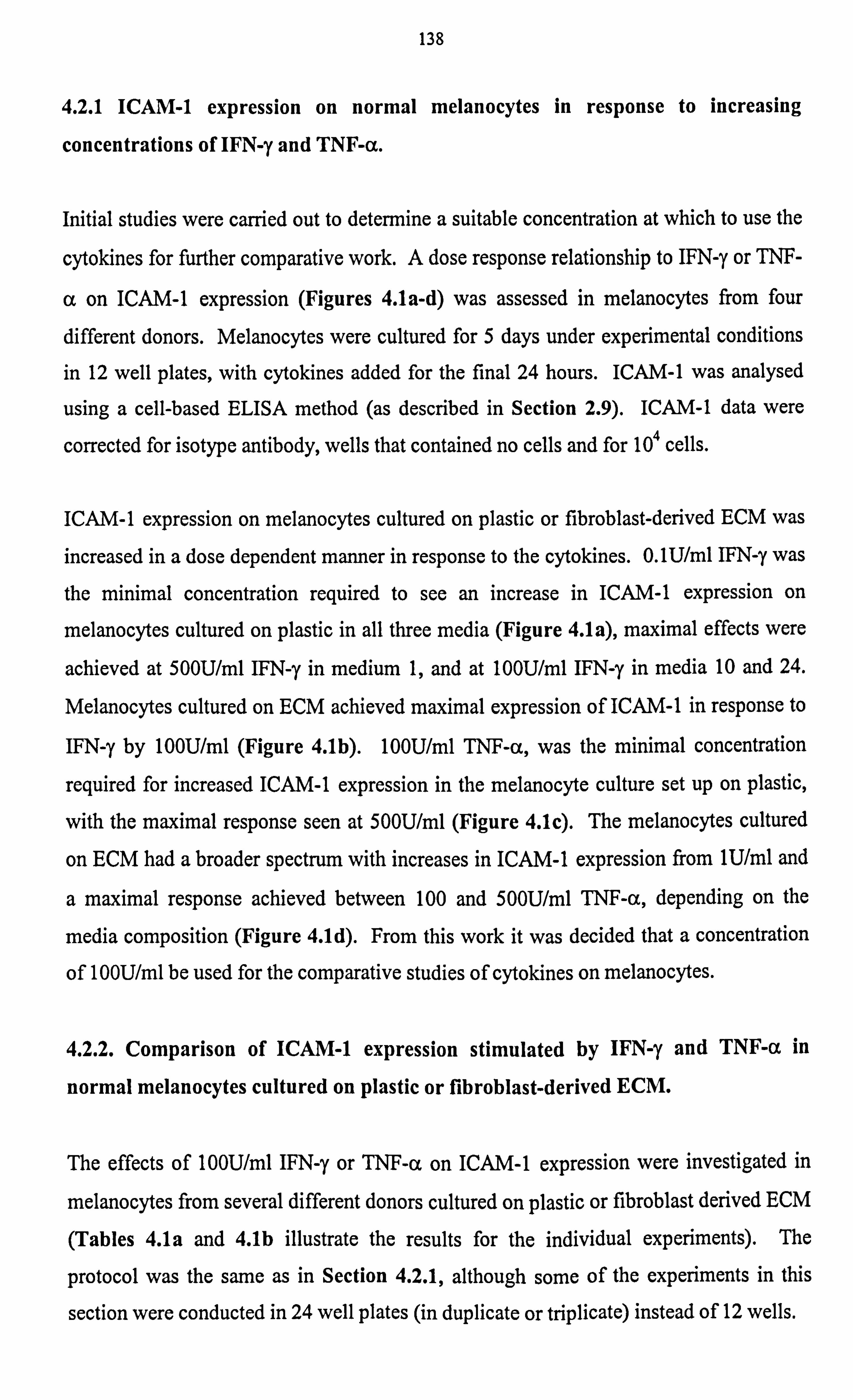
4.2.1 ICAM-1 expression on normal melanocytes in response to increasing
concentrations of IFN-y and TNF-a.
Initial studies were carried out to determine a suitable concentration at which to use the
cytokines for further comparative work. A dose response relationship to IFN-y or TNF-
a on ICAM-1 expression (Figures 4.1a-d) was assessed in melanocytes from four
different donors. Melanocytes were cultured for 5 days under experimental conditions
in 12 well plates, with cytokines added for the final 24 hours. ICAM-1 was analysed
using a cell-based ELISA method (as described in Section 2.9). ICAM-1 data were
corrected for isotype antibody, wells that contained no cells and for 104 cells.
ICAM-1 expression on melanocytes cultured on plastic or fibroblast-derived ECM was
increased in a dose dependent manner in response to the cytokines. O. 1U/ml IFN-y was
the minimal concentration required to see an increase in ICAM-1 expression on
melanocytes cultured on plastic in all three media (Figure 4.1a), maximal effects were
achieved at 500U/ml IFN-y in medium 1, and at 100U/ml IFN-y in media 10 and 24.
Melanocytes cultured on ECM achieved maximal expression of ICAM-1 in response to
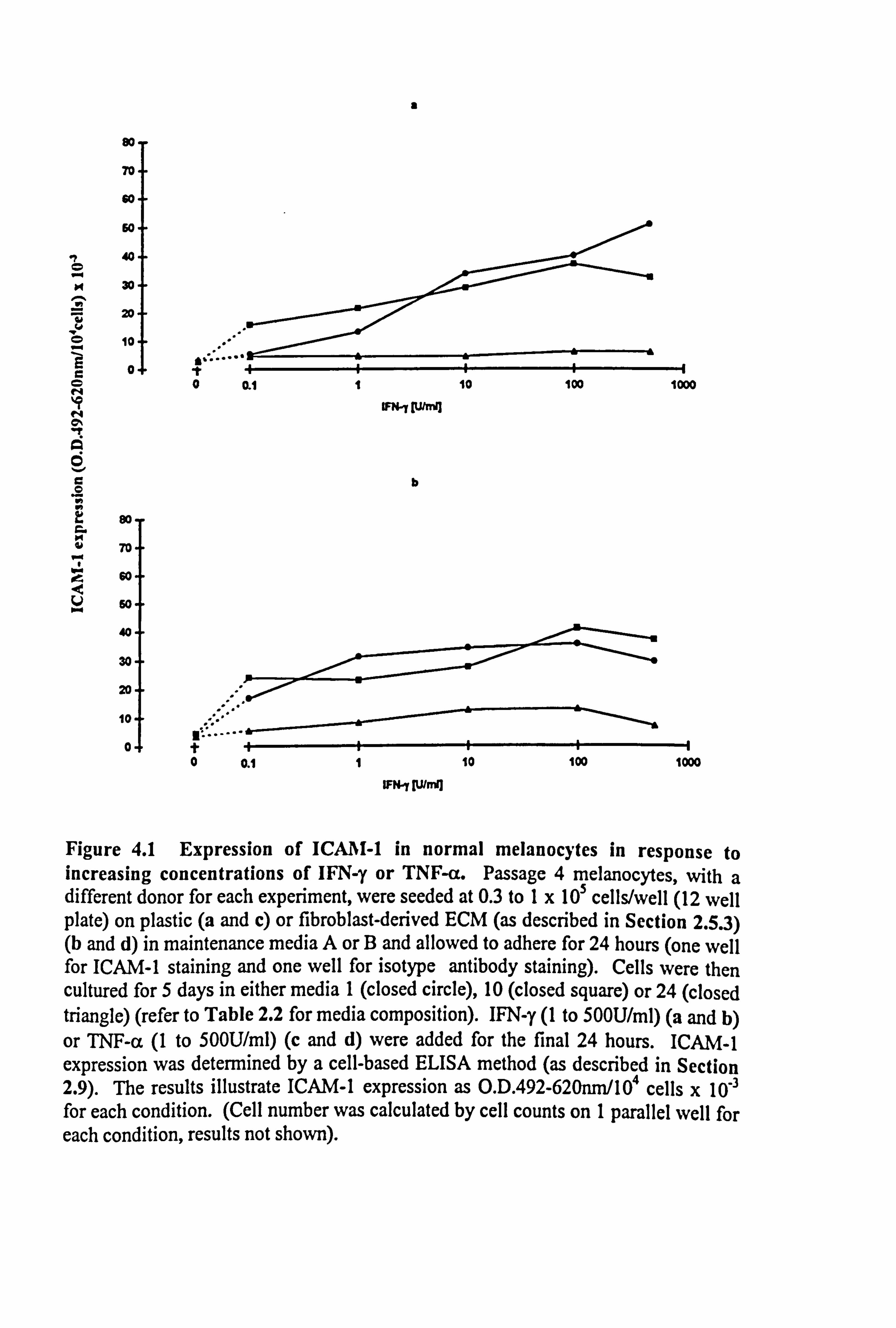
IFN-y by 100U/ml (Figure 4.1b). 100U/ml TNF-a, was the minimal concentration
required for increased ICAM-1 expression in the melanocyte culture set up on plastic,
with the maximal response seen at 500U/ml (Figure 4.1c). The melanocytes cultured
on ECM had a broader spectrum with increases in ICAM-1 expression from 1U/ml and
a maximal response achieved between 100 and 500U/ml TNF-a, depending on the
media composition (Figure 4.1d). From this work it was decided that a concentration
of 100U/ml be used for the comparative studies of cytokines on melanocytes.
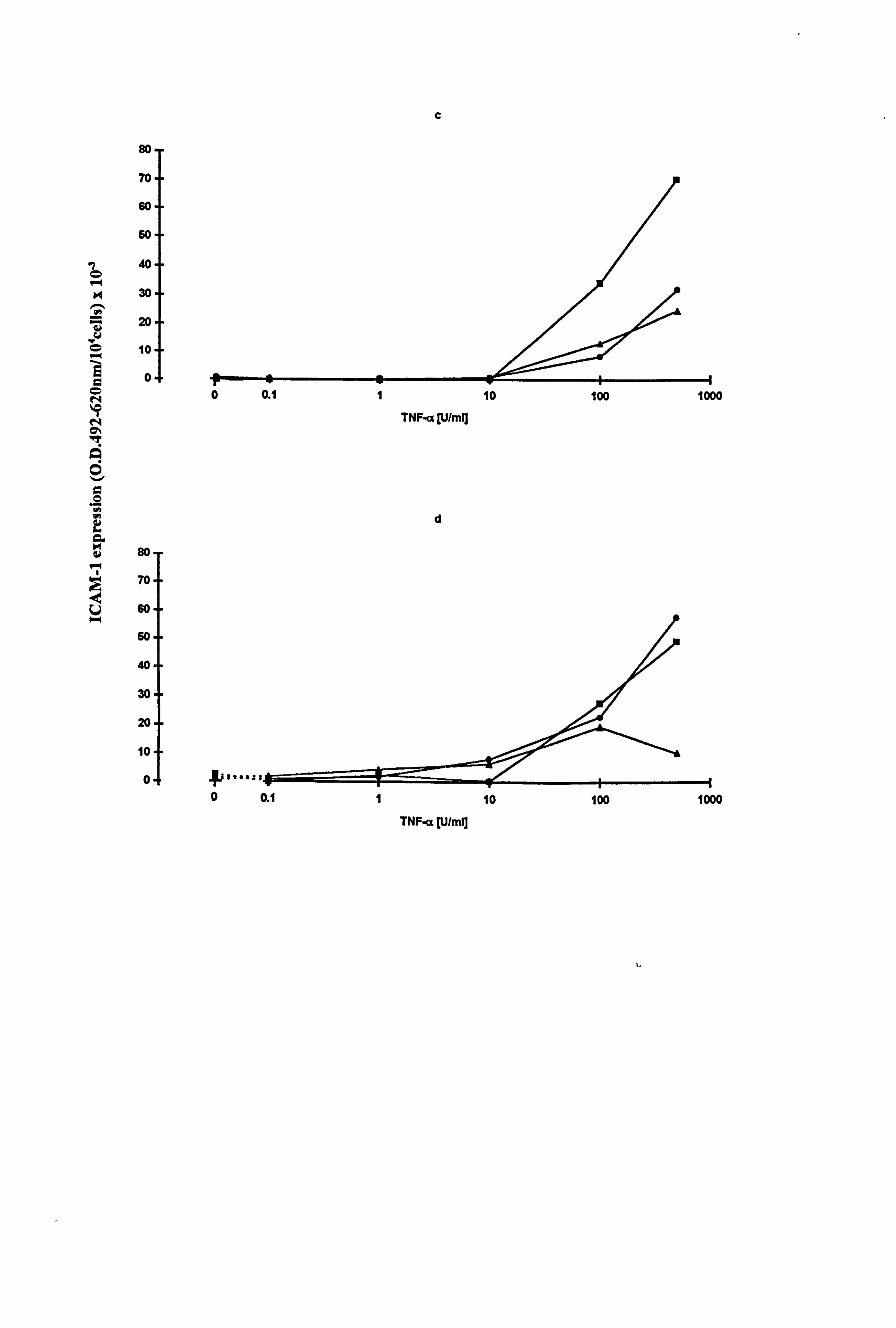
4.2.2. Comparison of ICAM-1 expression stimulated by IFN-y and TNF-a in
normal melanocytes cultured on plastic or fibroblast-derived ECM.
The effects of 100U/ml IFN-y or TNF-a on ICAM-1 expression were investigated in
melanocytes from several different donors cultured on plastic or fibroblast derived ECM
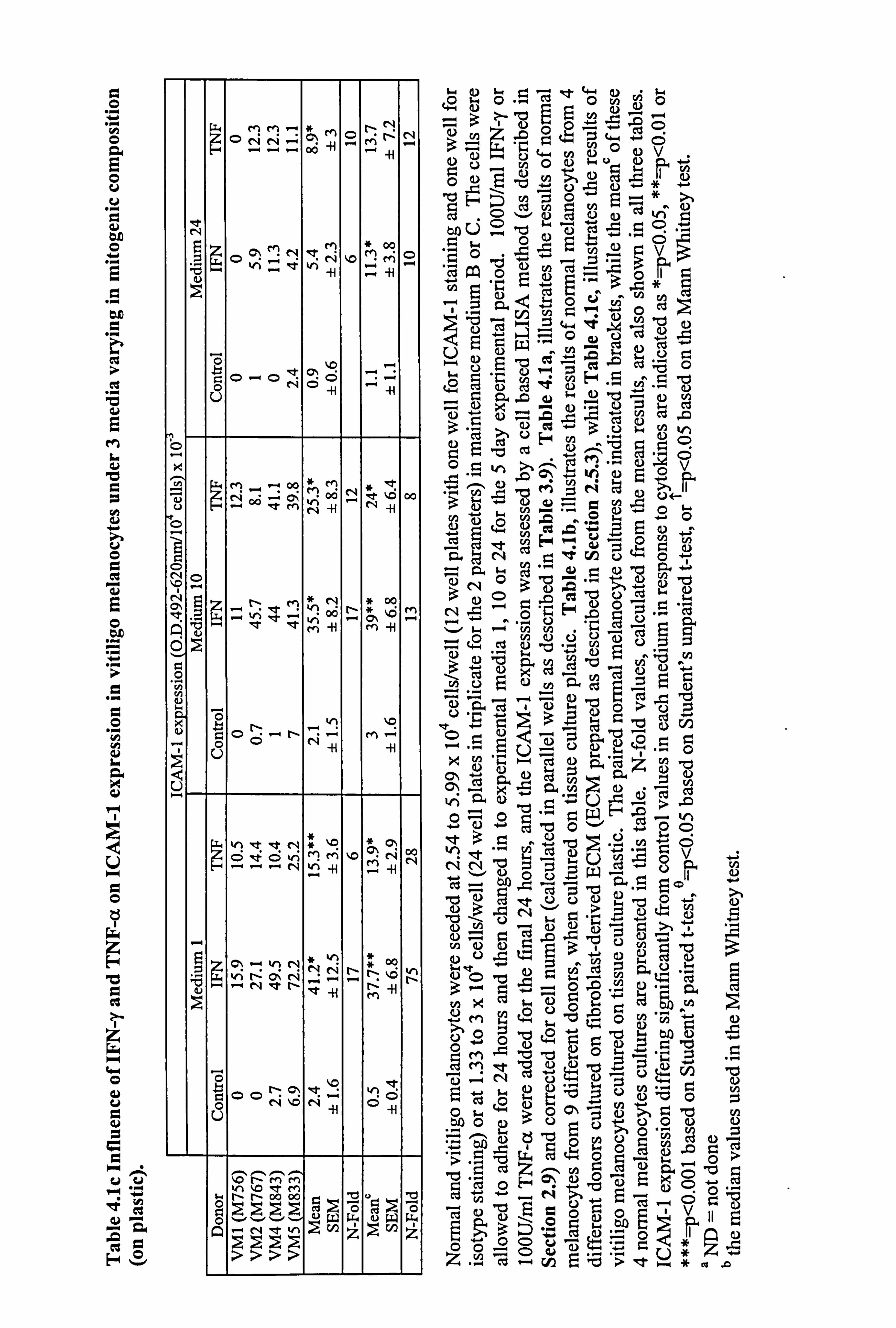
(Tables 4.1a and 4.1b illustrate the results for the individual experiments). The
protocol was the same as in Section 4.2.1, although some of the experiments in this
section were conducted in 24 well plates (in duplicate or triplicate) instead of 12 wells.
so
70
so
60
40
30
Z w 20
2 10
C0
C
O
80
70
60
ºVi 60
40
30
20
ýý; ... t 0 aý 1 10 100 1000
IF+-r "I
101
0 0.1 1 10 100 1000
WN-Y Iün3
Figure 4.1 Expression of ICA I-1 in normal melanocytes in response to increasing concentrations of IFN-y or TNF-a. Passage 4 melanocytes, with a different donor for each experiment, were seeded at 0.3 to 1x 10⁵ cells/well (12 well plate) on plastic (a and c) or fibroblast-derived ECM (as described in Section 2.5.3) (b and d) in maintenance media A or B and allowed to adhere for 24 hours (one well for ICAM-1 staining and one well for isotype antibody staining). Cells were then cultured for 5 days in either media 1 (closed circle), 10 (closed square) or 24 (closed triangle) (refer to Table 2.2 for media composition). IFN-y (1 to 500U/ml) (a and b) or TNF-a (1 to 50OU/ml) (c and d) were added for the final 24 hours. ICAM-1 expression was determined by a cell-based ELISA method (as described in Section 2.9). The results illustrate ICAM-1 expression as O. D. 492-620nm/104 cells x 10-3 for each condition. (Cell number was calculated by cell counts on 1 parallel well for each condition, results not shown).
C
I 0
e' v 0 r.
to 0 N
N D\
Q
fa O
.. r
a aý
d U
so
70
so
so 40
30
20
10
0
so
70
so
60
40
30
20
10
0 P/
.. att*
0 0.1
TNF-ac [U/mi]
d
TNF-a [U/mQ
W
0 0.1 1 10 100 1000
1 10 100 1000
v
0
co
es
U
U
U
C
ii
f3
E O
O
l) U
i4
r,
d U
a 0
z v c c4 z
O .ý U ci *=
ýa cc .
pi =
OO ^.. CL
CC E-ý ü
r 'oc -yc! %n CýM[- OM ly -H
.. r
N z NN o0 'ý ý! ýt
s tN 00 (7,00 41
oooýtýv! 41
MNNý"-M'DMýO n 0ÖN C> RT V - f
:M
en N
1 e I - en %agT-"MN--¢t _: 4{ . .
O _
N O
0, ý
Ei V1-1: -00-1Mei - I [-V10t- MN[-v1 * 0
Oý n
t Q %d w
rr V1 ýO 'ýt ýMNm «1 pp' . -,
M
0 y H
fei
X u
-o
b gv1oooo.. "ýowl%q ' t - 0' 00 %R6
N - t CN At
U
"
OOC4 N -ýýN
. -r
'- tN Nenn- q-t O If N t, - N Vl et MN V1 'ý "
N 41
lu It
b 00N-T nclC ww! v! 0 r-. 00
0 OOI.: -M O- -
U
W 0 r
p.. - a.. oas%DtNmr1 Nen M'-7d'in .OI ed.
b 0
- NNNNNNN0000 LW 4-
A 222 U) z
u GC C
e2
CS
U r
M I.
u
m
ä E 0
0
Cd L.
Yt Cd
0
0 tl
z H
cl z w w 0 üý oU a, W 0 ce co
.a° py .r " w_
"H
.n GI
Hü
Q\ ý
Oý O Qp
ýO ö M
ö
ü rz M °: N
O N 1 O
00 0 ;5 N N M ii
N
x
'10 en oq ý o0
"t vi N oo M M r n -H -e
~, ~ h of
41
Ö
0 ýp p 0 ^" ýi
0 0 \o ; (: 9 M r-b V . 0
94 O
O
0
G4 O
I
bA
. ti
Cy
bA
Ctý
V
0 t3
z H b
z C
A
hr ý vv
rt ý
Cý C H
MM O NN-
FM O N
N
. 00
N
O O .. i N
41 ýi vi r, ' vi
*' 00 MM O
ýUr
- O O~ON
00 --ý Ü -H -H
,ý [Cz M_. -40Q
*M 1
O _
O N O 9
00 ? A
G "0
LTr g"
N "-i
le 't vli M ii
Cý
M -I -4
o u
N - O
.. 0 0~-
N~ -H -H
Lt+ h ýTýtN Omot'CD vi K! M %O rN 00
N
* , 41
-H (n >r
Ö
ö 0C) ri "0 N
_H Ö
-H
ýýMM
Ö Ný. ýrNýr0ý0r0ý0r w 9 ýG O
o ý. `.. - , w uw w
ÖÖ". OÖ
w 0 -d-
a) a0c 4jo aa) v ä02"v2) u
"ý°- r a) CD ,wU9
ý"ý C: D
v
"CJ cd º'ý Z 4+ 'fit
-w --" CU
ýl . -" ou ce 2 .e rA ýý, 2 ý0
40 "U
R3 U
.0U I" 0 -cj "ý'~ zi 1, 112
O - ce -CýÖ
0 -41 en
9) w... oU 4- 0
120 NO '4 C! 2 Uo+
.. ý u2 -ß a_ aýN o3ý ; -d 30m "v o cs '4 114
výdýÄ tZ cd g) .d '_ u-0z
3U "CJ ><ce
ý
v3-bU O "--ý p 'U AS 4-. 0 x cu (7, rN
v, s: " C 4- U -ß -> o--4 o ="ý SWF' . ývý -d o" .o0 °?
(V N=
zi V
cßä V vý+ n
9.1 m ýt
Ü0u 'cj ON C) b
0 =-i mU U" y C"" >b 1 -
M wt;; bÖW "- a)
C) 4Z4 r= Gam) pb
e 4. ) wu b2 t) ävö
a s-4 t-.
U3U0ÜUÜÖ C))
" b^A NÖ 'd p0 0" N Q)
.COO >cýGýtn r- 9 -ýi >ne -. 5-0r.
*e
0 1, - _o o v-ý -p*,. o ". v. ' S
". 4 C%ý 'd "> t Ur
*x
139
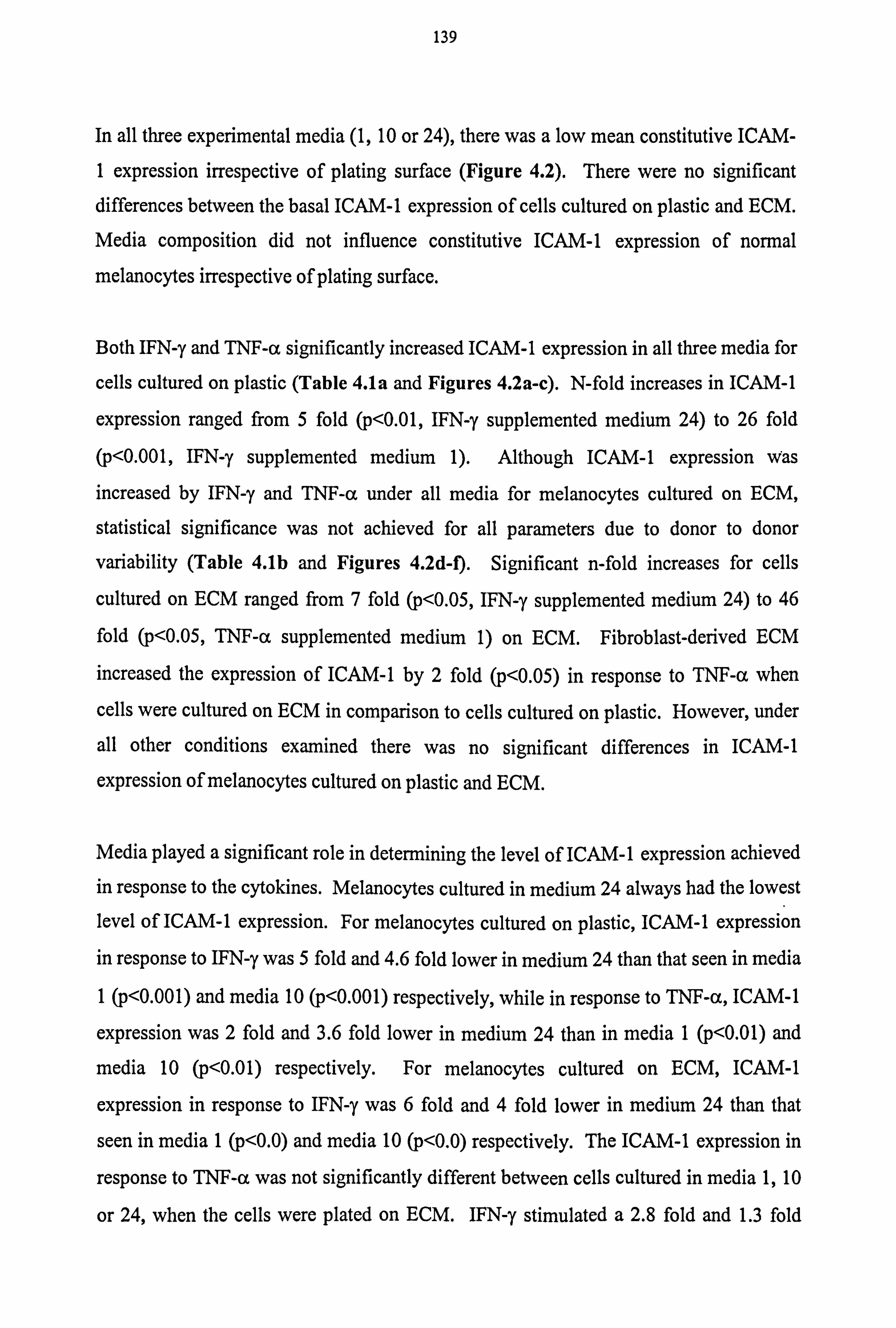
In all three experimental media (1,10 or 24), there was a low mean constitutive ICAM-
1 expression irrespective of plating surface (Figure 4.2). There were no significant differences between the basal ICAM-1 expression of cells cultured on plastic and ECM.
Media composition did not influence constitutive ICAM-1 expression of normal
melanocytes irrespective of plating surface.
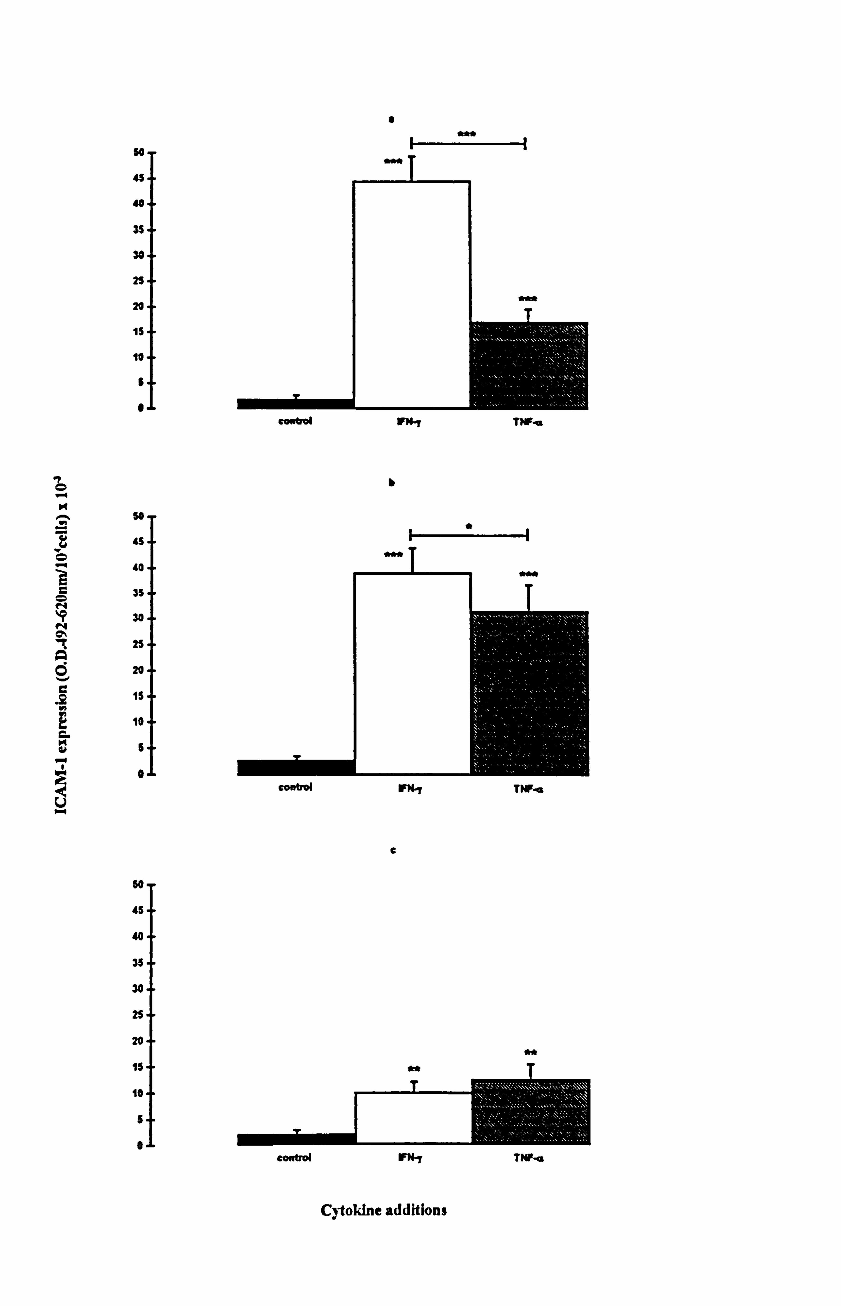
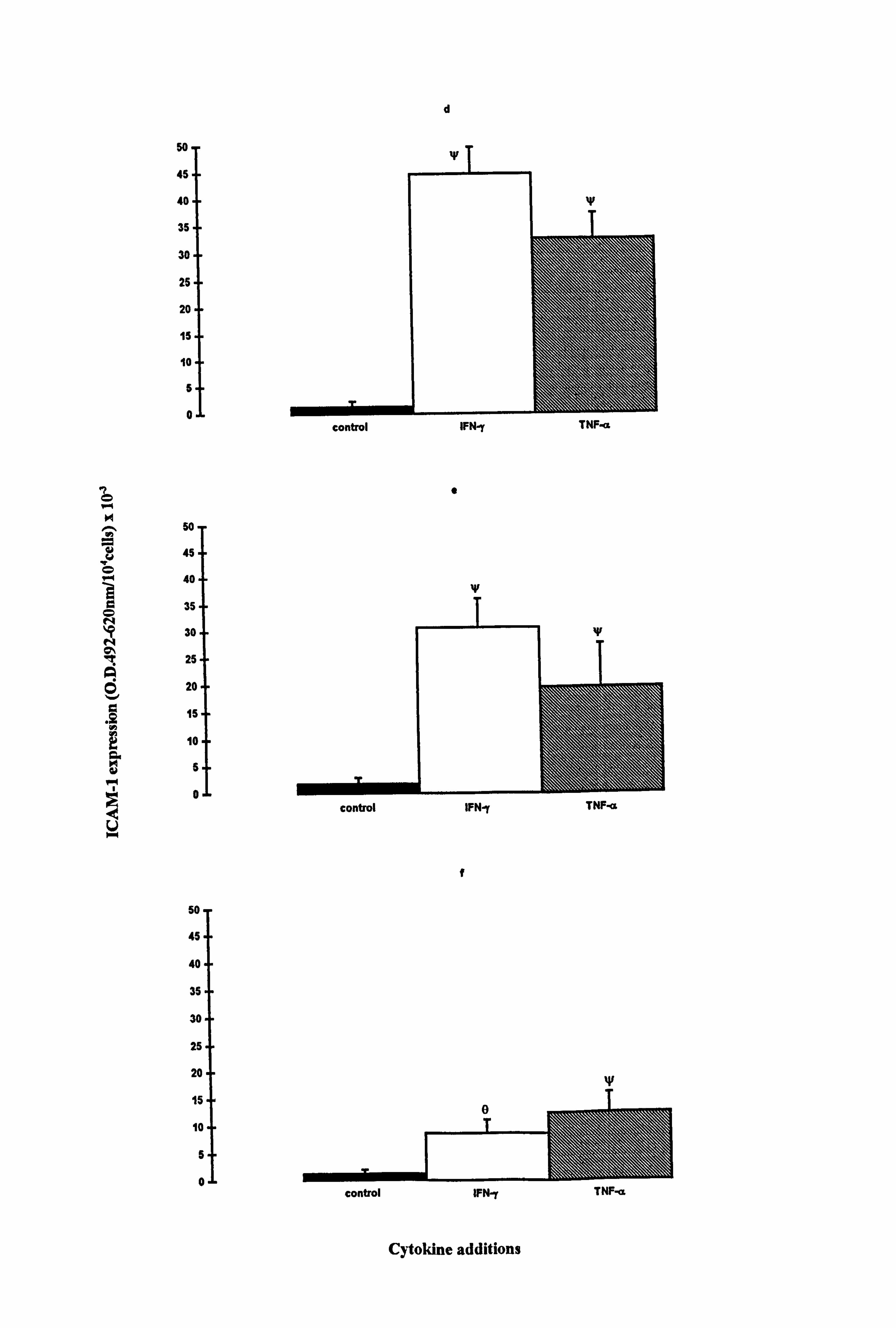
Both IFN-y and TNF-a significantly increased ICAM-1 expression in all three media for
cells cultured on plastic (Table 4.1a and Figures 4.2a-c). N-fold increases in ICAM-1
expression ranged from 5 fold (p<0.01, IFN-y supplemented medium 24) to 26 fold
(p<0.001, IFN-y supplemented medium 1). Although ICAM-1 expression was
increased by IFN-y and TNF-a under all media for melanocytes cultured on ECM,
statistical significance was not achieved for all parameters due to donor to donor
variability (Table 4.1b and Figures 4.2d-f). Significant n-fold increases for cells
cultured on ECM ranged from 7 fold (p<0.05, IFN-y supplemented medium 24) to 46
fold (p<0.05, TNF-(x supplemented medium 1) on ECM. Fibroblast-derived ECM
increased the expression of ICAM-1 by 2 fold (p<0.05) in response to TNF-a when
cells were cultured on ECM in comparison to cells cultured on plastic. However, under
all other conditions examined there was no significant differences in ICAM-1
expression of melanocytes cultured on plastic and ECM.
Media played a significant role in determining the level of ICAM-1 expression achieved in response to the cytokines. Melanocytes cultured in medium 24 always had the lowest
level of ICAM-1 expression. For melanocytes cultured on plastic, ICAM-1 expression in response to IFN-y was 5 fold and 4.6 fold lower in medium 24 than that seen in media
1 (p<0.001) and media 10 (p<0.001) respectively, while in response to TNF-a, ICAM-1
expression was 2 fold and 3.6 fold lower in medium 24 than in media 1 (p<0.01) and
media 10 (p<0.01) respectively. For melanocytes cultured on ECM, ICAM-1
expression in response to IFN-y was 6 fold and 4 fold lower in medium 24 than that
seen in media 1 (p<0.0) and media 10 (p<0.0) respectively. The ICAM-1 expression in
response to TNF-a was not significantly different between cells cultured in media 1,10
or 24, when the cells were plated on ECM. IFN-y stimulated a 2.8 fold and 1.3 fold
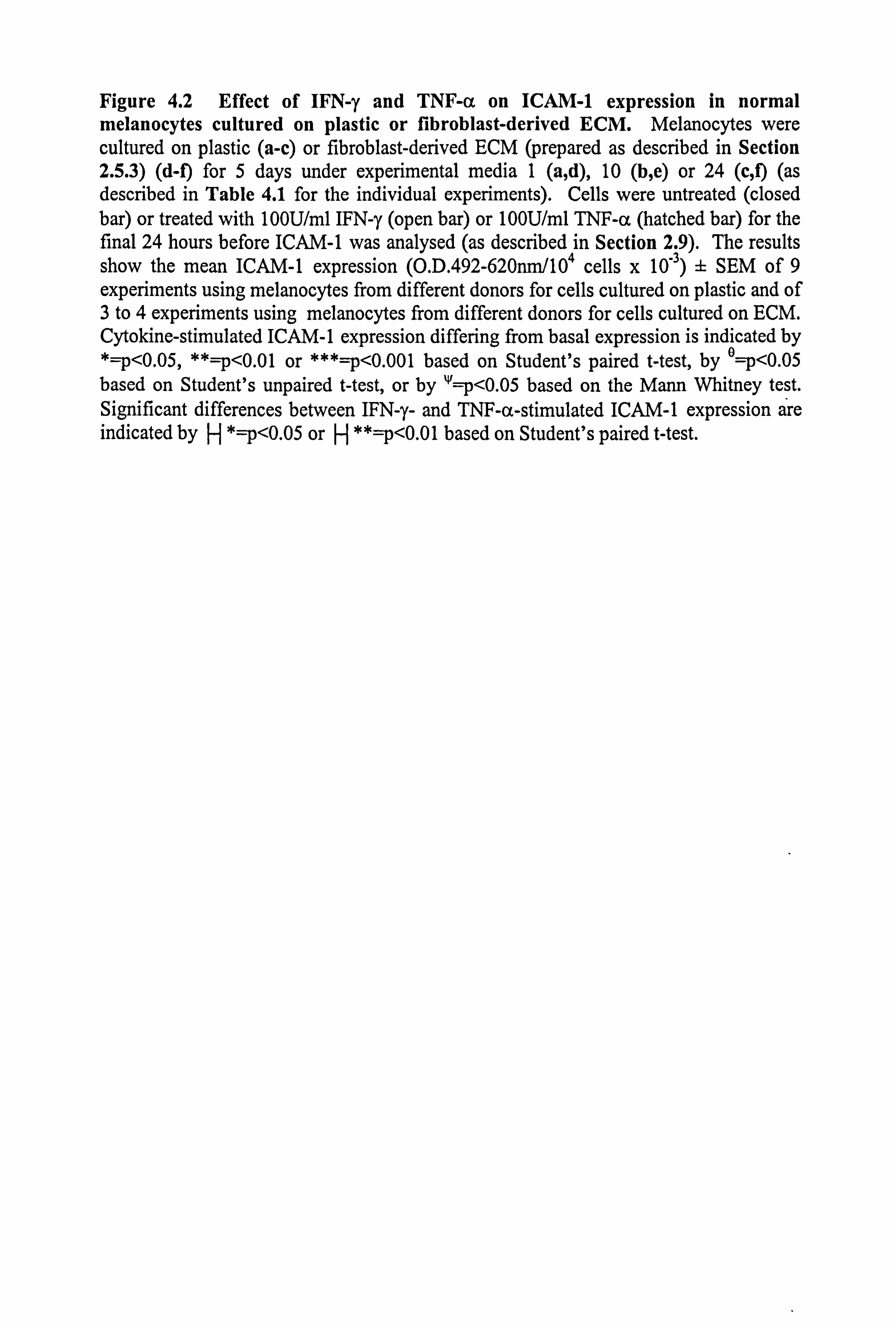
Figure 4.2 Effect of IFN-y and TNF-a on ICAM-1 expression in normal melanocytes cultured on plastic or fibroblast-derived ECM. Melanocytes were cultured on plastic (a-c) or fibroblast-derived ECM (prepared as described in Section 2.5.3) (d-f) for 5 days under experimental media 1 (a, d), 10 (b, e) or 24 (c, f) (as described in Table 4.1 for the individual experiments). Cells were untreated (closed bar) or treated with 100U/ml IFN-y (open bar) or 100U/ml TNF-a (hatched bar) for the final 24 hours before ICAM-1 was analysed (as described in Section 2.9). The results show the mean ICAM-1 expression (O. D. 492-620nm/104 cells x 10,3) ± SEM of 9 experiments using melanocytes from different donors for cells cultured on plastic and of 3 to 4 experiments using melanocytes from different donors for cells cultured on ECM. Cytokine-stimulated ICAM-1 expression differing from basal expression is indicated by *=p<0.05, **=p<0.01 or ***=p<0.001 based on Student's paired t-test, by e=p<0.05
based on Student's unpaired t-test, or by W=p<0.05 based on the Mann Whitney test. Significant differences between IFN-y- and TNF-a-stimulated ICAM-1 expression are indicated by H *=p<0.05 or H **=p<0.01 based on Student's paired t-test.
K
ö
a 0 N
N Oý
Q 0 v C
.Q h
C H to
.ý
U
so
45
40
35 30 25 20
1s
10
$
"
so
u
40
3S
30
2S
20
IS
10
5
0
so.
45
401
35
30
25
20
15
10
S
0
a
b
*
9
Cytokine additions
comb W" TTFa
conw FNq TNFa
26
r,
0 N
N
Q
ii .Q Vi
a as
5o"
45
40-
35
30
25
20
15
10
5
0
50 45
40
35
30
25
20
15
10
5
0
50
45
40
35
30
25
20
15
10
5
0
d
0
w
t
Cytokine additions
control IFN-y TNF.. a
control IFNN TNFß
140
higher expression of ICAM-1 than TNF-a in cells cultured on plastic in media 1
(p<0.001) and 10 (p<0.05) respectively. In cells cultured on ECM, the expression of
ICAM-1 in response to IFN-y and TNF-a was similar.
Therefore, ICAM-1 expression was stimulated by the incubation of melanocytes with
100U/ml IFN-y or TNF-a (24 hours) irrespective of plating substrata. Media played a
role in regulating the level of ICAM-1 expression obtained in response to the cytokines
with lowest levels seen in medium 24. IFN-y was the most potent stimulator of ICAM-
1 expression in melanocytes cultured on plastic but no significant differences were seen between the two cytokines when cells were cultured on ECM.
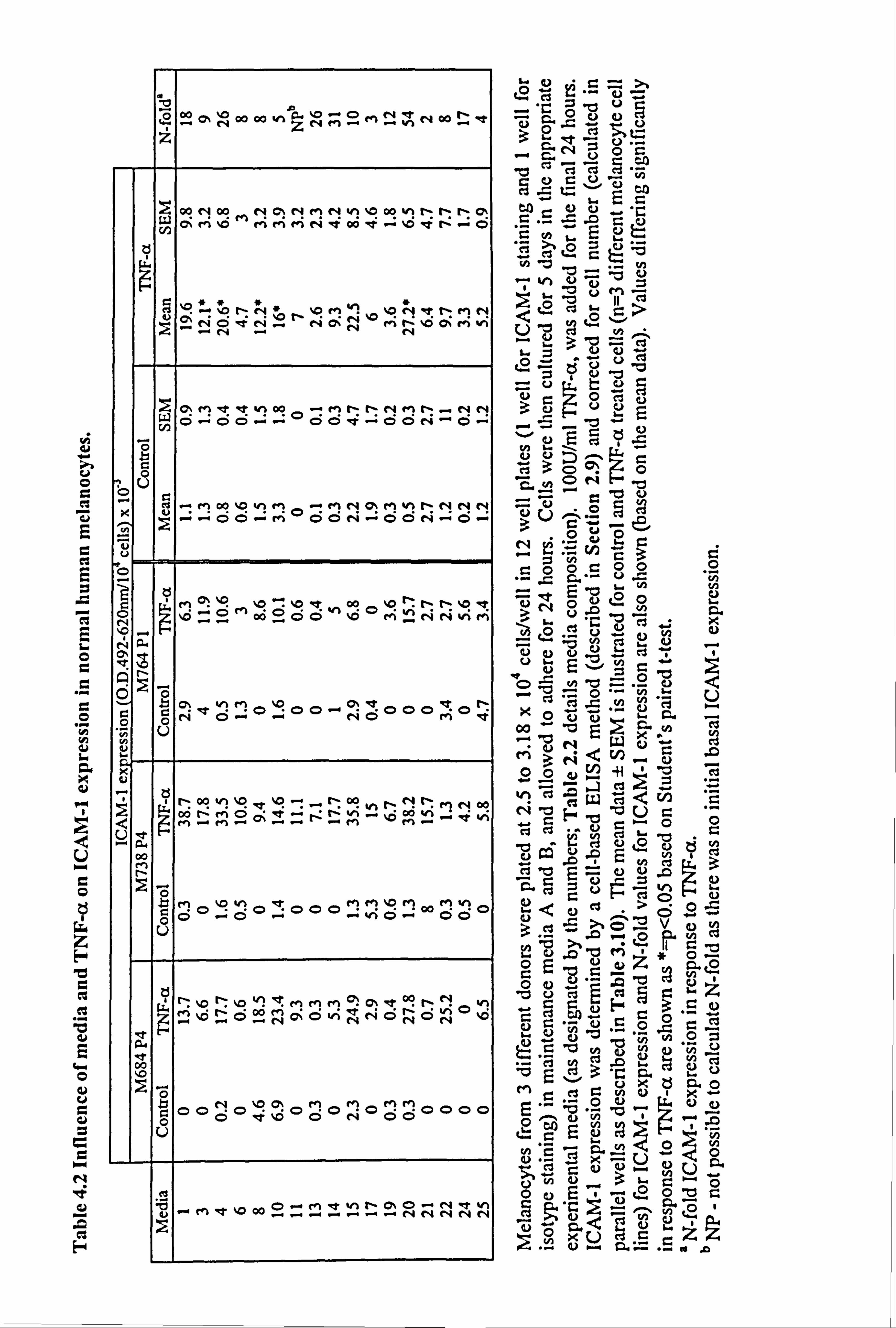
4.2.3. Comparison of TNF-a-stimulated ICAM-1 expression in 17 different media
variants.
To analyse the effect of media further, melanocytes from three different donors (2 at P4
and one at P1) were cultured under 17 different experimental media for 5 days, and
100U/ml TNF-a was added for the final 24 hours.
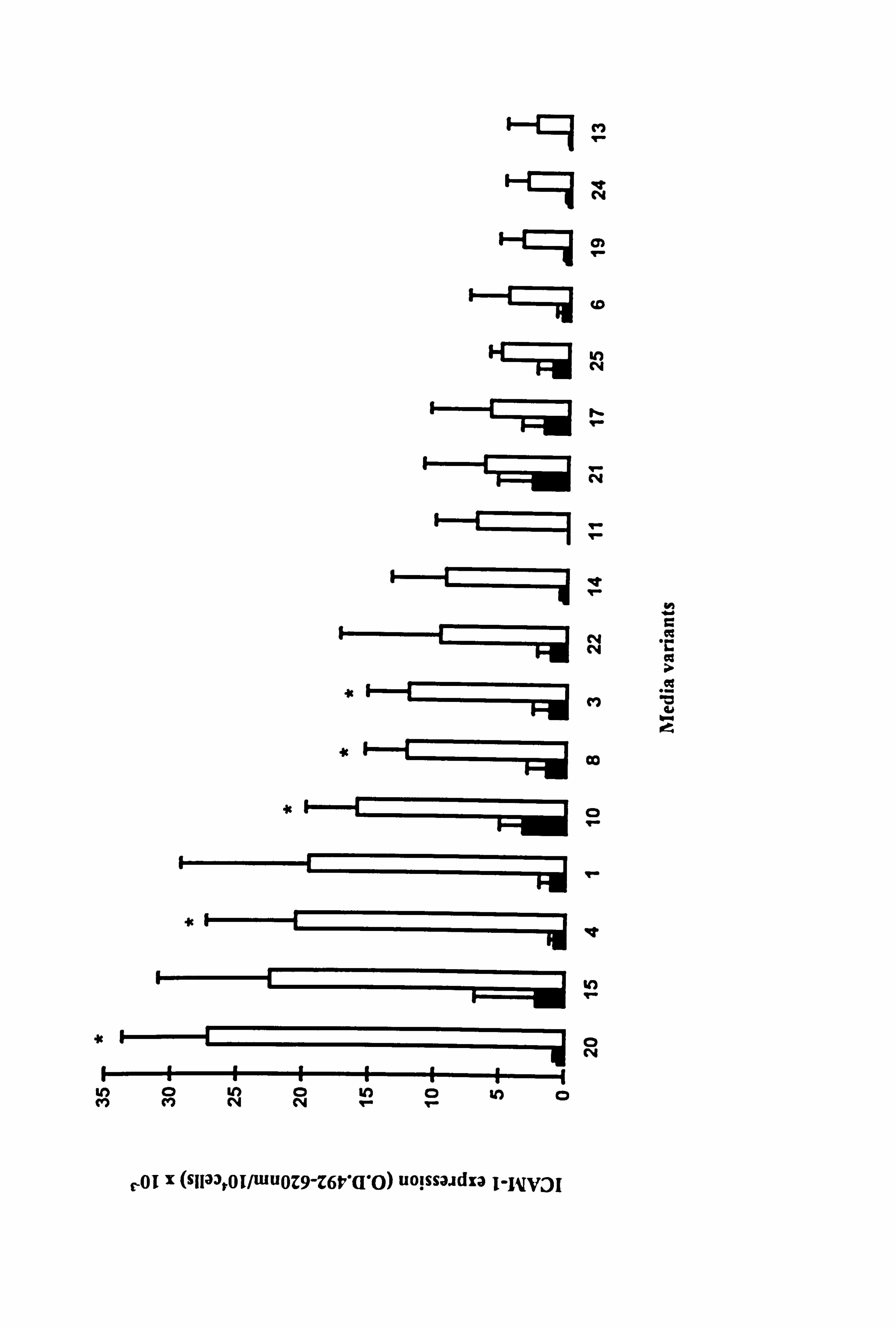
Constitutive expression of ICAM-1 was not affected by the different media variants (Figure 4.3). TNF-a stimulated ICAM-1 expression in all media although there was a
lot of donor to donor variability (as shown in Table 4.2). Significant increases in
ICAM-1 were only seen in media 3 (containing FCS/BPE/PMA, 9 fold), 4 (containing
FCS/BPE/CT, 26 fold), 8 (containing BPE/PMA/CT, 8 fold), 10 (containing FCS/BPE,
5 fold) and 21 (containing BPE, 54 fold) (all p<0.05) (Figure 4.3). BPE was a common factor in all the media where a significant response was seen. The basal levels in these 5
media were low. Unlike in Section 4.2.2, a significant increase in ICAM-1 expression
in response to TNF-a in medium 1 was not seen due to the donor to donor variability.
While media components were unable to influence constitutive ICAM-1 expression,
they did play a role in influencing the level of ICAM-1 expression reached in response
to TNF-a.
11-9 r N
CY)
N K::: ýi c4
ti r
r N
Hý ý.. r
it r
i . ýý N
-r-==ýi N
iti II '�
* Co
4c
*11R IV
N r
V) MNN
le- IO
0 N
N w C
lý r-ý
r-OI X (stlaar0i/wuoZ9-Z6ti'Q'O) uoissaadza I-I 1V3I

I-4 ce 2M"t
3a3öö
... 0i ý. W GO a
NUa N~^O
CJ 4 (D
yEZ
tgj
CLJ pab E"
.ýýNÖO
X (cZ
U)
0 ^Aý, O t cu 122
:1 -n `. - v b ~'d ö
vii 1v
G= `-ý y
ci p., N
ö
ca p
ei
op U' =p O
cli
13 öHb 9.0 w C,
.CNp= C ._Qc
0" 'ý'". r Op
Z -ý p in ö3-ö w Mm
? vv
Ire
p 79 >% NV
UU9O.. -,
y
r. r
V O
O
O
L CL is
2
C
C G
L
. - U 0
r. I 0 N
NQ C
Q
O
Q Uä
00 M N
It 00
b 22 Q% NOOOOhZNM2MNhNOO Ir',
.. ý'e
z
OO N 00 N O'. NMN Y1 %D OO nNNn 0ý M W O'M'. O c ei -e 00 -%0'ßf N-0
ü -'. 0 NNE
ý'. O M4 ý'.
O N1NMN c; NOON 1 MV1 ýG0 ý % -'N N ý. -.
iG c> el -tiAo0 -MNNNei S. -. NN Ö ý + Ö "" - - w
. r 00-t-0 N O O Ö
tu . -+ M CO 00 vl e+1 O r+ MN C+ MVNNNN ýýÖÖýMÖÖNý0C N-'C -+
V., ý'º pGOOÖ ZOMnNNvi M ýý0
O N Oý ýNÖM et
1-00 V1 %D -e'Ic00hlýNI"ý MN00
O t2 M '0 v1 'ct MM'. D en M V1
0000 O 0 p ýý CD 00
U
ýo vMMMIlýI O, -tc9 r- 11 h 0 %0 Oo OK Ö vi NNÖNON
p ÖOOOO OOÖO%ÖN CD
cl c
-en '0000. ^y.. _. e-ý. VI -_.
-- -CD%
NNNNN
.C =d fi g O 3 c
S= NV Ö .C O -cl -ä
C , CA
CA a) C) -. "C C. - =. fl C C)
'p O 0 '0 U II
t20 Z-
w .3 ZJ u ßv0
NV E CS v1
. 'ý...
Z Ov ^O c3
ou Oý
O
. - OO
"- OO U .C
0 'vn
N OE -0 O ~0 0y C)
s..
O C) O v O ý .O .
Vf 'Ci ý "N C) 0 0 � ý,
0 W-4 WO X
w b - Nb
0 () ý
O O
' 3c 3 . N ý
,,, 420
ý
Zi
cl 'o r= = o ý 0 0¢ > ö o 3m
° « v
`n U) öu °d mz 1 oO 'C .C. cb
ao c.
z "u cl Q 2ý ý CO
aýi C Öy "d y N Ov
, '
ti M O cs Ö vx aý
Z3 .
C. 0 X "ý
co 0 *o r.
c, x ... C)- a- ._ aý
141
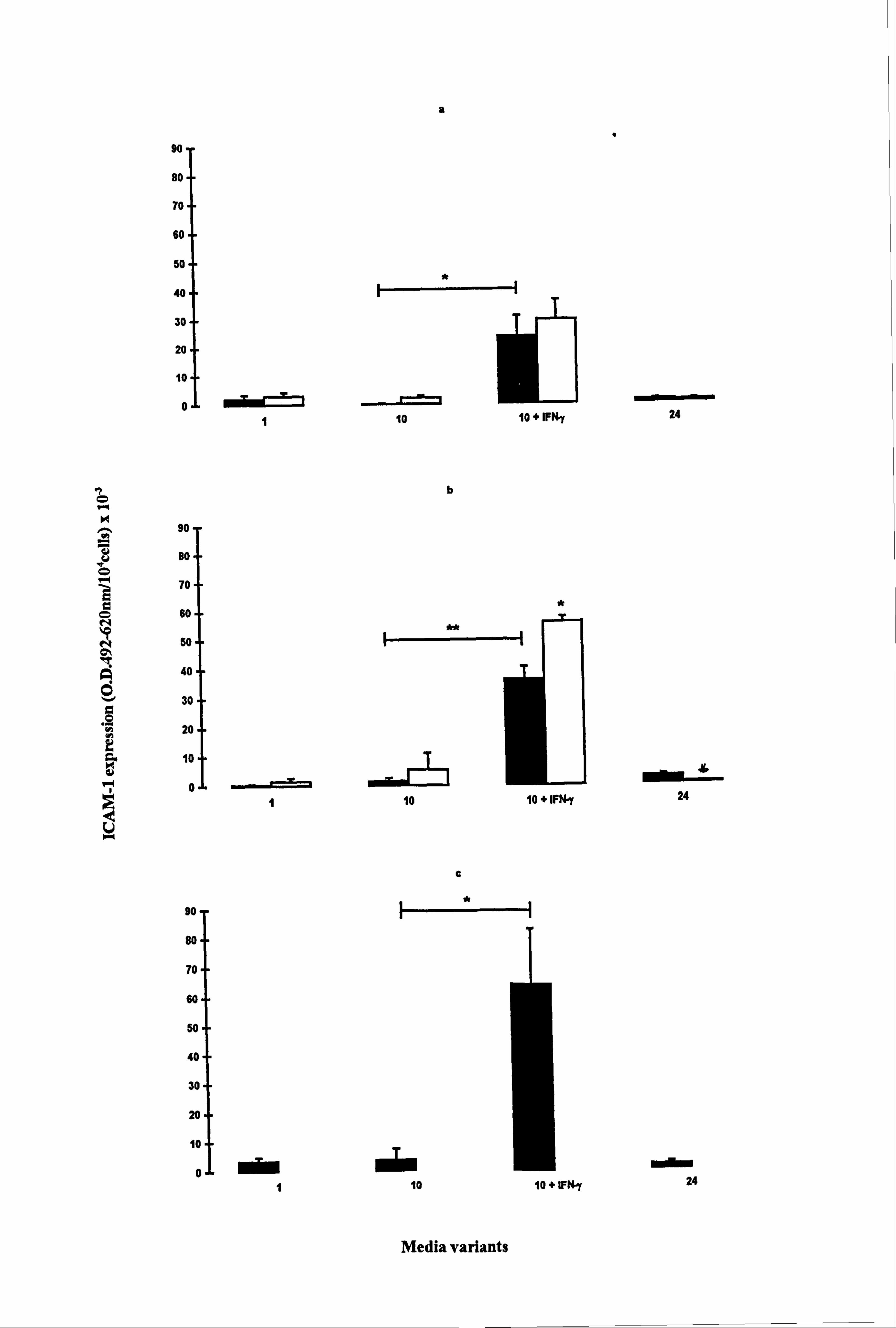
4.2.4. Effect of UVR on melanocyte basal and cytokine-stimulated expression of ICAM-1.
UV irradiation is known to have direct and indirect influences on melanocyte behaviour
as discussed in Chapter 3. Therefore the influence of UVB on ICAM-1 expression was
examined in melanocytes from three different donors (cell number analysed in parallel
wells as described in Section 3.3.2). IFN-y was examined in one of the media variants
(medium 10) as a positive control.
Basal ICAM-1 expression was low in all media (Figure 4.4). Overall UVB did not
significantly alter ICAM-1 expression except at timepoint 2 where there was a
significant decrease in ICAM-1 expression by 88% in medium 24 (p<0.05) (Figure
4.4b).
At all three timepoints, IFN-y, significantly stimulated ICAM-1 expression in medium
10, by 237 fold (timepoint 1, p<0.05) (Figure 4.4a), by 26 fold (timepoint 2, p<0.01)
(Figure 4.4b) and by 17 fold (timepoint 3, p<0.05) (Figure 4.4c). (It should be noted
that the mean basal levels in medium 10 changed from 0.1 at timepoint 1 to 3.8 by
timepoint 3 and the IFN-y levels changed from 23.7 at timepoint 1 to 64.2 at timepoint
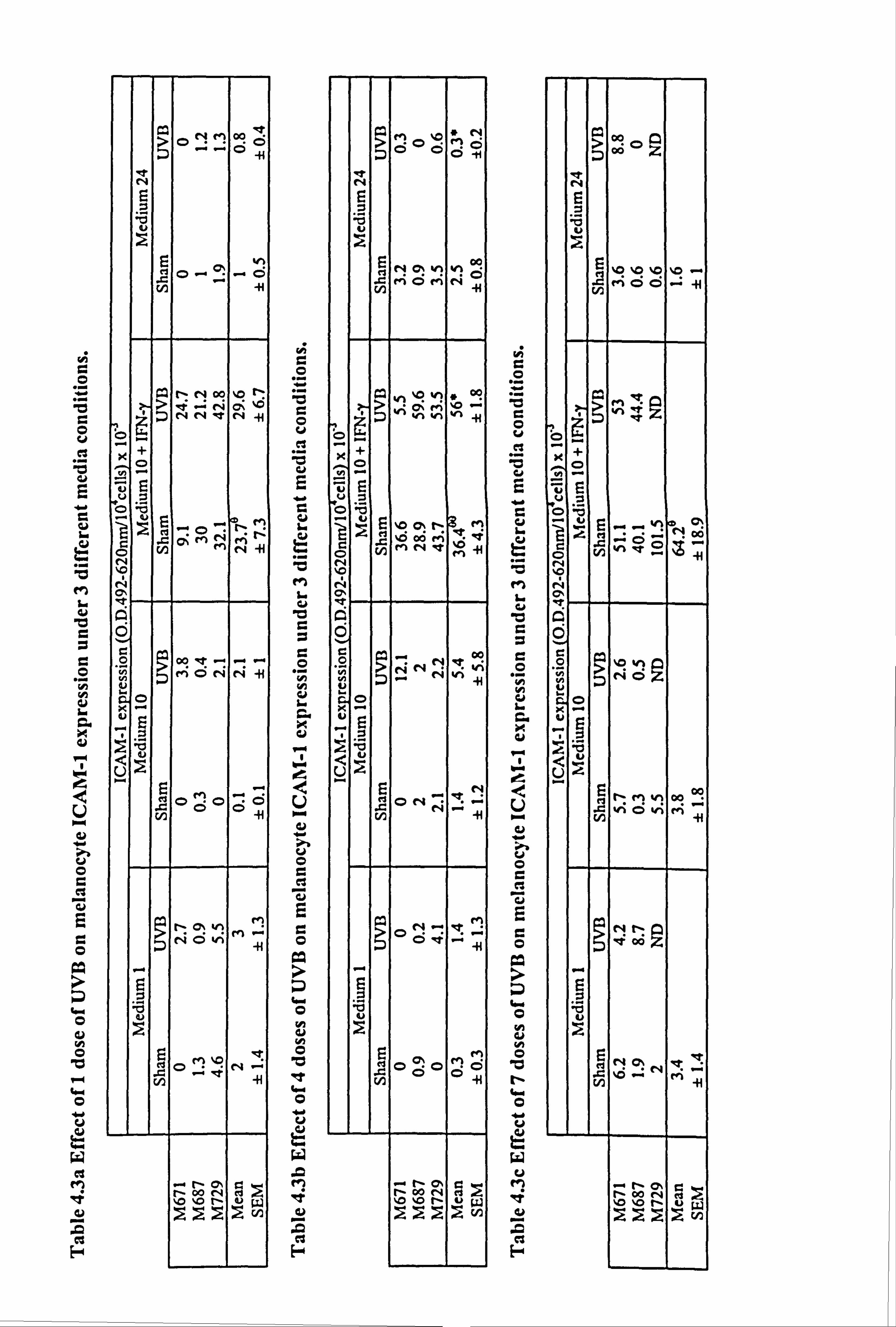
3). UVB increased IFN-y-stimulated ICAM-1 expression in two of the three
experiments at timepoint 1 (Table 4.3a). At timepoint 2, there was a significant 45%
increase in IFN-y-stimulated ICAM-1 expression in response to UVB (p<O. 05) (Table
4.3b and Figure 4.4b), while at timepoint 3, in the 2 completed experiments there were
only slight increases (Table 4.3c).
In conclusion, UVB alone did not upregulate ICAM-1 expression under any media
conditions examined. UVB did slightly enhance the response to IFN-y in one of these
media examined.

Figure 4.4 Effect of UVB and IFN-y on normal melanocyte ICA11I-1 expression. Melanocytes were sham irradiated (closed bar) or irradiated (open bar) daily for 1 (a), 4 (b) or 7 (c) days with 143mJ/cm2 of UVB (as described in Table 4.3 for individual experiments and Section 2.6). 100U/ml IFN-y was added for 24 hours to the appropriate cells in medium 10 after the final irradiation for each protocol and then ICAM-1 was analysed (as described in Section 2.9) for each time point. The data shows the mean ICAM-1 expression (O. D. 492-620nm/104 cells x 10"3) ± SEM of 3 experiments using melanocytes from different donors. Significant differences in ICAM-1 expression in response to UVB are indicated by *=p<0.05 based on Student's paired t-test, while significant differences in ICAM-1 expression in response to IFN-y are indicated by H *=p<0.05 or H **=p<0.01 based on Student's paired t-test. Only two experiments were complete where cells were irradiated on 7 occasions therefore the results of these are not shown (absence of open bars in c).
a
90
BO. -
70--
60 50
40
30 20
10
0 = =! = 1 10 10+IFNy
"t b
90-
BO. j
78 Ed
!k 60
ä 50
q 40
30
20 VyJ
10
1 10 10 + IFt
0,
c
w 90--
BO. -
70--
60.1
50
40
30
20
10
i i 1 10 10+WN-y
I
womMOMMOMMEMN 24
ý` 8
r 24
modbm 24
Media variants
r. +
O
ea
10
.0
E4
?ONM 00
N
'd L
F ei O- cý
r0
SNOOO 2r- >i ^i r4 Qz
"ý N N' N4
O
NG
OV
t/] ýMMN
N
O
y MÖ NN -}i V
C-)
0 fi CD -: (: 5 C> 0
cn -IH
b N
M AO ON
,eýNUW
O r. +
O u ei
E
"r
ci wo
C3
E-4
Lý MýsN MO
i+
'U u
9N 0% kn %A col
MÖMN -H
; O; M %0
Ö ,.., WH
Xt
O
'-' 42 5 %0a, t- M %6 06 V4 't
'H N
C
VO
K-
Ei
U `" EN "" et
41
ON-! of M
'U
M
pO .CÖ0
gn ý+ 00 Nr ýG %. 0 %0 r- uW
O
O
Cd
u
.,
v
E-
?
'L1
E %0 %0 %0 %0 . N e+i ÖÖ "-" {H
qMq
ü
0
vi CD ti 00
N
19
`rl
�q vi cV Ö
O
;k -ti
NM Y1 00 ViÖvi M
41
9 Nr, 2 ý! 00
'Li u
V7 ~M N
ýi
- f- O. r- I- 00 r4 of >
Na \O fN uw En
O -4 Cý V O
ý. -: 30 a
4 'Z Uär.
.ß
U -q 3 ; -y 'S u v ,Zv
CZ >C
Ns . sý, o
... o ý OO N
0O U
I ce ý
ý jpý 4i N 4:
tn $ý 4- "i Ü
22 Ö ý O 0
O: v `
x "o O
UP wO O W
O CD . "0 ö'ý
4A -0 En v ý o
ö -H I
bW tg *
tn "d r. Nn r, o
tý v 'O U
`e C "d ', N .
O cd v v)
OO 10 c: ~ ý'ý
Ein
4 .ý Gn 0
9
"C ö ý. W
o0 .dj VZ" -4 U) Ul O
OQ (D +-4
'G r. O i-4 O
cn a ý" ö 0
ý "ý v cn O " CA
a) s4 U ý-
, C. )
O t4 ÜM
-e ý M
, zt .4 i le r. .
d . Oý^ w p -
ýz H cý 'd M 'C7 r=ý ýa
142
4.2.5 Comparison of ICAM-1 expression in normal and vitiligo melanocytes
cultured on plastic.
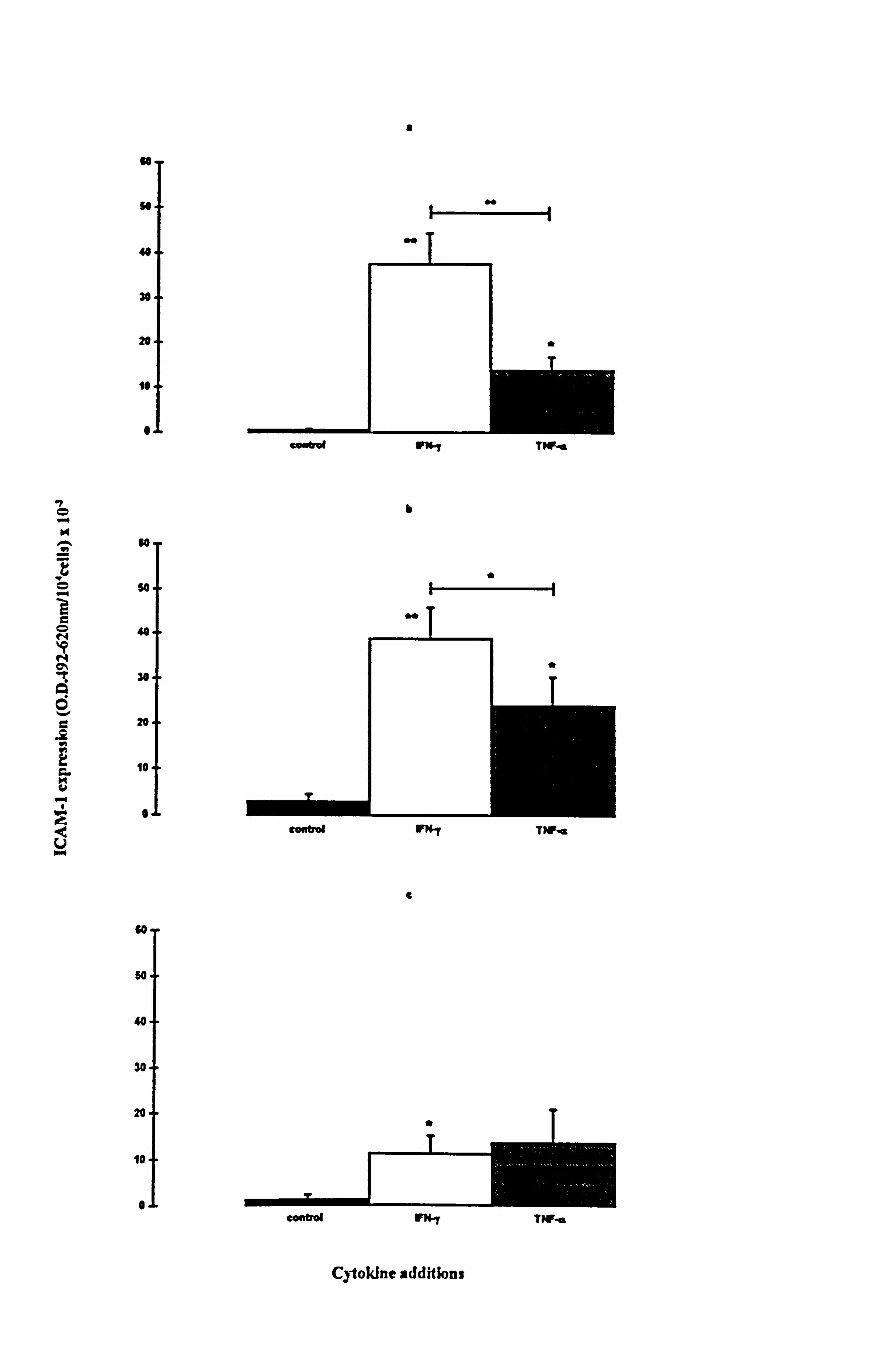
Media and cytokines had prominent effects on ICAM-1 expression in normal
melanocytes. Therefore the ICAM-1 response to cytokines was analysed in normal and
vitiligo melanocytes under three media varying in mitogenic potential. A mean
constitutive expression of ICAM-1 was seen under all three media conditions, in both
normal and vitiligo melanocytes (Figure 4.5), but not all cultures exhibited basal
ICAM-1 expression (Table 4.1a and 4.1c). There were no significant differences in
basal levels between normal and vitiligo melanocytes, and media did not influence basal
ICAM-1 expression.
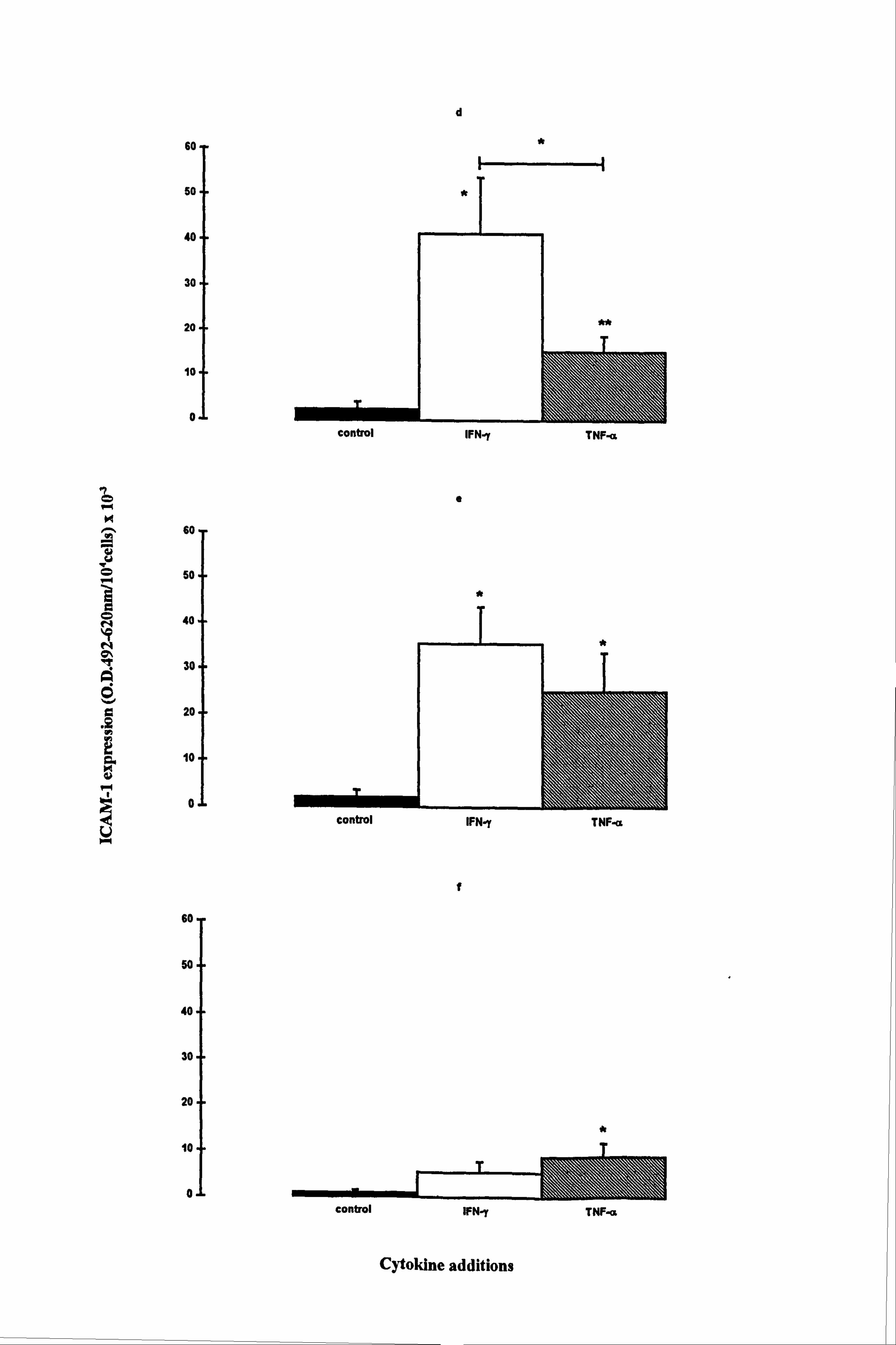
IFN-y and TNF-a significantly increased ICAM-1 levels in both normal and vitiligo
melanocytes when cultured in media 1 (Figures 4.5a and 4.5d respectively) and 10
(Figures 4.5b and 4.5e respectively). In normal melanocytes, the n-fold values ranged
from 8 fold (p<0.05, TNF-a supplemented medium 10) to 75 fold (p<0.01, IFN-y
supplemented medium 1), while in vitiligo melanocytes the n-fold values ranged from 6
fold (p<0.01, TNF-a supplemented medium 1) to 17 fold (p<0.05, IFN-y supplemented
media 1 and 10). Normal melanocyte ICAM-1 expression was significantly increased
(10 fold) by IFN-y (p<0.05) in medium 24 (Figure 4.5c) while ICAM-1 expression was
significantly increased (10 fold) by TNF-a (p<0.05) in vitiligo melanocytes (Figure
4.5f). ICAM-1 expression was much lower in medium 24 than in media 1 and 10 in
response to the cytokines as in accordance with the results in Section 4.4.2. IFN-y had a
significant 2.7 fold stronger effect on ICAM-1 expression than TNF-a, in medium 1
(p<0.01) and a significant 1.7 fold stronger effect in medium 10 (p<0.05) in normal
melanocytes. In vitiligo melanocytes, IFN-y was stronger than TNF-a by 3 fold in
medium 1 (p<0.05).
The expression of ICAM-1 in response to cytokines was not significantly different
between normal and vitiligo melanocytes under any of the conditions examined. Both
IFN-y and TNF-a increased ICAM-1 expression in all 3 media and IFN-y was generally
the most potent stimulator of ICAM-1 expression in normal and vitiligo melanocytes.
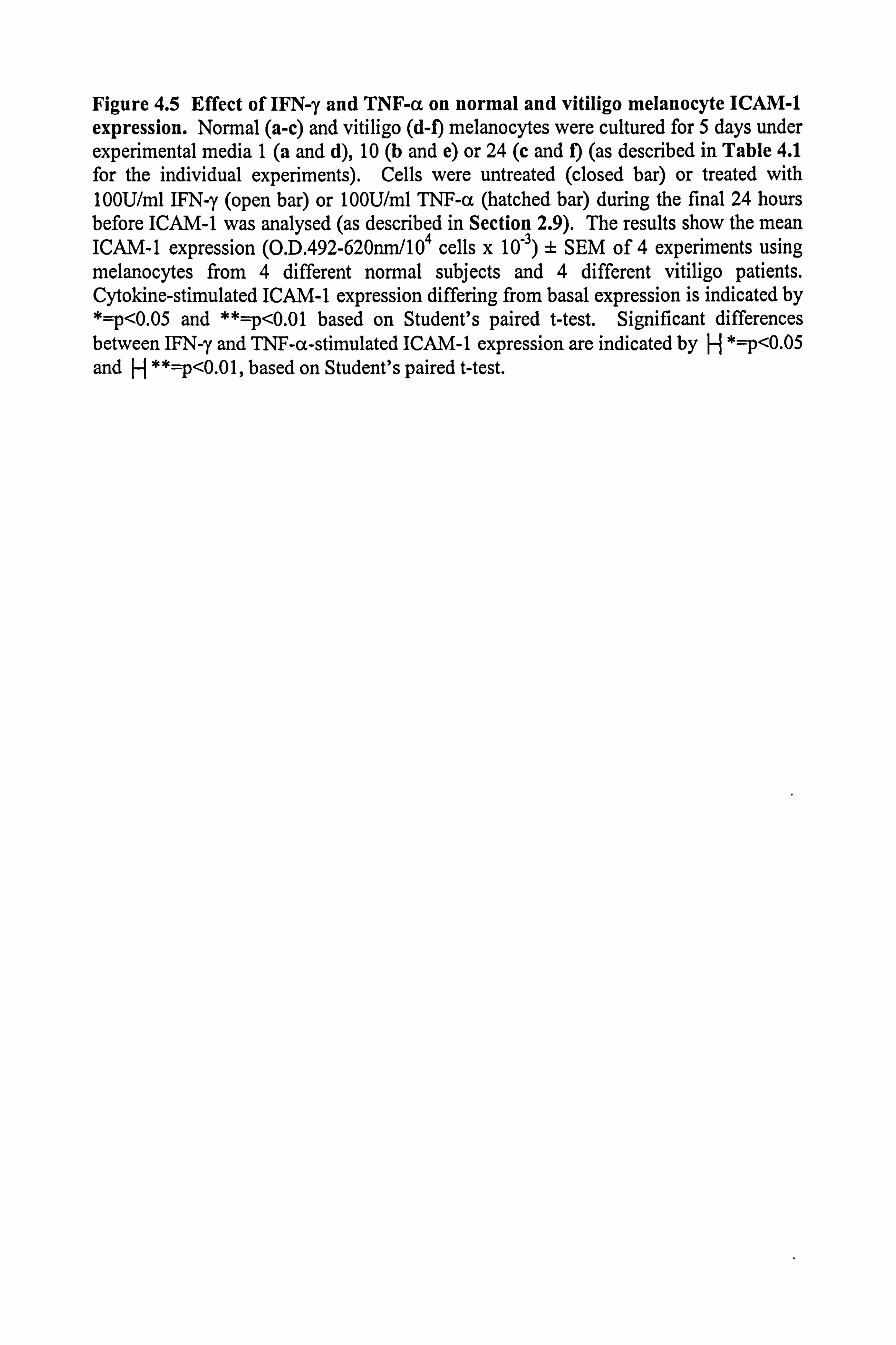
Figure 4.5 Effect of IFN-y and TNF-a on normal and vitiligo melanocyte ICAM-1 expression. Normal (a-c) and vitiligo (d-f) melanocytes were cultured for 5 days under experimental media 1 (a and d), 10 (b and e) or 24 (c and f) (as described in Table 4.1 for the individual experiments). Cells were untreated (closed bar) or treated with 100U/ml IFN-y (open bar) or 100U/ml TNF-a (hatched bar) during the final 24 hours before ICAM-1 was analysed (as described in Section 2.9). The results show the mean ICAM-1 expression (O. D. 492-620nm/104 cells x 10-3) ± SEM of 4 experiments using melanocytes from 4 different normal subjects and 4 different vitiligo patients. Cytokine-stimulated ICAM-1 expression differing from basal expression is indicated by *=p<0.05 and **=p<0.01 based on Student's paired t-test. Significant differences between IFN-y and TNF-a-stimulated ICAM-1 expression are indicated by H *=p<0.05 and H **=p<0.01, based on Student's paired t-test.
N
.. w
C O Nýp
T N a
Q
v G
. SC h
G K
rl
N e_
Ü
so
so
44
30
20
is
I
so
so
40
30
20
10
0
so
so
40
3o
20
10
0
a
I
b
*
I
e
Cytokine additions
eewtrd VN7 TNFß
coetrd F" T/Fa
I N
u e
U
60
50
40
30
20
10
0
Go-
50-
40-
60 7
soj
40
30
20 a
10
0
d
*
e
*
r
control IFN-y TNF- i
Cytokine additions
control IFN-y TNF-c&
control IFN-y TNF-a
143
4.3 MODULATION OF CYTOKINE-STIMULATED ICAM-1 EXPRESSION IN
NORMAL MELANOCYTES BY a-MSH AND RELATED PRO-
OPIOMELANOCORTIN PEPTIDES.
Melanocytes have the capacity to express immune related cell surface molecules (as
discussed in Section 4.2, and Sections 4.4-4.5). a-MSH is known to be produced in
response to UV by cells in the epidermis. In response to UV, inflammation can occur,
and the melanocyte could become a target for the cells of the immune system. In
addition to a pigmentary role (discussed in Chapter 3), a-MSH has other biological
actions, such as anti-inflammatory actions. In this section, preliminary work to examine
a role for a-MSH in damping down the effect of cytokines on the expression of ICAM-1
commenced.
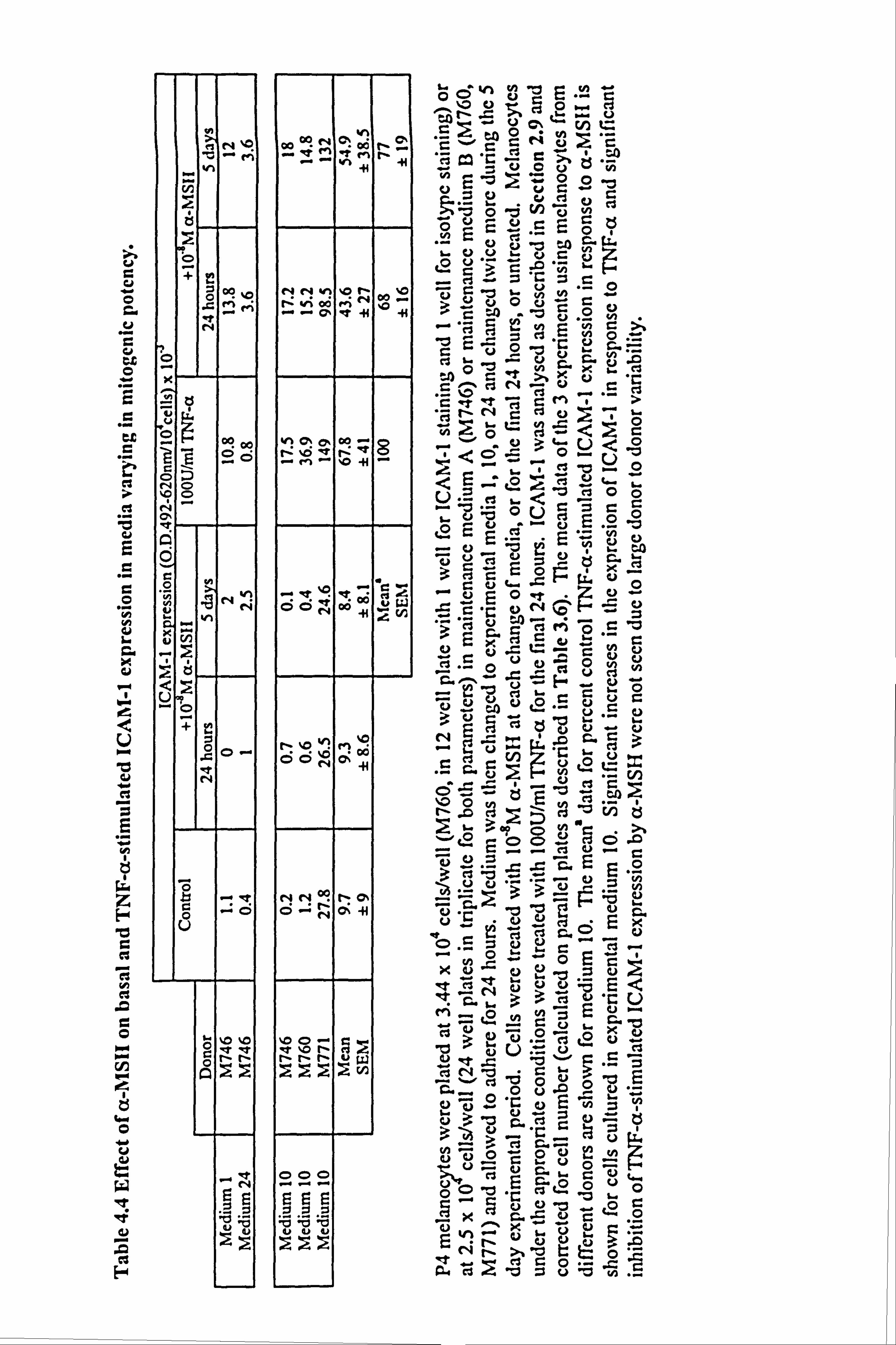
4.3.1 Effect of culture media on the ability of TNF-a to stimulate ICAM-1
expression and immunomodulatory effects of a-MSH.
Melanocytes from 3 different donors were initially used to examine the possiblility that
a-MSH may be able to oppose cytokine-stimulated ICAM-1 expression. The three
commonly used experimental medium 1,10 and 24 were employed in this preliminary
study. Although there was considerable donor to donor variation in the response to
TNF-a (as illustrated in Table 4.4), TNF-a-stimulated ICAM-1 expression on
melanocytes in media 1 and 10 (7 fold increase of ICAM-1 expression in medium 10,
failing to achieve statistical significance). In medium 24, there was a2 fold increase in
ICAM-1 expression, but the actual expression of ICAM-1 was very much lower than
that seen in media 1 and 10 (this reflects the results described in Section 4.2.2). 10-8M
a-MSH (present for 24 hours or 5 days) had little or no effect on basal levels of ICAM-
1 expression except in medium 24, where there was an increase (Table 4.4). a-MSH
was examined for its immunomodulatory effect on TNF-a-stimulated ICAM-1
expression in the three media (Table 4.4). No significant effects were seen in medium
1, the mitogen rich medium. In medium 24, there was a large n-fold increase in ICAM-
1 expression, but the basal and cytokine-stimulated levels were very low to begin with.
In 2 out of the 3 experiments conducted in medium 10, there was an inhibitory effect,
u O
td C Co tt O
e7
ei
Cd E
O
H Cd
Co x CJ
U
ad a
E H
z 00 r_ cl 12 h CS
O º+A
w
NFW
O
u 12 W
ad
E-4
w h y . y..
l` .G 2 w U
Gpý? N C'; OG 1ýý
"G C G N
yC7 ý' O O OGO
` C)
O G eG3
V v
&- UU v vG
NNv1 '0 ' e4
%_O Co
G I.. O
yy O
ýý0ý ýlý1
mil G
' 12 'er U .0 y
N C > r ey
C =
y- cl 'C
>
G U '!
C 'O
y ý� G'
.. . '. ' M O
G qpr y X .. 1
' E C -3 N .U
C C . et r_ 4 " >
N_p t" cº C v. N^
oo - g ".., `ý
p U ý
3 > e...
0 U -
0 '0
Irn No -fl _ "< V cl,. -
U 0
U ýý 0 0 _ *-v ý
Vu E ° 0 wCý c3 E w;, . _ vý -U Z; Q r3
O c)
- 0
y º. C-0
Ü E ~ ýt ' OON oo tJ C
' Q)
C ý U O -. O . - CS N y
U - .
nýOvý w G ' C" G
ý OON 00 O+ -44 CGv
C N
ti"' p U
,
p Zj 'O CG-0
ýz- 2 -2 v2 y >F c.
Oi ^r Q c> 0w g
Q .
NN C)
p y
Ö^'N cºii C "C 'O J
u Yý
M_0 rn
CJ 0 _~ Oý x ¢ýi °b 9> "u °
N % , . O uO ° O w
cý - 'C O "i,
: > 0 V Ö
OOO .. r . -+ ..
V '
c3 U '-" ö
U 4r öQ wý .C
C Cý
ý" wJ L
0
ä_ ye C ý .GU t". O
. A. 4 cs -i = C. ) w .5
04 1 120 as u 0 100 Coi
80
AÖ 60
40
2 20
r, 0 108M c -MSH (24 hours)
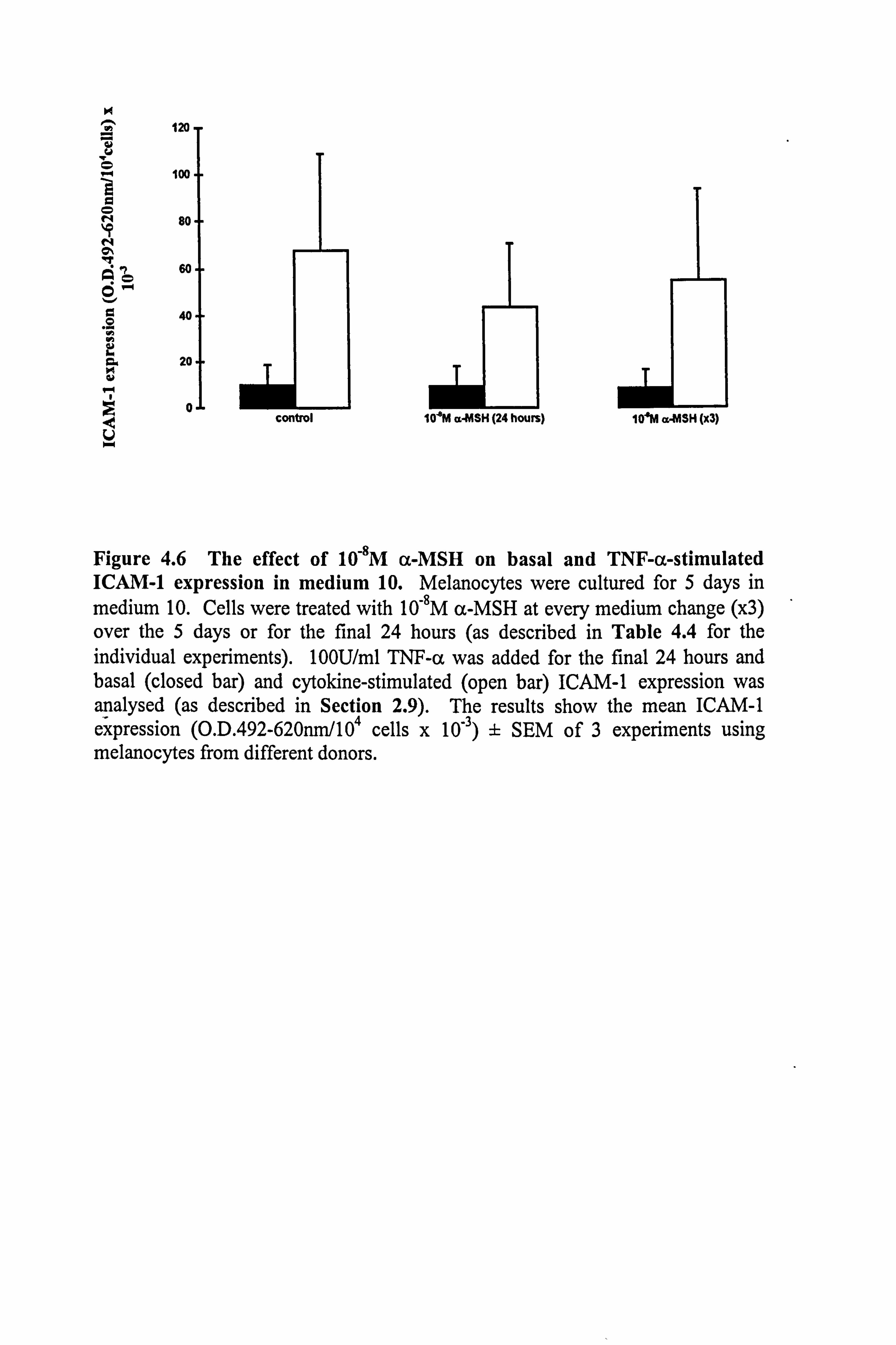
Figure 4.6 The effect of 10-8M a-MSH on basal and TNF-a-stimulated ICAM-1 expression in medium 10. Melanocytes were cultured for 5 days in medium 10. Cells were treated with 10-8M a-MSH at every medium change (x3) over the 5 days or for the final 24 hours (as described in Table 4.4 for the individual experiments). 100U/ml TNF-a was added for the final 24 hours and basal (closed bar) and cytokine-stimulated (open bar) ICAM-1 expression was analysed (as described in Section 2.9). The results show the mean ICAM-1 expression (O. D. 492-620nm/104 cells x 10-3) ± SEM of 3 experiments using melanocytes from different donors.
control 10'M a. MSH (x3)
144
but overall this did not achieve statistical significance (Table 4.4 and Figure 4.6).
M746P4, was initially cultured in maintenance medium A, which contained CT and this
could have been an influence on the results seen in the three media. Certainly, in
medium 10, the cells were unresponsive to a-MSH, unlike the M760P4 and M771P4,
which were initially cultured in maintenance medium B.
From this preliminary study there was a hint that a-MSH could oppose the actions of
TNF-a. The initial culture medium may have played a role in masking the results from
one of the donors, therefore in an attempt to standardise procedures, all further
experiments (discussed in the following Sections 4.3.2-4.3.5) were established in
maintenance medium C and cultured under experimental medium 23 for 5 days.
Maintenance medium C did not contain CT and experimental medium 23, was defined
as only containing bFGF and did not have non-physiological mitogens or
serum/pituitary extracts which could be possible sources of POMC-peptides and
cytokines.
4.3.2 Effect of a-MSH and TNF-a on constitutive ICAM-1 expression in
melanocytes cultured in a defined experimental medium.
Melanocytes from 8 different donors were utilised in various experiments throughout
the following Sections 4.3.2 to 4.3.5. All the cultures had been grown in maintenance
medium C and cultured in experimental medium 23. Cells were cultured with a-MSH
present either for the final 24 hours or throughout the 5 day experimental period. TNF-
a was always added for the final 24 hours to the appropriate cells.
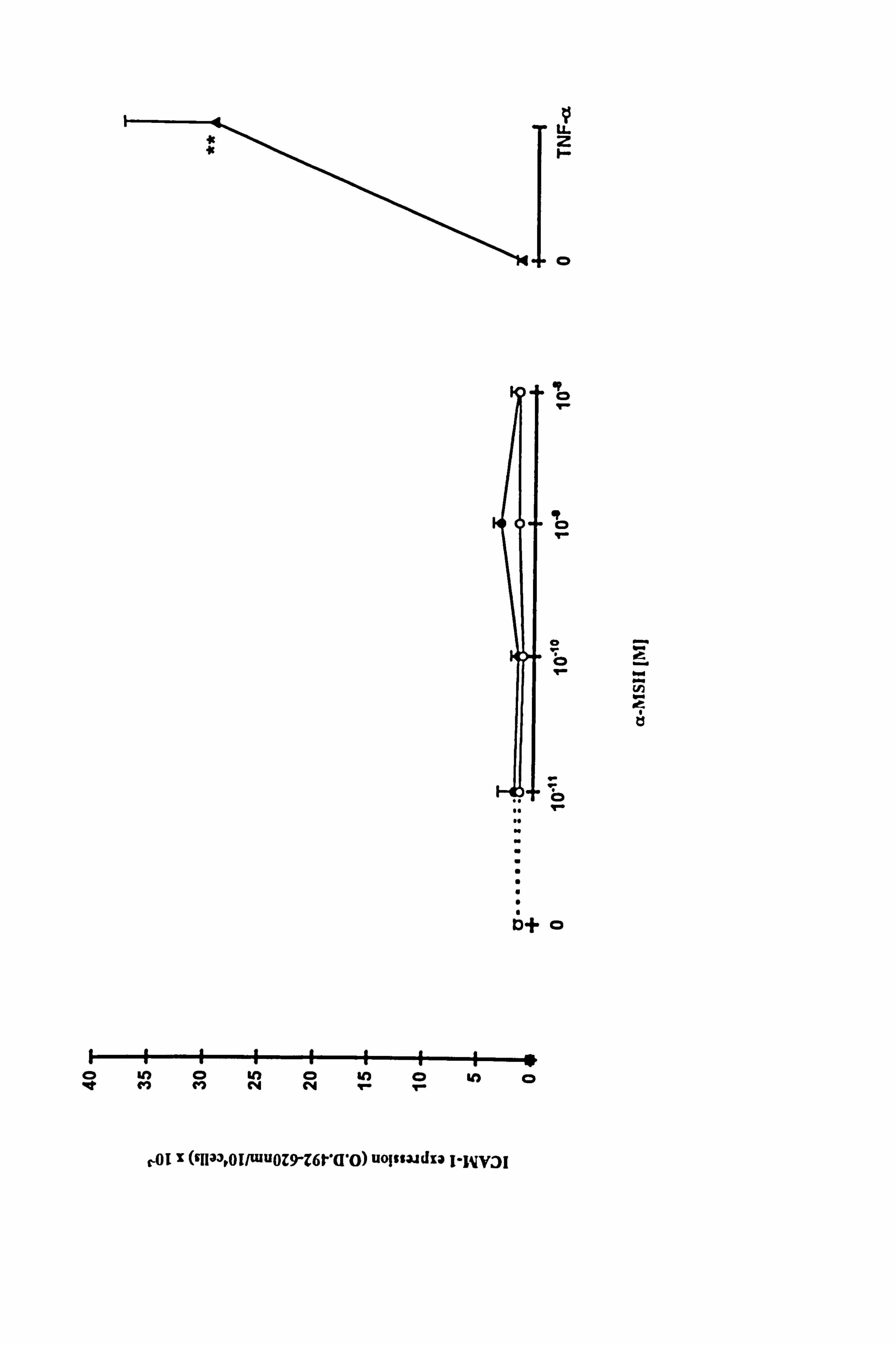
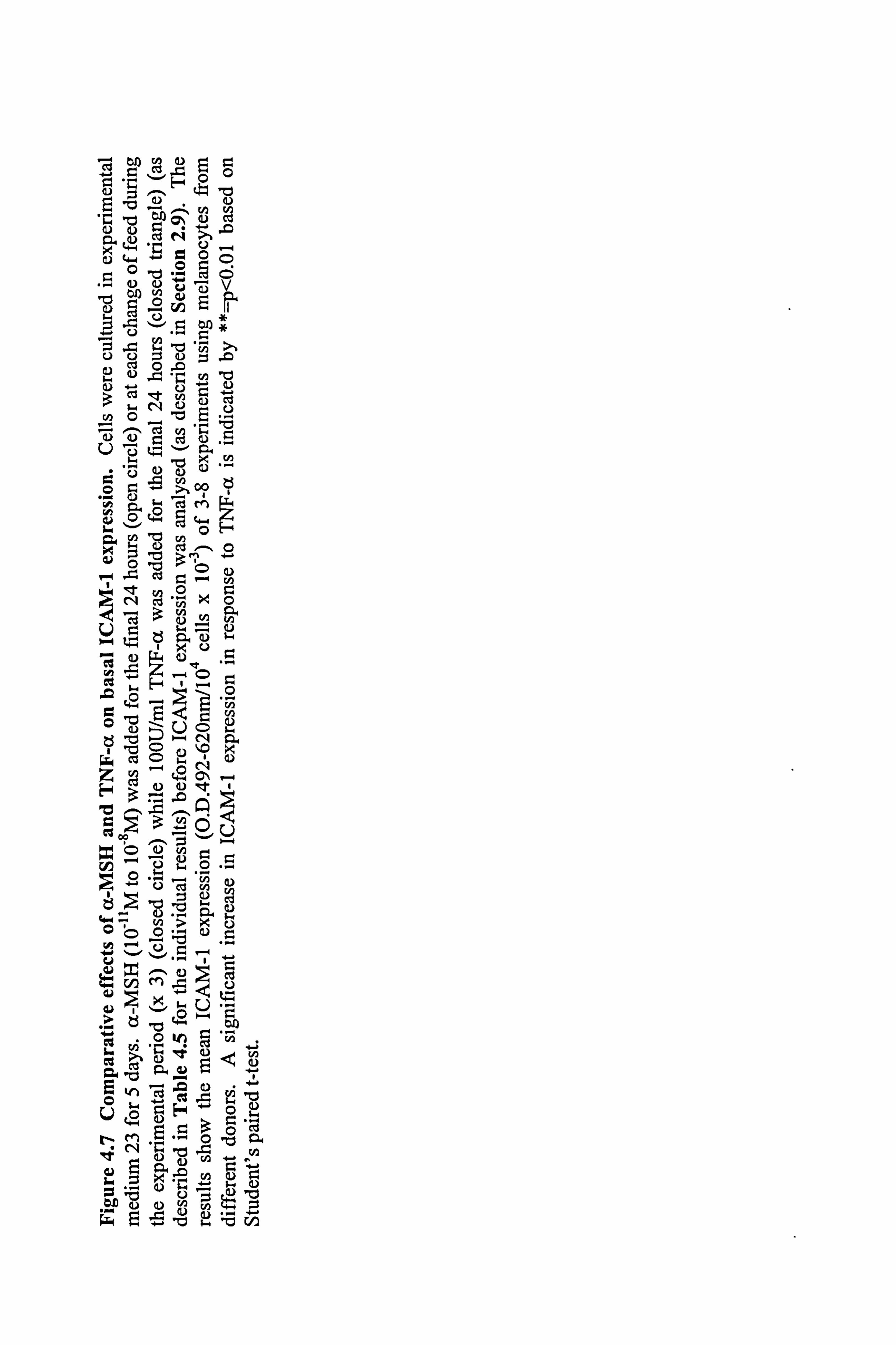
a-MSH over a range of concentrations had little or no effect on basal ICAM-1
expression, whether present for just 24 hours or for the full experimental period (Figure
4.7, individual results for each melanocyte culture shown in Table 4.5a). In contrast to
a-MSH, a significant 20 fold increase of ICAM-1 expression in response to 100U/ml
TNF-a (p<0.01) was demonstrated (Figure 4.7). In two of these experiments, IBMX
additionally had little or no effects on basal ICAM-1 expression (Table 4.5a) while the
v LL
F-
0
R 0
I 0 I-
Öe 9- bm
.r N
tl
Dý ,o ..
a S U U S
6+0
V) (3 ýd
M0NN 9- 0
rol z (9Tla3ºOI/wu0Z9-Z6t''Q'O) uojssa. idxa I"11yJI
Gb . Co-:,
äC
C)w"- a öö
cbi ý° Vý ý II
oo4 "N
a) 0 'O rn 'd
cd 3ö NC o cý v, "G 'b
5e-ßä' .z "Ci -
rA MM (A n.. 9. -4
öý 0 =- U) Nc
'ý Ö
3a
O 40
I�/b, ce CIO
Z 3.0ý p--4
xöäCO rA NW i-ß
. (2 ". - O
v "E ý-1 - cu Z
NýI t
-C- oI' CýC °'d i
NN. C" 0-
ýr aý r c' aý a . iD 2 rl C r,. ß
Zb 1"ý
E 'ý
uy
. fir A
u O
v
"r
O
L
iK v
r. r
"r
O v
O
cs
O
I
E-4
x v v, `"
OM
N
en OoONN Q
N h 44
l--"
2 y
'f "- a % O Oý q _: MO M
q0
ý-+
p N
H
U y
Ml qf O w 10
41 ? C
ý h
G O
.ý
tj 'o
rý r
O I.. :3 flý
o q 4N 0
M Q 11 41
O N
O N N
y
Cl%
O N H
ONN Ö
N
1" N CO
te)
p M t- t-
'ý O
ýt %nItv1 Cl N0
5" 'aýMOýON
0 r-
r- 00 00 00 000 4. ) W
ö
Cd L
v
Ü
v u
Z> 0 0
es
Z3
u
F-
. tý
E-0
x ro 00 N "' ýO
!ý clq
N m et
yý 'H
O Ö
ý
h... ~
et00 M«+
t N
r VS
a 'n V %o bOD 'f (ý ^' OÖ " co OÖ S
NN N V1 - 00 g
N - et .r.. + N en .. » N '41
L
ä IV VI o/1%0N"T N n N-1 - 00 to's + C
' ý: 1. C4týO;. M
- 44
fi " f N
y N 'Ö
00 ^' ýO h "' N
41
ý - C-i
N ii O N 44 N
r3 IR .q 't ýo 00
Ö NN ~
"H " -H y
N
V
04 Ü N 44
N %D NM000%
t, ýo fh 4 'T
ý-+ OW N
MNN N . }4 H
N
I Q
,C
C% 0% N 00 o0 N . - h V1 N tN
Q., el
00 S
S ^ NN N- N
b v Vi vNVi OR vi
0 -H U
Jý too en 00 OON I'N NNM tf V1 Y1
p 9 ei
tý 1ý 00 00 00 00 00 00 w uw uw
cUj -0 lý cif -d r ö
u a) 00
, t e ý. v2 Ei 1-1% 5 o
c ' O G) -ý "d
1 bA 8
0 O C/)
" d Ö O ý c ,ý
9 ci -4- U2 C%
"a = t2
t 0 `) tr
,2 o i-4 .,
= 4-
0 x ý+ cd m
N G O
--4 U N4 p"
V)
2
r ý", ý O
-ý 4 U 2 ö
0
U ý N ýb r'"ý bA
ý Q u2 10 o
ý ;j - ct U v b .ý O
M, " b b
N C i N
09 ý 0ý x O y
" CU
p 9« E
I_ U o0 , - `° b3 o ö ö
M
a ß Uý 2 t d c ä l'
o 0 00
x H; ý o ' E
O 2 "0 ce UU = © , r. , 0 0 "cýn ; z4
.a -cj I. U. w 00
145
effect of ACTH and y-MSH on basal levels were not examined in this medium because
there was not enough cells to set up complete experiments.
4.3.3 Effect of 10-8M a-MSH on TNF-a-stimulated ICAM-1 expression in
melanocytes.
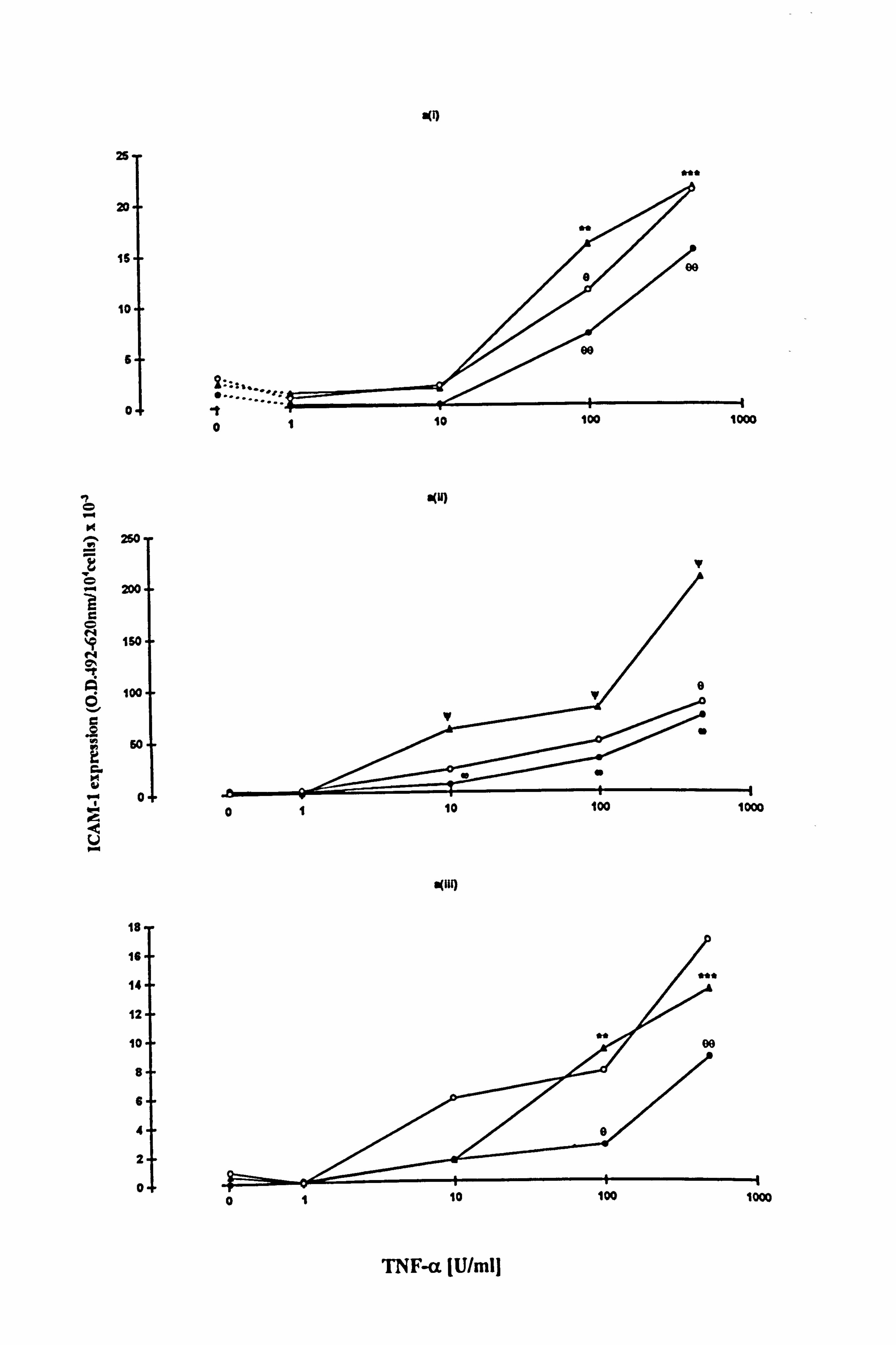
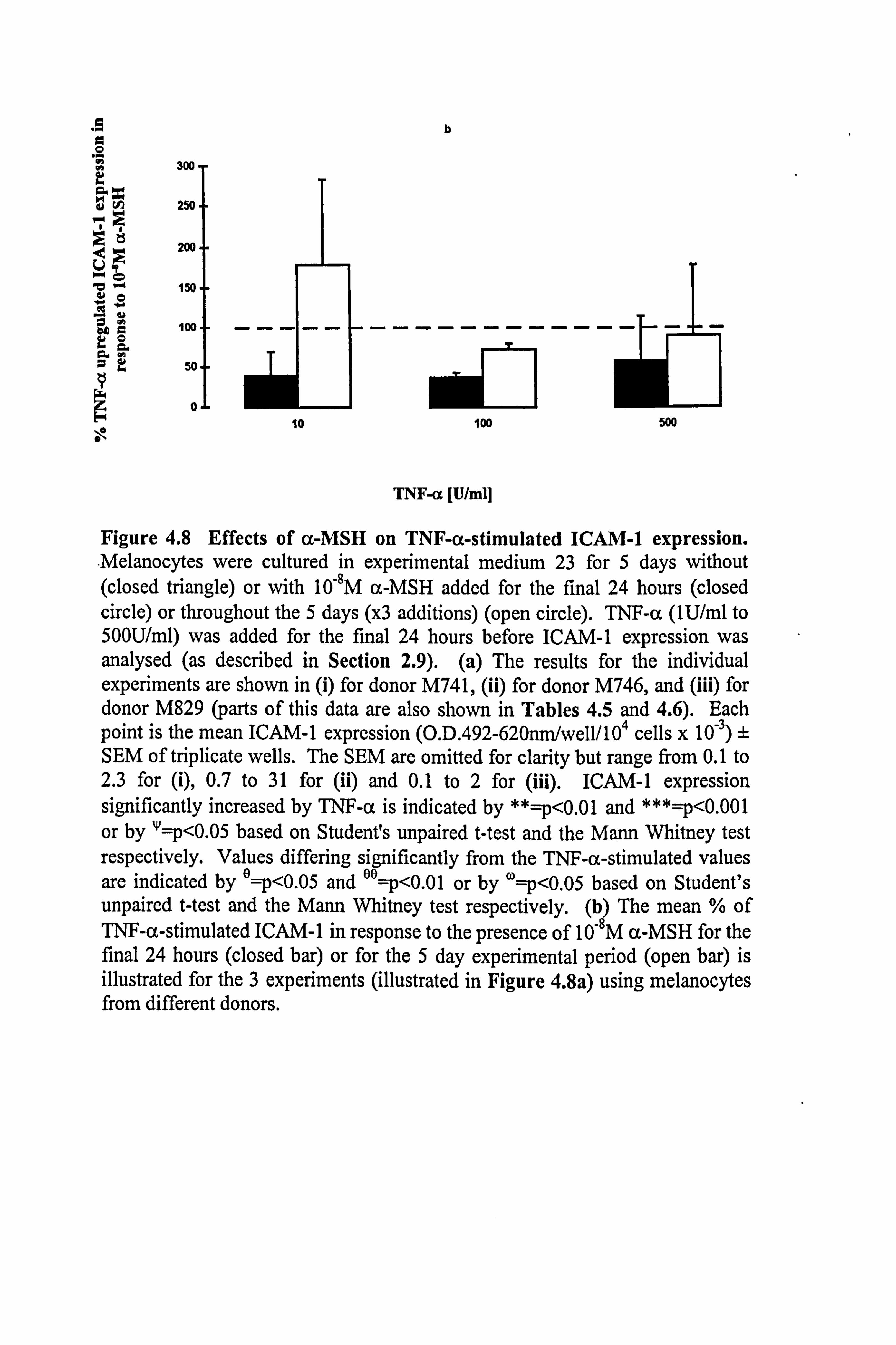
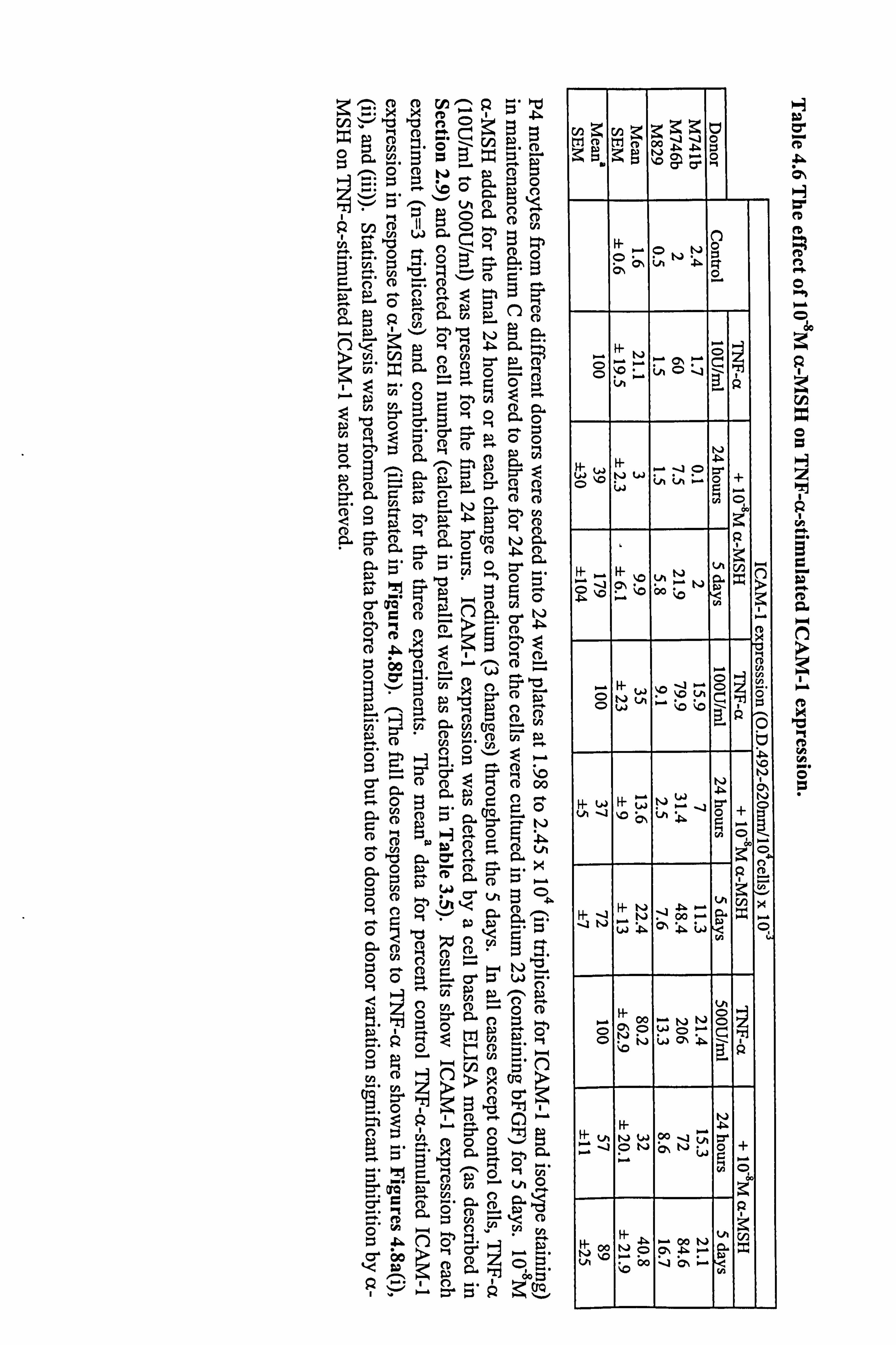
ICAM-1 expression was increased in a dose dependent manner by TNF-a in
melanocytes from 3 different donors (Figure 4.8a and Table 4.6). In all three
experiments, 10,8M a-MSH was able to inhibit the effect of 10-500U/ml TNF-a on
ICAM-1 expression when added for the final 24 hours. However, when cells were
preincubated with 10-8M a-MSH the only inhibition achieved was with 100U/ml TNF-
a. When the normalised data from the 3 experiments were combined (Table 4.6 and
Figure 4.8b), the average degree of inhibition produced by 10-8M a-MSH (when added
for the final 24 hours) was of the order of 50% (58% inhibition at 1OU/ml TNF-(x, 63%
at 100U/ml TNF-a and 43% inhibition at 500U/ml TNF-a) (statistical significance was
not achieved due to donor to donor variability). 10-8M a-MSH, when present for 5 days
reduced 100U/ml TNF-a-stimulated ICAM-1 expression by 28% (statistical
significance not achieved), when based on the mean of the normalised data.
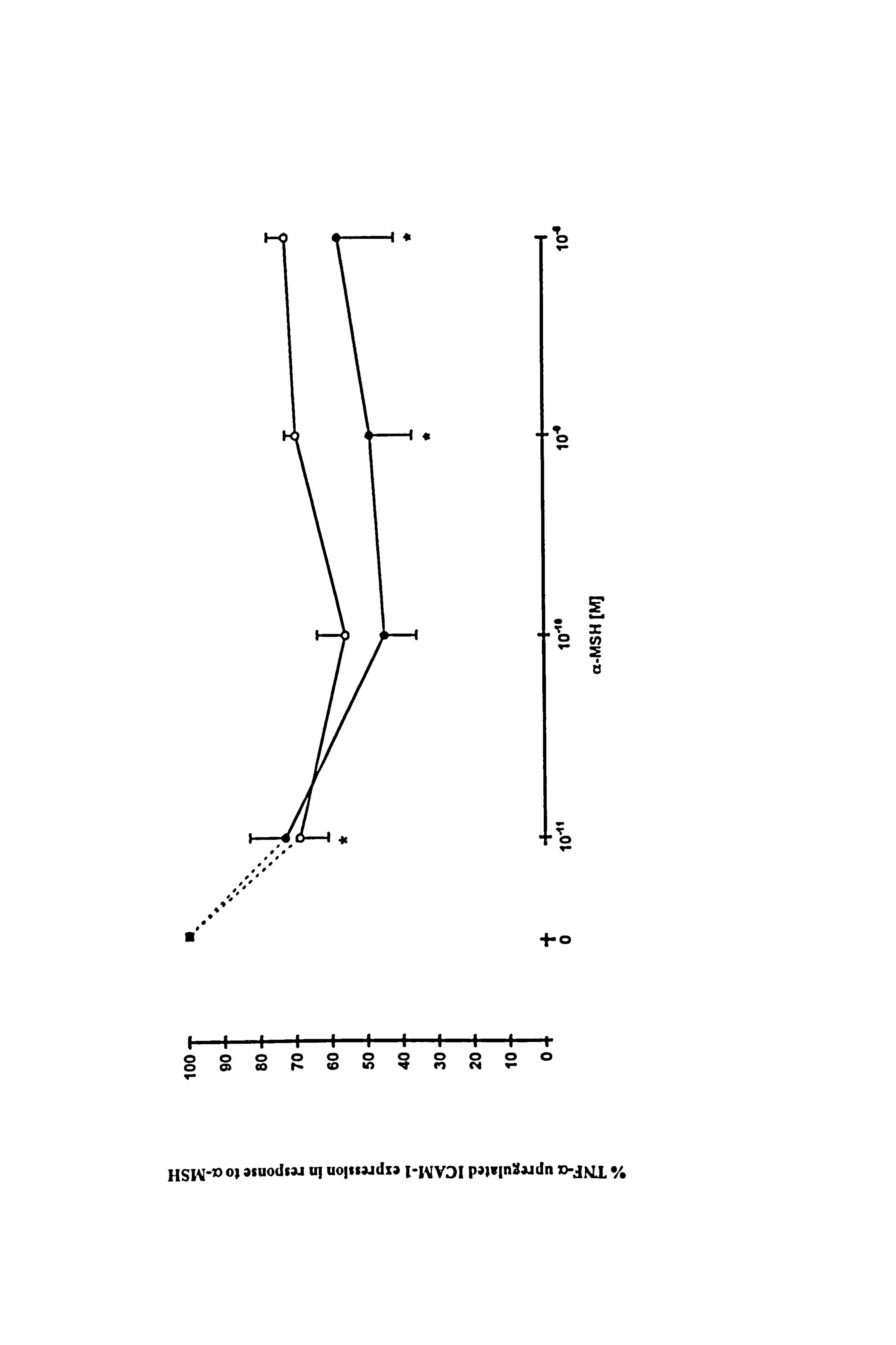
4.3.4 Effect of a-MSH on 100U/ml TNF-a-stimulated ICAM-1 expression in
melanocytes.
a-MSH over a range of concentrations from 10'11M to 10-8M, was able to inhibit
ICAM-1 expression in response to 100U/ml TNF-a (Table 4.5b and Figure 4.9). The
greatest reduction (of 55%) was seen when (x-MSH at 10'10M was present for the final
24 hours (just failing to achieve statistical significance, p<0.0697). However, when the
data from 8 experiments were taken into consideration, 10-8M a-MSH had a slightly
greater inhibition of 57% (Figure 4.10 and Table 4.5b). Inhibition achieved with a-
MSH present throughout the 5 days was not greater than that seen when a-MSH was
present for the last 24 hours only.
m(i)
25
20
16
10
6
0
K . ^, 250
2DO
150
O' A
100
60
H w
0
0
S(U)
alin 18
16
14
12
10
8
6
4
2
0
TNF-a JU/ml]
k.
v
10 100 1000
01 10 100 1000
01 10 lug 1000
O
ge 300
O.. 250
200
`"' 150 ww
aýi ö 100
a- a
W 50
o
0 10
b
------------
m[3 i-
100 500
TNF-a. [U/mll
Figure 4.8 Effects of a-MSH on TNF-a-stimulated ICAM-1 expression. Melanocytes were cultured in experimental medium 23 for 5 days without (closed triangle) or with 10-8M a-MSH added for the final 24 hours (closed circle) or throughout the 5 days (x3 additions) (open circle). TNF-a (lU/ml to 500U/ml) was added for the final 24 hours before ICAM-1 expression was analysed (as described in Section 2.9). (a) The results for the individual experiments are shown in (i) for donor M741, (ii) for donor M746, and (iii) for donor M829 (parts of this data are also shown in Tables 4.5 and 4.6). Each point is the mean ICAM-1 expression (O. D. 492-620nni/well/l04 cells x 10-3) ± SEM of triplicate wells. The SEM are omitted for clarity but range from 0.1 to 2.3 for (i), 0.7 to 31 for (ii) and 0.1 to 2 for (iii). ICAM-1 expression significantly increased by TNF-a is indicated by **=p<0.01 and ***=p<0.001 or by '1=p<0.05 based on Student's unpaired t-test and the Mann Whitney test respectively. Values differing significantly from the TNF-a-stimulated values are indicated by e=p<0.05 and eA=p<0.01 or by w=p<0.05 based on Student's unpaired t-test and the Mann Whitney test respectively. (b) The mean % of TNF-a-stimulated ICAM-1 in response to the presence of 10-8M a-MSH for the final 24 hours (closed bar) or for the 5 day experimental period (open bar) is illustrated for the 3 experiments (illustrated in Figure 4.8a) using melanocytes from different donors.
ýO cD N_ pý (D R
CD t2, (D CD w
c2 Eý >u 0 --Z (D 0
0 (D 05
0 Ef
(D CD CD -Oh ºd ý g.
n ,z -1 8
CZý Z- 00
CD CD
.,, v, Aý pNn cD C Äý Ö ?+dÖc.
CD
(D feCD IM- äa C pc cD 0 "ý-
..
C CD
Gew
p-h 04 (D (D ep (D
'oho CD 9Pn
CD (D v vÖOi (ýq
n rf'
On" 12
r. (D '- (D CD CD 00
r- CL Z. su 0
G0 Orr 0- 2D
CD Hr* o (D O CD LA oºýs ao" -a
ö"aa (D CD 0
C i.
0 (D r"
o CCD E O
ND
O C: 2.0
ý. R `ý Cr1v' Iºt
og Z- rCD s-. 4 s.. 4 rn
ýz R ,r ,ý Gý º--" 0 re. CD 0
' orä' aý N-N (1,1
rn "o
cý öya
d oo (D C> .. ýC Aý Op
-O (v trj 0 Do -141. . 4.0 4ý. 4ý t-j &0- o
0 .r
FI
(J
Fý' W ý-. -1 O p. WO ýD NWN (ý rO WM~ O
v 1_
öýýGý~0 xä
00
IZ
Q0
d
týlýJ W N~J Ö+
vý O Öo ~
ci Q 2.
17 .
I+ N P. l. + t" cn % H ý1 _Nv oo ... a xi .. JNWPWwO
H
kA
ý-+ Pr
pp NNÖ NOWO º- ci
H- 'LA NW Po -1N s -4 ON CN wO+
r-+ WM º+
wO
Q
H- oo NO 4- 00 1,4 Oý A r+ ý` `rýi
t! ý " 00 -l C' rr
w
H
. p. C'
eD W
IF 0
t O r
9 c
tl
I L)14 '0
,'
' 1-0
9-
HSNI-ic) o; asuodui uj uolsaadza i-WV3i Palvinäaidn D-JN. L %
cu +ý
4ö i 'ßo4
G. 0 0
51 oC "_" 0 tu ..,
0'- 2 ä2 cr,
U ca ° -CJ ö rA
0G hö c3 a' x 2,
,7Ö "2 $. te °' 0 z, 0 ä"aýi
UN xv 110 Z. 4 -d DO 9)
aý p,
U"0 0 w
Gtr bbNOý
HZ -n
oý. r=
1 4)
N bA Ö
0 vUi
Co
-"d tJ44 "3 b C) -
*II cii "0 CU *
4. w Ü
C Ici
3ý P. 4 U vi el V)
"ý
-ý v Q en 6-
o*
All ,u ai
uJ "d
-4 ýe
G) UUß. i/ "-ý
U Q. O\ eý N E"ý Ü
146
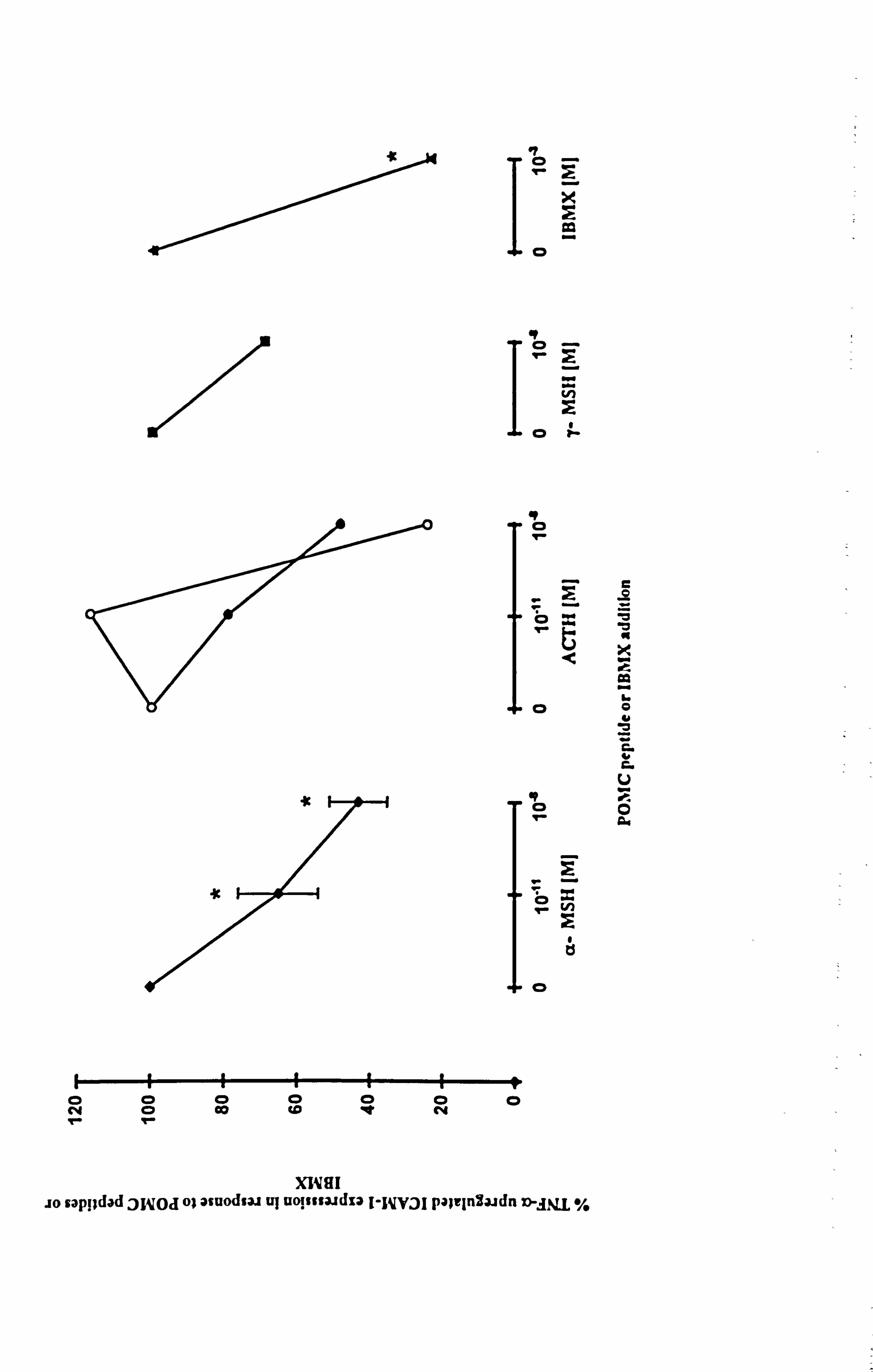
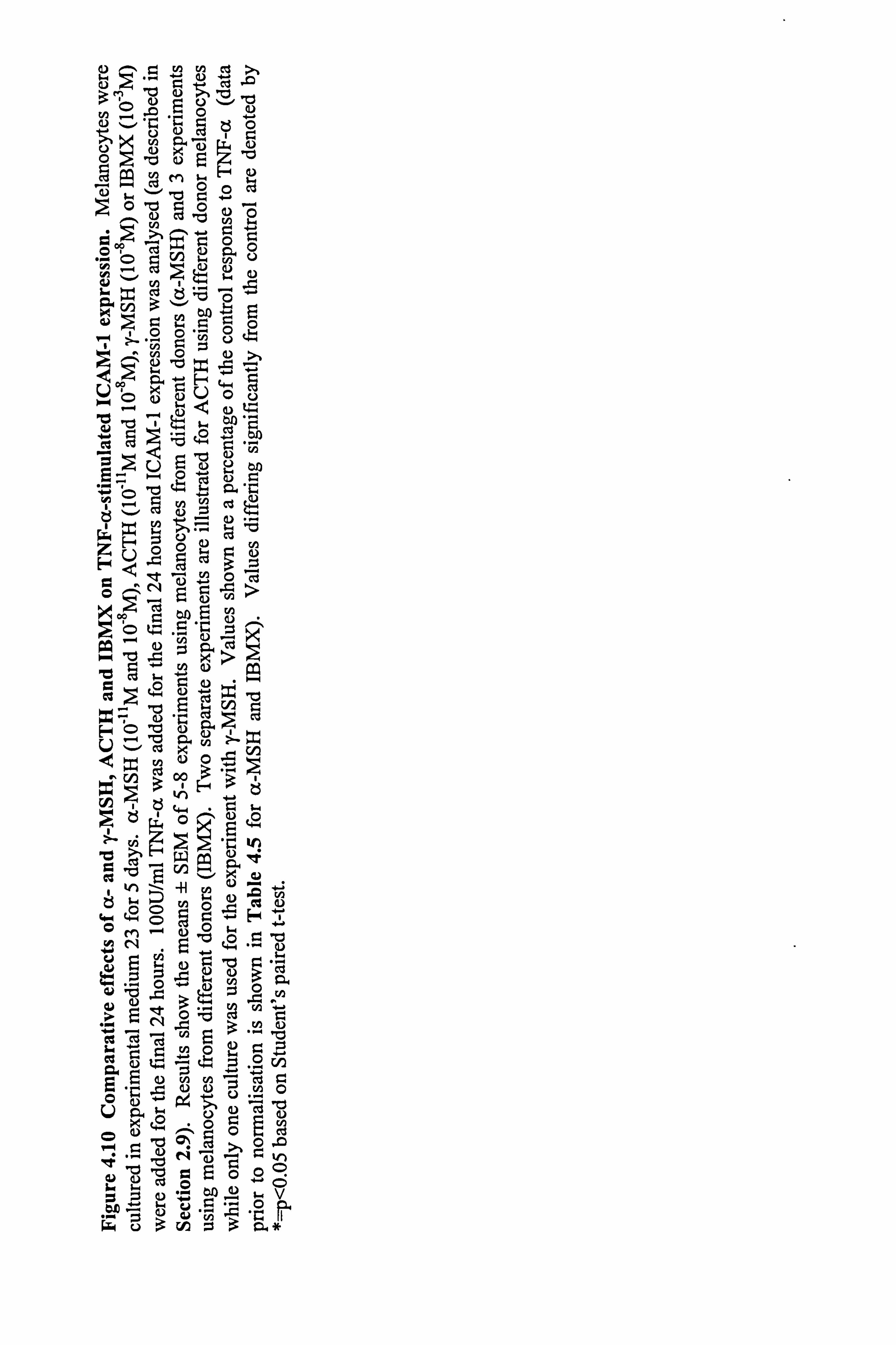
4.3.5 Comparison of the effect of a-MSH, y-MSH, ACTH and IBMX on 100U/ml
TNF-a-stimulated ICAM-1 expression in melanocytes.
The effects of ACTH and y-MSH on TNF-a-stimulated ICAM-1 expression were
compared to the effects of a-MSH to explore the possibility that all POMC-peptides
have immunomodulatory properties. IBMX was included in the study to examine the
possible role of the cAMP/PKA intracellular signalling system in modulating the effect.
The preliminary data for 10-11M ACTH and 10,8M y-MSH demonstrate that they are less
potent than a-MSH (at the respective concentrations) while 10-8M ACTH had similar
potency to 10-8M a-MSH (Figure 4.10). IBMX at 10-3M was a more potent inhibitor of
TNF-a-stimulated ICAM-1 expression than a-MSH and reduced TNF-a-stimulated
ICAM-1 expression by 77% (p<0.05) and 94% (p<0.05) when present for the final 24
hours (Figure 4.10 and Table 4.5b) and for 5 days (Table 4.5b) respectively. Unlike
a-MSH, IBMX showed a greater reduction of TNF-a-stimulated ICAM-1 expression
when present for the longer time period.
The data show that all the POMC-peptides examined appear to have immunomodulatory
activity although to varying extents. The results with IBMX indicate that the inhibition
of TNF-a-stimulated ICAM-1 expression may be working through the cAMP
intracellular signalling pathway.
4.3.6 Effect of a-MSH on IFN-y-stimulated ICAM-1 expression.
A role for a-MSH in opposing TNF-a-stimulated ICAM-1 expression has been
demonstrated in the previous sections. The next part of the study was to examine if a-
MSH could oppose other cytokines that modulate ICAM-1 expression. In this
preliminary study, IFN-y, a strong modulator of ICAM-1 expression was examined.
Melanocytes from 6 different donors were cultured in maintenance medium C and then
under experimental medium 23 as in previous sections. Flow cytometry (as described in
Section 2.10) was used to analyse ICAM-1 expression in response to 10-8M a-MSH
and/or 100U/ml IFN-y (mean results illustrated in Figure 4.11). IFN-y significantly
07
rl
co
14 CD r
r..
N
T- co
49 0
C
of 0
öz N
tl
0
NÖ CO Co 'e c4
Ir- 1-
wM
wV
rV
ry
W
V
C
7
V
ur
0 a
XWSI lo saps; dad jylpd o; asuodsm uJ uo! $ssa, idza I-icvai pa; iinHaadn n-jLq %
0- to o
>' 3
O 0 M
0 p
r U
U p "d
y G) b U ö
0 s.. ý cti Ö - Ö
U pu cn
Co 9
Q
Q)
O
vi 'Z `/ ý Cß�4
U H 4)
r = = r- ö
ý"ý1 0 C) -v 0 *
V ö ý " ' I
H
e c
W
e "
w W 11-4 Jl "~- H 'Lý
z u H 0 00 Co
I i oo r i ^
b g
H
tj4 9) '5 H 0 ) ri v--4 C x ul 3 v, E- 3 t
ý3 ý ö^ aýi ö
W "ý "d v
r, 0 H
" .,
4" p "U c02 124 O
C b i+ Q O y0., ýý.
O "ý V ß 'r ý U ä ö ý
ý iý E ÖOO Cý y j
U .ý
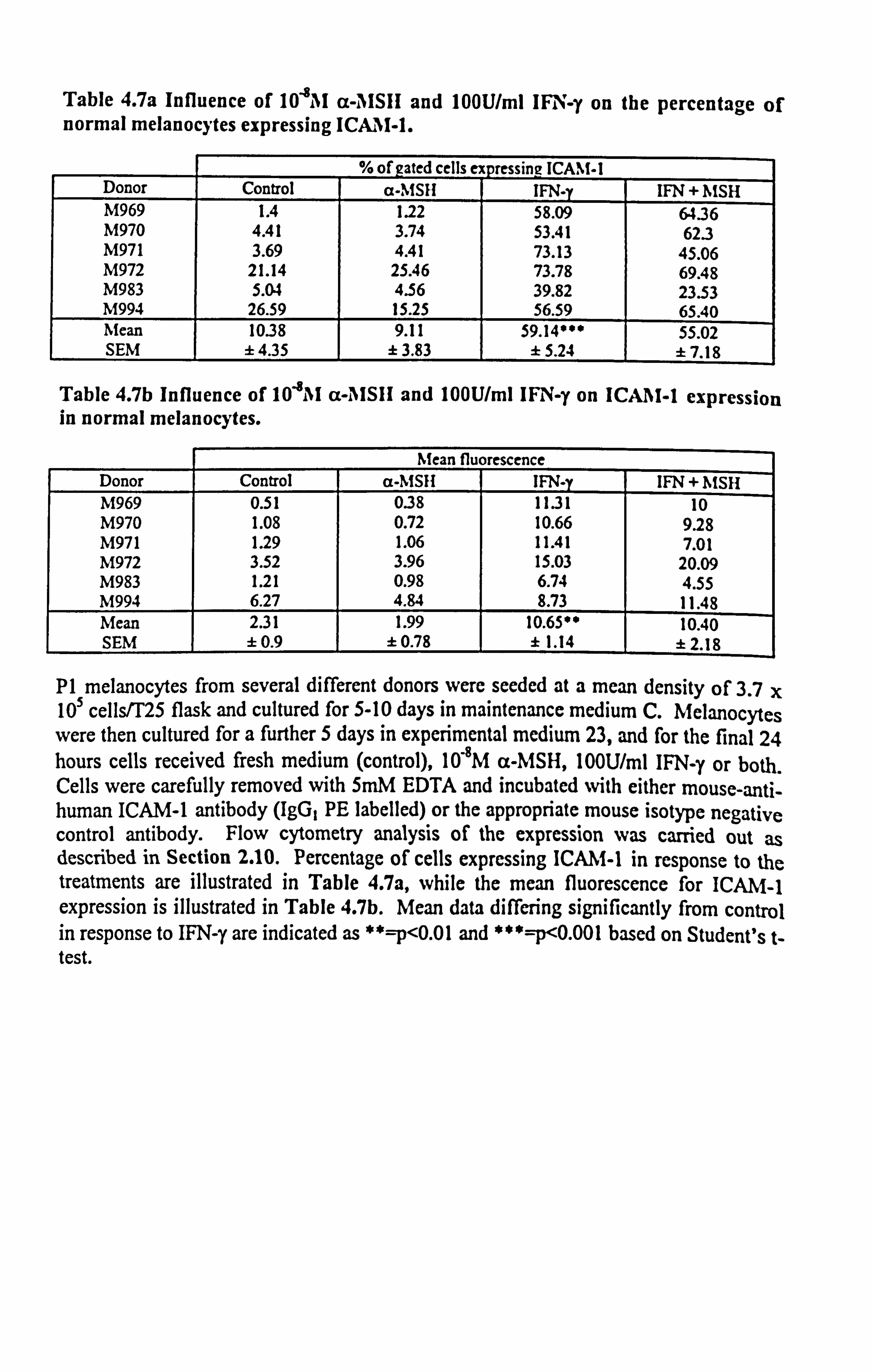
Table 4.7a Influence of 10"8AI cc-, IISIH and 100U/ml IFN-y on the percentage of normal melanocytes expressing ICAM-1.
% of gated cells ex pressing ! CAM-1 Donor Control a-MSII IFN-y IFN + MSH M969 1.4 1.22 58.09 64.36 M970 4.41 3.74 53.41 62.3 M971 3.69 4.41 73.13 45.06 M972 21.14 25.46 73.78 69.48 M983 5.04 4.56 39.82 23.53 M994 26.59 15.25 56.59 65.40 Mean 10.38 9.11 59.14*** 55.02 SEM ±4.35 ± 3.83 ±5.24 ±7.18
Table 4.7b Influence of 104M a-AISIi and 100U/ml IFNI -y on ICA11I-1 expression in normal melanocytes.
Mean fluorescence Donor Control a-MSH IFN-y IFN + 11MSH M969 0.51 038 1131 10 M970 1.08 0.72 10.66 9.28 M971 1.29 1.06 11.41 7.01 M972 3.52 3.96 15.03 20.09 M983 1.21 0.98 6.74 4.55 M994 6.27 4.84 8.73 11.48 Mean 2.31 1.99 10.6500 10.40 SEM ± 0.9 ± 0.78 ± 1.14 ±2.18
P1 melanocytes from several different donors were seeded at a mean density of 3.7 x 10⁵ cells/T25 flask and cultured for 5-10 days in maintenance medium C. Melanocytes were then cultured for a further 5 days in experimental medium 23, and for the final 24 hours cells received fresh medium (control), 10"8M a-MSH, l00U/ml IFN-y or both. Cells were carefully removed with 5mM EDTA and incubated with either mouse-anti- human ICAM-1 antibody (IgG, PE labelled) or the appropriate mouse isotype negative control antibody. Flow cytometry analysis of the expression was carried out as described in Section 2.10. Percentage of cells expressing ICAM-1 in response to the treatments are illustrated in Table 4.7a, while the mean fluorescence for ICAM-1 expression is illustrated in Table 4.7b. Mean data differing significantly from control in response to IFN-y are indicated as **=p<0.01 and ***=p<0.001 based on Student's t- test.
..
d U Co
as a
ed týD
\e e
70
so
40--
30--
20--
10-
0 control ar. MSH IFN-y a- MSH + IFN-T
14
12
10,
A m 8
G 0
6 eý n A Cý
4.
2
0
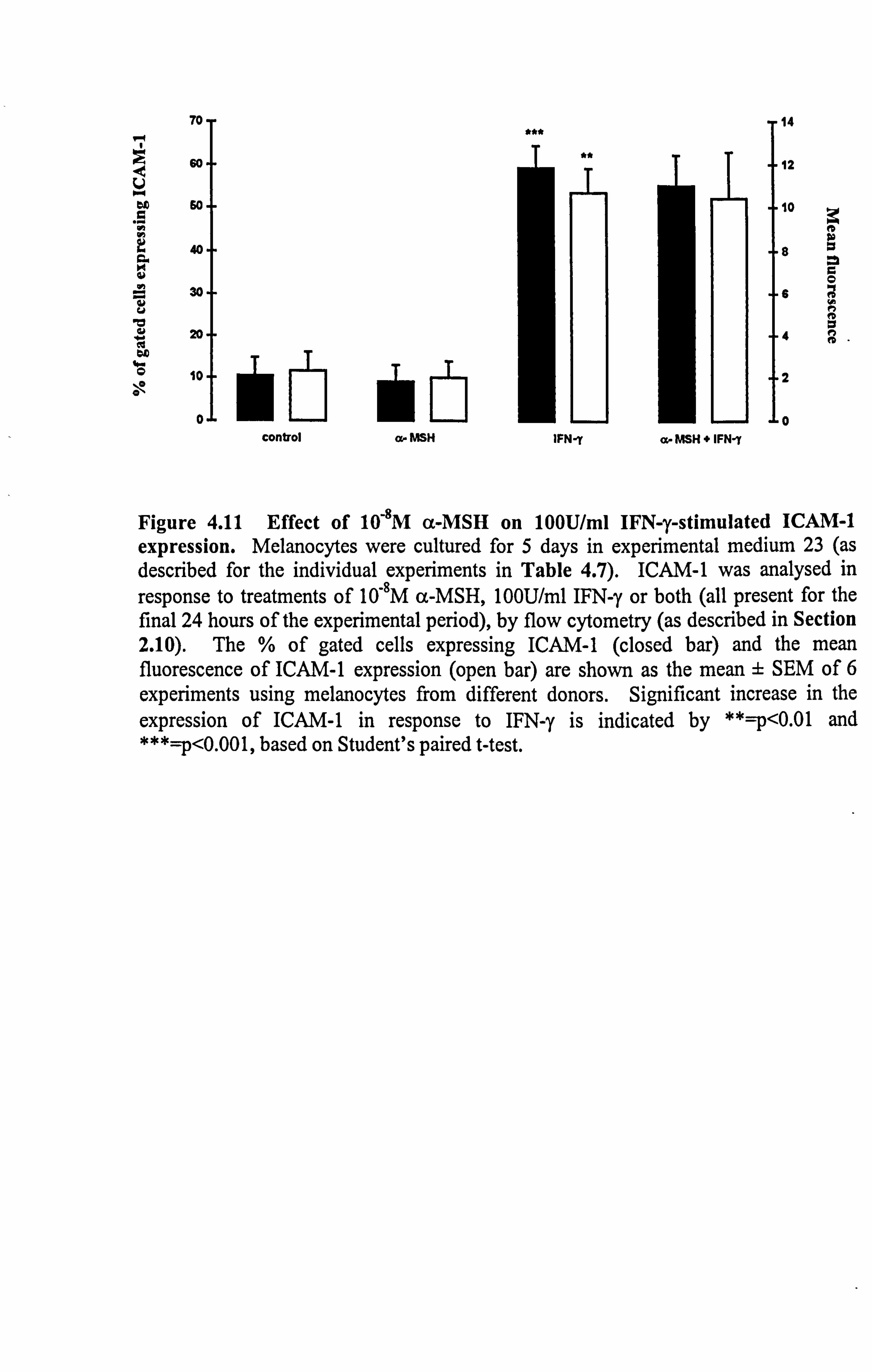
Figure 4.11 Effect of 10-8M a-MSH on 100U/ml IFN-y-stimulated ICAM-1 expression. Melanocytes were cultured for 5 days in experimental medium 23 (as described for the individual experiments in Table 4.7). ICAM-1 was analysed in
response to treatments of 10-8M a-MSH, 10OU/ml IFN-y or both (all present for the final 24 hours of the experimental period), by flow cytometry (as described in Section 2.10). The % of gated cells expressing ICAM-1 (closed bar) and the mean fluorescence of ICAM-1 expression (open bar) are shown as the mean ± SEM of 6 experiments using melanocytes from different donors. Significant increase in the expression of ICAM-1 in response to IFN-y is indicated by **=p<0.01 and ***=p<0.001, based on Student's paired t-test.
147
increased the number of cells expressing ICAM-1 (p<0.001) and stimulated ICAM-1
expression by 4.6 fold (p<0.01) (in agreement with results in Section 4.2.2). 10,8M a-
MSH had no significant effects on constitutive ICAM-1 expression but was able . to
inhibit IFN-y-stimulated ICAM-1 expression in some donor melanocytes while in others
it enhanced IFN-y-stimulated ICAM-1 expression (Table 4.7). Overall, 10"8M a-MSH
did not affect IFN-y-stimulated ICAM-1 expression.
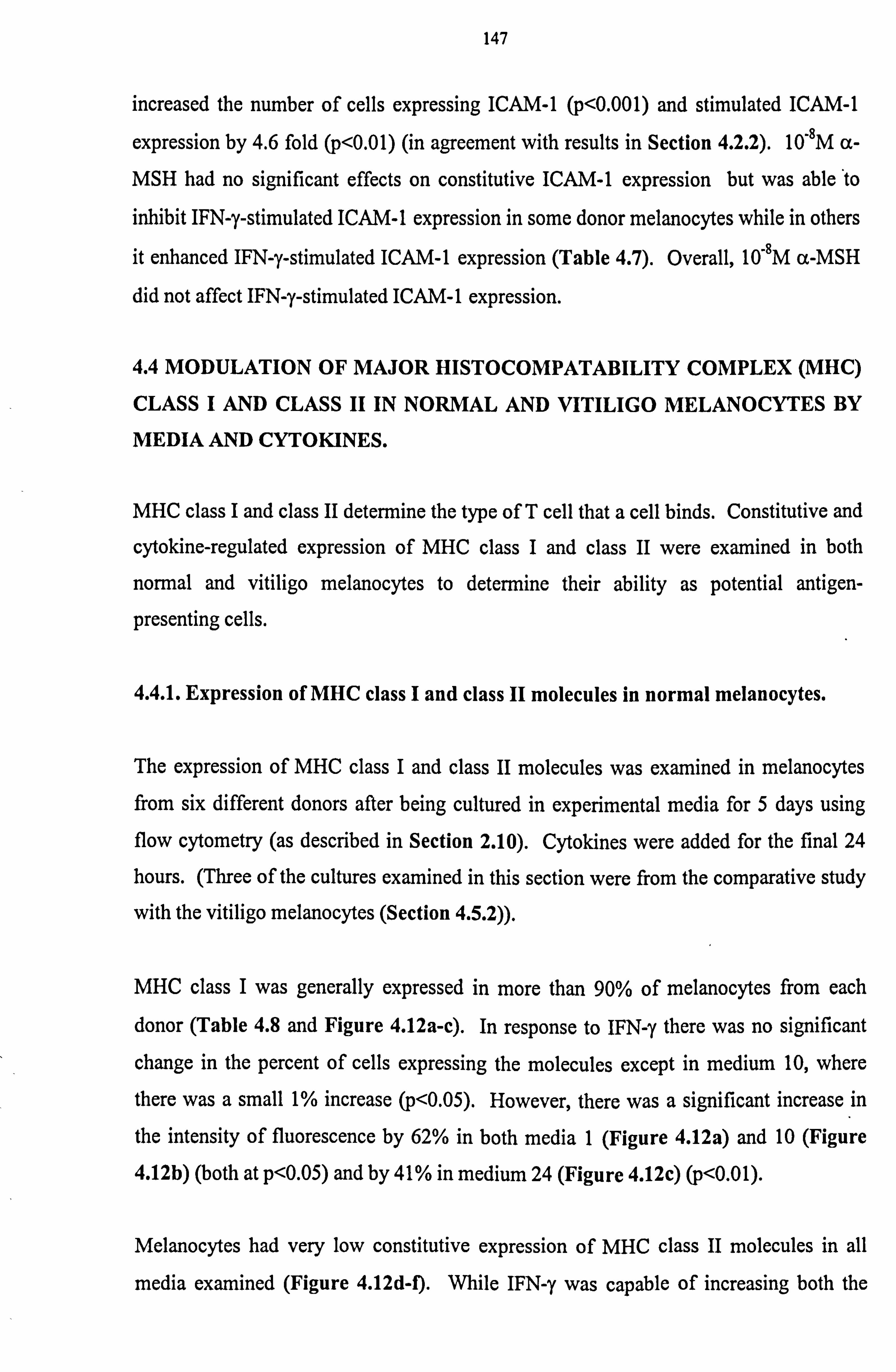
4.4 MODULATION OF MAJOR HISTOCOMPATABILITY COMPLEX (MHC)
CLASS I AND CLASS II IN NORMAL AND VITILIGO MELANOCYTES BY
MEDIA AND CYTOKINES.
MHC class I and class II determine the type of T cell that a cell binds. Constitutive and
cytokine-regulated expression of MHC class I and class II were examined in both
normal and vitiligo melanocytes to determine their ability as potential antigen-
presenting cells.
4.4.1. Expression of MHC class I and class II molecules in normal melanocytes.
The expression of MHC class I and class II molecules was examined in melanocytes from six different donors after being cultured in experimental media for 5 days using flow cytometry (as described in Section 2.10). Cytokines were added for the final 24
hours. (Three of the cultures examined in this section were from the comparative study
with the vitiligo melanocytes (Section 4.5.2)).
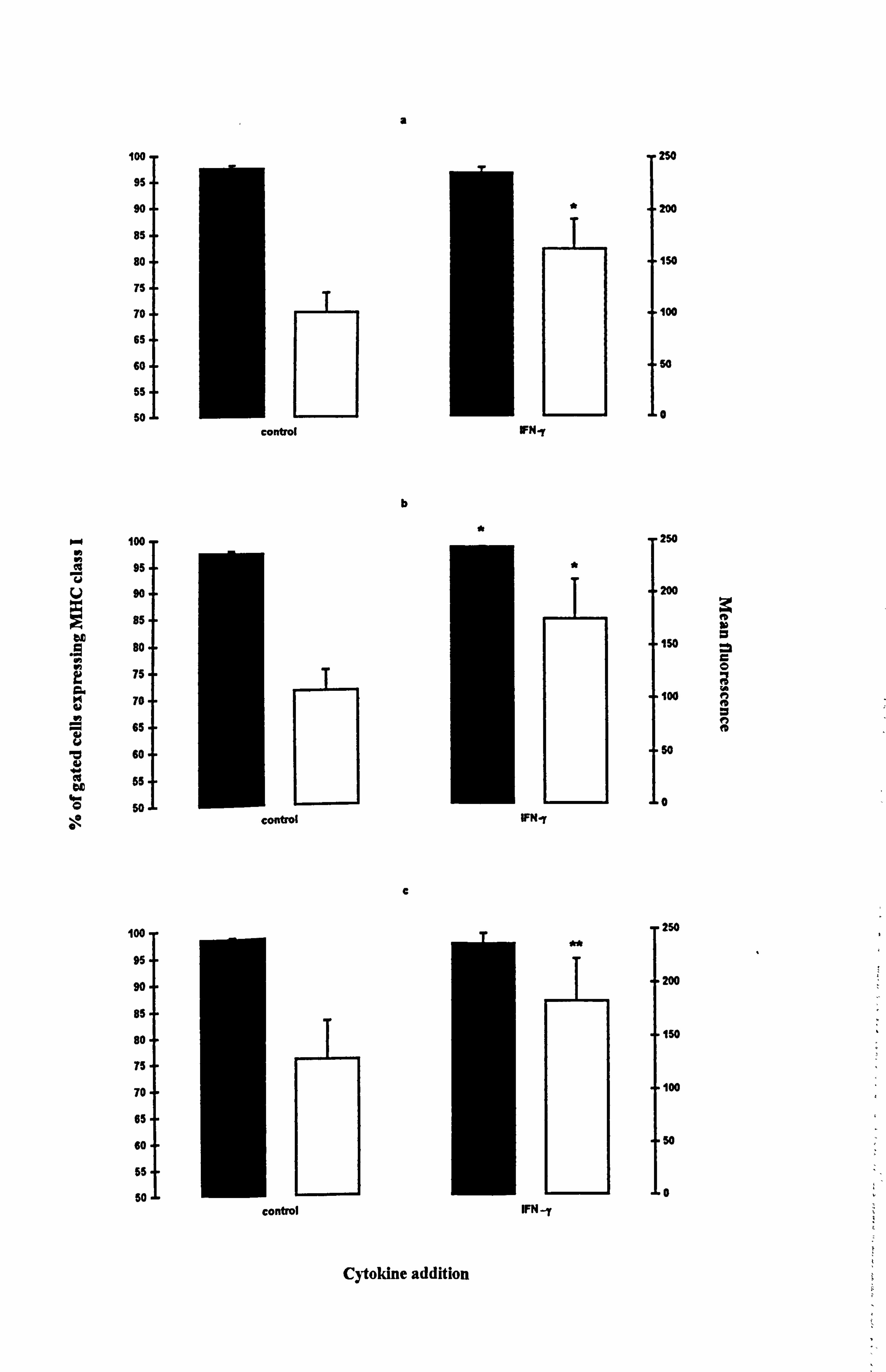
MHC class I was generally expressed in more than 90% of melanocytes from each
donor (Table 4.8 and Figure 4.12a-c). In response to IFN-y there was no significant
change in the percent of cells expressing the molecules except in medium 10, where
there was a small 1% increase (p<0.05). However, there was a significant increase in
the intensity of fluorescence by 62% in both media 1 (Figure 4.12a) and 10 (Figure
4.12b) (both at p<0.05) and by 41% in medium 24 (Figure 4.12c) (p<0.01).
Melanocytes had very low constitutive expression of MHC class II molecules in all
media examined (Figure 4.12d-I). While IFN-y was capable of increasing both the
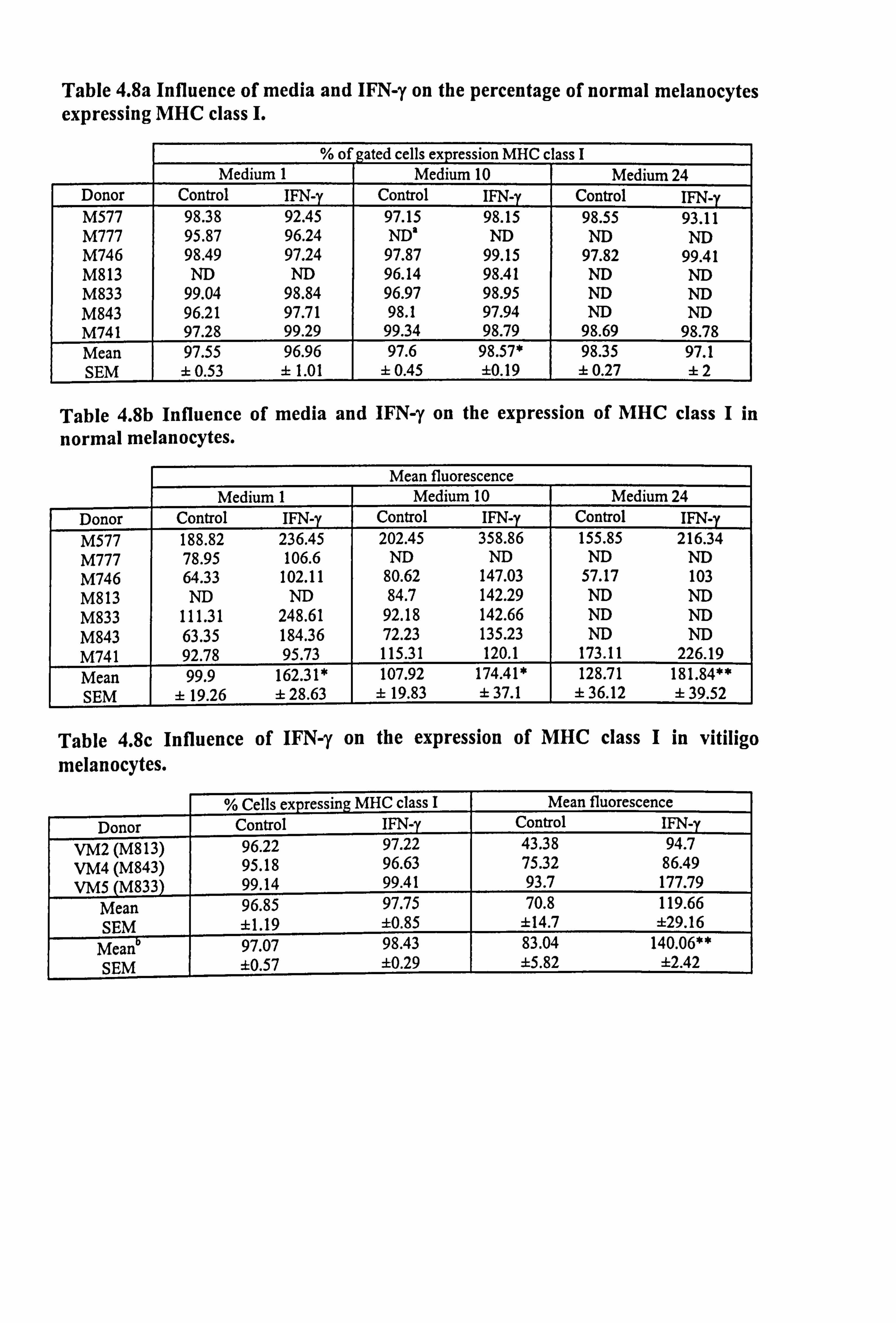
Table 4.8a Influence of media and IFN-y on the percentage of normal melanocytes expressing MHC class I.
% of gated cells expression MHC c lass I Medium 1 Medium 10 Medium 24
Donor Control IFN-y Control IFN-y Control IFN-y M577 98.38 92.45 97.15 98.15 98.55 93.11 M777 95.87 96.24 ND' ND ND ND M746 98.49 97.24 97.87 99.15 97.82 99.41 M813 ND ND 96.14 98.41 ND ND M833 99.04 98.84 96.97 98.95 ND ND M843 96.21 97.71 98.1 97.94 ND ND M741 97.28 99.29 99.34 98.79 98.69 98.78 Mean 97.55 96.96 97.6 98.57* 98.35 97.1 SEM ± 0.53 ± 1.01 ± 0.45 ±0.19 ± 0.27 ±2
Table 4.8b Influence of media and IFN-y on the expression of MHC class I in
normal melanocytes.
Mean fluorescence Medium 1 Medium 10 Medium 24
Donor Control IFN-y Control IFN-y Control IFN-y 188.82 236.45 202.45 358.86 155.85 216.34
M777 78.95 106.6 ND ND ND ND M746 64.33 102.11 80.62 147.03 57.17 103 M813 ND ND 84.7 142.29 ND ND M833 111.31 248.61 92.18 142.66 ND ND M843 63.35 184.36 72.23 135.23 ND ND M741 92.78 95.73 115.31 120.1 173.11 226.19 Mean 99.9 162.31* 107.92 174.41* 128.71 181.84** SEM ± 19.26 ± 28.63 ± 19.83 ± 37.1 ± 36.12 ± 39.52
Table 4.8c Influence of IFN-y on the expression of MHC class I in vitiligo melanocytes.
% Cells expressing MHC class I Mean fluorescence Donor Control IFN-y Control IFN-
VM2 (M813) 96.22 97.22 43.38 94.7 VM4 (M843) 95.18 96.63 75.32 86.49 VMS (M833) 99.14 99.41 93.7 177.79
Mean 96.85 97.75 70.8 119.66 SEM ±1.19 ±0.85 ±14.7 ±29.16
Mean 97.07 98.43 83.04 140.06** SEM ±0.57 ±0.29 ±5.82 ±2.42
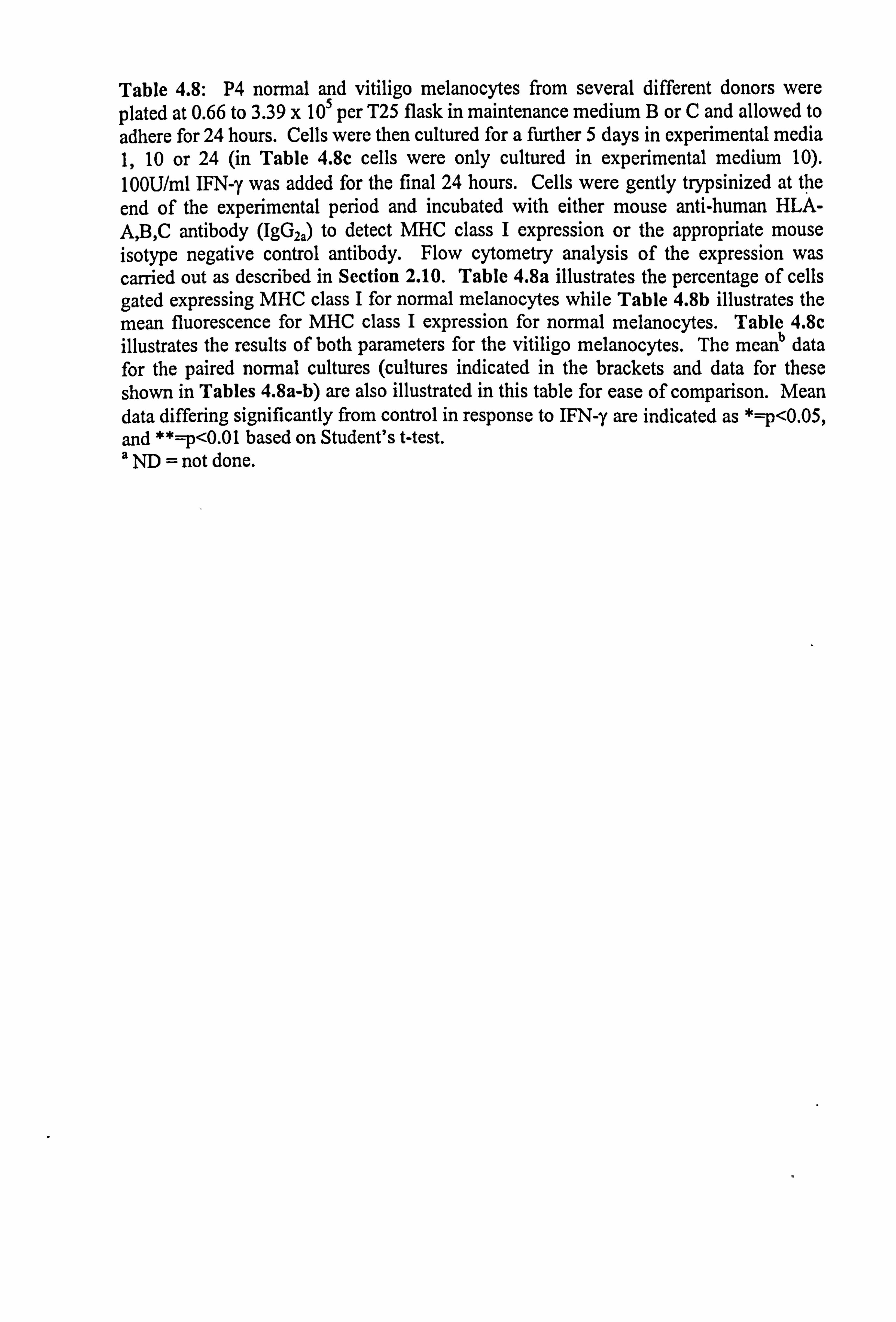
Table 4.8: P4 normal and vitiligo melanocytes from several different donors were plated at 0.66 to 3.39 x 10⁵ per T25 flask in maintenance medium B or C and allowed to
adhere for 24 hours. Cells were then cultured for a further 5 days in experimental media 1,10 or 24 (in Table 4.8c cells were only cultured in experimental medium 10). 100U/ml IFN-y was added for the final 24 hours. Cells were gently trypsinized at the
end of the experimental period and incubated with either mouse anti-human HLA- A, B, C antibody (IgG2a) to detect MHC class I expression or the appropriate mouse isotype negative control antibody. Flow cytometry analysis of the expression was carried out as described in Section 2.10. Table 4.8a illustrates the percentage of cells gated expressing MHC class I for normal melanocytes while Table 4.8b illustrates the mean fluorescence for MHC class I expression for normal melanocytes. Table 4.8c illustrates the results of both parameters for the vitiligo melanocytes. The meanb data for the paired normal cultures (cultures indicated in the brackets and data for these shown in Tables 4.8a-b) are also illustrated in this table for ease of comparison. Mean data differing significantly from control in response to IFN-y are indicated as * p<0.05, and **=p<0.01 based on Student's t-test. a ND = not done.
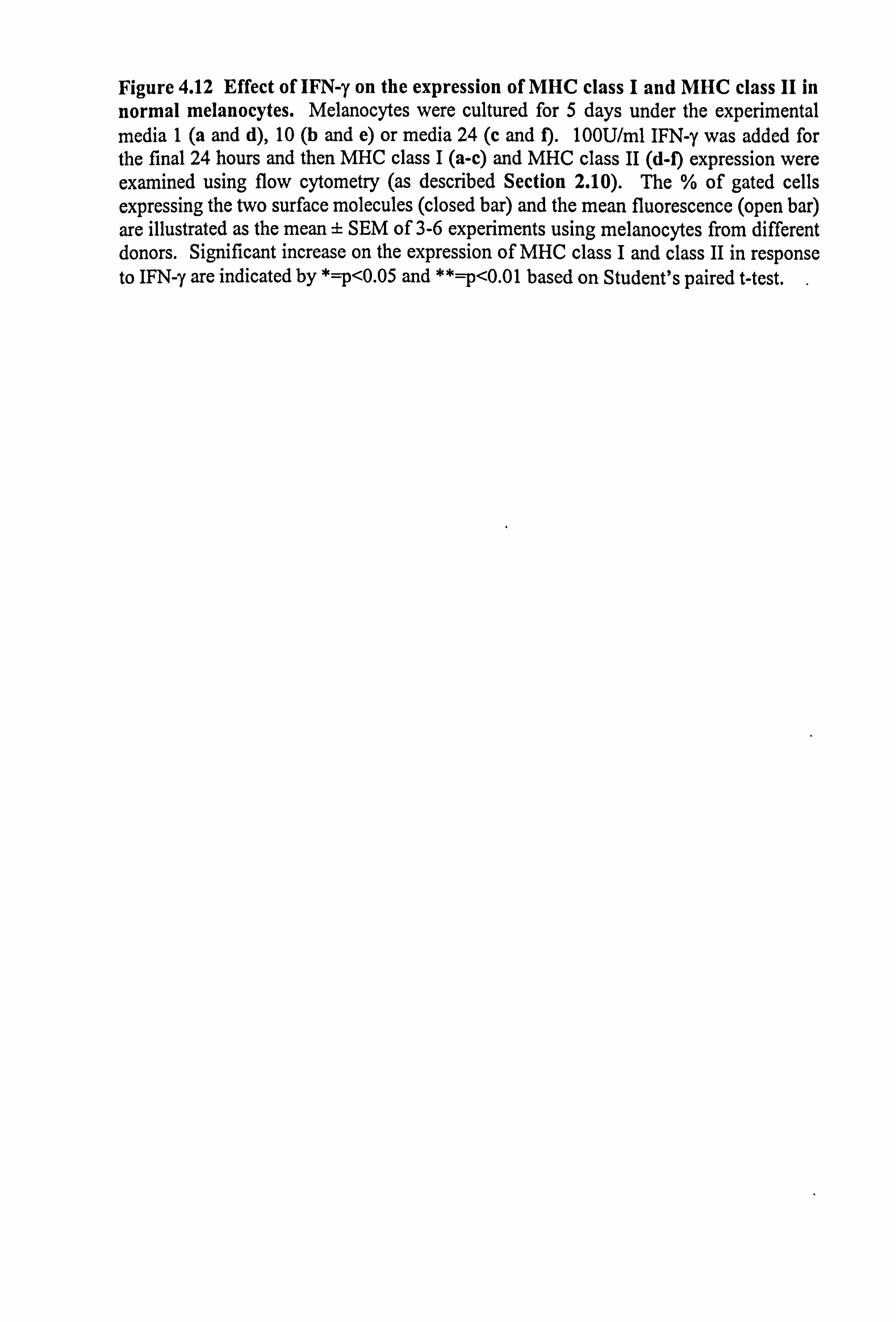
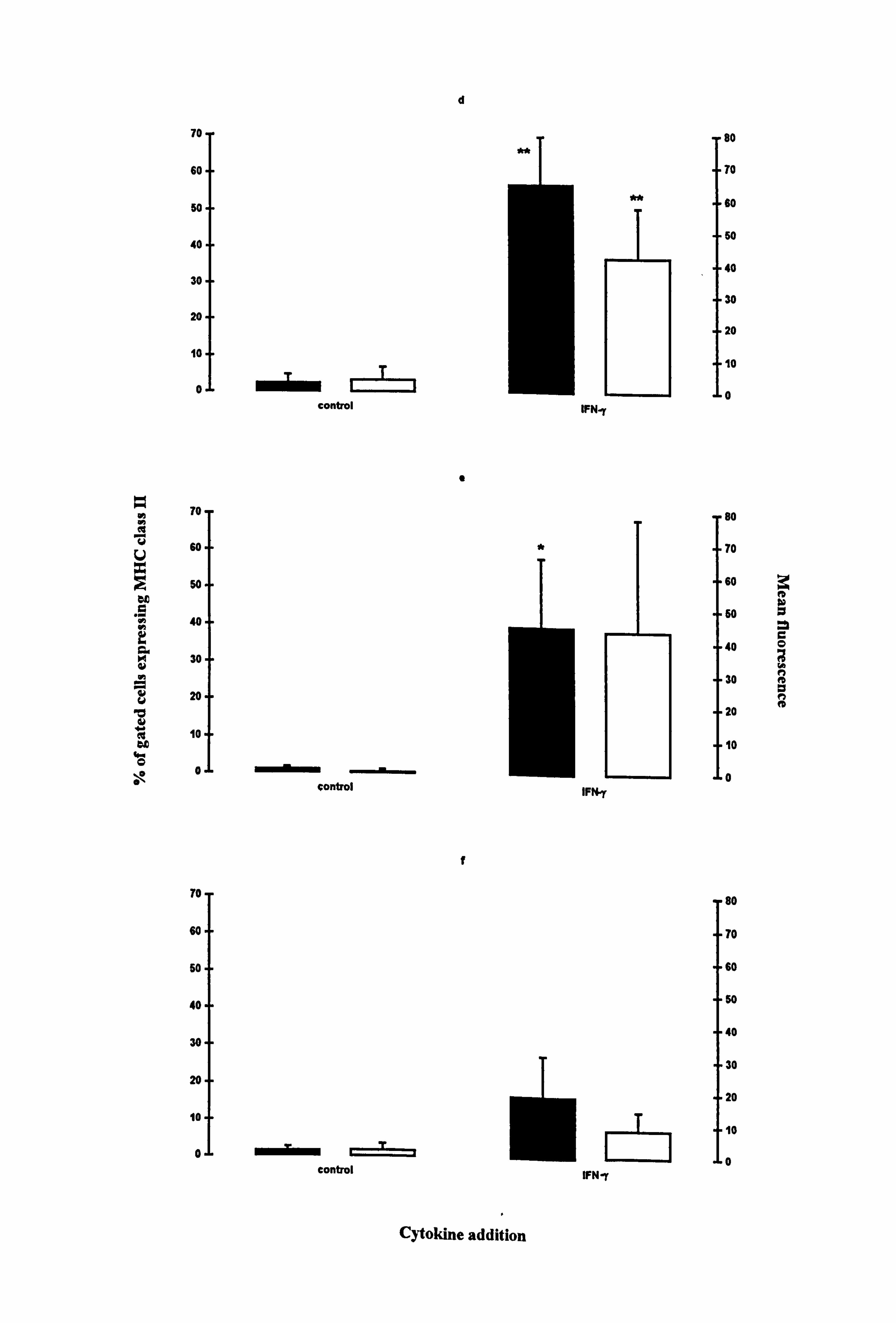
Figure 4.12 Effect of IFN-y on the expression of MHC class I and MHC class II in normal melanocytes. Melanocytes were cultured for 5 days under the experimental media 1 (a and d), 10 (b and e) or media 24 (c and f). 100U/ml IFN-y was added for the final 24 hours and then MHC class I (a-c) and MHC class II (d-f) expression were examined using flow cytometry (as described Section 2.10). The % of gated cells expressing the two surface molecules (closed bar) and the mean fluorescence (open bar) are illustrated as the mean ± SEM of 3-6 experiments using melanocytes from different donors. Significant increase on the expression of MHC class I and class II in response to IFN-y are indicated by *=p<0.05 and ** p<0.01 based on Student's paired t-test. .
mw
V
.9 10
a d h a
b d w oc a. 0 \o 0
goo
s5 90
85
80
75
70
65
60
55
50
100
95
90
85
80
75
70
65
60
55
50
100
95
90
85
80
75
70
65
60
55
50
control
II
coaftol
control
a
b
C
Cytokine addition
w
IFN-y
WNy
IFN -y
w
**
250
200
150
100
50
0
250
200
A IM a ISO 0
100
A
so
0
250
"200
"150
"100
"50
"0 i
r i
h
d
b m
v
.9
a
w u b v .r eý dc 0
\e
70
60
so
40
30
20
10
0
To
60
50
40
30
20
10
0
To
60
50
40
30
20
10
0
mim Lr- T
control
control
WMEIIIIIIIIIIII control
a
f
*
IFNry
IFt y
IFN-y
80
70
80
50
40
30
20
10
0
80
70
60
60
40
30
20
10
0
80
"70
- 60
S0
"40
"30
" 20
10
0
4
c 0 eý n A p n A
Cytokine addition
148
number of cells expressing the molecule and intensity of fluorescence, statistical
significance was not achieved under all media conditions due to large donor to donor
variability (Table 4.9). The number of cells expressing MHC class II were significantly
increased 31 fold in medium 1 (p<0.01) (Figure 4.12d) and 48 fold in medium 10
(p<0.05) (Figure 4.12e). The intensity of expression was significantly increased 16 fold
in medium 1 (p<0.01) (Figure 4.12d).
The expression of MHC class I and class II in donor melanocytes M777 was examined
in medium 1, in response to IFN-y, TNF-a and IL-1a. Both IFN-y (Figure 4.13b) and
TNF-a (Figure 4.13c) increased MHC class I expression by 35% and 40% respectively
in comparison to the control (Figure 4.13a) while IL-la reduced the expression slightly
by 15% (Figure 4.13d) (all values based an the mean fluorescence corrected for isotype
staining). IFN-y (Figure 4.13f and Table 4.9b) increased MHC class II expression in
comparison to the control (Figure 4.13e and Table 4.9b) whereas TNF-a and IL-la
(Figures 4.13g and 4.13h respectively) had no effects on MHC class II expression.
The majority of melanocytes in any donor population expressed MHC class I at a high
level while there was low constitutive expression of MHC class II. IFN-y was
influential in stimulating increased expression of MHC class I and MHC class II.
4.4.2. Comparison of MHC class I and class II molecule expression in normal and
vitiligo melanocytes.
Normal and vitiligo melanocytes constitutively expressed MHC class I surface
molecules in medium 10 (Table 4.8 and Figure 4.14). While IFN-y did not alter the
number of melanocytes expressing the MHC class I molecules, the mean fluorescence
was increased by 69% in both normal (p<0.01) and vitiligo melanocytes.
Constitutive expression of the MHC class II was minimal in both normal and vitiligo
melanocytes. IFN-y significantly increased the number of vitiligo melanocytes
expressing the antigen (p<0.05), with an associated increase of 15.86 mean fluorescence
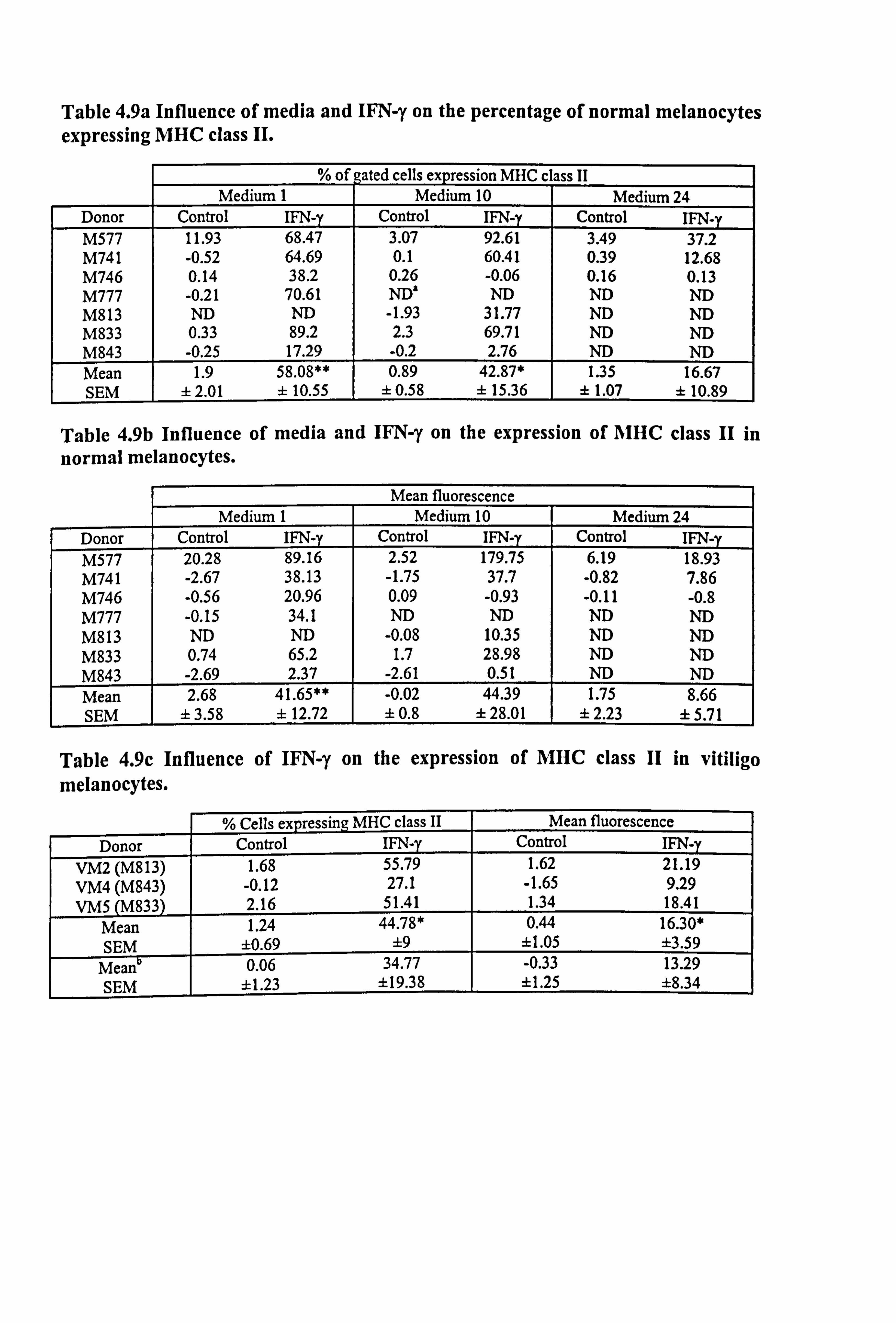
Table 4.9a Influence of media and IFN-y on the percentage of normal melanocytes expressing MHC class II.
% of gated cells expression MHC cl ass II Medium 1 Medium 10 Medium 24
Donor Control IFN-y Control IFN-y Control IFN-y M577 11.93 68.47 3.07 92.61 3.49 37.2 M741 -0.52 64.69 0.1 60.41 0.39 12.68 M746 0.14 38.2 0.26 -0.06 0.16 0.13 M777 -0.21 70.61 ND' ND ND ND M813 ND ND -1.93 31.77 ND ND M833 0.33 89.2 2.3 69.71 ND ND M843 -0.25 17.29 -0.2 2.76 ND ND Mean 1.9 58.08** 0.89 42.87* 1.35 16.67 SEM ± 2.01 ± 10.55 ± 0.58 ± 15.36 ± 1.07 ± 10.89
Table 4.9b Influence of media and IFN-y on the expression of AIHC class II in
normal melanocytes.
Mean fluorescence Medium 1 Medium 10 Medium 24
Donor Control IFN-y Control IFN-y Control IFN-y 20.28 89.16 2.52 179.75 6.19 18.93
M741 -2.67 38.13 -1.75 37.7 -0.82 7.86 M746 -0.56 20.96 0.09 -0.93 -0.11 -0.8 M777 -0.15 34.1 ND ND ND ND M813 ND ND -0.08 10.35 ND ND M833 0.74 65.2 1.7 28.98 ND ND M843 -2.69 2.37 -2.61 0.51 ND ND Mean 2.68 41.65** -0.02 44.39 1.75 8.66 SEM ± 3.58 f 12.72 ± 0.8 ± 28.01 ± 2.23 ± 5.71
Table 4.9c Influence of IFN-y on the expression of MHC class II in vitiligo melanocytes.
% Cells expressing MHC class II Mean fluorescence
Donor Control IFN-y Control IFN-y VM2 (M813) 1.68 55.79 1.62 21.19 VM4 (M843) -0.12 27.1 -1.65 9.29 VMS (M833) 2.16 51.41 1.34 18.41
Mean 1.24 44.78* 0.44 16.30* SEM ±0.69 ±9 11.05 ±3.59
Mean 0.06 34.77 -0.33 13.29 SEM ±1.23 ±19.38 ±1.25 ±8.34
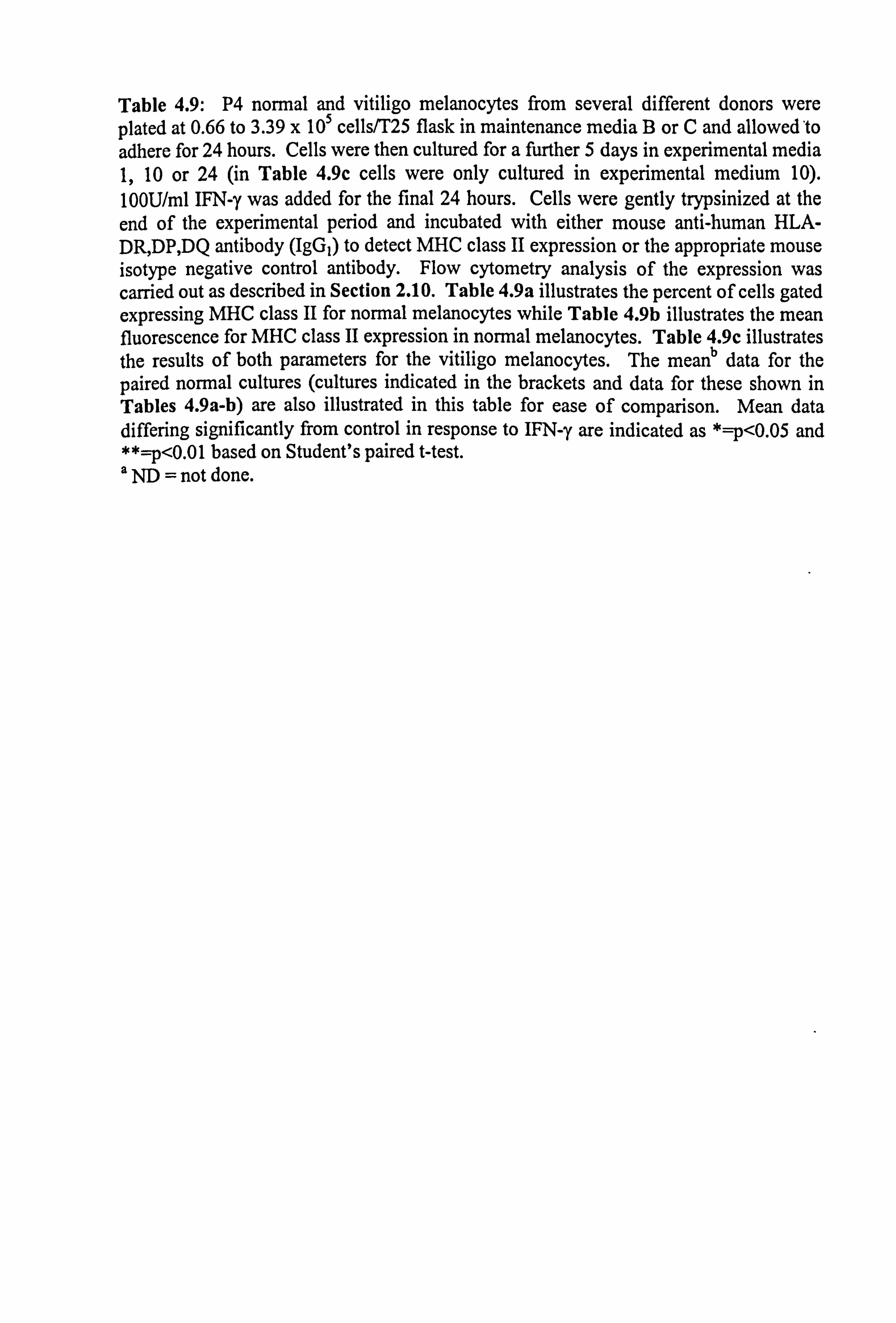
Table 4.9: P4 normal and vitiligo melanocytes from several different donors were plated at 0.66 to 3.39 x 10⁵ cells/T25 flask in maintenance media B or C and allowed'to adhere for 24 hours. Cells were then cultured for a further 5 days in experimental media 1,10 or 24 (in Table 4.9c cells were only cultured in experimental medium 10). 100U/ml IFN-y was added for the final 24 hours. Cells were gently trypsinized at the end of the experimental period and incubated with either mouse anti-human HLA- DR, DP, DQ antibody (IgGI) to detect MHC class II expression or the appropriate mouse isotype negative control antibody. Flow cytometry analysis of the expression was carried out as described in Section 2.10. Table 4.9a illustrates the percent of cells gated expressing MHC class II for normal melanocytes while Table 4.9b illustrates the mean fluorescence for MHC class II expression in normal melanocytes. Table 4.9c illustrates the results of both parameters for the vitiligo melanocytes. The mean data for the paired normal cultures (cultures indicated in the brackets and data for these shown in Tables 4.9a-b) are also illustrated in this table for ease of comparison. Mean data differing significantly from control in response to IFN-y are indicated as *=p<0.05 and **=p<0.01 based on Student's paired t-test. a ND = not done.
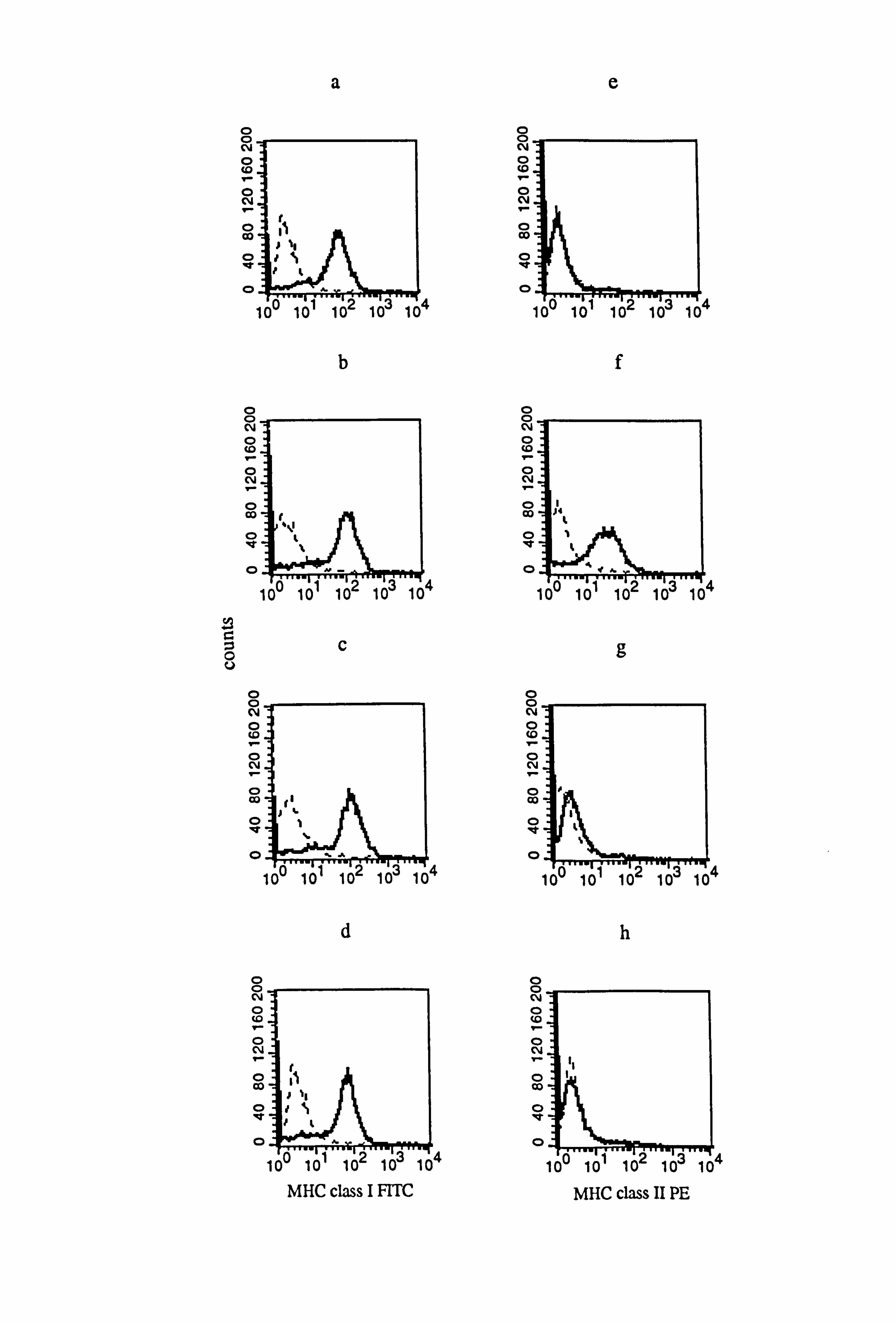
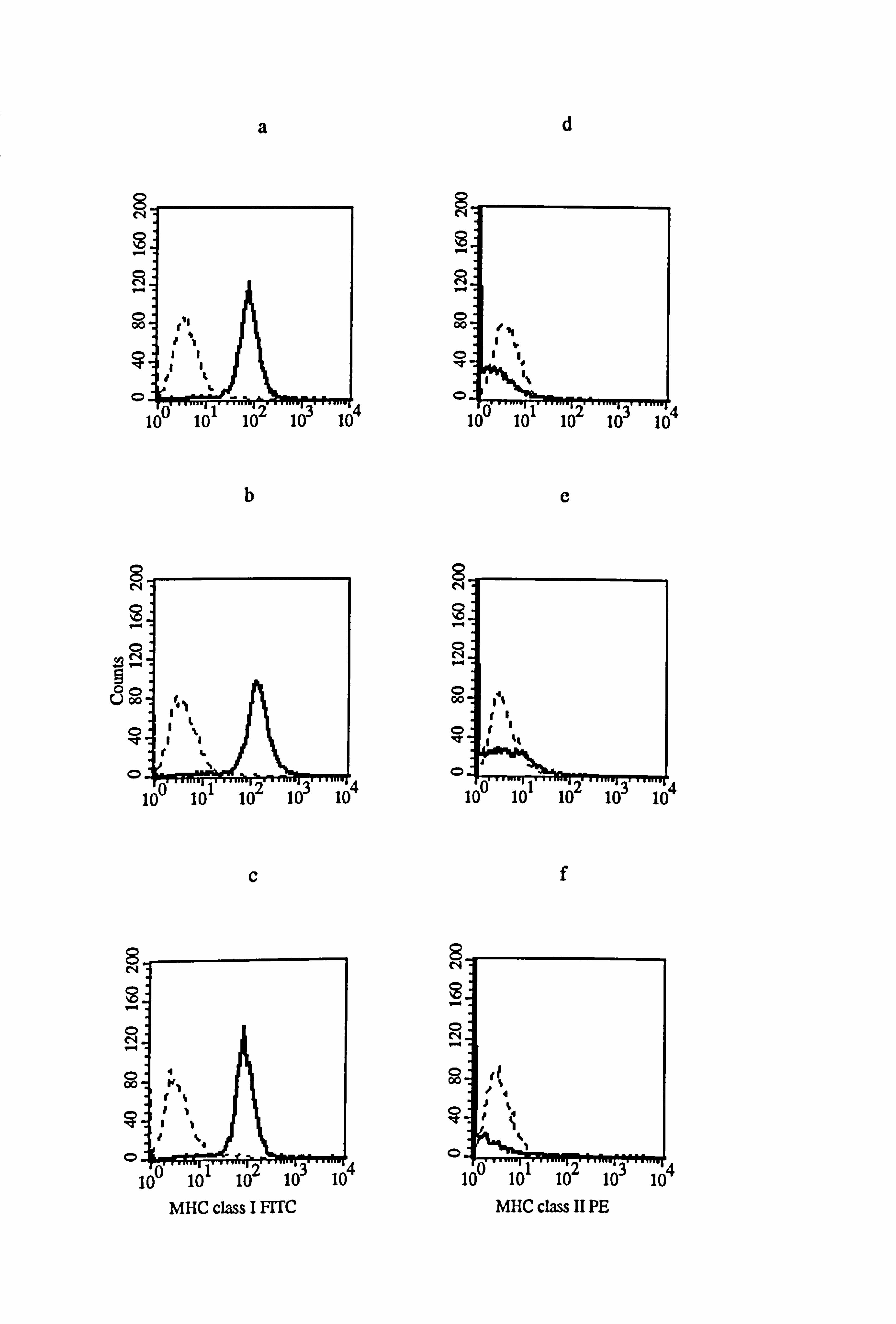
Figure 4.13 Effect of IFN-y, TNF-a and IL-la on the expression of AlHC class I and class II in normal melanocytes. Melanocytes from donor M777, were cultured for 5 days under the experimental medium 1 (as described in Tables 4.8 and 4.9). Cells
were untreated (a and e) or treated with 100U/ml IFN-y (b and f), l00U/ml TNF-a (c
and g) or 100U/ml IL-la (d and h) for the final 24 hours before MHC class I (a-d) and MHC class II (e-h) expression were examined using flow cytometry (as described in Section 2.10). All figures show isotype fluorescence (dashed line) and MHC class I or MHC class II fluorescence (solid line).
O O_ N
c00
N.
O CO
O
0. 1
O O CV 0 co
O N I- O co
0 "t
0
O U
O O (V 0 c0
O N
O CID f1
1ý O
100 1C
O O N 0 tG I- O N I-
O co
0 IV
0
a
b
C
d
4
MHC class I FITC
104
4
0 0 N 0 co
O N I- 0 Co
0
0
O O N O Co
O N
0 Co
O
0
O O N 0 t0
O N
O Co
0
0
O 0 N O
I- T O N
O Co
0 lit
0
e
f
g
h
4
4
4
MHC class 11 PE
a
0-, TA in cc u
U
ao .9
a d
a u d w ao a. 0 020
100
90
80
70
so
so
40
30
20
10
0
100
90
so
70
60
60
40
30
20
10
0
control
control
Cytokine addition
**
IFNry
IFNI
160
140
120
100
00
so
40
20
0
ISO
140
120
100
80
60
40
20
0
4 A m
e 0
A
A A
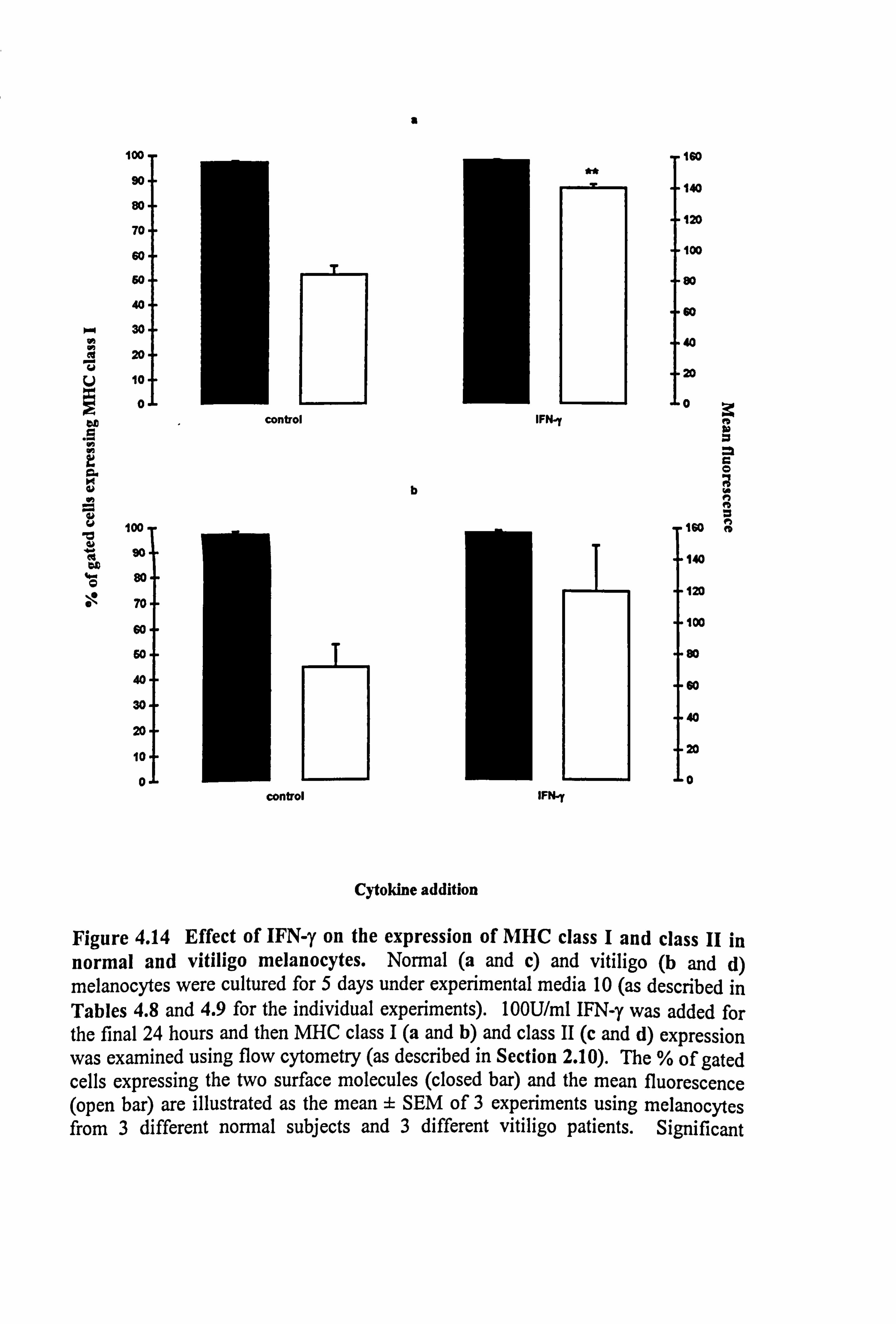
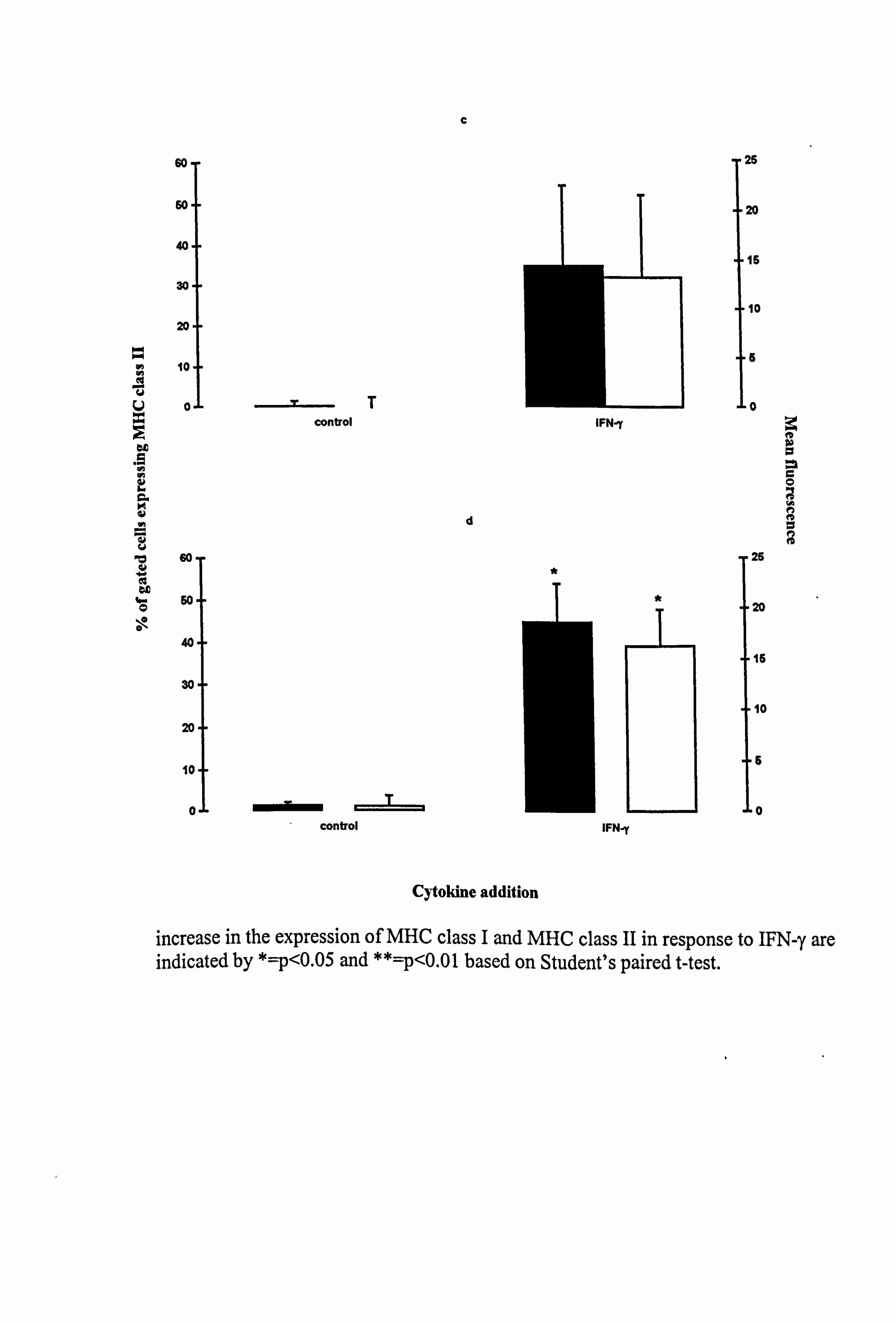
Figure 4.14 Effect of IFN-y on the expression of MHC class I and class II in normal and vitiligo melanocytes. Normal (a and c) and vitiligo (b and d) melanocytes were cultured for 5 days under experimental media 10 (as described in Tables 4.8 and 4.9 for the individual experiments). 100U/ml IFN-y was added for the final 24 hours and then MHC class I (a and b) and class II (c and d) expression was examined using flow cytometry (as described in Section 2.10). The % of gated cells expressing the two surface molecules (closed bar) and the mean fluorescence (open bar) are illustrated as the mean ± SEM of 3 experiments using melanocytes from 3 different normal subjects and 3 different vitiligo patients. Significant
b
C
60-
60-
40-
30-
20.
10
U o.
r
"0 ö0
bD
O $0
40
30
20
10
0
TT control
mmdbý control
d
IFN-y
IFN-r
25
20
15
10
,5
0
A a
c 0
n
26
20
16
10
5
0
Cytokine addition
increase in the expression of MHC class I and MHC class II in response to IFN-y are indicated by *=p<0.05 and **=p<0.01 based on Student's paired t-test.
*
149
(37 fold, p<0.05). A non-significant increase was seen in normal melanocytes for both
parameters.
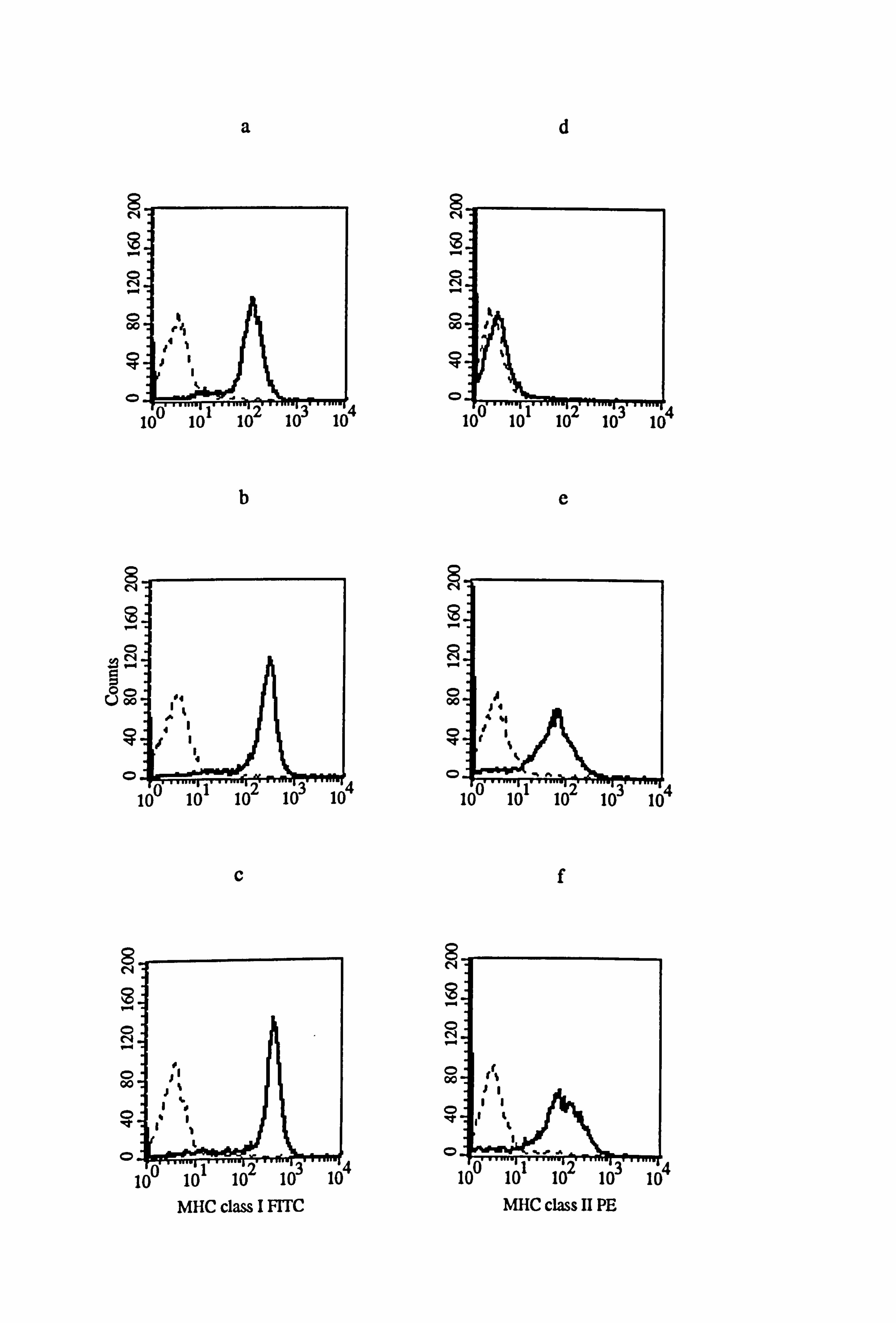
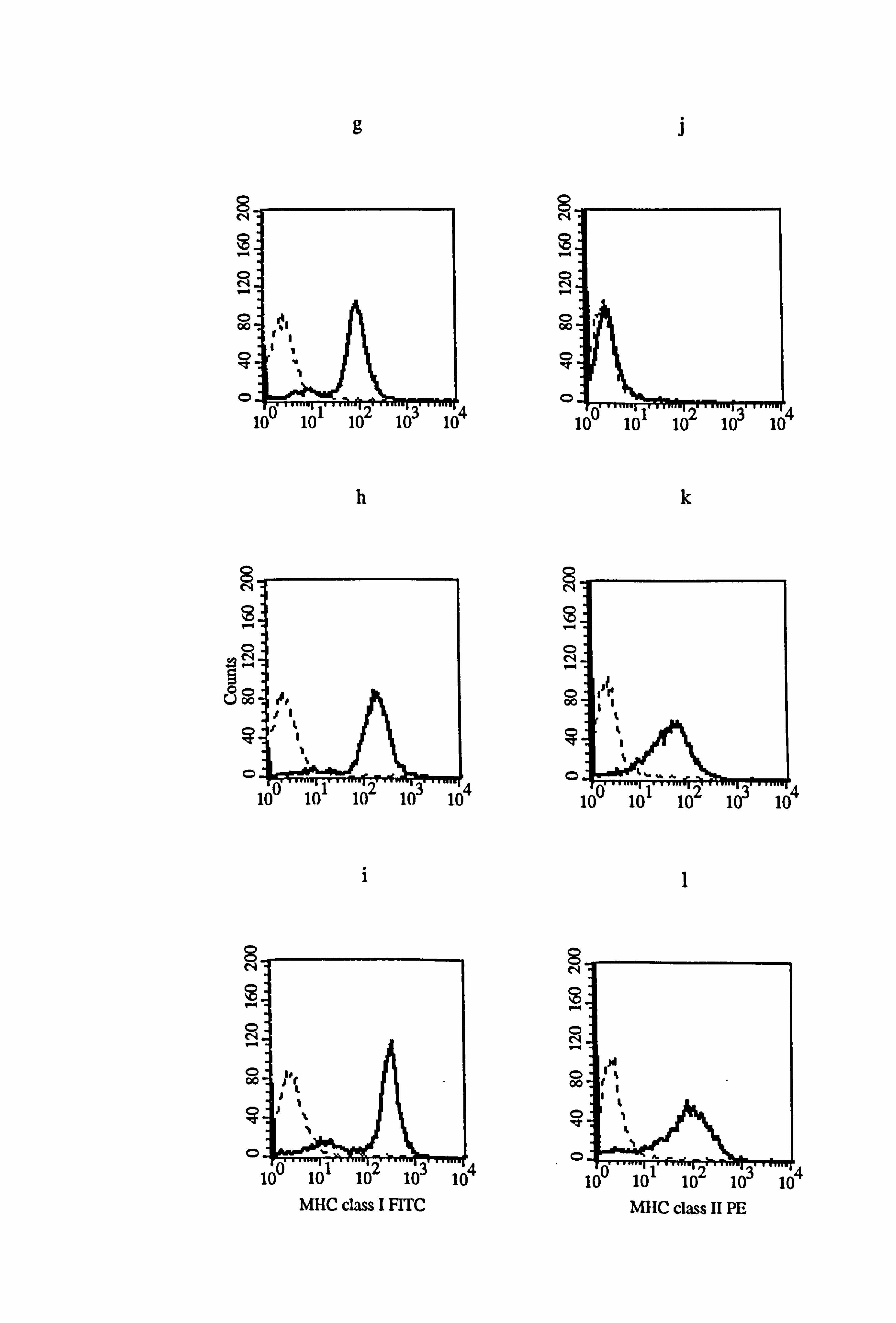
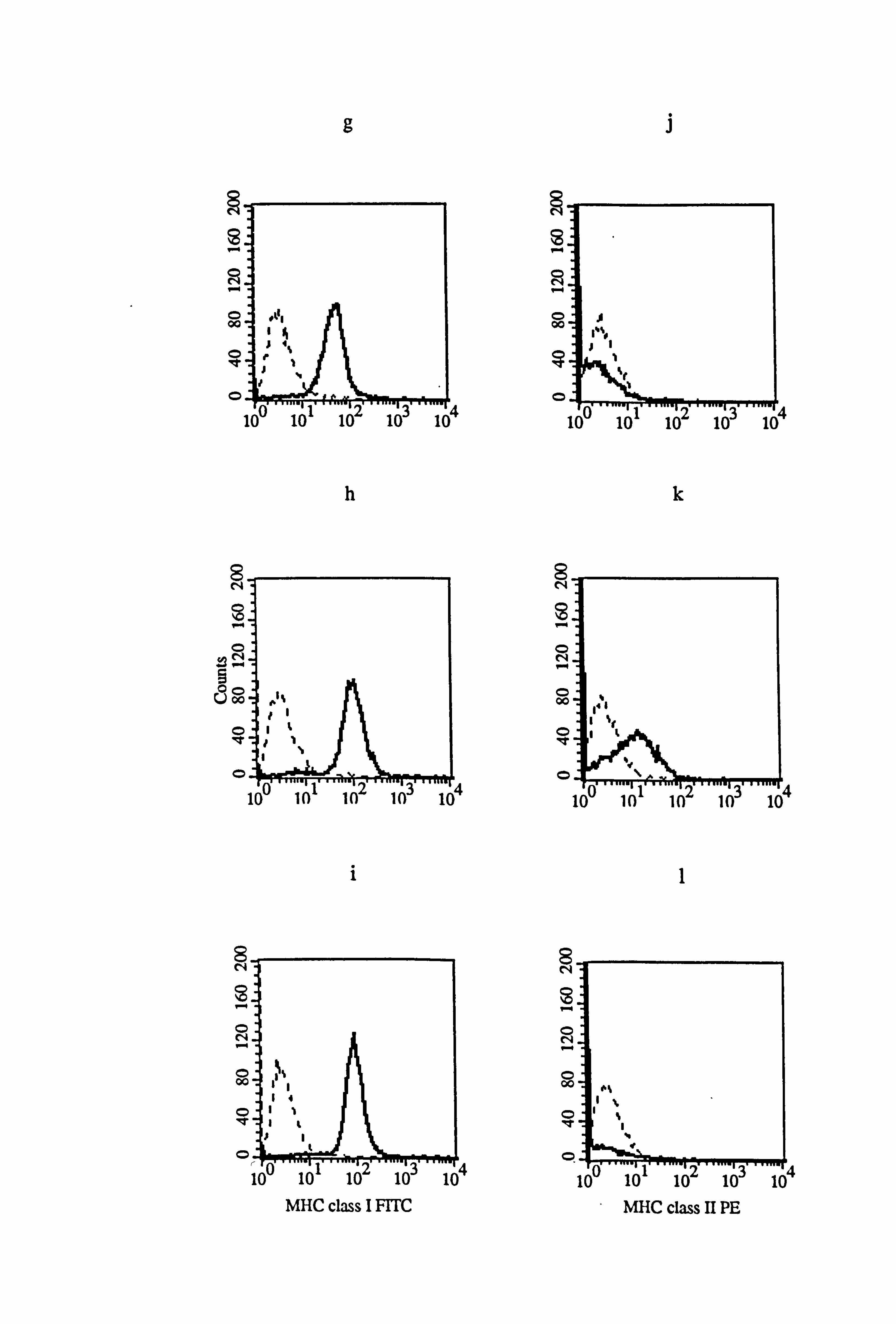
The effect of IFN-y and LCM on MHC class I and class II expression in medium 1 was
examined in donor melanocytes M833 and VM5 (Figure 4.15). IFN-y increased MHC
class I expression by 123% in M833 (Figure 4.15b) and by 120% in VM5 (Figure
4.15h), while LCM increased the expression of MHC class I by 226% in M833 (Figure
4.15c) and by 213% in VM5 (Figure 4.15i). ' The expression of MHC class II was
increased by 88 fold in response to IFN-y (Figure 4.15e) and by 149 fold in response . to
LCM (Figure 4.15f) in M833 while in VM5 the expression of MHC class II was
increased by 182 fold in response to IFN-y (Figure 4.15k) and by 373 fold in response
to LCM (Figure 4.151).
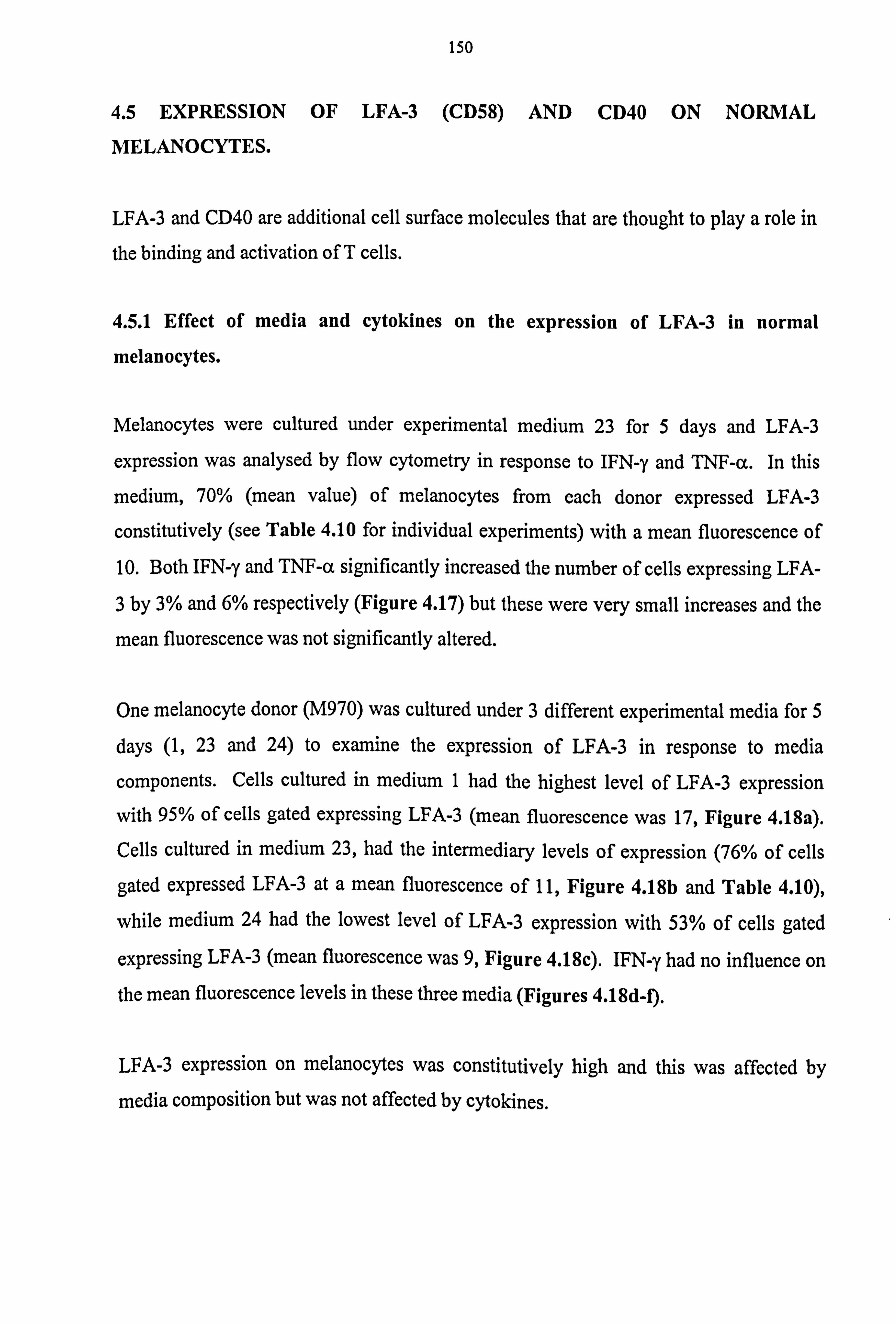
The effect of TNF-a on MHC class I and class II expression in comparison to IFN-y
was examined in donor melanocytes M843 and VM4 (Figure 4.16). The expression of
MHC class I in medium 10 was not influenced by TNF-a in M843 (only 10% increase,
Figure 4.16c), but in VM4, TNF-a increased MHC class I expression by 89% (Figure
4.16i), whereas IFN-y increased MHC class I expression by 87% in M843 (Figure
4.16b) and by 118% in VM4 (Figure 4.16h). However, as only one experiment was
performed using this cytokine, firm conclusions cannot be drawn from these results.
TNF-a) had no influence on the expression of MHC class II in M843 (Figure 4.16f) or --
VM4 (Figure 4.161) (this result is similar to what was seen with M777 in Section 4.4.1)
whereas IFN-y increased MHC class II expression in VM4 (Figure 4.16k and Table
4.9c).
There were no significant differences in the expression of MHC class I and class II
between normal and vitiligo melanocytes. The increase of both MHC class I and class
II expression by IFN-y was very similar.
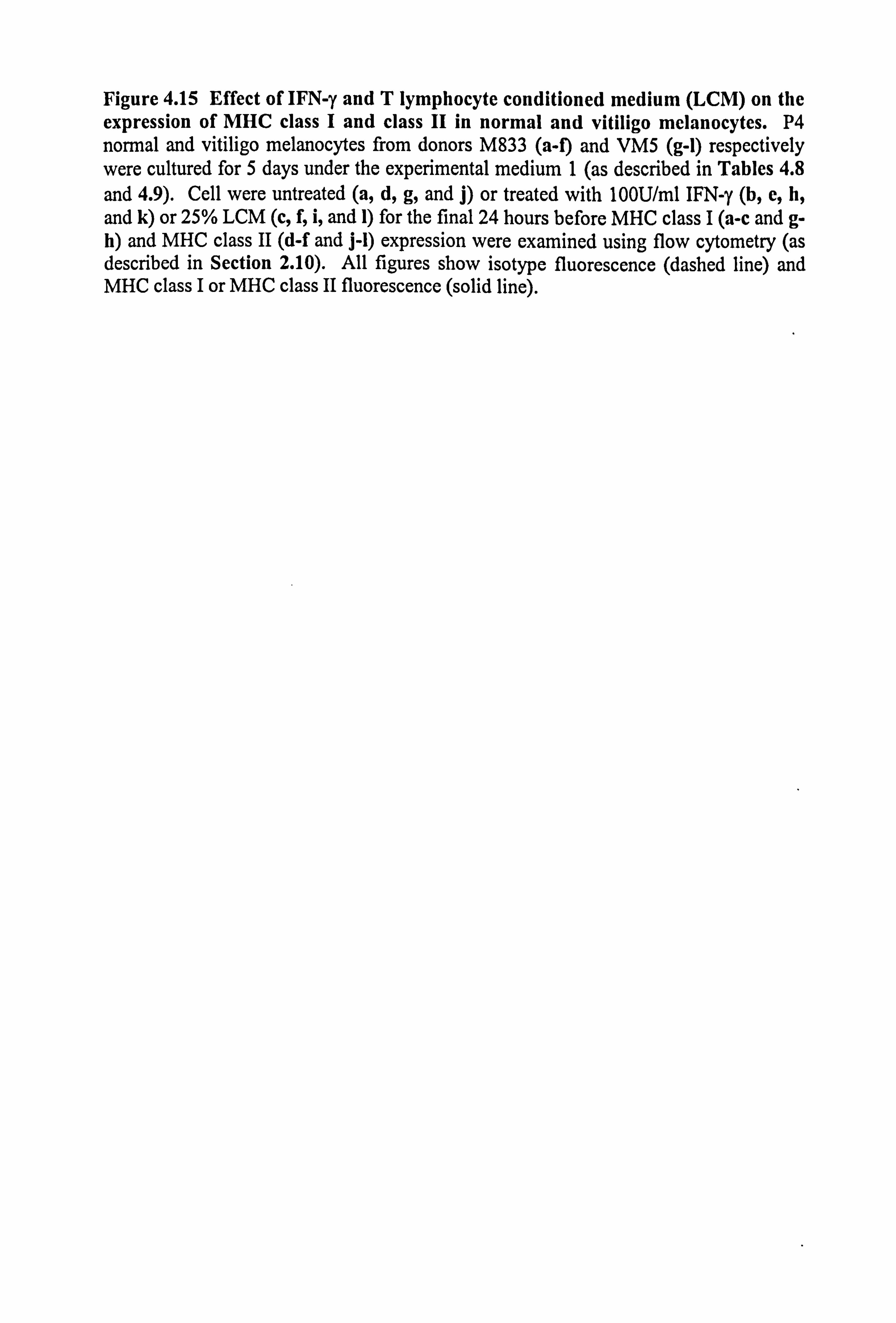
Figure 4.15 Effect of IFN-y and T lymphocyte conditioned medium (LCM) on the expression of MHC class I and class II in normal and vitiligo melanocytes. P4 normal and vitiligo melanocytes from donors M833 (a-f) and VM5 (g-1) respectively were cultured for 5 days under the experimental medium 1 (as described in Tables 4.8 and 4.9). Cell were untreated (a, d, g, and j) or treated with 100U/ml IFN-y (b, e, h, and k) or 25% LCM (c, f, i, and 1) for the final 24 hours before MHC class I (a-c and g- h) and MHC class II (d-f and j-l) expression were examined using flow cytometry (as described in Section 2.10). All figures show isotype fluorescence (dashed line) and MHC class I or MHC class II fluorescence (solid line).
8 N
s
00
0
c 00
q
0
8
rr
O 00
0 d
0
a
b
C
4
4
4
8 N
O N
O 00
0
9
0 00
0
8 N
O N
O 00
O `Q'
0
d
e
f
4
MHC class I FI TC MHC class II PE
8 N
O 00
vc
0
8
O
A
00 00
q
0
8 N
2
O 00
O
g
h
i
4
4
8 N
00
0
8
0 00
0
O
O 00
q
0
i
k
1
4
4
MHC class I FITC MHC class II PE
Figure 4.16 Effect of IFN-y and TNF-a on the expression of MHC class I and class II in normal and vitiligo melanocytes. Normal and vitiligo melanocytes from donors M843 (a-f) and VM4 (g-1) respectively were cultured for 5 days under the experimental medium 10 (as described in Tables 4.8 and 4.9). Cell were untreated (a, d, g, and j) or treated with 100U/ml IFN-y (b, e, h, and k) or with 100U/ml TNF-a (c, f, i, and 1) for the final 24 hours before MHC class I (a-c and g-h) and MHC class II (d-f and j-1) expression were examined using flow cytometry (as described in Section 2.10). All figures show isotype fluorescence (dashed line) and MHC class I or MHC class II fluorescence (solid line).
8 N
. ter
O 00
O
8 N
ýD
0
Üo°o
0
O
8 N
8 O N
O 00
q
0
a
b
C
4
4
4
8 N
. -r
O 00
0
qRT 0
8 N
N
O 00
O
IOT 0
8
00
q
0
d
e
f
4
4
4
MHC class II PE MHC class I FITC
c`ý
2
00
0
0
8 fV
0 Z -I o Uooo
0
0
8 N
O N
O 00
O
0
g
h
i
4
4
4
0 00
0
8 N
N ti
O 00
ý7
0
8 N
O 00
O
O
i
k
1
4
4
MHC class I FITC MHC class II PE
150
4.5 EXPRESSION OF LFA-3 (CD58) AND CD40 ON NORMAL
MELANOCYTES.
LFA-3 and CD40 are additional cell surface molecules that are thought to play a role in
the binding and activation of T cells.
4.5.1 Effect of media and cytokines on the expression of LFA-3 in normal
melanocytes.
Melanocytes were cultured under experimental medium 23 for 5 days and LFA-3
expression was analysed by flow cytometry in response to IFN-y and TNF-a. In this
medium, 70% (mean value) of melanocytes from each donor expressed LFA-3
constitutively (see Table 4.10 for individual experiments) with a mean fluorescence of
10. Both IFN-y and TNF-a significantly increased the number of cells expressing LFA-
3 by 3% and 6% respectively (Figure 4.17) but these were very small increases and the
mean fluorescence was not significantly altered.
One melanocyte donor (M970) was cultured under 3 different experimental media for 5
days (1,23 and 24) to examine the expression of LFA-3 in response to media
components. Cells cultured in medium 1 had the highest level of LFA-3 expression
with 95% of cells gated expressing LFA-3 (mean fluorescence was 17, Figure 4.18a).
Cells cultured in medium 23, had the intermediary levels of expression (76% of cells
gated expressed LFA-3 at a mean fluorescence of 11, Figure 4.18b and Table 4.10),
while medium 24 had the lowest level of LFA-3 expression with 53% of cells gated
expressing LFA-3 (mean fluorescence was 9, Figure 4.18c). IFN-y had no influence on
the mean fluorescence levels in these three media (Figures 4.18d-f).
LFA-3 expression on melanocytes was constitutively high and this was affected by
media composition but was not affected by cytokines.
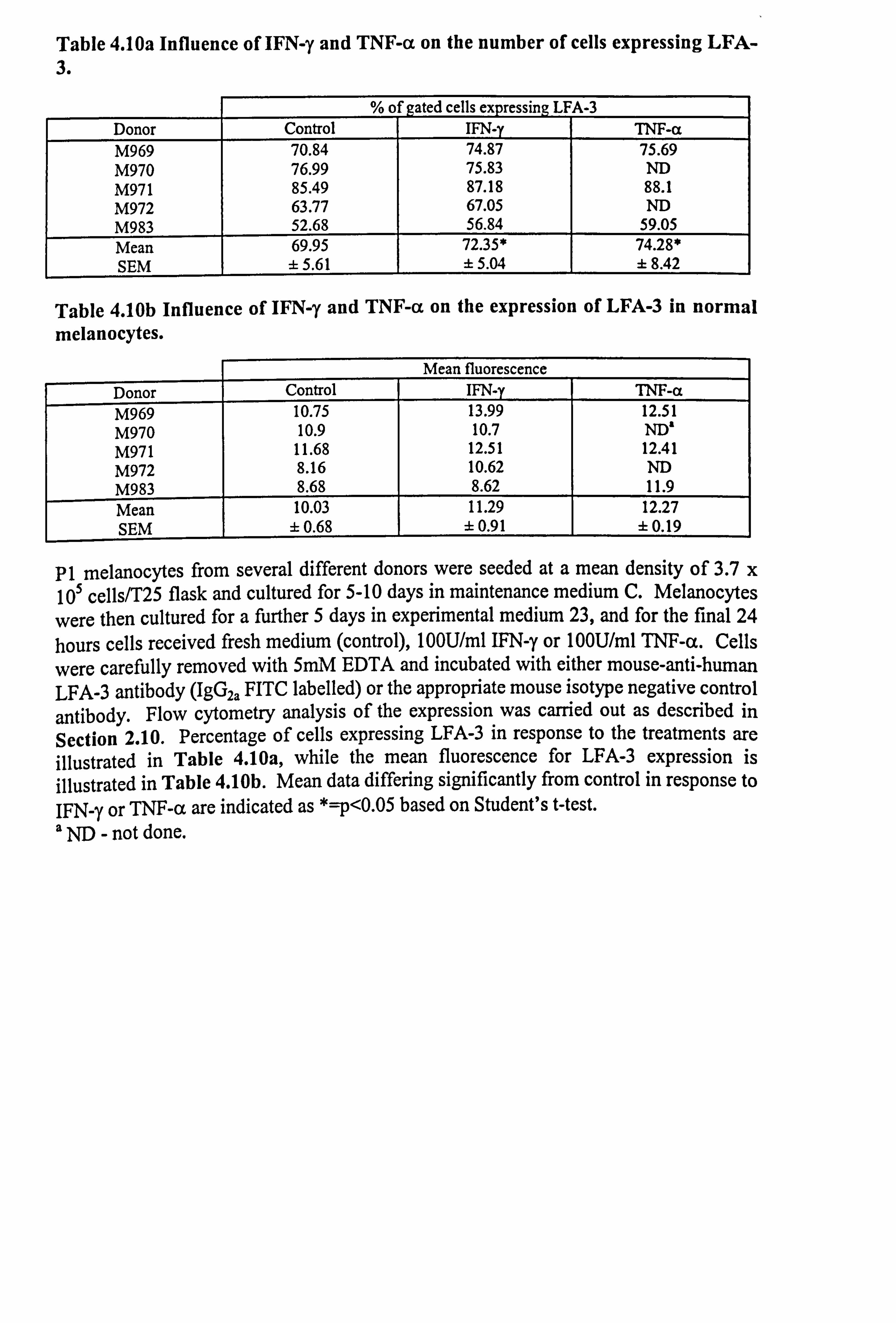
Table 4.10a Influence of IFN-y and TNF-a on the number of cells expressing LFA- 3.
%o f gated cells expressing LFA-3 Donor Control IFN-y TNF-a M969 70.84 74.87 75.69 M970 76.99 75.83 ND M971 85.49 87.18 88.1 M972 63.77 67.05 ND M983 52.68 56.84 59.05 Mean 69.95 72.35* 74.28* SEM ± 5.61 ± 5.04 ±8.42
Table 4.10b Influence of IFN-y and TNF-a on the expression of LFA-3 in normal melanocytes.
Mean fluorescence
Donor Control IFN-y TNF-a M969 10.75 13.99 12.51 M970 10.9 10.7 ND' M971 11.68 12.51 12.41 M972 8.16 10.62 ND M983 8.68 8.62 11.9 Mean 10.03 11.29 12.27 SEM ± 0.68 ±0.91 ±0.19
P1 melanocytes from several different donors were seeded at a mean density of 3.7 x 10⁵ cells/T25 flask and cultured for 5-10 days in maintenance medium C. Melanocytes
were then cultured for a further 5 days in experimental medium 23, and for the final 24 hours cells received fresh medium (control), 100U/ml IFN-y or 100U/ml TNF-a. Cells
were carefully removed with 5mM EDTA and incubated with either mouse-anti-human LFA-3 antibody (IgG2a FITC labelled) or the appropriate mouse isotype negative control antibody. Flow cytometry analysis of the expression was carried out as described in Section 2.10. Percentage of cells expressing LFA-3 in response to the treatments are illustrated in Table 4.10a, while the mean fluorescence for LFA-3 expression is illustrated in Table 4.10b. Mean data differing significantly from control in response to IFN-y or TNF-a are indicated as *=p<0.05 based on Student's t-test. a ND - not done.
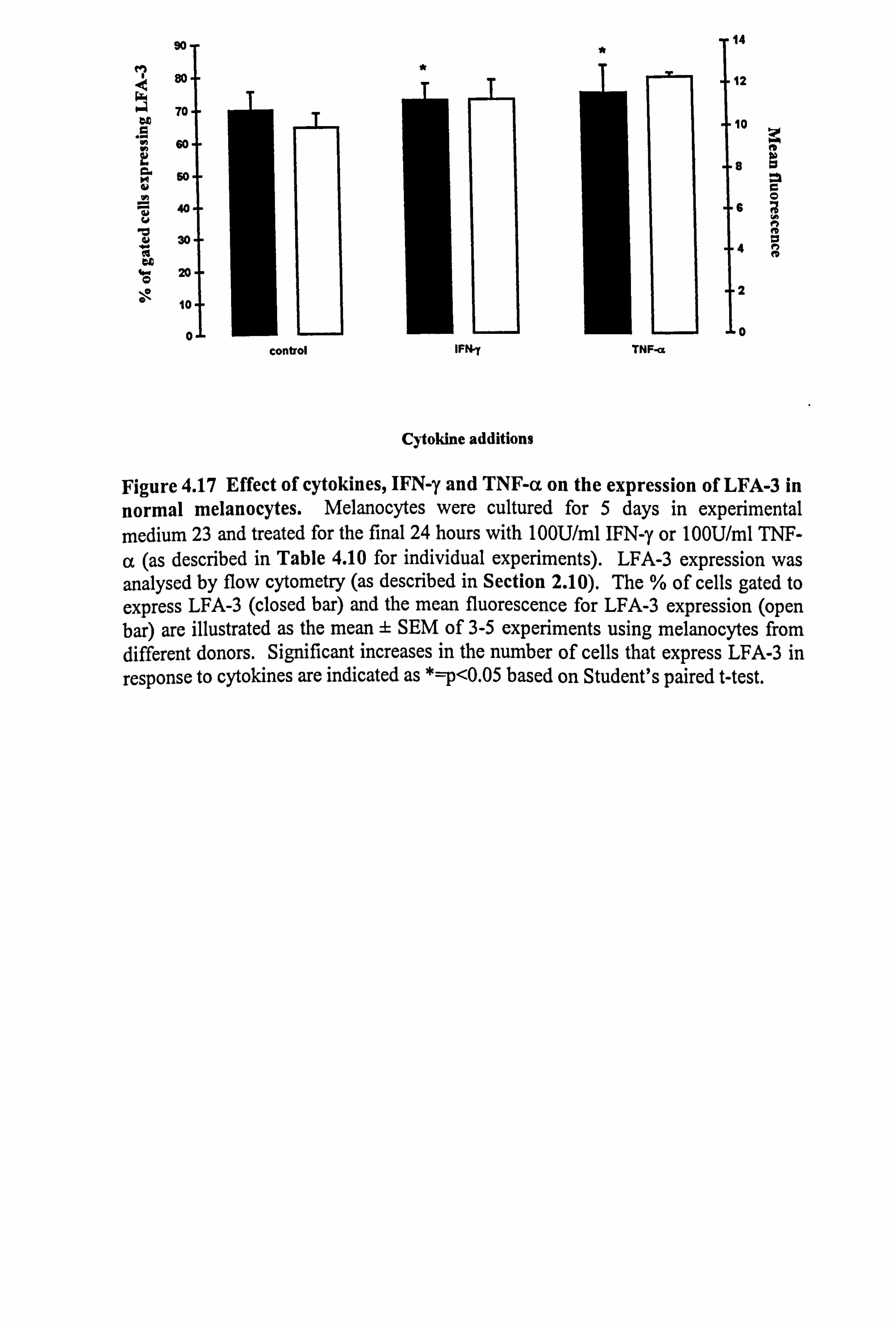
90 T Tja d 80.
70- U
Sp,
so
40
j30
20
10
* 12
0 control IFN y TNF-ct
Cytokine additions
1o
6
4
2
0
Figure 4.17 Effect of cytokines, IFN-y and TNF-a on the expression of LFA-3 in normal melanocytes. Melanocytes were cultured for 5 days in experimental medium 23 and treated for the final 24 hours with 100U/ml IFN-y or 100U/ml TNF-
a (as described in Table 4.10 for individual experiments). LFA-3 expression was analysed by flow cytometry (as described in Section 2.10). The % of cells gated to express LFA-3 (closed bar) and the mean fluorescence for LFA-3 expression (open bar) are illustrated as the mean ± SEM of 3-5 experiments using melanocytes from different donors. Significant increases in the number of cells that express LFA-3 in response to cytokines are indicated as *=p<0.05 based on Student's paired t-test.
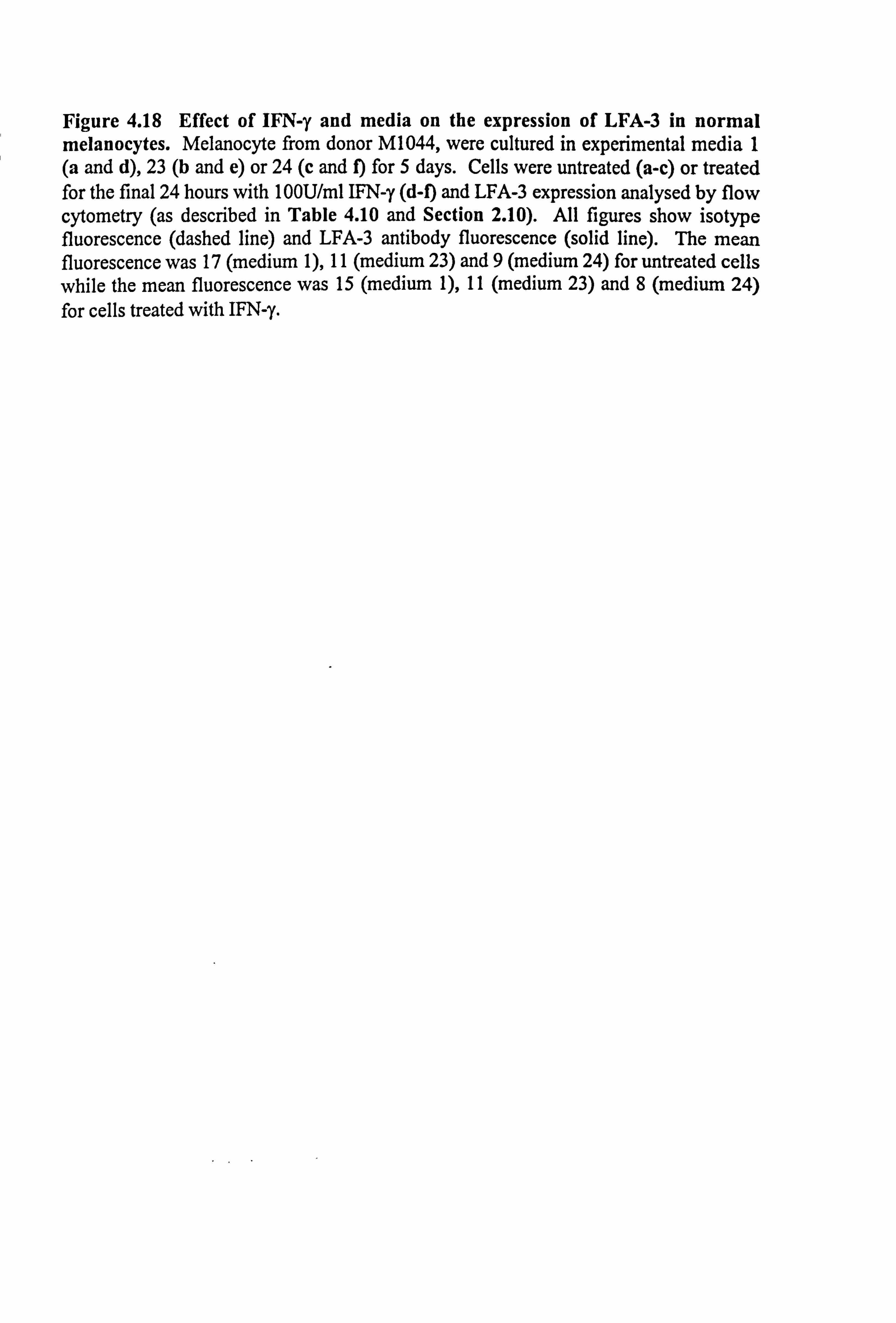
Figure 4.18 Effect of IFN-y and media on the expression of LFA-3 in normal melanocytes. Melanocyte from donor M1044, were cultured in experimental media 1 (a and d), 23 (b and e) or 24 (c and f) for 5 days. Cells were untreated (a-c) or treated for the final 24 hours with 100U/ml IFN-y (d-f) and LFA-3 expression analysed by flow cytometry (as described in Table 4.10 and Section 2.10). All figures show isotype fluorescence (dashed line) and LFA-3 antibody fluorescence (solid line). The mean fluorescence was 17 (medium 1), 11 (medium 23) and 9 (medium 24) for untreated cells while the mean fluorescence was 15 (medium 1), 11 (medium 23) and 8 (medium 24) for cells treated with IFN-y.
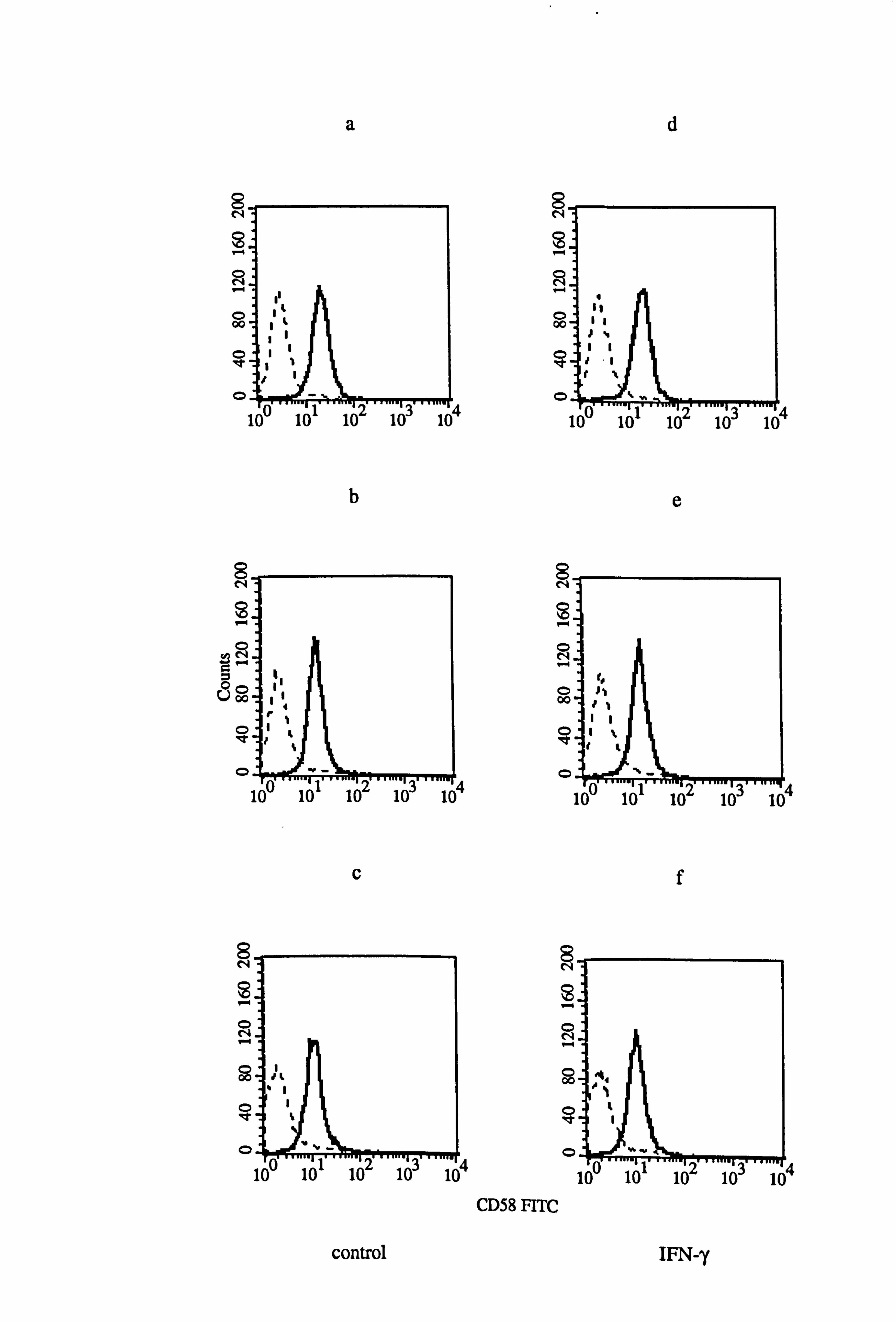
8 N
O 00
0
0
8 N
Üooo
0
8 N
O
0
a
b
C
control
4
8 N
53
O 00
0
8 N
O 00
!4 0
4
8 N
ýD
O N
0
4 0
CD58 FITC
d
e
f
IFN-, y
4
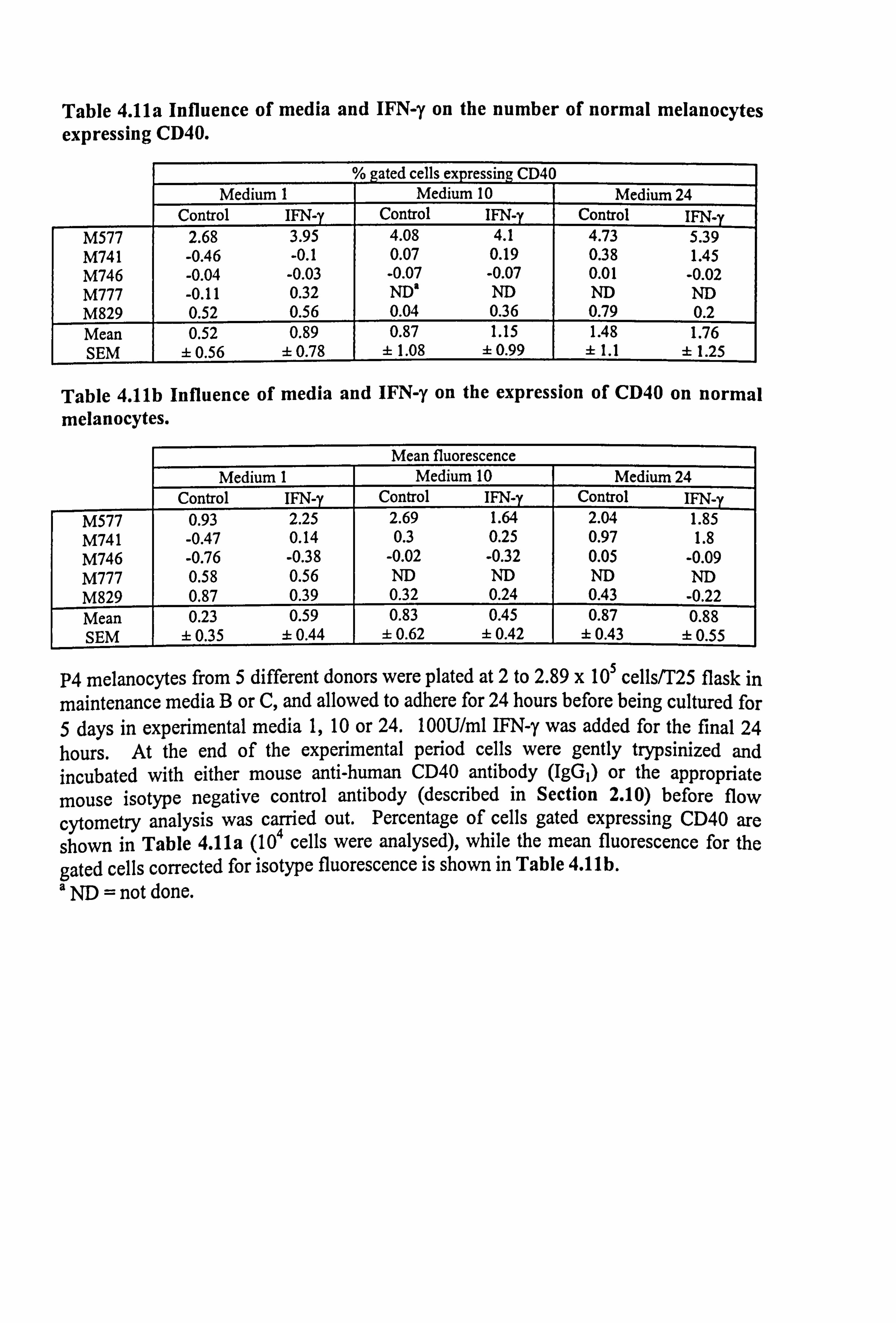
Table 4.11a Influence of media and IFN-y on the number of normal melanocytes expressing CD40.
gated cells expressing CD40 Medium I Medium 10 Medium 24
Control IFN-y Control IFN-y Control IFN_ M577 2.68 3.95 4.08 4.1 4.73 5.39 M741 -0.46 -0.1 0.07 0.19 0.38 1.45 M746 -0.04 -0.03 -0.07 -0.07 0.01 -0.02 M777 -0.11 0.32 ND' ND ND ND M829 0.52 0.56 0.04 0.36 0.79 0.2 Mean 0.52 0.89 0.87 1.15 1.48 1.76 SEM ± 0.56 ± 0.78 ± 1.08 ± 0.99 ± 1.1 ± 1.25
Table 4.11b Influence of media and IFN-y on the expression of CD40 on normal melanocytes.
Mean fluorescence Medium 1 Medium 10 Medium 24
Control IFN-y Control IFN-y IFN-y M577 0.93 2.25 2.69 1.64 2.04 1.85 M741 -0.47 0.14 0.3 0.25 0.97 1.8 M746 -0.76 -0.38 -0.02 -0.32 0.05 -0.09 M777 0.58 0.56 ND ND ND ND M829 0.87 0.39 0.32 0.24 0.43 -0.22 Mean 0.23 0.59 0.83 0.45 0.87 0.88 SEM ± 0.35 ± 0.44 ± 0.62 ± 0.42 ± 0.43 ±0.55
P4 melanocytes from 5 different donors were plated at 2 to 2.89 x 10⁵ cells/T25 flask in maintenance media B or C, and allowed to adhere for 24 hours before being cultured for 5 days in experimental media 1,10 or 24.100U/ml IFN-y was added for the final 24 hours. At the end of the experimental period cells were gently trypsinized and incubated with either mouse anti-human CD40 antibody (IgGI) or the appropriate mouse isotype negative control antibody (described in Section 2.10) before flow
cytometry analysis was carried out. Percentage of cells gated expressing CD40 are shown in Table 4. lla (104 cells were analysed), while the mean fluorescence for the gated cells corrected for isotype fluorescence is shown in Table 4. llb. a ND = not done.
151
4.5.2 Effect of IFN-y on the expression of CD40 on normal melanocytes.
Melanocytes cultured in experimental media 1,10 or 24 for 5 days were examined for
CD40 expression by flow cytometry. There was much donor-to-donor varation (Table
4.11) but overall the basal expression of CD40 was very low. Media and IFN-y did not
influence the expression of CD40 (Figure 4.19).
4.6 T CELL BINDING TO NORMAL HUMAN MELANOCYTES.
The ability of any cell to express immune surface molecules usually indicates their
ability to bind and activate T cells. In this preliminary study an ELISA method was
used to detect the ability of T cells to bind to unstimulated and cytokine-stimulated
melanocytes. The methodology used is well established in this laboratory for T
lymphocyte binding to keratinocytes.
Three experiments were performed using melanocytes from different donors for each
experiment. In the first experiment, the cells were cultured in 12 wells and the coloured
endpoint product transferred to 96 well plates for O. D. readings, while in the other two
experiments melanocytes were cultured in 96 well plates. The individual results are
shown in Table 4.12 while the mean data for control and IFN-y-treated cells cultured in
media 1 and 10 are shown in Figure 4.20. Although it is preferable to correct
parameters for cell number (or protein for example), the data were not corrected in these
experiments as cytokines do not have a significant effect on melanocyte proliferation. Additionally, the cell number plates set up for M767 were ruined in storage.
In all three experiments, T cells did bind to untreated melanocytes. However, T cells
were also bound to the tissue culture plastic, where melanocytes had washed away due
to changes in temperature during the ELISA method. The overall results in response to
cytokines were disappointing (Figure 4.20), with considerable donor-to-donor variation being apparent (see Table 4.13). M760 were unresponsive to cytokine stimulation
while, in the other two experiments, some increases in T cell binding were seen in
response to cytokine stimulation.
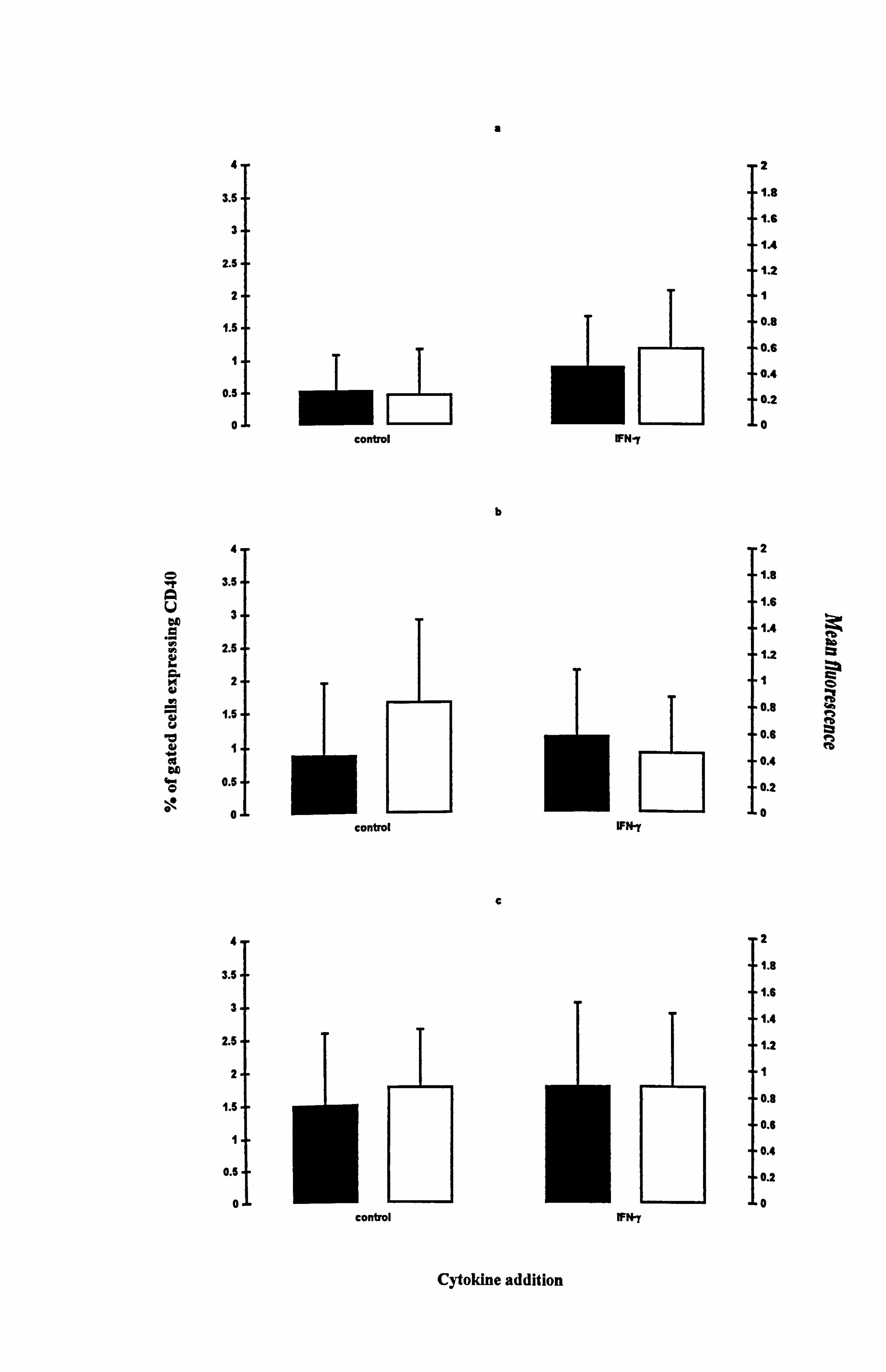
a
0 A V ao a .r vi Vi
ii
a w
= aý v
w
w O 0
4-
3.5.
3-
2.5-
2-
1
0.5.
0
4
3.5
3
2.5
2
1.5
1
0.5
0
4
3.5
3
2.5
2
1.5
1
0.5
0
control
control
control
b
C
2
1.8
1.6
1.4
1.2
1
0.8
0.6
0.4
0.2
0 IFN-y
2
1.6
1.6
1.4 1.2
I 0. s
0.6
0.4
0.2
0 IFN-r
2
1.8
1.6
1.4
1.2
1
0.8
0.6
0.4
0.2
0 WN y
bb a
0
n ea
Cytokine addition
Figure 4.19 Effect of IFN-y on the expression of CD40 in normal melanocytes. Melanocytes were cultured for 5 days under the experimental media 1 (a), 10 (b) or 24 (c) (as described in Table 4.11 for the individual experiments) .
100U/ml IFN-y was added for the final 24 hours and then CD40 expression was analysed using flow
cytometry (as described in Section 2.10). The % of gated cells expressing CD40 (closed bar) and the mean fluorescence of CD40 expression (open bar) are illustrated as the mean ± SEM of 4-5 experiments using melanocytes from different donors.
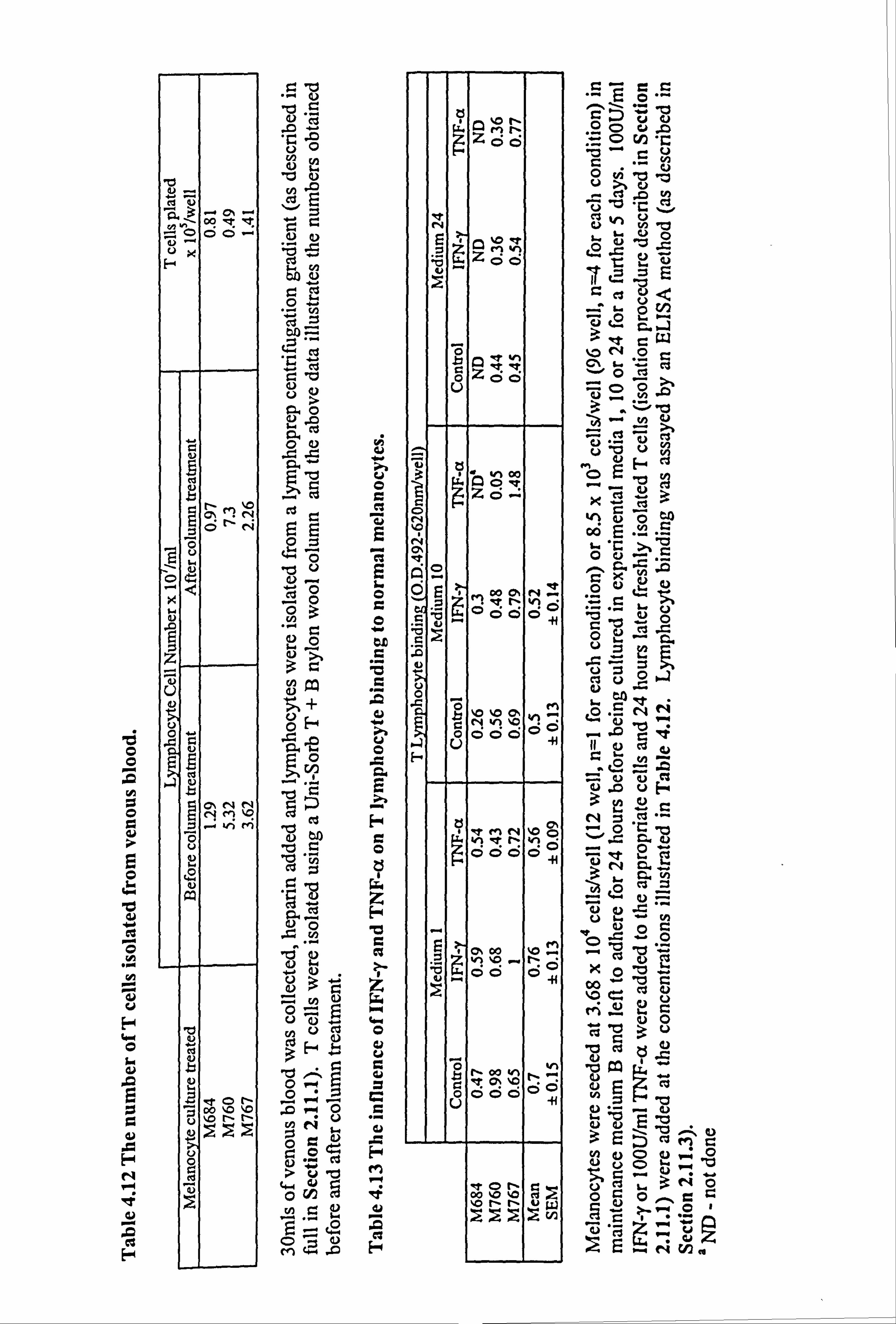
0 0
LA 0
E 0
'd W 4.6
0 H
0 u
a u .a H N
F-4
cu u a'N oO q 'l 0 C>
UO
C 00
01J
O- "- V1
9
a y ü
r- M MN c
4N OO
w0 U bU
k ¢ :ýOO
V "-4 r. °
Z C? o
51 UO
13
ce mg NMVý
j -V M .d to Ü Z) r. "
0
0 D
q G' w b yU CI O
H4 ei -o ma:
Co r_
Vb p Vý 7
E"~ a OO
ýN O z ri n
E e. Q %0 't Es Z ein
v,.,, z00
.o Avv ZCo
U
ü ýÄöv
zo- 0
QO
O Co o, ev Z
tr,
Nhe V) m
aV° doo °
ooöö°
o+ Co %0"" vi 'lý
Ch ZZ oooö°°
. tor- r- g
-
o c> üp
N ý .CO
- c3 ,A to
SUS Ü ý"ý�
U vý b
Q
2 c0 G. ý1. CW NO
c t..
- CD 9 j2 r
wý'C!
CJ V vii
ÖF
X. cl 3 yC
O"2Ci >
i-. X �. Ö ÖU`O
.Cs Vº
ä Cl Uc UO
.rý
cv'3 Uý º"ý dl ý
N- b
äi ä ri
F+
N_ in
0 00 äs
jN
ýSv OýO
ös °0
"" C) ti O ci C)
U
=3U -10 Cu ö
4) 0 J--
V) 'O 0 "Ö ""
CUO '-
OO^
rr N
0 en o
No
o U uz CIO
U(1
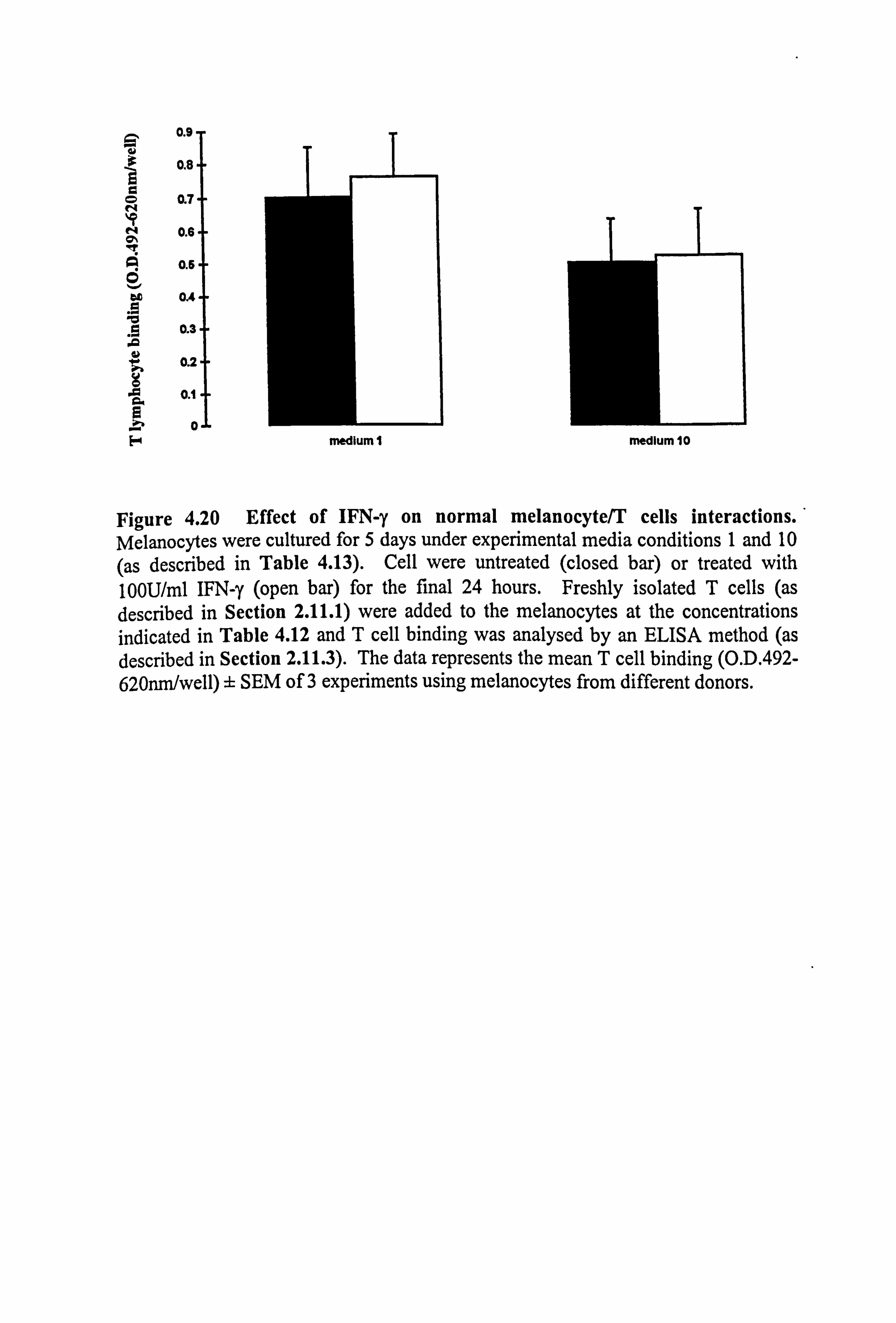
0.9
0.8.
0 0.7 N
0.6
0.6
ao 0.4
0.3
0.2
0.1 ä
8 .r0 F
Figure 4.20 Effect of IFN-y on normal melanocyte/T cells interactions. Melanocytes were cultured for 5 days under experimental media conditions 1 and 10 (as described in Table 4.13). Cell were untreated (closed bar) or treated with 100U/ml IFN-y (open bar) for the final 24 hours. Freshly isolated T cells (as described in Section 2.11.1) were added to the melanocytes at the concentrations indicated in Table 4.12 and T cell binding was analysed by an ELISA method (as described in Section 2.11.3). The data represents the mean T cell binding (O. D. 492- 620nm/well) ± SEM of 3 experiments using melanocytes from different donors.
medium I medium 10
152
4.7 DISCUSSION.
The etiology of vitiligo remains unclear. Several hypotheses exist e. g. neuronal,
autoimmune and an inherent melanocyte defect to explain the possible cause of vitiligo
as discussed in Section 1.4. An immune component has been implicated in vitiligo
(Section 1.4.3), but it still remains to be determined whether this is the primary defect
or a secondary response to damage caused by other mechanisms. Melanocytes are
capable of antigen presentation (Le Poole et al, 1993b) and the present study examined
whether normal and vitiligo melanocytes would differ in their constitutive expression of immune-related molecules or in their response to cytokines which induce their
expression. Additionally, an interesting theory has arisen suggesting the involvement of MSH-related peptides in the pathogenesis of vitiligo (Liu et al, 1995).
Recent data showing that the melanotropins are produced by cells of the epidermis and dermis (Thody et al, 1983; Farooqui et al, 1993; Schauer et al, 1994; Iyengar, 1995;
Kippenberger et al, 1995; Slominski et al, 1995; Chakraborty et al, 1996; Slominski et
al, 1995; Wintzen et al, 1996; Teofoli et al, 1997; Wakamatsu et al, 1997; Can et al, 1998), and that their production increases following UV exposure suggest a
paracrine/autocrine role for these peptides in skin (recently reviewed by Luger et al,
1997; Luger, 1998). MC1R are present on human melanocytes (Donatien et al, 1992;
De Luca et al, 1993; Thody et al, 1993; Suzuki et al, 1996), immortalized (Chakraborty
& Pawelek, 1993) and normal keratinocytes (Bhardwaj et al, 1996) and human
endothelial cells (Hartmeyer et al, 1997). Additionally many immunocompetent cells
express MC1R including murine macrophages (Star et al, 1995), human neutrophils (Catania et al, 1996) and human monocytes (Rajora et al, 1996; Bhardwaj et al, 1997).
Several groups now report on the polymorphic nature of the human MC1R gene (Valverde et al, 1995 & 1996a; Box et al, 1997; Koppula et al, 1997; Ichii-Jones et al, 1998; Smith et al, 1998). Red haired individuals who tan poorly and who commonly
suffer skin inflammation on UV exposure exhibit a variety of MC1R variants (Valverde
et al, 1995). They also have a susceptibility to melanoma and one variant of the MCIR
has been associated with melanoma (Valverde et al, 1996a). In contrast to the work by
Valverde and co-workers, Ichii-Jones et al. (1998) report that the polymorphic nature of
the human MC1R does not appear to relate to skin type determination or predisposing
153
an individual to melanoma development. Recently, the Argl51Cys variant, which is
associated with red haired fair skinned individuals, was found to be non-functional, as
cAMP production was not stimulated when NDP-MSH was bound (Frändberg et al,
1998). An alteration in the sensitivity of the MC1R may play a role in the development
of vitiligo (Liu et al, 1995). Pigment synthesis in response to UV is relatively complex;
melanin precursors and intermediates are formed into a range of coloured and colourless
hydroxyindoles and some forms of pigment production represent a greater risk for the
melanocyte in terms of cytotoxicity (Urabe et al, 1994). Following UV irradiation, the
skin produces a wide range of cytokines, many of which will upregulate adhesion
molecule expression on melanocytes (e. g. Yohn et al, 1990; Kirnbauer et al, 1992;
Krasagakis et al, 1995). The combination of increased pigmentation and UV/cytokine-
induced upregulation of adhesion molecules on melanocytes might make these cells a
target for infiltrating T cells. As a-MSH has many biological activities, it was
speculated that in human melanocytes, in addition to its role as a stimulator of
pigmentation (Hunt et al, 1994a, b&c; Abdel-Malek et al, 1995; McLeod et al, 1995;
Maeda et al, 1996; Suzuki et al, 1996), a-MSH may prevent the melanocyte from
becoming a premature target for the immune system. a-MSH was recently implicated
in modulating pigmentary responses following UVB irradiation (Im et al, 1998) and
ADF, an antioxidant was capable of increasing MC1R expression and the binding of
NDP-MSH to the receptor (Funasaka & Ichihashi, 1997), in a similar manner to UVB
(Funasaka & Ichihashi, 1997; Funasaka et al, 1998).
The data in this chapter demonstrated that both normal and vitiligo melanocytes were
capable of expressing ICAM-1, MHC class I and class II either constitutively or in
response to cytokines. However, there was no significant difference in expression
between normal and vitiligo melanocytes once these cells were established in culture.
This is the first study, to the best of my knowledge, to demonstrate that cultured vitiligo
melanocytes express these surface molecules and that the level of expression is
enhanced in response to cytokines in a manner comparable to normal melanocytes.
Integrin expression has also been shown to be similar on normal and vitiligo cultured
melanocytes and in biopsies from control, non-lesional, perilesional and lesional skin
(Le Poole et al, 1997). Firm evidence is provided to illustrate that a-MSH opposed the
actions of TNF-a on the expression of ICAM-1 in normal melanocytes.
154
The observation that a-MSH could reduce TNF-a-stimulated ICAM-1 expression in
human melanocytes was first reported in brief by Hewitt et al. (1993). Additionally, in
both a macrophage-like cell line (Luger et al, 1993) and monocytes (Köck et al, 1991),
a-MSH has been reported to reduce IFN-y-enhanced MHC class I molecule expression
although MHC class II and ICAM-1 were unaffected in monocytes (Köck et al, 1991).
ICAM-1 is involved in T cell activation, hence a decrease in ICAM-1 expression may
reduce the likelihood of T cell activation. However, other adhesion molecules are also involved in T cell binding, such as MHC class I and class II (which determine whether
CD8 or CD4 T cells bind respectively) and activation. These also need to be considered
in this context and studies with a-MSH and related peptides will need to be extended
accordingly. A preliminary study indicated that melanocytes could bind T cells as
previously reported (Nickoloff et al, 1991; Le Poole et al, 1993b) but the results were
not convincing due to problems with the technicalities of the assay employed (Section
4.6).
Both normal and vitiligo melanocytes had low constitutive expression of ICAM-1 under
all conditions examined and responded to cytokines with increased expression (Figures
4.1-4.3 and 4.5). The effect of various cytokines on ICAM-1 expression on normal
neonatal melanocytes has been reported previously by several groups (Yohn et al, 1990;
Krasagakis et al, 1991; Kirnbauer et al, 1992; Le Poole et al, 1993b; Krasagakis et al, 1995). In the current study, adult non-foreskin melanocytes were used. In this study it
was found that UVB did not affect constitutive melanocyte ICAM-1 expression following 1,4 and 7 doses of 143mJ/cm2 (Figure 4.4). UVB slightly enhanced IFN-y-
stimulated ICAM-1 response. Kirnbauer et al. (1992) showed that one dose of 100mJ/cm2 did not alter constitutive ICAM-1 expression on normal and melanoma cells
after 16 hours and that cytokine (IL-6, TNF-a and -ß, and IFN-y)-stimulated ICAM-1
expresssion was suppressed. However if the melanocytic cells were analysed 72 hours
later, constitutive ICAM-1 was significantly enhanced. This increase in ICAM-1 was
probably due to the production of cytokines by melanocytes (Swope et al, 1994).
The level of ICAM-1 expression in response to cytokines was greatly increased in more
mitogenic media i. e. medium 1 (containing PMA, CT, FCS and BPE) and medium 10
(containing FCS and BPE). This suggests a role for FCS and/or BPE rather than PMA
155
or CT in cytokine-stimulated ICAM-1 expression. Kirnbauer et al. (1992) and
Krasagakis et al. (1991 & 1995) demonstrated ICAM-1 expression in melanocytes
cultured in mitogen-rich medium, while Yohn et al. (1990) used melanocytes cultured
in mitogen poor conditions for several days prior to ICAM-1 analysis. PMA was shown
to increase cytokine-stimulated ICAM-1 expression (Yohn et al, 1990), while basal
expression of ICAM-1 was unaffected (Yohn et al, 1990; Danen et al, 1996) or reduced
by PMA (Krasagakis et al, 1993). PMA alone did not increase constitutive ICAM-1
expression in normal melanocytes (Figure 4.3, medium 21). Smit et al. (1993) showed
that constitutive ICAM-1 expression in melanocytes increased with their passage
number when cultured under mitogen rich conditions.
The extent of the ICAM-1 immunomodulation by a-MSH in a very defined melanocyte
medium (medium 23 containing one sole mitogen bFGF), averaged around 50% for
normal human melanocytes (Figure 4.8b). This phenomenon was also seen in a range
of human melanoma cell lines and those melanoma cells with the higher number of
MSH receptors were most susceptible to immunomodulation by a-MSH (receptor
number not investigated on normal melanocytes) (Morandini et al, 1998). Whatever the
extent of the inhibition, the maximal response to a-MSH was always achieved by 10-8M
a-MSH and indeed, in normal human melanocytes, 10-11M a-MSH was sometimes
sufficient to produce a significant inhibition in the expression of ICAM-1 (Figures 4.9-
4.10). The inhibitory effect of a-MSH evident at 24 hours was not improved by longer
exposure to a-MSH. Indeed, 5 days exposure to a-MSH was often less effective than
24 hour exposure (Figures 4.8-4.9). At present this is difficult to explain except that
MCI receptors are downregulated. IBMX was able to mimic the effects of a-MSH and
proved to have a greater degree of inhibition (Figure 4.10) and forskolin had the same
effect in melanoma cells (Morandini et al, 1998). This suggested that the inhibition of
the response to TNF-a by a-MSH was via the expected cAMP dependent protein kinase
pathway. Interestingly, a-MSH was unable to oppose the actions of IFN-y (Figure
4.11) but cAMP elevating agents could do so in melanoma cells (Morandini et al, 1997).
As IFN-y is generally a more potent cytokine than TNF-a, a-MSH may not be able to
oppose the effects of IFN-y whereas cAMP modulating agents can. However, IFN-y
156
may be affecting the binding of a-MSH to the MC1R, whereas the cAMP modulating
agents would be working post receptor. This requires further investigation.
Morandini et al. (1998) demonstrated that the expression of ICAM-1 was modulated at
the molecular level in melanoma cells but the expression of the ICAM-1 gene was not
examined in cutaneous melanocytes. Recent work from this laboratory has shown that
a-MSH will oppose TNF-a -stimulated activation of nuclear factor kappa B (NFiB), a
transcription factor for many genes including ICAM-1 in ocular melanocytes and ocular
and cutaneous melanoma cells (Haycock et al, 1998a & b). Additionally, a-MSH has
been found to oppose the IL-lß induction of NFKB in keratinocytes (Brzoska et al,
1998) and to reduce the endotoxin activation of NFKB in endothelial cells (Kalden et al,
1998). In addition to these molecular effects, a-MSH may be invoking both normal and
transformed melanocytes to shed ICAM-1 molecules. Melanoma cells are known to
shed ICAM-1 molecules in a soluble form (e. g. Kageshita et al, 1993; Nakayama et al,
1996) and this would also contribute to the evasion of immune surveillance. However,
this was not examined in the present study.
ICAM-1 is only one cell surface adhesion molecule involved in the binding and
activation of T cells. Other cell surface molecules involved include MHC class I and
class II molecules, LFA-3, B7 and CD40. The current data show that both normal and
vitiligo melanocytes exhibit a high basal expression of MHC class I molecules (Figure
4.12a-c and Figure 4.14a-b) as reported by other groups for normal melanocytes (Tsujisaki et al, 1987; Krasagakis et al, 1991 & 1993; Smit et al, 1993; Marincola et al, 1994; Krasagakis et al. 1995). Krasagakis et al. (1993) suggested that this basal
expression of MHC class I was in response to PMA, and that prolonged expression of MHC class I molecules lasted up to 2 weeks after the removal of PMA. The intensity of
MHC class I expression was increased in response to IFN-y (Figure 4.12a-c and Figure
4.14a-b), as previously reported (Krasagakis et al, 1991; Marincola et al, 1994).
Marincola et al. (1994) demonstrated that HLA-A molecules were already expressed at
a high level and remained unchanged when cells were treated with IFN-y, but that fewer
cells expressed HLA-B molecules, and that the number of cells expressing these
increased as did the intensity of expression in response to IFN-y. Krasagakis et al.
157
(1995) also saw increased MHC class I expression in response to the cytokines TNF-a,
IL-1a and IL-lß. TNF-a and LCM increased the intensity of MHC class I expression
on normal and vitiligo melanocytes (Figure 4.13 and Figures 4.15-4.16).
The literature concerning MHC class I expression on melanocytes in vivo is unresolved.
It has been reported as being absent (Bronstein et al, 1983), not determined/interpretable
(Harrist et al, 1983; Natali et al, 1984) or moderately expressed (van Duinen et al,
1984). A recent study by Moseley et al. (1997) concluded, from examining the
pilosebaceous adnexal unit which is low in keratinocytes MHC class I expression, that,
in vivo, melanocytes do not constitutively express MHC class I. This would not appear
to be exceptional in that many other cell types, including central nervous system
neurones, do not express MHC class I (Fleming et al, 1981; Daar et al, 1984; Natali et
al, 1984).
MHC class II expression was minimal or absent in both normal and vitiligo melanocytes
although Smit et al. (1993) found that high passage melanocytes started to express
MHC class II. IFN-y induced MHC class II expression in both normal and vitiligo
melanocytes (Figures 4.12d-f and 4.14c-d) as has previously been reported for normal
melanocytes (Houghton et al, 1984; Herlyn et al, 1987; Tsujisaki et al, 1987;
Krasagakis et al, 1991; Le Poole et al, 1993b). TNF-a and IL-la were not capable of
inducing MHC class II expression (Figure 4.13 and Figures 4.16) but LCM was (Figure 4.15) when melanocytes were exposed to the cytokines for only 24 hours.
However, Krasagakis et al. (1995) demonstrated increased MHC class II expression in
response to the cytokines TNF-a, IL-la and IL-lß when melanocytes were incubated
for 6 days with the cytokines.
Le Poole et al. (1993b) previously reported LFA-3 expression on melanocytes but there
was no mention of the effects of cytokines on the expression while in this present study,
LFA-3 expression was constitutive and unaffected by cytokines (Figure 4.17). LFA-3
expression was affected by the mitogenic content of the media (Figure 4.18). LFA-3
expression in melanoma cells was unresponsive to cytokine modulation (Altomonte et
al, 1993). CD40 expression in the melanocytes was negligible (Figure 4.19). The
expression of mRNA was detected in normal melanocytes but cell surface
158
expression was not examined (Thomas et al, 1996). Melanoma cells however express
CD40 (van den Oord et al, 1996; Thomas et at, 1996). B7-1 and B7-2 were not
examined in this study, but previous studies have indicated that melanocytes do not
express these cell surface molecules as determined by flow cytometry (Denfeld et al,
1995) and reverse transcriptase-polymerised chain reaction (Thomas et al, 1996).
In conclusion, this study showed that long term cultured melanocytes established from
patients with vitiligo did not differ from normal melanocytes with respect to expression
of immune-related molecules under several different media conditions. Accordingly, it
can be suggested that there are no intrinsic defects in vitiligo melanocytes. However,
the effect of pro-oxidants on the expression of cell surface molecules was not
undertaken in this study, but this is a valid area for further research. With the recent finding that melanocytes from patients with active vitiligo have altered antioxidant
defences (Maresca et al, 1997), exposure to pro-oxidants may reveal an intrinsic defect
and increase the susceptibility of vitiligo melanocytes to an immune attack.
Finally, the inability of UVB irradiation to affect constitutive ICAM-1 expression, the
production of POMC-peptides in epidermal cells including melanocytes in response to
UV irradiation and the immunomodulation function illustrated in this chapter indicate
that a-MSH may exhibit a wider biological role than just pigmentation by assisting
melanocytes to cope with UVR induced stress. If such an immunomodulatory role of a- MSH is also retained in situ for human melanoma cells, which are capable of local
production of these peptides in nanomolar concentrations, then this might provide one
mechanism by which these tumor cells could evade detection/interaction with the
immune system. It is unclear how or whether this may play a role in vitiligo but a defective MC1R in vitiligo melanocytes may render them susceptible to UV-induced
stress or other defects and lead to their destruction. As there have been no studies on the
effects of a-MSH on pigmentation or other aspects of the biology of cultured vitiligo
melanocytes it is difficult as yet to conclude a role for a-MSH in vitiligo.

CHAPTER 5
TREATMENT OF VITILIGO WITH CULTURED EPITHELIAL
AUTOGRAFTS.
160
5.1 INTRODUCTION.
Many patients with vitiligo suffer psychological problems because of the "cosmetic
stigma" often associated with the disease and therefore seek treatment. As the precise
aetiology of vitiligo is unknown, the treatment of vitiligo can be a problem for the
dermatologist.
Medical treatments such PUVA and steroids are often the first treatments considered (as
discussed in Sections 1.5.1a and f respectively). However, distal extremities and
periorificial lesions do not respond well to PUVA (Honig et al, 1994) and patients with
segmental vitiligo also do not generally respond well to PUVA (Honig et al, 1994) or
corticosteroid treatment (Koga, 1977). Even in patients that respond, 100%
repigmentation is rarely achieved; for example Grimes (1997) reported that 178 patients
had a mean repigmentation of 58% in response to topical PUVA. PUVA treatment is
very time-consuming and maintenance therapy may be required (Kenny, 1971). Both
topical and systemic PUVA and topical corticosteriods can have side effects (Section
1.5.1a and f; Drake et al, 1996). Khellin and phenylalanine photochemotherapies
(Sections 1.5.1c and d respectively) are evolving treatments with apparently fewer side
effects, but recent reports suggest they are not as effective as conventional PUVA
(Orrechia & Perfetti, 1992; Rosenbach et al, 1993; Procaccini et al, 1995; Alkhawajah,
1996).
For patients with stable vitiligo, surgery is a possibility. Surgical techniques (as
described in Section 1.5.2) such as blister-induced epidermal grafts (Suvanprakorn et al,
1985; Koga, 1988; Hatchome et al, 1990; Lee et al, 1991; Skouge et al, 1992;
Matsumura et al, 1993; Mutalik, 1993; Zachariae et al, 1993; Skouge et al, 1995; Hann
et al, 1995; Suga et al, 1996; Yang & Kye, 1998) have been used with positive results in
patients with localised and segmental vitiligo. Patients with generalised vitiligo were
less likely to respond and the Koebner phenomenon was a problem (Hatchome et al,
1990). Incidences of scarring were low with this method. Mini-grafting (Section
1.5.2c), another successful surgical technique, can be employed to determine patient
suitability for further grafting treatment (Boersma et al, 1995; Falabella et al, 1995a)
and for the completion of repigmentation in lesions where previous grafts did not
161
achieve 100% repigmentation (Falabella et al, 1995b). Many of these technqiues are
combined with PUVA treatment once the recipient site has healed, to help the new
pigmentation spread.
Lerner et al. (1987) first described the successful treatment of piebaldism with cultured
melanocytes and since then the use of non-cultured (Gauthier et al, 1992; Olsson &
Juhlin, 1998) and cultured epidermal cells have been explored (Brysk et al, 1989;
Falabella et al, 1989; Plott et al, 1989, Falabella et al, 1992; Jha et al, 1993; Olsson &
Juhlin, 1993; Zachariae et al, 1993; Löntz et al, 1994; Olsson et al, 1994; Olsson &
Juhlin, 1995; Kumagai & Uchikoshi, 1997; Andreassi et al, 1998; Arenberger &
Matouskovä, 1998; Kaufmann et al, 1998). Non-cultured cell suspensions avoid the use
of potentially harmful mitogens and serum factors and have been diluted to treat large
areas successfully in a patient with piebaldism (Olsson & Juhlin, 1998), whereas
cultured cells offer the possiblility of large expansion and lessen the need for several
biopsies. Obviously the long term effects of some of the cultured cell methods have not
been assessed. However, the use of CEA type sheets (Falabella et al, 1989; 1992;
Kumagai & Uchikoshi, 1997 used for vitiligo treatment) have been employed for many
years in the treatment of burns patients (e. g. 0' Connor et al, 1981; Hefton et al, 1983;
Gallico et al, 1984; Cuono et al, 1986; Madden et al, 1986; De Luca et al, 1989; Mol et
al, 1990), burn scar excision (e. g. Kumagai et al, 1988), leg ulcers (e. g. Faure et al,
1987; Leigh et al, 1987; Phillips et al, 1989), junctional epidermolysis bullosa (Carter et
al, 1987), giant congenital naevi (Gallico et al, 1989) and tattoo excision (Kumagai et
al, 1994).
Keratinocytes and melanocytes form a highly regulated pigmentary unit. The
importance of keratinocytes in the control of melanocyte behaviour has been shown in
in vitro co-culture studies (De Luca et al, 1998a & b; Valyi-Nagy et al, 1990; Haake &
Scott, 1991; Scott & Haake, 1991; Donatien et al, 1993; Valyi-Nagy et al, 1993;
Nakazawa et al, 1995a; Tenchini et al, 1995; Kippenberger et al, 1996; Nakazawa et al,
1997; Kippenberger et al, 1998a; Nakazawa et al, 1998). The culture of CEA sheets is
routine in the laboratory for the treatment of bums patients. Also, Falabella et al. (1989
& 1992) used a similar method which was successful in many patients and recently, 4
patients with generalised or segmental vitiligo were treated with a method almost
162
identical to ours (Kumagai & Uchikoshi, 1997). Therefore it was decided to explore the
use of CEA sheets in the treatment of vitiligo. Three patients with symmetrical
(generalised) vitiligo were selected for this preliminary study.
5.2 CULTURE OF VITILIGO EPIDERMAL CELLS AS CEA SHEETS.
The culture of CEA sheets is primarily a method for producing well integrated sheets of
keratinocytes for subsequent grafting on to patients. In the method employed in this
present study, CEA sheets were formed by plating epidermal cells onto i3T3 cells in
Green's medium (as described in Section 2.2.1). As the biopsy size from the vitiligo
patients was small (2 x 1cm elliptical) the total number of epidermal cells obtained
ranged from 1.02 to 2.26 x 10⁶ cells (as indicated in Table 5.1a). Therefore one T25
flask was set up for CEA culture with the few remaining surplus cells used for culture of
pure melanocytes or dopa staining purposes. The primary cultures were maintained for
8 days by which time they had become confluent. The cultures were split at this point
so that they would only be a few layers thick at most.
VM1, VM2 and VM5 cells were frozen down at first passage until required for use. In
the case of VM5 cells, these were very difficult to remove from the flask, even after 20
minutes of trypsin/EDTA treatment and therefore only a few cells were retrieved. VM1
cells were frozen for almost 6 months while VM5 cells were frozen for 10 days. VM2
cells were eventually used for experimental purposes as the patient declined treatment.
VM1 and VM5 secondary cells were cultured for 13 and 12 days respectively up to the
date of grafting (indicated in Table 5.1 a). The longer culture period allowed the sheets
to become several keratinocyte layers thick. VM4 epidermal cells were only cultured
for 7 days as secondaries as the patient was unable to come in for the original scheduled
grafting date. The cells were frozen for 2-3 weeks before being cultured as tertiary cells
for the longer period of 13 days prior to grafting.
In all cases, the keratinocytes formed colonies as expected with this methodology and
confluent sheets were attained by 8 days. Microscopic observations indicated no differences in morphology between keratinocytes from normal subjects (Figure 5.1a)
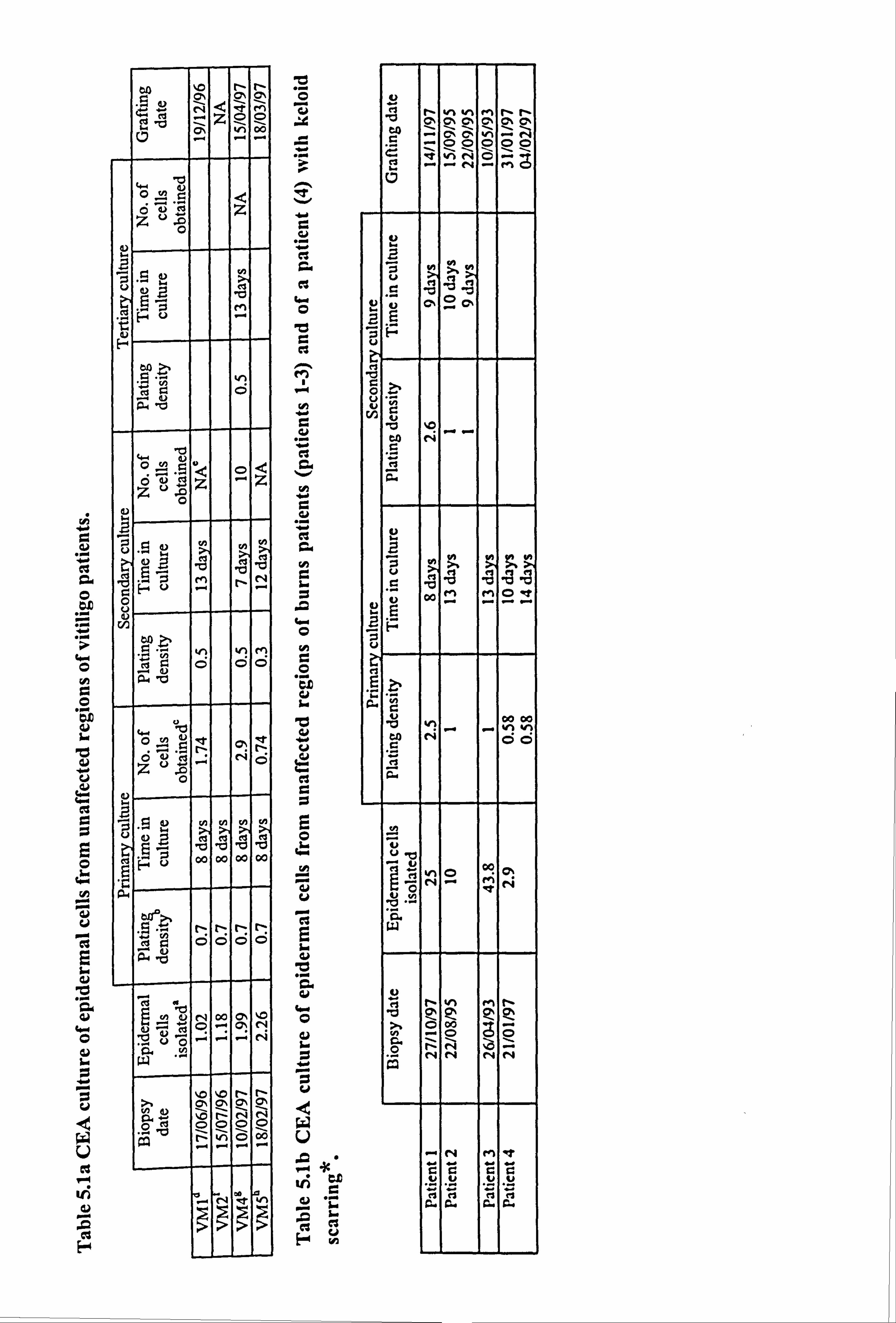
"O C
CA
.r CC
C
.r
C- C
H
C "r
it
b Q
r. +
Gur cl
rr
it
b
a
W
U
vi
F
u
.r
"r
G M
O
C e3
M "-r H
a 4.0
H
"r
H
t-I O
H
O "r
iI b
CLO , bd u
2
ci
cs
"O "r CO cu
irr
N.
r. +
ti "
C3 O H C. )
h
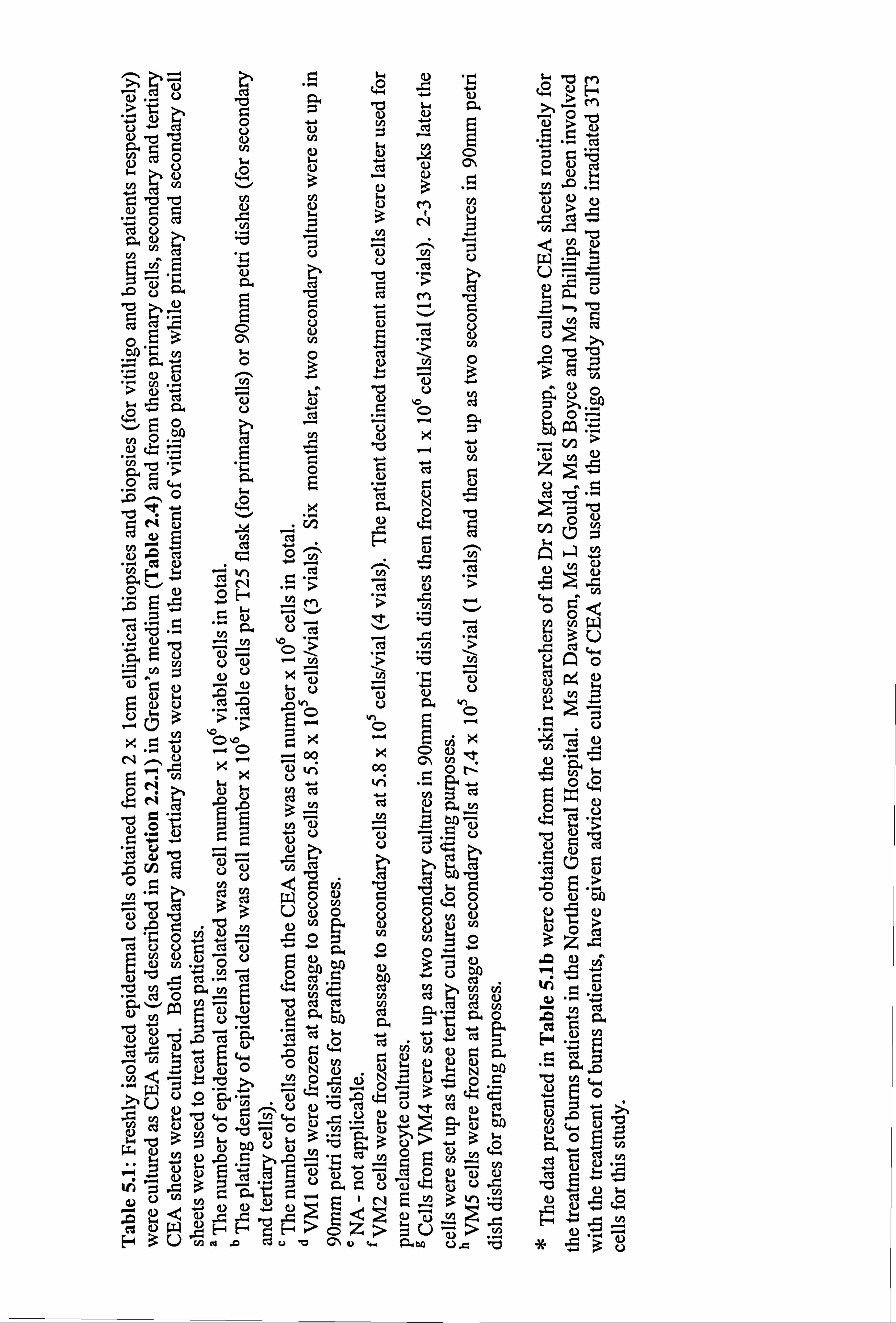
'ä cý r"CO,, ccý >
.nO Q) cad u -4 > ZJ
u4E ÖO Oý O cd
... Z 0 cu ce 4.2
to c3 w .OV "ýj
>
-NEQ5- ZJ =
03 rn 4) E ce U">,
rm. UCOÖ "b~A OÜ}..,
O im w'
Oý cd w+ pO- cVä u ".
C"
le +ý O-
-ri g" 0 ei
13
Ö
ý+ OC> cd a) >
0j 'd zi C"N- vý vý W .ý Cr U
OHCVCOVN
"äo 00 iC o C) `n .
00
7N iz CKJ U
p, rA pxO
"d `n ý bA ~ tý'i ý'
p "t'+
O v- N++O 9)
CIJ <or. u2 QÖÖÖC bA
o )0 -0 10 9) U) U g)
öö 't'. g E4
vOi U vOi
Op2 OO GOý. ý Z ,..,
3 .C vvv". ý p, OO 41
Z vv ßää ä äö
c & cl u cu
-t 2OO0O
N(u nO4 10.
ON to "" Q' JO O
0anýý u, 3oýu o aý,,,, N 75
ý"+ rn aý ý.
-4-4 o H3U ý0 ci
9. b rnu w aý ý. = vI *3
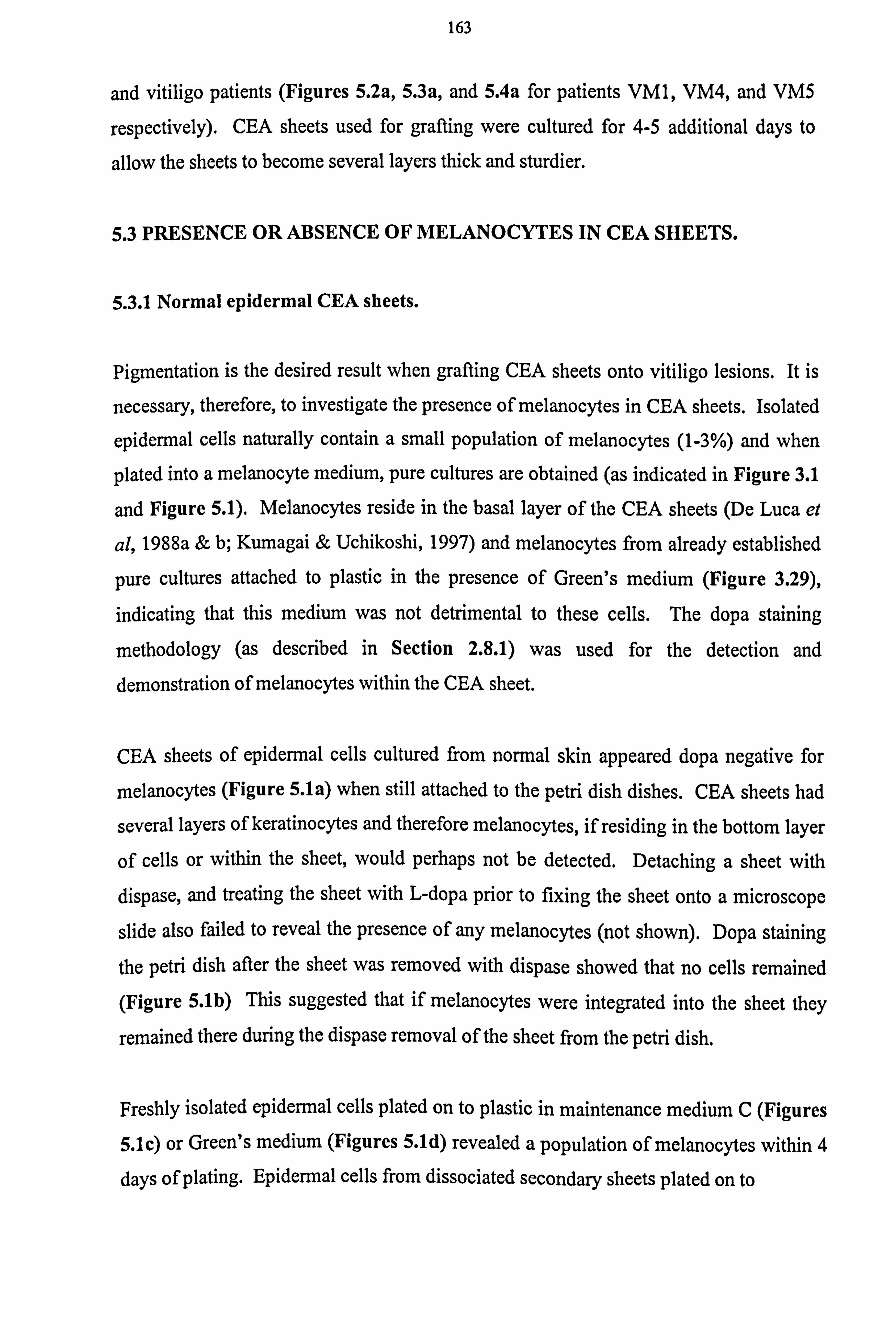
163
and vitiligo patients (Figures 5.2a, 5.3a, and 5.4a for patients VM1, VM4, and VM5
respectively). CEA sheets used for grafting were cultured for 4-5 additional days to
allow the sheets to become several layers thick and sturdier.
5.3 PRESENCE OR ABSENCE OF MELANOCYTES IN CEA SHEETS.
5.3.1 Normal epidermal CEA sheets.
Pigmentation is the desired result when grafting CEA sheets onto vitiligo lesions. It is
necessary, therefore, to investigate the presence of melanocytes in CEA sheets. Isolated
epidermal cells naturally contain a small population of melanocytes (1-3%) and when
plated into a melanocyte medium, pure cultures are obtained (as indicated in Figure 3.1
and Figure 5.1). Melanocytes reside in the basal layer of the CEA sheets (De Luca et
al, 1988a & b; Kumagai & Uchikoshi, 1997) and melanocytes from already established
pure cultures attached to plastic in the presence of Green's medium (Figure 3.29),
indicating that this medium was not detrimental to these cells. The dopa staining
methodology (as described in Section 2.8.1) was used for the detection and demonstration of melanocytes within the CEA sheet.
CEA sheets of epidermal cells cultured from normal skin appeared dopa negative for
melanocytes (Figure 5.1a) when still attached to the petri dish dishes. CEA sheets had
several layers of keratinocytes and therefore melanocytes, if residing in the bottom layer
of cells or within the sheet, would perhaps not be detected. Detaching a sheet with dispase, and treating the sheet with L-dopa prior to fixing the sheet onto a microscope
slide also failed to reveal the presence of any melanocytes (not shown). Dopa staining
the petri dish after the sheet was removed with dispase showed that no cells remained (Figure 5.1b) This suggested that if melanocytes were integrated into the sheet they
remained there during the dispase removal of the sheet from the petri dish.
Freshly isolated epidermal cells plated on to plastic in maintenance medium C (Figures
5.1c) or Green's medium (Figures 5.1d) revealed a population of melanocytes within 4
days of plating. Epidermal cells from dissociated secondary sheets plated on to

Figure 5.1 Dopa staining of normal epidermal cells in CEA sheets and as pure melanocytes. Epidermal cells were isolated from normal skin and some of these cells were grown as CEA sheets (as described in Section 2.2.1) then treated with L-dopa (as described in Section 2.8.1) (a). CEA sheets were carefully removed with dispase (as described in Section 2.2.2) and the remaining petri dish was treated with L-dopa (b). Additionally, some freshly isolated cells were plated onto plastic in maintenance medium C (c) or Green's (d). Pure culture cells were treated with L-dopa 4 days after plating. Epidermal cells from dissociated secondary CEA sheets were cultured on plastic in maintenance medium C (e) or Green's medium (f). Cells were treated with L- dopa 4 days after plating. At this time melanocytes could be clearly seen in all the cultures. Scale bars represent 15µm (a-b) and 30µm (c-f).
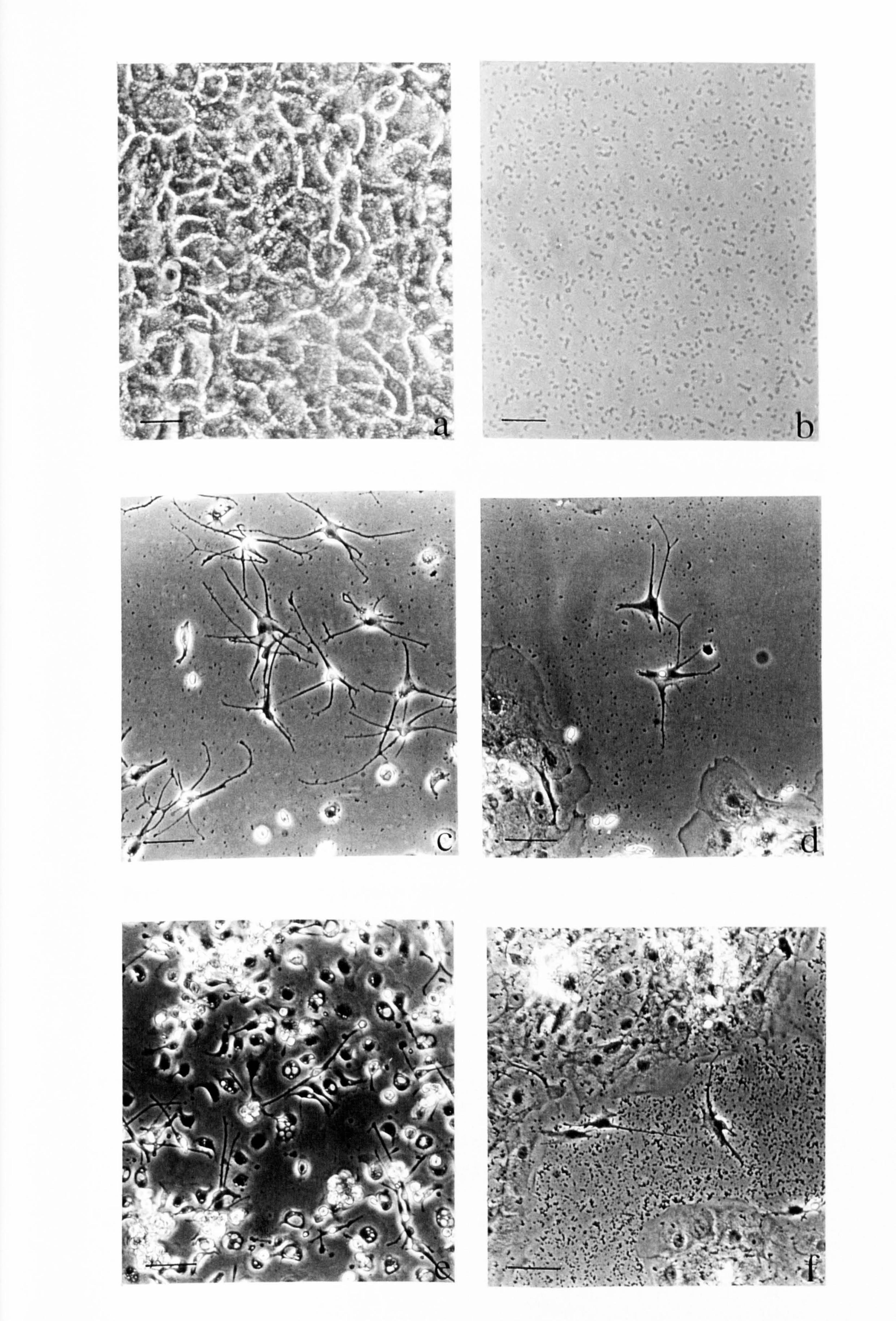
'r "
ýý.
ýýýý
`1t' !-a tr as 1
1°
i" Y r'
1`1 `
ia!
i
iý !rt `!
"O"e
S ! ti
1.
A°61
Q ,I1 1`
a1 .4t.
sir sb
.. . Alp
{i 1
..
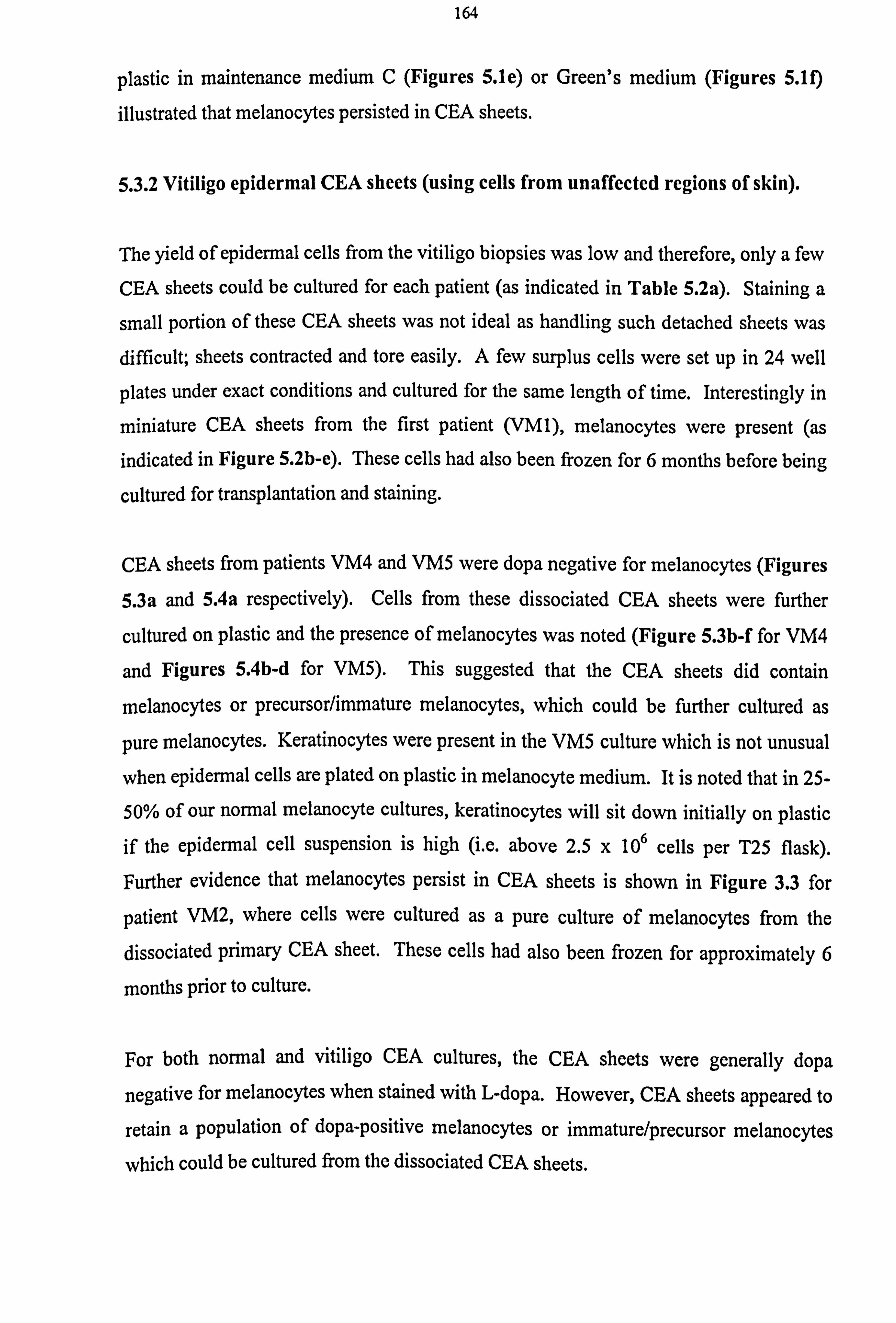
164
plastic in maintenance medium C (Figures 5.1e) or Green's medium (Figures 5.10
illustrated that melanocytes persisted in CEA sheets.
5.3.2 Vitiligo epidermal CEA sheets (using cells from unaffected regions of skin).
The yield of epidermal cells from the vitiligo biopsies was low and therefore, only a few
CEA sheets could be cultured for each patient (as indicated in Table 5.2a). Staining a
small portion of these CEA sheets was not ideal as handling such detached sheets was
difficult; sheets contracted and tore easily. A few surplus cells were set up in 24 well
plates under exact conditions and cultured for the same length of time. Interestingly in
miniature CEA sheets from the first patient (VM1), melanocytes were present (as
indicated in Figure 5.2b-e). These cells had also been frozen for 6 months before being
cultured for transplantation and staining.
CEA sheets from patients VM4 and VM5 were dopa negative for melanocytes (Figures
5.3a and 5.4a respectively). Cells from these dissociated CEA sheets were further
cultured on plastic and the presence of melanocytes was noted (Figure 5.3b-f for VM4
and Figures 5.4b-d for VM5). This suggested that the CEA sheets did contain
melanocytes or precursor/immature melanocytes, which could be further cultured as
pure melanocytes. Keratinocytes were present in the VM5 culture which is not unusual
when epidermal cells are plated on plastic in melanocyte medium. It is noted that in 25-
50% of our normal melanocyte cultures, keratinocytes will sit down initially on plastic if the epidermal cell suspension is high (i. e. above 2.5 x 10⁶ cells per T25 flask).
Further evidence that melanocytes persist in CEA sheets is shown in Figure 3.3 for
patient VM2, where cells were cultured as a pure culture of melanocytes from the
dissociated primary CEA sheet. These cells had also been frozen for approximately 6
months prior to culture.
For both normal and vitiligo CEA cultures, the CEA sheets were generally dopa
negative for melanocytes when stained with L-dopa. However, CEA sheets appeared to
retain a population of dopa-positive melanocytes or immature/precursor melanocytes
which could be cultured from the dissociated CEA sheets.

Figure 5.2 Dopa staining of melanocytes in a CEA sheet. Epidermal cells were isolated from patient VM1 biopsy and cultured as primary CEA sheets (as described in Section 2.2.1), then frozen. Surplus cells from frozen stocks were plated into 24 well plates and cultured for 13 days. They were fixed and treated with PBS (a) or L-dopa (as described in Section 2.8.1) (b-e). Scale bars represent 15µm.
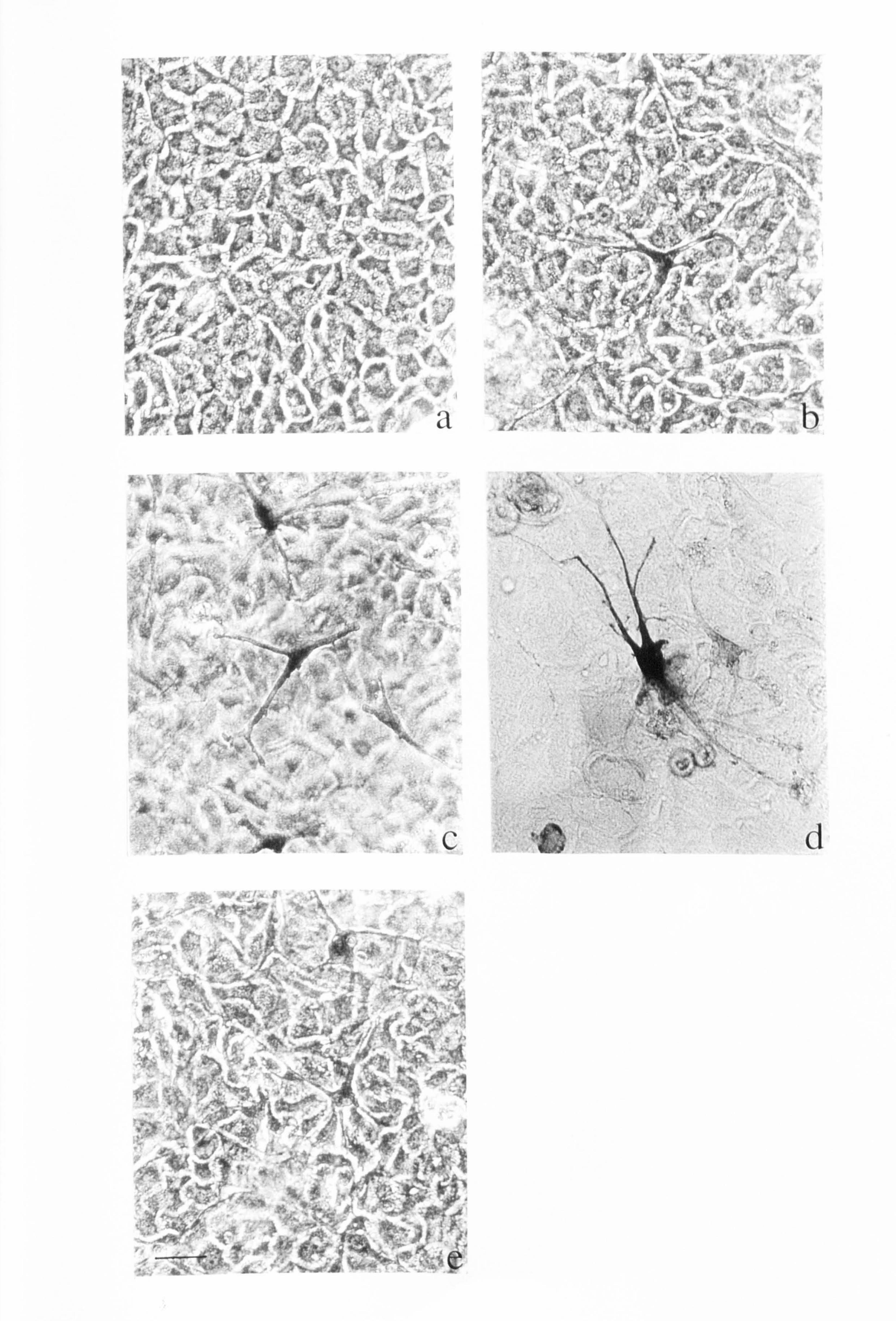
4r
i9 j
K "i
j"""ý ", ý ý}PýW
ý'. `ý. .yý;
ý' \ý
. '« ýý .
ý ". F
.:
n ., ý, Y^
d
Zp
r. q rR-
Ia
lit"
r, ý.

Figure 5.3 Dopa staining of a CEA sheet and pure culture melanocy-tes from a CEA sheet. Epidermal cells were isolated from patient VM4 biopsy and cultured as primary and secondary CEA sheets (as described in Section 2.2.1) before being frozen. Tertiary cells were cultured as CEA sheets in 24 well plates and treated with L-dopa (a) (as described in Section 2.8.1). Additional tertiary cells were cultured as pure melanocyte cultures on plastic in maintenance medium C (b for PBS or c-d for L-dopa treated cells) or Green's medium (e-f for L-dopa treated cells). Scale bars represent 15µm (a, d, f) and 30 µm (b, c, e)
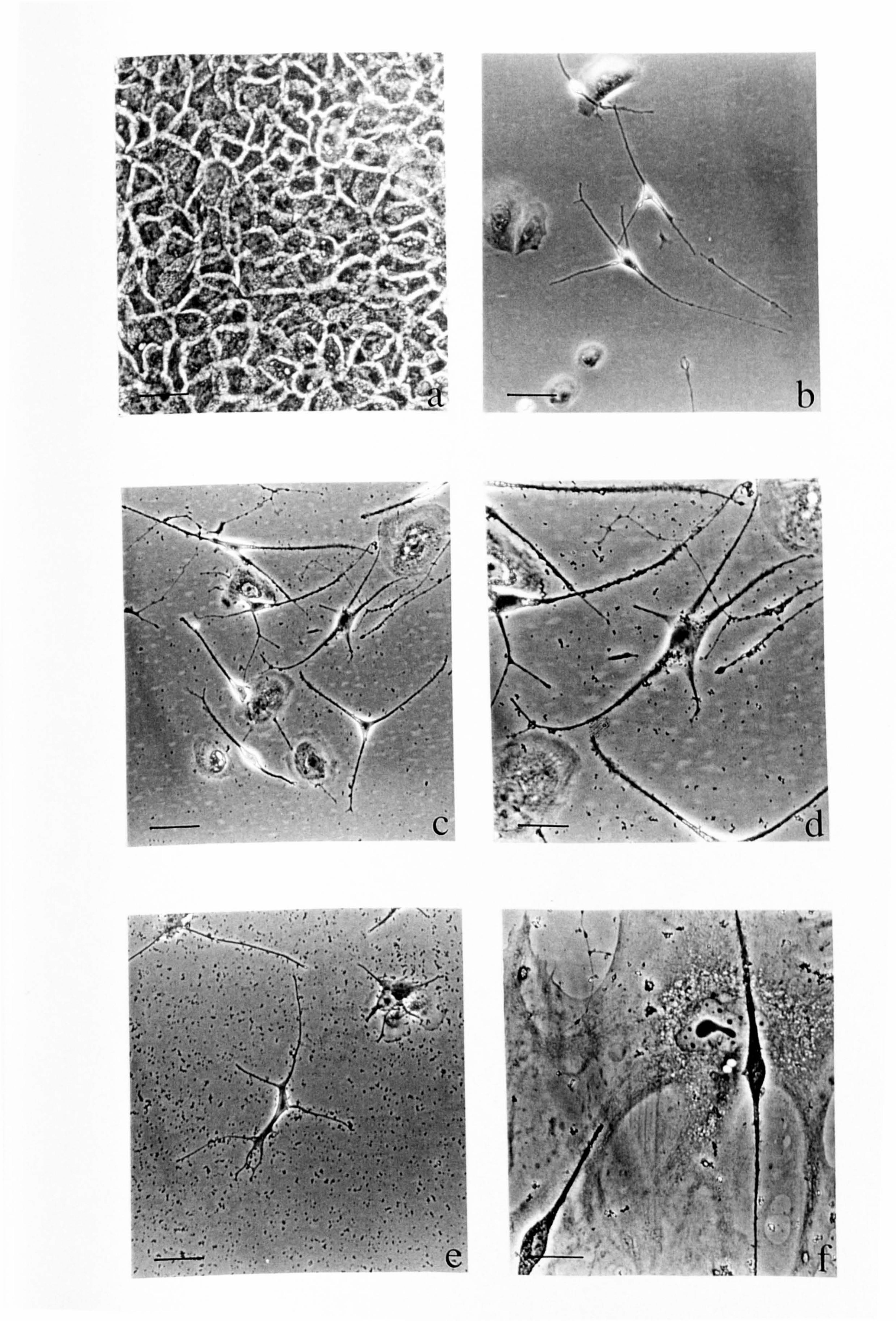
, l'.
.
C
r to -
ýý
ý. ý, " ,. "
4 oo,
-ý b
.......
V' 141
I
r
a' ,. ý'
f
.4
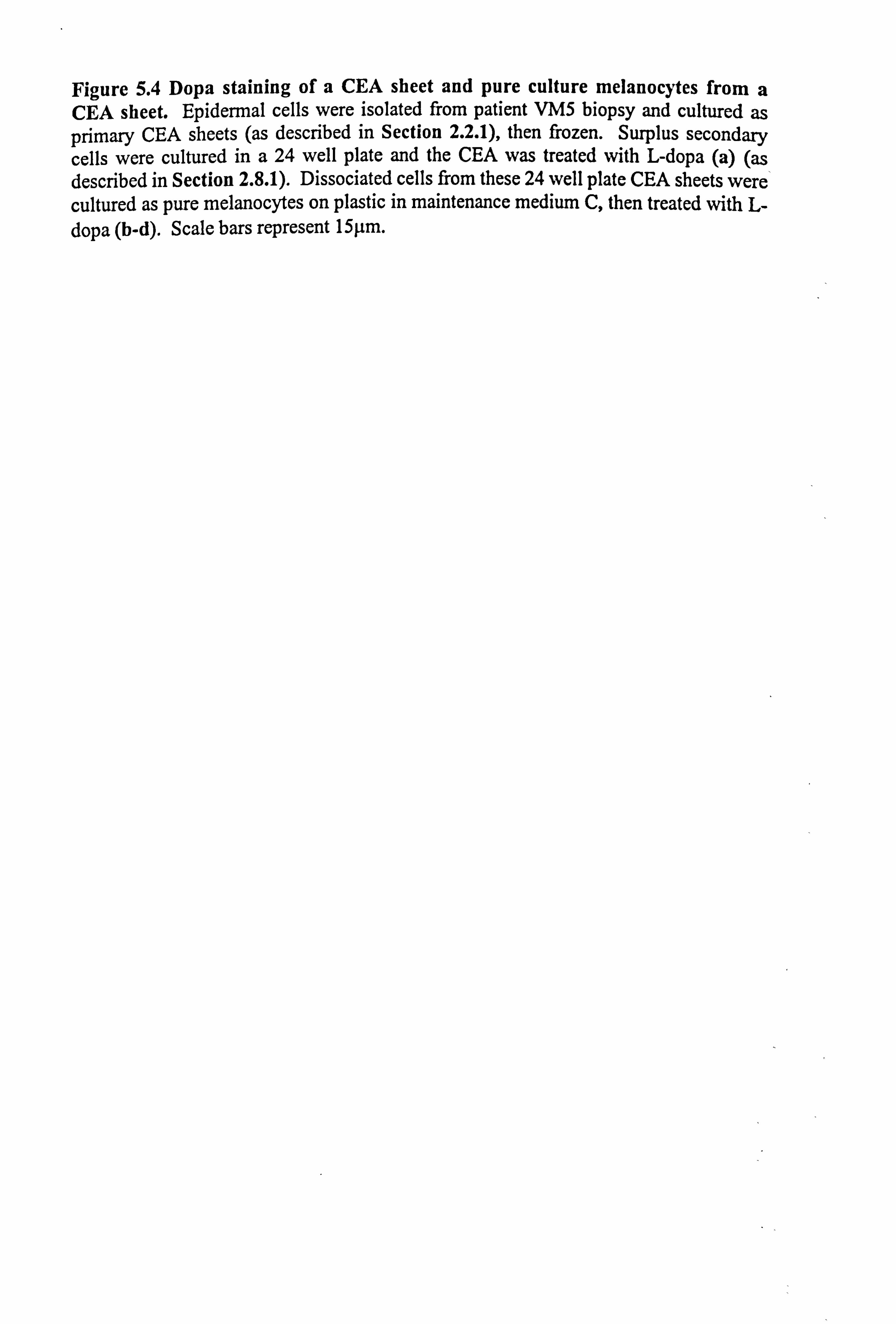
Figure 5.4 Dopa staining of a CEA sheet and pure culture melanocytes from a CEA sheet. Epidermal cells were isolated from patient VM5 biopsy and cultured as primary CEA sheets (as described in Section 2.2.1), then frozen. Surplus secondary cells were cultured in a 24 well plate and the CEA was treated with L-dopa (a) (as described in Section 2.8.1). Dissociated cells from these 24 well plate CEA sheets were cultured as pure melanocytes on plastic in maintenance medium C, then treated with L- dopa (b-d). Scale bars represent 15µm.
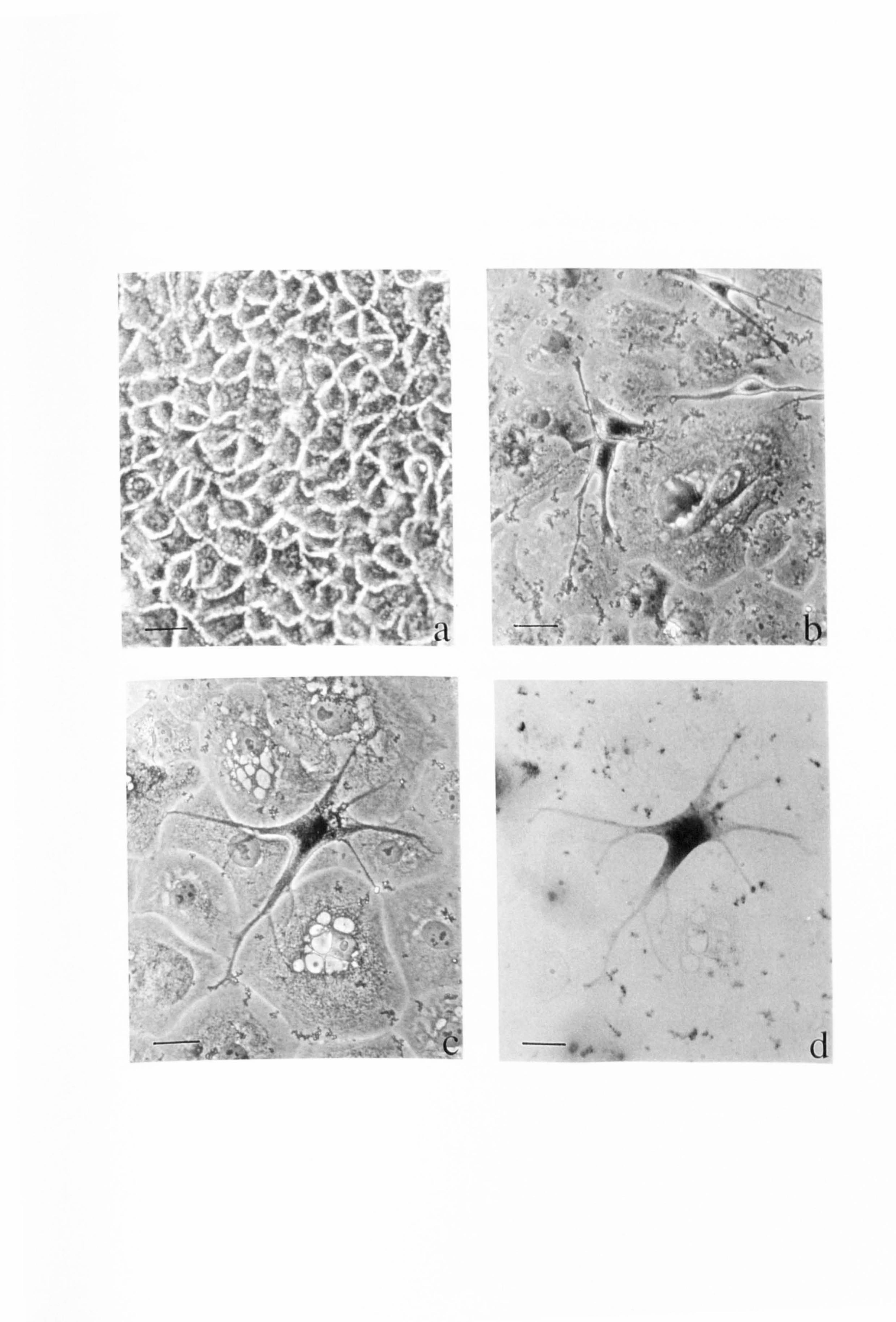
ILMI
Pik
_ ; ý`: w, ýý:
,, '. it
"
f 1. ý "' �F
Atý .*ý:
ý` "" ". `;
Fry ql, :,
ý... -"
161
165
5.4 GRAFTING OF CEA SHEETS ONTO VITILIGO LESIONS.
5.4.1 Case study 1- patient VM1
The patient had symmetrical vitiligo as shown for her left and right arms just below the
elbows (Figure 5.5a and 5.5b respectively). The patient developed vitiligo in 1995,
with no association with other autoimmune disorders. The sister of the patient also
suffered from vitiligo. At the time of biopsy, in June 1996 the vitiligo was stable. The
patient had been using topical Betnovate RD ointment prior to the biopsy. The patient
underwent surgery in December 1996. The site of the biopsy healed without
complications or Koebnerisation.
The lesion on the patient's left forearm (Figure 5.5c) was chosen as the grafting site.
The patient was dermabraded (Figure 5.5d; Figure 5.6 illustrates the dermabrader) and
the CEA sheets applied (Figures 5.5e and 5.5f; Figure 5.7 illustrates the CEA attached
to the Tegapore backing dressing prior to grafting). The CEA applied to the lower
region had a small hole in it. The region was dressed with yelonet, gauze and crepe.
The backing of the CEA was removed 11 days after grafting. Initially the region was
red and had a few crusty regions present (Figure 5.5g) but it became evident after only a
few weeks that there was some pigmentation and within two months, the inflammation
had settled down and pigmentation could be clearly seen (Figure 5.5h). The patient
used moisturiser once the site had healed. A white border remained where the CEA
sheets did not cover the dermabraded area.
Repigmentation of the majority of the region had occurred within nine months of
grafting and the pigmentation also appeared to have spread slightly (personal
communication with Dr DJ Gawkrodger). The patient did not receive any additional
PUVA treatment and sun exposure to the region was minimal during the healing and
follow up period.
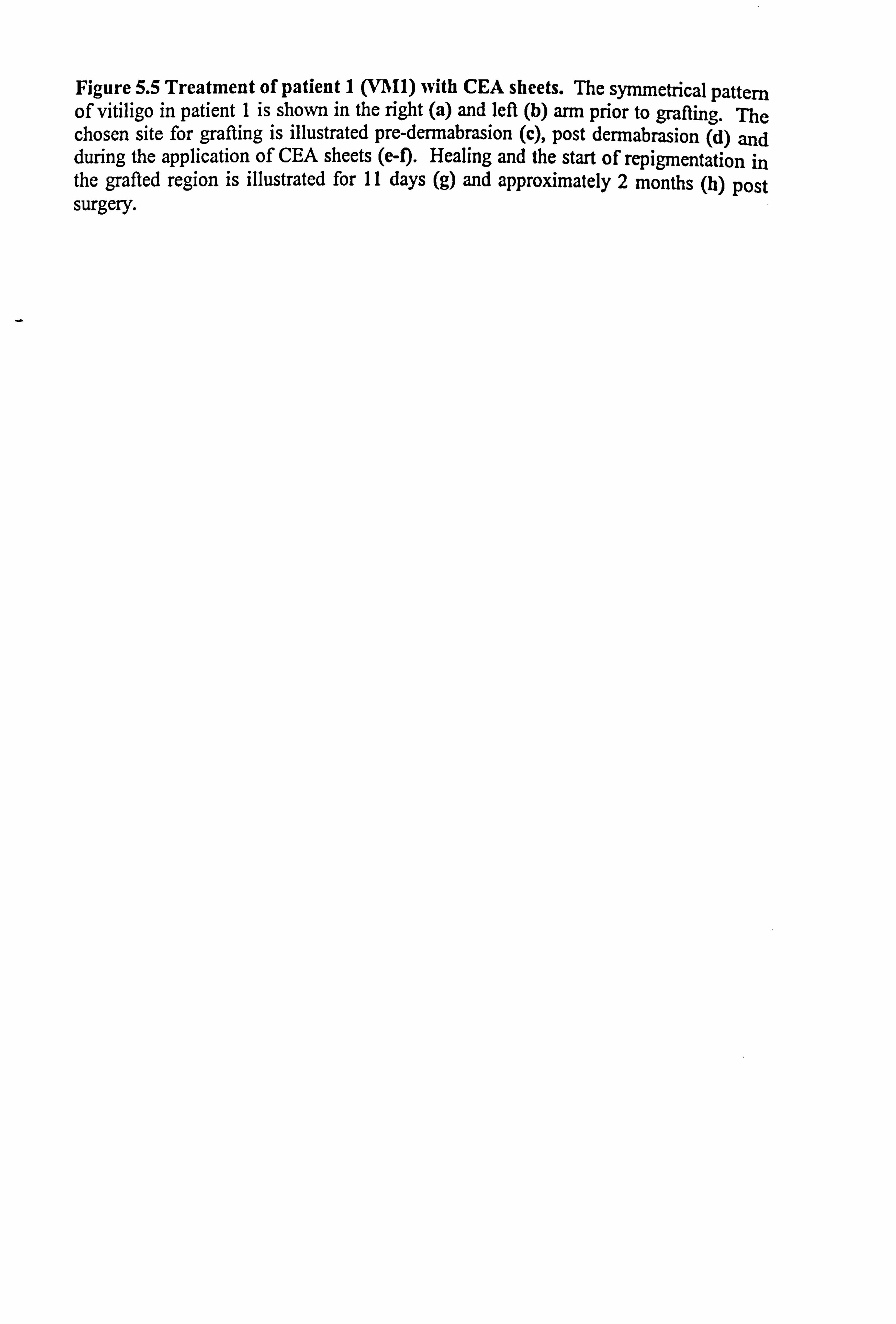
Figure 5.5 Treatment of patient 1 (V1111) with CEA sheets. The symmetrical pattern of vitiligo in patient 1 is shown in the right (a) and left (b) arm prior to grafting. The chosen site for grafting is illustrated pre-dermabrasion (c), post dermabrasion (d) and during the application of CEA sheets (e-f). Healing and the start of repigmentation in the grafted region is illustrated for 11 days (g) and approximately 2 months (h) post surgery.
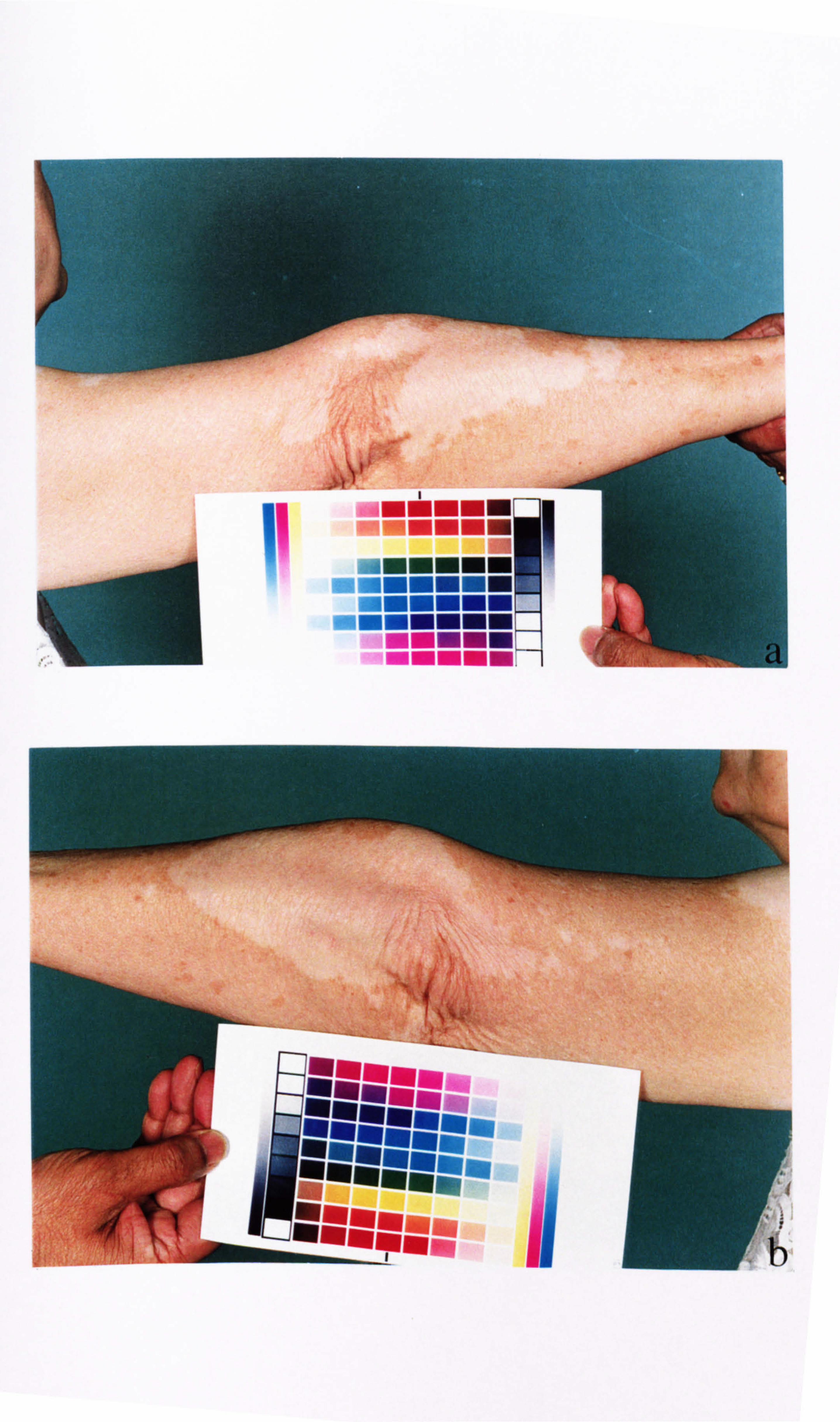
{
114
mi
U-0
amm
,ý
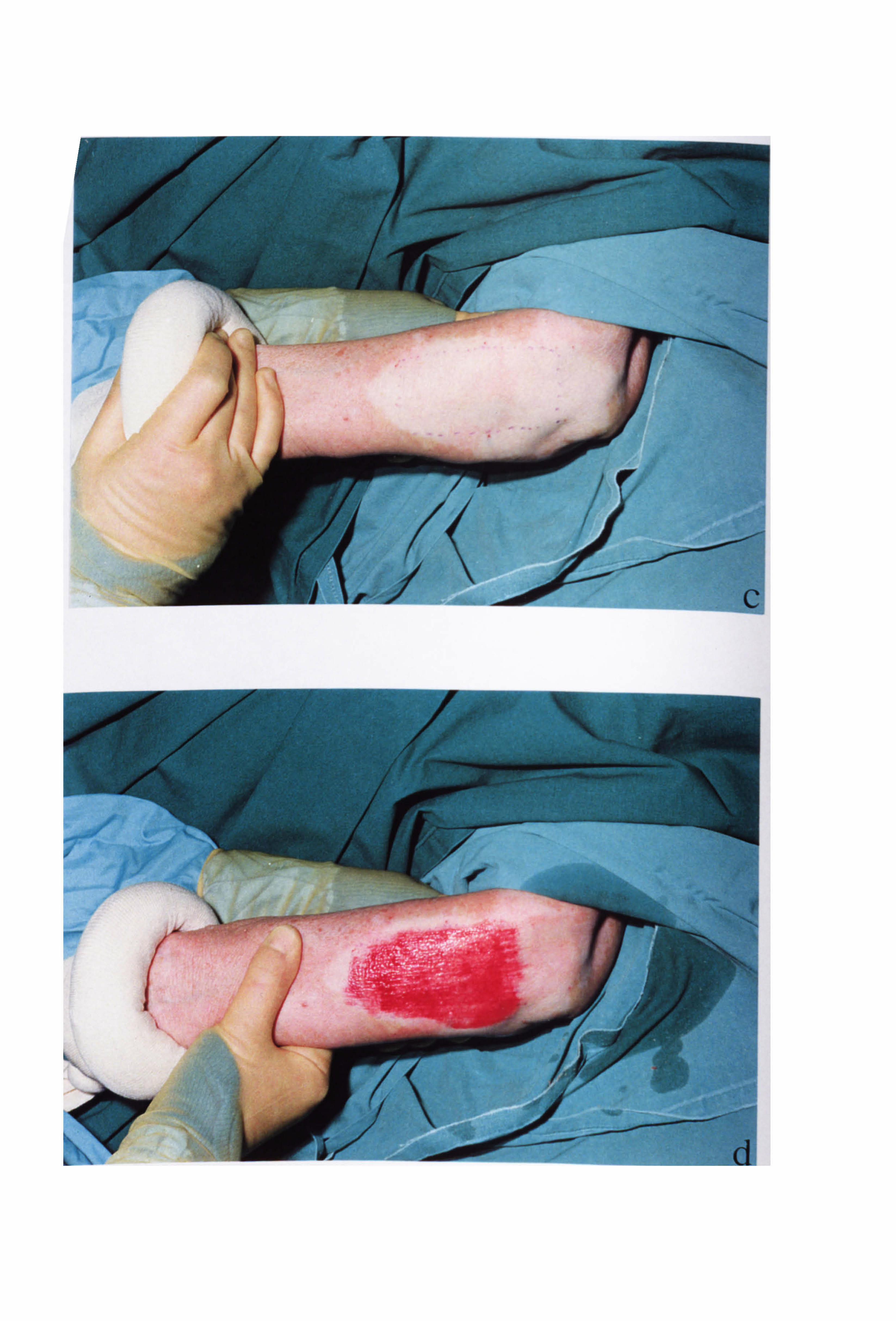
r-
ýs. p,.
S...
1.
ýý
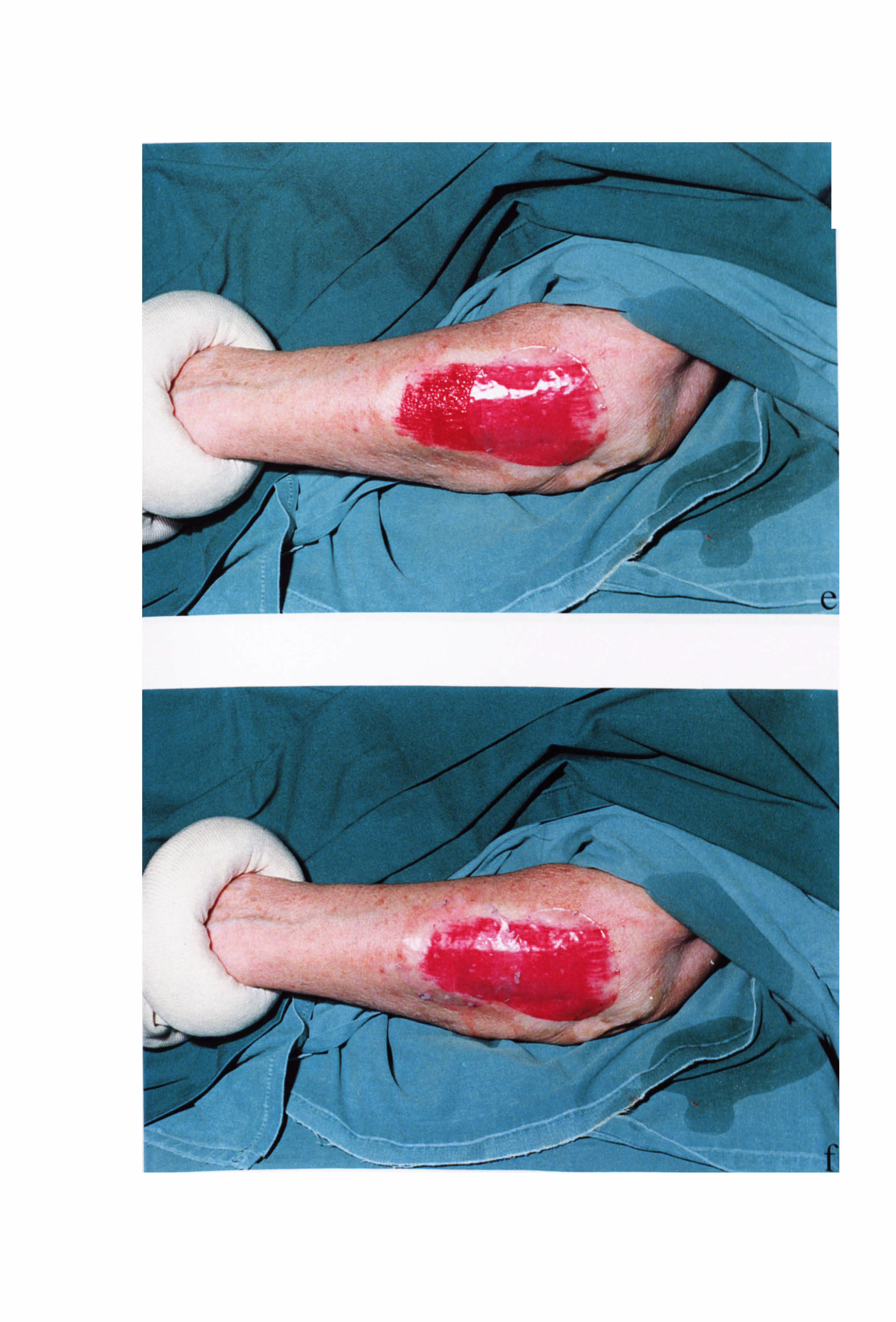
0
1
vif i ý' ýý ..........
, oo" ý.
)
- F------
- -. -------------.
-
/ I-
-. N-
e
woo,
f
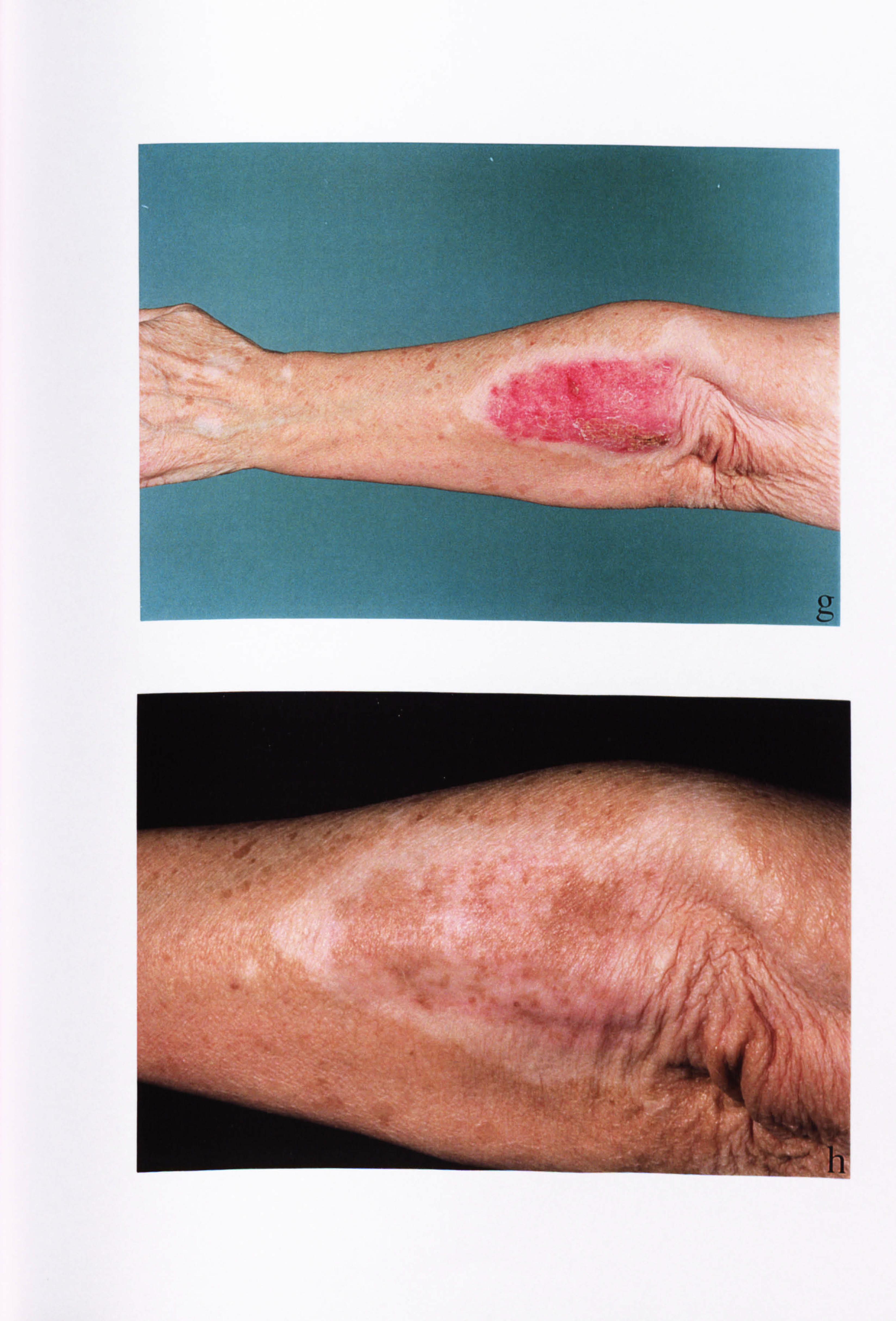
ý.. ý`,
w; :;, -ý, ý
.. ý, -
ý: -'. a, ßä
ýý ýc
R
ýi'iiý4 rý, %f
)ý
1ý'
s , "i'.. /'
/'40
. ý.
g

Figure 5.6 The dermabrador.
Figure 5.7 The CEA sheet. Epidermal cells (in this case from patient VM1) were cultured on i3T3 cells in Green's medium (as described in Section 2.2.1) up to secondary culture. On the day of grafting the sheet was carefully detached from the petri dish with dispase and attached to a Tegapore backing dressing (as described in Section 2.2.2). Until grafting the sheet was immersed in Green's medium (lacking CT and FCS).
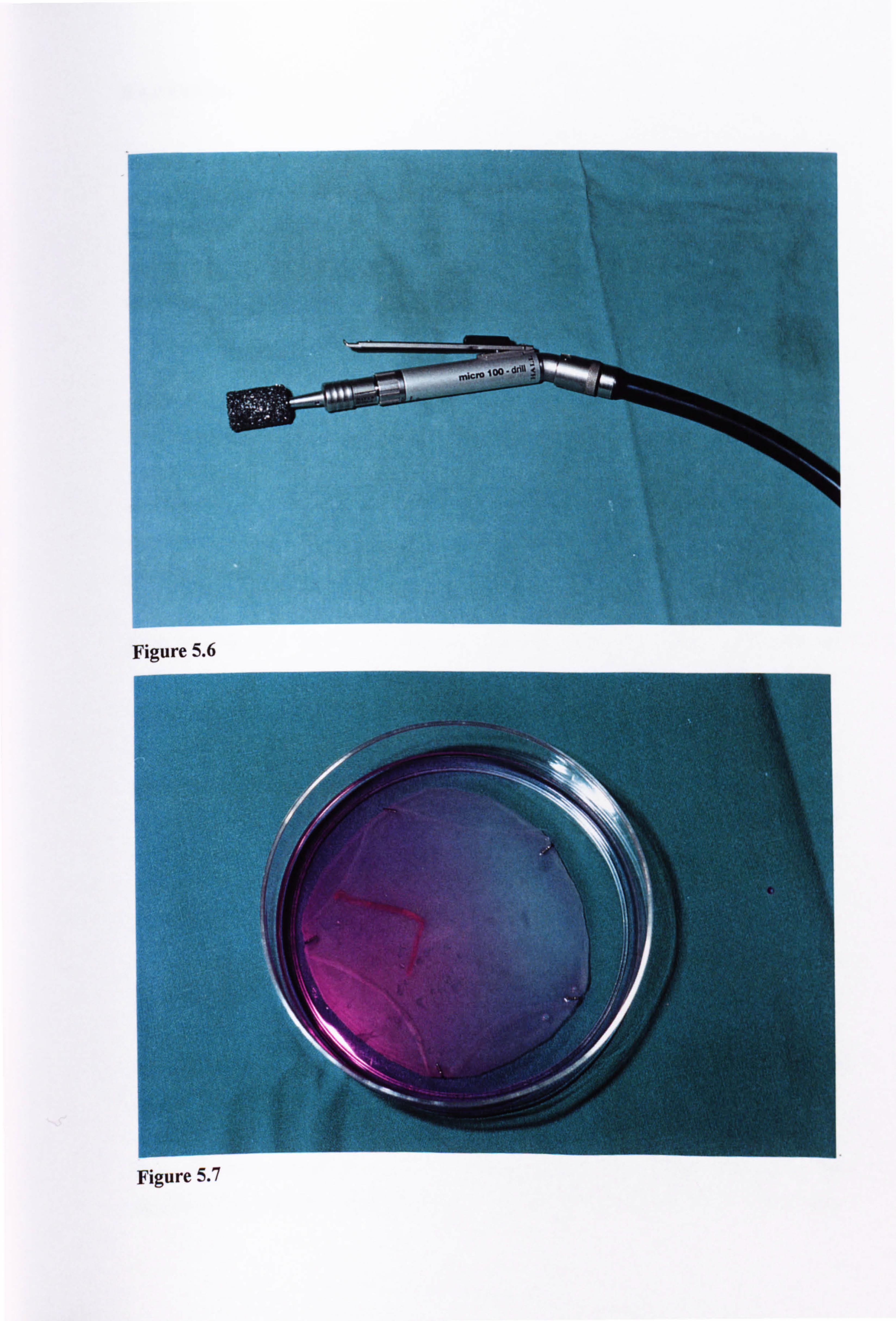
Figure 5.6
Figure 5.7
166
5.4.2 Case study 2- patient VM4
The patient had extensive symmetrical vitiligo which began in 1982 (the symmetry of
lesions is noted on the wrists, Figure 5.8a). At the time of biopsy, the vitiligo was
stable and the patient had received no treatment for the condition. Although the patient
had no association with other autoimmune diseases, an aunt had vitiligo and thyroid
disease. The patient underwent surgery in April 1997.
The ulnar border and palmar aspect of the right wrist (Figures 5.8b) and the right
olecranon (elbow region) (Figure 5.8c) were selected for treatment. Once the CEA
sheets were applied the areas were dressed as for patient 1 (Section 5.4.1) and the elbow
and wrist immobilised for the first few weeks. The healing was slow (Figure 5.8b-c),
and the elbow rubbed raw within the first week. The backing of the CEA sheets was
removed a few weeks after surgery. The areas were still tender several months after
grafting (Figures 5.8d-f).
Mild hypertrophic scarring became obvious by September 1997. This was an
unfortunate complication which has since largely resolved (as noted by Dr DJ
Gawkrodger, June, 1998). At the time of scarring the patient was in the early stages of
pregnancy although the pregnancy began post-grafting. Despite these complications,
there has been a small amount of repigmentation (in localised areas) within the grafted
sites, particularly at the wrist. The donor site did not scar or develop the Koebner
phenomenon.
5.4.3 Case study 3- Patient VM5.
The patient had extensive symmetrical vitiligo (Figure 5.9a) which was active prior to
the biopsy and has since continued after the biopsy. The donor site healed without Koebnerisation. The patient had not received any active topical treatment to the vitiligo
lesions. The patient underwent surgery March 1997.
Two regions on the lower back were chosen for grafting. Figure 5.9b shows the
regions a few months after grafting and illustrates the positioning of the chosen sites.
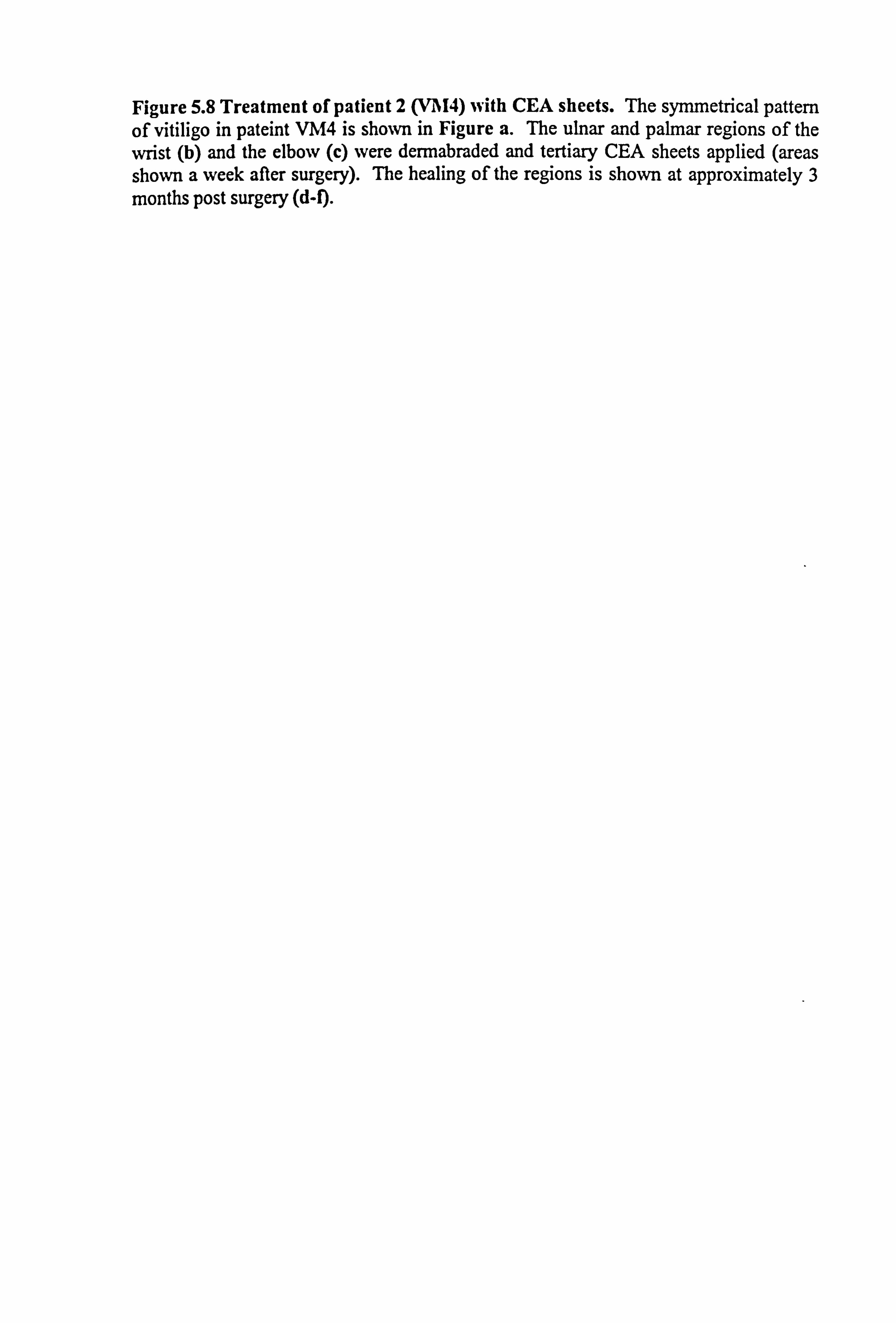
Figure 5.8 Treatment of patient 2 (Vl'I4) with CEA sheets. The symmetrical pattern of vitiligo in pateint VM4 is shown in Figure a. The ulnar and palmar regions of the wrist (b) and the elbow (c) were dermabraded and tertiary CEA sheets applied (areas shown a week after surgery). The healing of the regions is shown at approximately 3 months post surgery (d-1).
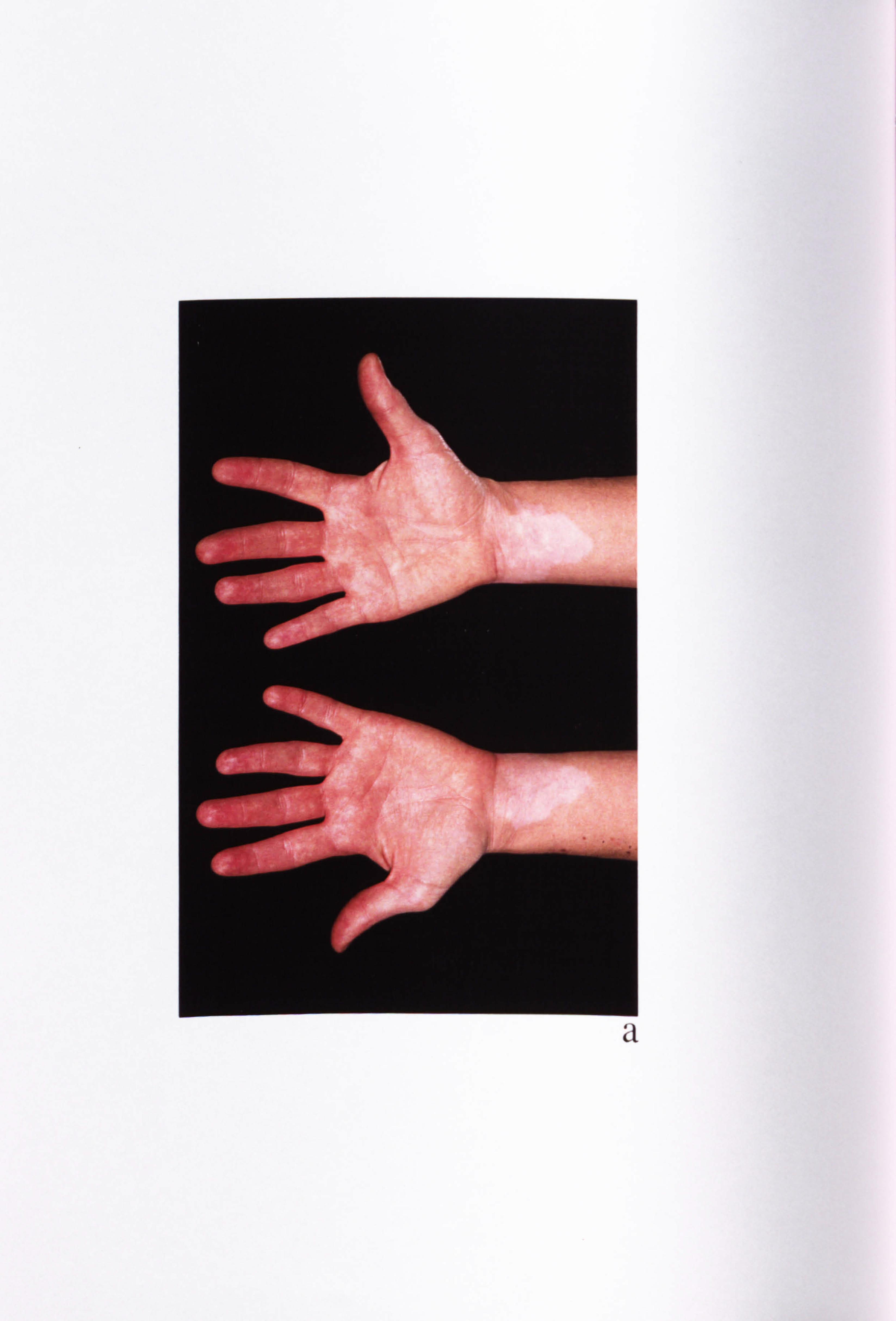
a
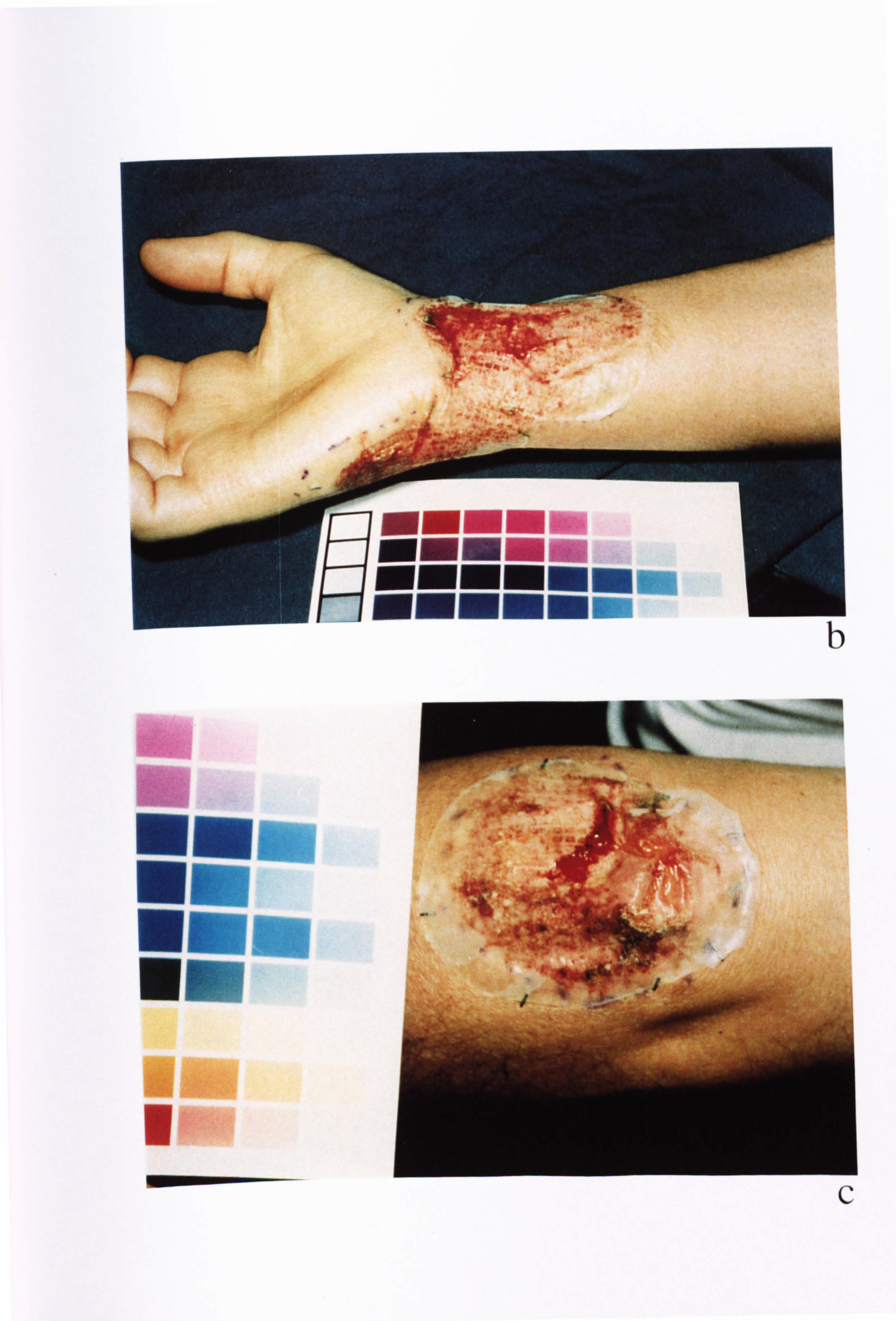
ý'
,:
b
-
ýi ýs
C
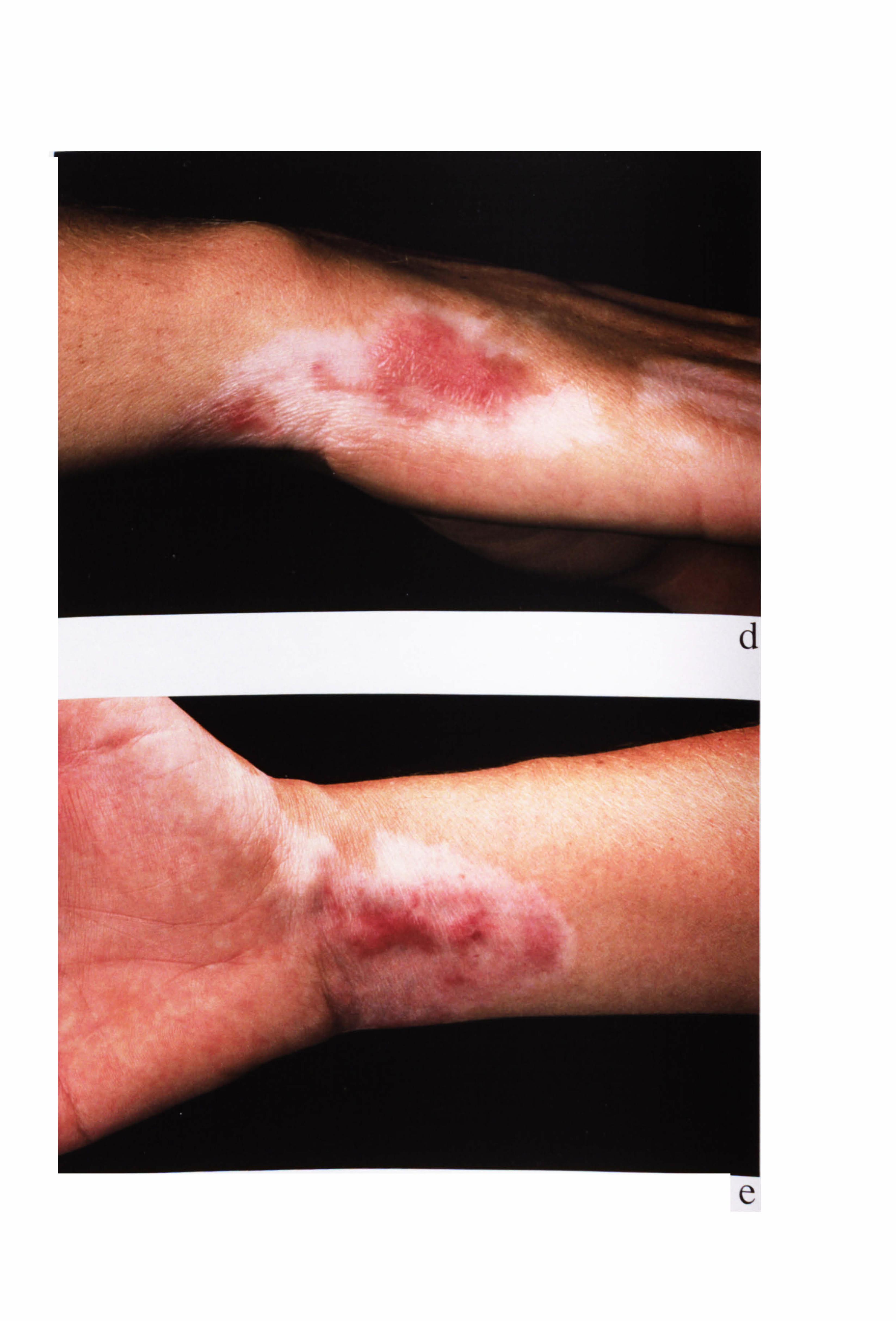
-
e
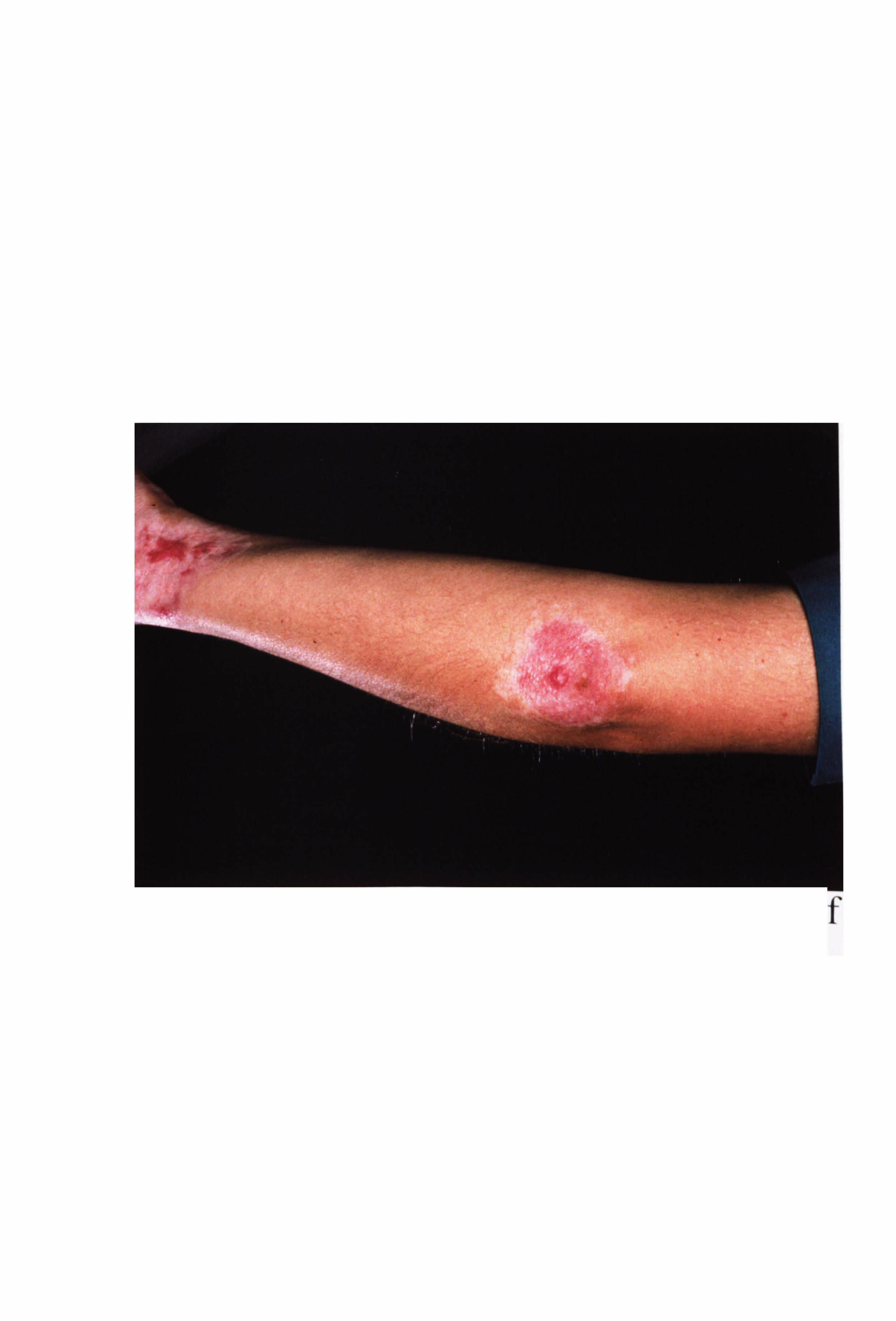
f

Figure 5.9 Treatment of patient 3 (VM5) with CEA sheets. The extensive symmetry of vitiligo in patient 3 is illustrated for the back (a). The chosen sites for grafting are shown approximately 2-3 weeks after surgery (b), while close up photographs of the left (c) and right (d) side are shown prior to dermabrasion and for the right/left side post dermabrasion (e) and CEA application (f). The healing of the regions are shown for 1
week (g-h), 2/3 weeks (i-j) and 2 months (k-1) post surgery. .
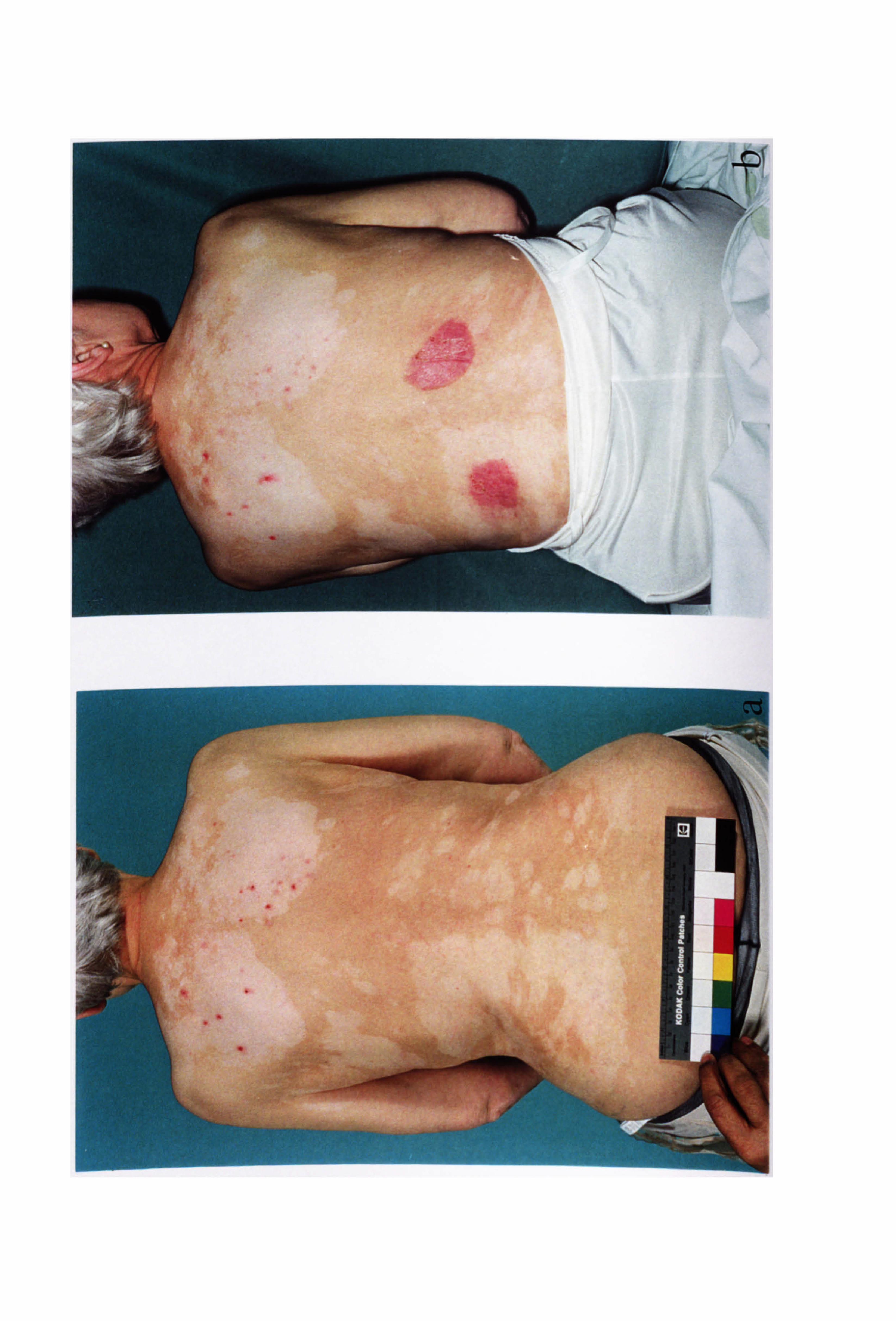
-
.. ,,
cr
1
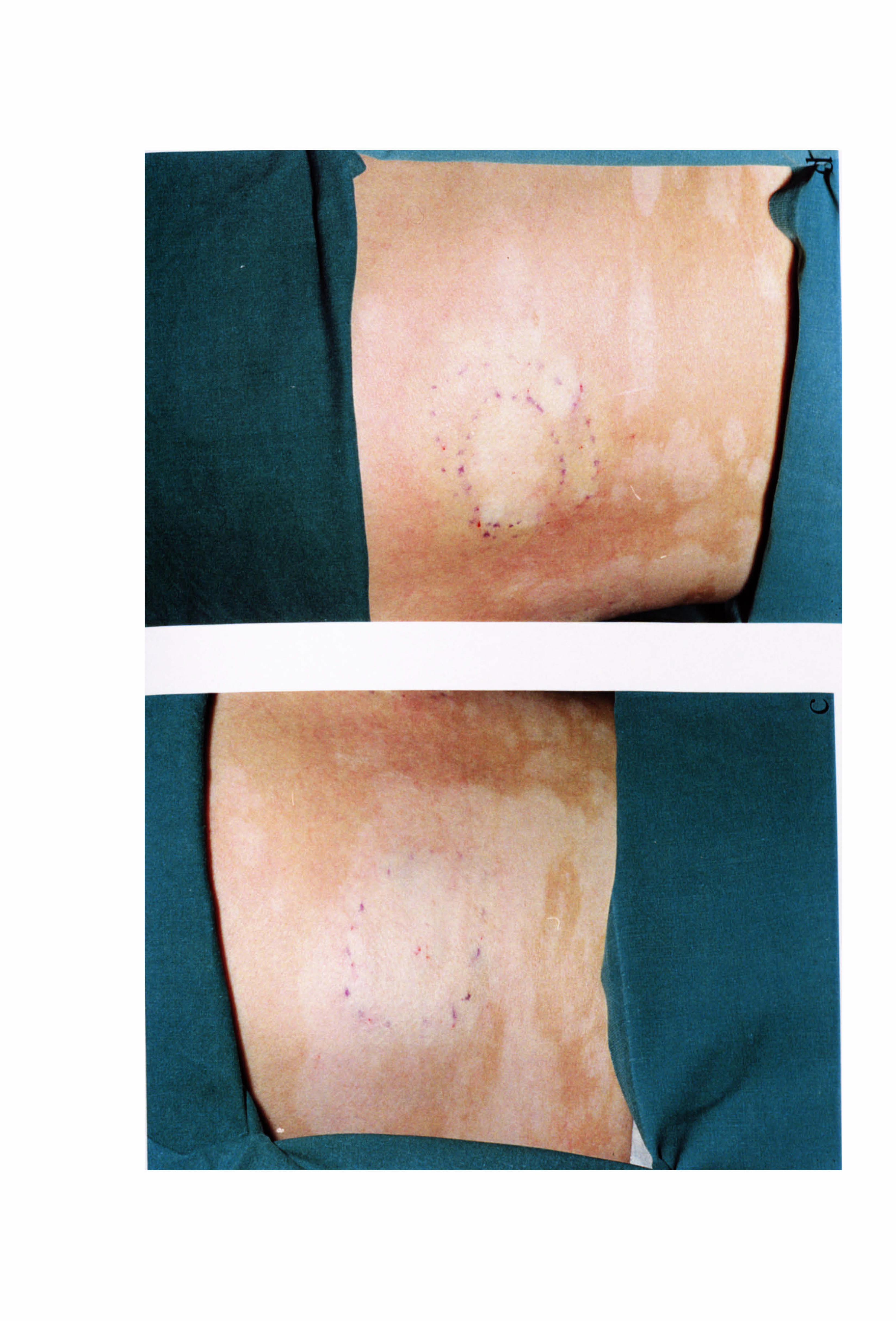
--
1%
C-)
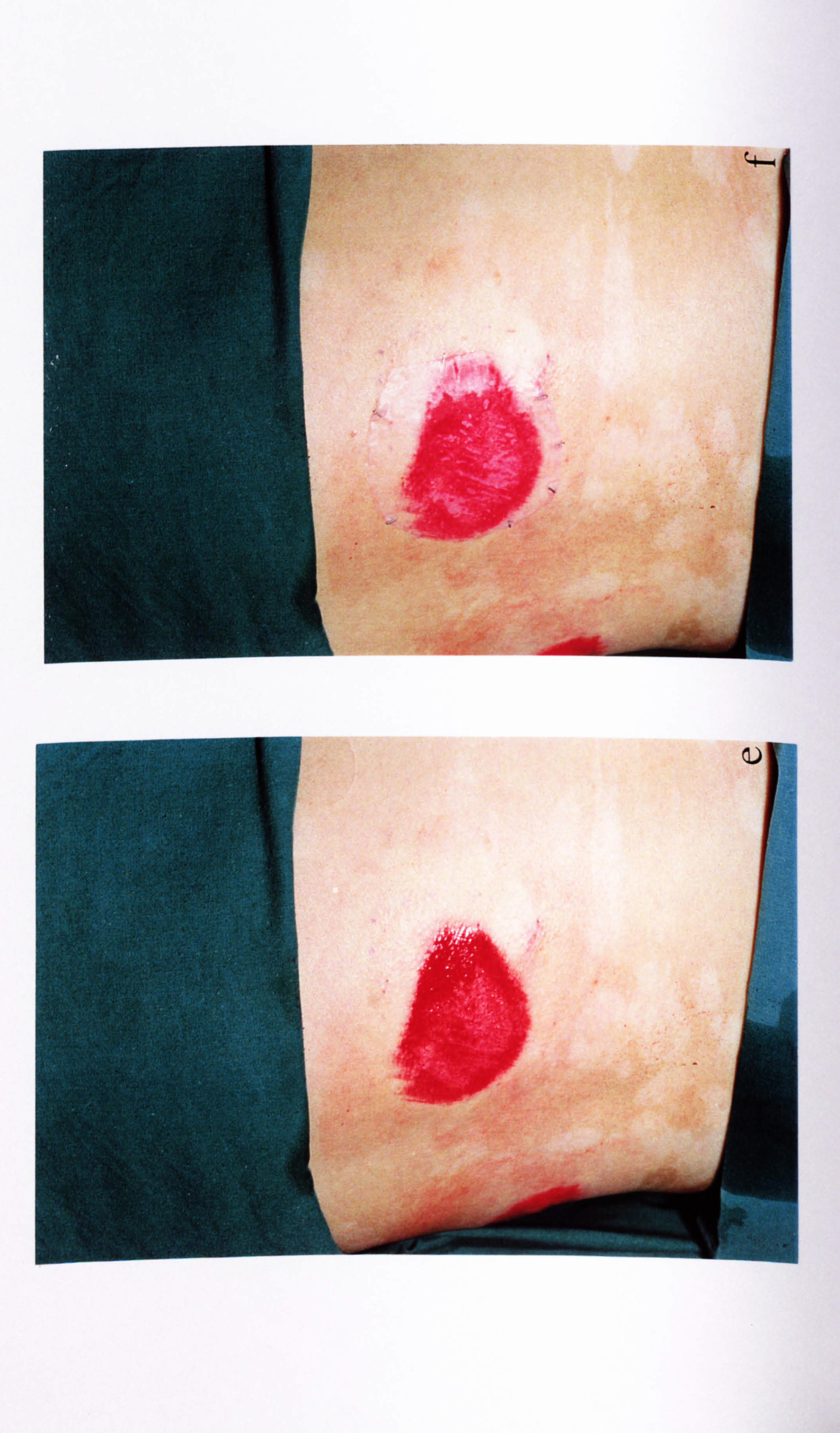
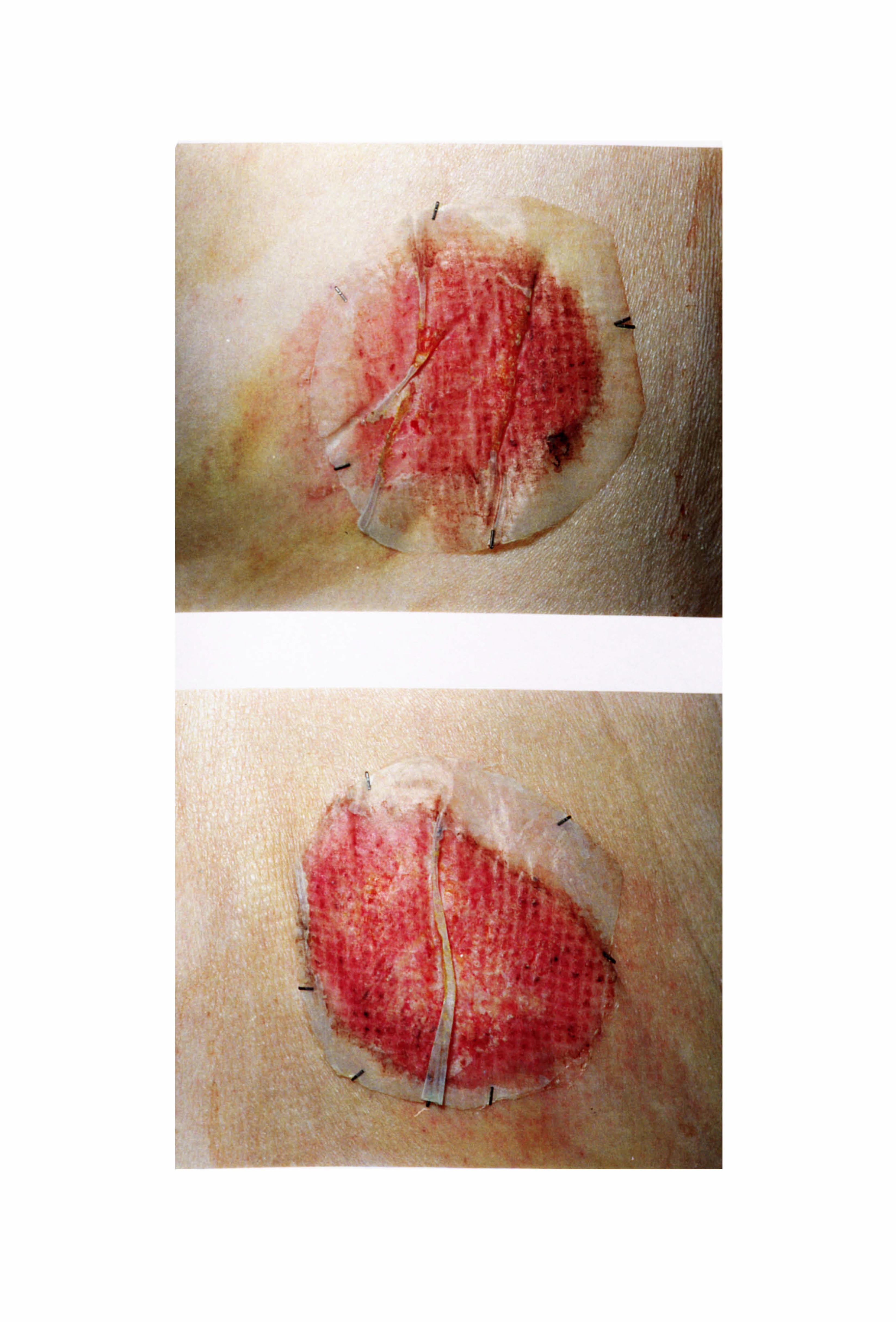
"a mift-,
owl. tA
wi,
t.: ýýb: ý
. #Výý
'
. t4
4V
rýw
.
ýrýý"sýi14 t ý+M
/
ýy /;, i
ýýý
.ýä
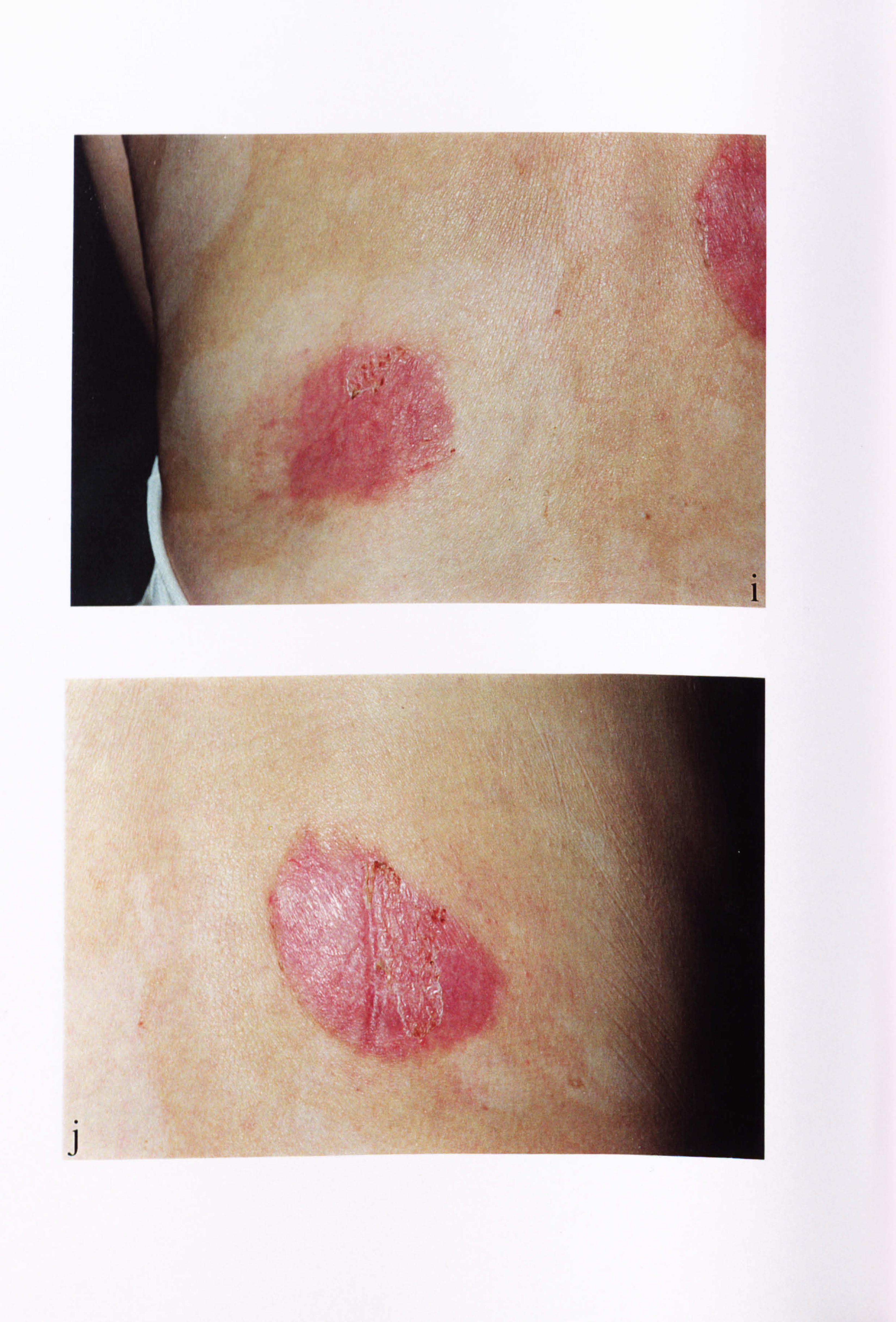
,t ti
-' ýi;
'ý
0
1
J
i, ýýý
ý'
;ý
ý' ,ýy
_!
\ý45
, '. Y' ýýL
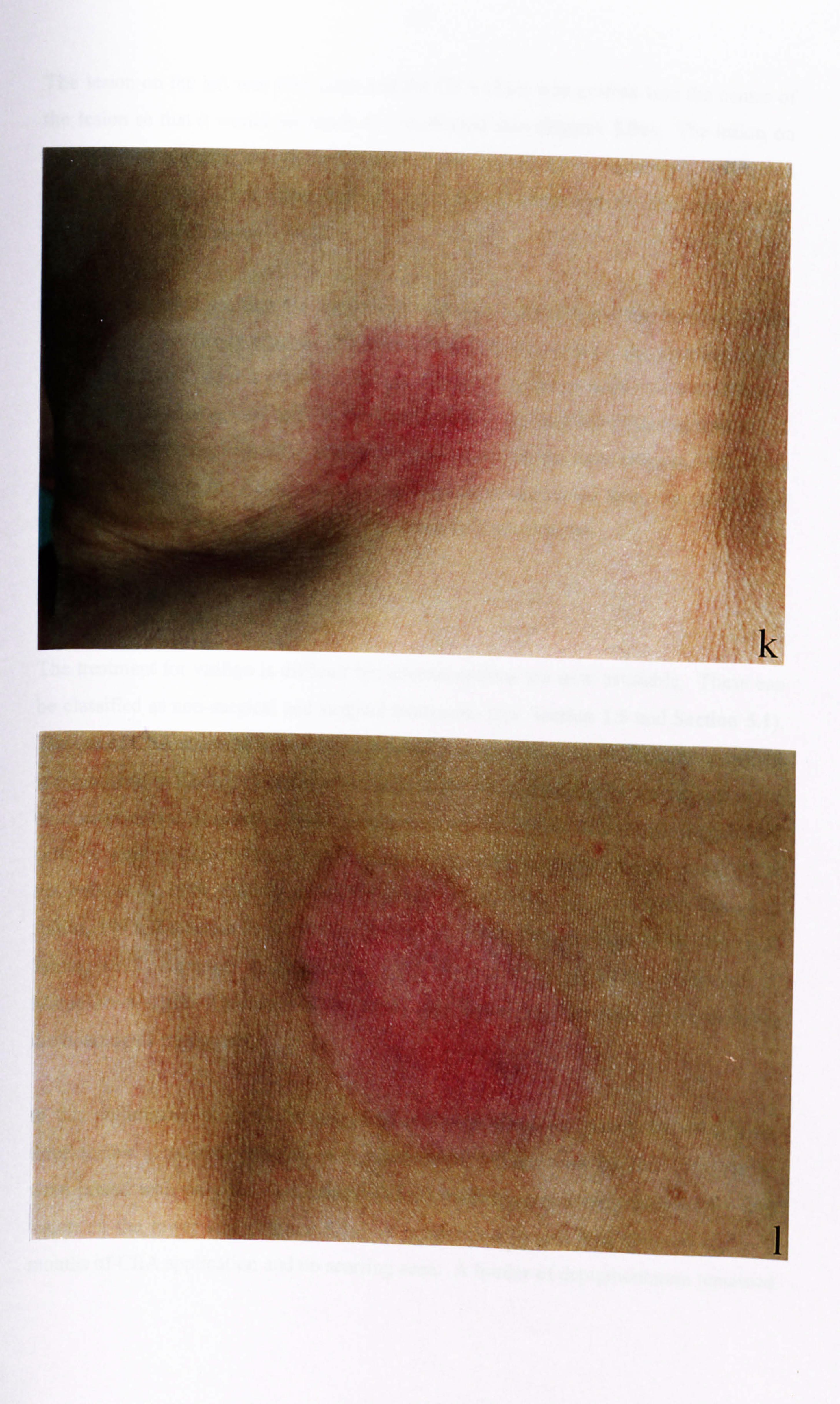
/ I
k
I
167
The lesion on the left was very large and the CEA sheet was grafted into the centre of
the lesion so that it would not touch any unaffected skin (Figure 5.9c). The lesion on
the right was a similar size to the CEA sheet and surrounded by apparently unaffected
skin (Figure 5.9d). The application of CEA sheets is illustrated for the lesion on the
right of the back (Figures 5.9e-f).
At one week after grafting the areas were still very inflamed and the backing on the
CEA sheets were firmly attached (Figures 5.9g-h). By two weeks, the sites had healed
but were still red. At this point the backing on the CEA sheets were removed (Figures
5.9i-j). Even after several months the two regions were very red (Figures 5.9k-1). A
hyperpigmented ring was noted around the grafted site on the right (Figure 5.91). This
was the site where the CEA was touching apparently unaffected skin. At the time of
writing, 12 months post grafting, no repigmentation had occurred.
5.5 DISCUSSION
The treatment for vitiligo is difficult but several options are now available. These can
be classified as non-surgical and surgical treatments (see Section 1.5 and Section 5.1).
The use of CEA sheets to treat vitiligo patients was decided upon as the culture method
is established in laboratory and routinuely used for the treatment of bums patients in the
Northern General Hospital. Several groups now use the culture of autologous epidermal
cells or pure culture melanocytes to treat vitiligo successfully (Brysk et al, 1989;
Falabella et al, 1989; Plott et al, 1989, Falabella et al, 1992; Jha et al, 1993; Olsson &
juhlin, 1993; Zachariae et al, 1993; Löntz et al, 1994; Olsson et al, 1994; Olsson &
Juhlin, 1995; Kumagai & Uchikoshi, 1997; Andreassi et al, 1998; Arenberger &
Matougkovä, 1998; Kaufmann et al, 1998) (see Table 5.2 for some of the different
methodologies employed).
In this preliminary study of the use of CEA sheets in the repigmentation of vitiligo,
three female Caucasian patients, with symmetrical vitiligo were treated. The results
were mixed with only the first patient (VM1) treated repigmenting successfully in a
lesion on the lower arm (Figure 5.5). A good colour match was obtained within 9
months of CEA application and no scarring seen. A border of depigmentation remained

.r
a 0 oA
C
C y
E
"o
0 ono 0 0
0
0 .r
cl
el
8 a? IN
u 0
0 y .r L Cl a
E O U
CC E-4
00 00 00
to O N ^^ 3
00 =L
ý M Q 0ý, Cb Ä H m
v3 C) C, 4 =L
v W
i+ oo ýbA N ed
0 'El -1 o 00 :a A N
N of ºýi
N
t. 0 en cd (7s
- RaI aý- a u
"5: 1
b t c 0 . 00 0 0 M- ^" Ö v1 Ö
N 0 V1 11
OO %n qn cl 00 A
t3 M E
v
p? 0
U
N
0 -0 4! V. U ä Ö NN ; Q.
U o
H yy : "i
=L , e1 C G1 "--ý N 14t
m
M
O i pý N ,
A o
00 ON 00
O O U
ý ö ý " 0' N j, 7a O
ö .0
v 1
".. _ U N
GU)
Cl) C - IRT N ~
N W
ý ýv W C;
U 01
eý
E 04
ýMg
.5 .-
rý ý4 W-0
E-4 .50 cl
u
CA 0
-5 4 >, y . 1.1 ä
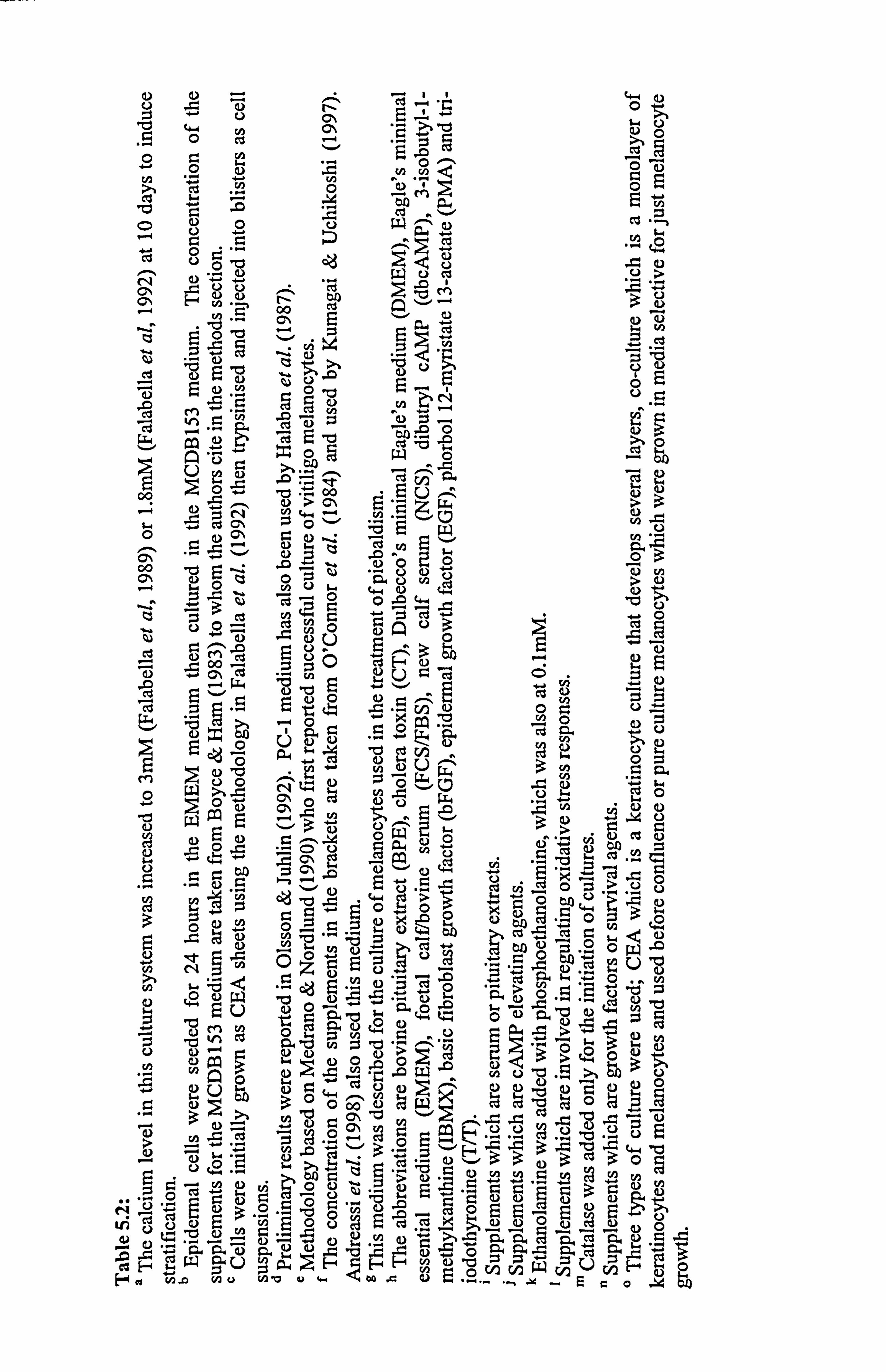
u2 iz --4 o° C) 0
CD oU wäß m -b "]^
+ý w
t) ce N 'V VI "Ü
aý UA3 aý
zi 0 "CJ -d 2ý
.22 ce
In r) =
AxO0-ä
e> 101 e 1-1 mi ., 6,. oo Eu -r, U ýo
9-4 e Co rn 'OZ, ' bWW=
CO aýi aas. ". ' vý " p.
00
E5 -: ri .-Uu
0 :J r2. >
Ct cd 0N5 ce .+O
1-1 Uw
O w+ c> Cd ý (n {Oy U ýýý-'! 1!
"ýý'
°o, ö0 °' v
0"0 .
cn °N'; U, 3cn =ä 15 0d aý w oE GA wý ý-3ý öWý, 3>
c> U
Uý bQ OýpU 14-4
rA gi Q2 -0 Cli w
O+ 00 r" ° 4-r Lam. 'ý" U
CJ VI ug -C-O2 &4 9.4 9 bul tuoi
pý 14 °' > ce E 3>ý°°" vom,
ý
. 9) 0 r. -- 5-. ý, ý3: Z: Z 1.4 9.
vi OUýU*
CU ý:
cn "C
aý ý''-°°'ý C-= Z aý U rA 0Q>, i-. as cýc
H wý U cý Hý H °' 0 cn CA
u2 U E' o E Co ýn 0 t02 v to vuw oo .eU2... ... . -, -19 _. Ec0 . ý4 th
168
around the perimeter where the grafts did not cover, although pigmentation had started
to spread slightly into this region. Unfortunately the second patient (VM4) developed
hypertropic scarring approximately 3-4 months after surgery. This complication has
since subsided (noted June, 1998 over a year after grafting) but it was difficult to assess
the presence of pigmentation (Figure 5.8). The patient became pregnant shortly after
surgery and it is unclear whether the grafting caused the scarring or whether the
pregnancy had contributed to the scarring during the healing period. Future patients
will be advised against starting a pregnancy for up to 6 months post-grafting following
the experience of patient VM4. The donor site in patient VM4 did not scar or show
Koebnerisation. The third patient (VM5) also treated in this study demonstrated no
repigmentation up to the time of writing (Figure 5.9). There were no complications
with this patient and the donor site showed no Koebnerisation, although the vitiligo was
still progressive. Interestingly, a ring of hyperpigmentation was seen around the CEA
graft that touched unaffected skin (Figure 5.91). All of the three patients had
generalised vitiligo. In mini-grafting tests, patients with generalised forms of the
disease were less responsive (Boersma et al, 1995; Falabella et al, 1995a) and
Hatchome et al. (1990) for example, has also described the failure of patients with
generalised vitiligo to repigment to surgical methods. However, this does not rule out
generalised patients for the treatment with surgical methods, as many patients with
generalised vitiligo have been treated successfully.
Kumagai & Uchikoshi (1997) treated four vitiligo patients (two with generalized and
two with segmental vitiligo) with CEA application to dermabraded skin (see Table 5.2).
Repigmentation with colour resembling the unaffected skin (recipient sites included the
abdomen) was obtained usually within 6-12 months and no scarring was seen. In a
slight variation to the method used in the thesis and Kumagai & Uchikoshi (1997), a
Colombian group treated numerous patients with vitiligo (mainly generalized or
segmental) with cultured epidermal autografts prepared by culturing the epidermal cells
on plastic in an EMEM based medium (see Table 5.2 for further details) (Falabella et
al, 1989 & 1992). The sheets were attached to petrolatum gauze following dispase
removal from the petri dish dishes and often cut smaller before being applied to denuded
regions (caused by liquid nitrogen induced blistering). Eleven patients showed
repigmentation that varied from 30-100% within 6 months (Falabella et al, 1989 &
169
1992). Some patients had a variegated effect while in others the colour was uniform.
With all patients, there was initial hyperpigmentation which receded by 6 months.
Indirect exposure to sunlight or UVA was advised (Falabella et al, 1989; Falabella et al,
1992; Kumagai & Uchikoshi, 1997) and patients were treated with steroid type
ointments during the first few months (Kumagai & Uchikoshi, 1997). The patients in
our study were advised to avoid sunlight and no PUVA/UVA or steroid treatments
given.
Jha et al. (1993) reported that the treatment of 3 patients with epidermal cell
suspensions (which were cultured in the medium as described in Falabella et al. (1992))
injected into blisters on the lesions, induced pigmentation as early as 2 weeks. This was
a preliminary report and only one patient had a5 months follow-up. By this stage, the
lesion of this patient was repigmented with a good colour match. Non-cultured
epidermal cells injected into liquid nitrogen induced blisters (Gauthier et al, 1992) or
spread onto dermabraded regions (Olsson & Juhlin, 1998) have been employed with
success. Gauthier et al. (1992) reported a success rate of 7 out of 11 patients with
segmental or focal vitiligo achieving 70-100% repigmentation including lesions located
on the wrist while Olsson & Juhlin (1998) reported that 20 out of 23 patients with either
segmental (3 patients) or vitiligo vulgaris achieved 80-100% repigmentation. PUVA
was used in the Gauthier study to help pigment spread.
Keratinocyte and melanocyte co-cultures established on bovine collagen membranes
were used to treat 3 patients successfully (Plott et al, 1989). Epidermal cell suspensions
were seeded onto the collagen membranes in an EMEM medium containing 10% FCS
for 24 hours and then switched to an MCDB153 medium (supplements described in
Table 5.2) (Plott et al, 1989; Brysk et al, 1989). When the cells were 50-70% confluent
they were transferred onto a superficially abraded region of affected skin.
Repigmentation, although not complete, was evident after 1 month and continued for
several months. A good colour match was obtained without scarring. In a slight
variation on this method, Zacharaie et al. (1993) treated a facial lesion in one Asian
patient with autologous cultured melanocytes grown in MCDB 153 medium on plastic (as described in Table 5.2). A good colour match was seen within 2 months after
170
grafting and the authors thought the region had a more homogenic appearance than
other areas grafted with more conventional means.
Olsson & Juhlin (1993) developed a unique culture method for melanocytes using a
medium referred to as PC-1 which did not involve serum/pituitary extracts or phorbol
esters (see Table 5.2). The melanocytes obtained were spread onto dermabraded
regions and secured with either a gel or mesh, or cells were injected into the
epidermal/dermal region of non-dermabraded skin. Ten patients of either Swedish or
middle-Eastern origin were treated. Nine patients repigmented with good cosmetic
results including one patient with active vitiligo. Another patient who had active
vitiligo did not repigment. This PC-1 culture method was compared with a similar type
of co-culture method used by the Brysk/Plott group (Löntz et al, 1994) but they
preferred the PC-1 culture method. Patients with segmental (just 2 patients) and
generalized vitiligo (both stable and moderatively active) were treated; 11 of the 27
patients demonstrating 90-100% repigmentation (including the 2 patients with
segmental vitiligo and 3 patients with active vitiligo) while 14 of the 27 patients had
less than 50% repigmentation. Therefore, this study illustrated that the response of
patients with generalised vitiligo were mixed. The trunk, face and back of the hands
proved the most likely regions to achieve successful repigmentation, with arms and legs
next. Elbows and fingers were the hardest. Similar results were reported by Olsson &
Juhlin (1995).
It is interesting to note that patient VM5 in our study, who was grafted on the back, a
site described as relatively successful by Löntz et al. (1994) showed no repigmentation.
Perhaps this was due to patient VM5 having a symmetrical (generalised) vitiligo or due
to the slight progressive nature of the vitiligo. Additionally, all the patients in our study
were treated with epidermal cells that had been in cryostorage from a few weeks to 6
months. Patient VM1 who showed the most successful repigmentation, was grafted
with cells that had been in storage for 6 months. Compton et al. (1998) reported
varying melanocyte loss from CEA cultures depending on what passage the epidermal
cells had been put in to crystorage. Patients of Scandinavian descent though were
successfully treated with pure cultures of melanocytes that had been in cryostorage
(Olsson et al, 1994).
171
Kaufmann et al. (1998) established pure cultures of melanocytes in a Ham's Flo
medium (described in Table 5.2) initially used by Lerner et al. (1987) to culture
melanocytes for the treatment of piebaldism. Melanocytes were then cultured on an
absorbable hyaluronic acid matrix for 7 days prior to being applied to Er; YAG laser-
ablated lesions. The grafted lesions were exposed to UVA radiation. In two patients,
repigmentation of varying extents was seen in many of their treated lesions with good
cosmetic results. The authors were aiming to improve the wound bed for grafting by
using the laser treatment. In another recent study, Andreassi et al. (1998) cultured
melanocytes in Green's medium on a biomaterial termed "Laserskin". Six out of 11
patients with localised vitiligo showed almost complete repigmentation when these
cultures were applied to the liquid carbon dioxide-treated lesions. Arenberger &
Matouskovä (1998) in abstract form, have described a system in which melanocytes and
keratinocytes were cultured on cell free pig dermis, and these composites applied to
vitiligo lesions. Approximately 50% of the patients treated repigmented. This
composite methodology has yet to be tried with human dermis, although patients with
burns have been treated with human epidermal/dermal type reconstructs (Boyce et al, 1995; Harriger et al, 1995).
All the groups showed that repigmentation can be obtained via the use of autologous
cultured cells. The degree of repigmentation varied and 90-100% repigmentation was
not always achieved. Mini-grafting was suggested as a means of inducing further
repigmentation in depigmented borders around grafts (Falabella et al, 1995b). Falabella
and co-workers suggest that these methods only be used as a last resort and for patients
with stable, nonprogressive vitiligo of the generalized or segmental type. Olsson and co
workers who have also treated large groups, have shown that patients with moderately
active vitiligo can be treated as well. Only one of the patients treated in our study
showed good detectable repigmentation, and it is therefore vital to know if melanocytes
are present or absent in the CEA culture method.
Freshly isolated epidermal cells obviously contain a small proportion of melanocytes as
pure culture of melanocytes can be achieved if cells are plated into the appropriate
conditions (for example see Figure 3.1 and Figure 5-lb-c). It is known that
172
keratinocytes and melanocytes interact in a tightly co-ordinated unit and several studies
have shown the importance of keratinocyte influence on melanocyte physiology in co-
culture (De Luca et al, 1998a & b; Valyi-Nagy et al, 1990; Haake & Scott, 1991; Scott
& Haake, 1991; Donatien et al, 1993; Valyi-Nagy et al, 1993; Nakazawa et al, 1995a;
Tenchini et al, 1995; Kippenberger et al, 1996; Nakazawa et al, 1997; Kippenberger et
al, 1998a; Nakazawa et al, 1998). Dopa staining of CEA sheets, attached and detached
from the petri dishes, generally proved negative for melanocytes (shown in Figures
5.1a, 5.3a, 5.4a), except in the case of vitiligo patient VM1 (Figure 5.2). Faure et al.
(1987) using an argentaffin stain reported the absence of melanocytes in CEA sheets but
in accordance with Gallico et al. (1984) saw the emergence of melanocytes in grafted
sites. De Luca et al. (1988b) showed that CEA sheets of primary, secondary and tertiary
level clearly contained melanocytes which was confirmed by Kumagi & Uchikoshi
(1997) in secondary CEA sheets. In other co-culture studies, where only one layer of
keratinocytes was present, melanocytes were easily detected by HMB-45 staining
(Haake & Scott, 1991; Scott & Haake, 1991), or by MEL-5 staining (Valyi et al, 1990;
Staiano-Coco et al, 1991; Valyi et al, 1993; Nakazawa et al, 1997 & 1998).
Melanocytes were noted at early culture in the EMEM methodology (Falabella et al,
1989; Falabella et al, 1992; Jha et al, 1993) but studies were not conducted on the sheets
once they became fully confluent and the keratinocytes stratified (Falabella et al, 1989
& 1992). Similarly with keratinocyte/melanocyte co-culture of Brysk et al. (1989) and
Plott et al. (1989), melanocytes were present in subconfluent cultures as detected by the
MEL-5 antibody. Melanocytes were detected in the grafted regions from these methods
(Falabella et al, 1989; Brysk et al, 1989; Plott et al, 1989; Falabella et al, 1992). In the
present study, the grafted site was not biopsied to check for the presence of
melanocytes.
It was difficult to assess if the CEA cultures were normal with respect to the length of
time in culture using comparisons with other studies. Table 5.1b illustrates the length
of time in culture for CEA sheets used on bums patients and a keloid scar patient from
our laboratory. The primary cultures in Table 5.1b were in culture longer than the
vitiligo CEA cultures (Table 5.1a) but in culture for a shorter period of time as
secondaries than the vitiligo CEA cultures (Table 5.1a). The vitiligo keratinocytes
formed colonies as expected by this method, and were confluent within 8 days. Gallico
173
et al. (1984), for example, noted that it took 10 days to obtain a confluent sheet of cells
from burns patients. Falabella et al. (1989 & 1992) used CEA sheets as primaries and
cultured the cells for 21 days. However, during this period, the calcium level was raised
to obtain stratified layers of keratinocytes. Kumagai & Uchikoshi (1997) generally
passaged the cells twice within a3 week period. On light microscopy the keratinocytes
from the vitiligo patients looked identical to keratinocytes from normal subjects (see
Figure 5.1a for normal keratinocytes and Figures 5.2a, 5.3a and 5.4a for vitiligo
keratinocytes).
From this study it has become apparent that the presence of melanocytes in CEA sheets
is difficult to detect. This could be due to skin type and the genetic predisposition for
low pigmentation activity in skin types I and II. Bessou et al. 1996 found that
melanocytes from skin type I in epidermal/dermal reconstructs were weakly dopa
stained when the epidermis was salt split from the dermis. It is also a possibility that
keratinocytes may downregulate melanocyte dopa oxidase activity in this culture system
and this requires further investigation. Kippenberger et al. (1996) however, have
demonstrated that melanocytes cultured in the presence of keratinocytes or phorbol
esters had enhanced transcription of melanogenic enzymes tyrosinase, TRP-1 and TRP-
2. The vitiligo keratinocytes in this present study did not appear to be abnormal from
visual observation and behaved similarly with respect to proliferation. The CEA
technique merits further investigation for repigmentation of vitiligo as the first patient
treated was clearly successful. In future, selection of patients will be more thorough. It
is perhaps of interest to include patients with focal or segmental forms of the disease in
the study. In an attempt to learn if patients are suitable for grafting, patients will
initially be treated in a small area as a test site to determine repigmentation and scarring.
This follows along the lines of using a few minigrafts as advocated by Boersma et al.
(1995) and Falabella et al. (1995a), but our patients will receive small CEA sheets.
Only then will patients be offered treatment of larger areas.

CHAPTER 6
FINAL DISCUSSION.
175
6.1 FINAL DISCUSSION.
Vitiligo is an acquired idiopathic hypomelanotic disease. While not life threatening, it can
psychologically affect many patients, in particular those with darker skin, where the
contrast between the normal skin colour and depigmented milky white patches is more
evident. The aetiology remains unclear although several hypotheses exist including
immune, neuronal and self-destructive (as discussed in Section 1.4). Le Poole et al.
(1993a) and Hautmann & Panconesi (1997) have described separate convergence theories
in which interaction between many of the individual theories were proposed. Various
distributional patterns exist such as segmental and acrofacial and it was proposed that
segmental vitiligo was a separate form of the disease to non-segmental (Koga, 1977). This,
together with the various overlapping hypotheses suggest that vitiligo is a syndrome of
diseases that results in the same endpoint. A range of medical and surgical techniques
(Sections 1.5,5.1 and 5.5) can improve the appearance of the lesions, by causing
repigmentation. However, many patients do not respond to any treatments. PUVA, one of
the main medical therapies, requires several sessions before a response is necessarily seen
and up to 100 treatments or more are often needed (summarised in Grimes, 1993; Drake et
al, 1996; Grimes, 1997). Mini-grafting is a useful technique for determining the suitability
of patients for surgical methods (Boersma et al, 1995; Falabella et al, 1995a).
The study of vitiligo is important. The determination of the aetiology will not only benefit
vitiligo patients in terms of their treatment and cessation of the progression of the disease,
but may be of some importance in the treatment of melanoma. At opposite ends of the
spectrum, vitiligo results in the destruction of melanocytes, and melanoma cells are
uncontrollable proliferating cells. Also patients with melanoma who develop
hypomelanotic rings which are vitiligo-like often have a better survival prognosis (Barnes
& Nordlund, 1989). However, before an understanding of vitiligo melanocytes can be
achieved, further characterisation of normal melanocytes is required. The main function of
melanocytes is to produce melanins which once transferred to keratinocytes, in theory
protect the skin against harmful ultraviolet irradiation.
176
The number and type of in vitro studies performed on normal melanocytes have increased
over the past decade. However, media conditions vary between the different investigators
and neonatal foreskin melanocytes are preferentially used as these are easier to culture
(discussed in Section 3.1.1). In vitiligo, cutaneous melanocytes throughout the body are
destroyed, and persons of any age can become affected by the disease. Therefore, the work
in this thesis was carried out using adult cutaneous melanocytes. Morphology, proliferation
and pigmentation of normal adult melanocytes was assessed in response to ECM proteins,
UVB irradiation and a-MSH under various media conditions.
A range of ECM proteins have been shown previously to influence morphology (Gilchrest
et al, 1985; Ranson et al, 1988a; McClenic et al, 1989; Hara et al, 1994; Nakazawa et al, 1995b), proliferation (Ranson et al, 1988a; Mortarini et al, 1995) and pigmentation (Ranson
et al, 1988a; Buffey et al, 1994; Nakazawa et al, 1995b) in normal melanocytes. In this
study (discussed fully in Chapter 3), a variety of individual and cell-derived ECM proteins
were compared and were found to be unable to stimulate melanocyte dendricity or have a
significant effect on the morphology of melanocytes (Section 3.2.4). The greatest and most
consistent effects of ECM proteins on proliferation and pigmentation were seen when
melanocytes were cultured in a mitogen reduced medium (Sections 3.3.4-3.3.5,3.4.4-
3.4.9). In contrast to ECM proteins, UVB stimulation of melanocytes was greatest when
the cells were cultured in a mitogen rich medium such as medium 1. Dendricity and cell body size were increased and pigmentation was significantly increased in response to UVB
irradiation (Sections 3.2.5 and 3.4.8) as previously reported by several authors (Friedmann
& Gilchrest, 1987; Libow et al, 1988; Tomita et al, 1988; Friedmann et al, 1990a&b;
Ramirez-Bosca et al, 1992; Aberdam et al, 1993; Abdel-Malek et al, 1994; Im et al, 1994;
Romero et al, 1994; Duval & Schmidt, 1997). UVB irradiation halted cell proliferation and
reduced cell viability (Section 3.3.4) in agreement with several other studies (Friedmann &
Gilchrest, 1987; Ramirez-Bosca et al, 1992; Abdel-Malek et al, 1994b; Barker et al, 1995).
In media where CT had been removed (media 10 and 24), the pigmentary response was
much reduced and this was in agreement with Im et al. (1998) who also reported that
melanocytes cultured in the absence of cAMP agents while undergoing UVB irradiation,
failed to pigment.
177
The role of a-MSH as a pigmentary hormone in man is controversial. While in vivo studies
have shown clear pigmentary responses (Lerner & McGuire, 1961 & 1964; Levine et al,
1991), only over the past 4 years have in vitro studies indicated a role for a-MSH in human
pigmentation (Hunt et al, 1994a, b&c; Abdel-Malek et al, 1995; McLeod et al, 1995;
Maeda et al, 1996; Suzuki et al, 1996). Recently, Im et al. (1998) reported that a-MSH
may play a part in modulating the melanocyte response to UVB irradiation. Additionally,
UVB increased the expression of MC1R (Funasaka & Ichihashi, 1997; Funasaka et al,
1998), as did UV-induced keratinocyte-derived factors ADF (Funasaka & Ichihashi, 1997)
and endothelin-1 (Funasaka et al, 1998; Tada et al, 1998). In this present study
reproducible melanogenic effects of a-MSH were only seen under a small number of media
conditions. Increased proliferative and melanogenic effects were much more evident in
media containing bFGF, (Sections 3.3.2 and 3.4.2); melanocytes cultured in medium 23,
which contains only bFGF, showing significant increases in proliferation, dopa oxidase
activity and melanin content. However, the evolving literature showing that POMC
peptides are now produced in the skin (Thody et al, 1983; Farooqui et al, 1993; Schauer et
al, 1994; Iyengar, 1995; Kippenberger et al, 1995; Slominski et al, 1995; Chakraborty et al,
1996; Wintzen et al, 1996; Teofoli et al, 1997; Wakamatsu et al, 1997; Can et al, 1998)
indicated a much wider role for a-MSH and other POMC peptides. a-MSH has been
shown to influence melanocyte attachment to ECM proteins (Hunt et al, 1993; Scott et al,
1997) and alter the melanocyte cytoskeletal protein structures (Scott et al, 1997). It is
proposed that a-MSH has a role in modulating oxidative stress through increasing
tyrosinase activity, which traps free radical species (Valverde et al, 1996b) and this was
recently linked to the pathogenesis of vitiligo as vitiligo melanocytes of the unaffected and
perilesional skin were shown to have lower amounts of IR a-MSH than control skin,
reported in brief (Graham et al, 1998). This theory, along with the one proposed by Liu et
al. (1995) (discussed in Chapter 4), remain speculative at the moment. A further putative
role for a-MSH as an immunomodulatory peptide is mentioned below.
178
An epidermal/dermal reconstruct model has been developed in Dr S Mac Neil's laboratory
over the past 6 years. In preliminary studies normal melanocytes were added to these
composites to examine the usefulness of this model for investigating pigmentation and for
the study of vitiligo (Section 3.4.10). Bessou et al. (1997a) proposed the use of an
epidermal/dermal reconstruct as a means of investigating vitiligo. Their model is well
established for the study of the effects of UVB irradiation (Bessou et al, 1995 & 1996) and
has been used to examine the effects of known pigmentary and depigmentary agents
(Bessou et al, 1997c). However, their model does not include the live fibroblasts being
added to the dermis. The work in this thesis shows that the presence of fibroblasts plays an
important role in preventing pigmentation as was noted in composites with (Figures 3.30
and 3.34) and without added melanocytes (Figure 3.33). This is intriguing since Bessou et
al. (1995 & 1996) reported ex vivo construction of skin types I-VI phototypes with and
without UVB stimulation in composite models without added fibroblasts. Hence it is
possible that soluble fibroblast-derived factors may remain in the dermis employed in the
experiments described by Bessou and coworkers.
The presence of fibroblasts and keratinocytes induced higher levels of pigmentation in
melanocytes cultured on a dermal substitute than melanocytes alone on a monolayer (Archambault et al, 1995). Moreover, the effect of fibroblasts alone on pigmentation was
not examined although fibroblasts were found to significantly enhance melanocyte survival in response to UVB irradiation (Archambault et al, 1995). A second finding of the current
work demonstrated the importance of the basement membrane proteins. When the dermis
was stripped of these, melanocytes migrated out of the basal layer of the reconstructed
epidermis and a significant amount of pigmentation was seen. Overall, there appeared to be
a loss of melanocytes from the composites. Melanocytes bind to ECM proteins through
integrins (Scott et al, 1992; Danen et al, 1993; Morelli et al, 1993; Zambruno et al, 1993)
but they also interact with keratinocytes through E-cadherins (Tang et al, 1994; Nakazawa
et al, 1995a). De Luca et al. (1988a) illustrated the importance of keratinocytes fixing the
position of melanocytes within the basal layer of CEA sheets. Against this the results seen in the composite model in the present study seem unusual. In the CEA sheet, a feeder layer
is present and might provide the additional signals or ECM proteins seen in composites
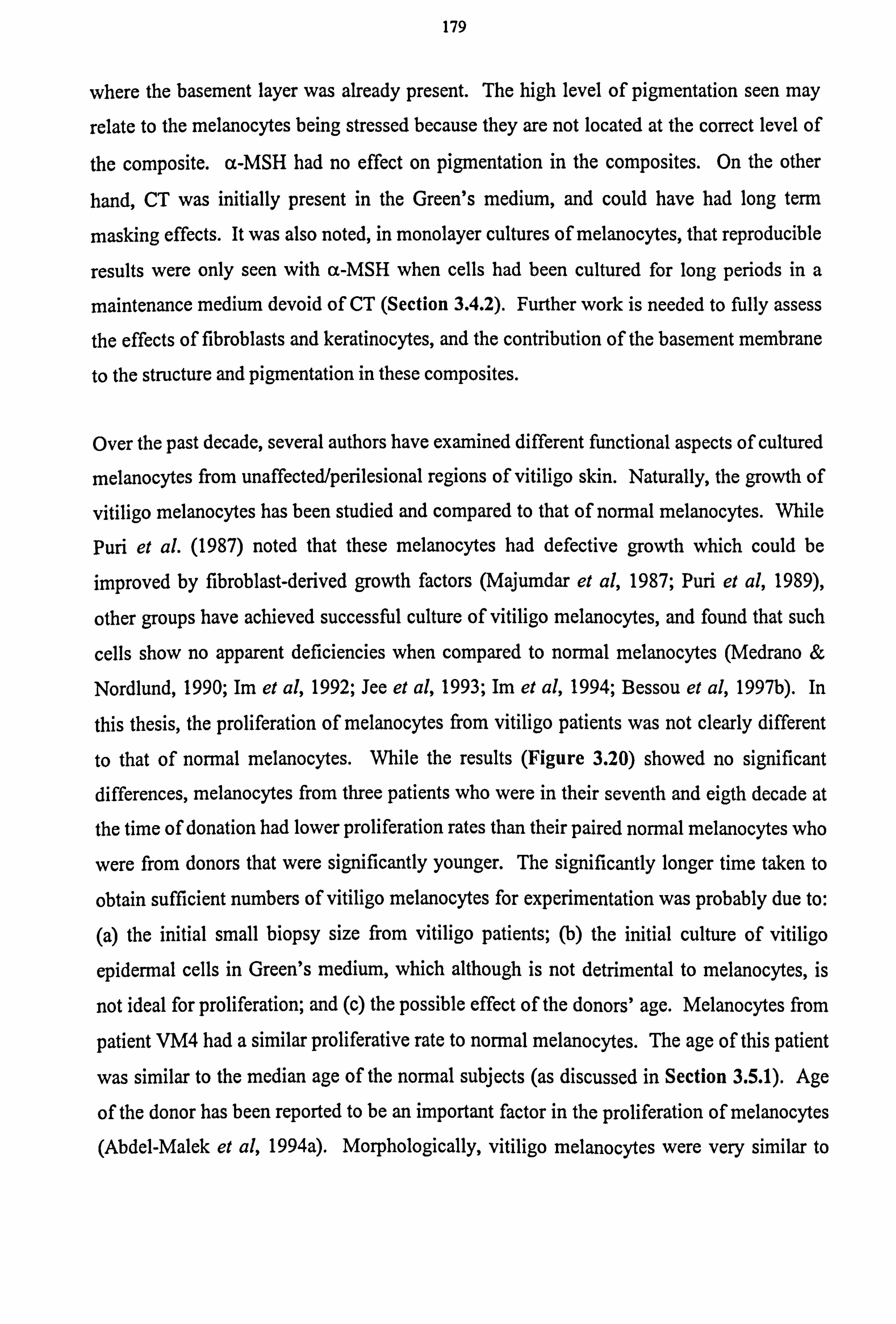
179
where the basement layer was already present. The high level of pigmentation seen may
relate to the melanocytes being stressed because they are not located at the correct level of
the composite. a-MSH had no effect on pigmentation in the composites. On the other
hand, CT was initially present in the Green's medium, and could have had long term
masking effects. It was also noted, in monolayer cultures of melanocytes, that reproducible
results were only seen with a-MSH when cells had been cultured for long periods in a
maintenance medium devoid of CT (Section 3.4.2). Further work is needed to fully assess
the effects of fibroblasts and keratinocytes, and the contribution of the basement membrane
to the structure and pigmentation in these composites.
Over the past decade, several authors have examined different functional aspects of cultured
melanocytes from unaffected/perilesional regions of vitiligo skin. Naturally, the growth of
vitiligo melanocytes has been studied and compared to that of normal melanocytes. While
Puri et al. (1987) noted that these melanocytes had defective growth which could be
improved by fibroblast-derived growth factors (Majumdar et al, 1987; Puri et al, 1989),
other groups have achieved successful culture of vitiligo melanocytes, and found that such
cells show no apparent deficiencies when compared to normal melanocytes (Medrano &
Nordlund, 1990; Im et al, 1992; Jee et al, 1993; Im et al, 1994; Bessou et al, 1997b). In
this thesis, the proliferation of melanocytes from vitiligo patients was not clearly different
to that of normal melanocytes. While the results (Figure 3.20) showed no significant
differences, melanocytes from three patients who were in their seventh and eigth decade at
the time of donation had lower proliferation rates than their paired normal melanocytes who
were from donors that were significantly younger. The significantly longer time taken to
obtain sufficient numbers of vitiligo melanocytes for experimentation was probably due to:
(a) the initial small biopsy size from vitiligo patients; (b) the initial culture of vitiligo
epidermal cells in Green's medium, which although is not detrimental to melanocytes, is
not ideal for proliferation; and (c) the possible effect of the donors' age. Melanocytes from
patient VM4 had a similar proliferative rate to normal melanocytes. The age of this patient
was similar to the median age of the normal subjects (as discussed in Section 3.5.1). Age
of the donor has been reported to be an important factor in the proliferation of melanocytes (Abdel-Malek et al, 1994a). Morphologically, vitiligo melanocytes were very similar to
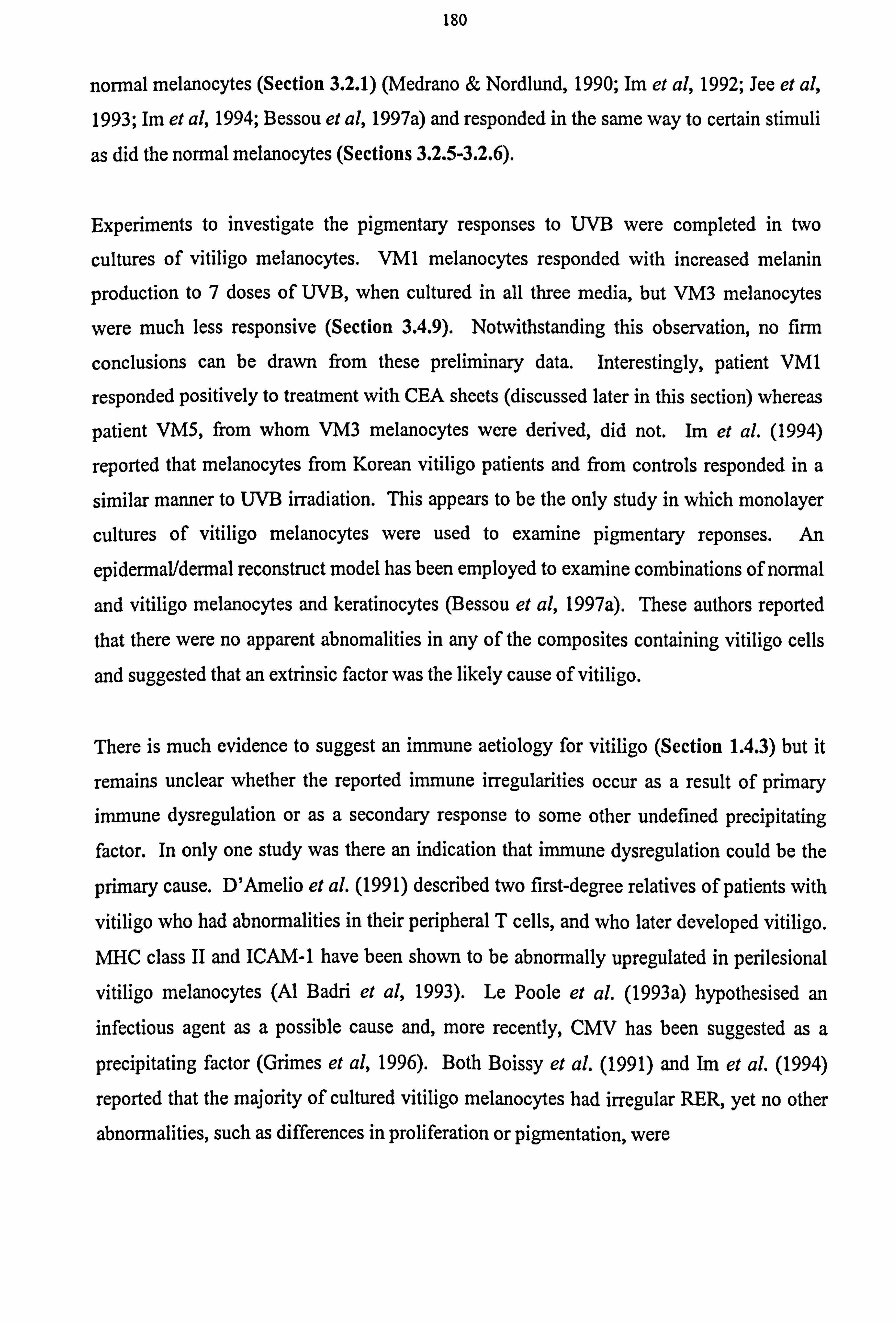
180
normal melanocytes (Section 3.2.1) (Medrano & Nordlund, 1990; Im et al, 1992; Jee et al,
1993; Im et al, 1994; Bessou et al, 1997a) and responded in the same way to certain stimuli
as did the normal melanocytes (Sections 3.2.5-3.2.6).
Experiments to investigate the pigmentary responses to UVB were completed in two
cultures of vitiligo melanocytes. VM1 melanocytes responded with increased melanin
production to 7 doses of UVB, when cultured in all three media, but VM3 melanocytes
were much less responsive (Section 3.4.9). Notwithstanding this observation, no firm
conclusions can be drawn from these preliminary data. Interestingly, patient VM1
responded positively to treatment with CEA sheets (discussed later in this section) whereas
patient VM5, from whom VM3 melanocytes were derived, did not. Im et al. (1994)
reported that melanocytes from Korean vitiligo patients and from controls responded in a
similar manner to UVB irradiation. This appears to be the only study in which monolayer
cultures of vitiligo melanocytes were used to examine pigmentary reponses. An
epidermal/dermal reconstruct model has been employed to examine combinations of normal
and vitiligo melanocytes and keratinocytes (Bessou et al, 1997a). These authors reported
that there were no apparent abnomalities in any of the composites containing vitiligo cells
and suggested that an extrinsic factor was the likely cause of vitiligo.
There is much evidence to suggest an immune aetiology for vitiligo (Section 1.4.3) but it
remains unclear whether the reported immune irregularities occur as a result of primary immune dysregulation or as a secondary response to some other undefined precipitating factor. In only one study was there an indication that immune dysregulation could be the
primary cause. D'Amelio et al. (1991) described two first-degree relatives of patients with
vitiligo who had abnormalities in their peripheral T cells, and who later developed vitiligo.
MHC class II and ICAM-1 have been shown to be abnormally upregulated in perilesional
vitiligo melanocytes (Al Badri et al, 1993). Le Poole et al. (1993a) hypothesised an infectious agent as a possible cause and, more recently, CMV has been suggested as a
precipitating factor (Grimes et al, 1996). Both Boissy et al. (1991) and Im et al. (1994)
reported that the majority of cultured vitiligo melanocytes had irregular RER, yet no other
abnormalities, such as differences in proliferation or pigmentation, were
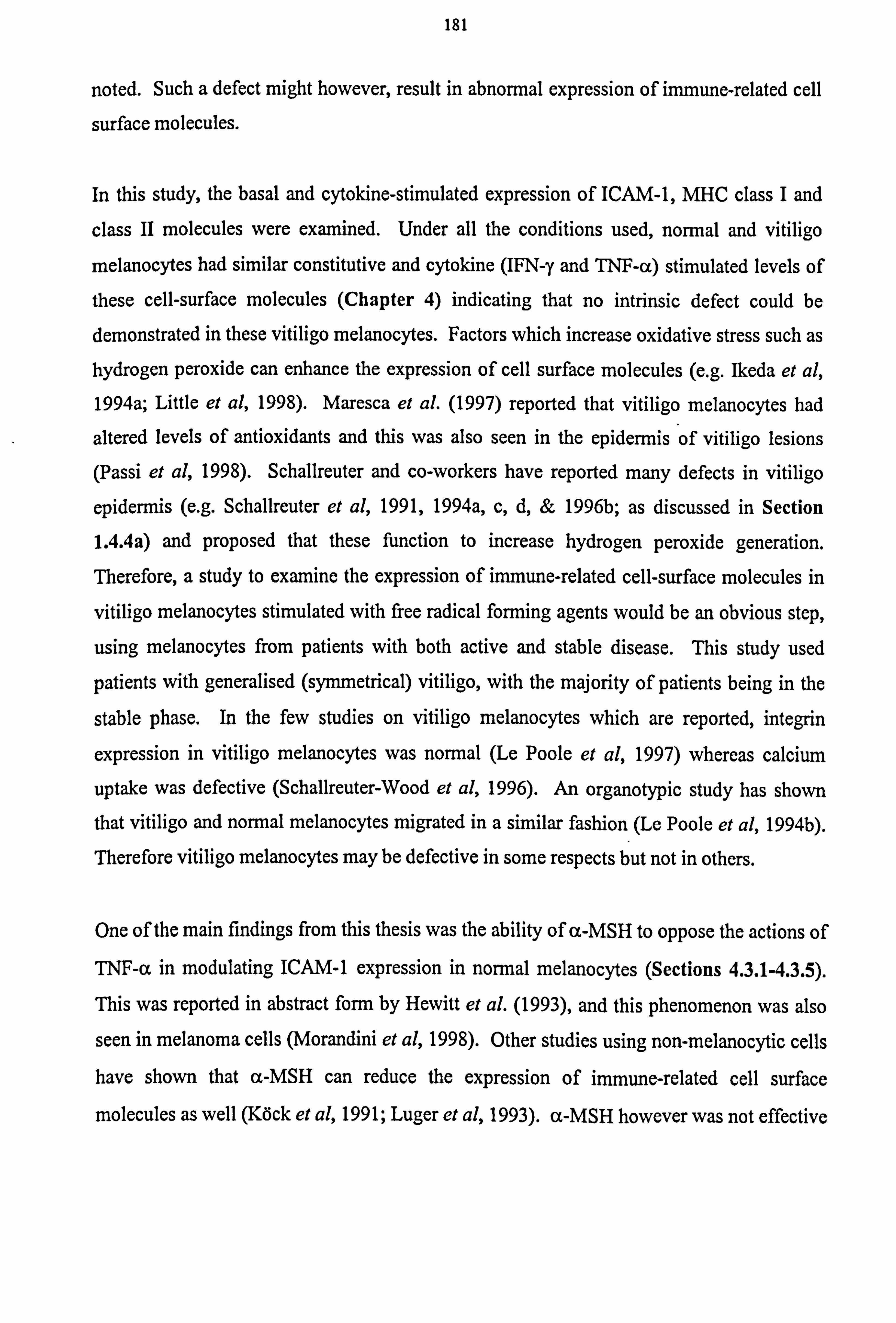
181
noted. Such a defect might however, result in abnormal expression of immune-related cell
surface molecules.
In this study, the basal and cytokine-stimulated expression of ICAM-1, MHC class I and
class II molecules were examined. Under all the conditions used, normal and vitiligo
melanocytes had similar constitutive and cytokine (IFN-y and TNF-a) stimulated levels of
these cell-surface molecules (Chapter 4) indicating that no intrinsic defect could be
demonstrated in these vitiligo melanocytes. Factors which increase oxidative stress such as hydrogen peroxide can enhance the expression of cell surface molecules (e. g. Ikeda et al, 1994a; Little et al, 1998). Maresca et al. (1997) reported that vitiligo melanocytes had
altered levels of antioxidants and this was also seen in the epidermis of vitiligo lesions
(Passi et al, 1998). Schallreuter and co-workers have reported many defects in vitiligo
epidermis (e. g. Schallreuter et al, 1991,1994a, c, d, & 1996b; as discussed in Section
1.4.4a) and proposed that these function to increase hydrogen peroxide generation. Therefore, a study to examine the expression of immune-related cell-surface molecules in
vitiligo melanocytes stimulated with free radical forming agents would be an obvious step,
using melanocytes from patients with both active and stable disease. This study used
patients with generalised (symmetrical) vitiligo, with the majority of patients being in the
stable phase. In the few studies on vitiligo melanocytes which are reported, integrin
expression in vitiligo melanocytes was normal (Le Poole et al, 1997) whereas calcium
uptake was defective (Schallreuter-Wood et al, 1996). An organotypic study has shown that vitiligo and normal melanocytes migrated in a similar fashion (Le Poole et al, 1994b).
Therefore vitiligo melanocytes may be defective in some respects but not in others.
One of the main findings from this thesis was the ability of a-MSH to oppose the actions of
TNF-a in modulating ICAM-1 expression in normal melanocytes (Sections 4.3.1-4.3.5).
This was reported in abstract form by Hewitt et al. (1993), and this phenomenon was also
seen in melanoma cells (Morandini et al, 1998). Other studies using non-melanocytic cells have shown that a-MSH can reduce the expression of immune-related cell surface
molecules as well (Köck et al, 1991; Luger et al, 1993). a-MSH however was not effective
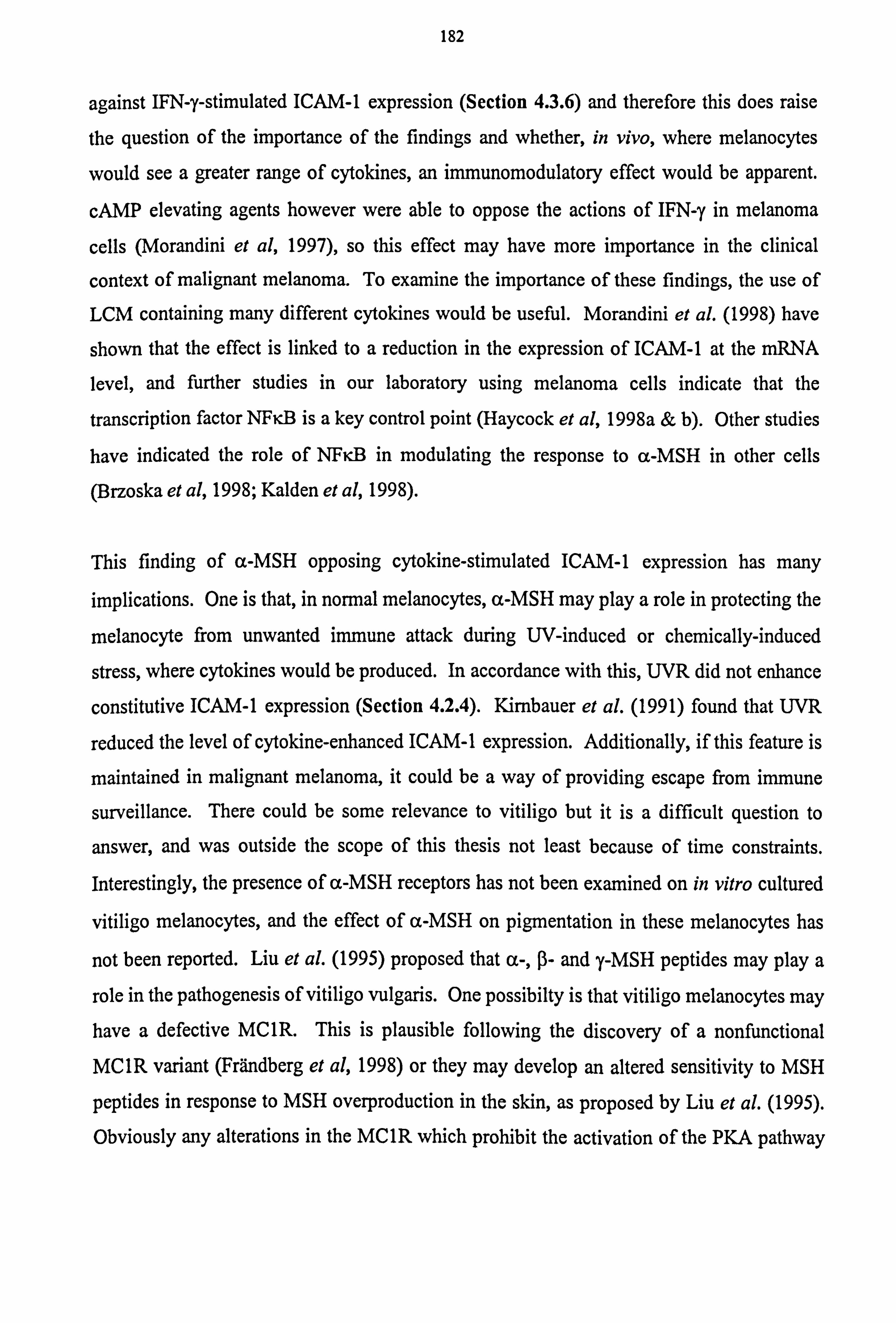
182
against IFN-y-stimulated ICAM-1 expression (Section 4.3.6) and therefore this does raise
the question of the importance of the findings and whether, in vivo, where melanocytes
would see a greater range of cytokines, an immunomodulatory effect would be apparent.
cAMP elevating agents however were able to oppose the actions of IFN-y in melanoma
cells (Morandini et al, 1997), so this effect may have more importance in the clinical
context of malignant melanoma. To examine the importance of these findings, the use of
LCM containing many different cytokines would be useful. Morandini et al. (1998) have
shown that the effect is linked to a reduction in the expression of ICAM-1 at the mRNA
level, and further studies in our laboratory using melanoma cells indicate that the
transcription factor NFi B is a key control point (Haycock et al, 1998a & b). Other studies
have indicated the role of NFiB in modulating the response to a-MSH in other cells
(Brzoska et al, 1998; Kalden et al, 1998).
This finding of a-MSH opposing cytokine-stimulated ICAM-1 expression has many
implications. One is that, in normal melanocytes, a-MSH may play a role in protecting the
melanocyte from unwanted immune attack during UV-induced or chemically-induced
stress, where cytokines would be produced. In accordance with this, UVR did not enhance
constitutive ICAM-1 expression (Section 4.2.4). Kinnbauer et al. (1991) found that UVR
reduced the level of cytokine-enhanced ICAM-1 expression. Additionally, if this feature is
maintained in malignant melanoma, it could be a way of providing escape from immune
surveillance. There could be some relevance to vitiligo but it is a difficult question to
answer, and was outside the scope of this thesis not least because of time constraints.
Interestingly, the presence of a-MSH receptors has not been examined on in vitro cultured
vitiligo melanocytes, and the effect of a-MSH on pigmentation in these melanocytes has
not been reported. Liu et al. (1995) proposed that a-, ß- and y-MSH peptides may play a
role in the pathogenesis of vitiligo vulgaris. One possibilty is that vitiligo melanocytes may have a defective MC1R. This is plausible following the discovery of a nonfunctional MC1R variant (Frändberg et al, 1998) or they may develop an altered sensitivity to MSH
peptides in response to MSH overproduction in the skin, as proposed by Liu et al. (1995).
Obviously any alterations in the MC1R which prohibit the activation of the PKA pathway
183
would suggest a loss of protective measures against the immune system. However, this
remains speculation at the time of writing.
While an immunomodulatory role of a-MSH can be demonstrated in vitro, it is necessary
to know if this reduction in ICAM-1 expression can translate into a reduction of T cell binding and activation. Melanocytes have been shown to bind T cells (Nickoloff et al, 1991; Le Poole et al, 1993b) and are capable of presenting antigen and activating T cells (Le Poole et al, 1993b). Preliminary studies of T cell binding to normal melanocytes were
unsuccessful (as discussed in Section 4.6) and other methods using flow cytometry may be
required to make progress in such sudies. Methods are currently being investigated to find
the best way to study T cell binding and activation in both normal and malignant
melanocytes.
The CEA approach was used to treat three patients with vitiligo. This culture, while
primarily a keratinocyte culture, has been reported to retain the stable keratinocyte to
melanocyte ratio (De Luca et al, 1988a & b). While other groups clearly demonstrated the
presence of melanocytes in CEA sheets (De Luca et al, 1988b; Kumagai & Uchiskoshi,
1997), in the majority of cultures set up in this laboratory melanocytes, were not detected
using L-dopa staining. Ongoing studies in the laboratory have recently identified the
presence of melanocytes in CEA sheets by staining melanocytes for TRP-1 (personal
communication J Phillips and S Mac Neil, 1998). Dissociated sheets of varying passage
number always yielded melanocytes (Figures 3.3,5.1,5.3-5.4, Section 5.3). Further
investigation is required to explore why the detection of melanocytes using L-dopa staining
was not possible in CEA sheets. One hypothesis is that the keratinocytes are actually downregulating the melanogenic capacity of the melanocyte, but a study by Kippenberger
et al. (1996) has shown that under co-culture conditions, melanocytes actually had
increased levels of the melanogenic enzymes. Bessou et al. (1996) noted that in epidermal
salt splits from epidermal/dermal reconstructs, melanocytes of skin type I donors were very
weakly stained with L-dopa and it was difficult often to identify melanocytes. Therefore it
seems likely the inherent genetics may make the melanocytes in the current study difficult
to detect as they were predominantly from skin types I and II. Orlow et al. (1993) reported
184
that tyrosinase was associated with stage III and stage IV melanosomes, of which few are
found in persons with skin types I and II whereas TRP-1 is associated with stage I and II
melanosmoes. This might indicate why melanocytes could be detected by TRP-1 staining
and not by L-dopa staining when in contact with their skin counterparts, the keratinocytes.
It was difficult to assess if the vitiligo keratinocytes proliferated at a similar rate to normal
keratinocytes in CEA sheets. However, light microscopy examination showed that normal
and vitiligo keratinocytes were morphologically identical. Vitiligo keratinocytes formed
colonies, and a confluent sheet was produced within 8 days -a time similar to that reported
by Gallico et al. (1984) for cells cultured from burns patients. Keratinocytes from vitiligo
patients have been implicated in the cause of the disease because of damage observed in the
lesional areas (Breathnach et al, 1966; Ishii & Hamada, 1981; Moellmann et al, 1982;
Bhawan & Bhutani, 1983; Kao & Yu, 1990; Hann et al, 1992; Ahn et al, 1994; Yagi et al,
1997). Results from a preliminary co-culture study implied that vitiligo keratinocytes may
be defective (Le Poole et al, 1991) while increased levels of tenascin seen in vitiligo lesions
might be explained by its overproduction by vitiligo keratinocytes (Le Poole et al, 1997).
Defects in the epidermis, as previously mentioned in this section, could also result from
defects in the keratinocytes. Bessou et al. (1997a) though, found no abnormalilties when
vitiligo keratinocytes were cultured in an epidermal/dermal model.
The treatment of the patients raised several questions. One patient (VM1) showed
successful repigmentation, although a perimeter of vitiligo remained around the edge,
where the CEA sheets did extend to the margins of dermabrasion (Figure 5.5). The
melanocytes from this patient were also highly responsive to UVB stimulation (Section
3.4.9). One patient (VM5) did not repigment, but this patient had active vitiligo.
Additionally, the cells from this patient were not responsive to UVB stimulation (Section
3.4.9). The third patient (VM4) developed transient hypertrophic scarring which was a
problem not anticipated. For this patient there has been slight pigmentation in small
regions and the scarring complications are still resolving so it is unclear what the final
outcome will be. Due to the success of the first patient and other successful reports using
similar methods in vitiligo patients (Falabella et al, 1989 & 1992; Kumagai & Uchishoki,
185
1997), further work is underway to explore this approach as a treatment for vitiligo patients.
Following the preliminary study the protocol is now to treat a small test area, to see if the
patient will respond positively to grafting and not develop scarring. The patients treated in
this study had symmetrical generalised vitiligo. While patients with generalised vitiligo
can respond to surgical methods with cultured cells, the success rate is approximately 50%
as illustrated by Löntz et al. (1994), who used cultured melanocytes, and Boersma et al.
(1995) and Falabella et al. (1995a) who used the mini-grafting technique. It would
therefore be of interest to include patients with segmental or focal forms of the disease in
the study.
In conclusion, vitiligo melanocytes that were in culture long term did not differ appreciably
from normal melanocytes in proliferation, morphology or in basal and cytokine stimulated
levels of ICAM-1, MHC class I and class II expression. This is in agreement with several
other studies in which no abnormalities in melanocyte parameters were noted (Im et al.
1994; Le Poole et al. 1994b; Bessou et al. 1997a; Le Poole et al. 1997) and would suggest
that an extrinsic factor is responsible. However, other studies, for example those by
Schallreuter-Wood et al. (1996) and Maresca et al. (1997), have indicated abnormalities in
cultured vitiligo melanocytes. Thus, in vitro studies so far provide conflicting data and the
aetiology of vitiligo remains elusive. As yet, there is little evidence to support an intrinsic
defect in the melanocyte; theories of autoimmunity, intrinsic keratinocyte defects or some
precipitating extrinsic factor are all under investigation.

APPENDICES.
187
MATERIALS.
Bovine pituitary extract was obtained from Advanced Protein Products, Brierly Hill, West Midlands, UK.
Acetic acid, buffered formalin, cuvettes UV range (disposable), eosin yellowish, ethanol (99.7-100%v/v), glutaraldehyde, hydrochloric acid, paraformaldehyde, poly-l-lysine coated slides, silver nitrate, sodium bicarbonate, sodium citrate, sodium dodecylsulphate, sodium hydroxide, sodium thiosulphate anhydrous and Tris(hydroxymethyl)methylamine-buffered saline (TBS) were obtained from BDH Merck Ltd, Magna Park, Lutterworth, Leicestershire, UK
HLA-DR, DQ, DP PE-conjugated antibody, mouse IgGI FITC-conjugated antibody control, mouse IgGI PE-conjugated antibody control, mouse IgG2a FITC-conjugated antibody control and mouse IgG2a PE-conjugated antibody control were obtained from Becton Dickinson, Cowley, Oxford, UK.
Dispase, y-interferon tumour (IFN-y) and necrosis factor-a (TNF-a) were obtained from Boehringer Mannheim UK, (Diagnostics and Biochemicals) Ltd, Bell Lane, Lewes, East Sussex, UK
OCT mounting medium was obtained from Brights' Instrument Company Ltd, Huntingdon, Cambridgeshire, UK.
Tissue culture plastics were obtained from Coming Costar Corporation, Cambridge, MA, USA.
Anti-CD45 (leukcyte common antige) polyclonal horseradish peroxidase HRP- conjugated antibody, immunoglobulin HRP negative control, rabbit anti-human S100 polyclonal antibody and mouse anti-human collagen IV antibody were obtained from Dako Ltd, High Wycombe, Bucks, UK.
Trypsin was obtained from Difco Laboratories, Detroit, Michigan, USA.
Ligaclips were obtained from Ethicon Inc, Somerville, New Jersey, USA.
Xylene was obtained from Genta Medical, York, UK
Bovine pituitary extract (BPE), Dulbecco's Modified Eagle's medium (DMEM), Fungizone, normal goat serum, newborn calf serum (NCS) and RPMI medium were obtained from Gibco BRL, Life Technologies, Paisley, UK.
Foetal calf serum (FCS) was obtained from GlobePharm Limited, Esher, Surrey, UK.
Horse and swine serum were obtained from Harlan Sera-lab Ltd, Crawley-Down, UK.
Acetone was obtained from Hillcoss Pharmoceuticals, Burnley, UK.
Gold chloride was obtained from Johnson Matthey Chemicals, Royston, Herts, UK
188
Tegapore backing dressings were obtained from 3M Medical-Surgical Division, St. Paul, MN USA.
LymphoprepT"i and Uni-Sorb T+B nylon wool columns were obtained from Nycomed Pharma AS, Oslo, Norway.
Mouse anti-human ICAM-1 monoclonal antibody was obtained from R and D Systems Europe, Barton Lane, Abingdon, Oxon, UK
CD40 FITC-conjugated antibody, HLA-ABC FITC-conjugated antibody, ICAM-1 (CD54) PE-conjugated antibody and LFA-3 (CD58) FITC conjugated antibody were obtained from Serotec Ltd, Kidlington, Oxford, UK.
Paraplast was obtained from Sherwood Medical, St. Louis, MO. USA.
Adenine, adrenocorticotropic hormone (ACTH), 3-amino-9-ethyl carbozole, bisbenzimide (Hoechst 33258), bovine serum albumin, calcium chloride, Chelex 100, collagen IV, collagenase A, cytochalasin D, deoxyribonucleic acid (bovine DNA), N, N- dimethyl formamide, basic fibroblast growth factor (bFGF), L-3,4- dihyroxyphenylalanine (L-dopa), dimethylsulphoxide (DMSO), epidermal growth factor (EGF), ethylenediaminetetracetic acid (EDTA), fibronectin, Giemsa solution, L- glutamine, glycerol, heparin, HEPES, HRP-conjugated f(ab') anti-mouse Ig, human
serum albumin, hydrocortisone, hydrogen peroxide, IgGI isotype control monoclonal antibody, insulin, 3-isobutyl-l-methylxanthine (IBMX), laminin, magnesium chloride, MCDB151, MCDB153, a-melanocyte-stimulating hormone, melanocyte growth medium kit, multiwell plates coated with bovine subendothelial matrix, nystatin, penicillin/streptomycin, o-phenylenediamine, phorbol 12-myristate 13 acetate (PMA), polyoxyethylenesorbitan monolaurate (Tween/Tween-20), sodium acetate, sodium azide, sodium chloride, sodium deoxycholate, synthetic melanin, apo-transferrin (human), triiodo-L-thyronine, Triton 100X, trypsin/EDTA and urea were obtained from Sigma Chemicals Ltd, Poole, Dorset, UK
Haematoxylin was obtained from Surgipath Medical Industries, Canada.
UV-B Helarium 40W lamps (Arimed B) were obtained from Sun Health Services Ltd, Jarvvis Brook, Crowborough, East Sussex, UK
Phosphate buffer saline tablets were obtained from Unipath Ltd, Basingstoke, Hampshire, UK
Biotinylated anti-rabbit IgG antibody, biotinylated anti-mouse IgG antibody, Vectastain ABC (Avidin/Biotin Complex) kit and Vector alkaline phosphatase substrate kit were obtained from Vector Laboratories, Peterborough, UK.
Safranin was obtained from Vickers Labs, Burley in Wharfedale, West Yorks, UK.
189
REFERENCES.
ABBOTT C, Jackson I, Carritt B, Povey S. The human homolog of the mouse brown gene maps to the short arm of chromosome 9 and extends the known regions of homology with mouse chromosome 4. Genomics 1991; 11: 471-473.
ABDEL-FATTAH A, Adoul-Enein MN, Wassel GM, El-Menshawi BS. An approach to the treatment of vitiligo by khellin. Dermatologica 1982; 165: 136-140.
ABDEL-MALEK Z, Swope VB, Pallas J, Krug K, Nordlund JJ. Mitogenic, melanogenic, and cAMP responses of cultured neonatal human melanocytes to commonly used mitogens. J Cell Physiol 1992; 150: 416-425.
ABDEL-MALEK Z, Swope V, Collins C, Boissy R, Zhao H, Nordlund J. Contribution of melanogenic proteins to the heterogeneous pigmentation of human melanocytes. J Cell Sci 1993; 106: 1323-1331.
ABDEL-MALEK ZA, Swope VB, Nordlund JJ, Medrano EE. Proliferation and propagation of human
melanocytes in vitro are affected by donor age and anatomical site. Pigment Cell Res 1994a; 7: 116-122.
ABDEL-MALEK ZA, Swope VB, Smalara D, Babcock G, Dawes S, Nordlund JJ. Analysis of the UV- induced melanogenesis and growth arrest of human melanocytes. Pigment Cell Res 1994b; 7: 326-332.
ABDEL-MALEK Z, Swope VB, Suzuki I, Akcali C, Harriger MD, Boyce ST, Urabe K, Hearing VJ. Mitogenic and melanogenic stimulation of normal human melanocytes by melanotropic peptides. Proc Natl Acad Sci USA 1995; 92: 1789-1793.
ABDEL-NASER MB, Gollnick H, Orfanos CE. Evidence of primary involvement of keratinocytes in vitiligo. Arch Dermatol Res 1991; 283: 47-48 [abstract].
ABDEL-NASER MB, Ludwig W-D, Gollnick H, Orfanos CE. Nonsegmental vitiligo: decrease of the CD45RA+ T-cell subset and evidence for peripheral T-cell activation. Int J Dermatol 1992; 31: 321-326.
ABDEL-NASER MB, Krüger-Krasagakes S, Krasagakis K, Gollnick H, Orfanos CE. Further evidence for involvement of both cell mediated and humoral immunity in generalized vitiligo. Pigment Cell Res 1994; 7: 1-8.
ABDEL-NASER MB, Hann S-K, Bystryn J-C. Oral psoralen with UV-A therapy releases circulating growth factors that stimulates cell proliferation. Arch Dermatol 1997; 133: 1530-1533.
ABDULLAH AN, Keczkes K. Cutaneous and ocular side-effects of PUVA photochemotherapy: a 10- year follow-up study. Clin Exp Dermatol 1989; 14: 421-424.
ABERDAM E, Romero C, Ortonne J-P. Repeated UVB irradiations do not have the same potential to promote stimulation of melanogenesis in cultured normal human melanocytes. J Cell Sci 1993; 106: 1015-1022.
ABEYSEKERA BF, Abramowsky A, Towers GHM. Genotoxicity of the natural furochromones, khellina and visgagin and the identification of a khellin-thymine photoadduct. Photochem Photobiol 1983; 38: 311-315.
AGRAWAL K, Agrawal A. Vitiligo - repigmentation with dermabrasion and thin split-thickness skin- graft. Dermatol Surg 1995; 21: 295-300.
AHN SK, Choi E, Lee SH, Won JH, Hann SK, Park Y-K. Immunohistochemical studies from vitiligo. Comparison between active and inactive lesions. Yonsei Med J 1994; 35: 404-410.
AL'ABADIE MSK, Senior HJ, Bleehen SS, Gawkrodger DJ. Neuropeptide and neuronal marker studies in vitiligo. BrJDermatol 1994; 131: 160-165.
AL'ABADIE MSK, Warren MA, Bleehen SS, Gawkrodger DJ. Morphologic observations on the dermal nerves in vitiligo: an ultrastructural study. Int J Dermatol 1995; 34: 837-840.
190
AL BADRI AMT, Foulis AK, Todd PM, Garioch JJ, Gudgeon JE, Stewart DG, Gracie JA, Goudie RB. Abnormal expression of MHC class II and ICAM-1 by melanocytes in vitiligo. J Pathol 1993a; 169: 203-206.
AL BADRI AMT, Todd PM, Garioch JJ, Gudgeon JE, Stewart DG, Goudie RB. An immunohistological
study of cutaneous lymphocytes in vitiligo. JPathol 1993b; 170: 149-155.
ALBERT DM, Wagoner MD, Pruett RC, Nordlund JJ, Lerner AB. Vitiligo and disorders of the retinal pigment epithelium. Br J Ophthalmol 1983; 67: 153-156.
AL-FOUZAN A, Al-Arbash M, Fouad F, Kaaba SA, Mousa MA, Al-Harbi SA. Study of HLA Class I/II
and T lymphocyte subsets in Kuwaiti vitiligo patients. EurJImmunogenet 1995; 22: 209-213.
AL-KASSAB AS, Raziuddin S. Immune activation and T cell subset abnormalities in circulation of patients with recently diagnosed type I diabetes mellitus. Clin Exp Immunol 1990; 81: 267-271.
ALKHAWAJAH MM. The failure of L-phenylalanine and UVA in the treatment of vitiligo. JDermatol Treatment 1996; 7: 181-182.
ALKHAWAJAH MM. Photochemotherapy for vitiligo: Seven years' experience at a university hospital. Ann Saudi Med 1997; 17: 175-178.
ALLISON JR, Curtis AC. Vitiligo and pernicious anemia. Arch Dermatol 1955; 72: 407.
ALTOMONTE M, Gloghini A, Bertola G, Gasparollo A, Carbone A, Ferrone S, Maio M. Differential expression of cell adhesion molecules CD54/CD 11 a and CD58/CD2 by human melanoma cells and functional role in their interaction with cytotoxic cells. Cancer Res 1993; 53: 3343-3348.
ANDO I, Chi H-I, Nakagawa H, Otsuka F. Differences in clinical features and HLA antigens between familial and non-familial vitiligo of non-segmental type. BrJDermatol 1993; 129: 408-410.
ANDREASSI L, Pianigiani E, Andreassi A, Taddeucci P, Biagioli M. A new model of epidermal culture for the surgical treatment of vitiligo. IntJDermatol 1998; 37: 595-598.
ANSTEY A, Hawk JLM. PUVA treatment of vitiligo. Br J Dermatol 1994; 131 (suppl 44): 18 [abstract].
ANTHONY FA, Laboda HM, Costlow ME. Psoralen-fatty acid adducts activate melanocyte protein kinase C: a proposed mechanism for melanogenesis induced by 8-methoxypsoralen and ultraviolet A light. Photodermatol Photoimmunol Phtotomed 1997; 13: 9-16.
ANTONIOU C, Schulpis H, Michas J, Katsambas A, Frajis N, Tsagaraki S, Stratigos J. Vitiligo therpay with oral and topical phenylalanine and UVA exposure. IntJDermatol 1989; 89: 545-547.
ANTONIOU C, Katsambas A. Guidelines for the treatment of vitiligo. Drugs 1992; 43: 490-498.
ARCHAMBAULT M, Yaar M, Gilchrest BA. Keratinocyte and fibrobalsts in a human skin equivalent model enhance melanocyte survival and melanin synthesis after ultraviolet irradiation. J Invest Dermatol 1995; 104: 859-867.
ARENBERGER P, Matougkovä E. Melanocyte and keratinocyte culture for vitiligo. Pigment Cell Res 1998; 11: 239 [abstract].
AROCA P, Garcia-Borrön JC, Solano F, Lozano JA. Regulation of mammalian melanogenesis I: partial purification and characterization of a dopachrome converting factor: dopachrome tautomerase. Biochim Biophys Acta 1990; 1035: 266-275.
ARONSON PJ, Hashimoto K. Association of IgA anti-melanoma antibodies in the sera of vitiligo patients with active disease. JInvest Dermatol 1987; 88: 475 [abstract].
191
ARONSON PJ, Hashimoto K. Timed inununoperoxidase-gray scale cytologic analysis coorelates elevated IgA antimelanoma antibodies in vitiligo patients' sera with depigmentation. J Invest Dermatol 1989; 92: 397 [abstract].
ARONSON PJ, O'Donnel DB, Hashimoto K. Factor XIIIA positive dermal dendrocytes appear abnormal in vitiligo patient skin. J Cutan Patho11990; 5: 285 [abstract].
ARONSON PJ, Sikha MVKN. IgA can displace IgG antipigment cell antibodies from cell membrane antigens in repigmenting vitiligo. Jlnvest Dermatol 1992; 98: 629 [abstract].
ARORA K, Willis I. Factors influencing methoxalen phototoxicity in vitiliginous skin. Arch Dermatol 1976; 112: 327-332.
ARRUNATEGUI A, Arroyo C, Garcia L, Covelli C, Escobar C, Carrascal E, Falabella R. Melanocyte reservoir in vitiligo. IntJDermatol 1994; 33: 484-487.
ASGHAR SS. Membrane of complement activation and their aberrant expression in disease. Lab Invest 1995; 72: 254-271.
ASHWORTH J, Kahan MC, Breathnach SM. PUVA therapy decreases HLA-DRR CD1a+ Langerhans' cells and epidermal cell antigen-presenting capacity in human skin, but flow cytometrically sorted residual HLA-DR+ CDla+ Langerhans' cells exhibit normal alloantigen-presenting function. Br J Dermatol 1989; 120: 329-339.
AUB1N F, Kripke M, Ullrich S. Activation of keratinocytes with psoralen plus UVA radiation induces the release of soluble factors that suppress delayed and contact hypersensitivity. J Invest Dermatol 1991; 97: 995-1000.
BAHARAV E, Meismski 0, Sheonfeld R, Zigelman R, Gilbrud B, Yecheskel G. Tyrosinase as an autoantigen in patients with vitiligo. Clin Exp Immunol 1996; 105: 84-88.
BAMSHAD J, Lerner AB. S-adenosylmethionine in skin. JInvest Dermatol 1964; 43: 115-117.
BARKER D, Dixon K, Medrano EE, Smalara D, Im S, Mitchell D, Babcock G, Abdel-Malek ZA. Comparison of the responses of human melanocytes with different melanin contents to ultraviolet B irradation. Cancer Res 1995; 55: 4041-4046.
BARNES L. Vitiligo and the Vogt-Koyanagi-Harada Syndrome. Dermatol Clin 1988; 6: 229-239.
BARNES L, Nordlund JJ. Depigmentation: Its significance in patients with melanoma. Clin Dermatol 1989; 2: 66-72.
BARONA MI, Arrunätegui A, Falabella R, Alzate A. An epidemiologic case-control study in a population with vitiligo. JAm Acad Dermatol 1995; 33: 621-625.
BARTOSIK J, Wulf HC, Kobayasi T. Melanin and melanosome complexes in long standing stable vitiligo - an ultrastructural study. EurJDermatol 1998; 8: 95-97.
BAUMER FE, Frisch W, Milbradt R, Holzmann H. Increased expression of OKM5 antigens in peripheral monocytes in vitiligo. ZHautkr 1990; 65 (10): 917-919.
BEHL PN. Treatment of vitiligo with homologous thin Thiersch's skin grafts. Cur Med Pract 1964; 8: 218-221.
BELL JA, Rinchik EM, Raymond S, Suffolk R, Jackson U. A high-resolution map of the brown (b, Trypl) deletion complex of mouse Chromosome 4. Mamm Genome 1995; 6: 389-395.
BENNETT DC, Huszar D, Laipis PJ, Jaenisch R, Jackson U. Phenotypic rescue of mutant brown melanocytes by a retrovirus carrying a wild-type tyrosinase-related protein gene. Development 1990; 110: 471-475.
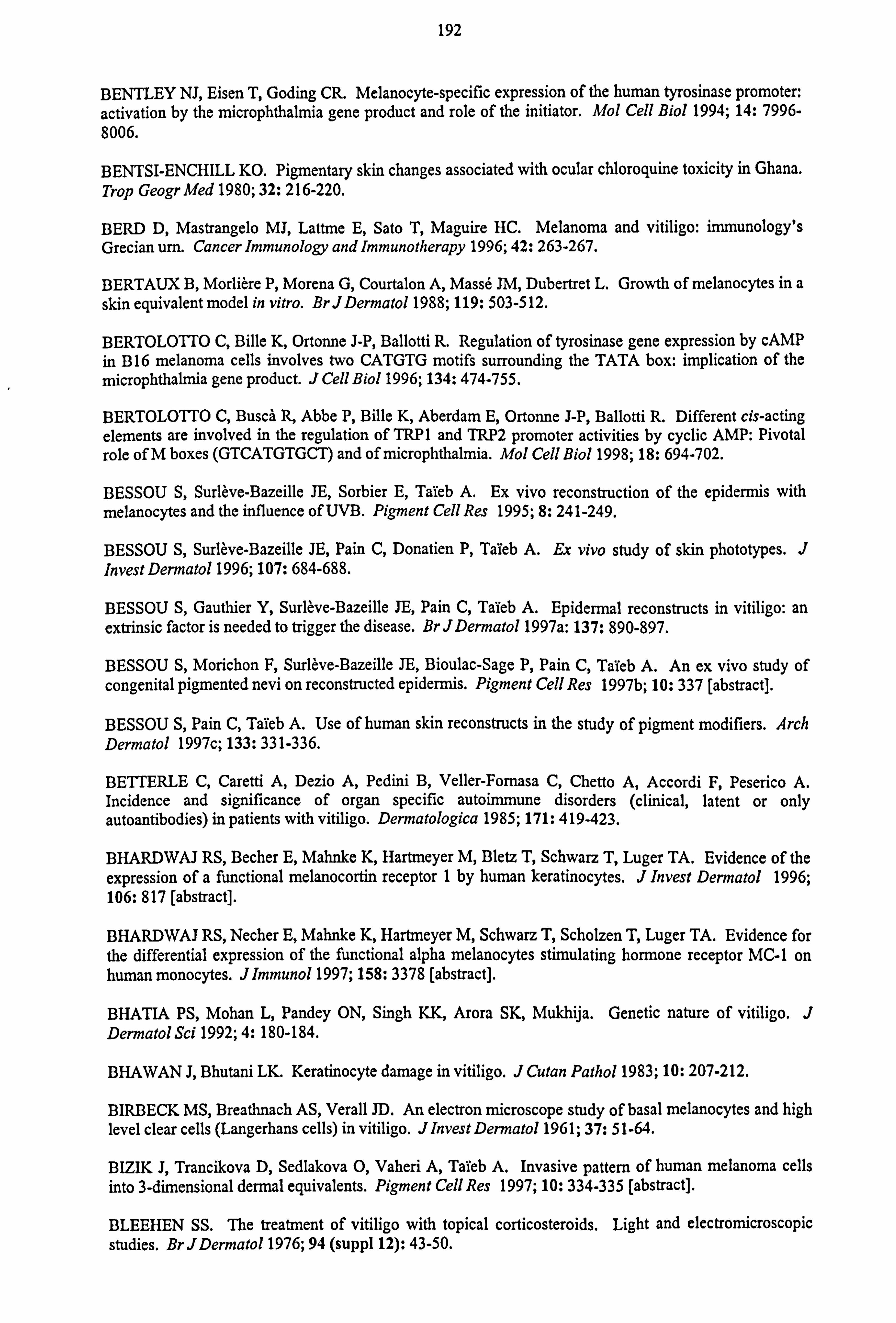
192
BENTLEY NJ, Eisen T, Goding CR. Melanocyte-specific expression of the human tyrosinase promoter: activation by the microphthalmia gene product and role of the initiator. Mol Cell Biol 1994; 14: 7996- 8006.
BENTSI-ENCHILL KO. Pigmentary skin changes associated with ocular chloroquine toxicity in Ghana. Trop Geogr Med 1980; 32: 216-220.
BERD D, Mastrangelo MJ, Lattme E, Sato T, Maguire HC. Melanoma and vitiligo: immunology's Grecian um. Cancer Immunology and Immunotherapy 1996; 42: 263-267.
BERTAUX B, Morliere P, Morena G, Courtalon A, Masse JM, Dubertret L. Growth of melanocytes in a skin equivalent model in vitro. BrJDermato11988; 119: 503-512.
BERTOLOTTO C, Bille K, Ortonne J-P, Ballotti R. Regulation of tyrosinase gene expression by cAMP in B16 melanoma cells involves two CATGTG motifs surrounding the TATA box: implication of the microphthalmia gene product. J Cell Bio11996; 134: 474-755.
BERTOLOTTO C, Buscä R, Abbe P, Bille K, Aberdam E, Ortonne J-P, Ballotti R. Different cis-acting elements are involved in the regulation of TRP1 and TRP2 promoter activities by cyclic AMP: Pivotal
role of M boxes (GTCATGTGCT) and of microphthalmia. Mol Cell Biol 1998; 18: 694-702.
BESSOU S, Surleve-Bazeille JE, Sorbier E, Taieb A. Ex vivo reconstruction of the epidermis with melanocytes and the influence of UVB. Pigment Cell Res 1995; 8: 241-249.
BESSOU S, Surleve-Bazeille JE, Pain C, Donatien P, Taieb A. Ex vivo study of skin phototypes. J Invest Dermato11996; 107: 684-688.
BESSOU S, Gauthier Y, Surleve-Bazeille JE, Pain C, Tafeb A. Epidermal reconstructs in vitiligo: an extrinsic factor is needed to trigger the disease. BrJDermato11997a: 137: 890-897.
BESSOU S, Morichon F, Surleve-Bazeille JE, Bioulac-Sage P, Pain C, Taieb A. An ex vivo study of congenital pigmented nevi on reconstructed epidermis. Pigment Cell Res 1997b; 10: 337 [abstract].
BESSOU S, Pain C, Taieb A. Use of human skin reconstructs in the study of pigment modifiers. Arch Dermatol 1997c; 133: 331-336.
BETTERLE C, Caretti A, Dezio A, Pedini B, Veller-Fornasa C, Chetto A, Accordi F, Peserico A. Incidence and significance of organ specific autoinnnune disorders (clinical, latent or only autoantibodies) in patients with vitiligo. Dermatologica 1985; 171: 419-423.
BHARDWAJ RS, Becher E, Malinke K, Hartmeyer M, Bletz T, Schwarz T, Luger TA. Evidence of the expression of a functional melanocortin receptor 1 by human keratinocytes. J Invest Dermatol 1996; 106: 817 [abstract].
BHARDWAJ RS, Necher E, Malinke K, Hartmeyer M, Schwarz T, Scholzen T, Luger TA. Evidence for the differential expression of the functional alpha melanocytes stimulating hormone receptor MC-1 on human monocytes. Jlmmuno11997; 158: 3378 [abstract].
BHATIA PS, Mohan L, Pandey ON, Singh KK, Arora SK, Mukhija. Genetic nature of vitiligo. J Dermatol Sci 1992; 4: 180-184.
BHAWAN J, Bhutani LK. Keratinocyte damage in vitiligo. J Cutan Patho11983; 10: 207-212.
BIRBECK MS, Breathnach AS, Verall JD. An electron microscope study of basal melanocytes and high level clear cells (Langerhans cells) in vitiligo. Jlnvest Dermato11961; 37: 51-64.
BIZIK J, Trancikova D, Sedlakova 0, Vaheri A, Taieb A. Invasive pattern of human melanoma cells into 3-dimensional dermal equivalents. Pigment Cell Res 1997; 10: 334-335 [abstract].
BLEEHEN SS. The treatment of vitiligo with topical corticosteroids. Light and electromicroscopic studies. BrJDermatol 1976; 94 (suppl 12): 43-50.
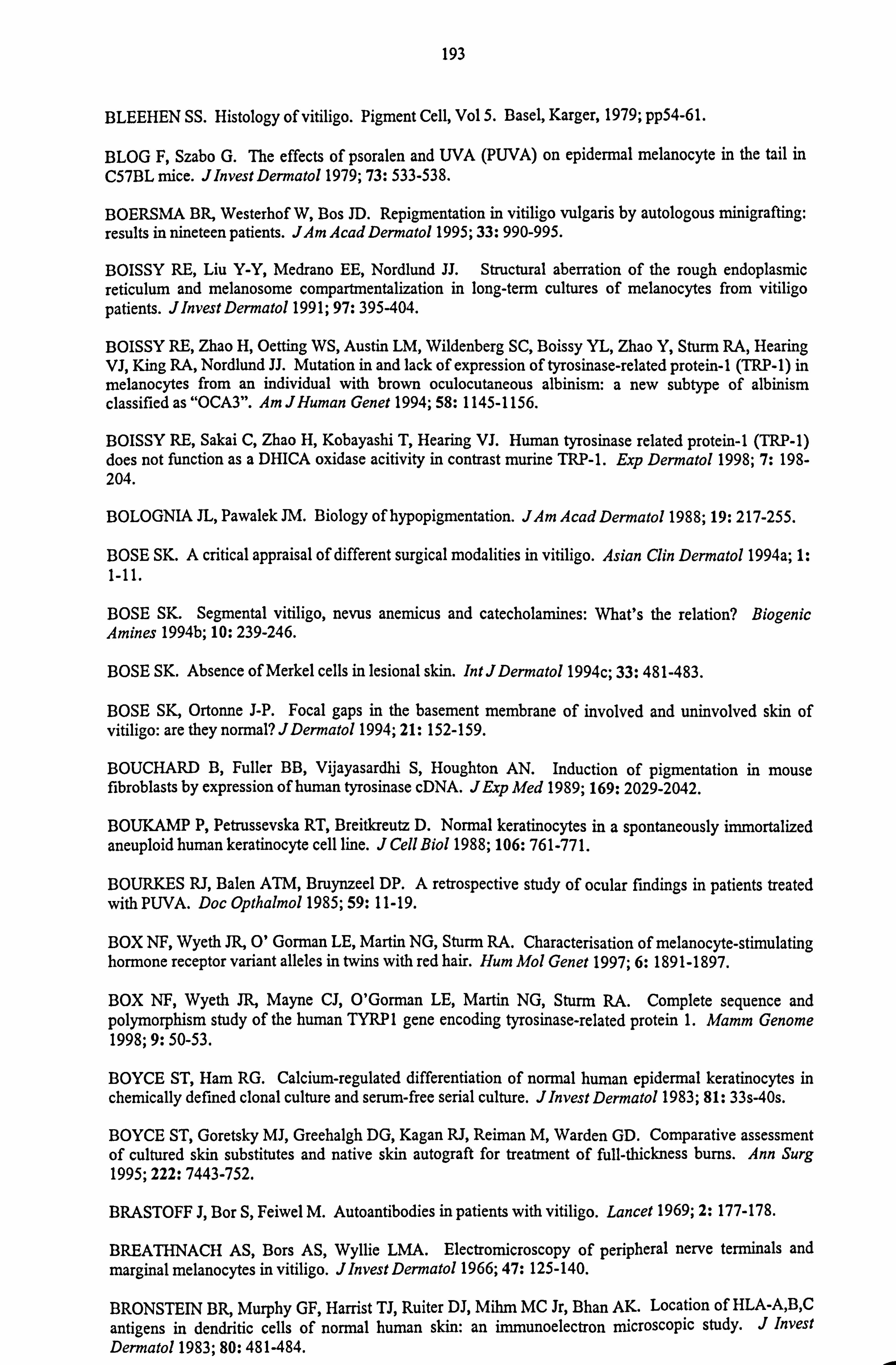
193
BLEEHEN SS. Histology of vitiligo. Pigment Cell, Vol 5. Basel, Karger, 1979; pp54-61.
BLOG F, Szabo G. The effects of psoralen and UVA (PUVA) on epidermal melanocyte in the tail in C57BL mice. JlnvestDermatol 1979; 73: 533-538.
BOERSMA BR, Westerhof W, Bos ID. Repigmentation in vitiligo vulgaris by autologous minigrafting: results in nineteen patients. JAm Acad Dermatol 1995; 33: 990-995.
BOISSY RE, Liu Y-Y, Medrano EE, Nordlund JJ. Structural aberration of the rough endoplasmic reticulum and melanosome compartmentalization in long-term cultures of melanocytes from vitiligo patients. J Invest Dermatol 1991; 97: 395-404.
BOISSY RE, Zhao H, Oetting WS, Austin LM, Wildenberg SC, Boissy YL, Zhao Y, Sturm RA, Hearing VJ, King RA, Nordlund JJ. Mutation in and lack of expression of tyrosinase-related protein-1 (TRP-1) in melanocytes from an individual with brown oculocutaneous albinism: a new subtype of albinism classified as "OCAS". Am JHuman Genet 1994; 58: 1145-1156.
BOISSY RE, Sakai C, Zhao H, Kobayashi T, Hearing VJ. Human tyrosinase related protein-1 (TRP-1) does not function as a DHICA oxidase acitivity in contrast murine TRP-1. Exp Dermatol 1998; 7: 198- 204.
BOLOGNIA JL, Pawalek JM. Biology of hypopigmentation. JAm Acad Dermatol 1988; 19: 217-255.
BOSE SK. A critical appraisal of different surgical modalities in vitiligo. Asian Clin Dermatol 1994a; 1: 1-11.
BOSE SK. Segmental vitiligo, nevus anemicus and catecholamines: What's the relation? Biogenic Amines 1994b; 10: 239-246.
BOSE SK. Absence of Merkel cells in lesional skin. IntJDermatol 1994c; 33: 481-483.
BOSE SK, Ortonne J-P. Focal gaps in the basement membrane of involved and uninvolved skin of vitiligo: are they normal? JDermato11994; 21: 152-159.
BOUCHARD B, Fuller BB, Vijayasardhi S, Houghton AN. Induction of pigmentation in mouse fibroblasts by expression of human tyrosinase cDNA. JExp Med 1989; 169: 2029-2042.
BOUKAMP P, Petrussevska RT, Breitkreutz D. Normal keratinocytes in a spontaneously immortalized aneuploid human keratinocyte cell line. J Cell Bio11988; 106: 761-771.
BOURKES RJ, Balen ATM, Bruynzeel DP. A retrospective study of ocular findings in patients treated with PUVA. Doc Opthalmo11985; 59: 11-19.
BOX NF, Wyeth JR, 0' Gorman LE, Martin NG, Sturm RA. Characterisation of melanocyte-stimulating hormone receptor variant alleles in twins with red hair. Hum Mol Genet 1997; 6: 1891-1897.
BOX NF, Wyeth JR, Mayne CJ, O'Gorman LE, Martin NG, Sturm RA. Complete sequence and polymorphism study of the human TYRP1 gene encoding tyrosinase-related protein 1. Mamm Genome 1998; 9: 50-53.
BOYCE ST, Ham RG. Calcium-regulated differentiation of normal human epidermal keratinocytes in chemically defined clonal culture and serum-free serial culture. Jlnvest Dermato11983; 81: 33s-40s.
BOYCE ST, Goretsky MJ, Greehalgh DG, Kagan RJ, Reiman M, Warden GD. Comparative assessment of cultured skin substitutes and native skin autograft for treatment of full-thickness bums. Ann Surg 1995; 222: 7443-752.
BRASTOFF J, Bor S, Feiwel M. Autoantibodies in patients with vitiligo. Lancet 1969; 2: 177-178.
BREATHNACH AS, Bors AS, Wyllie LMA. Electromicroscopy of peripheral nerve terminals and marginal melanocytes in vitiligo. JInvest Dermato11966; 47: 125-140.
BRONSTEIN BR, Murphy GF, Harnst TJ, Ruiter DJ, Mihm MC Jr, Bhan AK. Location of HLA-A, B, C
antigens in dendritic cells of normal human skin: an immunoelectron microscopic study. J Invest Dermato11983; 80: 481-484.
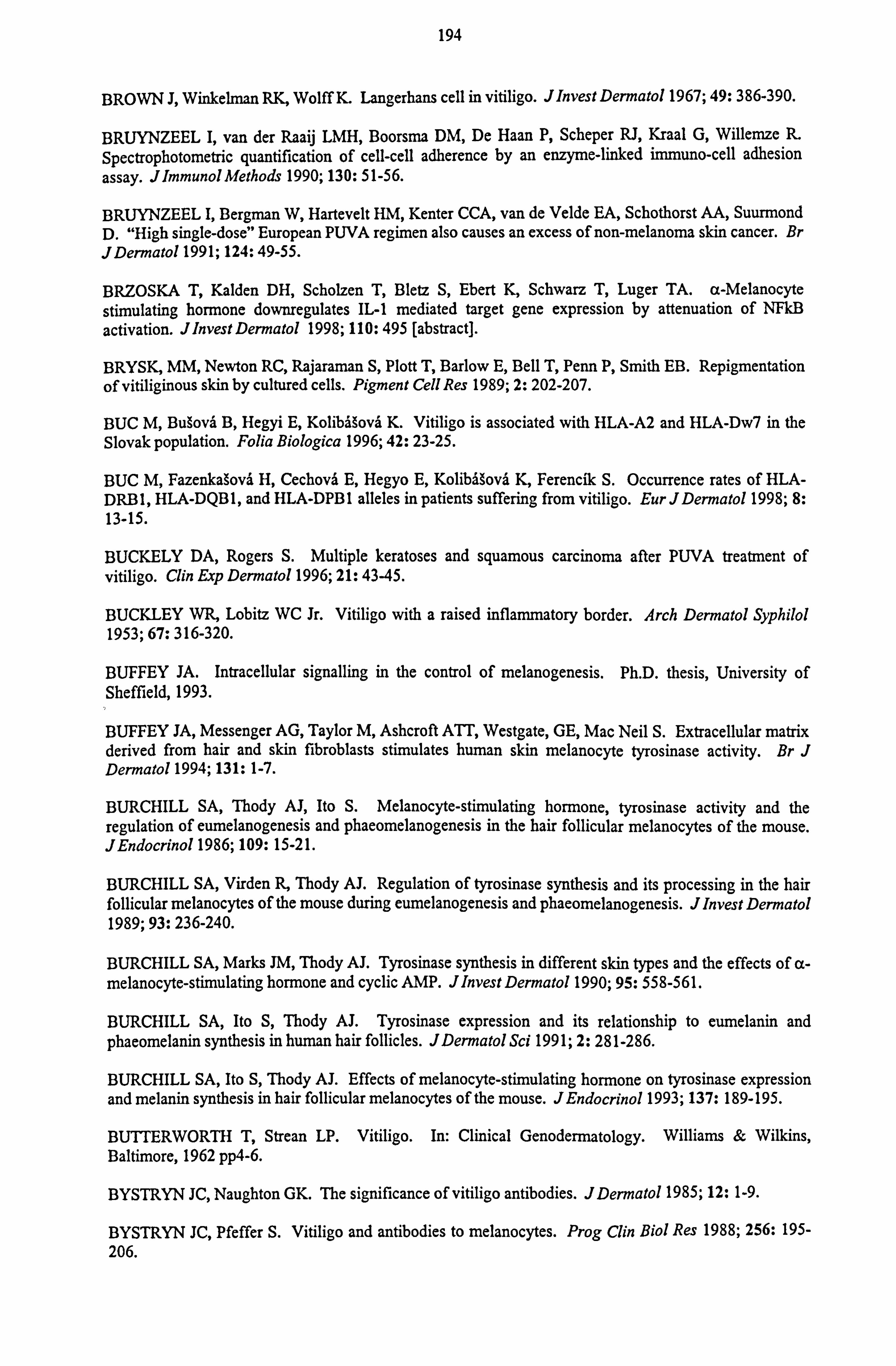
194
BROWN J, Winkelman RK, Wolff K. Langerhans cell in vitiligo. Jlnvest Dermatol 1967; 49: 386-390.
BRUYNZEEL I, van der Raaij LMH, Boorsma DM, De Haan P, Scheper RJ, Kraal G, Willemze R. Spectrophotometric quantification of cell-cell adherence by an enzyme-linked immuno-cell adhesion assay. Jlmmunol Methods 1990; 130: 51-56.
BRUYNZEEL I, Bergman W, Hartevelt HM, Kenter CCA, van de Velde EA, Schothorst AA, Suurmond D. "High single-dose" European PUVA regimen also causes an excess of non-melanoma skin cancer. Br J Dermato11991; 124: 49-55.
BRZOSKA T, Kalden DH, Scholzen T, Bletz S, Ebert K, Schwarz T, Luger TA. a-Melanocyte stimulating hormone downregulates IL-1 mediated target gene expression by attenuation of NFkB activation. JlnvestDermatol 1998; 110: 495 [abstract].
BRYSK, MM, Newton RC, Rajaraman S, Plott T, Barlow E, Bell T, Penn P, Smith EB. Repigmentation of vitiliginous skin by cultured cells. Pigment Cell Res 1989; 2: 202-207.
BUC M, Bugovä B, Hegyi E, Kolibä ova K. Vitiligo is associated with HLA-A2 and HLA-Dw7 in the Slovak population. Folia Biologica 1996; 42: 23-25.
BUC M, Fazenkagovi H, Cechovä E, Hegyo E, Kolibä ova K, Ferencilc S. Occurrence rates of HLA- DRB 1, HLA-DQBI, and HLA-DPB I alleles in patients suffering from vitiligo. Eur J Dermatol 1998; 8: 13-15.
BUCKELY DA, Rogers S. Multiple keratoses and squamous carcinoma after PUVA treatment of vitiligo. Clin Exp Dermatol 1996; 21: 43-45.
BUCKLEY WR, Lobitz WC Jr. Vitiligo with a raised inflammatory border. Arch Dermatol Syphilol 1953; 67: 316-320.
BUFFEY JA. Intracellular signalling in the control of melanogenesis. Ph. D. thesis, University of Sheffield, 1993.
BUFFEY JA, Messenger AG, Taylor M, Ashcroft ATT, Westgate, GE, Mac Neil S. Extracellular matrix derived from hair and skin fibroblasts stimulates human skin melanocyte tyrosinase activity. Br J Dermatol 1994; 131: 1-7.
BURCHILL SA, Thody AJ, Ito S. Melanocyte-stimulating hormone, tyrosinase activity and the regulation of eumelanogenesis and phaeomelanogenesis in the hair follicular melanocytes of the mouse. JEndocrinol 1986; 109: 15-21.
BURCHILL SA, Virden R, Thody AJ. Regulation of tyrosinase synthesis and its processing in the hair follicular melanocytes of the mouse during eumelanogenesis and phaeomelanogenesis. J Invest Dermatol 1989; 93: 236-240.
BURCHILL SA, Marks JM, Thody AJ. Tyrosinase synthesis in different skin types and the effects of a- melanocyte-stimulating hormone and cyclic AMP. JlnvestDermato11990; 95: 558-561.
BURCHILL SA, Ito S, Thody AJ. Tyrosinase expression and its relationship to eumelanin and phaeomelanin synthesis in human hair follicles. JDermatol Sci 1991; 2: 281-286.
BURCHILL SA, Ito S, Thody AJ. Effects of melanocyte-stimulating hormone on tyrosinase expression and melanin synthesis in hair follicular melanocytes of the mouse. JEndocrino11993; 137: 189-195.
BUTTERWORTH T, Strean LP. Vitiligo. In: Clinical Genodermatology. Williams & Wilkins, Baltimore, 1962 pp4-6.
BYSTRYN JC, Naughton GK. The significance of vitiligo antibodies. JDermato11985; 12: 1-9.
BYSTRYN JC, Pfeffer S. Vitiligo and antibodies to melanocytes. Prog Clin Biol Res 1988; 256: 195- 206.
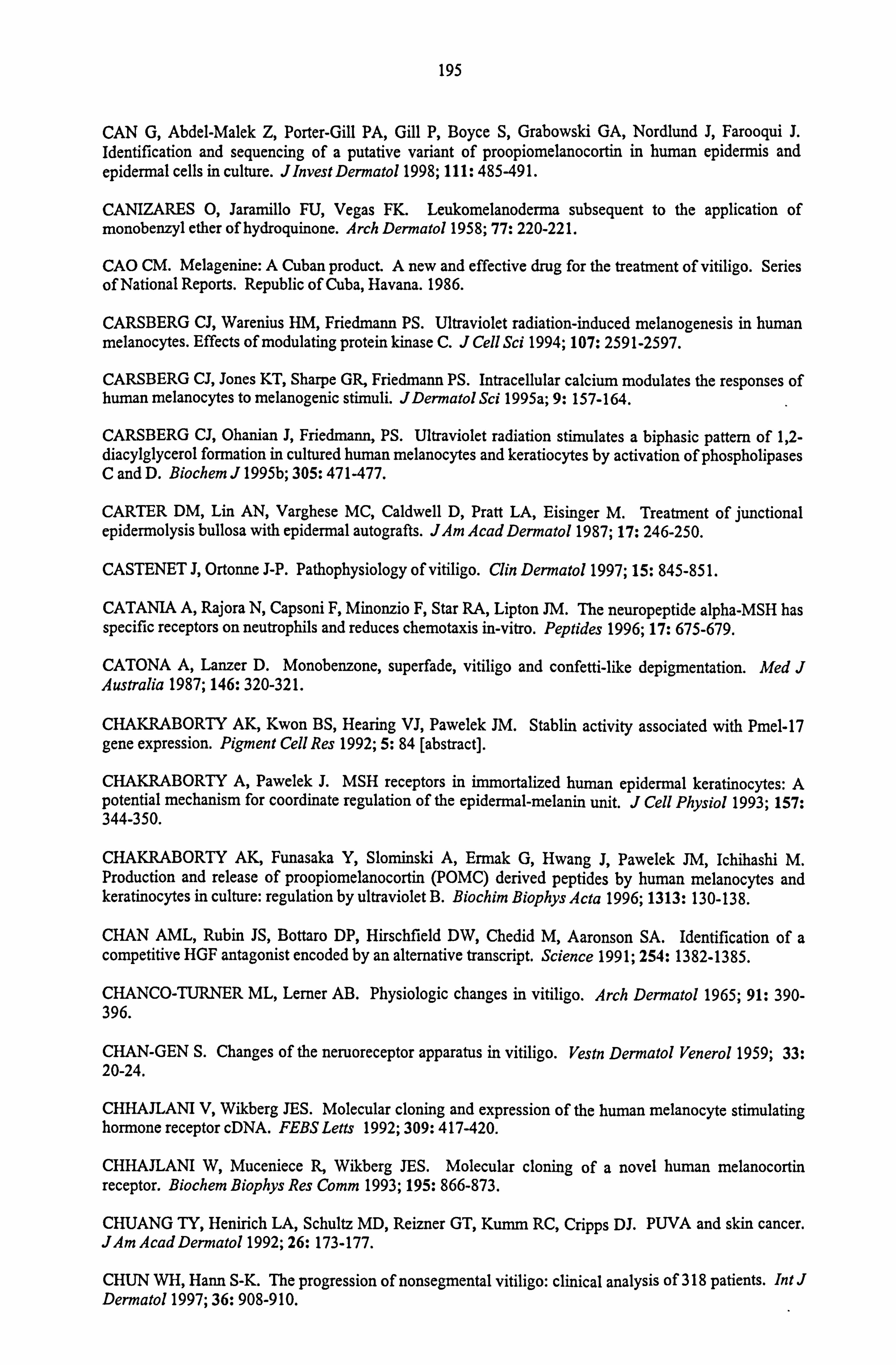
195
CAN G, Abdel-Malek Z, Porter-Gill PA, Gill P, Boyce S, Grabowski GA, Nordlund J, Farooqui J. Identification and sequencing of a putative variant of proopiomelanocortin in human epidermis and epidermal cells in culture. JlnvestDermatol 1998; 111: 485-491.
CANIZARES 0, Jaramillo FU, Vegas FK. Leukomelanoderma subsequent to the application of monobenzyl ether of hydroquinone. Arch Dermatol 1958; 77: 220-221.
CAO CM. Melagenine: A Cuban product. A new and effective drug for the treatment of vitiligo. Series of National Reports. Republic of Cuba, Havana. 1986.
CARSBERG CJ, Warenius HM, Friedmann PS. Ultraviolet radiation-induced melanogenesis in human melanocytes. Effects of modulating protein kinase C. J Cell Sci 1994; 107: 2591-2597.
CARSBERG CJ, Jones KT, Sharpe GR, Friedmann PS. Intracellular calcium modulates the responses of human melanocytes to melanogenic stimuli. JDermatol Sci 1995a; 9: 157-164.
CARSBERG CJ, Ohanian J, Friedmann, PS. Ultraviolet radiation stimulates a biphasic pattern of 1,2- diacylglycerol formation in cultured human melanocytes and keratiocytes by activation of phospholipases C and D. Biochem J 1995b; 305: 471-477.
CARTER DM, Lin AN, Varghese MC, Caldwell D, Pratt LA, Eisinger M. Treatment of junctional epidermolysis bullosa with epidermal autografts. JAm Acad Dermatol 1987; 17: 246-250.
CASTENET J, Ortonne J-P. Pathophysiology of vitiligo. Clin Dermatol 1997; 15: 845-851.
CATANIA A, Rajora N, Capsoni F, Minonzio F, Star RA, Lipton JM. The neuropeptide alpha-MSH has specific receptors on neutrophils and reduces chemotaxis in-vitro. Peptides 1996; 17: 675-679.
CATONA A, Lanzer D. Monobenzone, superfade, vitiligo and confetti-like depigmentation. Med J Australia 1987; 146: 320-321.
CHAKRABORTY AK, Kwon BS, Hearing VJ, Pawelek JM. Stablin activity associated with Pmel-17 gene expression. Pigment Cell Res 1992; 5: 84 [abstract].
CHAKRABORTY A, Pawelek J. MSH receptors in immortalized human epidermal keratinocytes: A potential mechanism for coordinate regulation of the epidermal-melanin unit. J Cell Physiol 1993; 157: 344-350.
CHAKRABORTY AK, Funasaka Y, Slominski A, Ermak G, Hwang J, Pawelek JM, Ichihashi M. Production and release of proopiomelanocortin (POMC) derived peptides by human melanocytes and keratinocytes in culture: regulation by ultraviolet B. Biochim Biophys Acta 1996; 1313: 130-138.
CHAN AML, Rubin JS, Bottaro DP, Hirschfield DW, Chedid M, Aaronson SA. Identification of a competitive HGF antagonist encoded by an alternative transcript. Science 1991; 254: 1382-1385.
CHANCO-TURNER ML, Lerner AB. Physiologic changes in vitiligo. Arch Dermatol 1965; 91: 390- 396.
CHAN-GEN S. Changes of the neruoreceptor apparatus in vitiligo. Veste Dermatol Venerol 1959; 33: 20-24.
CHHAJLANI V, Wikberg JES. Molecular cloning and expression of the human melanocyte stimulating hormone receptor cDNA. FEBS Letts 1992; 309: 417-420.
CHHAJLANI W, Muceniece R, Wikberg JES. Molecular cloning of a novel human melanocortin receptor. Biochem Biophys Res Comm 1993; 195: 866-873.
CHUANG TY, Henrich LA, Schultz MD, Reizner GT, Kunun RC, Cripps DJ. PUVA and skin cancer. JAm Acad Dermato11992; 26: 173-177.
CHUN WH, Hann S-K. The progression of nonsegmental vitiligo: clinical analysis of 318 patients. Int J Dermato11997; 36: 908-910.
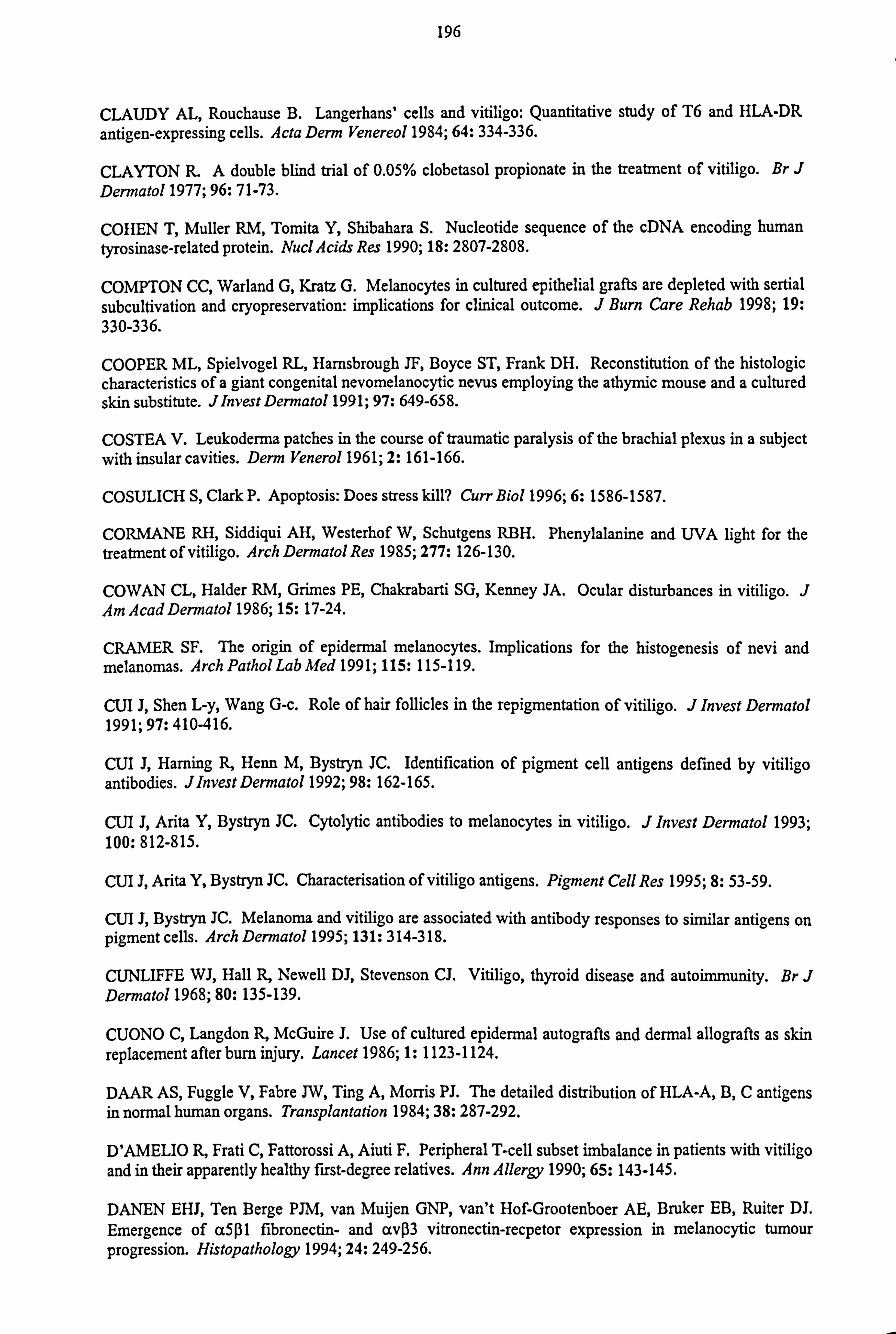
196
CLAUDY AL, Rouchause B. Langerhans' cells and vitiligo: Quantitative study of T6 and HLA-DR
antigen-expressing cells. Acta Derm Venereol 1984; 64: 334-336.
CLAYTON R. A double blind trial of 0.05% clobetasol propionate in the treatment of vitiligo. Br J Dermatol 1977; 96: 71-73.
COHEN T, Muller RM, Tonvita Y, Shibahara S. Nucleotide sequence of the cDNA encoding human tyrosinase-related protein. Nucl Acids Res 1990; 18: 2807-2808.
COMPTON CC, Warland G, Kratz G. Melanocytes in cultured epithelial grafts are depleted with sertial subcultivation and cryopreservation: implications for clinical outcome. J Burn Care Rehab 1998; 19: 330-336.
COOPER ML, Spielvogel RL, Harnsbrough JF, Boyce ST, Frank DH. Reconstitution of the histologic
characteristics of a giant congenital nevomelanocytic nevus employing the athymic mouse and a cultured skin substitute. JInvest Dermatol 1991; 97: 649-658.
COSTEA V. Leukoderma patches in the course of traumatic paralysis of the brachial plexus in a subject with insular cavities. Derm Venerol 1961; 2: 161-166.
COSULICH S, Clark P. Apoptosis: Does stress kill? Curr Biol 1996; 6: 1586-1587.
CORMANE RH, Siddiqui AH, Westerhof W, Schutgens RBH. Phenylalanine and UVA light for the treatment of vitiligo. Arch Dermatol Res 1985; 277: 126-130.
COWAN CL, Haider RM, Grimes PE, Chakrabarti SG, Kenney JA. Ocular disturbances in vitiligo. J Am Acad Dermatol 1986; 15: 17-24.
CRAMER SF. The origin of epidermal melanocytes. Implications for the histogenesis of nevi and melanomas. Arch Pathol Lab Med 1991; 115: 115-119.
CUI J, Shen L-y, Wang G-c. Role of hair follicles in the repigmentation of vitiligo. J Invest Dermatol 1991; 97: 410-416.
CUI J, Harping R, Henn M, Bystryn JC. Identification of pigment cell antigens defined by vitiligo antibodies. J Invest Dermatol 1992; 98: 162-165.
CUI J, Arita Y, Bystryn JC. Cytolytic antibodies to melanocytes in vitiligo. J Invest Dermatol 1993; 100: 812-815.
CUI J, Arita Y, Bystryn JC. Characterisation of vitiligo antigens. Pigment Cell Res 1995; 8: 53-59.
CUI J, Bystryn JC. Melanoma and vitiligo are associated with antibody responses to similar antigens on pigment cells. Arch Dermatol 1995; 131: 314-318.
CUNLIFFE WJ, Hall R, Newell DJ, Stevenson CJ. Vitiligo, thyroid disease and autoinimunity. Br J Dermatol 1968; 80: 135-139.
CUONO C, Langdon R, McGuire J. Use of cultured epidermal autografts and dermal allografts as skin replacement after burn injury. Lancet 1986; 1: 1123-1124.
DAAR AS, Fuggle V, Fabre JW, Ting A, Morris PJ. The detailed distribution of HLA-A, B, C antigens in normal human organs. Transplantation 1984; 38: 287-292.
D'AMELIO R, Frati C, Fattorossi A, Aiuti F. Peripheral T-cell subset imbalance in patients with vitiligo and in their apparently healthy first-degree relatives. Ann Allergy 1990; 65: 143-145.
DANEN EHJ, Ten Berge PJM, van Muijen GNP, van't Hof-Grootenboer AE, Bruker EB, Ruiter DJ. Emergence of a5ß1 fibronectin- and avß3 vitronectin-recpetor expression in melanocytic tumour progression. Histopathology 1994; 24: 249-256.
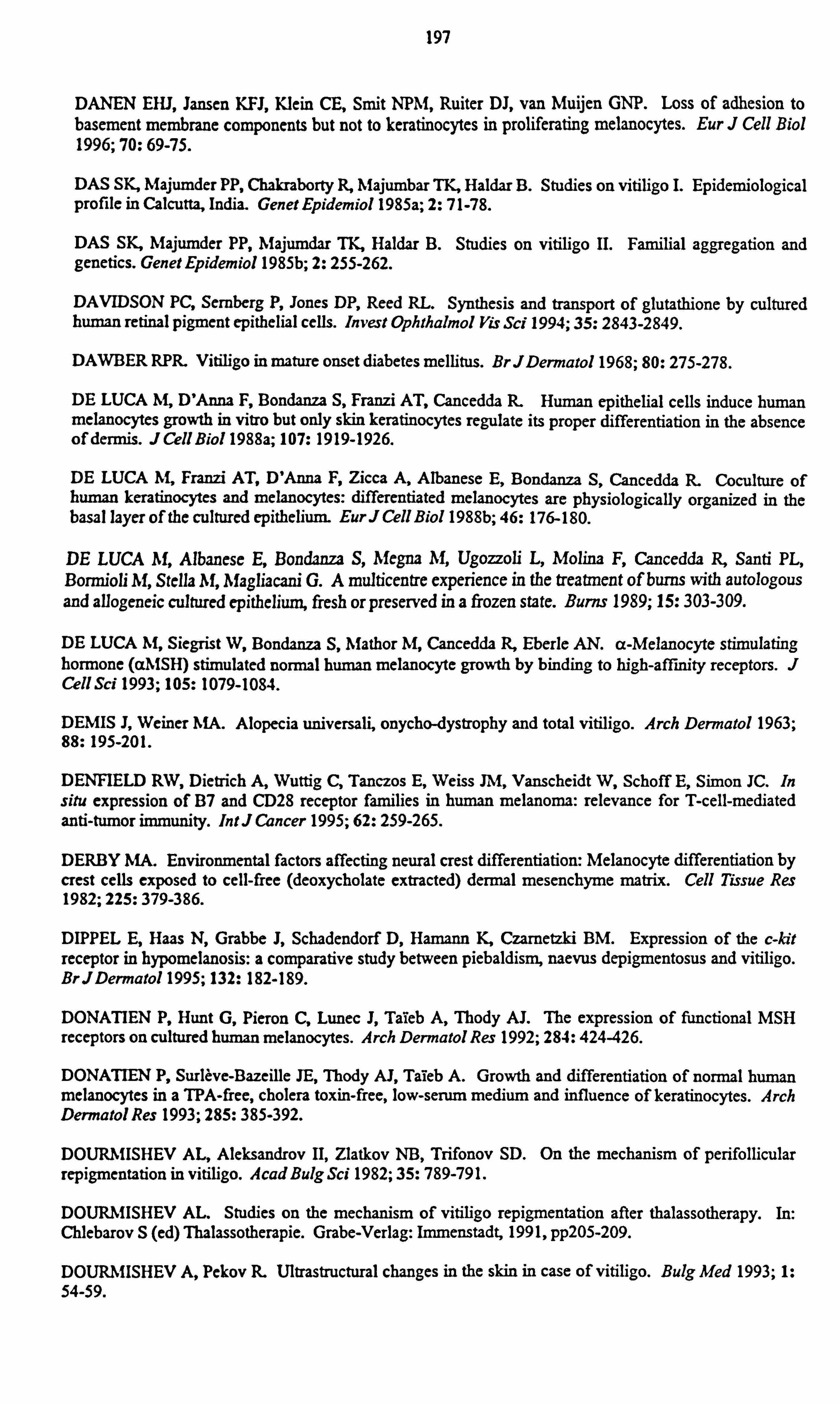
197
DANEN EHJ, Jansen KFJ, Klein CE, Smit NPM'I, Ruiter DJ, van Muijen GNP. Loss of adhesion to basement membrane components but not to keratinocytes in proliferating melanocytes. Eur J Cell Biol 1996; 70: 69-75.
DAS SK, Majumder PP, Chakraborty R, Majumbar TK, Haldar B. Studies on vitiligo I. Epidemiological profile in Calcutta, India. Genet Epidemiol 1985a; 2: 71-78.
DAS SK, Majumder PP, Majumdar TK, Haldar B. Studies on vitiligo II. Familial aggregation and genetics. Genet Epidemiol 1985b; 2: 255-262.
DAVIDSON PC, Sernberg P, Jones DP, Reed RL. Synthesis and transport of glutathione by cultured human retinal pigment epithelial cells. Invest Ophthalmol leis Sci 1994; 35: 2843-2849.
DAWBER RPR. Vitiligo in mature onset diabetes mellitus. BrJDermatol 1968; 80: 275-278.
DE LUCA M, D'Anna F, Bondanza S, Franzi AT, Cancedda R. Human epithelial cells induce human melanocytes growth in vitro but only skin keratinocytes regulate its proper differentiation in the absence of dermis. J Cell Biol 1988a; 107: 1919-1926.
DE LUCA M, Franzi AT, D'Anna F, Zicca A, Albanese E, Bondanza S, Cancedda R. Coculture of human keratinocytes and melanocytes: differentiated melanocytes are physiologically organized in the basal layer of the cultured epithelium. EurJ Cell Biol 1988b; 46: 176-180.
DE LUCA M, Albanese E, Bondanza S, Megna M, Ugozzoli L, Molina F, Cancedda R, Santi PL, Bormioli M, Stella M, Magliacani G. A multicentre experience in the treatment of burns with autologous and allogeneic cultured epithelium, fresh or preserved in a frozen state. Burns 1989; 15: 303-309.
DE LUCA M, Siegrist W, Bondanza S, Mathor M, Cancedda R, Eberle AN. a-Melanocyte stimulating hormone (aMSH) stimulated normal human melanocyte growth by binding to high-affinity receptors. J Cell Scf 1993; 105: 1079-1084.
DEMIS J, Weiner MA. Alopecia universali, onycho-dystrophy and total vitiligo. Arch Dermatol 1963; 88: 195-201.
DENFIELD RW, Dietrich A, Wuttig C, Tanczos E, Weiss JM, Vanscheidt W, Schoff E, Simon JC. In situ expression of B7 and CD28 receptor families in human melanoma: relevance for T-cell-mediated anti-tumor immunity. Int J Cancer 1995; 62: 259-265.
DERBY MA. Environmental factors affecting neural crest differentiation: Melanocyte differentiation by crest cells exposed to cell-free (deoxycholate extracted) dermal mesenchyme matrix. Cell Tissue Res 1982; 225: 379-386.
DIPPEL E, Haas N, Grabbe J, Schadendorf D, Hamann K, Czametzki BM. Expression of the c-kit receptor in hypomelanosis: a comparative study between piebaldism, naevus depigmentosus and vitiligo. BrJDermatol 1995; 132: 182-189.
DONATIEN P, Hunt G, Pieron C, Lunec J, Taieb A, Thody AJ. The expression of functional MSH receptors on cultured human melanocytes. Arch Dermatol Res 1992; 284: 424-426.
DONATIEN P, Surleve-Bazeille JE, Thody AJ, Taieb A. Growth and differentiation of normal human melanocytes in a TPA-free, cholera toxin-free, low-serum medium and influence of keratinocytes. Arch Dermatol Res 1993; 285: 385-392.
DOURMISHEV AL, Aleksandrov II, Zlatkov NB, Trifonov SD. On the mechanism of perifollicular repigmentation in vitiligo. Acad Bulg Sci 1982; 35: 789-79 1.
DOURMISHEV AL. Studies on the mechanism of vitiligo repigmentation after thalassotherapy. In: Chlebarov S (ed) Thalassotherapie. Grabe-Verlag: Immenstadt, 1991, pp205-209.
DOURNIISHEV A, Pekov It Ultrastructural changes in the skin in case of vitiligo. Bulg Med 1993; 1: 54-59.
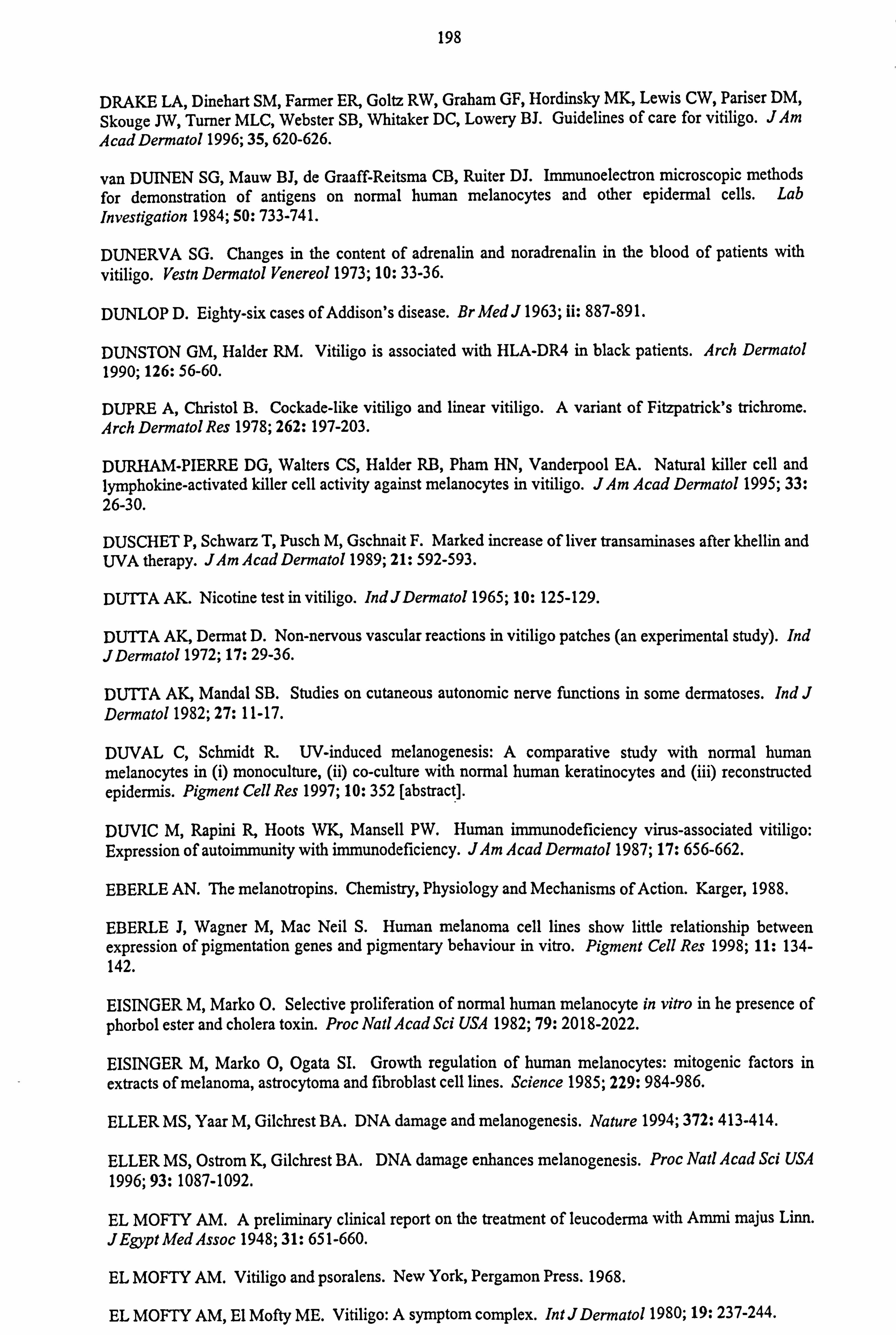
198
DRAKE LA, Dinehart SM, Farmer ER, Goltz RW, Graham GF, Hordinsky MK, Lewis CW, Pariser DM, Skouge JW, Turner MLC, Webster SB, Whitaker DC, Lowery BJ. Guidelines of care for vitiligo. J Am AcadDermatol 1996; 35,620-626.
van DUINEN SG, Mauw BJ, de Graaff-Reitsma CB, Ruiter DJ. Immunoelectron microscopic methods for demonstration of antigens on normal human melanocytes and other epidermal cells. Lab Investigation 1984; 50: 733-741.
DUNERVA SG. Changes in the content of adrenalin and noradrenalin in the blood of patients with vitiligo. Vestn Dermatol Venereol 1973; 10: 33-36.
DUNLOP D. Eighty-six cases of Addison's disease. Br Med J 1963; ii: 887-891.
DUNSTON GM, Haider RM. Vitiligo is associated with HLA-DR4 in black patients. Arch Dermatol 1990; 126: 56-60.
DUPRE A, Christol B. Cockade-like vitiligo and linear vitiligo. A variant of Fitzpatrick's trichrome. Arch Dermatol Res 1978; 262: 197-203.
DURHAM-PIERRE DG, Walters CS, Haider RB, Pham HN, Vanderpool EA. Natural killer cell and lymphokine-activated killer cell activity against melanocytes in vitiligo. JAm Acad Dermatol 1995; 33: 26-30.
DUSCHET P, Schwarz T, Pusch M, Gschnait F. Marked increase of liver transaminases after khellin and UVA therapy. JAm Acad Dermatol 1989; 21: 592-593.
DUTTA AK. Nicotine test in vitiligo. IndJDermatol 1965; 10: 125-129.
DUTTA AK, Dermat D. Non-nervous vascular reactions in vitiligo patches (an experimental study). Ind JDermatol 1972; 17: 29-36.
DUTTA AK, Mandal SB. Studies on cutaneous autonomic nerve functions in some dermatoses. Ind J Dermatol 1982; 27: 11-17.
DUVAL C, Schmidt R. UV-induced melanogenesis: A comparative study with normal human melanocytes in (i) monoculture, (ii) co-culture with normal human keratinocytes and (iii) reconstructed epidermis. Pigment Cell Res 1997; 10: 352 [abstract].
DUVIC M, Rapini R, Hoots WK, Mansell PW. Human immunodeficiency virus-associated vitiligo: Expression of autoimmunity with immunodeficiency. JAm Acad Dermatol 1987; 17: 656-662.
EBERLE AN. The melanotropins. Chemistry, Physiology and Mechanisms of Action. Karger, 1988.
EBERLE J, Wagner M, Mac Neil S. Human melanoma cell lines show little relationship between expression of pigmentation genes and pigmentary behaviour in vitro. Pigment Cell Res 1998; 11: 134- 142.
EISINGER M, Marko O. Selective proliferation of normal human melanocyte in vitro in he presence of phorbol ester and cholera toxin. Proc Natl Acad Sci USA 1982; 79: 2018-2022.
EISINGER M, Marko 0, Ogata SI. Growth regulation of human melanocytes: mitogenic factors in extracts of melanoma, astrocytoma and fibroblast cell lines. Science 1985; 229: 984-986.
ELLER MS, Yaar M, Gilchrest BA. DNA damage and melanogenesis. Nature 1994; 372: 413-414.
ELLER MS, Ostrom K, Gilchrest BA. DNA damage enhances melanogenesis. Proc Natl Acad Sci USA 1996; 93: 1087-1092.
EL MOFTY AM. A preliminary clinical report on the treatment of leucoderma with Ammi majus Linn. JEgypt Med Assoc 1948; 31: 651-660.
EL MOFTY AM. Vitiligo and psoralens. New York, Pergamon Press. 1968.
EL MOFTY AM, El Mofty ME. Vitiligo: A symptom complex. Int JDermatol 1980; 19: 237-244.

199
ENGASSER PG, Maibach HI. Cosmetics and dermatology; Bleaching creams. J Am Acad Dermatol 1981; 5: 143-147.
ENGLARO W, Rezzonico R, Durand-Clement M, Lallemand D, Ortonne JP, Ballotti R. Mitogen- activated protein kinase pathway and AP-1 are activated during cAMP-induced melanogenesis in B-16 melanoma cells. JBiol Chem 1995; 270: 24315-24320.
ESCALANTE-UGAIDE C, Poblano A, Montes de Oca E. No evidence of hearing loss in patients with vitiligo. Arch Dermatol 1991; 127: 1240.
FALABELLA R. Repigmentation of segmental vitiligo by autologous minigrafting. J Am Acad Dermatol 1983; 9: 514-521.
FALABELLA R. Treatment of localised vitiligo by autologous minigrafting. Arch Dermatol 1988; 124: 1649-1655.
FALABELLA R, Escobar C, Borrero I. Transplantation of in vitro-cultured epidermis bearing melanocytes for repigmenting vitiligo. JAm Acad Dermatol 1989; 21: 257-264.
FALABELLA R, Escobar C, Borrero I. Treatment of refractory and stable vitiligo by transplantation of in vitro cultured epidermal autografts bearing melanocytes. JAm Acad Dermatol 1992; 26: 230-236.
FALABELLA R, Arrunätegui A, Barona MI, Alzate A. The minigrafting test for vitiligo: Detection of stable lesions for melanocyte transplantation. JAm Acad Dermatol 1995a; 32: 228-232.
FALABELLA R, Barona MI, Escobar C, Borrero I, Arrunategui A. Surgical Combination therapy for vitiligo and piebaldism. Dermatol Surg 1995b; 21: 852-857.
FARGNOLI MC, Bolognia IL. Pentachrome vitiligo. JAm Acad Dermatol 1995; 33: 853-856.
FAROOQUI JZ, Medrano EE, Abdel-Malek Z, Nordlund J. The expression of proopiomelanocortin and various POMC-derived peptides in mouse and human skin. Ann NYAcad Sci 1993; 680: 508-510.
FAURE M, Maudit G, Schmitt D, Kanitakis J, Demidem A, Thivolet J. Growth and differentiation of human epidermal cultures used as auto- and allografts in humans. BrJDermatol 1987; 116: 161-170
FERGUSON CA, Kidson SH. The regulation of tyrosinase gene transcription. Pigment Cell Res 1997; 10: 127-138.
FIELDS JP, Fragola L, Hadley TP. Hypoparathyroidism, candidiasis, alopecia and vitiligo. Arch Dermatol 1971; 103: 687-689.
F1NCO 0, Cuccia M, Martinetti M, Ruberto G, Orecchia G, Rabbiosi G. Age of onset in vitiligo: Relationship with HLA supratypes. Clin Genetics 1991; 39: 48-54.
FISHMAN P, Azizi E, Shoenfeld Y, Sredni B, Yecheskel G, Ferrone S, Zigelman R, Chaitchik S, Floro S, Djaldetti M. Vitiligo autoantibodies are effective against melanoma. Cancer 1993; 72: 2365-2369.
FISHMAN P, Merimski 0, Baharav E, Shoenfeld Y. Autoantibodies to tyrosinase: The bridge between melanoma and vitiligo. Cancer 1997; 79: 1461-1644.
FITZPATRICK TB. Hypomelanosis. South Med J 1964; 57: 995-1005.
FITZPATRICKTB, Miyamoto M, Ishikaw K. The evolution of concepts of melanin biology. Arch Dermatol 1967; 96: 305-323.
FITZPATRICK TB. Mechanisms of phototherapy of vitiligo. Arch Dermatol 1997; 133: 1591-1592.
FLEMING KA, McMichael A, Morton JA, Woods J, McGee JO'D. Distribution of HLA class 1 antigens in nromal human tissue and in mammary cancer. J Clin Pathol 1981; 34: 779-784.
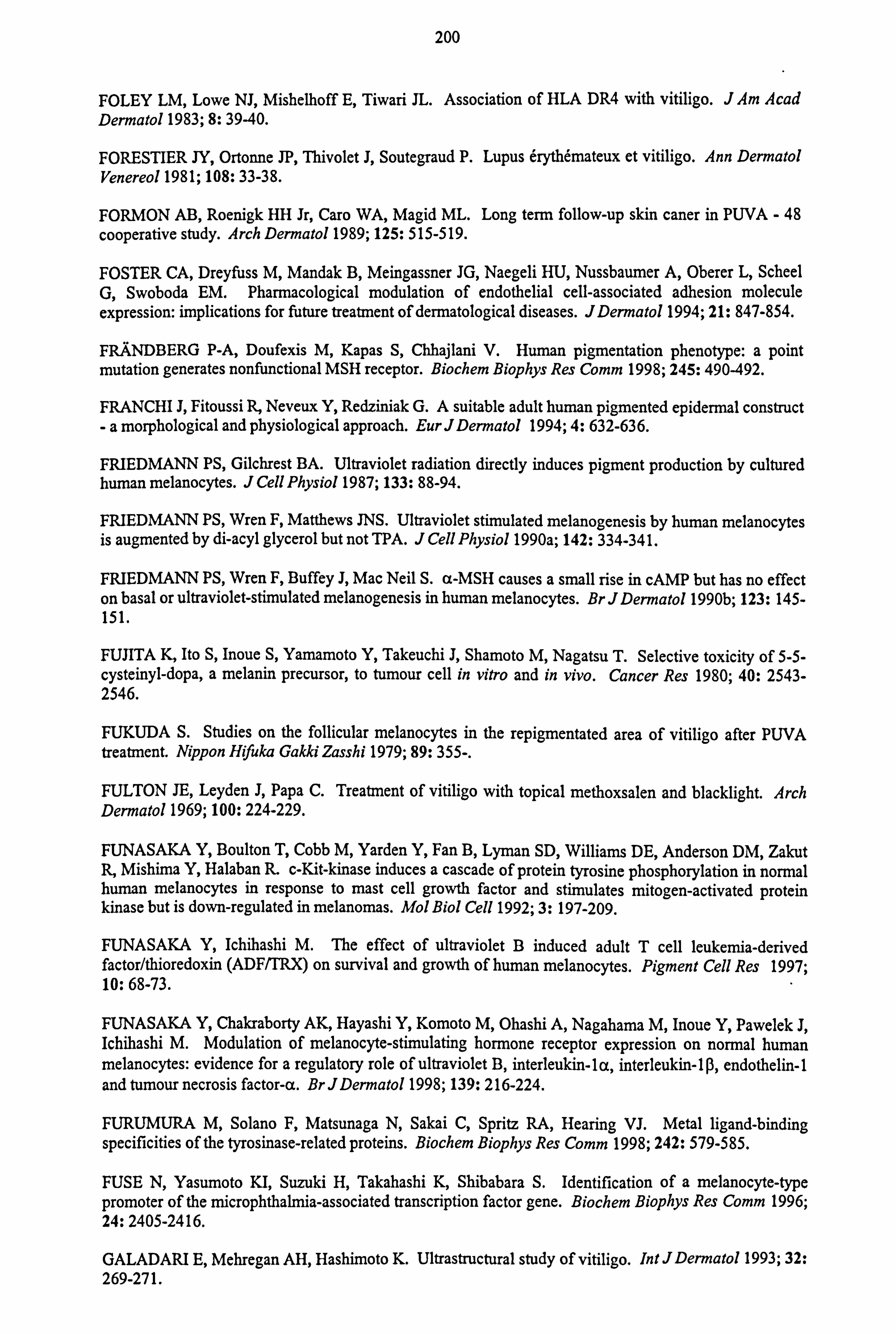
200
FOLEY LM, Lowe NJ, Mishelhoff E, Tiwari JL. Association of HLA DR4 with vitiligo. JAm Acad Dermatol 1983; 8: 39-40.
FORESTIER JY, Ortonne JP, Thivolet J, Soutegraud P. Lupus erythemateux et vitiligo. Ann Dermatol Venereol 1981; 108: 33-38.
FORMON AB, Roenigk HH Jr, Caro WA, Magid ML. Long term follow-up skin caner in PUVA - 48 cooperative study. Arch Dermatol 1989; 125: 515-519.
FOSTER CA, Dreyfuss M, Mandak B, Meingassner JG, Naegeli HU, Nussbaumer A, Oberer L, Scheel G, Swoboda EM. Pharmacological modulation of endothelial cell-associated adhesion molecule expression: implications for future treatment of dermatological diseases. JDermatol 1994; 21: 847-854.
FRANDBERG P-A, Doufexis M, Kapas S, Chhajlani V. Human pigmentation phenotype: a point mutation generates nonfunctional MSH receptor. Biochem Biophys Res Comm 1998; 245: 490-492.
FRANCHI J, Fitoussi R, Neveux Y, Redziniak G. A suitable adult human pigmented epidermal construct -a morphological and physiological approach. EurJDermatol 1994; 4: 632-636.
FRIEDMANN PS, Gilchrest BA. Ultraviolet radiation directly induces pigment production by cultured human melanocytes. J Cell Physiol 1987; 133: 88-94.
FRIEDMANN PS, Wren F, Matthews JNS. Ultraviolet stimulated melanogenesis by human melanocytes is augmented by di-acyl glycerol but not TPA. J Cell Physiol 1990a; 142: 334-341.
FRIEDMANN PS, Wren F, Buffey J, Mac Neil S. a-MSH causes a small rise in cAMP but has no effect on basal or ultraviolet-stimulated melanogenesis in human melanocytes. Br J Dermatol 1990b; 123: 145- 151.
FUJITA K, Ito S, Inoue S, Yamamoto Y, Takeuchi J, Shamoto M, Nagatsu T. Selective toxicity of 5-5- cysteinyl-dopa, a melanin precursor, to tumour cell in vitro and in vivo. Cancer Res 1980; 40: 2543- 2546.
FUKUDA S. Studies on the follicular melanocytes in the repigmentated area of vitiligo after PUVA treatment. Nippon Hifuka Gakki Zasshi 1979; 89: 355-.
FULTON JE, Leyden J, Papa C. Treatment of vitiligo with topical methoxsalen and blacklight. Arch Dermatol 1969; 100: 224-229.
FUNASAKA Y, Boulton T, Cobb M, Yarden Y, Fan B, Lyman SD, Williams DE, Anderson DM, Zakut R, Mishima Y, Halaban R. c-Kit-kinase induces a cascade of protein tyrosine phosphorylation in normal human melanocytes in response to mast cell growth factor and stimulates mitogen-activated protein kinase but is down-regulated in melanomas. Mol Biol Cell 1992; 3: 197-209.
FUNASAKA Y, Ichihashi M. The effect of ultraviolet B induced adult T cell leukemia-derived factor/thioredoxin (ADF/TRX) on survival and growth of human melanocytes. Pigment Cell Res 1997; 10: 68-73.
FUNASAKA Y, Chakraborty AK, Hayashi Y, Komoto M, Ohashi A, Nagahama M, Inoue Y, Pawelek J, Ichihashi M. Modulation of melanocyte-stimulating hormone receptor expression on normal human melanocytes: evidence for a regulatory role of ultraviolet B, interleukin-la, interleukin-1ß, endothelin-1 and tumour necrosis factor-a. Br JDermatol 1998; 139: 216-224.
FURUMURA M, Solano F, Matsunaga N, Sakai C, Spritz RA, Hearing VJ. Metal ligand-binding specificities of the tyrosinase-related proteins. Biochem Biophys Res Comm 1998; 242: 579-585.
FUSE N, Yasumoto KI, Suzuki H, Takahashi K, Shibabara S. Identification of a melanocyte-type promoter of the microphthalmia-associated transcription factor gene. Biochem Biophys Res Comm 1996; 24: 2405-2416.
GALADARI E, Mehregan AH, Hashimoto K. Ultrastructural study of vitiligo. Int J Dermatol 1993; 32: 269-271.
201
GALBRAITH GM, Miller D, Emerson DL. Western blot analysis of serum antibody reactivity with human melanoma cell antigens in alopecia areata and vitiligo. Clin Immunol Immunopathol 1988; 48: 317-324.
GALLICO GG, O'Connor NE, Compton CG, Kehinde 0, Green H. Permanent coverage of large burn wounds with autologous cultured human epithelium. New EnglJMed 1984; 311: 448-451.
GALLICO GG, O'Connor NE, Compton CC, Remensnyder JP, Kehinde 0, Green H. Cultured epithelial autografts for giant congenital nevi. Plast Reconst Surg 1989; 84: 1-9.
GAMBICHLER T, Maushagen-Schnaas E, Müller J, Boonen H, Schröpl F. Balneophototherapy in vitiligo. JDermatol Treatment 1997; 8: 133-135.
GANTZ I, Konda Y, Tashiro T, Shimoto Y, Miwa H, Munzert G, Watson SJ, DelValle J, Yamada T. Molecular cloning of a novel melanocortin receptor. JBiol Chem 1993a; 268: 8246-8250.
GANTZ I, Miwa H, Konda Y, Shimoto Y, Tashiro T, Watson SJ, DelValle J, Yamada T. Molecular cloning, expression, and gene localisation of a fourth melanocortin receptor. J Biol Chem 1993b; 268: 15174-15179.
GANTZ I, Shimoto Y, Konda Y, Miwa H, Dickinson, Yamada T. Molecular cloning, expression and characterisation of a fifth melanocortin receptor. Biochem Biophys Res Comm 1994; 200: 1214-1220.
GARCIA-CARMONA F, Garcia-Conovas F, Iborra JL, Lozano JA. Kinetic study of the pathway-of melanization between L-dopa and dopachrome. Biochem Biophys Acta 1982; 717: 124-131.
GARDNER JM, Nakatsu Y, Gondo Y, Lee S, Lyon MF, King RA, Brilliant MH. The mouse pink-eyed dilution gene: Association with human Prader-Will and Angelman syndromes. Science 1992; 257: 1121- 1124.
GAUTHIER Y, Surleve-Bazeille J-E. Autologous grafting with noncultured melanocytes: a simplified method for treatment of depigmented lesions. JAm Acad Dermatol 1992; 26: 191-194.
GAUTHIER Y. The importance of Koebner's phenomenon in the induction of vitiligo vulgaris lesions. Eur J Dermatol 1995; 5: 704-708.
GELLIN GA, Possick PA, Perone VB. Depigmentation from 4-tertiary butyl catechol. JInvest Dermatol 1970; 55 190-197.
GERALDEZ CB, Gutierrez GT. A clinical trial of clobetasol propionate in Filpino vitiligo patients. Clin Therapy 1987; 9: 474-482.
GHADERI A, Joubeh S, Davatolhagh Z, Vahedi F, Dalver C. Vitiligo and HLA-23, All, B21 and Cw4 in southern Iranian patients. Human Immunol 1996; 47: 187 [abstract].
GHONEUM M, Grimes PE, Gill G, Kelly AP. Natural killer cell-mediated cytotoxicity in vitiligo. JAm AcadDermatol 1987; 17: 600-605.
GHOSH MM, Boyce S, Layton C, Freedlander E, Mac Neil S. A comparison of methodologies for the preparation of human epidermal-dermal composites. Ann Plast Surg 1997; 39: 390-404.
GILCHREST BA, Vrabel MA, Flynn E, Szabo G. Selective cultivation of human melanocytes from newborn and adult epidermis. Jlnvest Dermatol 1984; 83: 370-376.
GILCHREST BA, Albert LS, Karassik RL, Yaar M. Substrate influences human epidermal melanocyte attachment and spreading in vitro. In Vitro Cell Dev Biol 1985; 21: 114-120.
GILCHREST BA, Zhai S, Eller MS, Yarosh DB, Yaar M. Treatment of human melanocytes and S91 melanoma cells with the DNA repair enzyme T4 endonuclease V enhances melanogenesis after ultraviolet irradiation. JInvestDermatol 1993; 101: 666-672.
202
GILHAR A, Aizen E, Ohana N, Etzioni A. Vitiliginous vs pigmented skin response to intradermal
administration of interferon gamma. Arch Dermatol 1993; 129: 600-604.
GILHAR A, Zelickson B, Ulman Y, Etzioni A. In vivo destruction of melanocytes by the IgG fraction of serum from patients with vitiligo. Jlnvest Dermatol 1995; 105: 683-686.
GLIMCHER ME, Kostick RM, Szabo G. The epidermal melanocyte system in newborn human skin. A
quantitative histologic study. JInvest Dermatol 1973; 61: 344-347.
GOLDEIEGER MH, Klein LE, Klein-Angerer S, Moellmann G, Nordlund JJ. The distribution of melanocytes in the leptomeninges of the human brain. Jlnvest Dermatol 1984; 82: 235-238.
GOKHALE BB, Gokhale TB. Corticotrophin and vitiligo (preliminary observations). Br J Dermatol 1976; 95: 329.
GOKHALE BB, Mehta IN. Histopathology of vitiliginous skin. Int J Dermatol 1983; 22: 477-480.
GONGGRYP, Kalla A. A. Chloroquine-induced vitiligo in a patient with juvenile onset rheumatoid arthritis. BrJRheumato11992; 31: 790-791.
GONZALEZ S, Pathsk MA, Cuevas J, Villarrubia VG, Fitzpatricj TB. Topical or oral administration with an extract of Polypodium leucotomos prevents acute sunburn and sporalen-induced phototoxic reactions as well as depletions of Langerhans cells in human skin. Photodermatol Photoimmunol Photomed 1997; 13: 50-60.
GOPINATHAN T. A study of the lesion of vitiligo. Arch Dermatol 1965; 91: 397-404.
GORDON PR, Gilchrest BA. Human melanogenesis is stimulated by diacyglycerol. J Invest Dermatol 1989; 93: 700-702.
GORDON PR, Mansur CP, Gilchrest BA. Regulation of human melanocyte growth, dendricity, and melanization by keratinocyte derived factors. Jlnvest Dermatol 1989; 92: 565-572.
GOSPODAROWICZ D, Delgrado D, Vlodavsky I. Permissive effect of the extracellular matrix on cell proliferation in vitro. Proc Natl Acad Sci USA 1980; 77: 4094-4098.
GOUDIE BM, Wilkieson C, Goudie RB. Skin maps in vitiligo. Scottish MedJ 1983; 28: 343-346.
GOUDIE RB, Soukop M, Dagg JH, Lee FD. Hypothesis: Symmetrical cutaneous lymphoma. Lancet 1990; 335: 316-318.
GOUDIE RB. Two views of the origin of vitiligo. Lancet 1991; 338: 306-307.
GOULD IM, Gray RS, Urbaniak SJ, Elton RA, Duncan LIP. Vitiligo in diabetes mellitus. BrJDermatol 1985; 113: 153-155.
GRAB J, Wise F. Vitiligo with raised borders. Arch Dermatol Syphilol 1948; 58: 148-153.
GRAHAM A, Westerhof W, Thody AJ. The expression of a-MSH by melanocytes is reduced in vitiligo. Pigment Cell Res 1998; 11: 252 [abstract].
GREAVES MW, Birkett D, Johnson C. Nevus anemicus: A unique catecholamine-dependent nevus. Arch Dermatol 1970; 102: 172-176.
GREINER D, Ochsendorf FR, Milbradt R. Vitiligo treatment with phenylalanine UV-A - follow-up
studies after 5 years. Hautarzt 1994; 45: 460-463.
GRICHNIK IM, Ali WN, Burch JA, Byers JD, Garcia CA, Clark RE, Shea CR. c-KIT expression reveals a population of precursor melanocytes in human skin. Jlnvest Dermatol 1996; 106: 967-971.
GRICHNIK JM, Burch JA, Burchette J, Shea CR. The SCFIKIT pathway plays a critical role in the control of normal human melanocyte homeostasis. Jlnvest Dermatol 1998; 111: 233-238.
203
GRIMES PE, Minus HR, Chakrabarti SG, Enterline J, Haider R, Gough JE, Kenny JA. Determination of optimal topical photochemotherapy for vitiligo. JAm Acad Dermatol 1982; 7: 771-778.
GRIMES PE, Haider RM, Jones C, Chakrabarty SA, Enterline J, Kenney JA. Autoantibodies and their
clinical significance in a black vitiligo population. Arch Dermatol 1983a; 119: 300-303.
GRIMES PE, Minus HR, Haider RM, Enterline J, Kenny JA. Vitiligo: Racial variations in the response to topical photochemotherapy. JInvest Dermatol 1983b; 80: 367 [abstract].
GRIMES PE, Ghonuem M, Stockton T, Payne C, Kelly P, Alfred L. T cell profiles in vitiligo. J Am Acad Dennatol 1986; 14: 196-201.
GRIMES PE, Pitts E, Kelly AP. The effects of PUVA therapy on cell-mediated immune defects in
patients with vitiligo. JInvest Dermatol 1988; 90: 564 [abstract].
GRIMES PE. Vitiligo. An overview of therapeutic approaches. Dermatol Clin 1993; 11: 325-338.
GRIMES PE, Sevall JS, Vojdani A. Cytomegalovirus DNA identified in skin biopsy specimens of patients with vitiligo. JAm Acad Dermatol 1996; 35: 21-26.
GRIMES PE. Psoralen photochemotherapy for vitiligo. Clin Dermatol 1997; 15: 921-926.
GROSS A, Tapis FJ, Mosca W, Perez RM, Briceno L, Henriques JJ, Convit J. Mononuclear cell subpopulations and infiltrating lymphocytes in erythema dyschromiscum perstans and vitiligo. Histol Histopathol 1987; 2: 277-287.
GSCHNAIT F, Brenner W, Wolff K. Photoprotective effect of a psoralen-UVA-induced tan. Arch Dermatol Res 1978; 263: 181-188.
GUPTA A, Anderson TF. Psoralen photochemotherapy. JAm Acad Dermatol 1987; 17: 703-734.
GUPTA AK, Ellis CN, Nickoloff BJ, Goldfarb MT, Ho VC, Rocher LL, Griffiths CEM, Cooper KD, Voorhees JJ. Oral cyclosporine in the treatment of inflammatory and non-inflammatory dermatoses. Arch Dermatol 1990; 126: 339-350.
GWIEZDZINSKI Z, Kallas D, Uchanska G. Vitiligo: new news views on the pathogenesis and treatment. Przegl Dermatol 1994; 81: 396-400.
HAAKE AR, Scott GA. Physiologic distribution and differentiation of melanocytes in human fetal and neonatal skin equivalents. JInvest Dermatol 1991; 96: 71-77.
HADLEY ME, Abdel-Malek ZA, Marwan MM, Kreutzfeld KL, Hruby VJ. [N1e4, D-Phe7]a-MSH: A superpotent melanotropic that "irreversibly" activates melanoma tyrosinase. Endocrine Res Comm 1985; 11: 157-170.
HAFEZ M, Sharaf L, El-Nabi SMA. The genetics of vitiligo. Acta Derm Venereol 1983; 63: 249-251.
HALABAN R, Pomerantz SH, Marshall S, Lambert DT, Lerner AB. Regulation of tyrosinase in human melanocytes grown in culture. J Cell Biol 1983; 97: 480-488.
HALABAN R, Alfano FD. Selective elimination of fibroblasts from cultures of normal human melanocytes. In Vitro J Tissue Culture 1984; 20: 447-450.
HALABAN R, Ghosh S, Duray P, KirkWood JM, Lerner AB. Human melanocytes cultured from nevi and melanomas. Jlnvest Dermatol 1986; 87: 95-101.
HALABAN R, Ghosh S, Baird A. bFGF is the putative natural growth factor for human melanocytes. In Vitro Cell Dev Biol 1987; 23: 47-52.
204
HALABAN R, Langdon R, Birchall N, Cuono C, Baird A, Scott G, Moellmann G, McGuire J. Basic fibroblast growth factor from human keratinocytes is a natural mitogen for melanocytes. J Cell Biol 1988; 107: 1611-1619.
HALABAN R, Moellmann G. Murine and human b locus pigmentation genes encode a glycoprotein (gp 75) with catalase activity. Proc Natl Acad Sci USA 1990; 87: 4809-4813.
HALABAN R, Rubin JS, Funasaka Y, Cobb M, Boulton T, Faletto D, Rosen E, Chan A, Yoko K, White W, Cook C, Moellmann G. Met and hepatocyte growth factor/scatter factor signal transduction in normal melanocytes and melanoma cells. Oncogene 1992; 7: 2195-2206.
HALDER RM, Walters CS, Johnson BA, Chakrabarti SG, Kenney JA. Aberrations in T lymphocytes and natural killer cells in vitiligo: a flow cytometric study. JAm Acad Dermatol 1986; 14: 733-737.
HALDER RM, Grimes PE, Cowan CA, Enterkine JA, Chakrabarti SG, Kenny JA. Childhood vitiligo. J Am Acad Dermatol 1987; : 948-954.
HALDER RM, Pham HN, Breadon JY, Johnson BA. Micropigmentation for the treatment of vitiligo. J Dermatol Surg Oncol 1989; 15: 1092-1098.
HALDER R, Battle EF, Smith EM. Cutaneous malignancies in patients treated with psoralen photochemotherapy (PUVA) for vitiligo. Arch Dermatol 1995; 131: 734-735.
HALDER RM. Childhood vitiligo. Clin Dermatol 1997; 15: 899-906.
HALL PA, McKee PH, Menage HP, Dover R, Lane DP. High levels of p53 protein in UV-irradiated normal human skin. Oncogene 1993; 8: 203-207..
HAN SK, Mytilineou C, Cohen G. L-DOPA up-regulates glutathione and protects mesencephalic cultures against oxidative stress. JNeurochem 1996; 66: 501-5 10.
HANN SK, Cho MY, Im S, Park YK. Treatment of vitiligo with oral 5-methoxylpsoralen. J Dermatol 1991; 18: 324-329.
HANN SK, Im S, Park YK, Hur W. Repigmentation of leukotrichia by epidermal grafting and systemic psoralen plus UVA. Arch Dermatol 1992a; 128: 998-999.
HANN SK, Park Y-K, Lee K-G, Choi EH, Im S. Epidermal changes in active vitiligo. J Dermatol 1992b; 19: 217-222.
HANN SK, Im S, Kim HI, Kim H-S, Lee YJ, Park Y-K. Increased incidence of antismooth muscle antobody in Korean vitiligo patients. JDermatol 1993a; 20: 679-683.
HANN SK, Kim HI, Im S, Park Y-K, Cui J, Bystryn J-C. The change on melanocyte cytotoxicity after systemic steroid treatment in vitiligo patients. JDermatol Sci 1993b; 6: 201-205.
HANN SK, Park Y-K, Chung K-Y, Kim HI, Im S, Won JH. Peripheral blood lymphocytes imbalance in Koreans with active vitiligo. IntJDermatol 1993c; 32: 286-289.
HANN SK, Im S, Bong HW, Park Y-K. Treatment of stable vitiligo with autologous epidermal grafting and PUVA. JAm Acad Dermatol 1995a; 32: 943-948.
HANN S-K, Kim J-B. Detection of antibodies to human melanoma cell in vitiligo by Western blot analysis. Yonsei Med J 1995b; 36: 457-461.
HANN SK, Lee HJ. Segmental vitiligo: clinical findings in 208 patients. JAm Acad Dermato11996; 35: 671-674.
HANN S-K, Park Y-K, Chun WH. Clinical features of vitiligo. Clin Dermato11997; 15: 891-897.
HARA M, Yaar M, Tang A, Eller MS, Reenstra W, Gilchrest BA. Role of integrins in melanocyte attachment and dendricity. J Cell Sci 1994; 107: 2739-2748.
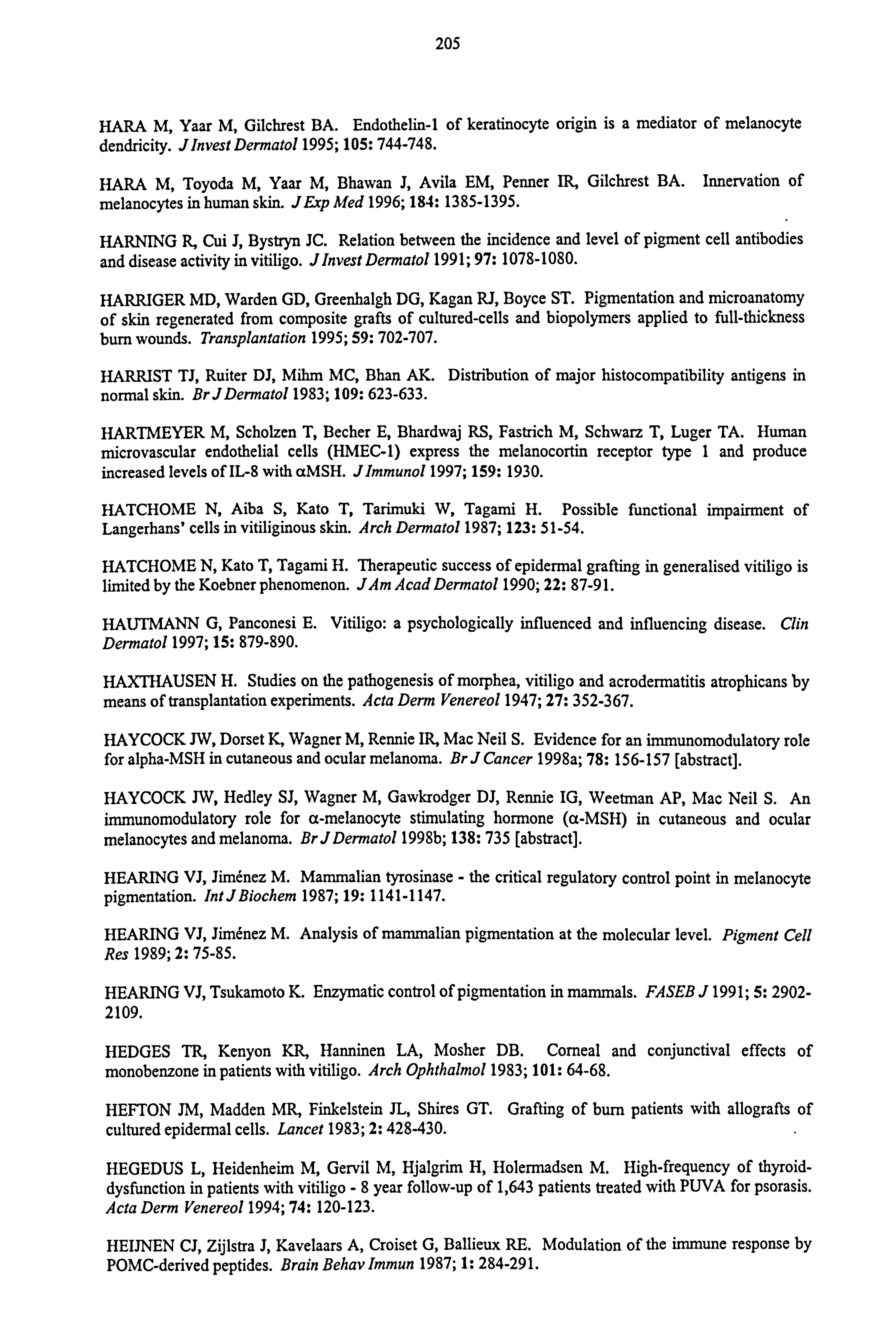
205
HARA M, Yaar M, Gilchrest BA. Endothelin-1 of keratinocyte origin is a mediator of melanocyte dendricity. JInvest Dermatol 1995; 105: 744-748.
HARA M, Toyoda M, Yaar M, Bhawan J, Avila EM, Penner IR, Gilchrest BA. Innervation of melanocytes in human skin. JExp Med 1996; 184: 1385-1395.
HARNING R, Cui J, Bystryn JC. Relation between the incidence and level of pigment cell antibodies and disease activity in vitiligo. JInvest Dermatol 1991; 97: 1078-1080.
HARRIGER MD, Warden GD, Greenhalgh DG, Kagan RJ, Boyce ST. Pigmentation and microanatomy of skin regenerated from composite grafts of cultured-cells and biopolymers applied to full-thickness bum wounds. Transplantation 1995; 59: 702-707.
HARRIST TJ, Ruiter DJ, Mihm MC, Bhan AK. Distribution of major histocompatibility antigens in
normal skin. BrJDermatol 1983; 109: 623-633.
HARTMEYER M, Scholzen T, Becher E, Bhardwaj RS, Fastrich M, Schwarz T, Luger TA. Human microvascular endothelial cells (HMEC-1) express the melanocortin receptor type 1 and produce increased levels of IL-8 with aMSH. Jlmmunol 1997; 159: 1930.
HATCHOME N, Aiba S, Kato T, Tarimuki W, Tagami H. Possible functional impairment of Langerhans' cells in vitiliginous skin. Arch Dermatol 1987; 123: 51-54.
HATCHOME N, Kato T, Tagami H. Therapeutic success of epidermal grafting in generalised vitiligo is limited by the Koebner phenomenon. JAm Acad Dermatol 1990; 22: 87-91.
HAUTMANN G, Panconesi E. Vitiligo: a psychologically influenced and influencing disease. Clin Dermatol 1997; 15: 879-890.
HAXTHAUSEN H. Studies on the pathogenesis of morphea, vitiligo and acrodermatitis atrophicans by means of transplantation experiments. Acta Derm Venereol 1947; 27: 352-367.
HAYCOCK JW, Dorset K, Wagner M, Rennie IR, Mac Neil S. Evidence for an immunomodulatory role for alpha-MSH in cutaneous and ocular melanoma. Br J Cancer 1998a; 78: 156-157 [abstract].
HAYCOCK JW, Hedley SJ, Wagner M, Gawkrodger DJ, Rennie IG, Weetman AP, Mac Neil S. An immunomodulatory role for a-melanocyte stimulating hormone (a-MSH) in cutaneous and ocular melanocytes and melanoma. BrJDermatol 1998b; 138: 735 [abstract].
HEARING VJ, Jimenez M. Mammalian tyrosinase - the critical regulatory control point in melanocyte pigmentation. Int JBiochem 1987; 19: 1141-1147.
HEARING VJ, Jimenez M. Analysis of mammalian pigmentation at the molecular level. Pigment Cell Res 1989; 2: 75-85.
HEARING VJ, Tsukamoto K. Enzymatic control of pigmentation in mammals. FASEB J 1991; 5: 2902- 2109.
HEDGES TR, Kenyon KR, Hanninen LA, Mosher DB. Corneal and conjunctival effects of monobenzone in patients with vitiligo. Arch Ophthalmol 1983; 101: 64-68.
HEFTON JM, Madden MR, Finkelstein JL, Shires GT. Grafting of burn patients with allografts of cultured epidermal cells. Lancet 1983; 2: 428-430.
HEGEDUS L, Heidenheim M, Gervil M, Hjalgrim H, Holermadsen M. High-frequency of thyroid- dysfunction in patients with vitiligo -8 year follow-up of 1,643 patients treated with PUVA for psorasis. Acta Derm Venereol 1994; 74: 120-123.
HEIJNEN CJ, Zijlstra J, Kavelaars A, Croiset G, Ballieux RE. Modulation of the immune response by POMC-derived peptides. Brain Behav Immun 1987; 1: 284-291.
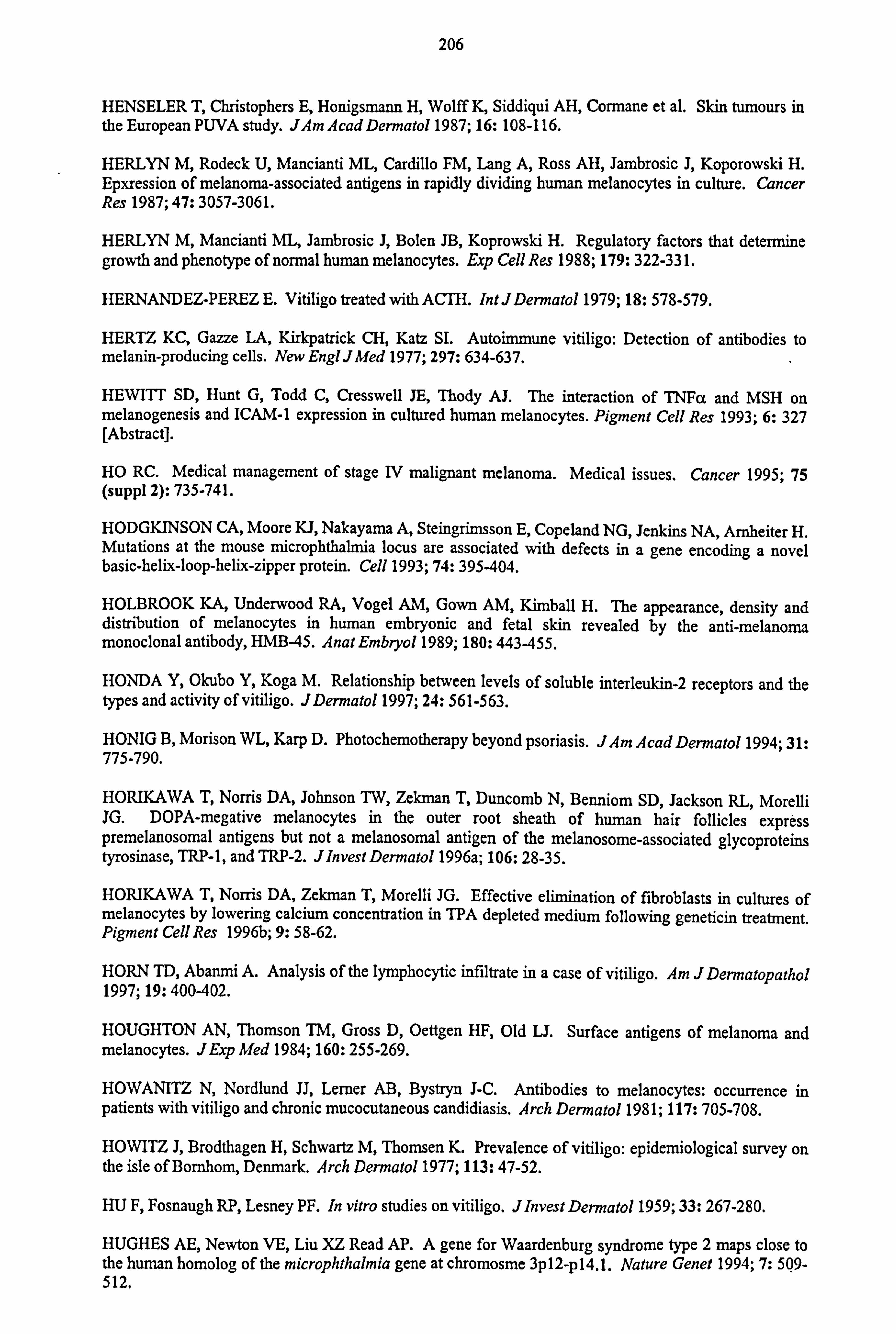
206
HENSELER T, Christophers E, Honigsmann H, Wolff K, Siddiqui AH, Cormane et al. Skin tumours in the European PUVA study. JAm Acad Dermatol 1987; 16: 108-116.
HERLYN M, Rodeck U, Mancianti ML, Cardillo FM, Lang A, Ross AH, Jambrosic J, Koporowski H. Epxression of melanoma-associated antigens in rapidly dividing human melanocytes in culture. Cancer Res 1987; 47: 3057-3061.
HERLYN M, Mancianti ML, Jambrosic J, Bolen JB, Koprowski H. Regulatory factors that determine growth and phenotype of normal human melanocytes. Exp Cell Res 1988; 179: 322-33 1.
HERNANDEZ-PEREZ E. Vitiligo treated with ACTH. IntJDermatol 1979; 18: 578-579.
HERTZ KC, Gazze LA, Kirkpatrick CH, Katz SI. Autoimmune vitiligo: Detection of antibodies to melanin-producing cells. New EnglJMed 1977; 297: 634-637.
HEWITT SD, Hunt G, Todd C, Cresswell JE, Thody AJ. The interaction of TNFa and MSH on melanogenesis and ICAM-1 expression in cultured human melanocytes. Pigment Cell Res 1993; 6: 327 [Abstract].
HO RC. Medical management of stage IV malignant melanoma. Medical issues. Cancer 1995; 75 (suppl 2): 735-741.
HODGKINSON CA, Moore KJ, Nakayama A, Steingrimsson E, Copeland NG, Jenkins NA, Arnheiter H. Mutations at the mouse microphthalmia locus are associated with defects in a gene encoding a novel basic-helix-loop-helix-zipper protein. Cell 1993; 74: 395-404.
HOLBROOK KA, Underwood RA, Vogel AM, Gown AM, Kimball H. The appearance, density and distribution of melanocytes in human embryonic and fetal skin revealed by the anti-melanoma monoclonal antibody, HMB-45. Anat Embryo! 1989; 180: 443-455.
HONDA Y, Okubo Y, Koga M. Relationship between levels of soluble interleukin-2 receptors and the types and activity of vitiligo. JDermatol 1997; 24: 561-563.
HONIG B, Morison WL, Karp D. Photochemotherapy beyond psoriasis. JAm Acad Dermato11994; 31: 775-790.
HORIKAWA T, Norris DA, Johnson TW, Zekman T, Duncomb N, Benniom SD, Jackson RL, Morelli JG. DOPA-megative melanocytes in the outer root sheath of human hair follicles express premelanosomal antigens but not a melanosomal antigen of the melanosome-associated glycoproteins tyrosinase, TRP-1, and TRP-2. Jlnvest Dermatol 1996a; 106: 28-35.
HORIKAWA T, Norris DA, Zekman T, Morelli JG. Effective elimination of fibroblasts in cultures of melanocytes by lowering calcium concentration in TPA depleted medium following geneticin treatment. Pigment Cell Res 1996b; 9: 58-62.
HORN TD, Abanmi A. Analysis of the lymphocytic infiltrate in a case of vitiligo. Am J Dermatopathol 1997; 19: 400-402.
HOUGHTON AN, Thomson TM, Gross D, Oettgen HF, Old LJ. Surface antigens of melanoma and melanocytes. JExp Med 1984; 160: 255-269.
HOWANITZ N, Nordlund JJ, Lerner AB, Bystryn J-C. Antibodies to melanocytes: occurrence in patients with vitiligo and chronic mucocutaneous candidiasis. Arch Dermatol 1981; 117: 705-708.
HOWITZ J, Brodthagen H, Schwartz M, Thomsen K. Prevalence of vitiligo: epidemiological survey on the isle of Bornhom, Denmark. Arch Dermatol 1977; 113: 47-52.
HU F, Fosnaugh RP, Lesney PF. In vitro studies on vitiligo. Jlnvest Dermatol 1959; 33: 267-280.
HUGHES AE, Newton VE, Liu XZ Read AP. A gene for Waardenburg syndrome type 2 maps close to the human homolog of the microphthalmia gene at chromosme 3pl2-p14.1. Nature Genet 1994; 7: 5Q9- 512.
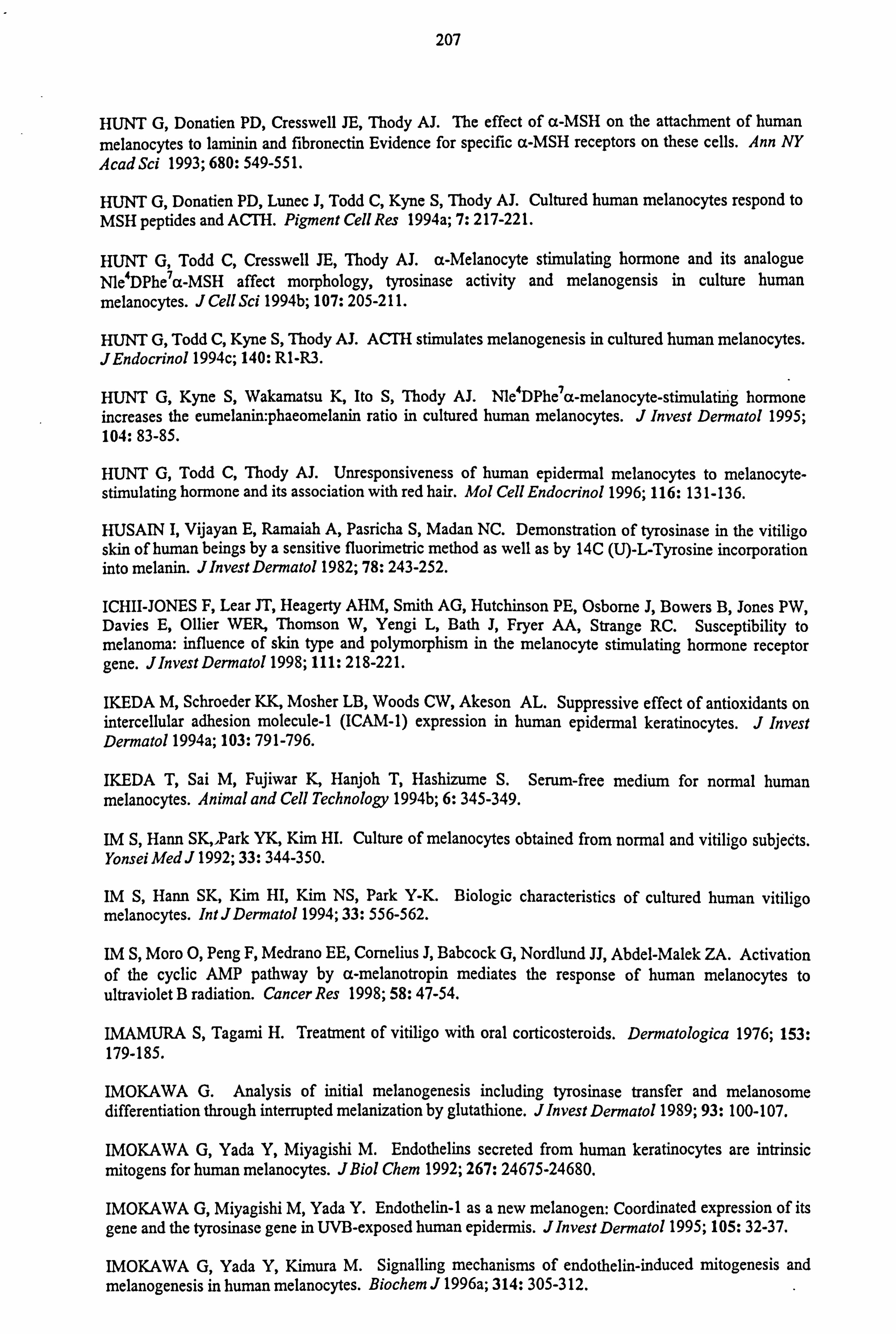
207
HUNT G, Donatien PD, Cresswell JE, Thody AJ. The effect of a-MSH on the attachment of human
melanocytes to laminin and fibronectin Evidence for specific a-MSH receptors on these cells. Ann NY AcadSci 1993; 680: 549-551.
HUNT G, Donatien PD, Lunec J, Todd C, Kyne S, Thody AJ. Cultured human melanocytes respond to MSH peptides and ACTH. Pigment Cell Res 1994a; 7: 217-221.
HUNT G, Todd C, Cresswell JE, Thody AJ. a-Melanocyte stimulating hormone and its analogue N1e4DPhe7a-MSH affect morphology, tyrosinase activity and melanogensis in culture human
melanocytes. J Cell Sci 1994b; 107: 205-211.
HUNT G, Todd C, Kyne S, Thody AJ. ACTH stimulates melanogenesis in cultured human melanocytes. JEndocrinol 1994c; 140: RI-R3.
HUNT G, Kyne S, Wakamatsu K, Ito S, Thody AJ. Nle4DPhe7a-melanocyte-stimulating hormone increases the eumelanin: phaeomelanin ratio in cultured human melanocytes. J Invest Dermatol 1995; 104: 83-85.
HUNT G, Todd C, Thody AJ. Unresponsiveness of human epidermal melanocytes to melanocyte- stimulating hormone and its association with red hair. Mol Cell Endocrinol 1996; 116: 131-136.
HUSAIN I, Vijayan E, Ramaiah A, Pasricha S, Madan NC. Demonstration of tyrosinase in the vitiligo skin of human beings by a sensitive fluorimetric method as well as by 14C (U)-L-Tyrosine incorporation into melanin. Jlnvest Dermatol 1982; 78: 243-252.
ICHII-JONES F, Lear JT, Heagerty AHM, Smith AG, Hutchinson PE, Osborne J, Bowers B, Jones PW, Davies E, Oilier WER, Thomson W, Yengi L, Bath J, Fryer AA, Strange RC. Susceptibility to melanoma: influence of skin type and polymorphism in the melanocyte stimulating hormone receptor gene. Jlnvest Dermatol 1998; 111: 218-221.
IKEDA M, Schroeder KK, Mosher LB, Woods CW, Akeson AL. Suppressive effect of antioxidants on intercellular adhesion molecule-1 (ICAM-1) expression in human epidermal keratinocytes. J Invest Dermatol 1994a; 103: 791-796.
IKEDA T, Sai M, Fujiwar K, Hanjoh T, Hashizume S. Serum-free medium for normal human melanocytes. Animal and Cell Technology 1994b; 6: 345-349.
IM S, Hann SK, 1Park YK, Kim HI. Culture of melanocytes obtained from normal and vitiligo subjects. Yonsei Med J 1992; 33: 344-350.
IM S, Hann SK, Kim HI, Kim NS, Park Y-K. Biologic characteristics of cultured human vitiligo melanocytes. Int JDermatol 1994; 33: 556-562.
IM S, Moro 0, Peng F, Medrano EE, Cornelius J, Babcock G, Nordlund JJ, Abdel-Malek ZA. Activation of the cyclic AMP pathway by a-melanotropin mediates the response of human melanocytes to ultraviolet B radiation. Cancer Res 1998; 58: 47-54.
IMAMURA S, Tagami H. Treatment of vitiligo with oral corticosteroids. Dermatologica 1976; 153: 179-185.
IMOKAWA G. Analysis of initial melanogenesis including tyrosinase transfer and melanosome differentiation through interrupted melanization by glutathione. J Invest Dermatol 1989; 93: 100-107.
IMOKAWA G, Yada Y, Miyagishi M. Endothelins secreted from human keratinocytes are intrinsic mitogens for human melanocytes. JBiol Chem 1992; 267: 24675-24680.
IMOKAWA G, Miyagishi M, Yada Y. Endothelin-1 as a new melanogen: Coordinated expression of its gene and the tyrosinase gene in UVB-exposed human epidermis. JInvest Dermatol 1995; 105: 32-37.
IMOKAWA G, Yada Y, Kimura M. Signalling mechanisms of endothelin-induced mitogenesis and melanogenesis in human melanocytes. Biochem J 1996a; 314: 305-312.
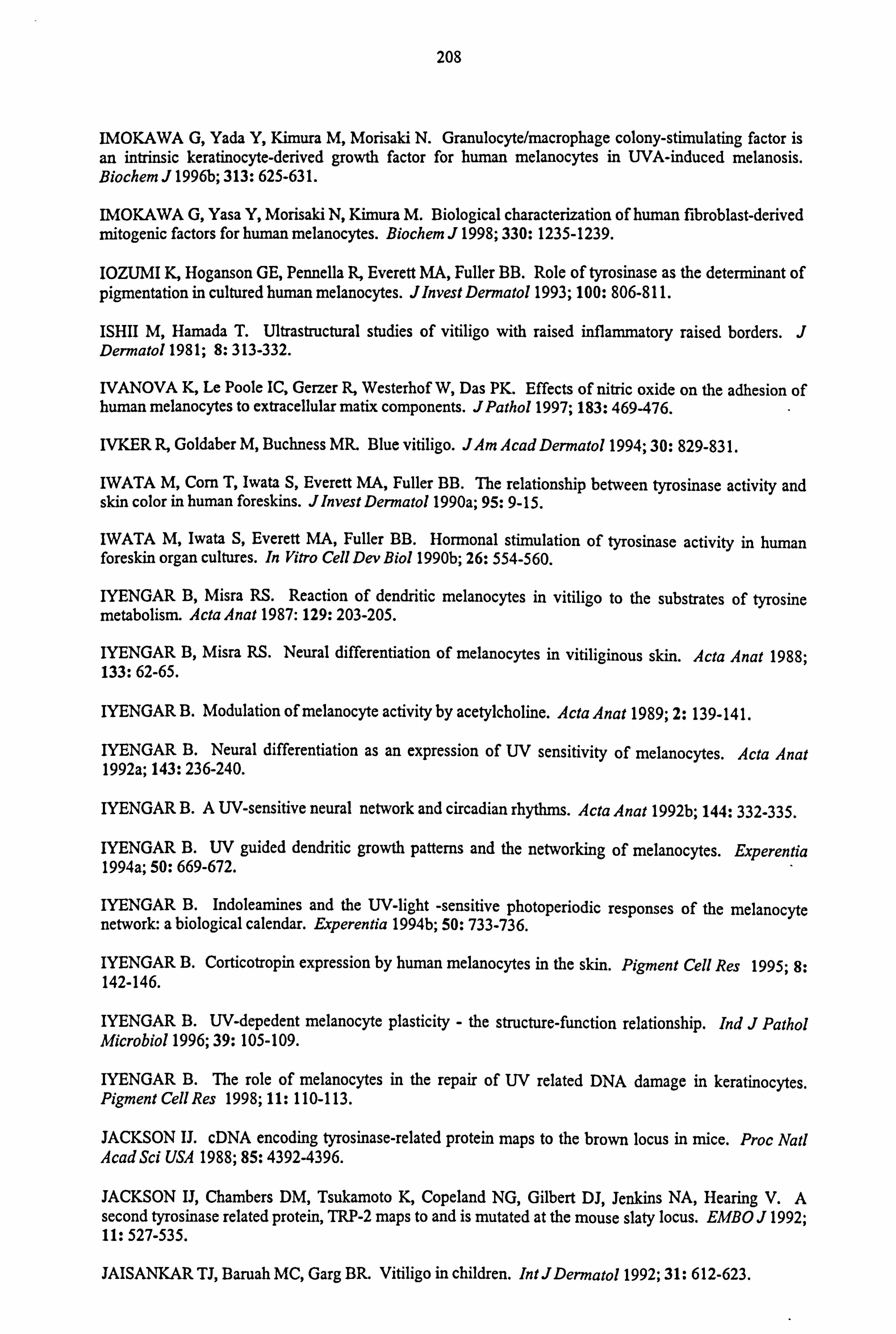
208
IMOKAWA G, Yada Y, Kimura M, Morisaki N. Granulocyte/macrophage colony-stimulating factor is an intrinsic keratinocyte-derived growth factor for human melanocytes in UVA-induced melanosis. Biochem J 1996b; 313: 625-631.
IMOKAWA G, Yasa Y, Morisaki N, Kimura M. Biological characterization of human fibroblast-derived mitogenic factors for human melanocytes. Biochem J 1998; 330: 1235-1239.
IOZUMI K, Hoganson GE, Pennella R, Everett MA, Fuller BB. Role of tyrosinase as the determinant of pigmentation in cultured human melanocytes. JInvest Dermatol 1993; 100: 806-811.
ISHII M, Hamada T. Ultrastructural studies of vitiligo with raised inflammatory raised borders. J Dermatol 1981; 8: 313-332.
IVANOVA K, Le Poole IC, Gerzer R, Westerhof W, Das PK. Effects of nitric oxide on the adhesion of human melanocytes to extracellular matix components. JPathol 1997; 183: 469-476.
IVKER R, Goldaber M, Buchness MR. Blue vitiligo. JAm Acad Dermatol 1994; 30: 829-83 1.
IWATA M, Corn T, Iwata S, Everett MA, Fuller BB. The relationship between tyrosinase activity and skin color in human foreskins. JInvest Dermatol 1990a; 95: 9-15.
IWATA M, Iwata S, Everett MA, Fuller BB. Hormonal stimulation of tyrosinase activity in human foreskin organ cultures. In Vitro Cell Dev Biol 1990b; 26: 554-560.
IYENGAR B, Misra RS. Reaction of dendritic melanocytes in vitiligo to the substrates of tyrosine metabolism. Acta Anat 1987: 129: 203-205.
IYENGAR B, Misra RS. Neural differentiation of melanocytes in vitiliginous skin. Acta Anat 1988; 133: 62-65.
IYENGAR B. Modulation of melanocyte activity by acetylcholine. Acta Anat 1989; 2: 139-141.
IYENGAR B. Neural differentiation as an expression of UV sensitivity of melanocytes. Acta Anat 1992a; 143: 236-240.
IYENGAR B. A UV-sensitive neural network and circadian rhythms. Acta Anat 1992b; 144: 332-335.
IYENGAR B. UV guided dendritic growth patterns and the networking of melanocytes. Experentia 1994a; 50: 669-672.
IYENGAR B. Indoleamines and the UV-light -sensitive photoperiodic responses of the melanocyte network: a biological calendar. Experentia 1994b; 50: 733-736.
IYENGAR B. Corticotropin expression by human melanocytes in the skin. Pigment Cell Res 1995; 8: 142-146.
IYENGAR B. UV-depedent melanocyte plasticity - the structure-function relationship. Ind J Pathol Microbiol 1996; 39: 105-109.
IYENGAR B. The role of melanocytes in the repair of UV related DNA damage in keratinocytes. Pigment Cell Res 1998; 11: 110-113.
JACKSON U. cDNA encoding tyrosinase-related protein maps to the brown locus in mice. Proc Nat! Acad Sci USA 1988; 85: 4392-4396.
JACKSON IJ, Chambers DM, Tsukamoto K, Copeland NG, Gilbert DJ, Jenkins NA, Hearing V. A second tyrosinase related protein, TRP-2 maps to and is mutated at the mouse slaty locus. EMBO J 1992; 11: 527-535.
JAISANKAR TJ, Baruah MC, Garg BR. Vitiligo in children. IntJDermatol 1992; 31: 612-623.

209
JANSEN T, Megahed M, Holzle E, Plewig G. Provocation of porphyria cutanea tarda by KUVA-therapy
of vitiligo. Acta Derm Yenereol 1995; 75: 232-233.
JANNIGER CK. Childhood vitiligo. Cutis 1993; 51: 25-28.
JARA JR, Solano F, Lozano JA. Assays for mammalian tyrosinase: A comparative study. Pigment Cell Res 1988; 1: 332-339.
JARA JR, Solano F, Garcia-Borrön JC, Aroca P, Lozano JA. Regulation of mammalian melanogenesis II. The role of metal cations. Biochem Biophys Acta 1990; 1035: 276-285.
JARRETT A, Szabo G. The pathological varieties of vitiligo and their response to treatment with meladinine. Br J Dermatol 1956; 68: 313.
JEE S-H, Kuo Y-F, Wu Y-C, Chang C-H, Kuo W-L, Lu Y-C. Growth of human melanocytes from suction blister of localized vitiligo patient and from foreskins of newborns and adults by modified culture medium. JFormos Med Assoc 1993; 92: 7-14.
JEE S-H, Lee S-Y, Chiu H-C, Chang C-C, Chen TJ. Effects of estrogen and estrogen receptor in normal human melanocytes. Biochem Biophys Res Comm 1994; 199: 1407-1412.
JHA AK, Pandey SS, Gulati AK, Bansal V, Shukla VK, Singh G. Inoculation of a cultured autolgous epidermal suspension containing melanocytes in vitiligo. Arch Dermatol 1993; 129: 785-786.
JIMBOW K, Obata H, Pathak MA. Mechanism of depigmentation by hydroquinone. J Invest Dermatol 1974; 62: 43 6-449.
JIMBOW K, Roth SI, Fitzpatrick TB, Szabo G. Mitotic activity in non-neoplastic melanocytes in vivo as determined by histochemical, autoradiographic, and electron microscope studies. J Cell Biol 1975; 66: 663-670.
JIMENEZ M, Tsukamoto K, Hearing VJ. Tyrosinase from two different loci are expressed by normal and by transformed melanocytes. JBiol Chem 1991; 266: 1147-1156.
JIMENEZ-CERVANTES C, Garcia-Borrön JC, Valverde P, Solano F, Lozano JA. Tyrosinase isoenzymes in mammalian melanocytes. I. Biochemical characterization of two melanosomal tyrosinases from B16 mouse melanoma cells. EurJBiochem 1993; 217: 549-556.
JIMENEZ-CERVANTES C, Solano F, Kobayashi T, Urabe K, Hearing VJ, Lozano JA, Garcia-Borrön JC. A new enzymatic function in the melanogenic pathway. The 5,6-dihydroxyindole-2-carboxylic acid oxidase activity of tyrosinase-related protein-1 (TRP-1). JBiol Chem 1994; 269: 17993-18001.
JOB CK, Nayar A, Narayanan JS. Electron microscopic study of hypopigmented lesions in leprosy. A preliminary report. BrJDermatol 1972; 87: 200.
JOHNSON R, Jackson IJ. Light is a dominant mouse mutation resulting in premature cell death. Nature Genet 1992; 1: 266-269.
JUHLIN L, Olsson MJ. Improvement of vitiligo after oral treatment with vitamin B12 and folic acid and the importance of sun exposure. Acta Denn Venereol 1997; 77: 460-462.
KACHRU RB, Telischi RB, Mittal KK. HLA antigens and vitiligo in an American Black population. Tissue Antigens 1978; 12: 396-397.
KAGESHITA T, Yoshii A, Kimura T, Kuriya N, Ono T, Tsujisaki M, Imai K, Ferrone S. Clinical
relevance of ICAM-1 expression in primary lesions and serum of patients with malignant melanoma. Cancer Res 1993; 53: 4927-4932.
KAHN AM, Cohen MJ, Kaplan L. Treatment for depigmentation resulting from burn injuries. J Burn Care Rehab 1991; 5: 468-473.
210
KAHN AM, Cohen MJ. Vitiligo: Treatment by dermabrasion and epithelial sheet grafting. J Am Acad Dermatol 1995; 33: 646-648.
KAHN AM, Cohen MJ. Repigmentation in vitiligo patients - melanocyte transfer via ultra-thin grafts. Dermatol Surg 1998; 24: 365-367.
KALDEN DH, Fastrich M, Brzoska T, Scholzen T, Hartmeyer M, Schwarz T, Luger TA. Alpha-
melanocyte-stimulating hormone reduces endotoxin-induced activation of nuclear factor-kB in
endothelial cells. JInvest Dermatol 1998; 110: 495 [abstract].
KANDIL E. Vitiligo-response to 0.2% betamethasone 17-valerate in flexible coolodion. Dermatologica 1970; 141: 277-281.
KANDIL E. Treatment of vitiligo with 0.1% betamethasone 17-valerate in isopropyl alcohol -a double- blind trial. Br J Dermatol 1974; 91: 457-460.
KANG H-Y, Kang W-H, Lee C-O. Endothelin-B receptor-mediated Ca2+ signaling in human
melanocytes. Pflügers Arch - EurJPhysiol 1998; 435: 350-356.
KANO Y, Shiohara T, Nagashima M. Epidermal Langerhans' cells in various skin diseases (2) Langerhans' cells in vitiligo. JDermatol 1984; 11: 103-110.
KANOF NB. Melanin formation in vitiliginous skin under the influence of external applications of 8-
methoxypsoralen. JlnvestDermato11955; 24: 5-10.
KANWAR AJ, Dhar S, Dawn G. Oral minipulse therapy in vitiligo. Dermatologica 1995; 190: 251- 252.
KAO CH, Yu H-S. Depletion and repopulation of Langerhans' cells in nonsegmental type vitiligo. J Dermatol 1990; 17: 287-296.
KAO C-H, Yu H-S. Comparison of the effect of 8-methoxypsoralen (8-MOP) plus UVA (PUVA) on human melanocytes in vitiligo vulgaris and in vitro. J Invest Dermatol 1992; 98: 734-740.
KASTAN MB, Onyekwere 0, Sidransky D, Vogelstein B, Craig RW. Participation of p53 protein in the cellular response to DNA damage. Cancer Res 1991; 51: 6304-6311.
KAUFMANN R, Greiner D, Kippenberger S, Bernd A. Grafting of in vitro cultured melanocytes onto laser-ablated lesions in vitiligo. Acta Derm Venereol 1998; 78: 136-138.
KEMP EH, Gawkrodger DJ, Mac Neil S, Watson PF, Weetman AP. Detection of tyrosinase autantibodies in patients with vitiligo using 35S-labeled recombinant human tyrosinase in
radioimmunoassay. JInvest Dermatol 1997; 109: 69-73.
KEMP EH, Gawkrodger DJ, Watson PF, Weetman AP. Immunoprecipitation of melanogenic enzyme autoantigens with vitiligo sera: evidence for cross-reactive autoantibodies to tyrosinase and tyrosinase- related protein-2 (TRP-2). Clin Exp Immunol 1997; 109: 495-500.
KENNY JA. Vitiligo treated by psoralen. A longterm follow-up study of the permanacy of repigmentation. Arch Dermato11971; 103: 465-480.
KENT G, Al'Abadie M. Psychologie effects of vitiligo: A critical incident analysis. J Am Acad Dermatol 1996; 35: 895-898.
KEYSTONE EC, Snow KM, Bombardier C, Chang CH, Nelson DL, Rubin LA. Elevated soluble interleukin-2 receptor levels in the sera and synovial fluids of patients with rheumatoid arthritis. Arthritis Rheum 1988; 31: 844-849.
KHALID M, Mujtaba G, Haroon TS. Comparison of 0.05% clobetasol propionate cream and topical Puvasol in childhood vitiligo. IntJDermatol 1995; 34: 203-205.
211
KHANSARI DN, Murgo AJ, Faith RE. Effect of stress on the immune system. Immunol Today 1990; 11: 170-175.
KIPPENBERGER S, Bemd A, Loitsch, S, Ramirez-Bosca A, Bereiter-Hahn J, Holzmann H. a-MSH is expressed in cultured human melanocytes and keratinocytes. EurJDermatol 1995; 5: 395-397.
KIPPENBERGER S, Bernd A, Bereiter-Hahn J, Ramirez-Bosca A, Kaufman, R, Holzmann H. Transcription of melanogenesis enzymes in melanocytes: Dependence upon culture conditions and co- cultivation with keratinocytes. Pigment Cell Res 1996; 9: 179-184.
KIPPENBERGER S, Bernd A, Bereiter-Hahn J, Ramirez-Bosca A, Kaufman R. The mechanism of melanocyte dendrite formation: the impact of differentiating keratinocytes. Pigment Cell Res 1998a; 11: 34-37.
KIPPENBERGER S, Loitsch S, Solano F, Bernd A, Kaufmann R. Quantification of tyrosinase, TRP-1, and TRP-2 transcripts in human melanocytes by reverse transcriptase-competitive multiplex PCR - regulation by steroid hormones. JInvest Dermatol 1998b; 110: 364-367.
KIRNBAUER R, Charvat B, Schauer E, Köck A, Urbanski A, Förster E, Neuner P, Assmann I, Luger TA, Schwarz T. Modulation of intercellular adhesion molecule-1 expression on human melanocytes and melanoma cells: Evidence for a regulatory role of IL-6, IL-7, TNFI, and UVB light. J Invest Dermatol 1992; 98: 320-326.
KNOELL KA, Schreiber AJ, Milgraum S. Treatment of vitiligo with the ultrapulse carbon dioxide laser in patients concomitantly receiving oral psoralen plus UV-A therapy. Arch Dermatol 1997; 133: 1605- 1606.
KOBAYASHI T, Urabe K, Winder A, Jimenez-Cervantes C, Imokawa G, Brewington T, Solano F, Garcia-Borrön JC, Hearing VJ. Tyrosinase related protein 1 (TRP1) functions as a DHICA oxidase in melanin biosynthesis. EMBO J 1994; 13: 5818-5825.
KOBAYASHI T, Viera WD, Potterf B, Sakai C, Imokawa G. Modulation of melanogenic protein expression during the switch from eu- to pheomelanogenesis. J Cell Sci 1995; 108: 2301-2309.
KÖCK A, Schwarz T, Kirnbauer R, Urbanski A, Perry P, Ansel JC, Luger TA. Human keratinocytes are a source for tumour necrosis factor a: evidence for synthesis and release upon stimulation with endotoxin or ultraviolet light. JExp Med 1990; 172: 1609-1614.
KOCK A, Schauer E, Schwartz T, Luger TA. Human keratinocytes synthesize and release neuropeptides such as aMSH and ACTH. In: Cellular and Cytokine Networks in Tissue Immunity. pp105-110,1991. Ed. Meltzer, M. (NY: John Wiley).
KOGA M. Vitiligo: A new classification and therapy. BrJDermatol 1977; 97: 255-261.
KOGA M. Epidermal grafting using the tops of suction blisters in the treatment of vitiligo. Arch Dermato11988; 124: 1656-1658.
KOGA M, Tango T. Clinical features and course of type A and type B vitiligo. Br J Dermatol 1988; 118: 223-228.
KOOPMANS-VAN DORP B, Goedhart-van Dijk B, Neering HE, van Dijk E. Treatment of vitiligo by local application of betamethasone 17-valerate in a dimethyl sulfoxide cream base. Dermatologica 1973; 146: 310-314.
KOPLON BS, Shapiro L. Poliosis overlying a neurofibroma. Arch Dermatol 1968; 98: 631-633.
KOPPULA SV, Robbins LS, Lu D, Baack E, White CR Jr, SwansonNA, Cone RD. Identification of common polymorphisms in the coding sequence of the human MSH receptor (MC1R) with possible biological effects. Hum Mutation 1997; 9: 30-36.
KÖRNER A, Pawelek J. Mammalian tyrosinase catalyze three reactions in the biosynthesis of melanin. Science 1982; 217: 1163-1165.
212
KORYTOWSKI W, Pilas B, Sarna T, Kalyanaraman B. Photoinduced generation of hydrogen peroxide and hyydroxyl radicals in melanins. Photochem Photobiol 1987; 45: 185-190.
KOSHEVENKO L. Alpha tocopherol in the combined treatment of vitiligo. Vestm Dermatol Venereol 1989; 10: 70-72.
KÖSTER W, Wiskemann A. Phototherapy with UVB in vitiligo. Z-Hautkr 1990; 65: 1022-1024.
KOVACS SO. Vitiligo. JAm Acad Dermatol 1998; 38: 647-666.
KRAMER M, Sachsenmaier C, Herrlich P, Rahmsdorf HJ. UV irradiation-induced interleukin-1 and basic fibroblast growth factor synthesis and release mediate part of the UV response. J Biol Chem 1993; 268: 6734-6741.
KRASAGAKIS K, Garbe C, Krüger S, Orfanos CE. Effects of interferons on cultured human melanocytes in vitro: interferon-beta but not -alpha or -gamma inhibit proliferation and all interferons significantly modulate the cell phenotype. JInvest Dermatol 1991; 97: 364-372.
KRASAGAKIS K, Garbe C, Krüger-krasagakes S, Orfanos CE. 12-O-tetradecanoylphorbol-13-acetate
not only modulates proliferation rates, but also alters antigen expresion and LAK-cell susceptibility of normal human melanocytes in vitro. JInvest Dermatol 1993; 100: 653-659.
KRASAGAKIS K, Garbe C, Eberle J, Orfanos CE. Tumour necrosis factors and several interleukins inhibit the growth and modulate the antigen expression of normal human melanocytes in vitro. Arch Dermatol Res 1995; 287: 259-265.
KUTTERS GRR, Hup JM, Siddiqui AH, Cormane RH. Oral phenylalanine loading and sunlight as source of UVA irradiation in vitiligo on the Caribbean island of Curacao. J Trop Med Hygiene 1986; 89: 149- 155.
KUMARI J. Vitiligo treated with topical clobetasol propionate. Arch Dermatol 1984; 120: 631-635.
KUMAGAI N, Nishina H, Tanabe H, Hosaka T, Ishida H, Ogina Y. Clinical application of autologous cultured epithelia for the treatment of bum wounds and burn scar. Plast Reconstr Surg 1988; 82: 99-108.
KUMAGAI N, Matsuzaki K, Fukushi S, Masayoshi T, Hideo 0, Ishida H. Grafting of autologous- cultured epithelium after excision of tattoos. Ann Plast Surg 1994; 33: 385-391.
KUMAGAI N, Uchikoshi T. Treatment of extensive hypomelanosis with autologous cultured epithelium. Ann Plast Surg 1997; 39: 68-73.
KUPPER TS, Chua AO, Flood P, McGuire J, Gubler U. Interleukin 1 gene expression in cultured human keratinocytes is augmented by ultraviolet irradiation. J Clin Invest 1987; 80: 430-436.
KWON BS, Haq AK, Pomerantz SH, Halaban R. Isolation and sequence of a cDNA clone for human tyrosinase that maps at the mouse c-albino locus. Proc Nat Acad Sci USA 1987; 84: 7473-7477.
KWON BS, Chintamaneni C, Kozak CA, Copeland NG, Gilbert DJ, Jenkins N, Barton D, Franke U, Kobayashi Y, Kim K-K. A melanocyte-specific gene, Pmel17 maps near the silver coat color locus on mouse chromosome and is in a synthetic region on human chromosome 12. Proc Natl Acad Sci USA 1991; 88: 9228-9232.
LACOUR JP, Dubois D, Pisani A, Ortonne JP. Anatomical mapping of Merkel cells in normal human adult epidermis. BrJDennatol 1991; 125: 535-542.
LACOUR JP, Gordon PR, Eller M, Bhawan J, Gilchrest BA. Cytoskeletal events underlying dendrite formation by cultured pigment cells. J Cell Physiol 1992; 151: 287-299.
LAIDLAW GF, Cahn LR. Melanoblasts in the gum. JDent Res 1932; 12: 534-537.
213
LANGHOF H, Feuerstein M, Schabinskin G. Melanin antibody formation in vitiligo. Hautarzt 1965; 16: 209-212.
LANIGAN SW. Failure of Q-switched Nd/YAG laser to repigment vitiligo. Clin Exp Dermatol 1996; 21: 244-251.
LASKIN JD, Lee E, Yurkow EJ, Laskin DL, Gallo MA. A possible mechanism of psoralen phototoxicity not involving direct interaction with DNA. Proc Natl Acad Sci USA 1985; 82: 6158-6162.
LEE AY, Moon SE, Lee YS. The effects of autograft using suction blistered epidermis in the treatment of vitiligo: the follow-up study of 22 patients. KorJDermatol 1991; 29: 187-192.
LEE AY, Jang JH. Autologous epidermal grafting with PUVA-irradiated donor skin for the treatment of vitiligo. Int J Dermatol 1998; 37: 551-554.
LEE S-T, Strunk KM, Bundey D, Laxova R, Musarella M, Spritz RA. Mutations of the P gene in oculocutaneous albinism, ocular albinism, and Prader-Willi syndrome plus albinism. New Engl J Med 1994; 330: 529-534.
LEIGH IM, Purkis PE, Navsaria HA, Phillips TJ. Treatment of chronic venous ulcers with sheets of cultured allogeneic keratinocytes. BrJDermatol 1987; 117: 591-597.
LE POOLE IC, van den Wijngaard RMJGJ, Das PK, Westerhof W. Epidermal cell coculutres from healthy and vitiligo donors to study the etiopathogenesis of vitiligo. 39th Annual Meeting of the European Tissue Culture Society, September 16-19,1991, Krakow, Poland. [abstract].
LE POOLE IC, Das PK, van den Wijngaard RMJGJ, Bos JD, Westerhof W. Review of the etiopathomechanism of vitiligo: a convergence theory. Exp Dermatol 1993a; 2: 145-153.
LE POOLE IC, Mutis T, van den Wijngaard RMJGJ, Westerhof W, Ottenhoff T, de Vries RRP, Das PK. A novel, antigen-presenting function of melanocytes and its possible relationship to hypopigmnetary disorders. Jlmmunol 1993b; 151: 7284-7292.
LE POOLE IC, van den Wijngaard RMJGJ, Westerhof W, Dutrieux RP, Das PK. Presence or absence of melanocytes in vitilgo lesions: an immunohistochemical investigation. J Invest Dermatol 1993c; 100: 816-822.
LE POOLE IC, van den Wijngaard RMJGJ, Westerhof W, Verkruisen RP, Dutrieux RP, Dingemans KP, Das PK. Phagocytosis by normal human melanocytes in vitro. Exp Cell Res 1993d; 205: 388-395.
LE POOLE IC, van den Wijngaard RMJGJ, Smit NPM, Oosting J, Westerhof W, Pavel S. Catechol-O- methyltransferase in vitiligo. Arch Dermatol Res 1994a; 286: 81-86.
LE POOLE IC, van den Wijngaard RMJGJ, Westerhof W, Dormans JAMA, van den Berg FM, Verkruisen RP, Dingemans KP, Das PK. Organotypic culture of human skin to study melanocyte migration. Pigment Cell Res 1994b; 7: 33-43.
LE POOLE IC, van den Wijngaard RMJGJ, Verkruisen RP, Lamers WH, Troost D, Westerhof W, Das PK. Foetal human melanocytes: In situ detection, in vitro culture and differentiation characteristics at 6- 11 weeks EGA. Pigment Cell Res 1996a; 9: 126-133.
LE POOLE IC, van den Wijngaard RMJGJ, Westerhof W, Das PK. Presence of T cells and macrophages in inflammotory vitiligo skin parallels melanocytes disappearance. Am JPathol 1996b; 148: 1219-1228.
LE POOLE IC, van den Wijngaard RMJGJ, Westerhof W, Das PK. Tenascin is overexpressed in vitiligo lesional skin and inhibits melanocyte adhesion. BrJDermatol 1997; 137: 171-178.
LERNER AB. Vitiligo. J Invest Dermatol 1959; 32: 285-310.
LERNER AB, Denten CR, Fitzpatrick TB. Clinical experimental studies with 8-methoxypsoralen in vitiligo. J Invest Dermatol 1959; 20: 299-305.
214
LERNER AB, McGuire JS. Effect of alpha- and beta-melanocyte stimulating hormones on the skin colour of man. Nature 1961; 189: 176-179.
LERNER AB, McGuire JS. Melanocyte-stimulating hormone and adrenocorticotrophic hormone: their relation to pigmentation. New EnglJMed 1964; 270: 539-546.
LERNER AB, Snell RS, Chanco-Turner ML, McGuire JS. Vitiligo and sympathectomy. The effect of sympathectomy and alpha-melanocytes stimulating hormone. Arch Dermatol 1966; 94: 269-278.
LERNER A. On the etiology of vitiligo and gray hair. Am JMed 1971; 51: 141-147.
LERNER AB, Halaban R, Klaus SN, Moellmann GE. Transplantation of human melanocytes. J Invest Dermatol 1987; 89: 219-224.
LERNER AB. Melanoma and vitiligo. Cancer Immunol Immunother 1997; 44: 352-354.
LEVER LR, Farr PM. Skin cancers or premalignant lesions occur in half of high-dose PUVA patients. BrJDermatol 1994; 131: 215-219.
LEVINE N, Sheftel SN, Eytan T, Dorr RT, Hadley ME, Weinrach JC, Ertl GA, Toth K, McGee DL, Hruby VJ. Induction of skin tanning by subcutaneous adminstration of a potent synthetic melanotropin. JAMA 1991; 266: 2730-2736.
LIBOW LF, Scheide S, DeLeo VA. Ultraviolet radiation acts as an independent mitogen for normal human melanocytes in culture. Pigment Cell Res 1988; 1: 397-401.
LIPTON JM, Catania A. Anti-inflammatory actions of the neuroimmunomodulatory a-MSH. Immunol Today 1997; 18: 140-145.
LISZEWSKI MK, Fames TC, Lublin DM, Rooney IA, Atkinson JP. Control of the complement system. Adv Immuno11996; 61: 201-283.
LITTLE MC, Metcalfe RA, Haycock JW, Healy J, Gawkrodger DJ, Mac Neil S. The participation of proliferative keratinocytes in the preimmune response to sensitizing agents. Br J Dermatol 1998; 138: 45-56.
LIU XQ, Shao CG, Jin PY, Wang HQ, Ye GY, Yawalkar S. Treatment of localised vitiligo with ulobetasol cream. Int J Dermato11990; 29: 295-297.
LIU PY, Löntz W, Bondesson L, Johansson 0. The possible role of a-, ß- and y3-melanocyte stimulating hormone-containing keratinocytes in the initiation of vitiligo vulgaris. Eur JDermatol 1995; 5: 625-630.
LIU PY, Bondesson L, Löntz W, Johansson 0. The occurrence of cutaneous nerve endings and neuropeptides in vitiligo vulgaris: a case-control study. Arch Dermatol Res 1996; 288: 670-675.
LOIR B, Bouchard B, Morrandini R, Del Marmol V, Deraemaecker R, Garcia-Borron JC, Ghanem G. Immunoreactive a-melanotropin as an autocrine effector in human melanoma cells. Eur J Biochem 1997; 244: 923-930.
LÖNTZ W, Olsson MJ, Moellmann G, Lerner AB. Pigment cell transplantation for treatment of vitiligo: a progress report. JAm Acad Dermato11994; 30: 591-597.
LORINI R, Orecchia G, Martinetti M, Dugoujon JM, Cuccia M. Autoimmunity in vitiligo: Relationship with HLA, G. and K. polymorphisms. Autoimmunity 1992; 11: 255-260.
LÜFTL M, Degitz K, Plewig G, Röcken M. Psoralen bath plus UV-A therapy. Arch Dermatol 1997; 133: 1597-1603
LUGER TA, Schauer E, Trautinger F, Krutmann J, Ansel J, Schwarz A, Schwarz T. Production of immunosuppressing melanotropins by human keratinocytes. Ann NYAcad Sci 1993; 680: 567-570.
215
LUGER TA, Scholzen T, Grabbe S. The role of a-melanocyte stimulating hormone in cutaneous biology. JInvest Dermatol;, Derm Symp Proc 1997; 2: 87-93
LUGER TA. Imunmodulation by UV light: role of neuropeptides. Eur JDermato11998; 8: 198-199.
LUNEC J, Pieron C, Sherbet GV, Thody AJ. Alpha-melanocyte-stimulating hormone immunoreactivity in melanoma cells. Pathobiology 1990; 58: 193-197.
MADDEN MR, Finkelstein JL, Staiano-Coico L, Goodwin CW, Shires GT, Nolan EE, Hefton JM. Grafting of cultured allogeneic epidermis on second and third-degree bum wounds on 26 patients. J Trauma 1986; 26: 955-961.
MAEDA K, Nagnuma M, Fukuda M, Matsunaga J, Tomita Y. Effect of pituitary and ovarian hormones on human melanocytes in vitro. Pigment Cell Res 1996; 9: 204-212.
MAEDA K, Yokokawa Y, Hatao M, Naganuma M, Tonvita Y. Comparison of the melanogenesis in human black and light brown melanocytes. JDermatol Sci 1997; 14: 199-206.
MAI DW, Omohundro C, Dijkstra JWE, Bailin PL. Childhood vitiligo successfully treated with bath PUVA. Pediatric Dermato11998; 15: 53-55.
MAIER H, Schemper M, Ortel B, Binder M, Tanew A, Honigsmann H. Skin tumors in photochemotherapy for psoriasis: a single-center follow-up of 496 patients. Dermatology 1996; 193: 185-191.
MAJUMDER PP, Das DK, Li CC. A genetical model for vitiligo. Am J Human Genet 1988; 43: 119- 125.
MAJUMDER PP, Nordlund JJ , Nath SK. Pattern of familial aggregation of vitiligo. Arch Dermatol 1993; 129: 994-998.
MALTEN KE, Seutter E, Hara I, Nakajima T. Occupational vitiligo due to paratertiary butyl phenol and homologues. Trans St John's Dermatl Soc 1971:; 57: 115-131.
MAL'TSEV VI, Kaliuzhnaia LD, Gubko LM. Experience in introducing the method of placental therapy in vitiligo in Ukraine. Likarska Sprava [Psychopharmacology Series] 1995; 7-8: 123-125.
MANDEL AS, Haberman HF, Pawlowski D, Goldstein E. Non PUVA nonsurgical therapies for vitiligo. Clin Dermatol 1997; 15: 907-919.
MANDRY RC, Ortiz LJ, Lugo-Somolinos A, Sanchez JL. Organ-specific autoantibodies in vitiligo patients and their relatives. IntJDermatol 1996; 35: 18-21.
MANGA P, Kromberg JG, Box NF, Sturm RA, Jenkins T, Ramsay M. Rufous oculocutaneous albinism in Southern African Blacks is caused by mutations in the TYRP 1 gene. Am J Human Genet 1997; 61: 1095-1101.
MANSUR JD, Fukuyama K, Gellin GA. Effects of 4-tertiary butyl catechol on tissue-cultured melanocytes. J Invest Dermatol 1978; 70: 275-279.
MANSUR CP, Gordon PR, Ray S, Holick MF, Gilchrest BA. Vitamin D, its precursors, and metabolites do not affect melanization of cultured human melanocytes. Jlnvest Dermatol 1988; 91: 16-21.
MARESCA V, Roccel) M, Roccella F, Camera E, Del Porto G, Passi S, Grammatico P, Picardo M. Increased sensitivity of eroxidative agents as a possible pathogenic factor of melanocyte damage in vitiligo. JlnvestDerm to11997; 109: 310-313.
MARGANIAN H, Grimes P, Peng SK, Sun N, Kelly AP. Degenerative changes of melanocytes, keratinocytes and Langerhans' cell in vitiligo. FASEB J 1990; 4: 4 [abstract].
216
MARINCOLA FM, Shamamian P, Alexander RB, Gnarra JR, Turetskaya RL, Nedospasov SA, Simonis TB, Taubenberger JK, Yannelli J, Mixon A, Restifo NP, Herlyn M, Rosenberg SA. Loss of HLA haplotype and B locus down-regulation in melanoma cell lines. Jlmmunol 1994; 153: 1225-1237.
del MARMOL V, Ito S, Jackson I, Vachtenheim J, Ben P, Ghanem G, Morandini R, Wakamatsu K, Huez G. TRP-1 expression correlated with eumelanogenesis in human pigment cells in culture. FEBS Letts 1993; 327: 307-310.
MARX JL, Auerbach R, Possick P, Myrow R, Gladstein AH, Kopf AW. Malignant melanoma in situ in two patients treated with psoralens and ultraviolet A. JAm Acad Dermatol 1983; 9: 904-911.
MASALA C, Frati C, Amendolea MA. Gasrtic parietal cell antibodies and chronic gastritis in subjects with vitiligo and in their apparently healthy first degree relatives. Immunol Clin Sper 1982; 1: 1.
MASON HS. The chemistry of melanin III. The mechanism of the oxidation of dihypdroxyphenylalanine by tyrosinase. JBiochem 1948; 172: 83-89.
MATSUMOTO K, Tajima H, Nakamura T. Hepatocyte growth factor is a potent stimulator of human melanocyte DNA synthesis and growth. Biochem Biophys Res Comm 1991; 176: 45-51.
MATSUMURA Y, Furukawa F, Imamura S. Epidermal grafting for treatment of vitiligo - associations among success rate, clinical type and autoantibodies. JDermatol Treatment 1993; 4: 109-112.
McBURNEY EI. Vitiligo. Clinical picture and pathogenesis. Arch Intern Med 1979; 139: 1295-1297.
McCLENIC BK, Mitra RS, Riser BJ, Nickoloff VM, Dixit VM, Varani J. Production and utilisation of extracellular matrix components by human melanocytes. Exp Cell Res 1989; 180: 314-325.
McGREGOR BC, Karz HI, Doe RP. Vitiligo and multiple glandular insufficiencies. JAMA 1972; 219: 724-725.
McLEOD SD, Ranson M, Mason RS. Effects of estrogens on human melanocytes in vitro. J Steroid Biochem Mol Biol 1994; 49: 9-14.
McLEOD SD, Mason RS. Isolation of enriched human melancoyte cultures from fetal, newborn and adult skin. Methods in Cell Science 1995; 17: 187-193.
McLEOD SD, Smith C, Mason RS. Stimulation of tyrosinsase in human melanocytes by pro- opiomelanocortin-derived peptides. JEndocrinol 1995; 146: 439-447.
MEDRANO EE, Nordlund JJ. Successful culture of adult human melanocytes obtained from normal and vitiligo donors. Jlnvest Dermato11990; 95: 441-445.
MEDRANO EE, Farooqui JZ, Boissy RE, Boissy YL, Akadiri B, Nordlund JJ. Chronic growth stimulation of human adult melanocytes by inflammatory mediators in vitro: Implications for nevus formation and innitial steps in melanocyte oncogenesis. Proc Natl Acad Sci USA 1993; 90: 1790-1794.
MEDRANO EE, Im S, Yang F, Abdel-Malek ZA. Ultraviolet B light induces G1 arrest in human melanocytes by prolonged inhibition of retinoblastoma protein phosphorylation associated with long-term expression of the p21 protein. Cancer Res 1995; 55: 4047-4052.
MEHTA NR, Shah KC, Theodore C, Vyas VP, Patel AB. Epidemiological study of vitiligo in Surat area, South Gujarat. Indian JMed Res 1973; 61: 145-154.
MENGEAUD V, Grob JJ, Bongrand P, Richard MA, Hesse S, Bonerandi JJ, Patrick V. Adhesive and migratory behaviors of nevus cells differ from those of epidermal melanocytes and are not linked to the histological type of nevus. JInvest Dennato11996; 106: 1224-1229.
MENGEAUD V, Ortonne JP. PUVA (5-methoxypsoralen plus UVA) enhances melanogenesis and modulated expression of melanogeneic proteins in cultured melanocytes. J Invest Dermatol 1996; 107: 57-62.
217
MERELLO M, Nogues M, Leiguard R, Lopez Saubidet C, Florin A. Abnormal sympathetic skin response in patients with autoimmune vitiligo and primary autoimmune hypothyroidism. J Neurol 1993; 240: 72-74.
MERIMSKY 0, Shoenfeld Y, Yescheskel G, Chaitchik S, Azizi E, Fishman P. Vitiligo and melanoma associated hypopigmentation: a similar appearance but a different mechanism. Cancer Immunol Immunother 1994; 38: 411-416.
MERIMSKY 0, Baharav E, Sheonfeld Y, Chaitchik S, Tsigelman R, Cohen-Aloro D, Fishman P. Anti- tyrosinase antibodies in malignant melanoma. Cancer Immunol Immunother 1996a; 42: 297-302.
MERIMSKY 0, Shoenfeld Y, Baharav E, Altomonte M, Chaitchik S, Maio M, Ferrone S, Fishman P. Melanoma-associated hypopigmentation: where are the antibodies? Am J Clin Oncol 1996b; 19: 613- 618.
MERIMSKY 0, Shoenfeld Y, Baharav E, Zigelman R, Fishman P. Reactivity to tyrosinase: Expression in cancer (melanoma) and autoimmunity (vitiligo). Hum Antibod Hybridomas 1996c; 7: 151-156.
METZKER A, Zamir R, Gazit E, David M, Feuerman EJ. Vitiligo and the HLA system. Dermatologica 1980; 160: 100-105.
MICHA1LSSON G. Vitiligo with raised borders: report of two cases. Acta Derm Venereol 1968; 48: 158-161.
MIKLASZEWSKA M, Ziakiewicz M, Kolodziej T. Application of selective photochemotherapy (SUP) in the treatment of vitiligo in children. Przegl Dermatol 1993; 80: 180-185.
MILLER-MILINSKA A, Staricco RG. Quantitative response of the melanocytes to ultraviolet light. J Invest Dermatol 1963; 41: 7-8.
MISHIMA Y, Miller-Milinska A. Junctional and high level dendritic cells revealed with osmium iodide reaction in human and animal epidermis under conditions of hyperpigmentation and depigmentation. J Invest Dermatol 1961; 37: 107-120.
MISHIMA Y, Widlan S. Enzymically active and inactive melanocyte populations and ultraviolet irradiation: combined dopa-premelanin reaction and electron microscopy. J Invest Dermatol 1967; 49: 273-281.
MIYAMOTO M, Fitzpatrick TB. On the nature of the pigment in the retinal pigment epithelium. Science 1957; 126: 449-450.
MOELLMANN G, Klein-Angerer S, Scollay DA, Nordlund JJ, Lerner AB. Extracellular granular material and degeneration of keratinocytes in the normally pigmentead epidermis of patrients with vitiligo. JInvest Dermatol 1982; 79: 321-330.
MOHAMMED A. Vitiligo repigmentation with anapsos. IntJDermatol 1989; 28: 479 [abstract].
MOHAN L, Singh G, Kaur P, Pandey SS, Mohan R, Niyogi AK. Vitiligo: a genetic evaluation. Ind J Dermatol 1982; 27: 77-85.
MOJAMDAR M, Puri N, Ramaiah A. Effect of mitogenic factors extracted from fetal lung fibroblasts on the in vitro growth of melanocytes obtained from normal and vitiligo subjects. JBiosci 1987; 11: 1-4.
MOL MAE, Nanninga PB, vanEEndenburg JP, Westerhof W, Mekkes JR, van Ginkel CJW. Grafting of venous leg ulcers - an intraindividual comparison between cultured skin equivalents and full-thickness skin punch grafts. JAm Acad Dermatol 1991; 24: 77-82.
MONCADA B, Sandoval M, Amaro RG, Torres-Ruvalcaba A. Increased numbers of Langerhans' cells in vitiligo. Arch Dermatol 1987; 123: 1267-1268.
MONK B. Topical fluoruracil in vitiligo. Arch Dermato11985; 121: 25-26.
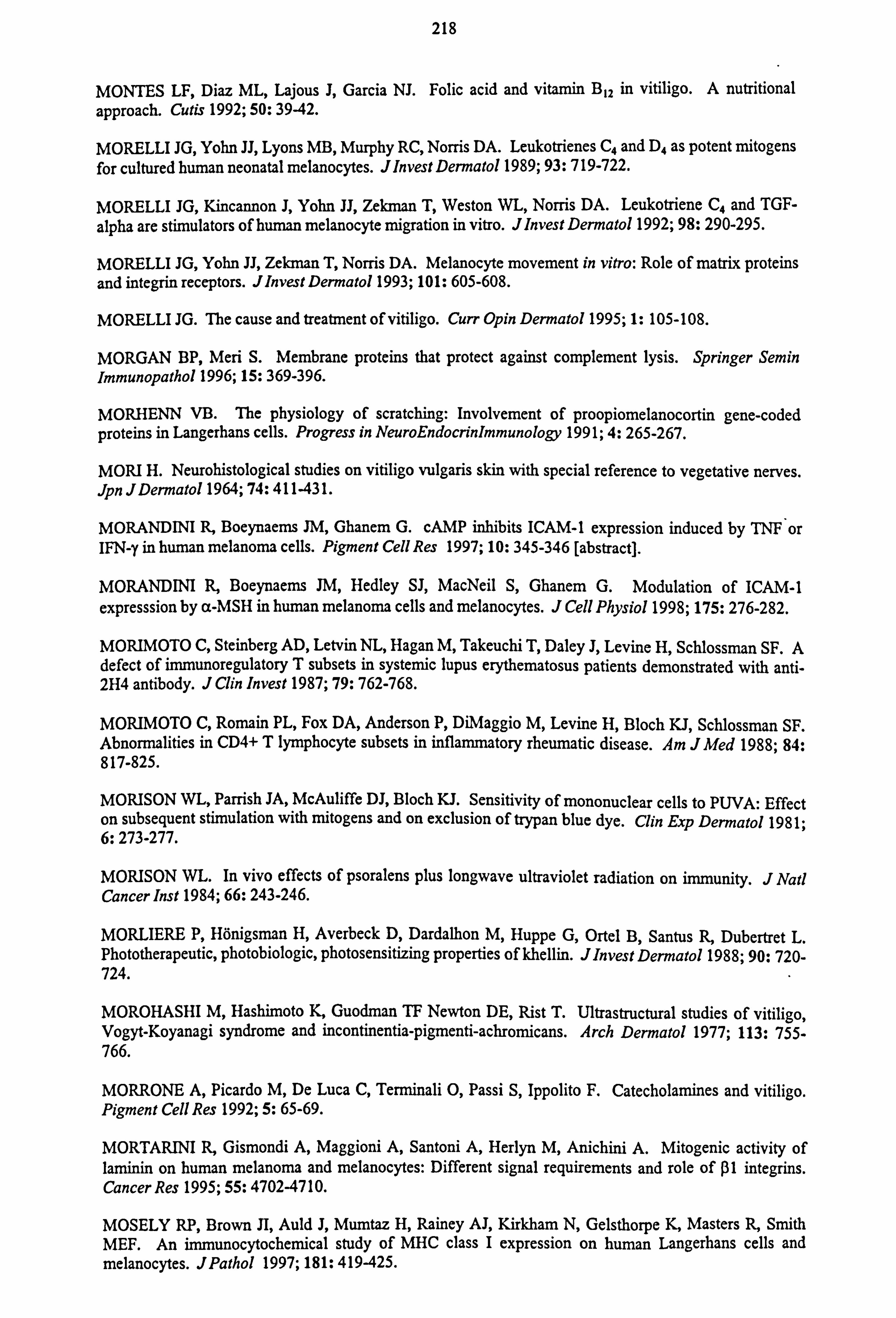
218
MONTES LF, Diaz ML, Lajous J, Garcia NJ. Folic acid and vitamin B12 in vitiligo. A nutritional approach. Cutis 1992; 50: 39-42.
MORELLI JG, Yohn JJ, Lyons MB, Murphy RC, Norris DA. Leukotrienes C4 and D4 as potent mitogens for cultured human neonatal melanocytes. J Invest Dermatol 1989; 93: 719-722.
MORELLI JG, Kincannon J, Yohn JJ, Zekman T, Weston WL, Norris DA. Leukotriene C4 and TGF-
alpha are stimulators of human melanocyte migration in vitro. J Invest Dermatol 1992; 98: 290-295.
MORELLI JG, Yohn JJ, Zekman T, Norris DA. Melanocyte movement in vitro: Role of matrix proteins and integrin receptors. Jlnvest Dermatol 1993; 101: 605-608.
MORELLI JG. The cause and treatment of vitiligo. Curr Opin Dermatol 1995; 1: 105-108.
MORGAN BP, Meri S. Membrane proteins that protect against complement lysis. Springer Semin Immunopathol 1996; 15: 369-396.
MORHENN VB. The physiology of scratching: Involvement of proopiomelanocortin gene-coded proteins in Langerhans cells. Progress in NeuroEndocrinlmmunology 1991; 4: 265-267.
MORI H. Neurohistological studies on vitiligo vulgaris skin with special reference to vegetative nerves. Jpn JDermatol 1964; 74: 411-431.
MORANDINI R, Boeynaems JM, Ghanem G. cAMP inhibits ICAM-1 expression induced by TNF or IFN-y in human melanoma cells. Pigment Cell Res 1997; 10: 345-346 [abstract].
MORANDINI R, Boeynaems JM, Hedley SJ, MacNeil S, Ghanem G. Modulation of ICAM-1 expresssion by a-MSH in human melanoma cells and melanocytes. J Cell Physiol 1998; 175: 276-282.
MORIMOTO C, Steinberg AD, Letvin NL, Hagan M, Takeuchi T, Daley J, Levine H, Schlossman SF. A defect of immunoregulatory T subsets in systemic lupus erythematosus patients demonstrated with anti- 2H4 antibody. J Clin Invest 1987; 79: 762-768.
MORIMOTO C, Romain PL, Fox DA, Anderson P, DiMaggio M, Levine H, Bloch KJ, Schlossman SF. Abnormalities in CD4+ T lymphocyte subsets in inflammatory rheumatic disease. Am JMed 1988; 84: 817-825.
MORISON WL, Parrish JA, McAuliffe DJ, Bloch KJ. Sensitivity of mononuclear cells to PUVA: Effect on subsequent stimulation with mitogens and on exclusion of trypan blue dye. Clin Exp Dermatol 1981; 6: 273-277.
MORISON WL. In vivo effects of psoralens plus longwave ultraviolet radiation on immunity. J Natl Cancer Inst 1984; 66: 243-246.
MORLIERE P, Hönigsman H, Averbeck D, Dardalhon M, Huppe G, Ortel B, Santus R, Dubertret L. Phototherapeutic, photobiologic, photosensitizing properties of khellin. JInvest Dermatol 1988; 90: 720- 724.
MOROHASHI M, Hashimoto K, Guodman TF Newton DE, Rist T. Ultrastructural studies of vitiligo, Vogyt-Koyanagi syndrome and incontinentia-pigmenti-achromicans. Arch Dermatol 1977; 113: 755- 766.
MORRONE A, Picardo M, De Luca C, Terminali 0, Passi S, Ippolito F. Catecholamines and vitiligo. Pigment Cell Res 1992; 5: 65-69.
MORTARINI R, Gismondi A, Maggioni A, Santoni A, Herlyn M, Anichini A. Mitogenic activity of laminin on human melanoma and melanocytes: Different signal requirements and role of ßl integrins. Cancer Res 1995; 55: 4702-4710.
MOSELY RP, Brown JI, Auld J, Mumtaz H, Rainey AJ, Kirkham N, Gelsthorpe K, Masters R, Smith MEF. An immunocytochemical study of MHC class I expression on human Langerhans cells and melanocytes. JPathol 1997; 181: 419-425.
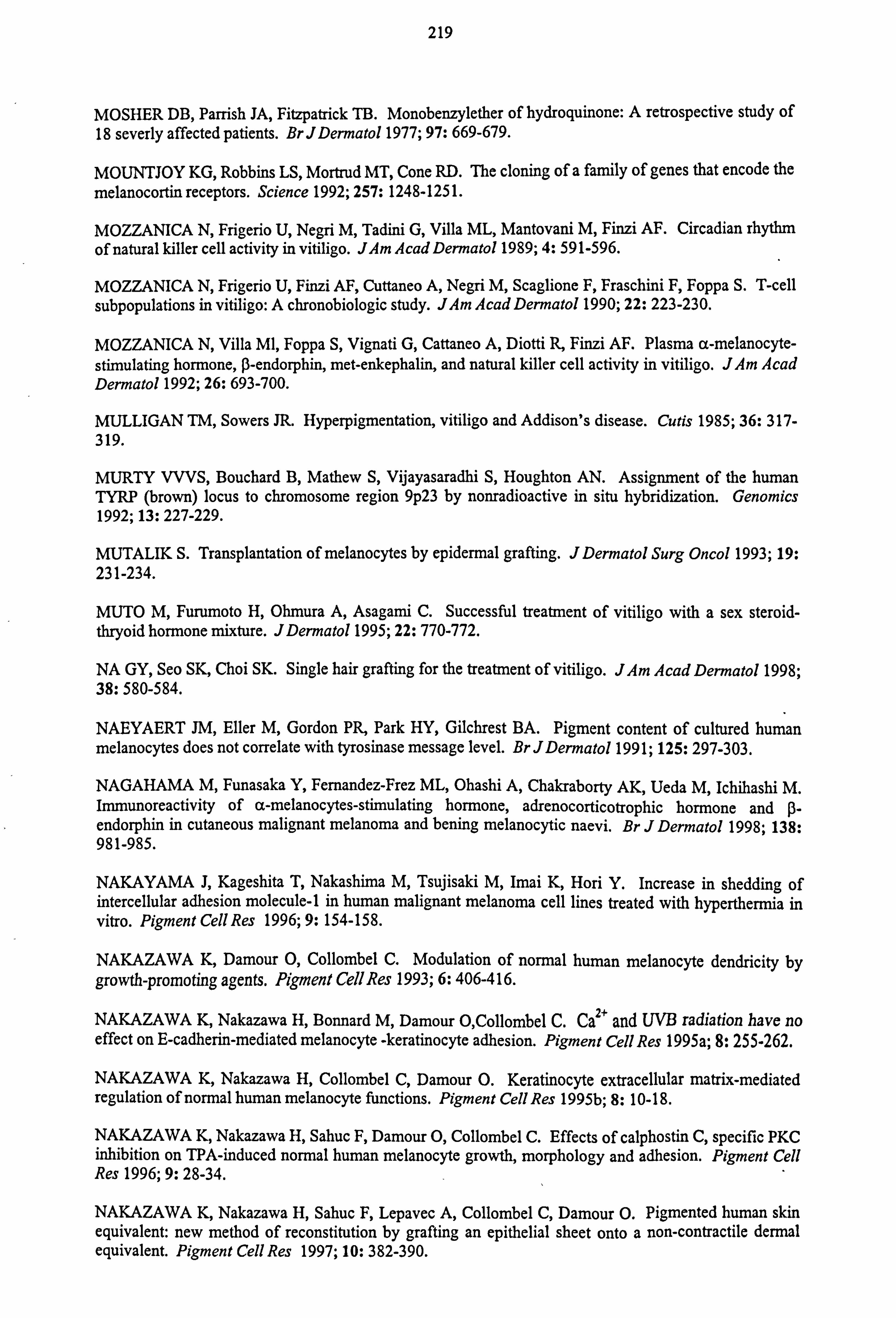
219
MOSHER DB, Parrish JA, Fitzpatrick TB. Monobenzylether of hydroquinone: A retrospective study of 18 severly affected patients. Br J Dermatol 1977; 97: 669-679.
MOUNTJOY KG, Robbins LS, Mortrud MT, Cone RD. The cloning of a family of genes that encode the melanocortin receptors. Science 1992; 257: 1248-1251.
MOZZANICA N, Frigerio U, Negri M, Tadini G, Villa ML, Mantovani M, Finzi AF. Circadian rhythm of natural killer cell activity in vitiligo. JAm Acad Dermatol 1989; 4: 591-596.
MOZZANICA N, Frigerio U, Finzi AF, Cuttaneo A, Negri M, Scaglione F, Fraschini F, Foppa S. T-cell
subpopulations in vitiligo: A chronobiologic study. JAm Acad Dermatol 1990; 22: 223-230.
MOZZANICA N, Villa Ml, Foppa S, Vignati G, Cattaneo A, Diotti R, Finzi AF. Plasma a-melanocyte- stimulating hormone, ß-endorphin, met-enkephalin, and natural killer cell activity in vitiligo. JAm Acad Dermatol 1992; 26: 693-700.
MULLIGAN TM, Sowers JR. Hyperpigmentation, vitiligo and Addison's disease. Cutis 1985; 36: 317- 319.
MURTY VVVS, Bouchard B, Mathew S, Vijayasaradhi S, Houghton AN. Assignment of the human TYRP (brown) locus to chromosome region 9p23 by nonradioactive in situ hybridization. Genomics 1992; 13: 227-229.
MUTALIK S. Transplantation of melanocytes by epidermal grafting. J Dermatol Surg Oncol 1993; 19: 231-234.
MUTO M, Furumoto H, Ohmura A, Asagami C. Successful treatment of vitiligo with a sex steroid- thryoid hormone mixture. JDermatol 1995; 22: 770-772.
NA GY, Seo SK, Choi SK. Single hair grafting for the treatment of vitiligo. JAm Acad Dermatol 1998; 38: 580-584.
NAEYAERT JM, Eller M, Gordon PR, Park HY, Gilchrest BA. Pigment content of cultured human melanocytes does not correlate with tyrosinase message level. BrJDermatol 1991; 125: 297-303.
NAGAHAMA M, Funasaka Y, Femandez-Frez ML, Ohashi A, Chakraborty AK, Ueda M, Ichihashi M. Immunoreactivity of a-melanocytes-stimulating hormone, adrenocorticotrophic hormone and (i- endorphin in cutaneous malignant melanoma and bening melanocytic naevi. Br J Dermatol 1998; 138: 981-985.
NAKAYAMA J, Kageshita T, Nakashima M, Tsujisaki M, Imai K, Hori Y. Increase in shedding of intercellular adhesion molecule-1 in human malignant melanoma cell lines treated with hyperthermia in vitro. Pigment Cell Res 1996; 9: 154-158.
NAKAZAWA K, Damour 0, Collombel C. Modulation of normal human melanocyte dendricity by growth-promoting agents. Pigment Cell Res 1993; 6: 406-416.
NAKAZAWA K, Nakazawa H, Bonnard M, Damour O, Collombel C. Ca2+ and UVB radiation have no effect on E-cadherin-mediated melanocyte -keratinocyte adhesion. Pigment Cell Res 1995a; 8: 25 5-262.
NAKAZAWA K, Nakazawa H, Collombel C, Damour O. Keratinocyte extracellular matrix-mediated regulation of normal human melanocyte functions. Pigment Cell Res 1995b; 8: 10-18.
NAKAZAWA K, Nakazawa H, Sahuc F, Damour 0, Collombel C. Effects of calphostin C, specific PKC inhibition on TPA-induced normal human melanocyte growth, morphology and adhesion. Pigment Cell Res 1996; 9: 28-34.
NAKAZAWA K, Nakazawa H, Sahuc F, Lepavec A, Collombel C, Damour O. Pigmented human skin equivalent: new method of reconstitution by grafting an epithelial sheet onto a non-contractile dermal equivalent. Pigment Cell Res 1997; 10: 382-390.
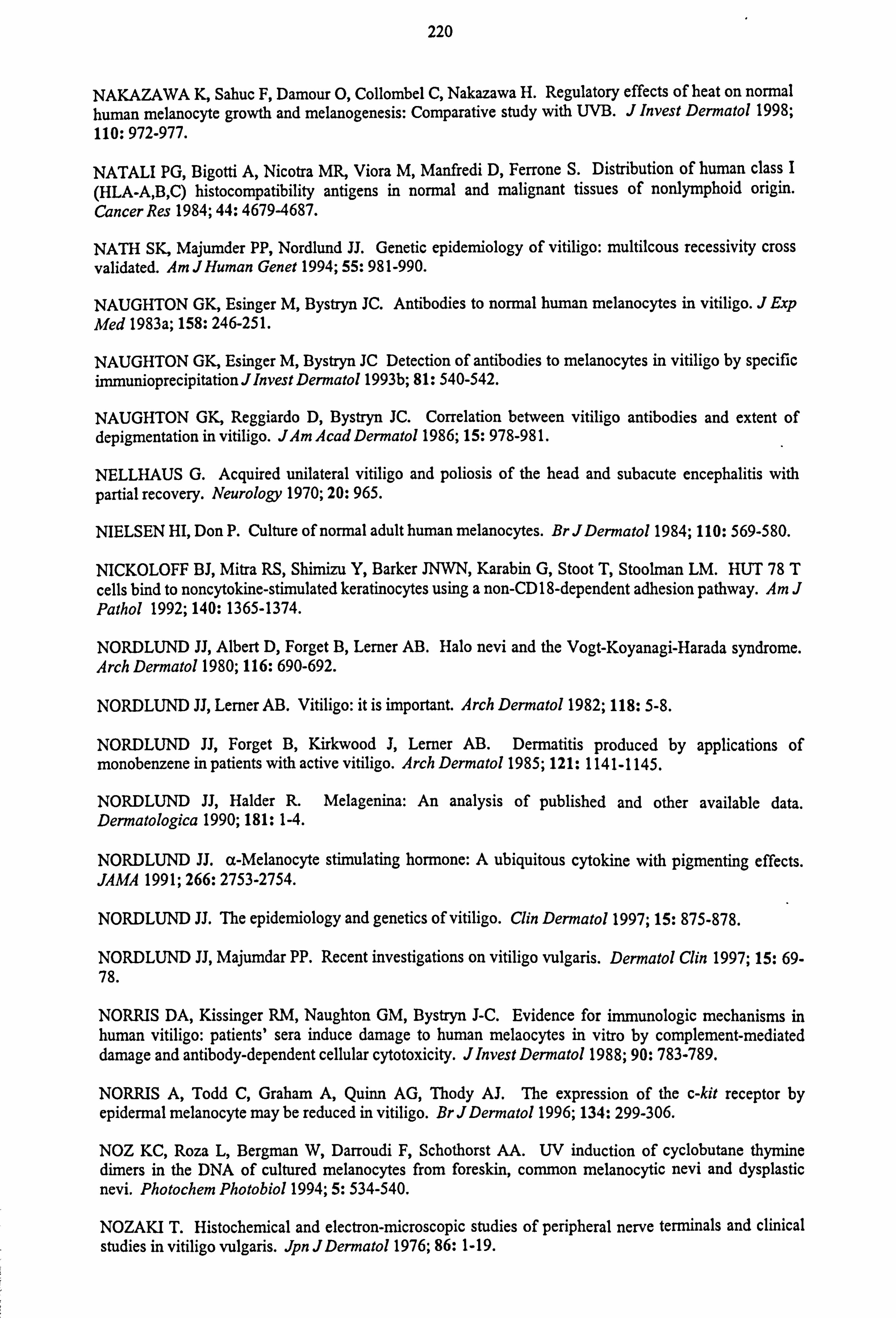
220
NAKAZAWA K, Sahuc F, Damour 0, Collombel C, Nakazawa H. Regulatory effects of heat on normal human melanocyte growth and melanogenesis: Comparative study with UVB. J Invest Dermatol 1998; 110: 972-977.
NATALI PG, Bigotti A, Nicotra MR, Viora M, Manfredi D, Ferrone S. Distribution of human class I (HLA-A, B, C) histocompatibility antigens in normal and malignant tissues of nonlymphoid origin. Cancer Res 1984; 44: 4679-4687.
NATH SK, Majumder PP, Nordlund JJ. Genetic epidemiology of vitiligo: multilcous recessivity cross validated. Am JHuman Genet 1994; 55: 981-990.
NAUGHTON GK, Esinger M, Bystryn JC. Antibodies to normal human melanocytes in vitiligo. J Exp Med 1983a; 158: 246-251.
NAUGHTON GK, Esinger M, Bystryn JC Detection of antibodies to melanocytes in vitiligo by specific immunioprecipitation J Invest Dermatol 1993b; 81: 540-542.
NAUGHTON GK, Reggiardo D, Bystryn JC. Correlation between vitiligo antibodies and extent of depigmentation in vitiligo. JAm Acad Dermatol 1986; 15: 978-98 1.
NELLHAUS G. Acquired unilateral vitiligo and poliosis of the head and subacute encephalitis with partial recovery. Neurology 1970; 20: 965.
NIELSEN HI, Don P. Culture of normal adult human melanocytes. Br JDermatol 1984; 110: 569-580.
NICKOLOFF BJ, Mitra RS, Shimizu Y, Barker JNWN, Karabin G, Stoot T, Stoolman LM. HUT 78 T cells bind to noncytokine-stimulated keratinocytes using a non-CD 18-dependent adhesion pathway. 4m J Pathol 1992; 140: 1365-1374.
NORDLUND JJ, Albert D, Forget B, Lerner AB. Halo nevi and the Vogt-Koyanagi-Harada syndrome. Arch Dermatol 1980; 116: 690-692.
NORDLUND JJ, Lerner AB. Vitiligo: it is important. Arch Dermatol 1982; 118: 5-8.
NORDLUND JJ, Forget B, Kirkwood J, Lerner AB. Dermatitis produced by applications of monobenzene in patients with active vitiligo. Arch Dermatol 1985; 121: 1141-1145.
NORDLUND JJ, Halder R. Melagenina: An analysis of published and other available data. Dennatologica 1990; 181: 1-4.
NORDLUND JJ. a-Melanocyte stimulating hormone: A ubiquitous cytokine with pigmenting effects. JAMA 1991; 266: 2753-2754.
NORDLUND JJ. The epidemiology and genetics of vitiligo. Clin Dermatol 1997; 15: 875-878.
NORDLUND JJ, Majumdar PP. Recent investigations on vitiligo vulgaris. Dermatol Clin 1997; 15: 69- 78.
NORRIS DA, Kissinger RM, Naughton GM, Bystryn J-C. Evidence for immunologic mechanisms in human vitiligo: patients' sera induce damage to human melaocytes in vitro by complement-mediated damage and antibody-dependent cellular cytotoxicity. Jlnvest Dermato11988; 90: 783-789.
NORRIS A, Todd C, Graham A, Quinn AG, Thody AJ. The expression of the c-kit receptor by epidermal melanocyte may be reduced in vitiligo. Br J Dermato11996; 134: 299-306.
NOZ KC, Roza L, Bergman W, Darroudi F, Schothorst AA. UV induction of cyclobutane thymine dimers in the DNA of cultured melanocytes from foreskin, common melanocytic nevi and dysplastic nevi. Photochem Photobio11994; 5: 534-540.
NOZAKI T. Histochemical and electron-microscopic studies of peripheral nerve terminals and clinical studies in vitiligo vulgaris. Jpn JDermato11976; 86: 1-19.
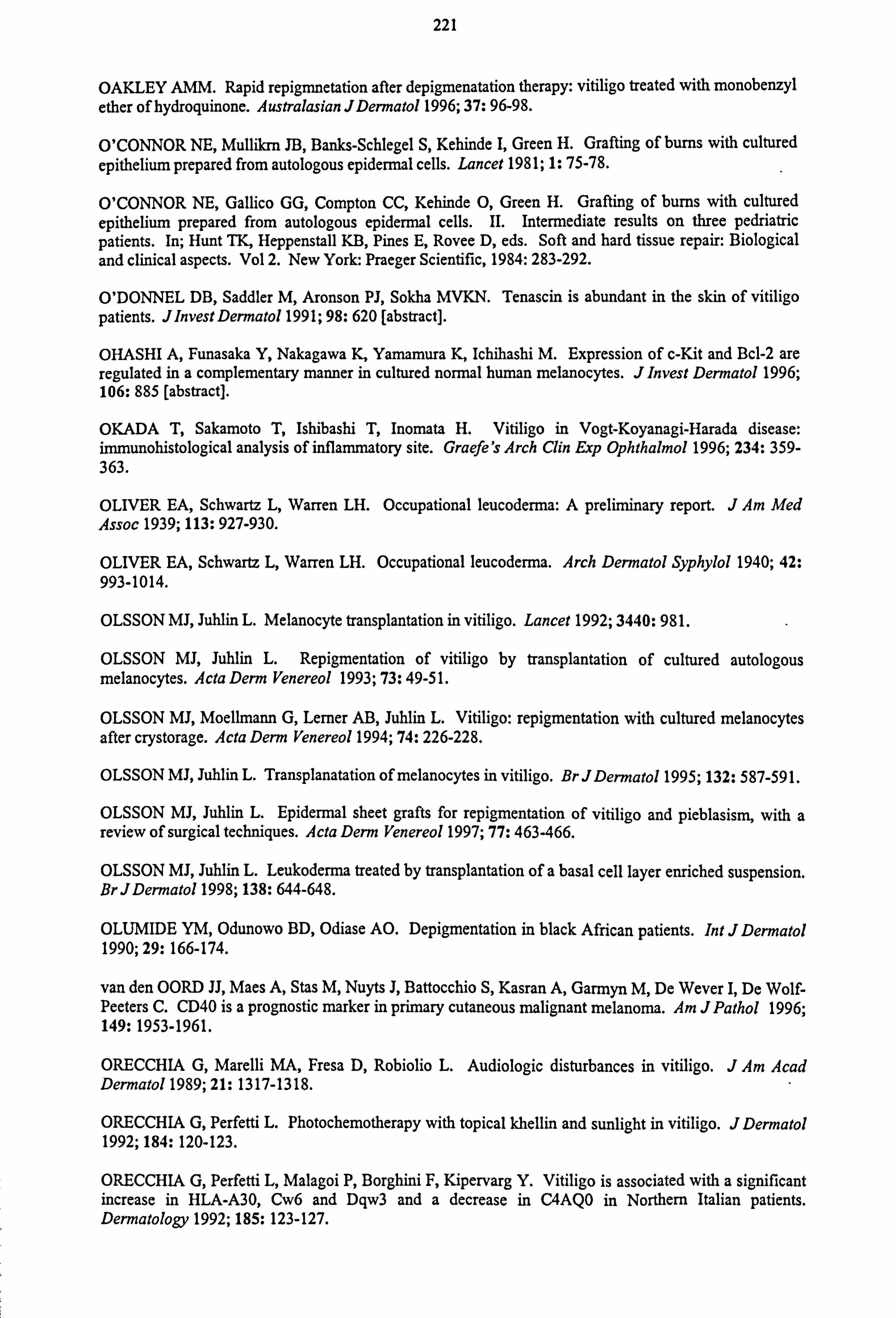
221
OAKLEY AMM. Rapid repigmnetation after depigmenatation therapy: vitiligo treated with monobenzyl ether of hydroquinone. Australasian JDermatol 1996; 37: 96-98.
O'CONNOR NE, Mullikrn JB, Banks-Schlegel S, Kehinde I, Green H. Grafting of burns with cultured epithelium prepared from autologous epidermal cells. Lancet 1981; 1: 75-78.
O'CONNOR NE, Gallico GG, Compton CC, Kehinde 0, Green H. Grafting of burns with cultured epithelium prepared from autologous epidermal cells. II. Intermediate results on three pedriatric patients. In; Hunt TK, Heppenstall KB, Pines E, Rovee D, eds. Soft and hard tissue repair: Biological
and clinical aspects. Vol 2. New York: Praeger Scientific, 1984: 283-292.
O'DONNEL DB, Saddler M, Aronson PJ, Sokha MVKN. Tenascin is abundant in the skin of vitiligo patients. Jlnvest Dermatol 1991; 98: 620 [abstract].
OHASHI A, Funasaka Y, Nakagawa K, Yamamura K, Ichihashi M. Expression of c-Kit and Bcl-2 are regulated in a complementary manner in cultured normal human melanocytes. J Invest Dermatol 1996; 106: 885 [abstract].
OKADA T, Sakamoto T, Ishibashi T, Inomata H. Vitiligo in Vogt-Koyanagi-Harada disease: immunohistological analysis of inflammatory site. Graefe's Arch Clin Exp Ophthalmol 1996; 234: 359- 363.
OLIVER EA, Schwartz L, Warren LH. Occupational leucoderma: A preliminary report. J Am Med Assoc 1939; 113: 927-930.
OLIVER EA, Schwartz L, Warren LH. Occupational leucoderma. Arch Dermatol Syphylol 1940; 42: 993-1014.
OLSSON MJ, Juhlin L. Melanocyte transplantation in vitiligo. Lancet 1992; 3440: 981.
OLSSON MJ, Juhlin L. Repigmentation of vitiligo by transplantation of cultured autologous melanocytes. Acta Derm Venereol 1993; 73: 49-5 1.
OLSSON MI, Moellmann G, Lerner AB, Juhlin L. Vitiligo: repigmentation with cultured melanocytes after crystorage. Acta Derm Venereol 1994; 74: 226-228.
OLSSON MJ, Juhlin L. Transplanatation of melanocytes in vitiligo. BrJDermatol 1995; 132: 587-591.
OLSSON MJ, Juhlin L. Epidermal sheet grafts for repigmentation of vitiligo and pieblasism, with a review of surgical techniques. Acta Derm Venereol 1997; 77: 463-466.
OLSSON MJ, Juhlin L. Leukoderma treated by transplantation of a basal cell layer enriched suspension. BrJDermatol 1998; 138: 644-648.
OLUMIDE YM, Odunowo BD, Odiase AO. Depigmentation in black African patients. Int J Dermatol 1990; 29: 166-174.
van den OORD JJ, Maes A, Stas M, Nuyts J, Battocchio S, Kasran A, Garmyn M, De Wever I, De Wolf- Peeters C. CD40 is a prognostic marker in primary cutaneous malignant melanoma. Am J Pathol 1996; 149: 1953-1961.
ORECCHIA G, Marelli MA, Fresa D, Robiolio L. Audiologic disturbances in vitiligo. J Am Acad Dermatol 1989; 21: 1317-1318.
ORECCHIA G, Perfetti L. Photochemotherapy with topical khellin and sunlight in vitiligo. J Dermatol 1992; 184: 120-123.
ORECCHIA G, Perfetti L, Malagoi P, Borghini F, Kipervarg Y. Vitiligo is associated with a significant increase in HLA-A30, Cw6 and Dqw3 and a decrease in C4AQO in Northern Italian patients. Dermatology 1992; 185: 123-127.
222
ORECCHIA G, Frattini P, Cucchi ML. Normal-range plasma catecholamines in patients with generalized and acrofacial vitiligo: preliminary report. Dermatology 1994; 189: 350-353.
ORECCHIA G, Sangalli ME, Gazzaniga A, Giordano F. Topical photochemotherapy of vitiligo with a new khellin formulation: preliminary clinical results. JDermatol Treatment 1998; 9: 65-69.
ORLOW SJ, Boissy RE, Moran DJ, Pifko-Hirst S. Subcellular distribution of tyrosinase and tyrosinase- related protein-1: Implications for melanosomal biogenesis. JInvest Dermatol 1993; 100: 55-64.
ORTEL B, Tanew A, Honigsmann H. Treatment of vitiligo with khellin and ultraviolet A. J Am Acad Dermatol 1988; 18: 693-701.
ORTONNE JP, MacDonald DM, Micoud A, Thivolet J. PUVA-induced repigmentation of vitiligo: A histochemical (split DOPA) and ultrastructural study. BrJDermatol 1979; 101: 1-7.
ORTONNE JP, Schmitt D, Thivolet J. PUVA-induced repigmentation of vitiligo: Scanning electron microscope of hair follicles. Jlnvest Dermatol 1980; 74: 40-42.
ORTONNE JP, Mosher DB, Fitzpatrick TB. In: Vitiligo and other hypomelanoses of hair and skin. New York: Plenum Medical Book Company, 1983.
ORTONNE JP, Bose SK. Vitiligo: Where do we stand? Pigment Cell Res 1993; 6: 61-72.
PAL P, Roy R, Datta PK, Dutta AK, Biswas B, Bhadra R. Hydroalcoholic human placental extract - skin pigmenting activity and gross chemical-composition. IntJDermatol 1995; 34: 61-66.
PALUMBO A, d'Ischia M, Misuraca G, Prota G, Schultz TM. Structural modifications in biosynthetic melanins induced by metal ions. Biochem Biophys Acta 1988; 964: 193-199.
PARISH-GAUSE D, Haider R, Berka N, Furbert-Harris P, Dunston GM. HLA-DRB1 associations with vitiligo. Human Immunol 1997; 55: 54 [abstract].
PARK KC, Youn JI, Lee YS. Clinical study of 326 cases of vitiligo. KorJDermatol 1988; 26: 200-205.
PARK SY, Youn JI, Lim SD. A clinical study of 217 cases of vitiligo. Kor J Dermatol 1981; 19: 145- 152.
PARK YK, Hann SK, Song MS, Yoon JK, Kim HI. The activity of antimelanocyte autoantibodies in vitiligo patients. KorJDermatol 1991; 29: 391-398.
PARK YK, Kim NS, Hann SK, Im S. Identification of autoantibody to melanocyte and characterization of vitiligo antigen in vitliogo patients. JDermatol Sci 1996; 11: 111-120.
PARRISH JA, Fitapatrick TB, Shea C, Pathak MA. Photochemotherapy of vitiligo. Use of orally adminstered psoralens and a high-intensity long-wave ultraviolet light systems. Arch Dermatol 1976; 112: 1531-1534.
PASRICHA JS, Khaitan BK. Oral mini-pulse therapy with betamethasone in vitiligo patients having extensive or fast-spreding disese. Int JDermatol 1993; 32: 753-757.
PASRICHA JS, Khera V. Effect of prolonged treatment with levamisole on vitiligo with limited and slow-spreading disease. IntJDermatol 1994; 33: 584-587.
PASSI S, Nazzaro-Porro M. Molecular basis of substrate and inhibitory specificity of tyrosinase: phenolic compounds. BrJDermatol 1981; 104: 659-665.
PASSI S, Picardo M, De Luca C, Grandinetti M, Ippolito F. Treatment of vitiligo with an antioxidant pool. Melanoma Res 1995; 5 (suppl2): 25 [abstract].
PASSI S, Grandinetti M, Maggio F, Stancato A, De Luca C. Epidermal oxidative stress in vitiligo. Pigment Cell Res 1998; 11: 81-85.
223
PATEL R, Aronson PJ, Sikha MVKN, Sandler B, Agha AA. The pattern of Vogt-Koyanagi-Harada syndrome pigment cell antibodies corresponds to that found in vitiligo patient sera. J Invest Dermatol 1992; 89: 629 [abstract].
PATHAK MA, Sinesi SJ, Szabo G. The effect of a single dose of ultraviolet radiation on epidermal melanocytes. J Invest Dermatol 1965; 45: 520-528.
PAVITHRAN K. Vitiligo and lepromatous leprosy. Int J Leprosy 1991; 59: 120-121.
PAWELEK J, Korner A, Bergstrom A, Bolognia JL. New regulators of melanin biosynthesis and the autodestruction of melanoma cells. Nature 1980; 286: 617-619.
PEACOCKE M, Yaar M, Mansur CP, Chao MV, Gilchrest BA. Induction of nerve growth factor receptors on cultured human melanocytes. Proc Natl Acad Sci USA 1988; 85: 5282-5286.
PEARS JS, Jung RT, Bartlett W, Browning MCK, Kenicer K, Thody AJ. A case of skin hyperpigmentation due to a-MSH hypersecretion. BrJDermatol 1992; 126: 286-289.
PEDEUX R, Al-Irani N, Marteau C, Pellicier F, Branche R, Ozturk M, Franchi J, Dore. Thymidine dinucleotides induce S phase cell cycle arrest in addition to increased melanogenesis in human melanocytes. JInvest Dermatol 1998; 111: 472-477.
PEDLEY J, Ablett EM, Pettit A, Meyer J, Dunn IS, Sturm RA, Parsons PG. Inhibition of retinoblastoma protein translation by UVB in human melanocytic cells and reduced cell cycle arrest following repeated irradiation. Oncogene 1996; 13: 1335-1342.
PELC NJ, Nordlund JJ. Pigmentary changes in the skin. Clin Plast Surg 1993; 20: 53-65.
PHILLIPS TJ, Kehinde 0, Green H, Gilchrest BA. Treatment of skin ulcers with cultured epidermal allografts. JAm Acad Dermatol 1989; 22: 191-199.
PICARDO M, Passi S, Morrone A, Grandinetti M, Di Carlo A, Ippolito F. Antioxidant status in the blood of patients with active vitiligo. Pigment Cell Res 1994; 7: 110-115.
PINKUS H. Vitiligo: What is it? Jlnvet Dermatol 1959; 32: 281-284.
PITTELKOW MR, Shipley GD. Serum-free culture of normal human melanocytes: Growth kinetics and growth factor requirements. J Cell Physiol 1989; 140: 565-576.
PITTS E, Grimes PE. The incidence and clinical significance of residual melanocytes in vitiligo as assessed by the split dopa technique. JAm Acad Dermatol 1991; 24: 113 [abstract].
PLETTENBERG A, Ballaun C, Pammer J, Milder M, Strunk D, Weninger W, Tschachler E. Human melanocytes and melanoma cells constitutively express the Bcl-2 proto-oncogene in situ and in cell culture. Am JPathol 1995; 146: 651-659.
PLOTT RT, Brysk MM, Newton RC, Rainier SS, Rajaraman S. A surgical treatment for vitiligo: autologous cultured-epithelial grafts. JDermatol Surg Oncol 1989; 15: 1161-1166.
PLOTT RT, Wagner RF. Modem treatment approaches to vitiligo. Cutis 1990; 45: 311-316.
POLOY A, Tibor L, Kramer J, Ngugen A-T, Kraszits E, Medgyessy I, Füst G, Stenszky V, Farid NR. HLA-DR1 is associated with vitiligo. Immunol Letts 1991; 27: 59-62.
PORTER JR, Beuf AH, Lerner A, Nordlund J. Psychosocial effect of vitiligo: a comparison of vitiligo patients with "normal" control subjects, with psoriasis patients, and with other pigmentary disorders. J Am Acad Dermatol 1986; 15: 220-224.
PORTER J, Beuf AH, Lerner A, Nordlund J. Response to cosmetic disfigurement: Patients with vitiligo. Cutis 1987; 39: 493-494.
224
PORTER JR, Beuf AH, Lerner AB, Nordlund JJ. The effect of vitiligo in sexual relationships. J Am AcadDermatol 1990; 22: 221-222.
PROCACCINI EM, Riccio G, Monfrecola G. Ineffectiveness of topical khellin in photochemotherapy of vitiligo. JDermatol Treatment 1995; 6: 117-120.
PROTA G. Recent advances in the chemistry of melanogenesis in mammals. J Invest Dermatol 1980; 75: 122-127.
PROTA G. Some new aspects of eumelanin chemistry. Prog Clin Biol Res 1988; 256: 101-124.
PURI N, Mojamdar M, Ramaiah A. In vitro growth characteristics of melanocytes obtained from adult normal and vitiligo subjects. Jlnvest Dermatol 1987; 88: 434-438.
PURI N, Mojamdar M, Ramaiah A. Growth defects of melanocytes in culture from vitiligo subjects are spontaneously corrected in vivo in repigmenting subjects and can be partially corrected by the addition of fibroblast-derived growth factors in vitro. Arch Dermatol Res 1989; 281: 178-184.
PURI N, van der Weel MB, de Wit FS, Asghar SS, Das PK, Ramaiah A, Westehof W. Basic fibroblast growth faactor promotes melanin synthesis by melanocytes. Arch Dermatol Res 1996; 288: 633-635.
QUEVEDO WC Jr, Szabo G, Virks J, Sinesi SJ. Melanocyte populations in UV-irradiated human skin. J Invest Dermatol 1965; 45: 295-298.
QUEVEDO WC, Szabo G, Virks J. Influence of age and UV on the populations of dopa-positive melanocytes inhuman skin. Jlnvest Dermatol 1969; 52: 287-290.
QUEVEDO WC, Fitzpatrick TB, Szabo G, Jimbow K. Biology of melanocytes. In: Dermatology in General Medicine, 3rd Ed. New York, McGraw Hill 1987; 224-251.
RAJORA N, Ceriani G, Catania A, Star RA, Mruphy MT, Lipton JM. a-MSH production, receptors, and influence on neopterin in a human monocyte/macrophage cell line. JLeukocyte Biol 1996; 59: 248-253.
RAMAIAH A, Mojamdar M, Amarnath VM. Vitiligo in the SSK community of Bangalore. Ind J Dermatol VenereolLeprol 1988; 54: 251-254.
RAMAIAH A, Puri N, Mojamdar M. Etiology of vitiligo. A new hypothesis. Acta Denn Venerol (Stockh) 1989; 69: 323-367.
RAMAN A, Lin ZX, Sviderskaya E, Kowalska D. Investigation of the effect of Angelica sinensis root extract on the proliferaion of melanocytes in culture. JEthnopharmacology 1996; 54: 165-170.
RAMIREZ-BOSCA A, Bernd A, Werner R, Dold K, Holzmann H. Effect of the dose of ultraviolet radiation on the pigment formation by human melanocytes in vitro. Arch Dermatol Res 1992; 284: 358- 362.
RAMSAY M, Coman MA, Stevens G, Zwane E, Kromberg J, Farrrall M, Jenkins T. The tyrosinase- positive oculocutaneous albinism locus maps to chromosome 15g11.2-q12. Am J Hum Genet 1992; 51: 879-884.
RANSON M, Posen S, Mason R. S. Extracellular matrix modulates the function of human melanocytes but not melanoma cells. J Cell Physiol 1988a; 136: 281-288.
RANSON M, Posen S, Mason R. S. Human melanocytes as a target tissue for hormones: in vitro studies with I cc-25, dihydroxyvitamin D3, a-melanocyte stimulating hormone, and beta-estradiol. J Invest Dermatol 1988b; 91: 593-598.
RAO J, Otto WR. Fluorimetric DNA assay for cell growth estimation. Anal Biochem 1992; 207: 186- 192.
RAWLES M. Origin of pigment cells from the neural crest in the mouse embryo. Physiol Zool 1947; 20: 248-266.
225
REGNIER M, Staquet M-J, Schmitt D, Schmidt R. Integration of Langerhans cells into a pigmented reconstructed human epidermis. Jlnvest Dermatol 1997; 109: 510-512.
RENFRO L. Lack of efficacy of the Q-switched ruby laser in the treatment of vitiligo. Arch Dermatol 1992; 128:: 277-278.
RETORNAZ G, Betuel H, Ortonne JP, Thivolet J. HL-A antigens and vitiligo. Br J Dermatol 1976; 95: 173-175.
RHEINS LA, Young EM, Nordlund ML, Berning RB, Nordlund JJ. Rapid induction of Thy-1 antigenic markers on keratinocytes and epidermal immune cells in the skin of mice following topical treatment with common preservatives used in topical medications an in food. JInvest Dermatol 1987; 89: 489-494.
RICCIO ML, Coratza G, Bovalini L, Martelli P. Investigation of mutagenic activity in Salmonella typhimurium of the furochromone khellin, proposed as therapeutic agents for skin diseases. Mutation Res 1992; 279: 103-108.
RINCHIK EM, Bultman SJ, Horstlemke B, Lee S-T, Strunk KM, Spritz RA, Avidano KM, Jong MTC, Nicholls RD. A gene for the mouse pink-eyed dilution locus and for human type II oculocutaneous albinism. Nature 1993; 362: 72-76.
RILEY P. Distribution of epidermal dendritic cells in pigmented and unpigmented skin. J Invest Dermatol 1967; 48: 28-38.
RILEY PA. Melanin. IntJBiochem Cell Bio11997; 29: 1235-1239.
ROMERO C, Aberdam E, Larnier C, Ortonne J-P. Retinoic acid as modulator of UVB-induced melanocyte differentiation. J Cell Sci 1994; 107: 1095-1103.
ROMERO-GRAILLET C, Aberdam E, Biagoli N, Ortonne J-P, Ballotti R. Ultraviolet B radiation acts through the nitric oxide and cGMP signal transduction pathway to stimulate melanogensis in human melnaocytes. JBio1 Chem 1996; 271: 28052-28056.
ROMERO-GRAILLET C, Aberdam E, Clement M, Ortonne J-P, Ballotti R. Nitric-oxide produced by ultraviolet-irradiated keratinocytes stimulates melanogenesis. J Clin Invest 1997; 99: 635-642.
ROSDAHL IK. Melanocyte mitosis in UVB-irradiated mouse skin. Acta Derm Venereol 1978; 58: 217- 221.
ROSDAHL IK, Szabo G. Mitotic activity of epidermal melanocytes in UV-irradiated mouse skin. J Invest Dermatol 1978; 70: 143-148.
ROSELLI-REHFUSS L, Mountjoy KG, Robbins LS, Mortrud MT, Low MJ, Tatro JB, Entwisttle ML, Simerly RB, Cone RD. Identification of a recpetor for gamma melanotropin and other proopiomelanocortin peptides in the hpothalamus and limbic system. Proc Nat! Acad Sci USA 1993; 90: 8856-8860.
ROSEN CF, Seki Y, Farinelli W, Stem RS, Fitzpatrick TB, Pathak MA, Gange RW. A comparison of the melanocyte response to narrow band UVA and UVB exposure in vivo. J Invest Dermatol 1987; 88: 774- 779.
ROSENBACH T, Wellenreuther U, Numberger F, Czarnetzki BM. Treatment of vitiligo with phenylalanine and UVA. Hautarzt 1992; 44: 208-209.
ROSENBERG SA, White DE. Vitiligo in patients with melanoma: normal tissue antigens can be targets for cancer immunotherapy. Jlmmunother 1996; 19: 81-84.
RYAN W, Carver M. Inhibition of antibody synthesis by L-phenylalanine. Science 1963; 43: 479-480.
SAKAMOTO T, Murata T, Inomata H. Class II major histocompatibility complex on melanocytes of Vogt-Koyanagi-Harada disease. Arch Ophthalmol 1991; 109: 1270-1274.
SALAFIA A. Vitiligo. Successful treatment with dapsone. Chron Dermato11995; 5: 171-188.
SALAMON T, Hadziselimovic R, Halepovic E. The heritability of vitiligo. Hautarzt 1989; 40: 141-145.
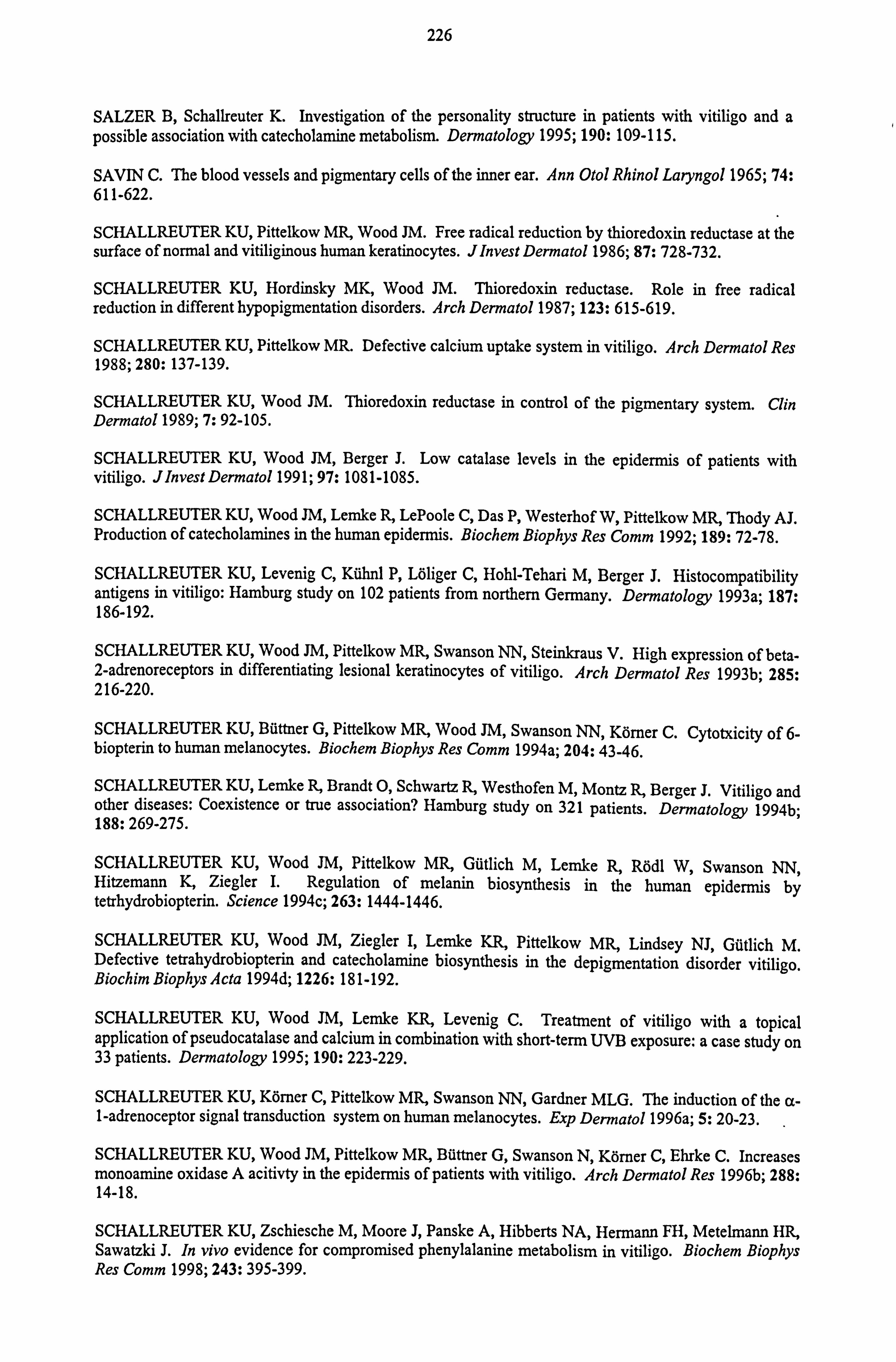
226
SALZER B, Schallreuter K. Investigation of the personality structure in patients with vitiligo and a possible association with catecholamine metabolism. Dermatology 1995; 190: 109-115.
SAVIN C. The blood vessels and pigmentary cells of the inner ear. Ann Otol Rhinol Laryngol 1965; 74: 611-622.
SCHALLREUTER KU, Pittelkow MR, Wood JM. Free radical reduction by thioredoxin reductase at the surface of normal and vitiliginous human keratinocytes. Jlnvest Dermatol 1986; 87: 728-732.
SCHALLREUTER KU, Hordinsky MK, Wood JM. Thioredoxin reductase. Role in free radical reduction in different hypopigmentation disorders. Arch Dermatol 1987; 123: 615-619.
SCHALLREUTER KU, Pittelkow MR. Defective calcium uptake system in vitiligo. Arch Dermatol Res 1988; 280: 137-139.
SCHALLREUTER KU, Wood JM. Thioredoxin reductase in control of the pigmentary system. Clin Dermatol 1989; 7: 92-105.
SCHALLREUTER KU, Wood JM, Berger J. Low catalase levels in the epidermis of patients with vitiligo. Jlnvest Dermatol 1991; 97: 1081-1085.
SCHALLREUTER KU, Wood JM, Lemke R, LePoole C, Das P, Westerhof W, Pittelkow MR, Thody AJ. Production of catecholamines in the human epidermis. Biochem Biophys Res Comm 1992; 189: 72-78.
SCHALLREUTER KU, Levenig C, Kühnl P, Löliger C, Hohl-Tehari M, Berger J. Histocompatibility antigens in vitiligo: Hamburg study on 102 patients from northern Germany. Dermatology 1993a; 187: 186-192.
SCHALLREUTER KU, Wood JM, Pittelkow MR, Swanson NN, Steinkraus V. High expression of beta- 2-adrenoreceptors in differentiating lesional keratinocytes of vitiligo. Arch Dermatol Res 1993b; 285: 216-220.
SCHALLREUTER KU, Büttner G, Pittelkow MR, Wood JM, Swanson NN, Körner C. Cytotxicity of 6- biopterin to human melanocytes. Biochem Biophys Res Comm 1994a; 204: 43-46.
SCHALLREUTER KU, Lemke R, Brandt 0, Schwartz R, Westhofen M, Montz R, Berger J. Vitiligo and other diseases: Coexistence or true association? Hamburg study on 321 patients. Dermatology 1994b; 188: 269-275.
SCHALLREUTER KU, Wood JM, Pittelkow MR, Gütlich M, Lemke R, Rödl W, Swanson NN, Hitzemann K, Ziegler I. Regulation of melanin biosynthesis in the human epidermis by tetrhydrobiopterin. Science 1994c; 263: 1444-1446.
SCHALLREUTER KU, Wood JM, Ziegler I, Lemke KR, Pittelkow MR, Lindsey NJ, Gütlich M. Defective tetrahydrobiopterin and catecholamine biosynthesis in the depigmentation disorder vitiligo. Biochim Biophys Acta 1994d; 1226: 181-192.
SCHALLREUTER KU, Wood JM, Lemke KR, Levenig C. Treatment of vitiligo with a topical application of pseudocatalase and calcium in combination with short-term UVB exposure: a case study on 33 patients. Dermatology 1995; 190: 223-229.
SCHALLREUTER KU, Körner C, Pittelkow MR, Swanson NN, Gardner MLG. The induction of the a- 1-adrenoceptor signal transduction system on human melanocytes. Exp Dermatol 1996a; 5: 20-23.
SCHALLREUTER KU, Wood JM, Pittelkow MR, Büttner G, Swanson N, Körner C, Ehrke C. Increases monoamine oxidase A acitivty in the epidermis of patients with vitiligo. Arch Dermatol Res 1996b; 288: 14-18.
SCHALLREUTER KU, Zschiesche M, Moore J, Panske A, Hibberts NA, Hermann FH, Metelmann HR, Sawatzki J. In vivo evidence for compromised phenylalanine metabolism in vitiligo. Biochem Biophys Res Comm 1998; 243: 395-399.
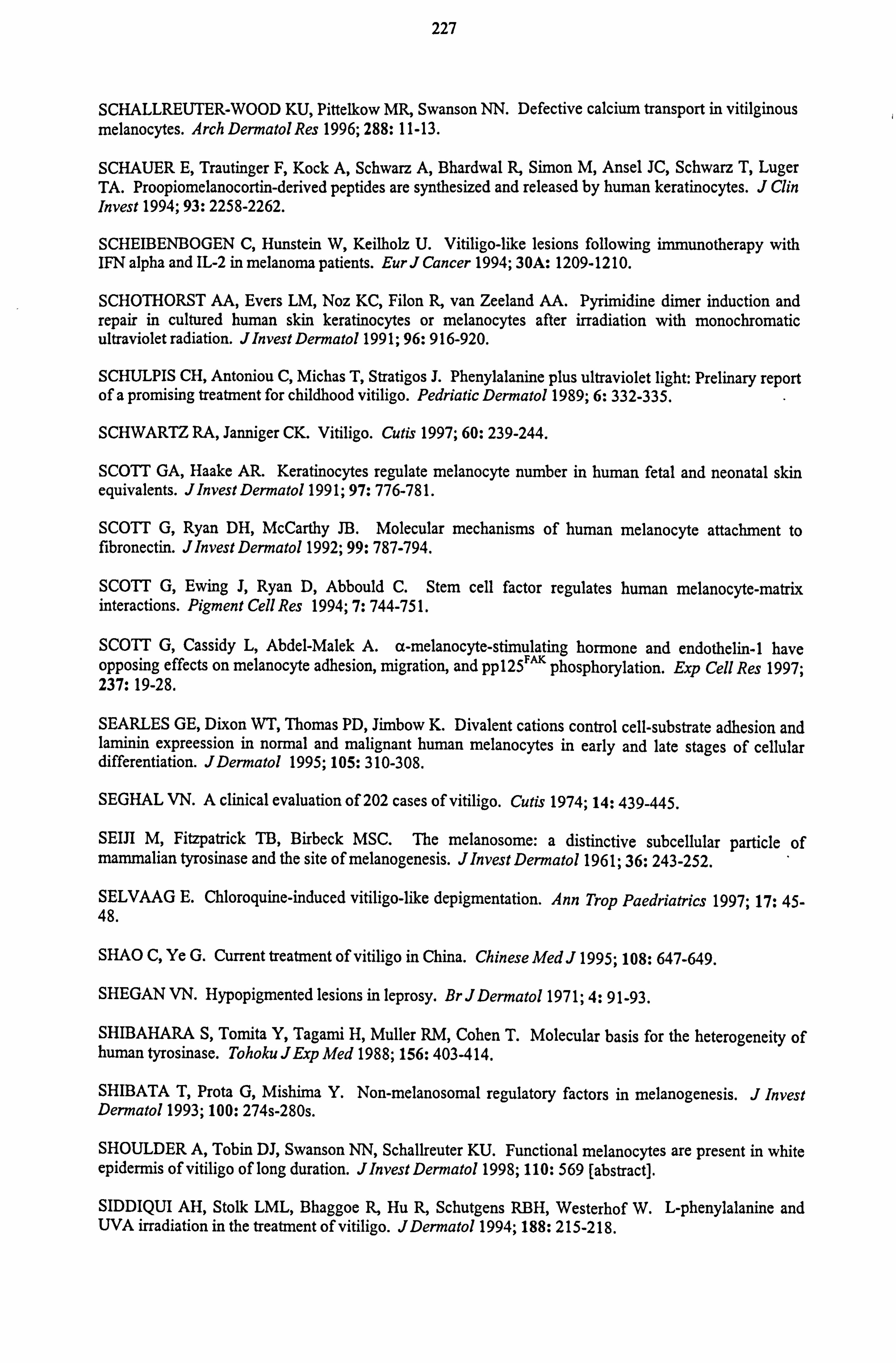
227
SCHALLREUTER-WOOD KU, Pittelkow MR, Swanson NN. Defective calcium transport in vitilginous melanocytes. Arch Dermatol Res 1996; 288: 11-13.
SCHAUER E, Trautinger F, Kock A, Schwarz A, Bhardwal R, Simon M, Ansel JC, Schwarz T, Luger TA. Proopiomelanocortin-derived peptides are synthesized and released by human keratinocytes. J Clin Invest 1994; 93: 2258-2262.
SCHEIBENBOGEN C, Hunstein W, Keilholz U. Vitiligo-like lesions following immunotherapy with IFN alpha and IL-2 in melanoma patients. Eur J Cancer 1994; 30A: 1209-1210.
SCHOTHORST AA, Evers LM, Noz KC, Filon R, van Zeeland AA. Pyrimidine dimer induction and repair in cultured human skin keratinocytes or melanocytes after irradiation with monochromatic ultraviolet radiation. JInvest Dermatol 1991; 96: 916-920.
SCHULPIS CH, Antoniou C, Michas T, Stratigos J. Phenylalanine plus ultraviolet light: Prelinary report of a promising treatment for childhood vitiligo. Pedriatic Dermatol 1989; 6: 332-335.
SCHWARTZ RA, Janniger CK. Vitiligo. Cutis 1997; 60: 239-244.
SCOTT GA, Haake AR. Keratinocytes regulate melanocyte number in human fetal and neonatal skin equivalents. JInvest Dermatol 1991; 97: 776-781.
SCOTT G, Ryan DH, McCarthy JB. Molecular mechanisms of human melanocyte attachment to fibronectin. J Invest Dermatol 1992; 99: 787-794.
SCOTT G, Ewing J, Ryan D, Abbould C. Stem cell factor regulates human melanocyte-matrix interactions. Pigment Cell Res 1994; 7: 744-751.
SCOTT G, Cassidy L, Abdel-Malek A. a-melanocyte-stimulating hormone and endothelin-1 have opposing effects on melanocyte adhesion, migration, and pp125F"K phosphorylation. Exp Cell Res 1997; 237: 19-28.
SEARLES GE, Dixon WT, Thomas PD, Jimbow K. Divalent cations control cell-substrate adhesion and laminin expreession in normal and malignant human melanocytes in early and late stages of cellular differentiation. JDermatol 1995; 105: 310-308.
SEGHAL VN. A clinical evaluation of 202 cases of vitiligo. Cutis 1974; 14: 439-445.
SEIR M, Fitzpatrick TB, Birbeck MSC. The melanosome: a distinctive subcellular particle of mammalian tyrosinase and the site of melanogenesis. Jlnvest Dermatol 1961; 36: 243-252.
SELVAAG E. Chloroquine-induced vitiligo-like depigmentation. Ann Trop Paedriatrics 1997; 17: 45- 48.
SHAO C, Ye G. Current treatment of vitiligo in China. Chinese Med J 1995; 108: 647-649.
SHEGAN VN. Hypopigmented lesions in leprosy. Br J Dermatol 1971; 4: 91-93.
SHIBAHARA S, Tonvita Y, Tagami H, Muller RM, Cohen T. Molecular basis for the heterogeneity of human tyrosinase. Tohoku JExp Med 1988; 156: 403-414.
SHIBATA T, Prota G, Mishima Y. Non-melanosomal regulatory factors in melanogenesis. J Invest Dermatol 1993; 100: 274s-280s.
SHOULDER A, Tobin DJ, Swanson NN, Schallreuter KU. Functional melanocytes are present in white epidermis of vitiligo of long duration. Jlnvest Dermatol 1998; 110: 569 [abstract].
SIDDIQUI AH, Stolk LML, Bhaggoe R, Hu R, Schutgens RBH, Westerhof W. L-phenylalanine and UVA irradiation in the treatment of vitiligo. JDermatol 1994; 188: 215-218.
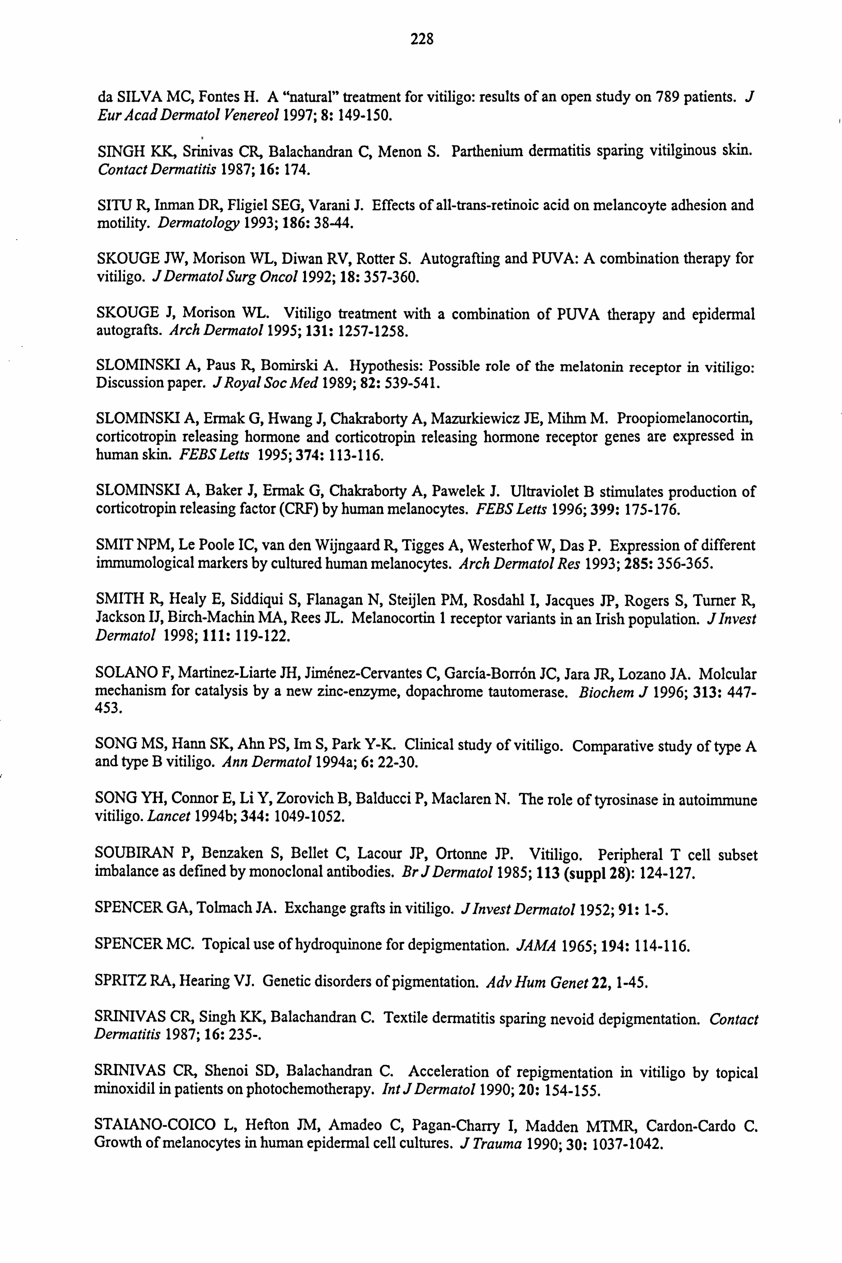
228
da SILVA MC, Fontes H. A "natural" treatment for vitiligo: results of an open study on 789 patients. J EurAcad Dermatol Venereol 1997; 8: 149-150.
SINGH KK, Srinivas CR, Balachandran C, Menon S. Parthenium dermatitis sparing vitilginous skin. Contact Dermatitis 1987; 16: 174.
SITU R, Inman DR, Fligiel SEG, Varani J. Effects of all-trans-retinoic acid on melancoyte adhesion and motility. Dermatology 1993; 186: 38-44.
SKOUGE JW, Morison WL, Diwan RV, Rotter S. Autografting and PUVA: A combination therapy for vitiligo. JDermatol Surg Oncol 1992; 18: 357-360.
SKOUGE J, Morison WL. Vitiligo treatment with a combination of PUVA therapy and epidermal autografts. Arch Dermatol 1995; 131: 1257-1258.
SLOMINSKI A, Paus R, Bomirski A. Hypothesis: Possible role of the melatonin receptor in vitiligo: Discussion paper. JRoyal Soc Med 1989; 82: 539-541.
SLOMINSKI A, Ermak G, Hwang J, Chakraborty A, Mazurkiewicz JE, Mihm M. Proopiomelanocortin, corticotropin releasing hormone and corticotropin releasing hormone receptor genes are expressed in human skin. FEBSLetts 1995; 374: 113-116.
SLOMINSKI A, Baker J, Ermak G, Chakraborty A, Pawelek J. Ultraviolet B stimulates production of corticotropin releasing factor (CRF) by human melanocytes. FEBS Letts 1996; 399: 175-176.
SMIT NPM, Le Poole IC, van den Wijngaard R, Tigges A, Westerhof W, Das P. Expression of different immumological markers by cultured human melanocytes. Arch Dermatol Res 1993; 285: 356-365.
SMITH R, Healy E, Siddiqui S, Flanagan N, Steijlen PM, Rosdahl I, Jacques JP, Rogers S, Turner R, Jackson IJ, Birch-Machin MA, Rees JL. Melanocortin 1 receptor variants in an Irish population. JInvest Dermatol 1998; 111: 119-122.
SOLANO F, Martinez-Liarte JH, Jimenez-Cervantes C, Garcia-Borrön JC, Jara JR, Lozano JA. Molcular mechanism for catalysis by a new zinc-enzyme, dopachrome tautomerase. Biochem J 1996; 313: 447- 453.
SONG MS, Hann SK, Ahn PS, Im S, Park Y-K. Clinical study of vitiligo. Comparative study of type A and type B vitiligo. Ann Dermatol 1994a; 6: 22-30.
SONG YH, Connor E, Li Y, Zorovich B, Balducci P, Maclaren N. The role of tyrosinase in autoinunune vitiligo. Lancet 1994b; 344: 1049-1052.
SOUBIRAN P, Benzaken S, Bellet C, Lacour JP, Ortonne JP. Vitiligo. Peripheral T cell subset imbalance as defined by monoclonal antibodies. BrJDermatol 1985; 113 (suppl 28): 124-127.
SPENCER GA, Tolmach JA. Exchange grafts in vitiligo. JInvest Dermatol 1952; 91: 1-5.
SPENCER MC. Topical use of hydroquinone for depigmentation. JAMA 1965; 194: 114-116.
SPRITZ RA, Hearing VJ. Genetic disorders of pigmentation. Adv Hum Genet 22,1-45.
SRINIVAS CR, Singh KK, Balachandran C. Textile dermatitis sparing nevoid depigmentation. Contact Dermatitis 1987; 16: 235-.
SRINIVAS CR, Shenoi SD, Balachandran C. Acceleration of repigmentation in vitiligo by topical minoxidil in patients on photochemotherapy. Int JDermatol 1990; 20: 154-155.
STAIANO-COICO L, Hefton JM, Amadeo C, Pagan-Charry I, Madden MTMR, Cardon-Cardo C. Growth of melanocytes in human epidermal cell cultures. J Trauma 1990; 30: 1037-1042.
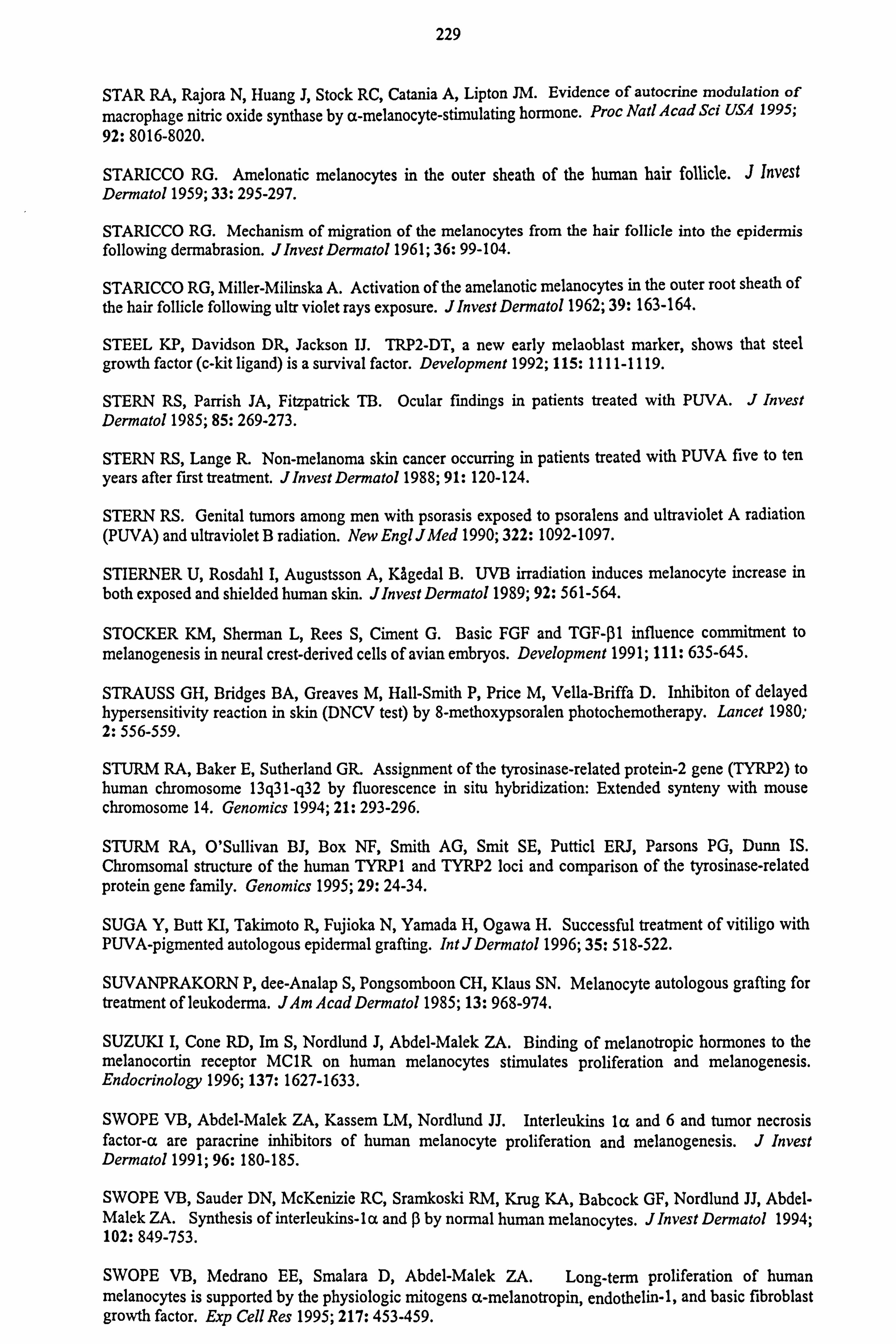
229
STAR RA, Rajora N, Huang J, Stock RC, Catania A, Lipton JM. Evidence of autocrine modulation of macrophage nitric oxide synthase by a-melanocyte-stimulating hormone. Proc Natl Acad Sci USA 1995;
92: 8016-8020.
STARICCO RG. Amelonatic melanocytes in the outer sheath of the human hair follicle. J Invest Dermatol 1959; 33: 295-297.
STARICCO RG. Mechanism of migration of the melanocytes from the hair follicle into the epidermis following dermabrasion. JlnvestDermatol 1961; 36: 99-104.
STARICCO RG, Miller-Milinska A. Activation of the amelanotic melanocytes in the outer root sheath of the hair follicle following ultr violet rays exposure. JInvest Dermatol 1962; 39: 163-164.
STEEL KP, Davidson DR, Jackson IJ. TRP2-DT, a new early melaoblast marker, shows that steel growth factor (c-kit ligand) is a survival factor. Development 1992; 115: 1111-1119.
STERN RS, Parrish JA, Fitzpatrick TB. Ocular fmdings in patients treated with PUVA. J Invest Dermatol 1985; 85: 269-273.
STERN RS, Lange R. Non-melanoma skin cancer occurring in patients treated with PUVA five to ten
years after first treatment. J Invest Dermatol 1988; 91: 120-124.
STERN RS. Genital tumors among men with psorasis exposed to psoralens and ultraviolet A radiation (PUVA) and ultraviolet B radiation. New Engl JMed 1990; 322: 1092-1097.
STIERNER U, Rosdahl I, Augustsson A, KAgedal B. UVB irradiation induces melanocyte increase in both exposed and shielded human skin. Jlnvest Dermatol 1989; 92: 561-564.
STOCKER KM, Sherman L, Rees S, Ciment G. Basic FGF and TGF-01 influence commitment to melanogenesis in neural crest-derived cells of avian embryos. Development 1991; 111: 635-645.
STRAUSS GH, Bridges BA, Greaves M, Hall-Smith P, Price M, Vella-Briffa D. Inhibiton of delayed hypersensitivity reaction in skin (DNCV test) by 8-methoxypsoralen photochemotherapy. Lancet 1980; 2: 556-559.
STURM RA, Baker E, Sutherland GR. Assignment of the tyrosinase-related protein-2 gene (TYRP2) to human chromosome 13g31-q32 by fluorescence in situ hybridization: Extended synteny with mouse chromosome 14. Genomics 1994; 21: 293-296.
STURM RA, O'Sullivan BJ, Box NF, Smith AG, Smit SE, Putticl ERJ, Parsons PG, Dunn IS. Chromsomal structure of the human TYRP 1 and TYRP2 loci and comparison of the tyrosinase-related protein gene family. Genomics 1995; 29: 24-34.
SUGA Y, Butt KI, Takimoto R, Fujioka N, Yamada H, Ogawa H. Successful treatment of vitiligo with PUVA-pigmented autologous epidermal grafting. IntJDermatol 1996; 35: 518-522.
SUVANPRAKORN P, dee-Analap S, Pongsomboon CH, Klaus SN. Melanocyte autologous grafting for treatment of leukoderma. JAm Acad Dermatol 1985; 13: 968-974.
SUZUKI I, Cone RD, Im S, Nordlund J, Abdel-Malek ZA. Binding of melanotropic hormones to the melanocortin receptor MC1R on human melanocytes stimulates proliferation and melanogenesis. Endocrinology 1996; 137: 1627-1633.
SWOPE VB, Abdel-Malek ZA, Kassem LM, Nordlund JJ. Interleukins la and 6 and tumor necrosis factor-a are paracrine inhibitors of human melanocyte proliferation and melanogenesis. J Invest Dermatol 1991; 96: 180-185.
SWOPE VB, Sauder DN, McKenzie RC, Sramkoski RM, Krug KA, Babcock GF, Nordlund JJ, Abdel- Malek ZA. Synthesis of interleukins-la and ß by normal human melanocytes. JInvest Dermatol 1994; 102: 849-753.
SWOPE VB, Medrano EE, Smalara D, Abdel-Malek ZA. Long-term proliferation of human melanocytes is supported by the physiologic mitogens a-melanotropin, endothelin-1, and basic fibroblast growth factor. Exp Cell Res 1995; 217: 453-459.
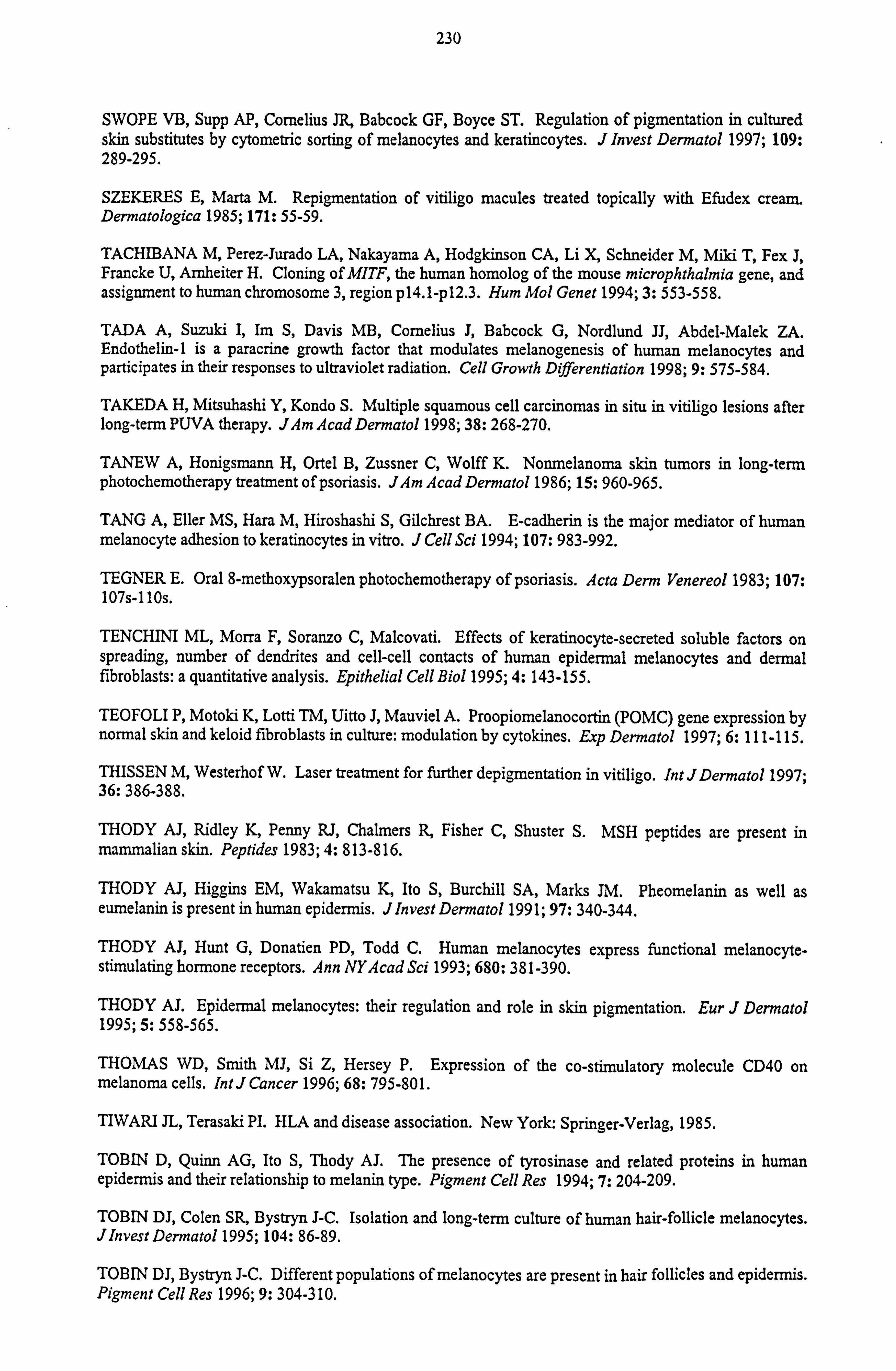
230
SWOPE VB, Supp AP, Cornelius JR, Babcock GF, Boyce ST. Regulation of pigmentation in cultured skin substitutes by cytometric sorting of melanocytes and keratincoytes. J Invest Dermatol 1997; 109: 289-295.
SZEKERES E, Marta M. Repigmentation of vitiligo macules treated topically with Efudex cream. Dermatologica 1985; 171: 55-59.
TACHIBANA M, Perez-Jurado LA, Nakayama A, Hodgkinson CA, Li X, Schneider M, Miki T, Fex J, Francke U, Arnheiter H. Cloning of MITF, the human homolog of the mouse microphthalmia gene, and assignment to human chromosome 3, region p14.1-pl2.3. Hum Mol Genet 1994; 3: 553-558.
TADA A, Suzuki I, Im S, Davis MB, Cornelius J, Babcock G, Nordlund JJ, Abdel-Malek ZA. Endothelin-1 is a paracrine growth factor that modulates melanogenesis of human melanocytes and participates in their responses to ultraviolet radiation. Cell Growth Differentiation 1998; 9: 575-584.
TAKEDA H, Mitsuhashi Y, Kondo S. Multiple squamous cell carcinomas in situ in vitiligo lesions after long-term PUVA therapy. JAm Acad Dermatol 1998; 38: 268-270.
TANEW A, Honigsmann H, Ortel B, Zussner C, Wolff K. Nonmelanoma skin tumors in long-term photochemotherapy treatment of psoriasis. JAm Acad Dermatol 1986; 15: 960-965.
TANG A, Eller MS, Hara M, Hiroshashi S, Gilchrest BA. E-cadherin is the major mediator of human melanocyte adhesion to keratinocytes in vitro. J Cell Sci 1994; 107: 983-992.
TEGNER E. Oral 8-methoxypsoralen photochemotherapy of psoriasis. Acta Derm Venereol 1983; 107: 107s-110s.
TENCHINI ML, Morra F, Soranzo C, Malcovati. Effects of keratinocyte-secreted soluble factors on spreading, number of dendrites and cell-cell contacts of human epidermal melanocytes and dermal fibroblasts: a quantitative analysis. Epithelial Cell Biol 1995; 4: 143-155.
TEOFOLI P, Motoki K, Lotti TM, Uitto J, Mauviel A. Proopiomelanocortin (POMC) gene expression by normal skin and keloid fibroblasts in culture: modulation by cytokines. Exp Dermatol 1997; 6: 111-115.
THISSEN M, Westerhof W. Laser treatment for further depigmentation in vitiligo. IntJDermatol 1997; 36: 386-388.
THODY AJ, Ridley K, Penny RJ, Chalmers R, Fisher C, Shuster S. MSH peptides are present in mammalian skin. Peptides 1983; 4: 813-816.
THODY AJ, Higgins EM, Wakamatsu K, Ito S, Burchill SA, Marks JM. Pheomelanin as well as eumelanin is present in human epidermis. JInvest Dermatol 1991; 97: 340-344.
THODY AJ, Hunt G, Donatien PD, Todd C. Human melanocytes express functional melanocyte- stimulating hormone receptors. Ann NYAcad Sci 1993; 680: 381-390.
THODY AJ. Epidermal melanocytes: their regulation and role in skin pigmentation. Eur J Dermatol 1995; 5: 558-565.
THOMAS WD, Smith MJ, Si Z, Hersey P. Expression of the co-stimulatory molecule CD40 on melanoma cells. Int J Cancer 1996; 68: 795-801.
TIWARI JL, Terasaki PI. HLA and disease association. New York: Springer-Verlag, 1985.
TOBIN D, Quinn AG, Ito S, Thody AJ. The presence of tyrosinase and related proteins in human epidermis and their relationship to melanin type. Pigment Cell Res 1994; 7: 204-209.
TOBIN DJ, Colen SR, Bystryn J-C. Isolation and long-term culture of human hair-follicle melanocytes. J Invest Dermato11995; 104: 86-89.
TOBIN DJ, Bystryn J-C. Different populations of melanocytes are present in hair follicles and epidermis. Pigment Cell Res 1996; 9: 304-310.
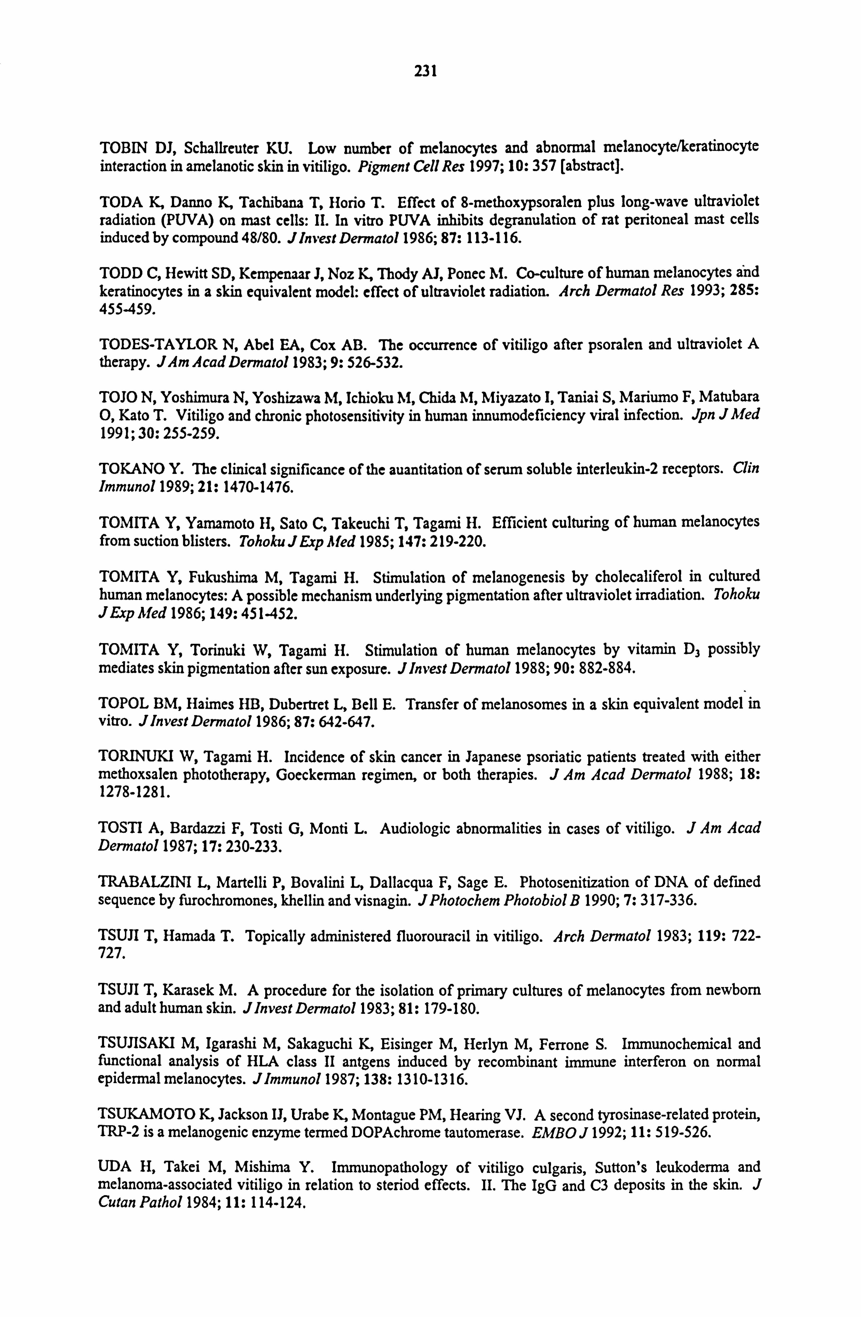
231
TOBIN DJ, Schallreuter KU. Low number of melanocytes and abnormal melanocyte/keratinocyte interaction in amelanotic skin in vitiligo. Pigment Cell Res 1997; 10: 357 [abstract].
TODA K, Danno K, Tachibana T, Horio T. Effect of 8-methoxypsoralen plus long-wave ultraviolet radiation (PUVA) on mast cells: H. In vitro PUVA inhibits degranulation of rat peritoneal mast cells induced by compound 48/80. JInvest Dermatol 1986; 87: 113-116.
TODD C, Hewitt SD, Kempenaar J, Noz K, Thody AJ, Ponec M. Co-culture of human melanocytes and keratinocytes in a skin equivalent model: effect of ultraviolet radiation. Arch Dermatol Res 1993; 285: 455-459.
TODES-TAYLOR N, Abel EA, Cox AB. The occurrence of vitiligo after psoralen and ultraviolet A therapy. JAm Acad Dermatol 1983; 9: 526-532.
TOJO N, Yoshimura N, Yoshizawa M, Ichioku M, Chida M, Miyazato I, Taniai S, Mariumo F, Matubara 0, Kato T. Vitiligo and chronic photosensitivity in human innumodeficiency viral infection. Jpn JMed 1991; 30: 255-259.
TOKANO Y. The clinical significance of the auantitation of serum soluble interleukin-2 receptors. Clin Immunol 1989; 21: 1470-1476.
TOMITA Y, Yamamoto H, Sato C, Takeuchi T, Tagami H. Efficient culturing of human melanocytes from suction blisters. Tohoku J Exp Med 1985; 147: 219-220.
TOMITA Y, Fukushima M, Tagami H. Stimulation of melanogenesis by cholecaliferol in cultured human melanocytes: A possible mechanism underlying pigmentation after ultraviolet irradiation. Tohoku JExp Med 1986; 149: 451-452.
TOMITA Y, Torinuki W, Tagami H. Stimulation of human melanocytes by vitamin D3 possibly mediates skin pigmentation after sun exposure. JInvest Dermatol 1988; 90: 882-884.
TOPOL BM, Haimes HB, Dubertret L, Bell E. Transfer of melanosomes in a skin equivalent model in vitro. Jhnvest Dermatol 1986; 87: 642-647.
TORINUKI W, Tagami H. Incidence of skin cancer in Japanese psoriatic patients treated with either methoxsalen phototherapy, Goeckerman regimen, or both therapies. J , 4m Acad Dermatol 1988; 18: 1278-1281.
TOSTI A, Bardazzi F, Tosti G, Monti L. Audiologic abnormalities in cases of vitiligo. J , 4m Acad Dermatol 1987; 17: 230-233.
TRABALZINI L, Martelli P, Bovalini L, Dallacqua F, Sage E. Photosenitization of DNA of defined sequence by furochromones, khellin and visnagin. JPhotochem Photobiol B 1990; 7: 317-336.
TSUJI T, Hamada T. Topically administered fluorouracil in vitiligo. Arch Dermatol 1983; 119: 722- 727.
TSUJI T, Karasek M. A procedure for the isolation of primary cultures of melanocytes from newborn and adult human skin. Jlnvest Dermatol 1983; 81: 179-180.
TSUJISAKI M, Igarashi M, Sakaguchi K, Eisinger M, Herlyn M, Ferrone S. Immunochemical and functional analysis of HLA class II antgens induced by recombinant immune interferon on normal epidermal melanocytes. Jlmmunol 1987; 138: 1310-1316.
TSUKAMOTO K, Jackson IJ, Urabe K, Montague PM, Hearing VJ. A second tyrosinase-related protein, TRP-2 is a melanogenic enzyme termed DOPAchrome tautomerase. EMBO J 1992; 11: 519-526.
UDA H, Takei M, Mishima Y. Immunopathology of vitiligo culgaris, Sutton's leukoderma and melanoma-associated vitiligo in relation to steriod effects. II. The IgG and C3 deposits in the skin. J Cutan Pathol 1984; 11: 114-124.
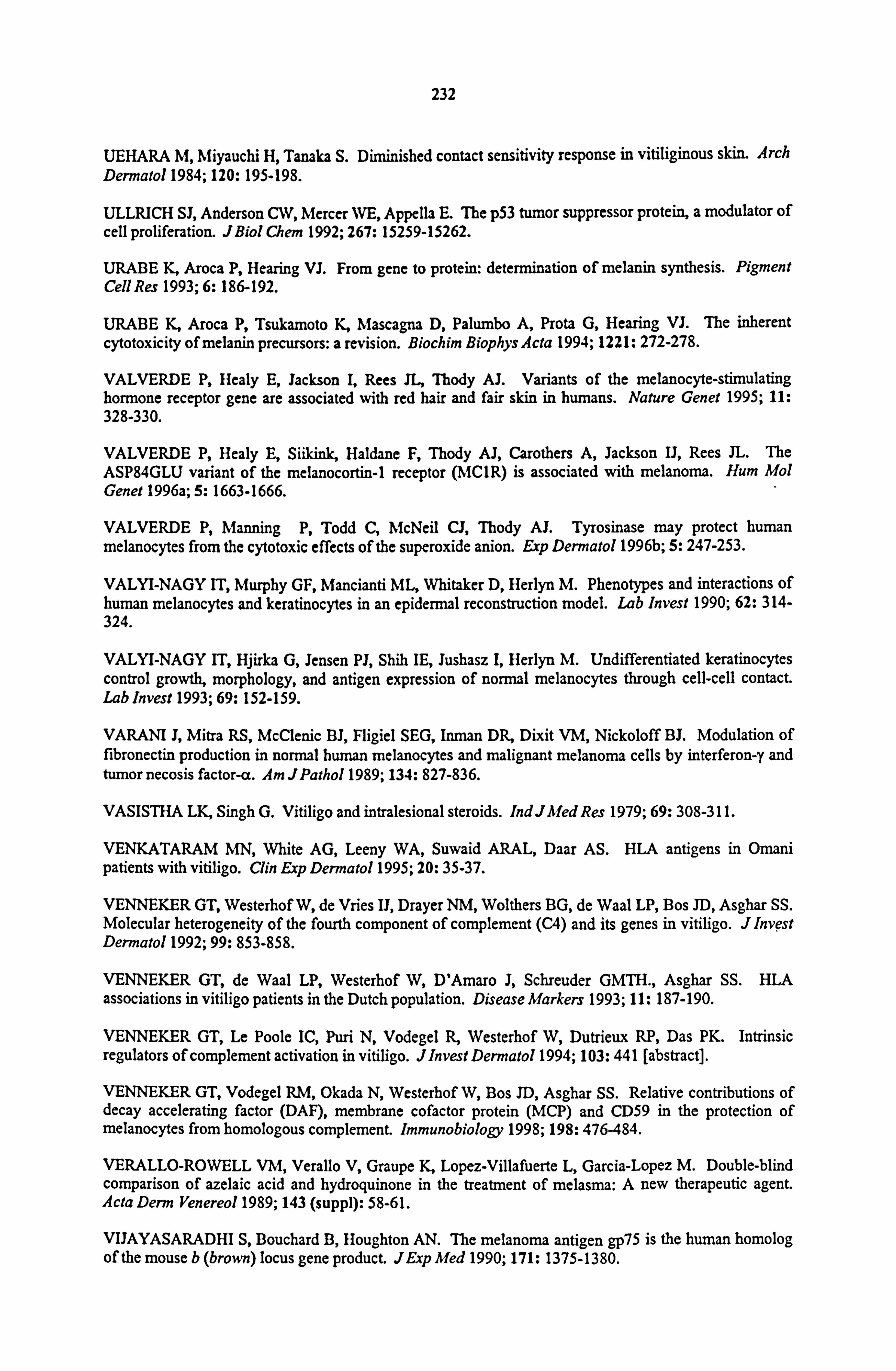
232
UEHARA M, Miyauchi H, Tanaka S. Diminished contact sensitivity response in vitiliginous skin. Arch Dermatol 1984; 120: 195-198.
ULLRICH SJ, Anderson CW, Mercer WE, Appella E. The p53 tumor suppressor protein, a modulator of cell proliferation. JBiol Chem 1992; 267: 15259-15262.
URABE K, Aroca P, Hearing VJ. From gene to protein: determination of melanin synthesis. Pigment Cell Res 1993; 6: 186-192.
URABE K, Aroca P, Tsukamoto K, Mascagna D, Palumbo A, Prota G, Hearing VJ. The inherent
cytotoxicity of melanin precursors: a revision. Biochim Biophys Acta 1994; 1221: 272-278.
VALVERDE P, Healy E, Jackson I, Rees JL, Thody AJ. Variants of the melanocyte-stimulating hormone receptor gene are associated with red hair and fair skin in humans. Nature Genet 1995; 11: 328-330.
VALVERDE P, Healy E, Siikink, Haldane F, Thody AJ, Carothers A, Jackson IJ, Rees JL. The ASP84GLU variant of the melanocortin-1 receptor (MC1R) is associated with melanoma. Hum Mol Genet 1996a; 5: 1663-1666.
VALVERDE P, Manning P, Todd C, McNeil CJ, Thody AJ. Tyrosinase may protect human melanocytes from the cytotoxic effects of the superoxide anion. Exp Dermatol 1996b; 5: 247-253.
VALYI-NAGY IT, Murphy GF, Mancianti ML, Whitaker D, Herlyn M. Phenotypes and interactions of human melanocytes and keratinocytes in an epidermal reconstruction model. Lab Invest 1990; 62: 314- 324.
VALYI-NAGY IT, Hjirka G, Jensen PJ, Shih IE, Jushasz I, Herlyn M. Undifferentiated keratinocytes control growth, morphology, and antigen expression of normal melanocytes through cell-cell contact. Lab Invest 1993; 69: 152-159.
VARANI J, Mitra RS, McClenic BJ, Fligiel SEG, Inman DR, Dixit VM, Nickoloff BJ. Modulation of fibronectin production in normal human melanocytes and malignant melanoma cells by interferon-y and tumor necosis factor-a. Am JPatho11989; 134: 827-836.
VASISTHA LK, Singh G. Vitiligo and intralesional steroids. Ind JMed Res 1979; 69: 308-311.
VENKATARAM MN, White AG, Leeny WA, Suwaid ARAL, Daar AS. HLA antigens in Omani patients with vitiligo. Clin Exp Dermato11995; 20: 35-37.
VENNEKER GT, Westerhof W, de Vries IJ, Drayer NM, Wolthers BG, de Waal LP, Bos JD, Asghar SS. Molecular heterogeneity of the fourth component of complement (C4) and its genes in vitiligo. J Invest Dermato11992; 99: 853-858.
VENNEKER GT, de Waal LP, Westerhof W, D'Amaro J, Schreuder GMTH., Asghar SS. HLA associations in vitiligo patients in the Dutch population. Disease Markers 1993; 11: 187-190.
VENNEKER GT, Le Poole IC, Puri N, Vodegel R, Westerhof W, Dutrieux RP, Das PK. Intrinsic regulators of complement activation in vitiligo. JInvest Dermato11994; 103: 441 [abstract].
VENNEKER GT, Vodegel RM, Okada N, Westerhof W, Bos JD, Asghar SS. Relative contributions of decay accelerating factor (DAF), membrane cofactor protein (MCP) and CD59 in the protection of melanocytes from homologous complement. Immunobiology 1998; 198: 476-484.
VERALLO-ROWELL VM, Verallo V, Graupe K, Lopez-Villafuerte L, Garcia-Lopez M. Double-blind comparison of azelaic acid and hydroquinone in the treatment of melasma: A new therapeutic agent. Acta Denn Venereo11989; 143 (suppl): 58-61.
VIJAYASARADHI S, Bouchard B, Houghton AN. The melanoma antigen gp75 is the human homolog of the mouse b (brown) locus gene product. JExp Med 1990; 171: 1375-1380.
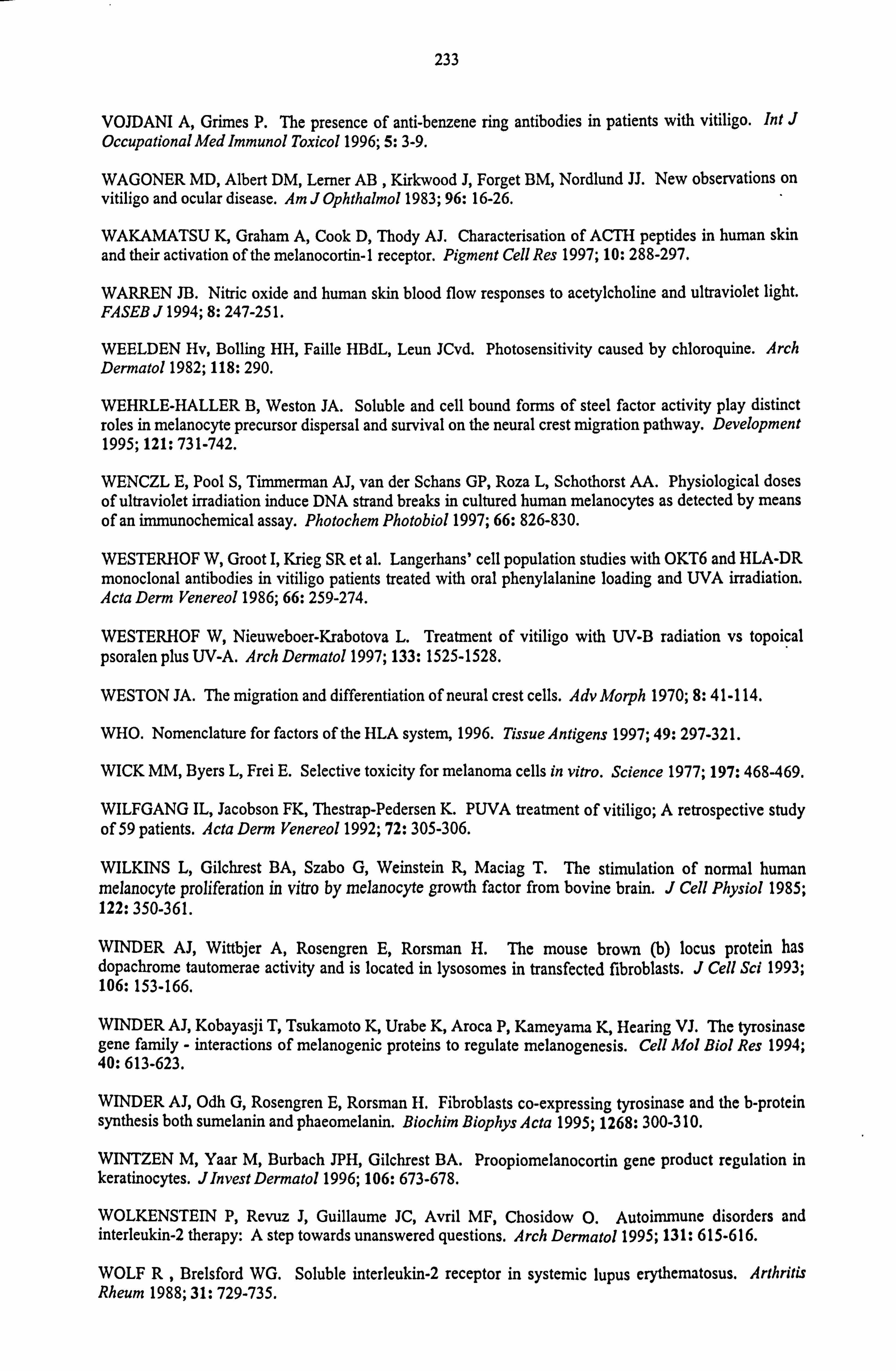
233
VOJDANI A, Grimes P. The presence of anti-benzene ring antibodies in patients with vitiligo. Int J Occupational Med Immunol Toxicol 1996; 5: 3-9.
WAGONER MD, Albert DM, Lerner AB , Kirkwood J, Forget BM, Nordlund JJ. New observations on
vitiligo and ocular disease. Am J Ophthalmol 1983; 96: 16-26.
WAKAMATSU K, Graham A, Cook D, Thody AJ. Characterisation of ACTH peptides in human skin and their activation of the melanocortin-1 receptor. Pigment Cell Res 1997; 10: 288-297.
WARREN JB. Nitric oxide and human skin blood flow responses to acetylcholine and ultraviolet light. FASEB J 1994; 8: 247-25 1.
WEELDEN Hv, Bolling HH, Faille HBdL, Leun JCvd. Photosensitivity caused by chloroquine. Arch Dermatol 1982; 118: 290.
WEHRLE-HALLER B, Weston JA. Soluble and cell bound forms of steel factor activity play distinct roles in melanocyte precursor dispersal and survival on the neural crest migration pathway. Development 1995; 121: 731-742.
WENCZL E, Pool S, Timmerman AJ, van der Schans GP, Roza L, Schothorst AA. Physiological doses of ultraviolet irradiation induce DNA strand breaks in cultured human melanocytes as detected by means of an immunochemical assay. Photochem Photobiol 1997; 66: 826-830.
WESTERHOF W, Groot I, Krieg SR et al. Langerhans' cell population studies with OKT6 and HLA-DR monoclonal antibodies in vitiligo patients treated with oral phenylalanine loading and UVA irradiation. Acta Derm Venereol 1986; 66: 259-274.
WESTERHOF W, Nieuweboer-Krabotova L. Treatment of vitiligo with UV-B radiation vs topoical psoralen plus UV-A. Arch Dermatol 1997; 133: 1525-1528.
WESTON JA. The migration and differentiation of neural crest cells. Adv Morph 1970; 8: 41-114.
WHO. Nomenclature for factors of the HLA system, 1996. Tissue Antigens 1997; 49: 297-321.
WICK MM, Byers L, Frei E. Selective toxicity for melanoma cells in vitro. Science 1977; 197: 468-469.
WILFGANG IL, Jacobson FK, Thestrap-Pedersen K. PUVA treatment of vitiligo; A retrospective study of 59 patients. Acta Derm Venereol 1992; 72: 305-306.
WILKINS L, Gilchrest BA, Szabo G, Weinstein R, Maciag T. The stimulation of normal human melanocyte proliferation in vitro by melanocyte growth factor from bovine brain. J Cell Physiol 1985; 122: 350-361.
WINDER AJ, Wittbjer A, Rosengren E, Rorsman H. The mouse brown (b) locus protein has dopachrome tautomerae activity and is located in lysosomes in transfected fibroblasts. J Cell Sci 1993; 106: 153-166.
WINDER AJ, Kobayasji T, Tsukamoto K, Urabe K, Aroca P, Kameyama K, Hearing VJ. The tyrosinase gene family - interactions of melanogenic proteins to regulate melanogenesis. Cell Mol Biol Res 1994; 40: 613-623.
WINDER AJ, Odh G, Rosengren E, Rorsman H. Fibroblasts co-expressing tyrosinase and the b-protein synthesis both sumelanin and phaeomelanin. Biochim Biophys Acta 1995; 1268: 300-3 10.
WINTZEN M, Yaar M, Burbach JPH, Gilchrest BA. Proopiomelanocortin gene product regulation in keratinocytes. Jlnvest Dermatol 1996; 106: 673-678.
WOLKENSTEIN P, Revuz J, Guillaume JC, Avril MF, Chosidow O. Autoimmune disorders and interleukin-2 therapy: A step towards unanswered questions. Arch Dermato11995; 131: 615-616.
WOLF R, Brelsford WG. Soluble interleukin-2 receptor in systemic lupus erythematosus. Arthritis Rheum 1988; 31: 729-735.
234
WOLFF K, Jimbow K, Fitzpatrick TB. Experimental pigment donation in vito. J Ultrastruct Res 1974; 47: 400-419.
WOOLFSON H, Finn OA, Mackie RM, McQueen A, MacSween RNM. Serum and anti-tumor antibodies and autoantibodies in vitiligo. BrJDermatol 1975; 92: 395-400.
XIA P, Geoghegan WD, Jordan RE. Vitiligo autoantibodies: Studies of subclass distribution and complement activation. Jlnvest Dermatol 1991; 96: 627 [abstract].
YAAR M, Grossman K, Eller M, Gilchrest BA. Evidence for nerve growth factor-mediated paracrine effects inhuman epidermis. J Cell Biol 1991; 115: 821-828.
YAAR M, Eller MS, DiBenedetto P, Reenstra WR, Zhai S, McQuaid T, Archambault M, Gilchrest BA. The trk family of receptors mediates nerve growth factor ans neurotrophin-3 effects in melanocytes. *J Clin Invest 1994; 94: 1550-1562.
YADA Y, Higuchi K, Imokawa G. Effects of endothelins on signal transduction and prolifeaton in human melanocytes. JBiol Chem 1991; 266: 18352-18357.
YAGI H, Tokura Y, Furukawa F, Takigawa M. Vitiligo with raised inflammatory borders: involvement of T cell immunity and keratinocytes expressing MHC class II and ICAM-1 molecules. Eur J Dermatol 1997; 7: 19-22.
YANG JS, Kye YC. Treatment of vitiligo with autologous epidermal grafting by means of pulse erbium: YAG laser. JAm Acad Dermatol 1998; 38: 280-282.
YASUMOTO KI, Yokoyama K, Takahashi K, Tomita Y, Shibahara S. Functional analysis of microphthalmia-associated transcription factor in pigment cell-specific transcription of the human tyrosinase family genes. JBiol Chem 1997; 272: 503-509.
YAVUZER U, Keenan E, Lowings P, Vachtenhein J, Currie G, Goding CR. The microphthalmia gene product interacts with the retinoblastoma protein in vitro and is a target for deregulation of melanocyte- specific transcription. Oncogene 1995; 10: 123-134.
YOHN JJ, Critelli M, Lyons MB, Norris DA. Modulation of melanocyte intercellular adhesion molecule- 1 by immune cytokines. JInvest Dermatol 1990; 93: 233-237.
YOHN JJ, Norris DA, Yrastorza DG, Buno IJ, Leff JA, Hake SS, Repine JE. Disparate antioxidant enzyme activities in cultured human cutaneous fibroblasts, keratinocytes and melanocytes. J Invest Dermato11991; 97: 405-409.
YOHN JJ, Lyons MB, Norris DA. Cultured human melanocytes from black and white donors have different sunlight and ultraviolet A radiation sensitivities. Jlnvest Dermatol 1992; 99: 454-459.
YOHN JJ, Morelli JG, Walchak SJ, Rundell KB, Norris DA, Zamora MR. Cultured human keratinocytes synthesize and secrete endothelin-1. Jlnvest Dermatol 1993; 100: 23-26.
YOKOYAMA K, Suzuki H, Tonvita Y, Shibahara S. Molecular cloning of and functional analysis of a cDNA coding for human DOPAchrome tautomerase/tyrosinase-related protein-2. Biochim Biophys Acta 1994; 1217: 317-321.
YOUNG M, Oger J, Blanchard MH, Asdourian H, Amos H, Amason BG. Secretion of a nerve growth factor by primary chick fibroblast cultures. Science 1975; 187: 361-362.
YU HS, Kao CH, Yu CL. Coexistence and relationship of antikeratinocyte and antimelanocyte antibodies in patients with non-segmental-type vitiligo. J Invest Dermatol 1993; 100: 823-828.
YU H-S, Chang K-L, Yu C-L, Li H-F, Wu M-T, Wu C-S, Wu C-S. Alterations in IL-6, IL-8, GM-SCF, TNF-a, and IFN-y relaease by peripheral mononuclear cells in patients with active vitiligo. J Invest Dermato11997; 108: 527-529.
235
ZACHARIAE H, Zachariae C, Deleuran B, Kristen P. Autotransplantation in vitiligo: Treatment with epidermal grafts and cultured melanocytes. Acta Derm Venereol 1993; 73: 46-48.
ZAK FG, Lawson W. The presence of melanocytes in the nasal cavity. Ann Otol Rhinol Larynogol 1974; 83: 515-519.
ZAMAN TU, Begum S, Waheed MA. In vitro assessment of `T' lymphocyte functioning in vitiligo. Acta Derm Venereol 1992; 72: 266-267.
ZAMBRUNO G, Marchisto PC, Melcjiori A, Bondanza S, Cancedda R, De Luca M. Expression of integrin receptors and their role in adhesion, spreading and migration of normal human melanocytes. J Cell Sci 1993; 105: 179-190.
ZAULI D, Tosti A, Biasco G, Miserocchi F, Patrizi A, Azzaroni D, Andriani G, Di Febo G, Callegari C. Prevalence of autoimmune atrophic gastritis in vitiligo. Digestion 1986; 34: 169-172.
ZAYNOUM S, Konrad K, Gschnait F, Wolff D. The pigmentary response to photochemotherapy. Acta Derm Venereol 1977; 57: 431-440.
ZELICKSON A, Mottaz JH. Epidermal dendritic cells: A quantiative study. Arch Dermatol 1968; 98: 652-659.
ZELICKSON AS, Mottaz JH, Muller SA. Melanocytes changes following PUVA therapy. J Am Acad Dermatol 1979; 1: 422-430.
ZHAI S, Yaar M, Doyle SM, Gilchrest BA. Nerve growth factor rescues pigment cells from ultraviolet- induced apoptosis by upregulating BCL-2 levels. Exp Cell Res 1996; 244: 335-343.
ZHAO H, Boissy Y, Nordlund JJ, Boissy RE. Melanocytes cultured from a patient with tyrosinase- positive albinism lack TRP-1. Mol Cell Biol 1992; 3: 85.
ZHAO H, Boissy YL, Abdel-Malek Z, King RA, Nordlund JJ, Boissy RE. On the analysis of the pathophysiology of Chediak-Higashi Syndrome. Lab Invest 1994a; 71: 25-34.
ZHAO H, Zhao Y, Nordlund JJ, Boissy RE. TRP-1 has tyrosine hydroxylase but no DOPA oxidase activity. Pigment Cell Res 1994b; 7: 131-140.
ZHOU BK, Boissy RE, Pifko-Hirst S, Moran DJ, Orlow SJ. Lysosome associated membrane protein-1 (LAMP-1) is the melanocyte vesicular membrane glycoprotein Band H. J Invest Dermatol 1993; 100: 110-114.
ZVULUNOV A, Esterly NB. Neurocutaneous syndromes associated with pigmentary skin lesions. JAm Acad Dermatol 1995; 32: 915-935.
Top Related
Copyright © 2022 FDOKUMEN

