


Bahasa
Halaman
Hukum


spine clinical article
J neurosurg Spine March 20, 2015
abbreviationS ASD = adult spinal deformity; CL = cervical lordosis; LIV = lower-most instrumented vertebra; LL =lumbar lordosis; PI = pelvic incidence; PSO = pedicle subtraction osteotomy; PT = pelvic tilt; SPI = spinopelvic inclination; SS = sacral slope; SVA = sagittal vertical axis; T1S = T-1 slope; TK = thoracic kyphosis; UIV = upper-most instrumented vertebra; VCR = vertebral column resection; 3CO = 3-column osteotomy.Submitted August 12, 2014. accepted October 21, 2014.include when citing Published online March 20, 2015; DOI: 10.3171/2014.10.SPINE14829.diScloSure The International Spine Study Group Foundation, through which this study was conducted, is funded through research grants from DePuy Spine and indi-vidual donations. Dr. Klineberg has received speaker’s fees from DePuy Synthes and AOSpine and has received clinical or research support from OREF, DePuy Synthes, and AOSpine for the study described. Dr. Bess is a consultant for K2 Medical and Allosource; has received clinical or research support from DePuy Synthes Spine for the study described; and has received support from Medtronic for non–study-related clinical or research effort. Dr. Ames is a consultant for DePuy, Stryker, and Medtronic; has direct stock ownership in Doctors Research Group and Baxano Surgical; holds a patent with Fish & Richardson, P.C.; and has received royalties from Aesculap and Biomet Spine. Dr. Lafage has direct stock ownership in and is an employee of Nemaris Inc.; has been a speaker for MSD, K2 Medical, DePuy, and NuVasive; and has received support from DePuy, SRS, ISSG, and NIH for non–study-related clinical or research effort. Dr. Schwab has direct stock ownership in Nemaris Inc.; holds a patent with MSD, K2 Medical, NuVasive, and Nemaris Inc.; is a consultant for MSD, DePuy, K2 Medical, and Medicrea; has been a speaker for MSD, Nemaris Inc., and K2 Medical; and has received support from DePuy, MSD, and AO for non–study-related clinical or research effort. Dr. Smith is a consultant for Biomet, NuVasive, Cerapedics, DePuy, and Medtronic; has received clinical or research support from DePuy/ISSG for the study described; has received support from DePuy/ISSG for non–study-related clinical or research effort; and has received royalties from Biomet. Dr. Gupta has direct stock ownership in Johnson & Johnson, Proctor & Gamble, Pfizer, and Pioneer; has received royalties from DePuy; and is a consultant for Medtronic, Medicrea, DePuy Synthes, and Orthofix. Dr. Deviren is a consultant for NuVasive, Guidepoint, and Stryker. Dr. Shaffrey is a consultant for Biomet, Globus, Medtronic, NuVasive, and Stryker; has direct stock ownership in NuVasive; and holds patents with and receives royalties from Biomet, Medtronic, and NuVasive. Dr. Eastlack is a consultant for NuVasive, Alphatec, Don Joy, DePuy/Synthes, Aesculap, Stryker, Invuity, Carevature, DiFusion, and K2 Medical; holds patents with Invuity, NuTech, and Globus; has direct stock ownership in NuVasive and Alphatec; has received clinical or research support from NuVasive and Pioneer/RTI for the study described; and is part of the speaker’s bureau for Eli Lilly. Dr. Hostin is a consultant for DePuy Spine and has received clinical or research support from NuVasive, Seeger, DJO, DePuy Spine, and K2 Medical for the study described. Dr. Protopsaltis is a consultant for Medicrea, Biomet Spine, and DePuy Synthes Spine and has received support from Zimmer Spine for non–study-related clinical or research effort.
Cervical compensatory alignment changes following correction of adult thoracic deformity: a multicenter experience in 57 patients with a 2-year follow-uptaemin oh, ba,1 Justin K. Scheer, bS,1 robert eastlack, md,2 Justin S. Smith, md, phd,3 virginie lafage, phd,4 themistocles S. protopsaltis, md,4 eric Klineberg, md,5 peter g. passias, md,4 vedat deviren, md,6 richard hostin, md,7 munish gupta, md,5 Shay bess, md,8 Frank Schwab, md,4 christopher i. Shaffrey, md,3 christopher p. ames, md,9 and the international Spine Study group1Department of Neurological Surgery, Northwestern University, Feinberg School of Medicine, Chicago, Illinois; 2Scripps Clinic, San Diego; 5Department of Orthopaedic Surgery, University of California, Davis, Sacramento; Departments of 6Orthopaedic Surgery and 9Neurological Surgery, University of California, San Francisco, California; 3Department of Neurosurgery, University of Virginia Health System, Charlottesville, Virginia; 4Department of Orthopaedic Surgery, NYU Hospital for Joint Diseases, New York, New York; 7Department of Orthopaedic Surgery, Baylor Scoliosis Center, Plano, Texas; and 8Rocky Mountain Hospital for Children, Denver, Colorado
obJect Alignment changes in the cervical spine that occur following surgical correction for thoracic deformity remain poorly understood. The purpose of this study was to evaluate such changes in a cohort of adults with thoracic deformity treated surgically.methodS The authors conducted a multicenter retrospective analysis of consecutive patients with thoracic deformity. Inclusion criteria for this study were as follows: corrective osteotomy for thoracic deformity, upper-most instrumented ver-tebra (UIV) between T-1 and T-4, lower-most instrumented vertebra (LIV) at or above L-5 (LIV ≥ L-5) or at the ilium (LIV-ilium), and a minimum radiographic follow-up of 2 years. Sagittal radiographic parameters were assessed preoperatively as well as at 3 months and 2 years postoperatively, including the C-7 sagittal vertical axis (SVA), C2–7 cervical lordosis (CL), C2–7 SVA, T-1 slope (T1S), T1S minus CL (T1S-CL), T2–12 thoracic kyphosis (TK), apical TK, lumbar lordosis (LL), pelvic incidence (PI), PI-LL, pelvic tilt (PT), and sacral slope (SS).reSultS Fifty-seven patients with a mean age of 49.1 ± 14.6 years met the study inclusion criteria. The preoperative prevalence of increased CL (CL > 15°) was 48.9%. Both 3-month and 2-year apical TK improved from baseline (p <
1©AANS, 2015

t. oh et al.
J neurosurg Spine March 20, 20152
Adult spinal deformity (ASD) is a pathological condition defined as spinal malalignment in the axial, coronal, or sagittal plane and is derivative
of congenital, iatrogenic, degenerative, or idiopathic eti-ology.30 The restoration of sagittal alignment, as estab-lished by the sagittal vertical axis (SVA; target < 5 cm) and pelvic tilt (PT; target < 20°) on sagittal radiography, is important in surgical deformity correction.6,21,22 Although coronal plane correction also has clinical relevance, sag-ittal corrections appear to have greater importance,5,6 principally because sagittal radiographic parameters have consistently been associated with physical disability, emotional disability, and diminished health-related qual-ity of life.2,5,6,10,13,20,23,26
Sagittal cervical malalignment merits proper consider-ation since it has been associated with significantly worse patient outcomes. In a recent cohort study of 113 patients, Tang et al. demonstrated that cervical malalignment cor-relates with physical disability.26 In another study by Vil-lavicencio et al., less disability was seen among the patients demonstrating postoperative improvements in cervical sag-ittal alignment.27 In practice, achieving optimal correction of cervical malalignment can be challenging. Patients with ASD can present with concomitant deformities given that compensatory changes within adjacent areas (for example, pelvic retroversion, increased cervical lordosis [CL]) can occur as the patients attempt to maintain upright align-ment, normal gait, and horizontal gaze.10,11,19 Additionally, one potential consequence of surgical instrumentation is that unfused regions of adjacent spine can demonstrate dy-namic changes postoperatively, a phenomenon referred to as “reciprocal change.”
Reciprocal changes in neighboring unfused segments have been described following instrumentation and fusion, and these relationships carry compelling implications for patient management. At times, reciprocal changes have been shown to improve patient outcomes, as evidenced by spontaneous correction of increased CL following lumbar pedicle subtraction osteotomy (PSO).25 Occasionally, how-ever, the converse has been observed. In a study conducted by Lafage et al., for example, 34 patients underwent lum-bar PSO, but 18 patients had reciprocal increases in tho-racic kyphosis (TK) that led to poor sagittal alignment.9
Improving the understanding of such reciprocal rela-tionships will likely form a critical component in the future
management of patients with ASD. However, studies thus far have focused disproportionately on reciprocal changes following lumbar deformity correction, with fewer reports of reciprocal changes following thoracic deformity cor-rection.8,17 More specifically, cervical changes following thoracic deformity surgery have yet to be fully character-ized, and this knowledge gap provided the impetus for the current study, in which we endeavored to evaluate changes in spinal alignment following the correction of thoracic deformity. We hypothesized that thoracic deformity cor-rection would lead to significant radiographic improve-ment in cervical and spinopelvic alignment.
methodspatient population
We conducted a retrospective radiographic review of a large, multiinstitutional database of consecutively treated ASD patients, which had been collected with institutional review board approval at each institution. Inclusion crite-ria for this primary database were 1) an age ≥ 18 years and 2) a diagnosis of spinal deformity (scoliosis Cobb angle ≥ 20°, SVA ≥ 5 cm, PT ≥ 25°, or TK ≥ 60°). Neuromuscu-lar spinal deformities and patients with active infections or malignancy were excluded. For the present study, ad-ditional inclusion criteria were as follows: 1) at least 1 corrective osteotomy for a primary thoracic deformity, 2) upper-most instrumented vertebra (UIV) between T-1 and T-4, 3) lower-most instrumented vertebra (LIV) at L-5 or above (LIV ≥ L-5) or at the ilium (LIV-ilium), and 4) a minimum radiographic follow-up of 2 years. Patients who had osteotomies extending into the thoracic spine from the correction of a primary lumbar deformity and those who had undergone lumbar 3-column osteotomies were excluded.
data collection and radiographic assessmentData pertaining to patient demographics, surgical oste-
otomy type, and radiographic parameters were collected. The surgical osteotomies included Smith-Petersen and 3-column osteotomies (3COs), the latter involving either PSO or vertebral column resection (VCR).
Full-length, free-standing, 36ʺ cassette, lateral spine radiographs were obtained preoperatively and at the 3-month and 2-year postoperative time points in accor-
0.05, statistically significant). At the 2-year follow-up, only the C2–7 SVA increased significantly from baseline (p = 0.01), whereas LL decreased from baseline (p < 0.01). The prevalence of increased CL was 35.3% at 3 months and 47.8% at 2 years, which did not represent a significant change. Postoperative cervical alignment changes were not significantly different from preoperative values regardless of the LIV (LIV ≥ L-5 or LIV-ilium, p > 0.05 for both). In a subset of patients with a maximum TK ≥ 60° (35 patients) and 3-column osteotomy (38 patients), no significant postoperative cervical changes were seen.concluSion Increased CL is common in adult spinal deformity patients with thoracic deformities and, unlike after lumbar corrective surgery, does not appear to normalize after thoracic corrective surgery. Cervical sagittal malalignment (C2–7 SVA) also increases postoperatively. Surgeons should be aware that spontaneous cervical alignment normaliza-tion might not occur following thoracic deformity correction.http://thejns.org/doi/abs/10.3171/2014.10.SPINE14829Key wordS adult spinal deformity; cervical deformity; thoracic deformity; sharp angular kyphosis; reciprocal change; 3-column osteotomy

cervical compensatory alignment changes
J neurosurg Spine March 20, 2015 3
dance with established standards for proper radiographic evaluation.1 Outcome measures were sagittal radiographic parameters at the time of follow-up, which were analyzed using validated software (Spineview, ENSAM, Labora-tory of Biomechanics).4,18 All radiographic measurements were performed at a central location using conventional techniques.15,19,24
Radiographic parameters for regional and global spinal alignment were represented as follows: C2–7 SVA (C-2 plumb line relative to C-7), CL (Cobb angle between the superior endplate of C-2 and the inferior endplate of C-7), T-1 slope (T1S), and T1S minus CL (T1S-CL) for cervi-cal alignment; TK (T2–12; Cobb angle between the su-perior endplate of T-2 and the inferior endplate of T-12) and apical TK for thoracic alignment (maximal angle for the apex of the sagittal thoracic curve); lumbar lordosis (LL; Cobb angle between the superior endplates of L-1 and the sacrum), PT, pelvic incidence (PI), sacral slope (SS), and PI-LL mismatch for spinopelvic alignment; and C-7 SVA (C-7 plumb line relative to S-1), T-1 spinopelvic inclination (T1SPI), and T9SPI for global sagittal align-ment. Cervical malalignment was classified as kyphosis (CL < 0°), increased CL (CL > 15°),25 and cervical sagittal malalignment (C2–7 SVA > 4 cm).26
Statistical analysisContinuous variables for summary statistics were de-
scribed with mean ± standard deviation estimates. Radio-graphic parameters were compared between baseline and postoperative values, and differences were assessed using the Wilcoxon signed-rank test. Contingency analysis for categorical variables was conducted using the chi-square test. Subgroup analysis was done on patients with an LIV ≥ L-5, LIV-ilium, maximum TK ≥ 60°, and those who un-derwent 3CO (that is, PSO and VCR). The threshold for statistical significance was p < 0.05. All analyses were per-formed using Intercooled Stata, version 12 (StataCorp.).
resultspatient population
Ninety-seven patients with ASD were treated for tho-racic deformity and thus were reviewed further for this study. Forty patients were excluded for not having a UIV between T-1 and T-4 and not having an LIV ≥ L-5 or at the ilium. Overall, 57 patients (44 females, 13 males) met all criteria for inclusion in this analysis. Demographics are summarized in Tables 1 and 2. The mean patient age was 49.1 ± 14.6 years (range 21–80 years), and the mean body mass index was 27.8 ± 7.3 kg/m2 (range 18–57 kg/m2). Thirty-five patients presented with a maximum TK angle ≥ 60°. Nineteen patients underwent Smith-Petersen oste-otomy, and the 3 most common osteotomy levels were T-8 (95%), T-7 (89%), and T-9 (89%). Thirty-eight patients un-derwent 3COs, of which 27 were VCR and 11 were PSO. In the VCR group, osteotomies were most commonly per-formed at T-9 (22%), T-7 (15%), and T-10 (15%). In the PSO group, osteotomies were most commonly performed at T-7 (27%), T-8 (18%), T-11 (18%), and T-12 (18%). The mean preoperative radiographic values did not demon-strate significant global sagittal malalignment in the over-
all cohort: C-7 SVA, 7.6 ± 49.9 mm; T1SPI, -5.0° ± 4.8°; and T9SPI, -16.2° ± 6.8°.
cervical radiographic outcomesAmong the entire study cohort, the mean preoperative
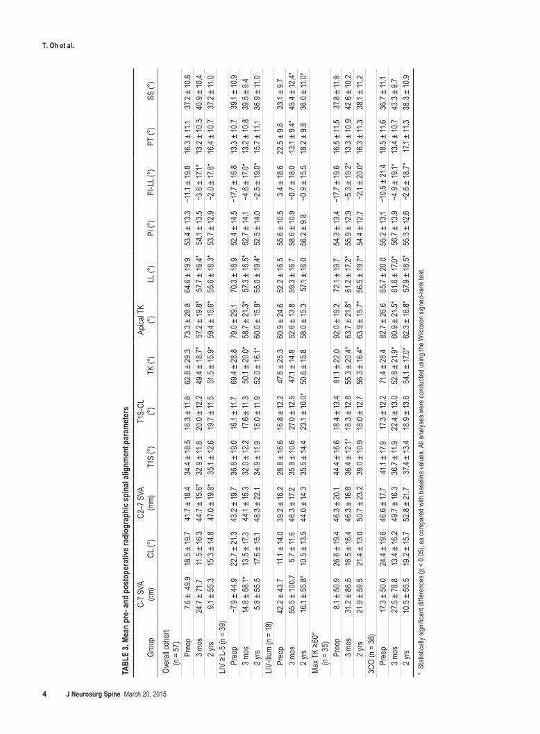
cervical parameters revealed cervical malalignment, with increased CL (18.5° ± 19.7°) and cervical sagittal mal-alignment (41.7 ± 18.4 mm) both evident (Table 3; Fig. 1).
table 1. Summary of demographics for 57 aSd patients with thoracic deformity
Factor No.
Mean age in yrs (range) 49.1 ± 14.6 (21–80)Sex (%) Male 13 (23) Female 43 (77)BMI in kg/m2 (range) 27.8 ± 7.3 (18–57)UIV termination level (%) T-1 11 (19) T-2 26 (46) T-3 15 (26) T-4 5 (9)LIV termination level (%) T-11 1 (2) T-12 2 (4) L-1 5 (9) L-2 11 (19) L-3 9 (16) L-4 11 (19) Ilium 18 (32)Osteotomy type (%) SPO 19 (33) VCR 27 (47) PSO 11 (19)
BMI = body mass index; SPO = Smith-Petersen osteotomy.
table 2. osteotomy level in 57 aSd patients with thoracic deformity
Osteotomy Level No. (%)
SPO VCR PSO
T-2 1 (5) 0 (0) 0 (0)T-3 2 (11) 1 (4) 0 (0)T-4 6 (32) 2 (7) 0 (0)T-5 11 (58) 1 (4) 1 (9)T-6 15 (79) 2 (7) 0 (0)T-7 17 (89) 4 (15) 3 (27)T-8 18 (95) 1 (4) 2 (18)T-9 17 (89) 6 (22) 1 (9)T-10 16 (84) 4 (15) 0 (0)T-11 11 (58) 3 (11) 2 (18)T-12 8 (42) 3 (11) 2 (18)
t. oh et al.
J neurosurg Spine March 20, 20154
tabl
e 3.
mean
pre
- and
pos
tope
rativ
e rad
iogr
aphi
c spi
nal a
lignm
ent p
aram
eter
s
Grou
pC-
7 SVA
(cm
)CL
(°)
C2–7
SVA
(m
m)T1
S (°)
T1S-
CL
(°)TK
(°)
Apica
l TK
(°)
LL (°
)PI
(°)
PI-L
L (°)
PT (°
)SS
(°)
Over
all co
hort
(n
= 57
)
Pr
eop
7.6 ±
49.9
18.5
± 19
.741
.7 ±
18.4
34.4
± 18
.516
.3 ±
11.8
62.8
± 29
.373
.3 ±
28.8
64.6
± 19
.953
.4 ±
13.3
−11.1
± 19
.816
.3 ±
11.1
37.2
± 10
.8
3 m
os24
.7 ±
71.7
11.5
± 16
.344
.7 ±
15.6
*32
.9 ±
11.8
20.0
± 12
.249
.4 ±
18.7*
57.2
± 19
.8*
57.7
± 16
.4*54
.1 ±
13.5
−3.6
± 17
.1*13
.2 ±
10.3
40.9
± 10
.4
2 y
rs9.1
± 5
5.3
15.3
± 14
.847
.0 ±
19.8
*35
.1 ±
12.6
19.7
± 11
.551
.5 ±
15.9
*59
.4 ±
15.6
*55
.6 ±
18.3
*53
.7 ±
12.9
−2.0
± 17
.8*
16.4
± 10
.737
.2 ±
11.0
LIV ≥
L-5 (
n = 3
9)
Preo
p−7
.9 ±
44.9
22.7
± 21
.343
.2 ±
19.7
36.8
± 19
.016
.1 ±
11.7
69.4
± 28
.879
.0 ±
29.1
70.3
± 18
.952
.4 ±
14.5
−17.7
± 16
.813
.3 ±
10.7
39.1
± 10
.9
3 mos
14.8
± 5
8.1*
13.5
± 17
.344
.1 ±
15.3
32.0
± 12
.217
.6 ±
11.3
50.1
± 20
.0*
58.7
± 21
.3*
57.3
± 16
.5*
52.7
± 14
.1−4
.6 ±
17.0
*13
.2 ±
10.8
39.5
± 9.
4
2 yrs
5.8 ±
55.
517
.6 ±
15.1
48.3
± 2
2.134
.9 ±
11.9
18.0
± 11
.952
.0 ±
16.1*
60.0
± 15
.9*
55.0
± 19
.4*52
.5 ±
14.0
−2.5
± 19
.0*
15.7
± 11
.136
.9 ±
11.0
LIV-ili
um (n
= 18
)
Preo
p42
.2 ±
43.7
11.1
± 14
.039
.2 ±
16.2
28.8
± 16
.616
.8 ±
12.2
47.6
± 25
.360
.9 ±
24.6
52.2
± 16
.555
.6 ±
10.5
3.4 ±
18.6
22.5
± 9.
633
.1 ±
9.7
3 mos
55.5
± 10
0.75.7
± 11
.646
.3 ±
17.2
35.9
± 10
.627
.0 ±
12.5
47.1
± 14
.852
.6 ±
13.8
59.3
± 16
.758
.6 ±
10.9
−0.7
± 18
.013
.1 ±
9.4*
45.4
± 12
.4*
2 yrs
16.1
± 55
.8*
10.5
± 13
.544
.0 ±
14.3
35.5
± 14
.423
.1 ±
10.0
*50
.6 ±
15.8
58.0
± 15
.357
.1 ±
16.0
56.2
± 9.
8−0
.9 ±
15.5
18.2
± 9.
838
.0 ±
11.0
*M
ax T
K ≥6
0°
(n =
35)
Preo
p8.1
± 5
0.926
.6 ±
19.4
46.3
± 20
.144
.4 ±
16.6
18.4
± 13
.481
.1 ±
22.0
92.0
± 19
.272
.1 ±
19.7
54.3
± 13
.4−1
7.7 ±
19.6
16.5
± 11
.537
.8 ±
11.8
3 mos
31.2
± 8
6.5
16.5
± 16
.446
.3 ±
16.8
36.4
± 12
.1*18
.3 ±
12.8
55.3
± 20
.4*63
.7 ±
21.8
*61
.2 ±
17.2
*55
.9 ±
12.9
−5.3
± 19
.2*
13.3
± 10
.942
.6 ±
10.2
2 yrs
21.9
± 59
.521
.4 ±
13.0
50.7
± 23
.239
.0 ±
10.9
18.0
± 12
.756
.3 ±
16.4*
63.9
± 15
.7*56
.5 ±
19.7*
54.4
± 12
.7−2
.1 ±
20.0
*16
.3 ±
11.3
38.1
± 11
.23C
O (n
= 3
8)
Preo
p17
.3 ±
50.0
24.4
± 19
.646
.6 ±
17.7
41.1
± 17
.917
.3 ±
12.2
71.4
± 28
.482
.7 ±
26.6
65.7
± 20
.055
.2 ±
13.1
−10.
5 ± 21
.418
.5 ±
11.6
36.7
± 11
.1
3 mos
27.5
± 78
.813
.4 ±
16.2
49.7
± 16
.336
.7 ±
11.9
22.4
± 13
.052
.8 ±
21.9
*60
.9 ±
21.5
*61
.6 ±
17.0
*56
.7 ±
13.9
−4.9
± 19
.1*13
.4 ±
10.7
43.3
± 9.
7
2 yrs
10.5
± 5
5.5
19.2
± 15
.752
.8 ±
21.7
37.4
± 13
.418
.9 ±
13.6
54.1
± 17
.0*
62.3
± 16
.8*
57.9
± 18
.5*
55.3
± 12
.6−2
.6 ±
18.7*
17.1
± 11
.338
.3 ±
10.9
* St
atist
ically
sign
ifican
t diff
eren
ces (
p < 0.
05),
as co
mpar
ed w
ith ba
selin
e valu
es. A
ll ana
lyses
wer
e con
ducte
d usin
g the
Wilc
oxon
sign
ed-ra
nk te
st.

cervical compensatory alignment changes
J neurosurg Spine March 20, 2015 5
Rates of preoperative cervical kyphosis, cervical sagittal malalignment, and increased CL were 17.8%, 47.8%, and 48.9%, respectively (Table 4). Following thoracic defor-mity correction, significant changes were seen in the C2–7 SVA at both 3 months and 2 years postoperatively (p < 0.05 for both) but not in the corresponding rates of cer-vical malalignment (p > 0.05 for both). Rates for cervi-cal deformity correction at 2 years postoperatively were 29%, 26%, and 21% for cervical kyphosis, cervical sagittal malalignment, and increased CL, respectively (Table 5). Among patients undergoing distal fusion at or above L-5 versus at the ilium, no significant cervical changes in CL or C2–7 SVA were observed at 3 months or 2 years post-operatively (p > 0.05 for all). Among patients with maxi-mum TK ≥ 60° or those undergoing a 3CO, cervical pa-rameters did not significantly change at either time point (p > 0.05 for all). Several patients developed new-onset cervical deformity: among those without concurrent base-line cervical spine deformity, the incidence of new cervi-
cal deformity at the 2-year follow-up was 12%, 55%, and 24% for cervical kyphosis, cervical sagittal malalignment, and increased CL, respectively (Table 6).
thoracic radiographic outcomesThe mean preoperative thoracic parameters within the
entire cohort revealed thoracic sagittal malalignment (TK: 62.8° ± 29.3°; Table 3). After thoracic osteotomy, the mean TK and apical TK significantly decreased from baseline at both the 3-month and 2-year follow-ups (p < 0.05 for both; Fig. 1). Among patients with an LIV ≥ L-5, the mean TK and apical TK were significantly lower at 3 months and 2 years postoperatively (p < 0.05 for both). In contrast, patients with an LIV-ilium did not show statistically sig-nificant changes in the mean kyphosis angle or apical TK (p > 0.05 for both). Patients with a maximum TK ≥ 60° or those who underwent 3CO showed significant decreases in TK and apical TK at the 3-month and 2-year follow-ups (p < 0.05 for both).
Spinopelvic radiographic outcomesAt baseline, global and pelvic sagittal malalignment
were not seen, although there was significant TK (Fig. 1). However, 3-month and 2-year postoperative radiographic values revealed significant changes in LL and PI-LL (p < 0.05 for both; Table 3). Patients with an LIV ≥ L-5 had significantly reduced LL and PI-LL at 3 months and at 2 years, as compared with preoperative values (p < 0.01 for both). Patients with an LIV-ilium had an increased SS at 3 months and at 2 years, again as compared with preop-erative values (p < 0.05 for both). Following surgery, the mean values for LL and PI-LL were also significantly low-er among patients with a maximum TK ≥ 60° and those with 3CO (p < 0.05 for both).
Fig. 1. Case example. Preoperative (left) and postoperative (right) sagittal radiographs showing measurements. Note the increased preop-erative CL (> 15°) that slightly decreases and the persistent large C2–7 SVA in the setting of normal lumbopelvic parameters. CL = C2–7 cervi-cal lordosis.
table 4. preoperative and postoperative rates of cervical spine deformity differences compared to baseline
Factor Rate (no./total) p Value*
Cervical kyphosis Preop 17.8% (8/45) 3 mos 28.6% (10/35) 0.25 2 yrs 19.6% (9/46) 0.83C2–7 SVA >4 cm Preop 47.8% (22/46) 3 mos 65.7% (23/35) 0.11 2 yrs 61.7% (29/47) 0.18Increased CL (CL >15°) Preop 48.9% (22/45) 3 mos 35.3% (12/34) 0.23 2 yrs 47.8% (22/46) 0.92Increased CL + C2–7 SVA >4 cm Preop 6.8% (3/44) 3 mos 14.3% (5/35) 0.27 2 yrs 8.7% (4/46) 0.74
* All analyses were conducted using the chi-square test. A p < 0.05 was considered statistically significant.

t. oh et al.
J neurosurg Spine March 20, 20156
discussionSpontaneous reciprocal changes in the cervical and
thoracic spines have been shown following lumbar cor-rective surgery. Klineberg et al. showed a reciprocal +13° change in TK;8 Jang et al. treated 53 patients for lumbar degenerative kyphosis and showed spontaneous correction of TK from 1.1° to 17.6°, with greater corrections of LL leading to incremental improvements in TK;7 and, perhaps most importantly, as reported by Smith et al., increased CL spontaneously corrected following lumbar PSO.25 Re-ciprocal changes following thoracic deformity correction have been reported less often. In Klineberg et al.’s series of 29 thoracic PSOs, the authors revealed a reciprocal 8° reduction in LL.8 Peelle et al. reported 50% spontaneous correction of the lumbar curve in the coronal plane and 68% on side-bending radiographs following thoracic fu-sion for scoliosis.17 And although they conducted a study on adolescent idiopathic scoliosis patients, Newton et al. demonstrated that when these patients were treated us-ing thoracic fusion with an LIV of T11–L1, LL decreased postoperatively.14
Our study is the first attempt to characterize radiograph-ic changes in both cervical and lumbar regions following thoracic osteotomies. Given evidence in the literature, we hypothesized that thoracic deformity correction would lead to reciprocal improvements in cervical and spinopel-vic radiographic parameters. The results of our analysis indicated that some significant reciprocal changes in the cervical spine do occur postoperatively, as evidenced by the 5.3-mm increase in the C2–7 SVA at 2 years; however, this increase suggested a possible worsening of cervical sagittal alignment. Furthermore, the mean 2-year postop-erative values for CL (15.3° ± 14.8°) and C2–7 SVA (47.0 ± 19.8 mm) suggested a lack of consistent improvement in
CL and the persistence of sagittal cervical malalignment, respectively. Rates of cervical deformity also did not sig-nificantly change from baseline, and several patients even developed a new cervical spine deformity after undergo-ing thoracic osteotomy. On the other hand, in the lumbar spine, significant changes were observed in the form of improved PI-LL. No other changes were seen in the lum-bar parameters.
Collectively, these results suggested that thoracic os-teotomies have some impact on postoperative cervical or spinopelvic alignment. To validate these findings, we con-ducted several subgroup analyses. In the first subgroup analysis, for two reasons we aimed to ascertain whether the level of distal fusion was a contributory factor in reciprocal change. From a mechanistic standpoint, patients with lower instrumentation reaching the ilium should be more suscep-tible to reciprocal changes in the cervical spine given the operative fixation of spinopelvic alignment. Theoretically, these patients would be unable to compensate for thoracic deformity correction in the lumbar spine and would have increased difficulty in the pelvis, thus increasing the likeli-hood for change to occur in the cervical spine. Additional-ly, literature evidence has pointed to fusion levels influenc-ing the directionality of reciprocal change, with one study showing that thoracic PSOs with lumbar fusion increased postoperative LL while those without resulted in a net loss of LL.8 Nevertheless, patients in either the LIV ≥ L-5 or the LIV-ilium group failed to show significant improvement in postoperative cervical parameters. However, patients with an LIV ≥ L-5 had a postoperative loss of 15.3° in LL.
In a subsequent subgroup analysis, we aimed to deter-mine whether severe thoracic deformity was associated with reciprocal change. Although concrete thresholds for defining sharp angular sagittal TK have not been clearly defined, severe TK was represented by selecting patients with a maximum TK ≥ 60°. In this cohort of 35 patients, the single observed change was a mean loss of 15.6° in LL. No cervical changes were seen. Severe deformity was also estimated by analyzing only those patients who had undergone 3CO (38 patients), which is typically reserved for severe deformity and affords the appropriate surgical platform for inducing the greatest degree of correction.3 Application of the 3CO techniques, specifically VCR and PSO, for thoracic deformity correction has been described elsewhere.12,16,28,29 In this cohort of 38 patients, significant reciprocal improvements in cervical parameters were again absent, but as regards the lumbar parameters, there was -7.8° in LL.
table 5. Spontaneous correction of preoperative cervical malalignment and persistent malalignment among those presenting with concomitant cervical malalignment and thoracic deformity
Factor
Deformity Rates (no./total [%])3 Mos 2 Yrs
Corrected Persistent Corrected Persistent
Cervical kyphosis (n = 8) 3/7 (43) 4/7 (57) 2/7 (29) 5/7 (71)C2–7 SVA >4 cm (n = 22) 2/14 (14) 12/14 (86) 5/19 (26) 14/19 (74)Increased CL (n = 22) 4/12 (33) 8/12 (67) 4/19 (21) 15/19 (79)Increased CL + C2–7 SVA >4 cm (n = 3) 2/3 (67) 1/3 (33) 1/2 (50) 1/2 (50)
table 6. new postoperative cervical deformity among patients presenting without baseline cervical deformity
FactorDeformity Rates (no./total [%])
3 Mos 2 Yrs
Cervical kyphosis 5/23 (22) 4/33 (12)C2–7 SVA >4 cm 9/16 (56) 12/22 (55)Increased CL 2/17 (12) 5/21 (24)Increased CL + C2–7 SVA >4 cm 4/26 (15) 3/37 (8)

cervical compensatory alignment changes
J neurosurg Spine March 20, 2015 7
The explanation for the lack of reciprocal change in CL remains unclear. As Klineberg et al. posit, in much the same way that compensatory changes initially occur to preserve balance and horizontal gaze, alignment of regions distal to the correction site should similarly “re-compensate” once focal correction is achieved.8 Given that many of our patients were hyperlordotic in their cervical spine, it was expected that thoracic deformity correction would normalize the sagittal cervical Cobb angle. Our analysis revealed that the index surgery resulted in a sig-nificant change in TK, which would be expected to induce a reciprocal decrease in the T-1 slope (T1S) as well as a loss in CL to maintain horizontal gaze. However, while some reductions in T1S and CL were observed, they did not reach statistical significance.
There are several possible explanations for the failure of cervical alignment to compensate. First, we should note that in the population without fusion to the pelvis, a sig-nificant adjacent segment change (loss of lordosis) did oc-cur in the lumbar spine. It is possible that this change is perhaps more likely to occur because of the greater impact of the lumbar area on overall global balance and energy expenditure. Second, the absence of change in the cervical spine may be a result of incomplete correction of the en-tire thoracic spine given a focus on apical (or sharp angle) kyphosis. Furthermore, given the difficulty in visualizing the T1–4 area on radiographic studies, it is possible that a compensatory increase in kyphosis occurred in the re-gion. We were not able to visualize this area well enough to include T1–4 (upper TK) in our analysis. A lack of sig-nificant change in T1S may account for some of the absent reciprocal change as T1S and CL are closely related.19 In-creases in T1S cause increases in CL if the patient is to maintain a horizontal gaze.19 Furthermore, although over-all TK significantly decreased, the patients, on average, still had a large TK with a mean of 51.5°, and those with an initial maximum TK ≥ 60° had a mean of 56.3° at 2 years. Thus, it is possible that the deformity correction was focused around the apical thoracic deformity, resulting in a decrease in the sharp angle but not as great an increase in overall TK and thus no change in T1S and continued elevation of CL.
It is also interesting to note the statistically significant increase in C2–7 SVA, which would translate to worsen-ing cervical deformity. On average, patients increased in their cervical SVA, further confounding the anticipated favorable reciprocal change. As mentioned above, an in-complete correction of total TK or an increase in T1–4 kyphosis may have resulted in these persistent or worsened alignment values.
Overall, these findings highlight the importance of req-uisitioning full standing radiographs in the workup of pa-tients with thoracic deformity. Preoperatively, many such patients also demonstrate cervical deformity. Importantly, the correction of TK does not normalize cervical align-ment and may worsen cervical sagittal alignment. The strengths of the present study, compared with previous analyses, are its multiinstitutional nature and its accrual of a larger number of patients with thoracic deformity.8,17 However, these factors also pose limitations, as individual variations in the surgical technique or procedural bias may
be present. Furthermore, the study’s retrospective nature imposes inherent limitations. Future studies should em-phasize the prospective collection and analysis of patient data, which would allow for more detailed analyses of the presence and types of reciprocal change that can occur fol-lowing thoracic deformity surgery. In addition, EOS imag-ing may allow higher-definition imaging of changes in the T1–4 region often obscured by the shoulders and upper thorax. Correlation with patient outcomes and cervical health-related quality of life parameters could also prove to be an important issue to investigate in the future as well.
conclusionsDynamic reciprocal changes following corrective sur-
gery for thoracic deformity have been largely unexplored to date. This multicenter analysis of a consecutive cohort of patients with ASD who were treated for thoracic de-formity revealed that concurrent cervical malalignment is prevalent in this population and that cervical malalign-ment persists despite thoracic deformity correction. As a result, the evaluation of concomitant increased CL and cervical sagittal malalignment should be encouraged, and clinicians must be cognizant of the fact that, unlike in lum-bar corrective surgery, the risk for persistent cervical mal-alignment remains high following selective correction of the thoracic deformity.
references 1. Angevine PD, Kaiser MG: Radiographic measurement tech-
niques. Neurosurgery 63 (3 Suppl):40–45, 2008 2. Bess S, Boachie-Adjei O, Burton D, Cunningham M, Shaf-
frey C, Shelokov A, et al: Pain and disability determine treatment modality for older patients with adult scoliosis, while deformity guides treatment for younger patients. Spine (Phila Pa 1976) 34:2186–2190, 2009
3. Bridwell KH: Decision making regarding Smith-Petersen vs. pedicle subtraction osteotomy vs. vertebral column re-section for spinal deformity. Spine (Phila Pa 1976) 31 (19 Suppl):S171–S178, 2006
4. Champain S, Benchikh K, Nogier A, Mazel C, Guise JD, Skalli W: Validation of new clinical quantitative analysis software applicable in spine orthopaedic studies. Eur Spine J 15:982–991, 2006
5. Glassman SD, Berven S, Bridwell K, Horton W, Dimar JR: Correlation of radiographic parameters and clinical symp-toms in adult scoliosis. Spine (Phila Pa 1976) 30:682–688, 2005
6. Glassman SD, Bridwell K, Dimar JR, Horton W, Berven S, Schwab F: The impact of positive sagittal balance in adult spinal deformity. Spine (Phila Pa 1976) 30:2024–2029, 2005
7. Jang JS, Lee SH, Min JH, Maeng DH: Influence of lumbar lordosis restoration on thoracic curve and sagittal position in lumbar degenerative kyphosis patients. Spine (Phila Pa 1976) 34:280–284, 2009
8. Klineberg E, Schwab F, Ames C, Hostin R, Bess S, Smith JS, et al: Acute reciprocal changes distant from the site of spinal osteotomies affect global postoperative alignment. Adv Or-thop 2011:415946, 2011
9. Lafage V, Ames C, Schwab F, Klineberg E, Akbarnia B, Smith J, et al: Changes in thoracic kyphosis negatively im-pact sagittal alignment after lumbar pedicle subtraction oste-otomy: a comprehensive radiographic analysis. Spine (Phila Pa 1976) 37:E180–E187, 2012

t. oh et al.
J neurosurg Spine March 20, 20158
10. Lafage V, Schwab F, Patel A, Hawkinson N, Farcy JP: Pelvic tilt and truncal inclination: two key radiographic parameters in the setting of adults with spinal deformity. Spine (Phila Pa 1976) 34:E599–E606, 2009
11. Lafage V, Schwab F, Skalli W, Hawkinson N, Gagey PM, Ondra S, et al: Standing balance and sagittal plane spinal de-formity: analysis of spinopelvic and gravity line parameters. Spine (Phila Pa 1976) 33:1572–1578, 2008
12. Lafage V, Smith JS, Bess S, Schwab FJ, Ames CP, Klineberg E, et al: Sagittal spino-pelvic alignment failures following three column thoracic osteotomy for adult spinal deformity. Eur Spine J 21:698–704, 2012
13. McAviney J, Schulz D, Bock R, Harrison DE, Holland B: Determining the relationship between cervical lordosis and neck complaints. J Manipulative Physiol Ther 28:187–193, 2005
14. Newton PO, Yaszay B, Upasani VV, Pawelek JB, Bastrom TP, Lenke LG, et al: Preservation of thoracic kyphosis is critical to maintain lumbar lordosis in the surgical treatment of adolescent idiopathic scoliosis. Spine (Phila Pa 1976) 35:1365–1370, 2010
15. O’Brien MF, Kuklo TR, Blanke KM, Lenke LG (eds): Spi-nal Deformity Study Group Radiographic Measurement Manual. Memphis, TN: Medtronic Sofamor Danek, 2005
16. O’Shaughnessy BA, Kuklo TR, Hsieh PC, Yang BP, Koski TR, Ondra SL: Thoracic pedicle subtraction osteotomy for fixed sagittal spinal deformity. Spine (Phila Pa 1976) 34:2893–2899, 2009
17. Peelle MW, Boachie-Adjei O, Charles G, Kanazawa Y, Mes-fin A: Lumbar curve response to selective thoracic fusion in adult idiopathic scoliosis. Spine J 8:897–903, 2008
18. Rillardon L, Levassor N, Guigui P, Wodecki P, Cardinne L, Templier A, et al: [Validation of a tool to measure pelvic and spinal parameters of sagittal balance.] Rev Chir Orthop Reparatrice Appar Mot 89:218–227, 2003 (Fr)
19. Scheer JK, Tang JA, Smith JS, Acosta FL Jr, Protopsaltis TS, Blondel B, et al: Cervical spine alignment, sagittal deformity, and clinical implications. A review. J Neurosurg Spine 19:141–159, 2013
20. Schwab F, Dubey A, Pagala M, Gamez L, Farcy JP: Adult scoliosis: a health assessment analysis by SF-36. Spine (Phi-la Pa 1976) 28:602–606, 2003
21. Schwab F, Lafage V, Patel A, Farcy JP: Sagittal plane consid-erations and the pelvis in the adult patient. Spine (Phila Pa 1976) 34:1828–1833, 2009
22. Schwab F, Patel A, Ungar B, Farcy JP, Lafage V: Adult spinal deformity-postoperative standing imbalance: how much can you tolerate? An overview of key parameters in assessing alignment and planning corrective surgery. Spine (Phila Pa 1976) 35:2224–2231, 2010
23. Smith JS, Fu KM, Urban P, Shaffrey CI: Neurological symp-toms and deficits in adults with scoliosis who present to a surgical clinic: incidence and association with the choice of operative versus nonoperative management. J Neurosurg Spine 9:326–331, 2008
24. Smith JS, Shaffrey CI, Fu KM, Scheer JK, Bess S, Lafage V, et al: Clinical and radiographic evaluation of the adult spinal deformity patient. Neurosurg Clin N Am 24:143–156, 2013
25. Smith JS, Shaffrey CI, Lafage V, Blondel B, Schwab F, Hos-tin R, et al: Spontaneous improvement of cervical alignment after correction of global sagittal balance following pedicle subtraction osteotomy. J Neurosurg Spine 17:300–307, 2012
26. Tang JA, Scheer JK, Smith JS, Deviren V, Bess S, Hart RA, et al: The impact of standing regional cervical sagittal align-ment on outcomes in posterior cervical fusion surgery. Neu-rosurgery 71:662–669, 2012
27. Villavicencio AT, Babuska JM, Ashton A, Busch E, Roeca C, Nelson EL, et al: Prospective, randomized, double-blind clin-ical study evaluating the correlation of clinical outcomes and cervical sagittal alignment. Neurosurgery 68:1309–1316, 2011
28. Wang Y, Lenke LG: Vertebral column decancellation for the management of sharp angular spinal deformity. Eur Spine J 20:1703–1710, 2011
29. Yang BP, Ondra SL, Chen LA, Jung HS, Koski TR, Salehi SA: Clinical and radiographic outcomes of thoracic and lum-bar pedicle subtraction osteotomy for fixed sagittal imbal-ance. J Neurosurg Spine 5:9–17, 2006
30. Youssef JA, Orndorff DO, Patty CA, Scott MA, Price HL, Hamlin LF, et al: Current status of adult spinal deformity. Global Spine J 3:51–62, 2013
author contributionsConception and design: Ames. Acquisition of data: Lafage. Analysis and interpretation of data: Oh, Scheer, Ames. Drafting the article: Oh, Scheer. Critically revising the article: Oh, Scheer, Eastlack, Smith, Lafage, Bess, Schwab, Ames. Reviewed submit-ted version of manuscript: all authors. Approved the final version of the manuscript on behalf of all authors: Oh. Statistical analy-sis: Oh. Study supervision: Ames.
correspondenceTaemin Oh, Department of Neurological Surgery, Northwestern University, Feinberg School of Medicine, 676 N. St. Clair St., Ste. 2210, Chicago, IL 60611. email: [email protected].
Top Related
Copyright © 2022 FDOKUMEN

