


Bahasa
Halaman
Hukum

Journal of Analytical Oncology, 2014, 3, 000-000 1
ISSN: 1927-7210 / E-ISSN: 1927-7229/14 © 2014 Lifescience Global
Caspase Pathway Activation and Reactive Oxygen Species Generation in Apoptotic Cell Death of Human Leukemic U937 and K562 Cell Line in Response to King Cobra (Ophiophagus hannah) Venom
Tanmoy Bhowmik1, Ajoy Kumar Biswas2, Amrita Sarkar1, Partha Pratim Saha1, Aparna Gomes3 and Antony Gomes1,*
1Laboratory of Toxinology & Experimental Pharmacodynamics, Department of Physiology, University of
Calcutta, 92, APC Road, Kolkata 700 009, India
2GD Hospital & Diabetes Institute, Kolkata-700016, India
3Indian Institute of Chemical Biology, Kolkata-700032, India
Abstract: Resistance and decreasing efficacy of current synthetic drug for chemotherapy of leukemic cancer draws attention for development of newer anticancer agent from natural resources. In the present study, king cobra venom (OHV) significantly inhibited leukemic cell growth in dose and time dependent manner. For U937 and K562 cell line, the
IC50 dose (72 h) was found to be 4.1 g/ml and 3.9 g/ml respectively, observed by trypan blue exclusion method and tetrazolium bromide reduction assay. OHV treated morphometry of leukemic cell showed the characteristic features of apoptosis. Both U937 and K562 cells were arrested in the G1 phase of cell cycle with most cells exhibiting the
biochemical feature of early and late apoptosis. Mitochondrial membrane potential was lost and reactive oxygen species generated highly in OHV treated leukemic cell line (U937 and K562). Western blot analysis showed OHV increased expression of Bax and decreased expression of Bcl2 in OHV treated cell as compared to untreated control U937 and
K562 cell. Upregulation of Cytochrome c, Bid, Bad, Caspase 3/8/9, p21 and NF- B down regulation of Cyclin D1, CDK4 was also showed by western blot analysis which revealed the possible pathway of OHV in cellular level. The results of this study demonstrated that OHV significantly and selectively induced leukemic cell death through both extrinsic and intrinsic apoptotic pathway.
Keywords: Ophiophagus hannah, Venom, Leukemic cell, Apoptosis, Flow cytometry, Cell cycle.
1. INTRODUCTION
Cancer is the major public health concern in all
developed and developing countries. In year 2012 a
total of 1,638,910 new cancer cases and 577,190
deaths from cancer are estimated [1]. There is urgent
need of find better treatment as now cancer becomes
the second leading cause of death worldwide. In recent
years remarkable progress has been made towards the
understanding of molecular pathway of cancer
development and hormonal therapy. Various therapies
have been used for treating cancer e.g. radiotherapy,
immunotherapy, gene therapy and chemotherapy [2].
In these days, predominant option of cancer prevention
is the use of chemotherapeutic drug. However, one of
the major problems with cancer therapy using
chemotherapeutics are that patients often do not
respond or eventually resistance is developed after
initial treatment, high toxicity and high cost. This has
led to the increased use of anticancer agents
*Address correspondence to this author at the Laboratory of Toxinology & Experimental Pharmacodynamics, Department of Physiology, University of Calcutta, 92, APC Road, Kolkata 700 009, India; Tel: 91-33-23508386/ (M) 09433139031; Fax: 91-33-2351-9755/2241-3288; E-mail: [email protected]
developed from natural resources. The biodiversity of
venoms or toxins of animal, plant, fungal and bacteria
made it a unique tool from which new therapeutic
agents may be developed. Currently, Snake venom
has been shown to possess a wide spectrum of
biological activities [3].
From past few decades, research on isolation and
characterization of anticancer agents from the snake
venom has been undertaken. The antimetastatic
activities of crude venom of Indian monocellate cobra
(Naja kaouthia), Indian cobra (Naja naja) Russell’s
viper (Vipera russelli) and Banded krait (Bungarus
fasciatus) were studied on carcinoma, sarcoma and
leukemia models [4-6]. Now we focussed on anticancer
activity of Indian king cobra’s (Ophiophagus hannah)
crude venom.
The king cobra of the elapidae family is the world’s
largest venomous snake [7]. It is known that king cobra
snake is extremely poisonous and have strong
neurotoxic action [8]. Recently, Lee et al. isolated a
heat stable form of L-amino acid oxidase from king
cobra venom having antibacterial activity [9]. L-amino
acid oxidase (LAAO) isolated from King cobra
(Ophiophagus hannah) venom, is an extremely potent
2 Journal of Analytical Oncology, 2014, Vol. 3, No. 3 Bhowmik et al.
antiproliferative agent against cancer cells. L-amino
acid oxidase (OH-LAAO), a heat stable enzyme, has
been shown to exhibit very potent anti-proliferative
activity against human breast and lung tumorigenic
cells but not in their non-tumorigenic counterparts [10,
11]. Previously from our lab, a novel fibrinolytic peptide,
Hannahpep was isolated from the Indian king cobra
venom [12]. The present investigation reports the
antileukemic activity of crude venom of Indian king
cobra against two human leukemic cell lines (K562 and
U937) and molecular action of this venom to
understand its signalling pathway.
2. MATERIALS AND METHODS
2.1. Chemicals
3-(4, 5-dimethylthiazol-2-yl)-2,5- diphenyltetrazolium
bromide (MTT), Sodium dodecyl sulphate (SDS),
Sodium bi carbonate (NaHCO3), Ethidium bromide,
RNase A, Propium Iodide, Acridine Orange, Trypan
Blue, Lactate dehydrogenase, De- Methoxy sulphoxide
(DMSO) were purchased from Sigma- Aldrich co. (St.
Louis, MO, USA). RPMI1640 medium, fetal bovine
serum, penicillium-streptomycin was purchased from
invitrogen, (USA). Annexin V-FITC, Cell cycle kit, FITC-
BrdU kit, Ki-67 anti human antibody kits were
purchased from BD-Bioscience, USA. Primary antibody
(Bax, Bcl2, Caspase 8, Caspase 9, Cytochrome C, Bid,
Bad, Cyclin D, CDK4, p21, NF- B3 and -actin) and
HRP-conjugated goat anti-rabbit secondary antibody
were procured from Santa Cruz, CA, USA All other
chemicals were purchased locally and were of
analytical grade. Lyophilized O. hannah crude venom
(OHV) was purchased from the Calcutta snake park,
Kolkata, India.
2.2. Cell Culture
U937 (human leukemic lymphoma cell line) and
K562 (human myelogenous leukemic cell line) were
purchased from National facility for Animal Tissue and
cell culture, Pune India. Cells were cultured in RPMI
1640 supplemented with 10% heat inactivated FBS,
glucose (1.5g/L), NaHCO3 (1.5g/L), Penicillin (100 units)
and streptomycin (10 g/ml). At 95% humidified atmos-
phere and 5% CO2 cells were grown to confluence at
37oC in inside an incubator (Heal Force, China).
2.3. Determination of IC50 and Cytotoxicity Studies on Leukemic Cells
The 50% of inhibition concentration (IC50) values of
OHV in U937 and K562 cells were determined using
different concentration (1, 2, 5 and 10 μg/ml) of the
crude venom at 24 h, 48 h and 72 h. The respective
viable cells were counted in the phase contrast
microscope using Trypan blue [13]. The respective
concentration of venom and the percentage of cell
death were plotted on the x axis and the y axis of the
graph respectively. For cytotoxicty test, MTT assay of
treated cells were done to confirm its cytotoxic effect
[14]. 40 l of MTT solution (5 mg/ml) was added 4 h
prior to end of 24h, 48 h and 72 h of incubation. The
violet colour formazan granules formed by viable cells
were dissolved in DMSO and the absorbance at 570
nm was estimated by measuring with the plate reader
(BioTek, ELx800).
2.4. Detection of Apoptosis
2.4.1. Observation under Florescence Microscope
1x 10⁶ cells of U937 and K562 cell were treated with
OHV (IC50 dose of respect to two cell lines) for 24 hour
and observed using a fluorescence microscope for
changes of morphometry. After harvesting, the
untreated control cells and the OHV treated cells
washed with PBS and then stained with Acridine
orange (100 g/ml) and Ethidium bromide (100 g/ml)
(1:1) [15, 16]. With mounting on slides, the cells were
then immediately observed under fluorescence
microscope (Motic Image plus) for determination of the
cell apoptosis.
2.4.2. Flow Cytometric Analysis
Flow cytometric analysis was done by performing
dot plot assay to investigate the type of cell death
induced by OHV [17, 18]. The leukemic cells (1 x 10⁶)
were treated with IC50 (for respective cell) dose for
24 h. The cells were pelleted down and washed with
Annexin V-FITC binding buffer. After washing, cell
pellets were dissolved in Annexin V-FITC binding buffer
containing annexin V-FITC and Propidium iodide. With
15 min incubation in dark at room temperature flow
cytometric analysis was done. All data were acquired
with a Becton-Dickinson FACS Verse double laser
cytometer.
2.5. Lactate Dehydrogenase (LDH) Activity Assay
Plasma membrane integrity loss was indicated by
release of LDH from the cytoplasm of cultured cells into
the medium. Culture medium was collected after OHV
treatment and cell debris was removed by
centrifugation. LDH activity was measured using a
commercial LDH assay kit according to manufacturer’s
protocol (Sigma –Aldrich, USA) [17]. Briefly, the
Caspase Pathway Activation and Reactive Oxygen Species Generation Journal of Analytical Oncology, 2014, Vol. 3, No. 3 3
samples were read on a spectrophotometer at the
absorbance of 440 nm. LDH activity was calculated
according to a standard curve and expressed in U/L.
2.6. Study of Cell Cycle by Flow Cytometric Analysis
1x 10⁶ leukemic cells were treated with respective
IC50 dose of OHV for 24 h. Cells were washed with
PBS, fixed with ice-cold 70% ethanol. After pelleted
down, cells were washed twice with cold PBS and
finally dissolved in PBS treated with RNase A for 30
min at 37oC and stained with propium iodide (20 l
from 50 g/ml) and kept in dark for 15 min. Cell cycle
phase distribution of nuclear DNA was determined on
FACS, fluorescence detector [19, 20, 21].
2.7. Estimation of BrdU Incorporation Using FACS
Exponentially growing U937 and k562 cells (1 x 10⁶)
were cultured on RPMI medium containing 10 l BrdU
(10 g/ml) [22, 23]. After 1 h, cells were treated with
respective IC50 dose of OHV and kept at CO2 incubator
for 24 h. Cells were fixed with permealization buffer for
20 min. After washing again with PBS, Dnase (5 g/ml)
was added and incubated for 1 h at 37oC. Anti Brdu
antibody was added to it and kept at dark for 20 min
after PBS washing. Samples were analysed with FACS
after incubated with 7-ADD for 15 min at dark condition.
2.8. Reactive Oxygen Species (ROS) Assay
The method was obtained from the report of
Condello et al. [24]. The H2DCF-DA is the probe that
could quantify the amount of intracellular ROS. For the
assay, the leukemic cell lines (U937 and K562) were
seeded at 1x 10⁶cells/well in the 6 –well plate and cells
were treated with IC50 dose of OHV for respective cell
for 24 h. Cells were incubated with 5 μM H2DCF-DA
for 30 min at 37oC, and then cells were washed twice
with PBS and resuspended in PBS. After centrifugation
at 5000 rpm for 10 min, the fluorescence of the
supernatant intensity was analysed at an excitation
wavelength of 480 nm and an emission wavelength of
540 nm by a Spectrofluorometer (Bio-Rad, USA) [25].
2.9. Effect of OHV on Caspase 3 Activity
U937 and K562 cells from OHV treated plate were
collected after 24 h incubation. Cell lysate were
prepared by BD cell lysis buffer (BD Bioscience).
Caspase 3 activity was performed according to
manufacture protocol by spectrofluorometer (Bio-Rad,
USA) [26].
2.10. Western Blot Analysis of Pro and Anti-Apoptotic Protein Expression of Leukemic Cells
OHV treated U937 and K562 cell lysate of were
prepared by cell lysis buffer. Equivalent amount of
proteins were electrophoresed in 10% SDS-PAGE and
transferred to a PVDF membrane which was then
blocked with 5% skimmed milk in PBS-T. Incubation
with primary antibodies of Bax, Bcl2, caspase 8/9,
Cytochrome C, Bid, Bad, Cyclin D1, p21, CDK4, NF-
B3 and -actin were done followed by subsequent
wash with PBS-T (phosphate buffer saline with 0.1%
Tween 20). Then membrane was incubated with horse-
redox peroxidase conjugated goat antirabbit secondary
antibody followed by a detection reagent (Amersham
Bioscience) [19, 27].
2.11. Separation and Culture of Normal Lymphocytes
After taking informed consent (Ref. No. IHEC/AG/
HUM/P17/12), blood was collected aseptically from the
vein of healthy adults and transferred to heparinised
vial. Using Ficoll-histopaque, lymphocytes were
collected from whole blood [16]. Then it was cultured in
sterile complete RPMI 1640 media. 1x 10⁶ cells were
treated with sterile PBS, OHV (twice IC50 dose of U937
cell) and Imatinib mesylate (standard drug – 100 μg/ml)
and cells were grown in a CO2 incubator at 37o C with
5% CO2 for 72 h in humidified condition.
2.12. Statistical Analysis
All values are represented as arithmetic mean ±
SEM. Statistical analysis was done by student’s t test.
A probability value of *P 0.05 was chosen as the
criterion of statistical significance.
3. RESULTS
3.1. IC50 Value of OHV in U937 and K562 Cell Line
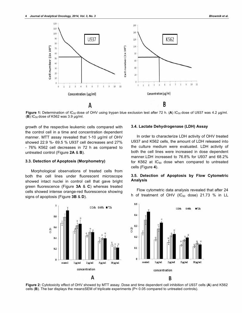
After 72 h, IC50 value of OHV in U937 cells was 4.2
μg/ml at 95% of fiducial probalities the confidence
intervals (Figure 1A). In K562 cells at 95% of fiducial
probabilities the confidence intervals of IC50 value of
OHV was 3.9 μg/ml after 72 h (Figure 1B). Viability of
cell dereased as incubation time increase from 24 h to
72 h.
3.2. Cytotoxicity Study
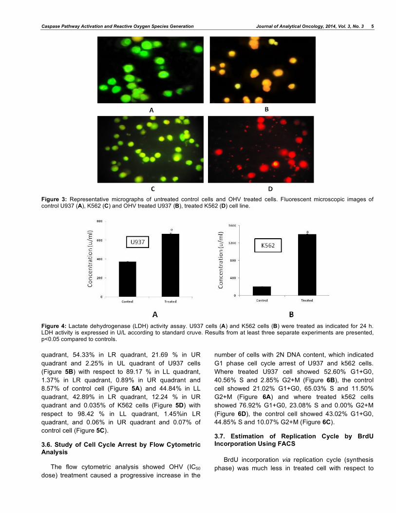
OHV at concentrations of 1-10 g/ml (U937 cell line)
and 1-10 g/ml (K562 cell line) significantly inhibited the
4 Journal of Analytical Oncology, 2014, Vol. 3, No. 3 Bhowmik et al.
growth of the respective leukemic cells compared with
the control cell in a time and concentration dependent
manner. MTT assay revealed that 1-10 g/ml of OHV
showed 22.9 %- 69.5 % U937 cell decreases and 27%
- 76% K562 cell decreases in 72 h as compared to
untreated control (Figure 2A & B).
3.3. Detection of Apoptosis (Morphometry)
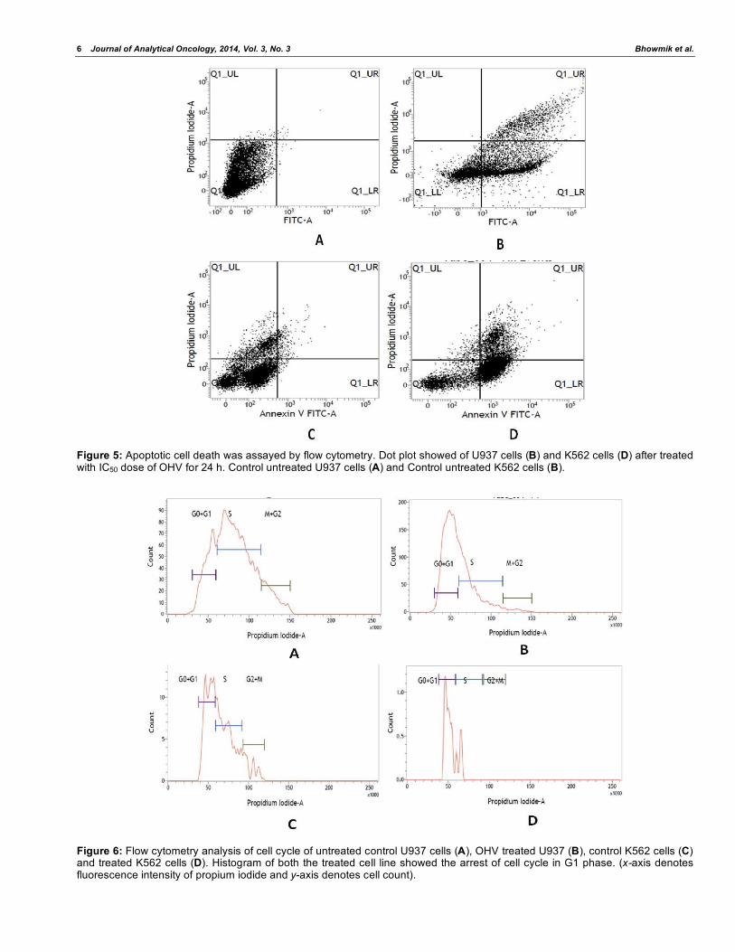
Morphological observations of treated cells from
both the cell lines under fluorescent microscope
showed intact nuclei in control cell that gave bright
green fluorescence (Figure 3A & C) whereas treated
cells showed intense orange-red fluorescence showing
signs of apoptosis (Figure 3B & D).
3.4. Lactate Dehydrogenase (LDH) Assay
In order to characterize LDH activity of OHV treated
U937 and K562 cells, the amount of LDH released into
the culture medium were evaluated. LDH activity of
both the cell lines were increased in dose dependent
manner.LDH increased to 76.8% for U937 and 68.2%
for K562 at IC50 dose when compared to untreated
cells (Figure 4).
3.5. Detection of Apoptosis by Flow Cytometric Analysis
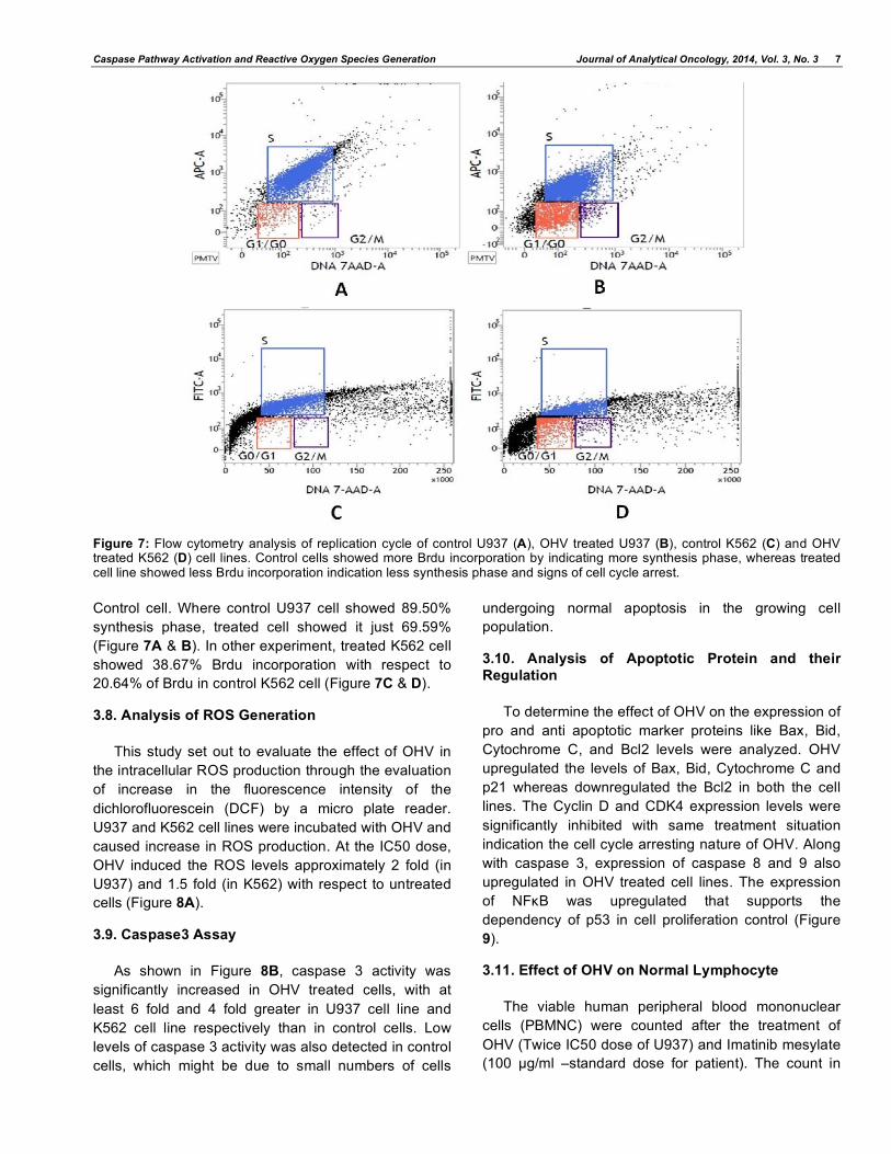
Flow cytometric data analysis revealed that after 24
h of treatment of OHV (IC50 dose) 21.73 % in LL
Figure 1: Determination of IC50 dose of OHV using trypan blue exclusion test after 72 h. (A) IC50 dose of U937 was 4.2 μg/ml. (B) IC50 dose of K562 was 3.9 μg/ml.
Figure 2: Cytotoxicity effect of OHV showed by MTT assay. Dose and time dependent cell inhibition of U937 cells (A) and K562 cells (B). The bar displays the mean±SEM of triplicate experiments (P< 0.05 compared to untreated controls).
Caspase Pathway Activation and Reactive Oxygen Species Generation Journal of Analytical Oncology, 2014, Vol. 3, No. 3 5
Figure 3: Representative micrographs of untreated control cells and OHV treated cells. Fluorescent microscopic images of control U937 (A), K562 (C) and OHV treated U937 (B), treated K562 (D) cell line.
Figure 4: Lactate dehydrogenase (LDH) activity assay. U937 cells (A) and K562 cells (B) were treated as indicated for 24 h. LDH activity is expressed in U/L according to standard cruve. Results from at least three separate experiments are presented, p<0.05 compared to controls.
quadrant, 54.33% in LR quadrant, 21.69 % in UR
quadrant and 2.25% in UL quadrant of U937 cells
(Figure 5B) with respect to 89.17 % in LL quadrant,
1.37% in LR quadrant, 0.89% in UR quadrant and
8.57% of control cell (Figure 5A) and 44.84% in LL
quadrant, 42.89% in LR quadrant, 12.24 % in UR
quadrant and 0.035% of K562 cells (Figure 5D) with
respect to 98.42 % in LL quadrant, 1.45%in LR
quadrant, and 0.06% in UR quadrant and 0.07% of
control cell (Figure 5C).
3.6. Study of Cell Cycle Arrest by Flow Cytometric Analysis
The flow cytometric analysis showed OHV (IC50
dose) treatment caused a progressive increase in the
number of cells with 2N DNA content, which indicated
G1 phase cell cycle arrest of U937 and k562 cells.
Where treated U937 cell showed 52.60% G1+G0,
40.56% S and 2.85% G2+M (Figure 6B), the control
cell showed 21.02% G1+G0, 65.03% S and 11.50%
G2+M (Figure 6A) and where treated k562 cells
showed 76.92% G1+G0, 23.08% S and 0.00% G2+M
(Figure 6D), the control cell showed 43.02% G1+G0,
44.85% S and 10.07% G2+M (Figure 6C).
3.7. Estimation of Replication Cycle by BrdU Incorporation Using FACS
BrdU incorporation via replication cycle (synthesis
phase) was much less in treated cell with respect to
6 Journal of Analytical Oncology, 2014, Vol. 3, No. 3 Bhowmik et al.
Figure 5: Apoptotic cell death was assayed by flow cytometry. Dot plot showed of U937 cells (B) and K562 cells (D) after treated with IC50 dose of OHV for 24 h. Control untreated U937 cells (A) and Control untreated K562 cells (B).
Figure 6: Flow cytometry analysis of cell cycle of untreated control U937 cells (A), OHV treated U937 (B), control K562 cells (C) and treated K562 cells (D). Histogram of both the treated cell line showed the arrest of cell cycle in G1 phase. (x-axis denotes fluorescence intensity of propium iodide and y-axis denotes cell count).
Caspase Pathway Activation and Reactive Oxygen Species Generation Journal of Analytical Oncology, 2014, Vol. 3, No. 3 7
Figure 7: Flow cytometry analysis of replication cycle of control U937 (A), OHV treated U937 (B), control K562 (C) and OHV treated K562 (D) cell lines. Control cells showed more Brdu incorporation by indicating more synthesis phase, whereas treated cell line showed less Brdu incorporation indication less synthesis phase and signs of cell cycle arrest.
Control cell. Where control U937 cell showed 89.50%
synthesis phase, treated cell showed it just 69.59%
(Figure 7A & B). In other experiment, treated K562 cell
showed 38.67% Brdu incorporation with respect to
20.64% of Brdu in control K562 cell (Figure 7C & D).
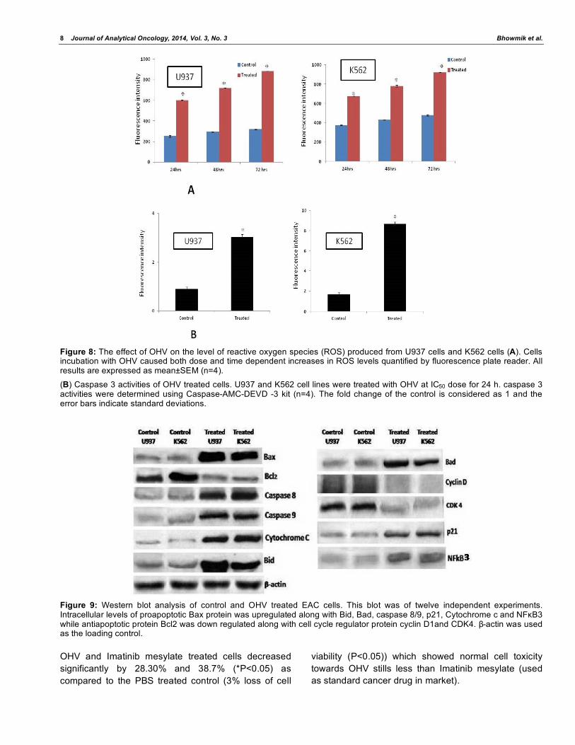
3.8. Analysis of ROS Generation
This study set out to evaluate the effect of OHV in
the intracellular ROS production through the evaluation
of increase in the fluorescence intensity of the
dichlorofluorescein (DCF) by a micro plate reader.
U937 and K562 cell lines were incubated with OHV and
caused increase in ROS production. At the IC50 dose,
OHV induced the ROS levels approximately 2 fold (in
U937) and 1.5 fold (in K562) with respect to untreated
cells (Figure 8A).
3.9. Caspase3 Assay
As shown in Figure 8B, caspase 3 activity was
significantly increased in OHV treated cells, with at
least 6 fold and 4 fold greater in U937 cell line and
K562 cell line respectively than in control cells. Low
levels of caspase 3 activity was also detected in control
cells, which might be due to small numbers of cells
undergoing normal apoptosis in the growing cell
population.
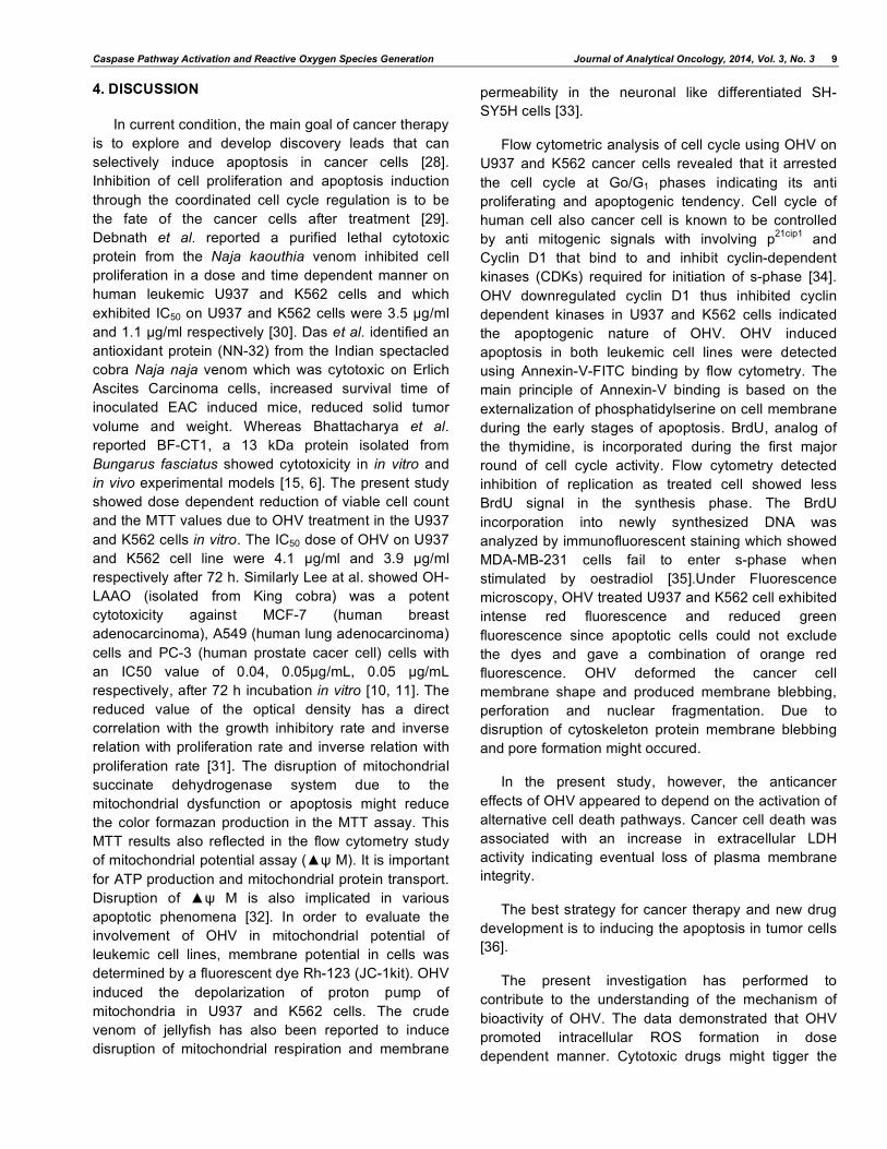
3.10. Analysis of Apoptotic Protein and their Regulation
To determine the effect of OHV on the expression of
pro and anti apoptotic marker proteins like Bax, Bid,
Cytochrome C, and Bcl2 levels were analyzed. OHV
upregulated the levels of Bax, Bid, Cytochrome C and
p21 whereas downregulated the Bcl2 in both the cell
lines. The Cyclin D and CDK4 expression levels were
significantly inhibited with same treatment situation
indication the cell cycle arresting nature of OHV. Along
with caspase 3, expression of caspase 8 and 9 also
upregulated in OHV treated cell lines. The expression
of NF B was upregulated that supports the
dependency of p53 in cell proliferation control (Figure
9).
3.11. Effect of OHV on Normal Lymphocyte
The viable human peripheral blood mononuclear
cells (PBMNC) were counted after the treatment of
OHV (Twice IC50 dose of U937) and Imatinib mesylate
(100 μg/ml –standard dose for patient). The count in
8 Journal of Analytical Oncology, 2014, Vol. 3, No. 3 Bhowmik et al.
Figure 8: The effect of OHV on the level of reactive oxygen species (ROS) produced from U937 cells and K562 cells (A). Cells incubation with OHV caused both dose and time dependent increases in ROS levels quantified by fluorescence plate reader. All results are expressed as mean±SEM (n=4).
(B) Caspase 3 activities of OHV treated cells. U937 and K562 cell lines were treated with OHV at IC50 dose for 24 h. caspase 3 activities were determined using Caspase-AMC-DEVD -3 kit (n=4). The fold change of the control is considered as 1 and the error bars indicate standard deviations.
Figure 9: Western blot analysis of control and OHV treated EAC cells. This blot was of twelve independent experiments. Intracellular levels of proapoptotic Bax protein was upregulated along with Bid, Bad, caspase 8/9, p21, Cytochrome c and NF B3 while antiapoptotic protein Bcl2 was down regulated along with cell cycle regulator protein cyclin D1and CDK4. -actin was used as the loading control.
OHV and Imatinib mesylate treated cells decreased
significantly by 28.30% and 38.7% (*P<0.05) as
compared to the PBS treated control (3% loss of cell
viability (P<0.05)) which showed normal cell toxicity
towards OHV stills less than Imatinib mesylate (used
as standard cancer drug in market).
Caspase Pathway Activation and Reactive Oxygen Species Generation Journal of Analytical Oncology, 2014, Vol. 3, No. 3 9
4. DISCUSSION
In current condition, the main goal of cancer therapy
is to explore and develop discovery leads that can
selectively induce apoptosis in cancer cells [28].
Inhibition of cell proliferation and apoptosis induction
through the coordinated cell cycle regulation is to be
the fate of the cancer cells after treatment [29].
Debnath et al. reported a purified lethal cytotoxic
protein from the Naja kaouthia venom inhibited cell
proliferation in a dose and time dependent manner on
human leukemic U937 and K562 cells and which
exhibited IC50 on U937 and K562 cells were 3.5 μg/ml
and 1.1 μg/ml respectively [30]. Das et al. identified an
antioxidant protein (NN-32) from the Indian spectacled
cobra Naja naja venom which was cytotoxic on Erlich
Ascites Carcinoma cells, increased survival time of
inoculated EAC induced mice, reduced solid tumor
volume and weight. Whereas Bhattacharya et al.
reported BF-CT1, a 13 kDa protein isolated from
Bungarus fasciatus showed cytotoxicity in in vitro and
in vivo experimental models [15, 6]. The present study
showed dose dependent reduction of viable cell count
and the MTT values due to OHV treatment in the U937
and K562 cells in vitro. The IC50 dose of OHV on U937
and K562 cell line were 4.1 μg/ml and 3.9 μg/ml
respectively after 72 h. Similarly Lee at al. showed OH-
LAAO (isolated from King cobra) was a potent
cytotoxicity against MCF-7 (human breast
adenocarcinoma), A549 (human lung adenocarcinoma)
cells and PC-3 (human prostate cacer cell) cells with
an IC50 value of 0.04, 0.05μg/mL, 0.05 g/mL
respectively, after 72 h incubation in vitro [10, 11]. The
reduced value of the optical density has a direct
correlation with the growth inhibitory rate and inverse
relation with proliferation rate and inverse relation with
proliferation rate [31]. The disruption of mitochondrial
succinate dehydrogenase system due to the
mitochondrial dysfunction or apoptosis might reduce
the color formazan production in the MTT assay. This
MTT results also reflected in the flow cytometry study
of mitochondrial potential assay ( M). It is important
for ATP production and mitochondrial protein transport.
Disruption of M is also implicated in various
apoptotic phenomena [32]. In order to evaluate the
involvement of OHV in mitochondrial potential of
leukemic cell lines, membrane potential in cells was
determined by a fluorescent dye Rh-123 (JC-1kit). OHV
induced the depolarization of proton pump of
mitochondria in U937 and K562 cells. The crude
venom of jellyfish has also been reported to induce
disruption of mitochondrial respiration and membrane
permeability in the neuronal like differentiated SH-
SY5H cells [33].
Flow cytometric analysis of cell cycle using OHV on
U937 and K562 cancer cells revealed that it arrested
the cell cycle at Go/G1 phases indicating its anti
proliferating and apoptogenic tendency. Cell cycle of
human cell also cancer cell is known to be controlled
by anti mitogenic signals with involving p21cip1
and
Cyclin D1 that bind to and inhibit cyclin-dependent
kinases (CDKs) required for initiation of s-phase [34].
OHV downregulated cyclin D1 thus inhibited cyclin
dependent kinases in U937 and K562 cells indicated
the apoptogenic nature of OHV. OHV induced
apoptosis in both leukemic cell lines were detected
using Annexin-V-FITC binding by flow cytometry. The
main principle of Annexin-V binding is based on the
externalization of phosphatidylserine on cell membrane
during the early stages of apoptosis. BrdU, analog of
the thymidine, is incorporated during the first major
round of cell cycle activity. Flow cytometry detected
inhibition of replication as treated cell showed less
BrdU signal in the synthesis phase. The BrdU
incorporation into newly synthesized DNA was
analyzed by immunofluorescent staining which showed
MDA-MB-231 cells fail to enter s-phase when
stimulated by oestradiol [35].Under Fluorescence
microscopy, OHV treated U937 and K562 cell exhibited
intense red fluorescence and reduced green
fluorescence since apoptotic cells could not exclude
the dyes and gave a combination of orange red
fluorescence. OHV deformed the cancer cell
membrane shape and produced membrane blebbing,
perforation and nuclear fragmentation. Due to
disruption of cytoskeleton protein membrane blebbing
and pore formation might occured.
In the present study, however, the anticancer
effects of OHV appeared to depend on the activation of
alternative cell death pathways. Cancer cell death was
associated with an increase in extracellular LDH
activity indicating eventual loss of plasma membrane
integrity.
The best strategy for cancer therapy and new drug
development is to inducing the apoptosis in tumor cells
[36].
The present investigation has performed to
contribute to the understanding of the mechanism of
bioactivity of OHV. The data demonstrated that OHV
promoted intracellular ROS formation in dose
dependent manner. Cytotoxic drugs might tigger the
10 Journal of Analytical Oncology, 2014, Vol. 3, No. 3 Bhowmik et al.
intrinsic or mitochondrial mediated apoptosis. Bcl2
family proteins consisting of antiapoptotic as well as
proapoptotic proteins play a key role in the intrinsic
pathway of apoptosis. Bax, a proapototic protein is
considered to be an important marker for the apoptosis
pathway. This study demonstrated that OHV induce
apoptosis through alternations in Bax: Bcl2 ratio with
activation of the caspase 3 cascade in U937 and k562
leukemic cell line. Activation of caspases was occured
by nuclear fragmentation of DNA due to apoptogenic
trigger. Caspase3 share both caspase 9 (intrinsic) and
caspase 8 (extrinsic) mediated pathway of apoptogenic
signalling [37]. It was found that OHV diverse
proapoptotic signals converged at the mitochondrial
level, inducing the translocation of Cytochrome c into
the cytosol. Cytochrome c triggered caspase9
activation by initiating a downstream caspase cascade
through the complex formation in the cytosol that
followed by the activation of the executioner caspase 3
and finally cell death. Similarly, caspase3 mediated
apoptosis in A549 cells induced by cinobufocini; a toad
skin preparation had been reported [38].
An alternative extrinsic pathway also induced by
OHV in U937 and K562 cell line. This study showed
protein signalling from plasma membrane receptors to
the cytosol, occurred by activation of caspase 8
followed by increased expression of Bid which was
induced by upregulation of Bad, hence apoptotic cell
death by NF B signalling. Recently, the inhibition of
CDK2 and CDK4 and upregulation of CDKs inhibitor
(p21 and p27) was found by the causal of curcumin
together with BM-ANF1 [39]. As OHV was crude
venom it might contain many cytotoxic fractions which
might alone induced cell death by both intrinsic and
extrinsic apoptotic pathway.
Interestingly, OHV was less toxic towards normal
peripheral mononuclear cells than the standard drug
(Imatinib mesylate). The advantage in treatment of
cancer with advanced, natural agent based therapies is
the inherent leaky vasculature present serving
cancerous cells. The defective vascular architecture for
serve fast-growing cancers along with poor lymphatic
drainage allows them enhanced permeation and
retention effect (EPR effect). The ability to target
treatment to very specific cancer cells also uses a
cancer's own structure in that many cancers
overexpress particular antigens, even on their surface.
This makes them ideal targets for drug delivery as long
as the targets are not expressed in significant
quantities anywhere in normal healthy cell [40]. As
OHV is crude venom it contains many active subunits.
This gives them more cytotoxicity and lesser toxic to
normal cell as some of this active subunit might mask
the toxic effect of toxic subunit of OHV in normal
healthy cell. In other way, Imatinib mesylate is a
chemically synthetic drug and its chemical constitute
might advrse affect more in healthy normal cell. But the
toxicity study was not complete, which requires further
study in other normal cell lines in future.
5. CONCLUSION
It may be concluded that, Indian king cobra
(Ophiophagus hannah) crude venom (OHV) possessed
anti proliferative and apoptogenic activity on leukemic
(U937 and K562) cell line acting on both extrinsic and
extrinsic signalling pathway.
ACKNOWLEDGEMENT
This wok was partly sponsored by UGC-BSR
fellowship (Govt. of India) to Prof. Antony Gomes.
CONFLICT OF INTEREST
No confliction of interest among authors.
REFERENCE
[1] Siegel R, Naishadham D, Jemal A. Cancer statistics, 2012. CA: A Cancer J Clin 2012; 62: 10-29. http://dx.doi.org/10.3322/caac.20138
[2] Baskar R, Lee KA, Yeo R, Yeoh KW. Cancer and radiation therapy: current advances and future directions. Int J Med
Sci 2012; 9: 193-9. http://dx.doi.org/10.7150/ijms.3635
[3] Gomes A, Bhattacharjee P, Mishra R, Biswas KA, Dasgupta CS, Giri B. Anticancer potential of animal venoms and toxins. Indian J Exp Biol 2010; 48: 93-103.
[4] Debnath A, Chatterjee U, Das M, Vedasiromoni RJ, Gomes
A. Venom of Indian monocellate cobra and Russell’s viper show anticancer activity in experimental models. J Ethnopharmacol 2007; 111: 681-684. http://dx.doi.org/10.1016/j.jep.2006.12.027
[5] Debnath A, Saha A, Gomes A, Biswas S, Chakraborty P, Gomes A, et al. A lethal cardiotoxic-cytotoxic protein from the Indian monocellate cobra (Naja kaouthia) venom. Toxicon
2010; 56: 569-579. http://dx.doi.org/10.1016/j.toxicon.2010.05.016
[6] Bhattacharya S, Das T, Biswas A, Gomes A, Gomes A, Dungdung RS. A cytotoxicity protein (BF-CT1), purified from Bungarus fasciatus venom acts through apoptosis,
modulation of PI3K/AKT, MAPKinase pathway and cell cycle regulation. Toxicon 2013; 74: 138-150. http://dx.doi.org/10.1016/j.toxicon.2013.08.052
[7] Whitaker R. In Common Indian snakes, A field guide (The Macmilan Company of India Limited, New Delhi, India) 1978.
[8] Lee CY, In Advances in cytopharmacology, Vol. 3, edited by Ceccarelli B & Clementi F (Raven press, New York) 1979,1.
[9] Lee ML, Tan NH, Fung SY, Shamala DS. Antibacterial action
of a heat stable form of L-amino acid oxidase isolated from king cobra (Ophiophagus hannah) venom. Comp Biochem Physiol C 2011; 153: 237-242.
Caspase Pathway Activation and Reactive Oxygen Species Generation Journal of Analytical Oncology, 2014, Vol. 3, No. 3 11
[10] Lee ML, Chung I, Fung SY, Kanthimathi MS, Tan NH.
Antiproliferative activity of king cobra (Ophiophagus hannah) venom L-amino acid oxidase. Basic Clin Pharmacol Toxicol 2014; 114: 336-343. http://dx.doi.org/10.1111/bcpt.12155
[11] Lee ML. Fung SY, Chung I, Palioor J, Cheah SH, Tan NH.
King cobra (Ophiophagus hannah) venom L-amino acid oxidase induces apoptosis in PC-3 cells and suppresses PC-3 solid tumor xenograpt mouse model. Int J Med Sci 2014;
11: 593-601. http://dx.doi.org/10.7150/ijms.8096
[12] Gomes A, De P. Hannahpep: a Novel Fibrinolytic Peptide from the Indian King Cobra (Ophiophagus hannah) venom.
Biochem Biophys Res Comp 1999; 266: 488-491. http://dx.doi.org/10.1006/bbrc.1999.1818
[13] Agrawal S, Ikeuchi T, Sun D, Sarin PS, Konopka A, Maizel J, et al. Inhibition of human immunodeficiency virus in early infected and chronically infected cells by antisense
oligodeoxynucleotides and their phosphorothiooate analogues. Proc Natl Acad Sci 1989; 86: 7790-7794. http://dx.doi.org/10.1073/pnas.86.20.7790
[14] Kawada K, Yonei T, Ueoka H, Kiura K, Tabata M, Takigawa
N, Harada M, Tanimoto M. Comparison of chemosensitivity tests: clonogenic assay versus MTT assay. Acta Med okayama 2002; 56: 129-134.
[15] Das T, Bhattacharya s, Halder B, Biswas A, Dasgupta S, Gomes A, et al. Cytotoxic and antioxidant property of a
purified fraction (NN-32) of Indian Naja naja venom on Ehrlich ascites carcinoma in BALB/c mice. Toxicon 2011; 57: 1065-1072. http://dx.doi.org/10.1016/j.toxicon.2011.04.012
[16] Bhowmik T, Saha PP, Dasgupta A, Gomes A. Antileukemic
potential of PEGylated Gold nanoparticle conjugated with protein toxin (NKCT1) isolated from Indian cobra (Naja kaouthia) venom. Cancer Nano 2013; 4: 39-55. http://dx.doi.org/10.1007/s12645-013-0036-5
[17] Wu M, Ming W, Tang Y, Zhou S, Dong W. The anticancer effect of cytotoxin 1 from Naja atra Cantor venom is mediated by a lysosomal cell death pathway involving
lysosomal membrane permeabilization and Cathepsin b release. Am J Chin Med 2013; 41(3): 643-63. http://dx.doi.org/10.1142/S0192415X13500456
[18] Roy S, Besra ES, De T, Banerjee B, Mukherjee J, Vedasiromoni RJ. Induction of apoptosis in Human leukemic
cell lines U937, K562 and HL-60 by Litchi chinensis leaf extract via activation of mitochondria mediated caspase cascades. Open Leukemia J 2008; 1: 1-14. http://dx.doi.org/10.2174/1876816400801010001
[19] Acharya S, Sahoo KS. Sustained targeting of Bcr-Abl+ leukemia cells by synergistic action of dual drug loaded nanoparticles and its implication for leukemia therapy.
Biomaterials 2011; 32: 5643-5662. http://dx.doi.org/10.1016/j.biomaterials.2011.04.043
[20] McGowan ME, Alling N, Jackson AE, Yagoub D, Hass KN, Allen DJ, et al. Evaluation of cell cycle arrest in estrogen responsive MCF7 Breast cancer cells: pitfalls of the MTS
assay. PLoS ONE 2011; 6(6): e20623. doi:10.1371/jounal.pone.0020623.
[21] Lin PJ, Yang SJ, Lee HJ, Hsieh TW, Chung GJ. Berberine induces cell cycle arrest and apoptosis in human gastric
carcinoma SNU-5ncell line. World J Gastroenterol 2006; 12(1): 21-28.
[22] Jung JH, Park Y-J, Jeon S-H, Kwon H-T. Aquaporin-5: A marker protein for proliferation and migration of Human Breast Cancer Cells. PLoS ONE 2011; 6(12): e28492. Doi: 10.1371/journal.pone0028492.
[23] Levokoff HL, Marsall PG, Ross HH, Caldeira M, Reynolds AB, Cakiroglu M, et al. Bromodeoxyuridine inhibits cancer cell proliferation in vitro and in vivo. Neoplasia 2008; 10(8): 804-816.
[24] Condello S, Curro M, Ferlazzo N, Caccamo D, Satriano J,
Ientile R. Agmatine effects on mitochondrial membrane potential and NF- B activation protect against rotenone-induced cell damage in human neuronal-like SH-SY5Y cells.
J Neurochem 2011; 116: 67-75. http://dx.doi.org/10.1111/j.1471-4159.2010.07085.x
[25] Malik F, Kumar A, Bhushan S, Khan S, Bhatia A, Suri AK, et al. Reactive oxygen species generation and mitochondrial dysfunction in the apoptotic cell death of human myeloid
leukemia HL-60 cells by a dietary compound withaferin A with concomitant protection by N-acetyl cysteine. Apoptosis 2007; 12: 2115-2133. http://dx.doi.org/10.1007/s10495-007-0129-x
[26] Achiwa Y, Haegawa K, Komiya T, Udagawa Y. Ursolic acid induces bax-dependent apoptosis through the caspase-3 pathway in endometrial cancer SNG-II cells. Oncol Rep 2005; 13(1): 51-57.
[27] Chaudhary H, Dhuna V, Shing J, Kanboj SS, Seshadri S.
Evaluation of hydro-alcoholic extract of Elipta alba for its anticancer potential: an in vitro study. J Ethnopharmacol 2011; 136(2): 363-367. http://dx.doi.org/10.1016/j.jep.2011.04.066
[28] Denicourt C, Dowdy SF. Medicine: Targeting apoptotic pathways in cancer cells. Science 2004; 305: 1411-1413. http://dx.doi.org/10.1126/science.1102974
[29] Lam MH, Liu Q, Elledge SJ, Rosen JM. Chk1 is haplo in sufficient for multiple function critical to tumor suppression.
Cancer Cell 2004; 6(1): 45-59. http://dx.doi.org/10.1016/j.ccr.2004.06.015
[30] Debnath A, Saha A, Gomes A, Biswas S, Chakrabarti P, Giri B, et al. A lethal cardiotoxic –cytotoxic protein from the Indian monocellate cobra (Naja kaouthia) venom. Toxicon 2010; 56:
569-579. http://dx.doi.org/10.1016/j.toxicon.2010.05.016
[31] Andrew KJ, Liu H, Suzi M, Vural ME, Xiao D, Weinstein IB. Resveratrol induces growth inhibition, s-phase arrest,
apoptosis and changes in biomarker expression in several human cancer cell lines. Clin Cancer Res 2002; 8: 893-903.
[32] Kwong JQ, Henning MS, Starkov AA, Manfredi G. The mitochondrial respiratory chain is a modulator of apoptosis. J Cell Biol 2007; 179: 1163-1177. http://dx.doi.org/10.1083/jcb.200704059
[33] Morabito R, Condello S, Curro M, Marino A, Ientile R, La Spada G. Oxidative stress induced by crude venom from the jellyfish Pelagia noctiluca in neural- like differentiated SH-
SY5Y cell. Toxicol In Vitro 2012; 26: 694-699. http://dx.doi.org/10.1016/j.tiv.2012.03.002
[34] Lou Y, Hurwitz J, Massagne J. Cell cycle inhibition by independent CDK and PCNA binding domains in p
21cip1.
Nature 1995; 375 (6527): 159-161. http://dx.doi.org/10.1038/375159a0
[35] Castoria G, Barone VM, Domenico DM, Bilancio A, Ametrano D, Migliaccio A, et al. Non-transcriptional action of oestradiol and progestin triggers DNA synthesis. EMBO J 1999; 18:
2500-2510. http://dx.doi.org/10.1093/emboj/18.9.2500
[36] Cheng X, Xiao Y, Wang X, Wang P, Li H, Liu Q. Anti-tumor and proapoptotic activity of ethanolic extract and its various fractions from Polytrichum commune L. ex Hedw in L1210
cells. J Ethnopharmacol 2012; 143: 49-56. http://dx.doi.org/10.1016/j.jep.2012.05.054
[37] Ghobrial IM, Witzig TE, Adjei A. Targeting apoptosis pathways in cancer therapy. CA. Cancer J Clin 2012; 55:
178-194. http://dx.doi.org/10.3322/canjclin.55.3.178
[38] Wang J, Jin Y, Xu Z, Zheng Z, Wan S. Involvement of caspase-3 activity and surviving downregulation in cinobufocini-induced apoptosis in A594 cells. Exp Biol Med
2009; 234(5): 566-572.

12 Journal of Analytical Oncology, 2014, Vol. 3, No. 3 Bhowmik et al.
http://dx.doi.org/10.3181/0811-RM-326
[39] Giri B, Gomes A, Sengupta R, Banerjee S, Nautiyal J, Sarkar FH, et al. Curcumin synergizes the growth inhibitory
properties of Indian toad (Bufo melanostictus, Schneider) skin derived factor (BM-ANF1) in HCT-116 colon cancer
cells. Anticancer Res 2009; 29(1): 395-401.
[40] Brannon-Peppas L, Blanchette JO. Nanoparticle and targetted systems for cancer therapy. Adv Drug Deliv Rev
2012; 64: 206-212. http://dx.doi.org/10.1016/j.addr.2012.09.033
Top Related
Copyright © 2022 FDOKUMEN

