


Bahasa
Halaman
Hukum


Brucellosis Vaccines: Assessment of Brucella melitensisLipopolysaccharide Rough Mutants Defective in Coreand O-Polysaccharide Synthesis and ExportDavid Gonzalez1, Marıa-Jesus Grillo2, Marıa-Jesus De Miguel3, Tara Ali4, Vilma Arce-Gorvel 5,6,7,
Rose-May Delrue8, Raquel Conde-Alvarez1, Pilar Munoz3, Ignacio Lopez-Goni1, Maite Iriarte1, Clara-M.
Marın3, Andrej Weintraub9, Goran Widmalm4, Michel Zygmunt10, Jean-Jacques Letesson8, Jean-Pierre
Gorvel 5,6,7, Jose-Marıa Blasco3, Ignacio Moriyon1*
1 Department of Microbiology and Parasitology, University of Navarra, Pamplona, Spain, 2 Instituto de Agrobiotecnologıa, CSIC-UPNA-Gobierno de Navarra, Pamplona,
Spain, 3 Centro de Investigacion y Tecnologıa Agroalimentaria (CITA), Sanidad Animal, Gobierno de Aragon, Zaragoza, Spain, 4 Arrhenius Laboratory, Stockholm
University, Stockholm, Sweden, 5 Centre d’Immunologie de Marseille-Luminy, Aix Marseille Universite, Faculte de Sciences de Luminy, Marseille, France, 6 INSERM, U631,
Marseille, France, 7 CNRS, UMR6102, Marseille, France, 8 Laboratoire d’Immunologie et Microbiologie - Unite de Recherche en Biologie Moleculaire (URBM), Facultes
Universitaires - Notre-Dame de la Paix (FUNDP), Namur, Belgium, 9 Karolinska Institute, Department Laboratory Medicine, Division of Clinical Bacteriology, Karolinska
University Hospital, Stockholm, Sweden, 10 INRA, UR1282, Infectiologie Animale et Sante Publique, IASP, Nouzilly, France
Abstract
Background: The brucellae are facultative intracellular bacteria that cause brucellosis, one of the major neglected zoonoses.In endemic areas, vaccination is the only effective way to control this disease. Brucella melitensis Rev 1 is a vaccine effectiveagainst the brucellosis of sheep and goat caused by B. melitensis, the commonest source of human infection. However, Rev1 carries a smooth lipopolysaccharide with an O-polysaccharide that elicits antibodies interfering in serodiagnosis, a majorproblem in eradication campaigns. Because of this, rough Brucella mutants lacking the O-polysaccharide have beenproposed as vaccines.
Methodology/Principal Findings: To examine the possibilities of rough vaccines, we screened B. melitensis forlipopolysaccharide genes and obtained mutants representing all main rough phenotypes with regard to coreoligosaccharide and O-polysaccharide synthesis and export. Using the mouse model, mutants were classified into fourattenuation patterns according to their multiplication and persistence in spleens at different doses. In macrophages,mutants belonging to three of these attenuation patterns reached the Brucella characteristic intracellular niche andmultiplied intracellularly, suggesting that they could be suitable vaccine candidates. Virulence patterns, intracellularbehavior and lipopolysaccharide defects roughly correlated with the degree of protection afforded by the mutants uponintraperitoneal vaccination of mice. However, when vaccination was applied by the subcutaneous route, only two mutantsmatched the protection obtained with Rev 1 albeit at doses one thousand fold higher than this reference vaccine. Thesemutants, which were blocked in O-polysaccharide export and accumulated internal O-polysaccharides, stimulated weakanti-smooth lipopolysaccharide antibodies.
Conclusions/Significance: The results demonstrate that no rough mutant is equal to Rev 1 in laboratory models andquestion the notion that rough vaccines are suitable for the control of brucellosis in endemic areas.
Citation: Gonzalez D, Grillo M-J, De Miguel M-J, Ali T, Arce-Gorvel V, et al. (2008) Brucellosis Vaccines: Assessment of Brucella melitensis Lipopolysaccharide RoughMutants Defective in Core and O-Polysaccharide Synthesis and Export. PLoS ONE 3(7): e2760. doi:10.1371/journal.pone.0002760
Editor: Kirsten Nielsen, University of Minnesota, United States of America
Received April 23, 2008; Accepted June 24, 2008; Published July 23, 2008
Copyright: � 2008 Gonzalez et al. This is an open-access article distributed under the terms of the Creative Commons Attribution License, which permitsunrestricted use, distribution, and reproduction in any medium, provided the original author and source are credited.
Funding: This work was funded by the European Commission (Research Contract QLK2-CT-2002-00918) and the Ministerio de Ciencia y Tecnologıa of Spain(Proyecto AGL2004-01162/GAN).
Competing Interests: The authors have declared that no competing interests exist.
* E-mail: [email protected]
Introduction
Brucellosis is a group of closely related zoonotic bacterial
diseases caused by the members of the genus Brucella, a group of
gram-negative bacteria that behave as facultative intracellular
parasites. There are several Brucella species, and they infect a wide
range of mammals in which they are a main cause of abortions
and infertility. In addition, they are readily transmitted to human
beings where they produce a grave and debilitating disease that
requires a long and costly antibiotic therapy and that often leaves
permanent sequelae [1]. Because of its high incidence in
developing countries, economic consequences, and difficult
eradication, the World Health Organization considers brucellosis
as one of the seven neglected zoonoses, a group of diseases that
contribute to the perpetuation of poverty [2].
Ruminants are highly susceptible to brucellosis. Cattle are most
often infected by B. abortus whereas sheep and goats are the
preferred hosts of B. melitensis, the Brucella species most virulent for
PLoS ONE | www.plosone.org 1 July 2008 | Volume 3 | Issue 7 | e2760

humans [3]. Although there is no human vaccine, vaccination of
animals against brucellosis is one of the most cost-effective
measures to improve human health in endemic areas [4] as well
as an essential tool to achieve eradication [5,6]. For these
purposes, vaccines B. abortus S19 and B. melitensis Rev 1 have
been successfully used in some developed countries, but both
induce abortions when applied during pregnancy, are virulent for
humans and elicit antibodies to the smooth (S) lipopolysaccharide
(LPS) of the Brucella surface that interfere in serodiagnosis.
Moreover, Rev 1 is resistant to streptomycin, an antibiotic used
to treat the disease. Although the serodiagnosis problem can be
partially solved by using the conjunctival route, by avoiding adult
vaccination, and by an individual serological follow up, the
breeding conditions characteristic of small ruminants make these
measures unrealistic in large areas of the world. Therefore,
effective brucellosis vaccines not interfering in diagnosis would
represent a major breakthrough [7,8].
Rough (R) Brucella mutants lack the LPS immunodominant N-
formylperosamine O-polysaccharide (O-PS) and are attenuated.
Thus, they have been the subject of great attention as alternative
vaccines [7,8]. Some R vaccines or candidates are spontaneous
mutants selected after repeated passage on antibiotic-containing
media. This approach was used to obtain RB51, a B. abortus R
mutant that carries a IS711-disrupted wboA (putative glycosyl-
tranferase gene) as well as unknown mutations also affecting LPS.
However, RB51 has yielded controversial results in cattle, is not
effective in sheep and is resistant to rifampin, an antibiotic used to
treat brucellosis [8]. B. melitensis RBM9, RBM11, RBM15,
RBM17 and RBM19 have been obtained by a similar method
and, as expected, they carry undefined LPS defects and are
rifampin resistant [9]. Targeted and transposon mutagenesis have
also been used. Disruption of per, wboA and wbkA (putative
perosamine synthetase and glycosyltranferase genes) results in R
mutants that outperform RB51 in the mouse model [10–12],
showing that empirically R vaccines can be improved. However, R
mutants can result from mutations affecting O-PS precursor
synthesis, its polymerization and transport or from a variety of
defects in the inner core oligosaccharide. Presently, it is not known
which of these mutations results in the best R vaccine or how such
a vaccine would compare with S19 or Rev 1. To address these
questions, we investigated 14 LPS B. melitensis mutants with respect
to LPS defects, intracellular multiplication and virulence and
vaccine efficacy in mice. The results provide the basis for selecting
the best R candidates for sheep and goat vaccination against B.
melitensis, and are also relevant for an appraisal of the value of
brucellosis R vaccines.
Results
Mutagenesis, mutant selection and growthcharacteristics
Perusal of the literature shows that 16M (reference strain of B.
melitensis biovar 1 and the one sequenced) does not always show the
virulence levels of other S brucellae [12–14]. Because of this, we
used this strain and also B. melitensis H38 (fully virulent [15]) to
obtain the nalidixic acid resistant (NalR) derivatives necessary for
mutagenesis and confirmed that this selection did not affect their
characteristics in mice (not shown). Then, we performed
transposon mutagenesis and obtained ca. 16,500 kanamycin
resistant mutants from which we selected 23 that displayed a
stable R phenotype both in vitro and in mice, as judged by the
crystal violet test, lack of reactivity with anti O-PS antibodies and
phage typing. These mutants mapped in 16 open reading frames
(ORFs) scattered into eight regions. Complementation failed to
restore the S phenotype in mutants in four of these ORFs.
Moreover, mutagenesis of the adjacent ORFs ruled out both
possible polarity effects and the involvement in LPS synthesis of
some ORFs with suggestive annotations (Table S1). We confirmed
the R phenotype of the remaining mutants (Table 1) by examining
the LPS by the sodium dodecylsulfate-proteinase K method and,
for ORFs BMEI1326, BMEI1414, BMEI1426 and BMEI1427, by
making in frame deletion mutants. Finally, since some mutants
showed differences in colony size suggestive of altered growth
rates, we obtained the corresponding growth curves. Mutants in
BMEI1415 (wzm), BMEI1886 (pgm) and BMEI1326 (wa**) were
retarded with respect to the parental strain whereas BMEII0899
(manBcore) grew faster (Figure S1).
Table 1. ORFs shown to be involved in B. melitensis LPS synthesis.
ORF Strain (nu mutants) 1Mutagenesis Role (R phenotype) LPS gene Annotation
BMEI0997 16M (1) Transposon O-PS synthesis (R1) wboB Mannosyltransferase
BMEI0998 H38 (1) Transposon O-PS synthesis (R1) wboA Mannosyltransferase
BMEI1326 16M (1) Transposon and in frame deletion Core synthesis (R2) wa** Glycosyltransferase
BMEI1393 H38 (2),16M (1) Transposon O-PS synthesis (R1) wbkE Mannosyltransferase
BMEI1396 16M (1) Targeted mutagenesis Uncertain manB Phosphomannomutase
BMEI1404 16M (1) Transposon O-PS synthesis (R1) wbkA Mannosyltransferase
BMEI1413 16M (1) Transposon O-PS synthesis (R1) gmd GDP-mannose dehydratase
BMEI1414 16M (1), H38 (2) Transposon and in frame deletion O-PS synthesis (R1) per Perosamine synthetase
BMEI1415 16M (1) Transposon O-PS synthesis (R1) wzm ABC transporter
BMEI1426 H38 (3) Transposon and in frame deletion O-PS synthesis (R1) wbkF Undecaprenyl-glycosyltransferase
BMEI1427 H38 (2) Transposon and in frame deletion O-PS synthesis (R1) wbkD Epimerase/dehydratase
BMEI1886 16M (1) Transposon Core synthesis (R2) pgm Phosphoglucomutase
BMEII0899 H38 (1) Transposon Core synthesis (R3) manBcore Phosphomannomutase
116M, B. melitensis 16M NalR; H38, B. melitensis H38NalR2n.a., not applicable.doi:10.1371/journal.pone.0002760.t001
Brucella melitensis R Vaccines
PLoS ONE | www.plosone.org 2 July 2008 | Volume 3 | Issue 7 | e2760

The mutations block the major LPS polysaccharidesynthesis pathways
In order to assign the mutations to the O-PS or the core
oligosaccharide biosynthetic pathways, we first classified the
mutants as R1, R2 or R3 according to the decrease in LPS
molecular weight (Figure 1 and Table 1). Three R1 mutants
mapped in per (BMEI1414, Table 1). Since per is involved in the
synthesis of perosamine [16], the only Brucella O-PS sugar [17], we
took this as evidence for a complete core in R1 and, consequently,
for a mutation affecting the O-PS. Likewise, the progressively
defective core in R2 and R3 mutants indicated that the
corresponding ORF belonged to core synthesis pathways.
Consistent with these interpretations, the 2-keto-3-deoxyoctulo-
sonic acid ([Kdo] inner core marker) content of solvent-extracted
R-LPS of BMEI1414 and BMEI1427 (R1), BMEI1326 (R2) and
BMEII0899 (R3) mutants was 3.8, 4.0, 6.8 and 9.5%, respectively.
Mutants affected in O-PS synthesis. Mutants in
BMEI0997 and BMEI0998 and in ORFs from BMEI1393 to
BMEI1427 were of the R1 type (Table 1 and Figure 2A). The two
first ORFs, putatively encoding glycosyltransferases, included the
previously described wboA (BMEI0998) [18]. According to their
R1 phenotype, these two ORFs are implicated in O-PS synthesis
and BMEI0997 was thus named wboB. Likewise, the R1
phenotype of BMEI1393 matched its annotation as a
mannosyltransferase gene (mannose and perosamine are related)
and its location close to wbkA (BMEI1404), the first ORF of the O-
PS wbk region [16] (Figure 2B). Because of this, we named the
gene wbkE. The R1 phenotype of mutants in genes wbkA, gmd, per
and wzm, all in the wbk region, was also in keeping with the roles
proposed in previous works (Table 1 and Figure 2A) [11,16]. Two
other R1 mutants mapped close to wbk (Figure 2B). The first one
(BMEI1426), separated by several IS from wbkC (BMEI1418), was
identified before as a LPS gene in B. abortus [19] and encodes a 335
amino acid protein with homology to polyisoprenyl-phosphate N-
acetylhexosamine-1-phosphate transferases (PNPT). Known
PNPTs include Pseudomonas aeruginosa PAO1 WbpL and
Escherichia coli WecA, both involved in bactoprenol priming but
belonging to two subfamilies that differ in aminosugar specificity
[20]. A search for WbpL (339 amino acids) homologues in B.
melitensis identified BMEI1426 as the closest one (BLAST E-
value = 4e222). For WecA (367 amino acids), not only BMEI1426
(1e1028) but also BMEII0839 (1e10242) were identified. However,
the only WecA match in B. abortus genomes was the BMEI1426
ortholog (BruAb1_0533 and BAB1_05359-49 in B. abortus 9-49
and 2308, respectively). This result, which is due to a large deletion
in B. abortus chromosome II encompassing the position of the
BMEII0839 ortholog [21], supports the role of BMEI1426 as the
Brucella PNPT gene. The BMEI1426 protein resembled WecA and
WbpL in the Asp rich consensus motif of the catalytic site [20] but
did not show the motifs thought to relate to substrate specificity in
either subfamily. Since it could not be ascribed to any of these
subfamilies, we named this gene wbkF. Finally, we named the
adjacent ORF (BMEI1427) wbkD. The protein is annotated as an
epimerase/dehydratase and carries a NAD or NADP binding motif,
two features of the trifunctional UDP-N- D-acetylglucosamine 4,6-
dehydratase/5-epimerase/3-epimerase enzymes that take part in the
synthesis of 2-acetamido-2,6-dideoxy-L-hexoses such as N-
acetylfucosamine and N-acetylquinovosamine [22]. Quinovosamine
(2-amino-2,6-dideoxy-D-glucose) has been reported in the LPS of S
Brucella spp. [23–27] but its location is unclear. Since N-
acetylquinovosamine synthesis would require an epimerase/
dehydratase using NADP as the coenzyme [22], we investigated
the S-LPS hydrolytic polysaccharides of B. melitensis and B. abortus. In
addition to the N-formylperosamine signals, a resonance at
2.06 ppm. indicated the presence of an N-acetyl group in these
materials. The integrals of these signals compared to those from the
methyl groups of perosamine at ,1.3 ppm showed that the former
derived from ,1% of the material. Moreover, the 1H NMR
spectrum at 25uC of the B. abortus material (which was highly pure)
showed a resonance at 4.56 ppm having JH1,H2 = 7.9 Hz. Further
analysis was performed at 70uC (Figure S2) using in particular 2D 1H,1H- total correlation spectroscopy (TOCSY) with spin-lock times up
to 120 ms by which a complete spin-system corresponding to a
quinovosamine residue was traced out. The following chemical shifts
were identified: d 4.54 (H1), 3.79 (H2), 3.66 (H3), 3.35 (H4), 3.50
(H5), and 1.30 (H6). Thus, the analysis revealed the characteristic 1H
NMR chemical shifts of a b-linked quinovosamine, which should be
N-acetylated and is presumably 3-substituted [28,29]. Indeed, this N-
acetylquinovosamine could be the substrate of the putative PNPT
WbkF, since WbkF is close to WbpL and the latter transfers either N-
acetylfucosamine or N-acetylquinovosamine [20]. Moreover, the
adjacent position of wbkF and wbkD also suggests a functional
connection.
Extension of the wbk region up to BMEI1393 (wbkE) (Figure 2B)
suggested that BMEI1394, BMEI1395 and BMEI1396 (putative
mannose-6-phosphate isomerase [manA], mannose-1-phosphate
guanylyltransferase [manC] and phosphomannomutase [manB])
could be involved in the synthesis of mannose, the precursor of
perosamine (Figure 2A). However, when we disrupted BMEI1396
(manB), the mutant still expressed O-PS demonstrating that the
gene was not essential for mannose synthesis.
Mutants affected in core oligosaccharide synthesis.
Mutant BMEI1326 had a R2 LPS like that observed before in a
B. abortus orthologous mutant [11], and encodes a predicted
glycosyltransferase of family 25. This is worth mentioning because
this family contains LPS glycosyltransferases, including the WaaX
protein that takes part in the synthesis of some E. coli core
chemotypes [30]. BMEI1326 is isolated from other LPS genes and,
in the absence of more information, we maintained its provisional
Figure 1. LPS profiles by SDS-PAGE. SDS-proteinase K extracts ofthe 16M NalR strain and of mutants representative of the R1(Bm16MRwboB [lane 1] and Bm16MRper [lane 2]), R2 (Bm16MRpgm[lane 3]) and R3 (BmH38RmanBcore [lane 4]) LPS types were analyzed ingels of the indicated acrylamide % and then periodate-silver stained.doi:10.1371/journal.pone.0002760.g001
Brucella melitensis R Vaccines
PLoS ONE | www.plosone.org 3 July 2008 | Volume 3 | Issue 7 | e2760

name (wa**) [11]. Mutant in BMEI1886 was affected in a putative
phosphoglucomutase (Pgm) and had a R2 LPS (Figure 2), in
agreement with a previous observation in B. abortus [31] and with
the presence of glucose in the Brucella LPS core [26]. Finally,
BMEII0899 disruption generated a R3 LPS (Figure 2) like the one
reported for the orthologous B. abortus mutants [11,19]. It is
annotated as phosphomannomutase gene and, because of the severe
core defect, we proposed before the name of manBcore [11]. However,
Figure 2. Genetics of B. melitensis S-LPS biosynthesis. (A), Pathways. Brucella grows with glucose as the only C source and is thus able toderive all S-LPS precursors from this sugar. The steps leading to N-formylperosamine synthesis and to its polymerization by Wbo and Wbkglycosyltransferases are in blue, and those leading to bactroprenol priming for N-formylperosamine polymerization in green. Once this happens, O-PSis translocated to the periplasm by the Wzm/Wzt ABC transporter (also in blue) and ligated to the core oligosaccharide which results from thepathways marked in red. The steps disrupted in this work are indicated by black triangles in which R1, R2, or R3 refer to the LPS phenotypes (thewbkC mutant is described in reference [16]). An empty triangle indicates a mutation that does not generate R phenotype, and a black triangle markedwith S a mutation that, while blocking the synthesis of a core lateral branch, does not prevent O-PS linkage to the core (Conde-Alvarez, R.,unpublished results). (B), The major (wbk) genetic region of Brucella O-PS synthesis. This region contains genes coding for enzymes necessaryfor N-formylperosamine synthesis (gmd, per, wbkC), two O-PS glycosyltransferases (wbkE, wbkA), the ABC transporters (wzm, wzt), the PNPT enzyme(wbkD) and at least one enzyme necessary for the synthesis of an N-acetylaminosugar (wbkF), as well as groups of insertion sequences (ISs) that makeit unstable. The mutations analyzed in this work are marked with triangles. Mutations in manB and wbkB do not generate R mutants (this work andreference [16]).doi:10.1371/journal.pone.0002760.g002
Brucella melitensis R Vaccines
PLoS ONE | www.plosone.org 4 July 2008 | Volume 3 | Issue 7 | e2760

taking into account the lack of R phenotype in the mutant in the
manB gene of region wbk (see above) and the absence of additional
manB annotations in Brucella, manBcore plus the contiguous manC seem
the only genes of the pathway providing mannose for both
perosamine and core synthesis (Figure 2A).
Mutants blocked in O-PS export. To study whether the R
mutants could elicit antibodies to the O-PS, we infected mice and
tested the sera in an enzyme-linked immunosorbent assay with
Brucella native hapten, a N-formylperosamine polysaccharide that
lacks core sugars [32]. We observed reactivity in the sera of mice
infected with 108 or 1010 colony forming units (CFU) of the wzm
(BMEI1415) mutant or with 1010 CFU of the wa** (BMEI1326)
mutant (0,810 and 0,714 optical density readings, respectively;
1:25 dilution). However, the antibody levels were lower than those
induced by 106 CFU of the 16M NalR parental strain (1,480
readings for the same dilution). When we tested extracts of these
mutants by gel immunodiffusion with sera from Brucella infected
cattle, we observed a component giving a reaction of identity with
the native hapten polysaccharide (Figure 3). Moreover, 1H-NMR
analysis of the extracts showed the signals of a1,2- a1,3-linked N-
formylperosamine polysaccharides [17] plus a small signal at
2.06 ppm corresponding to the N-acetyl group of an unidentified
N-acetylated aminosugar. Upon cell fractionation, the
polysaccharides were detected in the envelope and cytosol of the
wzm and wa** mutants, respectively.
Mutant designation. Based on the above analyses, we
designated the mutants according to the original strain (Bm16M
or BmH38), the phenotype (R) and the LPS gene disrupted
(Table 1 and Figure 2).
The LPS defects alter key topological, physicochemicaland biological surface properties of Brucella
It is known that outer membrane proteins (Omp) are more
exposed on R than on S brucellae [11,33], and we confirmed this for
R1, R2, and R3 mutants using monoclonal antibodies (Moabs) to
Omp1, Omp2b, Omp31, Omp25, Omp19, Omp16 and Omp10
(not shown). Less is know about the topology of R-LPS epitopes and,
therefore, we tested the mutants with the appropriate Moabs. In all
cases, absence of O-PS correlated with exposure of the outer core
(Figure 4, left panel) and the lipid A disaccharide (Moab Bala-1; not
shown). Surprisingly, the R1 and R2 but not the R3 mutants failed
Figure 3. B. melitensis mutants in genes wa** and wzmsynthesize N-formylperosamine polysaccharides. The figureshows a gel immunodiffusion analysis of the polysaccharides obtainedfrom mutants Bm16MRwa** and Bm16MRwzm. Well 16MNalR containedthe LPS of the parental strain and shows both the slow diffusing S-LPSand the fast diffusing native hapten polysaccharide precipitin lines; (S),serum from naturally infected cattle.doi:10.1371/journal.pone.0002760.g003
Figure 4. LPS epitopes in smooth B. abortus and B. melitensis strains and their cognate R mutants. Whole bacteria (left panel) or B.melitensis R-LPS representative of the R1, R2 and R3 types (right panel) were probed with monoclonal antibodies of the indicated specificity (Ba2308,B. abortus 2308; Bm16MNalR, B. melitensis 16M NalR; codes for the R mutants are those used in the text).doi:10.1371/journal.pone.0002760.g004
Brucella melitensis R Vaccines
PLoS ONE | www.plosone.org 5 July 2008 | Volume 3 | Issue 7 | e2760

to react with the inner core Moab Baro-2 (Figure 4, left panel). Since
Baro-2 was produced by immunization with B. abortus [34], we
confirmed its reactivity using B. abortus 2308 mutants in per
(Ba2308Rper), wbkA (Ba2308RwbkA), wa** (Ba2308Rwa**) and
manBcore (Ba2308R manBcore) (Figure 4 left panel). As expected, all
these R mutants but wa** [11] reacted with Baro-2. Then, we tested
representative B. melitensis R-LPS by Western blot. The LPS of
BmH38RwbkD (R1), Bm16MRper (R1) and Bm16MRwa** (R2) but
not that of BmH38RmanBcore (R3) reacted with Baro-1 (Figure 4,
right panel). Conversely, Baro-2 failed to detect an inner core
epitope in the R1 and R2 but not in the R3 LPS (Figure 4, right
panel). These results show hitherto undescribed LPS core
differences between B. abortus and B. melitensis.
All R mutants displayed an increased surface hydrophobicity,
more markedly in B. melitensis than in B. abortus (Figure 5, left
panel), and also in sensitivity to the polycationic lipopeptide
polymyxin B (not shown). The latter effect was explained by the
increase in negative Zeta potential (surface charge) of the R
mutants (Figure 5 right panel). However, we did not observe a
clear correlation between Zeta potential and LPS defects (258 to
264 mV for R1, 255 to 258 mV for R2, and 266 to 268 mV
for R3), like that obtained for the S. minnesota R chemotypes (Ra,
239 mV; Rc, 244 mV, and Re, 253 mV). Since the LPS core is
complete in Ra, lacks the distal N-acetyl-glucosamine in Rc, and is
reduced to two Kdo residues in Re, the comparison shows that the
outer core does not balance the negative charge of the Kdo-lipid A
section in Brucella LPS. In support of this interpretation, all Brucella
R mutants behaved like the S. minnesota Re mutant when the Zeta
potential was measured in the presence of the polycation poly-L-
lysine (Figure 5, right panel). The Zeta potential value and the
polycation effects were clearly reduced in the S parental strains S.
minnesota HL63 and B. melitensis H38 NalR (Figure 5, right panel) or
B. melitensis 16M NalR (not shown).
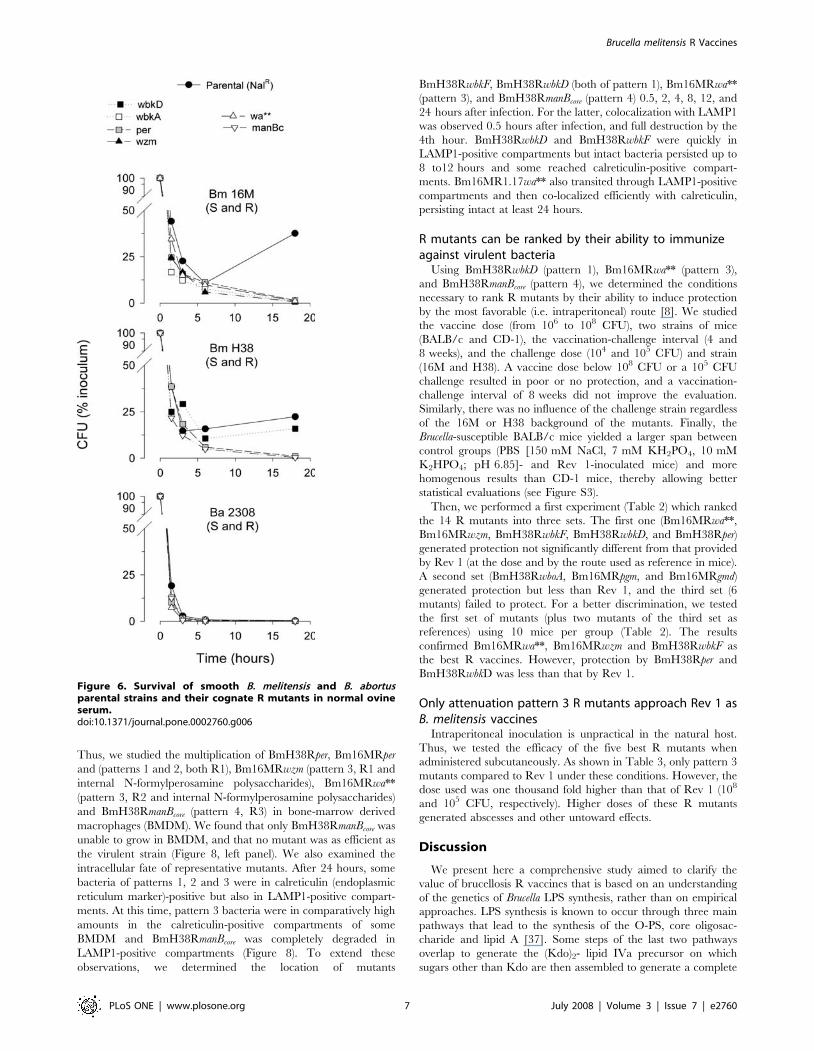
Resistance to complement-mediated killing in normal serum is a
biologically significant property of S brucellae [35]. We found that,
whereas the B. melitensis S strains were only partially affected by
normal sheep (Figure 6) or cattle serum (not shown), all B. melitensis
R mutants but BmH38RwbkD were killed within 20 hours.
Interestingly, neither the B. abortus 2308 NalR S strain nor the
cognate R mutants survived under the same conditions (Figure 6).
R mutants show different patterns of attenuationTo assess attenuation, we inoculated BALB/c mice with three
doses of each R mutant or with 106 CFU of the parental strains and
vaccine Rev 1, and determined the CFU numbers in the spleens. All
R mutants were less persistent than the parental strains, with
differences that allowed classifying them into four broad attenuation
patterns (Figure 7A). Pattern 1 comprised BmH38Rper,
BmH38RwbkD and BmH38RwbkF, all of the R1 type. They
multiplied at 106 CFU/mouse and, no matter the dose, reached
spleen counts at week 2 similar to those of the parental strain. Then,
the CFU decreased markedly. Pattern 2 included BmH38RwboA,
Bm16MRwboB, Bm16MRwbkE, BmH38RwbkE, Bm16MRwbkA,
Bm16MRgmd, Bm16MRper (all R1) and Bm16MRpgm (R2).
Although able to replicate, they never reached the parental strain
level and were cleared at rates directly related to the inoculum size.
Pattern 3 included Bm16MRwzm and Bm16MRwa**, two mutants
with internal N-formylperosamine polysaccharides and reduced in
vitro growth rates. Although not multiplying when inoculated at 106
CFU, they persisted at relatively high numbers at the end of the
experiment (week 6) for the 107 or 108 CFU doses. To further
examine their persistence, we inoculated mice with 108 CFU/
mouse and examined them 6, 9 and 12 weeks after infection. The
CFU/spleen at week 6 were similar to those of the first experiment,
declined more than 2 logs at week 9 and the mutants were cleared
by week 12. Remarkably, the results of pattern 3 mutants (107 or 108
CFU) and Rev 1 almost overlapped at weeks 2, 3, 6 (Figure 7), 9 and
12 (not shown). Finally, we separated BmH38RmanBcore (R3 and
accelerated growth in vitro) into pattern 4 because its clearance
started as early as week 2 and was complete between weeks 3 and 6.
As reported before [36], splenomegaly caused by virulent
Brucella increased throughout the experiment (Figure 7B). In
contrast, the weights of the spleens of Rev 1 inoculated mice
peaked at week 2 and declined afterwards. Only pattern 1 R
mutants inoculated with 108 CFU induced transient splenomegaly
similar to that of Rev 1 (Figure 7B).
Some R mutants reach the Brucella replicative niche inmacrophages
Since a brucellosis vaccine must persist long enough to trigger
protective immunity, it has to be able to multiply intracellulary.
Figure 5. Surface hydrophobicity (partition in hexanol/water) and charge (zeta potential in dependence of poly-L-lysine) of smoothB. abortus and B. melitensis parental strains and their cognate R mutants. For Brucella, the genes (left panel) or the strain codes (right panel)indicated are those used in the text. S. minnesota controls were: wild type, SmWT; Ra LPS mutant, SmRa; Re LPS mutant, SmRe.doi:10.1371/journal.pone.0002760.g005
Brucella melitensis R Vaccines
PLoS ONE | www.plosone.org 6 July 2008 | Volume 3 | Issue 7 | e2760
Thus, we studied the multiplication of BmH38Rper, Bm16MRper
and (patterns 1 and 2, both R1), Bm16MRwzm (pattern 3, R1 and
internal N-formylperosamine polysaccharides), Bm16MRwa**
(pattern 3, R2 and internal N-formylperosamine polysaccharides)
and BmH38RmanBcore (pattern 4, R3) in bone-marrow derived
macrophages (BMDM). We found that only BmH38RmanBcore was
unable to grow in BMDM, and that no mutant was as efficient as
the virulent strain (Figure 8, left panel). We also examined the
intracellular fate of representative mutants. After 24 hours, some
bacteria of patterns 1, 2 and 3 were in calreticulin (endoplasmic
reticulum marker)-positive but also in LAMP1-positive compart-
ments. At this time, pattern 3 bacteria were in comparatively high
amounts in the calreticulin-positive compartments of some
BMDM and BmH38RmanBcore was completely degraded in
LAMP1-positive compartments (Figure 8). To extend these
observations, we determined the location of mutants
BmH38RwbkF, BmH38RwbkD (both of pattern 1), Bm16MRwa**
(pattern 3), and BmH38RmanBcore (pattern 4) 0.5, 2, 4, 8, 12, and
24 hours after infection. For the latter, colocalization with LAMP1
was observed 0.5 hours after infection, and full destruction by the
4th hour. BmH38RwbkD and BmH38RwbkF were quickly in
LAMP1-positive compartments but intact bacteria persisted up to
8 to12 hours and some reached calreticulin-positive compart-
ments. Bm16MR1.17wa** also transited through LAMP1-positive
compartments and then co-localized efficiently with calreticulin,
persisting intact at least 24 hours.
R mutants can be ranked by their ability to immunizeagainst virulent bacteria
Using BmH38RwbkD (pattern 1), Bm16MRwa** (pattern 3),
and BmH38RmanBcore (pattern 4), we determined the conditions
necessary to rank R mutants by their ability to induce protection
by the most favorable (i.e. intraperitoneal) route [8]. We studied
the vaccine dose (from 106 to 108 CFU), two strains of mice
(BALB/c and CD-1), the vaccination-challenge interval (4 and
8 weeks), and the challenge dose (104 and 105 CFU) and strain
(16M and H38). A vaccine dose below 108 CFU or a 105 CFU
challenge resulted in poor or no protection, and a vaccination-
challenge interval of 8 weeks did not improve the evaluation.
Similarly, there was no influence of the challenge strain regardless
of the 16M or H38 background of the mutants. Finally, the
Brucella-susceptible BALB/c mice yielded a larger span between
control groups (PBS [150 mM NaCl, 7 mM KH2PO4, 10 mM
K2HPO4; pH 6.85]- and Rev 1-inoculated mice) and more
homogenous results than CD-1 mice, thereby allowing better
statistical evaluations (see Figure S3).
Then, we performed a first experiment (Table 2) which ranked
the 14 R mutants into three sets. The first one (Bm16MRwa**,
Bm16MRwzm, BmH38RwbkF, BmH38RwbkD, and BmH38Rper)
generated protection not significantly different from that provided
by Rev 1 (at the dose and by the route used as reference in mice).
A second set (BmH38RwboA, Bm16MRpgm, and Bm16MRgmd)
generated protection but less than Rev 1, and the third set (6
mutants) failed to protect. For a better discrimination, we tested
the first set of mutants (plus two mutants of the third set as
references) using 10 mice per group (Table 2). The results
confirmed Bm16MRwa**, Bm16MRwzm and BmH38RwbkF as
the best R vaccines. However, protection by BmH38Rper and
BmH38RwbkD was less than that by Rev 1.
Only attenuation pattern 3 R mutants approach Rev 1 asB. melitensis vaccines
Intraperitoneal inoculation is unpractical in the natural host.
Thus, we tested the efficacy of the five best R mutants when
administered subcutaneously. As shown in Table 3, only pattern 3
mutants compared to Rev 1 under these conditions. However, the
dose used was one thousand fold higher than that of Rev 1 (108
and 105 CFU, respectively). Higher doses of these R mutants
generated abscesses and other untoward effects.
Discussion
We present here a comprehensive study aimed to clarify the
value of brucellosis R vaccines that is based on an understanding
of the genetics of Brucella LPS synthesis, rather than on empirical
approaches. LPS synthesis is known to occur through three main
pathways that lead to the synthesis of the O-PS, core oligosac-
charide and lipid A [37]. Some steps of the last two pathways
overlap to generate the (Kdo)2- lipid IVa precursor on which
sugars other than Kdo are then assembled to generate a complete
Figure 6. Survival of smooth B. melitensis and B. abortusparental strains and their cognate R mutants in normal ovineserum.doi:10.1371/journal.pone.0002760.g006
Brucella melitensis R Vaccines
PLoS ONE | www.plosone.org 7 July 2008 | Volume 3 | Issue 7 | e2760

R-LPS (R1 in the classification used in the present work).
Accordingly, deficiencies in these processes generate deeper R
(R2 and R3) phenotypes than those affecting O-PS synthesis.
Polymerization of the latter takes place on the bactoprenol carrier
which subsequently shuttles the polymer to the periplasm, the
place where it is linked to R1-type R-LPS. Therefore, mutants in
O-PS synthesis generate R1 phenotypes which may be accompa-
nied or not by accumulation of O-PS precursors and may have
side effects on other envelope located functions. In addition to the
above-summarized pathways, there are ancillary routes providing
the core and O-PS building units. Genomic surveys show that the
three main pathways are present in Brucella [38], and the
mutations studied here affect key steps of the O-PS and core
pathways as well as the synthesis of precursors.
Although we screened about 16,500 transposon mutants from B.
melitensis H38 NalR and 16M NalR, and expanded the investigation
to those ORFs with suggestive annotations that flanked some
eventually spurious R mutants, we only found one gene (wbkD)
that, to the best of our knowledge, had not been identified as a
LPS gene or as a virulence-related gene [11,16,19,24,39–41]. On
this basis, it seems that only the wbk (Figure 2B) and wbo regions
encode proteins dedicated to Brucella O-PS synthesis. Intriguingly,
wbk contains several genes that, upon disruption, do not generate
R phenotypes. They include wbkB [16] and BMEI1396, annotated
Figure 7. Attenuation patterns of B. melitensis R mutants and vaccine Rev 1 in BALB/c mice in comparison with smooth B. melitensisparental strains . Panel A, evolution of CFU/spleen; panel B, spleen weights.doi:10.1371/journal.pone.0002760.g007
Brucella melitensis R Vaccines
PLoS ONE | www.plosone.org 8 July 2008 | Volume 3 | Issue 7 | e2760

as manB. We proposed before that the putative man genes in wbk
could be involved in O-PS synthesis [11] but the evidence that
disruption of BMEI1396 fails to generate a R phenotype is against
this hypothesis. It is still possible that manBcore (BMEII0899) could
internally complement the defect in the wbk manB mutant.
However, since manBcore mutants show a deep R phenotype, it
Figure 8. Multiplication and intracellular localization in BMDM of representative B. melitensis R mutants. Left panel, evolution ofintracellular bacteria CFU numbers. Right panel, colocalization of selected R mutants (immunostained in red) with LAMP1 (immunostained in blue) orcalreticulin (immunostained in green) in BMDM.doi:10.1371/journal.pone.0002760.g008
Table 2. Protective efficacy against B. melitensis of B. melitensis R mutants (108 CFU) administered intraperitoneally.
Experiment 1 (5 mice per group) Experiment 2 (10 mice per group)
VaccineTypeof LPS
Attenuationpattern Log10 CFU in spleen (X6SD)
Units ofprotection a
Log10 CFU in spleen(X6SD)
Units ofprotection a
Bm16MRwa** R2 3 2.0761.33 b, c 3.90 1.8361.23 b, c 4.37
Bm16MRwzm R1 3 2.6561.88 b, c 3.32 2.4861.73 b, c 3.72
BmH38RwbkF R1 1 2.9160.87 b, c 3.06 3.3261.50 b, c 2.88
BmH38RwbkD R1 1 3.4061.51 b, c 2.57 3.7661.41 b, d 2.44
BmH38Rper R1 1 3.7061.72 b, c 2.27 4.1060.82 b, d 2.10
BmH38RwboA R1 2 4.3160.60 b, d 1.66
Bm16MRpgm R2 2 4.6260.56 b, d 1.35
Bm16MRgmd R1 2 4.7260.50 b, d 1.25
BmH38RwbkE R1 2 4.9960.73 d 0.98
Bm16MRwbkA R1 2 5.0560.69 d 0.92
Bm16MRwbkE R1 2 5.0660.43 d 0.91
Bm16MRwboB R1 2 5.3860.36 d 0.59
Bm16MRper R1 2 5.3860.70 d 0.59 5.4460.51 d 0.76
BmH38RmanBcore R3 4 4.8860.57 d 1.09 5.4960.52 d 0.71
Rev 1 e S 2.7660.44 b 3.21 2.6360.90 b 3.57
PBS 5.9760.19 6.2060.08
aAverage of log10 CFU in the spleens of saline inoculated mice minus average of log10 CFU in the spleens of vaccinated mice.bP,0.005 versus PBS.cP.0.05 versus Rev 1 vaccinated.dP,0.005 versus Rev 1 vaccinated.e105 CFU subcutaneously.doi:10.1371/journal.pone.0002760.t002
Brucella melitensis R Vaccines
PLoS ONE | www.plosone.org 9 July 2008 | Volume 3 | Issue 7 | e2760

would remain to be explained why the converse internal
complementation is not effective. Studies on the expression and
activity of the proteins encoded by the putative man genes in wbk
are necessary for a definite conclusion. The wbk region also
contains the ABC transporter genes (wzm and wzt) typical of
homopolymeric O-PS, and the implication of wzm and wzt in O-
PS export was established in a previous work [16]. We extended
this demonstration by purifying the N-formylperosamine polysac-
charide built up by Bm16MRwzm and showing its location in the
cell envelope, in all likelihood bound to bactoprenol, a linkage that
could overcome its haptenic nature and account for its
immunogenicity. In contrast, the polysaccharide built up by
mutant BmR16MRwa** was found mostly in the soluble fraction.
This suggests a transient bactoprenol linked state and, consistent
with the R2 LPS pattern, it may be that Wa** transfers a sugar
necessary for the linkage of the O-PS to the core. This step occurs
in the periplasm [37], but our results are not in disagreement with
this because the physical fractionation methods used do not allow
to distinguish periplasmic and cytosoluble fractions in Brucella.
Interestingly, both the wa** and the wzm mutants showed delayed
generation times consistent with a linkage of the O-PS precursor to
bactoprenol and a reduced availability of the latter for other
envelope biogenesis processes. The retarded growth of mutant pgm,
however, can be explained by the role of Pgm in the metabolic
steps that use glucose-nucleotides. Obviously, it can be reasoned
that the manBcore mutant owes its accelerated growth to absence of
O-PS synthesis. These differences in growth rates are relevant
traits in a live vaccine candidate.
The attenuation of R Brucella mutants has been known for
decades [3] and it evidently relates to changes in the bacterial
surface that affect the interaction of bacteria with cells and soluble
effectors of the immune system. Concerning the host cells, it has
been observed that R mutants penetrate more actively and display
higher adherence to nonphagocytic and phagocytic cells than S
Brucella [42–48]. In addition, Porte et al. [39] found that, whereas
virulent B. suis cells select lipid rafts to enter into murine
macrophages, R B. suis manBcore cells do not, hence showing the
critical role of the O-PS at the port of entry. This role could be
that of a negative modulator of non specific adherence which
would allow receptors to act, that of the ligand of an unknown cell
receptor, or both [39,41]. Indeed, a definition of the nature of the
surface changes should contribute to better understand this key
aspect of Brucella virulence. Despite this, only one study conducted
with the spontaneous R mutant B. abortus 45/20 has partially
addressed the overall effects of the O-PS deficiency [49]. Here, we
demonstrate that the surface of the Brucella outer membrane, once
devoid of the O-PS, becomes simultaneously highly hydrophobic
and negatively charged, and that the negative charge relates
exclusively to inner core-lipid A groups. That this negative charge
was similarly abrogated by the O-PS in S. minnesota HL63 and S
Brucella leads to the conclusion that we are dealing with general
physicochemical properties not linked to the particular sugar (N-
formylperosamine) in S Brucella O-PS. Accordingly, the overall
physicochemical picture for the R mutant surface is that of a
mosaic of negative charges scattered among hydrophobic spots.
Such a surface should both allow and bring about multiple non
specific interactions with eukaryotic membranes that should
override any specific binding. This picture is in keeping with the
observations on the invasiveness and attachment of R Brucella
mutants and supports the hypothesis that the O-PS acts at least in
part as a negative modulator. Clearly, the loss of the ability to
select the port of entry and the possible unspecific interactions with
the membranes of the internal compartments where Brucella
multiplies are factors that can contribute to attenuation.
Concerning the surface topological changes, we confirmed the
differences in Omp and lipid A epitope exposure existing between
R and S bacteria [11,33]. However, the differences in LPS core
epitopes observed between B. abortus and B. melitensis were
unexpected. This divergence adds to the absence of Omp31 and
possible surface elements related to the 25 Kb deletion of B. abortus
chromosome II [21], and both have to be considered in the light of
other cell surface properties. Over 40 years ago, it was observed
that normal human serum has a more powerful killing activity on
B. abortus than on B. melitensis or B. suis and that R Brucella mutants
are cytophatic for monocytes, a phenomenon that would leave the
invaders exposed to the bactericidal action of serum [3,50–52].
These findings and hypothesis have been updated in recent works.
Fernandez-Prada et al. [53] noted that B. abortus 2308 wboA
mutants (R1 according to our results) are more sensitive to normal
human serum than homologous B. melitensis 16M mutants. This
species difference is supported and extended by our experiments
with the R1, R2, and R3 mutants from B. abortus 2308 NalR, B.
melitensis H38 NalR and 16M NalR and sheep and cattle sera.
Moreover, since the difference persisted in the B. abortus and B.
melitensis manBcore mutants (which display similar core stubs), it is
unlikely that core variations in B. abortus and B. melitensis could
account for it. Therefore, it seems that the absences of Omp31 and
possible surface elements related to the 25Kb deletion in B. abortus
[21] account for the greater sensitivity to normal serum, as
proposed by Fernandez-Prada et al. [53]. These same authors also
found that B. melitensis 16M and its wboA R mutant are resistant to
normal human serum. In our conditions, however, only one of the
seven R mutants (BmH38RwbkD) survived, indicating that
complement sensitivity is a usual characteristic of B. melitensis R
mutants whose contribution to attenuation cannot be dismissed.
Interestingly, not all R mutants were equivalent in surface
properties, suggesting that they could display different degrees of
cell interaction and virulence which would allow the selection of
optimal vaccines. Assessment of this point required appropriate
laboratory models and we used both animals and cultured cells.
The animal used in the vast majority of works evaluating Brucella
virulence is the mouse, and spleen CFU is the main criterion.
However, literature perusal shows that mouse studies with R
mutants vary in infectious dose from 104 to 108 CFU [8].
Preliminary observations and some apparently conflicting results
reported in the literature led us to test the possibility that a
Table 3. Protective efficacy against B. melitensis of selected B.melitensis R mutants administered subcutaneously.
Vaccine (dose)Log10 CFU in spleen(X6SD)
Units ofprotection a
Bm16MRwa** (108 CFU) 3.1960.63 b, c 3.10
Bm16MRwzm (108 CFU) 2.2261.48 b, c 4.07
BmH38RwbkF (108 CFU) 4.9260.51 b, d 1.37
BmH38RwbkD (108 CFU) 5.0860.54 b, d 1.21
BmH38Rper (108 CFU) 4.9660.67 b, d 1.33
Rev 1 control (105 CFU) 3.0261.01 b 3.27
PBS 6.2960.15 -
aAverage of log10 CFU in the spleens of saline inoculated mice minus average oflog10 CFU in the spleens of vaccinated mice.
bP,0.005 versus PBS.cP.0.05 versus Rev 1 vaccinated.dP,0.005 versus Rev 1 vaccinated.doi:10.1371/journal.pone.0002760.t003
Brucella melitensis R Vaccines
PLoS ONE | www.plosone.org 10 July 2008 | Volume 3 | Issue 7 | e2760

combination of doses could yield better analyses. This was proved
correct and we classified the mutants into four broad patterns.
Pattern 1 mutants inoculated at the lower dose multiplied
transitorily reaching spleen counts similar to those of Rev 1 and
the S virulent strains. Consistent with this, they reached
compartments bearing the endoplasmic reticulum marker calre-
ticulin and multiplied there. These R mutants were the only ones
inducing a splenomegaly similar to that of Rev 1. Since
splenomegaly correlates with IFN-c and IL-12 levels in mouse
brucellosis and both cytokines are decisive for mounting effective
immunoresponses to Brucella [54], it seemed that pattern 1 mutants
stimulated immunity better than other R mutants. This interpre-
tation was born out by the ranking of the mutants by their
immunizing ability. Despite this, pattern 1 mutants were not
equivalent to Rev 1 because the latter strain persisted longer in
spleens. Concerning pattern 2, a remarkable feature was a
conversely related dose-persistence relationship, a trend that,
although less clearly, was also perceived in pattern 1. It has been
shown that Brucella behaves as a stealthy parasite that avoids
detection by innate immunity at the onset of infection, thus
retarding an adaptive cellular response and making possible for the
parasite to reach sheltered intracellular niches [35]. This ability is
not related to the induction of regulatory cytokines such as IL-10
but rather to a reduction in the pathogen-associated molecular
pattern of those envelope molecules (most notably LPS) normally
recognized by TLR bearing cells [35]. According to this, it can be
predicted that mutants in LPS, a molecule critical in avoiding
innate immunity by Brucella, could elicit innate immunity (and
hence cellular immunity) in a dose-dependent fashion and be thus
eliminated faster at higher than at lower doses. Indeed, our results
are consistent with this prediction. Concerning pattern 3 mutants,
it can be proposed that their longer generation times account for
the lower CFU in spleen and, in keeping with the Brucella stealthy
strategy, for a lower stimulation of innate immunity that favors
persistence. These mutants did not multiply at the level of the
parental strains in BMDM but they were surprisingly efficient in
reaching endoplasmic reticulum-derived compartments. Although
the first characteristic could relate to their long generation time,
we have no clear hypothesis to propose for the second observation.
We can speculate that it is somehow connected to the internal O-
PS precursor they carry, since these mutants were the only ones
that had detectable amounts of it. It has to be considered that a
fraction of the pattern 3 bacteria was destroyed by BMDM, and
that this should release the O-PS precursor within the cell. The
effect of free N-formylperosamine polysaccharides within the
Brucella containing vacuoles has not been studied and, in the light
of our results and of the presence of these free polysaccharides in S
brucellae [32], this is an aspect that deserves attention. Finally, the
marked attenuation of the pattern 4 mutant was evident in all
experiments and in keeping with the notion [11] that a severe core
LPS damage abrogates useful immunogenicity.
It is interesting that all pattern 1 mutants derived from B.
melitensis H38 and that some homologous ones in B. melitensis 16M
(like Bm16MRper) belonged to pattern 2 and were poor vaccines.
These mutants behaved similarly in BMDM, suggesting that early
events in host cells are not the reason for the differences in mice. It
seems, therefore, that the overall genetic background of the
parental strains is relevant and that selection of Brucella vaccines
cannot be based only on the identification of a target gene whose
dysfunction generates attenuation. In this regard, it is worth
commenting that the literature reveals that strain16M stocks
sometimes yield CFU/spleen lower that those obtained in our
experiments with either H38 or our 16M. We have tested the
virulence of 16M obtained from several laboratories and found
that, in fact, some were less virulent than others, suggesting that
the stability of this strain cannot be taken for granted (M.J. Grillo,
and J.M. Blasco, unpublished results).
Like virulence studies, the laboratory assessment of brucellosis
vaccines has been almost always based on experiments in mice,
and there is a useful mouse model for the evaluation of S19 and
Rev 1 vaccine stocks. This model uses a tight set of conditions
chosen to reflect in mice field observations on residual virulence
and immunogenicity [55]. However, there are no studies in the
natural hosts with different Brucella R vaccines. Therefore, one aim
of our work was to set up a model to rank R mutants for their
ability to induce protection, and we defined some experimental
conditions for this. Using these conditions, we found that the top
candidates were Bm16MRwa**, Bm16MRwzm, BmH38RwbkF,
BmH38RwbkD, and BmH38Rper. These mutants belonged to
patterns 1 and 3, were able to multiply in BMDM and to reach the
niche where virulent brucellae replicate, and all but
Bm16MRwa** were of the R1 phenotype. This result with
Bm16MRwa** is not in contradiction with our previous
observation that a complete core was required for maximal
efficiency of R B. abortus vaccines in mice [11] because
Bm16MRwa** (and Bm16MRwzm) elicited anti-O-PS antibodies,
and these are known to be protective in mouse brucellosis [54].
Therefore, we cannot rule out that priming by the O-PS
precursors could contribute to an antibody response to the
challenge strain accounting in part for protection. In fact, these
two mutants are reminiscent of a wboA complemented RB51
construct that produces internal O-PS and that has been shown to
generate better protection in mice than RB51 [18]. Also in keeping
with the idea that the internal O-PS set the difference with other R
mutants, they were the only ones that matched Rev 1 when we
assessed subcutaneously, even though at doses one thousand fold
higher. Therefore, our results lead to the conclusion that no R
mutant completely devoid of O-PS is equal to Rev 1 in the mouse
model. On these grounds, and taking into account the range of
genetic defects and properties of the R mutants tested, it seems
unlikely that any R vaccine lacking the ability to induce anti S-LPS
antibodies could match classical brucellosis vaccines in the natural
host. Nevertheless, a definite conclusion requires studies with a
definite set of R vaccine candidates in sheep and goats.
Materials and Methods
Bacterial strains and culture conditionsB. melitensis 16M (S, virulent) is the reference strain of biovar 1
[56] and B. melitensis H38 (S, virulent) is a virulent biovar 1 strain
[15]. The corresponding NalR strains were selected on tryptic soy
agar containing 25 mg/ml of the antibiotic. B. abortus 2308 NalR
(S, virulent) and its R mutants (Ba2308Rper [formerly Ba 9.49],
Ba2308RwbkA [Ba 2.17], Ba2308Rwa** [Ba 80.16] and Ba2308R-
manBcore [Ba55.30]) have been described previously [11]. B. abortus
bvrS::Tn5 2.13 is an avirulent S strain that carries a normal O-PS
but is defective in several outer membrane proteins tightly linked
to the LPS [57]. S. minnesota HL63 is a S strain and its HL100 Ra,
HL105R5 Rc, and HL111R595 Re mutants are bacteria bearing
a R-type LPS with intact (Ra), truncated outer core (Rc) or highly
defective (deep R) core [58]. All these bacteria were kept in skim
milk at 280uC, and aliquots grown on tryptic soy agar or broth
when needed (repeated in vitro passage was systematically avoided).
The reference B. melitensis Rev 1 vaccine was originally obtained
from the INRA station at Nouzilly (France) and conserved freeze-
dried. For routine use, the stocks were rehydrated and cultured
only once before preparing the appropriate bacterial suspensions
(see below) [56].
Brucella melitensis R Vaccines
PLoS ONE | www.plosone.org 11 July 2008 | Volume 3 | Issue 7 | e2760

Mutagenesis, ORF identification and complementationanalysis
Mini-Tn5 mutagenesis was performed by mating B. melitensis
16M NalR or H38 NalR with Escherichia coli SM10 (lpir) carrying the
suicidal plasmid pUT/Km [59], and mutants selected on tryptic soy
agar with nalidixic acid (25 mg/ml) and kanamycin (50 mg/ml). R
mutants were identified by the crystal violet exclusion test and the
lack of reactivity with anti-S-LPS IgG [11]. To test the stability of
the mutants, 36103 CFU of each mutant were first seeded on tryptic
soy agar, either plain or supplemented with nalidixic acid (25 mg/
ml) or kanamycin (50 mg/ml). Mutants showing no variation in
CFU numbers (one-way ANOVA) on these media and a stable R
phenotype were inoculated intraperitoneally into 8–10 weeks old
BALB/c mice (106 CFU/mouse). Two weeks later, the spleens were
seeded on tryptic soy agar and colonies retested for roughness and
for the presence of Tn5 by Southern blot. The DNA flanking the
mini-Tn5 insertion in the selected mutants was cloned and
sequenced [45], and ORFs identified using the database and links
at http://urbm59.urbm.fundp.ac.be/%7Edharbi/aPAGe/. When
the regions adjacent to the mini-Tn-5 included ORFs with
annotations suggestive of sugar or polysaccharide synthesis, mutants
were generated in B. melitensis 16M using a disruptive strategy [60].
To this end, an internal fragment of 300 bp. localized in 59 part of
the ORF was amplified by polymerase chain reaction (PCR) and
cloned into the EcoRV restriction site of plasmid pSKoriTKan.
Constructs were transferred into B. melitensis 16M NalR by
conjugation with E. coli S17 (lpir), and the recombinant clones
were selected on tryptic soy agar with nalidixic acid and kanamycin
(see above). Chromosome insertion was assessed either by Southern
blot with a probe specific for the kanamycin-resistance cassette, or
by PCR using two primers annealing at the start of the disrupted
ORF and a primer specific for the pSKoriTKan vector. When
necessary, confirmation of the disrupted sequence was achieved by
PCR. In frame deletion mutants were constructed by overlapping
PCR as described previously [61].
For complementation, the PCR amplified gene was first cloned
into plasmid pCRH2.1 (Invitrogen S.A., Barcelona, Spain) using
T4 ligase and transformed into E. coli TOP10 F’. From this, the
construct was subcloned into pBBR1MCS-4, transformed first into
E. coli Xl1Blue KS+ and then into E. coli SM10 l pir for
conjugation with the corresponding mutant. Alternatively, the B.
melitensis ORFeome [62] was used. The appropriate clones were
extracted and the ORF was subcloned into plasmid pRH001 [63].
The construct was introduced into the R mutant by mating with E.
coli S17-1 and the conjugants were selected on tryptic soy agar
with nalidixic acid and chloramphenicol.
Bacteriological characterizationCrystal violet exclusion and sensitivity to S (Tb, Wb, Iz) and R
(R/C) brucellaphages were studied as described by Alton et al.
[56]. Growth curves were obtained simultaneously for all bacteria
in a BioScreen C (http://www.bioscreen.fi) instrument. For this
purpose, multi-well plates containing 250 ml/well of tryptic soy
broth were prepared, and 56106 CFU of each strain inoculated
into triplicate wells. To monitor growth, the optical density at
470 nm (OD470) was measured at 10 min intervals.
LPS and polysaccharide extractionWhole cell LPS. Bacteria (0.5 g wet weight) were extracted
with 2% SDS, 60 mM Tris-HCl (pH 6.8) (10 ml), digested with
DNase (30 mg), RNase (30 mg) and proteinase K (1.5 mg), the LPS
precipitated with isopropanol and analyzed directly by SDS-
PAGE [64].
Extraction of S- and R-LPS with organic solvents. S-LPS
was obtained from the phenol phase of a water-phenol extract, and
purified by nuclease and proteinase K digestion and by removal of
free lipids [26,32]. LPS from R mutants was extracted with
phenol-chloroform-light petroleum (2:5:8) (165 mg of freeze-dried
bacteria/ml) [65].
Extraction and purification of intracellular
polysaccharides. BmR16MRwzm and BmR16MRwa** cells
were resuspended in 0.5 N trichloroacetic acid (30 g wet weight in
150 ml) and stirred at 4uC overnight. After removal of cell debris
(150006 g for 15 min), the supernatant was neutralized with
NaOH, mixed with 3 volumes of ethanol (24 h at 220uC), the
precipitate collected (50006 g for 15 min), dialyzed and freeze-
dried. This crude extract was chromatographed at room
temperature on a Bio-Gel P-10 (Bio-Rad Laboratories S.A.,
Madrid, Spain) column (Vt 150 ml, Vo 48 ml) in 5 mM
phosphate buffered saline (pH 7.2), 0.05% NaN3. Fractions
(2 ml) were examined for cyclic b-glucans by high performance
thin layer chromatography and for immunoreactive
polysaccharides by immunodiffusion with sera from Brucella
infected cattle [32], and those free from glucans were pooled,
dialyzed and freeze-dried. In addition, cells were disintegrated in
the presence of nucleases in a 40K French Pressure Cell Press
(SLM Instruments Inc., Urbana, Ill.) operating at 140 Kg/cm2,
and the soluble and cell envelope fractions separated by
ultracentrifugation (600006 g, 2 h). The soluble fractions and
crude trichloroacetate envelope extracts were tested for
immunoreactive polysaccharides without further purification.
S-LPS hydrolytic polysaccharide and native hapten
polysaccharide. The S-LPS hydrolytic polysaccharides and
the native hapten polysaccharide of B. melitensis 16M were
prepared as described before [32]. The hydrolytic
polysaccharide of B. abortus 2.13 S-LPS, which is highly pure
due to the absence of group 3 Omp tightly bound to LPS in this
bvrS mutant [57], was obtained by a similar procedure.
LPS and polysaccharide characterization. The Kdo
assay, and the SDS-PAGE and Western blot procedures have
been described previously [11,32]. Moabs Baro-1 and Baro-2
recognize the outer and inner core epitopes of Brucella LPS,
respectively, and Bala-1 recognizes the lipid A disaccharide [34].
Moab 31D2 (Ingenasa, S.A., Madrid, Spain) is specific for the C-
epitope of the S-LPS. 1H-NMR spectra were recorded at 25 and
70uC in D2O solution using Varian Inova 600 and 800
spectrometers equipped with 5 mm PFG triple-resonance
probes. Chemical shifts are reported in ppm. using external
sodium 3-trimethylsilyl-(2,2,3,3-2H4)-propanoate (TSP, dH 0.00).
Data processing was performed using vendor-supplied software.
TOCSY [66] experiments with mixing times of 10, 30, and
120 ms were used for assignment of resonances.
Surface hydrophobicity and Zeta potentialTo assess cell surface hydrophobicity [67], stationary phase
bacteria were inactivated in 0.5% NaN3 at 37uC overnight,
washed twice with 97 mM K2HPO4, 53 mM KH2PO4, 0.8 mM
MgSO4, 21 mM urea, and adjusted to an OD470 of 1.0. Equal
volumes of this suspension and n-hexadecane were mixed, stirred
briefly, incubated at room temperature for 15 min, and the
partition coefficient (1- [DO470 water phase)/DO470 n-hexade-
cane]) calculated. The surface charge density was measured as the
elec-trophoreti-cally effective potential (Zeta potential) [68]. For
this, bacteria were inactivated with 0.5% phenol, washed and
resuspended in 1 mM CsCl, 10 mM HEPES 10 mM (pH 7.2) at
an OD600 of 0.2. Measurements were performed at 25uC in a
Zetamaster instrument using the PCS 1.27 software (Malvern
Brucella melitensis R Vaccines
PLoS ONE | www.plosone.org 12 July 2008 | Volume 3 | Issue 7 | e2760

Instruments Ltd., Malvern, UK) and the settings of aqueous
solutions (viscosity = 1,002 cP; dielectric constant = 80,4), either in
plain buffer or in buffer supplemented with poly-L-lysine (MW
14600, Sigma-Aldrich Quımica, S.A., Madrid, Spain).
Surface epitope mappingEpitope exposure was tested by dot blot [11] using the above
anti-LPS Moabs and the anti-Omp Moabs Omp10 (A68/07G11/
C10), Omp16 (A68/08C03/G03), Omp19 (A76/05C10/A08),
Omp25 (Omp25) (A70/06B05/A07, A76/02C12/C11, A68/
04B10/F05, A68/07D11/B03 and A68/28G06/C07), Omp31
(A59/10F09/G10), Omp2b (A63/03H02/B01), and Omp1
([Omp89] A53/10B02/A01) [11,33,69]. The reaction was
measured (OD per mm2) using the Imagemaster system (Pharma-
cia Biotech, Uppsala, Sweden).
Sensitivity to non immune serumExponentially growing bacteria were adjusted to 104 CFU/ml
and then mixed with fresh sheep or cattle normal serum (45 ml of
cells plus 90 ml of serum per well) in microtiter type plates in
duplicate. After incubation for 1, 3, 6 or 18 h at 37uC h with
gentle stirring, brain heart infusion broth (200 ml/well) was added,
mixed and 100 ml aliquots plated out. The results were expressed
as the % survival with respect to the CFU in the inoculum.
Animal studiesFemale BALB/c mice of 7 weeks of age (Charles River
Laboratories, Barcelona, Spain) were housed in the animal
building of the CITA laboratory (registration number ES
50297012005) with water and food ad libitum. Animals were
randomly allotted and acclimated for 1–2 weeks before the start of
the experiments. Animal handling and experimental procedures
were in accordance with European (DOCE 86/609/EEC),
National (RD1201/2005), and Regional (Ley 11/2003) directives,
and were supervised by the Ethical Committee of the Institution.
To prepare the inocula, bacteria were suspended in PBS and
adjusted spectrophotometrically to the appropriate CFU/ml (a
suspension with an optical density at 600 nm of 0.170 contained
ca. 109 CFU/ml) in the same buffer. In all the experiments, the
number of CFU administered was determined retrospectively by
culturing triplicate aliquots of each inoculum.Virulence. Groups of 20 BALB/c mice were inoculated
intraperitoneally with ca. 106, 107 or 108 CFU/mouse in 0.1 ml of
PBS. One, 2, 3, and 6 weeks after inoculation, 5 mice of each
group were anaesthetized by CO2 inhalation, bled intracardiacally
and the spleens removed. For the Bm16MRwzm and
Bm16MRwa** mutants, additional groups of 15 BALB/c mice
were inoculated IP at 108 CFU/mouse, and spleen CFU counts
performed 6, 9, and 12 weeks later. Controls received 106 CFU/
mouse of B. melitensis H38, 16M or Rev 1 by the same route. The
spleens were processed [70] to calculate the mean and SD (n = 5)
of the log10 of CFU per spleen, and the results evaluated using the
Fisher’s Protected Least Significant Differences test.Antibodies to O-PS. Antibodies were measured in each
mouse of the above-described groups. In addition, BALB/c mice
(n = 5) were inoculated IP with 1010 CFU of the appropriate R
mutant and bled 6 weeks after this inoculation. Individual blood
samples were taken by intracardiac puncture, incubated at room
temperature for 4 hours, centrifuged at 1500 rpm for 10 minutes,
and sera collected and frozen at 280uC until use. The antibody
response was assessed in an indirect ELISA [70] with the N-
formylperosamine native hapten polysaccharide adsorbed to the
plates [32]. Sera from non-inoculated mice were used as negative
controls.
Protection. The following conditions were determined to be
optimal to rank R mutants by their ability to generate protection
against virulent B. melitensis. Groups of 5 BALB/c mice were
inoculated intraperitoneally with 106, 107 or 108 CFU/mouse of B.
melitensis R mutant and challenged 4 weeks later with 16104 CFU of
B. melitensis H38 injected by the same route. Two weeks later, the
spleens processed for CFU counting [70]. To differentiate the
challenging strain from the R mutants persisting in the spleens,
samples were plated on both BAB and BAB supplemented with
25 mg/ml of kanamycin, and plates with isolated colonies flooded
with a crystal violet-oxalate solution [56]. The results, expressed as
the mean and SD of the log10 of CFU per spleen of the challenge
strain, were analyzed using the Fisher’s Protected Least Significant
Differences (more than 4 groups) or Bonferroni’s test (4 or less
groups). In a second experiment, the 5 top ranked mutants plus the
2 mutants inducing the lowest protection were reassessed using 10
mice per group. After ranking, the mouse model of protection was
used for evaluating the efficacy of the 5 best R mutants (i.e.
Bm16MRwa**, Bm16MRwzm, BmH38RwbkF, BmH38RwbkD,
and BmH38Rper) as vaccines when inoculated (108 CFU/mouse)
subcutaneously in mice (n = 10). In all experiments, controls were
mice (n = 5 or n = 10) inoculated subcutaneously with 105 CFU/
mouse of Rev 1 or with 0.1 ml PBS.
Studies in BMDMR mutants representative of the main attenuation patterns were
studied in comparison with the parental strains. BMDM were
obtained from 8 to 10 week-old female C57BL/6 black mice,
infected after 6 days of in vitro maturation (50 CFU per cell) and, at
appropriate times, coverslips were processed and immunostained
[71]. Briefly, R mutants were labeled using the serum of a B. ovis
infected sheep and a Texas Red conjugated monoclonal antibody
to sheep IgG and the parental strains using the serum from a B.
abortus infected cattle and an AlexaH 594 (Molecular Probes;
Eugene, OR) conjugated anti-cattle monoclonal antibody. The
lysosomal-associated membrane protein 1 (LAMP1) and the
endoplasmic reticulum marker calreticulin were detected using
rat anti LAMP1 MoAb plus a Cys5 conjugated anti-rat IgG
monoclonal antibody and a specific rabbit anti-calreticulin serum
plus an AlexaH 488 (Molecular Probes) conjugated anti- rabbit
IgG monoclonal antibody. The images were obtained in a
confocal microscope [71]. For mutants BmH38RwbkF,
BmH38RwbkD, Bm16MRwa**, and BmH38RmanBcore the images
were taken at 0.5, 2, 4, 8, 12, 24, and 48 hours after infection. In
addition, 24 h post-infection images were obtained for
BmH38Rper, Bm16MRwzm, and Bm16MRper.
Supporting Information
Figure S1 Growth curves of smooth parental strains and
representative B. melitensis R mutants in tryptic soy broth at
37AuC. Plots labeled as A, B and C correspond to accelerated,
normal or retarded growth, respectively.
Found at: doi:10.1371/journal.pone.0002760.s001 (3.20 MB TIF)
Figure S2 1H-NMR spectrum of B. abortus 2.13 LPS polysac-
charide.
Found at: doi:10.1371/journal.pone.0002760.s002 (0.09 MB TIF)
Figure S3 BALB/c mice allow a better discrimination of
vaccines than CD-1 mice. Plots represent the 50 (line within
box), 25 and 75 (lower and upper box limits) percentiles and
minimal and maximal values (lower and upper lines) of the CFU/
spleen in mice vaccinated with Rev 1 or PBS and challenged with
BmH38 or Bm16M.
Brucella melitensis R Vaccines
PLoS ONE | www.plosone.org 13 July 2008 | Volume 3 | Issue 7 | e2760

Found at: doi:10.1371/journal.pone.0002760.s003 (1.23 MB TIF)
Table S1 ORF shown not to be involved in B. melitensis LPS
synthesis.
Found at: doi:10.1371/journal.pone.0002760.s004 (0.04 MB
DOC)
Author Contributions
Conceived and designed the experiments: MJG MZ JJL JPG JMB IM.
Performed the experiments: DG MJDM TA VAG RMD RC PM CM.
Analyzed the data: DG MJG ILG MI AW GW JJL JPG JMB IM. Wrote
the paper: IM.
References
1. Ariza J (1999) Brucellosis: an update. The perspective from the Mediterraneanbasin. Rev Med Microbiol 10: 125–135.
2. Maudlin I, Weber S (2006) The control of neglected zoonotic diseases: a route to
poverty alleviation. Geneva.
3. Spink WW (1956) The nature of brucellosis. Minneapolis: Lund Press Inc.
4. Zinsstag J, Schelling E, Roth F, Bonfoh B, de SD, et al. (2007) Human benefits of
animal interventions for zoonosis control. Emerg Infect Dis 13: 527–531.
5. Garin-Bastuji B, Blasco JM, Grayon M, Verger JM (1998) Brucella melitensis
infection in sheep: present and future. Vet Res 29: 255–274.
6. Nicoletti PL (1990) Vaccination. In: Nielsen KH, Duncan JR, eds. Animal
Brucellosis. Boca Raton: CRC Press. pp 283–299.
7. Schurig GG, Sriranganathan N, Corbel MJ (2002) Brucellosis vaccines: past,present and future. Vet Microbiol 90: 479–496.
8. Moriyon I, Grillo MJ, Monreal D, Gonzalez D, Marın C, et al. (2004) Rough
vaccines in animal brucellosis: structural and genetic basis and present status.Vet Res 35: 1–38.
9. Adone R, Ciuchini F, Marianelli C, Tarantino M, Pistoia C, et al. (2005)
Protective properties of rifampin-resistant rough mutants of Brucella melitensis.Infect Immun 73: 4198–4204.
10. Winter AJ, Schurig GG, Boyle SM, Sriranganathan N, Bevins JS, et al. (1996)
Protection of BALB/c mice against homologous and heterologous species ofBrucella by rough strain vaccines derived from Brucella melitensis and Brucella suis
biovar 4. Am J Vet Res 57: 677–683.
11. Monreal D, Grillo MJ, Gonzalez D, Marın CM, de Miguel MJ, et al. (2003)Characterization of Brucella abortus O-polysaccharide and core lipopolysaccha-
ride mutants and demonstration that a complete core is required for roughvaccines to be efficient against Brucella abortus and Brucella ovis in the mouse
model. Infect Immun 71: 3261–3271.
12. Kahl-McDonagh MM, Ficht TA (2006) Evaluation of protection afforded byBrucella abortus and Brucella melitensis unmarked deletion mutants exhibiting
different rates of clearance in BALB/c mice. Infect Immun 74: 4048–4057.
13. Fretin D, Fauconnier A, Kohler S, Halling SM, Leonard S, et al. (2005) Thesheathed flagellum of Brucella melitensis is involved in persistence in a murine
model of infection. Cell Microbiol 7: 687–698.
14. Rajashekara G, Glover DA, Krepps M, Splitter GA (2005) Temporal analysis ofpathogenic events in virulent and avirulent Brucella melitensis infections. Cell
Microbiol 7: 1459–1473.
15. Cloeckaert A, Jacques I, Grillo MJ, Marın CM, Grayon M, et al. (2004)Development and evaluation as vaccines in mice of Brucella melitensis Rev.1 single
and double deletion mutants of the bp26 and omp31 genes coding for antigensof diagnostic significance in ovine brucellosis. Vaccine 22: 2827–2835.
16. Godfroid F, Cloeckaert A, Taminiau B, Danese I, Tibor A, et al. (2000) Genetic
organisation of the lipopolysaccharide O-antigen biosynthesis region of Brucella
melitensis 16M (wbk). Res Microbiol 151: 655–668.
17. Perry MB, Bundle DR (1990) Lipopolysaccharide antigens and carbohydrates of
Brucella. In: Adams LG, ed. Advances in brucellosis research. College Station:Texas A&M University Press. pp 76–88.
18. Vemulapalli R, He Y, Buccolo LS, Boyle SM, Sriranganathan NM, et al. (2000)
Complementation of Brucella abortus RB51 with a functional wboA gene results inO-antigen synthesis and enhanced vaccine efficacy but no change in rough
phenotype and attenuation. Infect Immun 68: 3927–3932.
19. Allen CA, Adams LG, Ficht TA (1998) Transposon-derived Brucella abortus roughmutants are attenuated and exhibit reduced intracellular survival. Infect Immun
66: 1008–1016.
20. Price NP, Momany FA (2005) Modeling bacterial UDP-HexNAc: polyprenol-PHexNAc-1-P transferases. Glycobiology 15: 29R–42R.
21. Vizcaıno N, Cloeckaert A, Zygmunt MS, Fernandez-Lago L (2001) Character-
ization of a Brucella species 25-Kilobase DNA fragment deleted from Brucella
abortus reveals a large gene cluster related to the synthesis of a polysaccharide.
Infect Immun 69: 6738–6748.
22. Kneidinger B, Larocque S, Brisson JR, Cadotte N, Lam JS (2003) Biosynthesis of2-acetamido-2,6-dideoxy-L-hexoses in bacteria follows a pattern distinct from
those of the pathways of 6-deoxy-L-hexoses. Biochem J 371: 989–995.
23. Bowser DV, Wheat RW, Foster JW, Leong D (1974) Occurrence ofquinovosamine in lipopolysaccharides of Brucella species. Infect Immun 9:
772–774.
24. McQuiston JR, Vemulapalli R, Inzana TJ, Schurig GG, Sriranganathan NM, etal. (1999) Genetic characterization of a Tn5-disrupted glycosyltransferase gene
homologue in Brucella abortus and its effect on lipopolysaccharide composition
and virulence. Infect Immun 67: 3830–3835.
25. Caroff M, Bundle DR, Perry MB, Cherwonogrodzky JW, Duncan JR (1984)
Antigenic S-type lipopolysaccharide of Brucella abortus 1119-3. Infect Immun 46:
384–388.
26. Velasco J, Bengoechea JA, Brandenburg K, Lindner B, Seydel U, et al. (2000)
Brucella abortus and its closest phylogenetic relative, Ochrobactrum spp., differ in
outer membrane permeability and cationic peptide resistance. Infect Immun 68:
3210–3218.
27. Moreno E, Jones LM, Berman DT (1984) Immunochemical characterization of
rough Brucella lipopolysaccharides. Infect Immun 43: 779–782.
28. Ito M, Aono R, Horikoshi K (1993) Identification of 2-amino-2,6-dideoxy-D-
glucose (D-quinovosamine), isolated from the cell walls of the alkaliphilic Bacillus
sp. Y-25, by 500-MHz 1H NMR spectroscopy. Carbohydr Res 242: 173–
180.
29. Knirel YA, Paredes L, Jansson PE, Weintraub A, Widmalm G, et al. (1995)
Structure of the capsular polysaccharide of Vibrio cholerae O139 synonym Bengal
containing D-galactose 4,6-cyclophosphate. Eur J Biochem 232: 391–396.
30. Heinrichs DE, Yethon JA, Amor PA, Whitfield C (1998) The assembly system
for the outer core portion of R1- and R4-type lipopolysaccharides of Escherichia
coli. The R1 core-specific beta-glucosyltransferase provides a novel attachment
site for O-polysaccharides. J Biol Chem 273: 29497–29505.
31. Ugalde JE, Czibener C, Feldman MF, Ugalde RA (2000) Identification and
characterization of the Brucella abortus phosphoglucomutase gene: role of
lipopolysaccharide in virulence and intracellular multiplication. Infect Immun
68: 5716–5723.
32. Aragon V, Diaz R, Moreno E, Moriyon I (1996) Characterization of Brucella
abortus and Brucella melitensis native haptens as outer membrane O-type
polysaccharides independent from the smooth lipopolysaccharide. J Bacteriol
178: 1070–1079.
33. Bowden RA, Cloeckaert A, Zygmunt MS, Bernard S, Dubray G (1995) Surface
exposure of outer membrane protein and lipopolysaccharide epitopes in Brucella
species studied by enzyme-linked immunosorbent assay and flow cytometry.
Infect Immun 63: 3945–3952.
34. Rojas N, Freer E, Weintraub A, Ramırez M, Lind S, et al. (1994)
Immunochemical identification of Brucella abortus lipopolysaccharide epitopes.
Clin Diagn Lab Immunol 1: 206–213.
35. Barquero-Calvo E, Chaves-Olarte E, Weiss DS, Guzman-Verri C, Chacon-
Diaz C, et al. (2007) Brucella abortus uses a stealthy strategy to avoid activation of
the innate immune system during the onset of infection. PLoS ONE 2: e631.
36. Crawford RM, van der Verg LL, Yuan L, Hadfield TL, Warren RL, et al.
(1996) Deletion of purE attenuates Brucella melitensis infection in mice. Infect
Immun 64: 2188–2192.
37. Raetz CR, Whitfield C (2002) Lipopolysaccharide endotoxins. Annu Rev
Biochem 71: 635–700.
38. Iriarte M, Gonzalez D, Delrue R-M, Monreal D, Conde R, et al. (2004) Brucella
Lipopolysaccharide: Structure, Biosynthesis and Genetics. In: Lopez-Goni I,
Moriyon I, eds. Brucella: Molecular and Cellular Biology. Wymondham, UK:
Horizon Bioscience. pp 159–192.
39. Porte F, Naroeni A, Ouahrani-Bettache S, Liautard JP (2003) Role of the Brucella
suis lipopolysaccharide O antigen in phagosomal genesis and in inhibition of
phagosome-lysosome fusion in murine macrophages. Infect Immun 71:
1481–1490.
40. Kohler S, Foulongne V, Ouahrani-Bettache S, Bourg G, Teyssier J, et al. (2002)
The analysis of the intramacrophagic virulome of Brucella suis deciphers the
environment encountered by the pathogen inside the macrophage host cell. Proc
Natl Acad Sci U S A 99: 15711–15716.
41. Delrue RM, Lestrate P, Tibor A, Letesson JJ, de Bolle X (2004) Brucella
pathogenesis, genes identified from random large-scale screens. FEMS Microbiol
Lett 231: 1–12.
42. Jimenez de Bagues MP, Terraza A, Gross A, Dornand J (2004) Different
responses of macrophages to smooth and rough Brucella spp.: relationship to
virulence. Infect Immun 72: 2429–2433.
43. Fernandez-Prada CM, Zelazowska EB, Nikolich M, Hadfield TL, Roop RM, et
al. (2003) Interactions between Brucella melitensis and human phagocytes:
Bacterial surface O-polysaccharide inhibits phagocytosis, bacterial killing, and
subsequent host cell apoptosis. Infect Immun 71: 2110–2119.
44. Pei J, Ficht TA (2004) Brucella abortus rough mutants are cytopathic for
macrophages in culture. Infect Immun 72: 440–450.
45. Sola-Landa A, Pizarro-Cerda J, Grillo MJ, Moreno E, Moriyon I, et al. (1998) A
two-component regulatory system playing a critical role in plant pathogens and
endosymbionts is present in Brucella abortus and controls cell invasion and
virulence. Mol Microbiol 29: 125–138.
46. Rittig MG, Kaufmann A, Robins A, Shaw B, Sprenger H, et al. (2003) Smooth
and rough lipopolysaccharide phenotypes of Brucella induce different intracel-
lular trafficking and cytokine/chemokine release in human monocytes. J Leukoc
Biol 74: 1045–1055.
Brucella melitensis R Vaccines
PLoS ONE | www.plosone.org 14 July 2008 | Volume 3 | Issue 7 | e2760

47. Detilleux PG, Deyoe BL, Cheville NF (1990) Penetration and intracellular
growth of Brucella abortus in nonphagocytic cells in vitro. Infect Immun 58:2320–2328.
48. Detilleux PG, Deyoe BL, Cheville NF (1990) Entry and intracellular localization
of Brucella spp. in Vero cells: fluorescence and electron microscopy. Vet Pathol27: 317–328.
49. Schurig GG, Pringle AT, Breese SS (1981) Localization of brucella antigens thatelicit a humoral immune response in Brucella abortus-infected cells. Infect Immun
34: 1000–1007.
50. Braun W, Pomales-Lebron A, Stinebring WR (1958) Interactions betweenmononuclear phagocytes and Brucella abortus strains of different virulence. Proc
Soc Exp Biol Med 97: 393–397.51. Freeman BA, Kross DJ, Circo R (1961) Host-parasite relationships in brucellosis.
II. Destruction of macrophage cultures by Brucella of different virulence. J. J InfectDis 108: 333–338.
52. Wilson JB, Dasinger BL (1960) Biochemical properties of virulent and avirulent
strains of brucellae. Proc New York Acad Sci 88: 1155–1166.53. Fernandez-Prada CM, Nikolich M, Vemulapalli R, Sriranganathan NM,
Boyle SM, et al. (2001) Deletion of wboA enhances activation of the lectinpathway of complement in Brucella abortus and Brucella melitensis. Infect Immun 69:
4407–4416.
54. Baldwin CL, Goenka R (2006) Host immune responses to the intracellularbacteria Brucella: does the bacteria instruct the host to facilitate chronic infection?
Crit Rev Immunol 26: 407–442.55. Grillo MJ, Bosseray N, Blasco JM (2000) In vitro markers and biological activity
in mice of seed lot strains and commercial Brucella melitensis Rev 1 and Brucella
abortus B19 vaccines. Biologicals 28: 119–127.
56. Alton GG, Jones LM, Angus RD, Verger JM (1988) Techniques for the
brucellosis laboratory. Paris, France: INRA.57. Guzman-Verri C, Manterola L, Sola-Landa A, Parra A, Cloeckaert A, et al.
(2002) The two-component system BvrR/BvrS essential for Brucella abortus
virulence regulates the expression of outer membrane proteins with counterparts
in members of the Rhizobiaceae. Proc Natl Acad Sci U S A 99: 12375–12380.
58. Holst O (1999) Chemical structure of the core region of lipopolysaccharides. In:Brade H, Opal SM, Vogel SN, Morrison DC, eds. Endotoxin in health and
disease. New York: Marcel Dekker. pp 115–154.59. Sangari FJ, Aguero J (1991) Mutagenesis of Brucella abortus: comparative
efficiency of three transposon delivery systems. Microb Pathog 11: 443–446.
60. Haine V, Sinon A, Van Steen F, Rousseau S, Dozot M, et al. (2005) Systematic
targeted mutagenesis of Brucella melitensis 16M reveals a major role for GntR
regulators in the control of virulence. Infect Immun 73: 5578–5586.
61. Conde-Alvarez R, Grillo MJ, Salcedo SP, de Miguel MJ, Fugier E, et al. (2006)
Synthesis of phosphatidylcholine, a typical eukaryotic phospholipid, is necessary
for full virulence of the intracellular bacterial parasite Brucella abortus. Cell
Microbiol 8: 1322–1335.
62. Dricot A, Rual JF, Lamesch P, Bertin N, Dupuy D, et al. (2004) Generation of
the Brucella melitensis ORFeome version 1.1. Genome Res 14: 2201–2206.
63. Hallez R, Letesson JJ, Vandenhaute J, De B, X (2007) Gateway-based
destination vectors for functional analyses of bacterial ORFeomes: Application
to the min system in Brucella abortus. Appl Environ Microbiol 73: 1375–1379.
64. Garin-Bastuji B, Bowden RA, Dubray G, Limet JN (1990) Sodium dodecyl
sulfate-polyacrylamide gel electrophoresis and immunoblotting analysis of
smooth-lipopolysaccharide heterogeneity among Brucella biovars related to A
and M specificities. J Clin Microbiol 28: 2169–2174.
65. Galanos C, Luderitz O, Westphal O (1969) A new method for the extraction of
R lipopolysaccharides. Eur J Biochem 9: 245–249.
66. Braunschweiler L, Ernst RR (1983) Coherence transfer by isotropic mixing:
application to proton correlation spectroscopy. J Magn Reson 53: 521–528.
67. Rosenberg M, Gutnick D, Rosenberg E (2007) Adherence of bacteria to
hydrocarbons: a simple method for measuring cell-surface hydrophobicity.
FEMS Microbiol Lett 9: 33.
68. Cafiso D, McLaughlin A, McLaughlin S, Wimshi A (1983) Measuring
electrostatic potentials adjacent of membranes. Methods Enzymol 171: 342–346.
69. Cloeckaert A, de Wergifosse P, Dubray G, Limet JN (1990) Identification of
seven surface-exposed Brucella outer membrane proteins by use of monoclonal
antibodies: immunogold labeling for electron microscopy and enzyme-linked
immunosorbent assay. Infect Immun 58: 3980–3987.
70. Grillo MJ, Manterola L, de Miguel MJ, Munoz PM, Blasco JM, et al. (2006)
Increases of efficacy as vaccine against Brucella abortus infection in mice by
simultaneous inoculation with avirulent smooth bvrS/bvrR and rough wbkA
mutants. Vaccine 24: 2910–2916.
71. Celli J, De Chastellier C, Franchini DM, Pizarro-Cerda J, Moreno E, et al.
(2003) Brucella evades macrophage killing via VirB-dependent sustained
interactions with the endoplasmic reticulum. J Exp Med 198: 545–556.
Brucella melitensis R Vaccines
PLoS ONE | www.plosone.org 15 July 2008 | Volume 3 | Issue 7 | e2760
Top Related
Copyright © 2022 FDOKUMEN

