
Bahasa
Halaman
Hukum

www.wjpls.org
265
Pratik et al. World Journal of Pharmaceutical and Life Science
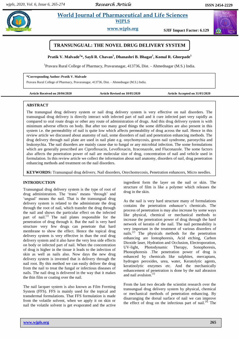
TRANSUNGUAL: THE NOVEL DRUG DELIVERY SYSTEM
Pratik V. Malvade1*, Sayli R. Chavan
1, Dhanashri B. Bhagat
1, Komal R. Ghorpade
1
1Pravara Rural College of Pharmacy, Pravaranagar, 413736, Dist. - Ahmednagar (M.S.) India.
Article Received on 20/04/2020 Article Revised on 10/05/2020 Article Accepted on 31/05/2020
INTRODUCTION
Transungual drug delivery system is the type of root of
drug administration. The „trans‟ means „through‟ and
„ungual‟ means the nail. That is the transungual drug
delivery system is related to the administrate the drug
through the root of nail, which transfer the drug through
the nail and shows the particular effect on the infected
part of nail.[1]
The nail plates responsible for the
penetration of drug through it. But the nail is very hard
structure very few drugs can penetrate that hard
membrane to show the effect. Hence the topical drug
delivery system is very effective in than the oral drug
delivery system and it also have the very less side effects
on body or infected part of nail. When the concentration
of drug is higher in the tissue it leads to the infection of
skin as well as nails also. Now days the new drug
delivery system is invented that is delivery through the
nail root. By this method we can easily deliver the drug
from the nail to treat the fungal or infectious diseases of
nails. The nail drug is delivered in the way that it makes
the thin film or coating over the nail.
The nail lacquer system is also known as Film Forming
System (FFS). FFS is mainly used for the topical and
transdermal formulations. That FFS formulation is made
from the volatile solvent, when we apply it on skin or
nail the volatile solvent is get evaporated and the active
ingredient form the layer on the nail or skin. The
structure of film is like a polymer which releases the
drug in the skin.
As the nail is very hard structure many of formulations
contains the penetration enhancer‟s chemicals. The
process of penetration is may also increase by some ways
like physical, chemical or mechanical methods to
increase the penetration power of drug through the hard
network of keratin of the nail. The nail permeability is
very important in the treatment of various disorders of
nails.[2]
The physicals methods for the penetration
enhancing are Iontophoresis, Acid etching, Carbon
Dioxide laser, Hydration and Occlusion, Electroporation,
UV-light, Photodynamic Therapy, Sonophoresis,
Phonophoresis .The penetration power of drug is
enhanced by chemicals like sulphites, mercaptans,
hydrogen peroxides, urea, water, Keratolytic agents,
keratinolytic enzymes etc. And the mechanically
enhancement of penetration is done by the nail abrasion
and nail avulsion.[3]
From the last two decade the scientist research over the
transungual drug delivery system by physical, chemical
or mechanical methods of penetration enhancing. By
disarranging the dorsal surface of nail we can improve
the effect of drug on the infectious part of nail.[4]
The
Research Article
ISSN 2454-2229 wjpls, 2020, Vol. 6, Issue 6, 265-274
World Journal of Pharmaceutical and Life Sciences WJPLS
www.wjpls.org SJIF Impact Factor: 6.129
*Corresponding Author: Pratik V. Malvade
Pravara Rural College of Pharmacy, Pravaranagar, 413736, Dist. - Ahmednagar (M.S.) India.
ABSTRACT
The tranungual drug delivery system or nail drug delivery system is very effective on nail disorders. The
transungual drug delivery is directly interact with infected part of nail and it cure infected part very rapidly as
compared to oral route drugs or other any route of administration of drugs. And this drug delivery system is with
minimum adverse effects on body. But after too many good things the some difficulties are also present in this
system i.e. the permeability of nail is quite low which affects permeability of drug across the nail. Hence in this
review article we discussed about anatomy of nail, some disorders of nail and penetration enhancing methods. The
drug delivery through nail plate are used in nail plate e.g. onychomycosis, green nail syndrome, paronychia and
leukonychia. The nail disorders are mainly cause due to fungal or any microbial infection. The some formulations
which are generally prescribed are Ciprofloxacin, Levofloxacin, Itraconazole, and Fluconazole. The some factors
also affects the penetration power of nail are molecular size of drug, concentration of nail and vehicle used in
formulation. In this review article we collect the information about nail anatomy, disorders of nail, drug penetration
enhancing methods and treatment on the nail disorders.
KEYWORDS: Transungual drug delivery, Nail disorders, Onychomycosis, Penetration enhancers, Micro needles.

www.wjpls.org
266
Pratik et al. World Journal of Pharmaceutical and Life Science
human nails are highly susceptible to various diseases
like Leuconychia, Onychauxis, and Onychatrophia etc.
and may be infections cause due to bacteria, viruses or
fungi. Sometimes this majorly leads to loss of appetite as
well as psychological stress.[5]
There are many treatments
on that disorders but mainly oral drugs and antifungal
treatment is given. The newly discovered techniques
shows that, we can developed the more effective drug by
the other root of administration such as nail drug delivery
system which is also known as Transungual drug
delivery system. To treat the many types of nail disorders
mainly the ultrasound technique is used by which the
permeability of nail is somewhat increased. The
physiochemical properties of nail shows that the nail
behaves more like a hydrophilic membrane. Which
results into the very typical drugs can penetrate that
membrane to show there effect. The topical application
of drug is mostly preferred because of localised action,
very less or no adverse effects and drug interaction
which make patient compatible to therapy and it is also
have the low cost.
The main purpose of this review paper is to check the
difficulties of drug during penetration through the nail
and also to increase the availability of antiseptic and
antifungal drugs. The researchers conducted on human
skin and nails gives us the information about the
permeability of specific drug across the skin, it also
shows us the structure and functions of skin and nail in
our body mechanism. But the thing is that the scientists
knows very little about the nail and nail keratin
properties. The purpose of this study is to understand the
permeability of drug across the nail plate to treat the
various nail disorders and other thing is it is also
necessary to study that, the drug is goes through the
systemic circulation and show effect of neighbouring
target sites or infected sites.
The current situation is that the drugs do not show much
more good activity on the infection because they cannot
penetrate the nail plate to show the effective therapeutic
effect on body. The current situations are mainly focuses
on the nail permeability and to reduce the barrier across
the nail plate by physical, chemical or mechanical
method,[6,7]
and penetration enhancers also.[8]
Anatomy of human nail
The nails consists of the nail plate, the nail matrix and
the nail bed below it, and the grooves surrounds all the
above structures.[9]
The chemical composition of nail is
very different from the other bod parts. It consists of
many disulphide bonds. It consists of lipids. The nail is
more likely to lipophilic in nature.
The Matrix The Matrix is also known as matrix unguis,
Keratogenous membrane, nail matrix or onychostroma. It
is made of tissues which provide the protection to the
nail.[10]
The matrix is the part of the nail bed which is
present below the nail bed where the nerves and blood
vessels are also present which supply the nerve impulse
and blood to the nail.[11]
The width of nail plate is
detected by the size, length and thickness of the of the
matrix. If the nail plate is flat, hocked or arched it is
shown by the fingertip. The matrix produces that cells
which forms the layer known as nail plate.[12]
The
production of nail plate layer is depend on the nutrition‟s
and keep the nail in healthy condition.[13]
As the new
layer is continue forms they pushes the old layer on
upper side. The old nail plate is converts into flat
compressed and transparent layer. That makes the nail
pinkish in colour because of presence of blood vessels
below to the old nail plate.[14]
Nail bed The nail bed is the skin below to the matrix.
[15] Like the
skin is made up of dermis and epidermis.[16]
That is the
layer which is just below to the nail plate which covers
the fingertip. The epidermis and dermis are attached to
each other by matrix crests (cristae matricis unguis).[16]
During the old age the matrix crests are easily visible to
eyes.[12]
www.wjpls.org
267
Pratik et al. World Journal of Pharmaceutical and Life Science
Nail sinus It is also known as sinus unguis. It is present there where
the nail root is present. Nail root is the base of nail
present below and behind the skin. The nail sinus is
formed from the tissues made by the matrix.[10, 11]
The Lunula It is also called as “Moon” or “Small Moon”. It is visible
part of nail which is whitish yellow half circle shaped or
half-moon shaped structure which is present at the base
or root of the nail.[15]
The lunula is clearly seen on the
thumb and may not be seen on little finger.
The Nail plate The another name for nail plate is corpus unguis.
[10] It is
most hard part of nail made up of transparent keratin
protein. It is made up of several layers of dead cells
which are highly compact in nature which makes nail
strong as well as flexible also.[12]
The shape of nail plate
is depend on the underlying bone i.e. The shape of nail
plate is changes from person to person.[12]
The peoples
tell the nail to this visible part.
The Distal edge It is also known as free edge, margo liber or anterior
margin. It is the margin of nail which is come out from
the nail or that part which does not have contact with
skin. The hyponychium also known as “quick” is the
layer of epithelial cells which is located below to the nail
plate and at distal edge.[17]
The hyponychium forms the
cover for the protection of nail bed.[16]
Onchodermal band In some peoples, the greyish coloured layer is seen
known as onchodermal band. This band is present in all
the peoples but in some cases it can be easily seen by
naked eyes. The onchodermal band is present in between
the hyponychium and the nail plate.[12]
The Nail root It is the last part of nail present in the nail sinus. It is also
known as radix sinus or germinal matrix. It is form from
the matrix tissues.[10]
This par extends in the nail about
several millimetre. It forms the maximum mass of the
nail.
The Nail wall The nail wall is fold of cutaneous membrane which
covers the both side of nails and the root of nail also. The
scientific name of nail wall is “valum unguis”. Below to
the nail wall and to both sides of nail the margin is seen
known as “lateral margin” or “margo lateralis”.
The Hyponychium
It is another part of nail which forms the junction in
between free edge and skin of fingertip. The functions of
hyponychium is to form the cover over the nail and
protect the nail from any infection from any type of
microorganism. The other function is that to make nail
water resistant.[18]
The Eponychium
The eponychium is also known as “cuticle” or “The
proximal fold”. The proximal fold is present at end of
eponychium. Proximal fold acts as a shield over the
newly formed nail bed. This layer is made up off dead
cells and it is almost invisible skin.[20]
It is the layer of
epithelial cells which extends from nail wall to the nail
root.[19]
The cuticle is formed from layer of dead cells
and this layer is may remove during manicure, but the
eponychium is formed from layer of living cells and
touching to it may cause any disorder of nail.[20]
The
Periyonix is the edge of eponychium which covers the
strip of the lunula.[19]
The Paronychium
It is the layer of soft tissues present around the complete
nail.[21]
The infection of this area is known as
paronychia. Paronychium is bordering of the nail formed
from epidemic layer.
Growth of Nail
In mammals, the rate of growth of nail is depend on the
length of last phalanges of finger. As in humans the
index finger has long phalanges the rate of growth of nail
is fast in index finger as compared to the little finger. In
humans the hand finger nails are grows at higher rate
than the leg fingers.[22]
The growing part of nail is present below the skin under
the epidermis. The epidermis is only living part of the
nail. In humans the average rate of growth of finger nail
is about 4-5 mm (0.20 in) per month and average growth
rate of toenail is about 2-3 mm (0.08 in) per month.[23]
To regrow completely the finger nail requires 6-8 months
and the toenail requires 16-18 months to regrow
completely. The growth rate of nail is faster in the
summer season as compared to another seasons.[24]
The
growth rate of nail is depend on the age, sex, diet,
nutrition, environmental conditions, season and exercise.
The nail growth rate also depend on the hereditary factor
also.[25]
Some researchers says that the nails grows after
death but the reason behind this is when the death of
person occurs the skin present around the nail is get
continues shrinking due to dehydration process, hence it
looks like the nails are growing. As this is slow process
this took much more time.[26]
Permeability
The nail are made up of very highly complex structure of
keratinized tissues. Hence the researches considered that
the nails are impermeable to any type of substance, but
this is not completely true. The nails are permeable to
some drugs, which shows the effect on nail drugs. The
interesting thing is that the nails are more permeable than
the skin. The aquatic concentration in nail is about 10-
15%.[27]
This permeability is very useful for the
penetration of drugs on nail disorders. But toxic
substances are also penetrate the nail and hence they are
also cause nail risks. Water can easily penetrate the nail
and hence for the treatment of nail disorders the
pharmaceutically active ingredient are prepared as base
water. The active ingredients are such as salicylic acid,
miconazole, natamycin and sodium hypochlorite.

www.wjpls.org
268
Pratik et al. World Journal of Pharmaceutical and Life Science
Clinical Importance of Nails
The nail are very important for any disease
manifestations. When the any person get dehydrated then
the colour of nails get changed which is indication. The
nails also give the indication about shock of any
person.[28]
That test is known as CRT (Capillary Nail
Refill Test) or Blanch test. As this tests are conducted on
the nails and hence this test are not suitable for old age
peoples. The tests are performed by EMT (Emergency
Medical Technician).[29]
The CRT test is very simple to do. This test is carried out
on nail. Firstly the pressure is applied on the nail so
colour of nail changes to white. After the pressure is
released. Here there are two cases; first is if the colour of
nail become red then you are in healthy state and if after
removal of pressure if the colour is remain as it is means
white then it is concluded that you are in shock.[30, 31]
The growth rate of nail and colour of nail also for the
indication for many disorders and hence our ancestors
also knows that thing.[32]
The Beau‟s line present in the
nail, that line is indication for ageing. The some
symptoms of disorder of nail are thinning, thickening,
splitting, convex nail, concave nail, white spots,
decolourization, flat nail, etc. are all the indication for
deficiency of various nutrients within body.
The some diseases and their indication are given below
1. Onychogryphosis- Nail become thick
2. Onychodystrophy-Nail degenerates
3. Onycholysis- Nail is detaches from body
4. Onychomycosis- Nail is infected by fungus
5. Onychocryptosis- The toe nail ingrown within skin
6. Koilonychias- The nail ridges become flat and
concave
7. Psoriasis- The nail bed detaches from skin.
Function of nails in human body
Nails have many functions in human body such as the
nails protect the delicate tips of finger nails and toe nails
tip from any type of external injury. They exerts an
opposite pressure or counter pressure on fingertip, which
helps in the movement of tip and touch sensation.[9]
Nails also work as a valuable tool for cutting, scraping or
pinching very small particles. The main function of nail
is that the helps in holding of any object by hand that is
known as “extended precision function grip”.[32]
Effect of different nutrients on nails
1. Vitamin A Vitamin A is an essential micro-nutrient. The vitamin
affect the many organs of our body such as eyes,
reproduction and immune system also. The deficiency of
vitamin A in nails can cause brittleness, dryness and
increases fragility of nails.
2. Calcium and Vitamin D
The calcium and vitamin are both work in pair means
they are dependent on each other. They caught the
muscle contraction, maintains the body homeostasis
mechanism, blood clotting mechanism and transmission
of impulse through nerve cells is also do by this two
nutrients.
3. Vitamin B12 Deficiency of Vitamin B12 can cause dryness of nails
and dark colour of nails. Insufficient Vitamin B12 level
changes the shape of nail i.e. rounded or curled nails and
ridges on nails.[27]
4. Proteins The proteins are the building blocks of our body
therefore insufficient dietary protein can cause the reduce
Haemoglobin level in body and due to reduced
Haemoglobin level the oxygen carrying capacity of
blood decreases and body suffers with anemia like
condition. Resulting to this below the nails the white
patches are begin to seen. In iron deficiency anemia like
condition the colour of nails become pale yellow and
they become fragile and shape of nails become
convex.[28]
5. Fatty Acids
Not all the fatty acids are involved in skin and nail
metabolism but only the essential fatty acids are play
important role for healthy skin and nails. Due to
deficiency of fatty acid (Linoleic Acid) the splitting or
flatting of nails can occur.
Nail Diseases and Disorders
1. Onychomycosis (Tinea unguium)
Fig. 1: Onychomycosis (Fungal Infection).
The Onychomycosis is a fungal and yeast infection to the
nail. The other name for this disorder is Dermatophytic
onychomycosis.[29]
The name Onychomycosis is given to
this disorder because in this disorder the “Onycholysis”
occur. The onycholysis means separation of nail plate
from nail bed. The infection nail discoloration,
thickening of the nail and separation of the nail from the
nail bed.[30,31]
In this disorder both finger nails as well as
toe nails are infected.[31]
The cellulitis of nails may also
occur in this disorder. The fungus which cause
Onychomycosis are dermatophytes, Fusarium,
Trichophyton rubrum and T.mentagrophytes. The other
causes of this disorder are athlete‟s foot, other nail
diseases, peripheral vascular diseases and low immune
power is also one of the reason to cause this disorder.[31]
The diagnosis is carried out in equipped laboratory.
The treatment for Onychomycosis is not always
necessary. The antifungal drugs terbinafine is very
effective on this disorder but this drug has side effect on
liver.[32]
The mechanical method for this disorder is that
to remove the infected nail. The medications for this

www.wjpls.org
269
Pratik et al. World Journal of Pharmaceutical and Life Science
disorder have some antifungal drugs like Terbinafine,
Itraconazole, Fluconazole, and Ketoconazole.[30]
The
topical agents like Ciclopirox, Amorolfine and
Efinaconazole.[34]
The Onychomycosis is mainly has four types-
a. Distal subungual onychomycosis - This is most
common type of Onychomycosis. The causative
agent is Trichophyton rubrum, which occupies the
space in between nail plate and nail bed.[30]
b. White superficial onychomycosis (WSO) - It is the
fungal accumulation in nail plate which looks like
„White patches‟. Sometimes white patches are also
occurs due to the protein deficiency. Hence for
diagnosis the laboratory test is carried out.[33]
c. Proximal subungual onychomycosis – This is not
more common but this may occur into the
immunosuppressed peoples. In this case the fungus
penetrates into the newly formed nail plate.[30]
d. Candidal onychomycosis – This is occurs due to
microorganism of species Candida. This type of
onychomycosis may cause due to continuous contact
of nails with water.
2. Green Nail Syndrome
Fig. 2: Pseudomonas bacterial infection.
Green nail syndrome is also known as Chloronychia.
This is an infection caused due to Pseudomonas
aeruginosa. In this disorder the Pseudomonas bacterium
get trapped in between nail plate and nail blade and it
looks like green strip. If the infection is not treated on
time it can separate the nail plate from nail bed. The
peoples are more susceptible to this disease whose nails
have always contact with moisture or water. If any
person has artificial coating on nail then the infection of
this disease may also cause in between artificial nail
coating and natural nail bed.[35]
Treatment is available on
this disorder as oral dose of Quinolone (Ciprofloxacin).
The topically applicable drugs are Silver Sulfadiazine,
Nadifloxacin, Ciprofloxacin and Gentamicin. Some
antibiotics are also have therapeutic effect on green nail
such as Polymyxin B or Bacitracin.[36]
3. Paronychia Infection
Fig. 3: Paronychia Infection.
Paronychia is a fungal or bacterial infection to nails of
hands or toe. The infection occurs at where the nails and
skin touches i.e. at the sides of nails.
The Paronychia is mainly classified in two classes,
a. Acute Paronychia
This type of infection stars suddenly.[37]
This type of
Paronychia is caused due to bacteria Candida albicans
and Staphylococcus aureus.[38]
The infection is start with
local pain, redness and swelling. The infection is last
long for six weeks. The infection may cause due to
trauma, injury from thorn, nail biting, finger sucking,
etc.[39]
The antibiotic treatment of drugs like
Clindamycin (Cleocin) and combination of amoxicillin–
clavulanate potassium (Augmentin).
b. Chronic Paronychia
This type of infection is long lasting and appears slowly.
This was mainly caused due to continuous contact of
nails with moisture. In this infection the cuticle separates
form nail plate and nail plate get susceptible for
infection.[40]
The causative agents for chronic type of
Paronychia are similar as that of acute type. The drug
treatment includes combination of penicillin with
cephalosporin and aminoglycosides, Acyclovir and
Valacyclovir.
4. Leukonychia
Fig. 4: Leukonychia (Milky Spot).
The leuko means „white‟ and onyx means „nails‟. This is
the most common type of nail injury in between nail
plate and nail bed. In this white line or white spot
appears on one or more nails. The spot may occurs due
to air bubble trapped in between nail bed and nail late
due to trauma.
www.wjpls.org
270
Pratik et al. World Journal of Pharmaceutical and Life Science
There are several types of Leukonychia as
1. Leukonychia totalis
As name indicates in this condition the complete nail
become white. This condition is mainly causes due to the
hypoalbuminaemia i.e. low albumin level in blood. The
hypoalbuminaemia condition is caused due to kidney
failure, liver failure and protein dysfunctioning. This is
genetic type of disorder and the person having allergy
from drugs of sulphonamide family can also cause this
condition in that patient.
2. Leukonychia partialis
As the name indicates in this condition the some part of
nail become white. If the partial leukonychia is did not
treated at specific time then it leads to Leukonychia
totalis.
3. Leukonychia striata
This condition is also known as transverse leukonychia
or Mees‟ lines leukonychia. In this condition the nail
become white parallel to the nail base i.e. lunula. This is
mostly caused due any physical injury to nail or infection
to matrix of nail. This condition cause due to heavy
metal like lead, arsenic poisoning or over manicure also
leads to this disorder.[41, 42]
The leukonychia striata is
also caused due to liver cirrhosis i.e. degeneration of
liver or chemotherapy. In some cases this disorder is
also genetically inherited. There is a same condition like
leukonychia striata named as Muehrcke's lines (apparent
leukonychia) but this is not any disorder and the white
patch is easily get removed on application of pressure on
nail.[43]
4. Leukonychia punctata
This is the most common type of leukonychia, which
occurs in young children‟s and nail bitters. It caused due
to trapping of air in between nail plate and nail bed and it
also cause due to trauma.[44]
The white spot disappear
within eight months because the nail completely regrow
within this time period.
5. Longitudinal leukonychia
It is not common disorder and mostly not seen in any
patient. In this condition an about 1-1.5 cm white line
appears on nail. For the treatment of any type of
leukonychia the all nutrient level in body is checked and
the necessary supplement is given.
Drug Penetration Enhancing Methods across the Nail
There are mainly three methods to increase the
penetration power of drug across the nail as,
1. Physical Methods
In this method that type of agents or drugs are used
which degenerates the lipid molecules present in nail
plate to increase drug penetration power and its activity.
The physical method for penetration enhancing is better
than the chemical methods. The some physical methods
are enlisted below;
1. Hydration and Occlusion
In the hydration process pore size of nail matrix is
increased to enhance the penetration. Due to hydration
the nails become more soft and porous. The solution
which used in the hydration that solutions pH and
concentration do not affect the hydration process. When
the layer of skin i.e. Stratum Corneum is get saturated
the penetration process of drug across the skin and nail
become much faster.[45]
2. Etching
In this type of physical method the nail is exposed to
phosphoric acid. The phosphoric acid increases
microsporocytes. The microsporocytes increases the
surface area. As the nail get etched a hydrophilic or
polymer film drug delivery system is applied and due to
bioadhesion the tranungual drugs are applied.[46]
3. Iontophoresis
Iontophoresis is the special type of physical method in
which the electromagnetic field is applied around the
skin or nail. This method is applicable in various medical
and paramedical fields. By this method drug penetration
through the hydrated stratum corneum is enhanced. The
some other types are;
Electrorepulsion/Electrophoresis – This is
interaction in between electric field and ionic field of
drug.
Electro-osmosis – Convective solvent flow in old
and newly created charge pathways.
Permeabilization/Electroporation – Electric field
induces pore induction for drug.
As compared to any other type of drug penetration
enhancing method the ionotophoresis is the most
preferable and convenient process for treatment of nail
disorders.[47]
4. Carbon Dioxide Laser
The carbon dioxide laser method give positive result but
the chances are very less. In this method the nail plate is
penetrated by CO2 laser beam. By this methods the
antifungal drugs are given.[48]
5. Micro Needles
It is new drug delivery system. In which the small
needles are used to release the drug in stratum corneum
layer of skin. This is mostly preferred because it has low
pain during treatment.
2. Chemical Methods
Effect of chemicals which are used as penetration
enhancer is different for different organism‟s nails. Some
of chemicals are given following;
1. 2-n-nonyl-1,3-dioxolane
The penetration of nail lacquer through the nail is
enhanced by 2-n-nonyl-1,3-dioxolane. In researches it is
found that the nail lacquer containing 2-n-nonyl-1,3-
dioxolane can penetrate nail seven times faster than
another nail lacquer containing identical enhancer. The
concentration of this chemicals should ne maintain for
stopping of microbial growth within nail.[49]
2. Mercaptan compounds and n-acetyl-l-cysteine
This two drugs are used in combination because they are
effective in combination to increase permeability of
antifungal drug through nail. The researchers studied the
penetration property of n-acetyl-l-cysteine with an
antifungal drug Oxiconazole.[50]
3. Keratolytic Enhancers
Some examples of keratolytic drugs are salicylic acid,
urea, etc. When the keratolytic agents are studied with

www.wjpls.org
271
Pratik et al. World Journal of Pharmaceutical and Life Science
some antifungal drugs such as miconazole, ketoconazole,
and itraconazole. It is seen that when the keratolytic
agents are not used then this antifungal drugs doesn‟t
penetrate the nail within 60 days long period. This
keratolytic drugs can induce antimycotic penetration.[51]
4. Organic Solvents
Ethanol, isopropanol, propylene glycol, etc. are
especially used in transungual drugs to enhance the
penetration of active ingredient through nail. The organic
solvents increases the moisture level within nail and the
drug penetrate the nail.
3. Mechanical Methods This methods have high cost and they are very painful.
This method is only used by the dermatologist and nail
experts. Some of the mechanical methods are discussed
below;
1. Nail Abrasion
It is very painful method. In this method the thickness of
nail is reduced or completely removal of nail by
mechanical way to penetrate the drug. To decrease the
nail plate thickness health expert use the sand paper. The
abrasion of nail plate is done from the edges of nail to
penetrate drug. Other way is also used by doctors i.e.
drilling the nail plate and make the small hole within nail
plate and they place that drug in that hole.[52]
2. Nail Avulsion
The avulsion is defined as it is an injury in which body
structure is torn off by either trauma or surgery. This is
very painful than the nail abrasion because in this
method the nail is fully or partially removed from nail
bed. When the nail is removed from nail bed then nail
bed again forms the nail bed.[53]
On the removal of nail
plate the nail bed become very sensitive to any type of
impulse or injury and hence the nail bed is still covered
until new nail doesn‟t forms.[54]
For removal of nail it is
necessary to make nail soft and hence the keratolytic
agents are used.
4. Latest methods in nail drug delivery system In this methods the latest drug formulations are used
such as nail lacquer, nail patches, etc. The some of the
latest method are discussed below;
1. Mesoscissioning technology
This is painless method for drug delivery. In this method
firstly approximately 300-400 micron cut is given on
skin or nail. This process is very fast and without any
sensation of pain. The micro conduits are also used for
blood glucose level testing. In nail they reduces painful
pressure.[55, 56]
2. Electro Chemotherapy
It is believed that this method have the high success rate
and it decreases the time period of treatment, because it
is very effective. This method is similar to iontophoresis
but it enhances the drug delivery.[55]
3. Nano Patch Nail fungus
In this method the drug is supplied by alternate current or
direct current. Both of the currents push the active
ingredient to the actual location i.e. infected area of nail
by fungus. The drug is delivered over the nail membrane
i.e. cuticle.[57]
Different Drugs Used in the nail disease treatment
1. Antibacterial drugs
a. Fluroquinoles
E.g. - Ciprofloxacin, Levofloxacin, Ofloxacin
b. Antipseudomonals
E.g. - Penicillin, Cephalosporin

www.wjpls.org
272
Pratik et al. World Journal of Pharmaceutical and Life Science
c. Aminoglycosides
E.g. - Amikacin, Neomycin, kanamycin, Tobramycin,
Gentamycin.
d. Echinocandins & Heterocyclic benzofurans
E.g.-Caspofungin, Micafungin, Anidulafungin,
Griseofulvin, etc.
2. Antifungal drugs
a. Azoles
E.g.-Itraconazole, Fluconazole, Posaconazole,
Voriconazole, Ravuconazole, etc.
b. Ally amines and Benzyl amine
E.g.-Terbinafine hydrochloride, Naftifine, Butenafine.
Factors affecting drug penetration through nails
1. Molecular size
The drug penetration across the nail is inversely
proportional to molecular size of drug. That means that
penetration of drug increases as molecular size of drug
decreases. Hence the doctors always prescribes that
drugs for nail disorders which have low molecular
size.[58]
2. Hydrophilic and Lipophilic molecules
The lipopilic drugs penetrates the nail by lipid pathway,
if the size of lipophilic drug molecule is large then it
can‟t penetrate nail membrane. The hydrophilic pathway
is completely opposite to lipophilic pathway. When the
hydrophilic drug come in contact of nail the nail
membrane become hydrated as result the pores of nail
are get opened and drug get easily penetrate from nail.[59]
3. Nature of drug vehicle
The polar solvents like water are mainly used as a base
or vehicle for transungual drugs. The water wets the nail
membrane which results into swelling of keratin network
present in nail. As the keratin network swells the pores
get open and the drug can easily penetrate. But if the
non-polar solvent is used as a vehicle then it can‟t
hydrate the nail and the chances of penetration of drug
decreases.[60]
Concentration of formulation
The concentration of formulation of drug is also affects
the penetration period through nail. The weak acidic
formulation rapidly penetrate nail plate but weak basic
formulation can‟t do that.[61]
RESULT
Topical drug delivery or transungual drug delivery
system is typically used for treatment of various types of
nail disorders such as onchiomycosis, paronychia, etc. In
the world about 3% to 4% of total population suffers
with different type nail or skin disorder. The transungual
drug therapy is usually applied topically to treat the nail
disorders. The treatment includes antifungal as well as
antibacterial drugs. The pharmaceutical formulations are
mainly placed in between nail plate and nail bed. But the
success rate of nail treatment is very less due to less
permeability of nail. As the permeability of nail is less in
market very small formulations which can successfully
penetrate the nail plate and show their good effect on
infected area. The penetration power of formulation is
very important to treat any nail disorder. As the nail plate
permeability is low it acts as a barrier to the drug.
In this review article we describe the anatomy of nail. In
this paper the four diseases are briefly described which
are onychomycosis, green nail syndrome, paronychia and
leukonyhia. In this article various methods for the drug
penetration through the nail are discussed. We also
involved the factors affecting the nail permeability for
drugs like molecular size, concentration of formulation,
etc. the Physical, Chemical and Mechanical methods for
drug penetration enhancement. The researchers need to
research in this field because in this field it is needed to
explore more about the nail and their disorders. The use
of suitable or identical penetration enhancer should be
preferred for nail disease treatment. Finally, in this
review article we included all of the information which is
necessary to treat nail disorders.
CONCLUSION
It is most necessary to identify the different nail barriers
for pharmaceutical formulation to enhance the success
rate of nail disease treatment. The more research should
be done on permeability of nail plate for efficient drug
delivery. Hence the drug delivery through the nail is

www.wjpls.org
273
Pratik et al. World Journal of Pharmaceutical and Life Science
major challenge for doctors due to barriers. The barriers
should be physical or chemical. But over the all of the
above the transungual drug therapy is always better than
the oral drug therapy because the effect of drug is
directly on the infected part of nail. But the researchers
doesn‟t found the specific permeability enhancer which
can used in all formulations. If we want to improve the
nail formulations then it is necessary to study the
physiochemical properties of drug, property of
penetration enhancers and final is use of formulation. It
is possible in future the researchers found the best acting
drug on nail disorder.
REFERENCE
1. Jeremiah C. Review on transungual drug delivery
system. Indo AmJ Pharm Res, 2017; 7(08).
2. Vivek B, Rajendra. Transungual drug delivery: an
overview. J Appl Pharm Sci, 2012; 02(01): 203-09.
3. Pradeep S, Patil, Sangita V, Badgujar, Ashwin A,
Torne. Nailing the nail trouble by transungal drug
delivery. Eur J PharmMed Res, 2015; 2(2): 551-71.
4. Shivakumar H, Repka M, Narasimhamurthy S.
Transungual drug delivery: an update. J Drug Del
Sci Tech, 2014; 24(3): 301-10.
5. Berker DA, Andre J, Baran R. Nail biology andnail
science. International Journal of Cosmetic Science,
2007; 29: 241-275.
6. Kobayashi Y, Miyamoto M, Sugibayashi K,
Morimoto Y. Enhancing effect of N-acetyl-lcysteine
or 2-mercaptoethanol on the in vitro permeation of
5- fluorouracil or tolnaftate through the human nail
plate. Chem. Pharm. Bull. 1998; 46:1797–1802.
7. Malhotra GG, Zatz JL. Investigation of nail
permeation enhancement by chemical modification
using water as a probe. J. Pharm. Sci. 2002; 91:312–
323.
8. Hui X, Baker SJ, Wester RC, Barbadillo S,
Cashmore AK, Sanders et al. In vitro penetration of
a novel oxaborole antifungal (AN2690) into the
human nail plate. J. Pharm. Sci., 2007; 96: 2622–
2631.
9. Onumah, Neh; Scher, Richard K (May 2009). "Nail
Surgery". eMedicine. Retrieved 10 March, 2010.
10. Feneis, Heinz Pocket Atlas of Human Anatomy (4th
ed.). Thieme, 2000; 392–95. ISBN 3-13-511204-7.
11. "Glossary of Nail Technology Terminology". 2008.
Retrieved 10 March. 2010.
12. "Understanding Your Natural Nails". 2000.
Retrieved March 10, 2010.
13. D. Schoon, Dougles Nail Structure and Products
Chemistry. Milady, 2005; 6.
14. Lellipop (August 2006). "Anatomy of the nail".
Salon Geek. Retrieved 10 March, 2010.
15. "Nail Anatomy". Nail Doctors. 2005. Retrieved
March 10, 2010.
16. "Glossary of Nail Conditions". The Achilles Foot
Health Centre.
17. Crouch, James Ensign Functional human anatomy.
Lea & Febiger, 1985; 80. ISBN 9780812109306.
18. Rich P. An Atlas of Diseases of the Nail
(Encyclopedia of Visual Medicine Series).
Parthenon Publishing, 2003.
19. Feneis Heinz. Pocket Atlas of Human Anatomy. (4th
ed.). Thieme. pp. 392– 95. ISBN 3-13-511204-7,
2000.
20. Lellipop. "Anatomy of the nail"
(http://www.salongeek.com/health-
safetyunnatural/40362-anatomy-nail.html). Salon
Geek. Retrieved Feb, 2010.
21. Jordan, Christopher; Mirzabeigi, Edwin (2000-04-
01). Atlas of orthopaedic surgical exposures.
Thieme, 101. ISBN 0-86577-776-4.
22. Cartmill, Matt; Lemelin, Pierre; Schmitt, Daniel
(2007). "Primate Gaits and Primate Origins". In
Ravosa, Matthew J.; Dagosto, Marian (eds.).
Primate Origins: Adaptations and Evolution.
pp. 403–35. doi:10.1007/978-0-387- 33507-0_12 .
ISBN 978-0-387-30335-2.
23. Toenail Definition - Medicine.net.
(http://www.medterms.com/script/main/art.asp?artic
lekey=7740).
24. Hunter JAA, Savin J, Dahl MV. Clinical
dermatology. Malden, Mass: Blackwell Science,
2002; 173.
25. Hunter, J. A. A., Savin, J., & Dahl, M. V. (2002).
Clinical dermatology. Malden, Mass: Blackwell
Science, 173. ISBN 0-632-05916-8.
26. http://www.bmj.com/cgi/content/full/335/7633/1288
).BMJ, 2007; 335(7633): 1288 Dec 22.
doi:10.1136/bmj.39420.420370.25.
27. Zempleni, J; R. B. Rucker; D.B. McCormick; J. W.
Suttie Handbook of vitamins (4th ed.), 2007.
28. Cashman MW, Sloan SB "Nutrition and nail
disease". Clinics in Dermatology, 2010; 28(4): 420–
25. doi:10.1016/j.clindermatol.2010.03.0 37. PMID
20620759.
29. Rapini, Ronald P.; Bolognia, Jean L.; Jorizzo,
Joseph L. Dermatology: 2-Volume Set. St. Louis:
Mosby, 2007; 1135.
30. Westerberg DP, Voyack MJ (Dec 1,
"Onychomycosis: current trends in diagnosis and
treatment". American Family Physician, 2013;
88(11): 762–70.
31. Merck Manuals Professional Edition. February
2017. Retrieved 2 June, 2018.
32. Kreijkamp-Kaspers, S; Hawke, K; Guo, L; Kerin, G;
Bell-Syer, SE; Magin, P; Bell-Syer, SV; van Driel,
ML (14 July). "Oral antifungal medication for
toenail onychomycosis", 2017.
33. "AAPA". Cmecorner.com. Retrieved, 2010-08-05.
34. Crawford F, Hollis S Crawford F (ed.). "Topical
treatments for fungal infections of the skin and nails
of the foot". Cochrane Database Syst Rev, 2007; (3):
CD001434. doi:10.1002/14651858.CD001434.pub2.
35. "Pseudomonas aeruginosa Infections: Clinical
Presentation". eMedicine. Retrieved 1 February,
2014.
www.wjpls.org
274
Pratik et al. World Journal of Pharmaceutical and Life Science
36. Agger WA, Mardan A. Pseudomonas aeruginosa
infectionof intact skin. Clin Infect Dis., 1995; 20(2):
302-308.
37. Rigopoulos D, Larios G, Gregoriou S, Alevizos A
(February). "Acute and chronic paronychia". Am
Fam Physician, 2008; 77(3): 339–46.
38. James, William D.; Berger, Timothy G. Andrews'
Diseases of the Skin: clinical Dermatology.
Saunders Elsevier. ISBN 978-0-7216-2921-6, 2006.
39. Rigopoulos, Dimitris; Larios, George; Gregoriou,
Stamatis; Alevizos, Alevizos (2008). "Acute and
Chronic Paronychia" (PDF). American Family
Physician. 77 (3): 339–346. PMID 18297959.
Retrieved January 7, 2013.
40. Rigopoulos, Dimitris; Larios, George; Gregoriou,
Stamatis; Alevizos, Alevizos (2008). "Acute and
Chronic Paronychia" (PDF). American Family
Physician. 77 (3): 339–346. PMID 18297959.
Retrieved January 8, 2013.
41. Maino KL, Stashower ME. “Traumatic Transverse
Leukonychia”
(https://www.medscape.com/viewarticle/467074).
Medscape.
42. Baran, Robert et al. Baran and Dawber's Diseases of
the Nails and Their Management. John Wiley &
Sons, 2012.
43. Huang, T.-C.; Chao, T.-Y. (14 December). "Mees
lines and Beau lines after chemotherapy". Canadian
Medical Association Journal, 2009; 182(3): E149.
44. Tüzün, Yalçın; Karakuş, Özge (2009).
"Leukonychia" (PDF). Journal of Turkish Academy
of Leukonychia: 1–3. Archived from the original
(PDF) on March 3, 2016. Retrieved April 2, 2017.
45. Chen LJ, Meng QF, Chen YM, Smales RJ, Yip KH.
Effect of fluoride Iontophoresis on themicrotensile
bond strength between dentin and two adhesive
systems. J. Dent., 2008; 36: 697–702.
46. Gradisar H, Friedrich J, Krizaj I, Jerala R.
Similarities and specificities of fungal keratinolytic
proteases: comparison of keratinases of
Paecilomycesmar- quandii and
Doratomycesmicrosporus to some known proteases.
Appl. Environ. Microbiol, 2005; 71: 3420–3426.
47. Grover C, Bansal S, Nanda S, Reddy BS, Kumar V.
Combination of surgical avulsion and topical
therapy for single nail onychomycosis: a randomized
controlled trial. Br. J. Dermatol, 2007; 157: 364–
368.
48. Sudaxshina Murdan. Drug delivery to the nail
following topical application. International Journal
of Pharmaceutics, 2002; 236: 1–26.
49. Pierard G. Onychomycosis and other superficial
fungal infections of the foot in the elderly: a Pan-
European survey. Dermatology, 2001; 202: 220–
224.
50. Zaias N. The Nail in Health and Disease, 2nd ed.
Appleton and Lange, Connecticut, 1990; 1–255.
51. De Berker DAR, Baran R, Dawber RPR. The nail in
dermatological disease. In: Handbook of Diseases of
the Nails and their Management. Blackwell Science
Ltd, Oxford, 1995c; 64–84.
52. De Berker DAR, Baran R, Dawber RPR. Normal
nail anatomy and physical signs in nail disease. In:
Handbook of Diseases of the Nails and their
Management. BlackwellScience Ltd, Oxford, 1995a;
1–31.
53. Rischer, C.E., & Easton, T.A. Focus on human
biology (2nded.). New York: Harper Collins College
Publishers, 1995.
54. National Center for Emergency Medicine
Informatics. Nail Off. Retrieved January 16, 2009,
from[2] Archived 2016-08-14 at the Wayback
Machine.
55. Patel RP, Naik SA, Patel NA, Suthar AM. Drug
Delivery across Human NailInternational Journal of
Current Pharmaceutical Research, 2009; 1(1).
56. Badola A, Satish SB. A review: transungal drug
delivery a new and novel system. Asian Journal of
Pharmaceutical Science & Technology, 2015; 5(4):
227-33.
57. Firoz S, Sirisha MN, Raj Lakshmi R. Transungual
drug delivery system–A Review. International
Journal of Innovative Drug Discovery, 2011; 1(1):
9-14.
58. Murdan S. 1stmeeting on topical drug delivery to the
nail. Expert. Opin. Drug Deliv, 2007; 4: 453–455.
59. Kumar K, Fateh V, Ahmad S. Drug delivery across
human nail: A newer approach .International journal
of research and development in pharmacy and life
science, 2014 Nov 3(6): 1217-1222.
60. Murthy SN, Waddell DC, Shivakumar HN, Balaji A,
Bowers CP. Iontophoretic permselective property of
human nail. J. Dermatol. Sci., 2007a; 46: 150–152
61. Murthy SN, Wiskirchen DE, Bowers CP.
Iontophoretic drug delivery across human nail. J.
Pharm. Sci., 2007b; 96: 305–311.
Top Related
Copyright © 2022 FDOKUMEN

