


Bahasa
Halaman
Hukum

Aa
DN
a
ARRA
KANCHE
1
acribrtssSnft
n
0d
Journal of Neuroscience Methods 190 (2010) 155–163
Contents lists available at ScienceDirect
Journal of Neuroscience Methods
journa l homepage: www.e lsev ier .com/ locate / jneumeth
ddition of glutamate to serum-free culture promotes recovery of electricalctivity in adult hippocampal neurons in vitro�
arin Edwards, Mainak Das, Peter Molnar, James J. Hickman ∗
anoScience Technology Center, 12424 Research Parkway, Suite 400, Orlando, FL 32826, United States
r t i c l e i n f o
rticle history:eceived 13 December 2009eceived in revised form 29 April 2010ccepted 29 April 2010
eywords:dulteuronultureippocampallectrophysiology
a b s t r a c t
A long-term cell culture system utilizing normal adult hippocampal neurons would represent an impor-tant tool that could be useful in research on the mature brain, neurological disorders and age-relatedneurological diseases. Historically, in vitro neuronal systems are derived from embryonic rather thanmature brain tissue, a practice predicated upon difficulties in supporting regeneration, functional recov-ery and long-term survival of adult neurons in vitro. A few studies have shown that neurons derivedfrom the hippocampal tissue of adult rats can survive and regenerate in vitro under serum-free condi-tions. However, while the adult neurons regenerated morphologically under these conditions, both theelectrical activity characteristic of in vivo neurons as well as long-term neuronal survival was not consis-tently recovered in vitro. In this study, we report on the development of a defined culture system withthe ability to support functional recovery and long-term survival of adult rat hippocampal neurons. Inthis system, the cell-adhesive substrate, N-1 [3-(trimethoxysilyl) propyl]-diethylenetriamine, supported
neuronal attachment, regeneration, and long-term survival of adult neurons for more than 80 days invitro. Additionally, the excitatory neurotransmitter glutamate, applied at 25 �M for 1–7 days after mor-phological neuronal regeneration in vitro, enabled full recovery of neuronal electrical activity. This lowconcentration of glutamate promoted the recovery of neuronal electrical activity but with minimal exci-totoxicity. These improvements allowed electrically active adult neurons to survive in vitro for severalmonths, providing a stable test-bed for the long-term study of regeneration in adult-derived neuronaluma
systems, especially for tra. Introduction
As people live longer, neurological disorders and diseases thatffect the aging brain have become more common, however suc-ess to date for reversing these diseases has been limited. One of theeasons this research has been hindered has been the flawed, lim-ted, or non-existent research models of aged or diseased humanrain tissue. Neuronal cell culture, one of the most commonly usedesearch models, is generally derived from embryonic rat or mouseissue. Although this system has been used effectively in manytudies, a neuronal culture system derived from adult brain tis-ue could be a much more relevant and effective model system.
uch a system could be used to study the function of neurons,euronal interactions, aging and neurodegenerative disease butrom a new perspective where the different ion channels, recep-ors and other cellular components matured in vivo instead of
� Grant information: Department of Energy DE-FG-02-04ER 46171 and UCF inter-al funds and NIH grant number 5R01EB005459.∗ Corresponding author. Tel.: +1 407 823 1925; fax: +1 407 882 2819.
E-mail address: [email protected] (J.J. Hickman).
165-0270/$ – see front matter © 2010 Elsevier B.V. All rights reserved.oi:10.1016/j.jneumeth.2010.04.030
tic brain injury (TBI).© 2010 Elsevier B.V. All rights reserved.
in vitro (Ginsberg, 2005). It also would be useful in drug stud-ies, neuroprosthetic devices, neurocomputing and biorobotics forthe same rational (Heiduschka and Thanos, 1998; Simpson et al.,2001; Harms et al., 2006), and has a unique application to studyingregeneration of injured adult neurons, especially in traumatic braininjury (TBI). These applications were the goal behind an earlierattempt at creating an adult hippocampal culture system, devel-oped using a serum-free culture media with a biological surface(Brewer, 1997). While this system supported the morphologicalrecovery of adult hippocampal neurons in vitro, issues with thesupport of both long-term survival and full recovery of electricalactivity of neurons in this culture system appears to have preventedits widespread use as a research tool (Bousse, 1996; Heiduschka andThanos, 1998; Simpson et al., 2001).
To overcome these issues, modifications were made to adefined neuronal culture system previously developed for embry-onic hippocampal neurons (Schaffner et al., 1995). First, culture
surfaces were modified with the chemical substrate N-1 [3-(trimethoxysilyl) propyl]-diethylenetriamine (DETA), creating acovalently modified interface with exposed cell-adhesive aminegroups, which has previously been shown to promote the attach-ment, regeneration and long-term survival of embryonic neurons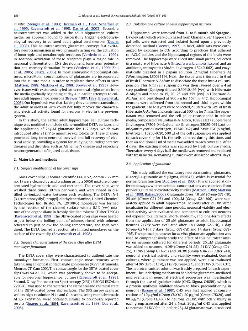
1 oscien
ianmoatoInteti(ett2tis
ntiptdn
2
2
ncwt[Tbt(trds
2m
mtMsaS2owAr2
56 D. Edwards et al. / Journal of Neur
n vitro (Stenger et al., 1993; Hickman et al., 1994; Schaffner etl., 1995; Ravenscroft et al., 1998; Das et al., 2003). Second, aeurotransmitter was added to the adult hippocampal cultureedia, an approach found to successfully trigger electrophysi-
logical recovery in cultured adult spinal cord neurons (Das etl., 2008). This neurotransmitter, glutamate, conveys fast excita-ory neurotransmission in vivo, primarily acting via the activationf ionotropic and metabotropic receptors (Verderio et al., 1999).n addition, activation of these receptors plays a major role ineuronal differentiation, CNS development, long-term potentia-ion and memory formation in vivo (Verderio et al., 1999; Zhut al., 2005; Balazs, 2006). In most embryonic hippocampal cul-ures, microMolar concentrations of glutamate are incorporatednto the culture media in order to replicate these effects in vitroMattson, 1988; Mattson et al., 1988; Brewer et al., 1993). How-ver, issues with excitotoxicity led to the removal of glutamate fromhe media gradually beginning at day 4 in earlier attempts to cul-ure adult hippocampal neurons (Brewer, 1997, 1998; Brewer et al.,005). Our hypothesis was that, lacking this vital neurotransmitter,he adult neurons in vitro could not fully recover the character-stic electrical activity found for neurons in vivo in the previousystem.
In this study, the earlier adult hippocampal cell culture tech-ique was modified to include silane-modified DETA surfaces andhe application of 25 �M glutamate for 1–7 days, which wasntroduced after 21 DIV to minimize excitotoxicity. These changesromoted long-term neuronal survival and full recovery of elec-rical activity, providing a system for studying neurodegenerativeiseases and disorders such as Alzheimer’s disease and especiallyeuroregeneration of injured adult tissue.
. Materials and methods
.1. Surface modification of the cover slips
Glass cover slips (Thomas Scientific 6661F52, 22 mm × 22 mmo. 1) were cleaned by acid washing using a 50/50 mixture of con-entrated hydrochloric acid and methanol. The cover slips wereashed three times, 30 min per wash, and were rinsed in dis-
illed de-ionized water between each washing. The DETA (N-13-(trimethoxysilyl) propyl]-diethylenetriamine, United Chemicalechnologies Inc., Bristol, PA, T2910KG) monolayer was formedy the reaction of the cleaned surface with a 0.1% (v/v) mix-ure of the organosilane in freshly distilled toluene (Fisher T2904)Ravenscroft et al., 1998). The DETA-coated cover slips were heatedo just below the boiling point of toluene, rinsed with toluene,eheated to just below the boiling temperature, and then ovenried. The DETA formed a reaction site limited monolayer on theurface of the cover slip (Ravenscroft et al., 1998).
.2. Surface characterization of the cover clips after DETAonolayer formation
The DETA cover slips were characterized to authenticate theonolayer formation. First, contact angle measurements were
aken using an optical contact angle goniometer (KSV Instruments,onroe, CT, Cam 200). The contact angle for the DETA-coated cover
lips was 54.2 ± 0.2, which was previously shown to be accept-ble for neuronal hippocampal culture (Ravenscroft et al., 1998).econd, X-ray Photoelectron Spectroscopy (XPS) (FISONS ESCALab20i-XL) was used to characterize the elemental and chemical state
f the DETA-coated cover slip surfaces. The XPS survey scans asell as high-resolution N 1s and C 1s scans, using monochromaticl K� excitation, were obtained, similar to previously reportedesults (Spargo et al., 1994; Ravenscroft et al., 1998; Das et al.,005).ce Methods 190 (2010) 155–163
2.3. Isolation and culture of adult hippocampal neurons
Hippocampi were removed from 3- to 6-month-old Sprague–Dawley rats, which were purchased from Charles River. Hippocam-pal cells were extracted and isolated based upon a previouslydescribed method (Brewer, 1997). In brief, adult rats were euth-anized by exposure to CO2 according to practices that adheredto IACUC policies and the hippocampal regions of the brain wereremoved. The hippocampi were sliced into small pieces, collectedin a mixture of Hibernate A (http://www.brainbitsllc.com) and anantibiotic/antimycotic (Ab/Am, Invitrogen, 15240-062), and enzy-matically digested in a papain solution (2 mg/ml Hibernate A)(Worthington, LS003119). Next, the tissue was triturated in 6 mlof fresh Hibernate A-Ab/Am to dissociate the tissue into a cell sus-pension. This 6 ml cell suspension was then layered over a 4 mlstep gradient (Optiprep diluted 0.505:0.495 [v/v] with HibernateA-Ab/Am and made to 15, 20, 25 and 35% [v/v] in Hibernate A-Ab/Am) and centrifuged at 800 × g for 15 min, 4 ◦C. Hippocampalneurons were collected from the second and third layers withinthe gradient. These layers were collected, diluted with 5 ml of freshHibernate A-Ab/Am and centrifuged at 500 × g for 7 min. The super-natant was removed and the cell pellet resuspended in culturemedia, composed of Neurobasal-A (Gibco, 10888), B27 supplement(Invitrogen, 17504-044), glutamax (Invitrogen, 35050-061), antibi-otic/antimycotic (Invitrogen, 15240-062) and basic FGF (5 ng/ml,Invitrogen, 13256-029). 500 �l of the cell suspension was appliedto each cover slip for 1 h, the cells adhered during this time, andthen an additional 2 ml of media was added to each cover slip. After4 days, the existing media was replaced by fresh culture media.Thereafter, every 4 days half the media was removed and replacedwith fresh media. Remaining cultures were discarded after 90 days.
2.4. Application of glutamate
This study utilized the excitatory neurotransmitter glutamate,N-acetyl-l-glutamic acid (Sigma, 855642), which is essential fornormal brain function (Auditore et al., 1966). It was applied at dif-ferent dosages, where the initial concentrations were derived fromprevious glutamate excitotoxicity studies (Mattson, 1988; Mattsonet al., 1988; Balazs, 2006). Glutamate doses, 10 �M (Group G21-10),25 �M (Group G21-25) and 100 �M (Group G21-100), were sep-arately applied to adult hippocampal neurons after 21 DIV. Afterincubating for different time periods, neuronal viability and elec-trical activity were evaluated and compared to cultured neuronsnot exposed to glutamate. Short-, medium-, and long-term effectsfrom the application of 25 �M glutamate to adult hippocampalneurons 21 DIV were evaluated after 1 h (Group G21-1h), 1 day(Group G21-1d), 7 days (Group G21-7d) and 14 days (Group G21-14d). The optimal parameter for in vitro glutamate application wasused to comprehensively study the effect of this neurotransmit-ter on neurons cultured for different periods. 25 �M glutamatewas added to neurons 14 DIV (Group G14-25), 21 DIV (Group G21-25), 31 DIV (Group G31-25) and 38 DIV (Group G38-25). After 24 h,neuronal electrical activity and viability were evaluated. Controlcultures, where glutamate was not applied, were also evaluatedafter 14 DIV (Group C14), 21 DIV (Group C21), and 31 DIV (Group C31).The neurotransmitter solution was freshly prepared for each exper-iment. The underlying mechanism behind the glutamate-mediatedimprovement in neuronal electrical properties was investigatedthrough the use of cycloheximide (CHX, Sigma, C4859), which isa protein synthesis inhibitor shown to block preconditioning in
neurons (Barone et al., 1998). CHX was first applied at concen-trations of 10 �g/ml (Group CHX10), 20 �g/ml (Group CHX20), and80 �g/ml (Group CHX80) to neurons 21 DIV, with cell viability ineach group assessed after 24 h. Next, 20 �g/ml CHX was appliedto neurons 21 DIV for 1 h before 25 �M glutamate was introduced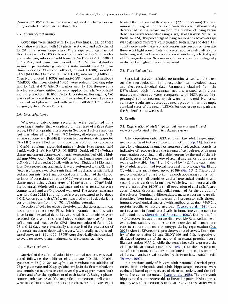
oscien
(b
2
cftposc2C(tlmwoi
2
rs(e(11ctaf(stcicw1c
blsr2gtt
2
uc2tbcw
D. Edwards et al. / Journal of Neur
Group G21CHX20). The neurons were evaluated for changes in via-ility and electrical properties after 1 day.
.5. Immunocytochemistry
Cover slips were rinsed with 1× PBS two times. Cells on theseover slips were fixed with 10% glacial acetic acid and 90% ethanolor 20 min at room temperature. Cover slips were again rinsedhree times with 1× PBS. Cells were permeabilized for 5 min with aermeabilizing solution (5 mM lysine + 0.5% Triton X-100 + 100 mlf 1× PBS), and were then blocked for 2 h (5% normal donkeyerum in permeabilizing solution). Anti-neurofilament M poly-lonal antibody (Chemicon, AB1981, diluted 1:1000), anti-MAPA/2B (MAB364, Chemicon, diluted 1:1000), anti-nestin (MAB5326,hemicon, diluted 1:1000) and anti-GFAP monoclonal antibodyMAB360, Chemicon, diluted 1:400) were added in blocking solu-ion for 12 h at 4 ◦C. After 3× washes with 1× PBS, fluorescentlyabeled secondary antibodies were applied for 2 h. Vectashield
ounting medium (H1000, Vector Laboratories, Burlingame, CA)as used to mount the cover slips onto slides. The cover slips were
bserved and photographed with an Ultra VIEWTM LCI confocalmaging system (Perkin Elmer).
.6. Electrophysiology
Whole-cell, patch-clamp recordings were performed in aecording chamber that was placed on the stage of a Zeiss Axio-cope, 2 FS Plus, upright microscope in Neurobasal culture mediumpH was adjusted to 7.3 with N-2-hydroxyethylpiperazine-N′-2-thane-sulfonic acid [HEPES]) at room temperature. Patch pipettes6–8 M�) were filled with intracellular solution (K-gluconate40 mM, ethylene glycol-bis[aminoethylether]-tetraacetic acidmM, MgCl2 2 mM, Na2ATP 5 mM, HEPES 10 mM; pH 7.2). Voltagelamp and current clamp experiments were performed with a Mul-iclamp 700A (Axon, Union City, CA) amplifier. Signals were filteredt 3 kHz and digitized at 20 kHz with an Axon Digidata 1322A inter-ace. Data recordings and analysis were performed with pClamp 8Axon) software. Inward currents that had the characteristics of fastodium currents (ISCs), and outward currents that had the charac-eristics of potassium currents (OPCs) were measured in voltagelamp mode using voltage steps of 10 mV from a −70 mV hold-ng potential. Whole-cell capacitance and series resistance wereompensated and a p/6 protocol was used. The access resistanceas less than 22 M� and tight seals were measured to be aboveG�. Action potentials (APs) were measured with 1 s depolarizingurrent injections from the −70 mV holding potential.
Selection of cells for electrophysiological characterization wasased upon morphology. Phase bright pyramidal neurons with
arge branching apical dendrites and small basal dendrites wereelected. Cells with this morphology stained positive for neu-ofilament and negative for GFAP. Neurons cultured for 14, 21,8 and 38 days were electrically characterized for evaluation oflutamate-mediated electrical recovery. Additionally, neurons cul-ured between 1–14 and 39–87 DIV were electrically characterizedo evaluate recovery and maintenance of electrical activity.
.7. Cell survival study
Survival of the cultured adult hippocampal neurons was eval-ated following the addition of glutamate (10, 25, 100 �M),ycloheximide (10, 20, 80 �g/ml), or simultaneous addition of
5 �M glutamate and 20 �g cycloheximide. In the first method, theotal number of neurons on each cover slip was approximated bothefore and after the application of each factor(s). Using a phase-ontrast microscope at 20× magnification, neuronal cell countsere made from 20 random spots on each cover slip, an area equalce Methods 190 (2010) 155–163 157
to 4% of the total area of the cover slip (22 mm × 22 mm). The totalnumber of living neurons on each cover slip was mathematicallydetermined. In the second method, the number of living versusdead neurons was quantified using a Live/Dead Assay kit (MolecularProbe, L-3224). The percentage of living neurons on each cover slipswas calculated from total cells counted, both living and dead. Cellcounts were made using a phase-contrast microscope with an epi-fluorescent light source. Total cells were approximated after cells,both living and dead, were counted on 20 randomly selected spotsat 20× magnification. Neurons in vitro were also morphologicallyevaluated throughout the culture period.
2.8. Statistical analysis
Statistical analysis included performing a two-sample t-teston the morphological, immunocytochemical, live/dead assayand electrophysiological data. Parameters obtained from theDETA-plated adult hippocampal neurons treated with gluta-mate ± cycloheximide were compared with DETA-plated hip-pocampal control neurons which were not treated. Numericalsummary results are reported as a mean, plus or minus the samplestandard error of the mean (±SEM). For two-group comparisons,the Student’s t-test was used.
3. Results
3.1. Regeneration of adult hippocampal neurons with limitedrecovery of electrical activity in a defined system
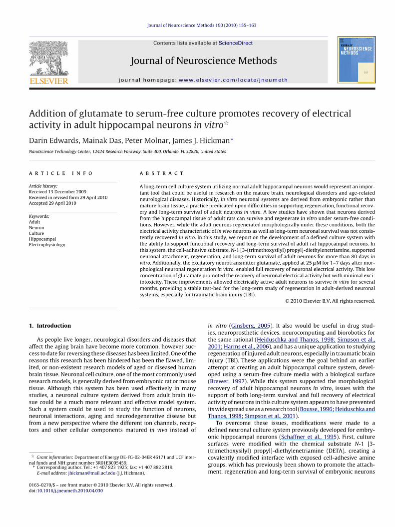
After deposition onto DETA surfaces, the adult hippocampalneurons adhered to the surface within 60 min (Fig. 1A). Immedi-ately following attachment, most neurons displayed characteristicsindicative of recovery from the trauma of cell culture, with someregeneration occurring in all viable neurons by the end of the ini-tial 24 h. After 2 DIV, recovery of axonal and dendritic processeswas clearly visible (Fig. 1B and C) and by 14 DIV the vast major-ity of adult neurons had typical neuronal morphology (Fig. 1B andC), which was maintained up to 80 DIV (Fig. 1D–I). These adultneurons exhibited phase bright, smooth-appearing somas, withone or more small dendrites and a large apical dendrite withsecond-order dendritic branching. While few non-neuronal cellswere present after 14 DIV, a small population of glial cells (astro-cytes, oligodendrocytes, microglia) remained for the duration ofthe cultures. Terminally differentiated, mature neurons were dis-tinguished from immature neurons and progenitor cells throughimmunocytochemical analysis with antibodies against MAP-2, aprotein specific to mature neurons (Coceres et al., 1984), andnestin, a protein found specifically in immature and progenitorcell populations (Stemple and Anderson, 1992). During the first14 DIV, recovering adult neurons displayed MAP2 as well as nestinexpression, possibly pointing to the regression of mature neu-rons to a more immature phenotype during regeneration (Das,2008). After 14 DIV, nestin expression was not observed. The major-ity of the cells after 21 and 38 DIV (90 and 94%, respectively)displayed expression of the neuronal structural proteins neuro-filament and/or MAP-2, while the remaining cells expressed theglial-specific structural protein GFAP (Fig. 1J–L). The low percent-age of non-neuronal cells may be attributed to the poor support ofglial growth and survival provided by the Neurobasal-A/B27 media(Brewer, 1997).
In a previous study of in vitro adult neuronal electrical prop-
erties, both embryonic and adult hippocampal neurons wereevaluated based upon recovery of electrical activity and the abil-ity to fire action potentials (Evans et al., 1998). The embryonichippocampal neurons were able to fire action potentials in approx-imately 84% of the neurons studied at 14 DIV in this earlier work.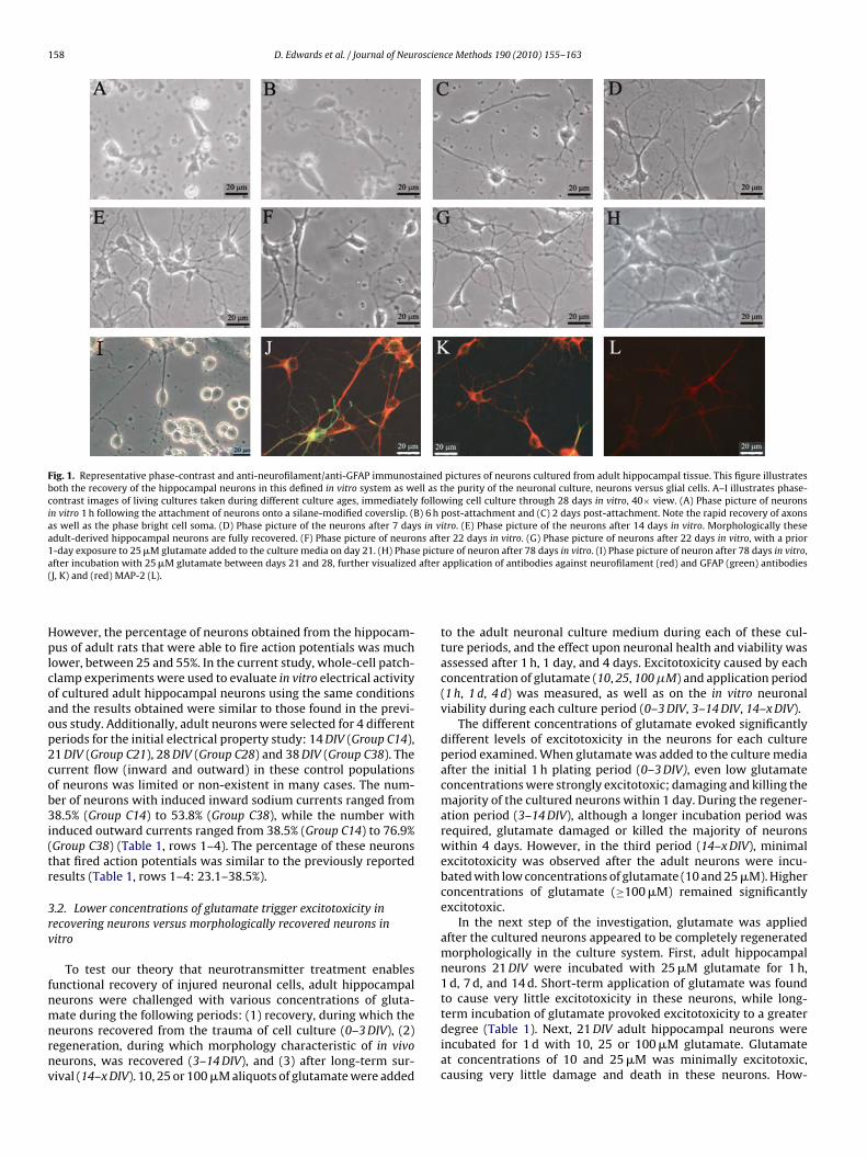
158 D. Edwards et al. / Journal of Neuroscience Methods 190 (2010) 155–163
Fig. 1. Representative phase-contrast and anti-neurofilament/anti-GFAP immunostained pictures of neurons cultured from adult hippocampal tissue. This figure illustratesboth the recovery of the hippocampal neurons in this defined in vitro system as well as the purity of the neuronal culture, neurons versus glial cells. A–I illustrates phase-contrast images of living cultures taken during different culture ages, immediately following cell culture through 28 days in vitro, 40× view. (A) Phase picture of neuronsin vitro 1 h following the attachment of neurons onto a silane-modified coverslip. (B) 6 h post-attachment and (C) 2 days post-attachment. Note the rapid recovery of axonsas well as the phase bright cell soma. (D) Phase picture of the neurons after 7 days in vitro. (E) Phase picture of the neurons after 14 days in vitro. Morphologically theseadult-derived hippocampal neurons are fully recovered. (F) Phase picture of neurons after 22 days in vitro. (G) Phase picture of neurons after 22 days in vitro, with a prior1-day exposure to 25 �M glutamate added to the culture media on day 21. (H) Phase picture of neuron after 78 days in vitro. (I) Phase picture of neuron after 78 days in vitro,a after(
Hplcoaop2cob3i(tr
3rv
fnmnrnv
fter incubation with 25 �M glutamate between days 21 and 28, further visualizedJ, K) and (red) MAP-2 (L).
owever, the percentage of neurons obtained from the hippocam-us of adult rats that were able to fire action potentials was much
ower, between 25 and 55%. In the current study, whole-cell patch-lamp experiments were used to evaluate in vitro electrical activityf cultured adult hippocampal neurons using the same conditionsnd the results obtained were similar to those found in the previ-us study. Additionally, adult neurons were selected for 4 differenteriods for the initial electrical property study: 14 DIV (Group C14),1 DIV (Group C21), 28 DIV (Group C28) and 38 DIV (Group C38). Theurrent flow (inward and outward) in these control populationsf neurons was limited or non-existent in many cases. The num-er of neurons with induced inward sodium currents ranged from8.5% (Group C14) to 53.8% (Group C38), while the number with
nduced outward currents ranged from 38.5% (Group C14) to 76.9%Group C38) (Table 1, rows 1–4). The percentage of these neuronshat fired action potentials was similar to the previously reportedesults (Table 1, rows 1–4: 23.1–38.5%).
.2. Lower concentrations of glutamate trigger excitotoxicity inecovering neurons versus morphologically recovered neurons initro
To test our theory that neurotransmitter treatment enablesunctional recovery of injured neuronal cells, adult hippocampaleurons were challenged with various concentrations of gluta-
ate during the following periods: (1) recovery, during which theeurons recovered from the trauma of cell culture (0–3 DIV), (2)egeneration, during which morphology characteristic of in vivoeurons, was recovered (3–14 DIV), and (3) after long-term sur-ival (14–x DIV). 10, 25 or 100 �M aliquots of glutamate were added
application of antibodies against neurofilament (red) and GFAP (green) antibodies
to the adult neuronal culture medium during each of these cul-ture periods, and the effect upon neuronal health and viability wasassessed after 1 h, 1 day, and 4 days. Excitotoxicity caused by eachconcentration of glutamate (10, 25, 100 �M) and application period(1 h, 1 d, 4 d) was measured, as well as on the in vitro neuronalviability during each culture period (0–3 DIV, 3–14 DIV, 14–x DIV).
The different concentrations of glutamate evoked significantlydifferent levels of excitotoxicity in the neurons for each cultureperiod examined. When glutamate was added to the culture mediaafter the initial 1 h plating period (0–3 DIV), even low glutamateconcentrations were strongly excitotoxic; damaging and killing themajority of the cultured neurons within 1 day. During the regener-ation period (3–14 DIV), although a longer incubation period wasrequired, glutamate damaged or killed the majority of neuronswithin 4 days. However, in the third period (14–x DIV), minimalexcitotoxicity was observed after the adult neurons were incu-bated with low concentrations of glutamate (10 and 25 �M). Higherconcentrations of glutamate (≥100 �M) remained significantlyexcitotoxic.
In the next step of the investigation, glutamate was appliedafter the cultured neurons appeared to be completely regeneratedmorphologically in the culture system. First, adult hippocampalneurons 21 DIV were incubated with 25 �M glutamate for 1 h,1 d, 7 d, and 14 d. Short-term application of glutamate was foundto cause very little excitotoxicity in these neurons, while long-
term incubation of glutamate provoked excitotoxicity to a greaterdegree (Table 1). Next, 21 DIV adult hippocampal neurons wereincubated for 1 d with 10, 25 or 100 �M glutamate. Glutamateat concentrations of 10 and 25 �M was minimally excitotoxic,causing very little damage and death in these neurons. How-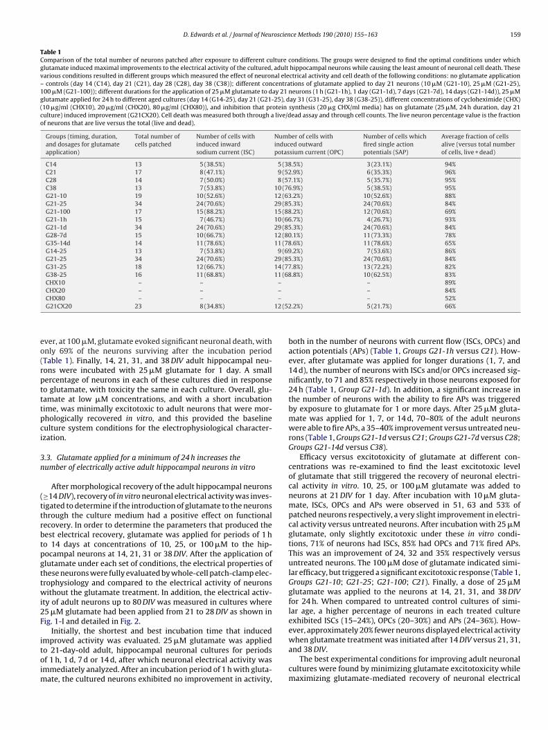
D. Edwards et al. / Journal of Neuroscience Methods 190 (2010) 155–163 159
Table 1Comparison of the total number of neurons patched after exposure to different culture conditions. The groups were designed to find the optimal conditions under whichglutamate induced maximal improvements to the electrical activity of the cultured, adult hippocampal neurons while causing the least amount of neuronal cell death. Thesevarious conditions resulted in different groups which measured the effect of neuronal electrical activity and cell death of the following conditions: no glutamate application– controls (day 14 (C14), day 21 (C21), day 28 (C28), day 38 (C38)); different concentrations of glutamate applied to day 21 neurons (10 �M (G21-10), 25 �M (G21-25),100 �M (G21-100)); different durations for the application of 25 �M glutamate to day 21 neurons (1 h (G21-1h), 1 day (G21-1d), 7 days (G21-7d), 14 days (G21-14d)), 25 �Mglutamate applied for 24 h to different aged cultures (day 14 (G14-25), day 21 (G21-25), day 31 (G31-25), day 38 (G38-25)), different concentrations of cycloheximide (CHX)(10 �g/ml (CHX10), 20 �g/ml (CHX20), 80 �g/ml (CHX80)), and inhibition that protein synthesis (20 �g CHX/ml media) has on glutamate (25 �M, 24 h duration, day 21culture) induced improvement (G21CX20). Cell death was measured both through a live/dead assay and through cell counts. The live neuron percentage value is the fractionof neurons that are live versus the total (live and dead).
Groups (timing, duration,and dosages for glutamateapplication)
Total number ofcells patched
Number of cells withinduced inwardsodium current (ISC)
Number of cells withinduced outwardpotassium current (OPC)
Number of cells whichfired single actionpotentials (SAP)
Average fraction of cellsalive (versus total numberof cells, live + dead)
C14 13 5 (38.5%) 5 (38.5%) 3 (23.1%) 94%C21 17 8 (47.1%) 9 (52.9%) 6 (35.3%) 96%C28 14 7 (50.0%) 8 (57.1%) 5 (35.7%) 95%C38 13 7 (53.8%) 10 (76.9%) 5 (38.5%) 95%G21-10 19 10 (52.6%) 12 (63.2%) 10 (52.6%) 88%G21-25 34 24 (70.6%) 29 (85.3%) 24 (70.6%) 84%G21-100 17 15 (88.2%) 15 (88.2%) 12 (70.6%) 69%G21-1h 15 7 (46.7%) 10 (66.7%) 4 (26.7%) 93%G21-1d 34 24 (70.6%) 29 (85.3%) 24 (70.6%) 84%G28-7d 15 10 (66.7%) 12 (80.1%) 11 (73.3%) 78%G35-14d 14 11 (78.6%) 11 (78.6%) 11 (78.6%) 65%G14-25 13 7 (53.8%) 9 (69.2%) 7 (53.6%) 86%G21-25 34 24 (70.6%) 29 (85.3%) 24 (70.6%) 84%G31-25 18 12 (66.7%) 14 (77.8%) 13 (72.2%) 82%G38-25 16 11 (68.8%) 11 (68.8%) 10 (62.5%) 83%CHX10 – – – – 89%CHX20 – – – – 84%
–12 (52
eo(rptttpci
3n
(ttrbtpgttwi2F
itoim
CHX80 – –G21CX20 23 8 (34.8%)
ver, at 100 �M, glutamate evoked significant neuronal death, withnly 69% of the neurons surviving after the incubation periodTable 1). Finally, 14, 21, 31, and 38 DIV adult hippocampal neu-ons were incubated with 25 �M glutamate for 1 day. A smallercentage of neurons in each of these cultures died in responseo glutamate, with toxicity the same in each culture. Overall, glu-amate at low �M concentrations, and with a short incubationime, was minimally excitotoxic to adult neurons that were mor-hologically recovered in vitro, and this provided the baselineulture system conditions for the electrophysiological character-zation.
.3. Glutamate applied for a minimum of 24 h increases theumber of electrically active adult hippocampal neurons in vitro
After morphological recovery of the adult hippocampal neurons≥14 DIV), recovery of in vitro neuronal electrical activity was inves-igated to determine if the introduction of glutamate to the neuronshrough the culture medium had a positive effect on functionalecovery. In order to determine the parameters that produced theest electrical recovery, glutamate was applied for periods of 1 ho 14 days at concentrations of 10, 25, or 100 �M to the hip-ocampal neurons at 14, 21, 31 or 38 DIV. After the application oflutamate under each set of conditions, the electrical properties ofhese neurons were fully evaluated by whole-cell patch-clamp elec-rophysiology and compared to the electrical activity of neuronsithout the glutamate treatment. In addition, the electrical activ-
ty of adult neurons up to 80 DIV was measured in cultures where5 �M glutamate had been applied from 21 to 28 DIV as shown inig. 1-I and detailed in Fig. 2.
Initially, the shortest and best incubation time that induced
mproved activity was evaluated. 25 �M glutamate was appliedo 21-day-old adult, hippocampal neuronal cultures for periodsf 1 h, 1 d, 7 d or 14 d, after which neuronal electrical activity wasmmediately analyzed. After an incubation period of 1 h with gluta-ate, the cultured neurons exhibited no improvement in activity,
– 52%.2%) 5 (21.7%) 66%
both in the number of neurons with current flow (ISCs, OPCs) andaction potentials (APs) (Table 1, Groups G21-1h versus C21). How-ever, after glutamate was applied for longer durations (1, 7, and14 d), the number of neurons with ISCs and/or OPCs increased sig-nificantly, to 71 and 85% respectively in those neurons exposed for24 h (Table 1, Group G21-1d). In addition, a significant increase inthe number of neurons with the ability to fire APs was triggeredby exposure to glutamate for 1 or more days. After 25 �M gluta-mate was applied for 1, 7, or 14 d, 70–80% of the adult neuronswere able to fire APs, a 35–40% improvement versus untreated neu-rons (Table 1, Groups G21-1d versus C21; Groups G21-7d versus C28;Groups G21-14d versus C38).
Efficacy versus excitotoxicity of glutamate at different con-centrations was re-examined to find the least excitotoxic levelof glutamate that still triggered the recovery of neuronal electri-cal activity in vitro. 10, 25, or 100 �M glutamate was added toneurons at 21 DIV for 1 day. After incubation with 10 �M gluta-mate, ISCs, OPCs and APs were observed in 51, 63 and 53% ofpatched neurons respectively, a very slight improvement in electri-cal activity versus untreated neurons. After incubation with 25 �Mglutamate, only slightly excitotoxic under these in vitro condi-tions, 71% of neurons had ISCs, 85% had OPCs and 71% fired APs.This was an improvement of 24, 32 and 35% respectively versusuntreated neurons. The 100 �M dose of glutamate indicated simi-lar efficacy, but triggered a significant excitotoxic response (Table 1,Groups G21-10; G21-25; G21-100; C21). Finally, a dose of 25 �Mglutamate was applied to the neurons at 14, 21, 31, and 38 DIVfor 24 h. When compared to untreated control cultures of simi-lar age, a higher percentage of neurons in each treated cultureexhibited ISCs (15–24%), OPCs (20–30%) and APs (24–36%). How-ever, approximately 20% fewer neurons displayed electrical activity
when glutamate treatment was initiated after 14 DIV versus 21, 31,and 38 DIV.The best experimental conditions for improving adult neuronalcultures were found by minimizing glutamate excitotoxicity whilemaximizing glutamate-mediated recovery of neuronal electrical
160 D. Edwards et al. / Journal of Neuroscience Methods 190 (2010) 155–163
F s of ap p of ac e singo tamat
a2tt
Fin
ig. 2. Representative phase-contrast pictures and electrophysiological recordingictures of neurons 80 DIV. (C) Representative traces for voltage and current clamells through the voltage-gated ion channels (voltage clamp trace) as well as to firriginated from adult hippocampal neurons after 78 days in vitro, where 25 �M glu
ctivity in vitro. Challenging cultured adult neurons at 21 DIV with5 �M glutamate for 1–7 days caused manageable levels of exci-otoxicity, while significantly improving the electrical potential ofhese cultured neurons. This combination of culture age, glutamate
ig. 3. Representative traces for voltage and current clamp of an adult neuron 21 DIV. Neuron channels (voltage clamp trace) as well as to fire single action potentials after electricaleurons after 21 days in vitro, where 25 �M glutamate had been applied to the culture m
dult hippocampal neurons after approximately 80 DIV. (A and B) Phase-contrastn adult neuron 78 DIV. Neurons retain the ability to move current into and out ofle action potentials after electrical stimulation (current clamp trace). These tracese had been applied to the culture medium between days 21 and 28.
concentration and incubation time yielded significantly improvedelectrical activity with minimal neuronal cell loss. Representativevoltage and current clamp traces (21 and 78 DIV) are displayed inFigs. 2 and 3.
ons retain the ability to move current into and out of cells through the voltage-gatedstimulation (current clamp trace). These traces originated from adult hippocampaledium between days 20 and 21.

oscience Methods 190 (2010) 155–163 161
3fin
bimrrsmtdwaidnGgttwab
tnitfgc
4
naaltsaiocmpaheegaemoegrmfs
he
elec
tric
alp
rop
erti
esof
thos
en
euro
ns
that
exh
ibit
edac
tion
pot
enti
als.
The
dif
fere
nt
glu
tam
ate
app
lica
tion
con
dit
ion
sre
sult
edin
dif
fere
nt
grou
ps,
wh
ich
mea
sure
dth
eef
fect
ofn
euro
nal
elec
tric
alp
rop
erti
esin
nd
itio
ns:
no
glu
tam
ate
app
lica
tion
–co
ntr
ols
(day
14(C
14),
day
21(C
21),
day
28(C
28),
day
38(C
38))
;dif
fere
nt
con
cen
trat
ion
sof
glu
tam
ate
app
lied
tod
ay21
neu
ron
s(1
0�
M(G
21-1
0),2
5�
M(G
21-2
5),1
00�
Mer
ent
du
rati
onfo
rth
eap
pli
cati
onof
25�
Mgl
uta
mat
eto
day
21n
euro
ns
(1h
(G21
-1h
),1
day
(G21
-1d
),7
day
s(G
21-7
d),
14d
ays
(G21
-14d
)),2
5�
Mgl
uta
mat
eap
pli
edfo
r24
hto
dif
fere
nt
aged
cult
ure
s(d
ay14
1(G
21-2
5),d
ay31
(G31
-25)
,day
38(G
38-2
5)),
and
inh
ibit
ion
that
pro
tein
syn
thes
is(2
0�
gC
HX
/mlm
edia
)h
ason
glu
tam
ate
(25
�M
,24
hd
ura
tion
,day
21cu
ltu
re)
ind
uce
dim
pro
vem
ent
(G21
CX
20).
C14
C21
C28
C38
G21
-10
G21
-25
G21
-100
G21
-1h
G21
-7d
G21
-14d
G14
-25
G31
-25
G38
-25
G21
CX
20
fore
n/a
n/a
n/a
n/a
2020
2021
2121
1330
3720
itro
1421
2838
2121
2121
+1
h28
3514
3138
21ro
ns
1317
1413
1934
1715
1514
1318
1623
ial
−70
−69
±1.
56−6
9.05
±1.
05−6
9.63
±0.
45−7
0.25
±1.
92−7
1.4
±0.
88−7
0.9
±0.
54−7
1.4
±1.
36−6
9±
1.24
−69.
1±
1.42
−70.
3±
0.84
−69.
75±
1.06
−69.
5±
0.92
−68.
1±
0.53
e23
.123
.1±
9.62
23.1
±2.
3422
.9±
1.74
23±
14.1
525
.74
±10
.06
19.1
1±
5.64
23.1
±11
.49
22.7
5±
2.21
23.1
±6.
326
.86
±9.
5122
.3±
11.1
527
.44
±6.
0616
.11
±8.
34
F)15
.41
15.7
7±
8.1
14.5
7±
6.5
14.6
7±
4.8
12.8
6±
11.6
910
.55
±4.
968.
9±
3.88
13.1
4±
9.28
11.2
2±
2.7
10.6
3±
5.76
11.1
3±
6.75
12.1
3±
3.65
10.9
6±
7.62
9.35
±7.
45rr
ent
−172
.4−1
88.3
±72
.4−1
65.2
±57
.3−1
78.1
±66
.3−2
37.2
±77
.3−4
03.6
±98
.5−3
88.3
±97
.4−1
73.3
±88
.9−4
02.2
±77
.5−3
43.7
±87
.4−3
02.5
±93
.6−3
78.8
±73
.4−3
79.8
±43
.9−2
55.6
±92
.4
)28
5.4
304.
2±
144.
229
8.6
±74
.928
5.6
±71
.447
9.3
±17
7.7
923.
5±
201.
278
8.3
±24
3.6
276.
3±
81.4
802.
4±
344.
573
4.0
±13
3.2
723.
4±
174.
389
3.2
±20
2.4
933.
1±
355.
340
2.5
±16
6.3
al28
.229
.7±
2.1
31.8
±3.
130
.3±
2.8
34.2
±4.
230
.6±
2.8
27.1
±4.
334
.2±
4.4
31.7
±1.
932
.6±
3.2
26.4
±2.
129
.1±
3.2
28.4
±1.
131
.2±
2.3
D. Edwards et al. / Journal of Neur
.4. Extracellular glutamate applied for a minimum of 24 h toully recovered adult hippocampal neurons in vitro resulted inncreased inward and outward current flow in electrically activeeurons
The effect from glutamate at various concentrations and incu-ation times to neurons at different days in vitro was measured
n relation to the changes in mean resting membrane potential,embrane resistance, membrane capacitance, peak inward cur-
ent, peak outward current and action potential amplitude. Theseesults are summarized as mean ± SE (Table 2). Statistical analy-is performed on the results between the different group’s restingembrane potential, membrane resistance, membrane capaci-
ance and action potential height indicated no significant changeue to adult neuronal exposure to glutamate. However, incubationith glutamate triggered significant changes in the peak inward
nd outward currents (Table 2). The amplitude of current flownto the cultured neurons after application of glutamate on averageoubled as compared to the current flow observed in the controleurons, from −188.3 ± 72.4 (Group C21) to −403.6 ± 98.5 (Group21-25). Likewise, the amplitude of current flowing out of thelutamate-treated cultured neurons increased on average 2.5–3imes versus the control neurons, from 304.2 ± 144.2 (Group C21)o 923.5 ± 201.2 (Group G21-25). In the different cultures incubatedith 25 �M glutamate for at least 1 d (1, 7, 14 d), no significant
mplitude differences in mean peak ISCs or OPCs were evidentetween these cultures.
In addition, cycloheximide (CHX), an inhibitor of protein syn-hesis that had previously been used to block preconditioning ineurons (Barone et al., 1998), was used to block all possible changes
n protein synthesis induced by incubation of the neurons with glu-amate. When hippocampal neurons (21 DIV) were pre-incubatedor 1 h with CHX (20 mg/ml) prior to the introduction of 25 mMlutamate, all improvements in neuronal electrical activity wereompletely blocked (Tables 1 and 2).
. Discussion
In this study, a defined culture system for adult hippocampaleurons was developed that included a cell-adhesive silane surfacend a serum-free media containing glutamate. This culture systemllowed adult neurons to recover full electrical activity and surviveong-term in vitro for more than 80 DIV. First, culture surfaces, inhis case glass cover slips, were modified with the chemical sub-trate DETA to create stable surfaces with exposed cell-adhesivemine groups, that has been shown to be stable for longer periodsn culture because the silanes are covalently linked to the surface aspposed to PDL and poly-ornithine which are physisorbed. Thesehemically modified surfaces not only promoted neuron attach-ent, regeneration and long-term culture compared to PDL and
oly-ornithine (Schaffner et al., 1995; Das et al., 2003), but werelso stable, reproducible and can be further modified to createigh-resolution patterns, which could be useful for the creation ofngineered networks of these adult hippocampal neurons (Stengert al., 1993, 1998; Ravenscroft et al., 1998). Second, the optimallutamate concentration, culture age for glutamate applicationnd duration of glutamate exposure were determined such thatxcitotoxicity was minimized and neuronal electrical improve-ents was maximized, which resulted in the long-term survival
f electrophysiologically functional adult neurons. Based on thexperimental results, challenging 21 DIV adult neurons with 25 �M
lutamate for 1–7 d caused minimal excitotoxicity, while inducingecovery of full electrical activity in vitro. Together, these improve-ents allowed adult neurons to functionally recover and to surviveor several months in vitro, providing a stable test-bed for long-termtudy of the mature mammalian brain. Ta
ble
2C
omp
aris
onof
tth
efo
llow
ing
co(G
21-1
00))
;d
iff
(G14
-25)
,day
2
Day
sin
vitr
obe
add
itio
nof
glu
tam
ate
Tota
lday
sin
vN
um
ber
ofn
eu(n
)R
esti
ng
pot
ent
(mV
)In
pu
tre
sist
anc
(m�
)C
apac
itan
ce(p
Peak
inw
ard
cu(p
A)
Peak
outw
ard
curr
ent
(pA
Act
ion
pot
enti
hei
ght
(mV
)
1 oscien
gmnmwmeorectbmceewlpnn
satngsragwag(cTdCuckrt(tgwdpg1deabaswavir(
62 D. Edwards et al. / Journal of Neur
In fast excitatory neurotransmission, the neurotransmitterlutamate primarily acts via the activation of ionotropic andetabotropic receptors. In addition to conveying fast excitatory
eurotransmission, activation of these channels appears to play aajor role in neuronal differentiation and CNS development, asell as in processes that give rise to long-term potentiation andemory formation (Verderio et al., 1999). Recovery of the neuronal
lectrical activity in this culture system, induced by the applicationf glutamate, illustrates a role for glutamate beyond simple neu-otransmission. The application of glutamate triggered this changeither through transient or persistent cellular changes. Transienthanges in activity could have occurred as a direct result fromhe presence of glutamate, triggering changes in passive mem-rane properties (capacitance and resistance), alteration in theagnitude of the voltage-activated sodium, potassium and calcium
urrents (Daoudal et al., 2002; Zhang and Linden, 2003; Walmsleyt al., 2006; Sjostrom et al., 2008), or alteration in the activity lev-ls of the ion channels. Persistent changes to neuronal excitabilityould be dependent upon the induction of gene expression to cause
ong-lasting changes in the adult neurons. Results from this studyoint to the induction of persistent changes in these cultured adulteurons as improved electrical activity was not evident in the adulteurons after only 1 h of glutamate application.
In this culture system, glutamate appears to activate gene tran-cription through the same mechanism found in vivo. Throughctivation of ionotropic and metabotropic receptors in vivo, glu-amate has been shown to trigger gene activity in immature,ewborn and mature hippocampal neurons. In immature neurons,lutamate regulates cell proliferation, neuronal differentiation andurvival responses primarily through the N-methyl-d-aspartateeceptor (NMDAR) channel’s activation (Dalva et al., 2000; Husi etl., 2000; Kornhauser et al., 2002). However, in mature neuronslutamate regulates the expression of adaptive response genes asell as genes that regulate more complex neural functions, such
s learning and memory, though the activation of L-type voltage-ated calcium channels (L-VGCC) rather than NMDAR channelsKornhauser et al., 2002). Activation of these channels triggersalcium influx, thus activating a number of signaling molecules.hese molecules potentiate the signals through the activation ofownstream signaling proteins such as protein kinase A (PKA),a2+/calmodulin dependent protein kinase type IV (CaMK-IV), reg-lated S6 protein kinase (Rsk) and other pathways to amplify thealcium signal and carry it to the nucleus. In the nucleus theseinase phosphorylate transcription factors, such as cyclic AMPesponse element binding protein (CREB) or myocyte enhancer fac-or 2 (MEF2), make them competent to mediate gene transcriptionWest et al., 2001). Although glutamate activates transcription fac-ors like CREB in both mature and immature neurons, differentenes are activated in each case. Overall, different signaling path-ays trigger the expression of genes that regulate the survival,ifferentiation and function of immature neurons (cAMP–CREBathway) (Nakagawa et al., 2002; Balazs, 2006), neurite out-rowth (Mattson, 1988; Mattson et al., 1988; Verderio et al.,999) or activity-dependent synaptic plasticity and trophic factor-ependent neuronal survival (West et al., 2001; Tominaga-Yoshinot al., 2008). In this adult neuronal culture system, glutamateppeared to induce gene activation, resulting in increased num-ers of neurons firing APs and in significantly increased sodiumnd potassium current amplitude. The initiation of protein synthe-is by the glutamate treatment is supported by the experimentshere CHX addition blocked any functional recovery of electrical
ctivity due to glutamate addition. These differences in the acti-ated signaling pathways in the adult neurons from that observedn development may give insight to pathways necessary for neu-oregeneration, specifically for recovery in traumatic brain injuryTBI).
ce Methods 190 (2010) 155–163
Another explanation from a mechanistic perspective is the pos-sible conditioning in the mature brain to the reliance of electricalpotential between neurons and supporting cells such as astrocyticrecycling of glutamate. Without this feedback between the sup-porting cells and neurons, low concentrations of glutamate maynot be enough to be able to reestablish the membrane potential.Instead, lack of glutamate recycling in the synaptic cleft limits theamount of available glutamate for stimulation of the recovery ofthe neuronal electrical properties. Thus, increasing the levels ofglutamate in vitro restores the natural concentration of glutamateto post-synaptic terminals, restoring the natural “conditioning” ofneurons to glutamate and the cellular changes resulting from thislevel of glutamate. These changes (i.e. gene expression changes)can result in increased expression of ion channels in the neuronalcell membranes. This results in a more “normal” electrical poten-tial as compared to in vivo membrane potential results due to therecovery of in vivo like ion channel expression levels.
In this study, silane-modified DETA surfaces and the transientapplication of glutamate have been incorporated into an adult,hippocampal cell culture system in order to sustain long-termneuronal survival as well as to promote full recovery of electricalactivity in vitro. Together, these improvements allowed electricallyactive adult neurons to survive for several months in vitro, provid-ing a stable system with potential for a wide range of applications.Promising applications include the long-term study of the maturebrain, neurological disorders, and diseases affecting the aging brain.In addition, because DETA monolayers can be applied not only toglass cover slips but also to electrodes, this system can be extendedto integrate living and electronic systems. Overall, potential usesfor such a system range from research into the function of neu-rons, neuronal interactions, aging, and neurodegenerative disease(Ginsberg, 2005) as well as drug studies, neuroprosthetic devices,neurocomputing, and biorobotics (Heiduschka and Thanos, 1998;Simpson et al., 2001; Harms et al., 2006), or most importantly,neuroregeneration, especially in TBI.
Acknowledgements
The authors would like to acknowledge funding support fromthe Department of Energy, grant number DE-FG-02-04ER 46171and National Institute of Health, grant number 5R01EB005459.
References
Auditore JV, Wade L, Olson EJ. Occurrence of N-acetyl-l-glutamic acid in the humanbrain. J Neurochem 1966;13(11):1149–55.
Balazs R. Trophic effect of glutamate. Curr Top Med Chem 2006;6(10):961–8.Barone FC, White RF, Spera PA, Ellison J, Currie RW, Wang X, et al. Ischemic precon-
ditioning and brain tolerance: temporal histological and functional outcomes,protein synthesis requirement, and interleukin-1 receptor antagonist and earlygene expression. Stroke 1998;29(9):1937–51.
Bousse L. Whole cell biosensors. Sens Actuators B-Chem 1996;34(1–3):270–5.Brewer GJ. Isolation and culture of adult rat hippocampal neurons. J Neurosci Meth-
ods 1997;71(2):143–55.Brewer GJ. Age-related toxicity to lactate, glutamate, and beta-amyloid in cultured
adult neurons. Neurobiol Aging 1998;19(6):561–8.Brewer GJ, Lim A, Capps NG, Torricelli JR. Age-related calcium changes, oxyradical
damage, caspase activation and nuclear condensation in hippocampal neuronsin response to glutamate and beta-amyloid. Exp Gerontol 2005;40(5):426–37.
Brewer GJ, Torricelli JR, Evege EK, Price PJ. Optimized survival of hippocampal-neurons in B27-supplemented neurobasal(Tm), a new serum-free mediumcombination. J Neurosci Res 1993;35(5):567–76.
Coceres A, Banker G, Steward O, Binder L, Payne M. MAP2 is localized to the dendritesof hippocampal neurons which develop in culture. Brain Res 1984;315:314–8.
Dalva MB, Takasu MA, Lin MZ, Shamah SM, Hu L, Gale NW, et al. EphB receptorsinteract with NMDA receptors and regulate excitatory synapse formation. Cell2000;103(6):945–56.
Daoudal G, Hanada Y, Debanne D. Bidirectional plasticity of excitatory postsynapticpotential (EPSP)-spike coupling in CA1 hippocampal pyramidal neurons. PNAS2002;99(22):14512–7.
Das M, Bhargava N, Bhalkikar A, Kang J-F, Hickman JJ. Temporal neurotransmitterconditioning restores the functional activity of adult spinal cord neurons in long-term culture. Exp Neurol 2008;209:171–80.
oscien
D
D
E
G
H
H
H
H
K
M
M
N
R
D. Edwards et al. / Journal of Neur
as M, Bhargava N, Gregory C, Riedel L, Molnar P, Hickman JJ. Adult rat spinal cordculture on an organosilane surface in a novel serum-free medium. In Vitro CellDev Biol-Anim 2005;41(10):343–8.
as M, Molnar P, Devaraj H, Poeta M, Hickman J. Electrophysiological and mor-phological characterization of rat embryonic motoneurons in a defined system.Biotechnol Prog 2003;19:1756–61.
vans MS, Collings MA, Brewer GJ. Electrophysiology of embryonic, adult andaged rat hippocampal neurons in serum-free culture. J Neurosci Methods1998;79(1):37–46.
insberg SD. Glutamatergic neurotransmission expression profiling in the mousehippocampus after perforant-path transection. Am J Geriatr Psychiatry2005;13(12):1052–61.
arms H, Wells MC, van der Meer JR. Whole-cell living biosensors—are they readyfor environmental application. Appl Microbiol Biotechnol 2006;70(3):273–80.
eiduschka P, Thanos S. Implantable bioelectronic interfaces for lost nerve functions.Prog Neurobiol 1998;55(5):433–61.
ickman JJ, Bhatia SK, Quong JN, Shoen P, Stenger DA, Pike CJ, et al. Rational patterndesign for in-vitro cellular networks using surface photochemistry. J Vac SciTechnol A-Vac Surf Films 1994;12(3):607–16.
usi H, Ward MA, Choudhary JS, Blackstock WP, Grant SG. Proteomic analy-sis of NMDA receptor-adhesion protein signaling complexes. Nat Neurosci2000;3(7):661–9.
ornhauser J, Cowan C, Shaywitz A, Dolmetsch R, Griffith E, Hu L, et al. CREBtranscriptional activity in neurons is regulated by multiple, calcium-specificphosphorylation events. Neuron 2002;34:221–33.
attson MP. Neurotransmitters in the regulation of neuronal cytoarchitecture. BrainRes 1988;472(2):179–212.
attson MP, Dou P, Kater SB. Outgrowth-regulating actions of glutamate in isolatedhippocampal pyramidal neurons. J Neurosci 1988;8(6):2087–100.
akagawa S, Kim J, Lee R, Chen J, Fujioka T, Malberg J, et al. Localization of phospho-
rylated cAMP response element-binding protein in immature neurons of adulthippocampus. J Neurosci 2002;22:9868–76.avenscroft MS, Bateman KE, Shaffer KM, Schessler HM, Jung DR, Schneider TW, etal. Developmental neurobiology implications from fabrication and analysis ofhippocampal neuronal networks on patterned silane-modified surfaces. J AmChem Soc 1998;120(47):12169–77.
ce Methods 190 (2010) 155–163 163
Schaffner AE, Barker JL, Stenger DA, Hickman JJ. Investigation of the factors necessaryfor growth of hippocampal neurons in a defined system. J Neurosci Methods1995;62(1–2):111–9.
Simpson ML, Sayler GS, Fleming JT, Applegate B. Whole-cell biocomputing. TrendsBiotechnol 2001;19(8):317–23.
Sjostrom PJ, Rancz EA, Roth A, Hausser M. Dendritic excitability and synaptic plas-ticity. Physiol Rev 2008;88(2):769–840.
Spargo BJ, Testoff MA, Nielsen TB, Stenger DA, Hickman JJ, Rudolph AS. Spa-tially controlled adhesion, spreading, and differentiation of endothelial cellson self-assembled molecular monolayers. Proc Natl Acad Sci USA 1994;91(23):11070–4.
Stemple DL, Anderson DJ. Isolation of a stem cell for neurons and glia from themammalian neural crest. Cell 1992;71(6):973–85.
Stenger DA, Hickman JJ, Bateman KE, Ravenscroft MS, Ma W, Pancrazio JJ, et al.Microlithographic determination of axonal/dendritic polarity in cultured hip-pocampal neurons. J Neurosci Methods 1998;82(2):167–73.
Stenger DA, Pike CJ, Hickman JJ, Cotman CW. Surface determinants of neu-ronal survival and growth on self-assembled monolayers in culture. Brain Res1993;630(1–2):136–47.
Tominaga-Yoshino K, Urakubo T, Okada M, Matsuda H, Ogura A. Repetitiveinduction of late-phase LTP produces long-lasting synaptic enhancementaccompanied by synaptogenesis in cultured hippocampal slices. Hippocampus2008;18(3):281–93.
Verderio C, Coco S, Pravettoni E, Bacci A, Matteoli M. Synaptogenesis in hippocampalcultures. Cell Mol Life Sci 1999;55:1448–62.
Walmsley B, Berntson A, Leao RN, Fyffe REW. Activity-dependent regulation ofsynaptic strength and neuronal excitability in central auditory pathways. J Phys-iol 2006;572(2):313–21.
West A, Chen W, Dalva M, Dolmetsch R, Kornhauser J, Shaywitz A, et al. Calciumregulation of neuronal gene expression. PNAS 2001;98:11024–31.
Zhang W, Linden DJ. The other side of the engram: experience-driven changes inneuronal intrinsic excitability. Nat Rev Neurosci 2003;4(11):885–900.
Zhu D, Wu X, Strauss KI, Lipsky RH, Qureshi Z, Terhakopian A, et al. N-Methyl-d-aspartate and TrkB receptors protect neurons against glutamate excitotoxicitythrough an extracellular signal-regulated kinase pathway. J Neurosci Res2005;80(1):104–13.
Top Related
Copyright © 2022 FDOKUMEN

