


Bahasa
Halaman
Hukum

256 © 2021 The Journal of Indian Prosthodontic Society | Published by Wolters Kluwer - Medknow
A study to evaluate the influence of condylar and incisal guidance in canine guided and group function occlusal schemes
Vinita Rajesh Sippy, Chethan Hegde, Ganaraj ShettyDepartment of Prosthodontics, AB Shetty Memorial Institute of Dental Sciences, Nitte (Deemed to be University), Deralakatte,
Mangaluru, Karnataka, India
Original Article
INTRODUCTION
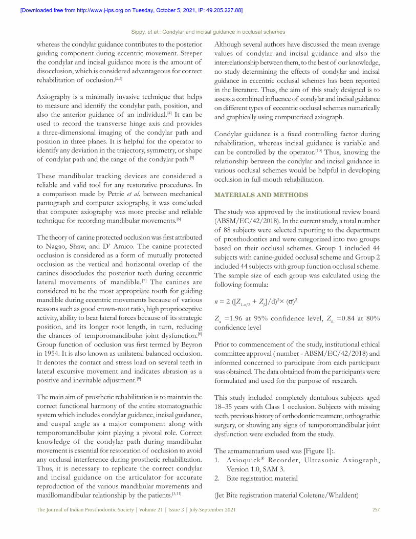
Occlusal scheme is the form and arrangement of occlusal contacts in natural and artificial dentition. The choice of an occlusal scheme will determine the pattern of occlusal
contacts between opposing teeth during centric relation and functional movement of mandible.[1] The combination of anterior guidance and posterior guidance is an important determinant for eccentric occlusal schemes. The incisal guidance contributes to the anterior guiding component,
Aims: This study aimed to evaluate the combined influence of condylar and incisal guidance in canine-guided and group function occlusal schemes.Settings and Design: In vivo - Cross sectional study.Materials and Methods: A total number of 88 subjects were selected and classified into two groups: 44 subjects with canine guided and 44 subjects with group function occlusal schemes. Condylar and incisal guidance tracings of both the groups were recorded using SAM AXIOQUICK RECORDER (School Articulator Munich) and evaluated.Statistical Analysis Used: Kolmogorov–Smirnov test and Shapiro–Wilk test were employed to test the normality of data. Independent sample t-test and Mann–Whitney U-test was performed for quantitative variables.Results: The condylar and incisal guidance among canine-guided individuals was 31.38 ± 12.01 and 55.83 ± 14.04, respectively, while in group function individuals, it was 29.44 ± 12.65 and 43.74 ± 20.27.Conclusions: Within the limitations of the present study, condylar guidance was similar in subjects with both schemes of occlusion, whereas steeper incisal guidance was noticed in canine-guided individuals as compared to group function.
Keywords: Axiography, canine guided, condylar guidance, group function, incisal guidance, occlusal scheme
Abstract
Access this article onlineQuick Response Code:
Website:www.j-ips.org
DOI:10.4103/jips.jips_183_21
How to cite this article: Sippy VR, Hegde C, Shetty G. A study to evaluate the influence of condylar and incisal guidance in canine guided and group function occlusal schemes. J Indian Prosthodont Soc 2021;21:256-61.
Address for correspondence: Dr. Chethan Hegde, Department of Prosthodontics, A B Shetty Memorial Institute of Dental Sciences, NITTE (Deemed to be University), Deralakatte, Mangaluru ‑ 575 018, Karnataka, India. E‑mail: [email protected] Submitted: 29‑Apr‑2021, Revised: 15‑Jul‑2021, Accepted: 17‑Jul‑2021, Published: 10‑Aug‑2021
This is an open access journal, and articles are distributed under the terms of the Creative Commons Attribution‑NonCommercial‑ShareAlike 4.0 License, which allows others to remix, tweak, and build upon the work non‑commercially, as long as appropriate credit is given and the new creations are licensed under the identical terms.
For reprints contact: [email protected]
[Downloaded free from http://www.j-ips.org on Tuesday, October 5, 2021, IP: 49.205.227.88]
Sippy, et al.: Condylar and incisal guidance in occlusal schemes
The Journal of Indian Prosthodontic Society | Volume 21 | Issue 3 | July-September 2021 257
whereas the condylar guidance contributes to the posterior guiding component during eccentric movement. Steeper the condylar and incisal guidance more is the amount of disocclusion, which is considered advantageous for correct rehabilitation of occlusion.[2,3]
Axiography is a minimally invasive technique that helps to measure and identify the condylar path, position, and also the anterior guidance of an individual.[4] It can be used to record the transverse hinge axis and provides a three‑dimensional imaging of the condylar path and position in three planes. It is helpful for the operator to identify any deviation in the trajectory, symmetry, or shape of condylar path and the range of the condylar path.[5]
These mandibular tracking devices are considered a reliable and valid tool for any restorative procedures. In a comparison made by Petrie et al. between mechanical pantograph and computer axiography, it was concluded that computer axiography was more precise and reliable technique for recording mandibular movements.[6]
The theory of canine protected occlusion was first attributed to Nagao, Shaw, and D’ Amico. The canine‑protected occlusion is considered as a form of mutually protected occlusion as the vertical and horizontal overlap of the canines disoccludes the posterior teeth during eccentric lateral movements of mandible.[7] The canines are considered to be the most appropriate tooth for guiding mandible during eccentric movements because of various reasons such as good crown‑root ratio, high proprioceptive activity, ability to bear lateral forces because of its strategic position, and its longer root length, in turn, reducing the chances of temporomandibular joint dysfunction.[8] Group function of occlusion was first termed by Beyron in 1954. It is also known as unilateral balanced occlusion. It denotes the contact and stress load on several teeth in lateral excursive movement and indicates abrasion as a positive and inevitable adjustment.[9]
The main aim of prosthetic rehabilitation is to maintain the correct functional harmony of the entire stomatognathic system which includes condylar guidance, incisal guidance, and cuspal angle as a major component along with temporomandibular joint playing a pivotal role. Correct knowledge of the condylar path during mandibular movement is essential for restoration of occlusion to avoid any occlusal interference during prosthetic rehabilitation. Thus, it is necessary to replicate the correct condylar and incisal guidance on the articulator for accurate reproduction of the various mandibular movements and maxillomandibular relationship by the patients.[3,11]
Although several authors have discussed the mean average values of condylar and incisal guidance and also the interrelationship between them, to the best of our knowledge, no study determining the effects of condylar and incisal guidance in eccentric occlusal schemes has been reported in the literature. Thus, the aim of this study designed is to assess a combined influence of condylar and incisal guidance on different types of eccentric occlusal schemes numerically and graphically using computerized axiograph.
Condylar guidance is a fixed controlling factor during rehabilitation, whereas incisal guidance is variable and can be controlled by the operator.[10] Thus, knowing the relationship between the condylar and incisal guidance in various occlusal schemes would be helpful in developing occlusion in full‑mouth rehabilitation.
MATERIALS AND METHODS
The study was approved by the institutional review board (ABSM/EC/42/2018). In the current study, a total number of 88 subjects were selected reporting to the department of prosthodontics and were categorized into two groups based on their occlusal schemes. Group 1 included 44 subjects with canine‑guided occlusal scheme and Group 2 included 44 subjects with group function occlusal scheme. The sample size of each group was calculated using the following formula:
n = 2 ([Z1‑α/2 + Zβ]/d)2× (σ)2
Zα =1.96 at 95% confidence level, Zß =0.84 at 80% confidence level
Prior to commencement of the study, institutional ethical committee approval ( number ‑ ABSM/EC/42/2018) and informed concerned to participate from each participant was obtained. The data obtained from the participants were formulated and used for the purpose of research.
This study included completely dentulous subjects aged 18–35 years with Class 1 occlusion. Subjects with missing teeth, previous history of orthodontic treatment, orthognathic surgery, or showing any signs of temporomandibular joint dysfunction were excluded from the study.
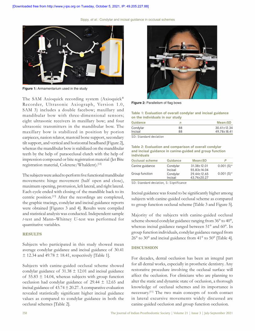
The armamentarium used was [Figure 1]:.1. Axioquick® Recorder, Ultrasonic Axiograph,
Version 1.0, SAM 3.2. Bite registration material
(Jet Bite registration material Coletene/Whaldent)
[Downloaded free from http://www.j-ips.org on Tuesday, October 5, 2021, IP: 49.205.227.88]
Sippy, et al.: Condylar and incisal guidance in occlusal schemes
258 The Journal of Indian Prosthodontic Society | Volume 21 | Issue 3 | July-September 2021
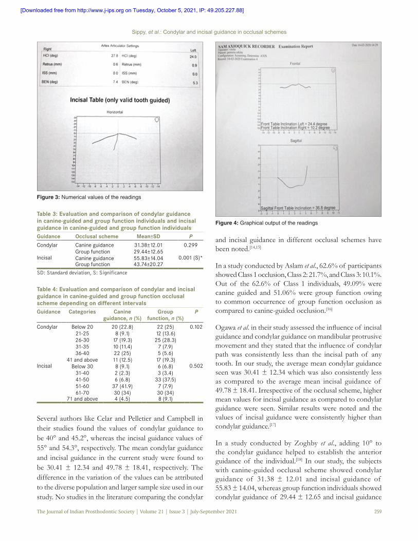
The SAM Axioquick recording system (Axioquick® Recorder, Ultrasonic Axiog raph, Vers ion 1.0 , SAM 3) includes a double facebow; maxillary and mandibular bow with three‑dimensional sensors; eight ultrasonic receivers in maxillary bow; and four ultrasonic transmitters in the mandibular bow. The maxillary bow is stabilized in position by porion earpieces, nasion relator, mastoid bone support, secondary tilt support, and vertical and horizontal headband [Figure 2], whereas the mandibular bow is stabilized on the mandibular teeth by the help of paraocclusal clutch with the help of impression compound or bite registration material (Jet Bite registration material, Coletene/Whaldent).[12]
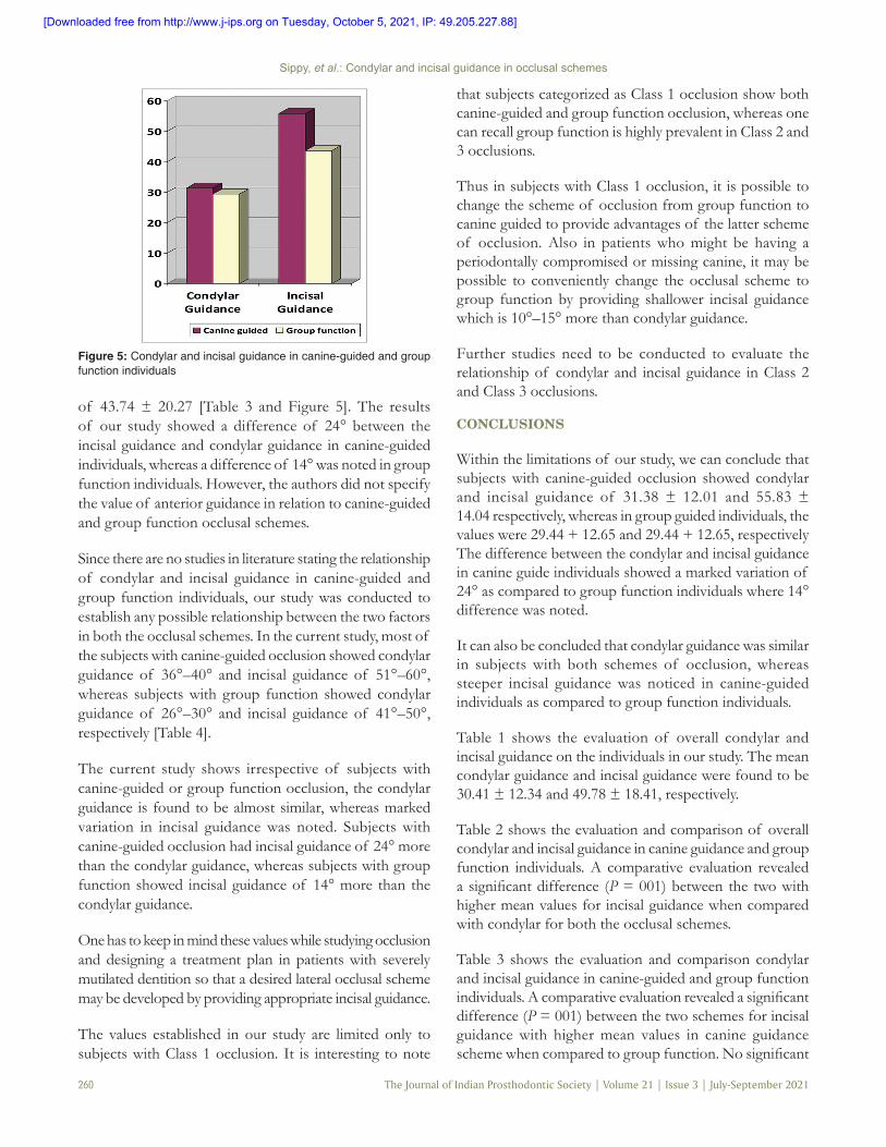
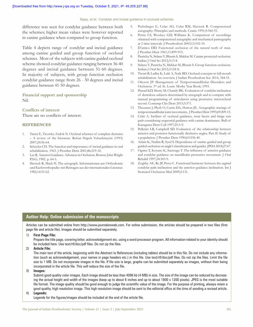
The subjects were asked to perform five functional mandibular movements: hinge movement (half open and close), maximum opening, protrusion, left lateral, and right lateral. Each cycle ended with closing of the mandible back to its centric position.[13] After the recordings are completed, the graphic tracings, condylar and incisal guidance reports were obtained [Figures 3 and 4]. Results were compiled and statistical analysis was conducted. Independent sample t‑test and Mann–Whitney U‑test was performed for quantitative variables.
RESULTS
Subjects who participated in this study showed mean average condylar guidance and incisal guidance of 30.41 ± 12.34 and 49.78 ± 18.41, respectively [Table 1].
Subjects with canine‑guided occlusal scheme showed condylar guidance of 31.38 ± 12.01 and incisal guidance of 55.83 ± 14.04, whereas subjects with group function occlusion had condylar guidance of 29.44 ± 12.65 and incisal guidance of 43.74 ± 20.27. A comparative evaluation revealed statistically significant higher incisal guidance values as compared to condylar guidance in both the occlusal schemes [Table 2].
Incisal guidance was found to be significantly higher among subjects with canine‑guided occlusal scheme as compared to group function occlusal scheme [Table 3 and Figure 5].
Majority of the subjects with canine‑guided occlusal scheme showed condylar guidance ranging from 36° to 40°, whereas incisal guidance ranged between 51° and 60°. In group function individuals, condylar guidance ranged from 26° to 30° and incisal guidance from 41° to 50° [Table 4].
DISCUSSION
For decades, dental occlusion has been an integral part for all dental works, especially in prosthetic dentistry. Any restorative procedure involving the occlusal surface will affect the occlusion. For clinicians who are planning to alter the static and dynamic state of occlusion, a thorough knowledge of occlusal schemes and its importance is necessary.[11] The two main concepts of tooth contact in lateral excursive movements widely discussed are canine‑guided occlusion and group function occlusion.
Table 1: Evaluation of overall condylar and incisal guidance on the individuals in our studyGuidance n Mean±SD
Condylar 88 30.41±12.34Incisal 88 49.78±18.41
SD: Standard deviation
Table 2: Evaluation and comparison of overall condylar and incisal guidance in canine‑guided and group function individualsOcclusal scheme Guidance Mean±SD P
Canine guidance Condylar 31.38±12.01 0.001 (S)*Incisal 55.83±14.04
Group function Condylar 29.44±12.65 0.001 (S)*Incisal 43.74±20.27
SD: Standard deviation, S: Significance
Figure 1: Armamentarium used in the study
Figure 2: Parallelism of flag bows
[Downloaded free from http://www.j-ips.org on Tuesday, October 5, 2021, IP: 49.205.227.88]
Sippy, et al.: Condylar and incisal guidance in occlusal schemes
The Journal of Indian Prosthodontic Society | Volume 21 | Issue 3 | July-September 2021 259
Several authors like Celar and Pelletier and Campbell in their studies found the values of condylar guidance to be 40° and 45.2°, whereas the incisal guidance values of 55° and 54.3°, respectively. The mean condylar guidance and incisal guidance in the current study were found to be 30.41 ± 12.34 and 49.78 ± 18.41, respectively. The difference in the variation of the values can be attributed to the diverse population and larger sample size used in our study. No studies in the literature comparing the condylar
and incisal guidance in different occlusal schemes have been noted.[14,15]
In a study conducted by Aslam et al., 62.6% of participants showed Class 1 occlusion, Class 2: 21.7%, and Class 3: 10.1%. Out of the 62.6% of Class 1 individuals, 49.09% were canine guided and 51.06% were group function owing to common occurrence of group function occlusion as compared to canine‑guided occlusion.[16]
Ogawa et al. in their study assessed the influence of incisal guidance and condylar guidance on mandibular protrusive movement and they stated that the influence of condylar path was consistently less than the incisal path of any tooth. In our study, the average mean condylar guidance seen was 30.41 ± 12.34 which was also consistently less as compared to the average mean incisal guidance of 49.78 ± 18.41. Irrespective of the occlusal scheme, higher mean values for incisal guidance as compared to condylar guidance were seen. Similar results were noted and the values of incisal guidance were consistently higher than condylar guidance.[17]
In a study conducted by Zoghby et al., adding 10° to the condylar guidance helped to establish the anterior guidance of the individual.[18] In our study, the subjects with canine‑guided occlusal scheme showed condylar guidance of 31.38 ± 12.01 and incisal guidance of 55.83 ± 14.04, whereas group function individuals showed condylar guidance of 29.44 ± 12.65 and incisal guidance
Table 3: Evaluation and comparison of condylar guidance in canine‑guided and group function individuals and incisal guidance in canine‑guided and group function individualsGuidance Occlusal scheme Mean±SD P
Condylar Canine guidance 31.38±12.01 0.299Group function 29.44±12.65
Incisal Canine guidance 55.83±14.04 0.001 (S)*Group function 43.74±20.27
SD: Standard deviation, S: Significance
Table 4: Evaluation and comparison of condylar and incisal guidance in canine‑guided and group function occlusal scheme depending on different intervalsGuidance Categories Canine
guidance, n (%)Group
function, n (%)P
Condylar Below 20 20 (22.8) 22 (25) 0.10221‑25 8 (9.1) 12 (13.6)26‑30 17 (19.3) 25 (28.3)31‑35 10 (11.4) 7 (7.9)36‑40 22 (25) 5 (5.6)
41 and above 11 (12.5) 17 (19.3)Incisal Below 30 8 (9.1) 6 (6.8) 0.502
31‑40 2 (2.3) 3 (3.4)41‑50 6 (6.8) 33 (37.5)51‑60 37 (41.9) 7 (7.9)61‑70 30 (34) 30 (34)
71 and above 4 (4.5) 8 (9.1)
Figure 3: Numerical values of the readings
Figure 4: Graphical output of the readings
[Downloaded free from http://www.j-ips.org on Tuesday, October 5, 2021, IP: 49.205.227.88]
Sippy, et al.: Condylar and incisal guidance in occlusal schemes
260 The Journal of Indian Prosthodontic Society | Volume 21 | Issue 3 | July-September 2021
of 43.74 ± 20.27 [Table 3 and Figure 5]. The results of our study showed a difference of 24° between the incisal guidance and condylar guidance in canine‑guided individuals, whereas a difference of 14° was noted in group function individuals. However, the authors did not specify the value of anterior guidance in relation to canine‑guided and group function occlusal schemes.
Since there are no studies in literature stating the relationship of condylar and incisal guidance in canine‑guided and group function individuals, our study was conducted to establish any possible relationship between the two factors in both the occlusal schemes. In the current study, most of the subjects with canine‑guided occlusion showed condylar guidance of 36°–40° and incisal guidance of 51°–60°, whereas subjects with group function showed condylar guidance of 26°–30° and incisal guidance of 41°–50°, respectively [Table 4].
The current study shows irrespective of subjects with canine‑guided or group function occlusion, the condylar guidance is found to be almost similar, whereas marked variation in incisal guidance was noted. Subjects with canine‑guided occlusion had incisal guidance of 24° more than the condylar guidance, whereas subjects with group function showed incisal guidance of 14° more than the condylar guidance.
One has to keep in mind these values while studying occlusion and designing a treatment plan in patients with severely mutilated dentition so that a desired lateral occlusal scheme may be developed by providing appropriate incisal guidance.
The values established in our study are limited only to subjects with Class 1 occlusion. It is interesting to note
that subjects categorized as Class 1 occlusion show both canine‑guided and group function occlusion, whereas one can recall group function is highly prevalent in Class 2 and 3 occlusions.
Thus in subjects with Class 1 occlusion, it is possible to change the scheme of occlusion from group function to canine guided to provide advantages of the latter scheme of occlusion. Also in patients who might be having a periodontally compromised or missing canine, it may be possible to conveniently change the occlusal scheme to group function by providing shallower incisal guidance which is 10°–15° more than condylar guidance.
Further studies need to be conducted to evaluate the relationship of condylar and incisal guidance in Class 2 and Class 3 occlusions.
CONCLUSIONS
Within the limitations of our study, we can conclude that subjects with canine‑guided occlusion showed condylar and incisal guidance of 31.38 ± 12.01 and 55.83 ± 14.04 respectively, whereas in group guided individuals, the values were 29.44 + 12.65 and 29.44 + 12.65, respectively The difference between the condylar and incisal guidance in canine guide individuals showed a marked variation of 24° as compared to group function individuals where 14° difference was noted.
It can also be concluded that condylar guidance was similar in subjects with both schemes of occlusion, whereas steeper incisal guidance was noticed in canine‑guided individuals as compared to group function individuals.
Table 1 shows the evaluation of overall condylar and incisal guidance on the individuals in our study. The mean condylar guidance and incisal guidance were found to be 30.41 ± 12.34 and 49.78 ± 18.41, respectively.
Table 2 shows the evaluation and comparison of overall condylar and incisal guidance in canine guidance and group function individuals. A comparative evaluation revealed a significant difference (P = 001) between the two with higher mean values for incisal guidance when compared with condylar for both the occlusal schemes.
Table 3 shows the evaluation and comparison condylar and incisal guidance in canine‑guided and group function individuals. A comparative evaluation revealed a significant difference (P = 001) between the two schemes for incisal guidance with higher mean values in canine guidance scheme when compared to group function. No significant
Figure 5: Condylar and incisal guidance in canine‑guided and group function individuals
[Downloaded free from http://www.j-ips.org on Tuesday, October 5, 2021, IP: 49.205.227.88]
Sippy, et al.: Condylar and incisal guidance in occlusal schemes
The Journal of Indian Prosthodontic Society | Volume 21 | Issue 3 | July-September 2021 261
difference was seen for condylar guidance between both the schemes; higher mean values were however reported in canine guidance when compared to group function.
Table 4 depicts range of condylar and incisal guidance among canine guided and group function of occlusal schemes. Most of the subjects with canine guided occlusal scheme showed condylar guidance ranging between 36‑40 degrees and incisal guidance between 51‑60 degrees. In majority of subjects, with group function occlusion condylar guidance range from 26 ‑ 30 degrees and incisal guidance between 41‑50 degrees.
Financial support and sponsorshipNil.
Conflicts of interestThere are no conflicts of interest.
REFERENCES
1. Tarazi E, Ticotsky‑Zadok N. Occlusal schemes of complete dentures – A review of the literature. Refuat Hapeh Vehashinayim (1993) 2007;24:56‑64.
2. Schuyler CH. The function and importance of incisal guidance in oral rehabilitation. 1963. J Prosthet Dent 2001;86:219‑32.
3. Lee R. Anterior Guidance. Advances in Occlusion. Boston: John Wright P.S.G; 1982. p. 64‑5.
4. Slavicek R, Mack H. The axiograph. Informationen aus Orthodontie und Kieferorthopadie: mit Beitragen aus der internationalen Literatur. 1982;14:53‑62.
5. Piehslinger E, Celar AG, Celar RM, Slavicek R. Computerized axiography: Principles and methods. Cranio 1991;9:344‑55.
6. Petrie CS, Woolsey GD, Williams K. Comparison of recordings obtained with computerized axiography and mechanical pantography at 2 time intervals. J Prosthodont 2003;12:102‑10.
7. D’amico DD. Functional occlusion of the natural teeth of man. J Prosthet Dent 1961;11:899‑915.
8. Pasricha N, Sidana V, Bhasin S, Makkar M. Canine protected occlusion. Indian J Oral Sci 2012;3:13‑8
9. Sidana V, Pasricha N, Makkar M, Bhasin S. Group function occlusion. Indian J Oral Sci 2012;3:124‑8.
10. Tiwari B, Ladha K, Lalit A, Naik BD. Occlusal concepts in full mouth rehabilitation: An overview. J Indian Prosthodont Soc 2014, 344‑51.
11. Okeson JP. Management of Temporomandibular Disorders and Occlusion. 3rd ed. St. Louis: Mosby Year Book; 1993.
12. Prasad KD, Shetty M, Chandy BK. Evaluation of condylar inclination of dentulous subjects determined by axiograph and to compare with manual programming of articulators using protrusive interocclusal record. Contemp Clin Dent 2015;3:371.
13. Theusner J, Plesh O, Curtis DA, Hutton JE. Axiographic tracings of temporomandibular joint movements. J Prosthet Dent 1993;69:209‑15.
14. Celar A. Inclines of occlusal guidance, wear facets and hinge axis path considering sequential guidance with canine dominance. Bull of Kanagawa Dent Coll 1997;25:3‑9.
15. Pelletier LB, Campbell SD. Evaluation of the relationship between anterior and posterior functionally disclusive angles. Part II: Study of a population. J Prosthet Dent 1990;63:536‑40.
16. Aslam K, Nadim R, Syed S. Dependence of canine‑guided and group guided occlusion on angle’s classification and gender. JPDA 2018;27:67.
17. Ogawa T, Koyano K, Suetsugu T. The influence of anterior guidance and condylar guidance on mandibular protrusive movement. J Oral Rehabil 1997;24:303‑9.
18. Zoghby AE, Re JP, Perez C. Functional harmony between the sagittal condylar path inclination and the anterior guidance inclination. Int J Stomatol Occlusion Med 2009;2:131.
Author Help: Online submission of the manuscripts
Articles can be submitted online from http://www.journalonweb.com. For online submission, the articles should be prepared in two files (first page file and article file). Images should be submitted separately.
1) First Page File: Prepare the title page, covering letter, acknowledgement etc. using a word processor program. All information related to your identity should
be included here. Use text/rtf/doc/pdf files. Do not zip the files.2) Article File: The main text of the article, beginning with the Abstract to References (including tables) should be in this file. Do not include any informa-
tion (such as acknowledgement, your names in page headers etc.) in this file. Use text/rtf/doc/pdf files. Do not zip the files. Limit the file size to 1 MB. Do not incorporate images in the file. If file size is large, graphs can be submitted separately as images, without their being incorporated in the article file. This will reduce the size of the file.
3) Images: Submit good quality color images. Each image should be less than 4096 kb (4 MB) in size. The size of the image can be reduced by decreas-
ing the actual height and width of the images (keep up to about 6 inches and up to about 1800 x 1200 pixels). JPEG is the most suitable file format. The image quality should be good enough to judge the scientific value of the image. For the purpose of printing, always retain a good quality, high resolution image. This high resolution image should be sent to the editorial office at the time of sending a revised article.
4) Legends: Legends for the figures/images should be included at the end of the article file.
[Downloaded free from http://www.j-ips.org on Tuesday, October 5, 2021, IP: 49.205.227.88]
Top Related
Copyright © 2022 FDOKUMEN

