


Bahasa
Halaman
Hukum


www.elsevier.com/locate/biochempharm
Biochemical Pharmacology 68 (2004) 2347–2358
A PXR reporter gene assay in a stable cell culture system:
CYP3A4 and CYP2B6 induction by pesticides
Geraldine Lemaire*, Georges de Sousa, Roger Rahmani
Laboratoire de Toxicologie Cellulaire, Moleculaire et Genomique INRA, UMR 1112 ROSE,
400 route des chappes, B.P. 167, 06903 Sophia Antipolis, France
Received 14 April 2004; accepted 16 July 2004
Abstract
A stable hepatoma cell line expressing the human pregnane X receptor (hPXR) and the cytochrome P4503A4 (CYP3A4) distal and
proximal promoters plus the luciferase reporter gene was developed to assess the ability of several xenobiotic agents to induce CYP3A4
and CYP2B6. After selection for neomycin resistance, one clone, displaying high luciferase activity in response to rifampicin (RIF), was
isolated and the stable expression of hPXR was confirmed by reverse transcription polymerase chain reaction (RT-PCR). Dose-response
curves were generated by treating these cells with increasing concentrations of RIF, phenobarbital (PB), clotrimazole (CLOT) or 5b-
pregnane-3,20-dione (5b-PREGN). The effective concentrations for half maximal response (EC50) were determined for each of these
compounds. RIF was the most effective compound, with maximal luciferase activity induced at 10 mM. The agonist activities of PXR-
specific inducers measured using our stable model were consistent with those measured in transient transfectants. The abilities of
organochlorine (OC), organophosphate (OP) and pyrethroid pesticides (PY) to activate hPXR were also assessed and found to be
consistent with the abilities of these compounds to induce CYP3A4 and CYP2B6 in primary culture of human hepatocytes. These results
suggest that CYP3A4 and CYP2B6 regulation through PXR activation by persistent pesticides may have an impact on the metabolism of
xenobiotic agents and endogenous steroid hormones. Our model provides a useful tool for studying hPXR activation and for identifying
agents capable of inducing CYP3A4 and CYP2B6.
# 2004 Elsevier Inc. All rights reserved.
Keywords: Pregnane X recepteur; Stable reporter assay; Human hepatocytes; Pesticides; Cytochrome P450; Endocrine disruptors
1. Introduction
The liver and the intestine are sites of major metabolic
activity for both endogenous and exogenous chemicals.
Members of the CYP monooxygenase family catalyze the
oxidative metabolism of a wide variety of endogenous
substances and xenobiotic agents [1]. CYP3A4 monoox-
Abbreviations: CAR, constitutive androstane receptor; CLOT, clotrima-
zole; CYP, cytochrome P450; DMEM, Dulbeccos modified Eagles medium;
DMSO, dimethyl sulfoxide; EC50, effective concentration for half maximal
response; FBS, fetal bovine serum; GADPH, glyceraldehyde-3-phosphate
dehydrogenase; hPXR, human pregnane X receptor; PB, phenobarbital; 5b-
PREGN, 5b-pregnane-3,20-dione; PY, pyrethroid pesticides; OC, organo-
chlorine pesticides; OP, organophosphate pesticides; RIF, rifampicin; RT-
PCR, reverse transcription polymerase chain reaction; Wy-14,643, [4-
chloro-6-(2,3-xylidino)-2-pyrimidylthio]acetic acid; XREM, xenobiotic-
responsive enhancer module
* Corresponding author. Tel.: +33 49 2386548; fax: +33 49 2386655.
E-mail addresses: [email protected] (G. Lemaire),
[email protected] (R. Rahmani).
0006-2952/$ – see front matter # 2004 Elsevier Inc. All rights reserved.
doi:10.1016/j.bcp.2004.07.041
ygenase plays a major role in drug biotransformation in
humans and is present at high concentrations in the liver
and intestine. This enzyme has a broad substrate specificity
and is able to metabolize more than 60% of the drugs that
are clinically available [2]. The induction of CYP3A
transcription forms the basis of a number of common
drug–drug interactions, making it important to identify
the agents responsible for this up regulation. However,
these agents are difficult to predict because this CYP
expression is enhanced by a wide variety of structurally
different compounds. This problem can be resolved by
developing inexpensive and reproducible tests using in
vitro systems for assessing the ability of new drug candi-
dates to enhance the expression of drug-metabolizing
enzymes. CYP2B6 was thought to play a minor role in
human drug metabolism and xenobiotic biotransformation.
However, recent studies have shown that up to 25% of
pharmaceutical drugs are metabolized by CYP2B6 [3].
Induction of CYP3A4 and CYP2B6 transcription results
G. Lemaire et al. / Biochemical Pharmacology 68 (2004) 2347–23582348
from the activation of hPXR (NR1I2) [4–7]. This receptor
forms a heterodimer with the retinoid X receptor (NR2B1),
and it has been proposed that this complex constitutes a
xenobiotic-responsive transcription factor that regulates
multiple drug-metabolizing enzymes [8]. Although, PXR
was originally identified as a regulator of CYP3A, many of
the agents that induce CYP3A transcription also induce
transcription of CYP2B [9].
The hPXR is activated by a wide variety of lipophilic
compounds, including agents that induce CYP3A4 and
CYP2B6 [7]. Although several compounds can activate
both mouse PXR and hPXR, these receptors exhibit diver-
gent activation profiles [10]. This difference between
mouse PXR and hPXR activation profiles correlates well
with the fact that the CYP3A induction profile is also
species–specific [10]. After activation, PXR binds to spe-
cific DNA sequence motifs in CYP3A genes and functions
as a transcription factor for target gene regulation. The
proximal promoter of the CYP3A4 gene contains two
copies of an AG(G/T)TCA hexamer, organized to form
an ER6 (everted repeat separated by six nucleotides) motif.
Barwick et al. [11] demonstrated using rabbit hepatocytes
that these half-sites were sufficient to confer rifampicin-
responsiveness on a reporter gene construct. However, in
comparison to hepatocytes, the extent of this rifampicin
response was limited, even when the reporter gene con-
struct contained multiple copies of the CYP3A4 proximal
ER-6 element. Additional regulatory regions of the
CYP3A4 gene, such as those involved in transcriptional
activation by xenobiotics, were defined by Goodwin et al.
[12]. These authors isolated and characterized a distal
xenobiotic-responsive enhancer module (XREM), which
regulates transactivation of the CYP3A4 gene in response
to agents that are also able to induce PXR activation. In
addition, these authors showed that the activation mediated
by the response element in the distal XREM and that
mediated by the proximal promoter region of CYP3A4
were a cooperation.
Goodwin et al. [9] showed that transcription of human
CYP2B6 is regulated directly by PXR. Transactivation of
CYP2B6 by PXR is mediated by the phenobarbital-respon-
sive element enhancer module (PBREM) region of the
gene. This 51-bp enhancer module regulates constitutive
androstane receptor (CAR)-mediated induction of
CYP2B6 [13]. The PBREM contains two DR4 (direct
repeat with four base pairs) elements that are capable of
binding PXR-RXRa. In addition, a distal region of the
CYP2B6 promoter, together with the PBREM, mediates
drug-induced transcription of CYP2B6. Wang et al. [14]
showed that this distal response region could be activated
by PXR.
In rats, OCs, such as DDT and methoxychlor, strongly
induce the transcription of CYP2B isoforms and, to a lesser
extent, that of the CYP3A subfamily members [15]. Chlor-
dane, dieldrin and endosulfan activate hPXR and subse-
quently CYP3A4 mRNA expression in human hepatocytes
[16]. PY have a toxicity profile similar to OCs and also
induce the CYP2B protein and mRNA in rat hepatocytes
[17]. CYP3A1 and CYP2B1 catalyze the hydroxylation of
testosterone in the rat liver and thus, agents inducing the
transcription of these enzymes lower the levels of testos-
terone in the circulation. Phenobarbital (PB), a well known
CYP2B and CYP3A inducing agent, increases androgen
hepatic metabolism [18] and as a result leads to develop-
mental abnormalities reflecting androgenic deficiency.
OCs have an antiandrogenic effect and may therefore
repress androgen-mediated gene activation. In addition,
the level of testosterone breakdown mediated by CYP2B
and CYP3A may increase as a result of PXR activation by
OCs. These synergetic effects may induce reproductive
abnormalities and cause demasculinization.
We have developed a stable cellular model to study the
activation of hPXR by persistent pesticides and to deter-
mine whether this activation was consistent with these
compounds inducing CYP3A4 transcription. In addition,
we examined the induction of CYP2B6 transcription by
these persistent pesticides. For the stable in vitro system,
human hepatoma cells were transfected with the entire
hPXR gene, the XREM region (bases �7836 to �7208)
and the CYP3A4 proximal promoter (bases �362 to +53)
plus the luciferase reporter gene. The ability of this cell line
to respond to human PXR-specific ligands was then inves-
tigated by analyzing the level firefly luciferase activity.
2. Materials and methods
2.1. Materials
All cell culture materials were purchased from Life
technologies. Carbaryl (99.5%), 2,40-DDT (98.0%), diflu-
benzuron (98.5%), lindane (99.4%), mancozeb (74.0%),
2,4,5-T (97.2%) were obtained from Cluzeau Info Labo.
Alachlor (99%), aldrin (98.5%), atrazine (98%), chlordane
(mix of isomers), chlorpyrifos (99.5%), cypermethrin
(39% trans and 59% cis), 2,4-D (98%), DDT mixture
(18% o,p0-DDT; 75% p,p0-DDT), dieldrin (98.8%), endo-
sulfan (99.9%), endrin (99%), pentachlorophenol (99%)
were obtained from ChemService. Trans-nonachlor
(99.3%) was obtained from AccuStandard Inc. Chlordecone
(99.2%), fenvalerate (99.9%), methoxychlor (98.7%), RIF,
CLOT, PB were purchased from Sigma–Aldrich. Steady-
Glo was purchased from Promega. WY 16,643 ([4-chloro-6-
(2,3-xylidino)-2-pyrimidylthio]acetic acid) was obtained
from Alexis. 5b-PREGN was purchased from Steraloids.
HotStar Taq DNA polymerase, Rneasy, Dnase and the
plasmid purification kits were obtained from Qiagen.
2.2. Cell culture
The human hepatocellular carcinoma cell line, HepG2,
was obtained from the American Type Culture Collection
G. Lemaire et al. / Biochemical Pharmacology 68 (2004) 2347–2358 2349
and cultured in Dulbecco’s modified Eagle’s medium
(DMEM) supplemented with 10% fetal bovine serum
(FBS, Biowest). The HepG2-derived cell line (hPXR/
HepG2) was cultured as described above, with the excep-
tion that the culture medium was also supplemented with
800 mg/mL geneticin (G418, Invitrogen).
Hepatocytes from human liver surgical biopsies
(resected from secondary tumors) were isolated by a
reverse, two-step, collagenase perfusion as previously
described [19]. The isolated cells were resuspended in
Williams’ medium E containing 10% FBS, penicillin
(50 UI/mL), streptomycin (50 mg/mL) and insulin
(0.1 UI/mL). Hepatocyte viability was determined using
the Erythrosin B exclusion test and was at least 80%.
Hepatocytes were seeded on collagen type I-coated dishes
and incubated for 4 h at 37 8C in a humidified atmosphere
consisting of 95% air and 5% CO2. The existing medium
was then discarded and replaced by medium identical to
that described above with the exception that no FBS was
added and this medium was supplemented with 1 mM
hydrocortisone hemisuccinate and 0.024% (m/v) bovine
serum albumin. Hepatocytes were treated over a 2-day
period (one treatment every 24 h) with 10 mM OC, OP, PY,
50 mM dexamethasone, 25 mM RIF, 0.5 mM PB, 1 mM 3-
MC. These molecules were solubilized in dimethyl sulf-
oxide (DMSO, final concentration 0.5%, v/v) and then
added directly to the cultures.
2.3. Plasmids
The pCDNA 3.1neo-hPXR expression plasmid was
constructed using pSG5-hPXRDATG (a kind gift from
Dr. Steve Kliewer [7]) as follows: pCDNA 3.1-hPXR
was generated by PCR amplification of pSG5-
hPXRDATG, using oligonucleotides (50-GGG AAT TCA
CCA CCA TGG AGG TGA GAC CCA AAG AAA GC-30
and 50-GGT CTA GAC TCA GCTACC TGT GAT GCC G-
30), which introduced an EcoRI and an XbaI restriction site.
This fragment was then cloned into the EcoRI/XbaI site of
a pCDNA 3.1 vector carrying a G418 resistance gene as a
selectable marker. This construction was verified by
enzyme digestion and confirmed, along with verification
of the correct reading frame, by sequencing. The p3A4-
362(7836/7208ins) reporter construct (carrying the XREM
region, the CYP3A4 proximal promoter and the luciferase
reporter gene [12]) was generously provided by Dr. Chris
Liddle.
2.4. Transient transfection
Transfection was carried out using plasmid cDNA pre-
pared by purification on Qiagen columns. HepG2 cells, in
100 mm culture plates at a density of 2.4 � 106 cells, were
cotransfected with 2 mg of the pCDNA 3.1neo-hPXR
expression plasmid, 8 mg of p3A4-362 (7836/7208ins)
reporter construct, 0.7 mg of pRL-CMV expression plas-
mid as an internal control, and 9 mg of pCDNA 3.1 carrier
plasmid using Lipofectamine 2000 and the procedure
recommended by the manufacturer (GIBCO BRL). After
24 h, the cells were trypsinized and seeded in 96-well
plates at a density of 20,000 cells/well. Twenty-four hours
later, RIF, CLOT, 5b-PREGN, pesticides (10 mM) or
DMSO (0.1%) were added to the medium. Luciferase
activity was determined 24 h later using the Dual-GloTM
Luciferase assay System (Promega). Firefly luciferase
activities were determined on three independent transfec-
tions and were normalized by comparison with the Renilla
luciferase activities of the internal control pRL-CMV
vector from the same culture.
2.5. Stable transfection
The HepG2 cells were seeded in 10-cm culture dishes at
a density of 1.2 � 106 cells in DMEM containing 10%
FBS. After a 48-h recovery period, the cells were trans-
fected overnight with a mixture containing the p3A4-
362(7836/7208ins) reporter construct and pCDNA
3.1neo-hPXR expression plasmid at a ratio of 10:1
(22 mg DNA in total) using Lipofectamine 2000 and the
procedure recommended by the manufacturer (Gibco
BRL). When the cells had been exposed to the precipitated
DNA for 24 h, the culture medium was removed and
replaced with fresh DMEM containing 10% FBS. After
a further 24 h, this medium was then replaced by DMEM
containing 800 mg/mL G418. The medium was renewed
every 3 days for 1 month until small colonies were visible.
One month after the initiation of G418 selection, clones
that expressed luciferase were identified as follows: the
culture medium was replaced with fresh DMEM contain-
ing 0.3 mM sterile luciferin. Plates were then placed in the
dark and luciferase activity was measured (in arbitrary
luminescence values) using a molecular light camera
(Night-Owl) linked to the Win light program. After ana-
lysis, the luciferin-containing medium was removed and
replaced by fresh DMEM containing 10 mM RIF. After
24 h, luminescent and inducible clones were identified
with the camera as described above. They were isolated
in culture using sterile Teflon cloning rings and grown in
DMEM supplemented with 10% FBS and G418 (800 mg/
mL).
2.6. PXR assay: stable gene expression assay
The HepG2-derived cell line (hPXR/HepG2) was
seeded in triplicate in white 96-well plates, with a density
of 30,000 cells/100 mL of DMEM supplemented with 10%
serum stripped of steroids by charcoal/dextran treatment.
After a 24-h recovery period, the cells were left to incubate
for 24 h in DMEM supplemented with the various com-
pounds being tested. Chemicals were stored as DMSO
stock solutions and the final DMSO concentration in the
culture medium never exceeded 0.1%. At the end of this
G. Lemaire et al. / Biochemical Pharmacology 68 (2004) 2347–23582350
incubation period, the culture medium was replaced by the
contents of the Steady-Glo1 Luciferase Assay System
(Promega). Luciferase activity was measured using a
MicroBeta Wallac luminometer (EG&G Wallac) and lumi-
nescence was measured following cellular lysis for 2 s per
well. Luminescence was stable for at least 2 h.
2.7. RNA preparation, RT-PCR and PCR
Total RNA from HepG2 was isolated according to the
manufacturer’s instructions using the Rneasy kit (Qiagen)
with on-column Dnase treatment. RNA was quantified
using the Ribogreen RNA Quantitation Kit (Molecular
Probes). First-strand cDNA was synthesized from 1 mg of
total RNA in a 20 mL reaction mixture using the Taqman
PCR Core Reagent kit and random hexanucleotide pri-
mers. The primers 50-GAC CCA AAG AAA GCT GGA-
30 (forward) and 50-AGC ACA TAC TCC TCC TCA-30
(reverse) for hPXR and, 50-AAT CCC ATC ACC ATC
TTC CA-30 (forward) and 50-GTC ATC ATA TTT GGC
AGG TT-30 (reverse) for human glyceraldehyde-3-phos-
phate dehydrogenase (hGADPH) were used to amplify
the 1014 and 557 bp fragments, respectively. The cDNA
obtained was diluted twenty times and 5 mL was ampli-
fied in a 20 mL reaction volume containing 0.5 U HotStar
Taq DNA polymerase, 1X PCR buffer, 50 pmol of both
primers and 0.2 mM dNTP mix. Amplifications were
performed using a thermocycler with the following
PCR profile: 95 8C for 45 s, 55 8C for 45 s and 72 8Cfor 45 s. Thirty cycles were used for amplification of both
hGADPH and hPXR.
2.8. Western blot
Hepatocytes were lysed in 100 mM phosphate buffer
(pH 7.4), scraped and disrupted. Protein concentration
was estimated using the Pierce bicinchoninic acid Protein
Assay Kit and BSA as the standard. Ten micrograms of
the cell protein extracts were loaded onto a 10% SDS-
polyacrylamide gel and subjected to electrophoresis. Pro-
teins were then transferred onto a PVDF membrane by
electroblotting. After blocking with 5% non-fat, skimmed
milk suspended in TTBS (10 mmol/L Tris–HCl [pH 7.5],
140 mmol/L NaCl, 0.1% Tween 20) overnight at 4 8C, the
membranes were incubated with anti-human CYP3A4
monoclonal antibody (Oxford) or anti-human CYP2B6
monoclonal antibody (Gentest) in TTBS containing 3%
BSA. The membranes were then washed with TTBS and
incubated for 1 h at room temperature with horseradish
peroxidase-conjugated secondary antibody. Peroxidase-
labeled proteins were revealed using the ECL1 detection
kit (Amersham Life Sciences) and Kodak ML-Biomax
films (Eastman Kodak). The films were scanned and
densitometric quantification (Scion imaging Software)
was used to asses the intensity of the CYP3A4 and
CYP2B6 immunoreactive bands.
3. Results
3.1. PXR expression in the stable HepG2 transfectant
A one-step transfection procedure was used to obtain a
stable cell line expressing functional PXR and the p3A4-
362 (7836/7208ins) reporter construct. Colonial luciferase
activity was monitored and one G418-resistant clone was
selected on the basis of its luminescence after incubation
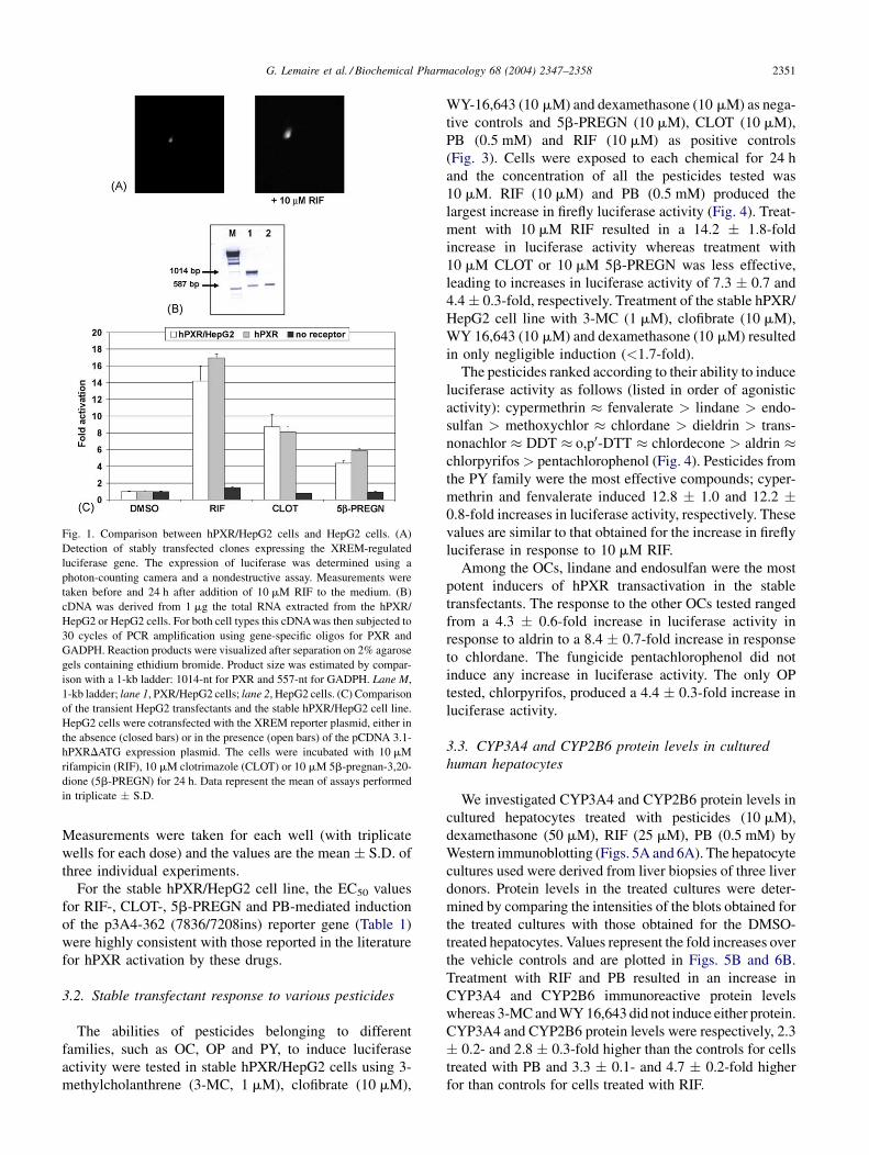
with 10 mM RIF (Fig. 1A).
The stable integration of PXR was verified by RT-PCR.
RNA from stable hPXR/HepG2 cells and HepG2 cells was
subjected to reverse transcription and hPXR mRNA was
assessed after PCR amplification. After 30 cycles, a 1014-
nt fragment corresponding to hPXR cDNA was detected
from the stable hPXR/HepG2 cells, whereas no fragment
of this size was detected from the HepG2 cells (Fig. 1B),
confirming that the lower expression of the hPXR in the
HepG2 cells. In addition, Fig. 1C shows that none of
the typical PXR inducers, such as CLOT (10 mM) and
5b-PREGN (10 mM), were able to induce the expression of
the reporter gene in cells transfected with the XREM
reporter plasmid but not the hPXR expression plasmid.
RIF induced expression of the reporter construct to a
limited extent (<2-fold) in the absence of cotransfected
PXR.
To validate our model system, we compared RIF-,
CLOT-, 5b-PREGN-regulated expression of luciferase in
cells transiently transfected with the luciferase reporter
construct with that in the stably transfected HepG2 cells.
The results of the transactivation studies were similar for
both the stable hPXR/HepG2 and the transiently trans-
fected HepG2 cells. There was no significant difference in
the levels of luciferase expression between transiently
transfected HepG2 cells and stable hPXR/HepG2 cells
incubated in the presence 10 mM concentrations of RIF,
CLOT and 5b-PREGN for 24 h. RIF (10 mM) induced the
highest level of luciferase expression in both the stable and
transient assays, followed by CLOT (10 mM) and then 5b-
PREGN (10 mM). The extent of the response (fold induc-
tion) to the three compounds was slightly lower in the
stable cell line compared to the transient cell line.
hPXR/HepG2 cell sensitivity and the reproducibility of
the assay were assessed by measuring the response to
known hPXR activators, such as RIF, CLOT, PB and
5b-PREGN (Fig. 2). Twenty-four hours after seeding,
the cells were treated with increasing concentrations of
RIF (1.5 � 10�9 to 3 � 10�5 M), CLOT (1.5 � 10�9 to 1 �10�5 M), 5b-PREGN (1.5 � 10�9 to 5.0 � 10�4 M) and
PB (7.6 � 10�8 to 1.5 � 10�3 M) for 24 h and the
concentrations required to obtain the EC50 value were
determined by non-linear sigmoidal analysis of the
dose-response of hPXR to these compounds. Values are
expressed as percentage luciferase activity and were cal-
culated by using the value obtained in the presence of
10 mM RIF as the standard for 100% luciferase activity.
G. Lemaire et al. / Biochemical Pharmacology 68 (2004) 2347–2358 2351
Fig. 1. Comparison between hPXR/HepG2 cells and HepG2 cells. (A)
Detection of stably transfected clones expressing the XREM-regulated
luciferase gene. The expression of luciferase was determined using a
photon-counting camera and a nondestructive assay. Measurements were
taken before and 24 h after addition of 10 mM RIF to the medium. (B)
cDNA was derived from 1 mg the total RNA extracted from the hPXR/
HepG2 or HepG2 cells. For both cell types this cDNA was then subjected to
30 cycles of PCR amplification using gene-specific oligos for PXR and
GADPH. Reaction products were visualized after separation on 2% agarose
gels containing ethidium bromide. Product size was estimated by compar-
ison with a 1-kb ladder: 1014-nt for PXR and 557-nt for GADPH. Lane M,
1-kb ladder; lane 1, PXR/HepG2 cells; lane 2, HepG2 cells. (C) Comparison
of the transient HepG2 transfectants and the stable hPXR/HepG2 cell line.
HepG2 cells were cotransfected with the XREM reporter plasmid, either in
the absence (closed bars) or in the presence (open bars) of the pCDNA 3.1-
hPXRDATG expression plasmid. The cells were incubated with 10 mM
rifampicin (RIF), 10 mM clotrimazole (CLOT) or 10 mM 5b-pregnan-3,20-
dione (5b-PREGN) for 24 h. Data represent the mean of assays performed
in triplicate � S.D.
Measurements were taken for each well (with triplicate
wells for each dose) and the values are the mean � S.D. of
three individual experiments.
For the stable hPXR/HepG2 cell line, the EC50 values
for RIF-, CLOT-, 5b-PREGN and PB-mediated induction
of the p3A4-362 (7836/7208ins) reporter gene (Table 1)
were highly consistent with those reported in the literature
for hPXR activation by these drugs.
3.2. Stable transfectant response to various pesticides
The abilities of pesticides belonging to different
families, such as OC, OP and PY, to induce luciferase
activity were tested in stable hPXR/HepG2 cells using 3-
methylcholanthrene (3-MC, 1 mM), clofibrate (10 mM),
WY-16,643 (10 mM) and dexamethasone (10 mM) as nega-
tive controls and 5b-PREGN (10 mM), CLOT (10 mM),
PB (0.5 mM) and RIF (10 mM) as positive controls
(Fig. 3). Cells were exposed to each chemical for 24 h
and the concentration of all the pesticides tested was
10 mM. RIF (10 mM) and PB (0.5 mM) produced the
largest increase in firefly luciferase activity (Fig. 4). Treat-
ment with 10 mM RIF resulted in a 14.2 � 1.8-fold
increase in luciferase activity whereas treatment with
10 mM CLOT or 10 mM 5b-PREGN was less effective,
leading to increases in luciferase activity of 7.3 � 0.7 and
4.4 � 0.3-fold, respectively. Treatment of the stable hPXR/
HepG2 cell line with 3-MC (1 mM), clofibrate (10 mM),
WY 16,643 (10 mM) and dexamethasone (10 mM) resulted
in only negligible induction (<1.7-fold).
The pesticides ranked according to their ability to induce
luciferase activity as follows (listed in order of agonistic
activity): cypermethrin � fenvalerate > lindane > endo-
sulfan > methoxychlor � chlordane > dieldrin > trans-
nonachlor � DDT � o,p0-DTT � chlordecone > aldrin �chlorpyrifos > pentachlorophenol (Fig. 4). Pesticides from
the PY family were the most effective compounds; cyper-
methrin and fenvalerate induced 12.8 � 1.0 and 12.2 �0.8-fold increases in luciferase activity, respectively. These
values are similar to that obtained for the increase in firefly
luciferase in response to 10 mM RIF.
Among the OCs, lindane and endosulfan were the most
potent inducers of hPXR transactivation in the stable
transfectants. The response to the other OCs tested ranged
from a 4.3 � 0.6-fold increase in luciferase activity in
response to aldrin to a 8.4 � 0.7-fold increase in response
to chlordane. The fungicide pentachlorophenol did not
induce any increase in luciferase activity. The only OP
tested, chlorpyrifos, produced a 4.4 � 0.3-fold increase in
luciferase activity.
3.3. CYP3A4 and CYP2B6 protein levels in cultured
human hepatocytes
We investigated CYP3A4 and CYP2B6 protein levels in
cultured hepatocytes treated with pesticides (10 mM),
dexamethasone (50 mM), RIF (25 mM), PB (0.5 mM) by
Western immunoblotting (Figs. 5A and 6A). The hepatocyte
cultures used were derived from liver biopsies of three liver
donors. Protein levels in the treated cultures were deter-
mined by comparing the intensities of the blots obtained for
the treated cultures with those obtained for the DMSO-
treated hepatocytes. Values represent the fold increases over
the vehicle controls and are plotted in Figs. 5B and 6B.
Treatment with RIF and PB resulted in an increase in
CYP3A4 and CYP2B6 immunoreactive protein levels
whereas 3-MC and WY 16,643 did not induce either protein.
CYP3A4 and CYP2B6 protein levels were respectively, 2.3
� 0.2- and 2.8 � 0.3-fold higher than the controls for cells
treated with PB and 3.3 � 0.1- and 4.7 � 0.2-fold higher
for than controls for cells treated with RIF.

G. Lemaire et al. / Biochemical Pharmacology 68 (2004) 2347–23582352
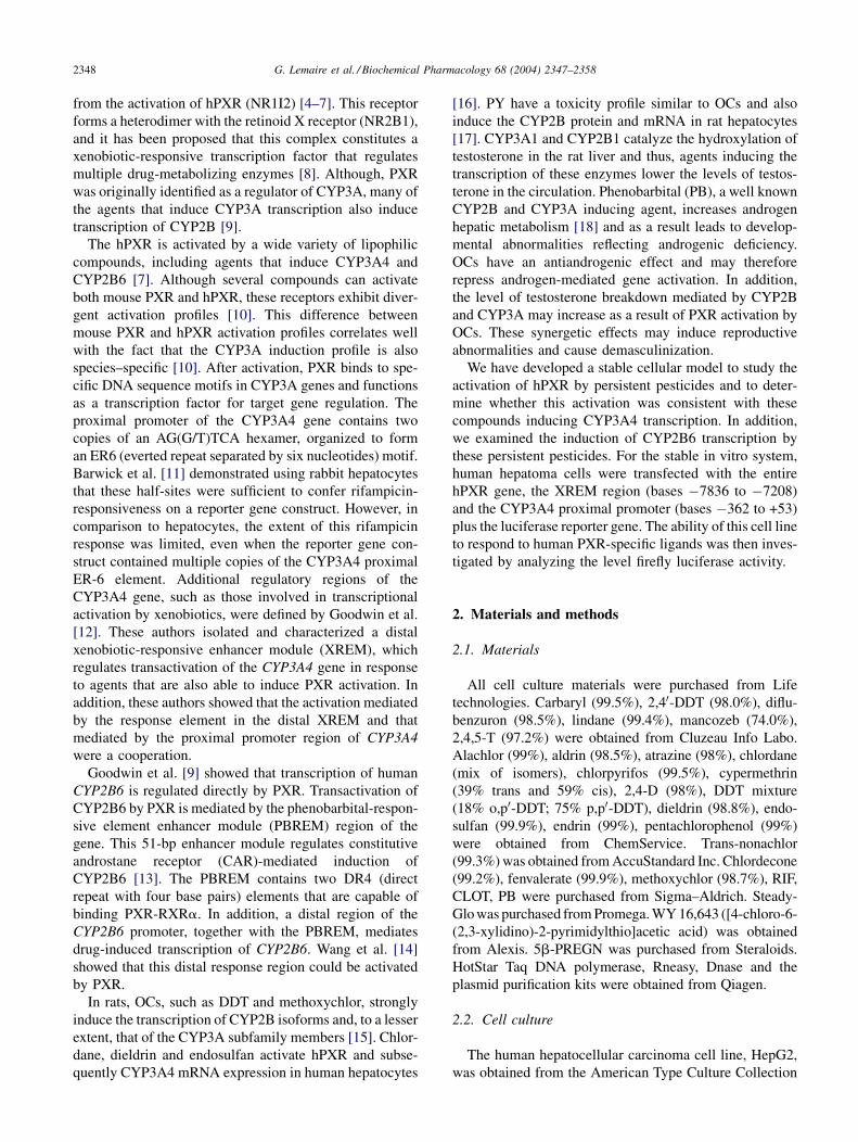
Fig. 2. Dose-response curves of the PXR/HepG2 cells to human PXR-specific ligands. Cells were incubated for 24 h with various concentrations of RIF (1.5 �10�9 to 3.0 � 10�5), CLOT (1.5 � 10�9 to 1.0 � 10�5), PB (7.6 � 10�8 to 1.5 � 10�3), and 5b-PREGN (1.5 � 10�9 to 5.0 � 10�5). Results are expressed as the
percentage of luciferase activity measured in each well (with triplicate wells for each dose). Percentages were calculated using the value obtained in the presence
of 10 mM RIF as the 100% value for luciferase activity. The values represent the means � S.D. of three separate experiments.
The abilities of the pesticides (10 mM) to induce
CYP3A4 protein were ranked as follows (listed in order
of agonistic activity): chlorpyrifos > lindane � o,p0-DDT
> chlordane � cypermethrin � DDT > chlordecone �dieldrin � pentachlorophenol > aldrin � endosulfan �fenvalerate > endrin � methoxychlor � trans-nonachlor
(Fig. 5).
Among the PYs, cypermethrin was more effective than
fenvalerate in inducing CYP3A4 protein. Among the OCs,
lindane and o,p0-DDT were the most potent inducers of
CYP3A4 protein in the human hepatocytes. The other OC
pesticides tested (methoxychlor and chlordane) resulted in
1.5 � 0.3 and 2.2 � 0.3-fold increases in CYP3A4 protein
level (respectively). The fungicide pentachlorophenol
induced a 1.8 � 0.3-fold increase in the level of CYP3A4
protein. Chlorpyrifos, the only OP tested, was an effective
pesticide and induced a 2.6 � 0.5-fold increase in the level
of CYP3A4 protein.
With regard to the increases in CYP2B6 protein levels,
the pesticides were ranked as follows (listed in order of
Table 1
EC50 values (mM) for activation of hPXR in stable hPXR/HepG2 cells
Chemicals EC50 (mM)
Rifampicin 1.8 � 0.2
Clotrimazole 2.5 � 0.3
5b-Pregnane-3,20-dione 20
Phenobarbital 370 � 11
EC50 values (mM) are given for rifampicin, clotrimazole, 5b-pregnagne-
3,20-dione and phenobarbital. Stable hPXR/HepG2 cells were treated with
several concentrations (from 1.5 � 10�9 to 1.5 � 10�3 M) of each
compound.
agonistic activity): trans-nonachlor > lindane � o,p0-DDT
> endosulfan � chlordane > dieldrin > aldrin � chlor-
pyrifos � cypermethrin � DDT � endrin � fenvalerate >methoxychlor � pentachlorophenol � chlordecone
(Fig. 6).
Among the PYs, cypermethrin and fenvalerate were
moderate CYP2B6 protein level inducers. Pesticides from
the OC family were the most effective compounds in
inducing CYP2B6 protein level: 3.2 � 0.7, 3.0 � 0.2,
2.9 � 0.6, 2.8 � 0.2, 2.7 � 0.3-fold increases were
found in response to treatment with trans-nonachlor, lin-
dane, o,p0-DDT, chlordane and endosulfan, respectively.
CYP2B6 protein level was moderately induced by dieldrin,
endrin and DDT but only weakly induced by methoxy-
chlor, pentachlorophenol and chlordecone. Chlorpyrifos
was also a moderate inducer (2.2 � 0.3-fold).
4. Discussion
We have used a one-step transfection procedure to
generate a stable HepG2-derived cell line that expresses
a functional human PXR gene product and the distal (bases
�7836 to �7208) and proximal (bases �362 to +53)
enhancer elements of the CYP3A4 promoter coupled to
the luciferase reporter gene. Analysis and selection of
transfectants expressing the bioluminescent reporter gene
was simplified by the use of a low-light imaging system.
Another model cell system expressing a functional PXR
gene product and carrying a plasmid containing the
CYP3A4 proximal promoter has been described in the
G. Lemaire et al. / Biochemical Pharmacology 68 (2004) 2347–2358 2353
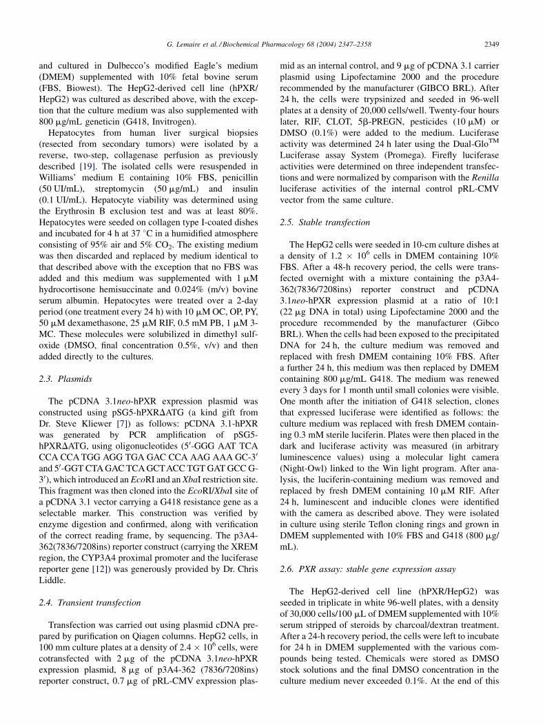
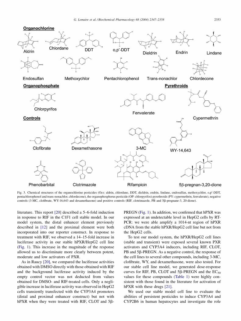
Fig. 3. Chemical structures of the organochlorine pesticides (Ocs: aldrin, chlordane, DDT, dieldrin, endrin, lindane, endosulfan, methoxychlor, o,p0-DDT,
pentachlorophenol and trans-nonachlor, chlordecone), the organophosphorus pesticide (OP: chlorpyrifos) pyrethroids (PY: cypermethrin, fenvalerate), negative
controls (3-MC, clofibrate, WY-16,643 and dexamethasone) and positive controls (RIF, clotrimazole, PB and 5b-pregnane-3, 20-dione).
literature. This report [20] described a 5–6-fold induction
in response to RIF in the C1F1 cell stable model. In our
model system, the distal enhancer element previously
described in [12] and the proximal element were both
incorporated into our reporter construct. In response to
treatment with RIF, we observed a 14–15-fold increase in
luciferase activity in our stable hPXR/HepG2 cell line
(Fig. 1). This increase in the magnitude of the response
allowed us to discriminate more clearly between potent,
moderate and low activators of PXR.
As in Raucy [20], we compared the luciferase activities
obtained with DMSO directly with those obtained with RIF
and the background luciferase activity induced by the
empty control vector was not deducted from values
obtained for DMSO- and RIF-treated cells. Only a negli-
gible increase in luciferase activity was observed in HepG2
cells transiently transfected with the CYP3A4 promoters
(distal and proximal enhancer construct) but not with
hPXR when they were treated with RIF, CLOT and 5b-
PREGN (Fig. 1). In addition, we confirmed that hPXR was
expressed at an undetectable level in HepG2 cells by RT-
PCR: we were able amplify a 1014-nt region of hPXR
cDNA from the stable hPXR/HepG2 cell line but not from
the HepG2 cells.
To test our model system, the hPXR/HepG2 cell lines
(stable and transient) were exposed several known PXR
activators and CYP3A4 inducers, including RIF, CLOT,
PB and 5b-PREGN. As a negative control, the response of
the cell lines to several other compounds, including 3-MC,
clofibrate, WY, and dexamethasone, were also tested. For
our stable cell line model, we generated dose-response
curves for RIF, PB, CLOT and 5b-PREGN and the EC50
values for these compounds (Table 1) were highly con-
sistent with those found in the literature for activation of
hPXR with these drugs [21].
We used our stable model cell line to evaluate the
abilities of persistent pesticides to induce CYP3A4 and
CYP2B6 in human hepatocytes and investigate the role
G. Lemaire et al. / Biochemical Pharmacology 68 (2004) 2347–23582354
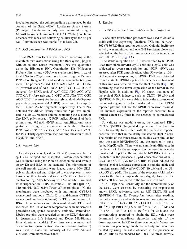
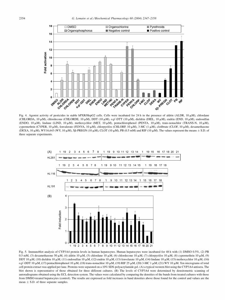
Fig. 4. Agonist activity of pesticides in stable hPXR/HepG2 cells. Cells were incubated for 24 h in the presence of aldrin (ALDR, 10 mM), chlordane
(CHLORDA, 10 mM), chlordecone (CHLORDE, 10 mM), DDT (10 mM), o,p0-DTT (10 mM), dieldrin (DIEL, 10 mM), endrin (END, 10 mM), endosulfan
(ENDO, 10 mM), lindane (LIND, 10 mM), methoxychlor (MET, 10 mM), pentachlorophenol (PENTA, 10 mM), trans-nonachlor (TRANS-N, 10 mM),
cypermethrin (CYPER, 10 mM), fenvalerate (FENVA, 10 mM), chlorpyrifos (CHLORP, 10 mM), 3-MC (1 mM), clofibrate (CLOF, 10 mM), dexamethasone
(DEXA, 10 mM), WY16,643 (WY, 10 mM), 5b-PREGN (10 mM), CLOT (10 mM), PB (0.5 mM) and RIF (10 mM). The values represent the means � S.D. of
three separate experiments.
Fig. 5. Immunoblot analysis of CYP3A4 protein levels in human hepatocytes. Human hepatocytes were incubated for 48 h with (1) DMSO 0.5%, (2) PB
0.5 mM, (3) dexamethasone 50 mM, (4) aldrin 10 mM, (5) chlordane 10 mM, (6) chlordecone 10 mM, (7) chlorpyrifos 10 mM, (8) cypermethrin 10 mM, (9
DDT 10 mM, (10) dieldrin 10 mM, (11) endosulfan 10 mM, (12) endrin 10 mM, (13) fenvelarate 10 mM, (14) lindane 10 mM, (15) methoxychlor 10 mM, (16
o,p0-DDT 10 mM, (17) pentachlotophenol 10 mM, (18) trans-nonachlor 10 mM, (19) RIF 25 mM, (20) 3-MC 1 mM; (21) WY 10 mM. Ten micrograms of tota
cell protein extract was applied per lane. Proteins were separated on a 10% SDS-polyacrylamide gel. (A) a typical western blot using the CYP3A4 antisera. The
blot shown is representative of those obtained for three different cultures. (B) The levels of CYP3A4 were determined by densitometric scanning o
autoradiograms obtained using the ECL detection system. The values were calculated by comparing the densities of the bands from treated cultures with those
from DMSO-treated hepatocytes (control). The results are expressed as fold increases in band densities above those found for the control and values are the
mean � S.D. of three separate samples.
)
)l
f

G. Lemaire et al. / Biochemical Pharmacology 68 (2004) 2347–2358 2355
Fig. 6. Immunoblot analysis of CYP2B6 protein levels in human hepatocytes. Human hepatocytes were incubated for 48 h with (1) DMSO 0.5%, (2) PB
0.5 mM, (3) dexamethasone 50 mM, (4) aldrin 10 mM, (5) chlordane 10 mM, (6) chlordecone 10 mM, (7) chlorpyrifos 10 mM, (8) cypermethrin 10 mM, (9)DDT 10 mM, (10) dieldrin 10 mM, (11) endosulfan 10 mM, (12) endrin 10 mM, (13) fenvelarate 10 mM, (14) lindane 10 mM, (15) methoxychlor 10 mM, (16)
o,p’-DDT 10 mM, (17) pentachlotophenol 10 mM, (18) trans-nonachlor 10 mM, (19) RIF 25 mM, (20) 3-MC 1 mM; (21) WY 10 mM. Thirty-five micrograms of
total cell protein extract per lane was applied. Proteins were separated on a 10% SDS-polyacrylamide gel. (A) a typical western blot using CYP2B6 antisera. The
blot shown is representative of those obtained for three different cultures. (B) The levels of CYP3A4 were determined by densitometric scanning of
autoradiograms obtained using the ECL detection system. The values were calculated by comparing the densities of the bands from treated cultures with those
from DMSO-treated hepatocytes (control). The results are expressed as fold increases in band densities above those found for the control and values are the
mean � S.D. of three separate samples.
played by hPXR in this induction. Previous studies have
found that CYP3A4 and CYP2B6 are induced by PXR
ligands [22,23]. Therefore, we also investigated whether
compounds that activated hPXR in our stable cellular
model also induced CYP3A4 and CYP2B6 proteins in
human hepatocytes.
According to the literature, pesticides such as PY, OC
and OP induce CYP3A and CYP2B in rat hepatocytes
[15,17,24]. In addition, Wyde [25] showed that p,p0-DDE,
the main derivative of DDT, induces hepatic CYP3A1 and
CYP2B1 via a mechanism that involves activation of rat
PXR.
Our study demonstrates, for the first time, that pesticides
induce CYP3A4 and CYP2B6 proteins in human hepato-
cytes. In addition, we showed that hPXR was also activated
by these pesticides. PXR was involved in CYP3A4 and
CYP2B6 induction in response to all the PY, OP and OC
pesticides tested except pentachlorophenol. We found that
our stable PXR reporter gene assay cell line and our
primary culture of human hepatocytes produced similar
but not identical CYP3A4 and CYP2B6 induction
responses. PB, RIF, lindane, endosulfan and chlordane
strongly activated hPXR and resulted in large increases
in the level of CYP3A4 protein. Dieldrin, DDT, chlorde-
cone, aldrin, endrin, and trans-nonachlor moderately acti-
vated PXR and produced intermediate increases in the
levels of the CYP3A4 and CYP2B6 proteins. Finally, 3-
MC and WY did not induce CYP3A4 and CYP2B6 in
either system. Cypermethrin and fenvalerate strongly acti-
vated PXR in the stable hPXR/HepG2 cells (Fig. 4) but
only induced moderate increases in the levels of the
CYP3A4 and CYP2B6 proteins (Figs. 5 and 6) in the
hepatocytes. In contrast, RIF treatment resulted in a large
increase in CYP2B6 and CYP3A4 protein levels in human
hepatocytes (Figs. 5 and 6) and a large increase in lucifer-
ase activity in the stable hPXR/HepG2 cells (Fig. 1). The
discrepancy between the extent of the hPXR activation
response and that of the CYP3A4 and CYP2B6 induction
response after exposure to pyrethroids may be due to the
fact that these pesticides are rapidly metabolized in human
hepatocytes via a mechanism involving hydroxylation and
cleavage of cypermethrin and fenvalerate. This mechanism
may not exist in the HepG2 cell line. Indeed, Ray [26]
found that in humans, urinary excretion of cypermethrin
metabolites was complete 48 h after the final dose of a five-
dose 1.5 mg/kg/day regime was administered. In addition,
cypermethrin is metabolized by hydroxylation and clea-
vage in rats, more than 99% being eliminated within hours
of administration. Chlorpyrifos moderately activated PXR
in the reporter gene assay but resulted in a large increase in

G. Lemaire et al. / Biochemical Pharmacology 68 (2004) 2347–23582356
the level of CYP3A4 but not CYP2B6 protein. The results
obtained in the reporter gene assay for methoxychlor and
pentachlorophenol were inconsistent with those obtained
in the hepatocyte cultures. Methoxychlor induced a
marked increase in luciferase activity (Fig. 4) but only
induced a moderate increase in the levels of the CYP3A4
and CYP2B6 proteins in hepatocytes (Figs. 5 and 6). In
contrast, pentachlorophenol did not result in an increase in
PXR and XREM promoter-mediated luciferase activity,
suggesting that pentachlorophenol does not induce
CYP3A4 and CYP2B6 protein level via a process invol-
ving the PXR. Alternatively, this absence of an increase in
luciferase activity but induction of CYP3A4 and CYP2B6
protein by pentachlorophenol in hepatocytes may be due to
an effect of the metabolites of this compound rather than
pentachlorophenol itself. In HepG2 cells, the activity of
many enzymes is minimal. The reason why methoxychlor
resulted in such a potent increase in luciferase activity but
such a small increase in CYP3A4 and CYP2B6 protein in
human hepatocytes is unclear but may also be related to the
metabolism of this compound. However, Blizard [27]
found that the major metabolites of methoxychlor were
able to induce CYP2B in rat liver microsomes. Trans-
nonachlor markedly induced the CYP2B6 protein but only
moderately activated PXR in the reporter assay system.
This discrepancy between PXR activation and CYP2B6
induction may be due to the involvement of CAR in trans-
nonachlor-mediated CYP2B6 induction.
In cultured hepatocytes and in vivo, PXR is the major
transcription factor involved in the CYP3A4induction.
However, other signaling pathways such as CAR may also
participate in CYP3A4 and CYP2B6 regulation. Indeed,
CAR may directly transactivate CYP3A4 promoters by
binding to the ER6 or DR3 motifs in the XREM region.
This XREM region also serves as a PXR binding site
[21,28,29]. In addition to PB, pesticides also induce
CYP3A4 and CYP2B6 via several pathways. Indeed,
methoxychlor and DDT derivatives have been shown to
activate CAR in rat hepatocytes [25,27].
In summary, the results from the stable PXR reporter
gene system and those from the primary hepatocyte culture
were generally consistent. Some discrepancy between the
extent of the PXR activation response and that of the
CYP3A4 and CYP2B6 protein response was observed with
cypermethrin, fenvalerate, pentachlorophenol and methox-
ychlor. This discrepancy was possibly due to the metabo-
lization of these compounds by a functional xenobiotic
metabolizing enzyme in the primary human hepatocyte
cultures. Furthermore, the stable PXR reporter gene assay
could not show the capacity of the compounds to induce
CYP3A4 and CYP2B6 via the CAR pathway. A previous
study has shown that in HepG2 cells, CAR expression was
too weak to induce CYP2B6 in response to treatment with
PB [28].
In addition to differences in metabolic activity between
the stable cell line and the hepatocytes, the discrepancy in
our results may also be related to competition between
PXR and other receptors, such as CAR, for ligands and
DNA binding sites. Hepatocyte nuclear factor-3 (HNF-3),
HNF-4 and CCAAT/enhancer-binding protein (C/EBP)
can all modulate the expression of the CYP3A4 and
CYP2B6 gene products in human hepatocytes. These
hepatocyte-specific transcription factors are capable of
physically interacting with elements within the CYP3A4
distal and proximal promoter [30,31].
In our stable cell culture model system, the p3A4-
362(7836/7208ins) reporter gene construct contained
two cis-acting elements: a functional HNF4a-binding site
(bases �7783 to �7771) within the XREM region [12,30],
and functional C/EBPa (bases �132 to �121) and HNF-3
(base �188) binding sites in the proximal CYP3A4 pro-
moter [31,32]. Two additional C/EBPa binding sites were
found at the distal position in the promoter (�1393/�1402
and �1659/�1668). In contrast to those at the proximal
site, these distal sites are functional in hepatic cells but not
in extrahepatic cells.
The HN4Fa response element is an important regulatory
region of CYP3A4 as it confers basal and maximal PXR-
mediated transcriptional activation [30]. In addition, the
level of HNF-4 expression in HepG2 cells was comparable
to that found in human hepatocytes [33]. Thus, the differ-
ence between the results obtained with the stable model
and those obtained with hepatocytes was not due to the
HNF-4 transcription factor. In contrast to HNF-4, the level
of C/EBPa and HNF-3 mRNAs were lower in HepG2 cells
than in human hepatocytes, particularly in the case of C/
EBPa. Thus, the discrepancy in our results may be related
to low levels of both C/EBPa and HNF-3 mRNA [33], as
well as the absence of the distal C/EBPa binding site in the
reporter construct [32].
Alternatively, the difference between the extent of PXR
activation and that of the CYP induction in response to
certain compounds may also be due to the presence of
different transport mechanisms in the two cell types. The
HepG2 cells may lack, or produce only low levels of, one
of the transporters present in the hepatocytes. This defi-
ciency may have led to the accumulation of pesticides in
HepG2 cells. PXR activation response resulting from this
accumulation of pesticides would be expected to be stron-
ger than CYP3A4 and CYP2B6 induction response in
human hepatocytes. Inversely, if the pesticide efflux is
increased in HepG2 cells compared to hepatocytes, these
compounds would be expected to be less potent PXR
activators in HepG2 cells than they are inducers of CYP
in hepatocytes. The presence of additional transporters in
HepG2 cells may diminish the importance of extensive
PXR activation in response to pesticides.
Both HepG2 cells and hepatocytes express detectable
levels of the MDR1, MRP2, MRP3 transporters [34,35].
However, no comparative studies of mRNA and protein
have been performed for MRP transporters in these cell
types. In addition, other transporters may also contribute to

G. Lemaire et al. / Biochemical Pharmacology 68 (2004) 2347–2358 2357
pesticide efflux and therefore maybe responsible for the
differences between PXR activation and CYP induction.
Our results demonstrate that potent inducers of human
CYP3A4 and CYP2B6 can be identified using a stable
transfectant cell system containing the appropriate CYP-
enhancer elements. This system can be used to rapidly,
reliably and reproducibly assess the potential of candidate
drugs to induce human CYP. However, tests should be
carried out in parallel using primary cultures of human
hepatocytes to assess the inductive potential of the meta-
bolic intermediates of these new drugs.
Our results indicate that PXR is a xeno-sensor that
protect against the presence of significant concentrations
of harmful environmental compounds. However, pesticides
that induce CYP2B and CYP3A in human hepatocytes may
lead to procarcinogen activation and thus disrupt the
metabolism of drugs and endogenous substances such as
steroid hormones. CYP3A4 metabolizes more than 60% of
all the drugs that are clinically available and CYP2B6
metabolizes up to 25%. The impact that induction of these
enzymes may have on the activity of these drugs may cause
serious health problems. In addition, normal endocrine
function may be affected by the by steroid hormone
(e.g., testosterone) metabolizing activity of these CYP.
As an example, PB increases androgen hepatic metabolism
and induces developmental disorders indicative of andro-
gen deficiency [36]. In addition, a link has been identified
between the effects of pesticides on enzyme induction and
reproductive disorders.
Acknowledgements
This work was supported by a grant from the Region
Provence-Alpes-Cote d’Azur. We grateful to Dr. Kliewer
(University of Texas) and Dr. Liddle (University of Sidney)
for the gift of the plasmids.
References
[1] Nebert DW, Gonzalez FJ. P450 genes: structure, evolution, and
regulation. Annu Rev Biochem 1987;56:945–93.
[2] Maurel P. The CYP3A family. In: Ioannides C, editor. Cytochrome
P450: metabolic and toxicological aspects. CRC Press: Boca Raton;
1996. p. 241–70.
[3] Ekins S, Wrighton SA. The role of CYP2B6 in human xenobiotic
metabolism. Drug Metab Rev 1999;31:719–54.
[4] Bertilsson G, Heidrich J, Svensson K, Asman M, Jendeberg L, Sydow-
Backman M, et al. Identification of a human nuclear receptor defines a
new signaling pathway for CYP3A induction. Proc Natl Acad Sci USA
1998;95:12208–13.
[5] Blumberg B, Sabbagh Jr W, Juguilon H, Bolado Jr J, van Meter CM,
Ong ES, et al. SXR, a novel steroid and xenobiotic-sensing nuclear
receptor. Genes Dev 1998;12:3195–205.
[6] Kliewer SA, Moore JT, Wade L, Staudinger JL, Watson MA, Jones SA,
et al. An orphan nuclear receptor activated by pregnanes defines a
novel steroid signaling pathway. Cell 1998;92:73–82.
[7] Lehmann JM, McKee DD, Watson MA, Willson TM, Moore JT,
Kliewer SA. The human orphan nuclear receptor PXR is activated
by compounds that regulate CYP3A4 gene expression and cause drug
interactions. J Clin Invest 1998;102:1016–23.
[8] Geick A, Eichelbaum M, Burk O. Nuclear receptor response elements
mediate induction of intestinal MDR1 by rifampin. J Biol Chem
2001;276:14581–7.
[9] Goodwin B, Moore LB, Stoltz CM, McKee DD, Kliewer SA. Reg-
ulation of the human CYP2B6 gene by the nuclear pregnane X
receptor. Mol Pharmacol 2001;60:427–31.
[10] Jones SA, Moore LB, Shenk JL, Wisely GB, Hamilton GA, McKee
DD, et al. The pregnane X receptor: a promiscuous xenobiotic receptor
that has diverged during evolution. Mol Endocrinol 2000;14:27–39.
[11] Barwick JL, Quattrochi LC, Mills AS, Potenza C, Tukey RH, Guzelian
PS. Trans-species gene transfer for analysis of glucocorticoid-indu-
cible transcriptional activation of transiently expressed human
CYP3A4 and rabbit CYP3A6 in primary cultures of adult rat and
rabbit hepatocytes. Mol Pharmacol 1996;50:10–6.
[12] Goodwin B, Hodgson E, Liddle C. The orphan human Pregnane X
Receptor mediates the transcriptional activation of CYP3A4 by
rifampicin through a distal enhancer module. Mol Pharmacol 1999;
56:1329–39.
[13] Zelko I, Negishi M. Phenobarbital-elicited activation of nuclear
receptor CAR in induction of cytochrome P450 genes. Biochem
Biophys Res Commun 2000;1277:1–6.
[14] Wang H, Faucette S, Sueyoshi T, Moore R, Ferguson S, Negishi M,
et al. A novel distal enhancer module regulated by pregnane X
receptor/constitutive androstane receptor is essential for the maximal
induction of CYP2B6 gene expression. J Biol Chem 2003;278:
14146–52.
[15] Li HC, Dehal SS, Kupfer D. Induction of the hepatic CYP2B and
CYP3A enzymes by the proestrogenic pesticide methoxychlor and by
DDT in the rat. Effects on methoxychlor metabolism. J Biochem
Toxicol 1995;10:51–61.
[16] Coumoul X, Diry M, Barouki R. PXR-dependent induction of human
CYP3A4 gene expression by organochlorine pesticides. Biochem
Pharmacol 2002;64:1513–9.
[17] Heder AF, Hirsch-Ernst KI, Bauer D, Kahl GF, Desel H. Induction of
cytochrome P4502B1 by pyrethroids in primary rat hepatocyte cul-
tures. Biochem Pharmacol 2001;62:71–9.
[18] Elias AN, Gwinup G. Effects of some clinically encountered drugs on
steroid synthesis and degradation. Metabolism 1980;29:582–95.
[19] Nicolas F, de Sousa G, Thomas P, Placidi M, Lorenzon G, Rahmani R.
Comparative metabolism of 30-azido-30-deoxythymidine in cultured
hepatocytes from rats, dogs, monkeys and humans. Drug Metab
Dispos 1995;23:308–13.
[20] Raucy J, Warfe L, Yueh MF, Allen SW. A cell-based reporter gene
assay for determining induction of CYP3A4 in a high-volume system.
J Pharmacol Exp Ther 2002;303:412–23.
[21] Moore LB, Parks DJ, Jones SA, Bledsoe RK, Consler TG, Stimmel JB,
et al. Orphan nuclear receptors constitutive androstane receptor and
pregnane X receptor share xenobiotic and steroid ligands. J Biol Chem
2000;275:15122–7.
[22] Chang TK, Yu L, Maurel P, Waxman DJ. Enhanced cyclophosphamide
and ifosfamide activation in primary human hepatocytes cultures:
response to cytochrome P-450 inducers and autoinduction by oxaza-
phosphorines. Cancer Res 1997;57:1946–54.
[23] Sahi J, Hamilton G, Sinz M, Barros S, Huang SM, Lesko LJ, et al.
Effect of troglitazone on cytochrome P450 enzymes in primary
cultures of human and rat hepatocytes. Xenobiotica 2000;30:273–84.
[24] Nims RW, Lubet RA. Induction of cytochrome P-450 in the Norway
rat, Rattus norvegicus, following exposure to potential environmental
contaminants. J Toxicol Environ Health 1995;46:271–92.
[25] Wyde ME, Bartolucci E, Ueda A, Zhang H, Yan B, Negishi M, et al.
The environmental pollutant 1,1-dichloro-2,2-bis (p-chloro-
phenyl)ethylene induces rat hepatic cytochrome P4502B and 3A

G. Lemaire et al. / Biochemical Pharmacology 68 (2004) 2347–23582358
expression through the constitutive androstane receptor and pregnane
X receptor. Mol Pharmacol 2003;64:474–81.
[26] Ray DE. Pesticides derived from plants and other organisms. In:
Hayes Jr WJ, Laws Jr ER, editors. Handbook of pesticide toxicology.
New York: Academic Press; 1991. p. 2–3.
[27] Blizard D, Sueyoshi T, Negishi M, Dehal SS, Kupfer D. Mechanism of
induction of cytochrome p450 enzymes by the proestrogenic endo-
crine disruptor pesticide-methoxychlor: interactions of methoxychlor
metabolites with the constitutive androstane receptor system. Drug
Metab Dispos 2001;29:781–5.
[28] Sueyoshi T, Kawamoto T, Zelko I, Honkakoski P, Negishi M.
The repressed nuclear receptor CAR responds to phenobarbital in
activating the human CYP2B6 gene. J Biol Chem 1999;274:
6043–6.
[29] Xie W, Barwick JL, Simon CM, Pierce AM, Safe S, Blumberg B, et al.
Reciprocal activation of xenobiotic response genes by nuclear recep-
tors SXR/PXR and CAR. Genes Dev 2000;14:3014–23.
[30] Tirona RG, Lee W, Leake BF, Lan LB, Cline CB, Lamba V, et al.
The orphan nuclear receptor HNF4alpha determines PXR- and
CAR-mediated xenobiotic induction of CYP3A4. Nat Med 2003;
9:220–4.
[31] Bombail V, Taylor K, Gibson GG, Plant N. Role of Sp1, C/EBPalpha,
HNF3, and PXR in the basal- and xenobiotic-mediated regulation of
the CYP3A4 gene. Drug Metab Dispos 2004;32:525–35.
[32] Rodriguez-Antona C, Bort R, Jover R, Tindberg N, Ingelman-Sund-
berg M, Gomez-Lechon MJ, et al. Transcriptional regulation of human
CYP3A4 basal expression by CCAAT enhancer-binding protein alpha
and hepatocyte nuclear factor-3 gamma. Mol Pharmacol 2003;63:
1180–9.
[33] Jover R, Bort R, Gomez-Lechon MJ, Castell JV. Re-expression of C/
EBP alpha induces CYP2B6, CYP2C9 and CYP2D6 genes in HepG2
cells. FEBS Lett 1998;431:227–30.
[34] Teng S, Jekerle V, Piquette-Miller M. Induction of ABCC3 (MRP3) by
pregnane X receptor activators. Drug Metab Dispos 2003;31:1296–9.
[35] Payen L, Courtois A, Campion JP, Guillouzo A, Fardel O. Character-
ization and inhibition by a wide range of xenobiotics of organic anion
excretion by primary human hepatocytes. Biochem Pharmacol 2000;
60:1967–75.
[36] Dessens AB, Cohen-Kettenis PT, Mellenbergh GJ, Koppe JG, Poll NE,
Boer K. Association of prenatal phenobarbital and phenytoin exposure
with genital anomalies and menstrual disorders. Teratology 2001;
64:181–8.
Top Related
Copyright © 2022 FDOKUMEN

