


Bahasa
Halaman
Hukum


A Dissertation entitled
Characterization of the Anti-Obesity and Anti-Adipogenic Effects of the Limonoid Prieurianin
by Rudel A. Saunders
Submitted to the Graduate Faculty as partial fulfillment of the requirement for the Doctoral of Philosophy Degree in Biomedical Sciences
_________________________________ Dr. Khew-Voon Chin, Committee Chair
_________________________________ Dr. Ivana de la Serna, Committee Member
_________________________________ Dr. Sonia Najjar, Committee Member
_________________________________ Dr. Sandrine Pierre, Committee Member
_________________________________ Dr. Manohar Ratnam, Committee Member
_________________________________ Dr. Cynthia Smas, Committee Member
___________________________________ Dr. Dorothea Sawicki, Associate Dean
College of Graduate Studies
The University of Toledo April 2010

ii

iii
DEDICATION
In loving memory of my sister and grandmother.
This work is dedicated to my parents, sisters, nephew and niece, family and friends,
who have always encouraged and believed in me. Without your support I would not be
here today. I love you and thank you.

iv
ABSTRACT
Phospholipid transfer protein (PLTP) is critically important for reverse cholesterol
transport (RCT), and its expression level and activity increase when mice are fed a high
fat diet. RCT is the process by which accumulated cholesterol from the blood vessel
walls, peripheral tissues and macrophages is transported back to the liver for excretion.
Interestingly, topoisomerase I inhibitors used in chemotherapy have been shown in our
laboratory to dose dependently induced PLTP expression in both in vivo and in vitro
studies. Since PLTP transports phospholipid as well as cholesterol into high density
lipoprotein (HDL), we asked whether elevated PLTP levels might increase the transfer of
drugs into HDL via RCT, thus increasing tumor cells resistance to the drug. However, we
found that camptothecin, topoisomerase I inhibitor, does not accumulate in HDL or in
other lipoprotein subfractions, thus ruling out the possibility of PLTP mediating the
transfer of camptothecin into HDL for liver metabolism.
The limonoid prieurianin, like topoisomerase I inhibitors, has also been shown to
dose dependently increase PLTP mRNA and proteins levels, and here we show that
prieurianin causes weight loss by reducing food intake in morbidly obese mice and in
mice on high-calorie diet. Additionally, prieurianin is anti-adipogenic and (i) inhibits the
proliferation and differentiation of preadipocytes into adipocytes, and (ii) induces either
dedifferentiation or delipidation of mature adipocytes. Gene expression profiling showed
that prieurianin suppresses the expression of a number of genes involved in fat
metabolism, and inhibits the transcriptional activity of the adipogenesis master regulators

v
including the CCAAT/enhancer binding proteins (C/EBPs) and the peroxisome
proliferator-activated receptor gamma (PPARγ).

vi
ACKNOWLEDGEMENTS
This dissertation would not have been possible without the support of my major
advisor Khew-Voon Chin, PhD. I am grateful for all his guidance, patience and support
throughout graduate school and for giving me the opportunity to work in his laboratory. I
also would like to thank my advisory committee members: Drs. Sonia Najjar, Manohar
Ratnam, Cynthia Smas, Ivana de la Serna, and Sandrine Pierre for all their helpful advice,
suggestions and support throughout my dissertation work. Special thanks to Dr. Randall
Ruch for agreeing to be the graduate school representative for my thesis defense.
I am grateful for the opportunity to work with a fantastic group of young men and
women and would like to thank lab mates – Ahmed Qasem, Qiong (Joae) Wu, Marysia
(Maria) Szkudlarek and Srinivas Vinod Saladi for there assistance with experiments as
well as making my graduate school experience most enjoyable. Especially, I would like
to thank Qiong (Joae) Wu for her patience in training, encouraging me to continually
excel, paddling me when necessary and, above all, being a great friend.
Above all, I thank God for allowing me to complete this degree against all odds.
In Him, I draw all my strength.

vii
TABLE OF CONTENTS
Dedication…………………………………………………………………....iii
Abstract ………….………………………………………………………......iv
Acknowledgements……………………………………………………….....vi
Table of contents………………………………………………………...….vii
Introduction…………………………………………………………………..1
Chapter I: Manuscript 1 – PLTP
Abstract……………………………………………………..………11
Introduction……………………………………………………..…..12
Materials and Methods………………………………..…….……....16
Results……………………………………………………….……...19
Discussion………………………………………………….……….27
Chapter II: Manuscript 2 – Prieurianin
Abstract……………………………………………….…………….31
Introduction……………………………………….……….………..32
Materials and Methods………………………….……….……….…34
Results…………………………………………….…….…….….…40
Discussion………………………………………….…….…....……64
Discussion…………………………………………………………………..66
Conclusion…………………………...…………………….…….…....……79
References………………………………………………….…………..…...80

1
INTRODUCTION
Obesity, Diabetes Mellitus and Insulin Resistance
Diabetes Mellitus (DM) is a heterogeneous group of disorders characterized by
hyperglycemia, and is due to deficiency of insulin secretion or to resistance of the body’s
cells to the action of insulin, or to a combination of these. According to the NDDG/WHO
classification scheme, there are four main types of DM and include type 1 diabetes
mellitus, type 2 diabetes mellitus, gestational diabetes mellitus and other specific types of
diabetes mellitus. Our primary focus is type 2 diabetes mellitus as it is closely associated
with obesity.
Type 2 diabetes mellitus comprises approximately 90 to 95% of cases in the
diabetes syndrome (King, Aubert, & Herman, 1998), and is caused by both genetic and
nongenetic factors, such as high caloric intake, overweight and sedentary lifestyle, that
culminates with insulin resistance in the liver, muscle, and adipose tissue and insulin
deficiency. Patients with type 2 DM require exogenous insulin only if fasting
hyperglycemia cannot be corrected with the use of diet or oral agents, and exercise. Most
patients are diagnosed with the disease in adult years. There is a strong correlation
between type 2 diabetes and obesity, and approximately 50 to 90% of all patients with
type 2 diabetes are obese (Harris MI, 1995).
Normal fasting glucose is less than 110mg/dL, in contrast to patients with
diabetes have fasting plasma glucose exceeding126mg/dL. Patients with a fasting glucose
between 110 and 126mg/dL are said to have an impaired fasting glucose. According to
2
the NHANES III study, the prevalence of DM rose with age and peaked at 19% at age 75
years and older (Harris, et al., 1998). Further, minority populations in United States have
higher rates of DM with black and Mexican Americans leading with 28% and 33% of
their population (Harris, et al., 1998).
Insulin is an anabolic hormone whose main function is to maintain blood glucose
within a narrow range. Following a meal, carbohydrates are digested, glucose levels rise,
and insulin is secreted from pancreatic β-cells into the hepatic portal vein. Insulin
regulates glucose levels through two mechanisms. First, it activates glycolytic and
glycogen synthetic pathways which are responsible for the uptake, utilization, and storage
of glucose in various tissues. Second, it inhibits glycogenolytic and gluconeogenic
pathways involved in glucose production and output. Both of these mechanisms reduce
the amount of circulating glucose in the blood. Circulating insulin is cleared by the liver
through a receptor mediated endocytosis via clathrin coated vesicles. Insulin binds to the
α-subunit of the insulin receptor, and induces a conformational change and
autophosphorylation within the tyrosine kinase domain. Phosphorylation of the tyrosine
residues is important for the downstream signaling proteins insulin receptor substrate
(IRS) and src homology 2 domain containing protein (SHC). Insulin signals through two
main pathways: (i) phosphoinositide-3-kinase (PI3K) via phosphorylation of IRS, and
(ii) RAS/RAF/mitogen activated protein kinase (MAPK) pathway via phosphorylation of
SHC which promotes the mitogenic action of insulin (Tanti, et al., 2004). Insulin receptor
phosphorylates and activates the plasma membrane glycoprotein CEACAM1 which
mediates insulin clearance in the liver (Najjar, et al., 1993).

3
Not surprisingly, insulin dysregulation can lead to altered fat metabolism and
insulin resistance, and this is a hallmark feature of T2DM which is also associated with
obesity, hyperglycemia and altered fat metabolism. In the liver, insulin decreases fatty
acid oxidation by increasing malonyl-coenzyme A (malonyl Co-A) which in turn inhibit
the rate limiting enzyme in control of fatty acid uptake and oxidation in the cell, carnitine
palmitoyl transferase 1 (CPT-1). Further, insulin stimulates de novo synthesis of free fatty
acids (FFA), which are then secreted into the circulation as very low density lipoprotein
triglycerides (VLDL-TG) and inhibits hydrolysis of stored triglycerides in adipocytes by
hormone sensitive lipase (HSL) (Kraemer & Shen, 2002; McGarry & Foster, 1980).
Within the plasma membrane of the adipose tissue, lipoprotein lipase (LPL) mediates the
hydrolysis and uptake of circulating trigylcerides in reponse to insulin (Eckel, 1989;
Mead, Irvine, & Ramji, 2002). However, the insulin resistant state is characterized by
high circulating nonesterified FFA which leads to the activation of atypical protein kinase
C (PKC) then serine/threonine kinases (Griffin, et al., 1999; Itani, Ruderman, Schmieder,
& Boden, 2002; Yu, et al., 2002) followed by a deactivation of the insulin signaling
cascade. Thus, the net result is a loss of insulin stimuated glucose uptake and oxidation in
muscle(Groop, et al., 1989; J. K. Kim, Wi, & Youn, 1996; Shulman, 2000).
Insulin resistance is associated with a state of chronic low-grade inflammation
and the release of several chemical mediators such as interleukins-1, 6, 10, 18, resistin,
retinol-binding protein 4 (RBP4), adipocyte-fatty acid binding protein (aP2, FABP4) and
nonesterified fatty acids (NEFA), from immune cells and adipocytes. Among the pro-
4
inflammatory cytokines secreted is tumor necrosis factor alpha (TNFα), which like
elevated nonesterified free fatty acids, stimulate the inhibitory phosphorylation of IRS-1
(Tilg & Moschen, 2008), decreases the insulin sensitizing protein and lipid senor PPARγ
mRNA and DNA binding activity (Ye, 2008), as well as suppresses the transcription of
adiponectin which (i) inhibits TNFα-induced NFκB activation, (ii) decreases
hyperglycemia and levels of plasma FFA, and (iii) improves insulin sensitivity. TNFα-
induced insulin resistance is mediated by IKKβ (Tilg & Moschen, 2008).
Obesity and Metabolic Syndrome
Metabolic syndrome is a combination of medical disorders that increase the risk
of developing cardiovascular disease and diabetes, and includes abdominal obesity,
atherogenic dyslipidemia, elevated blood pressure, insulin resistance or glucose
intolerance, prothrombotic state, and proinflammatory state (Bray & Bellanger, 2006). It
is estimated that over 50 million Americans have the metabolic syndrome and are at
increased risk of coronary heart disease and type 2 diabetes. The two principle risk
factors for this syndrome are abdominal obesity and insulin resistance. Obesity, defined
as having body-mass index (BMI) of more than 30, is an epidemic that affects an
estimated 300 million people worldwide and 30% of United States adults (Haslam &
James, 2005). The alarming rate of increase of obesity is due to sedentary lifestyle habits
coupled with overconsumption of energy-rich foods, and is known to be associated with
an increased risk for hypertension, type 2 diabetes, coronary heart disease, stroke,
hyperlipidemia and certain cancers (Z. Li, et al., 2005; Spiegelman, Choy, Hotamisligil,
Graves, & Tontonoz, 1993). Pharmacological intervention will undoubtedly have

5
numerous benefits in reducing the incidence of these comorbidities in obesity. However,
today there are only two FDA-approved drugs – orlistat and sibutramine – in the market
for the long-term treatment of obesity. Orlistat blocks absorption of dietary fats
(Guerciolini, 1997) while sibutramine (Ryan, Kaiser, & Bray, 1995) is a serotonin and
norepinephrine reuptake inhibitor that acts in the central nervous system (CNS) to reduce
energy intake. These drugs have limited efficacies and side effects are commonly
reported, which are further confounded by diminishing response in long-term treatment
(Fernstrom & Choi, 2008; Z. Li, et al., 2005). Moreover, most of the new anti-obesity
drug development continues to focus on either central or peripheral acting inhibitors of
food intake (Cooke & Bloom, 2006) and would likely encounter the above problems.
Thus, there is an urgent need to identify breakthrough drugs with paradigm shifting
pharmacodynamics for the treatment of obesity.
The main cause of obesity is an imbalance between energy intake and energy
expenditure. When energy intake exceeds expenditure, the excess is stored mainly in the
form of fat in adipose tissue, either under the skin or deep in the abdomen. During
adipogenesis, fat precursor cells differentiate and mature into white and brown adipose
tissue. There are many in vitro cell models for the study of lipid accumulation, and a few
mouse cell lines include 3T3-L1, ob/ob and 3T3-F442A cells. To initiate differentiation,
cells are cultured to confluence and contact inhibited. Upon exposure to differentiation
induction signals, the preadipocytes further undergo two to four rounds of cell division
called mitotic clonal expansion, which is followed by terminal differentiation,
characterized by: (i) the expression of the CCAAT/enhancer binding proteins (C/EBPs)

6
and peroxisome proliferator-activated receptor gamma (PPARγ) transcription factors; (ii)
synthesis of lipid transport and adipocyte-specific proteins; and (iii) secretion of several
cytokines including TNFα. Adipocyte differentiation culminates with a fibroblast-like to
spherical phenotypic change and the accumulation of intracellular fat droplets. In the
absence of differentiation inducers this process takes about two to three weeks.
However, this can be shortened to five to seven days if a cocktail of three inducers are
used: insulin, dexamethasone and 3-Isobutyl-1-methylxanthine (IBMX). Insulin
increases cellular glucose uptake, the glucocorticoid dexamethasone downregulates
preadipocyte factor 1 (Wolf, 1999), and IBMX inhibits phosphodiesterase (Beavo, et al.,
1970; Chasin & Harris, 1976; Montague & Cook, 1971; H. Oka, Kaneko, Yamashita,
Suzuki, & Oda, 1973; Peytremann, Nicholson, Liddle, Hardman, & Sutherland, 1973).
Signaling through nuclear factor kappa B
Nuclear factor kappa B (NFκB)/Rel proteins comprise a family of structurally-
related eukaryotic transcription factors that plays a critical role in inflammation, cell
migration and repair, immune response, apoptosis, and angiogenesis. These transcription
factors are persistently active in a number of disease states, including chronic
inflammation and heart disease. The family consists of five proteins: NFκB1 (p50 and its
precursor p105), NFκB2 (p52 and its precursor p100), RelA (p65), RelB, and c-Rel.
All NFκB/Rel proteins contain Rel homology (RH) domains located near the N-
terminus. RH domain is required for dimerization, DNA-binding, and interaction with
IκB. NFκB/Rel proteins can be divided into two classes: NFκB (p105 and p100) and Rel

7
(RelA (p65), RelB, and c-Rel) proteins. Members of the NFκB class have long C-
terminal domains that contain multiple copies of ankyrin repeats which inhibit NFκB
activation and nuclear translocation. Members of the NFκB class become active, shorter
DNA-binding proteins (p105 to p50, p100 to p52) by either limited proteolysis or arrested
translation. Members of the NFκB class are not activators of transcription generally,
except when they form dimers with the Rel proteins. Rel proteins contain C-terminal
transcription activation domains which promotes transcription by recruiting coactivators
and displacing repressor proteins.
In most cells, NFκB is present as an inactive heterodimer complexed to IκBα in
the cytoplasm. When a cell receives any of a multitude of extracellular signals, NFκB
rapidly enters the nucleus and activates gene expression. Within the nucleus,
acetyltransferases including p300 and cyclic AMP response element-binding protein
(CREB)-binding protein (CBP) acetylate and activate NFκB. The NFκB complex binds
to promoter and enhancer regions containing the consensus sequence GGGRNNYYCC
(N=any base, R=purine, and Y=pyrimidine) (Hayden & Ghosh, 2004), called κB sites
and this results in the activation of NFκB responsive genes such as TNFα and IκBα. In
the classical pathway, the newly-synthesized IκBα enters the nucleus, removes NFκB
from DNA, and exports the complex back to the cytoplasm to restore the original latent
state. Thus, the activation of the NFκB pathway is generally a transient process, lasting
from 30-60 minutes in most cells.
8
One of the most potent activators of NFκB is tumor necrosis factor-alpha (TNFα),
a soluble protein first isolated in 1975, that was discovered to be cytotoxic to tumor cells
and promote regression of mouse tumors (Carswell, et al., 1975). TNFα is a 17kDa
trimer protein and proinflammatory cytokine that is expressed in a number of tissues
including the bladder, heart, lung, lymph node and adipose tissue. TNFα blocks the action
of insulin by inhibiting insulin receptor tyrosine kinase activity and insulin-sensitive
glucose transporter (GLUT4) expression (Hotamisligil, Shargill, & Spiegelman, 1993;
Sethi, et al., 2000; Stephens & Pekala, 1991). Consequently obese individuals develop
insulin resistance, have high plasma insulin levels, and are at considerable risk for
developing noninsulin-dependent diabetes mellitus also called type 2 diabetes (Cseh,
Winkler, Melczer, & Baranyi, 2000; Uysal, Wiesbrock, & Hotamisligil, 1998; Uysal,
Wiesbrock, Marino, & Hotamisligil, 1997). Furthermore, TNFα causes cachexia (tissue
wasting), and in vitro inhibits preadipocyte differentiation (Kawakami, et al., 1989), and
stimulates lypolysis and de-differentiation of differentiated adipocytes. To date, there is
no known small molecule that mimics TNFα action.
The cellular action of the trimer TNFα protein is mediated by two ubiquitously
expressed and distinct receptors: TNFR1 (55kDa) and TNFR2 (75kDa). TNFRs are
classed as type I transmembrane protein and lack any enzymatic activity. They possess
an extracelluar amino-terminus with a cysteine-rich domain. However, their intracellular
carboxy-terminus is dissimilar and lacks sequence homology. TNFR1 can activate
NFκB, mediate apoptosis, and function as a regulator of inflammation. The adaptor

9
proteins TNFR-associated death domain (TRADD) and TNFR-associated factor 2
(TRAF2) have been shown to interact with TNFR1.
TNFR1 is one of the major receptors for ligand TNFα. When the cell receives an
extracellular signal, TRADD recruits TRAF2 and receptor-interacting protein (RIP).
TRAF2 in turn recruits IKK, enabling the serine-threonine kinase RIP to activate it. IKK
phosphorylates IκBα, which is normally bound to NFκB. The phosphorylated IκBα is
subsequently degraded, and the released NFκB translocates to the nucleus and activates
NFκB responsive genes.
In contrast, the binding of TNFα to TNFR2 results in the formation of
heterocomplex between TNFR2 and TNFR1 that mediates the recruitment of the anti-
apoptotic protein inhibitors of apoptosis 1 (IAP1). IAP1 possesses E3 ubiquitin ligase
activity, and mediates the ubiquitination and proteasomal degradation of TRAF2 (X. Li,
Yang, & Ashwell, 2002).
Limonoids and Prieurianin
Limonoids occur naturally only in plants species of the Rutaceae and Meliaceae
plant families. The term limonoids was derived from limonin, first isolated in 1938 by
Highby from Washington navel orange, and later showed it as the bitter principle of navel
orange juice in 1949. Limonoids are highly oxygenated, modified terpenoids, and have
attracted much attention because compounds belonging to this group have exhibited a
broad range of biological activities such as insecticidal, insect antifeedant and growth

10
regulating activity on insects, as well as antibacterial, antifungal, antimalarial, anticancer,
antiviral and numerous other pharmacological activities on humans such as
antineoplastic, anitoxidant and hypocholesterolemic activity (Koul, Singh, Singh,
Daniewski, & Berlozecki, 2004; Manners, 2007)
Prieurianin is isolated from Turraea obtusifolia which belongs to the Meliaceae
(Mohogany) family. The genus Turraea is named after Georgio della Turre, while the
species obtusifolia means blunt-leaved in Latin and refer to the often blunt tip of the
leaves. Although Turraea obtusifolia is said be very poisonous, its leaves, bark and
rootbark are used in traditional medicine to treat stomach and intestinal ailments. The
leaves contain limonoids which serves as an insect repellant to protect crop plants from
insect damage. Prieurianin, a small molecule with molecular weight of 762, has been
shown to deter insect feeding behavior by acting as antagonist to the molting hormone
20-hydroxyecdysone in the central nervous system (Sarker, Savchenko, Whiting, Sik, &
Dinan, 1997).

11
CHAPTER I. Role of PLTP in drug-induced resistance in cancer.
ABSTRACT
Accumulated cholesterol from the blood vessel walls, peripheral tissues and macrophages
is transported back to the liver for excretion via the reverse cholesterol transport (RCT)
pathway. A number of enzymes and transfer proteins are involved in this process, one of
which is called the phospholipid transfer protein (PLTP). The function of PLTP is to
shuttle phospholipids from triglyceride-rich lipoproteins to the high density lipoprotein
(HDL), thereby remodeling HDL for RCT. Interestingly, topoisomerase I inhibitors used
in chemotherapy have been shown in our laboratory to dose dependently induced PLTP
expression in both in vivo and in vitro studies. Since PLTP transports phospholipid as
well as cholesterol into HDL, we raise the hypothesis that PLTP may shuttle
topoisomerase I inhibitors such as camptothecin, into HDL for further metabolism in the
liver, thus increasing tumor cells resistance to the drug. Our results may yield insights
into a novel mechanism of drug resistance in cancer.

12
INTRODUCTION
Plasma phosopholipid transfer protein (PLTP) is a 53kDa secreted glycoprotein
that has two main functions. Firstly, it mediates the conversion of HDL3 particles into
larger HDL2 particles and smaller poorly lipidated prebeta-HDL particles, which are
efficient acceptors of cholesterol from peripheral cells and thus acts as an anti-
atherogenic factor preventing cellular cholesterol overload (von Eckardstein, et al., 1996).
Secondly, PLTP transfers surface phospholipids from triglyceride-rich lipoproteins,
chylomicrons and very low-density lipoproteins (VLDL) to high density lipoprotein
(HDL). Frequently referred to as the good cholesterol, HDL (i) plays a key role in the
process of reverse cholesterol transport (RCT), which promotes the efflux of excess
cholesterol from peripheral tissues and returns it to the liver for biliary excretion, and (ii)
is inversely related to risk of atherosclerotic cardiovascular disease. Thus, PLTP can be
envisioned to play an important role in the prevention of atherosclerosis. Secreted PLTP
can be found in the plasma and cerebrospinal fluid. There are two transcript variants
encoding different PLTP isoforms in human and size-exclusion chromatography analysis
revealed that there are two populations of PLTP – one catalytically active and the other
inactive (Attia, et al., 2007; Huuskonen, Olkkonen, Jauhiainen, & Ehnholm, 2001; Jiang,
2002; Karkkainen, et al., 2002; T. Oka, et al., 2000). Active PLTP has an average
molecular mass of 160kDa. In contrast, inactive PLTP has an average molecular mass of
520kDa and contains 70% of the total PLTP.

13
Surprisingly, PLTP also has been reported to be a pro-atherogenic factor. In
humans, increased plasma PLTP activity is associated with increased risk for coronary
heart disease (Schlitt, et al., 2003), and this is corroborated in transgenic mice studies
showing that overexpressing human PLTP decreased plasma HDL and increased VLDL,
and therefore elevates the susceptibility to atherosclerosis (Foger, et al., 1997; Jaari, et
al., 2001; Lie, et al., 2002; Lie, et al., 2004; van Haperen, et al., 2002; van Haperen, et al.,
2000; Yang, et al., 2003). PLTP knockout mice fed either regular chow or high fat diet
have (i) decreased plasma cholesterol, (ii) decreased expression of proinflammatory
genes, and (iii) increased cholesterol accumulation in macrophages (Ogier, et al., 2007;
Shelly, Royer, Sand, Jensen, & Luo, 2008), thus these studies further suggest that PLTP
is pro-atherogenic. On the other hand, numerous studies suggest that PLTP regulates
HDL metabolism and acts as an anti-atherogenic factor preventing cellular cholesterol
overload by generation of prebeta-HDL in vivo (Jiang, et al., 1999; Tu, et al., 1999; van
Haperen, et al., 2000), and total serum PLTP mass protects against coronary heart disease
(Yatsuya, et al., 2004)
PLTP is nearly ubiquitously expressed in all cell types and tissues, as well as a
broad range of tumors such as bladder, breast, cervix, colorectal, esophageal,
gastrointestinal, glioma, kidney, liver, lung, ovary, pancreas, prostate, skin and uterus. In
addition to being a secreted protein, PLTP is also widely expressed in the central nervous
system and the cerebrospinal fluid (Vuletic, et al., 2003). However, very little is known
about potential intracellular PLTP functions. Recently, Vuletic et al showed that PLTP is
present in the nucleus of neuronal, kidney and ovarian cells, and confirmed that

14
intracellular PLTP is active in phospholipid transfer (Vuletic, et al., 2003). This suggests
that PLTP may be involved in regulation, transport and distribution of phospholipids and
cholesterol in the nucleus. Besides phospholipds, PLTP also facilitates the transfer of
other lipophilic molecules such as vitamin E or α-tocopherol (Jiang, et al., 2002). α-
tocopherol is also present in the nuclei and its antioxidant properties aids in the
preservation of DNA integrity by preventing damage to DNA which may lead to the
development of cancer caused by free radicals. Since PLTP is critical for transfer of α-
tocopherol to the cells, it is possible that it may be also involved in its transfer to the
nucleus. Not surprisingly, recent studies have shown that PLTP mRNA transcript, protein
levels and activity are decreased in the pathophysiology of various diseases including
Alzheimer’s disease (Vuletic, et al., 2003) and breast cancer (Ferkingstad, Frigessi, &
Lyng, 2008).
Second to heart disease, cancer is the next leading cause of death in United States.
Surgical tumor resection, radiotherapy and chemotherapy are the main therapeutic
strategies in cancer treatment, and are very successful when patients are diagnosed in the
early stages of localized cancers. However, patients after long term treatment develop
drug resistant cancer cells, which unfortunately result in disease relapse and ultimately
death. The molecular mechanisms underlining multidrug resistance (MDR) is a current
topic of great interest for improving clinical therapies against cancers, and numerous
investigators have delineated many signaling molecules that may contribute to this
aberration. Previously, we observed that the chemotherapeutic topoisomerase I inhibitor
and camptothecin derivative, topotecan induces the expression of PLTP in HepG2 liver
15
cells by DNA micorarray analyses. Additionally, we have shown that topotecan and other
camptothecin derivatives induce both the expression of PLTP mRNA as well as the PLTP
promoter fused to a luciferase reporter construct transfected into HepG2 liver cells
suggesting that topotecan regulates PLTP expression at the transcriptional level. By
contrast, inactive derivatives of camptothecin and protoberberines (topoisomerase I and II
inhibitor) failed to enhance the expression of PLTP indicating that PLTP induction was
specific to camptothecin derivatives. Since camptothecin is cytotoxic to normal cells, we
hypothesized that the increase PLTP expression maybe to facilitate the transfer of drugs
from the peripheral tissues to the liver for detoxification and in so doing contribute the
drug-induced tolerance observed with camptothecin treatment.

16
MATERIALS AND METHODS
Construction of PLTP Adenovirus. The PLTP adenovirus was generated using the
ViraPower Adenoviral Expression System (Invitrogen) according to the manufacturer's
instructions. The encoding sequence of PLTP was cloned into the pENTR vector
(Invitrogen). The cDNA was transferred into the pAd/CMV/V5-DEST vector
(Invitrogen) using the Gateway LR Clonase II enzyme mix according to the
manufacturer's directions (Invitrogen). The ligation reaction was transformed into One
Shot TOP10 chemically competent Escherichia coli. Recombinant adenoviral DNA was
recovered from the TOP10 E. coli cells and transfected into 293A cells (Invitrogen) using
Lipofectamine 2000 reagent (Invitrogen) according to the manufacturer's directions. The
293A cells were maintained in Dulbecco's modified Eagle's medium (Invitrogen)
supplemented with 10% fetal bovine serum in a 37°C incubator with 5% CO2. Cell lysis
was typically apparent in 5–7 days post-transfection. Cells and media were collected and
subjected to three freeze/thaw cycles. The cell debris was pelleted at 3000 rpm for 15
min. Adenovirus was amplified and viral stock stored in 1mL aliquots at -80°C.
Western Blot Analysis. Proteins from HepG2 cells were harvested in lysis buffer
consisting of 50 mM Tris-Cl, pH 8.0, 150mM NaCl, 1% Nonidet P-40 and diluted
protease inhibitor mixture (Roche 11697498001). Cell debris were removed by
centrifugation for 10 min at 4°C. An equal amount of protein (50ug) was loaded onto a
12% SDS-PAGE gel and transferred to a 0.45 micron pure nitrocellulose membrane (Bio-
Rad). Blots were probed with anti-PLTP antibodies (BioVision, #3595-100) in
phosphate-buffered saline containing 5% nonfat dry milk powder and incubated with

17
horseradish peroxidase-conjugated anti-rabbit secondary antibody (Amersham
Biosciences, #NA934). Protein bands were resolved using HyGLO HRP detection kit
(Denville, #34080 or 34094) and quantified by Image J software (open source Image J
software available at http://rsb.info.nih.gov/ij/). Actin-peroxidase (42 KDa) (Sigma, #
A3854) was used as loading control for each lane.
PLTP Activity Assay. PLTP activity was measured using a fluorescence-based assay kit
(Cardiovascular Targets Inc., New York, NY, USA). The kit contains both donor and
acceptor particles, and a fluorescent phospholipid that is in the quenched state when
associated with the donor. Briefly, 3uL of human plasma (positive control) or 50µg
HepG2 conditioned media was incubated with donor and acceptor particles. Incubation of
donor and acceptor particles with plasma results in PLTP-mediated transfer of fluorescent
phospholipid from donor to acceptor particle, and increased fluorescence intensity.
Camptothecin Transport in HepG2. Briefly, HepG2 cells were seeded in 24-well plate
at 85% confluency (~250,000 cells/well). Day 0, media was refreshed and 20uL
adenovirus added. After 24 hours (Day 1) of standard cell culture conditions an additional
1mL media was added. Day 2, add 1uCi/well radioisotope. Day 3, conditioned media and
cell lysates were counted for radioactivity using a liquid scintillation counter.
Fractionation of Plasma Lipoproteins by Density Gradient Ultracentrifugation in
Swing-out Rotors. Male C57/B6J mice weighing 18-20 g were injected intra-peritoneal
with 50uCi of [1,2-3H(N)] cholesterol (Moravek, # MT 912) or [14-3H(N)]-campothecin
(Moravek, # MT 1733) in 0.2ml of saline buffer per mice; the mice were kept for up to 36

18
hr in cages until the animals were killed by CO2 asphyxiation and exsanguinated at 0.5,
2, 6, 12, 24, and 36 hours by cardiac puncture using a 27-gauge TB syringe (BD,
#305945) placed into the heart ventricular cavity and withdrawing the blood slowly to
prevent the heart from collapsing. About 0.5 to 1mL of blood was collected from each
donor into BD Vacutainer PST (BD, #367960) containing the anti-coagulant heparin for
plasma separation and centrifuged at 3000 rpm for 10 minutes at 4°C to yield plasma.
The lipoprotein fractionation was completed within 36 hours of collection using a
modified method from Kleinveld et al (Kleinveld, Duif, Pekelharing, & van Rijn, 1996).
After adjustment of the density of plasma samples to 1.24kg/L using KBr, solution was
overlayered with a stepwise NaCl-KBr-gradient (density 1.12-1.06kg/L) in a preparative
centrifuge tube (Beckman, #344060). After 9-h centrifugation (36,000 g) in a swing-out
SW-40 rotor, the very low density (VLDL), low density (LDL), and high density
lipoproteins (HDL) were well resolved from each other in samples. Using a 23G needle, a
hole was punctured at the bottom of the tube and twenty-four 0.5mL fractions collected.
The distribution of tritiated cholesterol or camptothecin among the lipoproteins was
determined by counting each fraction using liquid scintillation counter and correcting for
background radioactivity and quench. Fractions 8, 18, 24 correspond to peaks of HDL,
LDL and VLDL fractions respectively.

19
RESULTS
Overexpression of human PLTP in HepG2.
To evaluate the capacity of Ad-PLTP to enhance the expression and activity of
PLTP in cells, HepG2 cells were infected with Ad-PLTP or infected with Ad-LacZ or
Ad-GFP (Fig. 1) as a control. Forty-eight hours after infection, cell lysates were prepared
and subjected to Western blot analysis. Compared to cells without infection or infected
with Ad-GFP, the expression of PLTP in the total cell extract and supernatant from cells
infected with Ad-PLTP was increased 40-fold and 15-fold respectively (Fig. 2A). Similar
results were obtained by measuring PLTP phospholipid transfer activity (Fig. 2B). In the
conditioned media of cells infected with Ad-PLTP, transfer of fluorescent phospholipid
was 67pmol/min/mg of protein compared to the uninfected control 25pmol/min/mg of
protein. However, no appreciable PLTP phospholipid transfer activity was detected for
total cell extracts.
Effect of human PLTP overexpression on Camptothecin Transport in HepG2.
Human hepatoma HepG2 cells are a liver-derived cell line that assembly and
secrete lipoproteins, and PLTP mediate the transfer of cholesterol from cells to nacent
HDL. To test the role of plasma transfer protein-mediated drug induced tolerance, the
effect of PLTP on camptothecin transport was investigated. HepG2 were transduced for
24 hours with adenoviruses and incubated for an additional 24 hour with 1µCi
camptothecin. There was neither an increased efflux or uptake in the cells overexpressing
PLTP (Fig. 3).

20
Plasma lipoprotein analysis by ultracentrifugation.
Many cancers overcome chemotherapy by drug-induced tolerance mechanisms.
Here we test another possible mechanism for tolerance. We hypothesize that plasma
transfer proteins shuttle the drug via lipoproteins to the liver for detoxification and in so
doing contribute the drug-induced tolerance observed with camptothecin treatment. To
investigate if plasma transfer proteins would facilitate the transfer of camptothecin, like
cholesterol, into lipoproteins we injected C57/B6J mice with radiolabeled cholesterol and
camptothecin. Figure 4 illustrates the lipoproteins profile of nonfasting plasma from
C57/B6J mice injected with radioactive cholesterol and camptothecin. In agreement with
previously published data, cholesterol is distributed amongst all lipoprotein samples, and
this increases with a function of time. No cholesterol is associated with HDL and VLDL
at 30 minutes after injection (Fig 4B), although a small fraction seem to be associated
with LDL and intermediate density lipoprotein (IDL). Two hours after injection,
cholesterol is associated with LDL and VLDL fractions, and increases to a maximum of
15,000cpm and 25,000cpm at 24h for LDL and VLDL respectively (Fig 4B). In contract
to the LDL and VLDL, cholesterol is detected only in the HDL fractions between 12 and
24 hours (Fig. 4B).
On the other hand, camptothecin has a maximum between fractions 1-4 and this
quickly tappers to negligible traces of radioisotopes by as early as fraction 12 at 2 hours
suggesting that camptothecin may have lightly diffused across the gradient and is not
associated with any major lipoprotein fraction. The maximum camptothecin

21
concentration (22,000cpm) observed in fraction 2 at 2 hours may thus be a reflection of
the time for the drug to enter the blood supply. Taken together with the aforementioned,
this data suggests that camptothecin, unlike cholesterol, is not a substrate for plasma
protein mediated transfer to lipoproteins.

22
Figure 1. Infection of hepatocellular carcinoma HepG2 cells with recombinant
adenovirus encoding green fluorescent protein (GFP). The expression of GFP was
observed in 95-100% of the infected HepG2 cells at 20X magnification.

23
HepG2
HepG2/A
d-PLTP
mouseplas
ma0
20
40
60
80* *
PLTP
Act
ivity
(pm
olPC
tran
sfer
/min
/mg)
Figure 2. Infection of hepatocellular carcinoma HepG2 cells with recombinant
adenovirus encoding human PLTP protein. (A) HepG2 cells were infected with Ad-
A
B

24
PLTP virus. Forty-eight hours later a cell extract was prepared. Cell extracts from HepG2
cells infected with Ad-GFP were used as a control. Fifty micrograms of protein was
loaded onto each lane for 12% SDS-polyacrylamide gel electrophoresis, followed by
Western blot analysis with polyclonal rabbit anti-human PLTP antibody as described
under “Materials and methods”. (B) Conditioned media from uninfected cells or cells
infected with Ad-PLTP for 48 h were used to determine the activity of secreted PLTP. To
measure PLTP catalytic activity, we incubated fluorescent phospholipid that is in the
quenched state when associated with the donor together with acceptor particles and cell
culture supernatant. Results reflect the mean ± S.E.M. * indicates p value less than 0.05

25
Uni
nfec
ted
Ad-
vect
or
Ad-
GFP
Ad-
PLTP
Uni
nfec
ted
Ad-
vect
or
Ad-
GFP
Ad-
PLTP
0
500
1000
80000
100000
120000
140000
Supernatant Cell Lysate
3 H-C
PT(C
PM)
Figure 3. Camptothecin Transport in HepG2 overexpressing human PLTP. Human
hepatoma HepG2 cells were transduced with Ad-empty vector, -GFP or -PLTP as
described in ‘Methods’ and camptothecin (CPT) efflux in the cells and medium was
assessed. Open bars, uninfected cells; crossed bars, cells infected with empty vector;
crosshatched bars, GFP-infected; filled bars, PLTP-infected cells. Result reflects mean ±
S.E.M. * indicates p value less than 0.05, while NS indicates that there is no significant
difference.

26
Figure 4. Cholesterol and camptothecin distribution in plasma lipoproteins from
C57/B6J mice. (A) The relative positions of VLDL, LDL and HDL fractions after
separated by ultracentrifugation. (B) The plasma lipoproteins distribution in mice injected
with radioactive cholesterol (closed triangles) and camptothecin (open squares). The
elution positions of VLDL, LDL, HDL lipoproteins are indicated.
B
A

27
DISCUSSION
Drug resistance in cancer is a complex process involving the genetic alteration of
multiple cellular factors and aberrantly perturbed biological pathways during
tumorigenesis. Therefore, understanding how drug resistance against cytotoxic
chemotherapeutic agents develops in cancer is of paramount importance in improving
cancer treatment and outcome. In this report, we successfully overexpressed human
PLTP using an adenoviral expression system in HepG2 and demonstrated that the protein
is functionally active in mediating the transfer of the fluorescent labeled phospholipid
molecule from donor to acceptor (figure 2). In addition to transferring neutral lipids and
phospholipid, PLTP has been recently shown to transport vitamin E (Jiang, et al., 2002).
This seminal study together with our previous findings that camptothein induced PLTP
mRNA and protein expression, and increases HDL-cholesterol in C57/B6J mice led us to
postulate that PLTP might increase the transfer of other small molecules and drugs such
as camptothecin from the peripheral tissues into HDL, which is normally returned to the
liver for metabolism by reverse cholesterol transport process. However, both our in vitro
and in vivo results nullified our hypothesis. Our in vitro studies showed that human PLTP
overexpression in HepG2 cells do not increase camptothecin efflux (figure 3). While in
our in vivo studies, mouse plasma PLTP, which has markedly high PLTP activity, did
transport cholesterol (positive control) into HDL, LDL or VLDL, but failed to shuttle
camptothecin into lipoprotein particles (figure 4).

28
These observations pose the possibility that the induction of PLTP gene expression
by camptothecin is merely the result of bystander effect following drug exposure, which
activates the expression of hundreds genes (Guo, et al., 2006), and some of which may
have either no physiological consequences, such as PLTP, or whose functional impacts
remain to be identified. However, these studies are not absolute and do not rule out that
PLTP may indeed facilitate the transfer of other small molecules into lipoproteins.
However, it is clear that PLTP neither shuttles camptothecin into lipoproteins nor aids in
its cellular efflux. Additionally, PLTP may have failed to transport camptothecin because
lack of an adequate stoichiometric ratio, incompatibility of camptothecin to dock in the
PLTP binding pockets or unfeasibly high activation energy required for the transfer
reaction to occur. While it may be possible that water-soluble camptothecin derivatives
such as topotecan, which is also a more potent inducer of PLTP mRNA and protein
expression, maybe transported by PLTP, there was, however, no commercially
radiolabeled topotecan was available at the time of the study. Undoubtedly, as the role of
PLTP in cancer becomes clearer, we may learn about its role in drug transport and
cholesterol metabolism.
Previously we showed that the water-soluble camptothecin derivative topotecan
unexpectedly raised serum cholesterol and triglyceride levels, and a severely depleted
serum HDL in mice treated with high dose of topotecan. The abrupt rise in serum
triglyceride is associated with the onset of acute pancreatitis (Cappell, 2008). Though
under-reported and under-appreciated, drug-induced acute pancreatitis are commonly
encountered in drug therapy (Eland, van Puijenbroek, Sturkenboom, Wilson, & Stricker,

29
1999). The etiologic cause of most drug-induced acute pancreatitis and atherosclerosis
may be associated with hyperlipidemia, resulting from increased total serum triglyceride
and cholesterol levels (Yadav & Pitchumoni, 2003). The mechanisms of drug-induced
hyperlipidemia are unknown. Increase in PLTP activity has been linked to
hypertriglyceridemia (Jonkers, et al., 2003; Kaser, et al., 2004), which may trigger the
onset of acute pancreatitis (Miller, 2000; Yadav & Pitchumoni, 2003). It is noteworthy
that acute pancreatitis has been reported in cancer patients treated with topoisomerase I
inhibitors such as irinotecan (Govindan, Read, Faust, & Mc Leod, 2003a) and exatecan
(De Jager, et al., 2000), as well as other chemotherapeutic agents including Lasparaginase
(Jain, Naithani, Kapoor, & Nath, 2009; Parsons, et al., 1997), tamoxifen (Alagozlu,
Cindoruk, & Unal, 2006), interferon α (Wong, Jakowatz, & Taheri, 2004), and
capecitabine (Koutras, Habeos, Vagenakis, & Kalofonos, 2006). These observations,
together with our results, suggest that hypertriglyceridemia induced acute pancreatitis
may be associated with the increase in PLTP, thus offering the possibility that PLTP may
serve as a biomarker for camptothecin and other drug-induced hypertriglyceridemia and
acute pancreatitis.
In summary, our results here showed that though camtothecin induced PLTP
expression may have no functional consequence in drug resistance, but may be associated
with drug-induced hyperlipidemia and the onset of acute pancreatitis. Monitoring
changes in serum PLTP levels or activity, therefore, may serve as an important biomarker
for drug-induced hyperlipidemia and the development of acute pancreatitis. How

30
increased PLTP expression causes hyperlipidemia (hypercholesterolemia and
hypertriglyceridemia) and lowers HDL levels needs to be further investigated.

31
CHAPTER II. Characterization of the anti-obesity and anti-adipogenic effects of the
limonoid prieurianin.
ABSTRACT
The sharp rise in the number of overweight and obese people in the last decade has
become one of the most serious public health risks worldwide. Currently approved
therapies for obesity exhibit modest efficacy and limiting side effects. We show here that
prieurianin, a limonoid, causes weight loss by reducing energy intake in morbidly obese
mice and in mice on high-calorie diet. Prieurianin is also anti-adipogenic by inhibiting the
proliferation and differentiation of preadipocytes into adipocytes, and induces either
dedifferentiation or delipidation of mature adipocytes. Gene expression profiling showed
that prieurianin suppresses the expression of a number of genes involved in fat
metabolism, and inhibits the transcriptional activity of the adipogenesis master regulators
including the CCAAT/enhancer binding proteins (C/EBPs) and the peroxisome
proliferator-activated receptor gamma (PPARγ). The effects of prieurianin are
reminiscent of proinflammatory cytokines tumor necrosis factor alpha (TNFα) that
regulates appetite and adipogenesis.

32
INTRODUCTION
The alarming rate of increase in obesity, largely due to sedentary lifestyle habits
coupled with overconsumption of energy-rich foods, has significantly raised the risk and
incidence of associated comorbidities such as diabetes, hypertension, cardiovascular and
metabolic diseases (Bray & Bellanger, 2006). Despite the wealth of information and
understanding that had been gained in the past decade about the hormonal and
transcriptional mechanisms that regulate energy metabolism, there are only two drugs
currently approved by the FDA for the treatment of obesity, which include orlistat that
blocks the absorption of dietary fat (Guerciolini, 1997), and sibutramine, a specific
reuptake inhibitor for norepinephrine and serotonin that acts in the central nervous system
(CNS) to reduce energy intake (Ryan, et al., 1995). These drugs have limited efficacies
and side effects are commonly reported, which are further confounded by diminishing
response in long-term treatment (Fernstrom & Choi, 2007; Z. Li, et al., 2005). Moreover,
most of the new anti-obesity drug development continues to focus on either central or
peripheral acting inhibitors of food intake (Cooke & Bloom, 2006), which likely would
encounter the above problems (Pi-Sunyer, Aronne, Heshmati, Devin, & Rosenstock,
2006). There is, therefore, an urgent need to identify breakthrough drugs with paradigm
shifting pharmacodynamics for the treatment of obesity.
While studying the pharmacological response of tumor cells to anticancer drugs,
we found by DNA microarray a striking induction of the phospholipid transfer protein
(PLTP) gene expression by topotecan, a topoisomerase I inhibitor and camptothecin

33
derivative (Fig. 1). PLTP is critically involved in reverse cholesterol transport.
Subsequently, we identified by high throughput screen a natural product small molecule,
prieurianin, which transcriptionally activates PLTP gene expression. Prieurianin has
been shown to be an insect feeding deterrent by antagonizing 20-hydroxyecdysone in
Drosophila cells (Koul, Daniewski, Multani, Gumulka, & Singh, 2003; Sarker, et al.,
1997). These results prompted us to raise the hypothesis that the anti-feedant effects of
prieurianin can be exploited for the treatment of obesity.
Figure 1. Induction of PLTP gene expression by topotecan. a, Dendrograms of gene
expression profile of the pharmacological effects of topotecan in HepG2 cells by DNA
microarray in time-course and dose-response studies showing the induction of PLTP by
topotecan. b, Northern blot analysis confirmed the induction of PLTP gene expression by
topotecan dose-dependently.

34
MATERIALS AND METHODS
High-throughput screen. A transgenic cell line, derived from the human
hepatoblastoma HepG2 cells, harboring a luciferase reporter driven by the 1.5kb
promoter of the phospholipid transfer protein (PLTP) gene fused to the neomycin
selectable marker cassette and stably transfected into HepG2 cells was generated. The
transgenic line was used for a high-throughput screen with an all natural product library
(MicroSource Discovery Systems, Inc., Gaylordsville, CT). Identified hits were
subjected to further validation with the reporter cell line by dose-response and time-
course, as well as Northern and Western blots analyses for PLTP expression. Prieurianin
(MicroSource Discovery Systems, Inc., Gaylordsville, CT), the most potent hit, was
selected for the in vitro NIH-3T3/L1 (L1) cell culture adipogenesis assay and in vivo
efficacy study in mouse models of obesity.
Animals and diets. All animal procedures were approved by the Institutional Animal
Care and Use Committee. For all in vivo efficacy studies, prieurianin was given
intraperitoneally as a mixture in Cremophor to the animals and vehicle-treated controls
received equivolume injections of 0.5% of DMSO in Cremophor. Eight to nine weeks
old genetically obese ob/ob and db/db mice (The Jackson Laboratory) were acclimatized
for at least one week on a 12-hour light/dark cycle at 68-72oF before the start of
experiments. For DIO studies, C57BL/6 and Cc1-/- male mice were fed a high-fat diet
until their mean body weight reached approximately 30 g, and remained on the high-fat
diet for the duration of the study. Mice had access to the 45% kcal diet ad libitum during

35
treatment in all DIO studies. Blood glucose and insulin levels were measured and
adipose tissues were harvested for weighing at the end of the experiments. Food intake
and body weight were measured every two days for the duration of the study. Mice were
euthanized by CO2 asphyxiation according to AAALAC guidelines. The Cc1-/- mice
treated with 5 mg kg-1 prieurianin showed dramatic decrease in food intake and body
weight loss a week following treatment, and pronounce reduced physical activity. All the
animals in this group were euthanized at the end of one week of treatment. However,
postmortem necropsy showed no organ toxicity in the heart, kidney and liver.
Cell culture and adipocyte differentiation. NIH 3T3-L1 (L1) (American Type Culture
Collection) and OP9 stromal (a gift of Dr. Perry Bickel, University of Texas Health
Science Center, Houston, TX) cells were cultured at 37°C with 10% CO2 in Dulbecco's
modified Eagle's medium (DMEM) (Invitrogen) supplemented with 10% (v/v) fetal calf
serum (Invitrogen), 1 mM sodium pyruvate, 0.1 mM non-essential amino acids, 2 mM L-
glutamine, 100 µg mL-1 streptomycin sulfate, and 100 U mL-1 penicillin. To assess
preadipocytes proliferation, cells were plated in 12 well dishes and then treated with
various concentrations of prieurianin. Cell growth was measured daily on a Coulter cell
counter. To differentiate L1 cells into adipocytes, cells were incubated with 250 nM
dexamethasone, 450 µM 3-isobutyl-1-methylxanthine, and 167 nM insulin for 2 days,
followed by 167 nM insulin for an additional 3 days. For OP9 cells, differentiation was
initiated with a serum replacement medium composed of MEM-a with 15% KnockOut
SR (Invitrogen, Carlsbad, CA), 100 µg mL-1 streptomycin sulfate, and 100 U ml-1
penicillin, for 2 days and then replenished in the propagation medium as above. Nile Red

36
(Sigma) staining following differentiation was performed as described (Gonzales &
Orlando, 2007) by adding a 1 mg mL-1 stock solution to cultured cells to a final
concentration of 5 µg mL-1 and then visualized under fluorescence microscope. To
determine the effect of prieurianin on dedifferentiation/lipolysis, preadipocytes were
differentiated into adipocytes as above and further cultured for 5-6 days, and then treated
with or without drug for an additional 5-6 days before Nile Red staining.
Microarray analysis. L1 cells were differentiated in the presence of prieurianin (2 µM)
in a time course analysis from 0, 0.25, 0.5, 1, 2, 3, 5, 9, 12, 15, 24, 36, 48, 96, 144, 192,
240, to 288 hours, and compared to untreated and vehicle-treated controls. Total RNA
was purified at the indicated time using RNeasy Mini kit (Qiagen) and then labeled and
hybridized to the Mouse OneArray, a whole genome array, (Phalanx Biotech Group, Palo
Alto, CA) and analyzed as previously described (Zheng, et al., 2002). Raw microarray
data is available from the Gene Expression Omnibus (http://www.ncbi.nlm.nih. gov/geo/)
under series accession no. GSE15018).
Glucose, insulin and adipose tissues. Fed glucose and insulin levels were measured
from blood samples taken at the end of the treatment period. Glucose was measured
using CardioChek meter (PTS, Indianapolis, IL). Insulin levels were measured in plasma
using an ELISA kit (Crystal Chem Inc., Downers Grove, IL). Post-mortem necropsy
analysis of two fat depots, subcutaneous and visceral fat, were performed by weighing
the dissected tissues.

37
RT-PCR. The RNA from 0, 1, and 288 hr time points from the microarray analysis was
selected for amplification using Superscript III (Invitrogen, Carlsbad, CA) according to
the manufacturer’s instructions. The reverse-transcription reaction was carried out at
50ºC for 30 minutes, followed by 25 cycles of 95ºC for 30 seconds, 58ºC for 30 seconds,
72ºC for 60 seconds, and a final extension at 72ºC for 10 minutes. Primer sequences
were as follows (5’ to 3’, sense, antisense):
Lipe (CTCCATTGACTGTGACATCTCG, TCATGGACCCTCTTCTACCAC),
Hsd11b1 (AACCACATCACTCAGACCTC, TGATCTTCCTTCCTGGGTTC),
Cd36 (TTTGTTCTTCCAGCCAATGC, GCAACAAACATCACCACTCC),
Erg1 (AATCCCAGCTCATCAAACCCAG, GGGATGGGTAAGAAGAGAGTGAAG),
Fabp4 (ATGTGTGATGCCTTTGTGGGAAC, CTCTTGTGGAAGTCACGCCT),
Fasn (CCTGCTGGACGCCCTTTTTGA, CTCCCGAATGTGCTTGGCTTGGTA),
Pparg2 (ACTGCCTATGAGCACTTCAC, CAATCGGATGGTTCTTCGGA),
Cebpa (ACCACGACTTCCTCTCCGAC, ACAAGTTCCGCAGGGTGCTG),
Ppargc1b (GAGGAGGAGGAAGAAGAAGAAG, TTGGCTTGTATGGAGGTGTG),
β-Actin (GAGACCTTCAACACCCCAGC, CACGGAGTACTTGCGCTCAG).
Reporter assay. Human TNFα construct (gift from Dr. Sonia Najjar, University of
Toledo – Health Science Campus, Toledo, OH) was PCR cloned into pGEX-6P-2 vector
(Amersham Biosciences, Piscataway, NJ, # 27-4598-01) using BamHI and EcoRI linkers,
expressed in BL21-CodonPlus (DE3)-RIPL E. coli competent cells (Stratagene, La Jolla,
CA, # 230280), and purified by affinity chromatography using Glutathione Sepharose™
4 Fast Flow (Amersham Biosciences, Piscataway, NJ, # 17-0618-02) and PreScission
38
Protease (Amersham Biosciences, Piscataway, NJ , # 27-0843-01). Oligonucleotides
containing three CEBP- and PPARγ -response elements in tandem followed by TATA
box were subcloned into the pGL3-Luc reporter expression plasmid using XhoI and
HindIII restriction sites.
CEBP-response element (CEBPRE):
5'-ATTGCGCAATATTGCGCAATATTGCGCAATAGATCTGGGTATATAATGGAAGCT-3'
PPARγ -response elements (PPRE):
5'-TGAAACTAGGGTAAAGTTCATGAAACTAGGGTAAAGTTCATGAAACTAGGGTAAAGTTCAAGATC
TGGGTATATAATGGAAGCT-3'
The expression plasmids C/EBPα−pEMBL19, C/EBPβ-pCMV-Sport6, and PPARγ-
pBabe were gifts from Drs. Manohar Ratnam, James Wiley and Ivana de la Serna.
respectively. All constructs were sequence-verified using a forward primer from gene
and reverse vector specific primer by Eurofins MWG Operon. Reporter gene assays were
performed in the melanoma Mel501 cells. In a typical experiment, 250 ng of reporter
plasmids were mixed with 250 ng of expression constructs for transcription factors either
in the presence or absence of prieurianin, and then transfected into Mel501 cells using
LTX (Invitrogen) in 24-well plate. Equal amounts of DNA were used for all transfection
by adding the appropriate amount of salmon sperm DNA and the Renilla luciferase
reporter, pRL-CMV vector (Promega, # E2261) as internal control. Relative luciferase
activities were determined 24 h following transfection and normalized to the Renilla
luciferase activity. All transfection experiments were performed in triplicates.

39
Statistical analyses. Statistical analyses were performed using commercially available
software package (PRISM, GraphPad Software, Inc., La Jolla, CA) or the data analysis
add-in for Microsoft Excel. Either single factor ANOVA or Student’s t-tests were used
for all analysis. P values of less than or equal to 0.05 were considered statistically
significant.

40
RESULTS
Anorexigenicity
We examined the effects of prieurianin on feeding in four different mouse models of
obesity by administering the drug to the morbidly obese leptin-deficient ob/ob(Halaas, et
al., 1995; Y. Zhang, et al., 1994) and the leptin receptor-deficient db/db (H. Chen, et al.,
1996; Chua, et al., 1996; Lee, et al., 1996) mice, as well as the diet-induced obese (DIO)
insulin resistant Cc1-/-, with null mutation for Ceacam1 gene (DeAngelis, et al., 2008;
Leung, et al., 2006), and the C57BL/6 (B6) mice. Prieurianin was given intraperitoneally
to 12-14 week-old normal B6 and the ob/ob mice (2 or 5 mg kg-1) three times a week for
three weeks, and body weight and food intake were measured every three days. We
observed a dose-dependent weight loss in ob/ob mice after three weeks of treatment with
either 2 mg kg-1 (-7.4%) or 5 mg kg-1 (-9.5%) prieurianin (Fig. 2b). Food intake was
decreased by 14.3% and 56% in the 2 and 5 mg kg-1 treated group, respectively, relative
to the untreated or vehicle-treated controls (Table 1). In contrast, a modest weight loss
(1.5%) (Fig. 2a) and hypophagia (18%) were observed in both 2 and 5 mg kg-1
prieurianin-treated B6 mice compared to untreated and vehicle-treated controls (Table 1).
Further, ob/ob mice are hyperglycemic compared to B6 mice (Fig. 2e) and prieurianin
treatment normalized fed blood glucose level to that of B6 (Fig. 2e). Prieurianin also
significantly lowered insulin level in ob/ob mice compared to controls and B6 mice (Fig.
2f). In the leptin receptor-deficient db/db mice, no weight loss was observed with
prieurianin treatment, but weight gain was attenuated by 50% while untreated or vehicle-
treated mice continued to gain weight (Fig. 2c).

41
Table 1. Food Intake in mice measured over 48 hours (g/mouse/48 hrs). * B6 and ob/ob,
2 and 5 mg kg-1; and db/db and Cc1-/-, 3 and 5 mg kg-1. Values shown in table represent
average food consumption of ten mice ± SEM
DIO model has been widely used to investigate the underlying mechanisms of
obesity in human (Collins, Martin, Surwit, & Robidoux, 2004), and we examined the
effects of prieurianin in the B6 and the insulin resistant Cc1-/- mice. We fed Cc1-/- mice a
high calorie diet (45% kcal) for 4 weeks for fattening. Daily treatment with prieurianin
(3 or 5 mg kg-1) for 3 weeks resulted in approximately 20-26% body weight loss (Fig. 2d)
and >50% reduction in food intake (Table 1), compared to untreated and vehicle-treated
controls. Fed glucose and insulin levels were also markedly reduced at the end of
treatment period (Fig. 2g, h).
DIO B6 mice were given high fat diet for 15 weeks and then treated daily with
either 1 or 3 mg kg-1 prieurianin for three weeks. Prieurianin caused a dose-dependent
weight loss (Fig. 3a) that was accompanied by 70-80% decrease in food consumption,
Treatment B6 ob/ob db/db Cc1-/-
Untreated 7.4 ± 0.9 10.0 ± 1.8 13.7 ± 2.2 6.0 ± 1.2
Vehicle 7.9 ± 1.3 10.3 ± 1.7 11.5 ± 1.8 5.7 ± 0.9
2 or 3 mg kg-1* 6.3 ± 1.0 8.7 ± 0.9 11.3 ± 1.5 1.6 ± 0.3
5 mg kg-1 6.2 ± 0.8 4.5 ± 0.5 10.6 ± 1.8 1.0 ± 0.3

42
which eventually returned to normal levels by the end of the treatment period (Fig. 3b).
Notably, a gradual regain in body weight was observed following the first week of
treatment (Fig. 3a), suggesting the onset of drug-induced tolerance.
One of the major unresolved problems for the pharmacotherapy of obesity is the
diminishing efficacy of drugs with chronic treatment (Fernstrom & Choi, 2007). To
assess whether drug-induced tolerance can be circumvented by “drug holidays”, mice
were treated with either 3 or 5 mg kg-1 prieurianin for 5 or 3 days, respectively, followed
by 5 days of drug withdrawal, and repeating this regimen for 4 cycles (Fig. 4). DIO B6
mice from the experiments in Fig. 3a were maintained on the high calorie diet for four
additional weeks without treatment and then subjected to the drug holiday treatment
cycle. This treatment protocol, at either 3 or 5 mg kg-1 prieurianin, produced a greater
response than daily treatment, with up to 20% weight loss at either dosage, and no
observable weight regain at the end of the treatment cycle (Fig. 3c). Food consumption
decreased precipitously initially, but gradually returned to approximately 60-70% of
normal feeding and maintained at that level for the duration of the treatment cycle (Fig.
3d). Fed glucose and insulin levels also showed a trend of decrease at the end of
treatment period (Fig. 3e, f).
Anti-adipogenesis
Postmortem necropsy showed >50% decrease in subcutaneous and visceral fat depots in
prieurianin-treated ob/ob and DIO B6 mice, compared to untreated and vehicle-treated
controls (Fig. 5a), while no significant effects was observed in B6 mice on normal chow

43
(Fig. 5b). The ability of prieurianin to suppress appetite and reduce fat mass is
reminiscent of the cachexic effects of some cytokines such as tumor necrosis factor
α(TNFα) (Spiegelman & Hotamisligil, 1993) and macrophage inhibitory cytokine-1
(MIC-1) (Johnen, et al., 2007). Moreover, differentiation of preadipocytes into mature
adipocytes is completely inhibited by TNFα, IL-1β, IFNγ or TGFβ1 (Bortell, Owen,
Ignotz, Stein, & Stein, 1994; Simons, van den Pangaart, van Roomen, Aerts, & Boon,
2005). We examined the effects of prieurianin in adipogenesis using either the cultured
NIH-3T3/L1 (L1) preadipocytes (Green & Meuth, 1974) or the OP9 mouse stromal cells
(Wolins, et al., 2006), that are capable of differentiating into adipocytes. Prieurianin
inhibited the proliferation of 3T3-L1 cells in a dose-dependent manner (Fig. 6a) and
differentiation of OP9 stromal cells into adipocytes, as evident from the reduced Oil Red
O stained lipid-accumulating adipocytes relative to the untreated/undifferentiated and the
differentiated controls (Fig. 3c). TNFα also inhibited OP9 cells differentiation (Fig. 6c).
These effects of prieurianin were recapitulated in the L1 preadipocytes (Fig. 7a). The
decrease in adipocytes was not a result of prieurianin-induced apoptosis, as indicated by
the lack of annexin V binding to phosphatidylserine, in contrast to stromal cells treated
with doxorubicin, a cytotoxic drug that causes apoptosis (Fig. 6b). Similarly, apoptosis
was not observed in prieurianin-treated L1 preadipocytes (Fig. 7b). Prieurianin also
inhibited the postconfluent mitosis and clonal expansion of the L1 preadipocytes, evident
from the reduced 3[H]-thymidine uptake following differentiation induction (Fig. 7c). In
contrast, the appetite suppressant, sibutramine, did not inhibit preadipocytes
differentiation into adipocytes (Fig. 8a).

44
To assess the effects of prieurianin in mature adipocytes, L1 preadipocytes were
differentiated into adipocytes followed by treatment with either TNFα or prieurianin.
Consistent with previous report (Ryden & Arner, 2007), TNFα induced rapid lipolysis,
resulting in significant loss of Nile Red stained adipocytes (Fig. 6d). Though less potent,
prieurianin also reduced Nile Red positive adipocytes compared to differentiated control
and vehicle-treated cells (Fig. 6d).
Gene expression profiling of adipogenesis
These data demonstrate that prieurianin causes weight loss in obese mice by suppressing
appetite, thereby reducing energy intake, and disrupting adipogenesis by inhibiting
preadipocytes proliferation and differentiation, and dedifferentiation/delipidation of
adipocytes. To determine that prieurianin is anti-adipogenic, we performed whole-
genome gene expression profiling to determine how prieurianin disrupted the
transcription program of adipogenesis in L1 preadipocytes undergoing differentiation
across time. A subset of expression changes was verified by polymerase chain reaction
with reverse transcription (RT-PCR) (Fig. 9).
Global gene expression changes following the onset of differentiation exhibited a
temporal pattern of early, intermediate and late response clusters (Fig. 10a), suggestive of
an ordered and hierarchical transcriptional program that control adipogenesis. Distinct
induction and suppression of gene expression by prieurianin were also observed (Fig.
10b). The inhibition of preadipocytes differentiation by prieurianin was accompanied by
the suppression of C/EBPα, β, and δ, and PPARγ expression, a core group of

45
transcriptional regulators of adipogenesis (Tontonoz & Spiegelman, 2008) (Fig. 10c). It
is notable that Krox20, previously identified to be necessary for adipogenesis (Z. Chen,
Torrens, Anand, Spiegelman, & Friedman, 2005), though induced early transcriptionally
during differentiation (Fig. 10c), its expression persisted, however, despite the inhibition
of differentiation by prieurianin (Fig. 10c), thus raising question about its role in
adipogenesis under these conditions.
Whole-genome RNA mediated interference (RNAi) analysis of all 16,757 genes in
Caenorhabditis elegans for genes required for normal fat storage has identified 305 genes,
whose inactivation resulted in decreased body fat accumulation (Ashrafi, et al., 2003).
We examined these genes in the expression profile of prieurianin-treated preadipocytes.
The anti-adipogenic characteristic of prieurianin is further supported by its striking
inhibition of the expression of a cluster of the mammalian homologues of these fat
regulatory genes in C. elegans (Fig. 10d). Repression of these fat metabolism genes in
preadipocytes are consistent with the inhibition of their differentiation into adipocytes,
and also the reduced fat accumulation in prieurianin-treated adipocytes (Fig. 6d),
indicated by the downregulation of genes that promote lipolysis including insulin
receptor substrate-1 (van Harmelen, et al., 2007), hormone sensitive lipase(Arner &
Langin, 2007), and cell death-inducing DFFA-like effector c (Puri, et al., 2007). In
addition, prieurianin differentially regulated the expression of Tnfrsf1b, Trf5, Agt
(Serazin, Dos Santos, Morot, & Giudicelli, 2004), Tgfb1i1 (Drori, et al., 2005), Ilf2 and
Irak2 (J. A. Kim, et al., 2005), genes of the TNFα, TGFβ and interleukin cytokine
signalling pathways that also control satiety and adipogenesis.

46
Induction of NFκB reporter plasmid by prieurianin
The transcription factors C/EBPα, β, and δ, and PPARγ are key regulators of
adipogenesis. Since prieurianin disrupts the temporal patterns of gene expression and
inhibits preadipocytes differentiation, we wondered if it might pharmacologically repress
the transcriptional regulation of adipogenesis mediated by the C/EBPs and PPARγ.
Using luciferase reporters transcriptionally driven by either C/EBPα and β, or PPARγ
from their response elements, C/EBPRE and PPRE, respectively, we found prieurianin
directly inhibited transactivation from the promoters by these transcription factors (Fig.
11a, b). Sibutramine, in contrast, showed no effect on the promoter activity (Fig. 8b).
DNA microarray global expression profile analysis shows that tumor necrosis factor
receptor type 2 (TNFR2) (Fig. 12) and TNFR-associated factor 5 (TRAF5) expressions
are upregulated with prieurianin treatment. These results suggest that the prieurianin-
induced inhibition of adipogenesis may be mediated through the TNFR-NF-κB signaling
pathway. Based on these observations, we further speculate that an early transcriptional
repressor induced by prieurianin might inhibit the expression of C/EBPs and PPARγ (Fig.
4c) and their transcriptional activity (Fig. 5a, b).
To further investigate the mechanism of action of prieurianin, and we examined if
prieuriain signals through the TNFR-NFκB pathway. To do this we transfected SW620
cells with the NFκB reporter plasmid. Our results show that prieurianin dose-dependently
transactivated the NFκB reporter plasmid expression (Fig. 13A). Additionally, the
addition of IKK inhibitor (Calbiochem, CA) reduced the prieurianin-induced NFκB

47
activation (Fig. 13B). In our studies, TNFα is used as positive controls since it is known
to activate the NFκB signaling. Bufalin, similar to prieurianin in structure, also activates
PLTP promoter activity. However, bufalin neither inhibits 3T3-L1 adipocyte
differentiation (data not shown) nor activates NFκB signaling, and serves as a negative
control. To confirm our findings that prieurianin activates NFκB, we stimulated HeLa
cells for 30 minutes and isolated the cytoplamic and nuclear fractions for immunoblot
anaylsis (Figure 14). We showed that both TNFα and prieurianin treatment led to a
substantial loss, of detectable IκBα in cytoplasmic extracts. Since IκBα associates with
and inhibits NFκB-p65 activation and nuclear translocation, we next immunoblotted for
cytoplasmic and nuclear NFκ- p65. As expected, the changes in IκBα levels measured in
cytoplasmic cell extracts were correlated with the appearance in nuclear extracts of the
NFκB-p65 subunit. Thus, while nuclear NFκB-p65 protein was almost undetectable on
Western blots with extracts prepared from unstimulated cells, after prieurianin and TNFα
stimulation there were a strong induction within 30 minutes.

48
Figure 2. Prieurianin in genetically obese and DIO Cc1-/- mice. a-d, Prieurianin
causes weight loss. Body weights of B6 (a), ob/ob (b), db/db (c), and Cc1-/- (d) mice
given either 2, 3 or 5 mgkg-1 of prieurianin for two (a,b) or three (c, d) weeks.

49
Genetically insulin resistant Cc1-/- knockout mice were fed high fat diet for fattening for
4 weeks prior to prieurianin treatment. e-h, Decreased plasma glucose (e, g) and insulin
(f, h) levels in B6, ob/ob, and Cc1-/- mice measured at the end of prieurianin treatment.
All studies consisted of ten mice per group. Statistics were conducted as student t-test.
Asterisk, P<0.05 versus untreated and vehicle-treated controls. Error bars indicate s.e.m.

50
Figure 3. Prieurianin in mouse model of diet-induced obesity. a, b, Prieurianin causes
weight loss and reduced food consumption in DIO B6 mice compared to untreated and
vehicle-treated controls. c, d, A drug holiday treatment protocol with prieurianin

51
circumvented potential drug-induced tolerance and maintained weight loss and reduced
food intake. e, f, Plasma glucose (e) and insulin (f) levels measured at the end of
prieurianin treatment. All studies consisted of 10 animals per group, except for the 3 (a,
b) and 5 (c, d) mgkg-1 treated group (n=20). Statistics were conducted as student t-test.
Asterisk, P<0.05 versus untreated and vehicle-treated controls. Error bars indicate s.e.m.

52
Figure 4. A “drug holiday” treatment protocol for overcoming drug-induced
tolerance. a, b, This on-off or cyclical treatment strategy comprised of specified doses of
treatment for a defined duration coupled with intermittent drug holiday, can overcome
drug-induced tolerance, desensitization, or lack of response in the long-term treatment of
obesity.

53
Figure 5. Prieurianin reduces adipose mass in genetically obese ob/ob and high fat
diet fed mice. Significant reduction was observed in post-mortem distribution of adipose
mass in subcutaneous (a, b) and visceral (c, d) compartments of prieurianin treated ob/ob
(a, c) and DIO B6 (b, d) mice compared to untreated and vehicle (DMSO)-treated mice.
Asterisk, P<0.05 versus untreated controls. Error bars indicate s.e.m.

54
Figure 6. Prieurianin is anti-adipogenic. a, Prieurianin inhibits L1 preadipocytes
proliferation. b, Prieurianin does not cause apoptosis in L1 cells as measured by annexin
V binding to cell surface phosphatidylserine, compared to the cytotoxic drug,
doxorubicin. c, Prieurianin inhibits the differentiation of OP9 cells into adipocytes,
stained with Oil Red O. TNFα, a cytokine and potent inhibitor of differentiation, is used
as control. d, Prieurianin causes either dedifferentiation or delipidation of differentiated
adipocytes, stained with Nile Red, compared to untreated and vehicle-treated adipocytes.
TNFα, which causes lipolysis, serves as control.
Statistics were conducted as student t-test. Asterisk, P<0.05 versus untreated and DMSO-
treated controls. Error bars indicate s.e.m.

55
Figure 7. Prieurianin is anti-adipogenic. a, Prieurianin inhibits the differentiation of
L1 preadipocytes into adipocytes compared to untreated and vehicle-treated controls. b,
Treatment of preadipocytes undergoing differentiation with prieurianin does not cause
apoptosis as measured by the lack of annexin V binding to cell surface
phosphatidylserine. c, Prieurianin inhibits the postconfluent mitosis and clonal expansion
of L1 preadipocytes. Asterisk, P<0.05 versus untreated controls. Error bars indicate
s.e.m.

56
Figure 8. Sibutramine does not inhibit adipogenesis. a, In contrast to prieurianin,
sibutramine, an appetite suppressant, does not inhibit the differentiation of L1
preadipocytes into adipocytes compared to untreated control. b, Sibutramine also did
not inhibit the transactivation potential of CEBPα and β from a promoter reporter
plasmid containing CEBP-response elements. Error bars indicate s.e.m. * indicates p
value less than 0.05, and † indicates p value less than 0.05 compared to respective vehicle
treated and C/EBPα- or β-transfected controls
*
**
*
*,†
*,†
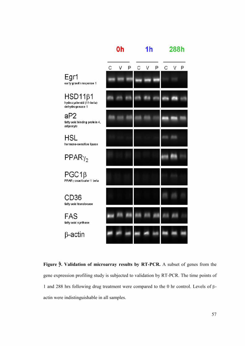
57
Figure 9 . Validation of microarray results by RT-PCR. A subset of genes from the
gene expression profiling study is subjected to validation by RT-PCR. The time points of
1 and 288 hrs following drug treatment were compared to the 0 hr control. Levels of β-
actin were indistinguishable in all samples.

58
Figure 10. Genome-wide gene expression profiling shows that prieurianin disrupts
the transcriptome of adipogenesis. Dendrograms of gene expression changes during

59
differentiation of L1 preadipocytes showing specific alterations induced by prieurianin
compared to DMSO or untreated control. a, Distinct temporal changes in gene expression
were clustered into early, intermediate and late responses during preadipocytes
differentiation into adipocytes. b, Specific clusters of genes whose expression were either
induced or inhibited by prieurianin. c, Attenuated expression of C/EBPα, β, and δ, and
PPARγ, transcriptional regulators of adipogenesis, and the induction of Zfp68 expression
by prieurianin. EGR1 and 2 expressions were not affected by prieurianin. d, Inhibition of
fat metabolism genes by prieurianin that correspond to those inactivated by genome-wide
RNAi in C. elegans, which causes reduced body fat accumulation. The complete data set
is available at Gene Expression Omnibus (http://www.ncbi.nlm.nih.gov/geo/) under
series accession
no. GSE15018).

60
Figure 11. Prieurianin suppress transcriptional activity of C/EBPs and PPARγ. a, b,
Prieurianin suppresses the transactivation potential of C/EBPα and β from the C/EBP-
response element (a) and PPARγ-transactivated PPAR-response element (b) luciferase
reporters. Statistics were conducted as student t-test. Asterisk, P<0.05 versus vector;
hash, P<0.05 versus the respective C/EBPα-, β-, or PPARγ-transfected controls. Results
are means ± s.e.m., normalized to Renilla luciferase activity.

61
0 0.25 0.5 1 2 3 5 9 12 15 24 36 48 96 144 192 240 2880
1000
2000
3000
4000
Prieurianin 2µM
ControlDMSO
TNFR2
Time, h
Expr
essi
onLe
vels
,RFU
0 0.25 0.5 1 2 3 5 9 12 15 24 36 48 96 144 192 240 2880
1000
2000
3000
4000
Prieurianin 2µM
ControlDMSOTRAF5
Time, h
Expr
essi
onLe
vels
,RFU
Figure 12. Expression profiling of pharmacological response of 3T3-L1 cells to
prieurianin by DNA microarray. 3T3-L1 cells were induced to differentiate to
adipocytes in the presence of DMSO (vehicle control) or 2µM prieurianin. RNA was
isolated at the indicated times (in hours). Dendrograms of TNFR2 and TRAF5 expression
changes are shown (A, C) and a plot of actual values (B, D) respectively.
B
A
D
C

62
0 0.5 1 2 40
5000
10000
15000
20000
25000
Prieurianin, µM
NFκ
BA
ctiv
ity,R
LU
TNFα Prieurianin Bufalin0
10000
20000
30000
Control
IKK-β Inhibitor 2µM
NFκ
BA
ctiv
ity,R
LU
Figure 13. Transactivation of NFκB by prieurianin in SW620. (A) The NFκB
consensus sequence fused to a luciferase reporter was transactivated by prieurianin dose-
dependently. (B) TNF- and prieurianin-induced NFκB activation is inhibited by IKKβ
inhibitor. Results are the mean ± S.E. of three experiments. * indicated p value less than
0.05.
A
B
*
*
*
* *

63
Figure 14. Prieurianin induces p65 nuclear translocation and IκBα degradation in
HeLa. Prieurianin treatment, like TNFα, led to a substantial loss, of detectable IκBα in
cytoplasmic extracts by Western blotting within 30 minutes. The changes in IκBα levels
measured in cytoplasmic cell extracts correlate with the appearance of NFκB-p65 in
nuclear extracts.

64
DISCUSSION
Obesity, the result of an imbalance in storage energy, is associated with chronic low
grade inflammation and insulin resistance. Obese adipose tissues are characterized by the
enhanced infiltration of macrophages, and a paracrine loop involving the secretion of
monocyte chemoattractant protein-1 (MCP-1), TNFα, and the free fatty acid between
adipocytes and macrophages. This signalling loop is accompanied by down-regulation of
PPARγ, glucose transport 4 (GLUT4) and IRS-1. Needless to say, the pathogenesis of
this disease is complex and there are several contributory genetic and environmental
factors. It should not be surprising that over 50% of diabetic patient are obese, owing to
the strong correlation between the development of insulin resistance and obesity. Thus,
elucidating mechanisms for insulin resistance has been an intense area of research in the
field of diabetes, and undoubtedly it would be of great benefit in the treatment of obesity
in the future.
The unique pharmacological profile of prieurianin in suppressing energy intake in
mice, and inhibiting the proliferation and differentiation of preadipocytes, and causing
dedifferentiation/delipidation of adipocytes, reveals a first-in-class anti-obesity drug. The
dual anorexigenic and anti-adipogenic action of prieurianin is also unique among
currently available pharmaceutical therapies and pipeline products in development that
either suppress satiety or inhibit energy homeostasis (Cooke & Bloom, 2006). With the
cytokine-mimicking effects on weight loss and lipolysis (Johnen, et al., 2007; Spiegelman
& Hotamisligil, 1993), as exemplified by prieurianin, the limonoids hold the promise to
be a novel class of small molecules that target multiple physiological mechanisms

65
including modulation of the neuronal input that controls anorexigenic function, and the
induction of a de novo transcriptional repressor that inhibits adipogenesis. The precise
molecular target of prieurianin, its long-term efficacy and toxicology profile remain to be
determined.
Our results further demonstrate that drug-induced tolerance resulting from chronic
pharmacotherapy for obesity can be overcome by a cyclical drug holiday treatment
strategy, which produces a greater and a more durable therapeutic response and the
maintenance of weight loss and reduced energy intake. Hence, the on-off treatment
protocol may potentially restore the efficacy of currently available anti-obesity drugs as
well as other experimental pharmacotherapeutics that are prone to drug-induced tolerance
in the long-term treatment of obesity. In addition, despite significant advances in the
transcriptional regulation of adipogenesis, many gaps remain, specifically in the anti-
adipogenic factors that control adipose tissue development. The activation of NFκB
responsive genes have previously been shown to be a repressor of C/EBPs and PPARγ
activity and demonstrates that prieurianin targets the regulatory factors that arrest
adipocyte differentiation. Thus, prieurianin may constitute an excellent pharmacological
tool for probing the transcriptional process that regulates adipogenesis. Taken together,
the findings in this study show that prieurianin is a novel anti-obesity therapeutic that
exhibits efficacy against obesity of various etiologic conditions. Its dual anorexigenic
and anti-adipogenic properties that parallel those of inflammatory cytokines raise the
possibility of a novel class of more potent and efficacious treatment for obesity with the
limonoids than currently available pharmacotherapeutics.

66
DISCUSSION
Part I
Drug resistance in cancer is a complex phenotypic process involving the genetic
alteration of multiple cellular factors and aberrantly perturbed biological pathways that
evolve during tumorigenesis. Therefore, understanding how drug resistance emerges in
cancer is of paramount importance in improving cancer treatment and outcome. In this
report, we identified the transcriptional induction of PLTP gene expression by
camptothecin (CPT) and its derivatives that are active inhibitor of the topoisomerase I
enzyme (Figs. 1 and 2). There are two types of topoisomerases (type I and type II), and
these enzymes function to unwind DNA and to facilitate (i) DNA replication, and (ii)
mRNA transcription and subsequent translation during protein synthesis. Topotecan and
irinotecan are two examples of commonly used chemotherapy drugs that work by
interfering with topoisomerase I in cancer cells. Topotecan is the water-soluble derivative
of camtothecin and used to treat ovarian and lung cancer, as well as other cancer types. It
acts by forming a stable covalent complex with the DNA/topoisomerase I aggregate and
this process leads to breaks in the DNA strand resulting in apoptosis (Tolis, Peters,
Ferreira, Pinedo, & Giaccone, 1999). According to the WHO, in 2004 lung cancer was
the most common cause of cancer-related death in men and women, and responsible for
1.3 million deaths worldwide annually. The vast majority of lung cancers are
carcinomas—malignancies that arise from epithelial cells, and the two main types of lung
cancer are small cell lung carcinoma (17%) and non-small cell lung carcinoma (80%).
While primary lung cancers most commonly metastasize to the adrenal glands, liver,

67
brain, and bone, the lungs are a common place for metastasis of tumors from other parts
of the body such as the breast. Because the lungs are a major site expressing PLTP
mRNA in humans and mice, this suggests that this protein might have an important role
in maintaining normal function of this organ. Using an emphysematous animal model,
Jiang et al showed that the lung of human collagenase transgenic mice expressed PLTP
mRNA 3-fold higher than in control mice but the mRNA in other tissues was not changed
(Jiang, et al., 1998). Thus, these observations suggest that a hypoxic stimulus occurring
in emphysema may be contribute to enhanced expression of PLTP. Our studies showed
that induction of PLTP expression by CPTs was not only drug-specific but also restricted
to the human liver cancer HepG2 cells only. We further demonstrated that topotecan
activated PLTP expression is mediated through the PKC signaling pathway and requires
specific PKC isozyme for its transcriptional regulation (Fig. 3). PLTP expression is also
induced in vivo in mice following treatment with topotecan that is accompanied by a rise
in serum triglyceride and cholesterol, and a marked loss of HDL (Fig. 5). In addition,
CPT induced increase in PLTP level did not result in the transfer of CPT to HDL.
The profound induction of PLTP expression by CPTs is intriguing and led us to
initially hypothesize that topoisomerase I inhibitors induced their own metabolism via the
reverse cholesterol transport pathway. Moreover, it has been shown that cholesterol is a
substrate for PLTP (Tu, Nishida, & Nishida, 1993), and high cholesterol diet increases
PLTP activity and PLTP mRNA in mice (Jiang & Bruce, 1995; Tu, et al., 1999). In
addition, crystal structure data of the bacterial permeability increasing (BPI) protein
(Beamer, Carroll, & Eisenberg, 1997; Huuskonen, et al., 1999) and the cholesteryl ester

68
transfer protein (CETP) (Qiu, et al., 2007) showing that proteins in this family, including
PLTP, contain intrinsic lipid binding sites that appear to act as carrier proteins or
channels that shuttle and redistribute lipids between lipoproteins. These observations
raised the possibility that CPTs might be substrates for PLTP-mediated transfer into HDL
and subsequently transported to the liver for metabolism via the reverse cholesterol
transport pathway. However, our results showed that, unlike cholesterol, CPT was not
found in HDL or other lipoproteins, suggesting that CPTs are not substrates for PLTP and
ruling out the possibility of PLTP-mediated transfer of CPT into HDL for liver
metabolism. These observations pose the possibility that the induction of PLTP gene
expression by CPTs is merely the result of bystander effect following drug exposure,
which inadvertently activates the expression of many genes, and some of which including
PLTP may have no physiological consequences or whose functional impacts are
unknown.
Besides topotecan, it has previously been reported that PLTP expression is
induced by hypoxia (Jiang, et al., 1998), fenofibrate (Bouly, et al., 2001), and the liver X
receptor (LXR) agonist, T0901317 (Cao, et al., 2002). Fenofibrates are drug of the
fibrate class, that is amphipathic carboxylic acids, and like other fibrates they are used to
reduce low-density lipoprotein (LDL), very low density lipoproteins (VLDL) and
triglycerides levels, as well as to increase high-density lipoproteins (HDL) levels, and
these effects are mediated by an increase in PLTP mRNA and activity (Bouly, et al.,
2001). The liver X receptors (LXR) belong to the nuclear receptor superfamily that can
regulate important lipid metabolic pathways. Cao et al showed that T0901317, a specific

69
LXR agonist, has been shown to elevate HDL cholesterol and phospholipid in C57/BL6
mice and generated enlarged HDL particles that were enriched in cholesterol, ApoAI,
ApoE, and phospholipid, and this was closely correlated with the increased plasma PLTP
activity and liver PLTP mRNA levels (Cao, et al., 2002). These observations suggest that
PLTP expression is responsive to stimulation by small pharmacological molecules.
While PLTP mRNA is found abundantly in a variety of tissues (Albers, et al., 1995),
however, the induction by topotecan is cell-type specific. The underlying reason for this
conditional expression pattern is unclear, and we speculate that it may be dependent on
the small pharmacological molecules as well as the presence of specific transcriptional
regulators in the respective tissues or cell-types.
Another curious observation in our study is the unexpected rise in serum
cholesterol and triglyceride levels, and a severe depletion of serum HDL in mice treated
with a high dose of topotecan. The abrupt rise in serum triglyceride is known to be
associated with the onset of acute pancreatitis (AP), a sudden inflammation of the
pancreas. (Cappell, 2008). There are many etiological risk factors for AP, including a
history of alcohol abuse, gallstones, endoscopic retrograde cholangiopancreatography and
manometry, trauma or surgical procedures near the pancreas, certain medications,
hyperlipidemia, infection, and chronic hypercalcemia (Sekimoto, et al., 2006). The
pancreas exocrine function is to produce digestive enzymes for release into the
gastrointestinal tract (Berardi RR, 2005). The acinar cells within the pancreas are responsible
for producing the proenzymes, which then are packaged into storage vesicles called
zymogens. The zymogens travel through the pancreatic duct and are secreted into the

70
duodenum. Within the duodenum, enterokinase converts trypsinogen to trypsin, and then
active trypsin facilitates the conversion of the other pancreatic proenzymes to the active
form. AP can occur if there is damage to the acinar cells and/or injury to the pancreatic
duct that leads to inappropriate accumulation and activation of proenzymes within the
pancreas. The activated pancreatic enzymes digest the cell membranes of the pancreas
and activate an inflammatory response, which increases the vascular permeability of the
pancreas. Hemorrhage, edema, ischemia, and necrosis can result (Berardi RR, 2005). In
severe AP, patients progress to systemic inflammatory response syndrome, sepsis, and
multiple organ failure (Neoptolemos, Raraty, Finch, & Sutton, 1998). About 3% to 13%
of AP cases progress to chronic pancreatitis (Sekimoto, et al., 2006). Though under-
reported and under-appreciated, drug-induced acute pancreatitis are commonly
encountered in drug therapy (Eland, et al., 1999). The etiologic cause of most drug-
induced acute pancreatitis and atherosclerosis may be associated with hyperlipidemia,
resulting from increased total serum triglyceride and cholesterol levels (Yadav &
Pitchumoni, 2003). The mechanisms of drug-induced hyperlipidemia are unknown.
Increase in PLTP activity has been linked to hypertriglyceridemia (Jonkers, et al., 2003;
Kaser, et al., 2004), which may trigger the onset of acute pancreatitis (Miller, 2000;
Yadav & Pitchumoni, 2003). It is noteworthy that acute pancreatitis has been reported in
cancer patients treated with topoisomerase I inhibitors such as irinotecan (Govindan,
Read, Faust, & Mc Leod, 2003b) and exatecan (De Jager, et al., 2000), as well as other
chemotherapeutic agents including L-asparaginase (Jain, et al., 2009; Parsons, et al.,
1997), tamoxifen (Alagozlu, et al., 2006), interferon α (Wong, et al., 2004), and
capecitabine (Koutras, et al., 2006). These observations, together with our results,

71
suggest that hypertriglyceridemia induced acute pancreatitis may be correlated with the
increase in PLTP, thus suggesting that PLTP may serve as a biomarker for CPTs and
other drug-induced hypertriglyceridemia and acute pancreatitis.
In summary, our results showed that CPTs induced PLTP expression has no
functional consequence in drug resistance, but may be associated with drug-induced
hyperlipidemia and the onset of acute pancreatitis. Monitoring changes in serum PLTP
levels or activity, therefore, may serve as an important biomarker for drug-induced
hyperlipidemia and the development of acute pancreatitis. How increased PLTP
expression causes hyperlipidemia (hypercholesterolemia and hypertriglyceridemia) and
lowers HDL levels needs to be further investigated.
Part II
There are currently only two FDA-approved pharmacotherapy for obesity in U.S.
Recent FDA issued heart risk warning in U.S. for sibutramine and its withdrawal in
Europe, as well as new concerns for potential liver toxicity with orlistat, further reduced
the armamentarium for the combat of obesity. Sibutramine is an orally administered
appetite suppressant used for the treatment of obesity, and is classified as a schedule IV
controlled substance in the United States, despite having no potential for abuse due to its
lack of appreciable dopaminergic effects. Sibutramine is well absorbed from the GI tract
(77%), but undergoes considerable first-pass metabolism, reducing its bioavailability.

72
The drug itself reaches its peak plasma level after 1 hour and has also a half-life of 1
hour. Sibutramine is metabolized by cytochrome P450 isozyme CYP3A4 into two
pharmacologically-active primary and secondary amines (called active metabolites 1 and
2) with half-lives of 14 and 16 hours, respectively. Peak plasma concentrations of active
metabolites 1 and 2 are reached after three to four hours. The following metabolic
pathway mainly results in two inactive conjugated and hydroxylated metabolites (called
metabolites 5 and 6). Metabolites 5 and 6 are mainly excreted in the urine. Sibutramine is
a neurotransmitter reuptake inhibitor that reduces the reuptake of serotonin (by 53%),
norepinephrine (by 54%), and dopamine (by 16%), thereby increasing the levels of these
substances in synaptic clefts and helping enhance satiety; the serotonergic action, in
particular, is thought to influence appetite. Older anorectic agents such as amphetamine
and fenfluramine force the release of these neurotransmitters rather than affecting their
reuptake (Heal, et al., 1998). Despite having a mechanism of action similar to tricyclic
antidepressants such as selective serotonin reuptake inhibitors (SSRIs) and serotonin-
norepinephrine reuptake inhibitors (SNRIs), sibutramine has failed to demonstrate
antidepressant properties in animal studies. In contrast, the primary function of orlistat is
to prevent the absorption of fats from the human diet, thereby reducing caloric intake. It
is intended for use in conjunction with a physician-supervised reduced-calorie diet.
Orlistat is the saturated derivative of lipstatin, a potent natural inhibitor of pancreatic
lipases isolated from the bacterium Streptomycen toxytricini (Barbier & Schneider, 1987).
However, due to simplicity and stability, orlistat rather than lipstatin was developed into
an anti-obesity drug (Pommier, Pons, & Kocienski, 1995). Orlistat works by inhibiting
gastric and pancreatic lipases, the enzymes that break down triglycerides in the intestine.

73
When lipase activity is blocked, triglycerides from the diet are not hydrolyzed into
absorbable free fatty acids, and are excreted undigested instead. Only trace amounts of
orlistat are absorbed systemically; the primary effect is local lipase inhibition within the
GI tract after an oral dose. The primary route of elimination is through the feces. Orlistat
prevents approximately 30% of dietary fat from being absorbed. Drug repositioning is the
application of known drugs and compounds to new diseases, and this field has been
growing in importance in the last few years as an increasing number of drug development
and pharmaceutical companies see their drug pipelines drying up and realize that many
previously promising technologies have failed to deliver ‘as advertised’. A significant
advantage of drug repositioning over traditional drug development is that since the
repositioned drug has already passed a significant number of toxicity and other tests, its
safety is known and the risk of failure for reasons of adverse toxicology are reduced.
Using drug repositioning, pharmaceutical companies have achieved a number of
successes, for example Pfizer’s Viagra in erectile dysfunction and Celgene’s thalidomide
in severe erythema nodosum leprosum. The use of repositioned drugs comprising of
combinatorial mixture of anticonvulsant, antidepressant, or psychoactive stimulant is also
being contemplated for the treatement of obesity, however questions arise of unknown
long-term safety concerns, as these drugs all have various known risks and side effects
(Robinson & Niswender, 2009). These issues all point to an urgent need for the
development of more efficacious and safe pharmacotherapeutics for obesity.
We described here the unexpected identification of a natural plant limonoid
product, prieurianin, which has a unique pharmacological profile of suppressing energy

74
intake in mice, and inhibiting the proliferation and differentiation of preadipocytes, and
causing dedifferentiation/delipidation of adipocytes. Further, we showed that prieurianin
mechanism of action is mediated in part by NFκB signaling. These findings ushered in a
novel first-in-class anti-obesity drug candidate. The dual anorexigenic and anti-
adipogenic action of prieurianin is unique among currently available pharmaceutical
therapies and pipeline products in development that focused on either suppressing satiety
or inhibiting energy homeostasis (Cooke & Bloom, 2006). Further, with the cytokine-
mimicking effects on weight loss and lipolysis (Johnen, et al., 2007; Spiegelman &
Hotamisligil, 1993), prieurianin holds promise to be a novel class of small molecules that
target multiple physiological mechanisms including the potential modulation of neuronal
input that controls anorexigenic function, and the prevention of new adipose mass
development by inhibiting adipogenesis. The precise molecular target of prieurianin, its
long-term efficacy and toxicology profile remain to be determined.
Long-term efficacy and effectiveness of obesity treatments are known to have
poor outcome (Mauro, Taylor, Wharton, & Sharma, 2008). The potential of drug-induced
tolerance associated with anti-obesity therapeutics, though recognized, but circumvention
of its development has not been fully addressed (Fernstrom & Choi, 2007, 2008). Our
results demonstrate that drug-induced tolerance resulting from chronic pharmacotherapy
for obesity can be overcome by a cyclical drug holiday treatment strategy, which
produces a greater and a more durable therapeutic response and the maintenance of
weight loss and reduced energy intake. A drug holiday is when a patient stops taking a
medication(s) for a period of time; anywhere from a few days to many months or even

75
years if they feel it is in their best interests. Planned drug holidays are used in numerous
fields of medicine. They are perhaps best known in HIV therapy, after a study showed
that stopping medication may stimulate the immune system to attack the virus. Further, a
drug holiday may be used because (i) it is part of a progression toward treatment
cessation, (ii) to reduce drug side effects, or (iii) to permit a drug to regain effectiveness
after a period of continuous use, and to reduce the tolerance effect that may require
increased dosages. Hence, the on-off treatment protocol may potentially restore the
efficacy of currently available anti-obesity drugs as well as other experimental
pharmacotherapeutics that are prone to drug-induced tolerance in the long-term treatment
of obesity. Besides, decreasing the dosage and frequency of drug exposure using the
drug holiday treatment protocol may reduce the incidence and disposition to the toxicities
of these anti-obesity drugs. Therefore, new trials should be initiated to test the potential
benefits of this approach in the treatment of obesity.
It is noteworthy that prieurianin was recently shown to inhibit endocytosis and
also block brassinosteroid-induced gene expression in germinating seedlings of
Arabidopsis thaliana (Robert, et al., 2008). It is unclear how prieurianin-inhibited
endocytosis translates into its anti-obesity effects. Nevertheless, the potential action of
prieurianin to inhibit both the hormone-induced transcription in germinating seedlings
and the transcriptionally regulated adipogenesis in preadipocytes, further suggests its
unique pharmacodynamics in disrupting gene expression program during differentiation.

76
Hence, identifying the molecular target(s) of prieurianin might yield insights into the
rate-limiting factor that drives the development of adipose tissue.
NF-kB is well known as a master regulator of gene transcription in white blood
cells and lymphocytes involved in inflammation and innate immunity. Recently, many
studies have shown that NF-κB signaling plays a role in obesity, a pathophysiological
state associated with a chronic low-grade inflammation accompanied by increasing levels
of proinflammatory cytokines (Hotamisligil, 2006; Shoelson, Herrero, & Naaz, 2007;
Wellen & Hotamisligil, 2005). According to the National Health and Nutrition
Examination Survey in the United States, it was estimated that over 65% of adults were
overweight or obese, and 16% of children were overweight (CDC, 2002). Obesity,
particularly that caused by visceral fat accumulation is a serious risk factor for diabetes,
coronary heart diseases, hyperlipidemia, hypertension and cancer (Nakamura, et al.,
1994). Further, insulin resistance is considered to be the most common underlying cause,
and is influenced by both genetic and environmental factors, as well as changes in diet
and exercise (Uauy & Diaz, 2005). While weight loss via diet and exercise improves
insulin sensitivity, the precise mechanisms modulating this improvement are not
completely understood, but there is mounting evidence that NF-κB signaling plays a
crucial role (Chiang, et al., 2009; X. Zhang, et al., 2008). NF-κB may be activated by
toll-like receptor-4 (TLR4) due to interactions with dietary fatty acids (F. Kim, et al.,
2007; Tsukumo, et al., 2007). Adipose tissue responds to overnutrition by secreting
cytokines and chemokines that recruit macrophages to adipose tissue (Lumeng,
DelProposto, Westcott, & Saltiel, 2008). In turn, macrophages secrete more cytokines

77
and chemokines that attenuate insulin action, and increase lipolysis and free fatty acid
release (Feingold, Doerrler, Dinarello, Fiers, & Grunfeld, 1992). Thus, the inflammatory
response is exacerbated. Interestingly, in our studies when C57/BL6 mice were treated
with topotecan, triglycerides levels decreased and HDL-cholesterols increased. This
suggests that PLTP may have played a role in preventing the accumulation of excess fatty
acids and cholesterol in the plasma, and mitigated the NF-κB induced inflammatory
response. Because of these desirable physiological effects, we set out to screen natural
compound libraries to identify compounds that can similarly increase PLTP gene
expression, reduced triglycerides and LDL-cholesterols, and increase HDL-cholesterols,
but are neither cytotoxic nor DNA damaging agents. From the screen of 720 natural
compounds and small molecules, prieurianin emerged the clear winner.
Prieurianin is a limonoids and occur naturally only in plants species of the
Rutaceae and Meliaceae plant families. It reduces food consumption in rodent models of
obesity by possibly diverting carbohydrates and fatty acids that would have become fat in
liver into hepatic glycogen. In turn, this metabolic change may send a signal to the brain
that results in appetite suppression. Our pair fed experiments suggests that prieurianin in
addition to suppressing food intake, by preventing mice from efficiently transforming the
nutrients fed into their own biomass, also stimulates the breakdown of adipose stores.
Paradoxically, the hydrolysis of the fat droplets and tissue was not accompanied by a
subsequent efflux of fatty acids and glycerol, the hallmark clues of lipolysis and thus
raise the question, so where did the fat go? One possibility is that prieurianin increased
the cell’s β-oxidative capacity. However, a number of the genes essential to β-oxidation

78
particularly PPARα was not upregulated in our studies. Another more plausible
explanation is that after lipolysis the excess glycerol was converted to glycogen, which
would not have been stained by either oil red-O or nile red, neutral lipids and
triglycerides staining dyes, rather peroidic acid-Schiff (PAS) staining should be used.
Lipolysis is a transient and rapid process, and usually can be evident in a few minutes to
hours. On the other hand, adipocytes treated with prieurianin takes 5-7 days before
marked differences can be observed when compared to untreated or vehicle treated
controls suggesting that the kinetics and action of prieurianin is slow. Lipogenesis is a
reciprocal biochemical process that goes hand in hand with lipolysis to maintain lipid
homeostasis, and the process is regulated by hormones and lipid metabolites that signal
chiefly to the lipogenic transcription factors PPARγ and C/EBPα, and β. Our studies
showed that prieurianin, in addition to stimulating the loss of lipid droplets, blocked the
transcriptional expression of these keys lipogenic transcription factors and in turn blocks
the differentiation of adipocytes.
Taken together, the findings in this study show that prieurianin is a novel anti-
obesity therapeutic candidate that exhibits efficacy against obesity of various etiologic
origins. Its dual anorexigenic and anti-adipogenic properties that parallel those of
inflammatory cytokines raise the possibility of a novel class of more potent and
efficacious treatment for obesity with the limonoids than currently available
pharmacotherapeutics.

79
CONCLUSION
1. Prieurianin, like camptothecin, increases PLTP mRNA and protein levels in
HepG2. However, it does not affect blood cholesterol levels. PLTP levels are
associated with increased adiposity and decreases with weight loss.
2. Prieurianin reduces adipose tissue mass in four animal models of obesity, and
decreases plasma insulin and glucose levels.
3. Prieurianin inhibits the expression of key adipogenic transcription factors
including C/EBP-α, -β, and –δ, and PPARγ
4. The prieurianin-induced block of the adipogenic transcription machinery is an
NFκB-p65 dependent event.

80
REFERENCES
Alagozlu, H., Cindoruk, M., & Unal, S. (2006). Tamoxifen-induced severe
hypertriglyceridaemia and acute pancreatitis. Clin Drug Investig, 26(5), 297-302.
Albers, J. J., Wolfbauer, G., Cheung, M. C., Day, J. R., Ching, A. F., Lok, S., et al.
(1995). Functional expression of human and mouse plasma phospholipid transfer
protein: effect of recombinant and plasma PLTP on HDL subspecies. Biochim
Biophys Acta, 1258(1), 27-34.
Arner, P., & Langin, D. (2007). The role of neutral lipases in human adipose tissue
lipolysis. Curr Opin Lipidol, 18(3), 246-250.
Ashrafi, K., Chang, F. Y., Watts, J. L., Fraser, A. G., Kamath, R. S., Ahringer, J., et al.
(2003). Genome-wide RNAi analysis of Caenorhabditis elegans fat regulatory
genes. Nature, 421(6920), 268-272.
Attia, N., Nakbi, A., Smaoui, M., Chaaba, R., Moulin, P., Hammami, S., et al. (2007).
Increased phospholipid transfer protein activity associated with the impaired
cellular cholesterol efflux in type 2 diabetic subjects with coronary artery disease.
Tohoku J Exp Med, 213(2), 129-137.
Barbier, P., & Schneider, F. (1987). Syntheses of Tetrahydrolipstatin and Absolute
Configuration of Tetrahydrolipstatin and Lipstatin. Helvetica Chimica Acta,
70(1), 196-202.
Beamer, L. J., Carroll, S. F., & Eisenberg, D. (1997). Crystal structure of human BPI and
two bound phospholipids at 2.4 angstrom resolution. Science, 276(5320), 1861-
1864.

81
Beavo, J. A., Rogers, N. L., Crofford, O. B., Hardman, J. G., Sutherland, E. W., &
Newman, E. V. (1970). Effects of xanthine derivatives on lipolysis and on
adenosine 3',5'-monophosphate phosphodiesterase activity. Mol Pharmacol, 6(6),
597-603.
Berardi RR, M. P. (2005). Pharmacotherapy: a pathophysiologic approach. (6th ed.).
New York: McGraw-Hill.
Bortell, R., Owen, T. A., Ignotz, R., Stein, G. S., & Stein, J. L. (1994). TGF beta 1
prevents the down-regulation of type I procollagen, fibronectin, and TGF beta 1
gene expression associated with 3T3-L1 pre-adipocyte differentiation. J Cell
Biochem, 54(2), 256-263.
Bouly, M., Masson, D., Gross, B., Jiang, X. C., Fievet, C., Castro, G., et al. (2001).
Induction of the phospholipid transfer protein gene accounts for the high density
lipoprotein enlargement in mice treated with fenofibrate. J Biol Chem, 276(28),
25841-25847.
Bray, G. A., & Bellanger, T. (2006). Epidemiology, trends, and morbidities of obesity
and the metabolic syndrome. Endocrine, 29(1), 109-117.
Cao, G., Beyer, T. P., Yang, X. P., Schmidt, R. J., Zhang, Y., Bensch, W. R., et al.
(2002). Phospholipid transfer protein is regulated by liver X receptors in vivo. J
Biol Chem, 277(42), 39561-39565.
Cappell, M. S. (2008). Acute pancreatitis: etiology, clinical presentation, diagnosis, and
therapy. Med Clin North Am, 92(4), 889-923, ix-x.

82
Carswell, E. A., Old, L. J., Kassel, R. L., Green, S., Fiore, N., & Williamson, B. (1975).
An endotoxin-induced serum factor that causes necrosis of tumors. Proc Natl
Acad Sci U S A, 72(9), 3666-3670.
CDC (2002). Chartbook on Trends in the Health of Americans. Excerpted from Health,
United States, 2002: Department of Health and Human Services, Centers for
Disease Control and Prevention, National Center for Health Statistics.
Chasin, M., & Harris, D. N. (1976). Inhibitory and activators of cyclic nucleotide
phosphodiesterase. Adv Cyclic Nucleotide Res, 7, 225-264.
Chen, H., Charlat, O., Tartaglia, L. A., Woolf, E. A., Weng, X., Ellis, S. J., et al. (1996).
Evidence that the diabetes gene encodes the leptin receptor: identification of a
mutation in the leptin receptor gene in db/db mice. Cell, 84(3), 491-495.
Chen, Z., Torrens, J. I., Anand, A., Spiegelman, B. M., & Friedman, J. M. (2005).
Krox20 stimulates adipogenesis via C/EBPbeta-dependent and -independent
mechanisms. Cell Metab, 1(2), 93-106.
Chiang, S. H., Bazuine, M., Lumeng, C. N., Geletka, L. M., Mowers, J., White, N. M., et
al. (2009). The protein kinase IKKepsilon regulates energy balance in obese mice.
Cell, 138(5), 961-975.
Chua, S. C., Jr., Chung, W. K., Wu-Peng, X. S., Zhang, Y., Liu, S. M., Tartaglia, L., et al.
(1996). Phenotypes of mouse diabetes and rat fatty due to mutations in the OB
(leptin) receptor. Science, 271(5251), 994-996.
Collins, S., Martin, T. L., Surwit, R. S., & Robidoux, J. (2004). Genetic vulnerability to
diet-induced obesity in the C57BL/6J mouse: physiological and molecular
characteristics. Physiol Behav, 81(2), 243-248.

83
Cooke, D., & Bloom, S. (2006). The obesity pipeline: current strategies in the
development of anti-obesity drugs. Nat Rev Drug Discov, 5(11), 919-931.
Cseh, K., Winkler, G., Melczer, Z., & Baranyi, E. (2000). The role of tumour necrosis
factor (TNF)-alpha resistance in obesity and insulin resistance. Diabetologia,
43(4), 525.
De Jager, R., Cheverton, P., Tamanoi, K., Coyle, J., Ducharme, M., Sakamoto, N., et al.
(2000). DX-8951f: summary of phase I clinical trials. Ann N Y Acad Sci, 922,
260-273.
DeAngelis, A. M., Heinrich, G., Dai, T., Bowman, T. A., Patel, P. R., Lee, S. J., et al.
(2008). Carcinoembryonic antigen-related cell adhesion molecule 1: a link
between insulin and lipid metabolism. Diabetes, 57(9), 2296-2303.
Drori, S., Girnun, G. D., Tou, L., Szwaya, J. D., Mueller, E., Xia, K., et al. (2005). Hic-5
regulates an epithelial program mediated by PPARgamma. Genes Dev, 19(3),
362-375.
Eckel, R. H. (1989). Lipoprotein lipase. A multifunctional enzyme relevant to common
metabolic diseases. N Engl J Med, 320(16), 1060-1068.
Eland, I. A., van Puijenbroek, E. P., Sturkenboom, M. J., Wilson, J. H., & Stricker, B. H.
(1999). Drug-associated acute pancreatitis: twenty-one years of spontaneous
reporting in The Netherlands. Am J Gastroenterol, 94(9), 2417-2422.
Feingold, K. R., Doerrler, W., Dinarello, C. A., Fiers, W., & Grunfeld, C. (1992).
Stimulation of lipolysis in cultured fat cells by tumor necrosis factor, interleukin-
1, and the interferons is blocked by inhibition of prostaglandin synthesis.
Endocrinology, 130(1), 10-16.

84
Ferkingstad, E., Frigessi, A., & Lyng, H. (2008). Indirect genomic effects on survival
from gene expression data. Genome Biol, 9(3), R58.
Fernstrom, J. D., & Choi, S. (2007). The development of tolerance to drugs that suppress
food intake. Pharmacol Ther.
Fernstrom, J. D., & Choi, S. (2008). The development of tolerance to drugs that suppress
food intake. Pharmacol Ther, 117(1), 105-122.
Foger, B., Santamarina-Fojo, S., Shamburek, R. D., Parrot, C. L., Talley, G. D., &
Brewer, H. B., Jr. (1997). Plasma phospholipid transfer protein. Adenovirus-
mediated overexpression in mice leads to decreased plasma high density
lipoprotein (HDL) and enhanced hepatic uptake of phospholipids and cholesteryl
esters from HDL. J Biol Chem, 272(43), 27393-27400.
Gonzales, A. M., & Orlando, R. A. (2007). Role of adipocyte-derived lipoprotein lipase
in adipocyte hypertrophy. Nutr Metab (Lond), 4, 22.
Govindan, R., Read, W., Faust, J., & Mc Leod, H. (2003a). Irinotecan and carboplatin in
metastatic or recurrent non-small-cell lung cancer. Oncology (Williston Park),
17(7 Suppl 7), 27-29.
Govindan, R., Read, W., Faust, J., & Mc Leod, H. (2003b). Irinotecan and carboplatin in
metastatic or recurrent non-small-cell lung cancer. Oncology (Huntingt), 17(7
Suppl 7), 27-29.
Green, H., & Meuth, M. (1974). An established pre-adipose cell line and its
differentiation in culture. Cell, 3(2), 127-133.
Griffin, M. E., Marcucci, M. J., Cline, G. W., Bell, K., Barucci, N., Lee, D., et al. (1999).
Free fatty acid-induced insulin resistance is associated with activation of protein

85
kinase C theta and alterations in the insulin signaling cascade. Diabetes, 48(6),
1270-1274.
Groop, L. C., Bonadonna, R. C., DelPrato, S., Ratheiser, K., Zyck, K., Ferrannini, E., et
al. (1989). Glucose and free fatty acid metabolism in non-insulin-dependent
diabetes mellitus. Evidence for multiple sites of insulin resistance. J Clin Invest,
84(1), 205-213.
Guerciolini, R. (1997). Mode of action of orlistat. Int J Obes Relat Metab Disord, 21
Suppl 3, S12-23.
Guo, X., Zhang, J., Fu, X., Wei, Q., Lu, Y., Li, Y., et al. (2006). Analysis of common
gene expression patterns in four human tumor cell lines exposed to camptothecin
using cDNA microarray: identification of topoisomerase-mediated DNA damage
response pathways. Front Biosci, 11, 1924-1931.
Halaas, J. L., Gajiwala, K. S., Maffei, M., Cohen, S. L., Chait, B. T., Rabinowitz, D., et
al. (1995). Weight-reducing effects of the plasma protein encoded by the obese
gene. Science, 269(5223), 543-546.
Harris MI, C. C., Reiber G, et al.,eds (1995). Diabetes in America, 2nd ed. Washington,
DC: US Government Printing Office.
Harris, M. I., Flegal, K. M., Cowie, C. C., Eberhardt, M. S., Goldstein, D. E., Little, R.
R., et al. (1998). Prevalence of diabetes, impaired fasting glucose, and impaired
glucose tolerance in U.S. adults. The Third National Health and Nutrition
Examination Survey, 1988-1994. Diabetes Care, 21(4), 518-524.
Haslam, D. W., & James, W. P. (2005). Obesity. Lancet, 366(9492), 1197-1209.

86
Hayden, M. S., & Ghosh, S. (2004). Signaling to NF-kappaB. Genes Dev, 18(18), 2195-
2224.
Heal, D. J., Aspley, S., Prow, M. R., Jackson, H. C., Martin, K. F., & Cheetham, S. C.
(1998). Sibutramine: a novel anti-obesity drug. A review of the pharmacological
evidence to differentiate it from d-amphetamine and d-fenfluramine. Int J Obes
Relat Metab Disord, 22 Suppl 1, S18-28; discussion S29.
Hotamisligil, G. S. (2006). Inflammation and metabolic disorders. Nature, 444(7121),
860-867.
Hotamisligil, G. S., Shargill, N. S., & Spiegelman, B. M. (1993). Adipose expression of
tumor necrosis factor-alpha: direct role in obesity-linked insulin resistance.
Science, 259(5091), 87-91.
Huuskonen, J., Olkkonen, V. M., Jauhiainen, M., & Ehnholm, C. (2001). The impact of
phospholipid transfer protein (PLTP) on HDL metabolism. Atherosclerosis,
155(2), 269-281.
Huuskonen, J., Wohlfahrt, G., Jauhiainen, M., Ehnholm, C., Teleman, O., & Olkkonen,
V. M. (1999). Structure and phospholipid transfer activity of human PLTP:
analysis by molecular modeling and site-directed mutagenesis. J Lipid Res, 40(6),
1123-1130.
Itani, S. I., Ruderman, N. B., Schmieder, F., & Boden, G. (2002). Lipid-induced insulin
resistance in human muscle is associated with changes in diacylglycerol, protein
kinase C, and IkappaB-alpha. Diabetes, 51(7), 2005-2011.
Jaari, S., van Dijk, K. W., Olkkonen, V. M., van der Zee, A., Metso, J., Havekes, L., et al.
(2001). Dynamic changes in mouse lipoproteins induced by transiently expressed

87
human phospholipid transfer protein (PLTP): importance of PLTP in prebeta-
HDL generation. Comp Biochem Physiol B Biochem Mol Biol, 128(4), 781-792.
Jain, S., Naithani, R., Kapoor, G., & Nath, T. (2009). L-asparaginase induced severe
hypertriglyceridemia in acute lymphoblastic leukemia with 11q23 abnormality.
Leuk Res, 33(11), e194.
Jiang, X. C. (2002). The effect of phospholipid transfer protein on lipoprotein metabolism
and atherosclerosis. Front Biosci, 7, d1634-1641.
Jiang, X. C., & Bruce, C. (1995). Regulation of murine plasma phospholipid transfer
protein activity and mRNA levels by lipopolysaccharide and high cholesterol diet.
J Biol Chem, 270(29), 17133-17138.
Jiang, X. C., Bruce, C., Mar, J., Lin, M., Ji, Y., Francone, O. L., et al. (1999). Targeted
mutation of plasma phospholipid transfer protein gene markedly reduces high-
density lipoprotein levels. J Clin Invest, 103(6), 907-914.
Jiang, X. C., D'Armiento, J., Mallampalli, R. K., Mar, J., Yan, S. F., & Lin, M. (1998).
Expression of plasma phospholipid transfer protein mRNA in normal and
emphysematous lungs and regulation by hypoxia. J Biol Chem, 273(25), 15714-
15718.
Jiang, X. C., Tall, A. R., Qin, S., Lin, M., Schneider, M., Lalanne, F., et al. (2002).
Phospholipid transfer protein deficiency protects circulating lipoproteins from
oxidation due to the enhanced accumulation of vitamin E. J Biol Chem, 277(35),
31850-31856.

88
Johnen, H., Lin, S., Kuffner, T., Brown, D. A., Tsai, V. W., Bauskin, A. R., et al. (2007).
Tumor-induced anorexia and weight loss are mediated by the TGF-beta
superfamily cytokine MIC-1. Nat Med, 13(11), 1333-1340.
Jonkers, I. J., Smelt, A. H., Hattori, H., Scheek, L. M., van Gent, T., de Man, F. H., et al.
(2003). Decreased PLTP mass but elevated PLTP activity linked to insulin
resistance in HTG: effects of bezafibrate therapy. J Lipid Res, 44(8), 1462-1469.
Karkkainen, M., Oka, T., Olkkonen, V. M., Metso, J., Hattori, H., Jauhiainen, M., et al.
(2002). Isolation and partial characterization of the inactive and active forms of
human plasma phospholipid transfer protein (PLTP). J Biol Chem, 277(18),
15413-15418.
Kaser, S., Laimer, M., Sandhofer, A., Salzmann, K., Ebenbichler, C. F., & Patsch, J. R.
(2004). Effects of weight loss on PLTP activity and HDL particle size. Int J Obes
Relat Metab Disord, 28(10), 1280-1282.
Kawakami, M., Watanabe, N., Ogawa, H., Kato, A., Sando, H., Yamada, N., et al.
(1989). Cachectin/TNF kills or inhibits the differentiation of 3T3-L1 cells
according to developmental stage. J Cell Physiol, 138(1), 1-7.
Kim, F., Pham, M., Luttrell, I., Bannerman, D. D., Tupper, J., Thaler, J., et al. (2007).
Toll-like receptor-4 mediates vascular inflammation and insulin resistance in diet-
induced obesity. Circ Res, 100(11), 1589-1596.
Kim, J. A., Yeh, D. C., Ver, M., Li, Y., Carranza, A., Conrads, T. P., et al. (2005).
Phosphorylation of Ser24 in the pleckstrin homology domain of insulin receptor
substrate-1 by Mouse Pelle-like kinase/interleukin-1 receptor-associated kinase:

89
cross-talk between inflammatory signaling and insulin signaling that may
contribute to insulin resistance. J Biol Chem, 280(24), 23173-23183.
Kim, J. K., Wi, J. K., & Youn, J. H. (1996). Plasma free fatty acids decrease insulin-
stimulated skeletal muscle glucose uptake by suppressing glycolysis in conscious
rats. Diabetes, 45(4), 446-453.
King, H., Aubert, R. E., & Herman, W. H. (1998). Global burden of diabetes, 1995-2025:
prevalence, numerical estimates, and projections. Diabetes Care, 21(9), 1414-
1431.
Kleinveld, H. A., Duif, P. F., Pekelharing, H. L., & van Rijn, H. J. (1996). Oxidation of
lipoprotein(a) and low density lipoprotein containing density gradient
ultracentrifugation fractions. Biochim Biophys Acta, 1303(1), 15-21.
Koul, O., Daniewski, W. M., Multani, J. S., Gumulka, M., & Singh, G. (2003).
Antifeedant effects of the limonoids from Entandrophragma candolei (Meliaceae)
on the gram pod borer, Helicoverpa armigera (Lepidoptera: Noctuidae). J Agric
Food Chem, 51(25), 7271-7275.
Koul, O., Singh, G., Singh, R., Daniewski, W. M., & Berlozecki, S. (2004). Bioefficacy
and mode-of-action of some limonoids of salannin group from Azadirachta indica
A. Juss and their role in a multicomponent system against lepidopteran larvae. J
Biosci, 29(4), 409-416.
Koutras, A. K., Habeos, I. G., Vagenakis, A. G., & Kalofonos, H. P. (2006).
Capecitabine-induced hypertriglyceridemia: a report of two cases. Anticancer Res,
26(3B), 2249-2251.

90
Kraemer, F. B., & Shen, W. J. (2002). Hormone-sensitive lipase: control of intracellular
tri-(di-)acylglycerol and cholesteryl ester hydrolysis. J Lipid Res, 43(10), 1585-
1594.
Lee, G. H., Proenca, R., Montez, J. M., Carroll, K. M., Darvishzadeh, J. G., Lee, J. I., et
al. (1996). Abnormal splicing of the leptin receptor in diabetic mice. Nature,
379(6566), 632-635.
Leung, N., Turbide, C., Olson, M., Marcus, V., Jothy, S., & Beauchemin, N. (2006).
Deletion of the carcinoembryonic antigen-related cell adhesion molecule 1
(Ceacam1) gene contributes to colon tumor progression in a murine model of
carcinogenesis. Oncogene, 25(40), 5527-5536.
Li, X., Yang, Y., & Ashwell, J. D. (2002). TNF-RII and c-IAP1 mediate ubiquitination
and degradation of TRAF2. Nature, 416(6878), 345-347.
Li, Z., Maglione, M., Tu, W., Mojica, W., Arterburn, D., Shugarman, L. R., et al. (2005).
Meta-analysis: pharmacologic treatment of obesity. Ann Intern Med, 142(7), 532-
546.
Lie, J., de Crom, R., van Gent, T., van Haperen, R., Scheek, L., Lankhuizen, I., et al.
(2002). Elevation of plasma phospholipid transfer protein in transgenic mice
increases VLDL secretion. J Lipid Res, 43(11), 1875-1880.
Lie, J., de Crom, R., van Gent, T., van Haperen, R., Scheek, L., Sadeghi-Niaraki, F., et al.
(2004). Elevation of plasma phospholipid transfer protein increases the risk of
atherosclerosis despite lower apolipoprotein B-containing lipoproteins. J Lipid
Res, 45(5), 805-811.

91
Lumeng, C. N., DelProposto, J. B., Westcott, D. J., & Saltiel, A. R. (2008). Phenotypic
switching of adipose tissue macrophages with obesity is generated by
spatiotemporal differences in macrophage subtypes. Diabetes, 57(12), 3239-3246.
Manners, G. D. (2007). Citrus limonoids: analysis, bioactivity, and biomedical prospects.
J Agric Food Chem, 55(21), 8285-8294.
Mauro, M., Taylor, V., Wharton, S., & Sharma, A. M. (2008). Barriers to obesity
treatment. Eur J Intern Med, 19(3), 173-180.
McGarry, J. D., & Foster, D. W. (1980). Regulation of hepatic fatty acid oxidation and
ketone body production. Annu Rev Biochem, 49, 395-420.
Mead, J. R., Irvine, S. A., & Ramji, D. P. (2002). Lipoprotein lipase: structure, function,
regulation, and role in disease. J Mol Med, 80(12), 753-769.
Miller, J. P. (2000). Serum triglycerides, the liver and the pancreas. Curr Opin Lipidol,
11(4), 377-382.
Montague, W., & Cook, J. R. (1971). The role of adenosine 3':5'-cyclic monophosphate
in the regulation of insulin release by isolated rat islets of Langerhans. Biochem J,
122(1), 115-120.
Najjar, S. M., Accili, D., Philippe, N., Jernberg, J., Margolis, R., & Taylor, S. I. (1993).
pp120/ecto-ATPase, an endogenous substrate of the insulin receptor tyrosine
kinase, is expressed as two variably spliced isoforms. J Biol Chem, 268(2), 1201-
1206.
Nakamura, T., Tokunaga, K., Shimomura, I., Nishida, M., Yoshida, S., Kotani, K., et al.
(1994). Contribution of visceral fat accumulation to the development of coronary
artery disease in non-obese men. Atherosclerosis, 107(2), 239-246.

92
Neoptolemos, J. P., Raraty, M., Finch, M., & Sutton, R. (1998). Acute pancreatitis: the
substantial human and financial costs. Gut, 42(6), 886-891.
Ogier, N., Klein, A., Deckert, V., Athias, A., Bessede, G., Le Guern, N., et al. (2007).
Cholesterol accumulation is increased in macrophages of phospholipid transfer
protein-deficient mice: normalization by dietary alpha-tocopherol
supplementation. Arterioscler Thromb Vasc Biol, 27(11), 2407-2412.
Oka, H., Kaneko, T., Yamashita, K., Suzuki, S., & Oda, T. (1973). The glucagon and
fluoride sensitive adenyl cyclase in plasma membrane of rat liver. Endocrinol
Jpn, 20(3), 263-270.
Oka, T., Kujiraoka, T., Ito, M., Egashira, T., Takahashi, S., Nanjee, M. N., et al. (2000).
Distribution of phospholipid transfer protein in human plasma: presence of two
forms of phospholipid transfer protein, one catalytically active and the other
inactive. J Lipid Res, 41(10), 1651-1657.
Parsons, S. K., Skapek, S. X., Neufeld, E. J., Kuhlman, C., Young, M. L., Donnelly, M.,
et al. (1997). Asparaginase-associated lipid abnormalities in children with acute
lymphoblastic leukemia. Blood, 89(6), 1886-1895.
Peytremann, A., Nicholson, W. E., Liddle, G. W., Hardman, J. G., & Sutherland, E. W.
(1973). Effects of methylxanthines on adenosine 3',5'-monophosphate and
corticosterone in the rat adrenal. Endocrinology, 92(2), 525-530.
Pi-Sunyer, F. X., Aronne, L. J., Heshmati, H. M., Devin, J., & Rosenstock, J. (2006).
Effect of rimonabant, a cannabinoid-1 receptor blocker, on weight and
cardiometabolic risk factors in overweight or obese patients: RIO-North America:
a randomized controlled trial. Jama, 295(7), 761-775.

93
Pommier, A., Pons, J.-M., & Kocienski, P. J. (1995). The First Total Synthesis of (-)-
Lipstatin. The Journal of Organic Chemistry, 60(22), 7334-7339.
Puri, V., Konda, S., Ranjit, S., Aouadi, M., Chawla, A., Chouinard, M., et al. (2007). Fat-
specific protein 27, a novel lipid droplet protein that enhances triglyceride
storage. J Biol Chem, 282(47), 34213-34218.
Qiu, X., Mistry, A., Ammirati, M. J., Chrunyk, B. A., Clark, R. W., Cong, Y., et al.
(2007). Crystal structure of cholesteryl ester transfer protein reveals a long tunnel
and four bound lipid molecules. Nat Struct Mol Biol, 14(2), 106-113.
Robert, S., Chary, S. N., Drakakaki, G., Li, S., Yang, Z., Raikhel, N. V., et al. (2008).
Endosidin1 defines a compartment involved in endocytosis of the brassinosteroid
receptor BRI1 and the auxin transporters PIN2 and AUX1. Proc Natl Acad Sci U
S A, 105(24), 8464-8469.
Robinson, J. R., & Niswender, K. D. (2009). What are the risks and the benefits of
current and emerging weight-loss medications? Curr Diab Rep, 9(5), 368-375.
Ryan, D. H., Kaiser, P., & Bray, G. A. (1995). Sibutramine: a novel new agent for
obesity treatment. Obes Res, 3 Suppl 4, 553S-559S.
Ryden, M., & Arner, P. (2007). Fat loss in cachexia--is there a role for adipocyte
lipolysis? Clin Nutr, 26(1), 1-6.
Sarker, S. D., Savchenko, T., Whiting, P., Sik, V., & Dinan, L. (1997). Two limonoids
from Turraea obtusifolia (Meliaceae), prieurianin and rohitukin, antagonise 20-
hydroxyecdysone action in a Drosophila cell line. Arch Insect Biochem Physiol,
35(1-2), 211-217.

94
Schlitt, A., Bickel, C., Thumma, P., Blankenberg, S., Rupprecht, H. J., Meyer, J., et al.
(2003). High plasma phospholipid transfer protein levels as a risk factor for
coronary artery disease. Arterioscler Thromb Vasc Biol, 23(10), 1857-1862.
Sekimoto, M., Takada, T., Kawarada, Y., Hirata, K., Mayumi, T., Yoshida, M., et al.
(2006). JPN Guidelines for the management of acute pancreatitis: epidemiology,
etiology, natural history, and outcome predictors in acute pancreatitis. J
Hepatobiliary Pancreat Surg, 13(1), 10-24.
Serazin, V., Dos Santos, E., Morot, M., & Giudicelli, Y. (2004). Human adipose
angiotensinogen gene expression and secretion are stimulated by cyclic AMP via
increased DNA cyclic AMP responsive element binding activity. Endocrine,
25(2), 97-104.
Sethi, J. K., Xu, H., Uysal, K. T., Wiesbrock, S. M., Scheja, L., & Hotamisligil, G. S.
(2000). Characterisation of receptor-specific TNFalpha functions in adipocyte cell
lines lacking type 1 and 2 TNF receptors. FEBS Lett, 469(1), 77-82.
Shelly, L., Royer, L., Sand, T., Jensen, H., & Luo, Y. (2008). Phospholipid transfer
protein deficiency ameliorates diet-induced hypercholesterolemia and
inflammation in mice. J Lipid Res, 49(4), 773-781.
Shoelson, S. E., Herrero, L., & Naaz, A. (2007). Obesity, inflammation, and insulin
resistance. Gastroenterology, 132(6), 2169-2180.
Shulman, G. I. (2000). Cellular mechanisms of insulin resistance. J Clin Invest, 106(2),
171-176.
Simons, P. J., van den Pangaart, P. S., van Roomen, C. P., Aerts, J. M., & Boon, L.
(2005). Cytokine-mediated modulation of leptin and adiponectin secretion during

95
in vitro adipogenesis: evidence that tumor necrosis factor-alpha- and interleukin-
1beta-treated human preadipocytes are potent leptin producers. Cytokine, 32(2),
94-103.
Spiegelman, B. M., Choy, L., Hotamisligil, G. S., Graves, R. A., & Tontonoz, P. (1993).
Regulation of adipocyte gene expression in differentiation and syndromes of
obesity/diabetes. J Biol Chem, 268(10), 6823-6826.
Spiegelman, B. M., & Hotamisligil, G. S. (1993). Through thick and thin: wasting,
obesity, and TNF alpha. Cell, 73(4), 625-627.
Stephens, J. M., & Pekala, P. H. (1991). Transcriptional repression of the GLUT4 and
C/EBP genes in 3T3-L1 adipocytes by tumor necrosis factor-alpha. J Biol Chem,
266(32), 21839-21845.
Tanti, J. F., Gual, P., Gremeaux, T., Gonzalez, T., Barres, R., & Le Marchand-Brustel, Y.
(2004). Alteration in insulin action: role of IRS-1 serine phosphorylation in the
retroregulation of insulin signalling. Ann Endocrinol (Paris), 65(1), 43-48.
Tilg, H., & Moschen, A. R. (2008). Inflammatory mechanisms in the regulation of insulin
resistance. Mol Med, 14(3-4), 222-231.
Tolis, C., Peters, G. J., Ferreira, C. G., Pinedo, H. M., & Giaccone, G. (1999). Cell cycle
disturbances and apoptosis induced by topotecan and gemcitabine on human lung
cancer cell lines. Eur J Cancer, 35(5), 796-807.
Tontonoz, P., & Spiegelman, B. M. (2008). Fat and beyond: the diverse biology of
PPARgamma. Annu Rev Biochem, 77, 289-312.

96
Tsukumo, D. M., Carvalho-Filho, M. A., Carvalheira, J. B., Prada, P. O., Hirabara, S. M.,
Schenka, A. A., et al. (2007). Loss-of-function mutation in Toll-like receptor 4
prevents diet-induced obesity and insulin resistance. Diabetes, 56(8), 1986-1998.
Tu, A. Y., Nishida, H. I., & Nishida, T. (1993). High density lipoprotein conversion
mediated by human plasma phospholipid transfer protein. J Biol Chem, 268(31),
23098-23105.
Tu, A. Y., Paigen, B., Wolfbauer, G., Cheung, M. C., Kennedy, H., Chen, H., et al.
(1999). Introduction of the human PLTP transgene suppresses the atherogenic
diet-induced increase in plasma phospholipid transfer activity in C57BL/6 mice.
Int J Clin Lab Res, 29(1), 14-21.
Uauy, R., & Diaz, E. (2005). Consequences of food energy excess and positive energy
balance. Public Health Nutr, 8(7A), 1077-1099.
Uysal, K. T., Wiesbrock, S. M., & Hotamisligil, G. S. (1998). Functional analysis of
tumor necrosis factor (TNF) receptors in TNF-alpha-mediated insulin resistance
in genetic obesity. Endocrinology, 139(12), 4832-4838.
Uysal, K. T., Wiesbrock, S. M., Marino, M. W., & Hotamisligil, G. S. (1997). Protection
from obesity-induced insulin resistance in mice lacking TNF-alpha function.
Nature, 389(6651), 610-614.
van Haperen, R., van Tol, A., van Gent, T., Scheek, L., Visser, P., van der Kamp, A., et
al. (2002). Increased risk of atherosclerosis by elevated plasma levels of
phospholipid transfer protein. J Biol Chem, 277(50), 48938-48943.
van Haperen, R., van Tol, A., Vermeulen, P., Jauhiainen, M., van Gent, T., van den Berg,
P., et al. (2000). Human plasma phospholipid transfer protein increases the

97
antiatherogenic potential of high density lipoproteins in transgenic mice.
Arterioscler Thromb Vasc Biol, 20(4), 1082-1088.
van Harmelen, V., Eriksson, A., Astrom, G., Wahlen, K., Naslund, E., Karpe, F., et al.
(2007). The vascular peptide endothelin-1 links fat accumulation with alterations
of visceral adipocyte lipolysis. Diabetes.
von Eckardstein, A., Jauhiainen, M., Huang, Y., Metso, J., Langer, C., Pussinen, P., et al.
(1996). Phospholipid transfer protein mediated conversion of high density
lipoproteins generates pre beta 1-HDL. Biochim Biophys Acta, 1301(3), 255-262.
Vuletic, S., Jin, L. W., Marcovina, S. M., Peskind, E. R., Moller, T., & Albers, J. J.
(2003). Widespread distribution of PLTP in human CNS: evidence for PLTP
synthesis by glia and neurons, and increased levels in Alzheimer's disease. J Lipid
Res, 44(6), 1113-1123.
Wellen, K. E., & Hotamisligil, G. S. (2005). Inflammation, stress, and diabetes. J Clin
Invest, 115(5), 1111-1119.
Wolf, G. (1999). The molecular mechanism of the stimulation of adipocyte
differentiation by a glucocorticoid. Nutr Rev, 57(10), 324-326.
Wolins, N. E., Quaynor, B. K., Skinner, J. R., Tzekov, A., Park, C., Choi, K., et al.
(2006). OP9 mouse stromal cells rapidly differentiate into adipocytes:
characterization of a useful new model of adipogenesis. J Lipid Res, 47(2), 450-
460.
Wong, S. F., Jakowatz, J. G., & Taheri, R. (2004). Management of hypertriglyceridemia
in patients receiving interferon for malignant melanoma. Ann Pharmacother,
38(10), 1655-1659.

98
Yadav, D., & Pitchumoni, C. S. (2003). Issues in hyperlipidemic pancreatitis. J Clin
Gastroenterol, 36(1), 54-62.
Yang, X. P., Yan, D., Qiao, C., Liu, R. J., Chen, J. G., Li, J., et al. (2003). Increased
atherosclerotic lesions in apoE mice with plasma phospholipid transfer protein
overexpression. Arterioscler Thromb Vasc Biol, 23(9), 1601-1607.
Yatsuya, H., Tamakoshi, K., Hattori, H., Otsuka, R., Wada, K., Zhang, H., et al. (2004).
Serum phospholipid transfer protein mass as a possible protective factor for
coronary heart diseases. Circ J, 68(1), 11-16.
Ye, J. (2008). Regulation of PPARgamma function by TNF-alpha. Biochem Biophys Res
Commun, 374(3), 405-408.
Yu, C., Chen, Y., Cline, G. W., Zhang, D., Zong, H., Wang, Y., et al. (2002). Mechanism
by which fatty acids inhibit insulin activation of insulin receptor substrate-1 (IRS-
1)-associated phosphatidylinositol 3-kinase activity in muscle. J Biol Chem,
277(52), 50230-50236.
Zhang, X., Zhang, G., Zhang, H., Karin, M., Bai, H., & Cai, D. (2008). Hypothalamic
IKKbeta/NF-kappaB and ER stress link overnutrition to energy imbalance and
obesity. Cell, 135(1), 61-73.
Zhang, Y., Proenca, R., Maffei, M., Barone, M., Leopold, L., & Friedman, J. M. (1994).
Positional cloning of the mouse obese gene and its human homologue. Nature,
372(6505), 425-432.
Zheng, X., Ravatn, R., Lin, Y., Shih, W. C., Rabson, A., Strair, R., et al. (2002). Gene
expression of TPA induced differentiation in HL-60 cells by DNA microarray
analysis. Nucleic Acids Res, 30(20), 4489-4499.
Top Related
Copyright © 2022 FDOKUMEN

