zootaxa 2374: 1–139 (2010) - Magnolia press
139
Accepted by D. Bickel: 16 Nov. 2009; published: 26 Feb. 2010 ZOOTAXA ISSN 1175-5326 (print edition) ISSN 1175-5334 (online edition) Copyright © 2010 · Magnolia Press Zootaxa 2374: 1–139 (2010) www.mapress.com/ zootaxa/ Monograph ZOOTAXA Revision of the femoralis group of Blepharoneura Loew (Diptera: Tephritidae) ALLEN L. NORRBOM 1 & MARTY CONDON 2 1 Systematic Entomology Lab., USDA, ARS, c/o Smithsonian Institution, P.O. Box 37012, MRC 168, Washington, DC 20013-7012, USA. E-mail: [email protected] 2 Department of Biology, Cornell College, 600 First Street West, Mount Vernon, IA 52314-1098, U.S.A. E-mail: [email protected] Magnolia Press Auckland, New Zealand 2374
-
Upload
khangminh22 -
Category
Documents
-
view
0 -
download
0
Transcript of zootaxa 2374: 1–139 (2010) - Magnolia press

Accepted by D. Bickel: 16 Nov. 2009; published: 26 Feb. 2010
ZOOTAXAISSN 1175-5326 (print edition)
ISSN 1175-5334 (online edition)Copyright © 2010 · Magnolia Press
Zootaxa 2374: 1–139 (2010) www.mapress.com/zootaxa/ Monograph
ZOOTAXA
Revision of the femoralis group of Blepharoneura Loew (Diptera: Tephritidae)
ALLEN L. NORRBOM1 & MARTY CONDON2
1Systematic Entomology Lab., USDA, ARS, c/o Smithsonian Institution, P.O. Box 37012, MRC 168, Washington, DC 20013-7012, USA. E-mail: [email protected]
2Department of Biology, Cornell College, 600 First Street West, Mount Vernon, IA 52314-1098, U.S.A.E-mail: [email protected]
Magnolia PressAuckland, New Zealand
2374

NORRBOM & CONDON2 · Zootaxa 2374 © 2010 Magnolia Press
ALLEN L. NORRBOM & MARTY CONDONRevision of the femoralis group of Blepharoneura Loew (Diptera: Tephritidae)(Zootaxa 2374)
139 pp.; 30 cm.
26 Feb. 2010
ISBN 978-1-86977-461-5 (paperback)
ISBN 978-1-86977-462-2 (Online edition)
FIRST PUBLISHED IN 2010 BY
Magnolia Press
P.O. Box 41-383
Auckland 1346
New Zealand
e-mail: [email protected]
http://www.mapress.com/zootaxa/
© 2010 Magnolia Press
All rights reserved.
No part of this publication may be reproduced, stored, transmitted or disseminated, in any form, or by anymeans, without prior written permission from the publisher, to whom all requests to reproduce copyrightmaterial should be directed in writing. This authorization does not extend to any other kind of copying, by any means, in any form, and for any purpose
other than private research use.
ISSN 1175-5326 (Print edition)
ISSN 1175-5334 (Online edition)

Zootaxa 2374 © 2010 Magnolia Press · 3REVISION OF THE FEMORALIS GROUP OF BLEPHARONEURA
Table of contents
Abstract ............................................................................................................................................................................... 5Introduction ......................................................................................................................................................................... 6Materials and methods ........................................................................................................................................................ 6Taxonomy............................................................................................................................................................................ 8Diagnosis of Blepharoneura .............................................................................................................................................. 8Description of Blepharoneura............................................................................................................................................. 8Diagnosis of the femoralis group ...................................................................................................................................... 10Key to species of the femoralis group ............................................................................................................................... 10
Blepharoneura amplihyalina Norrbom & Condon, new species ............................................................................... 15Blepharoneura apaapa Norrbom & Condon, new species ........................................................................................ 17Blepharoneura aspiculosa Norrbom & Condon, new species ................................................................................... 19Blepharoneura bidigitata Norrbom & Condon, new species .................................................................................... 29Blepharoneura bipunctata Norrbom & Condon, new species ................................................................................... 36Blepharoneura biseriata Wulp ................................................................................................................................... 47Blepharoneura bivittata Norrbom & Condon, new species ....................................................................................... 52Blepharoneura brevivittata Norrbom & Condon, new species .................................................................................. 53Blepharoneura species near brevivittata .................................................................................................................... 54Blepharoneura chaconi Norrbom & Condon, new species ....................................................................................... 57Blepharoneura cornelli Norrbom & Condon, new species ........................................................................................ 59Blepharoneura cyclantherae Norrbom & Condon, new species ............................................................................... 60Blepharoneura femoralis Wulp .................................................................................................................................. 62Blepharoneura fernandezi Norrbom & Condon, new species ................................................................................... 66Blepharoneura furcifer Hendel .................................................................................................................................. 67Blepharoneura species near furcifer .......................................................................................................................... 70Blepharoneura hirsuta Bates ..................................................................................................................................... 71Blepharoneura hyalinella Norrbom & Condon, new species .................................................................................... 73Blepharoneura io Giglio-Tos ..................................................................................................................................... 75Blepharoneura isolata Norrbom & Condon, new species ......................................................................................... 76Blepharoneura lutea Norrbom & Condon, new species ............................................................................................ 78Blepharoneura macwilliamsae Norrbom & Condon, new species ............................................................................ 79Blepharoneura marshalli Norrbom & Condon, new species ..................................................................................... 81Blepharoneura mexicana Norrbom & Condon, new species ..................................................................................... 84Blepharoneura mikenoltei Norrbom & Condon, new species ................................................................................... 86Blepharoneura multipunctata Norrbom & Condon, new species.............................................................................. 87Blepharoneura nigriapex Norrbom & Condon, new species..................................................................................... 89Blepharoneura nigrifemur Norrbom & Condon, new species ................................................................................... 90Blepharoneura osmundsonae Norrbom & Condon, new species ............................................................................... 92Blepharoneura punctistigma Norrbom & Condon, new species ............................................................................... 94Blepharoneura quadristriata Wulp ............................................................................................................................ 96Blepharoneura quetzali Norrbom & Condon, new species ....................................................................................... 98Blepharoneura regina Giglio-Tos ............................................................................................................................ 100Blepharoneura rupta (Wulp).................................................................................................................................... 102Blepharoneura ruptafascia Norrbom & Condon, new species ................................................................................ 104Blepharoneura septemdigitata Norrbom & Condon, new species .......................................................................... 105Blepharoneura sinepuncta Norrbom & Condon, new species ................................................................................. 107Blepharoneura splendida Giglio-Tos....................................................................................................................... 109Blepharoneura tau Norrbom & Condon, new species............................................................................................. 112Blepharoneura thetis Hendel ................................................................................................................................... 113Blepharoneura species near thetis ............................................................................................................................ 115Blepharoneura unifasciata Norrbom & Condon, new species ................................................................................ 116Blepharoneura variabilis Norrbom & Condon, new species.................................................................................... 117Blepharoneura wasbaueri Norrbom & Condon, new species ................................................................................. 119

NORRBOM & CONDON4 · Zootaxa 2374 © 2010 Magnolia Press
Blepharoneura zumbadoi Norrbom & Condon, new species .................................................................................. 121Biology............................................................................................................................................................................ 125Distribution ..................................................................................................................................................................... 126Phylogenetic relationships .............................................................................................................................................. 127Acknowledgments........................................................................................................................................................... 138References ....................................................................................................................................................................... 138

Zootaxa 2374 © 2010 Magnolia Press · 5REVISION OF THE FEMORALIS GROUP OF BLEPHARONEURA
Abstract
The femoralis species group of the genus Blepharoneura is revised. The following 42 species, including 32 new species,are recognized: amplihyalina, n. sp. (northwestern Argentina), apaapa, n. sp. (Bolivia), aspiculosa, n. sp. (Mexico),bidigitata, n. sp. (southern Brazil), bipunctata, n. sp. (Ecuador), biseriata Wulp (Mexico), bivittata, n. sp. (Nicaragua,Costa Rica), brevivittata, n. sp. (Costa Rica to Peru), chaconi, n. sp. (Costa Rica), cornelli, n. sp. (Costa Rica),cyclantherae, n. sp. (Mexico), femoralis Wulp (Mexico to Brazil), fernandezi, n. sp. (Venezuela, northern Brazil),furcifer Hendel (Venezuela to Bolivia and Brazil), hirsuta Bates (Venezuela, Guyana, Brazil), hyalinella, n. sp. (Bolivia),io Giglio-Tos (Mexico), isolata, n. sp. (Guatemala), lutea, n. sp. (Costa Rica), macwilliamsae, n. sp. (Costa Rica),marshalli, n. sp. (northwestern Argentina), mexicana, n. sp. (Guatemala, Mexico), mikenoltei, n. sp. (Costa Rica),multipunctata, n. sp. (Ecuador), nigriapex, n. sp. (Bolivia), nigrifemur, n. sp. (Bolivia), osmundsonae, n. sp. (Mexico),punctistigma, n. sp. (Mexico to Costa Rica), quadristriata Wulp (Mexico to Costa Rica; possibly Colombia), quetzali, n.sp. (Guatemala), regina Giglio-Tos (Mexico), rupta (Wulp) (Mexico to Costa Rica), ruptafascia, n. sp. (Ecuador),septemdigitata, n. sp. (Peru, Bolivia), sinepuncta, n. sp. (Costa Rica), splendida Giglio-Tos (Mexico to Ecuador), tau, n.sp. (Costa Rica), thetis Hendel (southern Brazil), unifasciata, n. sp. (Ecuador), variabilis, n. sp. (Mexico), wasbaueri, n.sp. (Ecuador), and zumbadoi, n. sp. (Costa Rica). Blepharoneura amazonensis Lima & Leite, 1952 is considered a newsynonym of B. hirsuta Bates, 1933, and a lectotype is designated for Blepharoneura furcifer Hendel, 1914. A key tospecies and phylogenetic analysis are provided, as well as descriptions, illustrations, distributions, and host plant data (asavailable) for each species.
Key words: Diptera, Tephritidae, taxonomy, phylogeny, host plant, Cucurbitaceae

NORRBOM & CONDON6 · Zootaxa 2374 © 2010 Magnolia Press
Introduction
Blepharoneura Loew is an endemic Neotropical genus of fruit flies that breed only in native Cucurbitaceae.We estimate that there may be 200 or more species, most of which are undescribed (Condon 1994, Condon &Norrbom 1999). Observations of the fascinating biology of various species and our desire to understand theevolution of their complex pattern of host relationships (Condon 1994, Condon & Norrbom 1994, 1999,Condon & Steck 1997, Condon et al., 2008a, b) led to our taxonomic study of the genus, which has never beencomprehensively revised. In this paper we revise the species of the femoralis group, the smaller of the twospecies groups within Blepharoneura.
Previous taxonomic treatment of Blepharoneura has been mainly limited to individual speciesdescriptions, most produced prior to 1935. Norrbom et al. (1999) cataloged the 22 described species. The onlykeys to the species of the genus, those of Wulp (1899) to 7 species and Hendel (1914) to 15 species, are badlyoutdated. Norrbom & Condon (1999) analyzed the relationships among Blepharoneura and the four othergenera of Blepharoneurinae, and Condon & Norrbom (1994) and Norrbom & Condon (1999) recognized twospecies groups within Blepharoneura, the poecilosoma and femoralis groups. Condon & Norrbom (1999)discussed Blepharoneura biology and behavior, providing new data, including brief host plant records forseveral species of the femoralis group. Full data to document these records also are provided here.
Materials and methods
Genetic and morphometric analyses have revealed that many species of Blepharoneura are extremely similarmorphologically (Condon & Norrbom 1994, Condon & Steck 1997, Condon et al. 2008a, Marsteller et al.2009). Molecular data are unavailable for most species of the femoralis group and thus the species limits wehave recognized are based mainly on gaps in morphological variation. The limits of these “morphospecies”may be too broad in some cases (e.g., the populations here treated as B. femoralis), but until more biologicalinformation is available, we prefer to take a conservative approach to species delimitation. As in manytephritid genera, characters of the female terminalia, particularly the shape of the aculeus tip, are importantdiagnostic characters for species of the femoralis group. We provide descriptions for several distinctivespecies known only from male specimens, but we have not formally named several others pending discoveryof females.
We follow the morphological terminology of White et al. (1999). Condon & Norrbom (1994) attempted tohomologize the hyaline spots and other markings in the wing patterns of three species of the Blepharoneurapoecilosoma group. We have followed this system with slight modification as explained below. Spot numbersare shown in Figures 2–5. In the descriptions, the spot numbers are preceeded by a # sign and are included inbrackets following the standard description of the spot, for example, “cell r1 with subapical hyaline spot [#6]
large.” A number followed by a ? indicates uncertainty about the homology of the spot or mark. The femoralisgroup is more variable in wing pattern (i.e., has a greater diversity of patterns) than the poecilosoma group.Many species have bands or large hyaline areas of various shapes that appear to be fusions of spots that areseparate in other species. Some of these hyaline areas probably include fusions of additional, novel (i.e.,previously unnumbered) spots. We have not assigned numbers to all of these areas, but included those thatappear to be homologous with markings in the poecilosoma group and those that have significance foridentification purposes or were used in the phylogenetic analysis. Novel wing markings not found in thespecies of the poecilosoma group treated by Condon & Norrbom (1994) and not numbered by them includethe following. Spot #18A is a marginal or submarginal spot in cell r4+5 anterior to #18. Spot #6B is a subapical
spot in the posterior part of cell r1 not reaching the costa. Spot #49 is a medial spot in the proximal part of cell
m near the midlength of crossvein dm-cu. Spot #50 is an anteromedial spot in cell dm distal to the level of thesubapical spot [#13] in cell br and the aligned spot in dm [#21]. Spots #51 and #52 are subbasal anterior andposterior spots, respectively, in cell dm proximal to the level of spot #13, and spot #53 is a posterior subapical

Zootaxa 2374 © 2010 Magnolia Press · 7REVISION OF THE FEMORALIS GROUP OF BLEPHARONEURA
hyaline spot in cell dm aligned with the subapical hyaline mark in cell cu1. The area interpreted by Condon &
Norrbom (1994) as the single spot #10 appears to be a fusion of multiple spots; the marginal part is hereinterpreted as #10A and the interior part as #10B. A label of #10 on the figures or in the descriptions indicatesthat both parts are present. The elongate mark in cell m interpreted as spot #26 by Condon & Norrbom (1994;e.g., Fig. 7) also appears to be a fusion of spots (Condon et al. 2008a, fig. 6C). Spot #26 is present in themiddle of the cell. Spot #26A is near or touches the anterior margin. The similar numbering of spots #10A and#10B, #18 and #18A, and #26 and #26A does not imply the homology of these pairs of spots, but is simply forthe convenience of having spots in adjoining areas with similar numbers. The hyaline markings in cell cu1 in
the femoralis group are fairly consistent in position relative to each other, but vary in position versus thehyaline marks in cell dm, which was a criterion used to hypothesize spot homology in the poecilosoma group(Condon & Norrbom, 1994, Condon et al., 2008a). In the femoralis group there are usually 2–3 anterior spotsor parts of more extensive fused markings. We have tentatively assigned the numbers 31, 32, and 33 to theseanterior spots, but they may not be strictly homologous with the spots labeled with those numbers in thepoecilosoma group. They often are larger than those in the poecilosoma group and may be fusions of spotsand/or they may sometimes be novel (not previously numbered) spots. The proximal spot [#31] is variablypresent and usually is aligned with the subbasal spots [#51, #52], if present, in cell dm. The middle spot [#32]and distal spot [#33] are usually present; the former is aligned slightly proximal to the medial marginal spot[#36], and the latter with or slightly distal to the medial marginal spot. Homologies of some spots and bandsare further discussed in Table 1.
In the descriptions, instead of strictly following a cell by cell format, we have described groups of spotsthat sometimes form larger markings that extend across multiple cells (i.e., we attempt to describe the broaderpattern rather than individual spots). These include groups of spots in the radial cells slightly distal to the apexof vein R1, a group in the apical parts of cells r2+3, r4+5 and m, and another in the posteromedial part of the wing
in cells dm and cu1. Wing measurements were generally made on five specimens, if available. Wing length
was measured from the base of the Costa to the wing apex (the longest distance from the base of the costa tothe costal margin between the apices of veins R4+5 and M); wing width at the broadest part, in the vicinity of
the apex of vein R1 to the margin of cell cu1. Oviscape length was measured from a line across the basal
corners to the apex medially, including the medial lobe. Aculeus length was measured including the partextended inside the eversible membrane. The length of the aculeus tip was measured from the corner of thelateral lobe to the level of the apex (Fig. 162) or for species with digitate lateral lobes, from the base of thesublateral gap (Fig. 157). Gaps were measured at their greatest lengths and widths, i.e., in species withconcave gaps to the margin of the concave area rather than the tip of the lobe (Fig. 162). Aculei usually arefigured in dorsal view (Figs. 126–187).
Label data for all examined specimens were recorded in the New World fruit fly specimen databaseavailable on the Systematic Entomology Laboratory web site (www.sel.barc.usda.gov:591/diptera/Tephritidae/TephIntro.html). A USNM barcode label was added to any specimen without its own barcodelabel. These labels do not indicate ownership, they are unique identifier numbers. In the Type data andSpecimen examined sections the barcode number is listed following the depository acronym for eachspecimen or series.
Acronyms for the institutions where specimens are deposited follow Thompson (1999): AMNH—American Museum of Natural History, New York; ANCB—Colección Boliviana de Fauna, Museo Nacionalde Historia Natural, La Paz; BMNH—Natural History Museum, London; CAS—California Academy ofSciences, San Francisco; CDFA—California Department of Food & Agriculture, Sacramento; CMP—Carnegie Museum of Natural History, Pittsburgh; CNC—Canadian National Collection, Ottawa; DEBUG—Department of Environmental Biology, University of Guelph; FMNH—Field Museum of Natural History,Chicago; FSCA—Florida State Collection of Arthropods, Gainesville; HNHM—Hungarian Natural HistoryMuseum, Budapest; IEXV—Instituto de Ecología, Xalapa, Mexico; IML—Instituto Miguel Lillo, Tucumán;IMZ—Museo ed Istituto di Zoologia Sistematica, Universitá di Torino; INBio—Instituto de Biodiversidad,

NORRBOM & CONDON8 · Zootaxa 2374 © 2010 Magnolia Press
Santo Domingo de Heredia, Costa Rica; INPA—Instituto Nacional de Pesquisas da Amazonia, Manaus;IZAM—Universidad Central de Venezuela, Maracay; MCZ—Museum of Comparative Zoology, HarvardUniversity, Cambridge; MSUL—Michigan State University, East Lansing; MZUSP—Museu de Zoologia,Universidade de São Paulo; NMW—Naturhistorisches Museum, Vienna; SMT—Staatliches Museum fürTierkunde, Dresden; TAMU—Texas A&M University, College Station; TAUI—Tel Aviv University; UCB—University of California, Berkeley; UCRSJ—Universidad de Costa Rica, San José; UFPC—UniversidadeFederal de Paraná, Curitiba; UKaL—University of Kansas, Lawrence; USNM—National Museum of NaturalHistory, Smithsonian Institution, Washington; USU—Utah State University, Logan; UVG—Universidad delValle de Guatemala; ZIL—Zoological Institute, Lund.
Vouchers of the following host plants determined by Steve Smith were deposited in the Smithsonianherbarium: Cyclanthera dissecta (Norrbom 91M24), C. langaei (Norrbom 95CR14), Microsechium helleri(Norrbom 91M18), Sechium sp. (Norrbom 95CR15), and Sicyos sp. (Norrbom 07G53). Duplicates of C.dissecta, C. langaei, and the Sicyos sp. were deposited in the IEXV, INBio, and UVG herbaria, respectively.
Methods used for analysis of phylogenetic relationships are discussed in that section. The data wereanalyzed using TNT (Goloboff et al. 2003). The cladogram figures were produced with Winclada ( K. C.Nixon 2002).
Taxonomy
Diagnosis of Blepharoneura
Blepharoneura species differ from all other Tephritidae by the following combination of characters:Prementum elongate and strongly convex; palpus not constricted near midlength; thorax with 1–2postpronotal setae, 3 scutellar setae, and 3 anepisternal setae, including 1 just anterior to phragma on dorsalfourth; and vein Cu1 setulose dorsally.
Description of Blepharoneura
Body nonmetallic, usually predominantly yellow, often with brown markings, occasionally predominantlybrown.
Head: Frons setulose, with 2 orbital setae and usually 2 frontal setae. Ocellar and postocellar setae welldeveloped. Frontal vitta usually orange, red or brown with narrow yellow area medially (Fig. 1). Facial carinaweak. Vibrissa absent. Antenna moderately long but usually not reaching ventral margin of face. Arista longpubescent to short plumose. Prementum elongate and strongly convex. Labella with sclerotized ridges orspicules (modified pseudotracheal ring tips; often not exposed in dry specimens).
Thorax: Yellow to orange, often with brown markings, particularly on mesonotum, in a few species offemoralis group predominantly brown (Figs. 90–108). Scutum posteriorly usually with pair of brownmarkings or single band or larger mark, often with 2–4 vittae, especially anteriorly. 1–2 postpronotal setae(second seta often varies intraspecifically), 2 notopleural, 1 presutural and 1 postsutural postalar, 1 intra-alar,1 postalar, often 1 intra-postalar, 1 dorsocentral, 1 acrostichal, 3 scutellar, 3 anepisternal (1 on upper fourthjust anterior to phragma), 1 anepimeral, and 1 katepisternal setae.
Wing: Vein Sc subapically anteriorly turned at 80–90° angle. Veins R1, R4+5, Cu1 and base of vein Cu
setulose dorsally. Crossvein r-m near or distal to midlength of cell dm (measured along vein M). Cell bcu withelongate posterodistal lobe. Pattern usually predominantly brown with hyaline spots and marginal incisions,sometimes on apical third with hyaline bands; cells dm and cu1 sometimes with large hyaline area or
occasionally 1–2 hyaline bands (Figs. 2–81).

Zootaxa 2374 © 2010 Magnolia Press · 9REVISION OF THE FEMORALIS GROUP OF BLEPHARONEURA
FIGURE 1. Habitus, dorsal: B. nigrifemur (Bolivia: Apa Apa, USNMENT00055924).
FIGURES 2–5. Wing: 2, B. apaapa (Bolivia: Apa Apa, USNMENT00055942); 3, B. macwilliamsae (Costa Rica: NECanon Genesis, USNMENT00048612); 4, B. rupta (Costa Rica: SE Rio Naranjo, USNMENT00213532); 5, B.wasbaueri (holotype). See Materials and Methods section for explanation of numbering system for hyaline markings.

NORRBOM & CONDON10 · Zootaxa 2374 © 2010 Magnolia Press
Abdomen: Occasionally entirely yellow, but usually yellow with brown spots or reticulate pattern topredominantly brown with medial yellow vitta or row of spots (Figs. 1, 113–125).
Female terminalia: Oviscape tapering, subconical to funnel-shaped (short, subconical, brown in femoralisgroup). Aculeus short, broad (especially in femoralis group), and flat; in femoralis group usually with acute orblunt scales on medial membrane (e.g., Figs. 126–135); tip in poecilosoma group triangular, with numerousdistinct serrations, each associated with internal channel (Condon & Norrbom 1994, figs. 14–16), in femoralisgroup truncate to subtriangular, with step-like or digitiform lobes (Figs. 126–187). 3 spermathecae (Figs. 188–193), subspherical or occasionally conical, usually with sclerotized neck and/or small to large basal apodeme,surface without denticles.
Male terminalia: Lateral surstylus relatively short (Figs. 196–203), with epandrium forming near oval inposterior view. Glans with single membranous, non-spiculose, basal lateral lobe; elongate, basal half slenderand membranous, distal half strongly sclerotized, cylindrical (poecilosoma group) or stout and bulbous(femoralis group). Proctiger sometimes weakly bilobed ventrally.
Egg (Figs. 194–195): Cylindrical ovoid, slightly curved, slightly tapered.
Diagnosis of the femoralis group
The femoralis and poecilosoma groups can be diagnosed reliably only by genitalic characters. In the femoralisgroup the aculeus is short and broad, usually with acute or blunt scales on the medial membrane. Its tip istruncate to subtriangular, with step-like or digitiform lobes. The sclerotized part of the glans is large andbulbous. In the poecilosoma group the aculeus lacks scales on the medial membrane and its tip is triangular,sometimes with slightly convex or concave margins, always with numerous distinct serrations, eachassociated with an internal channel. The sclerotized part of the glans is smaller and more cylindrical than inthe femoralis group. Species with any of the following external characters (except as noted) also belong to thefemoralis group; most but not all species of the group possess at least one of these characters: Anepisternumwith brown spot dorsally (Fig. 107) or with more extensive brown markings (Fig. 106); hind femur distallybrown or with brown spot (Figs. 109–112); scutum with unpaired medial anterior spot or vitta (Figs. 90–93) orwith submedial spots or vittae anterior to transverse suture (Figs. 94–105) (presutural medial marks arelacking and submedial marks are rare in the poecilosoma group (at least in described species) which morecommonly have only sublateral markings anteriorly; see Condon & Norrbom 1994, fig. 5); wing withpreapical hyaline band more or less parallel to costa in cells r4+5 and m and reaching margin in cell m (e.g.,
Figs. 9, 15–22, 25) (species 16 (Condon et al. 2008b) of the poecilosoma group reared from flowers ofGurania makoyana (Lemaire) Cogn. in Costa Rica has a pattern similar to Fig. 66); cell r4+5 with 2 apical or
subapical hyaline spots or 1 bilobed mark (e.g., Figs. 10–14, 40) or with single concave band extending fromcell r2+3 and reaching costal margin in both cells (e.g., Figs. 32–34, 44, 56, 61–62); and/or cell dm or cu1 (or
both) with large hyaline area filling more than half of cell (e.g., Figs. 2–3, 6–8, 23–24).
Key to species of the femoralis group
1. Anepisternum mostly brown or at least with medial spot extended dorsally to level of anterior seta (Fig. 106). Hindfemur with apical 1/2–1/4 entirely dark brown (Fig. 110) (B. femoralis rarely with only elongate anteroventral andposteroventral brown marks). ....................................................................................................................................... 2
- Anepisternum entirely yellow (Fig. 108) or at most with small dark brown spot dorsal or posterodorsal to anteriorseta (Fig. 107). Hind femur usually entirely yellow or at most with elongate anteroventral and posteroventral brownmarks apically (Figs. 109, 111–112) (more extensively brown in B. nigriapex and wasbaueri). .............................. 11
2. Cell r2+3 with solid dark band along costal margin (Figs. 9, 53–54, 63–65). Cell r1 with 1 well defined inverted hya-line triangle ................................................................................................................................................................... 3
- Cell r2+3 with 2 hyaline marginal spots (Figs. 13–14, 27–29). Cell r1 with 2–3 hyaline marks .................................... 6

Zootaxa 2374 © 2010 Magnolia Press · 11REVISION OF THE FEMORALIS GROUP OF BLEPHARONEURA
3. Cell dm distal to crossvein r-m with isolated hyaline spot in anterior half distinctly distal to hyaline posterior spot orband (Figs. 9, 53–54). Pterostigma with distinct subapical hyaline or yellow spot. Aculeus tip with steps shallow,lobes not strongly produced and relatively blunt (Figs. 136, 181) ............................................................................... 4
- Cell dm distal to crossvein r-m usually without isolated hyaline spot in anterior half, if present narrowly separatedfrom and aligned with hyaline posterior spot or band (Figs. 63–65). Pterostigma usually without subapical hyalinespot (occasionally present in splendida). Aculeus tip with steps deeper and lobes more pronounced and acute (Figs.144, 184–187) ............................................................................................................................................................... 5
4. Subapical hyaline area in cells r2+3 and r4+5 not constricted along vein R4+5 (Fig. 9). Cell dm with single very long andbroad hyaline area in basal 3/4. Aculeus (Fig. 136) without acute scales medially, with lobed apical part 0.55 timesas long as wide, with second and third lobes similar in size. Mexico ................................................aspiculosa, n. sp.
- Subapical hyaline area in cells r2+3 and r4+5 usually constricted or interrupted along vein R4+5 (Fig. 53–54). Cell dmoften with 2 separate hyaline areas in basal 3/4. Aculeus (Fig. 181) with acute scales medially, with lobed apical part0.41–0.45 times as long as wide, with third lobe from middle larger than second lobe. Mexico to Costa Rica. ................................................................................................................................................................... punctistigma, n. sp.
5. Submedial scutal vittae usually separate from spots on posterior margin, if connected at least narrowed near dorso-central setae. Aculeus tip short triangular, less than half as long as wide, with gaps between lobes wider than long(Figs. 184–187). Mexico to Ecuador............................................................................................. splendida Giglio-Tos
- Submedial scutal vittae uninterrupted, connected to spots on posterior margin. Aculeus tip elongate triangular,slightly more than half as long as wide, with gaps between lobes longer than wide (Fig. 144). Costa Rica. ............................................................................................................................................................................sinepuncta, n. sp.
6. Aculeus tip with lateral margin straight or slightly tapered proximal to well developed lateral lobe, lobed part nomore than 0.5 times as long as wide (Figs. 137, 145, 176–180)................................................................................... 7
- Aculeus tip with lateral margin rounded proximal to lateral lobe or with lateral lobe weak or absent, lobed part oftenmore than 0.5 times as long as wide (Figs. 143, 173–174, 183–183)........................................................................... 9
7. Aculeus tip with lateral lobe large, usually blunt or forming nearly 90° angle, sublateral lobe distinctly larger thansubmedial lobe (Figs. 145, 176–180). Medial membrane of aculeus with acute scales extending almost to tip ......... 8
- Aculeus tip with lateral lobe small and acute, sublateral lobe similar in size to submedial lobe (Fig. 137). Medialmembrane of aculeus with relatively few acute scales extending only about midway to tip. Mexico ................................................................................................................................................................................... cyclantherae, n. sp.
8. Aculeus tip with medial lobe convex, gaps between lobes deeply concave (Figs. 176–180). Mexico to Brazil .......................................................................................................................................................................... femoralis Wulp
- Aculeus tip with medial lobe truncate or slightly notched, gaps between lobes shallow (Fig. 145). Costa Rica ....................................................................................................................................................................... zumbadoi, n. sp.
9. Cell cu1 with subapical marginal hyaline spot moderately large, reaching vein Cu1 (Figs. 13, 52). Medial membraneof aculeus with scales acute or polygonal (Figs. 173, 182–183) ................................................................................ 10
- Cell cu1 with subapical marginal hyaline spot small, not reaching vein Cu1 (Fig. 51). Medial membrane of aculeuswith scales acute (Figs. 143). Bolivia ................................................................................................. nigrifemur, n. sp.
10. Medial membrane of aculeus with acute scales dorsally and ventrally (Fig. 173). Lobed part of aculeus tip short tri-angular, 0.43–0.54 times as long as wide, part distal to sublateral lobe about half as long as wide (Figs. 173–174).Mexico .................................................................................................................................................... biseriata Wulp
- Medial membrane of aculeus ventrally without scales, dorsally with scales mostly polygon-shaped, acute only prox-imally (Figs. 182–183). Lobed part of acuelus tip slender and elongate, 0.63–0.66 times as long as wide, part distalto sublateral lobe almost as long as wide. Mexico......................................................................... osmundsonae, n. sp.
11. Wing with oblique subapical hyaline band more or less parallel to costa, extending from wing margin in cell r4+5 intocell r2+3, cell r2+3 without marginal hyaline marks except very rarely subapical band very narrowly touching marginnear apex of vein R2+3 (Figs. 4, 15–22, 35–37). Cell r1 without subapical marginal hyaline spots............................. 12
- Wing with subapical spots or bands, subapical band, if present, usually with concave apical margin, cell r2+3 with atleast 1 hyaline mark broadly reaching margin (Figs. 10–12, 23–24, 30–34, 43–44, 70). Cell r1 often with subapicalmarginal hyaline spot or spots .................................................................................................................................... 24
12. Pterostigma with subapical hyaline spot (Figs. 17, 20–22, 41) .................................................................................. 13- Pterostigma without subapical hyaline spot (Figs. 4, 15–16, 18–19, 35–37) ............................................................. 1513. Cell c with single diffuse hyaline or subhyaline area, or at least without distinct brown medial mark (Figs. 17–20).
Scutum posterior margin with 1 broad brown mark (Figs. 96–97). Costa Rica to Peru......brevivittata, n. sp. (in part)- Cell c with 2 subrectangular hyaline marks separated by distinct brown medial mark (Figs. 21–22, 41). Scutum pos-
terior margin with 0 or 2 brown spots......................................................................................................................... 1414. Scutum without postsutural brown markings, with only pair of submedial vittae presuturally. Wing with apical
brown band much narrower in cell r2+3 than in cell r4+5 (Figs. 21–22). Proximal posterior hyaline spot in cell m reach-ing posterior margin, distal spot not extending into cell r4+5. Peru, northern Brazil .......................... sp. nr. brevivittata

NORRBOM & CONDON12 · Zootaxa 2374 © 2010 Magnolia Press
- Scutum with pair of brown spots on posterior margin and 2 pairs of brown vittae extending posterior to transversesuture (similar to Figs. 103–104). Apical brown band about as wide in cell r2+3 as in cell r4+5 (Fig. 41). Proximal pos-terior hyaline spot in cell m isolated from margin, distal spot extending into cell r4+5. Guatemala.......... isolata, n. sp.
15. Posterior third of anepimeron, dorsal third of katatergite and most of anatergite with continuous brown area. Abdo-men brown except yellow T-shaped mark and base and lateral margins of syntergite 1+2 (Fig. 118). Wing with hya-line mark extending across vein M between crossveins r-m and dm-cu and without hyaline spot in cell r2+3 alignedwith dm-cu (Fig. 66). Apical brown band as broad as subapical hyaline band. Costa Rica........................... tau, n. sp.
- Thoracic pleuron entirely yellow or (some B. quetzali and variabilis) at most with single brown spots on anepim-eron, katatergite, anatergite and/or basalare. Abdomen variable but with additional yellow areas. Wing without hya-line mark extending across vein M between r-m and dm-cu (Figs. 4, 15–20, 25, 35–37, 42) or if so (rarely in B.variabilis) with large hyaline spot in cell r2+3 aligned with dm-cu (Figs. 71–76). Apical brown band usually narrowerthan subapical hyaline band ........................................................................................................................................ 16
16. Abdominal syntergite 1+2 and tergites 3 and 4 without brown markings on anterior half, each only with even brownband on posterior margin, band narrowed or narrowly interrupted medially. Scutum usually with sublateral vitta orrow of spots, but without medial or submedial vittae or spots (excluding pair of spots or bands on posterior margin).Costa Rica ...................................................................................................................... n. sp. #16, poecilosoma group
[This species, which breeds in male flowers of Gurania makoyana (Lemaire) Cogn. is included in the key because it islikely to be confused with species of the femoralis group due to its wing pattern.]
- Abdominal markings various, if brown marking(s) present posteriorly on tergites 3 and 4, extending anteriorlybeyond midlength of tergite or tergite with additional spots or markings on anterior half. Scutum with medial or sub-medial vittae or spots if markings present .................................................................................................................. 17
17. Scutum with 2–4 brown vittae, at least anterior to transverse suture, including pair of submedial brown vittaebetween acrostichal and dorsocentral lines (Figs. 95–97, 102–104). Aculeus tip without minute serrations on lobes,with broad medial concavity, and with 2 or 4 pairs of lobes, lateral lobe absent (Figs. 150–151) or if present, spacebetween it and sublateral lobe very shallow (Fig. 171) or almost as broad as or broader than width of lateral lobe(Figs. 126–127). .......................................................................................................................................................... 18
- Scutum without markings, with 3 brown vittae or anterior spots (Figs. 90–93), including unpaired medial one andpair aligned with medial corner of postpronotal lobe, or with only latter pair. Aculeus tip either with minute serra-tions on lobes or without medial concavity; with large lateral lobe, space between it and sublateral lobe deep andnarrower than lateral lobe (Figs. 128–129, 153–154, 161)......................................................................................... 21
18. Scutum anterior to transverse suture with 1 submedial pair of brown vittae (Figs. 95–97); posterior margin with sin-gle broad brown mark, sometimes connected to submedial vittae or occasionally without brown markings. Abdomenwith 1 pair of solid brown vittae, not extended to lateral margin (Fig. 124). Cell r2+3 without hyaline spot aligned withcrossvein dm-cu and distal to basal marginal spot in cell r1 (Figs. 15–20)................................................................. 19
- Scutum anterior to transverse suture with 2 pairs of brown vittae (similar to Figs. 102–104), submedial pair some-times weak; posterior margin with pair of brown marks. Abdomen brown laterally and each tergite with 1–2 pairs ofisolated brown spots (at least submedial pair isolated). Cell r2+3 with large hyaline spot more or less aligned withcrossvein dm-cu and distal to basal marginal spot in cell r1 (Figs. 57, 71–76)........................................................... 20
19. Scutum with submedial vittae not interrupted at transverse suture (Fig. 95). Scutellum entirely yellow or with singlebroad brown mark. Wing with apical brown band not narrowed in cell r2+3 or widened at vein R4+5 (Figs. 15–16).Aculeus with medial part (exclusive of lateral lobes) half as wide as aculeus, relatively produced (Fig. 126). Nicara-gua, Costa Rica .......................................................................................................................................bivittata, n. sp.
- Scutum with submedial vittae interrupted at transverse suture (Figs. 96–97). Scutellum entirely yellow or with pairof well separated brown spots. Wing with apical brown band usually narrowed in cell r2+3 or widened at vein R4+5
(Figs. 17–20). Aculeus with medial part (exclusive of lateral lobes) more than 0.6 times as wide as aculeus, rela-tively shallow (Fig. 127). Costa Rica to Peru ......................................................................brevivittata, n. sp. (in part)
20. Wing with hyaline spot in cell r4+5 aligned slightly distal to crossvein dm-cu (Fig. 57). Aculeus with 2 pairs of lobesand shallow medial concavity (Figs. 150–151). Guatemala ..................................................... quetzali, n. sp. (in part)
- Wing with hyaline spot in cell r4+5 aligned with or proximal to crossvein dm-cu (Figs. 71–76). Aculeus with 4 pairsof lobes and deep medial concavity (Fig. 171). Mexico .........................................................variabilis, n. sp. (in part)
21. Vertex yellow surrounding medial vertical seta. Aculeus tip with large concavity medially and 4 pairs of large lobes(Fig. 129). Costa Rica .................................................................................................................................. lutea, n. sp.
- Vertex with brown spot or band surrounding medial vertical seta. Aculeus with projecting lobe medially, not con-cave (Figs. 128, 153–154, 161)................................................................................................................................... 22
22. Wing with subapical hyaline band not extended anteriorly to vein R2+3 and cell r1 without aligned posterior spot(Figs. 4, 25, 60). Aculeus tip with medial part (excluding lateral lobes) nearly semicircular or triangular, with minuteserrations except on narrow, more projecting medial lobe (Figs. 128, 161)............................................................... 23
- Wing with subapical hyaline band extended anteriorly at least to vein R2+3 and/or cell r1 with aligned posterior spot

Zootaxa 2374 © 2010 Magnolia Press · 13REVISION OF THE FEMORALIS GROUP OF BLEPHARONEURA
(Figs. 35–37). Aculeus tip with medial part truncate except for small, narrow, stepped medial lobe; width of mediallobe much less than width of flat area on each side of it (Figs. 153–154); without minute serrations except a few onextreme lateral corner of medial part and on lateral lobe. Venezuela, Guyana, Brazil..............................hirsuta Bates
23. Wing with subapical hyaline band not extended proximally beyond apex of proximal hyaline band originating in cellm (Figs. 4, 60). Aculeus tip with lateral lobe as broad as long, medial lobe two-thirds as long as wide (Fig. 161).Mexico to Costa Rica ................................................................................................................................. rupta (Wulp)
- Wing with subapical hyaline band extended proximally beyond apex of proximal hyaline band originating in cell m(Fig. 25). Aculeus tip with lateral lobe longer than wide, medial lobe half as long as wide (Fig. 128). Costa Rica ......................................................................................................................................................................cornelli, n. sp.
24. Wing with at least 1 uninterrupted subapical band (touching margin in both cells r2+3 and r4+5) (Figs. 32–34, 44, 56,61–62, 70) ................................................................................................................................................................... 25
- Wing without uninterrupted subapical band (Figs. 2–3, 8, 10–12, 24, 30–31, 43, 71–76) ........................................ 3225. Scutum with 2–4 brown vittae, at least anterior to transverse suture, including pair of submedial brown vittae
between acrostichal and dorsocentral lines (Figs. 95–99, 102–104). ......................................................................... 26- Scutum without markings or with 3 brown vittae, including unpaired medial vitta and pair aligned with medial cor-
ner of postpronotal lobe (Figs. 90–93)........................................................................................................................ 3026. Cell r2+3 proximal to level of crossvein r-m with 1–2 large, quadrate hyaline to yellowish spots (Figs. 3, 43–44); hya-
line areas in cells dm and cu1 covering more than half of those cells. Occiput medially with pair of brown vittae (sim-ilar to Figs. 85, 87). Katatergite, anatergite and meron with brown markings. Costa Rica................................................................................................................................................................................... macwilliamsae, n. sp. (in part)
- Cell r2+3 proximal to level of crossvein r-m entirely brown (Figs. 55–56, 61, 70); hyaline areas in cells dm and cu1
covering less than half of those cells. Occiput with or without (Fig. 89) brown vittae. Katatergite, anatergite andmeron without brown markings .................................................................................................................................. 27
27. Pterostigma without subapical hyaline spot (Fig. 57); cell r4+5 with posterior hyaline spot aligned with or connectedto hyaline marks in cell m. Abdominal tergites with 2 pairs of small brown spots in addition to lateral and posteriormarkings (similar to Fig. 114). Mid and hind femora with elongate anteroventral and posteroventral brown marks ondistal 1/5–1/4. Aculeus tip subtriangular, with 4 lobes and shallow medial concavity (Figs. 150–151). Guatemala ...................................................................................................................................................... quetzali, n. sp. (in part)
- Pterostigma with subapical hyaline or pale brown spot (Figs. 55–56, 61, 70); cell r4+5 without posterior hyaline spotaligned with hyaline marks in cell m. Abdominal tergites with 1 pair of large brown spots or vittae, sometimes withadditional lateral markings (Figs. 123–125). Mid and hind femora without elongate anteroventral and posteroventralbrown marks on distal 1/5–1/4. Aculeus tip with 5–8 lobes, often with convex medial lobe (Figs. 160, 162, 164).......................................................................................................................................................................................... 28
28. Cell r2+3 with 2 apical hyaline marks, proximal one extending into cell r4+5 (Figs. 55–56). Cell r1 often with subapicalhyaline spot. Aculeus tip (Fig. 160) gradually narrowing to nearly straight medial part, with small acute lateral lobes;length of lobed part more than half its width. Mexico to Costa Rica; possibly Colombia ........................................................................................................................................................................................ quadristriata Wulp (in part)
- Cell r2+3 with only 1 apical hyaline mark (Fig. 70) or if 2 present, proximal one not extending into cell r4+5 (Fig. 61).Cell r1 without subapical hyaline spot. Aculeus tip (Figs. 162, 164) broad and truncate, with relatively narrow pro-jecting medial part; length of lobed or stepped part less than 1/3 its width................................................................ 29
29. Cell r2+3 with only 1 apical hyaline mark (Fig. 70). Aculeus tip (Fig. 164) with lateral lobe gradually rounded. Ecua-dor ...................................................................................................................................................... unifasciata, n. sp.
- Cell r2+3 with 2 apical hyaline marks (Fig. 61). Aculeus tip (Fig. 162) with lateral lobe forming nearly 90° angle.Ecuador; possibly Costa Rica ............................................................................................................ruptafascia, n. sp.
30. Cell r1 without subapical hyaline spot (Fig. 34). Proximal marginal hyaline mark in cell r2+3 not extending into cellr4+5. Panama, Colombia .............................................................................................................................sp. nr. furcifer
- Cell r1 with subapical hyaline spot (Figs. 32–33, 62). Proximal marginal hyaline mark in cell r2+3 extending into cellr4+5................................................................................................................................................................................ 31
31. Aculeus tip with 1 pair of strong lobes laterally, medial part step-like (Figs. 157–158). Cell c with 2 small circularhyaline spots separated by broad brown medial area, usually with hyaline streak through it (Figs. 32–33). Venezuelato Bolivia and Brazil ...............................................................................................................................furcifer Hendel
- Aculeus tip with 7 strong, deep lobes (Fig. 163). Cell c with 1 elongate, diffuse hyaline spot or 2 spots narrowly sep-arated by pale brown medial area (Fig. 62). Peru, Bolivia .......................................................... septemdigitata, n. sp.
32. Cells br, dm and cu1 with single large medial hyaline area without brown spots within it, connected to or narrowlyseparated along vein R4+5 proximal to r-m from basal marginal mark in radial cells (Figs. 6–7, 45); crossvein r-mmore than 0.65 distance from bm-cu to dm-cu ........................................................................................................... 33
- Cells br, dm and cu1 with multiple hyaline spots or large medial hyaline area with brown spots within it, usually

NORRBOM & CONDON14 · Zootaxa 2374 © 2010 Magnolia Press
broadly separated along vein R4+5 basal to r-m from basal marginal mark in radial cells (Figs. 3, 10–12, 23–24, 30–31, 43–44, 46–50, 55–59, 67–68); crossvein r-m usually less than 0.60 distance from bm-cu to dm-cu (sometimes >0.60 in B. chaconi, nigriapex and mikenoltei) ............................................................................................................ 34
33. Aculeus tip tapered basolaterally, elongate triangular (lobed part 0.59 times as long as wide), particularly medial andsubmedial lobes (Fig. 165); medially with narrow V-shaped notch. Argentina .............................amplihyalina, n. sp.
- Aculeus tip angular basolaterally, short triangular (lobed part 0.39 times as long as wide), lobes similar in size andevenly spaced (Fig. 166); medially with moderately broad, shallow apical concavity. Argentina.......marshalli, n. sp.
34. Anepisternum with brown spot dorsal or posterodorsal to anterior seta (Fig. 107). Cell m with 3 marginal hyalinespots (Figs. 2, 8, 12, 38–39, 49–50). Scutellum with single medial spot ................................................................... 35
- Anepisternum without brown spot dorsal to anterior seta (Fig. 108). Cell m with 2 marginal hyaline spots (Figs. 3,23–24, 43–44, 46–48, 55–59, 67–68). Scutellum markings variable, sometimes entirely yellow or with 2 spots .... 39
35. Hind femur entirely yellow or at most narrowly brown on anteroventral and posteroventral apical ridges (Fig. 112).Cell dm with largest hyaline area narrower than that in cell cu1 and usually extending anteriorly less than 2/3 of dis-tance across cell (Figs. 2, 8, 12, 38–39, 49)................................................................................................................ 36
- Hind femur with entire distal 1/6 brown. Cell dm with largest hyaline area as broad as that in cell cu1 and extendinganteriorly to or almost to vein M (Fig. 50). Bolivia..............................................................................nigriapex, n. sp.
36. Cell r1 with 2 narrow basal marginal hyaline spots crossing cell, divided by narrow brown to yellow area (Figs. 12,49). Aculeus apex with distinct medial concavity (Fig. 167) or small rounded medial lobe (Fig. 131) .................... 37
- Cell r1 with 1 broad quadrate basal marginal hyaline spot crossing cell (Figs. 2, 8, 38–39). Aculeus apex with slightnotch in small, almost truncate medial lobe (Figs. 132–134) ..................................................................................... 38
37. Anepimeron with small medial brown spot. Aculeus apex with small rounded medial lobe (Fig. 131). Ecuador ....................................................................................................................................................................bipunctata, n. sp.
- Anepimeron entirely yellow. Aculeus apex with distinct medial concavity (Fig. 167). Ecuador .........................................................................................................................................................................................multipunctata, n. sp.
38. Cell dm with posteromedial hyaline spot relatively small, less than 0.20 times as long as cell along vein Cu1, withoutanterodistal extension or narrowly separated moderately large hyaline spot (Figs. 38–39). Aculeus tip with lobedpart 0.38–0.41 times as long as wide, gap between lateral and sublateral lobes less than 0.85 times as long as wide(Fig. 134). Bolivia ................................................................................................................................ hyalinella, n. sp.
- Cell dm with posteromedial hyaline spot larger, more than 0.25 times as long as cell along vein Cu1, with anterodis-tal extension or narrowly separated moderately large hyaline spot (Figs. 2, 8). Aculeus tip with lobed part 0.58–0.72times as long as wide, gap between lateral and sublateral lobes more than 1.40 times as long as wide (Fig. 132–133).Bolivia...................................................................................................................................................... apaapa, n. sp.
39. Hind femur brown on apical fourth. Cell r1 with 2 distinctly separated marginal hyaline areas immediately distal toapex of vein R1 (Figs. 5, 77). Cells r2+3 and r4+5 each with 2 small marginal or submarginal hyaline spots, anterior spotin r2+3 extending less than third of distance to vein r4+5. Aculeus tip with 7 lobes, including unpaired medial lobe, 2pairs of step-like lobes, and broad, digitiform pair of lateral lobes (Fig. 155). Ecuador .................... wasbaueri, n. sp.
- Hind femur apically at most with elongate anteroventral and posteroventral brown marks or with brown spot. Cell r1
with single narrow or broad hyaline area immediately distal to apex of vein R1, or if with 2 distinctly separatedmarks, cells r2+3 and r4+5 with marginal hyaline spots larger (Fig. 40). Aculeus tip variable. ..................................... 40
40. Cell r1 with 1–2 hyaline areas distal to apex of vein R1, basal hyaline area narrow or strongly tapered posteriorly(Figs. 30–31, 55–56, 67–68, 71–76). Vertex often with brown spot or band surrounding medial vertical seta (Figs.86, 88–89) ................................................................................................................................................................... 41
- Cell r1 with 3 or more hyaline areas distal to apex of vein R1, or if with only 2, basal hyaline area broad or notstrongly tapered posteriorly (Figs. 10–11, 23–24, 40, 43–44, 46–48, 58–59). Vertex without brown area surroundingmedial vertical seta (Fig. 87)....................................................................................................................................... 46
41. Pterostigma without subapical hyaline spot (Figs. 57, 71–76). Cell r2+3 with only 1 marginal hyaline mark, at apex ofvein R2+3. Cell r1 rarely with subapical hyaline spot. Aculeus tip with weak lobes and medial concavity (Figs. 150–151, 171). .................................................................................................................................................................... 42
- Pterostigma with subapical hyaline spot (Figs. 30–31, 55–56, 67–68). Cell r2+3 with 2 marginal hyaline marks. Cell r1
usually with subapical hyaline spot. Aculeus tip with more acute, produced lobes and with or without medial con-cavity (Figs. 156, 160) ................................................................................................................................................ 43
42. Wing with hyaline spot in cell r4+5 aligned slightly distal to crossvein dm-cu (Fig. 57). Aculeus with 2 pairs of lobesand shallow medial concavity (Figs. 150–151). Guatemala ..................................................... quetzali, n. sp. (in part)
- Wing with hyaline spot in cell r4+5 aligned with or proximal to crossvein dm-cu (Figs. 71–76). Aculeus with 4 pairsof lobes and deep medial concavity (Fig. 171). Mexico .........................................................variabilis, n. sp. (in part)
43. Cell r4+5 with 2 apical hyaline spots or single bilobed spot (Fig. 67–68). Scutum with pair of submedial dark brownvittae connected to large triangular brown mark on posterior margin (Fig. 94). Southern Brazil............. thetis Hendel

Zootaxa 2374 © 2010 Magnolia Press · 15REVISION OF THE FEMORALIS GROUP OF BLEPHARONEURA
- Cell r4+5 with 1 apical hyaline spot (Figs. 30–31, 55–56). Scutal and scutellar markings various ............................. 4444. Scutum with sublateral and submedial brown vittae or marks (Figs. 98–99). Cell r4+5 with straight extension of prox-
imal marginal hyaline mark in cell r2+3 (Figs. 55–56). Lobed part of aculeus tip more than half as long as wide (Fig.160). Mexico to Costa Rica; possibly Colombia ............................................................... quadristriata Wulp (in part)
- Scutum anteriorly at most with submedial vittae or spots, without sublateral brown vittae or marks (except mark onposterior margin sometimes narrowly reaching lateral margin). Cell r4+5 with spot aligned between marginal hyalinemarks in cell r2+3 or with these marks somewhat fused into inverted-V (Figs. 30–31). Lobed part of aculeus tip lessthan 1/4 as long as wide (Fig. 156) (unknown in sp. nr. thetis) .................................................................................. 45
45. Posterior margin of scutum and mediotergite yellow. Aculeus tip broad and short, with broad medial concavity (Fig.156). Venezuela, northern Brazil......................................................................................................... fernandezi, n. sp.
- Posterior margin of scutum with large dark brown trapezoidal mark. Mediotergite with pair of brown vittae. Femaleunknown. Bolivia .........................................................................................................................................sp. nr. thetis
46. Scutellum with pair of submedial dark brown spots or single inverted U-shaped or inverted triangular mark ......... 47- Scutellum entirely yellow ........................................................................................................................................... 4947. Cell r1 with 3 subequal, evenly spaced, marginal hyaline marks, basal 2 sometimes narrowly connected anteriorly
(Fig. 40). Cell R4+5 without hyaline spots aligned anteriorly with hyaline marks in cell m. Thorax without brownspots on notopleuron or anterior to postsutural supra-alar seta. Mexico ...................................................io Giglio-Tos
- Cell r1 with basal marginal hyaline mark much larger than the 1–5 distal spots that are usually unevenly spaced andsometimes do not reach anterior margin (Figs. 10–11, 23–24). Cell R4+5 with hyaline spots or mark aligned anteriorlywith hyaline marks in cell m. Thorax with brown spot lateral to posterior notopleural seta and another anterior topostsutural supra-alar seta........................................................................................................................................... 48
48. Cell r4+5 with 1–2 hyaline spots proximal to dm-cu and another aligned with hyaline marks in cell m, each at leasthalf as wide as cell, and with at most 1 spot touching apical margin (Figs. 23–24). Cell dm with large hyaline area inbasal 3/4 at least half as long as cell. Medial occipital sclerite with submedial pair of brown vittae on ventral half(Fig. 87). Anepimeron with brown spot or larger mark on posterior half (Fig. 108). Aculeus without digitiform lat-eral lobe (Fig. 149). Costa Rica .............................................................................................................. chaconi, n. sp.
- Cell r4+5 with variable number of hyaline spots proximal to or aligned with hyaline marks in cell m, none more than1/3 as wide as cell, and with 2 spots touching apical margin (Figs. 10–11). Cell dm with 1–3 hyaline areas in basal3/4, together less than half as long as cell. Medial occipital sclerite without submedial brown markings (although lat-eral margin may be narrowly brown along suture). Anepimeron entirely yellow. Aculeus with digitiform lateral lobe(Figs. 152, 172). Southern Brazil......................................................................................................... bidigitata, n. sp.
49. Cell dm with hyaline spots in proximal 2/3 fused or faintly separated (Figs. 3, 43–44, 46–47, 58–59). Basal marginalhyaline mark in cell r1 at least as wide as long. Aculeus less than 3 times as long as wide ....................................... 50
- Cell dm with hyaline spots in proximal 2/3 relatively small and well separated (Fig. 48). Basal marginal hyalinemark in cell r1 narrow, longer than wide. Aculeus 3.4 times as long as wide, tip truncate, shallowly stepped, mediallywith broad, shallow concavity (Fig. 168). Costa Rica ........................................................................ mikenoltei, n. sp.
50. Cell r4+5 with hyaline spot closest to dm-cu slightly proximal to crossvein and large, at least 3/4 width of cell or fusedwith subbasal spot to form 1 extremely large and broad hyaline area (Figs. 58–59). Aculeus tip rounded laterally,lobes small and restricted to middle third, lateral margin with minute serrations (Fig. 170). Mexico...................................................................................................................................................................................... regina Giglio-Tos
- Cell r4+5 with hyaline spot closest to dm-cu often aligned with or distal to crossvein and smaller, at most half width ofcell, always separate from subbasal spot (Figs. 3, 43–44, 46–47). Aculeus tip more truncate, lobes well spaced, lat-eral margin without minute serrations (Fig. 130, 146–148) ....................................................................................... 51
51. Cell r1 distally usually with 1–2 small marginal hyaline spots, rarely extending to vein R2+3, and without isolated pos-terior spots (Fig. 46–47). Aculeus tip shallowly lobed, medially with shallow concavity (Figs. 146–148). Mexico,Guatemala .............................................................................................................................................mexicana, n. sp.
- Cell r1 distally with 1–3 small pale posterior spots, subapical one rarely extending to costal margin (Figs. 3, 43–44).Aculeus tip distinctly lobed, medially with small convex lobe (Fig. 130). Costa Rica......................................................................................................................................................................................... macwilliamsae, n. sp. (in part)
Blepharoneura amplihyalina Norrbom & Condon, new speciesFigs. 6–7, 165, 200
Diagnosis. This species and B. marshalli differ from other species of Blepharoneura by the extremely largehyaline area in cells dm and cu1 that completely lacks brown spots within it and from most other species by

NORRBOM & CONDON16 · Zootaxa 2374 © 2010 Magnolia Press
the distal location of crossvein r-m, which is more than 0.65 of the distance from bm-cu to dm-cu (thisdistance exceeds 0.60 only in some B. zumbadoi, chaconi, nigriapex and mikenoltei). Blepharoneuraamplihyalina differs from B. marshalli in the shape of the aculeus, the tip of which is elongate triangular withweak step-like lobes and a strong narrow notch in the medial lobe. In B. marshalli the tip is shorter, with moreangular and evenly spaced lobes, and the medial apical concavity is broader and shallower.
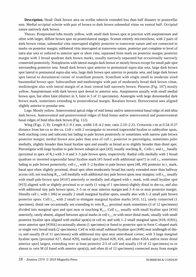
Description. Head: Dark brown area on ocellar tubercle extended less than half to half distance topostocellar seta. Medial occipital sclerite with pair of pale brown to dark brown submedial vittae on ventralhalf. Occipital suture narrowly orange brown to dark brown.
Thorax: Scutum entirely microtrichose, with 2 pairs of dark brown vittae or rows of spots; submedial vittainterrupted or narrowed posterior to transverse suture, well separated from marks on posterior margin;sublateral vitta with presutural part usually complete, extended to or almost to level of supra-alar seta,postsutural part elongate, reduced to spot posterior to transverse suture, or absent; posterior margin with 2well separated brown marks. Notopleuron usually with brown vitta on lateral margin, reduced to anterior andposterior spots in holotype. Small brown spot anterior to postsutural supra-alar seta, brown spot anterior topostalar seta, and small brown spot lateral to dorsolateral corner of scutellum present. Sometimes small spotlateral to postsutural supra-alar spot (aligned with postalar spot) also present. Scutellum with single medialspot, usually not extended beyond basal half, not extended to basal margin. Subscutellum and mediotergitewith pair of dark brown vittae, sometimes moderately broad but not reaching lateral margin of mediotergite.Pleuron entirely or mostly yellow. Anepisternum sometimes with small brown spot dorsal to anterior seta (1%,faint on left side in holotype). Anatergite with small dorsomedial red brown or brown spot. Basalare withbrown spot or entirely brown. Dorsocentral seta aligned slightly anterior to postalar seta.
Legs: Entirely yellow. Wing (Figs. 6–7): Length 6.05–7.20 mm, width 2.75–3.40 mm, ratio 2.12–2.39. Crossvein r-m at 0.70–
0.73 distance from bm-cu to dm-cu. Cell c with 2 rectangular to inverted triangular hyaline spots, bothreaching costa and subcosta; medial brown area sometimes fading posteriorly, anterior part as dark as todistinctly paler than area of cell r1 posterior to pterostigma, slightly narrower to broader than hyaline spots.
Pterostigma with pale brown subapical spot [#3], sometimes small (2 of 4 specimens), not reaching R1 in 1
specimen. Cells r1 and r2+3 basally (proximal to apex of R1) with 1–3 (usually 2–3) and 1–3 hyaline spots,
respectively. Radial cells medially with broad basal marginal hyaline mark [#5 fused with additional spot?] incell r1 and aligned broad hyaline mark [fused #8 and #9] in cell r2+3 forming tapering triangular to quadrate
mark broadly touching R4+5; cell r4+5 with hyaline spot [#15] near anterior end of dm-cu small to moderate
sized, sometimes almost touching vein M, aligned with or slightly proximal or distal to dm-cu, and also withadditional tiny hyaline to pale brown spots, 0–4 on or near anterior margin and 1–2 on or near posteriormargin. Distally cell r1 sometimes (2 of 4 specimens) with 1 small marginal hyaline spot [#6], usually with 1–
2 small pale brown posterior spots (absent in 1 wing of holotype). Cell r2+3 with 2 marginal hyaline marks,
brown area between them sometimes (1%) pale and diffuse, proximal mark [#10] extending to vein R4+5 or
divided into marginal and posterior spots, distal mark [#11] sometimes small and not reaching R4+5. Cell r4+5
usually with small hyaline spot [#16] anteriorly (absent on 1 wing of 1%), aligned between apical marks in cellr2+3 or with distal mark; with small posterior hyaline spot aligned with hyaline mark in cell m; and with 1 small
ovoid marginal or submarginal hyaline spot [#18], rarely with second more anterior spot [#18A] (left wing ofVilla Padre Monti %). Cell m usually (3 of 4 specimens) with small subbasal hyaline spot [#49] nearmidlength of dm-cu; with large irregular or posteriorly forked medial hyaline mark [fusion of at least #26A,#26, #27, #28 and sometimes #29], sometimes with isolated small distal marginal spot [#29], or with 3marginal spots, medial one [fusion of at least #26A, #26, #28] largest, somewhat mushroom-shaped, narrowposteriorly, broad anteriorly and extending to vein M. Cell br with subbasal pale brown spot [#12]. Cell bmwith single broad hyaline area [fused #19, #20]. Cell bcu occasionally with small pale brown spot in lobe. Cellbcu occasionally with small pale brown spot in lobe. Posteromedial part of wing with extremely largesubrectangular hyaline area; cell br with broad rectangular subapical hyaline area [at least #13, possibly fused

Zootaxa 2374 © 2010 Magnolia Press · 17REVISION OF THE FEMORALIS GROUP OF BLEPHARONEURA
with #44] extending from anterior to posterior margin, sometimes with small to minute pale brown spot wellproximal to broad subapical hyaline area; cell dm with large hyaline area [fusion of at least #51, #52, #21,#22, #23, #24, #50] aligned with hyaline area in cell cu1, anteriorly extending to at least level of r-m, distal
margin transverse or slightly oblique; cell cu1 with hyaline area covering medial half or more [broad fusion of
at least #31, #32, #33, #34, #36, #36A], very broad on posterior wing margin, sometimes with diffuse faintbrown submarginal spot subbasally but without other brown spots medially; subapical marginal hyaline spot[#37] small to moderate sized, usually not reaching vein Cu1. Cell dm without usual subapical hyaline spot
[#25] (unless fused with large medial hyaline area), occasionally with small more distal medial hyaline spot(1%) or small more distal posterior hyaline spot [#53] (1 wing of holotype) more or less aligned with subapicalmark in cell cu1.
Abdomen: Mostly yellow. Syntergite 1+2 with 4 evenly spaced dark brown spots, submedial andsublateral spots occasionally connected, and with spot on posterolateral corner; other tergites with 4 rows ofevenly spaced spots, anterolateral spot touching or almost touching lateral margin, and L-shaped band or pairof spots on lateral and posterior margins, separated medially, on tergite 5 and sometimes tergites 3–4connected to anterolateral spot, lateral margin always with at least small spot on tergite 3 but entirely brownon tergite 5; submedial and sublateral spots on tergite 4 sometimes connected; and some or all spots and bandson tergite 5 often connected or occasionally largely fused except medially.
Female terminalia: Oviscape length 1.10 mm. Aculeus (Fig. 165) 0.56 mm long, 2.04 times as long aswide, medial membrane without scales dorsally and ventrally; tip tapered basolaterally, elongate triangular(lobed part 0.59 times as long as wide), with large medial lobe with narrow but deep V-shaped notch (notch 1/3 as deep as length of lobe) and 3 pairs of small, somewhat step-like lobes separated by shallow gaps;sublateral and submedial lobes similar in size. Spermathecae subspherical, with straight to slightlyconvoluted, slender sclerotized neck and large cylindrical basal apodeme (similar to B. femoralis).
Male terminalia: Medial surstylus with prensisetae separated by several times width of medial prensiseta,medial prensiseta on long lobe, lateral prensiseta two-thirds as large to subequal to medial prensiseta (Fig.200).
Distribution. Northwestern Argentina (Tucumán). The type specimens for which elevation data wereprovided by the collectors were taken at 700 and 2000 m elevation.
Type data. Holotype & (IML USNMENT00213857), ARGENTINA: Tucumán: La Angostura [26°55'S65°41'W], 2000 m, 17 Feb 1953, P. Arnau. Paratypes: ARGENTINA: Tucumán: Burruyacu, Villa PadreMonti [26°29'S 64°58'W], 18 Jan - 7 Feb 1948, R. Golbach, 1% (USNM USNMENT00213859); c. 12 km Wof Tucumán, Horco Molle, 700 m, Malaise trap, 18–21 Mar 1974, C. R. Vardy, 1% (BMHNUSNMENT00213855); La Angostura, 2000 m, 17 Feb 1953, P. Arnau, 1% (IML USNMENT00213856).
Etymology. The name of this species is an adjective referring to the extremely large hyaline area in cellsdm and cu1.
Blepharoneura apaapa Norrbom & Condon, new speciesFigs. 2, 8, 107, 112, 132–133
Diagnosis. This species resembles B. amplihyalina, bipunctata, hyalinella, multipunctata and nigriapex inhaving a dark brown spot posterodorsally on the anepisternum, a single medial brown spot on the scutellum,and 3 hyaline marginal spots in cell m. It differs from B. bipunctata and multipunctata in having a singlebroad marginal hyaline mark basally in cell r1. It differs from the other three species in the size of the
posteromedial hyaline area in cell dm, which is smaller than in B. nigriapex and amplihyalina, but larger thanin B. hyalinella. It has a more elongate aculeus tip than in B. nigriapex and hyalinella, and the sublateral lobeis much closer to the submedial lobe than to the lateral lobe compared to B. amplihyalina, in which these lobesare more evenly spaced.
NORRBOM & CONDON18 · Zootaxa 2374 © 2010 Magnolia Press
Description. Head: Dark brown area on ocellar tubercle extended less than half distance to postocellarseta. Medial occipital sclerite with pair of brown to dark brown submedial vittae on ventral half. Occipitalsuture narrowly dark brown.
Thorax: Postpronotal lobe mostly yellow, with small dark brown spot at junction with anepisternum andoften with larger, diffuse brown spot on posterolateral margin. Scutum entirely microtrichose, with 2 pairs ofdark brown vittae; submedial vitta interrupted slightly posterior to transverse suture and not connected tomarks on posterior margin; sublateral vitta interrupted at transverse suture, posterior part complete to level ofintra-alar seta or reduced to anterior spot or short vitta, separated from mark on posterior margin; posteriormargin with 2 broad quadrate dark brown marks, usually narrowly separated but occasionally narrowlyconnected posteriorly. Notopleuron with lateral margin dark brown or mostly brown except for small pale spotsurrounding posterior seta. Small dark brown spot anterior to postsutural supra-alar seta, large dark brownspot lateral to postsutural supra-alar seta, large dark brown spot anterior to postalar seta, and large dark brownspot lateral to dorsolateral corner of scutellum present. Scutellum with single small to moderate sizedbasomedial brown spot. Subscutellum and mediotergite with pair of moderately broad dark brown vittae,mediotergite also with lateral margin of at least ventral half narrowly brown. Pleuron (Fig. 107) mostlyyellow. Anepisternum with dark brown spot dorsal to anterior seta. Anepimeron usually with small medialbrown spot, but often faint (distinct in 3, faint in 5 of 11 specimens). Anatergite with large dorsomedial darkbrown mark, sometimes extending to posterodorsal margin. Basalare brown. Dorsocentral seta alignedslightly anterior to postalar seta.
Legs: Mostly yellow. Anteroventral apical ridge of mid femur and/or anteroventral basal ridge of mid tibiadark brown. Anteroventral and posteroventral ridges of hind femur and/or anteroventral and posteroventralbasal ridges of hind tibia dark brown (Fig. 112).
Wing (Figs. 2, 8): Length 8.1–9.1 mm, width 3.8–4.2 mm, ratio 2.10–2.23. Crossvein r-m at 0.54–0.57distance from bm-cu to dm-cu. Cell c with 2 rectangular to inverted trapezoidal hyaline to subhyaline spots,both reaching costa and subcosta but fading to pale brown posteriorly or sometimes with narrow pale brownposterior margin; medial brown area paler than area of cell r1 posterior to pterostigma, sometimes paler
medially, slightly broader than basal hyaline spot and usually as broad as to slightly broader than distal spot.Pterostigma with large hyaline to pale brown subapical spot [#3], usually reaching R1. Cells r1 and r2+3 basally
(proximal to apex of R1) with 3–4 and 3–5 pale brown spots, respectively. Radial cells medially with 1 broad
quadrate or inverted trapezoidal basal hyaline mark [#5 fused with additional spot?] in cell r1, sometimes
fading to pale brown posteriorly; cell r2+3 with 1–2 hyaline to pale brown spots [#8, #9] posterior to r1 mark,
basal spot often slightly proximal, distal spot often moderately broad but rarely extended more than halfwayacross cell, not reaching R4+5, cell medially with additional tiny pale brown spots near margins; cell r4+5 usually
with small pale brown spot [#14?] anteriorly or medially and aligned with r1 mark, with small hyaline spot
[#15] aligned with or slightly proximal to or rarely (1 wing of 1 specimen) slightly distal to dm-cu, and alsowith additional tiny pale brown spots, 3–5 on or near anterior margin and 3–6 on or near posterior margin.Distally cell r1 with 1 [#6] or usually 2 small marginal hyaline spots, usually also with 1–2 small pale brown
posterior spots. Cell r2+3 with 2 small to elongate marginal hyaline marks [#10, 11], rarely connected (1
specimen), distal one occasionally not extending to vein R4+5, proximal mark sometimes (3 of 12 specimens)
divided into marginal spot and spot near or touching R4+5. Cell r4+5 usually with small hyaline spot [#16]
anteriorly, rarely absent, aligned between apical marks in cell r2+3 or with more distal mark; usually with small
posterior hyaline spot aligned with medial spot(s) in cell m; and with 1–2 small marginal spots [#18, #18A],more anterior spot [#18A] occasionally absent (1 specimen) or fused to posterior spot to form C-shaped markor single very broad mark (2 specimens). Cell m with small subbasal hyaline spot [#49] near midlength of dm-cu and usually (8 of 11 specimens) with additional tiny spot near anterobasal corner; with 3 large marginalhyaline spots [proximal #27, distal #29], medial spot [fused #28, #26, and often #26A and/or more distalanterior spot] largest, extending over at least posterior 2/3 of cell and usually (10 of 12 specimens) to oralmost to vein M (if fused with anterior spot(s)), and often (6 of 12 specimens) connected away from margin

Zootaxa 2374 © 2010 Magnolia Press · 19REVISION OF THE FEMORALIS GROUP OF BLEPHARONEURA
with proximal spot [#27]. Cell br with subbasal pale brown spot [#12]. Cell bm with circular subbasal andsubapical hyaline or pale brown spots [#19, #20]. Cell bcu with small hyaline or pale brown spot in lobe, oftenextending anteriorly into cell cu1 or that cell with similar nearby spot. Posteromedial part of wing with very
large hyaline area; cell br subapically with hyaline spot [#13] and small to minute, much more proximal palebrown spot; cell dm with 2–5 small subbasal spots, usually pale brown, and large ovoid medial posteriorhyaline mark [fusion of #22, #23, #24] (0.25–0.33 times as long as cell along vein Cu1) much shorter than
hyaline mark in cell cu1 and aligned with its distal half; cell cu1 with very large hyaline area covering medial
half or more [broad fusion of at least #32, #33, #34, #35?, #36, #36A], very broad on posterior wing margin,with subbasal submarginal brown spot and often 1 submarginal diffuse pale brown spot, but lacking palebrown spot anteriorly; subapical marginal hyaline spot [#37] small to moderate sized, not reaching vein Cu1.
Cell dm with subapical hyaline spot [#25] moderate sized, isolated (4 of 12 specimens) or often fused toposteromedial spot to form anterodistal extension, and with 1 anterior, usually 1 [#53] posterior (absent on 1wing of 3 specimens), and usually 1 medial (8 of 12 specimens) smaller more distal spots, the anterior andposterior spots aligned or nearly aligned with subapical mark in cell cu1.
Abdomen: Mostly yellow, with 4 rows of evenly spaced dark brown spots; tergites 3–5 also withanterolateral spot touching or almost touching lateral margin; also with pair of spots or more commonly withthem connected to form L-shaped band on posterior margin and posterolateral corner of all tergites, separatedmedially, on tergites 3–6 extending anteriorly on lateral margin, rarely connected to anterolateral spot, butlateral margin always partially yellow at least on tergites 3 and 4; submedial spots occasionally connected tosublateral spots on syntergite 1+2, and some or all spots on other tergites occasionally connected to posteriorbands or spots.
Female terminalia: Oviscape entirely dark brown; length 1.72–1.82 mm. Aculeus (Figs. 132–133) 1.10mm long, 2.00–2.12 times as long as wide, with acute scales dorsally and ventrally on membrane medially; tipangular basolaterally, elongate triangular (lobed part 0.58–0.72 times as long as wide), with small, truncate ornotched medial lobe and 3 pairs of step-like lobes separated by relatively deep gaps; sublateral lobe slightlylarger than submedial lobe; lateral gap 1.48–1.55 times as long as wide. Spermathecae subspherical, withslightly convoluted, slender sclerotized neck and small to large cylindrical basal apodeme.
Male terminalia: Medial surstylus with prensisetae separated by several times width of medial prensiseta,medial prensiseta on long lobe, lateral prensiseta subequal to medial prensiseta.
Distribution. Bolivia. The type locality is a remnant of Yungas forest. The specimens were collected at2000–2050 m elevation.
Type data. Holotype & (ANCB USNMENT00055947), BOLIVIA: La Paz: Apa Apa Reserve, upper trailin primary forest, 16°21'15S 67°30'20W, 2000 m, log site, cloudy AM or late afternoon, on undersides ofleaves of undetermined Cucurbitaceae (01–Bol-01) or supporting understory plant Solanaceae sp. (01–Bol-03), 1–3 Apr 2001, A. L. Norrbom. Paratypes: Same data as holotype, 1% (ANCB USNMENT00055926) 1%(CDFA USNMENT00055945) 1% (FSCA USNMENT00055946) 1% (TAUI USNMENT00055932) 4%3&(USNM USNMENT00055934, USNMENT00055937, USNMENT00055941–44).
Etymology. The name of this species is a noun in apposition that refers to the type locality.Biology. The digestive tract of a dissected female [USNMENT00055937] was green, indicating that
adults of this species rasp leaves or some other nonfloral part of its host plant.
Blepharoneura aspiculosa Norrbom & Condon, new speciesFigs. 9, 136
Diagnosis. This species, B. splendida, sinepuncta, and punctistigma differ from other Blepharoneura speciesin having the thoracic pleuron largely brown, the apical fourth or more of the hind femur dark brown, and thedistal part of the wing with oblique bands. Blepharoneura aspiculosa is very similar to B. punctistigma. It

NORRBOM & CONDON20 · Zootaxa 2374 © 2010 Magnolia Press
differs by aculeus shape (apex slightly more elongate, steps shallower) and lack of medial scales; the hyalinesubapical band not constricted along vein R4+5; and cell dm with a single very long and broad hyaline area in
the basal 3/4 (the narrowest part extends 3/4 of distance to anterior margin of cell; B. punctistigma usually has2 separate hyaline areas). These two species differ from B. splendida and sinepuncta by their markings in celldm (subapical spot [#25] isolated and distal to posterior spot aligned with r-m [#24]) and the pterostigma(always with a subapical spot, which is absent in B. sinepuncta and usually absent in B. splendida). Theaculeus tip differs from those of B. splendida and sinepuncta in having shallower gaps between the lobes.
Description. Head: Dark brown area on ocellar tubercle extended more than half distance to postocellarseta. Medial vertical seta in yellow area. Medial occipital sclerite with pair of pale brown submedial vittae onventral half. Occipital suture narrowly dark brown, on lateral side bordered by triangular brown areaextending to postocular setae, not including lateral vertical seta.
Thorax: Scutum entirely microtrichose, with 2 pairs of dark brown vittae or rows of spots; submedial vittanarrowed and almost interrupted posterior to transverse suture and separated from mark on posterior margin;sublateral vitta interrupted at transverse suture, narrowly connected to mark on posterior margin; posteriormargin with 1 broad brown mark narrowed medially. Notopleuron brown except for small pale areasurrounding posterior seta. Small brown spot anterior to postsutural supra-alar seta faint, diffuse. Brown vittaanterior to postalar seta present. Brown spot lateral to dorsolateral corner of scutellum present. Scutellum withsingle somewhat inverted U-shaped medial brown mark extended to basal margin. Subscutellum andmediotergite with pair of dark brown vittae, extended along lateral margin on ventral half of mediotergite.Pleuron mostly dark brown, yellow only on propleuron, anterior margin of anepisternum and broadlybordering phragma, all of katepimeron, greater ampulla, and dorsal margins of anepimeron and anatergite.Basalare brown. Dorsocentral seta aligned slightly anterior to postalar seta.
Legs: Mostly yellow. Mid femur with anteroventral and posteroventral brown marks on apical 2/5. Hindfemur with entire apical 1/4–1/2 dark brown.
Wing (Fig. 9): Length 6.40 mm, width 3.05 mm, ratio 2.10. Crossvein r-m at 0.57 distance from bm-cu todm-cu. Cell c with 2 rectangular hyaline spots, both reaching costa and subcosta; medial brown area as dark asto distinctly paler than area of cell r1 posterior to pterostigma, sometimes paler medially, narrower than both
hyaline spots. Pterostigma with large subapical hyaline to pale brown spot [#3] reaching R1. Cell r1 basally
(proximal to apex of R1) with 1 pale brown to hyaline spot posterior to apex of vein Sc; cell r2+3 basally with 1
spot. Radial cells medially with broad tapering basal marginal hyaline mark [#5 fused with additional spot?]in cell r1 and aligned spots in r2+3 [#8] and r4+5 [#14] forming acute triangular mark, extended slightly into r4+5;
cell r4+5 with hyaline spot [#15] near anterior end of dm-cu extending to vein M. Distally cell r1 without
hyaline spots [#6], and cell r2+3 without marginal hyaline marks. Cell r4+5 with hyaline band from posteroapical
margin, extending parallel to costa to or almost to vein R2+3, not extended into cell r1, not constricted along
vein R4+5, tapering anteriorly, broader than marginal brown area. Cells m and r4+5 with inverted V-shaped
hyaline mark [fusion of at least #26A, #26, #27, #29], extending anteriorly to vein R4+5, both arms reaching
margin in cell m. Cell br with subbasal hyaline spot [#12]. Cell bm with circular subbasal and subapicalhyaline spots [#19, #20]. Cell bcu with hyaline spot in lobe. Anal lobe hyaline except brown area from apicalpart of lobe of cell bcu and base of vein A1+Cu2 to apex of vein A2. Posteromedial part of wing with large,
broad hyaline areas; cell br with isolated subapical hyaline spot [#13]; cell dm with very long and broadhyaline mark [fused #51, #52, #21, #22, #23, #24] aligned with large hyaline mark in cell cu1, longer
posteriorly than anteriorly, proximal part extended to anterior margin, distal part extended 3/4 of distance tomargin; cell cu1 with very broad Y-shaped mark [#32, #33, #36] nearly fused proximally with mark across
vein A1+Cu2 [fused #34, #39] isolating 1 small anterior and 1 small submarginal brown spot; subapical
marginal hyaline spot [#37] large, reaching vein Cu1. Cell dm with anterior subapical hyaline spot [#25?]
isolated and distal to proximal mark, extending to vein M.

Zootaxa 2374 © 2010 Magnolia Press · 21REVISION OF THE FEMORALIS GROUP OF BLEPHARONEURA
FIGURES 6–15. Wing: 6–7, B. amplihyalina (Argentina: Horco Molle, USNMENT00213855; Villa Padre Monti,USNMENT00213859); 8, B. apaapa (Bolivia: Apa Apa, USNMENT00055943); 9, B. aspiculosa (Mexico: El Triunfo,USNMENT00213912); 10–11, B. bidigitata (Brazil: Nova Teutonia, USNMENT00213906; Interlagos,USNMENT00213839); 12, B. bipunctata (holotype); 13, B. biseriata (Mexico: Huipulco, USNMENT00213948); 14, B.sp. nr. biseriata (Mexico: E El Palmito, USNMENT00213947); 15, B. bivittata (holotype).

NORRBOM & CONDON22 · Zootaxa 2374 © 2010 Magnolia Press
FIGURES 16–25. Wing: 16, B. bivittata (Costa Rica: S Upala, USNMENT00213822); 17–20, B. brevivittata (CostaRica: Golfo Dulce, USNMENT00048492, USNMENT0000048516; Panama: Cano Saddle, USNMENT00213935; Peru:Tingo Maria, USNMENT00213533); 21–22, B. sp. nr. brevivittata (Peru: Pakitza, USNMENT00213931; Brazil:Santarém, USNMENT00213932); 23–24, B. chaconi (Costa Rica: San Gerardo, USNMENT00048385–86); 25, B.cornelli (holotype).

Zootaxa 2374 © 2010 Magnolia Press · 23REVISION OF THE FEMORALIS GROUP OF BLEPHARONEURA
FIGURES 26–35. Wing: 26, B. cyclantherae (Mexico: Rt. 95, USNMENT00052304); 27–29, B. femoralis (Mexico: ElTriunfo, USNMENT00213770; Brazil: Nova Teutonia, USNMENT00213754, USNMENT00052306); 30–31, B.fernandezi (Venezuela: El Limon, USNMENT00213886; Cumbre Roja, USNMENT00213885); 32–33, B. furcifer(Ecuador: Limoncocha, USNMENT00213928; Bolivia: San Carlos, USNMENT00213926); 34, B. sp. nr. furcifer(Panama: Barro Colorado USNMENT00213930); 35, B. hirsuta (Venezuela: Guatopo, USNMENT00213792).

NORRBOM & CONDON24 · Zootaxa 2374 © 2010 Magnolia Press
FIGURES 36–45. Wing: 36–37, B. hirsuta Guyana: Moraballi Creek, USNMENT00213793; Brazil: Rio de Janeiro,USNMENT00213794); 38–39, B. hyalinella (Bolivia: Apa Apa, USNMENT00055933, USNMENT00055940); 40, B. io(holotype); 41, B. isolata (holotype); 42, B. lutea (Costa Rica: SW Rincon, USNMENT00213791); 43–44, B.macwilliamsae (Zurqui, USNMENT00048601, USNMENT00048483); 45, B. marshalli (Argentina: Yuto,USNMENT00213860).

Zootaxa 2374 © 2010 Magnolia Press · 25REVISION OF THE FEMORALIS GROUP OF BLEPHARONEURA
FIGURES 46–55. Wing: 46–47, B. mexicana (Mexico: S Chapingo, USNMENT00213806; holotype); 48, B. mikenoltei(holotype); 49, B. multipunctata (holotype); 50, B. nigriapex (Bolivia: Apa Apa, USNMENT00055930); 51, B.nigrifemur (Bolivia: Apa Apa, USNMENT00055923); 52, B. osmundsonae (Mexico: El Yukon, USNMENT00213961);53–54, B. punctistigma (Mexico: Los Tuxtlas, USNMENT00213875; El Triunfo, USNMENT00213913); 55, B.quadristriata (Mexico: Los Tuxtlas, USNMENT00213849).

NORRBOM & CONDON26 · Zootaxa 2374 © 2010 Magnolia Press
FIGURES 56–65. Wing: 56, B. quadristriata (Mexico: Los Tuxtlas, USNMENT00054201); 57, B. quetzali (holotype);58–59, B. regina (lectotype; Mexico: Lagunas de Zempoala, USNMENT00213847); 60, B. rupta (lectotype); 61, B.ruptafascia (holotype); 62, B. septemdigitata (Peru: Pakitza, USNMENT00213939); 63, B. sinepuncta (Costa Rica: LaSuiza, USNMENT00213918); 64–65, B. splendida (Guatemala: Pacayal, USNMENT00213874; Venezuela: RanchoGrande, USNMENT00213864).

Zootaxa 2374 © 2010 Magnolia Press · 27REVISION OF THE FEMORALIS GROUP OF BLEPHARONEURA
FIGURES 66–75. Wing: 66, B. tau (holotype); 67–68, B. thetis (holotype; Brazi l : Nova Teutonia,USNMENT00213840); 69, B. sp. nr. thetis (Bolivia: Arroyo Tuhiri, USNMENT00056566); 70, B. unifasciata(holotype); 71–75, B. variabilis (Mexico: El Triunfo, USNMENT00213889, USNMENT00213893–96).

NORRBOM & CONDON28 · Zootaxa 2374 © 2010 Magnolia Press
FIGURES 76–83. Wing: 76, B. variabilis (Mexico: El Triunfo, USNMENT00213897); 77, B. wasbaueri (Ecuador:Huahua Sumaco, USNMENT00104212); 78, B. zumbadoi (Costa Rica: San Gerardo, USNMENT00048392); 79, B. diva(Costa Rica: Agua Buena, USNMENT00212445); 80, B. pulchella (holotype, Mexico: Teapa); 81, Blepharoneura sp.#50 (Ecuador: Tandapi, USNMENT00213798); 82, Hexaptilona hexacinioides (Taiwan: 45 km E Taichung); 83,Baryglossa trulla (Uganda: Ruwenzori Range, Fort Portal).
Abdomen: Syntergite 1+2 with pair of isolated submedial spots and irregular marks formed by sublateralspots connected to posterolateral band. Tergites 3–5 with isolated pairs of submedial, sublateral, andanterolateral brown spots and posterolateral brown bands.
Female terminalia: Oviscape entirely dark brown; length 1.05 mm. Aculeus (Fig. 136) 0.70 mm long, 2.23times as long as wide, without scales dorsally and ventrally on membrane medially; tip angular basolaterally,

Zootaxa 2374 © 2010 Magnolia Press · 29REVISION OF THE FEMORALIS GROUP OF BLEPHARONEURA
moderately long triangular (lobed part 0.55 times as long as wide), with small, convex medial lobe and 3 pairsof step-like lobes separated by shallow gaps; sublateral and submedial lobes similar in size; lateral gap 1.54times as long as wide. Spermathecae subspherical, with nearly straight, slender sclerotized neck and largecylindrical basal apodeme.
Distribution. Known only from the type locality in the Sierra Madre de Chiapas, Mexico.Type data. Holotype & (USNM USNMENT00213912), MEXICO: Chiapas: 49 km S of Jaltenango,
[Parque Natural] El Triunfo [15°40'N 92°48'W], 1300–2000 m, 13–15 May 1985, A. Freidberg. Etymology. The name of this species is an adjective formed from the Latin a- (without) and spica (point)
in reference to the lack of acute scales medially on the aculeus.
Blepharoneura bidigitata Norrbom & Condon, new speciesFigs. 10–11, 111, 152, 172
Blepharoneura sp. 9: Norrbom & Condon 1999: 146.
Diagnosis. This species differs from other species of Blepharoneura by the following combination ofcharacters: Anepisternum, anepimeron, and meron without brown markings; scutellum dorsally with pair ofbrown spots; cell r2+3 with 2 marginal hyaline marks; cell r4+5 with 2 ovoid marginal hyaline spots, neither
extending to vein R2+3, and with posterior hyaline spot(s) aligned with hyaline marks in cell m. The aculeus is
similar to that of B. hirsuta (which is dissimilar in external characters), with the digitate lateral lobe and acutesublateral lobe well separated from the medial and submedial lobes by a broad straight area.
Description. Head: Dark brown area on ocellar tubercle extended more than half distance to postocellarseta. Occipital suture narrowly dark brown.
Thorax: Scutum entirely microtrichose, with 2 pairs of dark brown vittae or rows of spots; submedial vittastrongly narrowed or interrupted slightly posterior to transverse suture and not connected to marks onposterior margin; sublateral vitta broadly interrupted at transverse suture, presutural part sometimes presentonly bordering postpronotal lobe, postsutural part sometimes absent, if present separated from marks onposterior margin; posterior margin with 2 quadrate dark brown marks. Notopleuron with small to large darkbrown spot near posterior seta on lateral margin. Small brown spot anterior to postsutural supra-alar setapresent, in one specimen fused to vitta anterior to postalar seta. Brown vitta (1 specimen), 2 spots (1 lateral tosupra-alar seta) (2 specimens), or 1 spot (1 specimen) usually present anterior to postalar seta, absent on 1specimen (posterior part not visible in 3 other specimens, but they lack anterior spot at least). Dark brown spotlateral to dorsolateral corner of scutellum present. Scutellum with pair of submedial brown spots.Subscutellum and mediotergite with pair of pale to dark brown vittae, sometimes reaching lateral margin ofmediotergite, or rarely (1%) both sclerites yellow except dorsolateral brown mark on subscutellum. Pleuronmostly or entirely yellow. Katatergite occasionally (2 of 9 specimens) brown on posterodorsal 2/5–3/5.Anatergite often (5 of 9 specimens) with small to large medial or anteromedial brown mark. Basalare withbrown spot, occasionally faint. Dorsocentral seta aligned with or usually slightly posterior to postalar seta.
Legs: Mostly or entirely yellow. Hind femur (Fig. 111) often (3 of 8 specimens distinctly, 2 weakly) withdark red brown spot on or very slightly separated from anterior or anteroventral apical margin [not just onanteroventral ridge, more extensive, usually extending almost to anterodorsal side].
Wing (Figs. 10–11): Length 7.05–7.71 mm, width 3.30–3.80 mm, ratio 1.91–2.14. Crossvein r-m at 0.52–
0.60 distance from bm-cu to dm-cu. Cell c with 2 broad rectangular to trapezoidal hyaline spots, both reachingcosta and subcosta; medial brown area distinctly paler than area of cell r1 posterior to pterostigma, sometimes
faint medially, almost as broad as to distinctly narrower than both hyaline spots. Pterostigma with subapicalhyaline spot [#3] usually reaching R1 (small in 1 specimen). Cells r1 and r2+3 basally (proximal to apex of R1)
with 1–2 and 0–2 hyaline spots, respectively. Radial cells medially with 1 moderately broad basal marginalhyaline mark [#5 fused with additional spot?] in cell r1, partially divided posteriorly by brown spot touching

NORRBOM & CONDON30 · Zootaxa 2374 © 2010 Magnolia Press
r2+3, or rarely (1 wing of 1 specimen) with brown area narrowly separating large anterior and small posterior
hyaline marks, the latter not reaching costa; cell r2+3 with 2 hyaline spots [#8, 9] aligned with lobes of r1 mark,
distal spot [#9] broader; cell r4+5 with small hyaline spot [#14] in anterior half aligned between lobes of r1
mark, with small hyaline spot [#15] aligned with or slightly distal to dm-cu, and often (6 of 8 specimens) withadditional tiny hyaline to pale brown spots, 0–4 near anterior margin, 0–3 near posterior margin. Distally cellr1 with 1–3 (usually 2) small marginal hyaline spots [#6 and additional spots], sometimes also with 1–2 small
hyaline or pale brown posterior spots. Cell r2+3 with 2 large marginal hyaline marks [#10, #11], both extending
to vein R4+5. Cell r4+5 with 1–2 small hyaline spots [1 of them #16?] anteriorly, aligned with or between apical
marks in cell r2+3; with 0–2 minute to moderate sized ovoid posterior hyaline spots aligned with hyaline marks
in cell m; and with 2 large ovoid marginal hyaline spots [#18 and #18A]. Cell m usually with small subbasalhyaline spot [#49] near midlength of dm-cu, occasionally with second more anterior spot; and with 3 largeovoid hyaline spots, 2 marginal [#27, #29] and 1 elongate anteromedial spot [fused #26, #26A], often (5 of 8specimens) reaching vein M. Cell br with subbasal hyaline spot [#12]. Cell bm with circular subbasal hyalinespot [#19] small or usually absent, subapical circular hyaline spot [#20] moderate sized to large.Posteromedial part of wing with multiple large hyaline spots or markings; cell br subapically with hyaline spot[#13], often with slightly more proximal yellowish or hyaline spot [#44], and occasionally with additionalmore proximal yellowish spot; cell dm with subbasal elongate hyaline mark across cell [fused #51, #52],posteriorly aligned with spot in cell cu1 [#31] and anteriorly aligned with spot in cell br [#44] when latter
present, medially often connected to more distal spots; with anterior spot [#21] aligned with subapical spot incell br [#13], often fused with subbasal mark or posterior hyaline spot of varying length [fusion of #23 and#22 and/or #24] aligned with all of or middle and either branch of Y-shaped mark in cell cu1; cell cu1 usually
with broad Y-shaped medial mark [fused #32, #33, #36, sometimes #36A], occasionally (2 specimens) withproximal branch [#32] or (1 specimen) distal branch [#33] narrowly separated from base [#36], aligned withposteromedial spot in cell dm to isolate brown spot anteriorly in cell cu1, proximal branch of mark [#32]
aligned slightly distal to line from proximal marginal mark across vein A1+Cu2 [#34, #39] to subapical spot in
cell br [#13], base of Y-shaped mark sometimes broad [fused #36, #36A] and partially divided by pale brownmarginal spot; subapical marginal hyaline mark [#37] moderate sized. Cell dm with subapical spot [#25]usually hyaline and moderate sized to small, occasionally minute, pale brown or absent; sometimes (3specimens) also with small to minute posterior subapical hyaline or pale brown spot [#53] aligned with orslightly proximal to subapical mark in cell cu1 touching or almost touching vein Cu1.
Abdomen: All tergites with 4 evenly spaced dark brown spots, sublateral pair sometimes absent onsyntergite 1+2, and 2 pairs of spots (more medial pair sometimes absent on syntergite 1+2) or pair ofposterolateral dark brown bands on posterior margin, on tergites 3–5 L-shaped but lateral margin yellowanteriorly, bands separated medially except sometimes on tergite 5; sublateral spot sometimes connected toposterior band on tergites 3 or 4, and both pairs sometimes connected to band on tergite 5.
Female terminalia: Oviscape entirely dark brown; length 1.10–1.20 mm. Aculeus (Figs. 152, 172) 0.89–0.96 mm long, 2.04–2.10 times as long as wide, with acute scales dorsally and ventrally on membranemedially; tip flared outward basolaterally, short (lobed part 0.20–0.29 times as long as wide), with small,convex or very weakly trilobed medial lobe and 3 pairs of lobes, lateral lobe large and digitiform, with minuteserrations apically, sublateral lobe large and acute, submedial lobe very small, separated from sublateral lobeby broad transverse area (width of submedial and medial lobes together 0.29–0.38 distance between apices ofsublateral lobes). Spermathecae subspherical, with slightly convoluted, slender sclerotized neck and smallcylindrical basal apodeme.
Male terminalia: Medial surstylus with prensisetae separated by several times width of medial prensiseta,medial prensiseta on long lobe, lateral prensiseta small, less than one-third as wide as medial prensiseta.
Distribution. Southern Brazil (São Paulo, Santa Catarina).

Zootaxa 2374 © 2010 Magnolia Press · 31REVISION OF THE FEMORALIS GROUP OF BLEPHARONEURA
FIGURES 84–89. Head, posterior: 84, B. femoralis (Mexico: Lagunas de Zempoala, USNMENT00213772); 85, B.variabilis (Mexico: El Triunfo, USNMENT00213890); 86, B. hirsuta (Brazil: Rio de Janeiro, USNMENT00213794);87, B. chaconi (Costa Rica: San Gerardo, INBio002539948); 88, B. sp. nr. thetis (Bolivia: Arroyo Tuhiri,USNMENT00056566); 89, B. quadristriata (Mexico: Los Tuxtlas, USNMENT00214950).

NORRBOM & CONDON32 · Zootaxa 2374 © 2010 Magnolia Press
FIGURES 90–93. Thorax, dorsal: 90–91, B. furcifer (Venezuela: Capirito, USNMENT00213927; Peru: Avispas,USNMENT00053665); 92, B. hirsuta (Brazil: Rio de Janeiro, USNMENT00054199); 93, B. septemdigitata (Peru: Erika,USNMENT00054208).

Zootaxa 2374 © 2010 Magnolia Press · 33REVISION OF THE FEMORALIS GROUP OF BLEPHARONEURA
FIGURES 94–97. Thorax, dorsal: 94, B. thetis (holotype); 95, B. bivittata (Costa Rica: S Upala, USNMENT00054202);96–97, B. brevivittata (Costa Rica: Rincon de Osa, USNMENT00054203; Costa Rica: Golfo Dulce,USNMENT00048518).

NORRBOM & CONDON34 · Zootaxa 2374 © 2010 Magnolia Press
FIGURES 98–101. Thorax, dorsal: 98–99, B. quadristriata (Mexico: Los Tuxtlas, USNMENT00054200,USNMENT00054201); 100–101, B. femoralis (Mexico: El Triunfo, USNMENT00054206; San Cristobal,USNMENT00054205).

Zootaxa 2374 © 2010 Magnolia Press · 35REVISION OF THE FEMORALIS GROUP OF BLEPHARONEURA
FIGURES 102–105. Thorax, dorsal: 102–104, B. marshalli (holotype; Argentina: Camino la Cornisa,USNMENT00054210, USNMENT00054211); 105, B. tau (holotype).

NORRBOM & CONDON36 · Zootaxa 2374 © 2010 Magnolia Press
FIGURES 106–108. Thorax, lateral: 106, B. femoralis (Mexico: El Triunfo, USNMENT00213770); 107, B. apaapa(Bolivia: Apa Apa, USNMENT00055944); 108, B. chaconi (Costa Rica: San Gerardo, USNMENT00048386).
Type data. Holotype & (MZUSP USNMENT00213907), BRAZIL: Santa Catarina: Nova Teutonia, Oct1965, F. Plaumann. Paratypes: BRAZIL: Santa Catarina: Nova Teutonia, 27°11'S 52°23'W, 300–500 m, 30Dec 1960 , F. P laumann , 1% (US NM US NME NT0 0213 910) ; same , May 1963 , 1% (CNCUSNMENT00213909); same, Dec 1964, 1& (CNC USNMENT00213908); same, May 1977, 1% (FMNHUSNMENT00213834); Nova Teutonia, Oct 1965, F. Plaumann, 1% (MZUSP USNMENT00213906); same,Jun 1967, 1% (MZUSP USNMENT00213905). São Paulo: Interlagos, Capital, 3 Dec 1980, M. Carrera, 1&(USNM USNMENT00213839).
Etymology. The name of this species is a Latin adjective referring to the pair of digitate lateral lobes onthe aculeus.
Blepharoneura bipunctata Norrbom & Condon, new speciesFigs. 12, 131
Diagnosis. This species resembles B. amplihyalina, apaapa, hyalinella, multipunctata and nigriapex inhaving a dark brown spot posterodorsally on the anepisternum, a single medial brown spot on the scutellum,and 3 hyaline marginal spots in cell m. It differs from all of them except B. multipunctata in having 2 narrow

Zootaxa 2374 © 2010 Magnolia Press · 37REVISION OF THE FEMORALIS GROUP OF BLEPHARONEURA
marginal hyaline marks basally in cell r1 rather than a single broad one, and from B. multipunctata in having a
small anepimeral brown spot and the aculeus tip with a convex medial apical lobe.Description. Head: Dark brown area on ocellar tubercle extended more than half distance to postocellar
seta or reaching it. Medial occipital sclerite with pair of dark brown submedial vittae on ventral half. Occipitalsuture narrowly dark brown.
Thorax: Mostly yellow. Postpronotal lobe with small brown spot at junction with anepisternum andsometimes with small diffuse brown spot on posterolateral margin. Scutum entirely microtrichose, with 2pairs of dark brown vittae or rows of spots; submedial vitta interrupted slightly posterior to transverse sutureand not connected to marks on posterior margin; sublateral vitta interrupted at transverse suture, postsuturalpart reduced to well separated anterior and posterior spots, latter separated from mark on posterior margin;posterior margin with 2 broad quadrate dark brown marks well separated. Notopleuron with lateral margindark brown. Small brown spot anterior to postsutural supra-alar seta, large dark brown spot lateral topostsutural seta, small dark brown spot anterior to postalar seta, and large dark brown spot lateral todorsolateral corner of scutellum present. Scutellum with single, small to moderately large, dark brown medialspot. Subscutellum and mediotergite with pair of dark brown vittae. Anepisternum with small dark brown spotdorsal to anterior seta. Anepimeron with small medial brown spot (faint on 1 side on 1 specimen). Katatergitepale brown dorsally and ventrally. Anatergite with large dark brown area medially and ventrally. Basalarewith brown spot or entirely brown. Dorsocentral seta aligned slightly anterior to postalar seta.
Legs: Mostly yellow. Mid and hind femora with red brown spot on margins of anteroventral andposteroventral apical ridges, extending more faintly slightly more basally very narrowly along ventralmargins. Hind tibia dark brown narrowly bordering and on anteroventral and posteroventral basal ridges.
Wing (Fig. 12): Length 7.55–7.72 mm, width 3.20–3.27 mm, ratio 2.36. Crossvein r-m at 0.55–0.60distance from bm-cu to dm-cu. Cell c with 2 inverted trapezoidal hyaline spots, both reaching costa andsubcosta but fading to pale brown posteriorly; medial brown area almost as dark as area of cell r1 posterior to
pterostigma, broader than basal hyaline spot but approximately as broad as distal spot. Pterostigma with largesubapical hyaline spot [#3] reaching R1. Cells r1 and r2+3 basally (proximal to apex of R1) with 3 and 2–3 pale
brown to hyaline spots, respectively. Radial cells medially with 2 narrow basal marginal hyaline marks in cellr1, both reaching R2+3, sometimes with tiny posterior hyaline spot between them, proximal mark [#5] ovoid;
cell r2+3 with 1 very broad hyaline mark posterior to r1 marks (1 wing of 1 specimen) or usually with 1 small
hyaline spot [#8] aligned with or slightly distal to basal mark in r1 and 1 broad spot [#9] aligned with second
mark in r1; cell r4+5 with small hyaline spot [#14] in anterior half or medially and aligned with basal r1 mark,
with small hyaline spot [#15] aligned with or slightly proximal or distal to dm-cu, and also with additionaltiny hyaline to pale brown spots, 1–3 near anterior margin and 0–4 on or near posterior margin. Distally cell r1
with 1 [#6] or usually 2 small marginal hyaline spots, on 1 wing of 1 specimen also with 2 minute posteriorspots. Cell r2+3 with 2 marginal hyaline marks [#10, 11], proximal mark extending to vein R4+5. Cell r4+5 with
small hyaline spot [#16] anteriorly, aligned between apical marks in cell r2+3 or with distal mark; sometimes
with small posterior hyaline spot aligned with medial spot in cell m; and with 1–2 small ovoid marginal orsubmarginal hyaline spots [#18, #18A], more anterior spot sometimes absent or connected away from marginwith posterior spot forming somewhat C-shaped mark. Cell m with small subbasal hyaline spot [#49] nearmidlength of dm-cu and tiny spot near anterobasal corner; with 3 ovoid marginal hyaline spots [#27, #28,#29], medial spot [fusion of at least #28, #26, #26A] largest, irregular, extending to or almost to vein M. Cellbr with subbasal pale brown spot [#12]. Cell bm with circular subbasal and subapical hyaline spots [#19, #20].Cell bcu with or without small hyaline spot in lobe. Posteromedial part of wing with very large hyaline area;cell br subapically with hyaline spot [#13] and small to minute, much more proximal, pale brown spot; celldm with 1 small anterior ovoid or narrow elongate medial hyaline spot very close to base and with large ovoidmedial posterior hyaline mark [fusion of #22?, #23, #24] (ca. 0.25 times as long as cell along vein Cu1) much
shorter than hyaline mark in cell cu1 and aligned with its distal part; cell cu1 with very large hyaline area
covering medial half or more [broad fusion of at least #31?, #32, #33, #34, #35?, #36, #36A], very broad on

NORRBOM & CONDON38 · Zootaxa 2374 © 2010 Magnolia Press
posterior wing margin, lacking anterior brown spots and including 1 submarginal pale brown spot subbasally;subapical marginal hyaline spot [#37] relatively small, not reaching vein Cu1. Cell dm with subapical hyaline
spot [#25] moderate sized, and with smaller more distal anterior spot and posterior spot [#53] aligned ornearly aligned with subapical mark in cell cu1.
Abdomen: Mostly yellow, all tergites with 4 rows of evenly spaced dark brown spots, on tergites 3–5 morelateral spot large and connected to or narrowly separated from lateral and posterior markings; tergites 3–5 alsowith anterolateral spot touching or almost touching lateral margin; all tergites also with pair of spots or bandon posterior margin and posterolateral corner, separated medially, on tergites 3–5 extending anteriorly onlateral margin and connected to anterolateral spot, but on tergites 3 and 4 margin with small yellow spot.
Female terminalia: Oviscape entirely dark brown; length 1.09 mm. Aculeus (Fig. 131) 0.87 mm long, 1.98times as long as wide, with acute scales dorsally and ventrally on membrane medially; tip angularbasolaterally, short triangular (lobed part 0.34 times as long as wide), with small, convex medial lobe and 3pairs of step-like lobes separated by deep gaps; sublateral lobe larger than submedial lobe; lateral gap 0.90times as long as wide.
Spermathecae subspherical, with straight to slightly convoluted, slender sclerotized neck and largecylindrical basal apodeme (similar to B. femoralis).
FIGURES 109–112. Left hind leg, anterior: 109, B. variabilis (Mexico: El Triunfo, USNMENT00213891); 110, B.nigrifemur (Bolivia: Apa Apa, USNMENT00055922); 111, B. bidigitata (Brazil: Nova Teutonia, USNMENT00213834);112, B. apaapa (Bolivia: Apa Apa, USNMENT00055934).

Zootaxa 2374 © 2010 Magnolia Press · 39REVISION OF THE FEMORALIS GROUP OF BLEPHARONEURA
Distribution. Ecuador. The type specimens were collected at 2000 m elevation.Type data. Holotype & (CMP USNMENT00213904), ECUADOR: Cotopaxi: San Francisco de las
Pampas, Otonga, 2000 m, 20 Oct 1996, G. Onore. Paratype: Same data as holotype, 1& (USNMUSNMENT00213903).
Etymology. The name of this species is an adjective referring to the two basal marginal hyaline marks incell r1.
FIGURES 113–118. Abdomen, dorsal: 113, B. femoralis (USNMENT00054204); 114–115, B. marshalli (Argentina:Camino la Cornisa, USNMENT00054210–11); 116–117, B. chaconi (InBio002151664; INBio002539948); 118, B. tau
(holotype).

NORRBOM & CONDON40 · Zootaxa 2374 © 2010 Magnolia Press
FIGURES 119–125. Abdomen, dorsal: 119–120, B. septemdigitata (Peru: Pakitza, USNMENT00213939–40); 121, B.cornelli (holotype); 122, B. furcifer (USNMENT00053665); 123, B. hirsuta (Brazil: Manaus, USNMENT00054198);124, B. brevivittata (Costa Rica: Rincon de Osa, USNMENT00054203); 125, B. quadristriata (Mexico: Los Tuxtlas,USNMENT00054201).

Zootaxa 2374 © 2010 Magnolia Press · 41REVISION OF THE FEMORALIS GROUP OF BLEPHARONEURA
FIGURES 126–135. Aculeus: 126, B. bivittata (holotype, USNMENT00213023); 127, B. brevivittata (Costa Rica:Golfo Dulce, USNMENT00048518); 128, B. cornelli (holotype, INBio001228576); 129, B. lutea (Costa Rica: Pitilla,INBio000424938); 130, B. macwilliamsae (Costa Rica: La Cangreja, USNMENT00048509); 131, B. bipunctata(Ecuador: Otonga USNMENT00213903); 132–133, B. apaapa (Bolivia: Apa Apa, USNMENT00055947,USNMENT00213943); 134, B. hyalinella (Bolivia: Apa Apa, USNMENT00055931); 135, B. nigriapex (Bolivia: ApaApa, USNMENT00055930).

NORRBOM & CONDON42 · Zootaxa 2374 © 2010 Magnolia Press
FIGURES 136–145. Aculeus: 136, B. aspiculosa (Mexico: El Triunfo, USNMENT00213912); 137, B. cyclantherae(Mexico: Rt. 95, USNMENT00052304); 138–142, B. femoralis (Guatemala: Coban, USNMENT00213771; Brazil:Nova Teutonia, USNMENT00213760, USNMENT00052320, USNMENT00213761; Peru: Chanchamayo,USNMENT00213960); 143, B. nigrifemur (Bolivia: Apa Apa, USNMENT00055925); 144, B. sinepuncta (Costa Rica:Zurquí, USNMENT00050130); 145, B. zumbadoi (Costa Rica: San Gerardo, INBio002578264).

Zootaxa 2374 © 2010 Magnolia Press · 43REVISION OF THE FEMORALIS GROUP OF BLEPHARONEURA
FIGURES 146–155. Aculeus: 146–148, B. mexicana (Mexico: S Chapingo, USNMENT00213805–06; holotype,USNMENT00213858); 149, B. chaconi (Costa Rica: San Gerardo, USNMENT00048388); 150–151, B. quetzali(holotype); 152, B. bidigitata (Brazil: Interlagos, USNMENT00213839); 153–154, B. hirsuta (Guyana: Moraballi Creek,USNMENT00213793; Brazil: Rio de Janeiro, USNMENT00054199); 155, B. wasbaueri (holotype).

NORRBOM & CONDON44 · Zootaxa 2374 © 2010 Magnolia Press
FIGURES 156–164. Aculeus: 156, B. fernandezi (Brazil: Santarem, USNMENT00213888); 157–158, B. furcifer(Ecuador: Limoncocha, USNMENT00213928; lectotype); 159, B. lutea (Costa Rica: Golfito, USNMENT00213824);160, B. quadristriata (Mexico: Los Tuxtlas, USNMENT00054200); 161, B. rupta (Mexico: Teapa,USNMENT00213796); 162, B. ruptafascia (holotype); 163, B. septemdigitata (Peru: Pakitza, USNMENT00213937);164, B. unifasciata (holotype). ln = length, wd = width (of tip and gap).

Zootaxa 2374 © 2010 Magnolia Press · 45REVISION OF THE FEMORALIS GROUP OF BLEPHARONEURA
FIGURES 165–172. Aculeus: 165, B. amplihyalina (holotype); 166, B. marshalli (holotype); 167, B. multipunctata(holotype); 168, B. mikenoltei (holotype); 169, B. mexicana (Mexico: Mexico City, USNMENT00213809); 170, B.regina (Mexico: Lagunas de Zempoala, USNMENT00213846); 171, B. variabilis (Mexico: El Triunfo,USNMENT00213899); 172, B. bidigitata (Brazil: Nova Teutonia, USNMENT00213907).

NORRBOM & CONDON46 · Zootaxa 2374 © 2010 Magnolia Press
FIGURES 173–181. Aculeus: 173–174, B. biseriata (holotype; Mexico: Huipulco USNMENT00213950; medialmembrane not shown in 158); 175, B. sp. nr. biseriata (Mexico: E El Palmito, USNMENT00213947); 176–180, B.femoralis (lectotype; Guatemala: Coban, USNMENT00213771; Brazil: Nova Teutonia, USNM00213761; Mexico: RioMolinos, USNMENT00213778; Mexico: Mexico City, USNMENT00213773); 181, B. punctistigma (Mexico: LosTuxtlas, USNMENT00213914).

Zootaxa 2374 © 2010 Magnolia Press · 47REVISION OF THE FEMORALIS GROUP OF BLEPHARONEURA
FIGURES 182–187. Aculeus: 182–183, B. osmundsonae (Mexico: Valle de Bravo, USNMENT00213962; Mexico: ElYukon, USNMENT00213953); 184–187, B. splendida (Mexico: San Blas, USNMENT00213873; Salto de Eyipantla,USNMENT00213877; Guatemala: Pochuta, USNMENT00213874; Venezuela: Rancho Grande).
Blepharoneura biseriata WulpFigs. 13, 173–174
Blepharoneura biseriata Wulp 1899: 411 [in key], 413 [description]; Hendel 1914: 21 [in key, in catalog]; Aczél 1950:195 [in catalog]; Foote 1965: 241 [type data]; Foote 1967: 18 [in catalog]; Norrbom et al. 1999: 105 [in catalog].
Blepharoneura btseriata: Aczél 1950: 195 [misspelling].
Diagnosis. This species belongs to the femoralis complex (see diagnosis of B. femoralis), species of which aredifficult to distinguish except by aculeus shape. The aculeus of B. biseriata is less elongate than that of B.nigrifemur, and it differs from those of the other species of the complex in having the lateral lobe very weak orabsent.
Description. Head: Dark brown area on ocellar tubercle extended to postocellar seta. Medial vertical setain yellow area. Medial occipital sclerite with pair of brown submedial vittae on ventral half. Occipital suture

NORRBOM & CONDON48 · Zootaxa 2374 © 2010 Magnolia Press
narrowly dark brown, on lateral side bordered by slightly paler triangular area, extended dorsolaterally beyondpostocular setae almost to eye margin except for small circular yellow area around lateral vertical seta.
FIGURES 188–195. Spermathecae: 188–190, B. femoralis (Mexico: Lagunas de Zempoala, USNMENT00213772;Oaxaca, USNMENT00213778; Brazil: Nova Teutonia, USNMENT00052306); 191, B. regina (2) (Mexico: Lagunas deZempoala, USNMENT00213846); 192, B. quadristriata (3) (Mexico: Los Tuxtlas, USNMENT00213949); 193, B.furcifer (3) (lectotype). Egg: 194, B. femoralis (Mexico: Mexico City, USNMENT00213773); 195, B. lutea (Costa Rica:Golfito, USNMENT00213824).
Thorax: Scutum nonmicrotrichose except posterior to acrostichal seta and laterally, with 2 pairs of darkbrown vittae or rows of spots; submedial vitta interrupted or narrowed slightly posterior to transverse sutureand not connected to mark on posterior margin; sublateral vitta broadly interrupted at transverse suture andseparated from mark on posterior margin; posterior margin with 1 broad brown mark narrowed medially.Notopleuron with dark brown vitta on lateral margin. Small brown spot anterior to postsutural supra-alar setapresent, narrowly connected along transverse suture to brown vitta anterior to postalar seta. Large brown spot

Zootaxa 2374 © 2010 Magnolia Press · 49REVISION OF THE FEMORALIS GROUP OF BLEPHARONEURA
lateral to dorsolateral corner of scutellum present. Scutellum with single inverted U-shaped medial brownmark extended to basal margin. Subscutellum and mediotergite with pair of dark brown vittae, extended tolateral margin on ventral half of mediotergite. Pleuron mostly dark brown, yellow only on propleuron, most ofanepisternum (except large dorsomedial dark brown spot extending ventrally to level of anterior seta, andnarrowly in posteroventral corner), extreme posterior corner of katepisternum, all of katepimeron, greaterampulla, narrow dorsal margin and sometimes narrow posterior and ventral margins of anepimeron, andnarrow dorsal margins of katatergite and anatergite expanding to larger spot posterodorsally on katatergite andanteriorly on anatergite. Basalare brown. Dorsocentral seta aligned slightly anterior to postalar seta.
FIGURES 196–199. Epandrium and surstyli, lateral and posterior: 196–197, B. femoralis (Brazil: Nova Teutonia); 198–199, B. thetis (holotype).

NORRBOM & CONDON50 · Zootaxa 2374 © 2010 Magnolia Press
FIGURES 200–203. Epandrium and surstyli, posterior: 200, B. amplihyalina (Argentina: Horco Molle,USNM00213855); 201, B. mexicana (Mexico, USNM00213821); 202, B. quadristriata (Mexico, Los Tuxtlas,USNM00213850); 203, B. rupta (Belize, Stann Creek, USNM00213797).
Legs: Mostly yellow. Hind femur with entire apical 1/5 dark brown. Wing (Fig. 13): Length 5.44–5.70 mm, width 2.67–2.70 mm, ratio 2.04–2.11. Crossvein r-m at 0.53–0.54
distance from bm-cu to dm-cu. Cell c with 2 rectangular hyaline spots, both reaching costa and subcosta;medial brown area as dark as to slightly paler than area of cell r1 posterior to pterostigma, narrower than both
hyaline spots. Pterostigma with large subapical hyaline spot [#3] reaching R1. Cells r1 and r2+3 basally
(proximal to apex of R1) with 1–2 and 1 pale brown or hyaline spots, respectively. Radial cells medially with 1
broad quadrate basal hyaline mark [#5 fused with additional spot?] in cell r1, usually partially divided
posteriorly by brown spot; cell r2+3 with 2 hyaline spots [#8, #9] aligned with r1 mark, both extending across

Zootaxa 2374 © 2010 Magnolia Press · 51REVISION OF THE FEMORALIS GROUP OF BLEPHARONEURA
cell, distal spot broader; cell r4+5 in anterior half with small hyaline spot [#14] aligned with middle of r1 mark,
approximately half as wide as cell, and with medial hyaline spot [#15] near anterior end of dm-cu small;sometimes with small to minute anterior hyaline spot [#48] near midlength not touching vein R4+5. Distally
cell r1 with 1 marginal subapical hyaline spot [#6]. Cell r2+3 with 2 marginal ovoid hyaline spots [#10A, #11]
and subapical spot posterior to proximal spot [#10B]. Cell r4+5 with small hyaline spot [#16] anteriorly, aligned
between apical marks in cell r2+3, or (1 wing of 1 specimen) with 2 spots aligned with apical marks in cell r2+3;
with 1 posterior ovoid hyaline spot aligned with distal mark in cell m; and with 2 ovoid marginal orsubmarginal hyaline spots [#18, #18A]. Cell m without subbasal hyaline spot [#49] near midlength of dm-cu;with 2 large marginal hyaline spots [#27, #29] and 1 large anteromedial spot [fused #26, #26A]. Cell br withsubbasal hyaline spot [#12]. Cell bm without subbasal hyaline spot [#19], with large circular subapical hyalinespot [#20]. Cell bcu usually without hyaline spot in lobe (present on 1 wing of 1 specimen). Posteromedialpart of wing with large hyaline areas; cell br with subapical hyaline spot [#13]; cell dm with broad and longhyaline area [fusion of at least #51, #52, #21, #22, #23, #24, sometimes #50], sometimes narrowly or partiallydivided by pale brown into 2–3 spots, aligned with large hyaline mark in cell cu1, anteriorly extending to or
almost to level of r-m, but distal margin oblique or tapered, extending farther posteriorly than anteriorly; cellcu1 medially with 5 barely separated spots, or with broad medial Y-shaped mark [fused #32, #33, #36] and
more proximal anterior [#31] and posterior [fused #34, #39] spots, or with all of these connected to form largeanteriorly trilobed and posteriorly bilobed mark isolating 2 small anterior and 1 small submarginal brownspot; subapical marginal hyaline spot [#37] reaching vein Cu1. Cell dm with anteromedial subapical hyaline
spot [#25] moderately large, isolated. Abdomen: Syntergite 1+2 with pair of irregular posterolateral brown marks, separated medially, formed
from fused submedial and sublateral spots and posterolateral bands. Tergites 3–5 with pair of irregularposterolateral brown marks, separated medially, formed from fused submedial and sublateral spots andposterolateral bands; also with isolated anterolateral spots.
Female terminalia: Oviscape entirely dark brown; length 0.90 mm. Aculeus (Fig. 173–174) 0.43 mm long,2.00–2.08 times as long as wide, with acute scales dorsally and ventrally on membrane medially; tipsomewhat blunt, slightly angular basolaterally, without distinct lateral lobe, short to slightly elongatetriangular (lobed part 0.43–0.54 times as long as wide), with small, weakly trilobed medial lobe and 2 pairs ofsmall, somewhat step-like lobes separated by shallow gaps, lobes and gaps slightly larger in Huipulco female;sublateral and submedial lobes similar in size. Spermathecae subspherical, with straight slender sclerotizedneck and large cylindrical basal apodeme (similar to B. femoralis).
Male terminalia: Medial surstylus with prensisetae subequal, separated by several times width of medialprensiseta, medial prensiseta on moderately long lobe, lateral prensiseta on short lobe.
Distribution. Mexico (Distrito Federal, Guerrero). The holotype was collected at over 2100 m elevation.Type data. Holotype & (BMNH), MEXICO: Guerrero: Sierra de las Aguas Escondidas, 7000 ft. [2134
m], Jul, H. H. Smith [examined].Other specimens examined. MEXICO: [Distrito Federal:] Huipulco, [19°17'N 99°09'W], 21 Aug 1922,
E. G. Smyth, 2%1& (USNM USNMENT00213948–50).Remarks. A single headless female from Sinaloa, Mexico (El Palmito, 2.5 mi E of, 21 Aug 1964, E. I.
Schlinger, UCR USNMENT00213947) may be B. biseriata or a very similar species. It has only minordifferences with B. biseriata in wing pattern (Fig. 14) (cell c with medial brown area almost as broad as bothhyaline spots; cell cell r2+3 basally without hyaline spot; distally cell r2+3 without hyaline spot posterior to
proximal marginal spot [posterior part of #10 absent]; cell bm with circular subbasal hyaline spot [#19]) and issimilar in other external characters, but the aculeus (Fig. 175) lacks scales on the medial membrane ventrally,and the dorsal scales are predominantly polygonal, becoming acute only proximally. Also, the base of theaculeus tip is rounded laterally, lacking the normal lateral lobe, whereas in B. biseriata there is a slight angle.Further specimens and data are needed to clarify the status of this female.

NORRBOM & CONDON52 · Zootaxa 2374 © 2010 Magnolia Press
Blepharoneura bivittata Norrbom & Condon, new speciesFigs. 15–16, 95, 126
Diagnosis. This species is among the Blepharoneura species with the apical part of the wing obliquelybanded, without marginal hyaline marks in cell r2+3. It differs from the other obliquely banded species by the
following combination of characters: vertex with brown spot or band surrounding medial vertical seta; scutumwith pair of submedial brown vittae, uninterrupted at transverse suture; anepisternum without brownmarkings; pterostigma without subapical hyaline spot; cell m with 2 hyaline marks extended into cell r4+5 and
connected to form inverted V-shaped mark; abdominal tergites with pair of solid dark brown vittae withoutyellow spots and with even medial margins, not extending to lateral margins of tergites, which are yellow. Theaculeus of B. bivittata resembles those of B. brevivittata and fernandezi in having a very broad, deep medialconcavity, but the part of the tip exclusive of the lateral lobes is narrower and more elongate.
Description. Head: Dark brown area (orange brown in 1%) on ocellar tubercle extended half distance toor beyond postocellar seta. Medial vertical seta in ovoid orange brown to dark brown spot mostlyposteromesal to seta or (Upala %) in broad band extended to opposite seta and connected to area on ocellartubercle. Occipital suture narrowly dark orange to dark orange brown. Sometimes (2%) with diffuse palebrown area including and ventral to lateral vertical seta, in 1 specimen extending along eye margin.
Thorax (Fig. 95): Scutum nonmicrotrichose except posterior to dorsocentral seta and laterally, with pair ofbroad dark brown submedial vittae, in holotype extended to midway between levels of postsutural supra-alarseta and intra-alar seta, presutural part dark brown, faded to orange brown posterior to transverse suture, inmales extended to posterior brown mark. Posterior margin yellow (holotype) or usually with bimodal orelongate triangular dark brown mark, in Turrialba male with sublateral vittae extending anteriorly from it.Scutellum entirely yellow (holotype) or usually with brown band or broad inverted triangular mark at level ofbasal seta and extended to it (weakly in 1%), sometimes (1%) almost divided medially. Subscutellum entirelyyellow; mediotergite entirely yellow or (Turrialba %) brown except medial yellow vitta. Pleuron entirelyyellow. Basalare entirely yellow. Dorsocentral seta aligned with or slightly posterior to postalar seta.
Legs: Mostly to entirely yellow. Hind tibia mostly pale to moderate brown in males. Wing (Figs. 15–16): Length 5.10–5.40 mm, width 2.57–2.82 mm, ratio 1.91–2.00. Crossvein r-m at 0.53–
0.59 distance from bm-cu to dm-cu. Cell c sometimes (holotype, Turrialba %) with 2 rectangular hyalinespots, separated by narrower medial brown area; in Upala males with 1 goggles-shaped hyaline area, roundedbasally and distally and narrower medially; medial brown area(s) distinctly paler than area of cell r1 posterior
to pterostigma. Pterostigma without subapical hyaline spot. Cells r1 and r2+3 basally (proximal to apex of R1)
without hyaline spots except sometimes (Turrialba %) r1 with tiny spot posterior to apex of vein Sc. Radial
cells medially with tapering basal marginal hyaline mark [#5] in cell r1 and aligned spot in r2+3 [#8] forming
acute triangular or inverted trapezoidal mark, usually reaching R4+5 or (holotype) extending less than halfway
across r2+3; no aligned spot present in r4+5, and medial hyaline spot [#15] near anterior end of dm-cu small and
faint (holotype) or absent. Distally cell r1 without hyaline spots [#6]. Cell r2+3 without marginal hyaline marks.
Cell r4+5 with hyaline band from posteroapical margin, extending more or less parallel to costa into cell r2+3 to
or almost to vein R2+3, slightly or not tapering anteriorly, slightly to much broader than marginal brown area.
Cells m and r4+5 with inverted V-shaped mark [fusion of #26A, #26, #27, #29 plus additional spots], extending
anteriorly more than halfway across r4+5 but not reaching vein R4+5, both arms reaching posterior margin in cell
m. Cell br with subbasal hyaline spot [#12], minute in 1 Upala male. Cell bm with circular subbasal andsubapical hyaline spots [#19, #20] or 1 large ovoid or irregular medial hyaline spot [fused #19 and #20].Posteromedial part of wing with 2 rows of more or less aligned hyaline spots or bands, these marks usuallypartially fused at least posteriorly in cell dm; cell br with subapical hyaline spot [#13] only, aligned with markin cell dm [#21]; cell dm with separate narrow oblique proximal mark [#21, #22? or #52?] and distalposteromedial spot [#24 or fused #23 and #24] (1 wing of holotype), with anteriorly narrow and posteriorlybroad mark [fusion of #21, #22, #23, #24], or with broad, anteriorly bifurcate mark [fusion of #51, #52?, #21,

Zootaxa 2374 © 2010 Magnolia Press · 53REVISION OF THE FEMORALIS GROUP OF BLEPHARONEURA
#22, #23, #24]; cell cu1 with 2 hyaline bands, often connected medially to form H-shaped mark, proximal
band [fusion of #34, #39, #32, sometimes #31?] crossing vein A1+Cu2 and aligned with proximal band or
proximal part of mark in cell dm, distal band [fusion of at least #33, #36] aligned with distal spot or distal partof mark in cell dm; subapical marginal hyaline mark [#37] absent. Cell dm without subapical spot [#25].
Abdomen: Mostly yellow. Tergites with pair of dark brown vittae, narrow in holotype but broad in males,mark on syntergite 1+2 sometimes isolated as spot or (holotype) absent, not extending to lateral margins oftergites and separated medially by broad, tapering, straight margined yellow area; brown vittae solid, withoutyellow spots within them; lateral margins of tergites without brown markings.
Female terminalia: Oviscape entirely dark brown; length 0.90 mm. Aculeus (Fig. 126) 0.65 mm long, 1.7times as long as wide, with acute scales dorsally and ventrally on membrane medially; tip flared outwardbasolaterally, short and broad (lobed part 0.23 times as long as wide), with broad, relatively shallow medialapical concavity (1/4 as deep as length of medial lobes and about half as broad as distance between apices ofsubmedial lobes) and with 4 pairs of lobes; lateral lobe projecting, moderately broad and blunt, separated fromsublateral lobe by deep gap; other lobes step-like, sublateral lobe slightly larger than submedial lobe.Spermathecae subspherical, neck with very short, straight, weakly sclerotized basal part, abruptly expanded tovery stout, cylindrical, sclerotized part, and with large stout cylindrical basal apodeme.
Male terminalia: Medial surstylus with prensisetae separated by several times width of medial prensiseta,medial prensiseta on long lobe, lateral prensiseta as large as medial prensiseta.
Distribution. Low to middle elevation areas of Costa Rica and Nicaragua. Type data. Holotype & (USNM USNMENT00213023), NICARAGUA: Atlántico Norte: Cerro Cola
Blanca, Río Las Latas, 14°04'N 84°33'W, 220 m, 2 Jun 1997, J. M. Maes & B. Hernandez. Paratypes: COSTARICA: Alajuela: Upala, 20 km S of, 11–15 May 1990, F. D. Parker, 1% (USU USNMENT00054202); same,Jun 1990, 1% (USU USNMENT00213822). Cartago: Turrialba, 24 Jun 1951, O. L. Cartwright, 1% (USNMUSNMENT00213823).
Etymology. The name of this species is an adjective referring to the single pair of scutal vittae.Remarks. The female holotype differs from the male paratypes by its lack of brown marks on the
scutellum and the posterior margin of the scutum. The holotype and the Turrialba male have a more distinctand transverse medial brown spot in cell c. Rearing and study of additional specimens would be useful to testour hypothesis that these specimens are conspecific.
Blepharoneura brevivittata Norrbom & Condon, new speciesFigs. 17–20, 96–97, 124, 127
Diagnosis. This species is among the Blepharoneura species with the apical part of the wing obliquelybanded, usually without marginal hyaline marks in cell r2+3. It differs from the other obliquely banded species
by the following combination of characters: vertex with brown spot or band surrounding medial vertical seta;scutum with pair of submedial brown vittae, interrupted at transverse suture, and with brown band ortriangular to trapezoidal mark on posterior margin; anepisternum without brown markings; abdominal tergiteswith pair of broad solid dark brown vittae without yellow spots and with even margins, not extending tolateral margins of tergites, which are yellow; cell c hyaline to diffuse pale brown except margins (withoutcomplete brown medial mark dividing distinct subbasal and subapical hyaline spots); distal hyaline mark incell m ending at vein M or at most extending midway across cell r4+5. The aculeus of B. brevivittata resembles
those of B. bivittata and fernandezi in having a very broad, deep medial concavity. It is intermediate betweenthose two species in the width of the medial concavity, and the relative length of the tip is less than in bothspecies.
Description. Head: Dark brown area on ocellar tubercle almost always (17 of 18 specimens) extendedbeyond postocellar seta. Medial vertical seta in ovoid or triangular dark brown spot mostly posteromesal to

NORRBOM & CONDON54 · Zootaxa 2374 © 2010 Magnolia Press
seta or in narrow to broad band extended to opposite seta and usually connected to area on ocellar tubercle.Occipital suture narrowly dark orange to red brown. Sometimes with diffuse paler brown spot or small areaincluding or ventral to lateral vertical seta.
Thorax (Figs. 96–97): Scutum nonmicrotrichose except posterior to dorsocentral seta and laterally, withpair of broad dark brown submedial presutural vittae, usually broadest at level of posterior margin ofpostpronotal lobe, usually extended almost to transverse suture. Posterior margin with broad, usually darkbrown mark, variable in shape, usually triangular or trapezoidal, sometimes elongate, occasionally short andnearly linear (in Peruvian & with 3 very narrow interruptions medially), often with paler submedial and/orsublateral vittae extending anteriorly, or with small isolated submedial spots. Scutellum entirely yellow (6specimens), or with pair of submedial or sublateral brown spots, usually small, but moderately large in 2males and extended to basal setae in one. Subscutellum and mediotergite rarely entirely yellow (Peruvian &),usually mediotergite with pair of broad brown vittae, sometimes reaching lateral margin, often (9 of 18specimens) extended onto ventral half of subscutellum. Pleuron entirely yellow. Basalare entirely yellow.Dorsocentral seta aligned slightly posterior to postalar seta or with intra-alar seta.
Legs: Entirely yellow. Wing (Figs. 17–20): Length 6.10–6.80 mm, width 2.80–3.37 mm, ratio 2.00–2.18. Crossvein r-m at 0.52–
0.59 distance from bm-cu to dm-cu. Cell c hyaline to diffuse pale brown except for darker brown basal area,very narrow slightly darker brown elongate medial or medial to distal area on costal margin, and dark browndistal area. Pterostigma occasionally (5 of 18 specimens) with large pale brown subapical spot [#3],sometimes reaching R1, but usually without spot. Cells r1 and r2+3 basally (proximal to apex of R1) without
hyaline spots. Radial cells medially with slightly to usually strongly tapering basal marginal hyaline mark [#5]in cell r1 and aligned spot in r2+3 [#8] and sometimes r4+5 [#14] forming acute triangular to nipple-shaped mark,
usually reaching R4+5 (except in 1 specimen and 1 wing of 2 others), or sometimes (5 of 17 specimens)
extended into but less than midway across r4+5 or rarely (1 specimen) with narrowly isolated aligned spot in
r4+5; cell r4+5 with medial hyaline spot [#15] near anterior end of dm-cu small or usually minute, often
yellowish, occasionally absent. Distally cell r1 without hyaline spots [#6]. Cell r2+3 usually without marginal
hyaline marks. Cell r4+5 with hyaline band from posteroapical margin, extending more or less parallel to costa
into cell r2+3, usually almost reaching vein R2+3, occasionally (3 specimens) reaching it and rarely (1 wing of 1
specimen) also reaching costa, often irregular with slight constrictions, especially along vein R4+5, slightly to
much broader than marginal brown area. Cells m and r4+5 usually with inverted V-shaped mark [fusion of
#26A, #26, #27, #29 plus additional spots], extending anteriorly halfway across cell r4+5, but distal arm often
constricted along vein M or connected to proximal arm only within cell m, occasionally (2 Costa Ricanspecimens) with 2 separate marks, only proximal mark extending into r4+5, or (Peruvian &) with 2 marks and
neither (1 wing) or only distal mark extending into r4+5; both arms or marks reaching posterior margin in cell
m. Cell br usually with subbasal hyaline spot [#12], absent in Peruvian female. Cell bm usually withoutcircular subbasal hyaline spot [#19] (present in 2 Costa Rican specimens), usually with circular medial orsubapical hyaline spot [#20], absent in Peruvian female. Posteromedial part of wing with 2 (Panama %) orusually 3 sets of aligned, partially fused spots and bands or with single broad, more fused irregular marking;cell br with subapical hyaline spot [#13] and often (10 specimens on at least 1 wing) with more proximalsmall posterior yellowish or hyaline spot [#44]; cell dm usually (except in Panama %) with elongate subbasalhyaline mark [fused #51, #52] aligned with spot [#44] in cell br (when present) and proximal anterior spot incell cu1 [#31], separate (Peru &) or (Costa Rican specimens) with this mark partially or completely fused to
somewhat L-shaped mark [fused #21, #22, #24, sometimes #50] to form elongate mark, longer posteriorlythan anteriorly; cell cu1 with proximal anterior spot [#31] aligned with subbasal mark in cell dm absent
(Panama %), small and isolated (Peru &), or usually (most Costa Rican specimens) connected with or fused toposteromedial hyaline mark [fusion of #32, #33, #36] to form trident shaped or vase shaped mark, or with thismark also connected to mark crossing vein A1+Cu2 [fused #34, #39]; subapical marginal hyaline mark [#37]

Zootaxa 2374 © 2010 Magnolia Press · 55REVISION OF THE FEMORALIS GROUP OF BLEPHARONEURA
usually present, sometimes small or diffuse, absent in 1 Costa Rican specimen. Cell dm usually withoutsubapical spot [#25], small in 3 Costa Rican specimens, rarely (1 wing of 1 Costa Rican &) larger but broadlyconnected to large medial hyaline area.
Abdomen (Fig. 124): Yellow, with pair of broad dark brown vittae on all tergites, mark on syntergite 1+2sometimes isolated as spot, vittae with relatively even margins, not extending to lateral margins of tergites andseparated medially by broad, tapering, straight margined yellow area; brown vittae solid, without yellow spotswithin them; lateral margins of tergites without brown markings.
Female terminalia: Oviscape entirely dark brown; length 0.85–1.00 mm. Aculeus (Fig. 127) 0.72–0.80mm long, 1.56–1.58 times as long as wide, with acute scales dorsally and ventrally on membrane medially; tipflared outward basolaterally, short and broad (lobed part 0.17–0.21 times as long as wide), with broad,moderately deep medial apical concavity (almost as deep as length of medial lobes and about half as broad asdistance between apices of submedial lobes) and with 4 pairs of lobes separated by deep gaps; lateral lobeprojecting, moderately broad and blunt; sublateral lobe smaller and narrower but also projecting, with 1 toseveral minute serrations; submedial lobe smaller and more step-like; medial lobe short and blunt.Spermathecae subspherical to subconical or with apical projection, neck with very short, straight, weaklysclerotized basal part, abruptly expanded to very stout, cylindrical, sclerotized part, and with large stoutcylindrical basal apodeme.
Male terminalia: Medial surstylus with prensisetae separated by several times width of medial prensiseta,medial prensiseta on long lobe, lateral prensiseta subequal to medial prensiseta.
Distribution. Lowland areas of Costa Rica, Panama, and eastern Peru. Type data. Holotype & (USNM USNMENT00048914), COSTA RICA: Puntarenas: R.F. Golfo Dulce, 3
km SW of Rincon, 84°02'N 83°29'W, 10 m, Mar 1993, P. Hanson. Paratypes: COSTA RICA: Limón:Guácimo, 7 mi N, 4–7 Mar 1988, F. D. Parker, 1% (USU USNMENT00213934). Puntarenas: R.F. GolfoDulce, 3 km SW of Rincon, 84°02'N 83°29'W, 10 m, Oct 1992, P. Hanson, 1%1& (UCR SJUSNMENT00048511, USNM ENT00048513) 1%1& (INBio USNMENT00048514–15) 1% (USNM USNMENT00048516); same, Nov - Dec 1992, 1%3& (USNM USNMENT00048518–21); same, Mar 1993, 1& (USUU S N M E N T 0 0 0 4 89 1 5 ) 1& (U S N M U S N M E N T 0 0 0 4 8 9 1 6 ) ; sa m e , A p r 1 9 9 3 , 2& ( U S N MUSNMENT00048491–92); Rincon de Osa, 31 Jul 1966, D. F. Veirs, 1% (USU USNMENT00054203).PANAMA: Cano Saddle, Gatun L., 13 May 1923, R. C. Shannon, 1% (USNM USNMENT00213935). PERU:Huánuco: Tingo María vicinity, 1–5 Jun 1999, W. Hanson & S. Keller, 1& (USNM USNMENT00213533).
Etymology. The name of this species is an adjective referring to the short or interrupted scutal vittae.
Blepharoneura species near brevivittataFigs. 21–22
Diagnosis. This species is among the Blepharoneura species with the apical part of the wing obliquelybanded, without marginal hyaline marks in cell r2+3, although the subapical band is very narrowly separated
from the costa anteriorly in that cell, and it might therefore be confused with B. unifasciata, in which the bandreaches the margin. It differs from the other obliquely banded species by the following combination ofcharacters: vertex with brown spot or band surrounding medial vertical seta; anepisternum and scutellumwithout brown markings; scutum with pair of presutural submedial brown vittae, without lateral vittae, andwithout brown markings posteriorly; cell c with 2 distinct subrectangular hyaline spots, both reaching costaand subcosta; pterostigma with subapical hyaline spot; hyaline marks in cell m not extending into cell r4+5, and
r4+5 without aligned spots.
Description. Head: Dark brown area on ocellar tubercle extended more than half distance to postocellarseta. Medial vertical seta in ovoid dark brown area mostly posteromesal to seta. Occipital suture narrowlyorange brown.

NORRBOM & CONDON56 · Zootaxa 2374 © 2010 Magnolia Press
Thorax: Scutum with pair of dark red brown submedial, presutural vittae, extended to slightly beyondtransverse suture. Posterior margin without brown marks. Scutellum nonmicrotrichose except posterior todorsocentral seta and laterally, entirely yellow. Subscutellum, mediotergite and pleuron entirely yellow.Basalare entirely yellow. Dorsocentral seta aligned slightly posterior to postalar seta.
Legs: Entirely yellow. Wing (Figs. 21–22): Length 5.45–5.84 mm, width 2.77–2.97 mm, ratio 1.96–1.97. Crossvein r-m at 0.52–
0.56 distance from bm-cu to dm-cu. Cell c with 2 subrectangular hyaline spots, both reaching costa andsubcosta; medial brown area slightly to distinctly paler than area of cell r1 posterior to pterostigma, as broad as
to distinctly narrower than hyaline spots. Pterostigma with large subapical hyaline spot [#3] reaching R1. Cells
r1 and r2+3 basally (proximal to apex of R1) without hyaline spots. Radial cells medially with tapering basal
marginal hyaline mark [#5] in cell r1 and aligned spot [#8] in r2+3, forming acute triangular to nipple-shaped
mark, extended to R4+5; cell r4+5 with small spot [#14], extended less than halfway across cell, slightly isolated
and aligned with r1 mark or partially touching it and slightly more distal; cell r2+3 sometimes with second,
slightly more distal, small hyaline spot [#9], touching R4+5 but not reaching R2+3; cell r4+5 with medial hyaline
spot [#15] near anterior end of dm-cu small to moderate sized. Distally cell r1 without hyaline spots [#6]
(Peruvian %) or with only posterior hyaline spot (Brazilian %). Cell r2+3 without marginal hyaline marks. Cell
r4+5 with hyaline band from posteroapical margin, extending almost to vein R2+3 and in anterior half of r2+3
almost reaching costa so that brown margin distinctly narrower than along vein R4+5. Cell m with 2 marginal
hyaline marks, proximal mark [#27] extending halfway to vein M or fused to anterior spots [#26, #26A] toform band extending to vein M, distal mark [at least #29] extending to vein M. Cell br with subbasal hyalinespot [#12]. Cell bm with or without circular subbasal hyaline spot [#19], with subapical spot [#20].Posteromedial part of wing with 3 elongate hyaline marks or series of aligned marks; cell br with subapicalhyaline spot [#13] and sometimes (Brazilian %) with more proximal small posterior hyaline spot [#44]; celldm with elongate subbasal hyaline mark [fused #51, #52], in Brazilian male aligned with more proximal spot[#44] in cell br and proximal spot in cell cu1 [#31]; cell dm also with small anterior hyaline spot [#21] and
broader posterior spot [#22, #23, #24] (1 wing of Peruvian %) or larger hyaline mark [fused #21, #22, #23,#24] broader posteriorly, aligned anteriorly with subapical spot in cell br [#13], in Brazilian male connectedposteriorly to subbasal mark; cell cu1 with (Brazilian %) or without small anterior proximal hyaline spot [#31],
medially with 2 elongate hyaline bands, more proximal one [fused #32, #34, #39] crossing vein A1+Cu2 and
aligned with (Brazilian %) or almost with (Peruvian %) proximal part of more distal mark in cell dm, distalband [including #33, #36] sometimes not reaching vein Cu1 anteriorly (1 wing of Peruvian %), aligned with
distal part of distal mark in dm, bands together with distal mark in dm forming or almost forming inverted-Vor Y shaped mark; subapical marginal hyaline mark [#37] small or absent or narrow and elongate. Cell dmwith subapical hyaline spot [#25] small to moderate sized, sometimes (Brazilian %) extended to vein M.
Abdomen: Mostly yellow. Tergites 4 and 5 (Peruvian %) or 3–5 (Brazilian %) with pair of broad red brownto dark brown submedial vittae or rows of spots, not extended to lateral margin except at posterolateral cornerof tergite 5; markings solid brown, without yellow spots within them, broadly separated medially, withtapering, straight margins; lateral margins of tergites without brown markings.
Male terminalia: Medial surstylus with prensisetae separated by several times width of medial prensiseta,medial prensiseta on long lobe, lateral prensiseta subequal to medial prensiseta.
Distribution. Lowland areas of Brazil (Pará) and Peru (Madre de Dios). Specimens examined. BRAZIL: Pará: Santarém, 1% (CMP USNMENT00213932). PERU: Madre de
Dios: Manu, Río Manu, Pakitza, 12°07'S 70°58'W, 250 m, 9–23 Sep 1988, A. Freidberg, 1% (USNMUSNMENT00213931).
Remarks. It is possible that the two examined males are variant specimens of B. brevivittata. They aresimilar to that species in wing pattern, especially to the Peruvian female, in the markings of cells dm and cu1
and m and r4+5. They differ from B. brevivittata in having distinct hyaline marks in cell c separated by a medial

Zootaxa 2374 © 2010 Magnolia Press · 57REVISION OF THE FEMORALIS GROUP OF BLEPHARONEURA
brown area and in lacking brown markings posteriorly on the scutum, although it should be noted that theposterior scutal mark is narrow and interrupted in the Peruvian female of B. brevivittata, which also is the onlyspecimen of that species lacking brown markings on the subscutellum and mediotergite. They also have asubapical hyaline spot in cell dm [#25] which is usually absent in B. brevivittata. We are uncertain whetherthey represent an undescribed species without having clearly associated female specimens. The male fromBrazil is in poor condition.
Blepharoneura chaconi Norrbom & Condon, new speciesFigs. 23–24, 87, 108, 116–117, 149
Diagnosis. This species differs from other species of Blepharoneura by the following combination ofcharacters: Scutellum with pair of submedial brown marks or with single large inverted U-shaped or invertedtriangular brown mark; anepisternum entirely yellow; notopleuron with brown spot lateral to posterior seta;anepimeron with brown mark on posterior half; and cell r4+5 proximal to dm-cu more extensively hyaline than
brown. The aculeus is most similar to B. mikenoltei and mexicana, lacking small scales on the medialmembrane, and the tip with a moderately broad medial concavity and 4 pairs of step-like lobes, but the gapbetween the medial and submedial lobes is shallower than in both of those species.
Description. Head (Fig. 87): Dark brown area on ocellar tubercle extended well beyond postocellar seta(by distance equal to distance between postocellar setae), subtriangular beyond seta. Medial occipital scleritewith pair of dark brown submedial vittae on ventral half. Occipital suture narrowly dark brown.
Thorax: Postpronotal lobe entirely yellow or with minute brown spot at junction with anepisternum.Scutum entirely microtrichose, with 2 pairs of dark brown vittae; submedial vitta occasionally narrowedslightly posterior to transverse suture but uninterrupted and extended to or almost to level of dorsocentral seta,rarely (1 specimen [USNMENT00048386]) connected to mark on posterior margin; sublateral vitta narrowlyinterrupted at transverse suture and narrowly separated from mark on posterior margin; posterior margin with1 broad brown mark narrowed medially. Notopleuron on lateral margin with brown spot posteriorly, usually(11 of 16 specimens) also with smaller anterior spot. Small brown spot anterior to postsutural supra-alar setapresent, sometimes (4 of 16 specimens) faint. Brown vitta anterior to postalar seta present, sometimes (5 of 16specimens) faint. Brown spot, usually large, lateral to dorsolateral corner of scutellum present. Scutellum withpair of triangular submedial brown marks or with single inverted U-shaped or large inverted triangular brownmark. Subscutellum and mediotergite brown except narrow medial vitta orange and extreme dorsolateralcorner of mediotergite yellow. Pleuron (Fig. 108) yellow, with following brown areas: small to large spot onposterior half of anepimeron; dorsal, posterior and ventral edges of katatergite; all of anatergite; large spot onmeron; and sometimes 1–2 small spots or (3%) 1 large triangular mark on katepisternum. Basalare entirelyyellow. Dorsocentral seta aligned with or usually slightly anterior to postalar seta.
Legs: Mostly yellow. Mid and hind femora with elongate, usually moderately broad anteroventral andposteroventral dark orange to red brown marks on apical 1/4–1/3.
Wing (Figs. 23–24): Length 6.50–8.02 mm, width 2.70–3.47 mm, ratio 2.18–2.41. Crossvein r-m at 0.58–0.63 distance from bm-cu to dm-cu. Cell c with 2 rectangular to inverted trapezoidal hyaline to subhyalinespots, both reaching costa and subcosta but often fading to pale brown on margins, especially posteriorly, orbasal spot often mostly pale brown; medial brown area as dark as to slightly paler than area of cell r1 posterior
to pterostigma, often paler medially, as broad as to broader than basal hyaline spot and usually as broad as tobroader than distal spot. Pterostigma often without subapical hyaline spot (9 of 16 specimens), but small tolarge spot [#3] frequently present, sometimes (4 of 16 specimens) reaching R1. Cells r1 and r2+3 basally
(proximal to apex of R1) with 2–3 and 0–2 pale brown to hyaline spots, respectively. Radial cells medially
with 1 relatively narrow, rounded quadrate to inverted trapezoidal basal hyaline mark [#5] in cell r1; cell r2+3
with broad hyaline spot [#8] aligned with or slightly proximal to r1 mark, and slightly more distal very broad

NORRBOM & CONDON58 · Zootaxa 2374 © 2010 Magnolia Press
hyaline spot [#9]; cell r4+5 with large hyaline spot [#14] aligned with r1 mark and very large spot [#15] (more
than 3/4 width of cell) slightly proximal to anterior end of dm-cu, usually connected or fused into 1 largeirregular hyaline spot, and with small anterior spot [#48] near midlength. Distally cell r1 usually with 1 small
marginal hyaline spot [#6] (absent in 2 specimens, second spot present on 1 wing of 1 specimen), usually alsowith 1–2 small hyaline posterior spots. Cell r2+3 with 1–2 marginal hyaline markings (with elongate proximal
marginal mark [#10] extending to R4+5 and 1 smaller rounded submarginal or marginal spot [#11] or with 1
elbow-shaped mark [fused spots #10 and 11] reaching costa proximally and sometimes distally). Cell r4+5 with
small hyaline spot [#16] on anterior margin aligned between apical marks in cell r2+3 or aligned with more
distal spot, with large posterior hyaline spot aligned with proximal spots in cell m and sometimes with smallerhyaline spot aligned with distal mark in m, and with 1 small rounded submarginal or marginal hyaline spot[#18]. Cell m often with small proximal hyaline spot [#49] near midlength of dm-cu and with 1–3 largehyaline markings (2 marginal [#27, 29] and 1 anteromedial [fused #26A, #26] spots, anteromedial andproximal mark often partially connected or fused, or occasionally all 3 connected to form inverted V),anteromedial and distal spots often touching vein M. Cell br usually with subbasal hyaline spot [#12]. Cell bmusually with circular subbasal hyaline spot [#19], with larger subapical hyaline spot [#20], or with singlebroad hyaline area [fused #19, #20]. Cell bcu occasionally with irregular diffuse area in lobe but withoutdistinct spot. Posteromedial part of wing with large broad hyaline areas; cell br with broad subapical hyalinespot [#13] and smaller more proximal spot [#44], occasionally partially fused; cell dm with long broad hyalinearea [fusion of at least #51, #52, #21, #22, #23, #24, #50] aligned with large hyaline mark in cell cu1,
anteriorly extended almost to or beyond level of r-m, transverse or tapered distally, extending as far as orfarther posteriorly than anteriorly; cell cu1 with large medial hyaline area [fusion of at least #31, #32, #33,
#34, #36], usually anteriorly trilobed and posteriorly bilobed, including 2 (rarely 1) anterior and 1 subbasalmarginal or submarginal brown spots; subapical marginal hyaline spot [#37] large, reaching or almostreaching vein Cu1. Cell dm with moderately large anterior subapical hyaline spot [#25?] and 1 small to
moderate sized posterior hyaline spot [#53] aligned with subapical mark in cell cu1.
Abdomen (Figs. 116–117): Yellow with dark brown spots to predominantly dark brown. Syntergite 1+2with 4 evenly spaced spots and 2–4 spots or bands on posterior margin, not connected medially, some or allmore anterior spots occasionally connected with posterior spots or bands. Tergites 3–6 with 4 rows of evenlyspaced dark brown spots and with lateral and posterior margins also dark brown, on posterior marginseparated medially except sometimes on male tergite 5. Some or all spots and bands on tergites 3–6 frequentlyconnected except medially where there is narrow irregularly margined vitta, in 3 specimens spots and bandslargely fused, but at least medial vitta, anterior submedial yellow spot on tergites 3–5, and anterior sublateralyellow spot on tergite 3 present.
Female terminalia: Oviscape entirely dark brown; length 1.20–1.40 mm. Aculeus (Fig. 149) 0.90 mmlong, 2.80–3.05 times as long as wide, without scales dorsally or ventrally on membrane medially; tip angularbasolaterally, relatively short triangular (lobed part 0.43–0.46 times as long as wide), with moderately broadshallow medial apical concavity (less than 1/3 as broad as distance between apices of submedial lobes) and 4pairs of step-like lobes; sublateral lobe larger than submedial lobe; lobes separated by relatively deep gaps,lateral gap 1.87–2.00 times as long as wide, gap between medial and submedial lobes about 1/3 as long aswide. Spermathecae subspherical, with straight, slender sclerotized neck and large cylindrical basal apodeme.
Male terminalia: Medial surstylus with prensisetae separated by several times width of medial prensiseta,medial prensiseta on curved lobe, lateral prensiseta small to minute, less than one-fourth as wide as medialprensiseta, acute, and orange brown (similar in color to medial surstylus, not dark brown like medialprensiseta).
Distribution. Costa Rica. The type specimens were collected at 2200–2600 m elevation.Type data. Holotype & (INBio INBio002151664), COSTA RICA: San José: San Gerardo de Dota, 400 m
W of Albergue Savegre, LS 389000 484200, 2200–2300m, collected on Cyclanthera sp., on underside of leaf,abdomen green when alive, 21 May 1997, E. Rojas 97–ER-076. Paratypes: COSTA RICA: San José: Cerro de

Zootaxa 2374 © 2010 Magnolia Press · 59REVISION OF THE FEMORALIS GROUP OF BLEPHARONEURA
la Muerte, Estación Biológico Cuericí, 9°32'N 83°42'W, 2600 m, Malaise trap, Dec 1999, P. Hanson, 1%(UCRSJ USNMENT00213572); Río Savegre, 9°35'N 83°48'W, 2450 m, 29 Jun 2001, A. Freidberg, 1%(TAUI USNMENT00056536); San Gerardo de Dota, along Río Savegre, 9°33'N 83°48'W, 2200 m, onSechium pittieri (Cogn.) C. Jeffrey (95CR15), possibly feeding on flowers, 8–10 Aug 1995, M. A. Condon &A. L. Norrbom, 2& (USNM USNMENT00048384–85); same, on Sechium pittieri (95CR15), 18–21 Aug1995, A. L. Norrbom, 2& (USNM USNMENT00048388–89); same, on Cyclanthera langaei Cogn. (95CR14)or Sechium pittieri (Cogn.) C. Jeffrey (95CR15), mixed patch of plants, end of log site, 21 Aug 1995, A. L.Norrbom, 1% (USNM USNMENT00048390); San Gerardo de Dota, Albergue Savegre, LS 389000 484200,2200–2500 m, 19–22 May 1997, F.A. Quesada, 2%2& (INBio INBIO002410492, INBIO002410545–46,INBIO002410549); same, 19 May 1997, E. Rojas, 1% (USNM INBIO002539948) 1% (INBioINBIO002539949); San Gerardo de Dota, Finca Miguel Viquez, on stems or leaves of Sechium pittieri(Cogn.) C. Jeffrey (95CR15), 18–21 Aug 1995, A. L. Norrbom, 1% (USNM USNMENT00048386) 1&(CDFA USNMENT00048387).
Etymology. The name of this species is a noun in the genitive case honoring Ephraim Chacon, who alongwith his family and neighbors, preserved the forest that includes the type locality.
Biology. Adult specimens have been collected at San Gerardo de Dota, Costa Rica on Cyclantheralangaei Cogn. and, more commonly, on Sechium pittieri (Cogn.) C. Jeffrey (Figs. 210–211). Plants of twoSechium species and C. langaei frequently grow together at this locality. The adults presumably feed on leaf,stem, or some type of green plant tissues because the abdomen of one specimen had green contents whenalive. Two females were observed with their heads in flowers of Sechium, possibly feeding, so this may be themore likely host plant. It is possible that the unidentified larvae feeding in nodes of this plant (see Biologysection and Figs. 212–214) could be B. chaconi.
Blepharoneura cornelli Norrbom & Condon, new speciesFigs. 25, 121, 128
Diagnosis. This species is among the Blepharoneura species with the apical part of the wing obliquelybanded, without marginal hyaline marks in cell r2+3. It differs from the other obliquely banded species by the
following combination of characters: vertex with brown spot or band surrounding medial vertical seta; medialoccipital sclerite with pair of brown submedial vittae on ventral half; anepisternum without brown markings;pterostigma without subapical hyaline spot; cell r4+5 without hyaline spot near crossvein dm-cu; subapical
hyaline band not extended anteriorly to vein R2+3 nor with aligned posterior spot in cell r1 but extended
proximally beyond the apex of the proximal hyaline band originating in cell m; and abdominal tergites 3–5with brown mark or spot on lateral margin. The aculeus is similar to those of B. rupta and furcifer, withminutely serrate, digitate lateral and broad slanted sublateral lobes, a step-like submedial lobe, and unpairedtruncate or convex medial lobe, but the lateral lobe is narrower than in those species and the medial lobe isshorter than in B. rupta.
Description. Head: Dark brown area on ocellar tubercle extended less than half distance to postocellarseta. Small dark brown area surrounding and extending posteromesal to medial vertical seta. Medial occipitalsclerite with pair of faint brown submedial vittae on ventral half. Occipital suture narrowly dark orange.
Thorax: Scutum nonmicrotrichose except posterior to dorsocentral seta and laterally, mostly yellow, withpair of short comma-shaped pale brown vittae near anterior margin aligned with medial corner of postpronotallobe. Scutellum, subscutellum, mediotergite, and pleuron entirely yellow. Basalare yellow. 2 pairs ofdorsocentral setae present (missing in holotype except left posterior seta), posterior pair aligned betweenpostalar and intra-alar setae, anterior (supernumery) pair aligned slightly anterior to postalar seta (right socketis slightly off dorsocentral line towards midline).
Legs: Mostly yellow. Mid femur with narrow anteroventral brown mark on apical 1/4.

NORRBOM & CONDON60 · Zootaxa 2374 © 2010 Magnolia Press
Wing (Fig. 25): Length 5.95 mm, width 2.92 mm, ratio 2.03. Crossvein r-m at 0.51 distance from bm-cu todm-cu. Cell c with 2 ovoid hyaline spots, neither reaching costa or subcosta, separated by very pale brownmedial area with incomplete dark brown margins, rest of brown area except in base of cell as dark as area ofcell r1 posterior to pterostigma, medial brown area broader than basal hyaline spot, as broad as distal spot.
Pterostigma without subapical hyaline spot. Cell r1 brown except tapering nipple-shaped basal marginal
hyaline mark [#5] extended to R2+3. Cell r2+3 without hyaline spots basal to level of dm-cu. Cell r4+5 with small
hyaline spot [#14] touching R4+5 slightly distal to level of hyaline mark in r1; without hyaline spot [#15] near
dm-cu. Distally cell r1 without hyaline spots [#6], and cell r2+3 without marginal hyaline marks. Cell r4+5 with
hyaline band from posteroapical margin, extending more or less parallel to costa into cell r2+3 almost reaching
vein R2+3, slightly tapering anteriorly, much broader than marginal brown area. Cells m and r4+5 with inverted
V-shaped hyaline mark [fusion of at least #26A, #26, #27, #29], extending anteriorly to vein R4+5, both arms
reaching margin in cell m. Cell br with subbasal hyaline spot [#12]. Cell bm with circular subbasal andsubapical hyaline spots [#19, #20]. Posteromedial part of wing with 2 elongate hyaline marks, proximal one[aligned or fused #13, #21, #22, #32, #34, #39] extended from subapically in cell br to apex of vein A1+Cu2,
sometimes narrowly interrupted anteriorly in cell dm or anteriorly in cell cu1, broadly connected posteriorly in
cell dm to distal mark [aligned and fused #23, #24, #33, #36] extending to posterior wing margin in middle ofcell cu1; cell cu1 subapical marginal hyaline mark [#37] moderate sized or absent. Cell dm without subapical
hyaline spot [#25]. Abdomen (Fig. 121): Tergites 3–6 each with submedial dark brown mark and narrow posterolateral dark
brown mark; submedial markings broadly separated but with irregular margins, on tergite 3 connected withlateral mark on posterior margin to form trimodal mark, on tergites 4 and 5 U-shaped, and on tergite 6 dividedinto 2 spots, more lateral one connected to lateral mark on anterior margin; lateral mark on tergites 5–6extended along entire lateral margin.
Female terminalia: Oviscape entirely dark brown; length 0.98 mm. Aculeus (Fig. 128) 0.87 mm long, 2.05times as long as wide, with acute scales dorsally and ventrally on membrane medially; tip slightly flaredoutward basolaterally, short triangular (lobed part 0.41 times as long as wide), with short broad truncatemedial lobe and 3 pairs of lobes; lateral lobe large and digitiform, with minute serrations apically; sublaterallobe very broad and slanted, minutely serrate; submedial lobe small and step-like. Spermathecae subspherical,with strongly convoluted, broad sclerotized neck and with or without small cylindrical basal apodeme (similarto B. furcifer).
Distribution. Costa Rica. The holotype was collected between 50–150 m elevation.Type data. Holotype & (INBio INBio001228576), COSTA RICA: Heredia: 3 km S of Puerto Viejo,
Estación Biológica La Selva, 10°26'N 84°01'W, 50–150 m, 1 Jul 1993. Etymology. In gratitude for financial support for our research on Blepharoneura, the name of this species
is a noun in the genitive case honoring Cornell College, an innovative liberal arts college in Mount Vernon,Iowa that supports collaborative research between students and faculty.
Blepharoneura cyclantherae Norrbom & Condon, new speciesFigs. 26, 137
Blepharoneura femoralis: Condon & Norrbom 1999: 161 [misidentification].
Diagnosis. This species belongs to the femoralis complex (see diagnosis of B. femoralis), species of which aredifficult to distinguish except by aculeus shape. That of B. cyclantherae differs from those of B. osmundsonaeand nigrifemur in having the tip stouter (lobed part half as long as wide) and not rounded proximal to thelateral lobe. The lobes, particularly the lateral one, are more acute than in other species except B.osmundsonae and the gaps are shallower than in B. femoralis which also has a convex medial lobe. The

Zootaxa 2374 © 2010 Magnolia Press · 61REVISION OF THE FEMORALIS GROUP OF BLEPHARONEURA
sublateral lobe also is similar in size to the submedial lobe, unlike in B. zumbadoi and femoralis.Blepharoneura cyclantherae also differs from B. osmundsonae in having acute rather than polygonal scales onthe medial membrane, and they are present only dorsally and are less extensive than in other species of thecomplex.
Description. Head: Dark brown area on ocellar tubercle extended more than half distance to postocellarseta (except in teneral %). Medial vertical seta in yellow area. Medial occipital sclerite with pair of brownsubmedial vittae on ventral half. Occipital suture narrowly dark brown, on lateral side bordered by slightlypaler band, extended dorsolaterally to postocular setae but well separated dorsally from lateral vertical seta.
Thorax: Scutum with large irregular medial nonmicrotrichose area, extending posteriorly to transversesuture and on submedial vittae to midway between suture and dorsocentral seta, extending laterally to lateralmargin of submedial vitta and on posterior half of presutural part to lateral margin of sublateral vitta, also withat least a small nonmicrotrichose area on anterior half of postsutural sublateral vitta; scutum also with 2 pairsof dark brown vittae or rows of spots; submedial vitta interrupted or narrowed slightly posterior to transversesuture and not connected to mark or marks on posterior margin; sublateral vitta broadly interrupted attransverse suture and separated from mark on posterior margin; posterior margin with 2 brown marks or with1 broad mark narrowed medially. Notopleuron with dark brown vitta on lateral margin. Small brown spotanterior to postsutural supra-alar seta present or absent. Brown vitta anterior to postalar seta and brown spotlateral to dorsolateral corner of scutellum present. Scutellum with pair of submedial brown marks or singleinverted U-shaped medial mark usually extended to basal margin. Subscutellum and mediotergite with pair ofdark brown vittae and ventral half of lateral margin of mediotergite also narrowly brown or both scleritesbrown except narrow medial vitta and dorsolateral corner of mediotergite yellow. Pleuron mostly dark brown,yellow only on propleuron, most of anepisternum (with large dorsomedial dark brown spot extendingventrally 1/3–2/3 distance to ventral margin, and narrowly brown in posteroventral corner), extreme anteriorand posterior corners of katepisternum, all of katepimeron, greater ampulla, narrow dorsal, posterior andventral margins of anepimeron, and narrow posterodorsal margin of katatergite and anterodorsal margin ofanatergite. Basalare brown. Dorsocentral seta aligned slightly anterior to postalar seta.
Legs: Mostly yellow. Mid femur sometimes with anteroventral and posteroventral brown marks on apical1/5–1/4. Hind femur with entire apical 1/4–1/3 dark brown.
Wing (Fig. 26): Length 5.50–6.00 mm, width 2.70–2.88 mm, ratio 2.04–2.08. Crossvein r-m at 0.56–0.57distance from bm-cu to dm-cu. Cell c with 2 broad rectangular hyaline spots, both reaching costa andsubcosta; medial brown area almost as dark as to distinctly paler than area of cell r1 posterior to pterostigma,
sometimes paler medially, distinctly narrower than both hyaline spots. Pterostigma usually with largesubapical hyaline spot [#3] (absent in 1 of 3 specimens) but usually not reaching R1 (on 1 wing of 1
specimen). Cell r1 basally (proximal to apex of R1) with pale brown spot posterior to apex of vein Sc; cell r2+3
occasionally (1 wing of 2 specimens) with 1 pale brown spot. Radial cells medially with 1 broad quadratebasal hyaline mark [#5 fused with additional spot?] in cell r1, sometimes with very small pale brown spot
partially dividing it posteriorly; cell r2+3 with 2 hyaline spots [#8, #9] or usually 1 broad hyaline spot aligned
with r1 mark, extending across cell; cell r4+5 in anterior half sometimes with small hyaline spot [#14] aligned
with middle of r1 mark, less than half as wide as cell, sometimes minute or absent, and with medial hyaline
spot [#15] near anterior end of dm-cu small. Distally cell r1 with or without 1 marginal subapical hyaline spot
[#6]. Cell r2+3 with 2 large marginal hyaline marks [#10, #11], proximal mark sometimes constricted medially
or divided into marginal and posterior spots. Cell r4+5 with small hyaline spot [#16] anteriorly, aligned with
more proximal or between apical marks in cell r2+3; with 1 ovoid hyaline spot in posterior half of cell aligned
with distal mark in cell m, in 1 specimen fused with posterior marginal spot; and with 2 ovoid marginal orsubmarginal hyaline spots [#18, #18A]. Cell m without subbasal hyaline spot [#49] near midlength of dm-cu;with 2 marginal hyaline spots [#27, #29] and 1 large anteromedial spot [fused #26, #26A], narrowly separatedfrom proximal marginal spot and vein M. Cell br with subbasal hyaline spot [#12]. Cell bm without subbasalhyaline spot [#19], with large circular subapical hyaline spot [#20]. Cell bcu with or without hyaline spot in

NORRBOM & CONDON62 · Zootaxa 2374 © 2010 Magnolia Press
lobe. Posteromedial part of wing with large broad hyaline areas; cell br with subapical hyaline spot [#13]; celldm with broad and long hyaline area [fusion of at least #52, #21, #22, #23, #24, sometimes #50], sometimesnarrowly or partially divided by pale brown medially, narrowly separated from or reaching vein M proximally,sometimes extended to level of r-m anteriorly, tapered distally, extending farther posteriorly than anteriorly;cell cu1 with large medial anteriorly trilobed and posteriorly 1–2 lobed mark or with more broadly fused area
with 0–2 anterior and 1–2 marginal brown spots [fused #31, #32, #33, #36, sometimes #36A], broad onposterior wing margin, proximal marginal part [#34, #39] across apex of vein A1+Cu2 sometimes narrowly
isolated; subapical marginal hyaline spot [#37] reaching vein Cu1. Cell dm with anteromedial subapical
hyaline spot [#25] sometimes connected to proximal hyaline area.Abdomen: Syntergite 1+2 with pair of submedial brown spots narrowly connected to posterolateral brown
bands to form pair of irregular marks. Tergites 3–5 with typical 2 pairs of spots and L-shaped posterolateralbands fused to form pair of irregular somewhat W-shaped lateral dark brown marks, separated medially, orwith submedial spots sometimes isolated, sublateral spots also isolated on tergite 5 of 1 female; usually alsowith isolated anterolateral spot.
Female terminalia: Oviscape entirely dark brown, length 0.97 mm. Aculeus (Fig. 137) 0.55 mm long,1.94–2.00 times as long as wide, with acute scales proximally on medial membrane dorsally, but withoutscales ventrally; tip angulate basolaterally, slightly elongate triangular (lobed part 0.48–0.49 times as long aswide), with small, notched medial lobe, and 3 pairs of step-like lobes with small acute apices separated byrelatively shallow gaps; sublateral and submedial lobes similar in size; lateral gap 0.89–1.00 times as long aswide. Spermathecae subspherical, with straight slender sclerotized neck and large cylindrical basal apodeme(similar to B. femoralis).
Male terminalia: Medial surstylus with medial prensiseta on long lobe, lateral prensiseta absent. Distribution. Mexico (Morelos). The type locality is at approximately 2170 m elevation.Type data. Holotype & (IEXV USNMENT00052303), MEXICO: Morelos: near Huertas de San Pedro,
Rt. 95 (libre), junction of road to Huitzilac (near km 65), [19°00'06”N 99°15'34W], emerged 24 Aug 1992reared ex fruit of Cyclanthera dissecta Arn. (91M24) coll. 27 Sep 1991, A. L. Norrbom. Paratypes: Same dataas holotype, 1& (USNM USNMENT00052304); same, except emerged 2 Jun 1992, 1% (IEXVUSNMENT00052302).
Etymology. The name of this species is derived from that of its only known host plant.Biology. We reared the three type specimens of this species from fruits of Cyclanthera dissecta Arn. in
central Mexico (Figs. 204–205). The larvae fed extensively on the developing seeds and other tissues withinthe fruits. They pupated and then apparently entered a diapause as the adults did not emerge (under laboratoryconditions) until 8–11 months after collection of the larvae.
Blepharoneura femoralis WulpFigs. 27–29, 84, 100–101, 106, 113, 138–142, 176–180, 188–190, 194, 197–197
Blepharoneura femoralis Wulp 1899: 411 [in key], 412 [description]; Hendel 1914: 20 [in key], 21 [in catalog]; Aczél1950: 196 [in catalog]; Foote 1965: 241 [type data and lectotype designation]; Foote 1967: 18 [in catalog]; Norrbomet al. 1999: 106 [in catalog].
Diagnosis. This species and B. cyclantherae, zumbadoi, biseriata, osmundsonae, and nigrifemur comprise thefemoralis complex. They differ from other species of Blepharoneura in having the thoracic pleuron largelybrown (or at least with a large brown spot medially on the anepisternum), the apical fourth or more of the hindfemur entirely dark brown, and the distal part of the wing with marginal hyaline spots in cells r2+3 and r4+5.
Species of the complex are difficult to distinguish except by aculeus shape. That of B. femoralis has large,nearly right angle or acute lobes separated by deeper gaps than in other species of the complex. The lobed partis relatively short (no more than half as long as wide) and the medial lobe is convex.

Zootaxa 2374 © 2010 Magnolia Press · 63REVISION OF THE FEMORALIS GROUP OF BLEPHARONEURA
Description. Head (Fig. 84): Dark brown area on ocellar tubercle extended more than half distance topostocellar seta or well beyond it, sometimes with small medial spot ventral to postocular seta (usually notextended beyond seta in Mexican specimens (22 of 29), but usually extended beyond it or with spot in SouthAmerican specimens (26 of 30)). Medial vertical seta in yellow area. Medial occipital sclerite with pair of redbrown to dark brown submedial vittae on ventral half, sometimes connected. Occipital suture narrowly darkbrown, on lateral side bordered by variably shaped brown area [orange in 4 San Cristobal specimens],extending as band or triangular mark towards postocular setae, sometimes reaching eye margin, with lateralvertical seta in small spot or large yellow area except in some Peruvian and many Brazilian specimens (inMexican specimens, yellow mark tends to extend ventrally along occipital suture and often along eye marginand postocular setae; in South American specimens it usually extends along the postocular setae).
Thorax (Fig. 100–101, 106): Scutum rarely (Mexico: L. Zempoala %) entirely microtrichose, usuallynonmicrotrichose at least anteromedially, on posterior part of presutural sublateral vitta, and anterior half ofpostsutural sublateral vitta, or occasionally mostly nonmicrotrichose (except posterior to dorsocentral seta andlaterally); scutum also with 2 pairs of dark brown vittae or rows of spots; submedial vitta strongly narrowed (6of 29 Mexican, 3 of 4 Peruvian, 1 Bolivian, and 16 of 28 Brazilian specimens) or interrupted posterior totransverse suture and usually separated from mark on posterior margin (connected only in 1 Peruvian, 1Bolivian, and 3 Brazilian specimens); sublateral vitta interrupted at transverse suture and separated from markon posterior margin; posterior margin with 1 broad brown mark narrowed medially or rarely with 2 separatemarks. Notopleuron rarely (several Chiapas specimens) with medial and posterior brown spots, usually brownon lateral margin to entirely brown, often (especially in South American specimens) brown except for smallpale area surrounding posterior seta. Small brown spot anterior to postsutural supra-alar seta often faint, rarelyabsent. Brown vitta or rarely pair of spots anterior to postalar seta, and brown spot lateral to dorsolateralcorner of scutellum present. Scutellum with pair of submedial brown marks or single somewhat inverted U-shaped medial mark usually extended to basal margin, or rarely with separate basal mark (3 marks total); siderarely (1 specimen [USNMENT00213756]) with faint brown spot near base. Subscutellum and mediotergitewith pair of dark brown vittae, usually [except 6 San Cristobal specimens] ventral half of lateral margin ofmediotergite also narrowly brown, sometimes connected to vittae, or both sclerites mostly brown (usually atleast narrow medial vitta and dorsolateral corner of mediotergite yellow). Pleuron usually mostly dark brown,yellow only on propleuron, part or most of anepisternum (at least with large dorsomedial dark brown spotextending ventrally at least 1/3 distance to ventral margin, and narrowly in posteroventral corner, these areasconnected in one Brazil ian [USNMENT00213767], 1 Bolivian, and 2 Peruvian specimens[USNMENT00213960, -213952]), usually extreme anterior and/or posterior corners of katepisternum, all ofkatepimeron, greater ampulla, usually narrow dorsal margin and sometimes narrow posterior and ventralmargins of anepimeron, and usually narrow dorsal margins of katatergite and anatergite expanding to largerspot posterodorsally on katatergite and anteriorly on anatergite (in 5 San Cristobal, Mexico specimens pleuronis more predominantly yellow, with large dorsomedial spot on anepisternum (extending 1/3 distance to ventralmargin), large triangular medial spot on katepisternum, medial spot on anepimeron, large medial spot onmeron, medial spot on katatergite, and posteroventral spot on katatergite and anatergite). Basalare brown.Dorsocentral seta aligned with or usually slightly anterior to postalar seta.
Legs: Mostly yellow. Mid femur occasionally with single ventral or separate anteroventral andposteroventral brown marks on apical 1/5–1/4. Hind femur with entire apical 1/5–1/3 dark brown, rarely (3 of10 specimens from Mexico: San Cristobal, USNMENT00054205, -213780, -213783) with only ventral half ofapex dark brown.
Wing (Figs. 27–29): Length 5.15–7.50 mm, width 2.55–3.66 mm, ratio 1.87–2.09. Crossvein r-m at 0.54–0.60 distance from bm-cu to dm-cu. Cell c with 2 broad rectangular hyaline spots, both reaching costa andsubcosta, or occasionally with 1 large partially divided hyaline area (spots connected posteriorly); medialbrown area almost as dark as to distinctly paler than area of cell r1 posterior to pterostigma, sometimes paler
medially, usually distinctly narrower than both hyaline spots, rarely as broad as 1 or both spots. Pterostigma

NORRBOM & CONDON64 · Zootaxa 2374 © 2010 Magnolia Press
with large subapical hyaline spot [#3] reaching or rarely (4 specimens) almost reaching R1. Cells r1 and r2+3
basally (proximal to apex of R1) with 1–2 and 0–2 pale brown or hyaline spots, respectively. Radial cells
medially with 1 broad to very broad quadrate basal hyaline mark [#5 fused with additional spot?] in cell r1,
sometimes partially divided anteriorly and/or posteriorly by brown spot, or with 2 narrower hyaline marks;cell r2+3 with 2 hyaline spots or 1 extremely broad hyaline spot aligned with r1 mark(s), proximal spot [#8]
small to large, distal spot [#9] large and broad; cell r4+5 with small to large hyaline spot [#14] aligned with
middle of r1 mark(s), usually no more than half as wide as cell, rarely (Peru: San Andrés &) almost reaching
vein M, and with medial hyaline spot [#15] near anterior end of dm-cu small; sometimes (7 of 26 Mexican, 2of 5 Andean, 17 of 28 Brazilian specimens) with small to minute anterior hyaline spot [#48] near midlengthusually not touching vein R4+5, rarely (some Brazilian and Peruvian specimens) with 2 spots in this area.
Distally cell r1 with 1 marginal subapical hyaline spot [#6]. Cell r2+3 with 2 large marginal hyaline marks [#10,
#11], proximal mark sometimes constricted medially or divided into marginal and posterior spots. Cell r4+5
with hyaline spot [#16] anteriorly, aligned between apical marks in cell r2+3, rarely (6 specimens) with second
more proximal spot; with 1–2 ovoid hyaline spots posteriorly, aligned with or between marks in cell m; andwith 2 small ovoid or 1 large usually bilobed marginal or submarginal hyaline spots [#18, #18A]. Cell mwithout subbasal hyaline spot [#49] near midlength of dm-cu; with 2 large marginal hyaline spots [#27, #29]or rarely (3 Mexican specimens) with third small spot [#28] between them, and 1 or rarely (2 Peruvian and 2Brazilian specimens) 2 anteromedial spots [#26A, #26], often connected, especially anteromedial andproximal marginal spots, more frequently in non-Brazilian specimens. Cell br usually with subbasal hyalinespot [#12] (absent in 1 Brazilian and 1 wing of 1 Mexican specimen). Cell bm with subbasal and subapicalcircular hyaline spots [#19, #20] (18 Mesoamerican, 2 Andean specimens), with subapical spot [#20] only (9Mexican, 3 Andean, 27 Brazilian specimens), or rarely with broad hyaline area [fused spots] (2 Mexicanspecimens) or no hyaline area (1 Brazilian specimen). Cell bcu with (10 of 29 Mesoamerican, 3 of 5 Andean,11 of 28 Brazilian specimens) or without hyaline spot in lobe. Posteromedial part of wing with large hyalinespots or marks or broad hyaline area; cell br with subapical hyaline spot [#13] and usually with smaller, moreproximal spot [#44]; cell dm medially with 2–3 hyaline spots, 1 subbasal [fused #51, #52], crossing or nearlycrossing cell and aligned with more proximal spot [#44] in cell br and proximal hyaline spot or part of mark incell cu1, 1 anteromedial [#21], usually connected or fused with subbasal mark [latter possibly also including
#22 when broad posteriorly], and 1 posteromedial [#24 or fused #23 and #24]), often connected or fused withproximal hyaline area (more commonly in Mesoamerican specimens), when all spots fused resulting largehyaline area tapered distally, extending farther posteriorly than anteriorly; cell cu1 medially with anteriorly
bilobed or trilobed and posteriorly 1–2 lobed mark or with more broadly fused area with 0–2 anterior and 1marginal brown spots [fused #31, #32, #33, #34, #36, #39], proximal anterior spot [#31] and/or marginal mark[#34, #39] across apex of vein A1+Cu2 sometimes isolated, especially in Brazilian specimens; subapical
marginal hyaline spot [#37] usually reaching vein Cu1. Cell dm usually with anteromedial subapical hyaline
spot [#25], rarely absent (1 Brazilian specimen) or more anterior, touching vein M (1 Brazilian, 2 Peruvianspecimens), sometimes (12 Mesoamerican, 1 Peruvian, 4 Brazilian specimens) connected to proximal hyalinearea; sometimes (6 Mesoamerican, 2 Peruvian, 11 Brazilian specimens on at least 1 wing) with posteriorsubapical hyaline spot [#53] aligned with subapical hyaline mark in cell cu1 or with more proximal spot.
Abdomen (Fig. 113): Syntergite 1+2 occasionally with pair of isolated submedial brown spots, but usuallywith spots connected or fused with pair of irregular posterolateral brown marks formed from fused sublateralspots and posterolateral bands. Tergites 3–5 rarely (some Mexican specimens) with 2 pairs of isolated brownspots, anterolateral brown spots, and L-shaped bands or 2 pairs of posterolateral spots, but usually all of theseconnected to form pair of irregular somewhat W-shaped lateral brown markings, separated medially;anterolateral spots often and submedial spots occasionally isolated.
Female terminalia: Oviscape entirely dark brown; length 0.78–1.00 mm. Aculeus (Figs. 138–142, 176–180) 0.57–0.70 mm long, 1.63–2.10 times as long as wide, with acute scales dorsally and ventrally on

Zootaxa 2374 © 2010 Magnolia Press · 65REVISION OF THE FEMORALIS GROUP OF BLEPHARONEURA
membrane medially; tip angular laterally, short to moderately elongate triangular (lobed part 0.29–0.50 timesas long as wide), with small, convex medial lobe and 3 pairs of step-like lobes separated by relatively deepgaps; sublateral lobe larger than submedial lobe; lateral gap 0.63–1.36 times as long as wide. Spermathecaesubspherical, with straight to moderately convoluted, slender sclerotized neck and large cylindrical basalapodeme (Figs. 188–190).
Male terminalia: Medial surstylus with prensisetae subequal, separated by several times width of medialprensiseta, medial prensiseta on long lobe, lateral prensiseta on short lobe (Figs. 196–197).
Distribution. Mexico (Chiapas, Distrito Federal, Guerrero, Morelos, Oaxaca, Veracruz), Guatemala,Peru, Bolivia, southern Brazil (Rio de Janeiro, São Paulo, Paraná, Santa Catarina). The elevations of thecollection sites in Mexico and the Andean countries, where provided by the collectors, are greater than 1000m and usually greater than 2000 m. They are lower for the one southern Brazilian site for which the elevationwas indicated (Nova Teutonia, 300–500 m).
Type data. Lectotype & (designated by Foote 1965: 241) (BMNH), MEXICO: Guerrero: Omilteme[17°30'N 99°40'W], 8000 ft. [2439 m], Jul, H. H. Smith [examined]. The lectotype bears a label with "to bemade lectotype RHF" in Foote's writing. We added a lectotype label.
Other specimens examined. BOLIVIA: La Paz: 8 km S of Chulumani, Apa Apa Reserve, 16°22'S67°30.4'W, 1950–2100 m, 10 Mar 2001, S. D. Gaimari, 1& (USNM USNMENT00056460). BRAZIL: Paraná:Curitiba, 28 Jan 1965, Centro de Dipterologia, 1& (UFPC USNMENT00213830). Rio de Janeiro: Itatiaia,Maromba, [no date], Barretto, 2% (MZUSP). Santa Catarina: Nova Teutonia, 27°11'S 52°23'W, 300–500 m,29 Jul 1936, F. Plaumann, 1% (BMNH USNMENT00052308); same, 7 Jan 1937, 1% (BMNHUSNMENT00052329); same, 30 Jan 1937, 1% (HNHM USNMENT00213757); same, 30 Nov 1937, 4%3&(BMNH USNMENT00052309–14, USNMENT00213665), 1%1& (FSCA USNMENT00052333–34); same, 1Dec 1937, 1% (HNHM USNMENT00213758); same, 15 Dec 1937, 1% (BMNH USNMENT00052330);s ame , 2 4 Ma r 19 38 , 1& (BMNH US NMENT0 0 05 23 21 ) ; sa me , 9 Sep 19 3 8 , 2& (BMNHUSNMENT00052322–23); same, 5 Oct 1938, 1& (BMNH USNMENT00052325); same, 11 Nov 1938, 1%(BMNH USNMENT00052332); same, 18 Nov 1938, 2%1& (BMNH USNMENT00052315–17), 2%2&(USNM USNMENT00052307, USNMENT00052318–20); same, 24 Nov 1938, 1& (USNMUSNMENT00052306), 1& (BMNH USNMENT00052324); same, 25 Nov 1938, 3& (BMNHUSNMENT00052326–28, USNMENT00213753); same, 15 Nov 1951, 1% (BMNH USNMENT00052331);same, 24 Aug 1961, 1% (CNC USNMENT00213765); same, Oct 1964, 1% (CNC USNMENT00213763) 1&(USNM USNMENT00213764); same, Apr 1966, 1& (CNC USNMENT00213766); same, Oct 1966, 1&(CNC USNMENT00213767); same, Nov 1966, 1& (CNC USNMENT00213762); same, Aug 1967, 1%1&(MZUSP USNMENT00052336–37); same, Sep 1967, 1& (MZUSP USNMENT00052335); same, Oct 1968,1& (MZUSP USNMENT00213760); same, 1 Oct 1971, 1% (MZUSP USNMENT00213755) 1&(USNMUSNMENT00213761); same, Nov 1971, 1% (MZUSP USNMENT00213756); same, Jan 1976, 1% (MZUSPUSNMENT00213754); same, Aug 1977, 1& (FMNH USNMENT00213833). São Paulo: CantareiraChapadão, Sep 1946, Barretto, 1& (MZUSP USNMENT00213759). GUATEMALA: Alta Verapaz: Coban, 22May 1926, J. M. Aldrich, 1& (USNM USNMENT00213771). MEXICO: Chiapas: 49 km S of Jaltenango,[Parque Natural] El Triunfo, [15°40'N 92°48'W], 1300–2000 m, 13–15 May 1985, A. Freidberg, 2& (TAUIUSNMENT00054206, USNMENT00213768) 2& (USNM USNMENT00213769–70); San Cristobal de LasCasas , 7200 f t . [2195 m] , 29 May 19 69 , W. R . Mason , 3& (CNC US NMENT 00054205 ,USNMENT00213782, USNMENT00213784) 1& (USNM USNMENT00213783); same, 27 Jun 1969, 1%(CNC USNMENT00213780); same locality, Malaise trap, 1 Jun 1969, 1& (USNM USNMENT00054204) 1&(CNC USNMENT00213790); San Cristobal de Las Casas, 10 mi NE of, 7500 ft. [2287 m], 22 May 1969, H.J. Teskey, 1& (CNC USNMENT00213781); San Cristobal de Las Casas, 3 mi N of, 7000 ft. [2134 m], 29 May1969, H. J. Teskey, 1%1& (CNC USNMENT00213785–86). Distrito Federal: Camino de Camarones, 6 Sep1937, W. E. Stone, 4& (USNM USNMENT00213774–77); Mexico City, 16 Aug 1956, R. & K. Dreisbach, 1&(USNM USNMENT00213773). Guerrero: Omilteme, 8000 ft., Jul, H. H. Smith, 2% paralectotypes (BMNH),1& paralectotype (MCZ USNMENT00213568). Morelos: Lagunas de Zempoala, 9400 ft. [2866 m], 22 Aug

NORRBOM & CONDON66 · Zootaxa 2374 © 2010 Magnolia Press
1969, G. W. Byers, 1& (UKaL USNMENT00213772); Lagunas de Zempoala, 10–11 Aug 1989, A. L.Norrbom, 1% (USNM USNMENT00052305). Oaxaca: Río Molinos, 2620 m, 17 Jun 1982, J. Butze, 1&(IEXV USNMENT00213778). Veracruz: Ciudad Mendoza, 27 km W of, 13 Aug 1987, Brown & Powell, 1&(UCB USNMENT00213828); Xalapa, 1–6 Aug 1961, R. & K. Dreisbach, 1& (MSUL USNMENT00213787);same, 28 Sep - 3 Oct 1961, 1& (MSUL USNMENT00213789) 1& (USNM USNMENT00213788). PERU:Cajamarca, 1 km SE of town, 8000 ft. [2439 m], fertile valley in Andes, shrubs on arid hillside, P. S. & H. L.Broomfield, 1& (BMNH USNMENT00213951); Chanchamayo, 25 Jul 1960, Salazar & Ramirez, 1& (USNMUSNMENT00213960); San Andrés, 20 km NE of Cuterra, 18 Jun 1956, W. Weyrauch, 1& (IMLUSNMENT00213803); Cusco: Machu Pichin, 21 Dec 1983, L. Huggert, 1& (ZIL USNMENT00213952).VENEZUELA: Distrito Federal: Los Venados, Serrania El Avila, 12 Dec 1958, M. Gelbez, 1% (IZAM).
Remarks. The populations recognized here as one widespread species may comprise several crypticspecies. The Mesoamerican populations appear to be isolated from those in South America.
Blepharoneura fernandezi Norrbom & Condon, new speciesFigs. 30–31, 156
Diagnosis. This species can be distinguished from other species of Blepharoneura by the followingcombination of characters: Anepisternum and scutellum entirely yellow; scutum usually with pair of brownsubmedial presutural vittae, but without sublateral vittae or brown mark on posterior margin; cell r1
proximally with single relatively narrow, tapering marginal hyaline mark; cell r2+3 with 2 marginal hyaline
marks, posterior one not connected to marginal hyaline spot in cell r4+5; cell r4+5 with single posterior marginal
hyaline spot. It is very similar to a probably undescribed species (see sp. nr. thetis), which has a large brownmark on the posterior margin of the scutum and brown vittae on the mediotergite, and B. thetis, which differsin having brown scutellar markings and 2 marginal hyaline spots or a bilobed spot in cell r4+5. The aculeus tip
has a very broad, deep medial concavity and is most similar to those of B. brevivittata and bivittata.Description. Head: Dark brown area on ocellar tubercle extended slightly to distinctly more than half
distance to postocellar seta. Small red brown spot often (3 of 5 specimens) present surrounding and mostlyposteromesal to medial vertical seta. Occipital suture narrowly dark orange to orange brown.
Thorax: Scutum nonmicrotrichose except posterior to dorsocentral seta and laterally, entirely yellow orusually (4 of 5 specimens) with pair of orange brown to dark brown submedial, presutural vittae, not extendedbeyond transverse suture; posterior margin without brown marks. Scutellum, subscutellum and mediotergiteentirely yellow. Pleuron entirely yellow. Basalare entirely yellow. Dorsocentral seta aligned with or slightlyposterior to postalar seta.
Legs: Entirely yellow.Wing (Figs. 30–31): Length 5.40–6.00 mm, width 2.80–3.10 mm, ratio 1.89–2.00. Crossvein r-m at 0.50–
0.55 distance from bm-cu to dm-cu. Cell c with 2 rectangular hyaline spots, both reaching costa and subcosta;medial brown area distinctly paler than area of cell r1 posterior to pterostigma, sometimes fainter anteriorly,
and slightly to distinctly narrower than hyaline spots. Pterostigma with large subapical hyaline spot [#3]reaching R1. Cells r1 and r2+3 basally (proximal to apex of R1) without hyaline spots or only with spot in r1
posterior to apex of vein Sc. Radial cells medially with tapering basal marginal hyaline mark in cell r1 [#5] and
usually with aligned spots in r2+3 and r4+5, but rarely forming continuous acute triangular to nipple-shaped
mark; spot in r2+3 [#8] usually narrowed anteriorly or not extending to R2+3 or absent; aligned spot in r4+5 [#14]
touching R4+5 but extended less than halfway across cell, occasionally absent; cell r2+3 with more distal
moderately large hyaline spot [#9], touching R4+5 and touching or almost touching R2+3; cell r4+5 with medial
hyaline spot [#15] near dm-cu small. Distally cell r1 with 1 marginal subapical hyaline spot [#6], or on 1 wing
of 1 specimen with only posterior hyaline spot. Cell r2+3 with 2 elongate marginal hyaline marks [#10, 11],
both extending to vein R4+5. Cell r4+5 with small hyaline spot [#16] on anterior margin aligned between

Zootaxa 2374 © 2010 Magnolia Press · 67REVISION OF THE FEMORALIS GROUP OF BLEPHARONEURA
marginal marks in cell r2+3, and with 1 slightly elongate to very broad marginal hyaline spot posteriorly [#18],
usually touching apex of vein M. Cell m with 2 marginal [#27, 29] and 1 anteromedial [fused #26, #26A]hyaline spots or with proximal and anteromedial spots [#26A, #26 and #27] fused to form band, distal or bothmarks occasionally reaching vein M. Cell br with subbasal hyaline spot [#12]. Cell bm with circular hyalinesubbasal and subapical spots [#19, #20] or with 1 broad ovoid spot medially. Posteromedial part of wing withlarge hyaline spots, sometimes partially fused into larger marks; cell br with moderately large subapicalhyaline spot [#13], occasionally (Brazilian &) with more proximal posterior hyaline spot [#44]; cell dm withsubbasal elongate hyaline mark across cell [fused #51, #52], aligned with more proximal spot in cell br [#44]and/or proximal spot [#31] in cell cu1 (if those present), usually connected to more distal markings (except on
1 wing of Brazilian &), distal mark [fused #21, #22, #23, #24] of fused or nearly fused spots L-shaped orsubtriangular, proximal part aligned with subapical spot in cell br [#13], broader posterior part aligned with Y-shaped medial mark in cell cu1; cell cu1 sometimes (Brazilian & and 1 Venezuelan &) with small proximal
anterior hyaline spot [#31], with broad Y-shaped or vase-shaped hyaline mark [fused #32, #33, #36], and withmark across apex of vein A1+Cu2 [fused #34, #39]; subapical marginal hyaline mark [#37] small to large. Cell
dm with subapical hyaline spot [#25] relatively large. Abdomen: Mostly yellow. Tergites 3–5 with pair of rows of moderate brown submedial spots, spots
usually narrow and L-shaped, or with 1–2 narrow anterior and 1 transverse posterior spots; spots notextending to lateral margins of tergites and separated medially by broad, tapering, straight margined yellowarea; lateral margins of tergites without brown markings.
Female terminalia: Oviscape entirely dark brown; length 0.80–1.00 mm. Aculeus (Fig. 156) 0.72–0.74mm long, 1.76–1.85 times as long as wide, with acute scales dorsally and ventrally on membrane medially; tipangular basolaterally, short and broad (lobed part 0.19–0.23 times as long as wide), with broad, moderatelydeep medial apical concavity (as deep as length of medial lobes and more than half as broad as distancebetween apices of submedial lobes) and with 4 pairs of moderately large lobes separated by deep gaps; laterallobe slightly projecting, broad and blunt, with a few minute apical serrations; sublateral and medial lobesnarrower and more acute; submedial lobe smaller and more step-like. Spermathecae subspherical, neck withshort, straight, weakly sclerotized basal part, abruptly expanded to very stout, cylindrical, sclerotized part, andwith large stout cylindrical basal apodeme.
Distribution. Brazil (Pará), Venezuela (Aragua, Miranda). Type data. Holotype & (IZAM USNMENT00213884), VENEZUELA: Miranda: Guaracarumbo, 27 Nov
1951, F. Fernandez Y. Paratypes: BRAZIL: Pará: Santarém, 1& (CMP USNMENT00213888). VENEZUELA:Aragua: El Limón, 450 m, 10 Nov 1950, F. Fernández Yepez, 1& (USNM USNMENT00213886); same, 15Dec 1955, C. J. Rosales, 1& (IZAM USNMENT00213887). Miranda: Cumbre Roja, 16 Oct 1950, J. R.Labrador, 1& (IZAM USNMENT00213885).
Etymology. The name of this species is a noun in the genitive case honoring the late Francisco FernándezYepez, professor at the Universidad Central de Venezuela, Maracay, and an avid collector of insects whocollected part of the type series.
Blepharoneura furcifer HendelFigs. 32–33, 90–91, 122, 157–158, 193
Blepharoneura furcifer Hendel 1914: 20 [in key], 23 [description]; Hering 1941: 133 [Peru, in key]; Aczél 1950: 196 [incatalog]; Foote 1967: 18 [in catalog]; Hardy 1968: 110 [type data]; Norrbom et al. 1999: 106 [in catalog].
Diagnosis. This species, B. ruptafascia, septemdigitata, an undescribed species (see B. sp. nr. furcifer) andsome specimens of B. quadristriata and macwilliamsae differ from other Blepharoneura species in havingtwo marginal hyaline marks in cell r2+3, the more distal of which forms a concave band ending on the
posteroapical margin of cell r4+5. B. furcifer differs from the undescribed species in having the proximal

NORRBOM & CONDON68 · Zootaxa 2374 © 2010 Magnolia Press
marginal hyaline spot in cell r2+3 extended into cell r4+5; cell dm with two hyaline marks in basal 2/3; cell cu1
with medial hyaline mark Y-shaped; and the medial surstylus with the prensisetae closely approximated. Itdiffers from B. macwilliamsae, ruptafascia, and quadristriata in having either three or no anterior brownspots or vittae on the scutum (there are 2 or 4 in the other species) and from the latter two species in havingyellow spots within the brown abdominal markings. It differs from B. septemdigitata in the markings of cell c(see key), but is most reliably distinguished from it by aculeus shape. The aculeus in B. furcifer is similar tothose of B. rupta and cornelli, with minutely serrate, digitate lateral and broad slanted sublateral lobes, a step-like submedial lobe, and unpaired truncate or convex medial lobe. The aculeus of B. septemdigitata hassimilar lateral and sublateral lobes, but the submedial and medial lobes are digitate and separated by deepgaps.
Description. Head: Dark brown area on ocellar tubercle usually extended more than half distance topostocellar seta or reaching it, sometimes faintly. Medial vertical seta in small ovoid brown spot mostlyposteromesal to seta or in narrow band extended to opposite seta and usually connected to brown area onocellar tubercle. Medial occipital sclerite with (2 of 8 specimens) or without pair of brown submedial vittae onventral half. Occipital suture narrowly dark orange to red brown.
Thorax (Fig. 90–91): Scutum nonmicrotrichose except anterolaterally, laterally, and posterior todorsocentral seta, entirely orange or usually (6 of 9 specimens) with unpaired presutural medial red brownvitta, extended midway to or reaching transverse suture, and (5 of 9 specimens) presutural pair of red brownspots or short vittae aligned with medial corner of postpronotal lobe but not reaching it; sometimes (4 of 7specimens) with uninterrupted narrow brown band near posterior margin. Scutellum, subscutellum,mediotergite and pleuron entirely yellow. Basalare entirely yellow. Dorsocentral seta aligned posterior topostalar seta, closer to or aligned with intra-alar seta.
Legs: Entirely yellow. Wing (Fig. 32–33): Length 5.90–6.73 mm, width 3.00–3.45 mm, ratio 1.90–1.97. Crossvein r-m at 0.49–
0.51 distance from bm-cu to dm-cu. Cell c with 2 rectangular to ovoid hyaline spots, basal spot sometimesnarrowly separated from costa or rarely from subcosta by pale brown area, distal spot reaching costa andsubcosta; medial brown area as dark or almost as dark as area of cell r1 posterior to pterostigma, usually with
medial, streak-like pale brown area, dark brown medial area broader than basal hyaline spot, as broad orbroader than distal spot. Pterostigma with subapical hyaline spot [#3], variable in size, minute to large andreaching R1. Cells r1 and r2+3 basally (proximal to apex of R1) without hyaline spots (Bolivian %%) or usually
only with spot in r1 posterior and slightly distal to apex of vein Sc. Radial cells medially with tapering basal
marginal hyaline mark [#5] in cell r1 and usually with aligned spots in r2+3 [#8] and r4+5 [#14], usually forming
acute triangular to nipple-shaped mark; spot in r2+3 rarely absent (Bolivian %%) or sometimes not extending to
R2+3 or R4+5 (3 specimens) leaving small aligned spot [#14] in r4+5 isolated; spot in r4+5 [#14] usually touching
R4+5 but extended at most one third across cell; cell r2+3 often (5 of 7 specimens) with slightly more distal,
small hyaline spot [#9], sometimes touching R2+3 or R4+5; cell r4+5 with medial hyaline spot [#15] near anterior
end of dm-cu small. Distally cell r1 with 1 subapical marginal hyaline spot [#6], rarely (1 wing of Colombian
%) minute and indistinct, pale brown, occasionally (Colombian %, 1 wing of paralectotype) with additionalsmall posterior hyaline spot. Cell r2+3 with 2 elongate marginal hyaline marks, proximal mark [#10] aligned
with mark in cell r4+5 forming band extending at least to middle of r4+5, sometimes connected to proximal mark
in cell m, distal mark [#11] aligned with mark in r4+5 [including #18] forming concave band extending to
posteroapical margin of r4+5. Cell m usually with 2 marginal hyaline marks, proximal mark rarely (Leticia %)
divided into 2 (marginal [#27] and anteromedial [fused #26, #26A]) spots, distal mark [#29] not extendinginto cell r4+5. Cell br with subbasal hyaline spot [#12]. Cell bm without subbasal spot [#19], with small to large
medial or subapical spot [#20]. Posteromedial part of wing with more proximal elongate hyaline band orseries of 2–3 aligned spots and more distalY-shaped hyaline mark in cell cu1 and aligned spot in cell dm; cell
br with subapical hyaline spot [#13] usually reaching posterior margin and often anterior margin; cell dm with

Zootaxa 2374 © 2010 Magnolia Press · 69REVISION OF THE FEMORALIS GROUP OF BLEPHARONEURA
usually slightly broader hyaline band [fused #21, #22], sometimes interrupted anteriorly along vein M (3specimens) and occasionally (1 specimen) also posteriorly in cell dm, aligned with subapical spot [#13] in cellbr and usually with or partially with proximal anterior spot in cell cu1 [#31?] if latter present, posteromedially
with ovoid hyaline spot [#23 and/or #24], occasionally (Venezuela %, 1 wing of Guyana &) connected toproximal band, aligned with Y-shaped mark in cell cu1 (variously aligned with middle of mark, or with
proximal branch and middle, or distal branch and middle of Y-shaped mark) isolating or nearly isolatingbrown mark anteriorly in cell cu1; cell cu1 with large marginal hyaline mark across apex of vein A1+Cu2 [fused
#34, #39] isolated, with small anterior proximal hyaline spot [#31?] aligned with or slightly proximal toproximal band in cell dm, and with large Y-shaped hyaline mark [fused #32?, #33, #36], proximal branch offork aligned distal to proximal marginal mark in cell cu1 [#34, #39] and proximal mark in cell dm; subapical
marginal hyaline mark [#37] moderate to large. Cell dm with subapical spot [#25] small and hyaline (1specimen) or usually faint, pale brown, or absent.
Abdomen (Fig. 122): All tergites with pair of broad irregular dark brown markings; mark on each tergiteextended along posterior margin to lateral margin of tergite; markings on tergites 3–5 bi- or trimodal or with1–2 small to large anterior yellow spots within them; markings broadly separated medially by slightlytapering, nearly straight margined yellow area.
Female terminalia: Oviscape entirely dark brown; length 1.00–1.10 mm. Aculeus (Fig. 157–158) 0.88 mmlong, 1.83–2.00 times as long as wide, with acute scales dorsally and ventrally on membrane medially; tipslightly flared outward basolaterally, short triangular (lobed part 0.38–0.39 times as long as wide), with shortbroad truncate or convex medial lobe and 3 pairs of lobes; lateral lobe large and digitiform, with minuteserrations apically; sublateral lobe very broad and slanted, minutely serrate; submedial lobe small and step-like. Spermathecae subspherical, with strongly convoluted, broad sclerotized neck and with or without smallcylindrical basal apodeme (Fig. 193).
Male terminalia: Medial surstylus long and slender, prensisetae subequal, closely approximated, separatedby less than width of medial prensiseta.
Distribution. Low to middle elevation areas of Venezuela (Monagas), Guyana, eastern Colombia,Ecuador, Peru, and Bolivia, and southern Brazil (São Paulo).
Type data. Lectotype & (NMW) [here designated to stabilize the usage of this name], PERU: Pachitea-Münd. [mouth of Río Pachitea], 150 m, 24 Nov 1903, [C. A. W. Schnuse]. Paralectotype % (SMTUSNMENT00213926), BOLIVIA: La Paz: Mapiri [area], San Carlos, 800 m, Apr 1903, [C. A. W. Schnuse].Both type specimens were examined. Each has a determination label with "Blepharoneura furcifer" inHendel's writing. The male also has a red Typus label with "Blepharoneura furcifer Hendel" in his writing, andthe lectotype has a red “Cotype &” label and a label with “Cotype & Marked by D. E. Hardy—1961”, bothadded by Hardy. The lectotype is lacking its right wing, but otherwise is in good condition. Its mesonotum isentirely orange.
Other specimens examined. BOLIVIA: La Paz: Mapiri, Arroyo Tuhiri, 15°17'26"S 68°15'46"W, 508 m,Malaise trap, 9–13 Apr 2004, S. D. Gaimari & M. Hauser, 1% (CDFA USNMENT00212451). BRAZIL: SãoPaulo: Araçatuba, Corrego Azul, Feb 1946, Barretto, 1% (MZUSP USNMENT00213923). COLOMBIA:Amazonas: Leticia, 20–26 Feb 1972, A. Sauvé, 1% (CNC USNMENT00213924). ECUADOR: Napo:Limoncocha, Rio Napo, 300 m, Malaise trap, 28 Dec 1973, B. Drummond, 1& (FSCA USNMENT00213928).GUYANA: [unspecified locality], 1908, K. S. Wise, 1& (BMNH USNMENT00213925). PERU: Madre deDios: Avispas, 20–30 Sep 1962, L. Peña, 1& (USNM USNMENT00053665). VENEZUELA: Monagas:Caripito, 4 May 1942, 1% (AMNH USNMENT00213927).

NORRBOM & CONDON70 · Zootaxa 2374 © 2010 Magnolia Press
Blepharoneura species near furciferFig. 34
Diagnosis. This species, B. furcifer, ruptafascia, septemdigitata, and some specimens of B. quadristriata andmacwilliamsae differ from other Blepharoneura species in having two marginal hyaline marks in cell r2+3, the
more distal of which forms a concave band ending on the posteroapical margin of cell r4+5. It differs from B.
macwilliamsae, ruptafascia, quadristriata, and some specimens of the other two species in lacking brownmarkings on the mesonotum. It differs from B. furcifer and septemdigitata as follows: cell r1 without subapical
hyaline spot [#6]; proximal marginal hyaline spot in cell r2+3 not extended into cell r4+5; cell dm with only one
hyaline mark in basal 2/3; cell cu1 with medial hyaline mark not Y-shaped; and medial surstylus with
prensisetae well separated. It also has a subapical hyaline spot [#25] in cell dm, whereas this spot is usuallyminute or absent in the other two species. The female is unknown.
Description. Head: Dark brown area on ocellar tubercle not extended towards postocellar seta. Medialoccipital sclerite with pair of brown vittae on ventral half. Small dot-like brown spot anterolateral to medialvertical seta. Occipital suture narrowly red brown to dark brown.
Thorax: Entirely yellow. Basalare entirely yellow. Scutum entirely microtrichose. Dorsocentral setaaligned slightly anterior to postalar seta.
Legs: Entirely yellow. Wing (Fig. 34): Length 5.10–5.35 mm, width 2.65–2.87 mm, ratio 1.86–1.92. Crossvein r-m at 0.50–0.53
distance from bm-cu to dm-cu. Cell c with 2 rectangular to inverted trapezoidal hyaline spots, basal spot verynarrow, both reaching costa and subcosta; medial brown area as dark as area of cell r1 posterior to pterostigma,
much broader than both hyaline spots, in Panama male with very narrow, slightly paler, streak-like medialarea. Pterostigma with large hyaline subapical spot [#3], sometimes reaching R1. Cells r1 and r2+3 basally
(proximal to apex of R1) without hyaline spots except for minute spot in r1 posterior to apex of vein Sc. Radial
cells medially with tapering acute triangular to nipple-shaped basal marginal hyaline mark [#5] in cell r1. Cell
r2+3 without hyaline spot aligned with r1 mark, but with more distal hyaline spot [#9], touching R2+3 or R4+5 and
more than half as wide as cell. Cell r4+5 with hyaline spot [#14] aligned with or slightly distal to r1 mark,
touching R4+5 and extending half or slightly more than halfway across cell; with medial hyaline spot [#15] near
anterior end of dm-cu small. Distally cell r1 without hyaline spots [#6]. Cell r2+3 with 2 elongate marginal
hyaline marks, proximal mark [#10] ending at vein R4+5, distal mark [#11] aligned with mark in cell r4+5
forming concave band extending to posteroapical margin of r4+5 [including #18]. Cell m usually with 2
elongate marginal hyaline marks, proximal band [fused #26A, #26 and #27] reaching vein M, distal band[#29] reaching or almost reaching (1 wing of both specimens) vein M; cell r4+5 without hyaline markings
aligned with bands in cell m. Cell br with subbasal hyaline spot [#12]. Cell bm with circular subbasal andsubapical hyaline spots [#19, #20]. Posteromedial part of wing with 2 elongate hyaline marks or series ofaligned spots; cell br with subapical hyaline spot [#13] reaching posterior margin; cell dm with subtriangularto L-shaped hyaline mark [fused #21, #22, #23, and possibly #24], broader posteriorly, its proximal marginaligned with subapical spot [#13] in br and with or almost with small proximal anterior spot in cell cu1; cell
cu1 with isolated narrow marginal hyaline mark across apex of vein A1+Cu2 [fused #34, #39], with small
proximal anterior hyaline spot [#31?] aligned with or slightly proximal to proximal part of mark in dm, andwith medial band [fused #32?, #36] or (1 wing of Colombian %) anterior spot [#32?] and band reachingmargin [#36], usually with proximal bend anteriorly, aligned slightly distal to mark in dm; subapical marginalhyaline mark [#37] moderate sized. Cell dm with subapical hyaline spot [#25] moderate sized.
Abdomen: Syntergite 1+2 with pair of small faint brown spots on posterior margin and small dark brownspot on posterolateral corner. Tergite 3 with 2 pairs of evenly spaced dark brown spots and L-shaped darkbrown band on posterolateral margin. Tergites 4 and 5 with similar markings but on tergite 4 sublateral spotconnected to posterolateral band, and on tergite 5 both spots fused with posterolateral band to form trilobedmark.

Zootaxa 2374 © 2010 Magnolia Press · 71REVISION OF THE FEMORALIS GROUP OF BLEPHARONEURA
Male terminalia: Medial surstylus relatively short and broad, prensisetae subequal, not on lobes, separatedby more than width of medial prensiseta.
Distribution. Lowland areas of Panama and Colombia. Specimens examined. COLOMBIA: 70 km E of Buenaventura, Anchicayá, 400 m, 17–20 Feb 1970, D.
M. Wood, 1% (CNC USNMENT00213929). PANAMA: Panamá: Barro Colorado National Monument, 20Feb - 13 Mar 1985, D. A. Grimaldi, 1% (AMNH USNMENT00213930).
Remarks. The two males examined probably represent an undescribed species, but we are reluctant toformally name it until female specimens become available.
Blepharoneura hirsuta BatesFigs. 35–37, 86, 92, 123, 153–154
Blepharoneura hirsuta Bates 1933: 48; Aczél 1950: 196 [in catalog]; Lima & Leite 1952: 309; Foote 1967: 18 [incatalog]; Norrbom et al. 1999: 106 [in catalog].
Blepharoneura amazonensis Lima & Leite 1952: 308; Foote 1967: 18 [in catalog]; Norrbom et al. 1999: 105 [in catalog].New synonymy.
Diagnosis. This species is among the Blepharoneura species with the apical part of the wing obliquelybanded, without marginal hyaline marks in cell r2+3. It differs from the other obliquely banded species by the
following combination of characters: vertex with brown spot or band surrounding medial vertical seta; scutumwithout vittae or with 3 vittae or anterior spots, including unpaired medial one; anepisternum without brownmarkings; pterostigma without subapical hyaline spot; cell m with distal hyaline mark extended to or almostto vein R4+5; and subapical hyaline band extended anteriorly at least to vein R2+3 and/or cell r1 with aligned
posterior spot. The aculeus is similar to that of B. bidigitata (which is dissimilar in external characters), withthe digitate lateral lobe and acute sublateral lobe well separated from the medial and submedial lobes by abroad straight area.
Description. Head (Fig. 86): Dark brown area on ocellar tubercle usually extended to or beyondpostocellar seta. Medial vertical seta in small ovoid dark brown spot mostly posteromesal to seta or in narrowband extended to opposite seta and usually connected to area on ocellar tubercle. Occipital suture narrowlydark orange to brown.
Thorax (Fig. 92): Scutum nonmicrotrichose except posterior to dorsocentral seta and laterally; sometimes(2 of 6 specimens) entirely orange, in 3 specimens with unpaired presutural medial red brown vitta, in 1specimen extended to posterior brown mark, and presutural pair of short red brown vittae aligned with medialcorner of postpronotal lobe but not reaching it or transverse suture; posterior margin in 2 specimens with smalltriangular medial brown spot between acrostichal setae, in 2 others with 5–lobed dark brown mark. Scutellumentirely yellow (4 specimens, 2 teneral) or with pair of faint brown submedial spots (1 specimen) or with pairof transverse red brown marks (1 Rio de Janeiro %, USNMENT00213794) on disk; side occasionally (1 Riode Janeiro %, USNMENT00213794) with 2 pairs of faint diffuse red brown spots, 1 bordering basal seta, 1 onventral margin ventral to medial seta. Subscutellum, mediotergite and pleuron entirely yellow. Basalareentirely yellow. Dorsocentral seta aligned slightly posterior to postalar seta or sometimes closer to level ofintra-alar seta.
Legs: Entirely yellow. Wing (Fig. 35–37): Length 5.75–6.95 mm, width 3.05–3.56 mm, ratio 1.89–2.01. Crossvein r-m at 0.51–
0.57 distance from bm-cu to dm-cu. Cell c with 2 subrectangular or usually ovoid hyaline spots or (3 of 4wings of Venezuelan specimens, B. amazonensis holotype) with 1 somewhat goggles-shaped or elongate area,hyaline area(s) reaching subcosta but not reaching costa except in Rio de Janeiro specimens, sometimes onlyhalf as wide as cell; brown area except along distal margin of cell paler than area of cell r1 posterior to
pterostigma, medial part variable in size compared to hyaline spots. Pterostigma without subapical hyaline

NORRBOM & CONDON72 · Zootaxa 2374 © 2010 Magnolia Press
spot. Cells r1 and r2+3 basally (proximal to apex of R1) without hyaline spots. Radial cells medially with
tapering basal marginal hyaline mark [#5] in cell r1 and aligned spot in r2+3 [#8] forming acute triangular to
nipple-shaped mark, extended at least one third across r2+3; cell r4+5 with (4 of 6 specimens) or without small
hyaline spot [#14] aligned with or slightly proximal or distal to level of r1 mark, extended less than one third
across cell, and with medial hyaline spot [#15] near anterior end of dm-cu usually absent, but small inholotype and moderate sized in Rio de Janeiro specimens. Distally cell r1 without hyaline spots [#6] (1
specimen) or usually with only posterior hyaline spot or extension of band from r2+3. Cell r2+3 without marginal
hyaline marks. Cell r4+5 with hyaline band from posteroapical margin, extending parallel to costa to vein R2+3
or slightly into r1 (if fused with subapical r1 spot), as broad as to much broader than marginal brown area. Cell
m with 2 elongate marginal hyaline marks, proximal mark [fused #26A, #26 and #27] extending to or almostto vein M, sometimes (Venezuelan %, Amazonas &) narrowly connected to distal mark along vein M in r4+5,
distal mark [#29] extending to or almost to vein R4+5. Cell br usually with subbasal hyaline spot [#12] (absent
only in Guatopo, Venezuela &). Cell bm usually without circular subbasal hyaline spot [#19] (present only inGuyanan &), but with medial or subapical hyaline spot [#20]. Posteromedial part of wing with 2 elongatehyaline marks; proximal one [aligned and connected #13, #21, #22, #32, #34, #39] extending from cell brsubapically, across cells dm and cu1 subbasally (except in B. amazonensis holotype extending anteriorly only
to middle of dm), and across apex of vein A1+Cu2; distal band [fused #24, #33, #36; also #25 or novel spot in
Guatopo specimens] extending from middle of cell dm, or in Guatopo, Venezuela specimens from vein M, toposterior wing margin, sometimes connected or almost connected posteriorly in cell dm or (B. amazonensisholotype) anteriorly in cell cu1; cell cu1 with subapical marginal or submarginal hyaline spot [#37] usually
small to moderate sized, not reaching vein Cu1, occasionally absent. Cell dm without isolated subapical
hyaline spot [#25] (possibly fused to distal band in Guatopo specimens). Abdomen (Fig. 123): All tergites with pair of broad dark brown submedial vittae, mark on syntergite 1+2
sometimes isolated as spot (faint or absent in amazonensis holotype), marks on other tergites sometimes (Riode Janeiro specimens, amazonensis holotype) U-shaped, with large medial yellow area, or fragmented into 2–
3 spots; vittae with even margins, not extending to lateral margins of tergites and separated medially by broad,tapering, straight margined yellow area; tergites 3–5 usually with narrow brown mark on at least posterior halfof lateral margin.
Female terminalia: Oviscape entirely dark brown; length 0.98–1.08 mm. Aculeus (Fig. 153–154) 0.86–0.92 mm long, 1.95–2.19 times as long as wide, with acute scales dorsally and ventrally on membranemedially; tip flared outward basolaterally, short (lobed part 0.17–0.21 times as long as wide), with small,convex or very weakly trilobed medial lobe and 3 pairs of lobes, lateral lobe large and digitiform, with minuteserrations apically, sublateral lobe large and acute, submedial lobe very small, separated from sublateral lobeby broad transverse area (width of submedial and medial lobes together 0.24–0.25 distance between apices ofsublateral lobes). Spermathecae subspherical, with straight to slightly convoluted, slender sclerotized neckand large cylindrical basal apodeme.
Male terminalia: Medial surstylus with prensisetae separated by several times width of medial prensiseta,medial prensiseta on moderately long lobe, lateral prensiseta small, about half as wide as medial prensiseta.
Distribution. Lowland areas of Venezuela (Miranda, Yaracuy), Guyana, and Brazil (Amazonas, Rio deJaneiro).
Type data. B. hirsuta: Holotype & (AMNH), VENEZUELA: Yaracuy: Aroa, [no date or collector], Batesslide No. 298, [examined]. B. amazonensis: Holotype % (Instituto Ozwaldo Cruz, no. 5531, slide preparations4771–2), BRAZIL: Amazonas: Rio Negro, São Gabriel [São Gabriel da Cachoeira], 3 Nov 1927, J. F. Zikan[not examined].
Other specimens examined. BRAZIL: Amazonas: Manaus, 1113 R.L.E., 3°06'48S 60°01'31W, 28 Nov1985, B. Klein, 1% (INPA USNMENT00054198). Rio de Janeiro: Rio de Janeiro, "Dist. Federal", Jun 1938,Serviço Febre Amarela M. E. S. Bras., 2& (USNM USNMENT00054199, USNMENT00213794). GUYANA:Essequibo R., Moraballi Creek, at light, 3 Sep 1929, Oxford University Expedition, 1& (BMNH

Zootaxa 2374 © 2010 Magnolia Press · 73REVISION OF THE FEMORALIS GROUP OF BLEPHARONEURA
USNMENT00213793). VENEZUELA: Miranda: Parque Nacional Guatopo, La Guzmanera, Fila La Raya,emerged 14 Feb 1980 reared ex stem of Gurania acuminata coll. 20 Jan 1980, M. A. Condon, 1% (IZAMUSNMENT00213795); same locality, reared ex stem of Gurania acuminata, M. A. Condon, 1& (USNMUSNMENT00213792).
Remarks. We were unable to examine the holotype of B. amazonensis Lima & Leite. The originaldescription is brief, but excellent photos of the wing and abdomen were provided. Except for having the moreproximal hyaline band on the posteromedial part of the wing shorter (the absence of a subapical hyaline spotor mark in cell br is particularly unusual), the holotype falls within the range of variation of the examinedspecimens. We therefore consider this name a synonym of B. hirsuta, a possibility considered by Lima &Leite (1952).
Biology. Two specimens were bred from larvae that mined shoots of Gurania acuminata Cogn. (Fig. 209)in the forest understory in Guatopo National Park, Venezuela (very humid premontane forest at approx. 600m). Each larva tunneled through more than a meter of actively growing young shoots of the plant near groundlevel (Condon & Norrbom 1999).
Blepharoneura hyalinella Norrbom & Condon, new speciesFigs. 38–39, 134
Diagnosis. This species resembles B. amplihyalina, bipunctata, apaapa, multipunctata and nigriapex inhaving a dark brown spot posterodorsally on the anepisternum, a single medial brown spot on the scutellum,and 3 hyaline marginal spots in cell m. It differs from B. bipunctata and multipunctata in having a singlebroad marginal hyaline mark basally in cell r1. It differs from the other three species by the small size of the
posteromedial hyaline area in cell dm. The aculeus tip is less elongate than in B. amplihyalina and apaapa,and differs from that of B. nigriapex in having the gap between the lateral and sublateral lobes wider thanlong.
Description. Head: Dark brown area on ocellar tubercle extended at least half distance to postocellar setaor reaching its level medially. Medial occipital sclerite with pair of dark brown submedial vittae on ventralhalf. Occipital suture narrowly dark brown.
Thorax: Postpronotal lobe mostly yellow, with small dark brown spot at junction with anepisternum,diffuse brown spot on posterolateral margin, and dorsal brown area connected to anterior end of sublateralscutal vitta. Scutum entirely microtrichose, with 2 pairs of dark brown vittae; submedial vitta narrowed orusually interrupted slightly posterior to transverse suture and not connected to marks on posterior margin;sublateral vitta interrupted at transverse suture, posterior part complete to or almost to level of intra-alar seta,separated from mark on posterior margin; posterior margin with 2 broad quadrate dark brown marks, usuallynarrowly separated but occasionally narrowly connected posteriorly. Notopleuron mostly brown except forsmall pale spot surrounding posterior seta. Small dark brown spot anterior to postsutural supra-alar seta, largedark brown spot lateral to postsutural supra-alar seta, large dark brown spot anterior to postalar seta, and largedark brown spot lateral to dorsolateral corner of scutellum present. Scutellum with single small to moderatesized basomedial brown spot. Subscutellum and mediotergite with pair of moderately broad dark brownvittae, mediotergite also with lateral margin of at least ventral half narrowly brown. Pleuron mostly yellow.Anepisternum with dark brown spot dorsal to anterior seta. Anepimeron usually without medial brown spot(small and faint in 3 of 11 specimens). Anatergite with large dorsomedial dark brown mark, sometimesextended to posterior margin. Basalare brown. Dorsocentral seta aligned slightly anterior to postalar seta.
Legs: Mostly yellow. Anteroventral apical ridge of mid femur and/or anteroventral basal ridge of mid tibiadark brown. Anteroventral and posteroventral ridges of hind femur and/or anteroventral and posteroventralbasal ridges of hind tibia dark brown.
Wing (Figs. 38–39): Length 7.40–8.00 mm, width 3.50–3.83 mm, ratio 2.02–2.11. Crossvein r-m at 0.55–0.58 distance from bm-cu to dm-cu. Cell c with 2 rectangular to inverted trapezoidal hyaline to subhyaline

NORRBOM & CONDON74 · Zootaxa 2374 © 2010 Magnolia Press
spots, both reaching costa and subcosta but fading to pale brown posteriorly or sometimes with narrow palebrown posterior margin; medial brown area paler than area of cell r1 posterior to pterostigma, sometimes paler
medially, slightly broader than basal hyaline spot and usually as broad as to slightly broader than distal spot.Pterostigma with large hyaline to pale brown subapical spot [#3], usually reaching R1. Cells r1 and r2+3 basally
(proximal to apex of R1) with 2–4 and 1–4 pale brown spots, respectively. Radial cells medially with 1
relatively narrow quadrate or usually inverted trapezoidal or subtriangular basal hyaline mark [#5] in cell r1,
often fading to pale brown posteriorly or occasionally (at least 1 wing on 5 specimens) with small brown spotpartially dividing it on posterior margin; cell r2+3 with 2 hyaline to pale brown spots posterior to r1 mark, basal
spot [#8] sometimes slightly proximal or distal spot [#9] usually slightly more distal, distal spot oftenmoderately broad but rarely extended more than halfway across cell, not reaching R4+5; cell r4+5 usually with
small pale brown spot [#14?] anteriorly or medially and aligned with r1 mark, with small hyaline spot [#15]
aligned with or slightly distal to dm-cu, and also with additional tiny pale brown spots, 1–3 on or near anteriormargin and 1–2 on or near posterior margin. Distally cell r1 with 2 or rarely 3 small marginal hyaline spots [#6
and additional spots], sometimes (at least 1 wing on 6 of 11 specimens) also with 1 small pale brown posteriorspot. Cell r2+3 with 2 small to elongate marginal hyaline marks [#10, 11], both usually not extending to vein
R4+5 (only proximal mark occasionally reaching it), proximal mark often divided into marginal spot and spot
near R4+5 or short and not extending beyond midwidth of cell. Cell r4+5 usually with small hyaline spot [#16]
anteriorly (absent in 1 specimen), aligned between apical marks in cell r2+3 or with more distal mark; with
small posterior hyaline spot aligned with medial mark or spots in cell m; and with 1–2 small rounded marginalor subapical spots [#18, #18A], more anterior spot [#18A] often (6 of 11 specimens) absent or fused toposterior spot to form C-shaped mark. Cell m with small subbasal hyaline spot [#49] near midlength of dm-cuand sometimes (at least 1 wing of 5 of 11 specimens) with tiny additional spot near anterobasal corner; withsmall anteromedial spot [#26A], and 3 usually ovoid marginal (medial spot occasionally submarginal) hyalinespots [proximal #27, distal #29], medial spot [fused #26, #28] usually largest, occasionally (2 of 11specimens) fused with anteromedial spot. Cell br with subbasal pale brown spot [#12]. Cell bm with circularsubbasal and subapical hyaline or pale brown spots [#19, #20]. Cell bcu usually with small hyaline or palebrown spot in lobe (absent in 1 wing of 2 specimens), often extending anteriorly into cell cu1 or that cell with
similar nearby spot. Posteromedial part of wing with large hyaline marginal area; cell br subapically withhyaline spot [#13] and small to minute, much more proximal pale brown spot; cell dm with 1–3 small spots inbasal half, usually pale brown, and larger ovoid posteromedial hyaline spot [#24 or fused #23, #24] (0.16–0.23 times as long as cell along vein Cu1) much shorter than hyaline mark in cell cu1 and aligned with its distal
half; cell cu1 with large hyaline area covering almost medial half [broad fusion of at least #32, #33, #34, #35?,
#36, #36A], broad on posterior wing margin, with subbasal submarginal brown spot, 1 submarginal diffusepale brown spot, and usually 1 anterior pale brown spot, the latter 2 occasionally narrowly connected tosubbasal brown spot faintly dividing hyaline area; subapical marginal hyaline spot [#37] small to moderatesized, not reaching vein Cu1. Cell dm with subapical hyaline spot [#25] minute (2 specimens) or usually
absent, well separated from posteromedial spot [#24] when present, and with 1–2 anterior and 1 posteriorsmall more distal spots, at least the posterior spot [#53] aligned or nearly aligned with subapical mark in cellcu1.
Abdomen: Mostly yellow, with 4 rows of evenly spaced dark brown spots; tergites 3–5 usually also withanterolateral spot touching or almost touching lateral margin (absent on tergites 3 and 4 in 2 &); also with pairof spots or more commonly with them connected to form L-shaped band on posterior margin andposterolateral corner of all tergites, separated medially, on tergites 3–6 extending anteriorly on lateral margin,sometimes connected to anterolateral spot, but lateral margin always partially yellow at least on tergites 3 and4; submedial spots occasionally connected to sublateral spots on syntergite 1+2, and some or all spots on othertergites occasionally connected to posterior bands or spots.

Zootaxa 2374 © 2010 Magnolia Press · 75REVISION OF THE FEMORALIS GROUP OF BLEPHARONEURA
Female terminalia: Oviscape entirely dark brown; length 1.50–1.70 mm. Aculeus (Fig. 134) 1.06–1.08mm long, 2.08–2.12 times as long as wide, with acute scales dorsally and ventrally on membrane medially; tipangular basolaterally, short triangular (lobed part 0.46–0.50 times as long as wide), with small, truncatemedial lobe and 3 pairs of step-like lobes separated by relatively deep gaps; sublateral lobe much larger thansubmedial lobe; lateral gap 0.71–0.83 times as long as wide. Spermathecae subspherical, with straight toslightly convoluted, slender sclerotized neck and small cylindrical basal apodeme.
Male terminalia: Medial surstylus with prensisetae separated by several times width of medial prensiseta,medial prensiseta on long lobe, lateral prensiseta subequal to medial prensiseta.
Distribution. Bolivia. The type locality is a remnant of Yungas forest. The specimens were collected at2000–2050 m elevation.
Type data. Holotype & (ANCB USNMENT00055938), BOLIVIA: La Paz: near Chulumani, Apa ApaReserve, upper trail in primary forest, 16°21'15S 67°30'20W, 2000 m, log site, cloudy AM or late afternoon,on undersides of leaves of undetermined Cucurbitaceae (01–Bol-01) or supporting understory plantSolanaceae sp. (01–Bol-03), 1–3 Apr 2001, A. L. Norrbom. Paratypes: Same data as holotype, 1% (ANCBUSNMENT00055936) 1% (CDFA USNMENT00055927) 1% (TAUI USNMENT00055929) 3%3& (USNMUSNMENT00055928, USNMENT00055931, USNMENT00055933, USNMENT00055935,USNMENT00055939–40). BOLIVIA: La Paz: 8 km S of Chulumani, Apa Apa Reserve, 16°35.6'S 68°51.2'W[sic; correct coordinates 16°22'S 67°30.4'W], 2050 m, 10 Mar 2001, A. Freidberg, 1& (TAUIUSNMENT00055054).
Etymology. The name of this species is an adjective referring to the relatively small size of theposteromedial hyaline area in cell dm.
Biology. The digestive tract of a dissected female (USNMENT00055054) was green, indicating thatadults of this species rasp leaves or some other nonfloral part of its host plant.
Blepharoneura io Giglio-TosFig. 40
Blepharoneura io Giglio-Tos 1893: 10; Giglio-Tos 1895: 57 [type data; additional description]; Hendel 1914: 21 [in key,in catalog]; Aczél 1950: 196 [in catalog]; Foote 1967: 18 [in catalog]; Norrbom et al. 1999: 105 [in catalog].
Diagnosis. This species differs from all other species of Blepharoneura by the hyaline marginal markings ofcell r1, which are subequal and nearly evenly spaced. Other useful diagnostic characters include: Scutum with
2 pairs of brown vittae; scutellum dorsally with inverted U-shaped brown mark (probably varies as a pair ofseparate spots); notopleuron and anepisternum without brown markings; cells r2+3 and r4+5 each with 2 separate
marginal hyaline marks; and the latter without a posterior hyaline spot aligned with hyaline marks in cell m.The female is unknown.
Description. Head: Dark brown area on ocellar tubercle extended less than half distance to postocellarseta. Occipital suture narrowly red brown.
Thorax: Scutum entirely microtrichose (surface of holotype largely obscured by debris, but clean areas aremicrotrichose), with 2 pairs of dark brown vittae, both interrupted posterior to transverse suture and notconnected to mark on posterior margin; posterior margin with 1 broad dark brown mark narrowed medially.Notopleuron and sides of scutum without brown markings except for faint brown spot lateral to dorsolateralcorner of scutellum. Scutellum with single inverted U-shaped dark brown medial mark, extended to basalmargin. Subscutellum and mediotergite with pair of dark brown vittae. Pleuron entirely yellow. Basalareentirely yellow. Dorsocentral seta aligned with postalar seta.
Legs: Entirely yellow. Wing (Fig. 40): Length 4.65 mm, width 2.48 mm, ratio 1.92. Crossvein r-m at 0.58 distance from bm-cu to
dm-cu. Cell c with 2 rectangular hyaline spots, both reaching costa and subcosta; medial brown area almost as

NORRBOM & CONDON76 · Zootaxa 2374 © 2010 Magnolia Press
dark as area of cell r1 posterior to pterostigma, paler anteriorly, approximately as broad as both hyaline spots.
Pterostigma with large hyaline subapical spot [#3], sometimes reaching R1. Cells r1 and r2+3 basally (proximal
to apex of R1) only with diffuse spot in r1 posterior and slightly distal to apex of vein Sc. Radial cells medially
with 2 relatively narrow basal marginal hyaline marks in cell r1, narrowly connected anteriorly, basal mark
[#5] tapered and reaching vein R2+3, distal mark reaching or almost reaching R2+3; cell r2+3 with small hyaline
spot [#8] aligned with basal mark in r1 and broad spot [#9] aligned with second mark in r1; cell r4+5 with small
hyaline spot [#14] in anterior half of cell between r1 marks, not reaching R4+5 and less than 1/4 width of cell,
and with hyaline spot [#15] near anterior end of dm-cu moderate sized, slightly distal to dm-cu. Distally cell r1
with 1 large marginal hyaline spot [#6], closer to second proximal marginal hyaline spot than to apex of veinR1. Cell r2+3 with 2 elongate marginal hyaline marks [#10, 11], both extending to vein R4+5. Cell r4+5 with 2
marginal hyaline marks, posterior one [fused #18 plus additional spot] elongate, proximally directed, anteriorone [fused #16, 18A] similar and extended to level of distal mark in cell r2+3, or (on right wing) divided into
smaller, rounded subapical [#16] and marginal [#18A] spots. Cell m with 3 moderate sized hyaline spots, 2marginal [#27, 29] and 1 anteromedial [fused #26, #26A], none extended to vein M. Cell br with subbasalhyaline spot [#12]. Cell bm with small circular subbasal and subapical spots [#19, #20]. Posteromedial part ofwing with hyaline spots mostly isolated; cell br with subapical spot [#13]; cell dm with elongate subbasalmark [fused #51, #52] across cell, connected with anterior proximal spot in cell cu1 [#31], with isolated
anterior spot [#21] aligned slightly distal to or (right wing) aligned with and connected to subapical spot in br;dm also with broad spot posteromedially [fused #22, #23, #24]; cell cu1 medially with 3 anterior and 2
marginal spots, proximal anterior spot [#31] aligned with proximal band in dm, medial and distal anteriorspots [#32, #33] aligned with posteromedial spot in dm and narrowly separated from proximal marginal mark[fused #34, #39] across apex of vein A1+Cu2 and medial marginal mark [#36]; subapical marginal spot [#37]
moderately large, almost reaching vein Cu1. Cell dm with subapical hyaline spot [#25] moderate sized, not
extended to vein M. Abdomen: All tergites with pair of submedial dark brown spots, isolated except on tergite 5. Syntergite
1+2 with pair of posterolateral dark brown bands. Tergites 3–4 with paired irregular dark brown posterolateralmarks, separated medially, formed by fused sublateral spot and posterolateral band, extending only slightlyanteriorly on lateral margin. Tergite 5 with pair of isolated dark brown spots and with pair of posterolateralbands, narrowly connected to submedial spots.
Male terminalia: Medial surstylus with prensisetae subequal, separated by several times width of medialprensiseta, medial prensiseta on moderately long lobe, lateral prensiseta on short lobe.
Distribution. Mexico (Veracruz). The type locality is at about 1200 m elevation.Type data. Holotype % (IMZ), MEXICO: Veracruz: Tuxpango [18°49'N 97°01'W], [1855–56?], Sumichr.
[A. L. J. de Sumichrast] [examined]. The holotype has labels with "867", "101.", and, in Giglio-Tos' writing,"Blepharoneura Io Giglio-Tos %".
Blepharoneura isolata Norrbom & Condon, new speciesFig. 41
Diagnosis. This species is among the Blepharoneura species with the apical part of the wing obliquelybanded, without marginal hyaline marks in cell r2+3. It differs from the other obliquely banded species by the
following combination of characters: thoracic pleuron, scutellum and hind femur without brown markings;scutum with pair of broad brown marks on posterior margin and 2 pairs of brown vittae; and pterostigma withsubapical hyaline spot. Another useful diagnostic character is that the proximal posterior hyaline spot in cellm is isolated from the posterior margin of the cell. The female is unknown.
Description. Head: Dark brown area on ocellar tubercle extended less than half distance to postocellarseta. Occipital suture narrowly orange brown.

Zootaxa 2374 © 2010 Magnolia Press · 77REVISION OF THE FEMORALIS GROUP OF BLEPHARONEURA
Thorax: Scutum entirely microtrichose, with 2 pairs of red brown to dark brown vittae; submedial vittanarrowed slightly posterior to transverse suture but extended to level of dorsocentral seta, not connected tomarks on posterior margin; sublateral vitta interrupted at transverse suture, postsutural part complete butseparated from marks on posterior margin; posterior margin with 2 large triangular dark brown marks.Notopleuron without brown markings. Small brown spot anterior to postsutural supra-alar seta, anotheranterior to postalar seta, and brown spot lateral to dorsolateral corner of scutellum present. Scutellum entirelyyellow. Subscutellum and mediotergite with pair of moderately broad brown vittae. Pleuron entirely yellow.Dorsocentral seta aligned slightly anterior to postalar seta.
Legs: Entirely yellow. Wing (Fig. 41): Length 7.13 mm, width 3.56 mm, ratio 2.00. Crossvein r-m at 0.47 distance from bm-cu to
dm-cu. Cell c with 2 rectangular hyaline spots, both reaching costa and subcosta; medial brown area distinctlypaler than area of cell r1 posterior to pterostigma, as broad as basal hyaline spot, narrower than distal spot.
Pterostigma with large subapical pale brown spot [#3] reaching R1. Cells r1 and r2+3 basally (proximal to apex
of R1) each with 2 hyaline spots, 1 posterior to apex of vein Sc and 1 more distally. Radial cells medially with
relatively narrow quadrate basal marginal hyaline mark [#5] in cell r1 and nearly aligned spots in r2+3 [#8] and
r4+5 [#14], that in r2+3 broad and slightly proximally offset, extended to R4+5; spot in cell r4+5 [#14] aligned with
r1 mark, small, extended less than halfway across cell, barely touching R4+5; cell r2+3 with second, more distal,
broad hyaline spot [#9], touching R2+3 but not reaching R4+5; cell r4+5 with medial hyaline spot [#15] near
anterior end of dm-cu moderate sized. Distally cell r1 without marginal hyaline spot [#6] but with 1 elongate
pale brown posterior spot. Cell r2+3 without marginal hyaline marks. Cell r4+5 with slightly irregular hyaline
band from posteroapical margin, extending parallel to costa almost to vein R2+3, slightly tapering anteriorly
and broader than marginal brown area. Cell m with 1 elongate [fused #26, #26A] or 2 smaller anteromedialhyaline spots, and with distal marginal hyaline mark [#29] extending to vein M and aligned with hyaline markin cell r4+5 forming irregular band extending anteriorly beyond middle of cell r4+5; also extended very narrowly
along posterior margin of cell m. Cell br with subbasal hyaline spot [#12]. Cell bm with circular subbasal andsubapical hyaline spots [#19, #20]. Posteromedial part of wing with large irregular hyaline marks; cell br withsubapical hyaline spot [#13]; cell dm with large L-shaped hyaline mark [fused #21, #22, #23, #24], proximalpart extending anteriorly to vein M and aligned and connected with subapical spot in cell br [ #13], distal partnarrower but more elongate, aligned with distal branches of mark in cell cu1 [#32, #33]; cell cu1 with large
anteriorly trilobed and posteriorly bilobed mark [fused #31, #32, #33, #34, #36, #36A?, #39] in medial halfisolating 2 large anterior and 1 posterior brown spots, its proximal part [#31, #34, #39] aligned proximal tohyaline mark in cell dm and extending across apex of vein A1+Cu2, narrowly connected to medial part;
subapical marginal hyaline spot [#37] moderate sized, not extending to vein Cu1. Cell dm with anteromedial
subapical hyaline spot [#25], small and faint on 1 wing.Abdomen: All tergites with 2 pairs of evenly spaced isolated dark brown spots, on tergites 3 and 4
irregular and extended to anterolateral corner (probably fused sublateral and anterolateral spots). All tergitesalso with pair of posterolateral dark brown bands, on tergites 3–5 extending slightly anteriorly on lateralmargin.
Distribution. Guatemala. The area of the likely type locality is at 350–450 m elevation.Type data. Holotype % (MCZ USNMENT00213933), GUATEMALA: La Providencia, Obispo, [no
date], C.M. Rouillard. The type locality may be the farm Finca La Providencia [14°19'N 90°58'W] near theRío Obispo and the Obispo train station [14°15’N 90°58’W] near Siquinalá in Escuintla Department. Theabdomen of the holotype was dissected prior to our study of it and unfortunately its terminalia are missing.
Etymology. The name of this species is an adjective referring to the isolated proximal hyaline spot in cellm.

NORRBOM & CONDON78 · Zootaxa 2374 © 2010 Magnolia Press
Blepharoneura lutea Norrbom & Condon, new speciesFigs. 42, 129, 159, 195
Diagnosis. This species is among the Blepharoneura species with the apical part of the wing obliquelybanded, without marginal hyaline marks in cell r2+3. It differs from the other obliquely banded species by the
following combination of characters: scutum and scutellum without brown markings; vertex without brownmarkings; cells cu1 and dm with 2 relatively straight and narrow hyaline bands; and cell r4+5 without hyaline
spot near crossvein dm-cu. The aculeus is similar to those of B. bivittata, B. brevivittata, and B. fernandezi inhaving a broad medial apical concavity, but unlike those species, it has a the sublateral lobe broad, slanted andfinely serrate, similar to B. furcifer, B. rupta, and B. cornelli, which differ in having a projecting medial lobeand much smaller submedial lobes.
Description. Head: Dark brown area on ocellar tubercle extended less than half distance to postocellarseta. Occipital suture narrowly orange brown.
Thorax: Scutum nonmicrotrichose except posterior to dorsocentral seta, laterally, and anterolateral third ofpresutural part; entirely yellow, without dark markings. Scutellum, subscutellum, mediotergite, pleuron andbasalare entirely yellow. Dorsocentral seta aligned slightly posterior to postalar seta.
Legs: Entirely yellow. Wing (Fig. 42): Length 5.25–6.53 mm, width 2.60–3.21 mm, ratio 1.91–2.03. Crossvein r-m at 0.47–0.55
distance from bm-cu to dm-cu. Cell c with 2 ovoid hyaline spots, sometimes narrowly separated from costaand/or subcosta by very pale brown areas; medial brown area as dark as area of cell r1 posterior to pterostigma,
with paler medial spot or streak, medial brown area equal to slightly broader than hyaline spots. Pterostigmawithout subapical hyaline spot. Cells r1 and r2+3 basally (proximal to apex of R1) without hyaline spots. Radial
cells medially with tapering basal marginal hyaline mark [#5] in cell r1 and aligned spot [#8] in r2+3 forming
acute triangular mark, reaching R4+5 or sometimes (1 specimen with small aligned anterior spot [#14])
extended slightly into r4+5; cell r4+5 without medial hyaline spot [#15] near anterior end of dm-cu. Distally cell
r1 without hyaline spots [#6]. Cell r2+3 without marginal hyaline marks. Cell r4+5 with hyaline band from
posteroapical margin, extending more or less parallel to costa into cell r2+3 almost reaching vein R2+3, slightly
to not tapering anteriorly, much broader than marginal brown area. Cells m and r4+5 with inverted V-shaped
mark [fusion of at least #26, #26A, #27, #29], extending almost to vein R4+5 anteriorly and both arms reaching
margin in cell m. Cell br with circular subbasal hyaline spot [#12]. Cell bm with circular subbasal andsubapical hyaline spots [#19, #20]. Posteromedial part of wing with 2 elongate hyaline marks, proximal one[aligned and connected #13, #21, #22, #32, #34, #39] extending from cell br subapically, across cells dm andcu1 subbasally, and across apex of vein A1+Cu2, and distal band [fused #24, #33, #36] extending from middle
of cell dm to posterior wing margin, bands sometimes (1 wing of 2 specimens) connected narrowly in cell cu1
along vein Cu1; cell cu1 with subapical marginal hyaline spot [#37] moderate to large. Cell dm without
subapical hyaline spot [#25]. Abdomen: All tergites with pair of broad irregular dark brown markings, on syntergite 1+2 sometimes
divided into 2 small marks on posterior margin; mark on each tergite extended along posterior margin tolateral margin of tergite; markings on tergites 3–5 bi- or trimodal or with 1–2 small to large anterior yellowspots within them; markings broadly separated medially by slightly tapering, nearly straight margined yellowarea.
Female terminalia: Oviscape entirely dark brown; length 0.90–1.00 mm. Aculeus (Fig. 129, 159) 0.70–0.75 mm long, 1.63–1.70 times as long as wide, with acute scales dorsally and ventrally on membranemedially; tip flared slightly outward basolaterally, short and broad (lobed part 0.27–0.30 times as long aswide), with broad, relatively deep medial apical concavity (1/2–2/3 as deep as length of medial lobes and 1/3–2/5 as broad as distance between apices of submedial lobes) and with 4 pairs of lobes separated by deep gaps;lateral lobe digitiform, broad, minutely serrate; sublateral lobe very broad and slanted, minutely serrate;submedial and medial lobes short and blunt, similar in size. 8th sternite with pair of large setulae on medial

Zootaxa 2374 © 2010 Magnolia Press · 79REVISION OF THE FEMORALIS GROUP OF BLEPHARONEURA
apical corner. Spermathecae subspherical, with convoluted sclerotized neck and small to moderately largecylindrical basal apodeme.
Distribution. Lowland areas of Costa Rica. Type data. Holotype & (INBio INBio000424938), COSTA RICA: Guanacaste: Parque Nacional
Guanacaste, 9 km S of Santa Cecilia, Estación Pitilla, LN 330200 380200, 700 m, 2–9 Mar 1992, C. Moraga.Paratypes: COSTA RICA: Puntarenas: Golfito, 1948, P. & D. Allen, 1& (MCZ USNMENT00213824); OsaPeninsula, Rincon, 2.5 mi SW of, 8E42'N 83E29'W, 8–12 Mar 1967, OTS Adv. Zoo. Course, 1& (USNMUSNMENT00213791).
Etymology. The name of this species is an adjective referring to its almost entirely yellow body.
Blepharoneura macwilliamsae Norrbom & Condon, new speciesFigs. 3, 43–44, 130
Blepharoneura sp. 43: Norrbom & Condon 1999: 138.
Diagnosis. This species can be distinguished from other species of Blepharoneura by the followingcombination of characters: scutum with 2 pairs of brown vittae; scutellum dorsally and anepisternum entirelyyellow; meron with at least a pale brown area; cell r2+3 between crossveins r-m and dm-cu more extensively
hyaline than brown; and cell r4+5 with spot nearest to crossvein dm-cu no more than half as wide as cell. The
aculeus tip has a small convex medial lobe rather than a notch or broad concavity as in similar species such asB. amplihyalina, chaconi, regina, mikenoltei, and mexicana.
Description. Head: Dark brown area on ocellar tubercle less than half distance to postocellar seta oralmost to seta. Small medial brown spot often (5 of 11 specimens) present slightly ventral to postocellar seta.Medial occipital sclerite with pair of dark brown submedial vittae on ventral half. Occipital suture narrowlydark brown.
Thorax: Scutum entirely microtrichose, with 2 pairs of dark brown vittae; submedial vitta sometimesinterrupted slightly posterior to transverse suture and not connected to marks on posterior margin; sublateralvitta sometimes narrowly interrupted at transverse suture, posterior part uninterrupted, separated from markon posterior margin; posterior margin with 2 broad quadrate dark brown marks narrowly separated.Notopleuron with small brown spot in posterolateral corner, sometimes faint. Small brown spot anterior topostsutural supra-alar seta present, sometimes faint. Small dark brown spot anterior to postalar seta present,sometimes second spot present lateral to postsutural supra-alar seta. Large dark brown spot lateral todorsolateral corner of scutellum present. Scutellum entirely yellow or with only narrow brown markventrolaterally, rarely (2 of 11 specimens) with pair of small, narrow basal brown spots aligned with centers ofposterior scutal spots on disk; side usually with small, sometimes faint, brown spot on or near ventrobasalmargin. Subscutellum dark brown except narrowly medially. Mediotergite brown except narrowly mediallyand more broadly dorsolaterally or sometimes (2 Zurquí males) with all of lateral margin yellow. Pleuronmostly yellow, with following markings: medial brown spot, usually small, usually (9 of 11 specimens)present on anepimeron; elongate brown mark ventrally on katatergite and covering most of anatergite or seriesof 2–3 spots ventrally on katatergite and ventrally and dorsally on anatergite; elongate spot or most of meronbrown, occasionally pale brown or orange brown; sometimes (5 of 11 specimens) with dorsal brown spot onkatatergite; or less commonly (3 of 11 specimens) with faint diffuse brown mark on posterior half ofkatepisternum. Basalare entirely yellow or with brown spot. Dorsocentral seta aligned with or usually slightlyanterior to postalar seta.
Legs: Mostly yellow. Mid and hind femora with elongate, usually moderately broad anteroventral andposteroventral orange to red brown marks on apical 1/4–1/3. Hind tibia sometimes basally or mostly darkorange.

NORRBOM & CONDON80 · Zootaxa 2374 © 2010 Magnolia Press
Wing (Figs. 3, 43–44): Length 7.72–9.01 mm, width 3.76–4.16 mm, ratio 1.98–2.17. Crossvein r-m at0.52–0.56 distance from bm-cu to dm-cu. Cell c with 2 rectangular to trapezoidal hyaline spots, both reachingcosta and subcosta but sometimes fading to pale brown anteriorly and/or posteriorly; medial brown area palerthan area of cell r1 posterior to pterostigma, sometimes paler medially or posteriorly, as broad as to distinctly
narrower than basal hyaline spot and usually slightly to distinctly narrower than distal spot. Pterostigmausually with pale brown subapical spot [#3], often poorly differentiated, often reaching R1 (7 of 11
specimens), but sometimes small (1 specimen) or absent (3 specimens). Cells r1 and r2+3 basally (proximal to
apex of R1) each with 1–2 hyaline or pale brown marks, longer than wide except sometimes distal spot in r1,
usually especially elongate if 1 mark present. Radial cells medially with 1 relatively narrow to slightly broadquadrate basal marginal hyaline mark [#5] in cell r1; cell r2+3 with broad hyaline spot aligned with r1 mark [#8]
and slightly more distal broad to very broad hyaline spot [#9]; cell r4+5 with broad hyaline spot [#14] aligned
with r1 mark, sometimes very broad and extending more distally, extending posteriorly slightly more than
halfway across cell, with hyaline spot [#15] aligned with or slightly proximal or distal to dm-cu small tomoderate sized, and often (6 of 11 specimens) with anterior hyaline spot near midlength [#48], rarely (1specimen) with 2 additional minute yellowish spots anteriorly. Distally cell r1 usually without marginal
hyaline spot [#6] (present on 1 wing of 1 specimen) but with 1–3 (usually 2) pale brown posterior spots. Cellr2+3 with 2 marginal hyaline marks [#10, 11], proximal mark extending to vein R4+5, distal mark often (8 of 11
specimens) fused with spots in cell r4+5 [#16, #18] to form concave band extending to posteroapical margin of
cell r4+5. Cell r4+5 with small hyaline spot [#16] anteriorly, aligned between apical marks in cell r2+3 or with
distal mark or fused with distal mark to form band, occasionally with second small spot aligned with proximalmark in r2+3; in males with small isolated medial and/or posterior hyaline spot(s) aligned with proximal mark
in cell m, in females with large posterior hyaline mark fused with proximal mark in cell m to form band, andusually also fused with distal mark in cell m to form irregular inverted V-shaped mark; with 1 marginal spot[#18], but in 1 wing each of 2 specimens crescent-shaped or constricted [fused spots #18, #18A ?], isolated [5specimens] or connected to anterior spot [#16]. Cell m without small proximal hyaline spot [#49] nearmidlength of dm-cu; with 2 hyaline marks, proximal mark [fused #26A, #26, #27?] large but usually narrowlyseparated from margin (touching in 1%, 3&), in male not reaching vein M, in female forming continuous bandwith spot in cell r4+5, distal mark [#29] in male usually not reaching vein M, in female reaching vein M and
usually connected with spot in r4+5 to form inverted V-shaped mark. Cell br with relatively large quadrate
subbasal hyaline spot [#12]. Cell bm with single broad hyaline area [fused #19, #20]. Cell bcu occasionally (3specimens) with pale brown spot in lobe, sometimes extending anteriorly into cell cu1. Posteromedial part of
wing with large broad hyaline areas; cell br with broad quadrate subapical hyaline spot [#13] and usuallysmaller but broad usually quadrate more proximal spot [#44], both reaching and anterior and posteriormargins of cell, occasionally fused into 1 very broad mark; cell dm with long broad hyaline area [fusion of atleast #51, #52, #21, #22, #23, #24, #50] sometimes partially narrowly divided medially by faint brown,aligned with broad hyaline area in cell cu1, anteriorly extending to or beyond r-m, but tapered distally,
extending farther posteriorly than anteriorly; cell cu1 with large medial hyaline area [fusion of at least #31,
#32, #33, #34, #36, #36A], usually anteriorly trilobed and posteriorly bilobed, including 2 anterior and 1subbasal marginal or submarginal brown spots, very broad on posterior wing margin; subapical marginalhyaline spot [#37] large, reaching or almost reaching vein Cu1, occasionally connected to large proximal
hyaline area. Cell dm with medial and/or anterior subapical hyaline spot [#25?] and usually with posteriorsubapical hyaline spot [#53] (absent in 3 specimens), occasionally connected to proximal hyaline area,aligned with subapical mark in cell cu1.
Abdomen: Mostly yellow, all tergites with 4 rows of evenly spaced dark brown spots, with additionalanterolateral spot slightly separated from margin (often fused with sublateral spot), and with paired bands onposterior margin, narrowly connected or usually narrowly separated medially, on tergites 3–6 extendinganteriorly on lateral margin; spots on syntergite 1+2 and tergite 5 sometimes connected to posterior band.

Zootaxa 2374 © 2010 Magnolia Press · 81REVISION OF THE FEMORALIS GROUP OF BLEPHARONEURA
Female terminalia: Oviscape mostly orange, distal 1/5–1/3 dark brown, dorsally up to basal half also darkbrown; length 1.10 mm. Aculeus (Fig. 130) 0.85–0.93 mm long, 1.98–2.04 times as long as wide, with acutescales dorsally and ventrally on membrane medially; tip angular basolaterally, short triangular (lobed part0.25–0.31 times as long as wide), with small, convex medial lobe and 3 pairs of step-like lobes; sublaterallobe larger than submedial lobe; lobes separated by deep gaps, lateral gap 1.07–1.23 times as long as wide.Spermathecae subspherical, with slightly convoluted, slender sclerotized neck and large cylindrical basalapodeme.
Male terminalia: Medial surstylus with prensisetae separated by several times width of medial prensiseta,medial prensiseta on long lobe, lateral prensiseta subequal to medial prensiseta.
Distribution. Costa Rica. The type specimens were collected between 1600–2350 m elevation.Type data. Holotype & (USNM USNMENT00048601), COSTA RICA: San José: Zurquí de Moravia,
10°03'N 84°01'W, 1600 m, May 1995, P. Hanson. Paratypes: COSTA RICA: Cartago: Cañon Genesis II, 4 kmNE of, 9°42'30N 83°54'30W, 2350 m, Apr 1995, P. Hanson, 1& (UCRSJ USNMENT00048929) 1& (TAUIUSNMENT00048930); same, Jun 1995, 1& (USNM USNMENT00048612); same, Mar 1996, 1& (USNMUSNMENT00048929); same, May 1996, 1& (USNM USNMENT00213841); La Cangreja, 9°48'N 83°58'W,1950 m, Sep-Dec 1992, P. Hanson, 1% (UCRSJ USNMENT000050085) 1& (USNM USNMENT00048509).San José: San Gerardo de Dota, 9°33'N 83°48'W, 2200 m, 12 Jan 1996, C. W. Young, 1% (CMPUSNMENT00213843); San Gerardo de Dota, 9°33'N 83°48'W, forest behind Albergue Savegre, Sendero LosRobles, approx. 1 km up trail, 2300 m, emerged 25 Aug 1997 reared by B. Gamboa from larva mining stem ofSechium sp. collected 21 May 1997, P. Quesada, A. L. Norrbom, E. Rojas, B. Gamboa, F. A. Quesada, J.Gonzalez & M. A. Zumbado, 97–BGR-025.3, 1% (INBio INBio002151652); Zurquí de Moravia, 10°03'N84°01'W, 1600 m, Apr-May 1993, P. Hanson, 1% (USNM USNMENT00048483); same, Aug 1995, 1%(USNM USNMENT00048624); San Gerardo, by secluded waterfall on stream in front of Albergue Savegrelodge, reared ex stem of Sechium pittieri collected 20 Jan 2000, pupariated 21–22 Jan, eclosed Feb 2000, M.A. Condon, Ble140, 1& (USNM).
Etymology. The name of this species is a noun in the genitive case named for Trinity McWilliams, astudent at Cornell College, who skillfully captured an adult of this species when it was on the terminalmeristem of a branch of Sechium pittieri.
Biology. A single adult male of this species was reared in 1997 from larvae mining stems of a cucurbitidentified as Sechium sp. by botanist José Gonzalez (INBio, Costa Rica) by spotting flowers in the canopy. Atotal of 8 larvae were found, mostly by Pancho Quesada, all of which had mined at least 2–3 feet of stemsfound on or near ground level.
The female reared from S. pittieri (Figs. 210–211) developed from one of six larvae found mining stems atSan Gerardo in January, 2000. We were unable to rear four of these larvae. The fifth developed into a malewith an unusual wing pattern. It may be an aberrant specimen of B. macwilliamsae. One of these larvae wasfound in a stem with gelatinous sap oozing from openings in two internodes (between second and third, andbetween third and fourth nodes from the meristem). One of the larvae consumed almost 5 meters of stem.
Blepharoneura marshalli Norrbom & Condon, new speciesFigs. 45, 102–104, 114–115, 166
Diagnosis. This species and B. amplihyalina differ from other species of Blepharoneura by the extremelylarge hyaline area in cells dm and cu1 that completely lacks brown spots within it and from most other species
by the distal location of crossvein r-m, which is more than 0.65 of the distance from bm-cu to dm-cu (thisdistance exceeds 0.60 only in some B. zumbadoi, chaconi, nigriapex and mikenoltei). Blepharoneuramarshalli differs from B. amplihyalina in the shape of the aculeus, the tip of which is shorter, with angular andevenly spaced lobes, and a broad and shallow medial apical concavity. In B. amplihyalina the tip is elongatetriangular with weak step-like lobes and a strong narrow notch in the medial lobe.

NORRBOM & CONDON82 · Zootaxa 2374 © 2010 Magnolia Press
Description. Head: Dark brown area on ocellar tubercle extended less than half to half distance topostocellar seta. Medial occipital sclerite with pair of pale brown to dark brown submedial vittae on ventralhalf. Occipital suture narrowly orange brown to dark brown.
Thorax (Figs. 102–104): Postpronotal lobe entirely yellow or often with minute brown spot at junctionwith anepisternum. Scutum entirely microtrichose, with 2 pairs of dark brown vittae or rows of spots;submedial vitta interrupted or narrowed posterior to transverse suture, occasionally interrupted anterior tosuture, well separated from marks on posterior margin; sublateral vitta with presutural part usually extendedto level of supra-alar seta (interrupted in 2 specimens), postsutural part nearly complete or (holotype and 1%)reduced to spot posterior to transverse suture; posterior margin with 2 well separated brown marks.Notopleuron usually with brown vitta on lateral margin, occasionally absent (1%) or reduced to anterior andposterior spots (1%). Small brown spot anterior to postsutural supra-alar seta, brown spot anterior to postalarseta, and small brown spot lateral to dorsolateral corner of scutellum present. Sometimes small spot lateral topostsutural supra-alar spot (aligned with postalar spot) also present. Scutellum with single medial spot,usually not extended beyond basal half (with elongate medial extension in 1%), not extended to basal margin.Subscutellum and mediotergite with pair of dark brown vittae, sometimes moderately broad but not reachinglateral margin of mediotergite. Pleuron entirely or mostly yellow. Anepisternum often (3%) with small brownspot dorsal to anterior seta. Anatergite usually (5 of 7 specimens) with small to moderately large dorsomedialred brown or brown spot. Basalare with brown spot or entirely brown. Dorsocentral seta aligned slightlyanterior to postalar seta.
Legs: Entirely yellow. Rarely [USNMENT00213853, left leg only] hind femur with orange brown spot onmargin of anteroventral apical ridge.
Wing (Fig. 45): Length 6.50–7.50 mm, width 3.00–3.56 mm, ratio 2.06–2.26. Crossvein r-m at 0.66–0.74distance from bm-cu to dm-cu. Cell c with 2 rectangular to inverted triangular hyaline spots, both reachingcosta and usually subcosta (distal spot smaller in holotype, not reaching subcosta); medial brown areasometimes fading posteriorly or medially, anterior part almost as dark as to distinctly paler than area of cell r1
posterior to pterostigma, slightly narrower to broader than hyaline spots. Pterostigma sometimes with palebrown subapical spot [#3], but usually small or absent, rarely (1%) large and reaching R1. Cells r1 and r2+3
basally (proximal to apex of R1) with 1–4 (usually 2–3) and 0–3 (usually 1–2) hyaline spots, respectively.
Radial cells medially with broad basal marginal hyaline mark [#5 fused with additional spot?] in cell r1 and
aligned broad hyaline mark [fused #8 and #9] in cell r2+3 forming tapering triangular to quadrate mark, broadly
touching R4+5; cell r4+5 with hyaline spot [#15] near anterior end of dm-cu moderate sized to minute, often pale
brown and/or touching vein M, aligned with or slightly proximal or distal to dm-cu, and also with additionaltiny hyaline to pale brown spots, 0–4 on or near anterior margin and 1–3 on or near posterior margin. Distallycell r1 rarely (1%) with 1 small marginal hyaline spot [#6], often (4 of 7 specimens) with 1–2 small pale brown
posterior spots. Cell r2+3 with 2 marginal hyaline marks, brown area between them sometimes (2 specimens)
pale and diffuse, proximal mark [#10] extending to vein R4+5 or faintly divided into marginal and posterior
spots, distal mark [#11] often small and not reaching R4+5. Cell r4+5 usually with small hyaline spot [#16]
anteriorly (absent in at least 1 wing of 3 specimens), aligned between apical marks in cell r2+3 or with distal
mark; usually with small posterior hyaline spot aligned with hyaline mark in cell m; and with 1 small ovoidmarginal or submarginal hyaline spot [#18], rarely with second more anterior spot [#18A] (minute in CampoQuijano %). Cell m usually (6 of 7 specimens) with small subbasal hyaline spot [#49] near midlength of dm-cu; with large irregular or posteriorly forked medial hyaline mark [fusion of at least #26A, #26, #27, #28] andwith small distal marginal spot [#29], or with one large irregular mark [fusion of at least all of these spots].Cell br with subbasal pale brown spot [#12]. Cell bm with circular subbasal and subapical hyaline spots orusually with single broad hyaline area [fused #19, #20]. Cell bcu occasionally with small pale brown spot inlobe. Posteromedial part of wing with extremely large subrectangular hyaline area; cell br with broadrectangular subapical hyaline area [at least #13] extending from anterior to posterior margin, rarely with smallto minute pale brown spot well proximal to broad subapical hyaline area; cell dm with large hyaline area

Zootaxa 2374 © 2010 Magnolia Press · 83REVISION OF THE FEMORALIS GROUP OF BLEPHARONEURA
[fusion of at least #51, #52, #21, #22, #23, #24, #50] aligned with large hyaline area in cell cu1, anteriorly
extended to or (1%) almost to level of r-m, distal margin transverse or slightly oblique; cell cu1 with hyaline
area [broad fusion of at least #31, #32, #33, #34, #36, #36A] covering medial half or more, very broad onposterior wing margin, usually with diffuse faint brown submarginal spot subbasally but without other brownspots medially; subapical marginal hyaline spot [#37] small to moderate sized, usually not reaching vein Cu1.
Cell dm occasionally (2%) with 1 small pale brown posterior subbasal spot, without usual subapical hyalinespot [#25] (unless fused with large medial hyaline area), sometimes with small anterior, medial and/orposterior [#53] more distal hyaline spot(s) more or less aligned with subapical mark in cell cu1.
Abdomen (Figs. 114–115): Mostly yellow. Syntergite 1+2 with 4 evenly spaced dark brown spots,submedial and sublateral spots occasionally connected, spot on posterolateral corner, and occasionally withfaint brown submedial band on posterior margin (much paler than other spots if present); other tergites with 4rows of evenly spaced spots, anterolateral spot touching or almost touching lateral margin, and L-shaped bandor pair of spots on lateral and posterior margins, separated medially, on tergite 5 and sometimes tergites 3–4connected to anterolateral spot, lateral margin always with at least small spot on tergites 3 but entirely brownon tergite 5; submedial and sublateral spots on tergite 4 sometimes connected; and some or all spots and bandson tergite 5 often connected or occasionally largely fused except medially.
Female terminalia: Oviscape entirely dark brown; length 1.20 mm. Aculeus (Fig. 166) 0.77 mm long, 2.01times as long as wide, without scales dorsally or ventrally on membrane medially; tip angular basolaterally,short triangular (lobed part 0.39 times as long as wide), with moderately broad, shallow medial apicalconcavity (less than 1/3 as broad as distance between apices of submedial lobes) and 4 pairs of step-like lobes;sublateral and submedial lobes similar in size; lobes separated by moderately deep gaps, lateral gap 1.18 timesas long as wide, gap between medial and submedial lobes slightly more than half as long as wide.Spermathecae subspherical, with straight to slightly convoluted, slender sclerotized neck and large cylindricalbasal apodeme (similar to B. femoralis).
Male terminalia: Medial surstylus with prensisetae separated by several times width of medial prensiseta,medial prensiseta on long lobe, lateral prensiseta two-thirds as large to subequal to medial prensiseta.
Distribution. Northwestern Argentina (Jujuy, Salta). The only type specimens for which elevation datawas provided by the collector were taken at 1500 m.
Type data. Holotype & (USNM USNMENT00054209), ARGENTINA: Salta: 30 km E [sic?, W?] ofSalta, Campo Quijano [24°55'S 65°39'W], El Corrallo, 20 Feb 1992, S. A. Marshall. ParatypesARGENTINA: Jujuy: Yuto [23°38'S 64°28'W], [no date], J. Foerster, 1% (CAS USNMENT00213860). Salta:10 km N La Caldera forest, Camino la Cornisa, 1500 m, 27 Feb 1992, S. A. Marshall, 1% (DEBUGUSNMENT00054211) 1% (IML USNMENT00213852) 1% (USNM USNMENT00054210); Campo Quijano,El Al isa l [24°51 'S 65°41 'W], fores t remnant , 18 Feb 1992, S . A. Marshal l , 1% (DEBUGU S N M E N T 0 0 21 3 8 5 4 ) ; C a ny a d a l a G o t e r a , 19 F e b 1 9 9 2 , S . A . M a r sh a l l , 1% ( D E B U GUSNMENT00213853).
Etymology. The name of this species is a noun in the genitive case honoring the outstanding collectingefforts and contributions to dipterology by Steve Marshall, who collected most of the type series.
Remarks. The label of the holotype indicates that it was collected east of the city of Salta, but the onlylocality in Salta named Campo Quijano that we could locate in gazetteers is approximately 28 km WSW ofSalta, and the “E” on the label may be a lapsus. The males of this species and B. amplihyalina were tentativelyseparated.

NORRBOM & CONDON84 · Zootaxa 2374 © 2010 Magnolia Press
Blepharoneura mexicana Norrbom & Condon, new speciesFigs. 46–47, 146–148, 169, 201
Blepharoneura sp. 8: Norrbom & Condon 1999: 146.
Diagnosis. This species differs from other species of Blepharoneura by the following combination ofcharacters: Scutellum dorsally and anepisternum entirely yellow; cell r2+3 between crossveins r-m and dm-cu
more extensively hyaline than brown (spots large); and cell r4+5 with hyaline spot nearest to dm-cu less than 2/
3 width of cell. The aculeus is most similar to those of B. chaconi and mikenoltei, lacking small scales on themedial membrane, and the tip with a moderately broad medial concavity and 4 pairs of step-like lobes. Thegap between the medial and submedial lobes is more pronounced than in B. chaconi, and the medial concavityis narrower than in B. mikenoltei.
Description. Head: Dark brown area on ocellar tubercle extended less than half distance to postocellarseta or reaching level of that seta medially. Medial occipital sclerite with pair of pale brown to red brownsubmedial vittae on ventral half. Occipital suture narrowly orange brown to dark brown.
Thorax: Scutum entirely microtrichose, often (16 of 24 specimens) with narrow brown spot or vittabordering postpronotal lobe extending posteriorly at most to level of presutural supra-alar seta, occasionally(5 of 19 specimens) with small submedial brown spot aligned with or slightly posterior to postsutural supra-alar seta; posterior margin with 2 well separated brown marks, sometimes small or pale; usually without otherbrown markings, rarely (1 specimen [USNMENT00213818]) with posterior brown spot on notopleuron andL-shaped vitta from anterior to postsutural supra-alar seta to postalar seta or (2 specimens) with small brownspot anterior to postalar seta. Scutellum entirely yellow. Subscutellum and mediotergite with pair of narrow tomoderately broad brown vittae, rarely (3 specimens) also with ventral half of lateral margin of mediotergitebrown or (1 specimen) both sclerites entirely yellow. Pleuron mostly or entirely yellow. Anepimeron usually(15 of 24 specimens) with small to moderately large pale brown to dark brown medial spot. Anatergiteoccasionally (3 specimens) with brown spot ventrally. Dorsocentral seta aligned with or slightly anterior orposterior to postalar seta.
Legs: Entirely or mostly yellow. Mid or hind femora occasionally (Guatemalan specimens, 2 Mexicanspecimens [USNMENT00213807–08]) with slightly darker orange to brown anteroventral and posteroventralapical marks.
Wing (Figs. 46–47): Length 6.14–6.80 mm, width 2.83–3.15 mm, ratio 2.10–2.27. Crossvein r-m at 0.53–0.55 distance from bm-cu to dm-cu. Cell c with 2 broad rectangular hyaline spots, both reaching costa andsubcosta; medial brown area almost as dark as to distinctly paler than area of cell r1 posterior to pterostigma,
sometimes faint medially or posteriorly, distinctly narrower than both hyaline spots. Pterostigma usually withsubapical hyaline to pale brown spot [#3] (absent in 2 of 24 specimens) but size of spot variable, often faint,minute or small but occasionally reaching R1. Cells r1 and r2+3 basally (proximal to apex of R1) usually without
hyaline spots or only with spot in r1 posterior to apex of vein Sc, occasionally (2 Guatemalan specimens) with
1–2 small hyaline spots in cell r2+3. Radial cells medially with 1 broad quadrate basal marginal hyaline mark
[#5 fused with additional spot?] in cell r1; cell r2+3 with 2 broad hyaline spots [#8, #9] or 1 extremely broad
hyaline mark, sometimes partially divided, posterior to r1 mark; cell r4+5 with large hyaline spot [#14] aligned
with r1 mark, extending from R4+5 halfway to completely across cell, small to large hyaline spot [#15] aligned
with or slightly distal to dm-cu, and often (11 of 21 specimens) with anterior hyaline spot [#48] nearmidlength not touching vein R4+5. Distally cell r1 usually with 1 marginal hyaline spot [#6], rarely (1
Guatemalan %) without spots or (3 Mexican specimens) with second small marginal spot; without posteriorspots. Cell r2+3 with 2 large marginal hyaline marks [#10, #11], occasionally partially connected or brown area
between them sometimes pale and diffuse, both extending to or almost to vein R4+5, proximal spot rarely (2
specimens) divided into marginal and posterior spots. Cell r4+5 usually (except 2 Guatemalan males) with
small hyaline spot [#16] anteriorly, usually aligned between apical marks in cell r2+3; with small to large ovoid

Zootaxa 2374 © 2010 Magnolia Press · 85REVISION OF THE FEMORALIS GROUP OF BLEPHARONEURA
posterior hyaline spot aligned with proximal mark or between hyaline marks in cell m; and with 1 large, oftenbilobed marginal or submarginal hyaline spot [probably fused #18 and #18A] or rarely (2 Mexican specimens,1 wing of holotype) with 2 separate small ovoid hyaline spots [#18, #18A]. Cell m sometimes (9 of 21specimens) with subbasal hyaline spot [#49] near midlength of dm-cu; and with 2–3 large ovoid hyaline spots,2 marginal [#27, #29] and 1 anteromedial spot [fused #26A, #26], or usually with anteromedial and proximalspots connected or fused to form elongate mark. Cell br with subbasal hyaline spot [#12]. Cell bm withcircular subbasal and subapical hyaline spots [#19, #20], often connected medially, or with single broadhyaline area [fused #19, #20]. Cell bcu rarely (2 specimens) with hyaline spot in lobe and extending anteriorlyinto cell cu1. Posteromedial part of wing with large broad hyaline areas; cell br with large subapical hyaline
spot [#13] and often with smaller more proximal spot [#44]; cell dm with long broad hyaline area [fusion of atleast #51, #52, #21, #22, #23, #24, #50, often #25?], sometimes partially divided medially, aligned with broadhyaline area in cell cu1, tapering distally and broadest posteriorly or more transverse distally or with
anterodistal extension [if connected or fused with #25]; cell cu1 with large medial hyaline area [fusion of at
least #31, #32, #33, #34, #36], anteriorly trilobed and posteriorly bilobed, or more broadly fused, including 1or usually 2 small anterior and 1 subbasal marginal or submarginal brown spots; subapical marginal hyalinespot [#37] large, reaching or almost reaching vein Cu1. Cell dm with large anterior subapical hyaline spot
[#25?] usually connected to or fused with proximal hyaline area (sometimes not obviously discernable), andoccasionally (5 specimens) with very small posterior hyaline spot [#53] aligned with subapical mark in cellcu1.
Abdomen: All tergites with 4 evenly spaced dark brown spots, sublateral pair sometimes absent onsyntergite 1+2, and 2 pairs of spots (more medial pair sometimes absent on syntergite 1+2) or pair ofposterolateral dark brown bands on posterior margin, on tergites 3–5 L-shaped but lateral margin yellowanteriorly, bands separated medially except occasionally on tergite 5; sublateral spot sometimes connected toposterior band on tergites 3 or 4.
Female terminalia: Oviscape entirely dark brown; length 1.00–1.20 mm. Aculeus (Figs. 146–148, 169)0.76–0.84 mm long, 2.45–2.86 times as long as wide, without scales dorsally or ventrally on membranemedially; tip angular basolaterally, relatively short triangular (lobed part 0.40–0.51 times as long as wide),with moderately broad, shallow medial apical concavity (less than 1/4 as broad as distance between apices ofsubmedial lobes) and 4 pairs of step-like lobes; sublateral and submedial lobes similar in size; lobes separatedby moderately deep gaps, lateral gap 1.50–2.00 times as long as wide, gap between medial and submediallobes about half as long as wide. Spermathecae subspherical, with straight to slightly convoluted, slendersclerotized neck and large cylindrical basal apodeme.
Male terminalia: Medial surstylus with prensisetae separated by several times width of medial prensiseta,medial prensiseta on curved lobe, lateral prensiseta small, less than half as wide as medial prensiseta, acute,dark brown (Fig. 201).
Distribution. Guatemala, Mexico (Distrito Federal, Mexico, Zacatecas). Type data. Holotype & (USNM USNMENT00671223) GUATEMALA: Guatemala: near Santa Elena
Barrillas, near start of road to TV antennas, 14.40511°N 90.53711°W, 1690 m, emerged 27–31 Aug 2008reared ex stem mines in Sicyos sp., poss. longisepalus (07G53) collected 22 Nov 2007, B.D. Sutton, G.J.Steck, A.L. Norrbom, J. Monzón. Paratypes: GUATEMALA: Guatemala: near Santa Elena Barrillas, nearstart of road to TV antennas, 14.40511°N 90.53711°W, 1690 m, emerged 27–31 Aug 2008 reared ex stemmines in Sicyos sp., poss. longisepalus (07G53) collected 22 Nov 2007, B.D. Sutton, G.J. Steck, A.L.Norrbom, J. Monzón, 1& (USNM USNMENT00671222); same, emerged 28–31 Jul 2008, 1% (USNMUSNMENT00104281), 1& (FSCA USNMENT00104281), 1% (UVG USNMENT00104281). MEXICO:[unspecified locality], A. C. Baker no. 191a, 1%2& (USNM USNMENT00213817, USNMENT00213820–21)1& (TAUI USNMENT00213818) 1& (CDFA USNMENT00213819); same, A. C. Baker no. 194, 1& (USNMUSNMENT00213816). Distrito Federal: Camino de Camarones, 6 Sep 1937, W. E. Stone, 2& (USNMUSNMENT00213814–15); Mexico City, 27 Aug 1922, E. G. Smyth, 1& (USNM USNMENT00213858);

NORRBOM & CONDON86 · Zootaxa 2374 © 2010 Magnolia Press
Mexico City, 16 Aug 1956, R. & K. Dreisbach, 3& (USNM USNMENT00213808–10); Tacubaya, Sep, 2%(IEXV USNMENT00213811–12). Mexico: Chapingo, 10 Jul 1962, Rios, 1% (IEXV USNMENT00213813);Chapingo, 3 km S of, 31 Jul 1962, R. F. Smith, 1& (UCB USNMENT00213804); Chapingo, 3 km S of,“Cucurbita”, 31 Jul 1962, P. D. Hurd, 1& (UCB USNMENT00213805); same, 3 Aug 1962, 1% (UCBUSNMENT00213807) 1& (USNM USNMENT00213806). Zacatecas: Nochistlan, 22 Sep 1975, B. Villegas,1& (UCD USNMENT00214443).
Etymology. The name of this species is an adjective referring to the country where most of the type serieswas collected.
Biology. One adult specimen was collected on a species of Cucurbita, but there is no indication that theplant is a host. The Guatemalan specimens were reared from larvae mining stems of a species of Sicyos,possibly longisepalus Cogn. (Figs. 215–218). The larvae are slender compared to those of fruit and flowerinfesting Blepharoneura species.
Blepharoneura mikenoltei Norrbom & Condon, new speciesFigs. 48, 168
Diagnosis. This species is similar to B. chaconi in wing pattern, the relatively distal position of crossvein r-m,and aculeus shape but differs as follows: Cell r4+5 distally with 2 marginal or submarginal spots; cell dm
proximal to r-m with 3–4 well separated hyaline spots; scutum without brown vittae; and scutellum dorsallywithout brown markings. The aculeus is similar to those of B. chaconi and mexicana, lacking small scales onthe medial membrane, and the tip with a moderately broad medial concavity and 4 pairs of step-like lobes, butthe tip is slightly more elongate and the medial concavity is broader than in those species. The holotype isteneral and although it is possible that its thoracic markings are not fully developed, the wing pattern andshape of the aculeus indicate that it is not conspecific with B. chaconi.
Description. Head: Dark brown area on ocellar tubercle extended less than half distance to postocellarseta. Medial occipital sclerite with pair of dark brown submedial vittae on ventral half. Occipital suturenarrowly dark brown.
Thorax: Scutum entirely microtrichose, yellow except pair of large quadrate dark brown spots nearposterior margin. Notopleuron entirely yellow. Small faint brown spot anterior to postsutural supra-alar seta,diffuse brown spot lateral to postsutural supra-alar seta, small brown spot anterior to postalar seta, and faintbrown spot lateral to dorsolateral corner of scutellum present. Scutellum entirely yellow. Subscutellum brownexcept medially. Mediotergite brown except medially and narrowly dorsolaterally. Pleuron mostly yellow.Anepimeron with large posteromedial brown spot extending to extreme anterior margin of katatergite.Anatergite with large posteroventral brown area. Basalare entirely yellow. Dorsocentral seta aligned slightlyposterior to postalar seta.
Legs: Mostly yellow. Hind femur with elongate, moderately broad anteroventral and posteroventralorange brown marks on apical 1/3. Mid femur with similar elongate but narrower anteroventral andposteroventral marks.
Wing (Fig. 48) [teneral]: Length 7.35 mm, width 3.40 mm, ratio 2.16. Crossvein r-m at 0.65 distance frombm-cu to dm-cu. Cell c with 2 hyaline spots, basal spot narrow, not reaching costa on one wing, distal spottrapezoidal, reaching costa and subcosta; medial brown area slightly paler than area in cell r1 posterior to
pterostigma, much broader than basal hyaline spot and slightly broader than distal spot. Pterostigma withsmall to large subapical hyaline spot [#3] touching R1 but not reaching costa. Cells r1 and r2+3 basally (proximal
to apex of R1) each with 1–2 hyaline spots. Radial cells medially with 1 relatively narrow, rounded quadrate to
slightly tapered basal marginal hyaline mark [#5] in cell r1; cell r2+3 with broad hyaline spot [#8] aligned with
r1 mark and slightly more distal very broad hyaline spot [#9]; cell r4+5 with anterior spot [#14] slightly distal to
r1 mark and large spot [#15] anterior to dm-cu, on left wing fused to form irregular spot, and with small

Zootaxa 2374 © 2010 Magnolia Press · 87REVISION OF THE FEMORALIS GROUP OF BLEPHARONEURA
anterior spot [#48] near midlength almost touching vein R4+5. Distally cell r1 with 1 small marginal subapical
hyaline spot [#6], and with 2 more proximal small posterior hyaline spots. Cell r2+3 with 1 elongate marginal
hyaline mark [#10] and 1 small rounded submarginal spot [#11], both extending to vein R4+5. Cell r4+5 with
small hyaline spot [#16] on anterior margin aligned between apical marks in cell r2+3, with large posterior
hyaline spot aligned with proximal spots in cell m, with 3 smaller rounded hyaline spots distal to it (1 nearposterior margin, aligned with distal spot in m; 1 submarginal spot near posterior margin [#18], and 1marginal or submarginal medial spot [#18A]). Cell m with 3 large hyaline spots, 2 marginal [#27, 29] and 1anteromedial [fused #26A, #26], anteromedial and distal spots almost touching vein M. Cell br with subbasalhyaline spot [#12]. Cell bm with circular subbasal and subapical hyaline spots [#19, #20]. Posteromedial partof wing with multiple large hyaline spots and marks; cell br with moderately large ovoid subapical hyalinespot [#13] and smaller more proximal spot [#44]; cell dm with broad spot subbasally across cell [fused #51,#52, sometimes #21?], broader anteriorly or subquadrate, its proximal part aligned with spot in cell br [#44]and proximal branch of trilobed fork in cell cu1 [#31]; cell dm sometimes (1 wing) with separate hyaline
anterior spot [#21] aligned with subapical spot in cell br [#13], with isolated anterior spot [#50] aligned withor slightly proximal to r-m, and with broad posteromedial spot [#23 and/or #24] aligned with distal branchesof tribobed mark in cell cu1; cell cu1 medially with large trilobed hyaline mark [fusion of at least #31, #32,
#33, #36], proximally narrowly separated from marginal mark [fused #34, #39] across apex of vein A1+Cu2;
medial and distal branches of mark sometimes (1 wing) largely fused; subapical marginal spot [#37] moderatesized. Cell dm with subapical hyaline spot [#25] moderately large, extended to vein M; also with posteriorsubapical hyaline spot [#53] touching vein Cu1 aligned with subapical spot in cell cu1.
Abdomen: Mostly yellow, all tergites with 4 rows of evenly spaced dark brown spots and with pairedbands on posterior margin, separated medially, on tergites 3–6 extending anteriorly on entire lateral margin.
Female terminalia: Oviscape entirely brown; length 1.32 mm. Aculeus (Fig. 168) 1.10 mm long, 3.41times as long as wide, without scales dorsally or ventrally on membrane medially; tip angular basolaterally,moderately elongate triangular (lobed part 0.54 times as long as wide), with broad shallow medial apicalconcavity (about half as broad as distance between apices of submedial lobes) and 4 pairs of step-like lobes;sublateral lobe larger than submedial lobe; lobes separated by long, relatively shallow gaps, lateral gap 1.95times as long as wide, gap between medial and submedial lobes slightly longer than wide. Spermathecaesubspherical, with straight, slender sclerotized neck and large cylindrical basal apodeme.
Distribution. Costa Rica. The holotype was collected at 2500 m elevation.Type data. Holotype & (INBio INBio001111275), COSTA RICA: Heredia: Parque Nacional Braulio
Carrillo, Estación Barva, LN 233400 523200, 2500 m, Jun 1990, A. Fernandez.Etymology. The name of this species is a noun in the genitive case named for Mike Nolte, the brother of
Matt Nolte, a student at Cornell College who helped us to score and homologize wing pattern characters.
Blepharoneura multipunctata Norrbom & Condon, new speciesFigs. 49, 167
Diagnosis. This species resembles B. amplihyalina, apaapa, hyalinella, bipunctata and nigriapex in having adark brown spot posterodorsally on the anepisternum, a single medial brown spot on the scutellum, and 3hyaline marginal spots in cell m. It differs from all of them except B. bipunctata in having 2 narrow marginalhyaline marks basally in cell r1 rather than a single broad one, and from B. bipunctata in lacking an anepimeral
brown spot and having a strong medial concavity on the aculeus tip.Description. Head: Dark brown area on ocellar tubercle extended less than half distance to postocellar
seta. Medial occipital sclerite with pair of brown submedial vittae on ventral half. Occipital suture narrowlydark brown.

NORRBOM & CONDON88 · Zootaxa 2374 © 2010 Magnolia Press
Thorax: Postpronotal lobe mostly yellow, with small brown spot at junction with anepisternum and larger,diffuse brown spot on posterolateral margin. Scutum entirely microtrichose, with 2 pairs of dark brown vittae;submedial vitta interrupted slightly posterior to transverse suture and not connected to marks on posteriormargin; sublateral vitta interrupted at transverse suture and separated from mark on posterior margin;posterior margin with 2 broad quadrate dark brown marks narrowly separated. Notopleuron dark brownexcept small yellow area surrounding posterior seta. Small brown spot anterior to postsutural supra-alar seta,large dark brown spot lateral to postsutural seta, small dark brown spot anterior to postalar seta, and large darkbrown spot lateral to dorsolateral corner of scutellum present. Scutellum with single moderately large medialbrown spot. Subscutellum and mediotergite with pair of moderately broad dark brown vittae. Pleuron mostlyyellow. Anepisternum with dark brown spot dorsal to anterior seta. Anatergite dark brown except anteriorcorner and posterodorsal 1/4. Basalare entirely brown. Dorsocentral seta aligned slightly anterior to postalarseta.
Legs: Mostly yellow. Mid femur with dark brown spot on margin of anteroventral apical ridge. Hindfemur with similar smaller spots on anteroventral and posteroventral apical ridges.
Wing (Fig. 49): Length 8.12 mm, width 3.47 mm, ratio 2.34. Crossvein r-m at 0.55 distance from bm-cu todm-cu. Cell c with 2 rectangular hyaline spots, both reaching costa and subcosta; medial brown area as dark asarea in cell r1 posterior to pterostigma, much broader than basal hyaline spot and slightly broader than distal
spot. Pterostigma with large subapical hyaline spot [#3] reaching or almost reaching R1. Cells r1 and r2+3
basally (proximal to apex of R1) with 3 and 2–4 pale brown spots, respectively. Radial cells medially with 2
narrow basal marginal hyaline marks in cell r1, both reaching R2+3, basal mark [#5] subovoid; cell r2+3 with 1
small hyaline spot [#8] aligned with basal mark in r1 and 1 broad or 2 narrowly connected spots [#9] aligned
with second mark in r1; cell r4+5 with small hyaline spot [#14] in anterior half of cell aligned with proximal r1
mark, with small hyaline spot [#15] aligned with dm-cu, and also with additional tiny hyaline spots, 4–5 nearanterior margin and 3–4 on or near posterior margin. Distally cell r1 with 2 small marginal hyaline spots [#6
and additional spot]. Cell r2+3 with 2 marginal hyaline marks [#10, 11], proximal mark extending to vein R4+5
on right wing but divided into marginal spot and spot touching R4+5 on left wing. Cell r4+5 with small hyaline
spot [#16] anteriorly, aligned between apical marks in cell r2+3 or with distal mark; with small posterior hyaline
spot aligned with medial spot in cell m; and with 2 small rounded marginal hyaline spots [#18, #18A]. Cell mwith small subbasal hyaline spot [#49] near midlength of dm-cu and another spot near anterobasal corner;with 3 ovoid marginal hyaline spots [proximal #27, distal #29], medial spot [fused #26, #26A, #28] largest,irregular, extended to vein M. Cell br with subbasal hyaline spot [#12]. Cell bm with circular subbasal andsubapical hyaline spots [#19, #20]. Cell bcu with small hyaline spot in lobe. Posteromedial part of wing withvery large hyaline marginal area; cell br subapically with hyaline spot [#13] and smaller, much more proximalspot; cell dm with 2–3 small subbasal hyaline spots [distal ones #51?, #52?] and larger ovoid posteromedialhyaline spot [fused #22?, #23, #24] (ca. 0.25 times as long as cell along vein Cu1) much shorter than hyaline
mark in cell cu1 and aligned with its distal half; cell cu1 with minute anterior spot near base, and with large
hyaline area covering medial half [broad fusion of at least #31?, #32, #33, #34, #36, #36A], very broad onposterior wing margin, lacking anterior brown spots and including 2 submarginal brown spots (distal 1fainter); subapical marginal hyaline spot [#37] relatively small, not reaching vein Cu1. Cell dm with subapical
hyaline spot [#25] moderate sized, and with smaller more distal anterior spot and posterior spot [#53] alignedwith subapical mark in cell cu1.
Abdomen: Mostly yellow, with 4 rows of evenly spaced dark brown spots; tergites 3–5 also withanterolateral spot touching or almost touching lateral margin; syntergite 1+2 with dark brown spot inposterolateral corner and submedial band on posterior margin, and tergites 3–6 with L-shaped band in thisarea, separated medially, on tergites 4 and 5 connected to anterolateral spot.
Female terminalia: Oviscape entirely dark brown; length 0.90 mm. Aculeus (Fig. 167) 0.70 mm long, 1.95times as long as wide, with acute scales dorsally and ventrally on membrane medially; tip angularbasolaterally, short triangular (lobed part 0.34 times as long as wide), with 4 pairs of lobes, medial pair short

Zootaxa 2374 © 2010 Magnolia Press · 89REVISION OF THE FEMORALIS GROUP OF BLEPHARONEURA
and convex, separated by medial concavity as broad as either lobe and as deep as half their length, other 3pairs of lobes step-like; sublateral lobe larger than submedial lobe; lobes separated by deep gaps, lateral gap0.89 times as long as wide. Spermathecae subspherical, with slightly convoluted, slender sclerotized neck andlarge cylindrical basal apodeme (similar to B. femoralis).
Distribution. Ecuador. The holotype was collected at 2600 m elevation.Type data. Holotype & (MZUSP USNMENT00213911), ECUADOR: Quito, Baeza, Papallacta, E of,
2600 m, Jan 1971, L. E. Peña.Etymology. The name of this species is an adjective referring to the numerous spots on the wing.
Blepharoneura nigriapex Norrbom & Condon, new speciesFigs. 50, 135
Diagnosis. This species resembles B. amplihyalina, apaapa, hyalinella, multipunctata and bipunctata inhaving a dark brown spot posterodorsally on the anepisternum, a single medial brown spot on the scutellum,and 3 hyaline marginal spots in cell m. It differs from all of these species in having the entire apical 1/6 of thehind tibia dark brown, and from all of them except B. amplihyalina by the large size of the hyaline area in celldm. The mediotergite lacks the narrow brown mark on the lateral margin found in B. apaapa and hyalinella,and crossvein r-m is more distal than in those species (at 0.62 distance from bm-cu to dm-cu vs. 0.54–0.58),although these characters may prove to be variable when additional specimens are discovered. The aculeus tipdiffers from those in B. amplihyalina, hyalinella and apaapa in having the sublateral lobe markedly largerthan the submedial lobe.
Description. Head: Dark brown area on ocellar tubercle extended slightly more than half distance topostocellar seta. Medial occipital sclerite with pair of dark brown submedial vittae on ventral half, connectedventrally. Occipital suture narrowly dark brown.
Thorax: Postpronotal lobe mostly yellow, with small dark brown spot at junction with anepisternum,diffuse brown spot on posterolateral margin, and pale brown dorsal spot connected to anterior end ofsublateral scutal vitta. Scutum entirely microtrichose, with 2 pairs of dark brown vittae; submedial vittainterrupted slightly posterior to transverse suture and not connected to marks on posterior margin; sublateralvitta interrupted at transverse suture, postsutural part narrower and paler but uninterrupted, separated frommarks on posterior margin; posterior margin with 2 large quadrate dark brown marks. Notopleuron darkbrown except small dorsal yellow area extended to posterior seta. Small dark brown spot anterior topostsutural supra-alar seta, another aligned with that seta on lateral margin, dark brown spot anterior topostalar seta, and large dark brown spot lateral to dorsolateral corner of scutellum present. Scutellum withsingle large somewhat diamond-shaped medial brown spot. Subscutellum and mediotergite with pair ofmoderately broad dark brown vittae. Pleuron mostly yellow. Anepisternum with dark brown spot dorsal toanterior seta. Anatergite with large dorsomedial dark brown mark. Basalare brown. Dorsocentral seta alignedslightly anterior to postalar seta.
Legs: Mostly yellow. Mid tibia with anteroventral apical ridge dark brown. Mid tibial anteroventral basalridge not visible in holotype. Hind tibia with entire apical 1/6 dark red brown, anteroventral andposteroventral ridges dark brown. Hind tibia with anteroventral and posteroventral basal ridges dark brown.
Wing (Fig. 50): Length 7.92 mm, width 3.66 mm, ratio 2.16. Crossvein r-m at 0.62 distance from bm-cu todm-cu. Cell c with 2 rectangular to inverted trapezoidal hyaline spots, both reaching costa and subcosta butfading to pale brown posteriorly, more extensively in basal spot; medial brown area almost as dark as area ofcell r1 posterior to pterostigma, slightly broader than basal hyaline spot but slightly narrower than distal spot.
Pterostigma with small subbasal and larger subapical [#3] pale orange spots. Cells r1 and r2+3 basally (proximal
to apex of R1) with 4–5 and 2–4 pale brown spots, respectively. Radial cells medially with 1 relatively narrow
inverted trapezoidal basal hyaline mark [#5] in cell r1, fading to pale brown posteriorly; cell r2+3 with 2 small
pale brown spots posterior to r1 mark, basal spot [#8] slightly proximal to or aligned with r1 mark, distal spot

NORRBOM & CONDON90 · Zootaxa 2374 © 2010 Magnolia Press
[#9] slightly distal to it, not reaching R4+5; cell r4+5 anteriorly without hyaline spot [#14] aligned with r1 mark
but with small yellowish spot on posterior margin, with small hyaline spot [#15] aligned slightly proximal todm-cu, and also with additional tiny pale brown spots, 3 on or near anterior margin and 3 on or near posteriormargin. Distally cell r1 with 3 small marginal hyaline spots [#6 and additional spots], also with 1 small pale
brown posterior spot. Cell r2+3 with 2 small to elongate marginal hyaline marks [#10, 11], proximal mark
extending to vein R4+5, slightly constricted on right wing. Cell r4+5 with small hyaline spot [#16] anteriorly,
aligned between apical marks in cell r2+3, with small posterior hyaline spot aligned with medial spot in cell m,
and with 1 relatively small rounded marginal spot [#18]. Cell m with small subbasal hyaline spot [#49] nearmidlength of dm-cu, and 3 ovoid marginal hyaline spots [proximal #27, distal #29], medial spot [fused #26,#26A, #28] largest, broad medially, extended almost to vein M. Cell br with subbasal pale brown spot [#12].Cell bm with circular subbasal and subapical hyaline spots [#19, #20]. Cell bcu with small pale brown spot inlobe. Posteromedial part of wing with very large hyaline area; cell br subapically with hyaline spot [#13] andsmall to minute, much more proximal pale brown spot; cell dm with 2 small pale brown subbasal spots andlarge hyaline area [fusion of at least #52, #21, #22, #23, #24, #50] as long as and aligned with hyaline area incell cu1, basal part not extending to vein M, distal margin transverse, anterior part extended well beyond level
of r-m; cell cu1 with hyaline area covering more than medial half [broad fusion of at least #31, #32, #33, #34,
#35?, #36, #36A], very broad on posterior wing margin, lacking anterior brown spots and with 2 submarginalbrown spots, more distal spot diffuse pale brown; subapical marginal hyaline spot [#37] relatively small, notreaching vein Cu1. Cell dm without usual subapical hyaline spot [#25] (unless fused with large medial hyaline
area) and with 1 anterior and 1 posterior [#53] small more distal hyaline spots aligned with subapical mark incell cu1.
Abdomen: Mostly yellow, with 4 rows of evenly spaced dark brown spots; tergites 3–5 also withanterolateral spot touching or almost touching lateral margin; all tergites also with pair of spots or band onposterior margin and posterolateral corner, separated medially, on tergites 3–6 extending farther anteriorly onlateral margin.
Female terminalia: Oviscape mostly orange, basal apodemes, very narrow lateral margins, and apical 2/5dark brown, on dorsal side extending medially almost to base; length 1.14 mm. Aculeus (Fig. 135) 0.80 mmlong, 2.13 times as long as wide, with acute scales dorsally and ventrally on membrane medially; tip angularbasolaterally, short triangular (lobed part 0.39 times as long as wide), with small, truncate medial lobe and 3pairs of step-like lobes separated by relatively shallow gaps; sublateral lobe much larger than submedial lobe;lateral gap 1.4 times as long as wide.
Distribution. Bolivia. The holotype was collected at 2000 m elevation in a remnant of Yungas forest.Type data. Holotype & (ANCB USNMENT00055930), BOLIVIA: La Paz: Sud Yungas, 8 km S of
Chulumani, Apa Apa Reserve, upper trail in primary forest, 16°21'15S 67°30'20W, 2000 m, log site, cloudyAM or late afternoon, on undersides of leaves of undetermined Cucurbitaceae (01–Bol-01) or supportingunderstory plant Solanaceae sp. (01–Bol-03), 1–3 Apr 2001, A. L. Norrbom.
Etymology. The name of this species is a noun referring to the dark brown apex of the hind femur.
Blepharoneura nigrifemur Norrbom & Condon, new speciesFigs. 51, 110, 143
Diagnosis. This species belongs to the femoralis complex (see diagnosis of B. femoralis), species of which aredifficult to distinguish except by aculeus shape. That of B. nigrifemur has a moderately elongate tip which issomewhat rounded proximal to the lateral lobe. The lobes, particularly the lateral one, are small and blunt, andthe sublateral and submedial lobes are similar in size. Blepharoneura nigrifemur also differs from the otherspecies of the complex except some specimens of B. femoralis by the small size of the subapical marginalhyaline spot in cell cu1 which does not reach vein Cu1.

Zootaxa 2374 © 2010 Magnolia Press · 91REVISION OF THE FEMORALIS GROUP OF BLEPHARONEURA
Description. Head: Dark brown area on ocellar tubercle extended to postocellar seta. Medial vertical setain yellow area. Medial occipital sclerite with pair of brown submedial vittae on ventral half. Occipital suturein broad dark brown area, triangular except for concave ventral margin, extended to eye margin and dorsallybeyond lateral vertical seta although sometimes diffuse yellow surrounding seta.
Thorax: Postpronotal lobe entirely yellow or with minute brown spot at junction with anepisternum.Scutum entirely microtrichose, with 2 pairs of dark brown vittae; submedial vitta strongly narrowed posteriorto transverse suture and not connected to mark on posterior margin; sublateral vitta interrupted at transversesuture and separated from mark on posterior margin; posterior margin with 1 broad brown mark narrowedmedially. Notopleuron entirely dark brown. Small brown spot anterior to postsutural supra-alar seta, brownvitta anterior to postalar seta, and large brown spot lateral to dorsolateral corner of scutellum present.Scutellum with single somewhat inverted U-shaped medial mark extended to basal margin. Subscutellum andmediotergite with pair of dark brown vittae, sometimes very narrowly separated medially, extended to lateralmargin on ventral 1/2–2/3 of mediotergite. Pleuron mostly dark brown, yellow only on propleuron, part ofanepisternum (at least with large dorsomedial dark brown spot extending ventrally at least 2/3 distance toventral margin, and ventral 2/3 posterior to phragma dark brown, these areas often connected ventrally),extreme anterior and posterior corners of katepisternum, all of katepimeron, greater ampulla, narrow dorsaland sometimes ventral and posterior margins of anepimeron, and narrow dorsal margin of anatergite. Basalarebrown. Dorsocentral seta aligned slightly anterior to postalar seta.
Legs: Mostly yellow. Fore femur sometimes with small subapical pale brown spot. Mid femur usuallywith dark brown mark on apical 1/5–1/4, at least on anterior, ventral and posterior sides, occasionally also ondorsal side. Hind femur (Fig. 110) with entire apical 1/3–1/2 dark brown.
Wing (Fig. 51): Length 6.30–6.95 mm, width 3.15–3.30 mm, ratio 2.00–2.14. Crossvein r-m at 0.56–0.60distance from bm-cu to dm-cu. Cell c with 2 rectangular to trapezoidal hyaline spots, both reaching costa andsubcosta; medial brown area as dark as to distinctly paler than area of cell r1 posterior to pterostigma,
sometimes faint medially, almost as broad as to distinctly narrower than basal hyaline spot, distinctly narrowerthan distal spot. Pterostigma with large subapical hyaline spot [#3] usually reaching R1 (except in 1 of 6
specimens). Cell r1 basally (proximal to apex of R1) with pale brown spot posterior to apex of vein Sc and
often with second more distal spot; cell r2+3 basally usually entirely dark brown, occasionally (1 specimen)
with small pale brown spot. Radial cells medially with 1 broad quadrate basal hyaline mark [#5 fused withadditional spot?] in cell r1 partially divided anteriorly and/or posteriorly by brown spot, or with 2 narrower
hyaline marks; cell r2+3 with 2 hyaline spots [#8, #9] or rarely 1 very broad hyaline spot aligned with r1 marks,
both extending across cell, distal spot broader; cell r4+5 in anterior half with small hyaline spot [#14] aligned
with middle of r1 mark(s), approximately half as wide as cell, and with medial hyaline spot [#15] near anterior
end of dm-cu small; usually (absent on 1 wing of 1 of 6 specimens) with small to minute anterior hyaline spot[#48] near midlength not touching vein R4+5. Distally cell r1 with 1 marginal subapical hyaline spot [#6]. Cell
r2+3 with 2 large marginal hyaline marks [#10, #11], proximal mark constricted or usually narrowly divided
medially by paler brown area into marginal and posterior spots. Cell r4+5 with hyaline spot [#16] anteriorly,
aligned between apical marks in cell r2+3; with 1–2 ovoid hyaline spots slightly posterior to midwidth aligned
with marks in cell m, in 1 wing of 1 specimen more distal spot connected to posterior marginal spot; and with2 ovoid marginal or submarginal hyaline spots [#18, #18A]. Cell m without subbasal hyaline spot [#49] nearmidlength of dm-cu; with 2 large marginal hyaline spots [#27, #29] and 1 large anteromedial spot [fused #26,#26A], sometimes only narrowly separated by pale brown, anteromedial spot almost circular to elongate,occasionally reaching vein M. Cell br with subbasal hyaline spot [#12]. Cell bm with subbasal hyaline spot[#19] small or usually absent, with large circular subapical hyaline spot [#20]. Cell bcu with hyaline spot inlobe, sometimes extended anteriorly into cell cu1. Posteromedial part of wing with large broad hyaline area;
cell br with subapical hyaline spot [#13] and often with smaller more proximal spot [#44]; cell dm with broadand long hyaline area [fusion of at least #51, #52, #21, #22, #23, #24, and sometimes #50 and/or, #25],partially divided by brown area(s) anteriorly and sometimes posteriorly, tapered distally, extending farther

NORRBOM & CONDON92 · Zootaxa 2374 © 2010 Magnolia Press
posteriorly than anteriorly, or with anterodistal extension [if fused to #25]; cell cu1 with broad hyaline area
[fusion of at least #31, #32, #33, #34, #36, #36A, #39] covering more than medial half, broad on posteriorwing margin, and with 1–2 anterior and 1 marginal brown spots, or occasionally with anterior proximal partnearly isolated as spot [#31]; subapical marginal hyaline spot small, not reaching vein Cu1. Cell dm with
anteromedial subapical hyaline spot [#25] moderately large, narrowly separated from or sometimes (2specimens) connected to proximal hyaline area.
Abdomen: Predominantly brown with yellow spots. Syntergite 1+2 sometimes with isolated pair ofsubmedial brown spots, but often narrowly connected to posterolateral brown area to form irregular mark.Tergites 3–5 with typical 2 pairs of spots, L-shaped posterolateral band, and anterolateral spot fused to formpair of irregular often somewhat W-shaped lateral dark brown marks, separated medially, or with submedialand anterolateral spots sometimes isolated.
Female terminalia: Oviscape entirely dark brown; length 0.84–0.88 mm. Aculeus (Fig. 143) 0.60 mmlong, 1.75 times as long as wide, with acute scales medially on membrane dorsally and ventrally; tip roundedbasolaterally, moderately elongate triangular (lobed part 0.58 times as long as wide), with convex medial lobeand 3 pairs of small, blunt, step-like lobes; lateral lobe very small; sublateral and submedial lobes similar insize; gap between lateral and sublateral lobes broad and shallow, other gaps deeper. Spermathecaesubspherical, with straight slender sclerotized neck and large cylindrical basal apodeme.Male terminalia: Medial surstylus with prensisetae subequal, separated by several times width of medialprensiseta, medial prensiseta on moderately long lobe, lateral prensiseta on short lobe.
Distribution. Bolivia. The type specimens were collected at 2000 m elevation in a remnant of Yungasforest.
Type data. Holotype & (ANCB USNMENT00055923), BOLIVIA: La Paz: Sud Yungas, 8 km S ofChulumani, Apa Apa Reserve, upper trail in primary forest, 16°21'15S 67°30'20W, 2000 m, log site, cloudyAM or late afternoon, on undersides of leaves of undetermined Cucurbitaceae (01–Bol-01) or supportingunderstory plant Solanaceae sp. (01–Bol-03), 1–3 Apr 2001, A. L. Norrbom. Paratypes: Same data ash o l o t y p e , 1% ( A N CB U S N M E N T 0 0 0 5 5 9 2 2 ) 2%2& ( U S NM U S N M E NT 0 0 0 5 5 9 2 0 – 2 1 ,USNMENT00055924–25).
Etymology. The name of this species is a Latin noun in reference to the dark brown apex of the hindfemur.
Blepharoneura osmundsonae Norrbom & Condon, new speciesFigs. 52, 182–183
Diagnosis. This species belongs to the femoralis complex (see diagnosis of B. femoralis), species of which aredifficult to distinguish except by aculeus shape. That of B. osmundsonae has the most elongate tip of thespecies of the complex (lobed part more than 0.60 times as long as wide) and it is somewhat rounded proximalto the lateral lobe. The lobes are small but acute and similar in size. The scales of the medial membrane arepresent only dorsally and are plate-like except proximally, differing from other species of the complex (exceptone female of uncertain identity; see Remarks for B. biseriata).
Description. Head: Dark brown area on ocellar tubercle not extended posteriorly [specimens teneral?] orextended more than half distance to postocellar seta. Medial vertical seta in yellow area. Medial occipitalsclerite with pair of dark brown submedial vittae on ventral half. Occipital suture narrowly dark brown,usually (except 1 specimens) on lateral side bordered by slightly paler band, extended dorsolaterally topostocular setae but well separated dorsally from lateral vertical seta.
Thorax: Scutum nonmicrotrichose except posterior to dorsocentral seta, laterally, and sometimesanteriorly medial to and lateral to submedial vitta, or often sparsely microtrichose on posterior half to 2/3 ofpostsutural sublateral vitta; scutum also with 2 pairs of dark brown vittae or rows of spots; submedial vittainterrupted or (1 specimen) narrowed slightly posterior to transverse suture and not connected to mark on

Zootaxa 2374 © 2010 Magnolia Press · 93REVISION OF THE FEMORALIS GROUP OF BLEPHARONEURA
posterior margin; sublateral vitta broadly interrupted at transverse suture and separated from mark onposterior margin; posterior margin with 1 broad brown mark narrowed medially. Notopleuron with darkbrown vitta on lateral margin. Small brown spot anterior to postsutural supra-alar seta usually present,sometimes faint or (3 specimens) absent. Brown vitta anterior to postalar seta and brown spot lateral todorsolateral corner of scutellum present. Scutellum with pair of submedial brown marks or single inverted U-shaped medial mark usually extended to basal margin. Subscutellum and mediotergite with pair of dark brownvittae, mediotergite also with lateral margin on ventral half narrowly brown. Pleuron mostly dark brown,yellow only on propleuron, most of anepisternum (except large dorsomedial dark brown spot extendingventrally at least to level of anterior seta but not more than midway to ventral margin, and narrowly inposteroventral corner), extreme posterior corner of katepisternum and sometimes irregular anterodorsal area,all of katepimeron, greater ampulla, narrow dorsal, ventral and posterior margins of anepimeron, and narrowdorsal margins of katatergite and anatergite expanding to larger spot posterodorsally on katatergite andanteriorly on anatergite. Basalare brown. Dorsocentral seta aligned with or usually slightly anterior to postalarseta.
Legs: Mostly yellow. Mid femur occasionally with anteroventral and posteroventral dark brown marks onapical 1/5. Hind femur with entire apical 1/5 dark brown.
Wing (Fig. 52): Length 5.94–6.60 mm, width 2.87–3.10 mm, ratio 2.06–2.13. Crossvein r-m at 0.55–0.60distance from bm-cu to dm-cu. Cell c with 2 rectangular hyaline spots, both reaching costa and subcosta;medial brown area almost as dark as to distinctly paler than area of cell r1 posterior to pterostigma, distinctly
narrower or (1 specimen) almost as broad as both hyaline spots. Pterostigma with subapical hyaline spot [#3],often reaching R1 (small in 3 of 9 specimens). Cells r1 and r2+3 basally (proximal to apex of R1) with 1–3 and 0–
2 (usually 0) pale brown or hyaline spots, respectively. Radial cells medially with 1 broad quadrate basalhyaline mark [#5 fused with additional spot?] in cell r1, usually partially divided posteriorly by brown spot, or
with 2 narrower hyaline marks; cell r2+3 with 2 hyaline spots [#8, #9] aligned with r1 mark(s), sometimes
connected, both extending across cell, distal spot broader; cell r4+5 in anterior half with small to moderately
large hyaline spot [#14] aligned with middle of r1 marks, at most slightly more than half as wide as cell, and
with medial hyaline spot [#15] near anterior end of dm-cu small; occasionally (2 of 9 specimens) with small tominute anterior hyaline spot [#48] near midlength not touching vein R4+5. Distally cell r1 usually with 1
marginal subapical hyaline spot [#6] (absent on 1 wing of 2 specimens). Cell r2+3 with 2 marginal ovoid
hyaline spots and usually with 1 smaller subapical spot posterior to proximal spot (absent in 1 of 9 specimens)[divided #10, #11]. Cell r4+5 usually with small hyaline spot [#16] anteriorly (absent in 2 of 9 specimens),
aligned between apical marks in cell r2+3; with 1 or usually (6 of 9 specimens) 2 posterior ovoid hyaline spots,
if only 1 present aligned between marks in cell m or with proximal mark; and with 2 ovoid marginal orsubmarginal hyaline spots, in 1 specimen connected to form bilobed mark [#18, #18A]. Cell m withoutsubbasal hyaline spot [#49] near midlength of dm-cu; with 2 large marginal hyaline spots [#27, #29] and 1large anteromedial spot [fused #26, #26A], proximal and anteromedial spots sometimes connected or fused toform band, usually not extending to vein M. Cell br with subbasal hyaline spot [#12]. Cell bm sometimes withsubbasal hyaline spot [#19], with large circular subapical hyaline spot [#20]. Cell bcu usually with hyalinespot in lobe. Posteromedial part of wing with large hyaline areas; cell br with subapical hyaline spot [#13] andusually with smaller more proximal spot [#44]; cell dm with broad and long hyaline area [fusion of at least#51, #52, #21, #22, #23, #24, #50], sometimes narrowly or partially divided by pale brown into 2–3 spots,tapered distally, extending farther posteriorly than anteriorly; cell cu1 medially with broad anteriorly trilobed
and posteriorly bilobed mark [fused #31, #32, #33, #34, #36, #36A], broad on posterior wing margin,sometimes with proximal anterior spot [#31] separate from rest of mark; subapical marginal hyaline spot[#37] reaching vein Cu1. Cell dm with separate anteromedial subapical hyaline spot [#25], rarely (Valle de
Bravo &) with posterior subapical spot proximal to level of subapical mark in cell cu1 and connected to
proximal hyaline area.

NORRBOM & CONDON94 · Zootaxa 2374 © 2010 Magnolia Press
Abdomen: Syntergite 1+2 sometimes with isolated pair of submedial brown spots, but usually spotnarrowly connected to posterolateral brown area to form irregular mark. Tergites 3–5 with typical 2 pairs ofspots, L-shaped posterolateral bands, and often anterolateral spots; sublateral spots and posterolateral bandsfused, except sometimes on tergite 5, to form pair of irregular, medially separated brown marks; submedialspots usually isolated, and anterolateral spots, if present, isolated.
Female terminalia: Oviscape entirely dark brown; length 1.00–1.40 mm. Aculeus (Fig. 182–183) 0.70–0.86 mm long, 2.39–2.61 times as long as wide, medial membrane with plate-like polygonal scales dorsally,becoming acute proximally, ventrally without scales; tip rounded basolaterally, elongate triangular (lobed part0.63–0.66 times as long as wide), with small, weakly trilobed medial lobe and 3 pairs of small, acute,somewhat step-like, distally directed lobes separated by relatively shallow gaps; sublateral and submediallobes similar in size. Spermathecae subspherical, with straight slender sclerotized neck and usually with largecylindrical basal apodeme (apodeme absent in 2 of 3 spermathecae of Valle de Bravo &) (similar to B.femoralis).
Male terminalia: Medial surstylus with prensisetae subequal, separated by several times width of medialprensiseta, medial prensiseta on moderately long lobe, lateral prensiseta on short lobe.
Distribution. Mexico (Mexico). The holotype was collected at nearly 2700 m elevation.Type data. Holotype & (MSUL USNMENT00213953), MEXICO: Mexico: El Yukon, 20 km W of
Toluca, 8800 ft. [2683 m], 4 Aug 1962, G. L. Bush. Paratypes: Same data as holotype, 1%2& (MSULUSNME NT 00213 955– 56 , US NMENT0 02139 61) ; same , exce p t 8 Aug 1962 , 1% (MS ULUSNMENT00213958) 1%2& (USNM USNMENT00213954, USNMENT00213957, USNMENT00213959).MEXICO: Mexico: Valle de Bravo [19°11'N 100°08'W], 7 Oct 1962, F. Pacheco, 1& (IEXVUSNMENT00213962).
Etymology. The name of this species is a noun in the genitive case named for Erika Osmundson, a studentat Cornell College who helped with this revision by devoting a summer to sketch the male terminalia ofnumerous species.
Blepharoneura punctistigma Norrbom & Condon, new speciesFigs. 53–54, 181
Diagnosis. This species, B. splendida, sinepuncta, and aspiculosa differ from other Blepharoneura species inhaving the thoracic pleuron largely brown, the apical fourth or more of the hind femur dark brown, and thedistal part of the wing with oblique bands. B. punctistigma is very similar to B. aspiculosa. It differs byaculeus shape (apex slightly shorter, gaps between lobes deeper) and the presence of medial scales; thehyaline subapical band usually constricted along vein R4+5; and cell dm often with 2 separate hyaline areas in
the basal 3/4 (occasionally connected as in B. aspiculosa). These two species differ from B. splendida andsinepuncta by their markings in cell dm (subapical spot [#25] isolated and distal to posteromedial spot alignedwith r-m [#24]) and the pterostigma (always with a subapical spot, which is absent in B. sinepuncta andusually absent in B. splendida). The aculeus tip is similar to that of B. splendida, but the gap between thelateral and sublateral lobes is shallower and more elongate (1.50–1.78 vs. 0.98–1.29 in B. splendida).
Description. Head: Dark brown area on ocellar tubercle extended more than half distance to postocellarseta. Medial vertical seta in yellow area. Medial occipital sclerite with pair of pale brown to dark brownsubmedial vittae on ventral half. Occipital suture narrowly dark brown, on lateral side bordered by variablyshaped brown area extending as band or triangular mark towards postocular setae, occasionally reaching eyemargin or including lateral vertical seta (4 of 14 specimens).
Thorax: Scutum entirely microtrichose, with 2 pairs of dark brown vittae or rows of spots; submedial vittanarrowed or interrupted posterior to transverse suture and usually separated from mark on posterior margin(connected in 3 of 14 specimens); sublateral vitta interrupted at transverse suture and narrowly separated frommark on posterior margin; posterior margin with 1 broad brown mark narrowed medially. Notopleuron

Zootaxa 2374 © 2010 Magnolia Press · 95REVISION OF THE FEMORALIS GROUP OF BLEPHARONEURA
entirely brown or often with small pale area surrounding posterior seta. Small brown spot anterior topostsutural supra-alar seta often absent or faint, sometimes connected along transverse suture to lateral vitta.Brown vitta anterior to postalar seta present. Brown spot lateral to dorsolateral corner of scutellum present.Scutellum with single inverted U-shaped or inverted mushroom shaped medial brown mark extended to basalmargin. Subscutellum and mediotergite with pair of dark brown vittae, on ventral half of mediotergiteextended to lateral margin or with separate narrow brown mark on margin. Pleuron mostly dark brown, yellowonly on propleuron, anterior margin of anepisternum and broadly bordering phragma, all or most ofkatepimeron, greater ampulla, and margins of anepimeron, and dorsal margin of anatergite. Basalare withbrown spot or entirely brown. Dorsocentral seta aligned with or slightly anterior or posterior to postalar seta. Legs: Mostly yellow. Mid femur sometimes with anteroventral and posteroventral brown marks on apical 1/5–1/4. Hind femur with entire apical 1/4–2/5 dark brown.
Wing (Figs. 53–54): Length 5.05–6.35 mm, width 2.60–3.17 mm, ratio 1.92–2.05. Crossvein r-m at 0.54–0.59 distance from bm-cu to dm-cu. Cell c with 2 rectangular hyaline spots, both reaching costa and subcosta;medial brown area as dark as to distinctly paler than area of cell r1 posterior to pterostigma, sometimes paler
medially, as broad as to distinctly narrower than both hyaline spots. Pterostigma with large subapical hyalineto pale brown spot [#3] reaching R1. Cell r1 basally (proximal to apex of R1) with 1 pale brown to hyaline spot
posterior to apex of vein Sc and occasionally with second more distal spot; cell r2+3 basally with 0–1 spots.
Radial cells medially with broad tapering basal marginal hyaline mark [#5 fused with additional spot?] in cellr1 and aligned spots in r2+3 [#8] and r4+5 [#14] (latter occasionally absent) forming acute triangular mark,
extended at most midway across r4+5; cell r4+5 with hyaline spot [#15] near anterior end of dm-cu usually
(except in 3 specimens) extending to vein M. Distally cell r1 rarely (Costa Rican and Xalapa, Mexico
specimens) with marginal hyaline spot [#6], or posteriorly (4 specimens) with small spot near apex of band incell r2+3. Cell r2+3 usually without marginal hyaline marks (1 Costa Rican, 2 Mexican specimens with tiny
hyaline spot at apex of vein R2+3). Cell r4+5 with hyaline band from posteroapical margin, extending parallel to
costa to or almost to vein R2+3, not extended into r1, usually constricted along vein R4+5, slightly to strongly
tapering anteriorly, as broad as to much broader than marginal brown area. Cells m and r4+5 with inverted V-
shaped hyaline mark [fusion of at least #26A, #26, #27, #29], extending to or almost to vein R4+5, both arms
reaching margin in cell m. Cell br with subbasal hyaline spot [#12]. Cell bm usually with broad hyaline area[fused #19, #20] or with only medial or subapical circular hyaline spot [#20], occasionally with circularsubbasal and subapical hyaline spots [#19, #20]. Cell bcu usually with hyaline spot in lobe. Anal lobe hyalineexcept brown area from apical part of lobe of cell bcu and base of vein A1+Cu2 crossing apex of vein A2, rarely
(Xalapa &) with small brown submarginal spot medially proximal to A2. Posteromedial part of wing with 2
broad hyaline bands, connected medially in cell cu1 and sometimes in cell dm (brown area between bands in
cu1 frequently faint or pale), often forming somewhat H-shaped hyaline mark; cell br with subapical hyaline
spot [#13], but without more proximal spot [#44]; cell dm with proximal hyaline band [fused #21, #22,sometimes #52? when broad posteriorly] aligned and usually connected with subapical hyaline spot [#13] inbr, also aligned and connected with proximal hyaline mark in cell cu1 [#32] and usually (10 of 13 specimens)
connected with posterior mark across vein A1+Cu2 [#34, #39]; cell dm posteromedially with broad hyaline
area [#23, #24], sometimes connected to proximal band, aligned and connected with band in cell cu1 [fusion of
at least #33, #36] to form band extending to wing margin; cell cu1 occasionally (2 specimens) also with small
anterior proximal hyaline spot [#31] aligned with proximal margin of hyaline band in dm (which is broadposteriorly in those specimens); subapical marginal hyaline spot [#37] large, reaching vein Cu1.Cell dm with
anteromedial subapical hyaline spot [#25] isolated and distal to distal part of proximal mark, usually extendedto vein M.
Abdomen: Syntergite 1+2 with pair of irregular posterolateral brown marks formed from fused submedialand sublateral spots and posterolateral band. Tergites 3–5 occasionally with isolated pair of submedial brownspots or less commonly also with pair of isolated sublateral brown spots or anterolateral brown spots, but

NORRBOM & CONDON96 · Zootaxa 2374 © 2010 Magnolia Press
usually with these spots connected or fused to posterolateral brown band to form irregular, sometimessomewhat W-shaped mark, separated medially, occasionally very broad laterally (without or with smallanterior sublateral yellow area).
Female terminalia: Oviscape entirely dark brown, length 0.85–1.25 mm. Aculeus (Fig. 181) 0.60–0.66mm long, 1.88–2.10 times as long as wide, with acute scales dorsally and ventrally on membrane medially; tipangular basolaterally, short triangular (lobed part 0.41–0.45 times as long as wide), with small, convex mediallobe and 3 pairs of step-like lobes separated by relatively shallow gaps; sublateral lobe larger than submediallobe; lateral gap 1.50–1.78 times as long as wide. Spermathecae subspherical, with straight to slightlyconvoluted, slender sclerotized neck and large cylindrical basal apodeme.
Male terminalia: Medial surstylus with prensisetae subequal, separated by several times width of medialprensiseta, medial prensiseta on moderately long lobe, lateral prensiseta on short lobe.
Distribution. Tropical Mexico (Chiapas, Veracruz), Guatemala, Costa Rica. Specimens for whichelevation data were provided by the collectors were taken at 480 to 2300 m elevation.
Type data. Holotype & (USNM USNMENT00048684), COSTA RICA: San José: Empalme, 2 km W of,2300 m, Jan 1995, P. Hanson. Paratypes: GUATEMALA: Baja Verapaz: Salama, Pantín - Santa Rosa road,1700 m, 6m Malaise trap, 10–15 Jun 2007, J. Monzón & F. Camposeco, 1& (USNM USNMENT00654010).Guatemala: Amatitlán, 6 Aug 1965, P. J. Spangler, 1% (USNM USNMENT00213880); Santa Catarina Pinula,Puerta Parada, Schuster property, 14.5564°N 90.4635°W, 1860 m, Malaise trap, 22–29 Sep 2007, J. C.Schuster, 2% (UVG USNMENT00212041–42), 2% (FSCA USNMENT00212043–44), 1%1& (USNMUSNMENT00212045–46). Suchitepéquez: Santa Barbara, Finca Panama, 1100 m, Malaise trap, 1–8 Nov2007, J. P. Pérez & J. Monzón, 2%1& (FSCA USNMENT00654003–05); same, 2–15 Jun 2007, J. P. Pérez, 1%(FSCA USNMENT006540011). MEXICO: Chiapas: 49 km S of Jaltenango, [Parque Natural] El Triunfo,[15°40'N 92°48'W], 1300–2000 m, 13–15 May 1985, A. Freidberg, 1& (USNM USNMENT00213913).Veracruz: Estación de Biología Los Tuxtlas, 15 Apr 1986, P. Sinaca, 1& (IEXV USNMENT00213914);Estación de Biología Los Tuxtlas, El Viguia, 480 m, 15 Mar 1986, E. Ramírez, 1% ( IEXVUSNMENT00213875); Xalapa, 28 Sep - 3 Oct 1961, R. & K. Dreisbach, 1& (MSUL USNMENT00213915).
Etymology. The name of this species is a noun referring to the hyaline spot on the pterostigma useful todifferentiate this species from B. splendida and B. sinepuncta.
Blepharoneura quadristriata WulpFigs. 55–56, 89, 98–99, 125, 160, 192, 202
Blepharoneura quadristriata Wulp 1899: 411 [in key], 413 [description]; Aczél 1950: 197 [in catalog, as synonym of B.poecilosoma]; Foote 1965: 241 [type data]; Foote 1967: 18 [in catalog, as synonym of B. poecilosoma]; Norrbom etal. 1999: 106 [in catalog, as valid name].
Blepharoneura poecilosoma var. quadristriata: Hendel 1914: 21 [in key, in catalog]. Blepharoneura poecilosoma f. quadristriata: Hering 1942: 134 [in key].[not] Blepharoneura quadristriata: Enderlein 1911: 432 [misidentification of B. diva Giglio-Tos].
Diagnosis. This species differs from other species of Blepharoneura by the following combination ofcharacters: scutum with 2 pairs of brown vittae or rows of spots, interrupted along transverse suture, andpostsutural lateral vitta usually connected to submedial spot on posterior margin; cell r2+3 with 2 elongate
marginal hyaline marks, proximal mark extending to middle of cell r4+5, distal mark often forming concave
band extending to margin of r4+5; and abdominal tergites each with 1 pair of large dark brown spots without
yellow spots within them. The aculeus tip is elongate triangular with a very broad, truncate or slightly concaveor convex medial lobe and 3 pairs of small, acute lobes.
Description. Head (Fig. 89): Dark brown area on ocellar tubercle usually extended less than half distanceto postocellar seta, occasionally (3 of 12 specimens) more than half distance to seta. Small brown spot often (7of 12 specimens) present surrounding and posteromesal to medial vertical seta. Occipital suture narrowly

Zootaxa 2374 © 2010 Magnolia Press · 97REVISION OF THE FEMORALIS GROUP OF BLEPHARONEURA
orange brown to brown. Small diffuse brown spot occasionally (4 of 12 specimens) touching and ventral tolateral vertical seta.
Thorax (Figs. 98–99): Scutum nonmicrotrichose except posterior to dorsocentral seta and laterally, with 2pairs of red brown to dark brown vittae; submedial vitta extended almost to transverse suture but often notreaching anterior margin, postsutural part at least pair of spots or vittae extending anteriorly to level ofpostsutural supra-alar seta, sometimes (5 of 11 specimens) connected to mark or marks on posterior margin;sublateral vitta with presutural part relatively broad, not extended anteriorly to medial corner of postpronotallobe, occasionally (4 of 12 specimens) narrowly connected anteriorly to submedial vitta, postsutural partusually (except 1 teneral specimen) connected to dark brown marks on posterior margin to form L-shaped orU-shaped marking; posterior margin with pair of dark brown marks, sometimes (5 of 11 specimens) connectedmedially. Notopleuron and sides of scutum without brown markings except sometimes with brown spot (1specimen) or extension of sublateral vitta (5 specimens) immediately posterior to intra-alar seta. Scutellumentirely yellow or (6 of 11 specimens) with pair of sublateral brown spots (aligned with or lateral to medialmarginal seta). Subscutellum and mediotergite dark brown except medially and dorsolateral corner andsometimes narrow lateral margin of mediotergite, or occasionally (3 of 12 specimens) both sclerites entirelyyellow. Pleuron entirely yellow. Basalare entirely yellow. Dorsocentral seta aligned posterior to postalar seta,closer to or aligned with intra-alar seta.
Legs: Mostly or entirely yellow. Tibiae, especially hind tibia, and anteroventral and posteroventral areason apical 1/5–1/4 of mid and hind femora [e.g., USNMENT00213851] often slightly darker orange or orangebrown.
Wing (Figs. 55–56): Length 4.85–5.20 mm, width 2.55–2.72 mm, ratio 1.85–1.96. Crossvein r-m at 0.51–0.59 distance from bm-cu to dm-cu. Cell c with 2 rectangular to trapezoidal hyaline spots, both reaching costaand subcosta; medial brown area slightly to distinctly paler than area of cell r1 posterior to pterostigma, as
broad as to much narrower than both hyaline spots, occasionally paler medially. Pterostigma with largesubapical hyaline or pale brown spot [#3], usually reaching R1. Cells r1 and r2+3 basally (proximal to apex of
R1) without hyaline spots except for spot in r1 posterior to (in 1 specimen also slightly distal to) apex of vein
Sc. Radial cells medially with tapering basal marginal hyaline mark [#5] in cell r1 and with aligned spots in r2+3
[#8] and r4+5 [#14], usually forming acute triangular to nipple-shaped mark, spot in r2+3 rarely not extending to
R2+3 (1 specimen); spot in r4+5 [#14] touching R4+5 but extended less than halfway across cell; cell r2+3 usually
(except in 1 wing of Costa Rican %) with more distal hyaline spot [#9], touching R4+5 and sometimes also R2+3;
cell r4+5 with medial hyaline spot [#15] near anterior end of dm-cu small to moderate sized. Distally cell r1
usually with 1 subapical marginal hyaline spot [#6], occasionally narrowly separated from costa or rarely (1Mexican %) without hyaline spot. Cell r2+3 with 2 elongate marginal hyaline marks, proximal mark [#10]
aligned with hyaline mark in cell cell r4+5 forming band extending to middle of cell r4+5, distal mark [#11] often
connected with hyaline marks in r4+5 [including #18] forming concave band extending to margin of r4+5, but r4+5
sometimes with isolated marginal spot [#18]. Cell m with 2 marginal hyaline marks [fused #26A, #26 and#27, and #29], 1 or both often reaching vein M, but neither extending into cell r4+5. Cell br with subbasal
hyaline spot [#12]. Cell bm usually without distinct subbasal spot [#19] (present in only 1 specimen andconnected posteriorly with subapical spot), with small circular to large elongate medial spot [when largepossibly fused #19 and #20], or with circular subapical spot [#20] only. Posteromedial part of wing with largehyaline mark including broad subtriangular subbasal area in cell dm [fused #21, #22, #23, #24, probably also#51, #52], connected with moderately large subapical hyaline spot [#13] in cell br and sometimes also withsmall more proximal posterior hyaline spot [#44] in br, posteriorly aligned and connected with H-shaped areamedially in cell cu1 [fusion of at least #32, #33, #34, #36, #39], isolating 2 brown spots (1 anterior, 1
posterior). Cell cu1 with subapical marginal hyaline mark [#37] moderate to large. Cell dm with subapical
hyaline spot [#25] usually moderate sized, occasionally small (1 specimen), faint, pale brown (1 specimen), orabsent (1 specimen), or connected to large proximal hyaline mark (2 specimens).

NORRBOM & CONDON98 · Zootaxa 2374 © 2010 Magnolia Press
Abdomen (Fig. 125): All tergites with 1 pair of broad dark brown spots, sometimes forming vittae; eachspot often (7 of 12 specimens) extended along posterior margin of tergite to lateral margin; spots evenmargined medially, separated by broad, tapering, straight margined yellow area; brown spots solid, withoutyellow spots within them.
Female terminalia: Oviscape entirely dark brown; length 0.64–0.75 mm. Aculeus (Fig. 160) 0.54–0.58mm long, 1.66–1.72 times as long as wide, dorsally and ventrally with acute scales in medial membrane; tiprounded basolaterally, elongate triangular (lobed part 0.57–0.60 times as long as wide), with very broad,truncate or very slightly concave or convex medial lobe and 3 pairs of small, acute, somewhat step-like,distally directed lobes separated by elongate, relatively shallow gaps; lateral gap 1.75–2.23 times as long aswide. Spermathecae subconical to semicircular, neck with very short, straight, weakly sclerotized basal part,abruptly expanded to very stout, cylindrical, sclerotized part, and with large stout cylindrical basal apodeme(Fig. 192).
Male terminalia: Medial surstylus with prensisetae subequal, separated by several times width of medialprensiseta, medial prensiseta on long curved lobe (Fig. 202).
Distribution. Lowland to moderately high elevation areas (up to 1600 m) of tropical Mexico (Veracruz,Tabasco), Guatemala, Costa Rica, and possibly Colombia. The only record from the latter country is based ona specimen intercepted in a shipment from Colombia. The records from Baños, Ecuador and Costa Ricareported by Enderlein (1911) were based on misidentified specimens of Blepharoneura diva Giglio-Tos.
Type data. Holotype % (BMNH), MEXICO: Tabasco: Teapa, Feb, H. H. Smith [examined].Other specimens examined. COLOMBIA: [unspecified locality; probably port interception, shipment
from Colombia], at light in banana hold, lot #41-9441, 5 Jun 1941, A. B. Wells, 1& (USNMUSNMENT00213861). COSTA RICA: Alajuela: Upala, 20 km S of, 13 Dec 1990, F. D. Parker, 1% (USUUSNMENT00214948). GUATEMALA: Huehuetenango: Jacaltenango, La Laguna, orillas Río Azul, 730 m,Malaise trap, 11–31 Nov 2007, F. Camposeco & J. Monzón, 1& (USNM USNMENT00654002).Suchitepéquez: Patulul, Finca Tarrales, 1000 m, 8–14 Feb 2007, J. Monzón & F. Camposeco, 1% (FSCAUSNMENT00654060); Santa Barbara, Reserva Refugio Quetzal UVG, 14.5418°N 91.1973°W, 1600 m,Malaise trap, 3–20 Aug 2007, J. P. Pérez & J. Monzón, 1% (FSCA USNMENT00654001). MEXICO:Veracruz: Estación de Biología Los Tuxtlas, Incand. light, 13 Jan 1984, G. J. Steck, 1& (FSCAUSNMENT00214950); Estación de Biología Los Tuxtlas, 160 m, 30 Apr 1985, P. Sinaca, 1% (IEXVUSNMENT00213851); same, 1 May 1985, 1% (USNM USNMENT00054201); same, 3 Jun 1985, 1% (IEXVUSNMENT00213850); same, 12 Aug 1985, 1& (IEXV USNMENT00054200); same, 5 Aug 1985, E.Ramírez , 1% ( IEXV USNMENT00213848) ; same, 12 Aug 1985 , E. Ramírez , 1& (USNMUSNMENT00213849); Lago Catemaco, 18 Jun 1969, B. V. Peterson, 1& (CNC USNMENT00214949).
Remarks. Following Hendel (1914), this name was long considered a variety or synonym of B.poecilosoma (Schiner), which belongs to the poecilosoma species group. It actually is quite distinct from thatspecies, differing by its scutal markings, more extensive hyaline wing markings, and genitalia.
A badly damaged male in the USNM from Higuito, San Mateo, Costa Rica is similar to this species inthoracic pattern and other characters, but it lacks hyaline subapical spots in the pterostigma and cell r1, and its
identity is uncertain.
Blepharoneura quetzali Norrbom & Condon, new speciesFigs. 57, 150–151
Diagnosis. This species differs from all other Blepharoneura species in the shape of its aculeus, which iselongate triangular with a broad, shallow medial concavity and only 2 pairs of small step-like lobes. It canalso be recognized by the following combination of characters: pterostigma without subapical hyaline spot;scutum with 2 pairs of brown vittae; anepisternum entirely yellow; cell r1 without subapical hyaline or pale
brown spot; cell r2+3 distally with 1 marginal hyaline spot; and cell cell r4+5 distally with hyaline band across

Zootaxa 2374 © 2010 Magnolia Press · 99REVISION OF THE FEMORALIS GROUP OF BLEPHARONEURA
cell. It is similar to B. variabilis in wing pattern, but the spot in cell r4+5 near crossvein dm-cu [#15] is slightly
distal to the crossvein, rather than aligned with or proximal to it as in B. variabilis. The aculeus has a muchshallower medial concavity and fewer lobes (2 rather than 4 pairs) than that of B. variabilis.
Description. Head: Dark brown area on ocellar tubercle extended more than half distance to postocellarseta. Medial occipital sclerite with pair of pale brown submedial vittae on ventral half. Occipital suturenarrowly brown.
Thorax: Scutum entirely microtrichose, with 2 pairs of dark brown vittae; submedial vitta paler anterior totransverse suture and interrupted posterior to suture, not connected to mark on posterior margin; sublateralvitta ending at transverse suture; posterior margin with pin medially in holotype, with brown marking(probably 2 separate spots). Notopleuron with small posterior brown spot on lateral margin. Small brownspots anterior to postsutural supra-alar seta, anterior to postalar seta, and lateral to dorsolateral corner ofscutellum present, latter faint. Scutellum entirely yellow. Subscutellum and mediotergite with pair of darkbrown vittae, moderately broad. Pleuron mostly yellow, sometimes with small brown medial spot onanepimeron (right side). Basalare with faint brown spot. Dorsocentral seta aligned slightly anterior to postalarseta.
Legs: Mostly yellow. Mid and hind femora with elongate, moderately broad anteroventral andposteroventral brown marks on apical 1/4.
Wing (Fig. 57): Length 6.83 mm, width 3.22 mm, ratio 2.12. Crossvein r-m at 0.55 distance from bm-cu todm-cu. Cell c with 2 rectangular hyaline spots, both reaching costa and subcosta; medial brown area distinctlypaler than area of cell r1 posterior to pterostigma, fainter medially, and very slightly narrower or broader than
hyaline spots. Pterostigma without subapical hyaline spot. Cells r1 and r2+3 basally (proximal to apex of R1)
with orange spot in cell r1 posterior to apex of vein Sc. Radial cells medially with relatively narrow, tapering
basal marginal hyaline mark [#5] in cell r1 and with aligned spots in r2+3 [#8] and r4+5 [#14], spot in r2+3 not
reaching R2+3 but touching R4+5; aligned spot [#14] in r4+5 broadly touching R4+5, about half as wide as cell, not
extended to vein M; cell r2+3 with more distal, moderately large hyaline spot [#9], touching R2+3 and R4+5; cell
r4+5 with spot [#15] anterior and slightly distal to crossvein dm-cu, without additional anterior hyaline spot
[#48] near midlength. Distally cell r1 without hyaline spot [#6]. Cell r2+3 with 1 marginal hyaline mark [#10] at
apex of R2+3, extending to vein R4+5, sometimes broad posteriorly [fused with interior part of #11] and
narrowly separated from distal hyaline mark in cell r4+5, or with separate more distal posterior hyaline mark
[interior part of #11] aligned with and connected to distal hyaline mark in cell r4+5 forming band extending to
posteroapical margin of r4+5, part of band in r4+5 relatively straight or concave. Cell r4+5 also with large posterior
hyaline spot aligned with hyaline marks in cell m. Cell m with 2 elongate marginal hyaline marks [proximalmark fusion of #26A, #26 and #27; distal mark #29], both or only distal mark connected to spot in cell r4+5
forming band or inverted V-shaped mark. Cell br with subbasal hyaline spot [#12]. Cell bm with circularsubbasal and subapical hyaline spots [#19, #20]. Posteromedial part of wing with several large hyalinemarkings; cell br with large subapical hyaline spot [#13]; cell dm with 1 broad broad hyaline mark in basal 3/5 [fused #51, #52, #21, #22, #23 and/or #24] extending proximal to subapical spot [#13] in cell br, proximalpart aligned with proximal anterior spot in cell cu1 [#31], broader posteriorly than anteriorly; cell cu1 medially
with 3 anterior [#31, #32, and #33] hyaline marks, at least distal 2 connected to medial marginal spot [#36] toform Y-shaped or trident shaped mark, separated from proximal marginal hyaline spot [#34]; subapicalmarginal hyaline spot [#37] not extending to vein Cu1. Cell dm with anterior subapical hyaline spot [#25]
relatively large, extended to vein M. Abdomen: All tergites with 4 evenly spaced dark brown spots, L-shaped posterolateral dark brown band
or separate lateral and posterior spots, separated medially, and on tergites 3–4 anterolateral dark brown spot. Female terminalia: Oviscape entirely dark brown; length 1.3 mm. Aculeus (Figs. 150–151) 0.9 mm long,
3.75 times as long as wide, without scales dorsally or ventrally on membrane medially; lateral margin withminute serrations basally on tip; tip subtriangular and relatively long (tapered part 0.71 times as long as wide),

NORRBOM & CONDON100 · Zootaxa 2374 © 2010 Magnolia Press
with broad shallow medial apical concavity bordered by 2 pairs of small step-like lobes; medial andsubmedial lobes separated by elongate shallow gap slightly longer than wide. Spermathecae subspherical,with long, straight, slender sclerotized neck and with small cylindrical basal apodeme.
Distribution. Guatemala. The holotype was collected at 1600 m elevation in the Universidad del Vallereserve on the Pacific side of Volcán Atitlan.
Type data. Holotype & (FSCA USNMENT00654000), GUATEMALA: Suchitepéquez: Santa Barbara,Reserva Refugio Quetzal UVG, 14.5418°N 91.1973°W, 1600 m, Malaise trap, 10–20 May 2007, J. P. Pérez.
Etymology. The name of this species is a noun in the genitive case derived from the name of theUniversidad del Valle reserve where the holotype was collected.
Blepharoneura regina Giglio-TosFigs. 58–59, 170, 191
Blepharoneura regina Giglio-Tos 1893: 9; Giglio-Tos 1895: 56 [type data; additional description]; Hendel 1914: 20 [inkey], 21 [in catalog]; Aczél 1950: 197 [in catalog]; Foote 1967: 18 [in catalog]; Norrbom et al. 1999: 106 [in cata-log].
Blepharoneura sp. 22: Condon & Norrbom 1999: 161.
Diagnosis. This species differs from other species of Blepharoneura by the following combination ofcharacters: Scutellum dorsally and anepisternum entirely yellow; cell r2+3 between crossveins r-m and dm-cu
more extensively hyaline than brown (spots large or fused); cell r4+5 with hyaline spot or mark nearest to dm-
cu more than 3/4 width of cell; and cell r1 with 2–3 marginal hyaline spots in addition to the large basal spot
distal to apex of vein R1. The aculeus differs from all other Blepharoneura species by the narrowness of the
lobed part (less than 1/3 of aculeus width) and the evenly rounded, minutely serrate lateral margin.Description. Head: Dark brown area on ocellar tubercle extended less than half distance to postocellar
seta or reaching it. Medial occipital sclerite occasionally (3 of 8 specimens) with pair of faint brownsubmedial vittae on ventral half. Occipital suture narrowly orange brown to dark brown.
Thorax: Scutum entirely microtrichose, with 1 pair of submedial brown marks on anterior margin(holotype) or usually 2 pairs of red brown to dark brown vittae or rows of spots; submedial vitta often reducedto 1–3 spots (at least spot on anterior margin present, and except in holotype, also 1 spot near midway betweentransverse suture and posterior margin present), at maximum narrow but continuous from anterior margin tomidway between transverse suture and posterior margin, always well separated from marks on posteriormargin; sublateral vitta usually almost complete, but broadly interrupted at transverse suture, postsutural partabsent in 2 teneral specimens, entire vitta absent in holotype; posterior margin with 2 well separated quadrateor subtriangular dark brown marks. Notopleuron often (4 of 8 specimens) with small brown spot near anteriorseta on lateral margin, sometimes (2 specimens) also with small brown spot near posterior seta on lateralmargin. Small brown spot anterior to postsutural supra-alar seta often (5 of 8 specimens) present, sometimesfaint. Sometimes (1 of 4 specimens with character visible) with small brown spot anterior to postalar seta.Small brown spot posterior to intra-alar seta often (5 of 8 specimens) present. Scutellum entirely yellow.Subscutellum and mediotergite with pair of brown vittae or entirely brown except medial vitta on bothsclerites and dorsolateral corner of mediotergite. Pleuron mostly yellow, with following brown marks: medialbrown spot on anepimeron; sometimes (4 of 8 specimens) with medial brown spot on meron and brown spotventrally on anatergite and katatergite; less commonly (2 of 8 specimens) with brown spot on posterior half ofkatepisternum. Basalare entirely yellow. Dorsocentral seta aligned with or usually slightly anterior to postalarseta.
Legs: Entirely yellow. Wing (Figs. 58–59): Length 6.70–8.24 mm, width 2.93–3.56 mm, ratio 2.22–2.43. Crossvein r-m at 0.48–
0.57 distance from bm-cu to dm-cu. Cell c with 2 broad rectangular to trapezoidal hyaline spots, both reaching

Zootaxa 2374 © 2010 Magnolia Press · 101REVISION OF THE FEMORALIS GROUP OF BLEPHARONEURA
costa and subcosta; medial brown area almost as dark as to distinctly paler than area of cell r1 posterior to
pterostigma, sometimes faint anteriorly and/or posteriorly, distinctly narrower than both hyaline spots.Pterostigma with large subapical hyaline spot [#3], usually reaching R1. Cells r1 and r2+3 basally (proximal to
apex of R1) with 1–4 and 2–3 hyaline spots, respectively. Radial cells medially with 1 slightly to very broad
quadrate basal hyaline mark [#5 fused with additional spot?] in cell r1; cell r2+3 with 2 broad hyaline spots [#8,
9] or 1 extremely broad hyaline mark [fused #8 and 9]; cell r4+5 with large, broad hyaline spot [#14] aligned
with r1 mark and large broad hyaline spot [#15] slightly proximal to anterior end of dm-cu, both at least 3/4
width of cell and often touching R4+5 and M, or with 1 extremely large and broad hyaline area [fused #14 and
15], sometimes partially divided; usually (9 of 10 specimens) with anterior hyaline spot [#48] near midlength,and often also with 1 to several additional tiny hyaline spots on or near anterior and/or posterior margins.Distally cell r1 with 2–3 small marginal hyaline spots [#6 and additional spots] (1 wing of 1 specimen with 1
broad spot), occasionally (2 specimens) also with 1 small more proximal posterior hyaline spot. Cell r2+3 with
2 large marginal hyaline marks [#10, #11], brown area between them sometimes pale and diffuse, bothextending to vein R4+5. Cell r4+5 with small hyaline spot [#16] anteriorly, aligned with either apical mark in cell
r2+3 or between them; with 2 or rarely 1 posterior hyaline spots aligned with hyaline mark(s) in cell m; and
with 2 small ovoid marginal or submarginal hyaline spots [#18, #18A], rarely with spots connected away frommargin to form C-shaped mark or anterior spot [#18A] absent (1 Ciudad Mendoza &). Cell m with basal orusually subbasal hyaline spot [#49] near midlength of dm-cu; and with 3 large ovoid hyaline spots, 2 marginal[#27, #29] and 1 anteromedial spot [fused #26, #26A], usually diffusely separated or partially fused,especially anteromedial and proximal spots, to form large irregular mark. Cell br with large subbasal hyalinespot [#12]. Cell bm with large circular subbasal and subapical hyaline spots [#19, #20] (3 specimens), orusually with single broad hyaline area [fused #19, #20]. Cell bcu occasionally (3 specimens) with smallirregular pale brown spot in lobe. Posteromedial part of wing with very large broad hyaline areas; cell br withbroad subapical hyaline spot [#13] and smaller more proximal spot [#44], occasionally connected posteriorly;cell dm with broad hyaline area [fusion of at least #51, #52, #21, #22, #23, #24, #50] aligned with largehyaline area in cell cu1, sometimes partially divided anteriorly (in 1 specimen with very narrowly separated
anteromedial and anterodistal spots), anteriorly extended beyond level of r-m, nearly transverse distally; cellcu1 with anteriorly trilobed and posteriorly bilobed mark [fusion of at least #31, #32, #33, #34, #39, #36,
#36A] or more broadly fused hyaline area covering more than medial half, very broad on posterior wingmargin, including 1 or usually 2 small anterior and 1 subbasal marginal or submarginal brown spots; subapicalmarginal hyaline spot [#37] large, reaching vein Cu1, rarely connected anteriorly to large proximal hyaline
area. Cell dm with moderate sized to large anterior subapical hyaline spot [#25?], sometimes (4 of 10specimens) connected to proximal hyaline area, and with moderate sized posterior hyaline spot [#53] alignedwith subapical mark in cell cu1.
Abdomen: All tergites with 4 evenly spaced dark brown spots and 2 pairs of spots (more medial pairusually weak or absent on syntergite 1+2) or pair of narrow posterolateral dark brown bands on posteriormargin, on tergites 3–5 L-shaped but lateral margin yellow anteriorly, bands separated medially; more anteriorspots not connected to posterior spots or bands.
Female terminalia: Oviscape entirely dark brown or occasionally with base and extreme apex orange;length 1.10–1.50 mm. Aculeus (Fig. 170) 0.74 mm long, 2.47 times as long as wide, without scales dorsally orventrally on medial membrane; tip evenly curved basolaterally, margin with minute serrations extending tolateral lobe; lobed part very narrow, less than 1/3 as wide as aculeus, and short (0.37–0.43 times as long aswide), with narrow medial concavity (about as broad and deep as medial lobe) and 4 pairs of small lobes;medial lobes convex; sublateral lobe larger than submedial lobe, both step-like; lateral lobe acute, directeddistally, separated from sublateral lobe by gap as deep and broad as lateral lobe. Spermathecae subspherical,with slender, convoluted neck and large cylindrical basal apodeme (Fig. 191).

NORRBOM & CONDON102 · Zootaxa 2374 © 2010 Magnolia Press
Male terminalia: Medial surstylus with prensisetae separated by several times width of medial prensiseta,medial prensiseta on curved lobe, lateral prensiseta small to minute, less than one-fourth as wide as medialprensiseta, acute, and orange brown (similar in color to medial surstylus, not dark brown like medialprensiseta).
Distribution. Mexico (Mexico, Morelos, Veracruz). The specimens for which elevation data wereprovided by the collectors were taken between 2400–2900 m.
Type data. Holotype % (IMZ), MEXICO: "senza indicazione de località" [unspecified locality]. Theholotype has labels with "N.A.", "1214", and, in Giglio-Tos' writing, "Blepharoneura regina Giglio-Tos %".
Other specimens examined. MEXICO: México: Atlacomulco, 22 mi N, 8100 ft. [2470 m], 18 Aug 1954,J. G. Chillcott, 1& (CNC USNMENT00213844). Morelos: Lagunas de Zempoala, 9400 ft. [2866 m], 22 Aug1969, G. W. Byers, 2& (UKaL USNM USNMENT00213845, USNMENT00213847) 1& (USNMUSNMENT00213846); Lagunas de Zempoala, emerged 13 Mar - 11 Apr 1993 reared ex fruit ofMicrosechium helleri (Peyr.) Cogn. collected 5 Nov 1992, M. A. Condon, 1% (IEXV USNMENT00213381)1& (USNM USNMENT00213380). Veracruz: Ciudad Mendoza, 27 km W of, 13 Aug 1987, Brown & Powell,1%1& (UCB USNMENT00213825–26) 1& (UCB USNMENT00213827).
Biology. This species was reared from fruit of Microsechium helleri (Peyr.) Cogn. in central Mexico.Larvae found in fruit of the same plant in 1991 at Lagunas de Zempoala (Norrbom plant voucher 91M18), butnot reared, fed on developing seeds and associated tissues (Figs. 206–208).
Blepharoneura rupta (Wulp)Figs. 4, 60, 161, 203
Hexachaeta rupta Wulp 1899: 402 [in key], 404 [description]; Foote 1965: 237 [type data and lectotype designation].Blepharoneura rupta: Hendel 1914: 20 [in key], 21 [in catalog]; Aczél 1950: 198 [in catalog]; Foote 1967: 18 [in cata-
log]; Norrbom et al. 1999: 106 [in catalog].
Diagnosis. This species is among the Blepharoneura species with the apical part of the wing obliquelybanded, without marginal hyaline marks in cell r2+3. It differs from the other obliquely banded species by the
following combination of characters: vertex with brown spot or band surrounding medial vertical seta; scutumwithout vittae or with 3 vittae or anterior spots, including unpaired medial one; anepisternum without brownmarkings; pterostigma without subapical hyaline spot; cell m with proximal hyaline mark extending at least tovein R4+5, distal mark often not extended beyond vein M; subapical hyaline band not extended anteriorly to
vein R2+3 nor with aligned posterior spot in cell r1 and not extended proximally beyond the apex of the
proximal hyaline band originating in cell m; and abdominal tergites 3–5 with brown mark extended to lateralmargin or with small separate spot on margin. The aculeus is similar to those of B. cornelli and furcifer, withminutely serrate, digitate lateral and broad slanted sublateral lobes, a step-like submedial lobe, and unpairedtruncate or convex medial lobe.
Description. Head: Dark brown area on ocellar tubercle extended more than half distance to postocellarseta or reaching it. Small dark brown area surrounding and mostly posterior or posteromesal to medial verticalseta. Medial occipital sclerite with or without pair of faint brown submedial vittae on ventral half. Occipitalsuture narrowly dark orange to brown.
Thorax: Scutum nonmicrotrichose except posterior to dorsocentral seta and laterally, entirely yellow (RioNaranjo &) or with narrow unpaired presutural medial red brown vitta extending to or beyond transversesuture, and presutural pair of short comma-shaped red brown vittae or spots aligned with medial corner ofpostpronotal lobe; posterior margin usually (2 of 3 specimens) with broad dark brown band or triangularmark. Scutellum, mediotergite and pleuron entirely yellow. Subscutellum entirely yellow (Rio Naranjo) orwith small brown spot or line dorsolaterally (USNM paralectotype, Stann Creek). Basalare entirely yellow.Dorsocentral seta aligned with or slightly posterior to postalar seta.

Zootaxa 2374 © 2010 Magnolia Press · 103REVISION OF THE FEMORALIS GROUP OF BLEPHARONEURA
Legs: Entirely yellow. Wing (Figs. 4, 60): Length 5.20–6.40 mm, width 2.50–3.15 mm, ratio 1.93–2.08. Crossvein r-m at 0.52–
0.57 distance from bm-cu to dm-cu. Cell c with 2 ovoid hyaline spots, basal spot and sometimes distal spot notreaching costa and/or subcosta; medial brown area as dark as area of cell r1 posterior to pterostigma, with
medial, streak-like pale area, dark brown medial area as broad or broader than both spots. Pterostigma withoutsubapical hyaline spot. Cells r1 and r2+3 basally (proximal to apex of R1) without hyaline spots or only with
spot in r1 posterior to apex of vein Sc. Radial cells medially with tapering basal marginal hyaline mark [#5] in
cell r1 and aligned spot in r2+3 [#8] forming acute triangular mark, usually extended to R4+5; no aligned spot
present in r4+5; cell r2+3 sometimes (1 of 4 specimens) with second, slightly more distal, small hyaline spot [#9],
extending from R2+3 more than halfway to R4+5; cell r4+5 with medial hyaline spot [#15] near anterior end of
dm-cu small. Distally cell r1 without hyaline spots [#6]. Cell r2+3 without marginal hyaline marks but with 2
large spots [apical parts of #10, #11] or 1 very broad hyaline area posteriorly bordering R4+5, proximal spot
isolated or connected to band in cell r4+5. Cell r4+5 with hyaline band from posteroapical margin, extending
parallel to costa into cell r2+3, as broad as to much broader than marginal brown area. Cell m with 2 elongate
marginal hyaline marks, proximal mark [fused #26A, #26, #27] aligned with hyaline band in cell r4+5 forming
band extending at least to vein R4+5, distal mark [#29] isolated and ending at vein M or connected to proximal
mark along posterior or both sides of vein M. Cell br with subbasal hyaline spot [#12]. Cell bm withoutsubbasal hyaline spot [#19], with small circular subapical spot [#20] only. Posteromedial part of wing with 2–
3 hyaline marks; cell br with subapical hyaline spot [#13] aligned and connected with anterior spot in cell dm[#21] to form narrow band; cell dm with 2 narrowly separated posteromedial spots or 1 broad spot [#22, #23?,#24], proximal part (or spot) sometimes connected to anterior mark, aligned with proximal part of Y- or H-shaped mark in cell cu1; cell cu1 with Y-shaped hyaline mark [fusion of #32, #33, #36], its proximal arm
aligned with and sometimes connected to spot [fused #34, #39] across apex of vein A1+Cu2 to form H-shaped
mark, isolating 1–2 brown spots; subapical marginal hyaline mark [#37] moderate to large. Cell dm withsubapical hyaline spot [#25] moderate sized (USNM paralectotype) or usually absent.
Abdomen: All tergites with pair of broad irregular dark brown markings; mark on each tergite extendedalong posterior margin to lateral margin of tergite except on tergite 5 which has isolated posterolateral spot;markings on tergites 3–5 bimodal or with large anterior yellow spot within them; markings broadly separatedmedially by slightly tapering, nearly straight margined yellow area.
Female terminalia: Oviscape entirely dark brown; length 0.95–1.00 mm. Aculeus (Fig. 161) 0.78–0.81mm long, 1.98 times as long as wide, with acute scales dorsally and ventrally on membrane medially; tipslightly flared outward basolaterally, short triangular (lobed part 0.40–0.49 times as long as wide), withmoderately long, broad truncate or blunt medial lobe and 3 pairs of lobes; lateral lobe large and digitiform,with minute serrations apically; sublateral lobe very broad and slanted, minutely serrate; submedial lobe smalland step-like. Spermathecae subspherical, with strongly convoluted, broad sclerotized neck and with orwithout small cylindrical basal apodeme (similar to B. furcifer).
Male terminalia: Medial surstylus long and slender, prensisetae subequal, closely approximated, separatedby less than width of medial prensiseta (Fig. 203).
Distribution. Lowland areas of tropical Mexico (Tabasco), Belize, and Costa Rica. Type data. Lectotype & [designated by Foote 1965: 237] (BMNH), MEXICO: Tabasco: Teapa, [17°33'N
92°57'W], Feb, H. H. Smith [examined]. The lectotype has a label with "to be the lectotype RHF" in Foote'swriting. We added a lectotype label.
Other specimens examined. BELIZE: Stann Creek, 20 Jun 1969, A. Lewis, 1% (USNMUSNMENT00213797). COSTA RICA: Guanacaste: Río Naranjo, 3 km SE, 11–20 Aug 1992, F. D. Parker, 1&(USU USNMENT00213532). MEXICO: Tabasco: Teapa, Mar, H. H. Smith, 1& paralectotype (BMNH), 1¶lectotype (USNM USNMENT00213796).

NORRBOM & CONDON104 · Zootaxa 2374 © 2010 Magnolia Press
Blepharoneura ruptafascia Norrbom & Condon, new speciesFigs. 61, 162
Diagnosis. This species is among the species of Blepharoneura with two marginal hyaline marks in cell r2+3,
the more distal of which is a concave band extending across cell r4+5. It differs from the other species with this
type of wing marking by the following combination of characters: cell r1 without subapical hyaline spot;
proximal marginal hyaline mark in cell r2+3 not extending into cell r4+5; and cell dm without hyaline subapical
spot in anterior half. The aculeus tip is short and broad, with 3 pairs of step-like lobes and a small weaklytrilobed medial lobe. The lateral lobe forms nearly a right angle, unlike in B. unifasciata in which the lobe isrounded.
Description. Head: Dark brown area on ocellar tubercle extended to level of postocellar seta. Occipitalsuture narrowly dark brown.
Thorax: Scutum entirely microtrichose, with 2 pairs of red brown vittae; submedial pair extended fromanterior margin to or almost to transverse suture and with pair of spots on anterior half of postsutural part ofscutum; sublateral vitta extended from margin with postpronotal lobe halfway to transverse suture and withpresutural spot on transverse suture; posterior margin with pair of rounded dark brown marks [holotype ispinned in this area, but spots probably well separated]. Notopleuron and scutellum entirely yellow. Pleuronentirely yellow. Subscutellum and mediotergite with pair of narrow brown vittae. Basalare entirely yellow.Dorsocentral seta aligned slightly anterior to postalar seta.
Legs: Mostly yellow. Mid femur with dark brown spot on margin of anteroventral apical ridge, hind femursometimes with similar but smaller and paler mark. Hind tibia slightly darker, orange.
Wing (Fig. 61): Length 7.33 mm, width 3.86 mm, ratio 1.90. Crossvein r-m at 0.57 distance from bm-cu todm-cu. Cell c with 2 rectangular to inverted trapezoidal hyaline spots, both reaching costa and subcosta;medial brown area paler than area of cell r1 posterior to pterostigma, narrower than both hyaline spots.
Pterostigma with large hyaline subapical spot [#3], reaching or almost reaching R1. Cells r1 and r2+3 basally
(proximal to apex of R1) without hyaline spots. Radial cells medially with tapering basal marginal hyaline
mark [#5] in cell r1 and aligned spot in r2+3 [#8] forming acute triangular to nipple-shaped mark, with spot
[#14] in cell r4+5 slightly distal to it, and sometimes (1 wing) with spot in r2+3 bilobed posteriorly [fused spot
#9?]; cell r4+5 spot [#14] barely touching R4+5 and extended halfway across cell, and with medial hyaline spot
[#15] very small and slightly distal to anterior end of dm-cu. Distally cell r1 without marginal [#6] or posterior
hyaline spots. Cell r2+3 with 2 elongate marginal hyaline marks, proximal mark [#10] ending at vein R4+5, distal
mark [#11] connected with hyaline marks in r4+5 [including #18] forming concave band extending to margin of
r4+5. Cell m with 2 elongate marginal hyaline marks [fused #26 and #27, #29], neither extending into cell r4+5.
Cell br with subbasal hyaline spot [#12]. Cell bm with only circular subapical hyaline spot [#20].Posteromedial part of wing with large inverted Y-shaped hyaline mark; proximal part [aligned and connected#13, #21, #22, #32, #34, #39] extending from cell br subapically, across cells dm and cu1 subbasally, and
across apex of vein A1+Cu2, broadly connected posteriorly in cell dm to distal band [fused #24, #33, #36]
extending from posteromedially in cell dm to posterior wing margin; cell cu1 with subapical marginal hyaline
mark [#37] moderate to large. Cell dm without subapical hyaline spot [#25]. Abdomen: Mostly yellow. Syntergite 1+2 with 1 pair of small brown submedial spots not reaching lateral
or posterior margins. Tergites 3–5 with pair of broad dark brown submedial vittae or rows of spots, notextended to lateral margin, but each tergite also with small posterolateral spot on margin; submedial spotssolid brown, without yellow spots within them but sometimes narrowed or broken into separate anterior andposterior spots, well separated medially, but with irregular margins.
Female terminalia: Oviscape entirely dark brown; length 1.30 mm. Aculeus (Fig. 162) 0.89 mm long, 1.93times as long as wide, with acute scales dorsally and ventrally on medial membrane; tip angular basolaterally,short triangular (lobed part 0.23 times as long as wide), with small, weakly trilobed medial lobe and 3 pairs of

Zootaxa 2374 © 2010 Magnolia Press · 105REVISION OF THE FEMORALIS GROUP OF BLEPHARONEURA
step-like lobes; sublateral lobe larger than submedial lobe, gap between them broad and transverse, submedialand medial lobe together almost 1/3 as wide as distance between apices of sublateral lobes; lateral lobeforming nearly 90° angle; lobes separated by deep gaps, lateral gap 1.04 times as long as wide. Spermathecaesubspherical, with slender, slightly convoluted, sclerotized neck and with short cylindrical basal apodeme.
Distribution. Ecuador. The holotype was collected at 340 m elevation. Type data. Holotype & (UKaL USNMENT00213922), ECUADOR: Napo: Santa Cecilia, 340 m, 8 Jun -
1 Aug 1968, W. G. Saul.Etymology. The name of this species is a Latin noun referring to the proximal hyaline marks in cells r2+3
and m which form an incomplete or broken second subapical band.Remarks. A male from Costa Rica (Alajuela: Upala, 20 km S of, 5–10 Oct 1990, F. D. Parker (USU
USNMENT00213921)) may be B. ruptafascia or a similar undescribed species. It differs as follows: scutumwithout postsutural submedial brown spots and with pair of spots on posterior margin faint and diffuse;notopleuron with small brown areas surrounding anterior and posterior setae; scutellum with pair of diffusesubmedial red brown spots; subscutellum yellow except small, narrow brown mark on dorsal margin laterally,mediotergite entirely yellow; pterostigma with hyaline subapical spot [#3] smaller, not reaching R1; cell r1
basally with small hyaline spot posterior to apex of vein Sc; radial cells medially with hyaline spots separated,marks in cell r1 [#5] and r2+3 not reaching R2+3, spot in r2+3 [#8 or #9] nearly aligned with mark in r1 and spot in
r4+5 [#14] in one wing and slightly distal to them in other; cell r4+5 with medial hyaline spot [#15] moderate
sized and aligned with anterior end of dm-cu; cell r2+3 with distal hyaline mark [#11] not reaching margin in
one wing; cell m on 1 wing with distal spot [#29] elongate but not reaching margin; cell bm with broad ovoidmedial hyaline spot [fused #19 and #20?]; cell cu1 with subapical marginal hyaline mark [#37] small. Further
specimens and data are needed to clarify the status of this male.
Blepharoneura septemdigitata Norrbom & Condon, new speciesFigs. 62, 93, 119–120, 163
Diagnosis. This species, B. ruptafascia, furcifer, an undescribed species (see B. sp. nr. furcifer) and somespecimens of B. quadristriata and macwilliamsae differ from other Blepharoneura species in having twomarginal hyaline marks in cell r2+3, the more distal of which forms a concave band ending on the posteroapical
margin of cell r4+5. B. septemdigitata differs from the undescribed species and B. ruptafascia in having the
proximal marginal hyaline mark in cell r2+3 extended into cell r4+5 (it connects to the proximal hyaline mark in
cell m to form a complete band only in some specimens of this species and B. furcifer); cell dm with twohyaline marks in basal 2/3; and cell cu1 with medial hyaline mark Y-shaped. It differs from B. macwilliamsae
and quadristriata in having either three or no anterior brown spots or vittae on the scutum (there are 2 or 4 inthe other species) and from the latter species in having yellow spots within the brown abdominal markings. Itdiffers from B. furcifer in the markings of cell c (see key), but is most reliably distinguished from it by aculeusshape. The aculeus differs from all other Blepharoneura species in having 7 digitiform lobes separated bydeep, narrow gaps. That of B. lutea is similar but differs in having a medial apical concavity rather than aprojecting medial lobe. In B. furcifer the submedial lobe is step-like and not deeply separated from the mediallobe.
Description. Head: Dark brown area on ocellar tubercle extended more than half distance to postocellarseta or reaching it. Often (4 of 7 specimens) with very small ovoid dark brown area surrounding medialvertical seta. Medial occipital sclerite with pair of faint brown submedial vittae on ventral half. Occipitalsuture narrowly orange brown to red brown.
Thorax (Fig. 93): Scutum nonmicrotrichose except lateral third anterior to transverse suture (this areasometimes sparsely microtrichose), and lateral margin and posterior to dorsocentral setae on postsutural part;scutum usually entirely orange (5 of 7 specimens), in 2 males posterior margin with pair of red brown marks
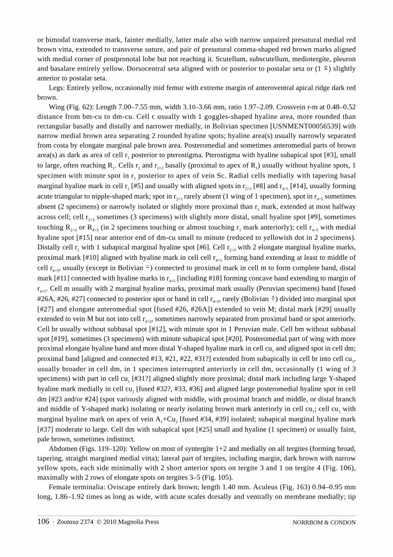
NORRBOM & CONDON106 · Zootaxa 2374 © 2010 Magnolia Press
or bimodal transverse mark, fainter medially, latter male also with narrow unpaired presutural medial redbrown vitta, extended to transverse suture, and pair of presutural comma-shaped red brown marks alignedwith medial corner of postpronotal lobe but not reaching it. Scutellum, subscutellum, mediotergite, pleuronand basalare entirely yellow. Dorsocentral seta aligned with or posterior to postalar seta or (1 &) slightlyanterior to postalar seta.
Legs: Entirely yellow, occasionally mid femur with extreme margin of anteroventral apical ridge dark redbrown.
Wing (Fig. 62): Length 7.00–7.55 mm, width 3.10–3.66 mm, ratio 1.97–2.09. Crossvein r-m at 0.48–0.52distance from bm-cu to dm-cu. Cell c usually with 1 goggles-shaped hyaline area, more rounded thanrectangular basally and distally and narrower medially, in Bolivian specimen [USNMENT00056539] withnarrow medial brown area separating 2 rounded hyaline spots; hyaline area(s) usually narrowly separatedfrom costa by elongate marginal pale brown area. Posteromedial and sometimes anteromedial parts of brownarea(s) as dark as area of cell r1 posterior to pterostigma. Pterostigma with hyaline subapical spot [#3], small
to large, often reaching R1. Cells r1 and r2+3 basally (proximal to apex of R1) usually without hyaline spots, 1
specimen with minute spot in r1 posterior to apex of vein Sc. Radial cells medially with tapering basal
marginal hyaline mark in cell r1 [#5] and usually with aligned spots in r2+3 [#8] and r4+5 [#14], usually forming
acute triangular to nipple-shaped mark; spot in r2+3 rarely absent (1 wing of 1 specimen), spot in r4+5 sometimes
absent (2 specimens) or narrowly isolated or slightly more proximal than r1 mark, extended at most halfway
across cell; cell r2+3 sometimes (3 specimens) with slightly more distal, small hyaline spot [#9], sometimes
touching R2+3 or R4+5 (in 2 specimens touching or almost touching r1 mark anteriorly); cell r4+5 with medial
hyaline spot [#15] near anterior end of dm-cu small to minute (reduced to yellowish dot in 2 specimens).Distally cell r1 with 1 subapical marginal hyaline spot [#6]. Cell r2+3 with 2 elongate marginal hyaline marks,
proximal mark [#10] aligned with hyaline mark in cell cell r4+5 forming band extending at least to middle of
cell r4+5, usually (except in Bolivian &) connected to proximal mark in cell m to form complete band, distal
mark [#11] connected with hyaline marks in r4+5 [including #18] forming concave band extending to margin of
r4+5. Cell m usually with 2 marginal hyaline marks, proximal mark usually (Peruvian specimens) band [fused
#26A, #26, #27] connected to posterior spot or band in cell r4+5, rarely (Bolivian &) divided into marginal spot
[#27] and elongate anteromedial spot [fused #26, #26A]) extended to vein M; distal mark [#29] usuallyextended to vein M but not into cell r4+5, sometimes narrowly separated from proximal band or spot anteriorly.
Cell br usually without subbasal spot [#12], with minute spot in 1 Peruvian male. Cell bm without subbasalspot [#19], sometimes (3 specimens) with minute subapical spot [#20]. Posteromedial part of wing with moreproximal elongate hyaline band and more distal Y-shaped hyaline mark in cell cu1 and aligned spot in cell dm;
proximal band [aligned and connected #13, #21, #22, #31?] extended from subapically in cell br into cell cu1,
usually broader in cell dm, in 1 specimen interrupted anteriorly in cell dm, occasionally (1 wing of 3specimens) with part in cell cu1 [#31?] aligned slightly more proximal; distal mark including large Y-shaped
hyaline mark medially in cell cu1 [fused #32?, #33, #36] and aligned large posteromedial hyaline spot in cell
dm [#23 and/or #24] (spot variously aligned with middle, with proximal branch and middle, or distal branchand middle of Y-shaped mark) isolating or nearly isolating brown mark anteriorly in cell cu1; cell cu1 with
marginal hyaline mark on apex of vein A1+Cu2 [fused #34, #39] isolated; subapical marginal hyaline mark
[#37] moderate to large. Cell dm with subapical spot [#25] small and hyaline (1 specimen) or usually faint,pale brown, sometimes indistinct.
Abdomen (Figs. 119–120): Yellow on most of syntergite 1+2 and medially on all tergites (forming broad,tapering, straight margined medial vitta); lateral part of tergites, including margin, dark brown with narrowyellow spots, each side minimally with 2 short anterior spots on tergite 3 and 1 on tergite 4 (Fig. 106),maximally with 2 rows of elongate spots on tergites 3–5 (Fig. 105).
Female terminalia: Oviscape entirely dark brown; length 1.40 mm. Aculeus (Fig. 163) 0.94–0.95 mmlong, 1.86–1.92 times as long as wide, with acute scales dorsally and ventrally on membrane medially; tip

Zootaxa 2374 © 2010 Magnolia Press · 107REVISION OF THE FEMORALIS GROUP OF BLEPHARONEURA
slightly flared outward basolaterally, short and broad (lobed part 0.32–0.37 times as long as wide), with 7digitiform, minutely serrate lobes separated by deep, narrow gaps; unpaired medial lobe almost twice as broadbut only slightly longer than submedial lobe; lateral lobe projecting, moderately broad and blunt; sublaterallobe very broad, slanted. 8th sternite usually with pair of setulae on medial apical corner. Spermathecaesubspherical, with strongly convoluted, broad sclerotized neck and with or without small cylindrical basalapodeme (similar to B. furcifer).
Male terminalia: Medial surstylus long and slender, prensisetae subequal, closely approximated, separatedby less than width of medial prensiseta.
Distribution. Low to middle elevation areas (250–750 m) of northeastern Bolivia and eastern Peru. Type data. Holotype & (USNM USNMENT00213937), PERU: Madre de Dios: Manu, Río Manu, 5 km E
of Pakitza, Aguajal, 19 Sep 1988, A. Freidberg. Paratypes: BOLIVIA: La Paz: 5 km W of Mapiri, ArroyoTuhiri, 15°17.8'S 68°15.6'W, 750 m, 19 Mar 2001, S. D. Gaimari, 1& (USNM USNMENT00056539). PERU:Madre de Dios: Manu, Río Manu, Pakitza, 12°07'S 70°58'W, 250 m, 9–23 Sep 1988, A. Freidberg, 1%(USNM USNMENT00213938) 1%1& (TAUI USNMENT00213939–40); Manu, near Salvación, Erika, 550m, 5–6 Sep 1988, A. Freidberg, 2% (USNM USNMENT00054208, USNMENT00213936).
Etymology. The name of this species is an adjective referring to the seven digitate lobes of the aculeus tip.
Blepharoneura sinepuncta Norrbom & Condon, new speciesFigs. 63, 144
Diagnosis. This species, B. splendida, punctistigma, and aspiculosa differ from other Blepharoneura speciesin having the thoracic pleuron largely brown, the apical fourth or more of the hind femur dark brown, and thedistal part of the wing with oblique bands. Blepharoneura sinepuncta differs from the other three species inhaving the submedial scutal vittae broadly connected to the marks on the posterior margin (they are rarelynarrowly connected in B. puntistigma and splendida), and it further differs from B. punctistigma andaspiculosa by its lack of a subapical hyaline spot in the pterostigma and its markings in cell dm (subapicalspot [#25] absent or aligned with or fused to posteromedial spot [#24]). The scutal microtrichial patternvaries, but at least the anterior 2/3 of the presutural part lateral to the submedial vitta is microtrichose, unlikein B. splendida. The aculeus tip is more elongate than in B. splendida and punctistigma, the gaps between thelobes are deeper than in B. aspiculosa, and the medial lobe is slightly concave or notched unlike all threespecies.
Description. Head: Dark brown area on ocellar tubercle extended more than half distance to postocellarseta or reaching it. Medial vertical seta in yellow area. Medial occipital sclerite with pair of red brown to darkbrown submedial vittae on ventral half, sometimes connected. Occipital suture narrowly dark brown, onlateral side bordered by variably shaped brown area, extending as band or triangular mark towards postocularsetae, sometimes (1 specimen) reaching eye margin and including lateral vertical seta or (2 specimens) withnarrow brown area along eye.
Thorax: Scutum microtrichose at least laterally, posterior to dorsocentral seta, and on anterior 2/3 ofpresutural part lateral to submedial vitta, sometimes more extensively or mostly microtrichose [obscured ordamaged on some specimens and full extent difficult to determine]; scutum also with 2 pairs of dark brownvittae; submedial vitta uninterrupted and connected to mark on posterior margin; sublateral vitta at mostnarrowly interrupted at transverse suture, narrowly separated from or occasionally (2 specimens) connected tomark on posterior margin; posterior margin with 1 brown mark, narrowed medially. Notopleuron dark brownexcept narrowly on anterodorsal margin. Small brown spot anterior to postsutural supra-alar seta usuallyabsent, small and faint in 2 specimens. Brown vitta anterior to postalar seta and brown spot lateral todorsolateral corner of scutellum present. Scutellum with pair of submedial brown marks, sometimes extendedto basal margin, or with single usually inverted U-shaped medial mark; side with small to moderately largebrown spot or linear or L-shaped mark on or near ventrobasal margin. Subscutellum and mediotergite mostly

NORRBOM & CONDON108 · Zootaxa 2374 © 2010 Magnolia Press
brown except narrow medial vitta and dorsolateral corner of mediotergite usually yellow or orange, or withsingle very broad or pair of dark brown vittae and ventral half of lateral margin of mediotergite also narrowlybrown. Pleuron mostly dark brown, yellow only on propleuron, anterior margin of anepisternum and usuallybroadly bordering phragma, all or most of katepimeron, greater ampulla, and often dorsal margins ofanepimeron, katatergite or anatergite. Basalare brown. Dorsocentral seta aligned with or usually slightlyanterior to postalar seta.
Legs: Mostly yellow. Mid femur sometimes with single ventral or separate anteroventral andposteroventral brown marks on apical 1/5–1/4. Hind femur with entire apical 1/4–2/3 dark brown.
Wing (Fig. 63): Length 6.35–7.35 mm, width 3.05–3.70 mm, ratio 1.86–2.09. Crossvein r-m at 0.52–0.57distance from bm-cu to dm-cu. Cell c with 2 or occasionally 3 hyaline spots, basal spot broad and rectangular,reaching costa and subcosta, distal spot slightly to distinctly smaller, rectangular or constricted medially oroccasionally divided into smaller semicircular anterior and posterior spots; medial brown area almost as darkas to slightly paler than area of cell r1 posterior to pterostigma, sometimes paler medially, slightly to distinctly
narrower than basal hyaline spot, narrower or broader than distal spot. Pterostigma without subapical hyalinespot. Cell r1 basally (proximal to apex of R1) with 1 pale brown to hyaline spot, rarely diffuse or indistinct; cell
r2+3 entirely dark brown or occasionally with 1 spot. Radial cells medially with tapering basal marginal hyaline
mark [#5 fused with additional spot?] in cell r1 and aligned spots in r2+3 [#8] and r4+5 [#14] forming acute
triangular mark, sometimes extended to vein M; cell r4+5 with hyaline spot [#15] near anterior end of dm-cu
narrow but extending to vein M. Distally cell r1 without marginal hyaline spot [#6], posteriorly often with
small extension from band in cell r2+3. Cell r2+3 without marginal hyaline marks. Cell r4+5 with hyaline band
from posteroapical margin, extending parallel to costa to vein R2+3 or slightly into r1, slightly tapering
anteriorly, approximately as broad as marginal brown area. Cells m and r4+5 with inverted V-shaped hyaline
mark [fusion of at least #26A, #26, #27, #29], extending to or almost to vein R4+5, both arms reaching margin
in cell m. Cell br with subbasal hyaline spot [#12]. Cell bm with broad hyaline area [fused #19, #20] or largecircular subapical hyaline spot [#20] only. Cell bcu rarely (1 specimen) with hyaline spot in lobe. Anal lobehyaline except brown area from apical part of lobe of cell bcu and base of vein A1+Cu2 crossing apex of vein
A2. Posteromedial part of wing with 2 elongate hyaline bands, usually connected medially in cell cu1 (except
in 1 male (USNMENT00048913) in which they are broadly connected only posteriorly in cell dm) andoccasionally narrowly posteriorly in cell dm; proximal band extending from subapically in cell br across celldm and at least midway across cell cu1 [aligned and fused #13, #21, #22, #32, possibly also #51, #52 or #23
when broad], often connected to mark across vein A1+Cu2 [fused #34, #39], and often broader on proximal or
distal side in cell dm; part in cell br [#13] broad at least posteriorly, slightly to strongly narrowed anteriorly,sometimes not reaching vein R4+5, br without more proximal hyaline spot [#44]; distal band [fusion of at least
#24, #33, #36 and usually #25] usually extending from anterior side of cell dm across cell cu1 to wing margin,
in 1 specimen reaching vein M, or occasionally with anteromedial subapical hyaline spot [#25] isolated (1specimen) or absent (1%, USNMENT00048913); cell cu1 with subapical marginal hyaline spot [#37] small to
large, usually reaching vein Cu1.
Abdomen: Syntergite 1+2 with pair of irregular posterolateral brown marks formed from fused submedialand sublateral spots and posterolateral band. Tergites 3–5 with pair of submedial brown spots, isolated excepton tergites 3 and 5 of Zurquí male (USNMENT00048913) and pair of irregular lateral brown markings,separated medially, often very broad laterally (without or with small anterior sublateral yellow area) formedfrom fused or connected submedial spots, anterolateral spots, and L-shaped posterolateral bands; sublateralspots occasionally isolated on tergite 4 or especially tergite 5, anterolateral spots rarely isolated.
Female terminalia: Oviscape entirely dark brown, length 1.16–1.30 mm. Aculeus (Fig. 144) 0.65–0.70mm long, 1.67–1.88 times as long as wide, with acute scales dorsally and ventrally on membrane medially; tipangular basolaterally, elongate triangular (lobed part 0.57–0.67 times as long as wide), with small, notched orslightly concave medial lobe and 3 pairs of step-like lobes separated by relatively deep gaps; sublateral and

Zootaxa 2374 © 2010 Magnolia Press · 109REVISION OF THE FEMORALIS GROUP OF BLEPHARONEURA
submedial lobes similar in size; lateral gap 1.36–1.54 times as long as wide. Spermathecae subspherical, withstraight to slightly convoluted, slender sclerotized neck and large cylindrical basal apodeme.
Male terminalia: Medial surstylus with prensisetae subequal, separated by several times width of medialprensiseta, medial prensiseta on long lobe, lateral prensiseta on short lobe.
Distribution. This species is known only from Costa Rica. Where provided by collectors, the elevationsof the collection sites range from 900–2200 m.
Type data. Holotype & (INBio INBIO002405749), COSTA RICA: San José: San Gerardo de Dota, nearAlbergue Savegre, LS 389000 484200, 2200 m, 19–22 May 1997, E. Zumbado. Paratypes: COSTA RICA:Alajuela: La Garita, 900 m, 5 May 1987, C. Godoy, 1% (UCRSJ USNMENT00213919); Cartago: La Suiza, 7Mar, P. Schild, 1% (USNM USNMENT00213916); same, 2 Mar 1924, 1% (USNM USNMENT00213918);same, Apr 1922, 1& (USNM USNMENT00213917); Heredia: Río Sucio, H. Rogers, “B.C.A. Dipt. II.,Hexachaeta splendida G.Tos.”, 1% (BMNH); San José: San Gerardo de Dota, along Río Savegre, 9°33'N83°48'W, 2200 m, on Cyclanthera langei Cogn. (95CR14), 8–10 Aug 1995, M. A. Condon & A. L. Norrbom,1& (USNM USNMENT00048391); San Gerardo de Dota, near Albergue Savegre, LS 389000 484200, 2200m, 19–22 May 1997, M. A. Zumbado, 1% (INBio INBIO002578262); Zurquí de Moravia, 10°03'N 84°01'W,1600 m, Mar 1995, P. Hanson, 1% (USNM USNMENT00048557); same, Sep 1995, 1& (USNMUSNMENT00050130). Puntarenas: Estación Biológico Pittier, LS 330900 577400, 1670 m, 26 Sep - 5 Oct1995, E. Navarro, 1& (INBio INBIO002340202).
Other specimens examined. COSTA RICA: San José: Zurquí de Moravia, 10°03'N 84°01'W, 1600 m,Aug-Sep 1994, P. Hanson, 1% (USNM USNMENT00048913).
Etymology. The name of this species is a Latin noun meaning without spots in reference to the absence ofspots in the pterostigma and in the apical part of cell dm, which distinguishes this species from B. aspiculosaand punctistigma.
Remarks. Cells dm and cu1 usually have a more or less H or M-shaped hyaline mark. In the La Suiza
female there is an isolated hyaline spot in the apical third of dm, but it is aligned with the distal part of thelarger mark that extends into cell cu1. The non-paratype male from Zurquí has a different shape, somewhat
more like an inverted V, and it also has the hyaline mark in cell r1 extended to vein M and very broad scutal
vittae and may possibly be a different species.Biology. One adult was collected on Cyclanthera langaei Cogn. at San Gerardo de Dota, Costa Rica, and
larvae were also found mining stems of this plant at this site in the same month (19 Aug 1995, ALN, pers.obs.), but unfortunately we were unable to rear the larvae to adults and their identity remains uncertain (alsosee Biology section).
Blepharoneura splendida Giglio-TosFigs. 64–65, 184–187
Blepharoneura splendida Giglio-Tos 1893: 10; Giglio-Tos 1895: 58 [type data, additional description]; Lima & Leite1952: 309; Norrbom et al. 1999: 106 [in catalog].
Hexachaeta splendida: Wulp 1899: 402 [in key], 404 [discussion]; Hendel 1914: 23 [in key, in catalog]; Aczél 1950: 195[in catalog]; Foote 1967: 18 [in catalog].
Diagnosis. This species, B. sinepuncta, punctistigma, and aspiculosa differ from other Blepharoneura speciesin having the thoracic pleuron largely brown, the apical fourth or more of the hind femur dark brown, and thedistal part of the wing with oblique bands. Blepharoneura splendida differs from B. sinepuncta in having thesubmedial scutal vittae separate from or at most narrowly connected to the marks on the posterior margin, andfrom B. punctistigma and aspiculosa by its markings in cell dm (subapical spot [#25] absent or aligned with orfused to posteromedial spot [#24]), its frequent lack of a subapical hyaline spot in the pterostigma, and itsmostly nonmicrotrichose scutum. The aculeus tip is stouter than in B. sinepuncta and aspiculosa, with the

NORRBOM & CONDON110 · Zootaxa 2374 © 2010 Magnolia Press
sublateral lobe more pronounced, and the gap between the lateral and sublateral lobes is deeper than in B.punctistigma.
Description. Head: Dark brown area on ocellar tubercle usually (17 of 22 specimens) extended more thanhalf distance to postocellar seta. Medial vertical seta in yellow area. Medial occipital sclerite with pair of palebrown to dark brown submedial vittae on ventral half. Occipital suture narrowly dark brown, on lateral sidebordered by variably shaped brown area extending as band or triangular mark towards postocular setae,sometimes reaching eye margin, and usually (16 of 21 specimens) including lateral vertical seta.
Thorax: Scutum nonmicrotrichose except posterior to dorsocentral seta and laterally, with 2 pairs of darkbrown vittae or rows of spots; submedial vitta narrowed or interrupted posterior to transverse suture andusually separated from mark on posterior margin (narrowly connected on at least 1 side in 3 specimens);sublateral vitta interrupted at transverse suture and usually separated from mark on posterior margin(connected in 8 of 17 specimens); posterior margin with 1 broad brown mark narrowed medially or rarely with2 separate marks. Notopleuron entirely brown or brown except for small pale area surrounding posterior seta.Small brown spot anterior to postsutural supra-alar seta sometimes absent or faint, sometimes connected alongtransverse suture to lateral vitta. Brown vitta anterior to postalar seta present. Brown spot lateral todorsolateral corner of scutellum present, connected to sublateral vitta in Costa Rican and Venezuelanspecimens. Scutellum with single inverted U-shaped or inverted mushroom shaped medial brown markextended to basal margin. Subscutellum and mediotergite with pair of dark brown vittae, on ventral half ofmediotergite extended to lateral margin or with separate narrow brown mark on margin. Pleuron mostly darkbrown, yellow only on propleuron, anterior margin of anepisternum and occasionally narrowly borderingphragma, all or most of katepimeron, greater ampulla, and often dorsal margins of anepimeron, katatergite oranatergite. Basalare with brown spot or entirely brown. Dorsocentral seta aligned with or slightly anterior orposterior to postalar seta.
Legs: Mostly yellow. Mid femur sometimes with anteroventral and posteroventral brown marks on apical1/5–1/4. Hind femur with entire apical 1/4–1/2 dark brown.
Wing (Figs. 64–65): Length 5.14–6.55 mm, width 2.60–3.37 mm, ratio 1.88–2.00. Crossvein r-m at 0.51–
0.56 distance from bm-cu to dm-cu. Cell c with 2 hyaline spots, basal spot broad and rectangular, reachingcosta and subcosta, distal spot slightly larger to distinctly smaller, rectangular or constricted anteriorly,occasionally not reaching costa; medial brown area as dark as to distinctly paler than area of cell r1 posterior to
pterostigma, sometimes paler medially, usually broader than or as broad as basal hyaline spot, usually asbroad as but sometimes narrower or broader than distal spot. Pterostigma usually without subapical hyalinespot, occasionally (6 Mexican specimens) with small to large subhyaline to pale brown spot [#3]. Cells r1 and
r2+3 entirely dark brown basally (proximal to apex of R1) or only with 1 diffuse, indistinct pale brown spot
posterior to apex of vein Sc. Radial cells medially with tapering basal marginal hyaline mark [#5 fused withadditional spot?] in cell r1 and aligned spots in r2+3 [#8] and r4+5 [#14] (latter rarely absent) forming acute
triangular mark, extended at most midway across r4+5; cell r4+5 with hyaline spot [#15] near anterior end of dm-
cu often extending to or almost to vein M, especially in Mesoamerican specimens. Cell r2+3 without marginal
hyaline marks. Cell r4+5 with hyaline band from posteroapical margin, extending parallel to costa almost to or
usually to vein R2+3 or slightly into r1 (fused with subapical hyaline spot), sometimes constricted along vein
R4+5 (some Mesoamerican specimens), usually slightly tapering anteriorly, approximately as broad as marginal
brown area or (some Mesoamerican specimens) slightly to much broader. Cells m and r4+5 with inverted V-
shaped hyaline mark [fusion of at least #26A, #26, #27, #29], extending to or almost to vein R4+5, occasionally
(1 Mexican, 1 Costa Rican specimen) connected anteriorly to most distal hyaline wing band to form 3branched mark, both arms reaching margin in cell m. Cell br with subbasal hyaline spot [#12]. Cell bm rarely(1 wing of 1 specimen) with subbasal hyaline spot [#19], usually with only medial or subapical circularhyaline spot [#20]. Cell bcu rarely (2 specimens) with hyaline spot in lobe. Anal lobe hyaline except brownarea from apical part of lobe of cell bcu and base of vein A1+Cu2 crossing apex of vein A2. Posteromedial part
of wing with large, usually somewhat Y-shaped hyaline mark, sometimes (more commonly in Mesoamerican

Zootaxa 2374 © 2010 Magnolia Press · 111REVISION OF THE FEMORALIS GROUP OF BLEPHARONEURA
specimens) connected to mark across vein A1+Cu2 [fused #34, #39] to form H- or X-shaped mark or rarely (3
Venezuelan, 1 Ecuadorean specimens) with anterior distal arm in cell dm isolated or small; anterior proximalpart of mark [aligned and fused #13, #21, #22, #32, possibly also #51, #52 or #23 when broad] extending fromsubapically in cell br across cells dm, sometimes broader on proximal or distal side especially posteriorly incell dm; part in cell br [#13] slightly to strongly narrowed anteriorly, often (15 of 26 specimens) not reachingvein R4+5, br without more proximal hyaline spot [#44]; posterior part of mark [fusion of at least #32, #33,
#36] extending across cell cu1 to wing margin; anterior distal part of mark [usually fusion of #25, #24] usually
extending from anterior side of cell dm but not reaching vein M, usually broadly connected to proximalbranch anteriorly and medially in cell cu1 and occasionally (some Mesoamerican specimens) posteriorly in
cell dm, or less commonly (some Venezuelan specimens) with anterior part of distal branch isolated in cell dmor constricted along vein Cu1, rarely (1 Ecuadorean male, 1 wing of 1 Venezuelan male) reduced to small
posterior hyaline area or spot in dm; cell cu1 with subapical marginal hyaline spot [#37] small to moderately
large, sometimes reaching vein Cu1.
Abdomen: Syntergite 1+2 with pair of irregular posterolateral brown marks formed from fused submedialand sublateral spots and posterolateral band. Tergites 3–5 with pair of irregular, shallow U-shaped or W-shaped marks, separated medially, formed from connected or fused submedial, sublateral and anterolateralspots and posterolateral bands, occasionally very broad laterally (without or with small anterior sublateralyellow area) or rarely (Upala %) entirely brown except base of syntergite 1+2 and irregular medial vitta.
Female terminalia: Oviscape entirely dark brown, length 0.78–1.08 mm. Aculeus (Figs. 184–187) 0.58–0.83 mm long, 1.49–2.02 times as long as wide, with acute scales dorsally and ventrally on membranemedially; tip angular basolaterally, short triangular (lobed part 0.32–0.40 times as long as wide), with small,convex medial lobe and 3 pairs of step-like lobes separated by relatively deep gaps; sublateral lobe larger thansubmedial lobe; lateral gap 0.98–1.29 times as long as wide. Spermathecae subspherical, with straight toconvoluted, slender sclerotized neck and large cylindrical basal apodeme (similar to B. femoralis).
Male terminalia: Medial surstylus with prensisetae subequal, separated by several times width of medialprensiseta, medial prensiseta on long lobe, lateral prensiseta on short lobe.
Distribution. Mexico (Nayarit, San Luis Potosí, Tamaulipas, Veracruz), Guatemala, Costa Rica,Venezuela, and Ecuador. Although B. splendida occurs in Costa Rica (and presumably also in the otherCentral American countries besides Guatemala), the male from Río Sucio, Costa Rica reported as this speciesby Wulp (1899) is B. sinepuncta. Specimens for which elevation data were provided by the collectors weretaken at 480 to 1600 m.
Type data. Holotype & (IMZ), MEXICO, "senza indicazione di località" [unspecified locality], [no date],A. Boucard [examined]. The holotype is a female, not a male as stated in the original description. Given thedifficulty of distinguishing the sexes in the femoralis group because of the small size of the oviscape, there isno reason to doubt that this specimen is the holotype. It has labels with "1215" and, in Giglio-Tos' writing,"Blepharoneura splendida Giglio-Tos &".
Other specimens examined. COSTA RICA: Alajuela: Upala, 20 km S of, 27–31 Mar 1991, F. D. Parker,1% (USU USNMENT00213872). San José: San Gerardo de Dota, 9°33'N 83°48'W, 2200 m, 7–11 Aug 1995,E. M. Fisher, 1% (CDFA USNMENT00104207); Zurquí de Moravia, 10°03'N 84°01'W, 1600 m, Sep 1995, P.Hanson, 1& (USNM USNMENT00050131). ECUADOR: Napo: Huahua Sumaco, Hollin - Loreto road, km45, Malaise trap, 18 Dec 1989, M. & J. Wasbauer & H. Real, 1% (CDFA USNMENT00104209). Pichincha:Maquipucuna Biological Reserve, Malaise trap, 2–3 Aug 1998, F. G. Andrews, 1% (CDFAUSNMENT00104208). GUATEMALA: Pochuta, Pacayal, 1000 m, Feb-Mar 1931, J. Bequaert, 1& (MCZUSNMENT00213874); Pochuta, Santa Emilia, 1000 m, Feb-Mar 1931, J. Bequaert, 1% (MCZUSNMENT00213879). Escuintla: Palín, 14°24'N 90°42'W, McPhail traps, 1992, J. López, 1& (USNMUSNMENT00213838). Guatemala: Santa Catarina Pinula, Puerta Parada, Schuster property, 14.5572°N90.4653°W, 1860 m, Malaise trap, 25 Mar - 1 Apr 2006, J. C. Schuster, 1%1& (FSCA USNMENT00654008–09) MEXICO: Nayar i t : San Blas , La Ba jada , 20–21 Mar 1983 , W. J . Hanson , 1& (USU

NORRBOM & CONDON112 · Zootaxa 2374 © 2010 Magnolia Press
USNMENT00213873). San Luis Potosí: Xilitla, River 1 km E of, 600 m, 7 Jul 1990, I. Yarom, 1& (USNMUSNMENT00213871). Tamaulipas: W of Gomez Farias, Rancho de Cielo, 3 Jul 1989, R. Jones, 1& (TAMUUSNMENT00212642); 74 mi S of Ciudad Victoria, Gómez Farias, W of, 15 Nov 1985, P. Kovarik, R. Jones& K. Haack, 1% (TAMU USNMENT002126421). Veracruz: Estación de Biología Los Tuxtlas, 3 Mar 1985,A. Ibarra, 1% (IEXV USNMENT00213878); Estación de Biología Los Tuxtlas, El Viguia, 480 m, 17 Mar1986, E. Ramírez, 1% (IEXV USNMENT00213881); Salto de Eyipantla, 21 Feb 1985, F. Arias, 1& (IEXVUSNMENT00213877); Xalapa, 22 Feb 1998, A. Freidberg, 1% (TAUI USNMENT00213876).VENEZUELA: Aragua: Rancho Grande, 1100 m, 20 Mar 1946, 1% (AMNH USNMENT00213863); same, 3May 1951, F. Fernandez Y., 2%2& (IZAM USNMENT00213864–65, USNMENT00213867,USNMENT00213883); 1%2& (USNM USNMENT00213868, USNMENT00213870, USNMENT00213882);same, 5 May 1951, 1% (IZAM USNMENT00213866); same, 10 Jun 1954, C. J. Rosales, 1& (IZAMUSNMENT00213869). Monagas: Caripe, 21 July 1953, C. J. Rosales & J. R. Requena, 1% (IZAMUSNMENT00213862).
Blepharoneura tau Norrbom & Condon, new speciesFigs. 66, 105, 118
Blepharoneura sp. 41: Norrbom & Condon 1999: 138.
Diagnosis. This species differs from most species of Blepharoneura by its T-shaped abdominal pattern whichis unique within the genus except for a few specimens of B. splendida, which differ in having theanepisternum brown medially and the apex of the hind femur brown. B. tau differs from other species withoblique apical bands on the wing in having the anepisternum with a dorsal brown spot, the marginal brownarea in cells r2+3 and r4+5 as broad as the subapical hyaline band, cells r4+5 and dm without hyaline marks
between crossveins r-m and dm-cu except for a short band extending across vein M, and cell cu1 without a
subapical hyaline spot. The female is unknown.Description. Head: Dark brown area on ocellar tubercle extended to postocellar seta. Medial occipital
sclerite with pair of faint brown submedial vittae on ventral half. Occipital suture narrowly dark brown. Smalldiffuse brown area lateral to occipital suture ventral to lateral vertical seta.
Thorax (Fig. 105): Scutum nonmicrotrichose except posterior to dorsocentral seta and laterally; with 2pairs of brown vittae; submedial vitta extended to transverse suture, red brown, posterior third faint; sublateralvitta red brown bordering medial corner of postpronotal lobe, continuing faintly almost to transverse suture(posterior 2/3 faint), anterior half of postsutural part faint and slightly more medial than dark brown posteriorhalf, very narrowly separated from mark on posterior margin and extending lateral to scutellum (fused withnormal spot lateral to scutellum); posterior margin with single broad triangular dark brown mark, broadestmedially. Notopleuron with large dark brown spot posteriorly on lateral margin, extended to posterior seta.Short dark brown vitta extended from anterior to lateral to postsutural supra-alar seta, but well separated fromlateral margin and postalar seta. Scutellum with pair of submedial brown spots on basal half of disk.Subscutellum narrowly brown laterally. Mediotergite with pair of brown vittae, yellow area between thembroader than lateral yellow margin. Pleuron mostly yellow, with following brown areas: anepisternum withsmall spot dorsal to anterior seta; posterior third of anepimeron; dorsal third of katatergite; and most ofanatergite except anterodorsal margin and posteroventral corner. Basalare entirely yellow. Dorsocentral setaaligned slightly anterior to postalar seta.
Legs: Mostly yellow. Mid femur with anteroventral brown mark on apical 1/4. Hind femur withanteroventral and posteroventral brown marks on apical 1/4.
Wing (Fig. 66): Length 5.54 mm, width 2.87 mm, ratio 1.93. Crossvein r-m at 0.54 distance from bm-cu todm-cu. Cell c with 2 hyaline spots, basal spot small, rounded or triangular, reaching subcosta, extending halfdistance to costa, distal spot subrectangular, reaching costa and subcosta; medial brown area slightly paler

Zootaxa 2374 © 2010 Magnolia Press · 113REVISION OF THE FEMORALIS GROUP OF BLEPHARONEURA
than area in cell r1 posterior to pterostigma, sometimes paler posteriorly, much broader than basal hyaline spot
and slightly broader to slightly narrower than distal spot. Pterostigma without subapical hyaline spot [#3].Cells r1 and r2+3 basally (proximal to apex of R1) without hyaline spots. Radial cells medially with tapering
basal marginal hyaline mark in cell r1 [#5] and aligned spot in r2+3 [#8] forming acute triangular mark,
extended to vein R4+5; cell r4+5 without hyaline spot aligned with r1 mark, but with posterior hyaline mark
[#15?] aligned between r1 mark and dm-cu, aligned and connected with anterior subapical hyaline mark
[#25?] in cell dm forming short band. Distally cell r1 without hyaline spots and cell r2+3 without marginal
hyaline marks. Cell r4+5 with hyaline band from posteroapical margin, extending more or less parallel to costa
into cell r2+3 reaching vein R2+3, slightly tapering anteriorly, as broad as marginal brown area. Cells m and r4+5
with inverted V-shaped mark [fusion of at least #26A, #26, #27, #29], extending to vein R4+5 anteriorly and
both arms reaching margin in cell m, distal arm slightly constricted along vein M. Cell br with subbasalhyaline spot [#12]. Cell bm with only ovoid medial hyaline spot [#20]. Posteromedial part of wing with only 2elongate hyaline marks in addition to elongate mark across apex of vein A1+Cu2 [fused #34, #39]; anterior
proximal band [fused #13, #21, #22] extending from cell br subapically, across cell dm; and posterior distalband [fusion of at least #33, #36] extended medially across cell cu1. Cell cu1 without subapical marginal
hyaline mark [#37].Abdomen (Fig. 118): Dark brown except following yellow areas: basal half of syntergite 1+2 extending
on lateral margin almost to posterior margin, then extending slightly medially, and somewhat T-shaped mark,with transverse medial mark subapically on syntergite 1+2, almost connected to lateral yellow area andconnected medially to basal area and posteriorly to narrow, tapering, irregular margined, medial vittaextending posteriorly to tergite 5.
Male terminalia: Medial surstylus with prensisetae separated by several times width of medial prensiseta,medial prensiseta on moderately long lobe, lateral prensiseta subequal to medial prensiseta.
Distribution. Costa Rica. The holotype was collected at 1600 m elevation at a site bordering BraulioCarrillo National Park.
Type data. Holotype % (USNM USNMENT00048626), COSTA RICA: San José: Zurquí de Moravia,10°03'N 84°01'W, 1600 m, Malaise trap, Aug 1995, P. Hanson.
Etymology. The name of this species is a noun, the Greek letter tau, in reference to the somewhat T-shaped yellow mark on the abdomen.
Blepharoneura thetis HendelFigs. 67–68, 94, 198–199
Blepharoneura thetis Hendel 1914: 21 [in key], 22 [description]; Aczél 1950: 198 [in catalog]; Foote 1967: 18 [in cata-log]; Norrbom et al. 1999: 106 [in catalog].
Diagnosis. This species can be distinguished from other species of Blepharoneura by the followingcombination of characters: Anepisternum entirely yellow; scutum with pair of brown submedial vittae,extended to trapezoidal brown mark on posterior margin, but without sublateral vittae; scutellum with band orpair of transverse brown marks extending to side and to basal seta but not to basal margin; cell r1 proximally
with single relatively narrow, tapering marginal hyaline mark; cell r2+3 with 2 marginal hyaline marks,
posterior one not connected to marginal hyaline spot in cell r4+5; cell r4+5 with 2 small marginal hyaline spots or
one bilobed mark (spots connected proximally). It is very similar to B. fernandezi and a probably undescribedspecies (see sp. nr. thetis), which differ in lacking brown scutellar markings and having only 1 marginalhyaline spot in cell r4+5. The female is unknown.
Description. Head: Dark brown area on ocellar tubercle extended less than half distance to postocellarseta. Large brown spot surrounding medial vertical seta extended ventromesally to level of postocellar seta

NORRBOM & CONDON114 · Zootaxa 2374 © 2010 Magnolia Press
[may connect to form band, medial part not visible in Nova Teutonia male, head is detached and glued tomounting card]. Occipital suture narrowly orange brown.
Thorax (Fig. 94): Scutum nonmicrotrichose except posterior to dorsocentral seta and laterally, with pair ofbroad dark brown submedial presutural vittae, broadest slightly anterior to level of posterior margin ofpostpronotal lobe, extended to posterior brown mark. Posterior margin with broad elongate trapezoidal darkbrown mark, extended laterally to intra-alar seta and lateral to base of scutellum. Scutellum with completetransverse band (Nova Teutonia %) or pair of irregular, transverse, lateral brown marks (holotype) includingbasal marginal seta but not extending to basal margin, continuing (at least in Nova Teutonia %) on side toventral margin (but ventrobasal corner yellow). Subscutellum yellow. Mediotergite with pair of broad,narrowly separated red brown vittae. Pleuron entirely yellow. Basalare entirely yellow. Dorsocentral setaaligned slightly posterior to postalar seta.
Legs: Entirely yellow. Wing (Figs. 67–68): Length 5.80–5.94 mm, width 2.90–3.07 mm, ratio 1.94–2.00. Crossvein r-m at 0.55–
0.57 distance from bm-cu to dm-cu. Cell c with 2 rectangular hyaline spots, both reaching costa and subcosta;medial brown area slightly paler than area of cell r1 posterior to pterostigma, sometimes fainter posteriorly,
and approximately as broad as to slightly narrower than hyaline spots. Pterostigma with large subapicalhyaline spot [#3] reaching R1. Cells r1 and r2+3 basally (proximal to apex of R1) without hyaline spots except
for minute spot in r1 posterior to apex of vein Sc. Radial cells medially with tapering basal marginal hyaline
mark in cell r1 [#5] and aligned spot in r2+3 [#8] forming acute triangular to nipple-shaped mark, extending at
least to R4+5 and sometimes into r4+5, but spot in cell r4+5 [#14] sometimes slightly more distal; spot in r4+5 [#14]
touching R4+5 but extended less than halfway across cell; cell r2+3 with more distal moderately large hyaline
spot [#9], touching R4+5 and R2+3; cell r4+5 with medial hyaline spot [#15] near dm-cu small. Distally cell r1 with
1 marginal subapical hyaline spot [#6]. Cell r2+3 with 2 elongate marginal hyaline marks [#10, 11], both
extending to vein R4+5. Cell r4+5 with small hyaline spot [#16] on anterior margin aligned between marginal
marks in cell r2+3, and with 2 small marginal hyaline spots [#18, 18A], on right wing of Nova Teutonia male
connected on proximal ends. Cell m with 3 small hyaline spots, 2 marginal [#27, 29] and 1 anteromedial [#26and/or #26A], none extended to vein M. Cell br with subbasal hyaline spot [#12]. Cell bm with small circularsubbasal and larger circular subapical spots [#19, #20]. Posteromedial part of wing with hyaline spots mostlyisolated; cell br with subapical spot [#13] and usually (except 1 wing of Nova Teutonia %) with smaller moreproximal posterior spot [#44]; cell dm with subbasal hyaline band [fused #51, #52] aligned or partially alignedwith more proximal spot in cell br [#44] and proximal anterior spot [#31] in cell cu1 (when latter spots
present) sometimes (Nova Teutonia %) narrowly connected anteriorly or posteriorly to more distal spots indm; cell dm also usually (except 1 wing of Nova Teutonia %) with anterior medial spot [#21] aligned withsubapical spot [#13] in cell br, and with broad posteromedial spot [fused #22, #23, #24]; cell cu1 medially
sometimes (holotype) with 3 anterior hyaline spots [#31, #32, #33] and 2 marginal spots, medial anterior spot[#32] elongate and almost connected to marginal spot, or (Nova Teutonia %) with or without proximal anteriorspot [#31] and with 2 distal anterior spots [#32, #33] fused with medial marginal spot [#36] to form Y-shapedmark aligned with posteromedial spot in cell dm; proximal marginal spot [fused #34, #39] across apex of veinA1+Cu2 isolated; medial marginal mark (or posterior part of Y-shaped mark) partially divided and relatively
broad [fused #36, #36A?] (holotype) or narrower and undivided [#36 only] (Nova Teutonia %); subapicalmarginal hyaline mark [#37] small. Cell dm with subapical hyaline spot [#25] moderate sized, not extended tovein M.
Abdomen: All tergites with pair of broad dark brown markings forming irregular vittae; mark on eachtergite extended broadly along posterior margin to lateral margin of tergite; spots broadly separated mediallyby tapering, nearly straight margined yellow area; brown spots solid except for 1 small anterior yellow spot inspots on tergites 3–5.
Male terminalia: Medial surstylus with prensisetae subequal, separated by width or slightly more thanwidth of medial prensiseta, medial prensiseta on short lobe (Figs. 198–199).

Zootaxa 2374 © 2010 Magnolia Press · 115REVISION OF THE FEMORALIS GROUP OF BLEPHARONEURA
Distribution. Brazil (Santa Catarina, Rio Grande do Sul).Type data. Holotype % (NMW), BRAZIL: Rio Grande do Sul, [no date], Stieglmayer [examined]. The
holotype was not marked as such, but it has a label with "Blepharoneura thetis H." in Hendel's writing. It is aholotype by monotypy, as Hendel stated that he examined just one male specimen. We added a holotype label.
Other specimens examined. BRAZIL: Santa Catarina: Nova Teutonia, 27°11'S 52°23'W, 2 Jan 1953, F.Plaumann, 1% (BMNH USNMENT00213840).
Blepharoneura species near thetisFigs. 69, 88
Diagnosis. This species can be distinguished from other species of Blepharoneura by the followingcombination of characters: Anepisternum and scutellum entirely yellow; scutum with pair of brownsubmedial vittae and broad triangular brown mark on posterior margin, but without sublateral vittae; cell r1
proximally with single relatively narrow, tapering marginal hyaline mark; cell r2+3 with 2 marginal hyaline
marks, posterior one not connected to marginal hyaline spot in cell r4+5; cell r4+5 with single posterior marginal
hyaline spot. It is very similar to B. thetis, which differs in having brown scutellar markings and 2 marginalhyaline spots or a bilobed spot in cell r4+5, and B. fernandezi, which lacks the brown mark on the posterior
margin of the scutum and brown vittae on the mediotergite. The female is unknown.Description. Head (Fig. 88): Dark brown area on ocellar tubercle extended more than half distance to
postocellar seta. Large brown spot surrounding medial vertical seta extended ventromesally to level ofpostocellar seta. Occipital suture narrowly dark brown.
Thorax: Scutum nonmicrotrichose except posterior to dorsocentral seta and laterally, with pair of broaddark brown submedial presutural vittae, broadly interrupted at transverse suture but with short postsutural partconnected to posterior brown mark. Posterior margin with broad elongate triangular dark brown mark,extended faintly lateral to base of scutellum. Scutellum entirely yellow. Subscutellum yellow. Mediotergitewith pair of broad, narrowly separated red brown vittae. Pleuron entirely yellow. Basalare entirely yellow.Dorsocentral seta aligned slightly posterior to postalar seta.
Legs: Entirely yellow. Wing (Fig. 69): Length 5.62 mm, width 2.90 mm, ratio 1.94. Crossvein r-m at 0.58 distance from bm-cu to
dm-cu. Cell c with 2 rectangular hyaline spots, both reaching costa and subcosta; medial brown area slightlypaler than area of cell r1 posterior to pterostigma, fainter anteriorly, and approximately as broad as hyaline
spots. Pterostigma with large subapical hyaline spot [#3] reaching R1. Cells r1 and r2+3 basally (proximal to
apex of R1) without hyaline spots. Radial cells medially with tapering basal marginal hyaline mark in cell r1
[#5] and with aligned spots in r2+3 [#8] and r4+5 [#14], sometimes forming acute triangular to nipple-shaped
mark, but spot in r2+3 sometimes not extending to R2+3; cell r4+5 spot [#14] touching R4+5 but extended less than
halfway across cell; cell r2+3 with more distal moderately large hyaline spot [#9], touching R4+5 but not R2+3;
cell r4+5 with medial hyaline spot [#15] near dm-cu small. Distally cell r1 with 1 marginal subapical hyaline
spot [#6]. Cell r2+3 with 2 elongate marginal hyaline marks [#10, 11], both extending to vein R4+5. Cell r4+5 with
small hyaline spot [#16] on anterior margin aligned between marginal marks in cell r2+3, and with 1 moderately
large, slightly elongate, obliquely anteriorly directed, marginal hyaline spot [#18] posteriorly, touching apexof vein M. Cell m with 3 small hyaline spots, 2 marginal [#27, 29] and 1 anteromedial [#26 and/or #26A],none extended to vein M. Cell br with subbasal hyaline spot [#12]. Cell bm with circular subbasal andsubapical hyaline spots [#19, #20]. Posteromedial part of wing with hyaline spots mostly isolated; cell br withsubapical spot [#13] and sometimes (1 wing) with smaller more proximal posterior spot [#44]; cell dm withsubbasal hyaline band [fused #51, #52] aligned with more proximal spot in cell br [#44] (if present) andproximal anterior spot spot [#31] in cell cu1; dm also with small anterior medial spot [#21] aligned or partially
aligned with subapical spot [#13] in cell br and with broad posteromedial spot [fused #22, #23, #24] or (1wing) with these spots connected to form L-shaped mark; cell cu1 medially with 3 anterior hyaline spots [#31,

NORRBOM & CONDON116 · Zootaxa 2374 © 2010 Magnolia Press
#32, #33] and 2 marginal spots [fused #34, #39; #36], proximal anterior spot [#31] small, aligned withsubbasal band in br, distal anterior spots [#32, #33] aligned with posteromedial spot in cell br, distal spot [#33]sometimes (1 wing) fused to medial marginal spot [#36] to form band; proximal marginal spot [fused #34,#39] across apex of vein A1+Cu2 isolated; subapical marginal hyaline mark [#37] small. Cell dm with
subapical hyaline spot [#25] moderate sized, not extended to vein M. Abdomen: Mostly yellow. Syntergite 1+2 with pair of small, faint brown sublateral spots, not reaching
lateral or posterior margins. Tergites 3–5 with pair of broad dark brown spots, forming irregular vittae; spot oneach tergite extended narrowly along posterior margin to lateral margin of tergite; spots broadly separatedmedially by tapering, straight margined yellow area; brown spots solid except for 1 small anterior yellow spotin spot on tergite 5.
Male terminalia: Medial surstylus with prensisetae subequal, separated by several times width of medialprensiseta, medial prensiseta on long lobe.
Specimens examined. BOLIVIA: La Paz: 5 km W of Mapiri, Arroyo Tuhiri, 15°17.8'S 68°15.6'W, 750m, 18 Mar 2001, S. D. Gaimari, 1% (USNM USNMENT00056566).
Remarks. The single male examined probably represents an undescribed species, but we are reluctant toformally name it as no female specimens that can be associated with it or B. thetis are currently available.
Blepharoneura unifasciata Norrbom & Condon, new speciesFigs. 70, 164
Diagnosis. This species differs from other species of Blepharoneura with the distal part of the wing banded inhaving only a single marginal hyaline mark in cell r2+3, and that mark broadly touching the costal margin in r2+3
and extending from the apex of vein R2+3 to the apex of vein M. Other useful diagnostic characters include:
scutellum without brown markings; pterostigma with subapical hyaline spot; cell r1 without subapical hyaline
spot; cell dm without hyaline subapical spot in anterior half; and abdominal tergites 3–5 with pair of broaddark brown submedial vittae and separate small posterolateral spot. The aculeus tip is very short and broad. Itresembles those of B. ruptafascia, bidigitata and hirsuta, but the lateral lobe is rounded unlike B. ruptafascia,but not digitate as in B. bidigitata and hirsuta.
Description. Head: Dark brown area on ocellar tubercle extended more than half distance to postocellarseta. Occipital suture narrowly dark brown.
Thorax: Scutum entirely microtrichose, with 1 submedial pair of dark brown vittae from anterior marginhalfway to transverse suture; posterior margin with pair of well separated rounded dark brown marks.Scutellum, subscutellum, mediotergite and pleuron entirely yellow. Basalare entirely yellow. Dorsocentralseta aligned slightly anterior to postalar seta.
Legs: Mostly yellow. Mid femur with dark red brown spot on margin of anteroventral apical ridge, hindfemur with similar but smaller and paler mark. Hind tibia slightly darker, orange.
Wing (Fig. 70): Length 7.43 mm, width 3.76 mm, ratio 1.97. Crossvein r-m at 0.48 distance from bm-cu todm-cu. Cell c with 2 rectangular to inverted trapezoidal hyaline spots, both reaching costa and subcosta;medial brown area faint anteriorly, paler than area of cell r1 posterior to pterostigma, narrower than both
hyaline spots. Pterostigma with large subapical hyaline spot [#3] reaching R1. Cells r1 and r2+3 basally
(proximal to apex of R1) without hyaline spots. Radial cells medially with tapering basal marginal hyaline
mark [#5] in cell r1 and aligned spots in r2+3 [#8] and r4+5 [#14] forming acute triangular to nipple-shaped mark,
extended slightly more than halfway across cell r4+5; cell r2+3 with more distal small hyaline spot [#9], touching
R4+5 and touching or almost touching R2+3; cell r4+5 with medial hyaline spot [#15] near anterior end of dm-cu
small. Distally cell r1 without marginal [#6] or posterior hyaline spots. Cells r2+3 and r4+5 distally with 1 broad,
nearly straight hyaline band extending from margin at apex of R2+3 to posteroapical margin of cell r4+5. Cell m
with 2 marginal hyaline marks, proximal mark [fused #26, #27] extending 3/4 distance to vein M, distal mark

Zootaxa 2374 © 2010 Magnolia Press · 117REVISION OF THE FEMORALIS GROUP OF BLEPHARONEURA
[#29] extending to vein M. Cell br with subbasal hyaline spot [#12]. Cell bm with only circular subapicalhyaline spot [#20]. Posteromedial part of wing with large inverted Y- or V-shaped hyaline mark; proximal part[aligned and connected #13, #21, #22, #32, #34, #39] extending from cell br subapically, across cells dm andcu1 subbasally, and across apex of vein A1+Cu2, broadly connected posteriorly in cell dm to distal band [fused
#24, #33, #36] extending from posteromedially in cell dm to posterior wing margin; cell cu1 with subapical
marginal hyaline mark [#37] moderate sized. Cell dm without subapical hyaline spot [#25].Abdomen: Mostly yellow. Syntergite 1+2 with 1 pair of dark brown spots not reaching lateral or posterior
margins. Tergites 3–5 with pair of broad dark brown submedial vittae, not extended to lateral margin, but eachtergite also with small posterolateral spot on margin; submedial vittae solid brown, without yellow spotswithin them but on tergites 4 and 5 narrowed on posterior half, well separated medially, but with irregularmargins.
Female terminalia: Oviscape entirely dark brown; length 1.30 mm. Aculeus (Fig. 164) 0.94 mm long, 1.96times as long as wide, with acute scales dorsally and ventrally on medial membrane; tip broad and short(lobed part 0.24 times as long as wide), with small convex weakly trilobe medial lobe and 3 pairs of additionallobes; sublateral lobe bluntly acute, much larger than submedial lobe, which is somewhat step-like and poorlydeveloped; lateral lobe broadly rounded but not digitate; gaps between lobes shallow except that betweenlateral and sublateral lobes, gap between sublateral and submedial lobes very broad and transverse, submedialand medial lobes together less than 1/4 as wide as distance between apices of sublateral lobes. Spermathecaesubspherical, with slender, straight sclerotized neck and with large cylindrical basal apodeme.
Distribution. Ecuador. The holotype was collected at 340 m elevation.Type data. Holotype & (UKaL USNMENT00213920), ECUADOR: Napo: Santa Cecilia, 340 m, 8 Jun -
1 Aug 1968, W. G. Saul.Etymology. The name of this species is a Latin adjective referring to the single apical hyaline band
crossing cell r2+3.
Blepharoneura variabilis Norrbom & Condon, new speciesFigs. 71–76, 85, 109, 171
Diagnosis. This species differs from all other Blepharoneura species in the shape of its aculeus, which isalmost bilobed due to the deeply concave medial apical area. It can also be recognized by the followingcombination of characters: pterostigma without subapical hyaline spot; scutum with 2 pairs of brown vittae;anepisternum entirely yellow; cell r2+3 between crossveins r-m and dm-cu more extensively brown than
hyaline, and distally at most with 1 (usually 0) marginal hyaline spot; and cell cell r4+5 distally with hyaline
band across cell. It is similar to B. quetzali in wing pattern, but the spot in cell r4+5 near crossvein dm-cu [#15]
is aligned with or proximal to the crossvein, rather than slightly distal to it as in B. quetzali. The aculeus has amuch deeper medial concavity and more lobes (4 rather than 2 pairs) than that of B. quetzali.
Description. Head (Fig. 85): Dark brown area on ocellar tubercle extended less than half distance topostocellar seta. Medial occipital sclerite with pair of brown submedial vittae on ventral half. Occipital suturenarrowly brown.
Thorax: Scutum entirely microtrichose, with 2 pairs of dark brown vittae or rows of spots; submedial vittaoften interrupted or narrowed slightly posterior to transverse suture and not connected to mark on posteriormargin; sublateral vitta interrupted at transverse suture and separated from mark on posterior margin;posterior margin with 2 well separated brown marks. Notopleuron usually (12 of 14 specimens) with smallposterior brown spot on lateral margin. Small brown spots anterior to postsutural supra-alar seta, anterior topostalar seta, and lateral to dorsolateral corner of scutellum present, sometimes small or faint. Scutellumentirely yellow or (4 specimens) with pair of minute brown spots on basal margin opposite posterior marks ofscutum. Subscutellum and mediotergite with pair of dark brown vittae, moderately broad or sometimes
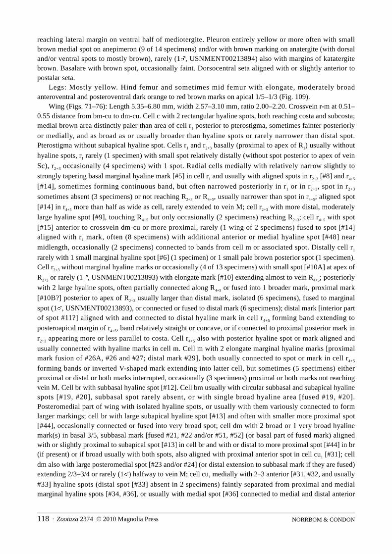
NORRBOM & CONDON118 · Zootaxa 2374 © 2010 Magnolia Press
reaching lateral margin on ventral half of mediotergite. Pleuron entirely yellow or more often with smallbrown medial spot on anepimeron (9 of 14 specimens) and/or with brown marking on anatergite (with dorsaland/or ventral spots to mostly brown), rarely (1%, USNMENT00213894) also with margins of katatergitebrown. Basalare with brown spot, occasionally faint. Dorsocentral seta aligned with or slightly anterior topostalar seta.
Legs: Mostly yellow. Hind femur and sometimes mid femur with elongate, moderately broadanteroventral and posteroventral dark orange to red brown marks on apical 1/5–1/3 (Fig. 109).
Wing (Figs. 71–76): Length 5.35–6.80 mm, width 2.57–3.10 mm, ratio 2.00–2.20. Crossvein r-m at 0.51–0.55 distance from bm-cu to dm-cu. Cell c with 2 rectangular hyaline spots, both reaching costa and subcosta;medial brown area distinctly paler than area of cell r1 posterior to pterostigma, sometimes fainter posteriorly
or medially, and as broad as or usually broader than hyaline spots or rarely narrower than distal spot.Pterostigma without subapical hyaline spot. Cells r1 and r2+3 basally (proximal to apex of R1) usually without
hyaline spots, r1 rarely (1 specimen) with small spot relatively distally (without spot posterior to apex of vein
Sc), r2+3 occasionally (4 specimens) with 1 spot. Radial cells medially with relatively narrow slightly to
strongly tapering basal marginal hyaline mark [#5] in cell r1 and usually with aligned spots in r2+3 [#8] and r4+5
[#14], sometimes forming continuous band, but often narrowed posteriorly in r1 or in r2+3, spot in r2+3
sometimes absent (3 specimens) or not reaching R2+3 or R4+5, usually narrower than spot in r4+5; aligned spot
[#14] in r4+5 more than half as wide as cell, rarely extended to vein M; cell r2+3 with more distal, moderately
large hyaline spot [#9], touching R4+5 but only occasionally (2 specimens) reaching R2+3; cell r4+5 with spot
[#15] anterior to crossvein dm-cu or more proximal, rarely (1 wing of 2 specimens) fused to spot [#14]aligned with r1 mark, often (8 specimens) with additional anterior or medial hyaline spot [#48] near
midlength, occasionally (2 specimens) connected to bands from cell m or associated spot. Distally cell r1
rarely with 1 small marginal hyaline spot [#6] (1 specimen) or 1 small pale brown posterior spot (1 specimen).Cell r2+3 without marginal hyaline marks or occasionally (4 of 13 specimens) with small spot [#10A] at apex of
R2+3 or rarely (1%, USNMENT00213893) with elongate mark [#10] extending almost to vein R4+5; posteriorly
with 2 large hyaline spots, often partially connected along R4+5 or fused into 1 broader mark, proximal mark
[#10B?] posterior to apex of R2+3 usually larger than distal mark, isolated (6 specimens), fused to marginal
spot (1%, USNMENT00213893), or connected or fused to distal mark (6 specimens); distal mark [interior partof spot #11?] aligned with and connected to distal hyaline mark in cell r4+5 forming band extending to
posteroapical margin of r4+5, band relatively straight or concave, or if connected to proximal posterior mark in
r2+3 appearing more or less parallel to costa. Cell r4+5 also with posterior hyaline spot or mark aligned and
usually connected with hyaline marks in cell m. Cell m with 2 elongate marginal hyaline marks [proximalmark fusion of #26A, #26 and #27; distal mark #29], both usually connected to spot or mark in cell r4+5
forming bands or inverted V-shaped mark extending into latter cell, but sometimes (5 specimens) eitherproximal or distal or both marks interrupted, occasionally (3 specimens) proximal or both marks not reachingvein M. Cell br with subbasal hyaline spot [#12]. Cell bm usually with circular subbasal and subapical hyalinespots [#19, #20], subbasal spot rarely absent, or with single broad hyaline area [fused #19, #20].Posteromedial part of wing with isolated hyaline spots, or usually with them variously connected to formlarger markings; cell br with large subapical hyaline spot [#13] and often with smaller more proximal spot[#44], occasionally connected or fused into very broad spot; cell dm with 2 broad or 1 very broad hyalinemark(s) in basal 3/5, subbasal mark [fused #21, #22 and/or #51, #52] (or basal part of fused mark) alignedwith or slightly proximal to subapical spot [#13] in cell br and with or distal to more proximal spot [#44] in br(if present) or if broad usually with both spots, also aligned with proximal anterior spot in cell cu1 [#31]; cell
dm also with large posteromedial spot [#23 and/or #24] (or distal extension to subbasal mark if they are fused)extending 2/3–3/4 or rarely (1%) halfway to vein M; cell cu1 medially with 2–3 anterior [#31, #32, and usually
#33] hyaline spots (distal spot [#33] absent in 2 specimens) faintly separated from proximal and medialmarginal hyaline spots [#34, #36], or usually with medial spot [#36] connected to medial and distal anterior

Zootaxa 2374 © 2010 Magnolia Press · 119REVISION OF THE FEMORALIS GROUP OF BLEPHARONEURA
spots [#32, #33] and sometimes to proximal anterior spot [#31] and/or proximal marginal spot [#34] to formY-shaped or trident shaped mark or posteriorly bilobed mark; subapical marginal hyaline spot [#37] moderateto large, often extending to vein Cu1. Cell dm with anterior to anteromedial subapical hyaline spot [#25]
relatively large, often extended to vein M. Abdomen: All tergites with 4 evenly spaced dark brown spots, L-shaped posterolateral dark brown band,
separated medially, and on tergites 3–5 anterolateral dark brown spot; some or all of these markings oftenconnected or fused to form irregular lateral markings, but at least submedial spots distinct except sometimeson tergite 5.
Female terminalia: Oviscape entirely dark brown; length 1.00–1.12 mm. Aculeus (Fig. 171) 0.9 mm long,3.6 times as long as wide, without scales dorsally or ventrally on membrane medially; lateral margin withminute, irregular serrations extending almost to lateral lobe; tip angular basolaterally, moderately long (lobedpart 0.37–0.43 times as long as wide), with broad deep medial apical concavity (deeper than length of mediallobes and more than half as broad as distance between apices of subapical lobes) bordered by moderatelylarge, triangular pair of medial lobes and with 3 pairs of small step-like lobes; sublateral and submedial lobessimilar in size; lobes separated by elongate shallow gaps, lateral gap 2.00 times as long as wide, gap betweenmedial and submedial lobes 1.5 times as long as wide. Spermathecae subspherical, with long, straight toslightly convoluted, slender sclerotized neck and with or without small cylindrical basal apodeme.
Male terminalia: Medial surstylus with prensisetae separated by several times width of medial prensiseta,medial prensiseta on curved lobe, lateral prensiseta small to minute, less than one-third as wide as medialprensiseta, acute, orange brown to dark brown.
Distribution. Mexico (Chiapas). The type specimens were collected between 1300–2000 m elevation inthe Sierra Madre de Chiapas.
Type data. Holotype & (USNM USNMENT00213892), MEXICO: Chiapas: 49 km S of Jaltenango,[Parque Natural] El Triunfo, [15°40'N 92°48'W], 1300–2000 m, 13–15 May 1985, A. Freidberg. Paratypes:Same data as holotype, 4%2& (USNM USNMENT00213890, USNMENT00213893, USNMENT00213896–97, USNMENT00213899–900) 3%2& (TAUI USNMENT00213889, USNMENT00213891,USNMENT00213895, USNMENT00213901–02) 1% (IEXV USNMENT00213898); El Triunfo, 1800 m, 14May 1985, A. Freidberg, 1% (USNM USNMENT00213894).
Etymology. The name of this species is a Latin adjective referring to the variable wing pattern.
Blepharoneura wasbaueri Norrbom & Condon, new speciesFigs. 5, 77, 155
Diagnosis. This species differs from other Blepharoneura species by the following combination of characters:scutum with 2 pairs of brown vittae; anepisternum entirely yellow; hind femur brown on apical fourth; cellsr2+3, r4+5 and m each with 2 marginal or submarginal hyaline marks, those in r2+3 and r4+5 small; and aculeus tip
with 7 lobes, including unpaired medial lobe, 2 pairs of step-like lobes, and broad, digitiform pair of laterallobes.
Description. Head: Dark brown area on ocellar tubercle extended to or almost to level of postocellar seta.Medial occipital sclerite with pair of brown submedial vittae on ventral half. Occipital suture narrowly brown.
Thorax: Scutum entirely microtrichose, with 2 pairs of dark brown vittae; submedial vitta broadlyinterrupted posterior to transverse suture and not connected to mark on posterior margin; sublateral vittasometimes (holotype) absent posterior to transverse suture, when present separated from mark on posteriormargin; posterior margin with 2 well separated brown marks. Notopleuron entirely yellow. Small brown spotsanterior to postsutural supra-alar seta and anterior to postalar seta absent. Small brown spot lateral todorsolateral corner of scutellum present. Scutellum with pair of small submedial brown spots (faint inparatype). Subscutellum with pair of dark brown spots on dorsolateral half, isolated from pair of narrowbrown vittae on mediotergite. Pleuron entirely yellow or with small brown medial spot on anepimeron

NORRBOM & CONDON120 · Zootaxa 2374 © 2010 Magnolia Press
(paratype). Basalare with brown spot. Dorsocentral seta aligned slightly anterior to postalar seta or almostmidway between postalar and postsutural supra-alar seta.
Legs: Mostly yellow. Mid femur sometimes with diffuse elongate anteroventral and posteroventral brownmarks. Hind femur brown on apical 1/4 but not extending to apex dorsally.
Wing (Figs. 5, 77): Length 7.04–7.30 mm, width 3.68–3.80 mm, ratio 1.91–1.92. Crossvein r-m at 0.55–0.56 distance from bm-cu to dm-cu. Cell c with 2 rectangular to inverted trapezoidal hyaline spots, bothreaching costa and subcosta; medial brown area paler than area of cell r1 posterior to pterostigma, fainter
medially, and as broad as to slightly narrower than hyaline spots. Pterostigma entirely brown or (1 wing ofholotype) with very small yellowish subapical spot. Cells r1 and r2+3 basally (proximal to apex of R1) with 1–2
small yellowish spots in r1 and 1 small yellowish or hyaline spot in r2+3. Radial cells medially with 2 narrow
basal marginal hyaline marks in cell r1 separated by narrower distinct dark brown area, both reaching R2+3,
basal mark [#5] quadrate; cell r2+3 with 1 moderately large hyaline spot [#8] aligned with basal mark in r1 and
1 broader spot [#9] aligned with second mark in r1, both reaching R2+3 and R4+5; cell r4+5 with hyaline spot [#14]
in anterior half of cell aligned between r1 marks and with moderate sized hyaline spot [#15] aligned distal to
dm-cu, sometimes (holotype) with additional posterior hyaline spot aligned with second mark in r1. Distally
cell r1 with small marginal hyaline spot [#6] (holotype) or small hyaline spot [#6A] in posterior half
(paratype). Cell r2+3 with 2 very small marginal hyaline spots [#10A, #11], neither extending half distance to
R4+5; with small to minute hyaline spot [#10B] in posterior half aligned with proximal marginal spot, touching
or isolated from R4+5; and also with 1–2 more proximal small to minute posterior hyaline or yellow spots
[between dm-cu and #10B]. Cell r4+5 with small hyaline spot near or slightly posterior to midwidth aligned
between hyaline marks in cell m; and with 2 small ovoid marginal or submarginal hyaline spots, anterior spot[18A] touching costa, posterior spot [#18] touching or narrowly isolated from costa; sometimes (1 wing ofholotype) with additional minute hyaline spot slightly proximal to posterior spot. Cell m without subbasalhyaline spot [#49] near midlength of dm-cu; with 3 large ovoid hyaline spots, 2 marginal [#27, #29] and 1medial spot [#26] aligned with proximal marginal spot, or (1 wing of holotype) with proximal marginal andmedial spots fused to form short band; none of hyaline marks reaching vein M. Cell br with subbasal hyalinespot [#12]. Cell bm with circular subbasal and subapical hyaline spots [#19, #20]. Cell bcu with (1 wing ofholotype) or without hyaline spot in lobe. Posteromedial part of wing with multiple large hyaline spots andmarkings; cell br subapically with moderately large hyaline spot [#13] and usually (except in 1 wing ofparatype) with smaller slightly more proximal hyaline spot [#44], both touching vein M but not R4+5; cell dm
with subbasal elongate hyaline mark across cell [fused #51, #52] or with only posterior spot [#52], posteriorlyaligned with spot in cell cu1 [#31] and anteriorly aligned with spot in cell br [#44] if present, posterior spot
sometimes medially connected to anterior part of more distal mark [#21]; elongate hyaline mark across cell[fused #21, #22?, #23?, #24?], broader posteriorly, anterior part aligned with subapical spot in cell br [#13],posterior part aligned with Y-shaped mark in cell cu1; cell cu1 with relatively large anterior subbasal hyaline
spot [#31] aligned with subbasal mark in dm, with broad Y-shaped medial mark [fused #32, #33, #36],narrowly or distinctly separated from proximal marginal mark across vein A1+Cu2 [#34, #39], proximal
branch of mark [#32] aligned slightly distal to line from proximal marginal mark [#34] to subapical spot incell br [#13], base of Y-shaped mark [#36] moderately broad; subapical marginal hyaline mark [#37]moderate sized, not reaching Cu1. Cell dm with subapical spot [#25] hyaline and relatively large, touching or
isolated from vein M; also with small to minute subapical hyaline or pale brown spot [#53] near midwidth orin posterior half aligned with or slightly proximal to subapical mark in cell cu1.
Abdomen: Syntergite 1+2 with pair of brown bands (probably fused submedial and sublateral spots) andsmall brown spot on posterolateral corner. Tergites 3–5 with pair of relatively small submedial dark brownspots, on tergite 5 of male connected to sublateral mark; with pair of large sublateral brown marks (formingvittae), each with narrow medial extension along posterior margin and narrow lateral extension toposterolateral corner.

Zootaxa 2374 © 2010 Magnolia Press · 121REVISION OF THE FEMORALIS GROUP OF BLEPHARONEURA
Female terminalia: Oviscape entirely dark brown; length 1.30 mm. Aculeus (Fig. 155) 0.95 mm long, 2.35times as long as wide, with acute scales dorsally and ventrally on membrane medially; tip relatively short andbroad (lobed part 0.32 times as long as wide), with small weakly trilobed medial lobe and 3 pairs of lobes,lateral lobe large and digitiform, with minute serrations apically, sublateral and submedial lobes acute, step-like, submedial lobe slightly smaller; medial, submedial and sublateral lobes separated by moderately deepgaps, ca. half as long as wide. Spermathecae subspherical, with nearly straight, slender sclerotized neck andsmall cylindrical basal apodeme.
Male terminalia (not dissected, but surstyli visible): Medial surstylus with prensisetae separated by severaltimes width of medial prensiseta, medial prensiseta on long lobe, lateral prensiseta slightly larger than medialprensiseta.
Distribution. Ecuador.Type data. Holotype & (CDFA USNMENT00104213), ECUADOR: Napo: Huahua Sumaco, Hollin -
Loreto road, km 44, Malaise trap, 14 Dec 1989, M. & J. Wasbauer & H. Real. Paratypes: Same data asholotype, 16 Dec 1989, 1% (CDFA USNMENT00104212).
Etymology. The name of this species is a noun in the genitive case in honor of Marius S. Wasbauer, one ofthe collectors of the type specimens and the author of a very useful catalog of tephritid host plants.
Blepharoneura zumbadoi Norrbom & Condon, new speciesFigs. 78, 145
Diagnosis. This species belongs to the femoralis complex (see diagnosis of B. femoralis), species of which aredifficult to distinguish except by aculeus shape. That of B. zumbadoi differs from those of B. osmundsonaeand nigrifemur in having the tip stouter (lobed part less than half as long as wide) and not rounded proximal tothe lateral lobe. The lobes, particularly the lateral one, are larger than in B. biseriata and they are blunter thanin B. cyclantherae, but the gaps are shallower than in B. femoralis which also has a rounded medial lobe. Thescutellum, in lateral view, sometimes has a ventrobasal brown mark, a character state that also occurs rarely inB. femoralis but is otherwise unknown in the complex.
Description. Head: Dark brown area on ocellar tubercle extended almost to postocellar seta. Medialvertical seta in yellow area. Medial occipital sclerite with pair of dark brown submedial vittae on ventral half.Occipital suture narrowly dark brown, on lateral side bordered by slightly paler, wedge-shaped area, broaderdorsally, just reaching postocular setae and sometimes almost reaching lateral vertical seta.
Thorax: Postpronotal lobe entirely yellow or with minute brown spot at junction with anepisternum.Scutum with 2 pairs of dark brown vittae; submedial vitta narrowed slightly posterior to transverse suture andconnected to or narrowly separated from mark on posterior margin; sublateral vitta narrowly interrupted attransverse suture, narrowly separated from mark on posterior margin; posterior margin with 1 broad brownmark, narrowed medially. Notopleuron dark brown except small yellow area surrounding posterior seta. Smallbrown spot anterior to postsutural supra-alar seta, brown vitta anterior to postalar seta, and brown spot lateralto dorsolateral corner of scutellum present. Scutellum with pair of large submedial brown marks or singleinverted U-shaped medial mark on disk usually extended to basal margin; side with or without small faintbrown spot to moderately large dark brown L-shaped mark on ventrobasal margin. Subscutellum andmediotergite with pair of dark brown vittae, ventral half of lateral margin of mediotergite also narrowlybrown, sometimes connected to vittae ventrally. Pleuron mostly dark brown, yellow only on propleuron, partof anepisternum (with large dorsomedial dark brown spot extending ventrally at least 2/3 distance to ventralmargin, and broadly dark brown in posteroventral corner), extreme anterior and posterior corners ofkatepisternum, all of katepimeron, greater ampulla, narrow dorsal, posterior and ventral margins ofanepimeron, and narrow anterodorsal margin of anatergite. Basalare brown. Dorsocentral seta aligned slightlyanterior to postalar seta.

NORRBOM & CONDON122 · Zootaxa 2374 © 2010 Magnolia Press
Legs: Mostly yellow. Mid femur with anteroventral and posteroventral brown marks on apical 1/5–1/4.Hind femur with entire apical 1/4–1/3 dark brown.
Wing (Fig. 78): Length 7.80–8.25 mm, width 3.70–3.80 mm, ratio 1.99–2.20. Crossvein r-m at 0.54–0.62distance from bm-cu to dm-cu. Cell c with 2 broad rectangular hyaline spots, both reaching costa andsubcosta; medial brown area almost as dark as area of cell r1 posterior to pterostigma, sometimes paler
medially, distinctly narrower than both hyaline spots. Pterostigma with large subapical hyaline spot [#3]reaching R1. Cell r1 basally (proximal to apex of R1) with 2 pale brown to hyaline spots (occasionally fused
into 1 elongate mark); cell r2+3 with 1–2 spots. Radial cells medially with 1 very broad quadrate basal hyaline
mark [#5 fused with additional spot?] in cell r1, sometimes partially divided posteriorly by pale brown spot;
cell r2+3 with 2 broad to very broad hyaline spots [#8, #9] posterior to r1 mark, both broadly reaching R4+5; cell
r4+5 with broad hyaline spot [#14] aligned with r1 mark, extended from R4+5 slightly more than halfway across
cell, and with medial hyaline spot [#15] near anterior end of dm-cu small; sometimes with small to minuteanterior hyaline spot [#48] near midlength not touching vein R4+5. Distally cell r1 with 1 marginal subapical
hyaline spot [#6]. Cell r2+3 with 2 marginal ovoid hyaline spots and subapical spot posterior to proximal spot
[divided #10, #11], and sometimes with 1–2 minute more proximal spots. Cell r4+5 with small hyaline spot
[#16] anteriorly, aligned with more proximal or between apical marks in cell r2+3; with 1–2 large ovoid
posterior hyaline spots aligned with hyaline marks in cell m; and with 2 ovoid or 1 bilobed marginal orsubmarginal hyaline spots [#18, #18A]. Cell m without subbasal hyaline spot [#49] near midlength of dm-cu;with 2–3 marginal hyaline spots, proximal and distal spots [#27, #29] large, medial spot [#28] very small ifpresent, and 1 large anteromedial spot [fused #26, #26A], narrowly separated from proximal marginal spot;distal and anteromedial spots narrowly and faintly separated from vein M. Cell br with subbasal hyaline spot[#12]. Cell bm with circular subbasal and subapical hyaline spots [#19, #20]. Cell bcu with large hyaline spotin lobe. Anal lobe hyaline except brown area from apical part of lobe of cell bcu and base of vein A1+Cu2
crossing apex of vein A2, occasionally (1%) with small brown submarginal spot medially proximal to A2.
Posteromedial part of wing with large broad hyaline area; cell br with subapical hyaline spot [#13] andsometimes with smaller more proximal spot [#44], sometimes fused into 1 broad spot; cell dm with broad andlong hyaline area [fusion of at least #51, #52, #21, #22, #23, #24, #50], sometimes narrowly or partiallydivided by pale brown medially, tapered distally, extending farther posteriorly than anteriorly; cell cu1
medially with broad anteriorly trilobed and posteriorly 1–3 lobed mark [fused #31, #32, #33, #36, #36A,sometimes #34] or with more broadly fused area with 2 anterior and 1–3 marginal brown spots, broad onposterior wing margin, proximally connected to or narrowly separated from mark [fused #34, #39] across veinA1+Cu2; subapical marginal hyaline spot [#37] reaching vein Cu1. Cell dm with anteromedial subapical
hyaline spot [#25] and with posterior hyaline spot proximal to level of subapical mark in cell cu1 and
connected to proximal hyaline area.Abdomen: Syntergite 1+2 with pair of isolated submedial brown spots and pair of irregular posterolateral
brown marks. Tergites 3–5 with pair of isolated submedial brown spots, anterolateral brown spots, sometimesisolated, and pair of irregular lateral brown markings formed from connected submedial spots and L-shapedposterolateral bands, sometimes connected to anterolateral spots.
Female terminalia: Oviscape entirely dark brown, length 1.33 mm. Aculeus (Fig. 145) 0.77 mm long, 1.71times as long as wide, with acute scales dorsally and ventrally on membrane medially; tip bluntly angulatebasolaterally, short triangular (lobed part 0.45 times as long as wide), with small, truncate, slightly notchedmedial lobe and 3 pairs of step-like lobes separated by relatively shallow gaps, that between sublateral andsubmedial lobes deeper; sublateral lobe larger than submedial lobe; lateral gap 1.05 times as long as wide.Spermathecae subspherical, with slightly convoluted, slender sclerotized neck and large cylindrical basalapodeme.
Male terminalia: Medial surstylus with prensisetae subequal, separated by several times width of medialprensiseta, medial prensiseta on long lobe, lateral prensiseta on short lobe.

Zootaxa 2374 © 2010 Magnolia Press · 123REVISION OF THE FEMORALIS GROUP OF BLEPHARONEURA
FIGURES 204–209. Host plants: 204, Cyclanthera dissecta, host of B. cyclantherae, Mexico: road to Huitzilac, 27 Sep1991, mass of vines; 205, same, flowers and fruit (arrow); 206, Microsechium helleri, host of B. regina, Mexico:Lagunas de Zempoala, 25 Sep 1991, vine with fruits (arrow); 207, same, fruits, on right with emergence hole of larva(arrow); 208, same, cross section of infested fruit; 209, Gurania acuminata, host of B. hirsuta, Venezuela: Guatopo, Jan1980, basal shoot mined by larva.
Distribution. Costa Rica. The collection sites are between 2200 and 2700 m elevation.Type data. Holotype & (INBio INBIO002578264), COSTA RICA: San José: San Gerardo de Dota, near
Albergue Savegre, LS 389000 484200, 2200–2300 m, 19–22 May 1997, M. A. Zumbado. Paratypes: COSTARICA: San José: Estación Biológico Cuericí, Sendero El Mirador, LS 389700 499600, 2640 m, 19 Feb - 15Mar 1996, B. Gamboa R., 1% (INBio INBIO002381175); San Gerardo de Dota, 9°33'N 83°48'W, along Río

NORRBOM & CONDON124 · Zootaxa 2374 © 2010 Magnolia Press
Savegre, end of log over river, 2200 m, on mixed patch of Cyclanthera langei Cogn. (95CR14) and Sechiumpittieri (Cogn.) C. Jeffrey (95CR15), 20 Aug 1995, A. L. Norrbom, 1% (USNM USNMENT00048392).
Etymology. The name of this species is a noun in the genitive case in honor of Manuel Zumbado, thecollector of the holotype, who is among the most prolific collectors of flies in Costa Rica.
Biology. No specimens have been reared to determine the host plants of B. zumbadoi. One adult wascollected on a mixed patch of Cyclanthera langei Cogn. (95CR14) and a Sechium species at San Gerardo deDota, Costa Rica (also see Biology section).
FIGURES 210–214. Host plants: 210, Sechium pittieri, host of Blepharoneura macwilliamsae, Costa Rica: San Gerardode Dota, May 1997; 211, same, flowers and leaves (leaf damage probably caused by Blepharoneura adults); 212,Sechium sp., node infested by larva of undetermined Blepharoneura species; 213, same, opened node with larva; 214,same, larva emerging from broken node.

Zootaxa 2374 © 2010 Magnolia Press · 125REVISION OF THE FEMORALIS GROUP OF BLEPHARONEURA
Biology
Adults of Blepharoneura and the closely related genera Baryglossa and Hexaptilona have rows of spines onthe labella, which are used, at least in Blepharoneura, for rasping leaves, flowers, or other tissues of theirhosts (Driscoll & Condon 1994, Condon & Norrbom 1994, 1999). This is the only group of Tephritidae inwhich adults are known to use their mouthparts to abrade and and damage solid plant tissue, although speciesof some other genera will feed on sap exuding from oviposition marks or other wounds.
FIGURES 215–218. Host plants: Sicyos sp., poss. longisepalus, host of Blepharoneura mexicana, Guatemala: nearSanta Elena Barrillas, 22 Nov 2007; 215, inflorescence; 216, fruits; 217–218, larva of B. mexicana ex stem mine. Photosby G.J. Steck.

NORRBOM & CONDON126 · Zootaxa 2374 © 2010 Magnolia Press
All species of Blepharoneura whose biology is known feed as larvae in species of Cucurbitaceae, as doesthe one species of Baryglossa for which host data are known. They are not only highly host specific but alsoare specialized as to the type of plant organ attacked. Individual species lay eggs in only one of the followingtypes of tissues: fruits, seeds, stems, or flowers (those attacking flowers usually specializing on either male orfemale flowers) (Condon & Norrbom 1999). Species of the poecilosoma group have been reared mostly fromflowers, although a few species feed as larvae in fruits or seeds.
Host data for the femoralis group are scant, but the larvae of the five species that have been reared as wellas larvae of two unidentified species were found feeding in fruits or stems. Condon & Norrbom (1999)provided a host plant table briefly listing the fly and plant species and the type of tissue attacked, but see theindividual species treatments in this paper for the detailed specimen data that was the basis of those recordsand, in some cases, updated identifications of the fly species. Blepharoneura cyclantherae has been rearedfrom fruits of Cyclanthera dissecta Arn. (Figs. 204–205) in central Mexico, B. regina Giglio-Tos from fruit ofMicrosechium helleri (Peyr.) Cogn. in central Mexico (Figs. 206–208), B. macwilliamsae from stem mines ofSechium pittieri (Cogn.) C. Jeffrey in Costa Rica (Figs. 210–211), B. mexicana from stem mines of a Sicyossp. in Guatemala (Figs. 215–218), and B. hirsuta Bates from mines of basal shoots of Gurania acuminataCogn. in Venezuela (Fig. 209). In May of 1995 additional larvae, which unfortunately were not successfullyreared, were found by INBio parataxonomists and ALN feeding in leaf nodes of an undetermined Sechiumspecies (Figs. 212–214). These larvae may possibly be B. chaconi, as adults of that species have beencollected on this plant. The infested leaf nodes showed only slight external signs of damage (Fig. 212). Thelarvae were nearly mature but had not tunneled extensively in the stems, unlike the larvae of B. hirsuta andmacwilliamsae which consume nearly the entire internal contents of at least two-thirds to over a meter ofstem. We also found slender larvae mining stems of Cyclanthera langaei Cogn. at San Gerardo that areprobably a species of the femoralis group. They may be B. sinepuncta or zumbadoi as those species were alsocollected as adults at San Gerardo on C. langaei or in a mixed patch of it and the Sechium species, whichfrequently grow together at this site.
Distribution
The overall distribution of the femoralis group extends from northwestern and northeastern Mexico (Sinaloa,Tamaulipas) to northwestern Argentina. No species are known from the West Indies or Chile. Known speciesdiversity by country is as follows (including undescribed species): Argentina 2, Belize 1, Bolivia 7, Brazil 7 (3northern, 4 southern), Colombia 3, Costa Rica 14, Ecuador 7, Guatemala 7, Guyana 2, Mexico 13, Nicaragua1, Panama 2, Peru 5, and Venezuela 4. These data undoubtedly are biased by uneven collecting (e.g., thedifference between Costa Rica and Panama), but the high diversity for Mexico is noteworthy. Alsonoteworthy is the high degree of sympatry among closely related or sister species within the femoralis group(e.g., B. apaapa, hyalinella and nigriapex known only from the same type locality in the Yungas region ofBolivia; B. ruptafascia and unifasciata known only from the same type locality in Ecuador; B. furcifer andseptemdigitata, sympatric at least in eastern Peru and northern Bolivia; and B. cornelli and lutea, both knownonly from Costa Rica; the aspiculosa-splendida clade, including 5 species from Mexico to Ecuador with atleast partially overlapping ranges; B. bipunctata and multipunctata, known only from adjoining provinces inEcuador; and B. amplihyalina and marshalli, known only from adjoining regions in northwestern Argentina).
For the following summary of altitudinal distributions, except as indicated, the data in parentheses werederived from specimen labels or in a few cases from independent information provided by the collector orlabels of specimens of other species collected at the same locality (e.g., Nova Teutonia, Brazil). We did notinvestigate in detail the elevations of other collection localities, but they appear to be consistent with thegeneral altitudinal patterns.
Species of the rupta clade have been collected predominantly at low to middle elevations (<1000 m): B.bidigitata (300–500 m), bivittata (200 m), brevivittata (10 m), sp. nr. brevivittata, cornelli (50–150 m),

Zootaxa 2374 © 2010 Magnolia Press · 127REVISION OF THE FEMORALIS GROUP OF BLEPHARONEURA
fernandezi (450 m), furcifer (150–800 m), sp. nr. furcifer (400 m), hirsuta (600 m), io (type locality at ca.1200 m), lutea (700 m), quadristriata (160–1600 m), rupta, ruptafascia (340 m), septemdigitata (250–750 m),thetis (300–500 m), sp. nr. thetis (750 m), and unifasciata (340 m).
Species of the splendida, nigriapex, and regina clades (except perhaps B. isolata of the regina clade (typelocality at 350–450 m?)) appear to occur predominantly at higher elevations: B. amplihyalina (700–2000 m),apaapa (2000–2050 m), aspiculosa (1300–2000 m), bipunctata (2000 m), biseriata (2134 m), chaconi (2200–
2600 m), cyclantherae (type locality at ca. 2170 m), femoralis (>1000 m, usually > 2000 m, except southernBrazilian specimens), macwilliamsae (1600–2350 m), marshalli (1500 m), mexicana, mikenoltei (2500 m),multipunctata (2600 m), nigriapex (2000 m), nigrifemur (2000 m), osmundsonae (2683 m), quetzali (1600m), sinepuncta (900–2200 m), regina (2400–2900 m), tau (1600 m), variabilis (1300–2000 m), and zumbadoi(2200–2700 m). Blepharoneura splendida (480–1860 m) and punctistigma (480–2300 m) have been collectedat a range of middle to moderately high elevations.
Phylogenetic relationships
As first noted by Munro (1957), Blepharoneura belongs to a clade that also includes the Afrotropical genusBaryglossa Bezzi and the southeastern Asian Hexaptilona Hering. The latter two genera form the sister groupof Blepharoneura, and the fossil genus Problepharoneura Norrbom & Condon, or it and the Neotropicalgenus Ceratodacus Hendel, probably are the sister group of the above three genera (Norrbom & Condon1999). These five genera comprise the subfamily Blepharoneurinae (Norrbom & Condon 1999, Korneyev1999).
Blepharoneura is considered monophyletic based on its dorsally setulose vein Cu1. This character state,
which is rare in other Tephritidae and unique to Blepharoneura within the Blepharoneurinae, was considereda synapomorphy of the genus by Norrbom & Condon (1999). Within Blepharoneura, two species groups havebeen recognized: the femoralis group and the poecilosoma group. The poecilosoma group is supported asmonophyletic by the structure of the aculeus tip, which has numerous marginal serrations, each associatedwith an internal channel (Condon & Norrbom 1994, Norrbom & Condon 1999).
For analysis of relationships within the femoralis group, the following outgroup taxa were selected:Hexaptilona (both sexes of H. palpata (Hendel) and a female of H. hexacinioides (Hering); there was littlevariation and they were scored as a composite in the matrix), and Baryglossa trulla Munro (female only). Twospecies of the Blepharoneura poecilosoma group, B. diva Giglio-Tos and an undescribed species (sp. x), wereincluded in the ingroup to test the monophyly of the femoralis group. The wings of the outgroup species andthe two species of the poecilosoma group included in the analysis are shown in Figures 79–83.
Characters used in the analysis are listed in Table 1, and the taxa were scored as indicated in the matrix inTable 2. The matrix was analyzed using TNT (Goloboff et al. 2003) (max trees = 10,000, traditional search,swapping algorithum = tree bisection reconnection, all characters non-additive and unweighted). The analysisresulted in 72 trees of 289 steps. The strict consensus of these trees is shown in Figures 219–221.
TABLE 1. Characters used in phylogenetic analysis of Blepharoneura femoralis group.
1. Vertex color – 0) Brown area on ocellar tubercle not extended beyond postocellar seta; 1) brown area on ocellartubercle extended beyond postocellar seta or small medial brown spot present slightly ventral to postocellar seta; 2)with brown markings surrounding medial vertical seta (may be band or mark covering most of vertex). The spot inB. diva and sp. x, which is mostly anterolateral to the medial vertical seta, may or may not be homologous with thatin some species of the femoralis group, in which it extends posteromesal to the seta.
2. Medial occipital sclerite – 0) with submedial pair of brown vittae on ventral half; 1) entirely yellow.3. Area lateral to occipital suture – 0) entirely yellow; 1) with brown band or triangular marking extending from medial
part of suture towards postocular setae or reaching eye margin, not extending medially beyond occipital suture; 2)with broad triangular brown marking also extending medially and fused with submedial vitta; 3) with diffuse brownspot dorsally, touching lateral vertical seta.

NORRBOM & CONDON128 · Zootaxa 2374 © 2010 Magnolia Press
4. Scutal microtrichia – 0) entirely microtrichose; 1) with large nonmicrotrichose area.5. Posterior margin of scutum, brown markings – 0) with 2 separate marks; 1) with 1 mark, bimodal, narrowest medi-
ally; 2) with 1 mark, trapezoidal or triangular, broadest medially, or narrow band; 3) without brown marks.6. Scutal brown vittae – 0) with 2 pairs of vittae or rows of spots or at least 2 presutural pairs of marks; 1) with 3 vittae
or rows of spots (or at least 3 presutural marks), including unpaired medial vitta and pair of spots or vittae alignedwith medial corner of postpronotal lobe and dorsocentral seta or [B. cornelli] with only the latter pair of marks; 2)with 1 pair of submedial vittae or rows of spots at least presuturally; 3) with 1 pair of lateral vittae only; 4) withoutbrown vittae. B. brevivittata and bivittata sometimes have a second postsutural vitta, but only 1 presutural pair andwere coded state 2. Vittae are sometimes absent in the poecilosoma group and often vary intraspecifically, but mostspecies have markings similar to B. diva or as in Condon & Norrbom (1994, fig. 5c).
7. Notopleuron – 0) with brown vitta on lateral margin extending over entire lateral margin of postpronotal lobe; 1)without brown marking; 2) with lateral brown vitta or spot(s) or mostly brown (postpronotal lobe entirely yellow orwith diffuse brown spot posterolaterally).
8. Anterodorsal corner of anepisternum and postpronotal lobe laterally – 0) entirely yellow; 1) anepisternum withbrown spot, usually triangular or sideways V-shaped (small in B. zumbadoi and chaconi); 2) with spot as in state 1,also with small diffuse brown spot laterally on postpronotal lobe ventral to postpronotal seta; 3) anepisternum withnarrow brown vitta on dorsal margin and postpronotal lobe with broad brown vitta laterally.
9. Spot anterior to postsutural supra-alar seta – 0) present; 1) absent. This spot is also sometimes absent in the poecilo-soma group.
10. Spot or vitta anterior to postalar seta – 0) present; 1) absent. This spot is also sometimes absent in the poecilosomagroup.
11. Spot lateral to base of scutellum (lateral to dorsolateral corner) – 0) present; 1) absent. B. thetis, sp. nr. thetis, andsome B. quadristriata have a mark in this area extending from the medial posterior scutal mark or (some B. quadris-triata) a spot immediately posterior to the intra-alar seta. They were tentatively coded state 0.
12. Scutellum dorsal color pattern – 0) entirely yellow; 1) with single medial spot, not narrowed medially; 2) with ovoidor elongate paired medial spots or single inverted U-shaped or inverted mushroom shaped spot, not extended later-ally to basal setae; 3) with widely separated or transversely elongate paired lateral spots, often extended to basalsetae, or with transverse band (spots connected medially); 4) brown, except laterally, extending to apex. Some spec-imens of B. variabilis and macwilliamsae have a pair of minute spots on the basal margin, not considered homolo-gous to the markings in states 1–3, and they were coded state 0. Hexaptilona species have a pair of sublateral spotson the basal margin that do not appear to be homologous with the markings in Blepharoneura. These markings weredisregarded in scoring this taxon. H. hexacinioides sometimes has a pair of submedial spots.
13. Scutellum lateral color – 0) without brown markings; 1) with brown spot or mark on or near ventrobasal corner. Themarkings in B. thetis and one specimen of B. hirsuta are in a different position and probably not homologous. Thesespecies were coded state 0.
14. Subscutellum and mediotergite – 0) both with pair of brown vittae or entirely brown except narrowly medially; 1)subscutellum entirely yellow, mediotergite with pair of brown vittae; 2) mediotergite entirely yellow, subscutellumwith narrow brown mark on dorsal margin laterally; 3) both entirely yellow.
15. Anepisternum – 0) entirely yellow; 1) with small dark brown spot dorsal or posterodorsal to anterior seta; 2) mostlybrown or at least with brown medial spot extended to level of anterior seta; 3) with narrow vitta on dorsal marginonly.
16. Katepisternum – 0) entirely yellow; 1) with brown marking. 17. Anepimeron – 0) entirely yellow; 1) with brown marking.18. Katatergite – 0) brown ventrally or dorsally and ventrally; 1) with brown margins; 2) brown only dorsally or poster-
odorsally; 3) mostly or centrally brown; 4) entirely yellow. State 4 also sometimes occurs in the poecilosoma group.19. Anatergite – 0) mostly brown or at least brown in posteroventral corner; 1) with dorsomedial brown spot or some-
times more extensively brown, but at least posteroventral corner yellow; 2) entirely yellow. State 2 also sometimesoccurs in the poecilosoma group.
20. Meron – 0) entirely yellow; 1) with brown marking.21. Basalare – 0) with brown spot or entirely brown; 1) entirely yellow. State 1 also sometimes occurs in the poecilo-
soma group.22. Femora color – 0) entirely yellow; 1) yellow except hind femur and sometimes mid femur with elongate anteroven-
tral and posteroventral brown marks on apical 1/5–1/4; 2) yellow except hind femur with entire apical 1/6–1/2brown, and sometimes mid femur with anterior and posteroventral brown marks on apical 1/5–1/4; 3) yellow excepthind femur with anterior apical spot [not just on anteroventral apical ridge]; 4) yellow except anteroventral apicalridge of mid femur and/or anteroventral basal ridge of mid tibia and/or anteroventral and posteroventral apical ridgesof hind femur and/or anteroventral and posteroventral basal ridges of hind tibia dark brown. B. nigriapex in whichthe apical 1/6 of the hind femur is brown was tentatively scored state 2; in other species with this state the apical1/4–1/2 is brown.

Zootaxa 2374 © 2010 Magnolia Press · 129REVISION OF THE FEMORALIS GROUP OF BLEPHARONEURA
Wing23. Costal cell – 0) with 2 distinct rectangular to triangular hyaline areas, margins well differentiated, or with single par-
tially fused area with medial brown mark higher than wide; 1) with 2 rounded hyaline areas or single goggles-shapedarea, or hyaline area diffuse and margins poorly differentiated.
24. Pterostigma subapical hyaline or pale brown spot [#3] – 0) present; 1) absent. The spot is variable in the poecilo-soma group.
25. Cells r1 and r2+3 proximal to level of apex of vein R1 – 0) only with spot in r1 (usually posterior to apex of Sc); 1)without hyaline or pale brown spots; 2) with more than 1 spot.
26. Shape of basal marginal spot [#5] in cell r1 – 0) tapered; 1) not tapered. 27. Basal marginal hyaline marks in cell r1 – 0) with single narrow mark; 1) with 2 marks, 1 partially divided mark, or 1
very broad mark (presumed fusion of 2 marks). 28. Hyaline spots in cell r2+3 between crossveins r-m and dm-cu – 0) with 1–2 spots, if only 1 [#9] it is distal to spot in r1
[#5], if 2 spots they are relatively small (combined less extensive than brown area(s) between crossveins); 1) with nomore than 1 spot [#8], more or less aligned with basomarginal spot in r1; 2) with 2–3 large spots [#8, #9] (combinedusually more extensive than brown area(s) between crossveins), or with 1 very broad hyaline area. In B. amplihy-alina and marshalli the hyaline area is usually broad and often aligned with r-m which is more distal than in mostother species. In B. diva and pulchella the spots are occasionally absent, but when present there is at least the moredistal spot.
29. Spots in apical part of r1 – 0) with 1 hyaline spot [#6] touching or almost touching margin; 1) without hyaline spots;2) with more than 1 spot [#6 plus additional spot(s)] touching or almost touching margin; 3) with only posteriorspot(s). In the poecilosoma group some species have state 0, some have state 1, and some are polymorphic for states0 and 1.
30. Cells r2+3 and r4+5 distal to dm-cu with numerous additional spots (usually tiny and/or pale brown) – 0) absent; 1)present.
31. Cell r4+5 with spot [#15] near crossvein dm-cu – 0) present; 1) absent.32. Cell r4+5 with spot [#15] near crossvein dm-cu and spot [#14] aligned with r1 hyaline mark both very large, some-
times fused – 0) no; 1) yes. Spot #14 is large in B. zumbadoi and macwilliamsae, but spot #15 is small and these spe-cies were scored state 0.
33. Hyaline markings in cell m and in cells r2+3 and r4+5 distal to dm-cu – 0) with marginal or submarginal spots, includ-ing 2 in r2+3 [#10, #11] (sometimes fused into 1 irregular mark) and at least 2 in m [#27, #29], often not elongate(usually forming triangular pattern with anteromedial spot [#26, or fused #26 + #26A] or fused into 1 large triangu-lar mark); 1) cells r2+3 and m each with 2 elongate hyaline marks [1 specimen each of B. furcifer and B. septemdigi-tata have 3 spots in m], distal mark in r2+3 usually aligned with mark in r4+5 forming concave band extending tomargin of r4+5 [including #11 and #18], proximal mark in r2+3 [#10] often aligned with mark in r4+5 forming bandextending into r4+5, sometimes connecting or almost connecting with proximal mark in cell m [fused #26, #26A and#27] to form band; 2) r2+3 with 0–1 marginal marks, wing with 3 bands extending from posterior margin (2 in cell m),most distal band largest, extending into r2+3, straight to convex (usually parallel to costa). Blepharoneura variabilissometimes has a separate marginal hyaline spot or elongate mark in cell r2+3; although not having 2 marginal hyalinespots in r2+3, it was scored as variable for states 1 and 2 because in many specimens the subapical band is convex (asin state 1) although it does not reach the margin in r2+3. In some species of Baryglossa the markings near the apex ofthe wing are fused, but the pattern differs from that within the femoralis group suggesting that it is independentlyderived. At least one species of the poecilosoma group is banded apically (state 2), but it is clearly related to specieswith a spotted pattern. Thus state 0 appears to be plesiomorphic for the femoralis group.
34. Cell r4+5 area aligned with proximal marginal hyaline mark in cell r2+3 [#10] – 0) without aligned, connected hyalinearea (i.e., wing without band extending into cell r4+5); 1) with hyaline mark aligned and connected with proximalmark in r2+3 forming band extending into cell r4+5.
35. Number of marginal or submarginal spots in cell r4+5 – 0) 1 spot [#18]; 1) 2 spots [#18, #18A] or 1 large bilobed spot[#18 and #18A connected proximally].
36. Number of marginal or submarginal hyaline marks in cell m – 0) 3 [#27, #28, #29]; 1) 2 [#27, #29]. In species withonly 2 marginal hyaline marks we hypothesize that the proximal one is number #27 and that #28 is lost because #28is small when rarely present in B. femoralis and zumbadoi. In the poecilosoma group some species have state 0,some have state 1, and some are polymorphic for states 0 and 1.
37. Size of medial marginal or submarginal hyaline spot in cell m – 0) medial spot [#28] similar in size to other 2 mar-ginal spots [#27, #29] (or absent); 1) medial spot [#28 fused with #26 and sometimes #26A] larger than other 2 mar-ginal spots. Blepharoneura amplihyalina and marshalli were difficult to score for this character because in mostspecimens all of the spots in cell m are connected or fused; both species were scored state 1 because in one specimenof B. amplihyalina there is a separate large medial mark [fused #28, #26, #26A].

NORRBOM & CONDON130 · Zootaxa 2374 © 2010 Magnolia Press
38. Hyaline marks in cell m and adjacent area in cell r4+5 – 0) cell r4+5 without spots aligned with either mark in cell m[#26, #26A, #29]; 1) r4+5 with spot or spots aligned with 1 or both marks in m, but neither connected to form bandextending into r4+5; 2) r4+5 with marks aligned and connected with both marks in m, forming bands or inverted V-shaped mark extending into r4+5; 3) r4+5 with mark aligned and connected with only distal mark in m to form bandextending into r4+5 (if present, spot aligned with proximal mark isolated); 4) r4+5 with mark aligned and connectedwith only proximal mark in m to form band extending into r4+5 (if present, spot aligned with distal mark isolated).
39. Cell m with subbasal spot [#49] near midlength of dm-cu – 0) absent; 1) present.40. Cell br subbasal spot [#12] – 0) present; 1) absent.41. Cell bm spots – 0) with 2 spots, subbasal and subapical [#19, #20]; 1) with 1 large spot [fused #19 and #20]; 2) with
only subapical or small medial spot [#20]; 3) without hyaline spots.42. Spot #25 – 0) present; 1) absent. Presumed fused with posterior spot or band in B. splendida and most B. sinepuncta;
possibly fused with basal marks in B. amplihyalina and nigriapex.43. Cell dm with posterior subapical hyaline spot [#53] aligned with subapical hyaline mark in cell cu1 [#37] – 0) absent;
1) present. 44. Cell dm with subbasal hyaline band or series of spots – 0) without hyaline band or aligned spots [#51, #52] proximal
to level of subapical spot in cell br [#13]; 1) with band or pair of aligned spots [#51, #52] or at least a posterior spot[#52] distinctly proximal to subapical spot in cell br [#13], usually aligned with more proximal anterior spot in cellbr [#44] and anterior proximal spot [#31] (or proximal anterior part of more extensive mark) in cell cu1 if the latterspots present (species with a large hyaline area extending well proximal to level of spot #13 were also coded state 1).
45. Cell dm with anteromedial hyaline spot [#50] distal to level of subapical spot in cell br [#13] and aligned spot [#21]in dm – 0) absent; 1) present (species with a large hyaline area extending to this area were also scored state 1).
46. Cell cu1 anteromedial spot [#32] – 0) aligned distal to line between proximal marginal spot [#34] and subapical spotin cell br [#13]; 1) aligned with proximal marginal spot [#34] and subapical spot in cell br [#13], often forming band.B. tau, in which spot #32 is absent, and B. amplihyalina and marshalli, which have a large hyaline area covering allof the middle of cell cu1 (presumably including fused spots #32 and #33), were scored state 0.
47. Cell cu1 medial marginal hyaline markings (excluding mark on apical part of vein A1+Cu2 [#34] and subapical mark[#37]) – 0) with 1 marginal spot [#36] or relatively narrow extension of larger fused area; 1) with 2 spots [#36,#36A] or extensions of larger fused area or with very broad hyaline marginal area [fused spots #36 and #36A], usu-ally with small marginal or submarginal brown spot within it.
48. Cell cu1 medial hyaline markings – 0) with medial anterior hyaline spot [#32] (aligned slightly proximal to medialmarginal spot [#36]) small to moderate sized and well separated from medial marginal spot [#36]; 1) with medialanterior hyaline spot [#32] large and connected or almost connected with medial marginal spot [#36] (often con-nected to 1–2 anterior spots [#31 and/or #33] and/or 1–2 other posterior spots [#34 and/or #36A] forming Y-shaped,trident-shaped, or H-shaped mark or very broad hyaline area.
49. Hyaline medial mark(s) in cell dm – 0) total area of spots or larger fused area relatively narrow, less than half as longas cell; 1) broadly fused into large hyaline area more than half as long as cell, narrowed distally; 2) as state 1, butbroad distally, anterodistal corner extended to or almost to anterior margin of cell; 3) reduced except for broad pos-teromedial spot [fused #23, #24, usually also #22], anterior spot [#21] aligned with subapical spot [#13] in br absent.
50. Cell cu1 subapical hyaline spot [#37] – 0) present; 1) absent.
Abdomen51. General pattern – 0) usually spotted or reticulate, if largely dark brown (markings fused), medial yellow area rela-
tively narrow and with irregular margins; 1) with submedial vittae, rows of large spots, or irregular (usually bi- ortrimodal) brown markings, widely separated medially by tapering yellow area with even or nearly even margins. B.cornelli, which has a broad yellow medial area with uneven margins, was tentatively scored state 0. B. fernandezi,which has reduced markings, was scored state 1 because their medial margins are aligned as in species with vittae.
52. Tergites 3–5 – 0) without anterolateral spot; 1) with anterolateral spot (on or near lateral margin) or with marginmore broadly brown anteriorly. Species in which the entire lateral margin is brown were difficult to score. Thosewith the margin more broadly brown anteriorly (e.g., B. chaconi, mikenoltei) were hypothesized to have the spotfused to posterior markings and were scored state 1. B. cornelli and septemdigitata, in which the mark is parallelsided or narrowed anteriorly (appearing more like an extension of the posterior mark) and sometimes not extendinganteriorly on tergite 3, were scored 0.
53. Syntergite 1+2 with submedial spot fused on its lateral side to posterolateral band – 0) no; 1) yes. B. macwilliamsae,in which the spot is sometimes narrowly connected near its midwidth, was scored state 0.
54. Brown markings solid, without yellow spots within them – 0) no (or all brown markings small); 1) yes.55. Lateral margins of tergites – 0) with brown markings; 1) entirely yellow, without brown markings.

Zootaxa 2374 © 2010 Magnolia Press · 131REVISION OF THE FEMORALIS GROUP OF BLEPHARONEURA
Aculeus56. Aculeus shape (length/ width) – 0) > 4.0; 1) 2.4 – 4.0; 2) < 2.4. 57. Medial membrane of aculeus – 0) without scales; 1) with acute triangular scales; 2) with mostly blunt scales.58. Aculeus tip lateral lobe shape – 0) not digitate; 1) digitate (projecting, rounded).59. Aculeus tip sublateral lobe – 0) not broad and slanted; 1) lobe broad and slanted.60. Gap between sublateral and submedial lobes – 0) no larger than gap between sublateral and lateral lobes; 1) much
broader than gap between sublateral and lateral lobes (not deep, mostly perpendicular to long axis of aculeus).61. Aculeus tip medially – 0) with convex, truncate, shallowly notched or minutely trilobed medial lobe; 1) with broad,
deep medial concavity; 2) with broad, shallow medial concavity; 3) with narrow, small, acute medial indentation.62. Aculeus tip marginal serrations – 0) without serrations; 1) with minute serrations, not associated with channels, on at
least lateral lobe; 2) with minute serrations on lateral margin (not on lobes); 3) with large serrations associated withchannels. Species of the Blepharoneura poecilosoma group have distinct serrations, each associated with an internalchannel. Where present in the femoralis group, the serrations are much smaller and not associated with a channel.Hexaptilona hexacinioides also has minute serrations, but none are present in H. palpada or Baryglossa trulla.
63. Spermathecal neck – 0) slender, straight to slightly convoluted; 1) moderately stout, strongly convoluted; 2) withshort, weak basal part and very stout, cylindrical distal part.
64. Medial surstylus – 0) moderately broad and short, prensisetae closely approximated; 1) moderately long, prensisetaewell separated, at least medial prensiseta on lobe; 2) elongate, slender, prensisetae closely approximated; 3) rela-tively short and broad, prensisetae well separated, neither on a lobe. The shape of the medial surstylus and the spac-ing of the prensisetae also varies within the poecilosoma group.
65. Lateral prensiseta – 0) subequal to as large as medial prensiseta; 1) minute to small, much smaller than medial pren-siseta; 2) absent.
66. Sclerotized part of glans – 0) small, slender, more or less cylindrical; 1) large, stout, bulbous.
The monophyly of the femoralis group is supported in the analysis, but no obvious, unequivocalsynapomorphies (i.e., easily defined and without homoplasy or subsequent character state changes) have beenidentified. We will further test this hypothesis with DNA sequences as samples become available. The shapeof the aculeus, which is broad and flat, usually with a rather truncate apex and distinct lobes, may be asynapomorphy, but there is no single aspect of it that is clearly a unique apomorphy. The length to width ratioof the aculeus (character 57) may be a synapomorphy for the femoralis group, but the there is overlap in therange of this character in Hexaptilona (thus the polarity of this character is uncertain). The aculeus is moreelongate in the poecilosoma group and in Baryglossa trulla, but may be shorter in other Baryglossa species,and the tip is somewhat triangular in some species of the femoralis group (e.g., B. aspiculosa, mexicana,nigrifemur, quadristriata, quetzali), and the lobes are sometimes weaker, similar to some species ofHexaptilona and Baryglossa. The bulbous shape and large size of the sclerotized part of the glans (character66), which may be correlated with aculeus shape, is another possible synapomorphy for the femoralis group.Several other character states that are not present in all species of the femoralis group also can be interpretedas synapomorphies of the group, but only assuming secondary loss in some species. Among these are theshape of the basal marginal hyaline spot [#5] in cell r1 (char. 26) (assumes reversal in most of the rupta clade
and within the splendida clade), the number of hyaline marginal spots in cell m (char. 36) (assumes reversal inthe nigriapex clade; also there is convergence within the poecilosoma group which includes many species thatare polymorphic for that character), and the presence of a subbasal hyaline mark in cell dm or a very broadhyaline area extending to this area (char. 44) (requires reversal within all four clades).
Four large clades within the femoralis group were recognized (Fig. 219), the regina, nigriapex, splendidaand rupta clades. The regina clade is hypothesized as the sister group of the rest of the femoralis group.Synapomorphies of the rupta + nigriapex + splendida clades include: The number of marginal or submarginalhyaline spots in cell r4+5 (char. 35) (assumes reversal within all 3 clades), the very short and broad shape of the
aculeus (char. 56.2), and the presence of sclerotized scales on the medial membrane of the aculeus (char. 57)(assumes reversal in B. amplihyalina and marshalli).

NORRBOM & CONDON132 · Zootaxa 2374 © 2010 Magnolia Press
TABLE 2. Character matrix used in phylogenetic analysis of Blepharoneura femoralis group.
111111111122222222223333333333444444444455555555556666666
Taxon 123456789012345678901234567890123456789012345678901234567890123456
Outgroup
Hexaptilona 002000030000003000000000201001000000010000000000000000010000000000
2 1 2 2
Barygl. trulla 002100031114002113010001001000000000110110101101000000000000000???
diva 200003100100000000000000000000000000000000000000000000000000030000
1 1
sp. x 20000010000000000000000020002000000000000000100000000000000003?000
2
Ingroup
splendida 001100200002002113010200001110002001020000000001000110021000000101
1 1 11 3 21 1 1
punctistigma 001010200002002113010200001100002001020000000101000100021000000101
1 2 1 1 1 1
3 2
aspiculosa 001010200002002113010200201110002001020000010101100100020000000???
sinepuncta 001010200002102113010201001110002001020010000100000110021000000101
1 1 2 3 21 1 1
femoralis 001000200002002113010100011200000010010000010001000100021000000101
1 11 1 1 2 2 1 1 1111 1 1
2
3
zumbadoi 001010200002002113010200211200000010010000011001100100021000000101
1 1 1 1
cyclantherae 001100200002002113010200011200000011010020010001100010021000000121
1 1 12 1 1 1 1
biseriata 001110200002002113010200211200000011010020010001100110021000000101
1
nr. biseriata ???110200002002113010200211200000011010000011001100110022000000???
osmundsonae 000110200002002113010200011200000011010000011001100000012000000101
1 1 2 1 2 11 2
nigrifemur 001010200002002113010200011200000011010000010011100100021000000101
1 2 2 1 1
multipunctata 000000220001001004000400211221000010111000110011300100021000100???
bipunctata 000000210001001010000400211201000010101000100011300100021000000???
2 2 1
apaapa 000000210001001004100400201001000000101000000011300100021000000101
1 2 1 1 2 1 1 1
hyalinella 000000220001001004100400200021000000011000100011300000021000000101
1 1 1 1 1 1 1 1
nigriapex 000000220001001004100200200021000000111001111011200100021000000???
amplihyalina 000000200001000004100000201200000000110011011011200100020000300101
1 1 31 1 1 1
marshalli 000000200001000004000000201100000000100001011011200100020000200101
1 1 1 1 1 211 1 11 1 1
3
tau 0031202000020010121011011001100020010200200000000101010????????10?
macwilliamsae 000000200000000000010100210200000001010010011011100100021000000101
1 1 11 1 1 31 1 1 2 1
4
bidigitata 010000200002000002000000010200000011000000010001000000021101010111
1 2 41 3 2 21 11 211 1

Zootaxa 2374 © 2010 Magnolia Press · 133REVISION OF THE FEMORALIS GROUP OF BLEPHARONEURA
Table 2. (continued).
111111111122222222223333333333444444444455555555556666666
Taxon 123456789012345678901234567890123456789012345678901234567890123456
----------------------------------------------------------------------------------
wasbaueri 00000010110200000420020021120100001101000001000100001002110001010?
1 1 3
mikenoltei 000004100000000011001100200200010011010000111001000100010000200???
1
chaconi 100010200002000011011100210200010001010000111001100100010000200111
1 1 1 1 111
2 2
quetzali 000000200000000004200101000010001001020000010001000100010000220???
1 2 3 1
variabilis 000000100000000001000101100000011001010000000000000100010000120111
2 142 21 1 2 2 1 1 1
3 2
4
mexicana 000000100010000004001000011200000001010000011001100000010000200111
42 11 3 1 2 1 11 1 1 1 1 1
2 2
regina 000000100000000010001000210220010001011000111011200000010000320111
1 22 111 1 421 1 1 1 1
isolata 0100001000000000042010002100300020010300000000110001000???????????
unifasciata 010002101110030004201400100010002001000021000100001001021001000???
ruptafascia 010000101110000004201400100010001001000021000100001001021001000???
1
thetis 2101221011030100042010000000000000110000000100000010000????????101
11
nr. thetis 2101221011000100042010001000000000010000000101000010010????????101
fernandezi 010132101110030004201000000000000001000000010001001001121100112???
2 4 1 3 1 1
nr.brevivittata 2101321011100300042010001000100020010000000101000010011????????101
13 2 1
brevivittata 210122101110000004201010100110002001000000000101001001121100112101
3 2 1 1 1 2 121 11 1
3 3 4 3
bivittata 210112101110010004201001000110002001020001000100011001121100102101
3 2 3 3 1 1 1 1 1 1
3
hirsuta 210121101110030004201001100110002001020001000100001000021101010111
34 2 1 3 1 3 12 1 1
3
lutea 000134101110030004201011100110102001020001000100001000021110111???
1
rupta 200121101110020004201011000010002001020020000101001000021110011201
1 34 3 1 1 4 1
cornelli 200131101110030004201111100110102001020001000100000000021110011???
1
nr. furcifer 2000341011100300042010000000100010010000000000000000000????????301
1
furcifer 200101101110030004201010000000001101000020000001001000021110011201
1 24 1 1 4 1
septemdigitata 000111101110030004201010000000001101000020000001001000021110011201
2 34 1 1 4 131
quadristriata 010100101100000004201000000000001101000000010101001001021000002101
2 3 1 13 3 1 11 11 1 2 2

NORRBOM & CONDON134 · Zootaxa 2374 © 2010 Magnolia Press
FIGURE 219. Relationships among clades of the femoralis group and among species within the regina clade. This andthe clades in Figures 220–221 are the strict consensus of 72 trees resulting from analysis of the matrix in Table 2.Character state changes, excluding infraspecific variation, are plotted assuming slow optimization. Hollow hashmarksrepresent homoplasious character state changes.
The relationship among the three remaining clades is not well resolved. Support for the closer relationhipof the nigriapex and splendida clades as the sister group of the rupta clade is based mainly on the presence ofthe anterolateral spot on abdominal tergites 3–5 (char. 52), which is the only unambiguous synapomorphy.This character also varies within the regina clade, however. Several other character states, such as the largesize of the hyaline medial areas in cell dm (char. 49.1) (assuming homoplasy within the regina clade), thepresence of brown markings on the anepisternum (char. 15.1) (assuming reversal in B. macwilliamsae), or thepresence of a second medial marginal hyaline spot in cell cu1 (char. 47) (assuming reversal in most of thesplendida clade), can also be interpreted as a synapomorphies depending upon the character optimization.
The regina clade (Fig. 219) includes B. chaconi, isolata, mexicana, mikenoltei, quetzali, regina, andvariabilis. These species are so far known only from Mesoamerica (Mexico to Costa Rica). Monophyly ofthis clade is supported mainly by the broad shallow medial apical concavity on the aculeus (char. 61.2)(assumes further change in this character in B. regina and variabilis and homoplasy in a few species of thenigriapex and rupta clades) and the reduced size of the lateral prensiseta (char. 65.1) (assumes homoplasy inB. bidigitata and hirsuta within the rupta clade). Relationships within the regina clade are not fully resolved, but twosubclades are recognized. Blepharoneura quetzali and variabilis appear to be sister species, perhaps most closely relatedto B. isolata, for which genitalic characters remain unknown. Among the synapomorphies for these species are thepresence of subapical hyaline wing bands in cells r4+5 and m (characters 33 and 38), and for B. quetzali and variabilis the

Zootaxa 2374 © 2010 Magnolia Press · 135REVISION OF THE FEMORALIS GROUP OF BLEPHARONEURA
FIGURE 220. Relationships among species of the rupta clade. See Fig. 219 for additional details.
FIGURE 221. Relationships among species of the nigriapex and splendida clades. See Fig. 219 for additional details.

NORRBOM & CONDON136 · Zootaxa 2374 © 2010 Magnolia Press
TABLE 3. Informal classification of species of the Blepharoneura femoralis group.
nigriapex cladeB. amplihyalinaB. apaapaB. bipunctataB. hyalinellaB. marshalliB. multipunctataB. nigriapex
regina cladeB. chaconiB. isolataB. mexicanaB. mikenolteiB. quetzaliB. reginaB. variabilis
rupta cladeB. bidigitataB. bivittataB. brevivittataB. sp. nr. brevivittataB. cornelliB. fernandeziB. furciferB. sp. nr. furciferB. hirsutaB. ioB. luteaB. quadristriataB. ruptaB. ruptafasciaB. septemdigitataB. thetisB. sp. nr. thetisB. unifasciataB. wasbaueri
splendida cladeB. aspiculosaB. biseriataB. cyclantheraeB. femoralisB. nigrifemurB. macwilliamsaeB. osmundsonaeB. punctistigmaB. sinepunctaB. splendidaB. tauB. zumbadoi

Zootaxa 2374 © 2010 Magnolia Press · 137REVISION OF THE FEMORALIS GROUP OF BLEPHARONEURA
loss of the hyaline spot in the pterostigma (char. 24) and the marginal serrations on the aculeus (char. 62.2) (also presentin B. regina). The second subclade, including B. regina, mikenoltei, and chaconi, is weakly supported by the presence ofa spot on the anepimeron (char. 17), which is variable in B. mexicana, quetzali and variabilis, the large size of the spot[#15] near crossvein dm-cu (char. 32), which also occurs in B. variabilis, and presence of a posterior subapical hyalinespot [#53] in cell dm (char. 43), which is variable in B. mexicana.
The rupta clade (Fig. 220) includes B. bidigitata, bivittata, brevivittata, sp. nr. brevivittata, cornelli,fernandezi, furcifer, sp. nr. furcifer, hirsuta, io, lutea, quadristriata, rupta, ruptafascia, septemdigitata, thetis,sp. nr. thetis, unifasciata, and wasbaueri. It is the most widespread clade within the femoralis group, generallyoccurring at lower elevations than the species of the other clades. Support for the rupta clade is based mainlyon the presence of a digitate lateral lobe on the aculeus (char. 58) (assumes reversal in B. quadristriata,ruptafascia, and unifasciata). The presence of minute serrations on this lobe (char. 62.1) can also beinterpreted as a synapomorphy (assumes reversal in the above 3 species and B. bivittata). The majority ofspecies within the rupta clade (except B. bidigitata, io, and wasbaueri) form a well supported subclade withthe following synapomorphies: vertex with brown marking surrounding medial vertical seta (char. 1.2)(assumes reversal in B. lutea, ruptafascia, and unifasciata); scutum with large nonmicrotrichose area (char. 4)(assumes reversal in B. ruptafascia, unifasciata and sp. nr. furcifer); scutum with posterior brown marktrapezoidal, triangular, or a narrow band (char. 5.2) and with medial brown vitta (char. 6.1) (both characterswith subsequent change in some species); subscutellum without brown vittae (char. 14.1); and abdominalmarkings more or less vittate (char. 51). Most species of this large subclade (except B. fernandezi, thetis, sp.nr. thetis, and some B. quadristriata) have subapical wing bands (char. 33.1–2). Within this subclade B.bivittata, brevivittata, cornelli, furcifer, hirsuta, lutea, rupta, and septemdigitata form a further subcladesupported by the shape of the hyaline area(s) in cell c (char. 23), unique to these species (assumes reversal insp. nr. furcifer; also variable in B. bivittata and hirsuta); the loss of the hyaline spot in the pterostigma (char.24) (assumes reversal in furcifer, sp. nr. furcifer and septemdigitata); cell r2+3 medially with only one spot
(char. 28.1); cells r2+3 and r4+5 with convex subapical band (char. 33.2) (assumes reversal to state 1, with
concave subapical band, in furcifer, sp. nr. furcifer and septemdigitata); and cell dm without subapical spot[#25] (assumes reversal in at least sp. nr. furcifer).
The nigriapex clade (Fig. 221) includes B. amplihyalina, apaapa, bipunctata, hyalinella, marshalli,multipunctata, and nigriapex. These species are so far known only from Andean countries (Ecuador, Bolivia,and northwestern Argentina). Support for the nigriapex clade includes, among other character states, thepresence of additional hyaline spots in the apical part of cell r1 (char. 29.2) and additional tiny spots in cells r2+3
and r4+5 (char. 30) (both assuming reversal in B. amplihyalina and marshalli), the presence (char. 36.0) and
large size (char. 37) of the median marginal spot in cell m (the latter unique to this clade, although variablewithin B. hyalinella), the subbasal medial hyaline spot in cell m (char. 39) (variable in B. amplihyalina andmarshalli), the posterior subapical hyaline spot [#53] in cell dm (char. 43) (variable in B. apaapa,amplihyalina and marshalli), and the reduced medial hyaline area in cell dm (char. 49.3) (assumes furtherchange to extremely large (char. 49.2) in B. nigriapex, amplihyalina and marshalli). Other character states thatcan be interpreted as synapomorphies include the single medial spot on the scutellum (char. 12.1) (unique tothis clade), the posterodorsal spot on the anepisternum (char. 15.1) (otherwise present only in B. tau), and theadditional marginal hyaline spot in cell cu1 (char. 47). Relationships within the nigriapex clade are unresolved,
except the two Argentine species, B. amplihyalina and marshalli, are sister species.The splendida clade (Fig. 221) includes B. aspiculosa, biseriata, sp. nr. biseriata, cyclantherae, femoralis,
macwilliamsae, nigrifemur, osmundsonae, punctistigma, sinepuncta, splendida, tau and zumbadoi. This groupis predominantly Mesoamerican except for B. nigrifemur, from Bolivia, and B. femoralis and splendida,which range into South America. Support for inclusion of B. macwilliamsae in the splendida clade is weak.The only unambiguous synapomorphy for the entire clade is the presence of brown marking on the meron(char. 20) (assumes reversal in B. tau and homoplasy in B. chaconi and regina). The species other than B.macwilliamsae form a subgroup strongly supported by the following synapomorphies: head with brown bandor triangular marking lateral to occipital suture (char. 3.1); scutum posterior brown mark bimodal (char. 5.1);

NORRBOM & CONDON138 · Zootaxa 2374 © 2010 Magnolia Press
and anepisternum with large medial brown spot or mostly brown (char. 15.2). Hind femur color (entire apicalsixth or more brown; char. 22.2) can also be interpreted as a synapomorphy for this subclade. Within thisgroup, the species with subapical wing bands (B. aspiculosa, punctistigma, sinepuncta, splendida, and tau)are recognized as a subclade based on a suite of wing characters, including the shape of the basal marginalhyaline spot [#5] in cell r1 (char. 26.0) (reversal), apical part of cell r1 without hyaline spot (char. 29.1)
(variable in some species), cells r2+3, r4+5 and m with subapical bands (characters 33.2, 38.2), cell r4+5 with only
one marginal hyaline mark (char. 35.0) (reversal), and cell cu1 with anterior hyaline spot [#32] aligned with
proximal marginal spot [#34]. Blepharoneura tau was included in the splendida clade based mainly on itswing pattern resemblance to B. splendida and similar species, but it lacks the dark markings on the occiput,thoracic pleuron, and hind femur that otherwise characterize the splendida clade and its largest subclade, andits inclusion is therefore suspect. Discovery of the female may help to clarify its relationships.
Acknowledgments
We sincerely thank the many scientists, curators and institutions who kindly loaned or provided specimens forstudy, particularly Paul Hanson (UCRSJ), Amnon Freidberg (TAUI), Wilford Hanson (USU), and ManuelZumbado (INBio). J. M. Maes and S. A. Marshall kindly permitted us to deposit the holotypes of B. bivittataand B. marshalli in the USNM. We thank Steve Smith (Smithsonian Institution) for identification of hostplants, and Matt Nolte for assistance scoring wing pattern characters. Lucrecia Rodriguez “inked” theterminalia images and enhanced the wing images for publication and prepared the plates. Linda Lawrenceproduced most of the thorax (dorsal) and abdomen images, J. Marie Metz drew the habitus, head, thorax(lateral), and leg images, and Erica Osmundson drew many of the male terminalia. Gary Steck kindly sharedimages of the host and larva of B. mexicana. Barbara and Lillian Christie-Pope, Holly Griswold, Mary Harris,Kerry Lewis, Trinity McWilliams, Jon Shik, and Adrienne Valesano assisted in the successful search forinfested host material in Costa Rica. We are also grateful to Bob Kula, Wayne Mathis, Norm Woodley, ValeryKorneyev and Gary Steck for their thorough reviews of the manuscript. We gratefully acknowledge thefollowing sources of funding, without which this work would not have been possible: Scientific Cooperationand Research Program, ICD, FAS, USDA (ALN project with INBio, Costa Rica), the SmithsonianInstitution (postdoctoral fellowship to MC), the National Science Foundation (NSF HRD91–03322, NSFDEB 0330845 to MC), and Cornell College (MC).
References
Aczél, M. L. (1950) Catalogo de la familia 'Trypetidae' (Dipt. Acalypt.) de la region neotropical. Acta Zoologica Lilloana(1949) 7, 177–328.
Bates, M. (1933) Notes on American Trypetidae (Diptera) II. Psyche, 40, 48–56.Condon, M. A. (1994) Tom Sawyer meets insects: How biodiversity opens science to the public. Biodiversity Letters, 2,
159–162.Condon, M. A., Adams, D. C., Bann, D., Flaherty, K., Gammons, J., Johnson, J., Lewis, M. L., Marsteller, S., Scheffer, S.
J., Serna, F. & Swensen, S. (2008a) Uncovering tropical diversity: six sympatric cryptic species of Blepharoneura(Diptera: Tephritidae) in flowers of Gurania spinulosa (Cucurbitaceae) in eastern Ecuador. Biological Journal of theLinnean Society, 93, 779–797.
Condon, M. A. & Norrbom, A. L. (1994) Three sympatric species of Blepharoneura (Diptera: Tephritidae) on a singlespecies of host (Gurania spinulosa, Cucurbitaceae): new species and new taxonomic methods. Systematic Entomol-ogy, 19, 279–304.
Condon, M. A. & Norrbom, A. L. (1999) Behavior of flies in the genus Blepharoneura (Blepharoneurinae). In: Aluja, M.& Norrbom, A. L. (Eds.), Fruit flies (Tephritidae): Phylogeny and evolution of behavior. [16] + 944 pp., CRC Press,Boca Raton, pp. 157–174.
Condon, M. A., Scheffer, S. J., Lewis, M. L. & Swensen, S. M. (2008b) Hidden Neotropical diversity: Greater than thesum of its parts. Science 320, 928–931 [+ 28 p. Supporting online material].

Zootaxa 2374 © 2010 Magnolia Press · 139REVISION OF THE FEMORALIS GROUP OF BLEPHARONEURA
Condon, M. A. & Steck, G. J. (1997) Evolution of host use in fruit flies of the genus Blepharoneura (Diptera: Tephriti-dae): Cryptic species on sexually dimorphic host plants. Biological Journal of the Linnean Society 60, 443–466.
Driscoll, C. A. & Condon, M. A. (1994) Labellar modifications of Blepharoneura (Diptera: Tephritidae): Neotropicalfruit flies that damage and feed on plant surfaces. Annals of the Entomological Society of America 87, 448–453.
Enderlein, G. (1911) Trypetiden-Studien. Zoologische Jahrbücher. Abteilung für Systematik, Geographie und Biologieder Tiere 31, 407–460.
Foote, R. H. (1965) A study of the types of Tephritidae described by F. M. van der Wulp in 'Biologia Centrali-Americana'(Diptera). Journal of the Kansas Entomological Society 38, 236–247.
Foote, R. H. (1967) Family Tephritidae (Trypetidae, Trupaneidae). In: Papavero, N. (Ed.), A catalogue of the Diptera ofthe Americas south of the United States. Departamento de Zoologia, Secretaria da Agricultura, São Paulo. Fascicle57, 91 pp.
Giglio-Tos, E. (1893) Diagnosi di nuovi generi e di nuove specie di Ditteri. IX. Bolletino dei Musei di Zoologia edAnatomia Comparata dell’ Universita di Torino 8 (158), 1–14.
Giglio-Tos, E. (1895) Ditteri del Messico. Parte IV. Muscidae Calypteratae: Muscinae, Anthomyinae. Muscidae Acalyp-teratae: Scatophaginae, Helomyzinae, Tetanocerinae, Ortalinae, Ulidinae, Sapromyzinae, Trypetinae, Sepsinae,Tanypezinae, Psilinae, Chloropinae, Ephydrinae, Drosophilinae. Memorie della Reale Accademia delle ScienzeTorino (2) 45, 74 p.
Goloboff, P., Farris, J. & Nixon, K. (2003) T.N.T.: Tree analysis using new technology. Program and documentation,available from the authors, and at www.zmuc.dk/public/phylogeny.
Hardy, D. E. (1968) The fruit fly types in the Naturhistorisches Museum, Wien. Annalen des Naturhistorischen Museumsin Wien 72, 107–155.
Hendel, F. G. (1914) Die Bohrfliegen Südamerikas. Übersicht und Katalog der bisher aus der neotropischen Region bes-chriebenen Tephritinen. Abhandlungen und Berichte des Königlichen Zoologischen und Anthropologisch-Ethnogra-phischen Museums zu Dresden (1912) 14 (3), 1–84.
Hering, E. M. (1941) Trypetidae (Dipt.). In: Titschack, E., Beitrage zur Fauna Perus, Hamburg. Band 1, p. 121–176, 1pl.
Korneyev, V. A. (1999) Phylogenetic relationships among higher groups of Tephritidae. In: Aluja, M. & Norrbom, A. L.(Eds.), Fruit flies (Tephritidae): Phylogeny and evolution of behavior. [16] + 944 pp., CRC Press, Boca Raton, pp.73–113.
Lima, A. M. da Costa & Leite, I. da Costa. (1952) Moscas dos generos Hexachaeta e Blepharoneura (Diptera — Trypeti-dae). Memórias do Instituto Oswaldo Cruz (Rio de Janeiro) 50, 297–310.
Munro, H. K. (1957) Trypetidae. In: Ruwenzori Expedition 1934–1935. Vol. 2, No. 9. British Museum (Natural History),p. 853–1054.
Marsteller, S., Adams, D. C., Collyer, M. L. & Condon, M. (2009) Six cryptic species on a single species of host plant:Morphometric evidence for possible reproductive character displacement. Ecological Entomology 34, 66–73.
Nixon, K. C. (2002) WinClada. Version 1.00.08.Norrbom, A. L. & Condon, M. A. (1999) Phylogeny of the subfamily Blepharoneurinae. In: Aluja, M. & Norrbom, A. L.
(Eds.), Fruit flies (Tephritidae): Phylogeny and evolution of behavior. [16] + 944 pp., CRC Press, Boca Raton, pp.135–155.
Norrbom, A. L., Carroll, L. E., Thompson, F. C., White, I. M. & Freidberg, A. (1999) Systematic database of names. In:Thompson, F. C. (Ed.), Fruit Fly Expert Identification System and Systematic Information Database. Myia (1998) 9,pp. 65–251, & Diptera Data Dissemination Disk (CD-ROM) (1998) 1.
Thompson, F. C. (1999) Data dictionary and standards. In: Thompson, F. C. (Ed.), Fruit Fly Expert Identification Systemand Systematic Information Database. Myia (1998) 9 pp. 49–63, & Diptera Data Dissemination Disk (CD-ROM)(1998) 1.
Wulp, F. M. van der. (1899) Fam. Muscidae [part]. In: Godman, F. D. & Salvin, O. (Eds.), Biologia Centrali-Americana.Zoologia. Class Insecta. Diptera. Or, contributions to the knowledge of the fauna and flora of Mexico and CentralAmerica. Vol. 2. 489 p. Taylor & Francis, London, pp. 393–408.
White, I. M., Norrbom, A. L., Headrick, D. H. & Carroll, L. E. (1999) Glossary. In: Aluja, M. & Norrbom, A. L. (Eds.),Fruit flies (Tephritidae): Phylogeny and evolution of behavior. [16] + 944 pp., CRC Press, Boca Raton., pp. 881–924.