WORKBOOK PATROL MEDICAL COURSE - Fichier-PDF.fr
269
WORKBOOK ( JANUARY 2002 ) PATROL MEDICAL COURSE INTERNATIONAL SPECIAL TRAINING CENTRE PFULLENDORF, FEDERAL REPUBLIC OF GERMANY
-
Upload
khangminh22 -
Category
Documents
-
view
2 -
download
0
Transcript of WORKBOOK PATROL MEDICAL COURSE - Fichier-PDF.fr

WORKBOOK( JANUARY 2002 )
PATROL MEDICALCOURSE
INTERNATIONAL SPECIAL TRAINING CENTREPFULLENDORF, FEDERAL REPUBLIC OF GERMANY

1

2
CONTENTS 2
1. THE ROLE OF THE SOF MEDIC 5
2. ETHICS AND THE SOF MEDIC 6
3. MEDICAL TERMINOLOGY 8
4. ANATOMICAL LANGUAGE 10
5. THE HUMAN BODY (CELLS, TISSUES, ORGANS & CAVITIES) 14
6. THE RESPIRATORY SYSTEM 16
7. DISEASES AND CONDITIONS OF THE RESPIRATORY SYSTEM 19
8. THE CARDIOVASCULAR SYSTEM 23
9. LYMPHATIC SYSTEM 33
10. CONDITIONS / DISEASES OF THE CARDIOVASCULAR SYSTEM 34
11. THE SKELETAL SYSTEM 37
12. THE MUSCULAR SYSTEM 42
13. FRACTURES 43
14. MUSCULOSKETAL SYSTEM: SOFT TISSUE INJURIES 51
15. THE NERVOUS SYSTEM 54
16. THE DIGESTIVE SYSTEM 58
17. ABDOMINAL DISEASES AND ACUTE ABDOMEN 64
18. THE GENITO-URINARY SYSTEM OF THE MALE 68
19. DISEASES OF THE GENITOURINARY SYSTEM 70
20. SEXUALLY TRANSMITTED DISEASES 74
21. THE SKIN 79
22. TREATMENT OF WOUNDS 84
23. THE EYE AND SIGHT 86
24. THE EAR 91
25. VITAL SIGNS 95

3
26. HEAT INJURIES 106
27. COLD INJURIES 109
28. MILITARY HYGIENE 116
29. STERILISATION IN THE FIELD 118
30. DENTAL MEDICINE AND DENTAL EMERGENCIES 120
31. WATER OPERATIONS AND NEAR DROWNING 123
32. COMMON INFECTIOUS DISEASES 124
33. ANAPHYLATIC SHOCK 133
34. BALLISTICS 134
35. ACTIONS ON TAKING A CASUALTY: THE PRIMARY SURVEY 137
36. BASIC LIFE SUPPORT 142
37. AIRWAY MANAGEMENT AND CERVICAL SPINE CONTROL 146
38. BREATHING: THORATIC AND NECK TRAUMA 154
39. CIRCULATION: SHOCK AND BLOOD LOSS 171
40. CONTROL OF HAEMORRHAGE AND FLUID REPLACEMENT 176
41. INTRAVENOUS THERAPY 179
42. DISABILITY: MANAGEMENT OF HEAD INJURIES 179
43. THE UNCONCIOUS CASUALTY 186
44. EXPOSE, EXAMINE, ENVIORMENTAL CONTROL / EVACUATION 188
45. MANAGEMENT OF BURNS 189
46. MANAGEMENT OF ABDOMINAL INJURIES 194
47. MANAGEMENT OF EXTREMITY TRAUMA 198
4
48. MAXILLOFACIAL TRAUMA 203
49. MANAGEMENT OF SPINAL INJURIES 206
50. PRE-MISSION PLANNING 211
51. TRIAGE AND MASS CASUALTIES 214
52. MEDICAL EVACUATION 217
53. LRRP MEDICAL PACK 223
54. BASIC PHARMACOLOGY 226
55. INJECTIONS 230
56. DRUGS 232
57. CONTROL OF PAIN: MORPHINE AND ANESTHESIA 250
58. NERVE BLOCKS 256
59. THE SECONDARY SURVEY 260
60. TRAUMA: THE NEXT SEVEN DAYS 263
61. THE INTERNATIONAL PATROL (SOF) MEDIC 267
5
1. THE ROLE OF THE SOF MEDICa. As an LRRP medic you will find yourself responsible for a variety of tasks. From the
difficult but exciting task of trauma management through to the more mundane tasksof general health and hygiene.
b. The first thing to remember is that you are attending the ISTC Medical Course to learna skill, not to become a full time medic. It must be remembered you are a soldier firstand a patrol medic in the second place. The skills you learn on this course are designedto compliment your existing skills, not replace them.
c. As previously mentioned, as a SOF medic you will be responsible for a variety oftasks. These will include, but are not limited to:
(1) The medical aspects of the pre-mission planning.
(2) Medical instruction for the rest of your patrol, i.e. first aid, health and hygiene,casualty evacuation procedures, etc.
(3) Day to day health of the patrol.
(4) Trauma management.
(5) Medical equipment.
d. By the end of the course, you will have realised that being a medic is not just a case ofsticking a dressing on somebody that is bleeding, and sending them off to a hospital.You will have learned a lot of new skills. It is now up to you to maintain and indeedimprove those skills for the future.
e. This course places a great deal of emphasis on the anatomy and physiology of thehuman body. As a training doctrine, Medical Division believes that the better youunderstand the systems of the body, the easier it will be for you to resuscitate anyproblems you may encounter. It can also be argued that some of the topics youencounter in this course have very little to do with being a SOF Medic. For example,why would a SOF Medic need to learn to diagnose asthma, or myocardial infarction?We realise that while unlikely, they may arise in situations that are not traditionallywithin the scope of SOF Operations. The reality of the situation is that you willprobably encounter 99% of the situations discussed in this Workbook while operatingoutside of the traditional spectrum of SOF Operations. In light of this reality, we haveattempted to create a course that you can use for the intended SOF role, but can alsouse in just about any military situation. This means, that irrespective of whether youare involved in Humanitarian Aid, Direct Action, Counter-terrorism, or just on atraining mission, all of the skills you learn here can be duly applied.
6
2. ETHICS AND THE SOF MEDIC
THE SCIENCE OF RIGHT AND WRONG, MORAL DUTIES AND IDEALBEHAVIOUR.
a. MEDICAL ETHICS(1) Medical ethics deal specifically with the health care of man. Whenever a person
undertakes the role as a health care professional, they undertake an unwrittenoath to do all in their power, medically, to aide a patient’s recovery.
(2) Ethical problems are therefore complex, as it is up to the individual to decidewhat is right and wrong. He has to ask himself if he is doing the best for thepatient, and following the legal constraints within which he is working. Eachmedical person is responsible for his own actions, and must face any subsequentconsequences (such as explaining why he did a certain thing).
(3) Medical ethics change depending on where we are and what we are doing (i.e.you would deal with a car crash differently than with a person shot in war).
b. THE SOF CONCEPT(1) In the concept of the SOF, you may have to decide, that if a man goes down and
you have no evacuation chain available to you, the only course of action will beto fix him up as well as you can, make him comfortable, warm, without pain,and then leave him so you can carry on with your mission. Since this isundoubtedly one of the most difficult decisions you may be required to make,let’s analyse the following decision making criteria:
(a) What is the tactical situation?
(b) Is the injury life threatening?
(c) How much time and equipment will it require to perform life saving firstaid?
(d) What is the best way to relieve the casualty’s pain and to make himcomfortable?
(e) What are the impacts to the mission, and is the mission still feasible?
(2) All these points and more will be going through your mind. If you decide themission is no longer possible, then you must consider evacuation. Again, thishas its own set of problems:
(a) How far to the nearest medical aid?
(b) Is the casualty in a fit condition to travel that distance, and will he survivethe journey?

7
(c) Has the mission been compromised, if so, do we have time to evacuatethe casualty?
(d) Remember, don’t become a casualty yourself.
(e) What will be the effect on the rest of the patrol?
(3) To leave a person to die in the 90’s is an extremely difficult decision to make.Civilians would have a different idea of how to handle the situation, becausethey do not have the constraints attached to them as we do. A military personmust be able to look past his moral duties and have a wider understanding ofwhat is at stake.
(4) Example of this concept can still be seen today:
In the Gulf War, a patrol of 4 men was being chased after their mission wascompromised. One of the soldiers had taken two rounds, and was unable tokeep up with the rest of the patrol. The soldier was given pain-relievingdrugs, made as comfortable as he could be, and left to die.
c. MISSION(1) When you, as the SOF soldier, are sent on a mission, it is vital that you complete
that mission. Some of the decisions you will be required to make will beextremely difficult ones and only when you are placed in that position and underthat pressure will you be able to make that decision. More often than not, thedecision will not be yours to make. At that point, you will be required to giveyour best opinion of the casualty’s condition to the mission commander, and hewill have to make the call.
(2) The skills taught on the SOF Medical Course are for use in the SOFenvironment. Outside of this, you may now have the knowledge, but youdo not have the authority to practice these skills!
8
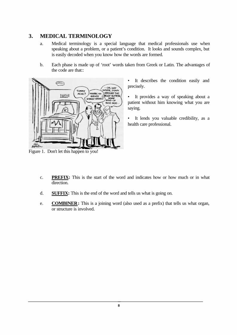
3. MEDICAL TERMINOLOGYa. Medical terminology is a special language that medical professionals use when
speaking about a problem, or a patient’s condition. It looks and sounds complex, butis easily decoded when you know how the words are formed.
b. Each phase is made up of ‘root’ words taken from Greek or Latin. The advantages ofthe code are that::
• It describes the condition easily andprecisely.
• It provides a way of speaking about apatient without him knowing what you aresaying.
• It lends you valuable credibility, as ahealth care professional.
c. PREFIX: This is the start of the word and indicates how or how much or in whatdirection.
d. SUFFIX: This is the end of the word and tells us what is going on.
e. COMBINER: This is a joining word (also used as a prefix) that tells us what organ,or structure is involved.
Figure 1. Don't let this happen to you!

9
f. PREFIXBRADY …………………………….DYS …………………………….ENDO …………………………….HYPER …………………………….HYPO …………………………….INTER …………………………….PARA …………………………….RETRO …………………………….POST …………………………….TACHY …………………………….
g. COMBINERSANGIO ………………………………ARTHRO ………………………………CARDIO ………………………………DERMA ………………………………HEM(A OR ATO) ………………………………PNEUMO ………………………………MYO ………………………………MENINGIO ………………………………NEURO ………………………………OSTEO ………………………………
h. SUFFIXALGIA …………………………………IT IS …………………………………PARESIS …………………………………PNEA …………………………………THORA …………………………………ECTOMY …………………………………PATHY …………………………………OLOGY …………………………………CENTESIS …………………………………SCOPY …………………………………
i. So, by the combination of a prefix and/or a combiner and a suffix, we can make upwords that only people, who know the code, understand.
j. Example THORO-CENTESIS = Chest cavity /puncturing. This is when small needleis put into the chest wall to release a build up of air in the pleural space. This is anemergency procedure that you will learn on this course.

10
4. ANATOMICAL LANGUAGE
When we speak of the body to other medical people, we refer to locations on the body in a setanatomical position
The body is facing forward with the arms at the side and the palms open and facing forward
a. ANATOMICAL LANDMARKS
Figure 1.
The anatomical terms are shown in boldface type, the common names are in plain type and theanatomical adjectives are in parentheses.

11
b. ANATOMICAL DIRECTIONS
)
Figure 4. Lateral View
Anatomical regions of the body are indicated in Figure 1 &2.The anatomy has its own language by which the preciselocation and movement of the structure are defined.In figure 2 its shown the abdominopelvic-thoracic region.These terms are very often used to identify symptomslinked with appropriate disease. Abbreviations like RUQor LLQ are used to locate the patient pain to recognize theeffected organs.
Define the followingterms in your ownwords :
• Superior
• Medial
• Inferior
• Lateral
• Anterior
• Proximal
• PosteriorFigure 3. Anatomical Directions (Frontal View)
Answer the question.Is the person in the picture above in ananatomical position?
Yes No
Fig. 1 & 2

12
c. TYPES OF MOVEMENT
All movements are described with reference to a figure in the anatomical position
The pictures below show the most common movements, the terms used are to identifyand describe the patient’s condition.
• ANGULAR MOVEMENTS
Figure 1
Figure 3
• ROTATIONAL MOVEMENT Figure 2
Figure 4 Figure 5
Abduction is movement away from thelongitudinal axis of the body in the frontalplane, moving it back constitutesAdduction.Figure. 1
Flexion is a movement in the anterior-posterior plane that reduces the anklebetween the articulating elements;Extension increases the ankle.Extension can be continued past theanatomical position to Hyperextension.Figure. 2
Moving an arm like a loop, that’sCircumduction.Figure.3
If you turn your limb inward, that’s aninternal or medial Rotation. Themovement outward describes an externalor lateral Rotation.Figure 4
To move the wrist and hand from palmfacing front to palm facing-back iscalled Pronation, the opposing movementis SupinationFigure 5

13
• SPECIAL MOVEMENT
Figure 6
Figure 7
d. PATIENT POSITION
A twisting motion of the foot that turnsthe sole inwards is called Inversion.( in, into + vertere, to turn)
Eversion ( e, out) is the opposingmovementFigure 6
Elevation and Depression occurs whena structure moves in a superior o inferiordirection.You depress your mandible when youopen your mouth and elevate it as youclose it.Figure 7
q Erect
q Recumbent
q Supine
q Prone
q Lateral recumbentRecovery position
q Shock position

14
5. CAVITIES OF THE BODY,CELLS, TISSUES, ORGANS ANDSYSTEMS.
a. CELLS
The cell is the basic structure and functional unitof all living things. Cells possess thecharacteristics of growth, metabolism, irritabilityand reproduction. The human body develops froma single cell. It is the cells themselves, which dothe work to provide an optimum environment
c. PHYSIOLOGY OF THE CELLThe human body is composed of about onehundred trillion cells arranged in tissues tocarry out specialised functions: Skeletalsupport, muscular contraction andconduction of electrical impulses and vitalgeneral functions.
Figure 2. Cavities of the Body
Figure 3. The Simple Cell
b. BASIC CELL
(1) The cell membrane surrounds andseparates the cell from its environment
(2) Nucleus is the genetic material of the cell
(3) Cytoplasm is the storage and workingarea of the cell
(4) Chromatin is the “Segment” of DNA

15
d. TISSUES
(1) Found in all organs and organ systems, tissues are collections of specific cells(Epithelial, connective, muscular, and nervous).
(2) Epithelial tissue. Covers body surfaces, lines body cavities providing protectionand absorption (skin), secretion and excretion (glands).
(3) Connective tissue. Bone cartilage, tendons, support nourishment, defence forbody.
(4) Muscular tissue. Voluntary, involuntary and cardiac.
(5) Nervous tissue. The most highly organised tissue in the body initiatingcontrolling and co-ordinating the body’s ability to adapt to its environment.
e. ORGANS AND SYSTEMS
(1) Tissues combine to form organs, example are the brain, kidneys and heart.
(2) A body system is a group of organs.
(3) Integumentary (Skin): Epidermal/dermal, hair, nails, glands (sebaceous + sweat)(protection, water and temperature regulation).
(4) Skeletal system: Bones, cartilaginous and membranous structures, protects,supports, levers body movement.
(5) Muscular system: Voluntary, involuntary, cardiac, smooth.
(6) Nervous system: brain, spine, cranial and peripheral nerves.
(7) Circulatory system: heart, arteries, veins, lymph and capillaries.
(8) Respiratory system: Oral cavity, pharynx, larynx, trachea, bronchi, and lungs.O2 -- CO2.
(9) Digestive system: lips to anus with associated glands. Food substances absorbedand utilised by the body.
(10) Urinary system: kidneys, ureters, bladder, urethra formation and elimination ofurine and maintenance of homeostasis.
(11) Endocrine System: pituitary, thyroid and parathyroid, suprarenals, pancreas,ovaries, testes, and chemical regulation of body functions.
(12) Reproductive system: ovaries, uterine, uterus, vagina, vulva, testes, penis, andprostate.

16
6. THE RESPIRATORY SYSTEM
Respiration is the process by which air is drawn into the lungs, the blood takes up oxygen, carbondioxide is removed from the blood and finally air is blown out of the lungs. Respiration is animportant task of the human body. Without air in our lungs we would die in 2-6 minutes.
Structure of the respiratory system
Upper respiratory system
- Nose- Nasal cavity- Paranasal sinuses- Pharynx
Lower respiratory system
- Larynx- Trachea- Bronchi- Lungs
a. THE TRANSPORT OF AIR FROM THE OUTSIDE TO THE BLOOD
(1). NOSE Air is inhaled through the nose into the nasal cavity. In the nasal cavity air is:
(a). Warmed (by mucous membrane)
(b). Moistened (by mucous membrane)
(c). Filtered (by nose hairs)
(d). Another important function of the nose cavity is smell.
(2). PHARINXAfter passing the nasal cavity, air enters the pharynx or throat. The pharynx
lies posterior to the nasal cavity and the mouth.
Figure 4. The Respiratory System

17
(3). LARYNXFrom the pharynx air enters the larynx. The larynx consists of the thyroid
cartilage, the cricoid cartilage, 2 vocal cords, the epiglottis and the hyoid bone. Theepiglottis closes the airway when a person swallows. The larynx is situated between thepharynx and trachea.
(4). TRACHEAVia the larynx, air enters the trachea or windpipe. The trachea is a long tube,
which connects the larynx with the lungs. In our thoracic cavity, the trachea divides intwo main bronchi.
(5). BRONCHI
The air goes via the trachea into the left and right main bronchi. The mainbronchi divide into a mass of smaller bronchi (bronchioles), like the roots of a tree.
(6). ALVEOLI
The air comes via the bronchioles into the alveoli. The alveoli are very small“grape” like clusters of thin air bags, which are surrounded by a network of smallblood vessels called capillaries. The exchange of gases takes place between the alveoliand the capillaries. The blood absorbs oxygen and gets rid of carbon dioxide.
Figure 5. The Upper Airway (passage of air)
Figure 6. The Bronchial Tree (with cutawaysection)


19
(7). LUNGSThese are two sponges like organs that almost fill the thoracic cavity. Lungs
are made up of bronchi, bronchioles, alveoli, blood vessels and some supportivetissue. Each lung is covered with a layer of serous membrane, called “pleuravisceralis”. This visceral pleura lies against the parietal pleura. The “space” betweenthese two pleura, called the pleural cavity, is filled up with serous fluid, which allowsfriction-free movement between the lungs and the inner surface of the thoracic cavity.
(8). CONTENTS OF THE THORAXThe thorax or chest consists of a cavity which is surrounded by the ribs, the
intercostal muscles (muscles between each rib) and the diaphragm. The diaphragm isa flat muscle, which divides the thoracic cavity from the abdominal cavity. The insideof the thoracic wall is covered by a serous membrane, called “parietal pleura”. Thethoracic cavity contains the heart, two lungs, and the trachea, the oesophagus andlarge blood vessels.
b. THE MECHANISM OF RESPIRATIONRespiration muscles are the diaphragm, intercostal muscles and abdominal muscles.
(1) INSPIRATIONThis is an active process caused by muscle contraction. Intercostal muscles anddiaphragm contract. Thoracic cavity increases in size. Pleura pull in with ribcage and negative pressure is created in the lungs. Atmospheric pressure isgreater than that inside the lungs, therefore air is forced in through the nose.Pause for gaseous exchange.
(2) EXPIRATIONThis is a passive process caused by muscle relaxation. Muscles relax. Positivepressure created in the lungs is greater than atmospheric pressure, so air isforced out. Cycle restarts.
The normal rate of respiration is about 12-20 breaths per minute.
** Gas exchange takes place at two locations of the body, in
the alveoli and the capillaries. **
20
7. DISEASES AND CONDITIONS OF THE RESPIRATORY SYSTEMa. There are 2 important diseases and 1 condition of the respiratory system, which you
can manage as a SOF medic:
• Acute bronchitis
• Pneumonia
• Asthma
b. The SOF medic must be able to recognise and treat these diseases.
c. ACUTE BRONCHITIS
The bronchi are inflamed
(1) AetiologyViral or bacterial infection
(2) HistoryProductive cough often following sore throat and/or nasal catarrh (rhinorrhea).The sputum can vary in colour from transparent-white (viral cause) toyellow-green (bacterial cause).
(3) Physical ExamRhonchi and often low-grade fever.
(4) TherapySymptomatic in case of viral infection (transparent-white sputum).
(5) Administer:
(a) Paracetamol (500mg +/- 6 x day)
(b) Cough suppressants (if tactically necessary)
(c) Antitussiva
(6) Antibiotics in case of bacterial infection (yellow-green sputum).Administer:
(a) Doxycycline: 200mg on first day, 100mg on following days for 14 days.
Note: If the sputum remains yellow-green for more than 3 days, there is a 90% chancethat the bronchitis is due to a bacterial infection. Administer antibiotics.

21
d. PNEUMONIAInflammation of the lungs or part of lung tissue.
(1) AetiologyBacterial infection. Sometimes it develops out of an untreated or ignored acutebronchitis.
(2) HistoryEither slow onset of the disease, following an infection of the upper respiratorytract, or a rapid onset with chills, fever, productive cough, possibly haemoptysis(blood in sputum), pain in chest, during respiration. The colour of the sputumvaries from green to rusty.
(3) Physical ExamFever, increased pulse rate. Auscultation of the affected side reveals rales(breath sounds can even be absent). Tachypnea.
(4) TherapyPenicillin-V is the drug of choice. 500mg 4 x day, during 10 days or more.Erythromycin or doxycyline are also suitable. Give patient plenty of fluids.
e. ASTHMAHypersensitivity of the bronchial tubes. It leads to generalised airway obstruction,which is paroxysmal and reversible.
(1) AetiologyAn allergic (hypersensitivity) state of the patient, which is leading toconstriction of the bronchi.
(2) HistoryAttacks of dyspnea, cough and wheezing. Provocative causes are allergens,infection (bronchitis), exercise. There is often a history of other allergicdiseases; Eczema, hay fever, allergies, tachypnea.
(3) Physical ExamDuring an attack of asthma there is generalised wheezing during auscultation.Dyspnea, cyanosis. These symptoms can be very serious. Young people are stilldying from asthma.
(4) TherapyThe first line of drugs is bronchodilatators (albuterol, salbutamol) which areadministered in inhalable sprays. Adrenaline (1:1000) is the drug of choicewhwn severe attack of asthma is present (status asthmaticus); give 0.5-1ml(1mg/ml solution of adrenaline) intra-muscular and massage site of injection for30 seconds after injection.

22
f. When listening by stethoscope to the lungs of patients with respiratory diseases youlook for adventitious breath sounds. These sounds cannot be heard in healthy people.
g. Adventitious breath sounds can be heard with a stethoscope:
(1) RHONCHI(a) Coarse rattling, which can be inspiratory or expiratory or both.
(b) Gurgling sounds produced by mucus in the bronchi.
(c) Present in bronchitis and pneumonia as well as in aspiration (penetrationof solid or liquid substances [vomit] in the patient’s respiratory tract).
(d) True Rhonchi will not clear by coughing.
(2) RALES(a) Fine rattling sounds, which can be inspiratory or expiratory or both.
(b) Sounds like “rice crisps” or hairs rubbing together.
(c) Rales are produced by fluid in the alveoli.
(d) Present in pneumonia.
(3) WHEEZING SOUNDS(a) Wheezing sounds especially during inspiration.
(b) Wheezing sounds can be high pitched.
(c) Wheezing is produced by constriction of the bronchi.
(d) Present in asthmatic attacks

23
8. THE CARDIOVASCULAR SYSTEM
a. The circulatory system has two major fluid transportation systems:the cardiovascular and the lymphatic system.
(1). Cardiovascular System.This system, which contains the heart and blood vessels, is a closedsystem, transporting blood to all parts of the body. Blood flowingthrough the circuit formed by the heart and blood vessels bringsoxygen, food and other chemical elements to tissue cells andremoves carbon dioxide and other waste products from the cell.
(2). Lymphatic System.
This system, which provides drainage for tissue fluid, is anauxiliary part of the circulatory system, returning an importantamount of tissue fluid to the bloodstream through its own system oflymphatic vessels.
b. HEART WALLThis muscular wall is made up of cardiac muscle called MYOCARDIUM.
c. HEART CHAMBERSThere are four chambers in the heart. These chambers are essentially the
same size. The upper chambers, the atria, area seemingly smaller than the lowerchambers (the ventricles). The apparent difference in total size is due to the thicknessof the myocardial (muscle) layer. The right atrium communicates with the rightventricle; the left atrium communicates with the left ventricle. The septum (partition),dividing the interior of the heart into right and left sides, prevents direct blood flowfrom right to left chambers or left to right chambers. This is important, because theright side of the heart receives deoxygenated blood returning from the systemic(body) circulation. The left side of the heart receives oxygenated blood returning fromthe pulmonary (lung) circulation. The special structure of the heart keeps the bloodflowing in its proper direction to and from the heart chambers.
d. HEART VALVESThe four chambers of the heart are lined with endocardium (membrane tissue).
This lining folds on itself and extends into the chamber opening to form valves. Thesevalves allow the blood to pass from a chamber but prevents backflow. Theatrioventricular valves, between the upper and lower chambers, are within the heartitself. The semilunar valves are within arteries attached to the right and left ventricles.

24
(1) Atrioventricular Valves. The tricuspid valve is located between the right atriumand right ventricle. It has three flaps or cusps. The bicuspid (mitral) valve islocated between the left atrium and left ventricle. It has two flaps or cusps.
(2) Semilunar Valves. The pulmonary semilunar (half-moon shaped) valve islocated at the opening into the pulmonary artery that is attached to the rightventricle. The aortic semilunar valve is located at the opening into the aortathat is attached to the left ventricle.
Figure 10. The Thoracic Cavity

25
e. FLOW OF BLOOD THROUGH THE HEART(1) It is helpful to follow the flow of blood through the heart, to understand the
relationship of the heart structures. Remember that the heart is the pump and isalso the connection between the systemic circulation and pulmonarycirculation. Blood returning from the systemic circulation must flow throughthe pulmonary circulation for theexchange of carbon dioxide andoxygen to take place. Blood from theupper part of the body enters the heartthrough the superior vena cava, andfrom the lower part of the bodythrough the inferior vena cava.
(2) Blood from the superior vena cava andinferior vena cava enters the heart atthe right atrium. The right atriumcontracts, and blood is forced throughthe open tricuspid valve into therelaxed right ventricle.
(3) As the right ventricle contracts, thetricuspid valve is closed, preventingback flow into the atrium. Thepulmonary semilunar valve opens asthe blood is forced through it and ispumped into the pulmonary artery.
(4) The blood is carried through the lung tissues, exchanging its carbon dioxide foroxygen in the alveoli. This oxygenated blood is collected from the mainpulmonary veins and delivered to the left atrium.
(5) As the left atrium contracts, the oxygenated blood flows through the openbicuspid (mitral) valve into the left ventricle.
(6) As the left ventricle contracts, the bicuspid valve is closed. The aortic semilunarvalve opens as the oxygenated blood is forced through it into the aorta, the mainartery of the body. The oxygenated blood now starts its flow to all body cellsand tissues. The systemic circulation starts from the left ventricle, thepulmonary circulation from the right ventricle.
Figure 7. Blood-flow through the Heart

26
f. BLOOD AND NERVE SUPPLY OF THE HEART(1) CORONARY ARTERIES
The heart gets its blood supply from the right and left coronary arteries. Thesearteries branch off the aorta just above the heart, then subdivide into manysmaller branches within the heart muscle. If any part of the heart muscle isdeprived of its blood supply, the muscle tissue cannot function properly andwill die. This is called a myocardial infarction. Blood from the heart tissue isreturned by coronary veins to the right atrium.
(2) NERVE SUPPLYThe nerve supply to the heart is from two sets of nerves originating in themedulla of the brain. The nerves are part of the involuntary (autonomic)nervous system. One set branches from the vagus nerve and keeps the heartbeating at a slow, regular rate. The other set, the cardiac accelerator nerve,speeds up the heart. The heart muscle has a special ability; it contractsautomatically, but the nerve supply is needed to control the contractions forblood circulation. Within the heart muscle itself are special groups of nervefibers that conduct impulses. These groups make up the conduction system ofthe heart. When the conduction system does not operate properly, the heartmuscle contractions are uncoordinated and ineffective. The impulses within theheart muscle are minute electric currents, which can be picked up and recordedby the electrocardiogram (ECG).
g. THE HEARTBEAT AND HEART SOUNDS
(1) HEARTBEATThis is a complete cycle of heart action - contraction (systole) and relaxation(diastole). During systole, blood is forced from the chambers. During diastole,blood refills the chambers. The term cardiac cycle means the completeheartbeat. The cardiac cycle repeated continuously at a regular rhythm, usually70-80 times per minute. Each complete cycle takes less than one second - inthis brief time, all of the heart action needed to move blood must take place, andthe heart must be ready to repeat its cycle.
(2) HEART SOUNDSWhen heard through a stethoscope, heart sounds are described as “lubb-dup”.The first sound, “lubb”, is interpreted as the sound, or vibration, of theventricles contracting and atrioventricular valves closing. The second, higherpitched sound, “dup”, is interpreted as the sound of the semilunar valvesclosing. The medic listening to the heart sounds can detect alterations of normalsounds; the interpretation of these heart sounds is part of the diagnosis of heartdisease.
27
h. BLOOD VESSELS
The blood vessels are the closed system of tubes through which the bloodflows. The arteries and arterioles are distributors. The capillaries are the vesselsthrough which the exchange of fluid, oxygen and carbon dioxide takes placebetween the blood and tissue cells. The venules and veins are collectors,carrying blood back to the heart. The capillaries are the smallest of thesevessels, but are of the greatest importance in the circulatory system.
(1). THE ARTERIES AND ARTERIOLESThe system of arteries and arterioles is like a tree, with the large trunk, the aorta,giving off branches, which repeatedly divide and subdivide. Arterioles are verysmall arteries about the diameter of a hair. In comparison, the aorta is more than 1inch (2.5 cm) in diameter. An artery wall has a layer of elastic, muscular tissue,which allows it to expand and recoil. When an artery is cut, the artery wall doesnot collapse; Bright red blood escapes from the artery in spurts. Arterial bleedingmust often be controlled by clamping and tying off (ligating) the vessel. Some ofthe principal arteries and the area they supply with blood are:
- Carotid arteries, external and internal, supply the neck, head and brain throughtheir branches.
- Subclavian arteries supply the upper extremities.
- Femoral arteries supply the lower extremities.
(2). CAPILLARIES
Microscopic in size, capillaries are so numerous that there is at least one or morenear every living cell. A single layer of endothelial cells forms the walls of acapillary. Capillaries are the essential link between arterial and venous circulation.The vital exchange of substances from the blood in the capillary with tissue cellstakes place through the capillary wall. Blood starts its route back to the heart as itleaves the capillaries.

28
(3). VEINS
Veins have thin walls and valves. Formed from theinner vein lining, these valves prevent blood fromflowing back toward the capillaries. Venules, thesmallest veins, unite into veins of larger and larger sizeas the blood is collected in its return to the heart. Thesuperior vena cava, collecting blood from all regionsabove the diaphragm, and the inferior vena cava,collecting blood from all regions below the diaphragm,return the venous blood to the right atrium of the heart.Superficial veins lie close to the surface of the bodyand can be seen through the skin. The medial basilicvein at the antecubital fossa (in the bend of the elbow)is commonly used for venipuncture to obtain bloodspecimens or to inject solutions of drugs or fluidintravenously. The great saphenous vein is the longestvein in the body, extending from the foot to the groin.The saphenous vein has a long distance to lift bloodagainst the force of gravity when an individual is in astanding position. It is therefore very susceptible tobecoming dilated and stretched with the valves nolonger functioning properly. When this occurs, thevein is said to be varicosed.
1.1 NOTE: Veins carry deoxygenated blood and arteries carry oxygenated blood. Theonly exception to this is the pulmonary vein, which carries oxygenated blood fromthe lungs to the heart and the pulmonary artery, which carries deoxygenated bloodfrom the heart to the lungs.
Figure 12. Major Veins

29
i. PULSE AND BLOOD PRESSURE
(1). PULSEPulse is the alternate expansion and recoil of an artery. With each heartbeat, blood isforced into the arteries causing them to dilate (expand). The arteries contract (recoil) asthe blood moves further along in the circulatory system. The pulse can be felt at certainpoints in the body where an artery lies close to the surface. The most common locationfor feeling the pulse is at the wrist, proximal to the thumb (radial artery), on the palmside of the hand. Alternate locations are in front of the ear (temporal artery), at the sideof the neck (carotid artery), and on the top (dorsum) of the foot (dorsalis pedis).
(2). BLOOD PRESSUREThe force that blood exerts on the walls of vessels through which it flows is calledblood pressure. All parts of the vascular system are under pressure, but the term bloodpressure usually refers to arterial pressure. Pressure in the arteries is highest when theventricles contract during systole. Pressure is lowest when the ventricles relax duringdiastole. The brachial artery, in the upper arm, is the artery usually used for bloodpressure measurement.
j. THE BLOODBlood is the red body fluid flowing through the arteries, capillaries and veins. Itvaries in color from bright red (oxygenated blood) when it flows from arteries, todark red (deoxygenated blood) when it flows from veins. The average man hasabout 6000 ml of blood.
(1). FUNCTIONS OF BLOOD
The six major functions of blood are all carried out as the blood circulates through thevessels. These functions are:
- To carry oxygen from the lungs to tissue cells and carbon dioxide from the cells tothe lungs.
- To carry food materials absorbed from the digestive tract to the tissue cells and toremove waste products for elimination by excretory organs (the kidneys,intestines, and skin).
- To carry hormones, which help regulate body functions, from ductless (endocrine)glands to the tissues of the body.
- To help regulate and equalise body temperature. Body cells generate largeamounts of heat, and the circulating blood absorbs this heat.
- To protect the body against infection.
- To maintain the fluid balance in the body.

30
(2). COMPOSITION OF BLOOD
Blood is made up of a liquid portion (plasma) and formed elements (blood cells)suspended in the plasma:
Plasma: Making up more than one half of the total volume of blood, plasma isthe carrier for blood cells, carbon dioxide, and other dissolved wastes. It bringshormones and antibodies (protective substances) to the tissues. Other components ofplasma are water, oxygen, nitrogen, fat, carbohydrates, and proteins. Fibrinogen, oneof the plasma proteins, helps blood clotting. When blood clots, the liquid portion thatremains is serum. Blood serum contains no blood cells.
Blood Cells.:The cellular elements in the blood are red cells (erythrocytes, orRBC), white cells (leukocytes, or WBC) and blood platelets thrombocytes).
(3). RED BLOOD CELLS: (ERYTHROCYTES)
There are about 5,000,000 red blood cells in 1 cubic millimetre (cmm) of blood.Individual red blood cells are disc-shaped. Red cells are formed in the red bonemarrow. Millions of red cells are destroyed daily, in the liver, the spleen, and thelymph nodes or in the vascular system itself.In a healthy person, the destruction rate is equalled by the production rate, maintaininga count of about 5,000,000 per cubic millimetre. Red blood cells have an average lifespan of about 90 to 120 days before becoming worn out.
(a). HEMOGLOBIN
Hemoglobin (Hgb) gives red cells their colour. Hemoglobin has the power tocombine with oxygen, carrying it from the lungs to the tissue cells.Hemoglobin assists in transporting carbon dioxide from the cells to the lungs. Thistransportation of gases is the principal function of the red cells. In order to carryoxygen, Hemoglobin needs iron, which is ordinarily available in a nutritionallyadequate diet.
(b). ANAEMIAAnaemia is due to a reduction in the number of red cells or a reduction in the
haemoglobin content of red cells.
31
(4). WHITE BLOOD CELLS: (LEUKOCYTE)
(a). White blood cells vary in size and shape, and are larger and much fewer innumber than red cells. The average number in an adult is 5,000 to 10,000 in 1 cmm ofblood. Their function is primarily one of protection. They can ingest and destroyforeign particles, such as bacteria, in the blood and tissues.
(b). White cells can pass through the walls of the capillaries into surroundingtissues. This ability to enter tissue makes them very useful in fighting infection - anarea of infection is characterised by a great increase of white cells, which gather aboutthe site to destroy the bacteria.
©. An example of this is seen in an ordinary boil (furuncle). The pus contained inthe boil is made up largely of white cells plus bacteria and dissolved tissue. Many ofthe white cells are killed in their struggle with invading bacteria.
(5). BLOOD PLATELETS: (THROMBOCYTES)
(a). Blood platelets, which are smaller than red blood cells, are thought to befragments of cells formed in the bone marrow. Platelets number about 300,000 percmm of blood.
(b). Their main function is to aid in the coagulation of blood at the site of a wound.Platelets release a substance to hasten formation of a blood clot.
(6). COAGULATION OF BLOOD
(a). Blood coagulation (clotting) is the body’s major method of preventing excessiveloss of blood when the walls of a blood vessel are broken or cut open. Whenundisturbed, blood circulates in its vascular system without showing a tendency toclot. Physical and chemical factors are changed when blood leaves its naturalenvironment and it begins to clot almost at once. At first, the clot is soft and jellylike,but soon becomes firm and acts as a plug, preventing further escape of blood.
(b). It takes 3 to 5 minutes for blood to clot, but sometimes it is necessary to holdback the clotting process. This is done with anticoagulant drugs.
(7). BLOOD TYPES
(a). This system of typing is used to prevent incompatible blood transfusion, whichcauses serious reactions and sometimes death. Certain types of blood are incompatible(not suited) to each other if combined.
(b). Two bloods are said to be incompatible when the plasma or serum of one bloodcauses clumping of the cells of the other. Two bloods are said to be compatible andsafe for transfusion if the cells of each can be suspended in the plasma or serum of the

32
other without clumping. Highly trained laboratory technicians do Blood typing andcross matching.
(8). IMPORTANCE OF BLOOD TYPES
The table below shows that if the donor’s blood is type “O” it is compatible with alltypes of recipient blood; Or, in other words, type “O” is the universal donor. If therecipient’s blood is type “AB”, it is compatible with all types of donor blood, or, in otherwords, type “AB” is the universal recipient. When a blood transfusion is given, the bloodtype of both donor and recipient should be identical, and their compatibility must beproven by a cross matching test. However, when blood of the same type is not availableand death may result if transfusion is delayed, a type “O” donor (universal donor) may beused if the cross matching is satisfactory.
(9). RH FACTOR
In addition to blood grouping and cross matching for compatibility, the Rh factormust be considered. The Rh factor is carried in red cells, and about 85 percent of allindividual have this factor and are, therefore, Rh positive. Individuals who do nothave the Rh factor are Rh negative. As a general rule, Rh-negative blood can begiven to anyone, provided it is compatible in the ABO typing system, but Rh-positiveblood should not be given to an Rh negative individual.
BLOOD TYPES
DONOR RECIPIENT
O A B AB
O COMPATIBLE COMPATIBLE COMPATIBLE COMPATIBLE
A INCOMPATIBLE COMPATIBLE INCOMPATIBLE COMPATIBLE
B INCOMPATIBLE INCOMPATIBLE COMPATIBLE COMPATIBLE
AB INCOMPATIBLE INCOMPATIBLE INCOMPATIBLE COMPATIBLE

33
9. LYMPHATIC SYSTEM
a. The lymphatic system consists of lymph, lymph vessels, and lymph nodes. The spleenbelongs, in part, to the lymphatic system. Unlike the cardiovascular system, thelymphatic system has no pump to move the fluid, which it collects, but muscularcontractions and breathing movement's aid in the movement of lymph through itschannels and its return to the bloodstream.
b. LYMPH AND TISSUE FLUID
Lymph, fluid found in the lymph vessels is clear and watery and is similar to tissuefluid, which is the colourless fluid that fills the spaces between tissues, between thecells of organs, and between cells and connective tissues. Tissue fluid serves as the“middleman” for the exchange between blood and body cells. Formed from plasma,it seeps out of capillary walls. The lymphatic system collects tissue fluid, and aslymph, it is started on its way back into the circulating blood.
c. LYMPH VESSELS
Starting as small ducts within the tissues, the lymphatic vessels enlarge to formlymphatic capillaries. These capillaries unite to form larger lymphatic vessels, whichresemble veins in structure and arrangement. Valves in lymph vessels preventbackflow. Superficial lymph vessels collect lymph from the skin and subcutaneoustissue; Deep vessels collect lymph from all other parts of the body.
d. LYMPH NODES
Occurring in groups of up to a dozen or more, lymph nodes lie along the course of thelymph vessels. Although variable in size, they are usually small oval bodies, whichare composed of lymphoid tissue. Lymph nodes act as filters for removal of infectiousorganisms from the lymph stream. Important groups of these nodes are located in theaxilla (armpit), the cervical region, the submaxillary region, the inguinal (groin)region, and the mesenteric (abdominal) region.
e. SPLEEN
The largest collection of lymphoid tissue in the body, the spleen is located high in theabdominal cavity on the left side (LUQ), below the diaphragm and behind thestomach. It is somewhat long and ovoid (egg shaped). Although it can be removed(splenectomy) without noticeable harmful effects, the spleen has useful functions,such as serving as a reservoir for blood and blood cells.
f. INFECTIONS AND THE LYMPHATIC SYSTEM
Lymph vessels and lymph nodes often become inflamed as the result of infection.An infection in the hand may cause inflammation of the lymph vessels as high asthe axilla. Sore throat may cause inflammation and swelling of lymph nodes inthe neck (submandibular nodes below the jaw and cervical nodes).

34
10. CONDITIONS AND DISEASES OF THE CARDIOVASCULAR SYSTEM
a. CORONARY ARTERY DISEASE AND ANGINA PECTORIS(1) As mentioned before, coronary arteries are blood vessels whose primary
function is to transport blood to the heart muscles and at the same time removecarbon dioxide and waste products. Sometimes the coronary arteries becomeblocked depriving the heart muscles of oxygen and nutrients and slowing downor stopping the removal of waste products. If this condition continues withoutproper treatment, the artery will eventually close off, resulting in death of theaffected tissue. Certain factors contribute to coronary artery disease. Some ofthese factors are controllable while others are not. These factors are:
(a) Hypertension (high blood pressure)
(b) Cigarette smoking
(c) Diabetes
(d) Elevated serum cholesterol
(e) Dietary habits (excessive intake of calories, carbohydrates, and/orsaturated fats)
(f) Obesity
(g) Sex (male)
(h) Hereditary (family history)
(i) Stress
(2) Early stages of coronary artery disease are asymptomatic. In the late stageof the disease, the blood flow no longer meets the demands of themyocardium for oxygen and the patient begins to experience chest pain.This pain is referred to as angina pectoris (choking of the heart). Thepatient with advanced coronary artery disease may have adequate oxygen atrest, however, during any form of stress or exercise, blood flow to the heartis inadequate. This results in angina pectoris. A patient can also experienceangina pectoris while at rest. If this occurs, that patient has much moresevere coronary artery disease than the one who only experiences pain withexercise and stress. The pain (angina) is characterised as a crushing chestpain, which usually radiates to the neck, jaw, shoulders and upperextremities. The duration of the pain is usually 2 to 3 minutes. Treatmentfor this condition is either stopping the stress or administering nitro-glycerine. The drug, nitro-glycerine, is a vasodilator. It causes the coronaryarteries to dilate and provides improved blood flow to the myocardium.
b. MYOCARDIAL INFARCTIONMyocardial infarction (MI) (heard attack) is a blockage in a coronary artery withresulting death to the affected tissue

35
c. SIGNS AND SYMPTONS OF A MI
(1). Chest pain similar to angina, more severe and longer lasting. The pain may not berelieved with nitro-clycerin. The patient complains of severe chrushing pain or tightnessin the chest. A clenched fist is usually used to describe the pain. In approximately 25% ofthe patients, pain will radiate down the left arm and into the fingers.
(2). Usually the pain radiate to the jaw, neck, upper back and epigastrium. A MI is sometimesmistaken for indigestion.
(3). Along with chest pain, the patient complains of nausea.
(4). Diaphoreses ( profuse perspiration ) usually accompanies an MI.
(5). The patient may also experience a fear of impending doom.
(6). Shortness of breath.
(7). Hypertension or hypotension.
(8). Cyanosis.
d. TREATING A MIThe physical findings of a MI may not always be obvious. They vary with the site andextent of cardiac muscle damage. Therefore, diagnosis in the field will dependprimarily on the history of the current complaint. Treatment and stabilization shouldbe started immediately with a detailed history. Early treatment can mean thedifference between life and death. The patient should be immediately transported to amedical facility where definitive treatment can be initiated. Early treatment shouldinclude:
- Starting an IV infusion (Hartmans).- Monitoring vital signs.- Positioning the patient in a semi-Fowlers or high-Fowlers (sitting) position to
reduce respiratory distress.- CASEVAC
e. CONGESTIVE HEART FAILURECongestive heart failure (CHF) is the inability of the heart to pump bloodefficiently. There are several contributing factors to CHF. Some of these include:
- Secondary to a MI- Pulmonary embolism- Administration of too much IV fluids- Excessive sodium intake
36
f. PRIMARY CAUSES OF HEART FAILURES: ACUTE PULMONARY EDEMAAND CHRONIC CONGESTIVE HEART FAILURE
(1). SIGNS AND SYMPTOMS OF PULMONARY EDEMA
(a) Congestion of the lungs
(b) Fatigue
(c) Dyspnea
(d) Cough
(e) Insomnia - often due to increased respiratory effort
(f) Haemoptysis
(g) Restlessness
(2). SIGNS AND SYMPTOMS OF CONGESTIVE HEART FAILURE
(a) Unexplained weight gain.
(b) Abdominal pain - usually in the upper region of the abdomen.
(c) Mild to moderate respiratory distress.
(d) Diaphoresis
(e) Weakness
(f) Anorexia
(g) Pitting oedema
(3). TREATMENTTreatment of heart failure is aimed at improving oxygenation, increasing myocardialcontractility and reducing venous return. Certain specific treatments are recommended for themedical specialist and include:
(a) Placing the patient in a sitting position, with the feet dangling. This position decreasesvenous return, making breathing easier.
(b) Starting an IV and adjusting the flow to maintain access only.
(c) Monitoring the patient’s vital signs.
(d) CASEVAC

37
11. THE SKELETAL SYSTEMa. The skeletal system provides a framework for the body, giving it form and protection,
and enclosing the vital organs, such as the brain, heart and lungs. The skeletal systemis composed of:
• Bones. 206 in number, which form the hard framework of the body.
• Cartilage. Which provides connecting and supporting structures.
• Ligaments. Which bind bones together.
b. Structure of a BoneBones are covered - except at the joints - by periosteum, a strong fibrous membrane.The blood supply to the bone pierces the periosteum. The next layer is compact bone,which makes up the diaphysis (shaft). Compact bone is very hard and strong. In thecentre of the shaft is a small cavity which forms the epiphysis, which differs from thediaphysis; It is called cancellous bone and is more spongy, porous and lightweightthan compact bone. Red bone marrow, found inside the porosities of cancellous bone,is vital to the production, maintenance and disposal of blood cells in adults.
c. FUNCTIONS OF THE SKELETAL SYSTEM(1) Support, The skeleton is the major supporting element of the body.
(2) Protection, The bones protect the organs.
(3) Movement, The skeletal muscles attach to the bones, and contraction of theskeletal muscles causes the bones to move.
(4) Storage, Certain minerals in the blood are taken into the bones and stored.Should blood levels of those minerals decrease, the minerals will be released(Calcium and phosphorus).
(5) Blood Cell Production, The cavities of the bones can contain bone marrowthat gives rise to blood cells and platelets.
d. STRUCTURE OF THE SKELETAL SYSTEM
(1) Bones of Various Types• Tubular (long/short)• Flat• Irregular shape
(2) Cartilage(3) Tendons
• Attach muscles to bones

38
(4) Ligaments• Attach bones to bones
STRUCTURE OF A BONE• Epiphysis• Diaphysis• Compact Bone• Cancellous Bone• Periosteum• Cavity with marrow
e. GROSS ANATOMYNote: Examination of skeletal gross anatomy uses dried, prepared bones. Theadvantage of this approach is that the major features of the bones can be seenclearly without being obstructed by associated soft tissues, such as muscles,tendons, ligaments, cartilage, nerves and blood vessels. The disadvantage is that itis easy to ignore the important relationships between bones and soft tissues, and thefact that bone itself has soft tissues.
f. THE SKELETON IS DIVIDED INTO CATEGORIESThe skeleton is divided into axial and appendicular skeleton.
(1). Axial skeleton The axial skeleton consists of the
bones of the skull, thorax, andvertebral column.
(a). Skull:• Cranial bones• Facial bones• Hyoid and auditory ossicles
(b). Few Important Bones of theskull:
• Frontal• Parietal• Temporal• Occipital• Skull base• Maxilla (upper jaw)• Mandible (lower jaw)• Nasal bone• Zygomatic bones (cheeks)• Zygomatic arch• Teeth• Orbit bones

39
©. Vertebral Column• 7 cervical vertebrae• 12 thoracic vertebrae• 5 lumbar vertebrae• 1 sacral bone• 1 coccygeal bone
(d). Thoracic Cage• Sternum.• 12 pairs of ribs, 7 true ribs, directly attached to the sternum and 5 false
ribs, do not directly attached to the sternum.
NOTE 1: When the ribs are to be felt on yourself or another person, be aware of thefact that the 1st rib is mostly not to be felt. Instead of the 1st rib, you feel the clavicle.
NOTE 2: Watch the xyphoid process (the lower end of the sternum); Because it isattached only at its upper end, it may be broken during (wrong performed)cardiopulmonary resuscitation (CPR), and then may lacerate the liver.
(2). APPENDICULAR SKELETON
(a). Shoulder Girdle
• Scapula (shoulder blade)• Clavicle (collar bone
(b). Arm• Humerus (upper arm bone)• Ulna• Radius
©. Wrist• Carpal bones
(d). Hand• Metacarpals• Phalanxes

40
(e). Pelvic Girdle (pelvis)• Ilium (groin bone)• Ischium (hip bone)• Pubis
(f).Thigh• Femur (thigh bone)• Patella (knee bone/disk)
(g). Leg• Tibia• Fibula
(h). Ankle• Tarsal bones
(i). Foot• Metatarsals• Phalanxes
g. JOINTSA place where two bones comes together; some joints permit extensive movement,other slight movement, and there are joints that competely prohibit movement.Types of Joints
(1). Fibrous JointsBones are united by fibrous tissue; Little/no movement.
(2). Cartilaginous Joints.Bones are united by means of cartilage; Little/no movement.
(3). Synovial JointsContaining synovial fluid; Considerable movement.
h. STRUCTURE OF A SYNOVIAL JOINT• Bone
• Joint Cavity contains synovial fluid
• Cartilage
• Joint Capsule
• Synovial Membrane
• Fibrous Capsule

41
i. In some joints the synovial membrane may extend as a pocket or sac, for somedistance away from the rest of the joint cavity. This sac is called the bursa.
j. The bursa contains synovial fluid and provides a fluid filled cushion betweenstructures that otherwise would rub against another, such as tendons rubbing onbones.
k. Inflammation of a bursa may cause considerable pain around the joint and inhibitmovement.

42
12. THE MUSCULAR SYSTEMMuscle is characterized by the ability to contract, or to shorten. The power of contractionenables a muscle to move parts of the body. All movements of the body, whetherconscious or unconscious, are due to the action of muscles. Muscle makes up much of thefleshy portions of the body. Muscles vary in shape and structure according to the workthey have to do. There are three main types of muscle:
• Voluntary• Involuntary (smooth)• Cardiac (heart)
a. VOLUNTARY MUSCLE(1) Voluntary Muscle is so called because it is controlled by the will through the
central nerve system (CNS). All the skeletal muscles (those attached to theskeleton) are of the voluntary type. Besides the skeletal muscles, those,which move the eye, tongue and pharynx, are voluntary.
(2) Functions. Voluntary muscles cause movement of the body as a whole andthe movements of its parts. They maintain posture, carry on the rhythmicmovements of respiration, produce most of the heat generated by the body,and serve to protect certain organs.
(3) Structure. Voluntary muscle is made of long, slender fibers, held together byconnective tissue to form muscle bundles. Groups of muscle bundles,enclosed in a fibrous sheath called fascia, form the individual muscles.
b. INVOLUNTARY (Smooth Muscle)(1) The involuntary muscle is called that because the nerve supply is from the
autonomic nervous system, which is not under the control of the will. It isalso called smooth muscle. Smooth muscle is found in the walls of the bloodvessels, respiratory passages, gastrointestinal tract, ureters and urinarybladder, and certain other organs.
(2) Functions. Smooth muscle performs many varied functions. It regulates thesize of blood vessels, which is essential to the maintenance of blood pressure.It moves food through the intestinal tract. It regulates the bronchioles (smallair passages) in the lungs. Still another function of smooth muscles is themovement of urine from the kidneys to the urinary bladder.
(3) Structure. Smooth muscle is made of spindle shaped fibers of cells. Thefibers are arranged in bundles or sheets to form a layer in the walls of bloodvessels and other viscera.
c. CARDIAC MUSCLE(1) Cardiac or heart muscle is involuntary muscle, but is found only in the heart.
The structure of heart muscle is different from that of other muscles. Heartmuscle forms the walls of the heart. The whole heart works together becauseall parts are connected with special bands of cardiac muscle.
(2) Structure. Mixture of involuntary and smooth muscles.
43
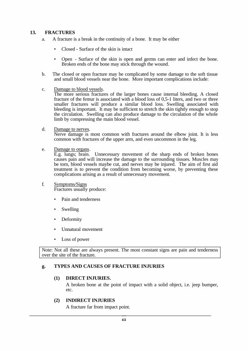
13. FRACTURESa. A fracture is a break in the continuity of a bone. It may be either
• Closed - Surface of the skin is intact
• Open - Surface of the skin is open and germs can enter and infect the bone.Broken ends of the bone may stick through the wound.
b. The closed or open fracture may be complicated by some damage to the soft tissueand small blood vessels near the bone. More important complications include:
c. Damage to blood vessels.The more serious fractures of the larger bones cause internal bleeding. A closedfracture of the femur is associated with a blood loss of 0,5-1 liters, and two or threesmaller fractures will produce a similar blood loss. Swelling associated withbleeding is important. It may be sufficient to stretch the skin tightly enough to stopthe circulation. Swelling can also produce damage to the circulation of the wholelimb by compressing the main blood vessel.
d. Damage to nerves.Nerve damage is most common with fractures around the elbow joint. It is lesscommon with fractures of the upper arm, and even uncommon in the leg.
e. Damage to organs.E.g. lungs; brain. Unnecessary movement of the sharp ends of broken bonescauses pain and will increase the damage to the surrounding tissues. Muscles maybe torn, blood vessels maybe cut, and nerves may be injured. The aim of first aidtreatment is to prevent the condition from becoming worse, by preventing thesecomplications arising as a result of unnecessary movement.
f. Symptoms/SignsFractures usually produce:
• Pain and tenderness
• Swelling
• Deformity
• Unnatural movement
• Loss of power
Note: Not all these are always present. The most constant signs are pain and tendernessover the site of the fracture.
g. TYPES AND CAUSES OF FRACTURE INJURIES
(1) DIRECT INJURIES.A broken bone at the point of impact with a solid object, i.e. jeep bumper,etc.
(2) INDIRECT INJURIESA fracture far from impact point.

44
(3) TWISTING INJURIESFractures, sprains and dislocations that occur when there is torsion of thejoint while the end of the limb remains fixed.
(4) POWERFUL MUSCLE CONTRACTIONSMuscle torn from the bone or muscle breaking away a piece of the bone.
(5) FATIGUE FRACTURESThese most commonly occur in the feet after prolonged marching (stressfracture).
h. TREATMENT/EXAMINATION(1) Inspect (look at) the overall situation and the injured area (diagnosis).
(2) Ask your patient where and how it hurts (listen).
(3) Palpate the injured area and its surroundings carefully (look + feel).
(4) Check the pulses located below the injury. Note: No pulse - the limb will be
lost.
(5) Evaluate the injuries found in the examination, and the overall condition ofyour patient.
(6) Palpate all other bones too. Somebody, who has got one broken bone, may
also have several ones.
(7) If you are not sure whether the injury is a fracture, treat it as a fracture.
(8) Immobilize
Note: Before moving a casualty who is not in any further danger, dress any wounds.Obtain all materials required before carrying out immobilization and immobilize brokenlimbs by using special splints, improvised splints or simply using the trunk or uninjuredlimbs as a means of support. Make sure that padding protects the body from pressure.Immobilize joints above and below the fracture site. Finally, you give the patient as muchcomfort as possible. Watch for and treat shock, which can be expected after all,fractures.
FRACTURES OF THE UPPER EXTREMITIES
i. THE CLAVICLE (COLLAR BONE)(1) Cause: More commonly by indirect violence, from a fall on the outstretched
hand. The collarbone is sometimes broken by direct violence.
(2) Picture: The area is very painful and tender over the fracture site. Thecasualty holds the arm, with the elbow bent, against the side. The head isoften tilted to the injured side. There is usually an obvious deformity. Thebroken ends of the bone may be felt under the skin.
(3) Treatment(a) Splint. The muscles attached to the bone act as a splint provided the
arm is not moved.

45
(b) Immobilize. The broken ends of the bone tend to over-ride each other.Keep the shoulders back. A 'figure-of-eight' bandage is wound roundboth shoulders and axilla with plenty of padding on the front of theshoulders. Put the forearm in a sling. The pulse must be checked atboth wrists. The 'shoulders back' position is achieved if the casualtylies down on his back on the stretcher, with a firm rolled pad betweenthe shoulders in line with the spine. Both are comfortable positions.
j. THE UPPER ARM (HUMERUS)(1) Cause: Again, more common by indirect violence, from a fall on the elbow
or the outstretched hand. The bone may be broken by direct violence.
(2) Picture: There is worse pain with any movement of the arm and tendernessover the fracture site. The casualty holds the broken arm against the side,and supports the forearm with the opposite hand. There may be no obviousdeformity at first, but swelling soon develops.
(3) Treatment(a) Immobilize. The side of the chest helps to support the fracture. The
casualty keeping it still prevents movement at the shoulder -.Movement is prevented at the elbow - the joint below the fracture - bysupporting the forearm in a sling with the wrist slightly raised. If asling is not available, use improvised slings.
Note: There is no need to bandage the upper arm to the chest. Use a plaster of Parissplint if the casualty faces a long uncomfortable journey.
k. THE ELBOW(1) Cause: Either by impact at the joint - direct violence or the fall on the
outstretched hand - indirect violence. Blood vessels and nerves round theelbow joint may be damaged with this injury.
(2) Picture: A swollen, painful elbow, with loss of normal movement. It is
supported by the opposite hand and arm in a comfortable position. The elbowmay be bent or almost straight.
(3) Treatment(a) Immobilize. Support the broken limb in the position of greatest
comfort and where the pulse can be felt at the wrist. Prevent movementby keeping the arm still, and supporting the joint in a sling, or ifnecessary with the arm straight and the casualty lying down.
l. THE FOREARM (RADIUS AND ULNA)(1) Cause: A broken wrist is commonly caused by a fall on the outstretched hand
- indirect violence. Fractures of the forearm are more commonly caused bydirect violence.
(2) Picture: There is pain and tenderness at the fracture site. The casualty
carefully supports the injured forearm with the other hand and is unwilling touse the hand or the arm. There is often visible deformity.

46
(3) Treatment(a) Immobilize by using splinting. Use padded “Cramer wire “, or
newspaper or magazine. Use plaster of Paris splint if the casualty has along journey ahead. Apply the splint from the knuckles to the elbowfor fractures at the wrist and above the elbow for fractures of theforearm. Fasten the splint securely; one bandage on each side of thefracture, one at the knuckles and one just below the elbow. Support theforearm in a sling.
m. THE HAND(1) Cause: Usually by direct violence; the bones of the hand are often crushed.
(2) Treatment(a) Immobilize. Support the hand in a bandage. Prevent movement by
using an arm sling.
n. COMPLICATION OF UPPER LIMB FRACTURES(1) Damage to Circulation. The danger signs are cyanosis, pallor, and absence of
pulse beyond the injury. These are most likely in fractures of the Humerusand fractures below the elbow.
(2) Damage to Nerves. Numbness, tingling and weakness beyond the injury are
signs that the nerves are damaged. This happens with fractures near theelbow. If this is suspected great care must be taken to make certain thatsplints or bandages are not causing undue pressure, which the casualtycannot feel. All joints must be supported.
(3) Damage to Skin. Tightness and pallor over the bony parts near the wrist
mean that the circulation to the skin is threatened. Damage is also caused byoutside pressure from unpadded splints or too tight bandages.
o. SUMMARY(1) Check sensory, motor function and circulation before and after splinting(2) Immobilize above and below the fracture site(3) Immobilize the joint above and below the fracture site in normal position of
function if possible(4) Pad all bony prominces, make patient comfortable, constantly reasses for
complications. Refer to rule one
FRACTURES OF THE LOWER LIMBS(5) The principle of treatment for limb fractures is applied.(6) Check sensory, motor function and circulation before and after splinting(7) Immobilize above and below the fracture site(8) Immobilize the joint above and below the fracture site in normal position of
function if possible(9) Pad all bony prominces, make patient comfortable, constantly reasses for
complications. Refer to rule one

47
p. THE THIGH (FEMUR)(1) Cause: In young and middle-aged people, fractures of the shaft of the femur
are produced by direct violence. The femur is the largest bone in the bodyand can only be broken by powerful force, such as impact of vehicleaccidents, fall from a height or gunshot wounds.
(2) Picture: The casualty is in great pain. If the casualty is lying down, the foot
on the injured side is turned outwards and the knee bent. There is adeformity - the broken ends of the bone overlap, producing 'shortening'between the knee and hip. There is always considerable bleeding into thethigh, which soon swells.
(3) Treatment(a) As a rule a fractured femur should be supported to prevent the bony
ends doing more internal damage and causing pain. However, for thejourney to hospital, suitable support must be applied to the fracturesite, and tied into position. In certain cases movement of injured limbsfor the purpose of applying splinting may cause further damage andtherefore the injured limb must be supported in the position in which itis found by using padding - blankets, jackets, etc.
(b) Immobilizing. Before immobilizing, the broken leg must bestraightened. At least three people are required. No 1 pulls steadilybut firmly on the foot, keeping the toes pointing up, while No 2supports the fracture site. The degree of immobilization will depend onthe casualty's journey and the material available. Here are three waysof immobilizing a fractured femur.
• The Thomas Splint. This is the best method if you have the splint and know how to put iton.
• The Long Wooden Splint. A long rigid plank cut to size is applied on the outer side of thebroken leg from the armpit to the sole of the foot. It is well padded, and firmly bandaged atthese levels:
- Around the lower chest - Around the hips - Above and below the fracture site - Around the ankle and foot - This prevents movement of the whole limb, which is securely lashed to a strong rigidsupport.
• The Other Leg. Bandage the ankle and foot using the 'figure-of-eight' bandage. Thecasualty then stretches the uninjured leg, which corrects the position of the broken one.Padding is pushed between the legs, which are firmly bandaged together at these levels:
• The upper thighs• Above and below the fracture site• Around the knees• Around the ankles.

48
q. THE LOWER LEG (TIBIA AND FIBULA)(1) Cause: More commonly by direct violence. If the larger bone is broken, the
more slender fibular usually breaks also.
(2) Picture: This is a large bone, near the skin so the fracture is often open.There is pain at the site of injury. The casualty lies down unable to supporthimself on the broken leg. The deformity is easily seen and the diagnosissimple.
(3) Treatment(a) If the fracture is open, dress the wound. Do not push backbones,
which are sticking, through the wound. Use of a built-up dressing isnecessary.
(b) Immobilize. Here are three satisfactory ways:
• Wooden Splints. Either padded on both sides of the legs, or supporting the back of the leg.This type of splint is used when the casualty faces a long uncomfortable journey.
• Plaster of Paris Splint. Ideal for this injury when the casualty faces a long journey.• Improvised Splints. A large pillow or cushion wrapped around the broken leg and foot is
effective for the short journey, and is more comfortable than using the other leg. Preventmovement by bandaging the limb to the support at these levels:
- Above the knee- Above the fracture and also below- The ankle and foot.
r. THE KNEE CAP (PATELLA)(1) Cause: Usually by direct violence. Falls, with the knee bent, produce this
injury.
(2) Picture: There is pain in the knee, which is usually swollen. The casualty islying down.
(3) Treatment(a) Immobilize. Supporting the leg from buttock to ankle prevents
movement at the hip, knee and ankle. Fix a straight, padded splint tothe back of the leg and tie at these levels:
The middle of the thighAbove and below the kneeAround the ankle and foot using a 'figure-of-eight' bandage.
(b) Raise the leg. This combined with the straight knee takes the weight
off the thigh muscles this is the best position for this injury.

49
s. THE FOOT(1) Cause: Commonly by direct violence. Heavy objects falling on the foot can
crush the bones.
(2) Picture: A swollen painful foot. The casualty may hobble or may have to takethe weight off the foot by sitting or lying down.
(3) Treatment Immobilize but remember:
(a) There is very little movement in the foot when the casualty lies down.
(b) There is no need to bandage or support if the casualty lies down.
t. COMPLICATIONS OF LOWER LIMB FRACTURES(1) Circulation. Bleeding complicates all fractures of the femur and tibia.
Swelling is not seen immediately in fractures of the femur where the thighmuscles will soak up two pints of blood without swelling. Swelling is acommon complication of fractures of the tibia, and may stretch the skin overthe ankle joint tightly enough to cut off circulation. The danger signs areswelling of the limb and tightness and pallor over the ankle joint.
(2) Nerves. Nerve damage is uncommon with lower limb fractures.
u. SUMMARY(1) Immobilize
(2) Make the leg comfortable
(3) Watch for complications
v. REMEMBER:(1) Treat the casualty where you find him.
(2) Handle injured limbs gently.
(3) Ask the casualty to try to relax the injured limb.
(4) Broken limbs swell - check your bandages.
(5) When in doubt treat as a fracture.

50
w. PRINCIPLES OF IMMOBILISATION
(1) After each step, check for decreased pulse/capillary refill.
(2) Straighten the injured limb unless:
(a) Causes decrease in pulse or increase in capillary refill time.
(b) Open fracture with prompt evacuation.
(3) Immobilize the joint above and below the fracture site.
(4) If circulation is halted, re-adjust splint.
(5) Upper extremity fracture can be splinted against the chest.
x. SUMMARY, Principles of splinting
(1) Check sensory, motor function and circulation before and after splinting(2) Immobilize above and below the fracture site(3) Immobilize the joint above and below the fracture site in normal position of
function if possible(4) Pad all bony prominces, make patient comfortable, constantly reasses for
complications. Refer to rule one.

51
14 MUSCULOSKELETAL SYSTEM: SOFT TISSUE INJURIES
a. STRAIN(1) Damage to joints, muscles, or tendons caused by abnormal use.(2) Diagnosis
• Pain/tenderness
• Little discoloration
• Partial loss of function
• Swelling.
Note: No pit in the muscle, little hematoma. It is hard to make a difference betweenstrain and muscular rupture.
(3) Treatment.
(a) Cool at once, apply warmth later (after at least 24 hrs), tape.
(b) You may administer anti-inflammatory drugs or similar ointment
b. BURSITIS(1) Inflammation of the bursa located around a joint.(2) Diagnosis
• Tenderness of a particular site located above the bursa.
• Condition due to trauma (violence)
• Pain becomes more intense at night. Warmth makes it worse, too.
(3) Treatment.(a) Cooling.
(b) Anti-inflammatory drugs.
(c) Rest and limited range of motion exercises.
(4) Occurrence. This condition affects middle-aged or elderly people or it mayalso be caused by chronic overexertion.
c. TENDONITIS(1) Inflammation of one or several tendons.
(2) Diagnosis
• Pain at the tendon whenever the patient moves against a resistance.
• The inflammation is usually limited to the affected tendon.

52
(3) Treatment.(a) Cooling.
(b) Anti-inflammatory drugs.
(c) Short-term immobilization.
(d) Stretching and range of motion exercises.
d. PERIOSTITIS(1) Diagnosis
• Caused either by dull trauma or
• By overexertion at the tendon bases (e.g. tennis elbow)
(2) Treatment.
(a) Cooling.
(b) Application of warmth in cases of old periostitis.
(c) Anti-inflammatory drugs.
(d) Short-term immobilization.
(e) Tape, if available.
e. RUPTURE OF A LIGAMENT OR CAPSULE(1) Diagnosis
• Swelling.
• Instability: the joint can be abnormally displaced or stretched.
• Very tender.
• Discoloration.
• Joints, which are often affected: ankle joint, finger joint.
(2) Treatment.(a) Cool immediately.
(b) Relieve the joint, from bearing the body weight and elevate.
(c) Immobilize.
(d) Compress (Pressure).
(e) Anti-inflammatory drugs.
Note: Elevate above heart, do everything that will prevent or limit the swelling.

53
f. RUPTURE OF A MUSCLE(1) Diagnosis
• Palpable and visible pit in the muscle.
• Hematoma.
• Sudden, sharp pain.
• Considerable loss of function.
(2) Treatment.(a) Cooling.
(b) Tape.
g. RUPTURE OF A TENDON(1) Diagnosis
• Clearly visible bulging of the muscle close to the muscle end that is still
firm.
• Sharp, stabbing pain.
• A cracking sound can often be heard.
• Complete loss of function in the affected muscle
(2) Treatment.(a) Cooling.
(b) Tape.
h. DISLOCATIONSThe normal topographic relationship of two bones forming a joint is disturbed.
(1) Diagnosis
• Abnormal angulation
• Pain
• The patient cannot move the affected joint; there is a springy resistance on
passive movement.
• Cramps may occur
• Joints often affected: shoulder, finger, and hip
(2) Treatment.
(a) Setting of the joint under anaesthesia, if possible (e.g. Valium).
(b) If the joint cannot be set, immobilize the affected extremity.

54
i. SUMMARY: THERAPEUTIC PRINCIPLES
R Rest immobilize, splint, plaster or bandages etc I ICE apply ice during the first 24 hours C Compress avoid making the bandage too tight E Elevate elevate above heart level Additional treatments: • Apply warmth for comfort after 24 hours and swelling has gone.
• Ibuprofen or other anti-inflammatory drugs to reduce swelling and control of pain.
• Open fractures, irrigate with sterile fluid to remove bacteria, also antibiotics to treatinfection.

55
15. THE NERVOUS SYSTEMa. The nervous system is composed of the brain, spinal cord and branches from the
spinal cord and brain called nerves. The system is divided anatomically into twoparts:
(1) The Central Nervous System (CNS)
(2) The Peripheral Nervous System (PNS)
b. The CNS includes the brain and the spinal cord. The PNS includes the nerves,which are either sensory or motor, or a combination of both. Sensory nerves areadapted to carry sensations of touch, taste, heat, cold and pain. Motor nerves areadapted to transmit impulses to muscles, causing them to move.
c. That part of the nervous system that regulates functions over which there isvoluntary control, is often called the sympathetic nervous system. There is also asubdivision called the autonomic or involuntary nervous system. Automaticfunctions such as digestion control of vessels, dilation, the ability to sweat and allsensations and responses that cannot be controlled by a voluntary act of consciouswill are under the direction of this system.
d. FUNCTIONS(1) Regulatory system (homeostasis)
(2) Coordinating system (homeostasis)
(3) Set of mental activities:
(a) Consciousness
(b) Memory
(c) Thinking
e. HOMEOSTASIS(1) This is the balance of several systems in the body.
(2) Homeostasis in the nervous system depends on the ability of the system todetect, interpret and respond to changes.
(3) Sensory organs monitor several functions in the body. These sensors transmitthe information to centers in the brain and the spinal cord.
(4) Based on this information, the brain assesses certain conditions within andoutside the body. This information may produce a response, may be storedfor later use or may be ignored.

56
DIVISIONS OF THE NERVOUS SYSTEM
f. CENTRAL NERVOUS SYSTEM (CNS)(1) Brain
(2) Spinal Cord
g. PERIPHERAL NERVOUS SYSTEM (PNS)• Nerves• Ganglia
• Sensory division• Motor division
- Somatic NS- Autonomic NS
- Sympathetic division- Parasympathetic division
h. CENTRAL NERVOUS SYSTEM (CNS)The CNS is divided into:(1) The brain
(2) The spinal cord
i. LOCATION(1) The brain is located in the skull and is situated on the skull base. The brain is
surrounded by the following membranes (from outside to inside):• Dura mater• Arachnoid mater• Pia mater
(2) The spinal cord is located within the vertebral canal.
(3) Both, the brain and the spinal cord are surrounded by a system ofmembranes. These membranes contain the so-called cerebrospinal fluid.
j. BRAIN• Cerebrum• Cerebellum• Brain Stem
(1) CerebrumThis is divided into lobes and contains specific functions, such as:• Memory• Motor functions• Motivation, aggression, etc.• Sensory information• Visual info, hearing, smell

57
(2) CerebellumCerebellum means little brain. Its major function is fine motor co-ordination.
(3) Brain StemThis connects the spinal cord to the brain and contains many essentialfunctions, such as breathing and heart rate.
k. SPINAL CORDThe spinal cord is the communication link between the brain and the peripheralnervous system (PNS). It contains 31 pairs of spinal nerves. The spinal cordextends to the level of the 2nd lumbar vertebra.
l. CEREBROSPINAL FLUIDThis is a fluid similar to plasma (bright yellow), which bathes the brain and thespinal cord, providing a protective cushion around the CNS. It also provides somenutrients to the CNS tissues.
m. PERIPHERAL NERVOUS SYSTEM (PNS)The PNS houses:
• 12 pairs of cranial nerves. Regulate the sensory and motor functions of thehead.
• 31 pairs of spinal nerves. Regulate the sensory and motor functions of therest of the body.
n. SENSORY NERVESTransmit impulses from receptors to the CNS.
o. MOTOR NERVES(1) Somatic division. This transmits impulses from the CNS to skeletal muscles.
(2) Autonomic division. This transmits impulses from the CNS to automaticfunctioning organs, such as the cardiac and the digestive system.
(3) The Autonomic division is divided into the sympathetic and thepara-sympathetic division.
(4) Sympathetic Division. This prepares organs for physical activity. Above all,this system promotes the ability of the body to fight or flight.
(5) Symptoms:• Increase of the respiratory rate• Increase of the pulse rate• Dilation of the coronary vessels• Dilation of the bronchi• Narrowing of the peripheral vessels (producing a rise in blood pressure
(6) Para-Sympathetic Division. This regulates resting functions of the organs.

58
16. THE DIGESTIVE SYSTEM
1. The digestive system is made up ofthe alimentary tract (food passageand the accessory organs of digestion.Part of this system is also known as thegastrointestinal (GI) tract. Its main functionsare to take in foods, initially process foods,digest the foods, and eliminate waste material.The products of the accessory organs helpto prepare food for digestion and its absorption.
2. Digestion consists of two processes: One mechanicaland the other chemical. The mechanical part ofdigestion includes chewing, swallowing,peristalsis(movement for propelling the stomach contents),and defecation. The chemical part of digestionconsists of breaking food into simple componentsthat can be absorbed and used by the body cells.In this process, enzymes break down foods in thedigestive juices formed by the digestive glands.Carbohydrates are changed into glucose (simple sugar),while fats are changed into fatty acids and proteins areconverted into amino acids.
The cells use these materials:
1. As energy for life processes.
2. For growth and repair of body tissues.
a. STRUCTURE OF THE DIGESTIVE SYSTEMThe digestive system consists of the following:
(1) The alimentary canal is about 28 feet long (8.52 m), extending from themouth (where food is taken in) to the anus (where solid waste products ofdigestion are expelled from the body). This passageway is divided into: Themouth, pharynx, oesophagus, stomach, small intestine and associated glands,large intestine (colon), rectum, and anal canal and anus.
(2) The accessory organs that aid the process of digestion are the salivary glands,pancreas, liver, gallbladder, and other intestinal glands.
b. ORAL COMPLEXThe oral cavity contains structures, which together are commonly known as themouth. The cavity takes in and initially prepares foods prior to the digestiveprocess.
c. LIPS AND CHEEKSThe structure of the oral cavity is covered with fleshy tissues known as cheeks.The margins of the cheeks around the oral opening are the lips. Muscles in the lipscontrol the opening and closing of the mouth.
59
d. JAWS(1) There are two jaws: the upper jaw, which is called the maxilla and the lower
jaw, which is called the mandible.
(2) In each jaw, there are sockets for the teeth. These sockets are known asalveoli. The bony parts of the jaws holding the teeth are known as alveolarridges.
(3) The upper jaw is fixed to the base of the cranium while the lower jaw ismovable. There is a special articulation (temporomandibular joint) withmuscles to bring the upper and the lower teeth together to perform theirfunctions.
e. TEETH(1) A tooth has two main parts: the crown and the root. A root canal passes up
through the central part of the tooth. The root is suspended within a socket(called the alveolus) of one of the jaws of the mouth. The crown extends upabove the surface of the jaw. The root and inner part of the crown are madeof a substance called dentin. The outer portion of the crown is covered with asubstance known as enamel. Enamel is the hardest substance of the body.The nerves and blood vessels of the tooth pass up into the root canal from thejaw substance.
(2) There are two kinds of teeth: anterior and posterior. The anterior teeth arealso known as incisors and canine teeth and serve as choppers. They chop offmouth sized bites of food items. The posterior teeth are called molars andare grinders. They increase the surface area of food materials by breakingthem into smaller and smaller particles.
(3) There are two sets of teeth: deciduous and permanent. Initially, the deciduousset includes 20 baby teeth. These are eventually replaced by a permanent setof 32 teeth.
f. PALATE(1) The palate serves as the roof of the mouth and the floor of the nasal chamber
above. Since the anterior two thirds is bony, it is called the hard palate.(2) The posterior one third is musculo-membranous and is called the soft palate.(3) The soft palate serves as a trap door to close off the upper respiratory
passageway during swallowing.
g. TONGUEThe tongue is a muscular organ that is capable of internal movement to shape itsbody. The tongue is moved as a whole by muscles outside of it. Interactionbetween the tongue and the cheeks keeps food between the molar teeth during thechewing process. When the food is properly processed, the tongue also initiates theswallowing process.
h. TASTE BUDSAssociated with the tongue and the back of the mouth are special clumps of cellsknown as taste buds. These taste buds literally taste the food; That is, they check itsquality and acceptability.

60
i. SALIVARY GLANDSDigestion is the process that converts food into chemical substances that can beabsorbed and assimilated by the body. The chewing process greatly increases thesurface area available. The surfaces are wetted by saliva produced by the salivaryglands in the oral complex.
j. PHARYNXThe pharynx is a continuation of the back of the mouth region, just in front of thevertebral column (spine). It is a common passageway for both the respiratory anddigestive systems.
k. ESOPHAGUSThe oesophagus is a tube with muscular walls. It extends from the pharynx, downthrough the neck and the thorax (chest), to the stomach. During swallowing, theoesophagus serves as a passageway for the food from the pharynx to the stomach.
l. STOMACH(1) The stomach is a sac like enlargement of the digestive tract specialised for
the storage of food. The presence of valves at each end prevents the storedfood from leaving the stomach before it is ready. The pyloric valve preventsthe food from going further. The inner lining of the stomach is in folds toallow expansion.
(2) While the food is in the stomach, juices from the wall of the stomach initiatethe digestive processes. The musculature of the walls thoroughly mixes thefood and juices while the food is being held in the stomach. The stomach hasan extra layer of muscle fibers for this purpose.
(3) When the pyloric valve of the stomach opens, a portion of the stomachcontents moves into the small intestine.
m. SMALL INTESTINES AND ASSOCIATED GLANDSSpecial chemicals called digestive enzymes facilitate the chemical process ofdigestion. The end products of digestion are absorbed through the wall of theintestine into the blood vessels. These end products are then distributed to bodyparts that need them for growth, repair, or energy. There are associated glands -the liver and the pancreas -, which produce additional enzymes to further theprocess.Most digestion and absorption takes place in the small intestine.
n. ANATOMY OF THE SMALL INTESTINE(1) The small intestine is divided into three areas: the duodenum, jejunum and
ileum. The duodenum is C shaped, about 10 inches (25.4 cm) long in anadult. It is looped around the pancreas. The jejunum is approximately 8 feet(2.4 m) long and connects the duodenum and the ileum. The ileum is about12 feet (3.6 m) long. The jejunum and the ileum are attached to the posteriorwall of the abdomen with a membrane called the mesentery. This membraneallows mobility and serves as a passageway for nerves and vessels to thesmall intestine.
(2) The small intestine is tubular. It has muscular walls that produce a wave likemotion (called peristalsis) which moves the contents along. The small

61
intestine is just the right length to allow the processes of digestion andabsorption to take place completely.
(3) The inner surface of the small intestine is NOT smooth; it has folds known asplicae. On the surface of these plicae are finger like projections called villi.These folds and the presence of villi increase the surface area available forabsorption.
o. LIVERThe liver is a large and complex organ. Most of its mass is on the right side of thebody and within the lower portion of the rib cage. Its upper surface is in contactwith the diaphragm. The liver is a complex chemical factory with many functions.These include aspects of carbohydrate, protein, lipid, and vitamin metabolism andprocesses related to blood clotting and red blood cell destruction. Its digestivefunction is to produce a fluid called bile or gall.
p. GALLBLADDERUntil needed, the bile is stored and concentrated in the gallbladder, a sac on theinferior surface of the liver. Fluid from the gallbladder flows through the cysticduct, which joins the common hepatic duct from the liver to form the common bileduct. The common bile duct then usually joins with the duct of the pancreas as thefluid enters the duodenum.
q. PANCREASThe pancreas is a soft, pliable organ stretched across the posterior wall of theabdomen. When needed, it secretes its powerful digestive fluid, known aspancreatic juice, into the duodenum. The pancreatic duct joins the common bileduct.
r. LARGE INTESTINE(1) The primary function of the large intestine is salvaging water and electrolytes
(salts). Most of the end products of digestion have already been absorbed inthe small intestine by the time they reach the large intestine. Within the largeintestine, the contents are first a watery fluid.
(2) Thus, the large intestine is important in the conservation of water for use bythe body. The large intestine removes water until a nearly solid mass isformed before defecation (the evacuation of feces). Micro-organisms locatedin the large intestine produce Vitamin K, which is very important in bloodclotting. Antibiotics potentially may decrease production of vitamin K, butthis is rarely of any practical significance.
(3) The major subdivisions of the large intestine are the cecum (with thevermiform or "worm shaped" appendix), ascending colon, transverse colon,descending colon, and sigmoid colon. The colon extends along the right sideof the abdomen from the cecum up to the region of the liver (ascendingcolon). There the colon bends (hepatic flexure) and continues across theupper portion of the abdomen turnovers colon) to the spleen. The colonbends again (splenic flexure) and goes down the left side of the abdomen(descending colon). The last portion makes an S curve (sigmoid colon)toward the centre and posterior of the abdomen and ends in the rectum of thepelvic cavity. The faecal mass is stored in the sigmoid colon until it is passedinto the rectum.

62
s. RECTUM, ANAL CANAL AND ANUS(1) The rectum is a tubular structure about 6 inches (15.24 cm) long and follows
the curve of the sacrum and coccyx until it bends back into the short analcanal.
(2) The anal canal is the last 1 1/2 inches (3.81-cm) beyond the rectum. It has anexternal opening (anus) to the exterior at the lower end of the digestivesystem.
(3) The anus is kept closed by strong sphincter muscles. By the action ofperistalsis, the rectum receives feces and periodically expels this materialthrough the anus. This elimination of waste is called defecation.
t. TIME REQUIRED FOR DIGESTION(1) The time required for digestion varies greatly depending (among other
things) on the type of meal consumed. In general, though, within a fewminutes after a meal reaches the stomach, it begins to pass through the lowervalve of the stomach. After the first hour the stomach is half-empty, and atthe end of the sixth hour none of the meal is present in the stomach. Themeal goes through the small intestine, and the first part of it reaches thececum in 20 minutes to 2 hours. At the end of the sixth hour, most of itshould have passed into the colon; in 12 hours all should be in the colon.Within 24 hours from the time food is eaten, the meal should reach therectum. However, part of a meal may be defecated (eliminated) at one timeand the rest at another time.
u. SPECIAL PROTECTIVE MECHANISMS(1) The digestive system is essentially a continuous tube open at both ends.
Therefore, the cavity connects directly with the surrounding environment.Along with the ingested food, toxic materials, micro-organisms, and evenforeign bodies can pass through the mouth into the digestive system.
(2) Within the body, there are many substances that aid in protection frombacteria, viruses and other foreign substances. The structures include cellsthat can phagocytize (engulf) foreign particles or manufacture antibodies(which help to inactivate foreign substances). Collectively, such cells makeup the reticuloendothelial system (RES). Such cells are found in bonemarrow, the spleen, the liver and the lymph nodes.
(3) Lymphoid structures make up the largest part of the reticuloendothelialsystem. Lymphoid structures are collections of cells associated with thecardio-vascular system.
(4) Tonsils are masses of lymphoid tissue. Tonsils are found in the region of thepharynx. Three pairs of tonsils (lingual, pharyngeal and faucial) are found atthe beginning of the pharynx. Together they form a ring of lymphoid tissue.This ring, called Waldeyer's ring, completely surrounds the entrance to thepharynx from both the mouth digestive entrance) and the nose and nasalchambers (respiratory entrance).
• In the upper recess of the pharynx is the pair of pharyngeal tonsils(commonly known as adenoids).
• On either side, below the soft palate, are the palatine (faucial) tonsils.These are the tonsils that one sees most frequently in small children.
• On the back of the root of the tongue are the lingual tonsils.

63
(5) Lymphoid aggregates of varying size are found in the walls of the smallintestine. In the ileum portion, in particular, these aggregates are largeenough to be observed and are called Peyer's patches. These might beconsidered "tonsils" of the small intestine.
(6) At the beginning of the large intestine, at the inferior end of the cecum is astructure known as the vermiform appendix. Since the vermiform appendix isactually a collection of lymphoid tissue, it should be considered the "tonsil"of the large intestine.

64
17. ABDOMINAL DISEASES AND ACUTE ABDOMENAcute abdomen is a name, which is given to various conditions of the gut, for whichprompt surgery may be required, so as to prevent death. Some examples of these areappendicitis and hernias. But there are other conditions of the gut which, if not treated,can lead to severe discomfort and pain. Some of these are vomiting, peptic ulcers,constipation and Haemorrhoids. If a person has continuous severe gut pain withvomiting but does not have diarrhoea, suspect acute abdomen.
a. VOMITINGVomiting can be caused by a number of complaints such as drug therapies,appendicitis and stomach ulcers. If the patient has no other ailments apart fromvomiting, then simply treat with an anti emetic such as Metoclopramide.
b. PEPTIC ULCERATIONPeptic ulcers are non-malignant ulcers usually found in the stomach or duodenum.A chronic dull or sharp pain in the pit of the stomach can recognise it. Often thepain lessens when the person eats food or drinks milk, but often gets worse acouple of hours after eating or if the person eats fatty, spicy foods or drinksalcohol. Smoking also makes the pain intensify. Vomiting may also occur and thismay contain blood. Treatment should take the form of avoiding fatty, spicy foods,drinking less alcohol and stop smoking. You may also use antacid drugs.
c. APPENDICITISAppendicitis is an inflammation or infection of the appendix and will usuallyrequire surgery. The main sign is a steady pain in the belly that gets worse. Thepain often starts towards the lower left side of the abdomen, but usually moves tothe right lower side. There may be a loss of appetite, vomiting, constipation ormild fever. To test if someone has appendicitis a rebound test may be carried out. Ifyou suspect someone as having appendicitis then Casevac should be sought to thenearest surgical facility.
d. CONSTIPATIONA person who has hard stools and has not had a bowel movement for two or moredays is said to be constipated. Constipation is often caused by poor diet. Drinkingmore water and eating fruit or foods with natural fibers will help cure theconstipation. If a person who has not had a bowel movement for three or moredays and providing he has not sharp stomach pains, a mild laxative may be given.
e. HAEMORRHOIDSHaemorrhoids or piles are varicose veins of the anus or rectum. They may bepainful or itchy. Haemorrhoids may also appear if a person is constipated. A gooddiet may help relieve the pain of Haemorrhoids by keeping the stool soft. You canalso use things such as suppositories or cream.
f. HERNIAA hernia is an opening or tear in the muscles covering the abdomen. This permits aloop of gut to push through and form a lump under the skin. Lifting heavy objectsusually causes hernias, and they usually require surgery to repair the damaged area.
g. HEPATITIS A + B(1) Hepatitis is an inflammation of the liver in response to toxins or infective
agents. It is usually accompanied by fever and gastro intestinal symptoms.Hepatitis A is an infective form of hepatitis and is caused by hepatitis A virusspread by the faecal oral route. Hepatitis B (serum hepatitis) is spread by the

65
hepatitis B virus and is only spread when inoculated or when someone comesinto contact with human blood or its products.
(2) Vaccinations for both, hepatitis A + B, are now available and take the formof three injections over a period of 6-12 months.
h. DIARRHEA(1) There are many causes of this condition, most of that are common in the
Middle and Far East and South America.
(2) Diarrhoea may be acute or chronic and this is the important in arriving at thecorrect diagnosis. There are 3 important questions that need to be asked.
(a) Is there FEVER?
(b) Is there BLOOD?
(c) Is it ACUTE or CHRONIC?
(3) From these, we arrive at 4 categories for acute and chronic diarrhoea. Thecategories are:
(a) Diarrhoea with fever and blood
(b) Diarrhoea with fever only
(c) Diarrhoea with blood only
(d) Diarrhoea without either
(4) Let's look at the main causes of diarrhoea in each category.
i. DIARRHEA with FEVER AND BLOOD
(1) Bacillary Dysentery
(2) Campylobacter enterocolitis
(3) Salmonella enterocolitis
All present with vomiting in addition to the diarrhoea.
j. TREATMENT(1) Fluids only by mouth. No solids at all.
(2) Fluid and electrolyte replacement by IV route in severe cases.
(3) Antibiotics in severe cases (Co-Trimoxazole)
(4) Rest

66
k. DIARRHEA with FEVER ONLY
(1) Salmonella enteritis (D+V)
(2) Malaria
(3) Pediatric infections - May be anywhere and require investigation fullyfollowed by appropriate treatment.
(4) Mild Shigellosis
(5) Campylobacter infections
l. TREATMENTAs for fever with blood but antibiotics are not indicated, except:
(1) If the fever has lasted in excess of 48 hrs.
(2) There is a rash over the trunk and splenomegaly (these are the firstindications that the infection is spreading into the blood).
(3) Infections in children.
m. DIARRHEA with BLOOD ONLY
(1) Ulcerative Colitis
(2) Amoebic Dysentery (most common)
(3) Balantidium Coli infections
(4) Schistosomiasis (blood fluke infestation).
Ameba are small organisms that ulcerate the lining of the intestines and may causesevere infection when they get into the tissues. The onset of signs and symptoms isgradual with no fever or malaise. The stool has a distinct "fish-like" odour.
n. TREATMENT(1) It is necessary to find out the exact cause of the diarrhoea using laboratory
facilities. If not correctly treated, the disease will continue unchecked andlead to complications.
(2) The basis of treatment in the field is as in the other categories and thecasualty needs to be casevaced as soon as possible.
(3) Amoebic dysentery is treated with Metronidiazole (Flagyl) given in highdoses - 2 tab TDS (800 mg) 10 days.

67
o. DIARRHEA ONLY
(1) Staphylococcal food poisoning (2-6 hrs)
(2) Escherichia Coli infection (traveller’s diarrhoea)
(3) Clostridial food poisoning
(4) Viral gut infections
(5) Food toxicants (capsican in chili)
p. TREATMENT
(1) Fluids only
(2) Rest
Normally the condition subsides within a few days and is accompanied by vomiting (thisusually precedes the onset of diarrhoea).

68
18. THE GENITO-URINARY SYSTEM OF THE MALE
a. THE URINARY SYSTEM
The primary function of the urinary systemis to maintain homeostasis by controllingthe amount of water, solutes, and wastes.The organs that make up the urinary systeminclude kidneys, ureters, bladder and urethra.
b. KIDNEYS
The kidney(s) are located in the retroperitoneal space on either side of the vertebral column. Kidneys are abdominal organs. In general, the upper and lower borders of these solid
organs are at the level of the 12th thoracic and 3rd lumbar vertebrae, respectively.Their primary
function is to maintain a normal composition and volume of the internal body fluids.
c. URETERS
The ureters are hollow tubular structures, about 25 cm long that extend from the renal pelvis to the bladder. At the terminal end of theureter is a flaplike fold of mucosal membrane. This flap acts as a one-way valve that allowsurine to enter the bladder and prevents it from flowing back to the kidneys.
a. URINARY BLADDERThe urinary bladder is a hollow muscular organlocated in the pelvic cavity behind the pubicbone. It stores the urine that is produced by the kidneys. Damage to the urinarybladder can either result from blunt or penetrating trauma to the abdomen or from apelvic fracture.
b. URETHRAThe urethra is a hollow tube that leads from the urinary bladder to the outside of thebody. In the male, the urethra serves as a passageway for urine and secretions fromvarious reproductive organs.
c. PENISThe penis is the external male genital organ used for urination and reproduction. Inthe penis are situated the distal part of the urethra and 3 erectile bodies. When a mangets aroused the erectile bodies fill with blood. This results in an erection. The tip ofthe penis is called the glans penis, which is covered by the foreskin or prepuce.
Figure 8. The Urinary System

69
d. GENITAL OR REPRODUCTIVE SYSTEMThe male reproductive system consists of the testes, epididymis, the vas deferens,penile urethra and the prostate gland.
e. TESTESThe testes are paired oval glands located outside the body and suspended in the saclike scrotum. The testes produce and secrete the male sex hormone testosterone andspermatozoa.
f. EPIDIDYMISThe epididymis is atightly coiled threadliketube that emerges fromthe top of the testes andbecomes the vasdeferens. The epididymishouses the sperm cellsfor 18 hours to 10 daysuntil they mature and areready to fertilise a femaleegg.
g. VAS DEFERENSThe vas deferens is asmall muscular tube thatbegins at the lower endof the epididymis andpasses upward along themedial side of the testesto become part of the spermatic cord. It conducts sperm from the epididymis to theurethra. As discussed earlier, the penile urethra is the terminal duct of the malereproductive system and serves as a common passageway for semen and urine.
h. PROSTATE GLANDThe prostate gland is an accessory organ of the male reproductive system. It is adoughnut shaped gland that surrounds the beginning of the urethra, just below theurinary bladder. The prostate secretes an alkaline fluid. This fluid neutralises seminalfluid and increases the mobility of sperm cells.
Figure 9. The Male Reproductive System
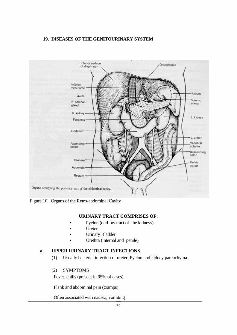
70
19. DISEASES OF THE GENITOURINARY SYSTEM
Figure 10. Organs of the Retro-abdominal Cavity
URINARY TRACT COMPRISES OF:• Pyelon (outflow tract of the kidneys)• Ureter• Urinary Bladder• Urethra (internal and penile)
a. UPPER URINARY TRACT INFECTIONS(1) Usually bacterial infection of ureter, Pyelon and kidney parenchyma.
(2) SYMPTOMSFever, chills (present in 95% of cases).
Flank and abdominal pain (cramps)
Often associated with nausea, vomiting

71
(3) DIAGNOSISOn grounds of the latter
Sometimes cloudy urine due to pus (untreated cases) or brick red urine due toblood (hematuria)
(4) CLINICAL COURSELeads to permanent renal damage if untreated.
(5) TREATMENTIntake of large amounts of fluids
Elimination of obstruction (urinary stasis).
Eradication of urinary bacteria by use of antibiotics secreted into urine.
Ciprofloxine
b. LOWER URINARY TRACT INFECTIONSInfection (usually caused by bacteria, rarely by fungi or chlamydia) of the urinary bladderor urethra, or both. The infection of the urinary bladder is commonly called cystitis.
(1) SYMPTOMSBurning on urination or painful urination
Urinary urgency and frequency
Often low back pain
Rarely nausea and vomiting.
If urethra is affected exclusively:
Burning on urination or painful urination
Frequent urination.
Sometimes involvement of the prostate:
Fever, chills, urinary urgency and frequency
Dysuria (obstruction to voiding due to swollen prostate gland). Painfulejaculation.

72
(2) TREATMENT (for all 3 conditions above)
Intake of large amounts of fluids
Ciprofloxacine.
c. RENAL STONES (Nephrolithiasis)Urinary stones may occur anywhere in the urinary tract and are common causes ofpain, haematuria, obstruction and secondary infection.
(1) CAUSESSupersaturation of stone forming salts (not enough fluid intake!)
Preformed nuclei (bacterial clots, natural crystals in urine)
(2) SYMPTOMSMany stones are “silent” (even for decades)
Back pain or “renal colic” may occur when stones obstruct any level of theurinary tract.
(3) RENAL COLICExcruciating intermittent, cramping pain, usually originating in the kidney area,radiating frequently into the genitalia and thighs.
Often accompanied by nausea, vomiting, even chills, fever and hematuria.
(4) DIAGNOSISX-ray,
ultrasound
Intravenous urography
Urine analysis
(5) TREATMENTRemoval of the stone. Until this can be carried out, give painkillers and smoothmuscle relaxants. Antibiotics if needed.
73
d. EPIDIDYMO ORCHITISBacterial infection of the testes and the origin of the spermatic duct. Usually acomplication of bacterial urinary tract infection most frequently caused by the agentthat causes gonorrhoea (neisseria gonorrhoea).
(1) SYMPTOMSFever, chills
Severe pain in scrotum
Swelling and induration of the testes which is quite painful on examination.
(2) TREATMENTBed rest, analgesics
Scrotal elevation
Scrotal ice packs
Antibiotics (3rd generation cephelosporin, Aminoglycosides)

74
20. SEXUALLY TRANSMITTED DISEASES
a. URETHRITISThis is the most frequent sexually transmitted disease in North America. Three agentsaccount for over 95% of the cases:
• Chlamydia trachomatis• Ureaplasma urealyticum• Neisseria gonorrhoea (causing gonorrhoea)
(1) SYMPTOMS
IN MEN• Appear 1-3 weeks after intercourse.• Mild Dysuria and urinary discomfort in the urethra (penile)• Clear to mucopurulent urethral discharge• A few drops in men and in women stuck together meatus and labia minora
Rarely copious purulent discharge
IN WOMEN
Most women are asymptomatic AA ----> carriers.
(2) TREATMENTCiprofloxacine 500mg (single dose) + Doxycycline 100mg BD for 7 days
NOTE: Sexual partners have to be treated, too!
b. SYPHILISCaused by Treponeuma Pallidum. A sexually transmitted disease which can affectany organ. It is transmitted by sexual contact, including orogenital and anorectalcontact, occasionally even by kissing. Incubation is 3-5 weeks.
SYPHILIS HAS 3 STAGES:
(1) PRIMARY SYPHILIS
Within 4 weeks of infection:
• At the inoculation site, a red papule develops and soon erodes to form apainless ulcer usually on the glans or on the labia, vulva > “Chancres”.

75
(2 ) SECONDARY SYPHILIS
Within 6-12 weeks after infection:
• Cutaneous rashes on skin and mucous membranes (mouth, genitals).• Generalised enlargement of lymph nodes• Some have involvement of eyes, bones, kidneys, liver, spleen.• Fever, anaemia, jaundice may be present.• The skin rashes occur in crops, and may be macules, papules or pustules.• Skin lesions heal without scar formation.
(3) TERTIARY SYPHILIS
• Tertiary Syphilis has 3 forms:• Beginning Tertiary Syphilis.
• 3-10 years after infection:
• The typical lesion is a “gumma” which is a big node, usually localised onskin surfaces, but may also infiltrate organs.
• Gummas are indolent, increase slowly in size, heal gradually and leavescars.
Cardiovascular Syphilis.
• 10-25 years after infection:
• Characterised by aneurysms, narrowing of the coronaries, and/or valvularinsufficiency.
• Leads to death sooner or later.
Neurosyphilis.
Develops in about 5% of untreated cases.
Principally the cerebral cortex is involved which leads to:
• Headache, dizziness• Insomnia, blurred vision• Mental confusion, aphasia• Epileptic attacks• Tabes dorsalis (involvement of part of the spinal column), resulting in
pain, ataxia, sensory changes and loss of reflexes.
76
TREATMENTIn primary and secondary syphilis, all sexual contacts of the past 3 months (in 1°syphilis) and those up to 1 year (2° syphilis) should be examined, treated andinformed that they may be contagious.
• Penicillin is the antibiotic of choice for all stages of syphilis: G ofPenicillin G is given IM.
• • Erythromycin is given to those who are allergic to penicillin.• • Post treatment surveillance is of utmost importance to confirm a permanent
cure.
c. GONORRHEAGonorrhoea is an acute bacterial infection of the urethra, vagina, rectum, pharynx oreyes. It is caused by neisseria gonorrhoea and sexually transmitted. Women(prostitutes) are frequently symptomless carriers.
SYMPTOMS(1) IN MEN
Begins with mild discomfort in the urethra after 2-10 days, followed by Dysuriaand purulent discharge (usually 1 or 2 drops in the morning).
(2) IN WOMEN
Generally mild, but in a few women the onset may be severe, with Dysuria andheavy vaginal discharge.
(3) In women and homosexual men, rectal gonorrhoea is common.
GONOCOCCAL PHARINGITISSore throat a few days after orogenital contact. Sometimes with a mucopurulentexudate.
TREATMENTThree accepted coequal regimens:
- Penicillin G 4.8 g IM plus 1 g of Probenicid.
- Ampicillin 3.5 g or Amoxycillin 3.0 g PO plus 1 g of Probenicid.
- Tetracycline 500 mg PO 4 x daily for 5 days.
77
d. HIV (Human Immunodeficiency Virus)(1) HIV is not transmitted by casual contact or even the close, non-sexual contact
that normally occurs at work, in school, or at home. Transmission to anotherperson requires transmission of body substances containing infected cells, e.g.blood, plasma, and less frequently saliva or semen.
(2) Infected cells can reach target cells in a new host directly (blood transfusion,injection) or indirectly crossing mucous membranes as happens during sexualintercourse of all sorts. Infected cell leaves blood stream and body through socalled microscars or microbreaks in mucous membranes or superficial skin >enter new host via similar microscars.
(3) Passage of an infected cell from one body into another is prevented (or can beprevented) by putting a thin rubber membrane in the route of an infected cell.This rubber membrane is called “condom”. Thus, the use of a condom yields aprotection close to 100%.
(4) HIV preferentially affects (infects) Ty Lymphocytes, also called T-helper cellswhich induce a variety of immune reactions. These cells are then depletedresulting in several qualitative and quantitative defects affecting the entireimmune system (humoral as well as cellular).
CHEMICAL STEPS OF INFECTION(1) Antibody – negative. Asymptomatic carrier state > The human body has not yet
produced antibodies against the HIV (lasts 1-3 months).
(2) Antibody – positive. Asymptomatic carrier state > 1-3 months after infection, theimmune system produced antibodies against HIV, which can be detected byserological means, yet the patient has no symptoms up to this point (This state isquite variable in length, lasting up to many years).
(3) AIDS related complex (ARC) is a constellation of chronic signs and symptoms:
• Generalised lymphadenopathy• Weight loss• Intermittent fever, malaise, lethargy• Chronic diarrhoea• Anaemia, lymphopenia, leukopenia
(4) Full blown AIDS syndrome:
• Wasting Syndrome. Progressive weight loss, associated with fever, nightsweats, oral thrush, diarrhoea lasting for > 3 months.
• Opportunistic infections (pneumocystis, pneumonia, candidiasis,cryptococconis, CMV, disseminated herpes)
• Certain cancers (Kapori’s sarcoma, non-Hodgkin lymphoma, primarylymphoma of the brain).

78
TREATMENTPresently, there is no known cure to AIDS! Some of the associated diseases can betreated more or less efficiently (opportunistic infections, cancers). The most effectivetreatment, however, is PREVENTION!
e. GENITAL HERPESHSV (human) Herpes Simplex Virus causes genital herpes. The most common causeof genital ulceration. It is moderately contagious, spreads by sexual contact anddevelops 4-7 days after contact. It tends to recur because the virus continues to live inhuman nerve cells from which is reactivates and reinfects the skin.
SYMPTOMSItching and soreness precedes a patch of painful vesicles, which erode and formcircular ulcers, which heal in about 10 days.
The lesions occur in males on prepuce, glans, and shaft of the penis; in femaleson the labia, clitoris and vagina. The anus and rectum in homosexuals.
Rarely generalised malaise and fever.
TREATMENTAcyclovir (IV, topical or oral) effectively treats 1° herpetic infections of themouth, genitalia, and rectum. However, it only reduced symptoms of 2°reinfections without permanent cure.
f. CRABSCrabs are caused by “pubis crab louse” which are transmitted venerally. The crablouse infects the anogenital hairs. The lice are large but not easily seen withoutdiligent search.
The most important sign of infestation is a scattering of minute dark brown specks(louse excreta) on undergarments where they come in contact with the anogenitalregion.
TREATMENTRapid with 1% lindane for 2 days in shampoo, cream or lotion (pyrethrin is also quiteeffective).
NOTE: After infestation, decontaminate combs, hats, clothing, bedding, etc.
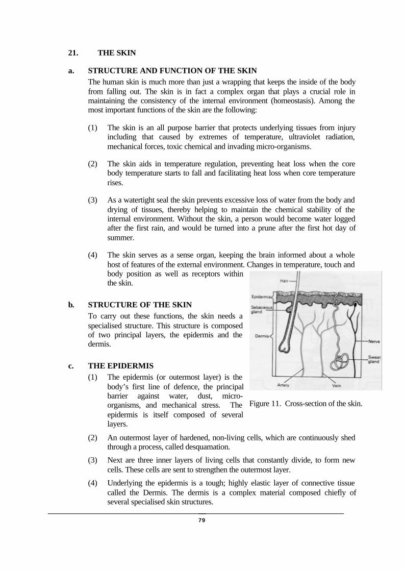
79
21. THE SKIN
a. STRUCTURE AND FUNCTION OF THE SKINThe human skin is much more than just a wrapping that keeps the inside of the bodyfrom falling out. The skin is in fact a complex organ that plays a crucial role inmaintaining the consistency of the internal environment (homeostasis). Among themost important functions of the skin are the following:
(1) The skin is an all purpose barrier that protects underlying tissues from injuryincluding that caused by extremes of temperature, ultraviolet radiation,mechanical forces, toxic chemical and invading micro-organisms.
(2) The skin aids in temperature regulation, preventing heat loss when the corebody temperature starts to fall and facilitating heat loss when core temperaturerises.
(3) As a watertight seal the skin prevents excessive loss of water from the body anddrying of tissues, thereby helping to maintain the chemical stability of theinternal environment. Without the skin, a person would become water loggedafter the first rain, and would be turned into a prune after the first hot day ofsummer.
(4) The skin serves as a sense organ, keeping the brain informed about a wholehost of features of the external environment. Changes in temperature, touch andbody position as well as receptors withinthe skin.
b. STRUCTURE OF THE SKINTo carry out these functions, the skin needs aspecialised structure. This structure is composedof two principal layers, the epidermis and thedermis.
c. THE EPIDERMIS(1) The epidermis (or outermost layer) is the
body’s first line of defence, the principalbarrier against water, dust, micro-organisms, and mechanical stress. Theepidermis is itself composed of severallayers.
(2) An outermost layer of hardened, non-living cells, which are continuously shedthrough a process, called desquamation.
(3) Next are three inner layers of living cells that constantly divide, to form newcells. These cells are sent to strengthen the outermost layer.
(4) Underlying the epidermis is a tough; highly elastic layer of connective tissuecalled the Dermis. The dermis is a complex material composed chiefly ofseveral specialised skin structures.
Figure 11. Cross-section of the skin.
80
d. THE DERMISThe dermis (or innermost layer) consists of the following structures:
(1) Nerve Endings. The sense of touch, temperature, pressure and pain.
(2) Blood Vessels. Like all blood vessels throughout the body, these carry oxygenand nutrients to the skin and bear away waste products, such as carbon dioxide.The small blood vessels are also serving a crucial role in regulating bodytemperature. They do this by regulating the volume of blood that flows from thebody’s core to its cooler surface.
(3) Sweat Glands. These produce sweat and discharge it through ducts passing tothe surface of the skin. Sweat consists of water and salts, which evaporate fromthe surface of the skin, so cooling the body down and loosing excessive coreheat.
(4) Hair Follicles. These are structures that produce hair and enclose the hair roots.Each follicle contains a single hair. Attached to each hair follicle is a smallmuscle that, on contraction, causes the follicle to assume a more verticalposition. Sensations such as cold or fright cause this to automatically occur(called Goosebumps). A hair can live as long as 2 to 5 years before beingreplaced.
(5) Sebaceous Gland. This gland, which is found at the neck of each hair follicle,produces an oily substance called sebum. The sebum coats the hair follicle andthe skin, so that they both remain supple without flaking or cracking. This iswhat causes greasy hair.
e. SUBCUTANEOUS LAYERThe layer beneath the dermis is called the Subcutaneous Layer. This layer containsfatty tissue, whose main purpose is to insulate the body from extreme temperatures ofheat and cold. Its other functions are to form protection against injury to the body andto provide the body with an energy reserve.
f. SKIN RELATED DISEASESThe skin related diseases are probably the most common complaint you will see. Theyare also the most easily preventable. As a SOF Medic, you can not be expected toidentify all of the many types of skin diseases on sight. Dermatologists have a hardenough time, even as experts. The most valuable thing for you to do is to be able todescribe, accurately using the proper terminology, what the skin condition looks like.If you can do this, you could describe it, on the radio, to a dermatologist who may behundreds of miles away. You would then be able to benefit from his opinion.
81
(1) An infection is when bacteria, viruses or fungi enter the body and cause harm.
(2) An inflammation is a reaction of the body’s defence system to infection, traumaand/or histamine release. There are four elements of inflammation: Swelling,redness, warmth, pain - tenderness.
(3) All skin infections or inflammations have to be taken seriously.
• Macule A discoloured spot on the skin
• Papule A solid elevation
• Nodule A small node which is solid and irregular
• Wheal A flat fluid filled elevation usually with itching
• Polyp A growth on the skin
• Pustule An elevation filled with pus
• Ulcer A loss of substance on the surface
• Cyst A sac with fluid other than pus
• Fissure A cleft or groove
g. FUNGAL INFECTIONS
h. CANDIDIASIS
(1) Well demarcated erythematus, sometimes itchy, exudative patches of varyingsize and shape. Glazed brick red patches with satellite lesions. Between toes =Athlete’s Foot. Occurs in skin folds. Warmth + Moisture.
(2) TREATMENT• A n t i- f u n g a l c r e a m s ( m y s t a t i n , C anes t en ) .• Good hyg iene . Keep sk in d ry .
i. DERMATOPHYTES
(1) Tinea Pedis: Ringworm of the feet. Red, itching, dry skin with scaling, usuallybetween the toes.
(2) Tinea Corporis: Itching, red, oval ring, often developing in body folds. Firstcentrifugal progress.
(3) Tinea Capitis : Dry, itching, scaly area of scalp with hair loss.

82
(4) TREATMENT• Anti-fungal creams (Miconazole 2%, Canesten). Good hygiene, keep skin
dry.
j. VIRAL INFECTIONSUsually only once, during childhood.
k. VARICELLA - CHICKENPOX(1) Eruption of macules, papules, vesicles and crusting. Mild headache, moderate
fever and malaise.
(2) TREATMENT• Local disinfection, anti-histamines, antibiotics if severe.
l. ZOSTER(1) Pain with closely grouped varicella-like lesions, which follow a nerve.
(2) TREATMENT• Local disinfection, antibiotics if severe. Rest, analgesics.
m. BACTERIAL INFECTIONS OF THE SKIN
n. IMPETIGO(1) A superficial vesiculopustular skin infection usually following superficial trauma, a
break in the skin, or the infection may be secondary to parasitic infections, fungalinfections, and various types of dermatitis or insect bites. Arms, legs and face aremore susceptible to impetigo. Lesions vary from pea-sized vesicopustules to largebizarre circinate ringworm like lesions to exudative and then honey coloured,crusted lesions. Itching is common and scratching may spread the infection.
(2) TREATMENT
• Topical anti-bacterial with disinfectant ointment for mild to severeinfections. Systemic antibiotics for severe infection (Erythromycin).
o. CELLULITIS(1) Red, swollen hot painful skin in the vicinity of a wound. The lesion is well
demarcated. High fever, chills and malaise. This is a deeper infection.
(2) TREATMENT(a) Erythromycin for at least 2 weeks.
(b) Warm, moist heat; Incision and drainage (be careful not to spread theinfection); Erythromycin (Cloxacillin), analgesics; Disinfection of thesite; PRN (more is better).

83
p. PARASITIC INFECTIONS OF THE SKIN
q. SCABIES(1) A transmissible parasitic skin infection. A female mite tunnels into stratum
corneum, deposits her eggs. Lesions are thought to result from sensitivity toparasites. Readily transmitted.
(2) TREATMENT• Good hygiene, then apply 1% Gamma Benzene Hexachloride or 25%
Benzyl Benzoate cream from the neck down. Repeat in 12 hours only once.Treat patient’s contacts.
r. LICE(1) Visible to the naked eye with itching scalp and fine dandruff. Nits (eggs)
transmitted by shared hats, combs, and brushes.
(2) TREATMENT• Uncommon under good hygienic conditions. Cut hair short, apply Jacutin
lotion, and watch contacts.
s. PEDICULOSIS(1) Pediculosis Corporalis. Body hairs found in underwear, itching lesions.
(2) Pediculosis Pubis. Itching with careful inspection. Ova attached to skin at baseof hair. Minute dark brown specks on undergarments.
(3) TREATMENT• 1% Gamma Benzene Hexachloride applied once/day for 2 days.
t. TICKS(1) Can cause troublesome bite reaction (Lyme, etc.).
(2) TREATMENT• Remove all of tick with oil, etc. (block air for tick, it will back out).
u. FLEAS(1) Papules, often with central blood crust. Grouped in lines or clusters.
(2) TREATMENT• Insect repellents. Good hygiene. Wash cloths, bedding.

84
22. TREATMENT OF WOUNDS
a. TYPES OF WOUNDS
• Abrasions• Gaping wounds• Lacerated wounds• Contused wounds• Stab wounds• Gunshot wounds
Under SOF conditions, wounds must always be considered as infected.
b. WOUND HEALING(1) General
(a) Some hours after the injury the cells located at the base and at theedges of the wound divide and begin to migrate.
(b) It is occasionally possible to suture the wound NLT six hours after theinjury.
(c) Deeper tissues are sealed after 48 hours.(d) The body attempts to produce even surfaces and to contract tissues.(e) After 3-4 months, the elasticity of the scar is back to normal.
(2) Nutrition(a) Vitamin C: Makes the scar stronger(b) Vitamin A: Makes the skin grow faster
c. WOUND INFECTION(1) Pain and/or numbness in the area around the wound.
(2) Fever
(3) Red skin
(4) Swelling
(5) Discharge from the wound/possibly pus
(6) The wound and the area around it are hot
(7) The proximal lymphatic nodes are swollen.
(8) Any infection slows healing. The scar becomes large. Infections must beexpected in large wounds in particular, as well as in visibly contaminatedwounds, whenever tissues have been lacerated or contused, in injuries of theintestines and in wound "pockets".

85
b. Preparations to be carried out before the wound treatment
(1) Sterilise instruments (boiling, soaking in iodine); lay them out ready for use.
(2) Lay out dressing material.
(3) If anaesthesia is necessary, prepare syringe with anaesthetic.
(4) Wash your hands with water and iodine solution (pay attention to yourfingernails).
(5) Scrub the skin and the wound.
(6) If necessary, administer a local anaesthetic.
(7) Prepare a sterile field (sterile cloths, insides of packages containing sterilematerial).
d. Wound treatment(1) Take all wounds seriously, even small ones.
(2) Irrigate the wound with sterile water.
(3) Debride dead tissues and wound pockets.
(4) Irrigate the wound once more (with a water and iodine solution).
(5) Apply a sterile, loose dressing. In the case of deep wounds, pack loosely;possibly soak the tamponage in iodine solution.
(6) Apply antibiotics (e.g. gyrase inhibitors).
e. Care after wound treatment
(1) If the wound is located at a limb: elevate the limb.
(2) Clean and pack your instruments.
(3) Take your patient's temperature, count his pulse, and write your findingsdown.
(4) Boil water for the next treatment.
(5) Scald the used dressing material.
(6) Make sure to have sufficient supply of fuel or wood for heating.

86
23. THE EYE AND SIGHT1 A. General
The eye is the organ of sight and sits in the orbital cavity, protected by pads of fat,the eyelids and the eyelashes.
(7) It is approximately 1" in diameter and has 3 layers.
• Sclera - The outer fibrous coat.
• Choroid - The middle vascular .Retina - The inner nervous coat
(3) At the front of the eye, we have a transparent "window" which allows lightto enter the eye.
(2) This is known as the CORNEA and is continuous with the sclera.
(8) The IRIS is the coloured disc behind the cornea, which acts like the shutteron a camera by adjusting the size of the PUPIL.
(9) Behind the iris, we find the LENS, which is encapsulated and alters shape toenable focusing on near or far objects.
(10) The capsule surrounding the lens is attached to the SUSPENSORYLIGAMENT, the opposite end of which is attached to muscles in theCILIARY BODY.
(11) There are 2 chambers in front of the lens:
(a) Anterior - Lies in front of the iris.(b) Posterior - Lies between iris and the lens.
(12) In these 2 chambers, we find a watery substance called AQUEOUSHUMOUR.
(13) Behind the lens, the main body of the eye is filled with a jelly like substancecalled VITREOUS HUMOUR, which gives the eye its shape.
(14) The OPTIC NERVE (2nd cranial) leaves the eye at the back, medial to thecentre. Through an ophtalmoscope, it can be seen entering the eye as theOPTIC DISC. Blood vessels can also be seen entering at this point.
(15) The MACULA LUTEA or "yellow spot" is seen directly in the centre of theeye and is the point of keenest focus.
(16) The CONJUNCTIVA is a mucous membrane covering the outside of the eyeand the inner aspect of the eyelids. It allows friction-free movement of theeyeball with the lids and helps protect the eye.
(17) Tears are produced in the LACRIMAL GLANDS found in the superiorlateral part of the orbit. They drain through the PUNCTA LACRIMA; thelittle cone shaped projections on the medial aspect of the lower lid.
(18) The lids contain SEBACEOUS GLANDS that lubricate the eye margins, andinflammation of a gland is termed a STYE.
(19) Movement of the eye is by 6 muscles supplied by the 3rd, 4th and 6th cranialnerves. They are:

87
(a) 4 Rectus muscles to allow medial lateral movement, and superiorinferior movement.
(b) Oblique muscles that allow crossways movement.
f. SIGHTThe retina has several layers. The cells concerned with sight and colour perceptionare:
(1) RODSEnable us to see in dim light. They have a substance called visual purple ontheir outer aspects, which bleaches in bright light. This reforms in dim lightand is the reason we can see in the dark.
(2) CONESAre found in the central area of the retina and allow sight in bright light.They also allow us to perceive colours.• The 2 optic nerves leave the back of the eye and converge to cross at the
optic chiasm. Here, the fibers from the outer halves go to opposite sidesso impulses from each eye go to both hemispheres. This is known asbinocular vision.
• Light rays are refracted (bent) by the aqueous humour, lens and vitreoushumour so the object is focused on the retina upside down. The visualcentre in the brain converts the impulses so that we see the object theright way up. It also translates impulses so we perceive the differentcolours and shadings.
g. HYPERMETROPIAIs long sightedness and is caused by the eyeball being too short. The image is thusfocused beyond the retina.
h. MYOPIA(1) The eyeball being elongated causes short sightedness, so the image is
focused in front of the retina.(2) Both conditions can be corrected by placing an appropriately shaped lens in
front of the eye - glasses or contact lenses.(3) When focusing on distant objects, the pupil will be dilated and the lens
flattened and elongated.(4) In focusing on near objects, the opposite occurs.
2 INFECTIONS
i. CONJUNCTIVITIS(1) General. Conjunctivitis can have various causes and is not necessarily caused
by an infection. It is the most common eye "disease" of all.(2) Symptoms.
(a) Red eye (due to congestion of the blood vessels).(b) Narrow position of the eyelids, edema. The patient may be unable to
open his actively.(c) Flow of tears.(d) The cornea may be dull due to edema.
88
(e) Usually there is no pain, but a feeling of heaviness and heat.(f) Pain indicates that the cornea may also be affected.(g) Discharge with or without pus.
(3) Treatment(a) No bandage(b) Antibiotic eyedrops or(c) Antibiotics administered through tablets or injections if there is pus.
j. BLEPHARITIS (INFLAMMATION OF THE MARGIN OF THE LID)(1) Symptoms
At first, there is some dry dandruff around the lashes. After an infection,ulcers can develop at the margins of the lid. The lashes either do not regrowat all or into the wrong direction, so that they can chafe on the conjunctivaand/or the cornea.
(2) TreatmentWash out the eyelids with lukewarm water in the morning and in theevening. Apply antibiotic ointment.
k. STYE (HORDEOLUM)(1) Symptoms
(a) Localised inflammation of glands, located mostly at the margin of thelid.
(b) The area is very tender.(c) Considerable redness and swelling of the lid.(d) Intense pain.
(2) Treatment(a) Application of warmth may be successful at the beginning only. Later
it will promote the process.(b) Antibiotic ointments in order to prevent further infections.(c) No bandage.(d) Stab incision carried out with a sterile cannula.
l. CHALAZION(1) Symptoms
(a) Hard, palpable nodule in the lid, which cannot be displaced and is nottender on pressure.
(2) Treatment(a) Small chalazion sometimes disappears without any treatment at all.(b) Under SOF conditions, treatment is not necessary.
3 INJURIES (TRAUMA)
m. EYEBALL CONTUSION(1) Symptoms
(a) Impaired vision.

89
(b) Dull pain in the affected eye.(c) Hematoma at the lid.(d) Subconjunctival haemorrhage.(e) In case of bleeding into the orbit, the eyeball bulges out. The motility
of the eyeball is impaired, and there is double vision.(2) Treatment
(a) Apply sterile bandage.
n. SUBCONJUNCTIVAL BLEEDING(1) Symptoms
(a) Blood clearly visible on white conjunctiva.(2) Treatment
(a) None. The condition will clear up without any treatment within 2weeks.
o. ORBITAL FRACTURES, BLOW OUT FRACTURE(1) Symptoms
(a) Bleeding into the lid and/or into the conjunctiva.(b) If there is a bleeding into the orbit, there is a massive haemorrhage
below the lid/conjunctiva.(c) The eyeball bulges out. The movability may be impaired.
(2) Principal Symptom(a) Double vision.(b) Vision may be normal.
(3) Treatment(a) Apply a sterile bandage.(b) No ointment.(c) Consider a fracture of the base of the skull.(d) Treat shock, if necessary.(e) Administer analgesics.
p. PENETRATING EYE INJURIES(1) Symptoms
(a) If the cornea is affected:• I
ntense pain.• I
ncreased flow of tears.• B
leeding into the cornea.(b) If the conjunctiva/sclera are affected:
• Compulsive eyelid closure.

90
• Conjunctival haemorrhages.
• Gaping wound of the sclera with the vitreous body bulging out.
(2) Diagnosis(a) Through examination of the eye with the flashlight.
(3) Treatment(a) Apply sterile bandage.(b) No eye ointment.(c) Never remove foreign bodies - leave this to an eye specialist.
q. FOREIGN BODIES(1) Foreign bodies located in the conjunctiva may be removed with cotton-wool
sticks or similar devices, unless the foreign body is burnt into theconjunctiva. Then apply antibiotic eye ointment and a sterile eye bandage.
(2) An eye specialist may remove foreign bodies located in the cornea only.Don't do anything apart from applying a sterile eye bandage and someantibiotic eye ointment.
r. BURNS THROUGH CAUSTIC SUBSTANCES(1) Symptoms
(a) Intense pain.(b) Compulsive eyelid closure(c) Considerably impaired vision.(d) In most cases, the patient's face is also affected.(e) The patient may show symptoms of shock.
(2) Treatment(a) Open the patient's eye - by force if necessary.(b) Irrigate the eye (from the inner to the outer side).(c) Remove all remaining particles of the substance with cotton-wool
swabs.(d) If necessary, carry out shock treatment and administer an analgesic to
the patient.

91
24. THE EAR
a. GENERAL(1) The ear is the organ of
hearing. It is supplied bythe eighth cranial nerve,which is stimulated byvibrations caused by soundwaves.
(2) With the exception of thepinna, the structures thatform the ear are encasedwithin the temporal bone.
b. STRUCTURE(1) The ear is divided into three
parts:
• External Ear
• Middle Ear
• Internal Ear
c. EXTERNAL EARThe external ear consists of the pinna and the external auditory meatus (meatus -opening or passage).
d. TYMPANIC MEMBRANEThe tympanic membrane or eardrum separates the external auditory meatus from themiddle ear. It is oval shaped with its slightly broader edge uppermost.
e. MIDDLE EAR(1) Lateral wall - the tympanic membrane
(2) Roof and floor - Temporal bone
(3) Posterior wall - Temporal bone with opening to mastoid air cells.
(4) Middle wall - Temporal bone with two openings
f. AUDITORY OSSICLES (OSSICLES - SMALL BONE)(1) Three small bones contained in middle ear.
(2) They transmit sound from tympanic membrane to the oval window and innerear.
(3) They are, from front to back, the malleus, incus and stapes.
Figure 12. The Ear.

92
g. INNER EARContains the organs of hearing and balance.
h. PHYSIOLOGY OF HEARING(1) Sound produces sound waves, which travel at 332 m/sec.
(2) The pinna concentrates sound waves and sends them along the externalauditory meatus to the tympanic membrane.
(3) Tympanic membrane vibrations are transmitted through the middle ear bymovement of ossicles.
(4) Stapes sets up fluid movement (waves) in the inner ear to stimulate nerveimpulses which pass along the number eight cranial nerve.
i. SEMI CIRCULAR CANALSHave no auditory function. They provide information about equilibrium and balance.
j. INJURIES AND DISEASES OF THE EAR, NOSE AND THROATThe external ear, external auditory meatus, tympanic membrane, auditory ossicles,Eustachian tube, cartilage’s of the nose, nasal bone, nasal septum, sinuses, tonsils andtongue are all susceptible to injury or disease. These include the following:
• Otitis Externa Fractures of the nose
• Otitis Media Sinusitis
• Boils in the external auditory meatus Hay Fever
• Dizziness Tonsillitis
• 5. Epistaxis Mouth Ulcers
k. OTITIS EXTERNA OR INFLAMMATION/INFECTION OF THEEXTERNAL EARThis is usually a local inflammation caused by staphylococcus aureus. Although thisis not always the case, sometimes the underlying cause might be an allergic reaction.Hearing may sometimes be impaired if the external auditory meatus is blocked, eitherdue to swelling or discharge. The pinna is usually tender on movement and anydischarge is thick and scanty. Treatment is very simply achieved by cleaning the earand using antibiotic eardrops.
l. OTITIS MEDIA OR INFLAMMATION/INFECTION OF THE MIDDLEEAR(1) Otitis Media can be broken into two forms. These are Acute Otitis Media and
Secretary Otitis Media.

93
(2) ACUTE OTITIS MEDIAThis is sometimes caused by the spread of an upper respiratory tract infection(URTI) via the Eustachian tube. There is a deep-seated pain with deafness andsome form of systemic illness, with fever. There is also a feeling of the earbeing blocked, followed by discharge, often by a perforation in the tympanicmembrane, which will often ease the pain and may help to reduce the fever.The treatment should be antibiotic therapy IM, the oral analgesia for the pain.
(3) SECRETORY OTITIS MEDIAWith secretary Otitis media, there is an unnatural reverberation of the patient’svoice, with hearing loss and feeling of the ear being blocked. However, therewill be very little pain and no fever. The treatment should simply bedecongestants to treat the cause of blockage.
m. BOILS IN THE EXTERNAL AUDITORY MEATUSWhen a resistant staphylococcal infection is present in the external auditory meatus, aboil often appears. Using Flucloxacillin should simply treat this.
n. DIZZINESSThis is a feeling of unsteadiness, usually accompanied by anxiety and sometimesnausea. There are several causes such as carsickness (or motion sickness) or evenMenieres Disease (increased fluid pressure within the middle ear). Giving an anti-emetic such as Metoclopramide can treat this.
o. EPISTAXISEpistaxis or nosebleeds are usually caused as a result of trauma. However, high bloodpressure, infections of the nose or even sneezing can cause someone to have a nosebleed. If there are no signs of trauma to the skull of spine simply sit the patient upwith his head slightly forward and pinch the nostrils shut. If unconscious, lay on backwith head elevated and to one side.
Note: If clear straw like fluid is coming out of the nose/ears, there is the possibility ofa skull fracture.
p. FRACTURES TO THE NOSECaused by trauma to the face. The nose may be bleeding but will usually look out ofshape and deformed. The patient will usually have two black eyes, and may be inpain. Always ensure that the patient has a patent airway. You may have to lean himslightly forward, to help drainage of blood. If there are no spinal or skull fractures,treat as for a nose bleed and give analgesics for pain.
94
q. SINUSITIS OR INFLAMMATION/INFECTION OF THE SINUSESSinusitis usually occurs after an upper respiratory tract infection, swimming, diving,or nasal allergies. There is usually pain, tenderness and a feeling of tiredness withheadache. The nose often feels blocked and there may be a purulent nasal discharge.The treatment should consist of analgesics, light diet, fluids, and nasal decongestants.There may also be a requirement for systemic antibiotics, should there be a purulentdischarge.
r. HAYFEVERHayfever is simply an allergy reaction to pollen. Signs and symptoms includesneezing, rhinitis, sore eyes and headache. The treatment should be in the form ofanti-histamine such as Terfenadine.
s. TONSILLITISTonsillitis is an inflammation or infection of the tonsils. The signs and symptomsinclude a sore throat, difficulty in swallowing. The may also be a slight temperatureand runny nose. The treatment at this stage should take the form of antibacteriallozenges, soluble aspirin and lots of clear fluid such as water, to ease the discomfortand reduce the temperature. If, however, the tonsils are very inflamed, and if thetemperature is high with white spots on the tonsils, then an antibiotic should be used,preferably penicillin.
t. MOUTH ULCERSMouth Ulcers may occur because of trauma, infections, nutritional deficiencies anddrug therapy. Treatments are designed to relieve pain or inflammation. These take theform of simple mouthwashes or gels such as saline rinses, Chlorohexidine Gluconateand Bonjetta.

95
25. VITAL SIGNS
a. GENERALTemperature, pulse, respiration (TPR), and blood pressure (BP) are called vital signsbecause they indicate a patient's life signs. Measurement of these signs aids in makinga diagnosis and prescribing treatment. Any marked deviation from the normal range isa signal of distress from the body; the interpretation of changes is as important as themeasurement itself.
b. BODY TEMPERATUREBody temperature is the result of a balance between the heat produced and the heatlost by the body. The hypothalamus is that portion of the brain that regulates bodytemperature by speeding up or slowing down the cells' use of food (metabolic rate).The higher the rate of metabolism, the more heat the body produces. The circulatingblood distributes this heat. Excessive heat is eliminated through the skin, lungs, andexcreta. When the balance is disturbed, deviations in body temperature result.
c. NORMAL TEMPERATUREBody temperature ranges between 96 and 100°F or 36 and 38°C. The normal oraverage temperature of most people is 98.6°F or 37°C. The temperature reading thatyou obtain will vary according to the site you use. The average oral temperature is98.6°F/37°C. Rectal temperature is usually about one degree higher (99.6°F/38°C),and the axillary (armpit) temperature is about one degree lower (97.6°F/36°C) thanwhen measured orally. A range of 1° lower or 1° higher from the average normaltemperature is usually considered to be within normal limits. When the bodytemperature changes from the normal average, it warns of body malfunction,infection, or dehydration.
d. ABNORMAL TEMPERATURE(1) Pyrexia (fever is an elevation in temperature above the normal average. During Pyrexia, heat is produced
faster than the body can eliminate it. Fever is a common symptom of infection or other disease.
(2) Hypothermia is a deviation in temperature, which persists below the average normal temperature. Shock,starvation, or a long lasting illness may cause a subnormal temperature. It indicates that body resistance todisease is low.
e. MEASURING BODY TEMPERATUREThe mouth, the rectum, or the axilla (armpit) can measure the body temperature.The method used depends on patient's age, physical condition and equipmentavailable.

96
f. METHODS OF MEASURING TEMPERATURE USING CLINICALTHERMOMETERS
(1) ORAL TEMPERATURE
(a) This is the most convenient method and can be used for responsiveadult patients. Before taking an oral temperature you should ask thepatient if he has recently had any food or drink of if he has beensmoking. If so, wait 15 minutes before taking the temperature.
CAUTIONWHEN HANDLING THERMOMETERS, HANDLE BY THE
STEM ONLY
(b) Wash your hands.
(c) Check thermometer to be sure it is clean and dry. Shake it down to94°F/35°C if necessary. When shaking down the thermometer, grasp thestem end firmly and with a sharp downward wrist motion, shake thethermometer Check the mercury column and repeat the shaking procedure,if necessary, to lower the column to the 94°F/35°C mark.
(d) Place bulb end under the patient's tongue. Instruct him to close lipsfirmly around stem, but not to bite down. Leave thermometer in place atleast 3 minutes.
(e) Remove thermometer. Wipe with a gauze tissue from stem to bulb toremove any saliva. Read and record the temperature, using decimals (forexample "98.4°F/37.0°C).
(f) Place thermometer in "used" oral thermometer holder.
(2) RECTAL TEMPERATURE
(a) This is the most accurate method. It is used for all infants and youngchildren and for adults who are unconscious, irrational, or who havedifficulty breathing with the mouth closed. It is not used on patientswho have had rectal surgery or have a rectal disorder.
(b) Provide patient privacy (if possible). Then turn him on his side (Simsposition) and expose the buttocks. The top knee should be flexed (bent).
(c) Insure that the tip of the thermometer is well lubricated. Use sterilelubricant for this procedure.
(d) Lift the upper buttock to expose the anus. Insert the well-lubricated bulbof the thermometer slowly and carefully about 1-1/2 inches into therectum.

97
(e) Hold thermometer in place for 2 minutes.
(f) Remove thermometer. Wipe downward with a gauze tissue from stem tobulb. Read and record temperature using the decimal followed by theinitial R [for example "99.8°F(R)/38.0°C(R)].
(g) Place rectal thermometer in "used" holder.
(3) AXILLARY TEMPERATURE
(a) When temperature can be taken neither orally or rectally, it can betaken under the arm where the thermometer bulb can be surrounded bybody tissue. To take the axillary (armpit) temperature, use an oralthermometer.
(b) Pat the armpit dry with a tissue or towel. Place the bulb of the oralthermometer in the centre of the armpit and pointed towards the patient'shead.
(c) Fold the patient's arm across his chest with his fingers on the oppositeshoulder.
(d) Leave thermometer in place for at least 10 minutes.
(e) Remove thermometer. Read and record temperature, using the decimalfollowed by the initial A [for example, "100.2°F (A) or 36.0°C (A)].
(f) Place thermometer in "used" holder.
g. CARE OF THERMOMETERS (FIELD EXPEDIENT METHOD)
(1) Remove thermometer from its plastic holder.
(2) Cleanse thermometer with 70 percent isopropyl alcohol pad. Use a twistingmotion to clean from stem to bulb end.
(3) Rinse the thermometer with cool water or with a gauze pad saturated withwater. Use a twisting motion from stem to bulb end.
(4) Shake down the thermometer to at least 94°F/34°C.
NOTEThis procedure is to be used prior to taking patient’s temperature and after temperature is taken.

98
h. PULSEPulse is defined as the rhythmic expansion and contraction of an artery. The beatingof the heart causes this action. When the heart contracts (systole), the blood is forcedfrom its chambers into the arteries. This action causes the arteries to dilate (expand).When the heart relaxes (diastole), blood refills its chambers. This action causes thearteries to contract, or recoil, as the blood moves further along in the circulatorysystem. A patient's pulse is measured to aid in determining his condition bycomparing it with a normal heart rate.
i. PALPATION OF THE PULSEThe pulse can be felt at points, where an artery lies close to the skin or where itcrosses over a bony area or hard tissue. The pulse sites can be found:
(1) At the wrist, proximal to the thumb (radial artery), on the palm side of thehand.
(2) At either side of the neck, near the windpipe (carotid artery).
(3) On the inside of the elbow about 1/2 inch proximal to the elbow point(brachial artery).
(4) Below the left nipple [5th intercostal space (apical artery)].
(5) In front of the ear (temporal artery).
(6) In the middle of the groin and leg joint (femoral artery).
(7) In the centre of the back of the knee along the inside medial tendon (poplitealartery).
(8) Behind the inner ankle bone (posterior tibial artery).
(9) Along the top (dorsum) of the foot (dorsalis pedis artery).
j. PULSE RATE
(1) N O R M A L P U L S E R A T E
Generally, the normal pulse is regular in rate, rhythm, and force (strength).A strong pulse is easily detected by the large amount of blood being pumped.The average range is between 60 and 80 pulse beats per minute. The rate ofthe normal pulse rate varies slightly in individuals as indicated below.
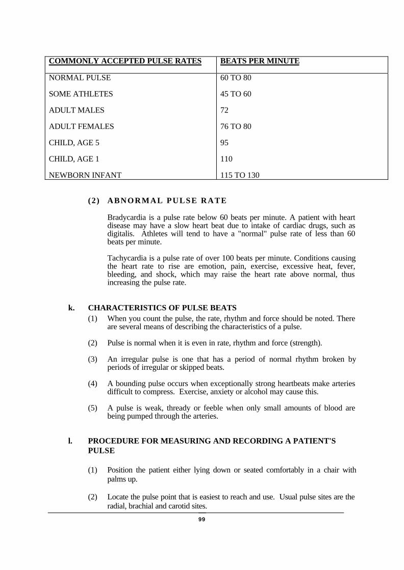
99
COMMONLY ACCEPTED PULSE RATES BEATS PER MINUTE
NORMAL PULSE
SOME ATHLETES
ADULT MALES
ADULT FEMALES
CHILD, AGE 5
CHILD, AGE 1
NEWBORN INFANT
60 TO 80
45 TO 60
72
76 TO 80
95
110
115 TO 130
(2) ABNORMAL PULSE RATE
Bradycardia is a pulse rate below 60 beats per minute. A patient with heartdisease may have a slow heart beat due to intake of cardiac drugs, such asdigitalis. Athletes will tend to have a "normal" pulse rate of less than 60beats per minute.
Tachycardia is a pulse rate of over 100 beats per minute. Conditions causingthe heart rate to rise are emotion, pain, exercise, excessive heat, fever,bleeding, and shock, which may raise the heart rate above normal, thusincreasing the pulse rate.
k. CHARACTERISTICS OF PULSE BEATS(1) When you count the pulse, the rate, rhythm and force should be noted. There
are several means of describing the characteristics of a pulse.
(2) Pulse is normal when it is even in rate, rhythm and force (strength).
(3) An irregular pulse is one that has a period of normal rhythm broken byperiods of irregular or skipped beats.
(4) A bounding pulse occurs when exceptionally strong heartbeats make arteriesdifficult to compress. Exercise, anxiety or alcohol may cause this.
(5) A pulse is weak, thready or feeble when only small amounts of blood arebeing pumped through the arteries.
l. PROCEDURE FOR MEASURING AND RECORDING A PATIENT'SPULSE
(1) Position the patient either lying down or seated comfortably in a chair withpalms up.
(2) Locate the pulse point that is easiest to reach and use. Usual pulse sites are theradial, brachial and carotid sites.
100
(3) Palpate the pulse site by placing either the fingertips of index and middlefingers on pulse point, or index, middle and ring fingers on pulse point.
(4) Record the pulse and report as appropriate.
m. RESPIRATIONRespiration, commonly called breathing, is the process by which oxygen (O2) andcarbon dioxide (CO2) are interchanged by the body. External respiration refers tothe delivery of oxygen (O2) to the lungs so that it can be taken into the bloodstream. Internal respiration is the process by which oxygen from the blood is takento the cells in the body and carbon dioxide (CO2) is removed from tissues andcarried into the blood. Both conscious and unconscious (involuntary) control ofrespiration is the function of a respiratory centre in the brain (medulla oblongata).
(1) Inhalation is the process of taking air into the lungs. During inhalation, thediaphragm descends as it contracts and the rib cage is lifted upward andoutward, giving the lungs more room to expand and create a slight vacuum inthe chest that draws air into the lungs.
(2) Exhalation is the process of expelling air from the lungs. During exhalation,the diaphragm descends as it contracts and the rib cage is drawn down andinward as air rushes out of the lungs.
n. NORMAL BREATHING RATESNormal breathing (eupnea) is easily done and does not require conscious thought.Normal respiratory rates are typically one-fourth of the normal heart rate.Respiratory rates vary according to age; the following are commonly accepted asbeing the normal limits:
RESPIRATIONS PER MINUTE
HEALTHY ADULT
ADOLESCENT YOUTH
CHILDREN
INFANTS
12 TO 20
18 TO 22
22 TO 28
30 OR MORE
o. PATTERNS OF BREATHING
(1) NORMAL RESPIRATIONA normal, relaxed breathing pattern is effortless, evenly placed, regular, andautomatic. Increased levels of carbon dioxide or lower levels of oxygen inthe blood trigger an increase in the respiratory rate to restore the chemicalbalance and rid the body of excess carbon dioxide.
101
(2) ABNORMAL RESPIRATIONA head injury or any increased intracranial pressure (ICP) will depress therespiratory centre and result in shallow or slow breathing. Certain drugs alsotend to depress the respiratory rate (for example: Morphine, Demerol).
(3) BREATHING VARIATIONS(a) Dyspnea. Difficult and laboured breathing, often with flared nostrils,
anxious appearance and statements such as "I can't get enough air". Itis important to know how much exertion or activity causes thedyspnea. Does it occur when walking, trying to eat a meal, or whentrying to talk?
(b) Tachypnea. Increased or rapid breathing; May be seen in fever and ina number of other diseases. Breathing rate increases markedly for each1° increase in temperature.
(c) Slow and shallow. There is a limited amount of air exchanged and lessoxygen is taken in. This type of breathing often leads to hypoxia, ordecreased level of oxygen in the blood. It is often seen in patients whoare under sedation, recovering from anaesthesia, have had abdominalsurgery or are in a weak or debilitated condition.
(d) Cheyne-Stokes Respiration. A pattern of dyspnea followed by a shortperiod of apnea (no respiration). Respirations are rapid and gasping innature for about 30 to 45 seconds, then are followed by a period of nobreathing for about 20 seconds. It is seen in critically ill patients withbrain conditions, heart or kidney failure, or drug overdose.
(e) Hyperventilation. A pattern of breathing in which there is a significantincrease in the rate of breaths and carbon dioxide is expelled from thebody, causing the blood level of CO2 to fall. The condition is seenafter severe exertion and during high levels of anxiety or fear and withfever and diseases such as diabetic acidosis.
(f) Kussmaul's Respirations. The increased rate and depth of respirations,with panting and long, grunting exhalation. It is frequently seen indiabetic acidosis and renal failure.
(g) Noisy Respirations. As a rule of thumb, you should regard any noisyrespirations as obstructed breathing. Some of the terms used todescribe noisy respirations are:• R
ales and Rhonchi. Rattling sound caused by secretions in the lungpassageways.
• Stertorous. A snoring sound produced when patients are unable tocough up secretions from the trachea or bronchi.
• Stridor. A crowing sound on inspiration due to the obstruction ofthe upper air passageways as occurs in croup or laryngitis.
• Wheeze. A whistling sound of air forced past a partial obstruction asfound in asthma or emphysema.

102
p. PROCEDURE FOR MEASURING AND RECORDING PATIENT’SRESPIRATION(1) For an accurate accounting of the respiration, the patient should be at rest and
unaware of the counting process. If adult patients are aware that you arecounting their respiration, they may voluntarily breathe faster or slower. Themost satisfactory time to count respiration is after the patient's pulse count.
(2) After taking the pulse, continue holding the patient's wrist. Lay the patient'sarm across his chest.
(3) Count respiratory rate for 1 full minute. Observe rate, depth, patterns, andsounds of respiration.
NOTEONE RESPIRATION INCLUDES BOTH THE INHALATION AND EXPIRATION.
(4) Record the respiration and report as appropriate.
q. BLOOD PRESSURE(1) Blood pressure (BP) may be defined as the pressure exerted by the blood on
the walls of the vessels. All parts of the vascular system are under pressure,but the term "blood pressure" usually refers to arterial pressure. The pressureis the product of
(2) The force of the contraction of the ventricles of the heart,(3) The amount of blood pumped out of the heart, and(4) The resistance of the blood vessels to the flow of blood through them.(5) By measuring the blood pressure, you obtain information about the
effectiveness of the heart contractions, the adequacy of the blood volume inthe system, and the presence of any obstruction or interference of flowthrough the blood vessels.
r. NORMAL RANGE OF BLOOD PRESSURE(1) Blood pressure consists of the systolic pressure written as a fraction over the
diastolic pressure. The systolic pressure is the level present duringcontraction of the heart. Diastolic pressure is the pressure during relaxationof the heart. The average blood pressure in a healthy young adult isconsidered to be 120/80 mm of mercury (Hg); 120 is the systolic pressure, 80is the diastolic pressure.
(2) Just as pulse and respiratory rates vary among individuals, so does bloodpressure.
(3) The normal blood pressure range is:
MALE FEMALE
SYSTOLIC
DIASTOLIC
100-140
60-90
90-130
50-80

103
s. FACTORS INFLUENCING BLOOD PRESSURE(1) As a result of the many factors influencing it, the blood pressure is a dynamic
force that can vary from minute to minute as the heart adjusts to demandsand responses of the body and brain. Many factors exert an influence onblood pressure:
(2) Age. Blood pressure is lower in children than it is in adults.
(3) Sex. Blood pressure is higher for men than women of the same age level.
(4) Body Build. Obese persons usually have higher blood pressure than do thosewho are of average weight and build.
(5) Exercise. Exertion temporarily elevates blood pressure.
(6) Pain. Pain will usually elevate blood pressure.
(7) Emotion. Fear, worry, or excitement will elevate blood pressure.
(8) Drugs. Vasoconstrictors elevate blood pressure. Vasodilators decrease bloodpressure. Narcotics decrease blood pressure.
(9) Disease. Any disorder affecting the circulatory or renal system may increaseblood pressure. A disease that weakens the heart may lower the bloodpressure.
(10) Haemorrhage. Decrease of blood volume lowers blood pressure and maylead to shock.
(11) Intracranial Pressure. Pressure in the space between the skull and the braincan elevate the blood pressure.
t. ABNORMALITIES OF BLOOD PRESSURE
(1) HYPERTENSIONPressure elevated above the normal range is called hypertension. Prolongedhypertension can cause permanent damage to the brain, the kidneys, theheart, and the retina of the eye.
(2) HYPOTENSIONLow blood pressure is called hypotension. Hypotension associated withsymptoms of shock or circulatory collapse is a dangerous condition that canrapidly progress to death unless treated.
(3) BLOOD PRESSURE MEASUREMENTBlood pressure is measured with a sphygmomanometer (an air-pressuredevice) and a stethoscope. The cuff of the sphygmomanometer contains anoblong rubber bag or bladder. When the cuff is wrapped around the upperarm or the mid-thigh and inflated with air, the air pressure registers on thesphygmomanometer gauge. Taking a blood pressure requires practice. Theindividual must apply the cuff properly, manipulate the air bulb andsimultaneously listen through the stethoscope while watching the gauge.

104
NOTETHE PRESSURE READINGS ARE IN MILLIMETERS (mm) OF MERCURY (Hg).
u. PROCEDURE FOR MEASURING AND RECORDING A BLOODPRESSURE USING THE BRACHIAL ARTERY
Determining blood pressure by auscultation is the most common method fordetermining blood pressure as a stethoscope is used to listen for characteristicsounds.
NOTEINSURE THAT CUFF IS COMPLETELY DEFLATED AND THE GAUGE
REGISTERS ZERO.
(1) Decontaminate stethoscope. Clean earpiece and diaphragm with 70 percentalcohol swabs and cotton-tipped applicators.
(2) Explain procedure to the patient.
(3) Position the patient.
(a) Patient should be seated or lying down.
(b) Support the arm to be used, palm up, at the level of the patient's heart.
CAUTIONIF INJURED, PATIENT SHOULD NOT BE MOVED SIMPLY FOR THE PURPOSE OF
DETERMINING BLOOD PRESSURE. TO DO SO MAY AGGRAVATE EXISTINGINJURIES. BLOOD PRESSURE SHOULD BE MEASURED WITHOUT MOVING THE
PATIENT.
(4) Expose the patient's upper arm. Remove garment if sleeve is tight.(5) Place the cuff on the patient's arm. Position the cuff 1-2 inches above the
elbow. Apply the cuff securely but not overly tight.
(a) If using aneroid-type manometer, clip the gauge to the cuff in line withpalm.
(b) If using mercury manometer, place column on a firm, level surface,outside patient's field of vision.

105
(6) Locate pulse of brachial artery by palpating in the bend of the elbow.
(7) Place the bell or diaphragm of the stethoscope over the pulse point. Do NOTapply the bell or diaphragm too firmly; excessive pressure distorts the pulsesounds.
(8) Tighten thumbscrew of air bulb (clockwise) with one hand while holdingstethoscope in place with the other hand.
(9) Inflate the cuff by pumping air bulb. You will hear the pulse sounds as thepressure in the cuff increases, then the sounds will disappear. Continueinflating the cuff until the pressure gauge indicates about 20-30 mm abovewhere the pulse sounds were last heard. It is at this point that the air pressurehas caused the arterial wall to collapse.
(10) Loosen thumbscrew of air bulb (counterclockwise) and allow the air toescape slowly (about 2-4 mm Hg per second). At the same time, watch thegauge. When the first distinct sound is heard, note the number on the gauge;this is the systolic pressure.
(11) Continue to release the air slowly. Look and listen. Note the number on thegauge at which the last distinct sound is heard. This is the diastolic pressure.
CAUTIONIN SOME PATIENTS, SOUNDS MAY BE HEARD TO EXTREMELY HIGH OR LOW LEVELS.

106
26. HEAT INJURIES
b. INTRODUCTION
(1) Heat Production. The human body constantly produces heat in order tomaintain its normal functions.
(2) Evaporation. Whenever the body temperature rises above a set point value, thebody will cool itself down by evaporating water.
(3) Temperature Regulation. The body temperature is regulated by an excessiveheat production (muscular tremor) in the case of hypothermia, and byevaporation in the case of hyperthermia. The brain according to the informationreceived from temperature sensor controls this.
b. HEAT CRAMPSThese are caused mainly by loss of salt due to excessive sweating. Symptoms aremoderate to severe muscle cramps in arms, legs or abdomen. These symptoms maystart as a mild muscular discomfort. This is the time when the person should stopactivity, get in the shade and drink water. If the person fails to recognise the earlysymptoms and continues with physical activity, he will have severe muscle crampsand pain.
(1) SYMPTOMS(a) Painful cramps of muscles in arms, legs or abdomen.
(b) Normal body temperature.
(c) Wet cool skin.
(2) TREATMENTReplace lost water either orally (preferred) or intravenously with normal0.9%saline solution.

107
c. HEAT EXHAUSTIONThis is caused by a large loss of body salt and water, caused by a lack of water andfood in combination with heavy physical activity in moist and hot weather withoutwind.
(1) S Y M P T O M S(a) Rapid pulse (> 170/min)
(b) Headache
(c) Mental confusion
(d) Irritability
(e) Excessive sweating
(f) Mental confusion
(g) weakness
(h) Dizziness, cramps
(i) Pale, moist, cold, clammy skin
(2) TREATMENT(a) Get the patient in the shade, loosen his clothing.
(b) Sprinkle him with water.
(c) Give him small amounts of water every 3 minutes.
(d) Speak to him and ensure that he remains quiet and rests.
(e) Elevate the patient’s feet.
d. HEAT STROKEThis is a severe heat injury caused by extreme loss of water and salt and the body’sinability to cool itself. The heat regulation centre in the brain is disturbed, so the bodycan’t cool itself anymore. The patient may die if not cooled immediately.
NOTE: In the US every year 4000 people die of heat stroke.
(1) S Y M P T O M S(a) Headache
(b) Rapid pulse 120-180/min
(c) No sweating
(d) Hot dry skin
(e) Dizziness
(f) Nausea and vomiting

108
(g) Mental confusion, leading to unconsciousness
(h) High body temperature (>41°C)
(i) When there is no treatment, patient will get into shock.
(j) Failure of many body systems.
(2) TREATMENT(a) Bring patient into the shade
(b) Loosen his clothing
(c) Lower the patient’s body temperature either through immersion in awater bath, or through evaporation. Look out: Do not lower thetemperature below 38°C (rectal).
(d) Check temperature every 5 minutes.
(e) Do not administer any sedatives or analgesics.
(f) Administer saline IV.
(g) Oxygen (if available).
NO TIME SHOULD BE LOST IN REDUCING TEMPERATURE!
e. PREVENTION OF HEAT INJURIES(1) Appropriate training and modified workload according to the conditions.
(2) Adequate intake of salt, water and food.
(3) Acclimatisation (two weeks).
(4) Suitable clothing.
(5) Physical fitness/good health.

109
27. COLD INJURIES
c. INTRODUCTION(1) The extreme cold has over the years been the downfall of many great armies.
(2) In 1941, during Hitler’s drive for Moscow, frostbite alone accounted for over aquarter of a million soldiers being put out of action. This is not an isolatedstatistic. Napoleon’s campaign in Russia was also a victim of cold injuries aswere troops in Korea, and Indian + Chinese troops fighting in the Himalayas.
(3) The US Army in Europe during the WWII campaign suffered 91,000 coldweather injuries. 2/3 of these injuries was from trench foot and 1/3 fromfrostbite. 89% of those injured were infantrymen! And the average time spenthospitalised was 50 days.
(4) Only 15% of those casualties actually returned to active duty! Quite alarmingstatistics, but they just show that the cold can be an even greater threat than theenemy, particularly for the unsuspecting.
(5) The following cold injuries are important:
(a) Non-freezing injuries
• Hypothermia
• Trenchfoot
• Immersion foot
(b) Freezing injuries
• Frostbite - superficial and deep
(c) Other problems in cold areas
• Snow blindness
• Constipation
• Carbon monoxide poisoning
b. HYPOTHERMIAHypothermia is when the whole body is chilled down to a core body temperature(rectal) below 35°C. Hypothermia is a MEDICAL EMERGENCY. Predisposingfactors of hypothermia are fatigue, poor physical condition, dehydration, faulty bloodcirculation, alcohol or other drug intoxication, trauma and immersion. Hypothermiamay be accompanied by varying degrees of frostbite.

110
(1) S Y M P T O M S
(a) Shallow (or absence of) respiration.
(b) Weak or impalpable pulse.
(c) Patient is first cold, then stops shivering.
(d) Loss of sensation or feeling.
(e) Almost none, or absence of pupillary reflexes.
(f) Mental disorientation, withdrawn appearance, depressed mood,uncoordinated movements, slurred speech, listlessness.
(2) TREATMENT
(a) Determine the patient’s level of consciousness
(b) Move the patient to a sheltered area to prevent further heat loss.
(c) Replace the patient’s wet clothing with dry clothing, blankets or sleepingbags. Provide heat by using a hot water bottle, electric blanket, campfireor body heat. The most effective method if available is to immerse thepatient’s torso (not the limbs) in a tub of warm water (42°C-44°C).
(3) CAUTION
(a) Dangers exist from cardiac disorders and shock. Cardiac disorders mayoccur when the heart reaches a low temperature.
(b) The patient is unable to generate his own body heat/temperature.Therefore merely placing him in blanket or sleeping bag is not sufficient.
(c) Hypothermic patients found in the water must be taken out horizontally.Treat patient horizontally (when possible with his head down for a goodcirculation to the brain).
c. IMMERSION AND TRENCH FOOT(1) Immersion foot is an injury sustained as a result of prolonged exposure within
12 hrs) in water at temperatures usually below 10°C. It is not limited to the feet,but may involve other areas following immersion. Exposure for several days inwater at 21°C at tropical latitudes also produces severe injuries.
(2) Trench Foot is an injury sustained as the result to cold and wet conditions, shortof freezing. The average duration of exposure resulting in trench foot is 3 days.
(3) Symptoms (3 stages)
(a) Loss of pain sensation and a weak pulse in the injured area.
(b) The limbs feel hot and as if burning with shooting pains.
(c) The blood vessels contract causing a decreased pulse and skindiscoloration.

111
(d) NOTE: Blisters, swelling, redness, skin surface heat, bleeding, andgangrene may develop in any phase.
(4) TREATMENT
(a) Gradually rewarm the affected area by exposure to warm air. NEVERMASSAGE THE SKIN!
(b) Dry the affected area gently.
(c) Protect the affected area from trauma and secondary infections. Dry,loose clothing or several layers of warm coverings are preferable to heat.
(d) Elevate the injuries.
(5) CAUTION
(a) Avoid extreme heat and ice application. In a field environment warm airfrom a heated source may not be available. Rewarm as for a frostbitecasualty.
d. FROSTBITEFrostbite is the most severe form of cold injury and is due to actual freezing of thetissue fluid. The cold exposure causes an initial spasm in the small blood vessels andslows or blocks circulation. Tissues freeze, and ice crystals form and grow betweencells. Eventually the cells will die. Especially the face and extremities (nose, ears,hands and feet) are at risk. The length of exposure necessary to produce frostbitevaries from a few seconds to several hours, depending upon the temperature (below0°C) of the environment and the wind chill index. Once frostbite has occurred, theskin is permanently damaged and further frostbite is more likely. We have two typesof frostbite:
• Superficial
• Deep
(1) SUPERFICIAL FROSTBITE(a) The skin, though white and frozen on the surface is soft and pliable when
pressed gently as only it and immediately adjacent tissue is affected.
(b) Nerves are damaged
(c) Skin blisters
(d) SYMPTOMS• Redness followed by powdery flaking of the skin. Affected areas of
dark skinned persons may appear dull and grayish.
• Blisters form 24-36 hours after exposure, followed by flaking ofsuperficial skin in large sheets.

112
(2) DEEP FROSTBITE(a) This is a more serious injury and involves not only the skin and
underlying tissue, but also the deeper structures, including muscles,bones and tendons.
(b) Nerves damaged
(c) Skin blisters
(d) Loss of tissue
(e) Gangrene
(f) Casting of fingers and toes
(g) SYMPTOMS• Lack of pain or loss of feeling in the affected area.
• When the frost-bitten area thaws (not frozen anymore), it is painless,pale yellow and waxy looking.
• When exposed to inside temperatures, the skin surface collects dropsof moisture. Unless rewarming is rapid, blisters appear in 12-36hours.
• Discoloration (red-violet) appears suddenly 1-5 days after the injury.
• Gangrene usually results, when the patient doesn’t receive propertreatment.
(h) TREATMENT• If the exposure time was short, then probably superficial.
• If the exposure time was for a longer duration, then probably deep.
• Move the casualty to a warm and sheltered area.
• Rewarm the face, nose or ears by placing your hands on the frozenarea.
• Rewarm frost-bitten hands by placing them under clothing andagainst the body of a buddy.
• Rewarm the feet by removing the socks and boots. Place the barefeet under the clothing against the abdomen of a buddy. If available,put on dry socks and boots, if not, he should exercise his feet bywiggling his toes.
113
• Loosen constricting clothes and remove jewellery. Loose layers ofclothing re effective insulators. Air is trapped between layers ofclothing and is warmed by the body.
• Increase insulation and exercise.
• Reassure the patient.
• Protect frozen tissue from further cold or trauma.
• Deep frostbite occurs most seriously in the feet and less common inthe hands and ears.
• Avoid thawing if it is possible that the injury may refreeze againbefore reaching adequate medical treatment (hospital)
• DO NOT Rub the frost-bitten area with snow or apply cold watersoaks, or rewarm the affected area by massaging or exposing it toopen fire.
• DON’T allow patient to use alcohol or tobacco. Alcohol increasesloss of body heat. Tobacco causes constriction of blood vessels inthe extremities.
• If you have to move on and don’t have the possibility to ensure thatthe foot want freez up again, then leave it frozen. It is more likely thatyou will have less complications after a definitive treatment if you doit this way.
e. SNOW BLINDNESS(1) Snow blindness occurs when the ultraviolet rays of the sun are reflected from a
snow, ice, water-covered surface (or desert). It is an inflammation of the eye’sconjunctiva and cornea. This condition can occur even in cloudy weather. Infact, it is more likely to occur in hazy, cloudy weather than when the sun isshining.
(2) S Y M P T O M S
(a) A scratchy feeling in the eyelids. It may feel like sand in the eyes.
(b) Observe eyes for redness and watering.
(c) Headache
(3) TREATMENT
(a) Blindfold the patient using a dark cloth.

114
(b) Relief the pain by using cold compresses (use boiled water).
(c) When the patient has a headache - Paracetamol.
NOTE: Protection. Always wear sunglasses on snow, water, and ice areas.When you don’t have sunglasses, make yourself an emergency pair. You canmake it from a thin piece of wood or cardboard. Cut slits for the eyes andattach strings to hold the improvised glasses in place.
f. CARBON MONOXIDE POISONING(1) CMP occurs by a lack of ventilation. It is not possible to smell the carbon
monoxide in the air. Carbon monoxide poisoning occurs especially enclosedspaces with heater usage and no proper ventilation.
(2) S Y M P T O M S
(a) May have irritation of nose, eyes, throat, and mouth.
(b) Headache
(c) Confusion
(d) And in severe phase:
(e) Hallucinations
(f) Collapse
(g) Coma
(h) Death
(3) TREATMENT
(a) Move into fresh air at once.
(b) Keep casualty still (risk of damage of heart muscles).
(c) Give him necessary oxygen
(d) Keep area ventilated
(e) Evacuate ASAP.

115
WIND CHILL CHART
WIND TEMPERATURE (FAHRENHEIT/CENTIGRADE)
(MILES PER HOUR)(KILOMETERS PER HOUR)
CALM 35 30 25 20 15 10 5 0 -5 -10 -15 -20 -25 -30CALM 27 -1 -4 -7 -9 -12 -15 -18 -21 -23 -26 -29 -32 -34
EQUIVALENT TEMPERATURE (FAHRENHEIT/CENTIGRADE)
5 MPH 33 27 21 16 12 7 1 -6 -11 -15 -20 -26 -31 -358 KPH 1 -3 -6 -9 -11 -14 -17 -21 -24 -26 -29 -32 -35 -37
10 MPH 21 16 9 2 -2 -9 -15 -22 -27 -31 -38 -45 -52 -5816 KPH -6 -9 -12 -17 -19 -23 -26 -30 -33 -35 -39 -43 -47 -50
15 MPH 16 11 1 -6 -11 -18 -25 -33 -40 -45 -51 -60 -65 -7024 KPH -9 -12 -17 -21 -24 -28 -32 -36 -40 -43 -46 -51 -54 -57
20 MPH 12 3 -4 -9 -17 -24 -32 -40 -46 -52 -60 -68 -76 -8132 KPH -11 -16 -20 -23 -27 -31 -36 -40 -43 -47 -51 -56 -60 -63
25 MPH 7 0 -7 -15 -22 -29 -37 -45 -52 -58 -67 -75 -83 -8940 KPH -14 -18 -22 -26 -30 -34 -38 -43 -47 -50 -55 -59 -64 -67
30 MPH 5 -2 -11 -18 -26 -33 -41 -49 -56 -63 -70 -78 -87 -9448 KPH -15 -19 -24 -28 -32 -36 -41 -45 -51 -53 -57 -61 -66 -70
35 MPH 3 -4 -13 -20 -27 -35 -43 -52 -60 -67 -72 -83 -90 -9856 KPH -16 -20 -25 -31 -33 -37 -42 -47 -51 -55 -58 -64 -68 -72
40 MPH 1 -4 -15 -22 -29 -36 -45 -54 -62 -69 -76 -87 -94 -10164 KPH -17 -20 -26 -30 -34 -38 -43 -48 -52 -56 -60 -66 -70 -74
116
28. MILITARY HYGIENE
a. FACTORS CONTRIBUTING TO STAYING HEALTHYThe following five factors are crucial for enabling a soldier to stay healthy:
(1) Adequate quantity of food and water.
(2) Good personal hygiene.
(3) Enough sleep.
(4) Medical kit/equipment up to the most recent standards.
(5) Thinking about the importance of health for completion of a mission.
b. ADEQUATE WATER SUPPLYOur body loses water through normal body processes, such as:
(1) Perspiration
(2) Urination
(3) Respiration(4) On the average, an adult loses 2.5 to 3.5 litres of fluid daily. At a temperature of 20°C.(5) This daily quantity of fluid must therefore be supplied to the body. Certain conditions,
such as heat exposure, great physical strain, great altitude, burns to the body, ordisease, may cause the body to lose even more fluid. This fluid must be replaced.
c. DEHYDRATION OF THE BODYThirst is no indication of how much water your body needs. Therefore, you should drinksmall amounts of water regularly in order to prevent dehydration, not matter whether youfeel thirsty or not. Normally 1.5 to 2.5 litres is excreted through urine in 24 hours. In a hotclimate, with heavy physical activity, you should drink up to 6 to 10 litres of water per day.
d. WATER HYGIENE(1) Field Disinfection of water (Sterilisation). Canteen (with a capacity of just under 1
litre) and Water sterilisation tablets:
(a) 1 tablet if the water is clear, 2 tablets for cloudy or cold water.
(b) Allow the tablets at least 30 minutes to take effect.
(c) Also disinfect the cap of the canteen.
(2) Other methods of disinfection:
(a) Boiling: Let water boil for at least 30 minutes.
(b) Preparations containing chlorine: Add at least 2 drops to each canteen.
(c) Bleach: 2 drops/canteen.
(3) Chemical disinfection preferable.

117
e. WASTE DISPOSAL
(1) Burial
(2) Human Waste: Collect in containers temporarily, as long as the situation makes thisnecessary.
(3) Urine: Collect in containers temporarily.
f. PERSONAL HYGIENE(1) Difference between cleanliness and personal hygiene: Cleanliness is commendable,
whereas personal hygiene is imperative.
(2) Requirements for personal hygiene and proper foot care:
(a) Personal Hygiene
• Cleanse your skin, hair and teeth daily, or at least as often as possible.
• Change your clothing daily, or at least as often as possible. Shake yourclothing out as often as possible.
• Avoid excessive sweating whenever possible.
(b) Proper Foot Care
• Wash your feet with cold water every day.
• Dry them thoroughly
• Apply foot powder
• Change your socks daily if possible.
• Avoid constricting footgear as well as shoes that cause pressure spots.
Treatment of Blisters
Never puncture and drain blisters unless absolutely necessary. Applying a dressing around the blistersuch as Moleskin, will reduce the friction and pressure. If the blister breaks on it’s own, clean itthoroughly. Remove any skin that may be still attached, and apply a dressing on it that will not slidearound, causing additional trouble. From this point on, manage the blister like any other wound.
NEVER, NEVER, NEVER INJECT TINCTURE OF BENZOIN INTO THE BLISTER!! NOTONLY IS THIS EXCRUCIATINGLY PAINFUL, TINCTURE OF BENZOIN IS CAUSTIC
(DAMAGING) TO TISSUE! TINCTURE OF BENZOIN IS NOT STERILE!

118
g. FOOD SANITATIONThough most of your food may be packaged, it might become necessary either tosupplement your diet with food from the area you are in, or even to rely entirely on food youcan get there.
h. FOOD SANITATION MEASURES
(1) Cook hot dishes sufficiently (i.e. pork).
(2) Do not eat or drink anything sold by unapproved vendors (food, drinks, and icecream).
(3) Whenever possible, wash your hands before eating.
(4) Keep foods hot until eaten.
Caution
(5) Inspect canned foods for damage and/or contamination. Do not eat the contentswhenever the lid of a can has a bulge.
(6) Clean mess gear and cutlery by washing them in warm water with some soap orwashing-up liquid added. Rinse in clean, boiling water for 15 seconds.
(7) If possible, do not pick up any fruits from the ground. Always clean and peel fruitsbefore eating them
(8) Cleaning and soaking of fresh fruit and vegetables in a fairly strong iodine solution(five times more concentrated than for sterilisation of water) usually produces goodresults.
(9) Fruit and vegetables can be safely eaten after having been scalded for a short time.
29. STERILISATION IN THE FIELDIn considering sterilisation in the field, it is important that we understand the meaning of certainwords.
• STERILISATION, this is a treatment which achieves the killing or removal of alltypes of micro-organisms including spores.
• DISINFECTION, this is the removal or destruction of harmful microbes but notusually bacterial spores.
• ANTISEPTIC, Chemical substances which destroy or inhibit the growth of microorganisms and can be applied to living tissues.
• DISINFECTANT , A word usually reserved for germicides, which are corrosive.
a. TYPES OF STERILISATION(1) Natural, i.e. sunlight and fresh air.(2) Dry heat, i.e. baking (this is a true form of sterilisation).

119
(3) Wet heat, i.e. boiling.(4) Steam pressure, i.e. autoclave or pressure cooker (true form of sterilisation).(5) Gamma irradiation (true form of sterilisation).(6) Chemical, i.e. savlon.
b. PRACTICAL METHODS OF FIELD STERILISATION(1) Remember, before trying to sterilise any item, you must ensure that it is cosmetically
clean first.(2) Natural
Fresh air and sunlight - This will inhibit the growth of some micro organisms. It isuseful for sleeping bags, blankets, sheets and clothing. Remember that germs likedark, damp and warm environments. If nothing else this does promote good health andhygiene.
(3) Wet HeatBoiling - Boil for 30 mins at sea level. Add 5 mins for every 1000 ft of elevation, i.e.45 mins on the top of Pen-y-Fan. This method will not kill spores but is probably thebest compromise of effectiveness against practicality.
(4) Steam PressurePressure cooker - Use 17 psi (this ensures a temperature of 250°C) for 15 mins. Thisis a true method of sterilisation and with a bit of initiative it is possible to produceeffective sterile packages containing instruments and home made dressings.
(5) ChemicalThere are 2 things to remember here. Firstly, read the instructions, secondly, this isnot true sterilisation.Chlorhexidine, Eusol, Hydrogen peroxide, Potassium permanganate are a smallexample of the products available. You would be best advised to find two or threeproducts that suit your job, learn about them and stick to them.
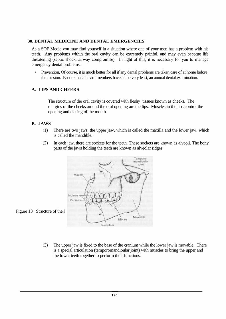
120
30. DENTAL MEDICINE AND DENTAL EMERGENCIES
As a SOF Medic you may find yourself in a situation where one of your men has a problem with histeeth. Any problems within the oral cavity can be extremely painful, and may even become lifethreatening (septic shock, airway compromise). In light of this, it is necessary for you to manageemergency dental problems.
• Prevention, Of course, it is much better for all if any dental problems are taken care of at home beforethe mission. Ensure that all team members have at the very least, an annual dental examination.
A. LIPS AND CHEEKS
The structure of the oral cavity is covered with fleshy tissues known as cheeks. Themargins of the cheeks around the oral opening are the lips. Muscles in the lips control theopening and closing of the mouth.
B. JAWS(1) There are two jaws: the upper jaw, which is called the maxilla and the lower jaw, which
is called the mandible.
(2) In each jaw, there are sockets for the teeth. These sockets are known as alveoli. The bonyparts of the jaws holding the teeth are known as alveolar ridges.
(3) The upper jaw is fixed to the base of the cranium while the lower jaw is movable. Thereis a special articulation (temporomandibular joint) with muscles to bring the upper andthe lower teeth together to perform their functions.
Figure 13 Structure of the Jaw

121
C. TEETH
A TOOTH HAS TWO MAIN PARTS:
The crown and the root.
A root canal passes up through the central part of the tooth. Theroot is suspended within a socket (called the alveolus) of one of thejaws of the mouth. The crown extends up above the surface of thejaw. The root and inner part of the crown are made of a substancecalled dentin. The outer portion of the crown is covered with asubstance known as enamel. Enamel is the hardest substance of thebody. The nerves and blood vessels of the tooth pass up into theroot canal from the jaw substance.
There are two kinds of teeth: anterior and posterior
The anterior teeth are known as incisors and canine teeth. They are used to rip and tear food. Theposterior teeth are called premolars and molars. They grind food into manageable sizes forswallowing.
(1) There are two sets of teeth: deciduous and permanent. Initially, the deciduous set includes 20baby teeth. These are eventually replaced by a permanent set of 32 teeth.
D. TOOTHACHEToothache is usually associated with one of the following: Caries (decay); tooth fractures; acuteabscess.
Figure2 Organisation of Anterior andPosterior Teeth (hard palate is visible)
Figure3 Types of Teeth
122
E. DENTAL CARIES (TOOTH DECAY)
(1) Diagnosis may be difficult. The patient will usually present with the followingsymptoms:
(a) Intermittent or continuous pain.
(b) Sensitive to heat or cold.
(c) Tooth grossly decayed.
(d) Discoloration.
(e) Pain on tapping with an instrument.
(f) The diagnostic test is to apply heat or cold to the suspect tooth to find out if it issensitive.
(2) Treatment. After finding the source of pain, local anaesthetic will probably be necessaryto carry out the following:
(a) Scrape the cavity.
(b) Irrigate the cavity with warm water
(c) Dry the cavity.
(d) Fill the cavity with a Zinc Oxide/ Eugenol mix.
The patient should be warned not to chew on the affected tooth for 24 hours.
b. ACUTE ABSCESS(1) Diagnosis. The patient will give a history of pain that becomes continuous and intense.
The affected tooth will feel "long". There is severe pain on tapping. There may beswelling. The patient may complain of malaise and anorexia and may have a fever.
(2) Treatment. Drainage usually provides immediate pain relief. You should consider givingantibiotics only if the systemic symptoms and signs are severe.
c. TOOTH FRACTURES(1) The anterior teeth are particularly liable to injury and as a result suffer fractures of the
crown. The emergency treatment of these injuries is summarised below:
(a) Simple fractures. Treatment: Smooth the rough edges of the tooth.
(b) Fractures involving dentine but not pulp. Treatment: Wash the tooth with warmsaline then isolate and dry it. Fill with the paste mix.
(c) Fractures involving dentine and exposed pulp. Treatment: Anaesthetise the tooth.Isolate and dry the tooth. Cover with any dental paste except zinc oxide/Eugenol.If that is all you have, consider extraction.

123
d. TRENCH MOUTH (ACUTE NECROTIZING ULCERATIVE GINGIVITIS)(1) Aetiology. This condition is caused by a variety factors including to a large extend, the
patient's lack of good oral hygiene. Other factors such as poor diet, stress and fatigue alsoplay a part.
(2) Diagnosis. Constant gnawing pain and marked gingival sensitivity is usually theoutstanding complaints. These symptoms are joined by pronounced gingival bleeding,foul smelling breath, and foul metallic taste, general malaise and anorexia. There may beulcers between the teeth and the patient may also have a fever.
(3) Treatment consists primarily of establishing good oral hygiene. Warm, salty watermouthwashes every 2-3 hours may be beneficial to begin with. Then gentle brushingregularly. Encourage the patient to drink plenty. Review his diet if possible. Considerappropriate antibiotics.
e. INJURIES TO THE JAWS(1) The immediate treatment of facial trauma consists of controlling the airway, bleeding and
shock. These basic measures MUST ALWAYS BE CONSIDERED FIRST.
(2) Dislocation of the mandible. The history will usually suggest quite heavily that adislocation is present. The condyles are usually displaced anteriorly. Speech is difficultfor the patient who will also dribble. Treatment consists of reducing the dislocation andcautioning the patient not to open his mouth too far for several weeks.
(3) Fractures of the mandible. Mandibular fractures must be differentiated from dislocationsof the jaw because of the great difference in treatment. Some of the features of fracturesare pain, abnormal "bite", crepitus and displacement of segments of the jaw. Once afracture has been diagnosed, the patient should be nursed in a face down or side positionto prevent aspiration of blood, vomit etc. Once the patient is stable, and if you arecompetent, the upper and lower teeth should be wired together.
31. WATER OPERATIONS AND NEAR DROWNING
An episode of suffocation followed by at least transient recovery.
a. ETIOLOGY
(4) Drowning is usually caused as a result of an inability to swim or from exhaustioncompounded by hypothermia. Trauma especially to the cervical spine may rendereven the strongest swimmer liable to drowning.
(5) As the victim goes under and water enters his mouth and nose, he begins to cough andgasp, and swallowing considerable amounts of water. A small amount of water isaspirated into the larynx and trachea, setting off spasms of the laryngeal muscles(LARYNGOSPASM), which seal off the airway, laryngospasm leads to ASPHYXIA,and the victim loses consciousness from hypoxemia. If the victim is not rescuedpromptly, progressive asphyxia will cause the laryngeal muscles to relax, permittingwater to enter the lungs in massive quantities.
F. APPROACH

124
(1) Whether a near drowning occurs in fresh of salt water, initial resuscitation of thesubmersion victim differs little, if at all, from the standard techniques ofcardiopulmonary resuscitation with which every SOF medic is familiar.
(2) First, of course, you must reach the victim, with due concern for your own safety; Itserves little purpose for an unqualified swimmer to enter the water in an attempt torescue a drowning victim, since the likely result is two drowning victims. When youreach the victim, give first priority to establishing an airway and initiating ventilation -even before the victim is removed from the water. In this circumstance, head tilt -chin lift may be the easiest way to maintain an airway.
(3) Do not, however, extend the victim's neck if you suspect cervical spine injury. Itshould be remembered, that successful resuscitations with complete neurologicalrecovery have been reported in cases where the victim has been submerged for as longas 45 minutes in cold water. Therefore, CPR in this situation should be maintained foras long as possible.
REMEMBER: A CASUALTY IS NOT DEAD UNTIL WARM AND DEAD.
G. CONSIDERATIONS
(1) The clinical picture seen in near drowning is not affected by whether submersion wasin salt or fresh water. There is a high incidence of neck injuries associated withdrowning. The victim may well be hypothermic.
(2) Attempting to drain water from the lungs makes little difference to the oxygen uptakebut it does delay treatment and potentially jeopardises an unstable neck.
(3) A drowning casualty should be evacuated as soon as possible, as delayed deaths due topulmonary oedema and aspiration pneumonia are not uncommon.
32. COMMON INFECTIOUS DISEASES
a. HEPATITISVirus that affects the hepatocytes in the liver
(1) SPREAD(a) Type A (Infective Hepatitis) Gamma G.
Faecal contamination of food and water. The virus is ingested and enters theblood via the intestinal mucosa.
(b) Type B (Serum Hepatitis).
Virus is carried in the blood and spread by:
• Contaminated needles, syringes and spilled blood.
• Contaminated faeces and urine.
• Sexual intercourse.
(2) INCUBATION
(a) Type A
• 15-40 days, average 28 days.

125
(b) Type B
• 40-150 days, average 100 days.
• Type B contains the Australia antigen. It presents a great risk to medical,paramedical personnel and health workers. It is as great, if not greater adanger than AIDS to such personnel.
(3) SIGNS & SYMPTOMSBoth types present in much the same way. There is:
(a) Malaise(b) Anorexia(c) Severe nausea which is brought on by even the sight or smell of food(d) Vomiting(e) Headache(f) Constipation(g) Pyrexia of various degrees(h) Tender hepatomegaly(i) Jaundice - first sign in white of eyes(j) Smoking becomes distasteful
The acute stage last 2-4 weeks followed by a gradual recovery and relapses can occur at anystage. In some cases, cirrhosis of the liver develops.
(4) PREVENTIONType A: Immunisation with Immune Serum Globulin (ISG).
Type B: Immunisation with Hyperimmune Globulin (HIG).
(5) TREATMENT(a) As the infection is viral, antibiotics are not of much use.(b) The patients need to be isolated and barrier nursed.(c) Care must be taken in disposing of body waste.(d) Rest in bed for 2 weeks as the disease is debilitating.(e) Diet is unrestricted, although fatty foods may be omitted.(f) NO ALCOHOL for a period of 3 months after recovery.(g) People who have had Type B hepatitis are potential sources of infection to
others, as they become carriers. Some people are asymptomatic carriers or mayhave had the disease and not known what it was.
(h) Drug addicts that use intravenous routes are highly likely to be potentialcarriers.
b. INFECTIVE MONONUCLEOSISBetter known as glandular fever or "kissing disease".(1) CAUSE
(a) Virus
(2) SPREAD(a) Direct and indirect contact
126
(3) INCUBATION(a) Not known
(4) SIGNS & SYMPTOMS(a) Sudden onset of:
• Fever• Sore
throat• Dysph
agia• Stiff
neck• Genera
l aches and pains(b) Adenitis follows in 2-3 days. The lymph nodes in the neck become enlarged and
tender, and the infection spreads to all lymphatic nodes to produce a generalisedAdenitis.
(c) Hepatomegaly and spenomegaly develops sometimes.(d) A generalised rash may also erupt.(e) Neurological involvement is a complication and may be fatal.(f) The attack may last from a few days to several weeks. Usually it lasts 3-6 weeks
during which time a relapse may occur.(5) TREATMENT
(a) Bed rest
(b) Fluids
(c) Aspirin
c. PAROTITISCommonly known as mumps.
(1) CAUSEVirus
(2) SPREADDirect contact. Airborne droplet.
(3) INCUBATION14-30 days
(4) SIGNS & SYMPTOMS(a) Fever. This subsides over 4-5 days.
(b) Malaise
(c) Nausea
(d) Anorexia
(e) Dysphagia
(f) Swelling of the parotid salivary glands

127
(g) Localised pain
(h) The swelling resolves over 7-10 days.
(5) COMPLICATIONS(a) Males. Orchitis - swelling of testes.
(b) Females. Ophoritis + Mastitis - Ovaries/breast swelling.
(c) Pancreatitis. With epigastric pain, nausea, vomiting and a tender pancreas onpalpation.
(6) TREATMENT(a) Isolation(b) 14 days bed rest(c) Oral fluids(d) Soft diet(e) Aspirin or Paracetamol for pain
d. RUBELLABetter known as German Measles
(1) CAUSEVirus
(2) SPREADContact
(3) INCUBATION17-18 days
(4) SIGNS & SYMPTOMS(a) Malaise with slight pyrexia
(b) Sore throat
(c) Stiff neck
(d) Occasionally conjunctivitis
(e) A rose spot rash appears within 24 hours on the face initially, then spreads to thetrunk and limbs. It disappears after the 3rd day.
(f) The cervical lymph nodes become enlarged and firm.
(g) The joints may become sore.
(5) COMPLICATIONS(a) Encephalitis
(b) Pregnant women who contract the disease in the first 3 months are at specialrisk. The virus can cross via the placenta to the foetus, and results in congenitalabnormalities.

128
e. MENINGOCOCCAL MENINGITIS
(1) CAUSEMeningococcus
(2) SPREADDirect/Indirect contact by airborne droplet
(3) INCUBATION1-3 days
(4) SIGNS & SYMPTOMS(a) Sudden onset of:
• Nasal infection• Headache• Pyrexia• Rigors, shivering/cold• Convulsions• Nausea & vomiting• Pain in the neck and limbs, rigidity in neck.Meningitis is characterized by
• Headache• Vomiting• Dizziness• Neck rigidity• Restlessness• Photophobia• Confusion leading to delirium and unconsciousness
(5) PREVENTION(a) Strict isolation with barrier nursing. Cefotaxime 2g with 8ml water.
(b) Suitable antibiotics, must x blood, brain barrier.
(c) Many complications involving the brain and nervous system can occur. Fatalityis not uncommon.
f. TYPHOID AND PARATYPHOID FEVER
(1) CAUSESalmonella Typhi
(2) SPREADIngestion of food and drink contaminated by faecal matter. May also be transmittedon the hands of infected carriers who do not wash their hands after defecating.
129
(3) INCUBATION10-12 days
(4) SIGNS & SYMPTOMS(a) The fever is very variable and may be mild, lasting 1-2 weeks or severe and
resulting in death within 10 days. Usually the illness will be severe and last for4-6 weeks.
(b) Onset - Gradual over several days with an intermittent fever, rising slightly eachday.
(c) Headache
(d) Malaise
(e) Anorexia
(f) Occasionally nausea and vomiting.
(g) Nose bleeds
(h) Cough
(i) Abdominal distension with discomfort and tenderness
(j) Constipation
(5) COURSE(a) Intermittent temperature may swing as high as 41°C and suddenly drop. This
will cause rigors.
(b) After 3-4 weeks the constipation will change to diarrhoea. The stools will bewatery and grey/green in colour.
(c) Abdominal distension and discomfort will be worse.
(d) After 2-3 weeks a rash will appear on the trunk, occasionally on the back andless frequently on the limbs. The spots are rose coloured and disappear after afew days.
(e) Delirium may ensue.
(f) If adequate fluids are not given, the patient becomes dehydrated.
(g) Parotitis, Herpes and bronchitis may develop. The tongue may become dry,brown and furred.
(h) Recovery is gradual over 3-4 weeks, in which time a relapse may occur.
(6) COMPLICATIONSInternal haemorrhage due to erosion of the intestinal wall.
(7) PREVENTIONImmunisation with boosters at 3 year intervals.

130
(8) TREATMENT(a) Isolation(b) Suitable antibiotic therapy under advice from a MO.(c) Casevac.
g. TETANUS
(1) CAUSEClostridium Tetani Bacillus. Found in soil, on rusty metals and in the gut of animalsand humans (especially farm workers).
(2) SPREADThrough a break in the skin which is contaminated with soil or dirty objects, i.e.accidents in the garden and on the farm. The organism produces a toxin, whichattacks the nervous tissues.
(3) SIGNS & SYMPTOMS(a) Onset:
• Feverwith chills
• Headache
• Restlessness
• Increase in muscular tone
• Stiffness in neck muscles
• Dyspnea
• Dysphagia
(4) COURSE(a) Initially, there are muscle spasms affecting the jaw. The spasms spread to the
muscles of the back, abdomen, neck and limbs.
(b) Severe spasms of the whole body can occur spontaneously or due to the slighteststimuli. The back arches, the teeth are clenched and the lips retracted.
(c) The mind remains clear all the time, but the patient is helpless.
(d) There is a pyrexia of 39.5-41°C.
(e) Death occurs due to exhaustion, anoxia and heart failure.
(f) In recovery, the spasms are less frequent and less severe.
(5) PREVENTION(a) School children are immunised against tetanus. However, booster doses are not
maintained later in life and it is possible to lose immunity because of this.

131
(b) Should anyone attend a casualty department or see their own doctor due toreceiving an injury, they are asked when they last had a tetanus injection.
(c) If the time lapse is less than 5 years, no booster is required. If longer than 5years, a 0.5ml booster dose is given. People who are unsure will be given abooster, and those never immunised will receive a complete course. In somehospitals, it is policy to give a course if 10 years have lapsed since the initialcourse at school.
(d) Course doses:
• 0.5-1ml by intra-muscular injection or deep subcutaneous injection.
• 0.5-1ml IMI or deep SC 6-12 weeks later.
• 0.5-1ml IMI or deep SC 4-12 months after 2nd dose.
(6) TREATMENT(a) Adequate wound hygiene is necessary using aseptic technique.
(b) Antibiotics intravenously if possible, intramuscularly as 2nd route.
(c) Immediate evacuation to a hospital.
(d) These are just a few infectious diseases that you may encounter on your 'travels'.In the Armed Forces all personnel are given booster doses at 3-year intervals ofPolio and TABT. Immunisation programs are carried out if personnel are goingto be working in high-risk areas overseas. In such cases, cholera vaccine,hepatitis and encephalitis immunisation is given to provide prophylaxis againstthese diseases.
(e) In suspected cases of meningitis, typhoid, paratyphoid and tetanus antibiotictherapy must be started immediately. Such therapy is indicated in treatinganyone who has sustained a wound, which has been contaminated.
h. MALARIA(1) Malaria is transmitted by the bite of the female Anopheles mosquito. It is the most
important of all tropical diseases, causing many deaths (2,000,000 a year). There arefour species of human malaria, all caused by species of the genus plasmodium.
• P. Falciparum
• P. Vivax
• P. Ovale
• P. Malariae
(2) CLINICAL FEATURES(a) Fever
132
(b) Anaemia
(c) Splenomegoly (enlarged spleen)
(d) Jaundice
(3) CLASSICAL STAGES OF FEVER(a) A cold stage (the patient shivers or has frank rigors, the temperature rises
sharply).
(b) A hot stage (the patient is flushed, has a rapid full pulse and a high temperatureis sustained for a few hours).
(c) A sweating stage (the patient sweats freely, or is even drenched, and thetemperature falls rapidly).
(4) PECULIARITIES OF P. FALCIPARUMP. Falciparum is by far the most severe of all the strains of malaria. The mostsignificant difference is the damage caused to the capillary walls causingmicrocirculatory arrest. It is also common that, following a single exposure to P.Falciparum, the patient will either die in the acute attack (within 8 hours) or survivewith the development of some immunity and residual anaemia.
(a) TREATMENT
Supportive Treatment
• Reduce the temperature if hyperpyrexia is present.
• Rehydration if diarrhoea and vomiting are presents. Do not over hydrate.
• Monitor urine output.
• Control fits with anticonvulsant therapy is possible.
Chemotherapy
• There are now many drugs available for anti malarial therapy. The best drug to usewill depend on the country you are operating in. Sadly, there is a lot of resistance toanti malarial drugs in use today. It is up to the SOF medic in his pre-missionplanning to find out which drugs to use. The drugs currently available are:
• Chloroquine 200 mg Dose:
• As prophylaxis: 400 mg weekly• As treatment: 600 mg stat, 300 mg 6 hrs later, then 300 mg daily for 2-6 days.
• Proaguanil 100 mg Dose:
As prophylaxis: 200 mg daily
• Mefloquine Hydrochlor 250 mg Dose:
As prophylaxis: 250 mg weekly
(b) PREVENTIVE MEASURES

133
• Prevention is by far better than cure. Some preventive measures for malaria are asfollows:
• Use of mosquito repellent.
• Use of mosquito nets.
• Cover as much of the body with clothing as possible.
• Always take malaria prophylaxis.
• Avoid making unnecessary pools of water.
• Dip clothing in an insecticide such as Peripel on a regular basis.
33. ANAPHYLACTIC SHOCKAnaphylactic shock is a massive allergic reaction that develops within seconds to minutes after theinjection of a drug or the sting of an insect to which the patient is sensitive. More rarely, it followsingestion of an allergen, such as penicillin in which case the reaction occurs somewhat moreslowly. Even more rarely, anaphylaxis occurs after inhalation of antigenic material.
The patient experiencing an anaphylactic reaction will be extremely dysgenic and often will besneezing, wheezing or coughing blood tinged sputum. He will frequently complain of tightness inthe chest of a sensation that his throat is closing. His face may be flushed or ashen, with marked

134
facial swelling, especially noticeable around the eyes. Sometimes he will complain of severeitching, either generalised or localised to the throat area. Urticaria (hives) is often present, or theskin may simply take on a red, inflamed appearance. Abdominal cramps, followed by nausea,vomiting and diarrhoea are common. The pulse is usually rapid, and the blood pressure falling,sometimes to the pint of complete circulatory collapse. The sequence of itching, cough, dispnea,and cardiorespiratory arrest may occur within seconds, and death is imminent unless propertreatment is instituted immediately.
TREATMENTThe treatment for anaphylactic shock is by an intra-muscular injection of adrenaline 1:1000(IMG/IMC). Note: This is a different dose to cardiac adrenaline, and must not be givenintravenously.
(1) Dose-Adrenalin
• 0.5-1 ml IM - These doses can be repeated every 10 minutes according to bloodpressure and pulse, until improvement occurs (may be repeated several times).
(2) Dose-Piriton
• Piriton 10 MG/ML 1 ml amp can also be given following administration ofadrenaline. 10-20 MG IV or IM to a maximum of 40 MG in 24 hours.
• Further treatment consists of laying the casualty on his back and elevating his legs.
34. BALLISTICS
Missile injuries are by no means new phenomena. It can be said that the first recorded account of amissile injury was when David killed Goliath using his slingshot. Although the missile itself wasonly a small stone, it is still defined as a missile injury.
A. WOUNDING AGENTSThere are two main classifications of wounding agents.
(5) PRIMARY MISSILES:
(a) Bullets (high and low velocity)
(b) Fragments (Grenade, shell, bomb, mortar, and booby traps).

135
(6) SECONDARY MISSILES:
(a) Extrinsic (Rock, glass, wood, clothing, equipment).
(b) Intrinsic (Bone fragments).
b. MISSILESThere are various factors, which can affect the amount of damage a missile will inflict on thehuman body. These are:
(1) Size
(2) Shape
(3) Stability
(4) Velocity
c. MISSILE ENERGY(1) It is important to have an understanding of the correct terminology used when
describing the way in which a missile travels and is absorbed by the body. Althoughthe terms high and low velocity is still widely used, they do not correctly describe theway in which the different types of missile can affect the body. To alleviate thisproblem, missile injuries are now described as high and low energy transfer wounds.
(2) It must also be remembered though, that a high velocity missile can produce a lowenergy transfer wound. And a low velocity missile can produce a high-energy transferwound.
d. CONTAMINATION OF WOUNDSA major problem associated with missile injuries is the amount of contamination produced.There are two main causes for this.
(1) Vacuum effect of temporary cavitation.
(2) Contaminated projectiles:
(a) Primary and secondary fragments
e. TYPES OF BLASTTypes of blast can be divided into three categories.
(1) AIR (Blast waves travel slowest in the air, most air blast injuries are caused by debris).
(2) WATER (Blast waves travel faster and farther in water, most damage from water blastoccurs to the hollow viscera).
(3) SOLID (Blast waves travel fastest through solid objects, most damage occurs to themajor blood vessels, often without breaking the skin).
f. COMMON BLAST INJURIES
There are three common blast injuries. These are:

136
(1) Ruptured Tympanic Membrane .
(a) Signs and Symptoms
• Sudden severe pain in ear.
• Bleeding from ear.
• Hearing Loss.
(b) Treatment
• Clean the meatus gently then leave it alone.
• DO NOT pack, syringe or put in any medication.
(2) Blast Lung
(a) Signs and Symptoms
• Cyanosis
• Rapid Pulse
• Chest pain
• Abdominal rigidity
• Haemoptysis
(b) Treatment
• Move casualty as little as possible.
• Oxygen (if available).
• Use IV with extreme caution.
• Antibiotics.
(3) Blast Abdomen
(a) Signs and Symptoms
• Sudden abdominal pain.
• Brief remission of pain.
• Recurrence of severe, unremitting and increasing pains.
• Frequent bowel evacuation.
• Passing of melana or frank blood in stools.
• Dysuria.
(b) Treatment

137
• If serious surgery is required
• Antibiotics
• NG intubation
• Use Morphine only with caution
(4) Other Blast Injuries
Other blast injuries include:
(a) Fractures
(b) Fragment injuries
(c) Scrotal contusions.
(d) Transient paresis of limbs.
(e) Flash burns
35. ACTIONS ON TAKING A CASUALTY: THE PRIMARY SURVEY
a. The care of the seriously injured casualty requires rapid assessment of his injuries and theinstitution of life saving procedures for him to stand the best chance of survival and for him toachieve the best possible outcome. These, purely medical considerations can be verycomplicated in the field with only the limited resources available to the SOF medic. But whenyou add the likelihood that the patrol will be operating in an unfriendly military environmentand its surgical back up may be anything from 1 hour to 7 days distant, the need for asystematic approach becomes even clearer. This systematic approach is part of the patrol'sstandard operating procedures (Sops) and this section covers the SOF medic's "actions on"taking a casualty.
b. Actions on taking a casualty are organised in a step by step approach as follows:
(5) Preparations
(6) Military considerations
(7) Primary survey (ABCs)
(8) Resuscitation
(9) Secondary survey (head to toe)
(10) Definitive care

138
c. This step by step approach reflects the relative importance of the various steps. The only stepsthat are covered concurrently are the primary survey, in which the casualty is examined for lifethreatening injuries, and the resuscitation phase in which the life threatening injuries aretreated as they are discovered during the primary survey and not at the end. Militaryconsiderations are likely to affect the management of the casualty throughout.
d. PREPARATION(1) Proper preparation and planning, both of the SOF medic and his equipment, is the key
to a successful outcome for the casualty. The practical skills that are learned on theSOF Medics Course are complex and "perish" in 6 to 9 months. It is thereforeessential for the medic to refresh his personal skills. Prior to deployment onoperations, the SOF medic should endeavour to revitalise his skills. For this, he has anethical obligation to his team-mates and himself.
(2) The amount of medical kit available to the SOF medic is already known to be theabsolute minimum. There is no rush to add weight to an already backbreakingrucksack. In order to be able to carry all the necessary equipment, it is necessary tocross load medical equipment to other patrol members. The location of sensitivemedical items (morphine syrettes), like any other sensitive team equipment (blastingcaps, radio SOI’s), must be known by all members of the team. All the equipmentmust be serviceable, in date and well packed. The location of items in the medicalpack should be waterproofed and easily found. Some degree of preparation can beperformed, for instance, keep all the necessary items for IV infusions together.Whatever is done, the medic must be able to work out of his pack in the worst ofconditions and in the dark.
e. MILITARY CONSIDERATIONSMilitary considerations are of key importance to the management of a casualty in the SOFsituation and can be divided into 2 main areas.
(1) The tactical situation
(2) The availability of Casevac (Casualty Evacuation)
f. The effect of these factors can probably best be illustrated by using 2 military scenarios asexamples. A 4-man patrol operating deep in enemy territory comes into contact with an enemypatrol and takes a casualty. Casevac time is 7 days by foot. The SOF medic's first priority isto protect his casualty and patrol as a whole by ensuring that the firefight is won. Manpowerin this situation does not allow the medic to operate as medic alone and only when the tacticalsituation is under control will he move forward to treat the casualty. It is likely that thesituation will not allow much time to treat the casualty at the point of wounding and the medicwould perform the primary survey/resuscitation asking himself 2 questions as he discoverslife-threatening conditions:
(1) What is the minimum treatment necessary for me to be able to get this casualty to asafe area to treat him definitively. An example would be to seal a chest wound butsaves the insertion of a chest drain until later.
(2) Given that we’ve got 7 days on foot, and given the resources available to me, is thisinjury compatible with survival (e.g. massive hemothorax or a military gunshot woundto the head).
g. This later question is not one, which is going to have to be faced very often, and when it does,the answer is usually obvious. The problem comes in following that answer to its logicalconclusion: should I save scarce medical resources in the event that the patrol takes a second

139
casualty? The question is even more important when there is the possibility of calling in ahelicopter Casevac. The SOF medic on the ground must be able to advise the PatrolCommander on the prognosis for the patient. With this information, the Patrol Commander canmake the correct decision as to whether the given wound is survivable before calling forCasevac, and compromising the patrol with all of the risks involved in an early extraction.
h. Once the patrol has withdrawn to a relatively safe position, he has fulfilled the first rule of firstaid in that he has removed the casualty to relative safety and is then may complete theresuscitation phase.
i. In the case of the second scenario of a direct action operation involving direct medical supportwith surgical capability, the decision making process would be fundamentally different fromthat illustrated above. The casualty would be evacuated to a central, pre-planned CasualtyCollecting Point (CCP), where the medic, within the hour, could transfer the patient todefinitive care. Again, the medic would only treat those life-threatening conditions that hediscovers to a degree necessary for the casualty to reach the definitive care alive. However, theSOF Medic in this type of operation is likely to be faced with multiple casualties. His primaryconcern in this situation is therefore to get his casualty to the CCP and ensure that the one mostappropriate casualty is delivered to the trauma team. This approach takes advantage of theprinciples of “triage".

140
a. THE PRIMARY SURVEY AND RESUSCITATION PHASE(1) The aim of the primary survey is to identify those injuries sustained by the casualty
which is life threatening. It is therefore logical to commence that search in the areaswhich will kill the casualty quickest and them move on to the next most lethal and soon. As life threatening conditions are discovered, they are managed to a sufficientdegree to stabilise the casualty (resuscitation phase) and the medic proceeds to thenext area while continually reassessing what he has done so far. The order in whichthese injuries are considered is known as the ABCs of trauma, and is shown in theprimary survey sequence below:
(2) In the hospital environment, where the casualty would be managed by a team, theseareas are usually managed simultaneously with one member assessing the airway andmanaging any other problem that they discover at the same time that another memberis assessing the chest, etc. The patrol medic is on his own and must therefore followthe ABCs regardless! Many a casualty has perished because the medic hasconcentrated on what is most obvious (usually bleeding) and ignored what is actuallykilling the casualty (usually an airway problem or a missed chest wound). It cannot bestressed too strongly that it takes very little time to establish that an airway is patent orto exclude life threatening chest trauma. If, on the other hand, the airway is not secureor life threatening chest trauma is discovered, then these take priority in that order, andmust be managed while simultaneously controlling life threatening haemorrhage.

141
PRIMARY SURVEY SEQUENCESafe approach to the sceneAsses patient for responsivness / level of consciousness, maintain C-spine controlDo a quick check for obvious arterial bleeding
AIRWAY:LOOK look in mouth for obstruction, look at chest for rise and fall/ injuriesLISTEN listen for breathing for 5 secondsFEEL feel for cortid pulse and breath on your faceCHECK C-SPINE after airway is secured and secure C-sine if needed!On the way to B check the chest, Check: that trachea is midline, or signs of jugulary veindistension
BREATHING:INSPECT - chest for signs of obvious traumaAUSCULTATE - listen to both lung fields & heart PMIPALPATE - the chest for broken ribs, signs of flail chestPERCUSS - for hollow, dull or abnormal breath sounds – hyper resonant for
excessive air or dull for bloodCheck down side for exit wounds !
CIRCULATION:Do a quick visual check for obvious major bleedingDirect pressure, pressure dressing or torniquet to stop bleedingCheck distal pulses first radial on both armsPrepare IV-kit and start INTRAVENOUS THERAPY ( TKO OR FLUID REPLACEMENT )
! ! After IV started reasses airway, braething and circulation to include IV ! !
DISABILITY: mini neurological examA - alert & oriented P - pupilsV - respond to verbal stimuli E - equalP - resond to pain only R - roundU - unresponsive R - react
L - lightA - accommodate if conscious
EXPOSE:Examine the whole body for any missed life threatning injuries; keep patient warm
FULL SET OF VITAL SIGNS:TemperaturePulseRespirationBlood pressure
** Note: The only reasson you will not immediately start with airway in your primary survey is ifthere is an obvious major arterial bleeding. Stop bleeding first then proceed with yourprimary survey sequence.
142
36. BASIC LIFE SUPPORTYou cannot begin the primary survey on a patient who does not have a pulse or respirations!Basic life support are the measures you take to regain a pulse and respiration’s so that youcan begin the primary survey. There is some commonality between BLS and the primarysurvey. For example, you still must get a patent airway.
Basic Life Support (BLS) means maintaining an airway and supporting breathing and circulationin anyone who is in need of such help without any equipment. No consideration of advanced lifesupport techniques can begin without the acquisition of BLS skills.
Survival from cardiac arrest, from whatever cause, is more likely when the arrest is witnessed, and"Buddy Buddy" resuscitation is commenced. Rapid initiation of BLS greatly improves survivalrates. The speed of initiation of BLS may be more important than the absolute quality of thetechnique employed.
In general, BLS should be started in all patients who have suddenly become unresponsive and whoare either apneic or have an absent major pulse. However, in some cases the patient may beobviously non-resuscitatable and therefore BLS is not appropriate.
a. THE SAFE APPROACH(1) Assessment should begin as soon as possible after the collapse occurs or the patient is
found. However, the rescuer must not become a second victim, and further harm tothe patient must be avoided. Don't forget the tactical situation. Thus, prior toassessing the condition of the casualty, the medic should shout to summon help,approach the casualty with care, free the casualty from continuing danger beforeevaluating the airway, breathing and circulation (ABC).
(a) Shout for help(b) Approach with care(c) Free from danger(d) Evaluate ABC
(2) Asking the patient "are you all right" and gently shaking them by the shoulder assessesunresponsiveness.
(3) Remember that in cases associated with blunt trauma above the line of the clavicle, ora mechanism of injury such as a fall, the neck and spine should be protected byimmobilising the head during this manoeuvre (the head is firmly held with one handflat on the forehead and one arm is gently shaken). If the patient responds byanswering or moving, they should not be moved unless they are not safe. Rememberthat they may deteriorate, so they must be re-assessed and help should be called for ifneeded. If there is no response to voice or touch, help should be called for again ifnone has arrived.
b. AIRWAY(1) An obstructed airway may be the primary problem and correction of this may achieve
recovery without further intervention. The relaxed tongue falling backwards onto theposterior wall of the pharynx most often causes this.
(2) The primary method of attaining an airway is by using the head tilt/chin lift technique.

143
(3) The medic places the hand near to the victim's head on the forehead, and gently tiltsthe head back. The fingers of the other hand are placed under the chin, and the jaw islifted upwards. As this may cause the mouth to close, it may be necessary to use thethumb to part the lips. The success of this manoeuvre should be assessed immediatelyby looking, listening and feeling.
(a) LookingTo see if the chest is rising and falling.
(b) ListeningTo hear any breathing or gurgling/snoring sounds.
(c) FeelingWith the cheek near the victim's mouth for exhaled breath or with the hand onthe chest for movement.
(4) If head tilt/chin lift is not appropriate, two other methods can be tried. First, if foreignmaterial may be obstructing the airway, attempts should be made to remove this usinga finger sweep. Care should be taken not to worsen the situation by impacting foreignbodies further down the airway.
(5) The mouth is opened and inspected, and two fingers are gently swept from the side tothe back. If any debris is found, attempts should be made to hook it out. Loose orbroken dentures should be removed at this stage.
(6) The jaw thrust may also be used to move the tongue forwards.
(7) Two fingers of the medic's hands are placed behind each angle of the casualty'smandible. The mandible is then thrust anteriorly. The jaw thrust alone should be usedto open the airway if an injury to the cervical spine is suspected.
(8) Looking, listening and feeling should assess the results of these manoeuvres. Thissequence of events should be performed rapidly.
(9) The carotid pulse should be felt for at least 5 seconds to make sure there is nocirculation. If the victim is hypothermic, then the pulse should be sought of at least 60seconds.
c. BREATHING(1) The patient may start to breath after the simple airway manoeuvres are carried out
during assessment. If this occurs, they should be placed in the recovery position andassessed for further injuries prior to evacuation.
(2) If the casualty is not breathing and a pulse is present, then 10 rescue breaths must begiven before help is sought.
(3) To perform expired air resuscitation effectively, there should be no obstruction orleakage to airflow between the medic and the casualty's lungs. This is best achievedby placing the flat of the uppermost hand on the casualty's forehead, tilting the headand using the finger of that hand to pinch the patient's nose shut. The other hand isused to lift the chin forward and open the mouth.
(4) The medic takes a deep breath, seals his lips round those of the casualty, and breathsout into his mouth, watching to ensure that the chest rises. Well fitting dentures shouldbe left in place as it is very difficult to get a good seal in an edentulous patient.
(5) Loose or broken dentures should be removed. If the chest does not rise, either theairway is not fully open or the seal may be inadequate.

144
(6) After each breath, the casualty should be allowed to exhale passively to theatmosphere. At the same time as the casualty exhales the medic should move his faceaway from the casualty and breath fresh air, not the casualty's expired gases.
(7) Mouth to mouth and nose ventilation can be used in children.(8) Mouth to mask ventilation, using a portable facemask is also recommended as a
suitable method, but rescue breathing should not be delayed if this device is notavailable.
(9) Each breath should last 1 1/2-2 seconds followed by 2-4 seconds for expiration.
d. CIRCULATION(1) If the patient is not breathing and is pulseless, help should be sought immediately. Do
not start BLS until you are sure further medical aid is on its way. If you are in asituation whereby no further medical aid can be sought within 4 minutes, themcommence BLS.
(2) If there is no response after 30 minutes of resuscitation, then the situation is probablyhopeless.
e. EXTERNAL CARDIAC COMPRESSION(1) To generate the optimum cardiac output, the position of the hands is important. If
they are too high, the efficiency of CPR is reduced and if too low the stomach iscompressed and aspiration becomes more likely. In an adult, the correct handplacement for external massage is two finger breaths above the xyphoid process. Themedic must reposition their hands after each interruption in cardiac compression. Heheel of one hand is place over the correct part of the sternum and the other hand isplaced on top of it, interlocking the fingers.
(2) In order to compress the chest wall downwards, the medic must have sufficient heightabove the casualty for his hands, elbows and shoulder to extend down in a verticalline. This is best achieved with the casualty lying on the floor and the medic kneelingalong his side. The surface needs to be firm and therefore a bed is inappropriate.Sternal compression of 4-5 cm is ideal in an adult. Optimum compression, withminimum medic fatigue is best achieved by keeping the arms straight and leaningforwards in a rocking motion. Flexing the elbows and pumping the arms up and downwill soon result in fatigue and reduced efficiency. Cardiac compressions should beapplied at a rate of 60-80 per minute. If a single medic is present, two breaths shouldbe delivered, followed by 15 compressions. Two rescuers should providecompressions and breaths in a ration of 5:1
(3) BLS must not be interrupted unless the patient starts to move or breath spontaneously.
f. SUMMARY OF BLS SEQUENCE(1) Safe approach(2) Assess responsiveness(3) Airway manoeuvres(4) Look, listen, feel(5) Check pulse(6) Call further medical aid
(7) Start CPR

145
g. RECOVERY(1) If the casualty starts to breathe spontaneously, he should be turned into the recovery
position, maintain the airway, and commence 15-minute observations. This willenable vomit to drain. The tongue will not fall back and obstruct the airway in thisposition. If a spinal injury is suspected, then this position is contraindicated and thecasualty should be managed on their back and their airway closely observed.
h. THE CHOKING CASUALTY(1) Just about any foreign body can obstruct the airway and the most common situation is
the so called "cafe coronary". This usually occurs in the person who attempts to eat,drink and talk all at the same time.
(2) The casualty who is choking usually present by being very anxious and often graspingat their throat. Casualties with partial obstruction may cough and have inspiratorystridor.
(3) Back blows may well cure the problem. Three blows with the heel of the hand to theback, between the shoulder blades, should be tried.
(4) If this is not successful, the Heimlich manoeuvre should be performed. In this methodforeign bodies in the air increasing intra-thoracic pressure may often expel passages.If the casualty is standing up the medic should move hind him and pass his armsaround the casualty. One hand is formed into a fist and placed in the epigastrium justbelow the xyphoid process. The other hand is placed over the fist and both are forcedupwards. His should be repeated 5-10 times unless the obstruction is relieved. If thecasualty is not conscious, he should be laid on his back so that the medic can kneelastride him and carry out the thrusts in the midline towards the head. If the foreignbody is not expelled at the first attempt, then a further two series of 10 thrusts shouldbe performed. In the pre-hospital situation, there is little else that can be done unless askilled person capable of surgical airway management is present.
DON'T FORGET(5) Maintenance of the airway and restoration of breathing is paramount in casualty care
and if this is at the expense of another injury then so be it.(6) Rapid advanced medical intervention such as defibrillation and cardiac drugs are the
best chance a patient has of recovering from cardio-respiratory arrest.(7) No one is dead until he is warm and dead.(8) BLS skills will fade rapidly if not used on a regular basis - ensure that these skills are
practised regularly.
146
37. AIRWAY MANAGEMENT AND CERVICAL SPINE CONTROL
On approaching his casualty, the SOF medic must be considering the mechanism of injury because insome situations this will have significant bearing on how he will manage his casualty. For themoment, however, we will consider the management of a casualty where the military situation issecure and the mechanism of injury is penetrating trauma (and therefore little risk to the cervicalspine). If the casualty is able to talk, then clearly he has a patent airway. If on the other hand, thecasualty can't talk, a blocked airway must be assumed and the airway must be rapidly assessed forpossible causes of blockage such as foreign bodies, maxillofacial, or tracheal injuries. Foreign bodiesshould be cleared using the finger sweep technique and should this be insufficient to establish a patentairway, then a simple manoeuvre such as a chin lift or jaw thrust should be attempted to achievepatency. These simple measures will work in the majority of cases. If they do, then either anoropharyngeal or nasopharyngeal tube should be inserted to maintain patency. This frees up theMedic’s hands for other things.
If maxillofacial trauma precludes the use of such an airway or simple techniques fail to achievepatency, the more advanced technique of emergency cricothyroidotomy should be performed. Oncesuccess has been achieved, it must be remembered that an airway, which requires support, is at risk,and it must be checked regularly.
a. THE CERVICAL SPINE IN THE SOF ENVIRONMENT(1) There is no way that the cervical spine can be properly protected on the battlefield. And
neither is it likely that injuries on the battlefield will leave the casualty with an unstablecervical spine with an intact spinal cord. Penetrating injuries to the neck of sufficientforce and accuracy to create a cervical fracture will sever the spinal cord at the momentof wounding, and the chances of an unstable fracture with intact cord are negligible.That is not to say that the cervical spine need not be considered when assessing acasualty, as unstable c-spine fractures are common in other situations. Parachutingaccidents, falls from rock faces and vehicle accidents, (particularly as seat belts are nottypically found in the back of military vehicles), are common sources of c-spine injuries.The decision as to whether the c-spine needs protecting or not must be based on thehistory of injury. There are methods of improvising cervical collars, but generallyspeaking, they are far from adequate. In a non-military environment, where a casualtyhas suffered an isolated c-spine injury, it would be negligent to move the casualtywithout adequate immobilisation of the c-spine. This would entail maintaining thepatient with manual in line immobilisation, pending the arrival of the emergency servicesequipped with the appropriate cervical immobilisation devices. Given those 2 extremesthe decision on how to manage the c-spine in a given set of circumstances rests with theSOF medic. When in doubt, err on the side of safety.
(2) Suspect injury in unconscious blunt trauma cases. Normally, aim to avoid neckmovement (causing more spinal cord damage), especially when securing the airway ormoving the casualty.
(3) This section concerns itself with the management of airways, using adjuncts, and simplemethods of airway management such as the finger sweep, head tilt, chin lift and jawthrust. Manoeuvres are methods used to clear airways while the adjuncts are used tomaintain an airway. Airway management is one of the most valuable skills the SOFMedic possesses. Only massive external haemorrhage is direr, and receives earlierattention. Unless the medic is capable of maintaining a patent airway all other treatmentsbecome somewhat unimportant in many situations.
147
b. RECOGNITION OF THE OBSTRUCTED AIRWAYThere are three main causes of obstruction to a patient's upper airway.
(1) The tongue falling back over the oropharynx (caused by reduction of muscle tone in thepatient with a decreased level of consciousness).
(2) Obstruction by foreign matter, i.e. vomit or blood (swelling of the tissues surrounding theupper airways will also cause obstruction and is normally seen in burns and occurs in thehours to days post injury).
(3) Facial trauma.
In order to assess a patient's airway, the medic should speak to the casualty, then:
c. LOOK(1) Is the chest rising and falling?
(2) Is the patient peripherally cyanosed?
(3) Are the intercostal muscles being drawn in?
(4) Is their tracheal tug?
d. LISTEN(1) Is the breathing noisy? (Inspiratory wheeze usually indicates upper airway obstruction,
expiratory wheeze usually indicates lower airway obstruction, i.e. asthma)
(2) Are there breath sounds?
e. FEEL(1) Is the rise and fall of the chest adequate?
(2) Is their breath on the side of the medic's cheek when checking for expiration?
An obstructed airway may be the primary problem and correction of this may achieve recoverywithout further intervention. The relaxed tongue falling backwards onto the posterior wall of thepharynx most often causes this. The primary method of attaining an airway is by using the headtilt/chin lift technique.
f. The Head tilt-Chin lift technique.
The medic places the hand nearest to the victim's head on the forehead, and gently tilts the head back.The fingers of the other hand are placed under the chin, and the jaw is lifted upwards. As this maycause the mouth to close, it may be necessary to use the thumb to part the lips. The success of thismanoeuvre should be assessed immediately by looking, listening and feeling as described earlier.
148
If head tilt/chin lift is not appropriate (because of suspected c-spine injury); two other methods can betried. First, if foreign material may be obstructing the airway, attempts should be made to remove thisusing a finger sweep. Care should be taken not to worsen the situation by impacting foreign bodiesfurther down the airway. Blindly sweeping fingers into the mouth is therefore not recommended.
g. The Finger Sweep.
The mouth is opened and inspected, and two fingers are gently swept from the side to the back. If anydebris is found, attempts should be made to hook it out. Loose or broken dentures should be removedat this stage.
The jaw thrust may also be used to move the tongue forwards.
h. The Jaw Thrust
Two fingers of the medic's hands are placed behind each angle of the casualty's mandible. Themandible is then thrust anteriorly. The jaw thrust alone should be used to open the airway if injury tothe cervical spine is suspected.
Looking, listening and feeling should assess the results of these manoeuvres. This sequence of eventsshould be performed rapidly.
i. SUCTION(1) Suction is an often-overlooked aid to airway management in the pre-hospital situation.
Quite often vomit, blood, etc. are the cause of a casualty's airway insufficiency. Themost common forms of suction that are available are:
(a) Vitalgraph hand held suction device.
(b) Laerdel portable suction device, operating off both, DC and internalre-chargeable battery supply.
(c) Cape suction, a foot operated device.

149
j. THE OROPHARYNGEAL AIRWAYThe oropharyngeal airway is a simple "J"shaped plastic tube. It is used to preventthe tongue from falling back over theoropharynx. This adjunct does notcompletely protect the airway, as it is stillpossible for the patient to aspirate his ownvomit or blood. It is available in anumber of sizes ranging from "000" forneonates up to "4" for large adults. Thecorrect size tube for a patient is gauged bycomparing the distance from the angle ofthe jaw to the patient's mouth. It isimportant that the vertical distance is usedand not the rotational distance or a tubetoo large will be selected. The mostcommon sizes in adults are 2, 3, and 4.
k. INSERTIONThe tube is inserted with the concavesurface facing uppermost until theposterior aspect of the hard palate isreached. The tube is then rotated through180 degrees and fully inserted. Insertionof the tube can be aided by the use of atongue depressor to stop the tonguefolding back over the oropharynx. If thecasualty gags when the tube is inserted,then he does not require it and it should beremoved. Once the tube has been inserted,looking, listening and feeling should assess the patency of the airway.
l. NASOPHARYNGEAL AIRWAY(1) The nasopharyngeal tube is also used to prevent the tongue falling back over the
oropharynx. It has an advantage over the oropharyngeal airway in that it is moremalleable and therefore better tolerated. The correct size of tube is selected bycomparing the internal diameter of the bevelled end with the size of the patient's externalnares. An alternative method is to compare the size of the tube with the patient's littlefinger. The most common sizes in adults are 6-8 mm with the length increasing inproportion to the diameter.
(2) Once the tube is lubricated it is inserted vertically along the floor of the nose using agentle twisting action. It is often useful to remember that it should be aimed at the pinnaof the ear, as the most common mistake is to direct the tube in an upward direction. It isusual to insert a safety pin in the flanged end of the tube prior to insertion, to preventfrom passing too far into the nasal passage.
Figure 14. Insertion of the oropharyngeal airway.

150
(3) The use of this device has some associated dangers. Firstly, bleeding can be caused in thenose. If the tube is too long, vomiting and laryngospasm can be induced.
The use of this tube in a patient, which is suspected of having a basal skull fracture,is contra-indicated.
(4) A further complication, of both the oro and nasopharyngeal tube, is that if ventilatoryassistance is being given there is a danger that air can enter the stomach causing gastricdistention. This in turn can induce vomiting, potentially crating an airway problem initself, and there is also a danger that gastric distention can restrict the movement of thediaphragm and make ventilation more difficult. This problem can be overcome with theinsertion of a nasogastric tube.
m. ENDOTRACHEAL INTUBATIONEndotracheal intubation is the "gold standard" in airway management. This method completelyisolates the airway thereby avoiding the dangers of gastric distention and the inhalation offoreign matter. Intubation does, however, require a considerable amount of skill, which willfade over time if not practised. A considerable amount of equipment is also required alongwith a patient who is either very deeply unconscious through his injuries or has beenanaesthetised The patrol medic who is faced with a patient who has a sufficiently decreasedlevel of consciousness to allow intubation is probably looking at a very poor prognosis forpatient recovery. Intubation can cause laryngospasm or induce vomiting. There is also adanger of causing damage to the larynx if the operator is over aggressive or inexperienced.Due to the factors listed, intubation is not a skill appropriate to the patrol medic.
n. CRICOTHYROIDOTOMY(1) There will come a time when all of the previously mentioned techniques have been
attempted and a patent airway has still not been achieved. This is most likely to occur inpatients who have severe facial trauma, severe facial and neck burns or foreign matterobstructing the airway. If this is the case then the casualty's only chance of survival iswith the performing of a cricothyroidotomy. This technique may also be appropriatewhen the casualty is to undergo a prolonged evacuation without reliable airwaymaintenance being carried out.
(2) If the Minitrach is used then an airway with an internal diameter of 4 mm is produced.The use of the pediatric tracheotomy tube allows for an airway of up to 7 mm internaldiameter. This is the largest tube than can fit into the adult's cricothyroid membrane.Remember that the cuff on a 7 mm tracheotomy tube may not completely seal an adulttrachea and some leakage may occur.
(3) Equipment required for surgical cricothyroidotomy
(a) 6 or 7 mm cuffed tracheotomy tube.
(b) Minitrach or similar device.
(c) 6 or 7 mm cuffed Endotracheal tube cut to appropriate length.

151
Note: A 6 or 7 mm cuffed tracheotomy tube is the ideal device and should be used whenavailable. If the Endotracheal tube is used, take care that the tube connecting the cuff tothe non returns valve is not damaged.
(d) A scalpel blade and handle (a number 11 blade is the most suitable).
(e) Tape and strapping.
(f) Curved artery forceps are useful though not essential.
(g) Local anaesthesia with suitable syringe and needle.
(h) A bag valve with oxygen is of considerable value if available.
(4) Procedure for surgical cricothyroidotomy
(a) Check and prepare the equipment required for the procedure.
(b) Ensure that an attempt to ventilate the patient is made whilst preparations are inhand. It may be necessary to carry out a needle cricothyroidotomy
Figure 15. The Cricothyroid Membrane

152
(c) Locate the cricothyroid membrane. This is often made easier by hyperextending the head and neck once any risk to the c-spine has been assessed.
(d) Make a transverse incision across the cricothyroid membrane. This initialincision should be fairly superficial and is only to expose the membrane, butshould be about 3 cm long. Be bold, this is not the time to worry about laterscars.
(e) Make a deep incision through the membrane andthen remove the scalpel and insert the handle end intothe incision. Rotate the handle through 180 degrees toenlarge the hole.
(f) If curved forceps are available then insert theseinto the opening and keep them open to maintain thehole prior to the insertion of the airway.
(g) Insert the airway and secure into place. If theairway is cuffed, then inflate the cuff (do not besurprised if there is some leakage for the reasonsmentioned previously).
(h) Connect to a bag, valve and oxygen system andventilate if this is available and ventilatory assistanceis required.
(i) As with all invasive surgical procedures, a good aseptic technique should beemployed if possible. However, this procedure should not be delayed simply inorder to locate the equipment required for a full aseptic technique.
o. VENTILATIONWhatever method of ventilation is used, the aim is always the same, to maximise the oxygenperfusion to the cells of the body. There are a number of techniques available to the medic allof who have their advantages and disadvantages.
p. EXPIRED AIR VENTILATIONThis method is routinely taught in the military and has the obvious advantage that it requires noequipment and a fairly limited skill level. The disadvantage is in the fact that only 16%concentration of oxygen is being delivered. There is also the danger of gastric dilatation.
q. THE LAERDAL POCKET MASKThe advantage of using a Laerdal pocket mask is that it provides a physical barrier between thecasualty and the medic and provides a good seal for ventilation. One size can be used in adultsand children. The most important advantage in the pre-hospital situation is the fact that a singlemedic can provide in line c-spine immobilisation, perform a two handed jaw thrust andventilates a patient. A newer version of the mask includes a port to which an oxygen supplycan be attached.
Figure 16. The Cricothyroidotomy

153
r. AIRWAY MANAGEMENT SUMMARY:
ONE HOUR FAST CASEVAC WITH 1 TO 3 CASUALTIES
(1) Do the simple things first.
(2) Consider using the finger sweep and postural drainage i.e. the recovery position.
(3) Jaw thrust should be used as the standard first line approach.
(4) Oropharyngeal and nasopharyngeal airways will deal with most problems and areuseful in maintaining the airway in the majority of cases.
(5) Cricothyroidotomy is of value when all else has failed. It may be used selectively inexpected airway obstruction such as burns. You can hold off from carrying out acricothyroidotomy if you think the casualty will not obstruct within 1 hour ofreaching 2nd line and you are able to observe him.

154
38. BREATHING: THORACIC AND NECK TRAUMA
a. A clear airway does not necessarily mean that sufficient air is reaching the lungs, or that efficientexchange of gases exists between the inhaled air and the blood. This requires adequateventilation, which is dependent on the proper function of the airway, lungs, chest wall, anddiaphragm. Failure of ventilation is the third fastest way in which a casualty will die if they arenot corrected. The chest is particularly important in military medicine, because chest woundsaccount for 40% of injuries in war. Approximately 85% of those who are not killed outright bydirect trauma to the heart or great vessels can be treated very successfully by the SOF medic usingchest drainage and IV infusion alone. In an ideal world, all patients with chest or neck injurieswould be removed immediately from the scene of conflict to receive definitive surgical ormedical care. Unfortunately, you will rarely find yourself in this position. More commonly, youwill have to deal with the problem of managing one or more casualties with limited resources in atactical situation, miles from medical help. To help you overcome these problems, this sectionhas been written with the following objectives:
(1) To discuss the mechanisms of thoracic injury and to revise the relevant anatomy andphysiology of the chest wall and thoracic contents.
(2) Describe how to recognise thoracic and neck trauma and initiate treatment.(3) Describe the principles of definitive care of these conditions when you are working in
a standard hospital emergency department, and show how this is modified to allowsafe and effective management of a patient in the field with an evacuation timevarying between 1 hour and 7 days.
(4) Discuss the principles of transporting a patient with thoracic trauma.
b. INTRODUCTION(1) During military conflicts, deaths following injuries to the thorax usually result from a
lack of oxygen (hypoxia) or circulating blood volume (hypovolemia). To improve thechances of the patient surviving, you must develop the expertise to recognise and managethese problems, and rapidly initiate treatment. In addition, you must also be aware ofthose problems, which require immediate evacuation for specialist care.
(2) To understand the medical management of thoracic trauma, it is helpful to consider howthe chest can be injured (i.e. the mechanism of injury) and the relevant applied anatomyand physiology of the thoracic region.
(3) MECHANISMS OF INJURYInjuries can be divided into "blunt" or "penetrating" depending on their cause. These canoccur separately or in combination, for example following a blast injury. In all cases,however, the severity of the injury is dependent upon the site of the impact and theenergy transferred to the tissues from the causative agent.
(4) BLUNT TRAUMAIn blunt trauma, the force can be spread over a wide area. This minimises the energytransfer at any one spot, and so reduces tissue damage. After low energy impacts,damage is usually localised to the superficial structures. In contrast, following high-energy transfer, considerable tissue disruption can be produced with the clinicalconsequences being dependent upon the organs involved.

155
(5) PENETRATING TRAUMAIt is evident that the significance of the local damage following penetrating trauma isdependent of both site and depth of penetration. However, it is also affected by theamount of energy transferred to surrounding tissues.
c. APPLIED ANATOMY
(1) CHEST WALL(a) The lungs and chest wall are both lined by a tough layer called pleura, with a
potential cavity between them (intrapleural space). They are also elastic structures,which are pulling in opposite directions, i.e. the chest wall is trying to open out andthe lungs are trying to collapse. The interface between these two opposing forcesresults in a vacuum (negative pressure) in most of the intrapleural space. If there isa breach in either the lungs or chest wall, then air is sucked into this vacuum(pneumothorax), or blood may be introduced (hemothorax).
(b) Note that the pleural cavity and apex of the lung project above the clavicle andconsequently a pneumothorax or lung injury can occur following penetratinginjuries of the lower neck.
(c) Injuries between the nipple line and the costal margin should be considered anindicator of both, possible underlying abdominal and chest damage.
(2) MEDIASTINUM(a) The trachea, oesophagus and major blood vessels lie in close proximity to one
another in the neck. In the chest, they are contained along with the heart within themediastinum. The surface landmarks of the mediastinum are medial to the nippleline anteriorly, or medial to the medial edges of the scapulae posteriorly.Penetrating injuries in this area may damage one or more of these structures.
(b) In patients who have sustained rapid deceleration (e.g. falls from a great height, carcrash at high speed), movement occurs between the fixed and mobile parts of thethoracic aorta. If only the inner two layers of the aorta are torn, blood escapes butis contained by the third outer layer (10% of cases). If this outer layer is alsobreached in the injury, then the patient rapidly exsanguinates and never reacheshospital alive (90% of cases).
(3) HEARTThe outline of the heart is formed by a line joining the third to sixth costal cartilage, halfan inch from the right of the sternum, from the right sixth costal cartilage to the apex beatand then from the apex beat to the left second costal cartilage, one inch lateral to thesternum.
d. PRIMARY SURVEY AND RESUSCITATION
(1) EXAMINATION(a) Your objective is to determine rapidly if any of the six immediately life threatening
thoracic conditions are present.

156
(2) IMMEDIATELY LIFE THREATENING THORACIC CONDITIONS:
(a) Tension Pneumothorax(b) Open chest wound(c) Massive hemothorax(d) Flail chest(e) Cardiac tamponage
(f) To be confident that these conditions have been found or excluded, it is importantthat you develop a systematic way of examining the neck and chest. Ideally, allthe clothing covering the thorax should be removed so that a full inspection can becarried out. However, if this is impractical, you will have to adapt to theimmediate situation.
(3) THE 5 POINT APPROACH TO EXAMINING THE NECK AND CHEST:
• Inspection: neck and chest.
• Auscultation: six fields.
• Palpation: trachea, shoulder girdle, and ribs.
• Percussion: six fields.
• Check the back: palpate (when the casualty is in the prone: inspect, osculate,palpate, and percuss the back)
(a) INSPECTIONAssuming the airway has been cleared and secured, inspect the neck for any neckvein distention, bruising and lacerations. The presence of distended veins and anywounds. The neck can then be immobilised and the chest examined. Check therespiratory ate, depth and effort at frequent interval. Rapid, shallow breathing,intercostal or supraclavicular indrawing are all sensitive indicators of underlyinglung pathology. Inspect both sides of the chest for symmetry of movement,bruising, abrasions and penetrating wounds.
(b) AUSCULTATIONListen in both axillae in the upper and lower half of the chest to determine if the airentry is equal. Listening over the anterior chest detects air movement in the largeairways, which can drown out sounds of pulmonary ventilation, particularly in anysecretions are present.

157
(c) PALPATIONCheck the position of the trachea by palpation and note any crepitus, or surgicalemphysema. Then starting at the apex palpate each rib and the clavicle todetermine if there is crepitus, instability or, in the conscious patient, pain.
(d) PERCUSSIONIf there is a difference in auscultation, both sides of the chest should be percussedand compared. The most likely findings are either hyperresonance (pneumothorax)or dullness (fluid or contusion) on one side compared to the opposite side.
(e) CHECK THE BACKIt is important that you remember to quickly assess the back to determine if there isany evidence of a penetrating injury. If there is time, complete the examination bypalpating, auscultating and percussing the posterior aspect of the chest.
(f) AIRWAYIf there is no air entry on either side of the chest, then either the patient is notbreathing or there is a complete upper airway obstruction. In this situation youmust immediately inspect and clear the mouth and carry out the basic airwayopening manoeuvres. If this fails to correct the problem, consider two further siteswhere complete or partial obstruction can occur:
• Larynx
Blunt trauma from a variety of caused, e.g. steering wheels, fists or feet canfracture this cartilaginous structure. Penetrating trauma can also produceextensive damage. On examination, the three classic signs may be present:hoarseness, crepitus and surgical emphysema. Any tenderness, bruisingaround the larynx, reduced prominence of the "Adam's Apple" and difficultyin breathing (dyspnea) should also be noted.
The first priority in these patients is to ensure they have a clear airway. Inthe cases where the airway has to be created, and intubation is eitherimpossible or not an option, a tracheotomy not a cricothyroidotomy isrequired.
• Trachea
Following blunt trauma, the trachea is usually torn at the junction betweenthe cricoid cartilage and trachea, by mechanisms similar to those fracturingthe larynx. Penetrating trauma can occur at any level but it is frequentlyassociated with injuries to other vital structures such as the carotid arteries,jugular veins, oesophagus, laryngeal and phrenic nerves.
Early on, in cases of complete tracheal obstruction, signs of respiratorydistress will be present and may be the only sign in the unconscious patient.If obstruction is only partial, basic airway control may be all that is requiredinitially until the patient can be evacuated. If he lumen of the trachea is
158
exposed, blood, spray and bubbles will be seen escaping from the base ofthe wound. In this situation, the most effective way of clearing and securingthe airway and preventing further aspiration by inserting a mini-trach tubedirectly into the trachea through the wound and inflating the cuff. Thesuccess of this manoeuvre must be checked immediately by feeling forexpired air from the tube and listening to the chest.
If the patient still does not breathe after the airway has been apparentlycleared and secured, the chances of survival are remote. However, 10expired air ventilations should be given through the tube. This will confirmthat it is correctly situated within the airway and also dislodge any debris orclot situated below the upper airway.
e. TENSION PNEUMOTHORAX(1) Following trauma, a one-way valve may develop. This can be from penetrating trauma
(hole to the outside), or a spontaneous rupture of the lung wall (hole to the inside). Thisallows air to leak into the pleural cavity during inspiration, but obstructs its escape duringexpiration. With subsequent respiratory cycles, the volume and pressure of air in thepleural cavity increases. This causes the underlying lung to collapse and profoundhypoxia to develop. Furthermore, the mediastinum is displaced towards the oppositehemithorax, impeding venous return and diminishing cardiac output.
(2) This condition is known as a tension pneumothorax and is fatal if not rapidly relieved.
(3) The classic signs of a tension pneumothorax are listed below. It is important toremember that this condition can develop rapidly at any stage of the resuscitation.
f. SIGNS OF A TENSION PNEUMOTHORAX(1) Rapid respiratory rate
(2) Decreased air entry to the hemithorax (unequal rise and fall of the chest)
(3) Hyperresonant hemithorax
(4) Rapid, weak pulse
(5) Decreasing level of consciousness
(6) Deviated trachea (late)
(7) Raised JVP (if no accompanying hypovolemia)
(8) Cyanosis (very late)

159
g. NEEDLE DECOMPRESSION
As an emergency measure a 16g cannula connected to a10 ml syringe should be inserted into the 2nd intercostalspace in the mid-clavicular line on the affected side. Theaim is to decompress the chest. A rapid release of airconfirms the diagnosis, following which, the cannula isslid over the needle, into the pleural cavity and thesyringe and needles removed. This procedure will giveyou enough time to treat the condition definitively byinserting a chest drain. A chest drain must be insertedbefore the patient is evacuated because the cannula caneasily kink or become dislodged with the result that thetension pneumothorax will re-occur during transfer.If, after insertion of the cannula, there is only slow releaseof air and froth, then the diagnosis remains in doubt.Furthermore, a simple pneumothorax may have beencreated as a result of the needle puncturing the lung. Thiscan be transformed into a tension pneumothoraxparticularly if the patient subsequently requiresventilation. In an Emergency Department, an urgent chestx-ray would now be performed to determine thediagnosis. In the field, where such facilities are notavailable, it is safer to insert a chest drain. Nevertheless,
you must remember that the cause of shock still has to be found, andfollowing penetrating trauma a strong possibility will be a cardiac tamponage.
h. CHEST DRAIN INSERTION PROCEDURE(1) Before carrying this out you must gain intravenous access by inserting a large bore
cannula into a peripheral vein. This will enable you to provide intravenous fluids rapidlyshould there be a massive release of blood from the chest during the insertion of thedrain.
(2) A chest drain should be inserted into he 5th intercostal space just anterior to themid-axillary line. The CHEST DRAIN INSERTION equipment you will haveavailable is:
(a) Betadine solution
(b) Non adherent dressings
(c) Local anaesthetic (10 mls or 1% lignocaine)
(d) Syringe and needle
(e) Scalpel and blade
(f) Silk suture (at least 0 in thickness)
(g) Silastic chest drain with plastic introducer
(h) A Portex chest drainage bag
(i) Spencer Well forceps
Figure 17. Needle Decompression

160
(3) The patient's arm is moved so that the side of the chest can be felt and the fifth intercostalspace located. This is usually at the level of the nipple in males. If, on palpation, there isan underlying rib fracture then an intercostal space immediately above or below ischosen.
(4) The selected space is then cleaned and local anaesthetic injected into the skin just anteriorto the mid-axillary line. The deeper structures are then infiltrated. If is important to drawback frequently on the syringe to make sure the needle has not entered a blood vessel.Finally, the needle is directed down onto the upper border of the 6th rib and localanaesthetic is injected onto its periosteum and over its superior surface into theunderlying pleura. This approach is important because the intercostal vessels lie underthe inferior surface of the ribs.
Figure 18. Chest Drainage Procedures
(5) A 3-cm transverse incision is then made down to the 6th rib, through the anaesthetisedarea. Using the forceps, blunt-dissect across the 6th rib towards the 5th intercostal space.Then breach the pleura. You must then insert a finger through the incision and sweeparound the intrapleural space to detect the presence of ruptured diaphragm or lungadhesions. If adhesions prevent the passage of a finger, then a fresh incision should bemade in the 4th or 6th intercostal space, again just anterior to the mid axillary line.
CHEST DRAINAGE
12
5
4 3

161
(6) The plastic trocar is then inserted into the chest wound, and the chest drain slid along itinto the intrapleural space. When the drain has been inserted completely past the lastfenestration (small side-holes), the trocar is removed and the end connected to the Portexchest drainage bag. The chest drain must then be secured with both suture (using the boxor purse-string stitch) and tape and the incision covered with a non-adherent dressing andtape.
(7) Once a chest drain has been inserted, the patient's chest needs to be re-examined toensure the lung is now ventilating. You must also monitor what drains into the plasticbag. In a hospital setting, a chest x-ray should then be taken to confirm the position ofthe drain and the presence of any residual lung pathology. In the field, however, you willhave to rely upon frequent re-examinations of the patient and his clinical state.
(8) Kinking of the drain, clogging with blood or displacement may cause a recurrence of theinitial problem. Therefore, constant vigilance is required. If the patient's conditiondeteriorates and simple manipulation of the drain does not correct obstruction then youmust re-insert the drain through a fresh incision.
(9) The long-term management of patients with chest drains is described in the section ofthis chapter dealing with definitive care.
i. CHEST DRAIN INSERTION COMPLICATIONS(1) Bleeding: Usually minimal and stops with direct pressure.
(2) Damage to intercostal vessels and nerves: Avoid by going above the rib
(3) Lung and mediastinal injury: Avoid by using the open technique described
(4) Damage to abdominal organ & vessels: Avoid by keeping above the 7th rib
(5) Infection: Clean technique, prophylactic antibiotics, keeps clean and dry.
j. OPEN CHEST WOUND(1) This will automatically produce an open pneumothorax on the same side. In addition, if
the wound is greater than two-thirds the diameter of the trachea, then air preferentiallyenters the chest through this hole during inspiration. This caused failure of ventilation ofthe lung, which eventually collapses. A particular dangerous situation is when thewound or an inadequately applied dressing acts as a one way valve and air can enter thechest via the hole but not escape (the "sucking chest wound"). this then gives rise to atension pneumothorax.
(2) The immediate management of an open chest wound is to insert a chest drain through thehole and to apply a sterile airtight dressing around it. If there are several holes in thechest, chose the most anterior one and cover the rest with airtight dressings. In the acutesituation of a tension pneumothorax developing, any occluding dressing must beremoved, thereby opening the wound and allowing air to escape. This is more effectivethan simply inserting a cannula into the chest because the hole will be wider than thecannula allowing more rapid decompression.
162
k. MASSIVE HEMOTHORAX(1) This is defined as greater than 1.5 litres of blood in the chest cavity, which drains on
insertion of a chest drain or the drainage of greater than 200 ml/hour for four consecutivehours. It usually results from a laceration of either an intercostal vessel of the internalmammary artery and these continue to bleed even when the lung has re-expanded.
(2) The physical signs are listed below and many of these patients will require early surgeryif they are to survive. In the meantime you should continue to resuscitate the patient withcolloid, blood (if available) and complete the rest of the primary and secondary survey.
1. SIGNS OF A MASSIVE HEMOTHORAX:
- Decreased air entry on the affected side.
- Dull percussion note over the affected side
- Shocked, grade 2-3.
- JVD usually low (if any).
g. FLAIL CHEST(1) A flail segment is when two or more ribs are fractured in two or more places or when the
clavicle and first rib are fractured. In these circumstances, paradoxical movement of thechest may be visible on inspection. However, in the early stages, the spasm of the chestwall musculature will splint these fractures, and may eliminate this sign. This spasmleads to increased energy expenditure for breathing. Over time the muscles becomefatigued and paradoxical movement becomes apparent.
(2) Normally, the chest moves out during inspiration and in with expiration. When a rib isfractures in two places, the middle section can move more independently from therelatively fixed end pieces and tends to be drawn in during inspiration and pushed out inexpiration. This is known as paradoxical movement.
(3) A flail segment can be a life threatening condition, particularly if there is an underlyingpulmonary contusion (see later) adding to the hypoxia already produces as a result of theimpaired breathing.
(4) The nipple line roughly demarcates the junction between the upper 5 ribs and the lower7. During expiration, when the diaphragm is elevated, the latter overlie the abdominalcavity. Therefore a penetrating wound in this area may enter the peritoneal cavity as wellas causing pulmonary injury. Furthermore, a fracture of these ribs can be associated withinjury to the underlying liver and spleen.
(5) These patients must be managed in such a way that their hypoxia is corrected. In themajority of cases this will require immediate evacuation so that high-flow, warmhumidified oxygen can be administered. However, when these are not immediatelyavailable, you can reduce the patient's discomfort, and consequently increase hisventilatory effort by using intercostal blocks (see later). Whilst awaiting evacuation,you must continue to monitor the patient to determine if he develops signs indicatingeither the development of a tension pneumothorax or a hemothorax.

163
h. PATIENTS WITH A FLAIL CHEST, REQUIRING ARTIFICIAL VENTILATION
(1) Exhaustion
(2) Respiratory rate > 30/min
(3) Significant associate injuries of the abdomen and head
(4) Pulse oximeter reading of 85% on air
(5) Pulse oximeter reading of 90% with supplemental oxygen
i. CARDIAC TAMPONADEThe heart is covered with a tough; elastic fibrous sac called the pericardium. A small collectionof blood within he pericardium wills ac to constrict the heart, compromising ventricular fillingand hence cardiac output. This condition is known as a Cardiac tamponage and usually followspenetrating wounds of the heart.
j. INDICATIONS OF CARDIAC TAMPONADE (Beck’s Triad)(1) History of blunt trauma
(2) Jugular vein distention
(3) Narrowing pulse pressures
PERICARDIOCENTESIS
XYPHOID PROCESS
AIM NEEDLE AT LEFT MIDCLAVICLE
Figure 19. Pericardiocentesis Procedures

164
k. SECONDARY SURVEYThe secondary survey involves a detailed head to toe examination to detect any potentially lifethreatening conditions as well as other minor conditions. As a result a definitive managementplan for the patient can be devised.
(1) THE NECK(a) This must be carefully re-examined for any crepitus, surgical emphysema, tracheal
position, neck vein distention, bruising and lacerations. Each of the cervicalspinous processes can then be palpated for tenderness or a "step off" deformity(rare). The posterior cervical muscles should also be palpated for tenderness orspasm. The conscious patient can assist by indicating if there is pain or tendernessin the neck and locating the site.
(b) Neck wounds should be classified according to the are in which they occur,because certain areas having greater potential for damage to vital structures.
(c) Zone 1: Extends from the level of the clavicles to the cricoid cartilage. Injuries toarea have the highest incidence of morbidity and mortality because they caninvolve the trachea, oesophagus, carotid arteries, jugular veins and cervical spine.In addition, haemorrhage is difficult to control and vital structures are difficult tovisualise.
(d) Zone 2: Lies between the cricoid cartilage and the angle of the mandible.Penetrating trauma to this area could potentially damage the same structures asmentioned in zone 1. However, haemorrhage control is simpler to apply as largevessels can be compressed against the cervical spine.
(e) Zone 3: Lies above the level of the angle of the mandible and it contains thepharynx and salivary glands.
(f) Lacerations occurring within any zone should only be inspected and never theprobed with metal instruments of fingers. If the wound penetrates muscle,evacuation will be required for formal surgical exploration.
(2) THE THORAX(a) Acceleration and deceleration forces can produce extensive intra-thoracic injuries.
Clues to the existence of these injuries may be found by the presence of marks onthe chest wall, which should lead you to consider particular types of injury. Forexample, the diagonal seat belt bruise may overlap a fractured clavicle, a thoracicaortic tear, pulmonary contusion or pancreatic laceration.
(b) Therefore, during the secondary survey, the chest wall must be re-inspected moreclosely for bruising, signs of obstruction, asymmetry of movement and wounds.You should then palpate the sternum and each rib. The presence of any crepitus,tenderness and subcutaneous emphysema must be noted. If feasible, auscultationand percussion of the whole chest can then be carried out to determine if there isany asymmetry between the right and left sides of the chest.
(c) Examination of the back of the chest is very important but it will require sufficientpeople to enable the patient to be logrolled safely onto his side. In this situation, itis usually deferred until the end of the secondary survey when the whole of theback (i.e. head to heals) is examined. However, if you are suspicious of a posterior
165
penetrating injury, e.g. blood staining of the clothing or ground, then the victim'sback must be inspected at the earliest opportunity. In doing so, you should take asmuch care as possible in stabilising the vertebral column especially in patients withneurological deficit, pain over the spine or where there is blunt injury above thelevel of the clavicles.
l. POTENTIALLY LIFE THREATENING CONDITIONSThere are six potentially life threatening thoracic conditions, which need to be consideredduring the secondary survey. Unlike the thoracic problems detected during the primary survey,these conditions cannot be immediately corrected or their presence easily confirmed. Thisapplies equally if you are working in the A/E department or in the field. However, theirdiscovery indicates the need for urgent evacuation for specialised surgical treatment. Therefore,knowledge of the mechanism of the injury and a detailed physical examination are essentialbecause these will help you detect these conditions early on.
POTENTIALLY LIFE THREATENING THORACIC CONDITIONS LIKELYFOUND IN THE SECONDARY:
• Pulmonary contusion
• Cardiac contusion
• Ruptured diaphragm
• Disrupted aorta
• Oesophageal rupture
• Ruptured bronchi
(1) PULMONARY CONTUSIONPulmonary contusions should be considered when the chest wall has received asignificant direct blow. On examination, respiration is often rapid and shallow and theremay be tenderness or marks on the chest wall due to the original injury. Overlyingfractured ribs may be present but in children the natural elasticity of the chest wall mayprevent this. Auscultation can be normal. As the disease process develops the respiratorydistress increases and ventilation becomes progressively more difficult. However, themost significant sign is slowing of the respiratory rate, suggesting that the patient isexhausted. The risk of pulmonary edema resulting from over-infusion is wellrecognised in these patients. What is less well known is the deleterious effect of undertransfusion. This leads to a fall in the cardiac output. The hypoxia, which results leadsto further damage to the alveolar membrane and the process, becomes self-perpetuating. Invasive monitoring is the only true way of determining the appropriatefluid requirements. Therefore, if you are dealing with this condition in the field, youshould request immediate evacuation. In the meantime you should manage the patientin the manner described for a flail chest to optimise oxygenation and reduce the workof ventilation.

166
(2) CARDIAC CONTUSION(a) In this condition, there is bleeding into the wall of the heart following blunt trauma
to the chest. It can lead to myocardial dysfunction or, occasionally, coronary arteryocclusion giving rise to further myocardial damage.
(b) On examination, the patient may have sternal bruising and tenderness due to theforce of the impact. There is also an association between cardiac contusions andfractures of the sternum or wedge fractures of the thoracic vertebrae.
(c) Should the contusion be significant then the patient may develop dysrhythmias(an irregular pulse), heart failure or hypotension, which do not response toresuscitation. The incidence of these complications is most common in the first48 hours post injury and increases if there is any associated hypoxia or acidosis.
(d) Ideally, patients in whom you suspect a cardiac contusion with dysrhythmiasrequire the facilities of a high dependency unit for their long-term management.However, cardiac contusions can be extremely difficult to diagnose, as in themajority of cases the patient is asymptomatic. Therefore you will have to makethe decision regarding the appropriateness of immediate evacuation based uponthe situation, the clinical state of the patient and the time since the injury.
(3) RUPTURED DIAPHRAGM(a) This can result from either blunt or penetrating trauma. In the latter case 75% are
associated with intra abdominal injury and there is usually a discrete tear in thediaphragm.
(b) On examination of the patient your suspicions should be raised if a wound is foundlying between the 5th and 12th rib. The patient may be breathless, have decreasedbreath sounds over the lower aspect of the affected side and bowel may be feltduring the insertion of a chest drain. Occasionally bowel sounds can be heard onauscultation of the chest.
(c) Unfortunately, these physical signs are not always present. In the field, your firstclue may come when you feel the tear whilst inserting a chest drain. A plain chestradiograph carried out in the medical receiving centre may help by showing theaffected hemidiaphragm to be elevated with bowel herniating through it into thepleural space. If a naso or orogastric tube has been inserted then its tip may be seenabove the diaphragm in the chest. A peritoneal lavage can be negative if there isno other intra abdominal injury other than a ruptured diaphragm and a chest drainhas not been inserted. Lavage fluid will leak out of a chest drain if the diaphragmis ruptured. This condition requires early surgical treatment. Therefore if it issuspected in the field, immediate evacuation is recommended.
(4) DISRUPTION OF THE THORACIC AORTA(a) 90% of these patients die at the scene of incident. In the remaining 10%, the outer
layer of the aorta provides a "pressure dressing" effect and only around 500 mls ofblood will be lost from the systemic circulation. Consequently the patient will notdemonstrate the classic signs of shock (providing there are no other sources of

167
haemorrhage). Variable signs, which may be, present are hoarseness, upper limbhypertension, and pulse differences between upper and lower limbs. In the A/Esetting an erect plain chest radiograph can also be very helpful.
(b) If these patients are to survive then the aorta needs to be surgically repaired beforethe outer layer ruptures. Time is crucial, because half the immediate survivors willdie each day if no operation is performed. Therefore immediate evacuation to aspecialist centre is required if this conditions is suspected.
(5) OESOPHAGEAL RUPTURE(a) Following a severe blow to the epigastrium gastric contents are forced into the
lower Oesophagus and this may result in it being torn. Penetrating trauma can alsorupture the Oesophagus at any level, and in these circumstances there is a highassociation with injuries of neighbouring structures.
(b) On examination, the patient has a degree of shock and pain greater than that due tothe apparent injuries. Surgical emphysema in the neck and upper chest maydevelop with time. Suspicions should be further raised if there is a leftpneumothorax (or pleural effusion) without being any history of left chest traumaor fractured ribs on the left side. These patients will require further investigations,and in almost all cases a surgical repair. Therefore evacuation is recommendedonce the secondary survey is finished.
(6) RUPTURED BRONCHI(a) The main bronchi are firmly anchored and so are unable to move when the body is
subjected to rapid deceleration forces. A tear can be partial or complete and isassociated with a high mortality rate (30%), due to the other injuries, which occurfollowing rapid deceleration.
(b) On examination there may be haemoptysis, surgical emphysema, a pneumothoraxand overt signs of a chest injury. After a chest drain has been inserted suspicionshould be further raised if the initial rush of air does not settle, but insteadcontinues to be produced with each expiration.
m. MINOR THORACIC PROBLEMS
• Simple Pneumothorax
• Minor Hemothorax
• Fractured sternum
• Surgical emphysema
• Fractured ribs
• Chest compression

168
(1) PNEUMOTHORAXAll traumatic pneumothoraces need to be evacuated with an appropriately sized chestdrain in the recommended location. If the lung becomes fully inflated and the pleuralspace dry, then there is a low chance of side effects such as infection and adhesions.
(2) HEMOTHORAX(a) Bleeding into the pleural cavity usually follows lung or vessel injury and
commonly stops once the lung has re-expanded. The only treatment required isdrainage of the blood, through a large chest drain, to allow re-expansion and tostop the formation of clots.
(b) Occasionally, bleeding does not stop and if uncorrected could cause hypovolemia.If this is the case, early evacuation should be arranged so that the haemorrhage canbe stopped surgically (see massive hemothorax). Meanwhile adequate fluidresuscitation should be maintained.
(3) FRACTURED STERNUM(a) Isolated sternal fractures, in the absence of clinical evidence of cardiac or
respiratory complications require no further treatment other than analgesia.
(b) If cardiac or pulmonary contusions are suspected then the patient should bemanaged in the manner described previously for these conditions.
(4) SURGICAL EMPHYSEMA(a) Air in the subcutaneous tissues (surgical emphysema) does not need treating unless
it is severe enough to interfere with respiration. However, a detailed examination isrequired to identify its cause, as this will require definitive treatment and possiblyearly evacuation.
(b) CAUSES OF SURGICAL EMPHYSEMA
• Laryngeal, tracheal or bronchial tears
• Oesophageal tear
• Sucking chest wound
• Blast injury
• Lung injuries
(5) FRACTURED RIBS(a) Ribs usually break at their point of maximum curvature, which is the lateral
thoracic position. It requires great force to fracture the upper three ribs and theseinjuries are usually associated with severe intra-thoracic pathology and a mortality
169
of 50%. Their presence requires the co-existence of any life threatening conditionsto be identified or excluded.
(b) Ribs 4-9 may be broken in the axillary region from either a direct blow or as aresult of an anterior-posterior compression force. The former will push the brokenends of the bone into the chest cavity, with an increased risk of lung damage.
(c) The lower ribs also cover the abdominal cavity and therefore, when fractured, maybe associated with intra-abdominal visceral injury. This needs to be excluded byexamination of the abdomen.
(d) In all cases of rib fractures, once underlying pathology has been ruled out, the patientsrequire analgesia. This is extremely important because the presence of severe pain willrestrict mobility, expansion of the lungs and coughing. This, in turn, will leadultimately to retention of secretions, consolidation and infection. An effective way ofproviding analgesia for several hours is by using an intercostal block.
INTERCOSTAL BLOCKAGE USING LOCAL ANAESTHESIA
(e) The aim of this technique is to provide pain relief using local anaesthetics to blockthe intercostal nerves of injured ribs. To be effective, the anaesthetic needs to beinjected posteriorly (further back) to the injury.
(f) The patient is laid on their "good side" and their upper arm placed above the headto remove the scapula from over the ribs. The sites chosen for injection should becleansed with antiseptic solution.
(g) In turn, each rib is identified and the skin immediately overlying it is pushedcranially (upwards). Holding the hub of the needle (with syringe containing localanaesthetic attached) between the index finger and thumb of the other hand, theneedle is introduced perpendicularly through the skin to make contact with the rib.The tension in the skin is then released and the needle "marched" down the ribuntil it slips off its inferior (lower) border. The needle is then angled at 45° andadvanced a further 0.5-cm, to place the tip in the groove beneath the rib whichcontains the neurovascular bundle. The syringe is then aspirated looking for bloodand air to ensure that the needle is not within a blood vessel or has penetrated thepleura. 3-4 ml of local anaesthetic is then injected and the needle withdrawn andthe process repeated at the next rib. 0.5% bupivacaine should be used, up to amaximum of 30 ml. This will provide analgesia for 4-8 hours, when it can berepeated.
(6) CHEST COMPRESSIONIf severe, this raises the intra-thoracic pressure, obstructs venous return and raises theintracranial pressure. The victim will usually be plethoric, cyanotic and have a grosslyswollen upper body. He may also be confused or unconscious. Other associated signsare petechial and scleral haemorrhages, and respiratory distress. Once the compressionforce has been relieved then the raised pressures abate and mortality is then related to anyassociated injuries.
170
n. DEFINITIVE CAREFollowing their immediate management, certain conditions require early evacuation fordefinitive medical and surgical care.
o. CONDITIONS REQUIRING EARLY EVACUATION
(1) Difficulty in securing the airway
(2) Open chest wounds
(3) Massive hemothorax
(4) Suspected rupture of the diaphragm, oesophagus or bronchi
(5) Penetrating wounds to the neck
(6) Suspected disruption of the thoracic aorta
(7) Cardiac tamponage*
(8) Flail chest and pulmonary contusion*
(9) Cardiac contusion
(10) The decision to evacuate these patients is dependent upon their clinical state and timesince the injury.
(11) The remaining conditions mentioned in this chapter can be managed in the field for upto 7 days. This is obviously far from ideal but circumstances may dictate you have nochoice in the matter. If you find yourself in this situation, bear in mind the following:
(12) How to manage a chest drain in the field for up to 7 days?
• Antibiotic policy? Flucloxacillin, cefuroxime. Add flagyl if ruptured diaphragm oroesophagus.
• Analgesia advice? Local anaesthetic techniques where possible or parenteral morphine.• Dressing changes? Every 24 hrs, more often in extremes, e.g. heat, dirt.• Policy on movement of the victim? Analgesia, secure drain to stop dragging on skin. If
lying down, have effected side most dependent.• How to monitor the victim? Pulse, respiratory rate, urine, conscious level, and
temperature.
p. SUMMARYThe majority of chest injuries can be treated initially by the SOF medic in the field. Thereare 6 immediately life-threatening conditions, which must be detected and treated during theprimary survey and resuscitation phase. However, these conditions may occurspontaneously at any stage, so careful monitoring of the patient must be maintained. Withthis thought in mind, it is important to remember that a patient developing even minor signsof irritation may indicate the development of hypoxia.During the secondary survey, there are 6 potentially life threatening conditions and 6 minorthoracic problems, which must be detected. The management of these conditions is carriedout during the definitive care phase.

171
39. CIRCULATION: SHOCK AND BLOOD LOSSHaemorrhage control is of paramount importance as the SOF medic carries very little fluid withwhich to replace any blood lost and the fluid that he does have available is a very poor substitute.However, where fluids are plentiful and blood transfusions are available, the emphasis is usuallyon achieving IV access with 2 wide bore cannulas before peripheral shut down occurs and the taskbecomes much more difficult. In this situation, once haemmhorage control has been achieved,where this is possible, the shock is only treated to a degree which will ensure the blood supply tothe vital organs (the brain and heart) is maintained, i.e. around 80mmHg or in practical terms, thepresence or absence of a radial pulse. The theory behind this practice is that by holding the bloodpressure at this low level the capillary beds will have shut down along with the smaller bloodvessels, thereby reducing further blood loss from oozing capillary beds and ensuring that whatblood remains is diverted centrally.
Having dealt with the casualty's airway and breathing the SOF medic turns his attention tocirculation with haemorrhage control. As previously state, the immediate priority is to stop anysignificant haemorrhage from the extremities, bleeding within the chest or abdomen being beyondthe scope of the SOF medic with the resources available to him. Venous or capillary bleeding willrespond to a pressure dressing comprising of a field dressing covered by a tight wide crepebandage (care being taken that it is not so tight as to constitute an arterial tourniquet) whereasarterial bleeding may require pressure over the wound or next proximal pressure point. Shoulddirect pressure be required then another member of the patrol should be enlisted to help as it isvery difficult to perform any other useful work whilst maintaining such pressure. Tourniquets arevery useful as a stop gap measure or if the situation dictates that a casualty, whose haemorrhage iscontrolled by pressure, has to be moved. The rules regarding the use of tourniquets have to beobserved.
Haemorrhage now being under control, an intravenous cannula should be inserted and fluidreplacement commenced as a base line assessment of the casualty's cardiovascular status isperformed. There may, on the other hand, be no obvious external haemorrhage but the casualtymay have lost considerable amounts of blood into fracture sites, the pleural cavity (where it shouldalready have been found) or into the abdomen where it may not become apparent for someconsiderable length of time. This can be assessed my checking the casualty's capillary refill time,pulse and respiratory rates, skin colour and the absence of various peripheral pulses, and ultimatelyhis level of consciousness. In the absence of pulmonary pathology, the respiratory and pulse rateis the most reliable monitors. There are various causes of shock but all of them are based aroundthe fact that either one of the three components of the cardiovascular system is not workingeffectively.
a. INTRODUCTIONShock is defined as "the inadequate perfusion of the cells of the body with oxygen" throughwhatever cause. There are two important points to remember when considering shock:
(1) Shock is not a disease in itself but a physiological response to inadequate oxygenperfusion. We should therefore always concern ourselves with the treatment of thecause of the shock and not the signs and symptoms themselves.
(2) The human body will do all within its powers to maintain blood pressure and thereforemaintain perfusion. In the fit young adult this will usually mean that many signs andsymptoms will not become obvious until it is too late. The medic must be alert tosubtle signs and symptoms in the casualty.
b. ANATOMY AND PHYSIOLOGYSome understanding of the workings of the cardiovascular system will help the medic tounderstand the progression of shock. The cardiovascular system has 3 major components.

172
(1) A PUMP (The heart). The pump can increase the pressure of the fluid within thesystem by increasing the rate of contraction, increasing the stroke volume or increasethe force of contraction. All these are possible reactions to shock.
(2) THE FLUID (Blood). If fluid is lost from the system, by whatever cause, fluid can beshifted from other areas of the circulation to the vital organs such as the heart, brainand kidneys. It is the pressure of the oxygen within the blood that is most important,so if volume of blood is lost simply increasing the amount of oxygen in the remainingblood is not sufficient as it will still not be under pressure and therefore it is importantto increase volume even if the fluid used to do this does not have an oxygen carryingcapacity.
(3) THE TUBES (Blood vessels). If the blood vessels are cut or damaged then blood islost and pressures decreases. The body can react to this by contracting and retractingthe damaged vessel and clotting. Sometimes the actual volume of the blood vesselsincreases but the volume of blood inside remains the same. The result is, again, areduction in pressure and therefore a reduction in tissue perfusion with oxygen.
c. TYPES OF SHOCK(1) The pump is not working (heart attack), or is not beating effectively (tension
pneumothorax, cardiac tamponage).
(2) The fluid volume is decreased (blood loss, burns, severe diarrhoea and vomiting).
(3) The tubes are damaged (trauma, loss of muscular tone controlling blood vessel size).
(4) All the types of shock have a medical name as follows:
(a) HYPOVOLAEMIC SHOCK - Fluid is missing from the system.
(b) CARDIO GENIC SHOCK - The heart is not pumping or is pumpinginefficiently.
(c) NEUROGENIC/SPINAL SHOCK - The control that the body can exert overthe size of the tubes is lost, usually because of a spinal or head injury, and sothey dilate effectively increasing the volume that the fluid has to fill.
(d) SEPTIC SHOCK - Infection in the body can lead to a generalised septicemia,which in turn leads to increased permeability of the blood vessels and vasodilation. In addition to this, the toxic effects of the infection can have anadverse effect on the function of the heart.
(e) ANAPHYLACTIC SHOCK - Certain individuals are sensitive to particularforeign proteins, which lead to a release of chemicals known as histamines. Therelease of these histamines can cause a general vasodilation, resulting in a dropin blood pressure.
d. SIGNS AND SYMPTOMS OF SHOCK(1) *TACHYCARDIA (Rapid pulse). This occurs because the heart is trying to increase
blood pressure.(2) *TACHYPNEA (Rapid respiratory rate). The supply of oxygen to the cells is
decreased and so the respiratory rate is increased in an attempt to compensate.(3) COLD CLAMMY SKIN. The skin is cold because the body has reduced the blood
supply to the extremities of the body in an attempt to maintain oxygen perfusion to the

173
vital organs. The skin is clammy as a result of nervous response to shock (vasovagal).
(4) *ALTERED MENTAL STATE. As oxygen perfusion to the brain is decreased so themental state decreases. This can very from aggression/apathy todelirium/unconsciousness.
(5) SLOW CAPILLARY REFILL TIME. The speed at which blood returns to the nailbed, once squeezed out, is reduced. This is due to vaso constriction, which is thebody's attempt to "pull" blood into the vital organs. This sign is unreliable in a coldenvironment. The normal refill time is < 2 seconds.
(6) PALLOR. As shock develops the colour of the skin changes as a result of thedecreased blood flow and oxygen supply to the periphery. This is often termedcyanosis, which is a blue/gray tinge to the extremities, such as the nose, ears, lips andfingers.
(7) DECREASED URINE OUTPUT. In an attempt to preserve blood pressure the bodywill reduce the output of urine through the kidneys. The normal flow of urine in theadult is 30 to 50 mls / hour.
(8) *LOSS OF PULSES. As blood pressure decreases so it will become impossible to feelvarious pulses. Those pulses furthest from the heart will become unpalpable first, i.e.the radial will be lost below a systolic pressure of 80 mm/Hg, the femoral at below 70mm/Hg and the carotid below 60 mm/Hg.The signs and symptoms marked "*" are the most useful to the medic on the ground.In the absence of chest injuries, the pulse and respiratory rates are the most reliablemonitors. Using these signs and symptoms, we can chart the progression ofhypovolemic shock in accordance to the amount of fluid lost. This will give us a goodestimation as to what treatment is required or how successful the treatment is.
BLOOD LOSS <750 ML 750-1500 ML 1500-2000 ML >2000 ML% BLOOD LOSS <15% 15-30% 30-40% >40%PULSE <100BPM >100 BPM >120 BPM >140 BPMRESPIRATIONS 14-20 20-30 30-40 >40CAPPILARYREFILL TIME
NORMAL (2 SECS) > 2 SECS > 4 SECS UNPALPABLE
MENTALSTATE
UNREMARKABLE SLIGHTLYANXIOUS
CONFUSED/VERY ANXIOUS
UNCONCIOUS
URINE OUTPUT NORMAL 20-30 ML/HR 5-15 ML/HR NEGLIGABLE
e. TREATMENT OF SHOCKThe treatment of shock should be aimed at correcting the underlying cause. If the underlyingcause is blood loss, then the haemorrhage should be controlled and fluid replaced.Alternatively, if the cause of the shock is a tension pneumothorax then the chest should bedecompressed.In general though, the treatment of shock could be listed as follows:(1) Treat the ABCs(2) Correct the underlying cause.(3) Elevate the legs above the level of the head.(4) Begin intravenous fluid therapy.

174
f. FLUID REPLACEMENT IN SHOCK(1) There is great argument amongst the medical profession as to the best type of fluid to
use in the correction of shock. This discussion has little relevance to the patrol medic,as he is so limited in the amount of fluid that he has available to him, he will simplyuse what he has got. In the ideal world, there should be no confusion over the besttype of fluid to use as it is simply a case of replacing the same type of fluid that hasbeen lost. So, if the fluid loss is blood, then the correct replacement would be blood ora colloid. If the fluid loss is extra vascular, then the replacement should be crystalloid.
(2) The initial fluid therapy should be aimed at gaining and maintaining access. Alladditional therapy should be aimed at maintaining a carotid pulse. The most valuableintervention a Patrol Medic can undertake is to achieve homeostasis. Or, in otherwords: Stop the haemorrhage!!
(3) In civilian practice, the aim is to increase fluid volume until a normal blood pressure isrestored. This is not true in the patrol medic's case, as it will deplete his limited fluidsupplies, causing him to lose access, and may worsen uncontrolled bleeding.Maintaining a carotid pulse will ensure that the heart and brain are adequatelyperfused. This is known as a controlled state of hypotension.
g. SUMMARYShock in itself is not a disease, but the body's response to inadequate cell perfusion withoxygen. The correction of the cause is the most important aspect of treatment followed bywise use of the available fluids.
h. TYPES OF BLEEDING AND ESTIMATION OF BLOOD LOSSIt is very important for the SOF Medic to be able to calculate a casualty’s blood loss accurately.When you have a good idea of the blood lost by the patient you can better determine the effectson his body AND DO SOMETHING EFFECTIVE ABOUT IT. However, it is not easy todo. It requires the medic to understand what happens when the human body loses blood fromthe circulation and how it compensates for that loss.
i. TYPES OF BLEEDING(1) Arterial Bleeding is bright red in colour and will spurt from the wound if the vessel is
superficial. If the artery is deep in the wound, blood may only pulsate in time with theheart.
(2) Venous Bleeding will be darker in colour. The blood will flow from the wound but don’tbe misled; the blood loss may still be significant if the vein is a large one.
(3) Capillary Bleeding is minor and may be a mixture of dark and brighter red. The bloodoozes rather than flows from the wound.
(4) Of course, if the wound is large enough you may see a combination of all types ofbleeding.
j. REACTIONS BY THE BODY(1) Several things happen when a blood vessel is severed. Because the vessel is under
tension in the tissues it may contract when cut and disappear back into the tissues. Veinsespecially also tend to collapse because they have thinner walls than arteries. Both ofthese reactions may help to reduce bleeding. Occasionally, as the result of injury by ananti-personnel weapon for instance, the blood vessels may be cauterised by a hotfragment and therefore become sealed.
(2) However, the body’s major method of preventing excessive loss of blood when a vesselis cut is coagulation (clotting). When undisturbed, blood circulates in its vessels withoutclotting. But when the blood leaves its natural environment, physical and chemical

175
changes take place and it begins to clot almost at once. At first the clot is soft andjelly-like, but soon becomes firm and acts as a plug.
k. ESTIMATION OF BLOOD LOSS(1) The amount on clothing/ground is very difficult to estimate. If the blood collects on a
non-porous surface it tends to spread and looks more than it actually is. However, a verylarge amount of blood can soak into a porous surface and not be noticed. Of course, ifthe bleeding is internal it may stay inside the body and not be seen at all.
(2) By signs especially the vital signs. By far the best way to calculate the amount of blood acasualty has lost is to look at the change in his vital signs, i.e. pulse, respirations and BP,and also other signs such as capillary refill time and mental status. The easiest way todemonstrate this is in the form of a chart:
CLASS 1 CLASS 2 CLASS 3 CLASS 4
BLOOD LOSS < 750 ML 750-1500 ML 1500-2000 ML 2000 ML
% BLOOD LOSS < 15% 15-30% 30-40% 40%
RESPIRATION 14-20 20-30 30-40 35
CRT NORMAL > 2 SEC > 4 SEC UNDETECTABLE
MENTAL STATE SLIGHTLYANXIOUS
MILDLYANXIOUS
ANXIOUS/CONFUSED
UNCONSCIOUS
URINE OUTPUT NORMAL 20-30 ML/HR 5-15 ML/HR NEGLIGABLE
176
40. CONTROL OF HAEMORRHAGE AND FLUID REPLACEMENTHaemorrhage can be concealed or obvious or described as internal or external. Control of externalhaemorrhage and follows care of the airway and assessment of breathing and following assessmentof circulation. The importance of minimising external blood loss is of paramount importanceparticularly in the patient with multiple injuries and where the opportunity for resuscitation andvolume replacement and prompt transfer to definitive care may not be possible. The following cancontrol external bleeding:
• Direct pressure
• Elevation
• Pressure points
• Tourniquet
• Hemostats
a. DIRECT PRESSURE(1) Direct pressure can control most sources of external bleeding. It is important to press
on the wound and approximate the wound edges. In the case of retained foreign bodiesin the wound, such as glass, shrapnel, knives, these should be left in situ and pressureapplied around the edges. In the case of small-retained foreign bodies it may bepossible to build a circular pad which allows pressure to be applied once the pad clearsthe protruding foreign object.
(2) Foreign bodies should always be left in place, with the exception of the oral cavity,where they can produce airway obstruction and bleeding which may compromise theairway. Following removal direct pressure can usually control haemorrhage.
(3) Large wounds, particularly scalp wounds, can be difficult to control. In significantscalp lacerations, turning the wound edge, completely over and back on itself willarrest haemorrhage immediately. Following 20 minutes in this position, it is usuallypossible to replace the skin flap and have satisfactory control with direct pressure.
b. ELEVATION(1) The value of elevation of a limb is often underestimated. This is best illustrated in
patients with profuse bleeding from simple trauma to varicose veins where bleedingcan be catastrophic.
(2) Simple elevation alone will reduce life threatening blood loss to a minimum. Inaddition, in the shock patient elevation of the legs will enhance venous return in theshocked patient by 20-30%.
c. PRESSURE POINTS(1) In the case of uncontrolled haemorrhage, pressure points can be used. This includes
radial, brachial, and the axillary and also foot pulses, popliteal and femoral arteries.The principle here is of direct pressure of an artery onto an underlying solid bone.
(2) After applying pressure for some 20 minutes, usually direct pressure and elevation ofthe wound can then control bleeding directly and where necessary the wound can bepacked to give increased local pressure.

177
d. TOURNIQUETS(1) In the case of catastrophic and uncontrolled bleeding, it may be necessary to use a
tourniquet improvising whatever resources are available.
(2) In broad terms, most tourniquets can only be applied for 2 hours without riskingserious long-term effects. However, if this is life saving, it is clearly worth sacrificinga limb. Again, after a sustained period of time it may be possible to controlhaemorrhage by packing, direct pressure and elevation and release of the tourniquet. Ifan artery has been divided it will go into spasm. The problem remains in thosevessels, which have longitudinal wounds where they are unable to contract down.
e. HEMOSTATSWhile it is always tempting to contemplate arresting haemorrhage using Hemostats, itinvariably involves a deep hole with blood welling up or bleeding profusely and willproduce more harm and in doing so permit greater blood loss. As a general rule these shouldnot be used unless there is an obvious local and superficial vessel bleeding for instance, thefacial artery.
f. WHICH FLUID AND HOW MUCH?(1) It is important to remember that fundamentally, patients die because of lack of volume
rather than because of the lack of red blood cells. With all patients, therefore, withblood loss it is important to get fluid on board, which includes the oral route and ifintravenously, this includes using colloids such as Haemaccel or crystalloids such asHartman's solution or Lactated Ringers.
(2) When colloids are used, the ratio of blood loss to fluid replacement is 1 to 1. Ifcrystalloids are used, the ratio is 1-ml blood loss, 3-ml crystalloid infusion.
(3) When Hartman's solution is infused, it leaves the circulation because it is isotonic withthe interstitial space and the patient's body cells. Therefore, for a more sustainedvolume response, Haemaccel is the solution of choice. Whilst it is possible to estimateblood loss by observing injuries and anticipating the associated loss, i.e. a fracturedshaft of femur, 1 litre for closed injury and up to 2 litres if it is an open injury, thesefigures are only estimates and one should be guided by the patient's physical signssuch as follows:
(a) The level of consciousness.
(b) Patient's colour
(c) Capillary refill time
(d) Systolic blood pressure
(e) Pulse rate
(4) Fluid replacement should be achieved through the placement of the largest gaugecannula available that the medic could successfully get in. A free flowing 14-gaugecannula can permit the infusion of 1 litre in 3 minutes. This can be increased ifpressure is applied. If placement of the cannula is unsuccessful, try the opposite limb.

178
g. TRACTIONIn fractures of long bones, such as the femur, appropriate traction and reduction of thefracture will reduce blood loss and pressure on vital structures and, importantly, will reducepain. Associated with a compound injury this should be treated with a sterile dressing andlocal direct pressure to relieve symptoms. The limbs, once immobilised, should be elevated.
h. UNEXPLAINED BLOOD LOSSIn the case of unexplained apparent blood loss this is likely to be associated with internalbleeding and is commonly seen in the chest and abdominal cavity and in the retroperitonealspace associated with, in particular, pelvic fractures. Control of internal bleeding requiresdefinitive surgical management, though following hypotension, bleeding may stopspontaneously. Clearly, if there is a major vessel injury then the situation is truly lifethreatening and demands, where possible, prompt casualty evacuation.
i. DECISION DILEMMAS IN FLUID REPLACEMENTA SOF medic will carry a litre of Lactated Ringers solution and 500 mls of Colloid. The 3 other patrolmembers will carry 500 mls of fluid (2 of Ringers and 1 of Colloid). All four-patrol members will carry500 mls of fluid in their packs, giving a total of 5 litres of fluid. As previously indicated, the colloidsolution gives the best response to volume replacement. The objective should be to monitor the patient'sblood pressure or, if not possible, the patient's pulse and to endeavour to keep a systolic pressure of 60. Aradial pulse is the optimum, of course. Adjust the flow of an IV in the presence of a palpable radial pulse atthe slowest drip rate. This will maintain access while minimising the risk of creating more bleeding oroverloading the system

179
41. INTRAVENOUS THERAPYThere are two reasons to start an Intravenous Infusion (IV) to provide a route to replace fluids,electrolytes or blood products and to provide a direct way of administering drugs.
a. EQUIPMENTThe over the needle catheter is the most common and for us the easiest to use. We use alarge bore needle, 14-16 gauge for an adult because of the need in trauma to quickly infuselarge quantities of fluid.
g. IV TUBING(1) Solutions
(a) Albumin or Haemaccel. These are the main colloids available. The advantageof colloids is that they will stay in the circulation for approximately 4 hours. Itdoes, however, draw fluid from other areas of the body, which can in turn causefurther dehydration.
(b) NS. (Normal Saline, 0.9%) used to correct excessive fluid loss or to correctexcessive acid or alcohol level in body fluids.
(c) Ringers Lactate or Hartmans. (Compound sodium lactate) resembles theelectrolyte structure of normal blood serum, used to treat dehydration and retainfluid. The best crystalloids.
(2) Tourniquet
(3) Antiseptic Swabs
(4) Tape
(5) Gauge
(6) Gloves
h. Inspect all Equipment(1) Catheter: For burns picks, freely rotate(2) Solution: For in date, holes, contamination(3) Giving set : Contaminated,broken, in date
i. Assemble the Equipment(1) Swabs, gauge, tape and constricting band.
(2) Put the tubing into the solution and run the air out of the line (tubing).
j. Select the Site(1) Apply the constricting band, locate a suitable vein and prepare the site with the swabs.
k. Start the LineStabilise the vein below the site with the non-dominant hand. Quickly enter the skin with thecatheter, level up over on to the side of the vein. Angle the catheter into the vein until aslight pop is felt. Blood will return through the hub of the catheter, then lower and advanceslightly 1-2 mm into the vein. Slide the catheter over the needle into the vein. Rememberthe needle does not move once the catheter is moved, until the catheter is well within thevein. Withdraw the needle and attach the infusing line. Remove the constricting band andstart the flow. Check for signs of infiltration, cover and tape down. Label the work, i.e. bag,time + date.

180
42. DISABILITY: MANAGEMENT OF HEAD INJURIESThis chapter describes the management of head injuries in the field.
The objectives are:
• Understand the anatomy and physiology of head injury.
• Identify and manage patients with survivable head injuries.
• Identify injuries that need specific treatment.
• Identify unsurvivable head injury.
a. ANATOMY AND PHYSIOLOGY(1) The brain is housed in the skull, which acts as a rigid box. The brain is covered by a
number of membranes and has a rich blood supply. Brain tissue is like jelly and canbe compressed by a rise of pressure inside the skull. Bleeding inside the head orswelling of brain tissue following brain injury can lead to an increase in pressure.This can in turn squash the rest of the brain. Primary injury is the injury caused by thebullet, fragment, etc. Secondary injury follows this due to obstructed airway, poorbreathing, poor blood pressure and brain swelling. We can do little about the primaryinjury but we can influence the secondary injury.
(2) The signs you will see are:
(a) A change in the patient's level of consciousness!
(b) Weakness or abnormal movements in the patient's arms and/or legs dependingon the area of the brain injured.
(c) Changes in the size of the pupil(s) and the speed in which they respond to light.
b. MANAGEMENTFaced with a head-injured patient, what are you going to do? Simply follow the standardSOF protocols and manage the primary survey. This has a direct effect on the secondaryinjury.
(1) Lack of oxygen leads to the brain swelling and worse injury.
(2) Poor breathing leads to an increase in CO2 and dilation of blood vessels inside thehead. This leads to worse bleeding, more pressure and worse injury.
(3) Low blood pressure leads to poor circulation to the brain and worse injury.
(4) Inability to recognise changes in neurological signs will lead to late recognition andinappropriate treatment of head injuries.
(5) Head injuries respond badly to changes in temperature and therefore good control ofthe patient's environment will reduce secondary injury. Speed of evacuation todefinitive surgery has a direct influence on long term prognosis.

181
c. ASSESSMENT OF HEAD INJURYThe names given to the different sorts of bleeds (subarachnoid, subdural and extra dural)refer to the location of the blood in relation to the membranes in the head. Do not worry orbecome confused about this because, in the field, initial management will be the same for allof them.
d. HISTORY(1) Mechanism of injury e.g. falls, stab and gun shot.
(2) Patient's state before injury (beware of looking for pupil signs in a glass eye!).
(3) Document findings and communicate this back to base if appropriate.
(4) Reassess the patient and see if the condition changes for better or worse.
e. THE MINI NEUROLOGICAL SURVEYThis is used to assess a patient whom you have identified as having a head injury. It consistsof:
(1) Level of consciousness
(2) Pupils
(3) Motor lateralisation (is their weakness on one side of the patient?)
f. ASSESS DISABILITY
(1) AVPU.This is the "D" of "ABCDE". In the primary survey we use the AVPU method.This asks, is the patient Alert or responding to Voice or responding to Pain, orUnresponsive. We also assess the pupils by using the acronym PERRLA:
PUPILS
EQUAL
ROUND
REACT to
LIGHT and
ACCOMMODATE
Note: Slow reacting pupils are a sign of hypoxia!
The patient should be reassessed every 5-10 minutes. It is a good sign if they becomemore alert, likewise a bad sign if they decrease their level of consciousness.
182
(2) GLASGOW COMA SCALE (GCS)
The Glasgow coma scale can provide a more formal assessment of head injury. TheGCS is the sum of scores for three areas of assessment:
(a) Eye opening(b) Verbal response(c) Best motor response
Each is graded separately.
(d) Eye Opening
• Scoring of eye opening is not valid if the eyes are swollen shut. This factmust be documented.
• Spontaneous - Already open with blinking.• E = 4 points
• To speech - Not necessarily to a request for eye opening.• E = 3 points
• To pain - Stimulus should not be applied to the face.• E= 2 points
• None• E = 1 point
(e) Verbal Response
• Scoring of verbal response is invalid if speech is impossible, e.g. in thepresence of Endotracheal intubation. This circumstance must bedocumented.
• Orientated - Knows name, age, etc.• V = 5 points
• Confused Conversation - Still answers questions.• V = 4 points
• Inappropriate Words - Speech is either exclamatory or random,• V = 3 points - But recognisable words are produced.
• Incomprehensible Sounds - Grunts and groans are produced, but• V = 2 points - Actual words are uttered; Do not
- Confuse with partial airway obstruction.
• None• V = 1 point

183
(f) Motor Response
The best response obtained from any extremity is recorded, even though worseresponses may be present in other extremities. The worst motor activity is alsoimportant, but is not used for the GCS; Worst response should be notedseparately. For patients not following verbal commands, a painful stimulus isapplied to the fingernail or toenail.
• Obeys - Moves limbs to command and pain is not required.• M = 6 points
• Localises - Changing the location of the pain stimulus• M = 5 points - causes purposeful motion towards the stimulus.
• Withdraws - Pulls away from painful stimulus.• M = 4 points
• Abnormal Flexion - Decorticate posture• M = 3 points
• Extensor Response - Decerebate posture• M = 2 points
• No movement• M = 1 point
(g) The sum of these three components is added together to give the total GCS, thehighest score being 15 and the lowest 3. If one component is not appropriate, i.e.the eyes do not open due to direct trauma, the sum is calculated out of the other2 components and this fact noted. It is important to note that a single GCS is notas important as its trend over the time.
(h) Do not try to memorise GCS, use your notes if you need to. AVPU is suitablein most field situations.
g. MANAGEMENT OF HEAD INJURY
(1) Remove from further threat.
(2) Assess and treat the ABCs.
(3) Assess D using the mini neurological survey.
The following indicate a deterioration in neurological state and need urgent evacuation.
(4) A fall in GCS of more than 2 points.
(5) Increase in severity of headache.
(6) Increase in size of one pupil.
(7) Development of one-sided weakness.

184
h. THE FOLLOWING INDICATE A SEVERE HEAD INJURY
(1) Unequal eye opening.
(2) Unequal motor response.
(3) Open injury with brain exposed.
(4) Deterioration in neurological state.
(5) Depressed skull fracture.
The following indicate coma:
(6) No eye opening.
(7) Does not follow commands.
(8) No verbalisation.
(9) GCS less than 8 (coma).
The significance of these is as follows:
(10) Casualties with a GCS of less than 8, after resuscitation in the field are unlikely tosurvive without immediate evacuation.
(11) Casualties who have suffered a cardio respiratory arrest following head injury in thefield who do not respond to 20 minutes of CPR are unlikely to survive.
(12) High velocity gun shot wounds causing through and through head injuries are usuallyfatal.
(13) Casualties with deteriorating neurological signs or severe head injury should beevacuated as soon as possible. If second line medical support is close and accessible,they may survive. If you are in a remote situation, they are unlikely to survive.
i. SPECIAL SITUATIONS(1) EXTRA DURAL HAEMATOMA
(a) You may be able to guess that the patient has this injury if he seems to initiallyrecover from the injury but later becomes drowsier and deteriorates.
(b) This is due to an expanding blood clot in the brain. It can squash one of thenerves controlling pupil size and cause a dilated pupil on the same side as theinjury. The treatment is surgical removal of the blood clot within 1-2 hourswithout which the patient will die. All you can do is treating the ABCs, think ofthis injury if the patient deteriorates rapidly and evacuate them urgently.
(2) HIDDEN FRACTURES(a) The signs on the following page may indicate a base of skull fracture. The
casualty will need to be started on antibiotics to try and prevent an infection ofthe central nervous system.

185
(3) SCALP INJURIES(a) Scalp injuries can, and generally do bleed a lot. This is because of its extremely
vascular nature. Often times direct pressure is either ineffective, or inadequate,particularly when the scalp has sustained a major avulsion. In this case, foldingthe avulsed flap back on itself is enough to control bleeding. Uncontrolled scalphemmhorage can be life threatening.
j. ANALGESIA AND SEDATION(1) You will find from reading text books, that there is a reluctance to give casualties with
head injury analgesia or sedation.
(2) The worry is that in a head injured patient, this will further decrease the level ofconsciousness, causing airway obstruction, possibly with depressed respiration,resulting in lower oxygen and higher carbon dioxide in the casualty's blood, makingthe injury worse as outlined earlier. Deterioration in the casualty's neurological stateand pupil changes may become impossible to detect. Some people are concernedabout using non-steroidal drugs (such as Ibuprofen or Ketoralac) where the possibilityof bleeding exists as they can make bleeding worse.
(3) A restless or noisy casualty may compromise you in the field. Listed below is a guideto managing these patients.
(a) For mild injuries and headaches, try Paracetamol then Ibuprofen, then try otheranalgesics you have in your pack
(b) For cuts use local anaesthesia.
(c) If you have to use opiates, be aware of the dangers outlined above.
(d) Titrate the intravenous drug until the effect you want is achieved.
(e) The patient's airway must be monitored and protected.

186
43. THE UNCONCIOUS CASUALTYDisturbed interaction of the different parts of the central and the peripheral nervous system. Thatis why an unconscious casualty is a casualty in danger. Many of the reflexes that protect a wakingperson are in abeyance (missing) when consciousness is depressed by any cause. The eyelids donot blink away dust and irritants. The Larynx does not gag and cough in reaction to secretionsoozing down the airway. The body does not seek a more comfortable position in response tocompression of a limb in an awkward position. The tongue goes slack. The airway is at risk. Thepatient is helpless and vulnerable.
a. CAUSES(1) Injuries of the skull(2) Inflammations of the CNS(3) Vascular damage of the brain, stroke(4) Lack of oxygen(5) Poisoning(6) Epilepsy(7) Disordered metabolism(8) Malfunction of the hormone system
b. DANGERS(1) Insufficient breathing(2) Aspirations (i.e. aspiration of foreign bodies into the air passages, failure of protective
reflexes, no coughing)(3) Apnea(4) Blockage of the air passages (base of the tongue)(5) Circulatory failure(6) Cardiac failure
c. TREATMENT(1) Secure and maintain a clear airway. Use jaw thrust, chin lift, external airway (see
attached information).(2) Look at history of what happened to a casualty and how he became unconscious.(3) Carry out 'C' spine protection and control.(4) Carry out primary survey ABCs for long term unconscious casualties - carry out
secondary survey - full head to toe examination.(5) Place casualty into 3/4 prone position, maintaining a check on the casualty's airway.
Change him to the alternate 3/4 prone position every 2 hours (to prevent a build up offluid in his lungs, called Hypostatic Pneumonia, and to prevent compression injuriesfrom not moving).
(6) Cover eyes with soft eye pads and gently tape shut.(7) Carry out regular observations - every 15-20 minutes. Have suction available.
(8) Constantly re-asses the casualty's level of consciousness (AVPU, The Glasgow ComaScale, Reaction to pain, touch, voice, light).
d. IN A PATROLAn unconscious casualty is difficult and very time consuming to properly care for. Manyfactors must be taken into account when a member of your patrol becomes unconscious forwhatever reason. You must always maintain airway control, as this will kill the majority ofunconscious casualties very quickly and quietly. You must carry out a long series of verydifficult time consuming tests, which require a quiet area and a still casualty. Think about theplacement of a naso gastric tube.

187
e. THE PLACEMENT OF THE CASUALTY IN THE RECOVERY POSITION(1) Since the arrival of ATLS style trauma management, the use of the recovery position
has gone out of vogue somewhat. However, once spinal injury has been excluded, itis a useful method of caring for the unconscious casualty, especially when a singleSOF medic has more than one casualty.
(2) Casualties rarely present in the classical "anatomical" position of being laid on theirbacks and so a laid down process of putting the casualty in the recovery position isinappropriate. It should be noted though, that the head should be supportedthroughout the procedure. The SOF medic also should note that sharp objects, such askeys, should be removed from the casualty's pockets and that care should be taken toensure that the radial pulse in the rearward facing arm is not compromised. Casualtieswith chest injuries should be placed with the injured side down and kept that way.
(3) Once the unconscious casualty is in this position, the following observations should becarried out every 15 minutes:(a) Level of consciousness using the Glasgow Coma Scale.(b) Pulse including strength, rate, regularity and where it is or is not palpable.(c) Respiration including rate, depth and character.(d) Pupils including equality and reaction to light.(e) Capillary refill time.(f) Rectal temperature every hour.(g) Urine output every hour.
(4) Constant assessment must be made of the casualty's airway patency. The casualtyshould be turned into the alternate recovery position every two hours so as to preventHypostatic pneumonia (a build up of fluid in the lung due to gravity that can lead toinfection) and pressure sores.

188
44. EXPOSE, EXAMINE, ENVIRONMENTAL CONTROL AND EVACUATION
a. This is your chance to assess for fractures and other non-life-threatening trauma. Remove onlythe areas of clothing that give you reason to suspect there is trauma underneath. Take greatcare to ensure that the casualty’s core temperature is maintained, as traumatised patients areoften hypothermic as well. Prior to evacuation, and during your secondary survey, you shouldbe preparing a casualty card. This card is a summary of everything you know about thepatients health status, and what you have done so far. This will help the next echelon.
b. Evacuation should be considered as a very important intervention, which removes the casualtyto a warm, well-lit and well-equipped environment with all the resources necessary to achievethe best possible result. There may be many factors operating which will probably overridethe medical factors (e.g. asset availability, weather, enemy activity) but the key question thatthe patrol medic has to ask himself before calling for Casevac is "will a Casevac in this casesignificantly improve the casualty's survival by the time that it is likely to occur?"
c. This precludes the Casevac of military gun shot wounds to the head or a massive hemothoraxthe following evening but otherwise the casualty is best casevaced where possible.
d. Prior to Casevac or movement of the casualty all IVs, tubes, etc. must be firmly secured to thecasualty to ensure that they are not pulled out. Casualties whose airways are at very high risksecondary to burns should have a crothyroidotomy performed prophylactically. The Casevachelicopter is likely to be small and stripped down to maximise range and speed and may wellbe flying blacked out and tactically. Any work once on board becomes impossible under thesecircumstances and must be carried out prior to boarding.
Casualties suffering from trauma or disease are at a greater risk of being affected by theirenvironments. Careful attention must be paid to maintaining the patients core temperature.In addition, extremities must also be protected. These guidelines become particularlyimportant when the casualty is unconscious and unable to complain about extreme oruncomfortable temperatures.

189
45. MANAGEMENT OF BURNSa. Burns injuries range in severity from the simple scald or sunburn to the catastrophic. Severe
burns are difficult to deal with in any situation, but none more so than that experienced by a 4man patrol deep in enemy territory, with any immediate access to CASEVAC. It is also to beremembered that taking a casualty of this type will undoubtedly have a psychological impacton the patrol of which the medic should be aware.
b. The aim of this chapter is to provide a workable management plan for assessment andtreatment of burns and their complications. Management of burns provides many pitfalls forthe unwary, particularly in terms of airway management, fluid replacement and recognition ofother associated injuries.
c. MANAGEMENT PRINCIPLES1. Once the tactical situation is under control, the most immediate priority is to stop the burning
process - that is, put out the fire! Even when the flames are extinguished, the process ofthermal injury will continue due to heat contained in smouldering cloth and underlying tissue.Local cooling with clean water should be undertaken. Beware that excessive application ofcool water may result in hypothermia.
2. The ABCs can then be addressed in the conventional way, assessing and treating any lifethreatening injuries. This brings out an important point, always think: BURNS AND WHATELSE?
3. You may have witnessed the event, but there may only be clues. If you arrive after injury, findout if the casualty has fallen from a height? Or been thrown from a vehicle? The burn itselfmay distract attention from more life threatening conditions. Always remember to assess andreassess the ABCs.
4. Having carried out a primary survey the burn it needs to be examined cleaned of any dirt ordebris and the area and depth of the burn estimated. With this information a management plancan be implemented for fluid resuscitation due to the burn. To evacuate or not depends on yourtactical situation and the severity of the burn.
(a) STOP THE BURNING PROCESS(b) ASSESS AND MANAGE ABCs(c) THINK: BURNS AND WHAT ELSE(d) ESTIMATE AREA AND DEPTH OF BURN(e) IMPLEMENT MANAGEMENT PLAN(f) EVACUATION CRITERIA(g) RE EVALUATE FREQUENTLY
d. THE AIRWAYAlways suspect airway obstruction, particularly when the injury has been sustained in anenclosed environment. The likelihood of inhalation injury can be assessed from both thehistory and the examination. Injury can be sustained from smoke inhalation, direct thermalinjury or blast.
(1) CLINICAL INDICATIONS OF INHALATION INJURY
(a) CARBONACEOUS SPUTUM(b) FACIAL BURNS(c) HAIR SINGEING(d) CARBON DEPOSITS(e) INFLAMED OROPHARYNX(f) HISTORY

190
(2) The onset of airway obstruction may occur very gradually, and by the time it hashappened it may be too late. If there is little improvement, then considercricothyroidotomy. Remember that it is much easier to perform an elective procedurethan an emergency one! If airway obstruction has occurred or appears to bedeveloping, arrange CASEVAC if at all possible.
(a) ONSET OF AIRWAY OBSTRUCTION MAY BE INSIDIOUS.(b) BE ALERT FOR SIGNS(c) REASSESS FREQUENTLY(d) SIT UP IF AIRWAY PROBLEMS ARE DEVELOPING AND THE
CASUALTY IS CONSCIOUS(e) CONSIDER EARLY CRICOTHYROIDOTOMY(f) EARLY EVACUATION IF POSSIBLE
e. BREATHINGSuspect inhalation injury as already described. Assess the rate and depth of respiration andobserve any trends. The neck and chest including the back should be fully examined.Remember to think of other life threatening conditions that occur in the chest which mayrequire treatment with an appropriately placed needle or chest drain.
(1) SUSPECT INHALATION INJURY(2) ASSESS RATE AND DEPTH OF RESPIRATION(3) EXAMINE NECK AND CHEST INCLUDING THE BACK USING THE IAPP
METHOD(4) ASSIST VENTILATION AS REQUIRED(5) REASSESS FREQUENTLY
f. CIRCULATION(1) Assessment of circulating fluid volume is essential for optimum management of the
burns casualty. In the field it is only possible to estimate. However, using simpleobservations, a fairly accurate picture can be obtained and trends observed. Obtain IVaccess as soon as possible. Assess pulse rate and quality of the radial, femoral andcarotid arteries. A rough estimate of the systolic blood pressure can be made:
(a) RADIAL PULSE PRESENT SYSTOLIC BP 80(b) FEMORAL PULSE PRESENT SYSTOLIC BP 70(c) CAROTID PULSE ONLY PRESENT SYSTOLIC BP 60
(2) The level of consciousness should also be assessed using the AVPU scale and trendsobserved. Recording or urine output is vital. If a urinary catheter is not used, urineshould be passed into a pre-measured container and the amount recorded beforediscarding.
(3) Urine is truly the "Golden Fluid in the Golden Hour" and is in the absence ofsophisticated monitoring devices your only means of assessing fluid balance andadequate resuscitation.
(4) Be aware that fluid depletion due to burns occurs over hours and any immediate signsof hypovolemic shock will be due to other causes, which must be carefully looked forand treated.

191
g. MANAGEMENT OF THE BURN
h. FLUID REPLACEMENT(1) When body tissues are burned, plasma leaks out of capillaries in the vicinity of the
burn into the surrounding tissues. This occurs because of altered capillarypermeability. If greater that 15% BSA is burnt, progressive fluid loss occurs from thecirculation which results in hypovolemic shock. This process occurs gradually overhours. It must be remembered that most soldiers are dehydrated prior to injury due tothe nature of their work and they require not only fluid replacement in respect of theirinjury, but also their normal fluid requirements plus the deficit that will have occurredover previous days.
At this stage the casualty should be stable. The burningprocess having been stopped and the ABCs managed asabove. Check that any smouldering material has beenremoved, plus any constricting clothes or rings.Contaminated wounds can be irrigated with clean water toremove any dirt or debris. The next stage in managementof the burn casualty involves estimation of the area anddepth of the burns. This information can then be used inorder to calculate the amount of fluid replacement that willbe required.
Area of BurnTo estimate the are of the burn, either use the RULE OF 9s,or an area the size of the casualty's palm which represents1% of the body surface area (BSA). This should then bedocumented.
Depth of BurnThe degree of burning is categorised into superficial,partial thickness of full thickness.SuperficialCharacterised by pain and erythema, characterised bysunburn.Partial ThicknessThese are more severe burns involving the dermis, with orwithout blistering and are also extremely painful.Full Thickness BurnsDestruction of the dermis and a varying degree ofinvolvement of the underlying tissues characterise these.The appearance is dirty white, brownish or even black. Thesuperficial nerve endings are destroyed and hence they arenot painful.The burns having been categorised in this way, that theycan be covered with Clingfilm or burns dressings to keepthem clean and dry.
192
(2) Burns Less Than 15% BSAIn burns less than 15% BSA oral fluid replacement will suffice. The normal dailyrequirement is approximately 2-3 l/day.URINE OUTPUT: AIM = 50 ml/hr = 1,200 ml/day
Observe urine colour the paler the better.
(3) Burns Greater Than 15% BSAIf the area of the burn is 15% or greater, much larger volumes of fluid are required,which need to be administered intravenously (IV). This poses problems as the patrolcarried extremely limited amounts of fluid.
i. SUMMARY: Burns Greater Than 15% BSA
(1) THE FLUID REQUIREMENTS ARE ENORMOUS(2) THIS CREATES PROBLEMS FOR YOU(3) REMEMBER THE ORAL ROUTE, BUT MAINTAIN ACCESS!!(4) SUCCESS IS INDICATED BY ADEQUATE URINE OUTPUT(5) ARRANGE EARLY EVACUATION IF POSSIBLE(6) PASS NASOGASTRIC TUBE
j. BURNS CAREHaving cleaned and covered the burns, elevate limbs if possible to reduce edema. If thecasualty is in pain, consider small amounts of intravenous morphine, taking into accountother injuries that may compromise respiration. Antiemetics, for example maxalon, may berequired and, if wounds were contaminated, broad-spectrum antibiotics such as Penicillin G.
k. CIRCUMFERENTIAL BURNSIf full thickness circumferential burns occur, particularly of the limbs, it is essential tomonitor distal perfusion and pulses of the extremities. The burned area is inelastic, andswelling occurs in the tissues beneath. This may compromise the capillary circulation andeventually larger vessels. This process occurs over hours and can only be relieved byescarotomy. This procedure is similar to fasciotomy in compartment syndrome. However,in this case it involves incising through the painless, contracted burn eschar through to livingtissue below. Be aware that the incised healthy tissue will bleed.
Bleeding is a sign of success, as well as the return of the distal pulse.
L. PHOSPHOROUS BURNSPhosphorus itself burns in air at an extremely high temperature. It is important to remove allcontaminated clothing and visible phosphorus. Rinsing with, copper sulphate 1-% solution,can highlight any remaining phosphorus. Continue to irrigate with water until no phosphorusremains and carefully wash off any remaining copper sulphate.
m. ELECTRICAL BURNS(1) This type of burn is frequently more serious than it appears. There may only be a
small entry or exit wound but massive destruction of tissues beneath. If there is limbinvolvement, continued assessment of distal circulation is essential and fasciotomymay be required. Destruction of muscle results in release of myoglobin into thecirculation, which is excreted by the kidneys and causes the urine to go dark in colour,this is termed myoglobinuria.

193
(2) The myoglobin itself leads to kidney damage. In your situation this will be difficult tomanage, but the aim is to increase fluid replacement and hence dilute the myoglobin,until pale urine is produced being careful not to overload the casualty.
b. DIETARY REQUIREMENTSAny major insult to the body, including burns, increases the metabolic rate. To avoiddepleting vital stores of fat and protein, an adequate, high calorie diet with vitamin and ironsupplements is essential.
c. SUMMARYBurns are difficult to manage in any situation however, both morbidity and mortality can bereduced, even in a hostile environment by observing simple management guidelines.
(1) STOP THE BURNING PROCESS(2) MANAGE THE ABCs(3) LOOK FOR AND TREAT ASSOCIATED INJURIES(4) ESTIMATE THE AREA AND DEPTH OF THE BURN(5) INITIATE FLUID REPLACEMENT AS REQUIRED(6) ATTEND TO WOUND CARE AND ASSESS THE NEED FOR ANALGESIA(7) CONSIDER EVACUATION CRITERIA(8) RE-EVALUATE FREQUENTLY
194
46. MANAGEMENT OF ABDOMINAL INJURIES
Abdominal injuries present experienced surgeons with problems in diagnosis and treatment,because these injuries are both subtle and complex. It is the unenviable task of the SOF medic,when faced with a wounded soldier, to sort these problems out by resuscitating the soldier andsubsequently making the correct triage decision under what might be difficult physical andoperational limitations. To make matters worse, a wounded soldier may have multiple injurieswhich include an abdominal component, the presentation of which may be delayed and theappearance of which might need a subsequent revision of the original triage decision.
g. OBJECTIVES(1) To describe the anatomy of the abdomen and its contents.(2) To outline the type of injuries which might occur together with their diagnosis and
treatment.(3) To describe the likely outcome of various degrees of injury.(4) To formulate a plan for caring for the wounded if casualty evacuation is delayed.(5) To discuss the diagnosis and treatment of conditions likely to be encountered in the
field.
b. ABDOMINAL ANATOMYThe abdomen contains a number of structures in four distinct regions, damage to any or allof which may lead to serious problems. The four regions are as follows:
(1) THE PERITONEUM(a) This is a potential space lined with a single layer of cells, into which protrude
the majority of the constituents of the bowel as well as the solid organs, the liverand spleen. The soft organs are prone to rupture with both blunt and penetratinginjury with the former, peritonitis or inflammation of the peritoneum, mayresult. In addition, if penetrating injury is the cause of the leak, the contents ofthe bowel may leak out through the abdominal wall. The stomach andduodenum contain a thin yellow coloured fluid, the small bowel contents arethicker yellow or green, and the large bowel contains feces. The appearance ofany of these fluids through the abdominal wall suggests early evacuation ifpossible.
(b) The liver and spleen bleed if damaged. The worse the damage, the more thebleeding. The peritoneal cavity fills with blood and the abdomen becomesgenerally tender fairly quickly. The blood pressure falls and the requirement fortransfusion becomes urgent. Occasionally, even quite major injuries to theseorgans spontaneously settle, provided the casualty is kept still.
(2) THE RETROPERITONEUM(a) This is the space between the muscles of the back and the peritoneum. It
contains parts of the large bowel, kidneys and ureters, as well as many importantnerves and blood vessels. The abdominal aorta and the inferior vena cava arethe largest vessels in the body. Their disruption leads to exanguination anddeath in a couple of minutes. Damage to these vessels and to the central nervoussystem are the usual cause of immediate death in battlefield casualties. Smallerleaks may be contained for longer, giving enough time to evacuate the casualty.Damage to the kidneys can lead to quite brisk haemorrhage and early shock,urinary leaks take longer to give rise to symptoms. In penetrating injury, muchof the blood comes out through the wound.

195
(3) THE PELVISThe pelvis contains the rectum, the bladder, and a lot of pelvic blood vessels and in thefemale, the uterus and vagina. Most of these structures are protected from blunttrauma but are prone to injury from penetration downwards through the abdominalwall or upwards through the perineum. Damage to the pelvic contents is a seriousproblem, which leads to major blood loss, pelvic sepsis and urinary leaks. Suchinjuries require prompt evacuation.
(4) BONY STRUCTURESBony structures such as the lumbar spine, sacrum and pelvic bones are prone todamage both from blunt and penetrating injury. Crushing injuries to the pelvis (e.g.RTA) lead to pelvic and sacral fractures. Falls from a height (e.g. parachute injuries)cause damage to both pelvic and lumbar spine. If the spinal cord is damaged, paralysiswill result and this may be temporary or permanent. This injury is dealt with in detailelsewhere. The main problem with a fractured pelvis is bleeding. 25% of complexpelvic fractures lead to death even with prompt treatment at 3rd/4th line. Thedefinitive treatment of the fractured pelvis is the external fixation. First aid treatmentis by tight bandaging of the pelvis with 3" Elastoplast. Penetrating injuries fromknives are unlikely to produce a bony injury. A low energy transfer round may lodgein the lumbar spine, damaging the spinal cord and causing paralysis below the waist.A high-energy transfer round hitting the lumbar spine or pelvis is likely to causeextensive disruption and probably lead to death.
c. FEATURES OF ABDOMINAL INJURY(1) Abdominal injury can be obvious, or it can fail to make its presence felt until some
time, often days after injury. Physical signs are subtle and require some skill to elicit.You should always suspect abdominal injury, especially in the case of blunt trauma,and continually re-examine the abdomen at intervals until satisfied that there are nodeveloping signs. The mechanism of injury is particularly important here, any blow tothe abdomen, or crush injury in particular, should be assumed to have led to internalinjury until proved otherwise.
(2) Physical examination should particularly include the following features. Bruising andabrasions. These may be superficial and painful, masking more serious underlyinginjuries. Their mere presence should make the SOF medic suspicious.
(3) The following signs of peritonitis should be sought:
(a) Rebound tenderness. The abdomen is not particularly tender on pressing butwhen the hand is suddenly withdrawn there is immediate, severe pain. This is asign of early peritonitis.
(b) Guarding. The abdomen is relaxed but becomes tense on pressing. The harder itis pressed, the tenser it becomes. This is a sign of well-developed peritonitis.
(c) Rigidity. The abdomen is completely rigid and tender. This is a sign of laterperitonitis. Rebound tenderness may resolve itself. If it progresses to guardingor rigidity, the casualty needs to be evacuated.
d. PENETRATING WOUNDSNote entry sites. It is impossible to tell where a knife injury has gone, or indeed a bulletinjury if there is no exit wound. Rounds can take strange courses in the abdomen, notnecessarily straight and often following tissue planes. Always look for an exit wound. Ifthere is one, the round is likely to have taken a straight course between entry and exit and arough guess can be made as to the nature and extend of the internal injuries. Note the colour

196
of any fluid escaping from a penetrating wound. This might give a clue to the organdamaged (see above). Large bowel perforation is much more serious than gastric or smallbowel perforation and if there is a delay in evacuation or only a limited number of casualtiescan be evacuated, this type of patient is high on the list, just below those with persistentbleeding. Examination on the lines above and in particular changes in vital signs willdetermine whether or not the patient has sustained a severe injury.
e. MECHANISMS OF INJURY(1) Blunt trauma can lead to early, late or relayed problems, depending on the severity of
the injury. A fall from a height of more than 16 feet, ejection from a vehicle, anotherdeath in the same vehicle or serious deformity of the vehicle should lead to a highindex of suspicion. Blast injury from explosives can cause disruption of theabdominal contents even in the absence of bruising or other outward signs of injury.The patrol medic should, if possible, keep anyone who has been subjected to blastunder close observation to detect signs of peritonitis.
(2) The effects of penetrating trauma are more likely to be immediate, and suddendeterioration is usually due to heavy bleeding. Fragments from grenades and minesare capable of doing a lot of drainage because of the number of projectiles and theirirregular outline, leading to considerable energy transfer. A shotgun injury is likely tobe serious for the same reason. Gunshot wounds all require surgical exploration andbe serious for the same reason. Gunshot wounds all require surgical exploration andsuch patients should be evacuated immediately.
f. POSSIBLE OUTCOMES(1) IMMEDIATE DEATH
Death is diagnosed by the presence of fixed (not reacting to light), dilated pupils, anabsent pulse with no heart sounds, and absent breathing with no chest movement orbreath sounds. In addition, the nature and extent of the injury in itself may beobviously unsurvivable.
(2) RAPID DETERIORATIONThis is usually caused by massive or persistent haemorrhage. Site an IV and starttransfusing as much fluid as is available and call forward or evacuate. If this isimpossible, the patient is lucky to die quite quickly.
(3) SLOW DETERIORATIONThis may be due to slow but persistent bleeding. Perforation of the abdomen alsocauses early, late or delayed sepsis, which might lead to gradual deterioration. This isusually associated with leakage of abdominal organs or urine from the bladder orureters. Look for signs of peritonitis. If the casualty can be evacuated within 24 hours,support the circulation with IV fluids, give antibiotics and pain relief, and keep thepatient still (if at all possible).
(4) If evacuation has to be delayed up to seven days the patient needs further supportivemeasures. These should include the insertion of a nasogastric tube to decompress thestomach. Ideally, the patient should be kept nil-by-mouth and given IV fluids only. Inpractice, oral fluids can and probably must be given as IV fluid stocks run dry. If thegut is not working, the fluid will come back up the NG tube.
(5) If there is a leak of bowel contents through the abdominal wall much of the water maycome out but some of it will be absorbed and it is worth persisting.
(6) Extra IV fluid supplies are necessary if the patient becomes persistently moredehydrated. Urine output should ideally be kept up to a minimum of 1 litre/day. If it
197
falls to below 400 ml/day, the patient will go into renal failure and the need forevacuation becomes urgent. Alternatives are to consider methods of caching fluid orgetting the patrol resupplied. Such logistic measures are beyond the scope of thischapter but should be discussed by the SOF medics in the context of their missions.
(7) In addition, the patient can be thought of as stable provided the pulse stays belowabout 110, the blood pressure above 80 mm/Hg (i.e. the radial pulse can be felt) andthe temperature below 39°C. If these parameters are exceeded the need for evacuationbecomes increasingly urgent.
(8) Stab wounds are less likely to cause major problems, but if the patient is unluckyenough to receive a wound in a major vessel, prompt deterioration is likely. In the caseof rounds, the velocity of the round is no longer thought to have so much of a bearingon the amount of tissue damage, as the energy transfer from the round to the tissue. Ahigh velocity round passing right through the abdomen without hitting solid structureis less likely to do fatal damage than one hitting a solid target such as bone or solidorgan. The amount of damage done by a handgun round is largely matter ofmarksmanship.
(9) Often a casualty will get away with a minor injury despite a fairly severe mechanismof injury. Quite severe blows to the abdomen or even penetrating injury can haveminimal consequences. Careful observation of the patient and stable vital signs willbe reassuring and allow the soldier to continue. Only if the vital signs becomeabnormal, should the SOF medic become concerned and consider the options as setout above.

198
47. MANAGEMENT OF EXTREMITY TRAUMAa. Extremity trauma is rarely life threatening if taken in isolation, and treated promptly. A
relatively trivial injury may, however, have significant consequences on the functional abilityof the casualty. Also, war injuries, which are frequently open, have a high incidence ofinfection, which may be, limb or even life threatening.
b. Thus, a basic knowledge of the diagnosis and treatment is required if the best possible resultsare to be obtained.
(1) FRACTURES: See section on FracturesDefinition: A break in the continuity of a bone.
(2) DISLOCATIONS: See section on Sprains, Strains, and DislocationsDefinition: A displacement of the articular surfaces forming a joint.
c. PRIMARY SURVEY & RESUSCITATION(1) Extremity trauma is usually obvious and so may divert attention, causing other
potentially more serious injuries to be missed. It is therefore mandatory to adhere tothe initial assessment priorities of ABC.
(2) The treatment priorities of extremity trauma are:
(a) Control of any bleeding
(b) Assessment of the limb
• Wounds• Perfusion of the limb• Deformity• Function
(c) Early treatment
• Align• Splint• Restore perfusion• Wound care• Pain relief• Consider need/priority of Casevac
d. .HAEMORRHAGE CONTROL(1) Blunt trauma to the extremities may cause fractures. Fractured bones bleed, and in the
case of a major bone, e.g. femur, this bleeding may be severe enough to cause shock.The best method to limit such bleeding is to reduce and stabilise the fracture by thebest means possible. Severe fractures may also cause damage to other structures inthe limb such as arteries, which again can be a potent cause of blood loss.
(2) Open, or penetrating, trauma may also fracture the bones, and is also more likely todamage other parts of the limb. Therefore, as well as reduction and splintage, directpressure to the bleeding wound may be necessary.

199
e. ASSESSMENT(1) HISTORY
(a) Assessment of the injured limb begins with the history of mechanism of injury.
(b) Blunt trauma, e.g. a fall or a traffic accident, will cause different injury patternsthan penetrating wounds such as land mines or gunshots. A fall from a heightmay cause heel fractures as well as a spine injury, which may not beimmediately obvious. A front passenger in a car may have an open patellarfracture (dashboard injury) and the same force can cause a dislocation at the hip.High velocity gunshot wounds have the potential to damage structures remotefrom their actual tract through the body. All this information is helpful inmaking a full assessment of the injury.
(2) EXAMINATIONThe whole of the limb needs to be examined carefully.(a) Look
• Colour & perfusion• Wounds• Deformity (angulation/shortening)• Swelling/discoloration/bruising
(b) Feel
• Tenderness• Sensation (to test nerve supply)• Warmth• Pulses
(c) Movement• Active - confirms that both nerve and muscle are functioning; it is rarely
normal in fractures and dislocations.• Passive - any degree of abnormal movement may suggest bony or
ligament injury causing instability.
f. VASCULAR INJURIES(1) Damage to blood vessels may cause two serious problems:
(a) Severe bleeding - life threatening
(b) Ischemia (loss of oxygenated blood flow to the limb) - limb threatening
(2) It is therefore crucial to correctly identify these problems as soon aspossible. Important signs of vascular injury are:
(a) Brisk, external bleeding.
(b) Expanding hematoma (rapid, progressive swelling) - very important.
(c) Abnormal pulses.
(d) Pallor.
(e) Empty veins

200
(f) Cool skin.
(g) Wounds close to a major artery.
(h) Decreases sensation.
(i) Weakness.
(j) Increasing pain despite immobilisation of the injured limb.
(3) If a vascular injury is suspected, you must:
(a) Check the immobilisation/splintage device.
(b) Reassess any bandage/dressing.
(c) Reassess fracture alignment.
(d) Reassess distal perfusion
(e) If a problem persists, the condition is limb and potentially life threatening. Thepriority for evacuation may then need to be reconsidered. Damage to the limbbegins immediately but it becomes irreversible after about six hours.
g. TRAUMA AMPUTATION & SEVERE OPEN FRACTURES(1) These injuries are similar and differ only in degree. Treatment should concentrate on
bleeding control and prevention of infection by good wound care.
(2) In a war environment, reattachment of a severed limb will be rare. However, incivilian practice, it may be possible, particularly for clean lacerations rather than crushor avulsion type wounds. The severed part should be chilled in a plastic bagsurrounded by ice and both the casualty and the limb should be evacuated.
(3) In severe open fractures, bleeding should be stopped, the wound covered and the limbstabilised as best possible.
(4) All severe open wounds should be treated in a similar way even if a fracture is notpresent.
h. COMPARTMENTAL SYNDROME(1) This is an emergency as it threatens the viability of the limb. It may occur following
any injury to a limb. It is due to an increase in pressure in the tissues, which maybecome high enough to stop the flow of blood to the muscles and nerves, etc. If leftuntreated, irreversible damage occurs.
(2) The signs and symptoms are:
(a) Pain, often increased by stretching of the involved muscles.
(b) Decreased sensation of nerves in the affected compartment.
(c) Weakness/paralysis of affected muscles.
(d) Tense, tender swelling of affected compartment.

201
(3) Once suspected, urgent Casevac is necessary so that surgical decompression may beperformed.
i. NERVE INJURY(1) This is identified by loss of skin sensation and muscle power. It is important to check
that it is not due to arterial injury or compartment syndrome, as these conditionsrequire urgent surgical treatment.
(2) Emergency treatment for the injured nerve itself is rarely needed. Abundant paddingshould be used when splinting an insensate limb.
j. OPEN FRACTURES(1) Appropriate wound care is essential to minimise the chances of infection, which can
threaten the survival of both the limb and casualty. Delay in treatment increases therisk of infection.
(2) Initial treatment consists of:
(a) Removal of gross contamination from wound.
(b) Prevent further contamination by application of dressing.
(c) Alignment and splinting the limb.
(d) Administration of antibiotics.
(e) Pain control.
(f) Consider Casevac priority.
k. TREATMENT PROTOCOLS(1) IMMEDIATE CASEVAC
(a) Closed Wounds
• Alignment and splintage of limb.
• Administration of antibiotics.
• Pain control.
• Observe casualty for development of complications.
• Consider Casevac priority.
(b) Open Wounds
• Align and splint limb.
• Administration of antibiotics.
• Pain control.
• Observe casualty for development of complications.
202
• Consider Casevac priority.
(2) DELAYED CASEVAC
(a) Closed Wounds
• Alignment and splintage of limb.
• Administration of antibiotics.
• Pain control.
• Observe casualty for development of complications.
• Consider Casevac priority.
(b) Open WoundsDelay greatly increases the chances of infection, which may spread and ifuntreated may be fatal. Antibiotics alone may not be sufficient. Treatment ofopen wounds will need to be more aggressive:
• Remove contamination from wound.
• Irrigate dirty wounds with clean (preferably sterile fluid IV infusion fluidsare ideal).
• Prevent further contamination by application of dressing.
• Align and splint limb.
• Administration of antibiotics.
• Pain control.
• Observe casualty for development of complications.
• Inspect wound frequently for the signs of infection.
l. SUMMARY(1) Initial assessment and resuscitation.
(2) Control of infection.
(3) Stabilisation of limb.
(4) Evacuation for definitive treatment.

203
48. MAXILLOFACIAL TRAUMAa. Maxillofacial injuries are those injuries involving the soft tissues of the face and the facial
skeleton. In the civilian situation, most injuries are caused by an assault (60%) or a road trafficaccident (10%). In an assault, the injury is often caused by a blunt or sharp object and may belimited to the face alone. In a traffic accident, there are frequently associated injuries includingdamage to the cervical spine, chest and abdomen, which are easy to overlook.
b. The civilian type of injury is also common in the military situation. However, in the battlefieldfragmentation devices may cause the injury. Wounds are usually multiple involving severalanatomical areas. In most conflicts around the globe, in this century, maxillofacial injurieshave accounted for some 17% of the total injuries. They are therefore common.
a. GENERAL CONSIDERATIONS(1) The head and neck accounts for only 9% of the body area but it contains a lot of
important anatomical structures. It contains most of the nervous tissue in the body(brain). It contains an airway and it contains the main organs of sense, eyes, ears andnose. It has an excellent blood supply with major blood vessels relatively near thesurface. Finally, it is held onto the body by a bone, which is easily damaged indeceleration injuries (cervical spine). Hence, the main cause of death in severe facialinjury is airway obstruction. In a deceleration injury (fall or traffic accident), thepatient has a fractured cervical spine until proved otherwise. Proving otherwiseinvolves an x-ray, which you will not have immediately available. If there is a fractureand the cervical spine has been correctly immobilised, you may have saved the victimspending the rest of his life in a wheelchair. Remember that cervical spine injury isunlikely in fragmentation or GSW cases.
(2) Facial and head wounds will bleed impressively. Nonetheless, if the patient isclinically shocked with a rapid pulse and low blood pressure, it is probably not thefacial injury to blame and you are missing a bleed elsewhere. Consider covertbleeding within the abdomen or thorax. Facial wounds often look worse than theyreally are, so do not be distracted.
(3) A force sufficient to fracture the facial bones may be enough to damage the brain orblood vessels in it. If there has been a transient loss of consciousness, watch for signsof intercranial pressure.
(4) Remember - facial injuries aren’t likely to kill the patient, an airway obstructionwill!
b. GENERAL POINTS IN THE MANAGEMENT OF FACIAL INJURIES(1) Soft tissue injuries of the face alone bleed profusely but can usually be controlled with
local pressure, shell dressings and sutures. In general, they will not be life threatening.Fractures of the facial bones like all other fractures give rise to pain, deformity andloss of function. Fractures of the mandible and maxilla are often open into the mouth,which is one of the dirtiest cavities in the body, and therefore by definition infected.Teeth or dentures may be broken and lying free in the mouth to obstruct the airway orbe inhaled or swallowed. The mouth is usually full of blood, further compromising theairway. If conscious, the casualty may well be able to maintain his own airway, but hewill be far more comfortable sitting or leaning forward so that the blood and saliva candrain forwards out of the mouth. If he is lying on his back, the blood trickles back intothe throat causing him to cough and splutter. If he cannot sit or stand, lie him in aprone or semi-prone position again to let blood drain out of the mouth.
(2) If he is unconscious, do not leave him unattended lying on his back. The tongue fallsback and occludes the airway (it is an anatomical impossibility to "swallow" thetongue). To clear the airway, suck the debris out of the mouth, or, if you have no
204
suction, sweep your fingers around the mouth to pull out loose teeth or brokendentures. Pull the jaw forward with a jaw thrust or chin lift procedure. If necessary,pull the tongue forward and hold it forward with a suture or a towel clip. The use ofan oral or nasopharyngeal airway may help secure the airway, especially forevacuation. The airway must be continually reassessed.
(3) In the case of extreme maxillofacial trauma, where the airway patency is suspect,an emergency cricothyroidotomy should be performed.
c. FRACTURES OF THE MANDIBLE(1) SIGNS AND SYMPTOMS
(a) Pain(b) Swelling(c) Teeth do not meet properly(d) Numbness of the chin(e) Bleeding from the mouth(f) Bruising in flood of mouth
(2) SIGNIFICANCE(3) Uncomfortable but not life threatening. However, the mandible gives anterior support
to the tongue and the tongue may fall back if there is a bilateral fracture of themandible or if there is gross cominution following a fragmentation injury or GSW.
(4) MANAGEMENT(5) Secure airway as required. Antibiotics and analgesics. Support bandages are often of
little use and may make the situation worse if incorrectly applied. Wiring the teethtogether in the field without good light is difficult. However, this should beconsidered if the evacuation is going to be prolonged (days) and the patient is in a lotof discomfort. Occasionally, wiring very mobile fragments of the jaw is the only wayto control bleeding from fracture sites.
d. FRACTURES OF THE MAXILLA(1) SIGNS AND SYMPTOMS
(a) There is gross swelling of the face - eyes close after a time.(b) The teeth do not meet(c) Bilateral nose bleed(d) The upper jaw can be moved(e) The face may look flat(f) There may be a leak of cerebrospinal fluid from the nose.
(2) SIGNIFICANCE(3) Only life threatening in two circumstances. If the maxilla has been pushed back by the
blow, it may occlude the airway. If so, hold the front teeth and pull the upper jawforward to clear it. There can be torrential bleeding from high up behind the maxillaat the base of the skull, which cannot be controlled by pressure due to itsinaccessibility.
(4) MANAGEMENT(5) Antibiotics. Otherwise apart from the circumstances above, no form of treatment is
required during the evacuation. Any problems are likely to be related to continuedbleeding.
e. INJURIES TO THE LARYNX AND TRACHEA(1) SIGNS AND SYMPTOMS

205
(2) Evidence of direct trauma to the neck. May be noisy breathing - snoring, gurgling andcroaking sounds (stridor) suggests partial obstruction of larynx or pharynx. Voicehoarse. As swelling increases, complete airway obstruction may ensue.
(3) MANAGEMENT(4) Quickly try conservative methods of airway control. If there is no improvement and an
injury is present above the larynx - perform a cricothyroidotomy. If the injury is at thelevel of the larynx, a tracheotomy is required. Injury below this level requires athoracic surgeon and urgent evacuation is indicated.
f. FRACTURES OF THE CHEEK BONE (MALAR/ZYGOMA)(1) SIGNS AND SYMPTOMS
(a) Black eye(a) Swollen side of the face(b) Swelling (may mask side of face flat)(c) Double vision(d) Numb skin over cheek(e) Unilateral nose bleed
(5) MANAGEMENTNone required other than analgesia. However, sometimes there is associated damageto the globe of the eye including detachment of the retina. The double vision maycause difficulty during evacuation by foot, especially at night, and if so, cover up oneeye.
g. FEEDINGEven in the presence of multiple facial injuries it is still usually possible for the casualty totake oral fluids and maybe even soft food. This is important during a prolonged evacuation,particularly in a hot climate, or you will quickly run out of intravenous fluids. Do not betempted to pass a nasogastric tube in the presence of the outward signs of a basilar skullfracture, as the tube may enters the brain through a weakened nasopharynx.

206
49. MANAGEMENT OF SPINAL INJURIES
a. OBJECTIVES:
(1) Recognition and diagnosis of spinal injuries
(2) First aid, resuscitation and subsequent management.
b. FREQUENCY OF SPINAL INJURIES(1) Approximately 10-15 per million per year, 50% from car accidents, 20-30% from
accidents at work, 20% sporting injuries, 6% bullet or stab wounds.
(2) During wartime spinal injuries become very much more common. The spine is mostvulnerable at areas of maximum movement such as in the lower cervical spine andthoracolumbar spine.
c. BEWARE(1) Always be alert to the possibility of a spinal injury.
(2) Aim to prevent further damage.
(3) Handle with care.
d. MECHANISMS OF SPINAL INJURY(1) Any combination of flexion, extension, compression and rotation.
(2) High velocity missiles.
(3) Fragmentation.
e. TYPES OF SPINAL INJURY(1) Open, either direct or indirect.
(2) Closed, either direct or indirect.
f. SPINAL CORD INJURY(1) Complete with no sensation and no motor power. In this case the outlook is dismal.
(2) Incomplete, in which case sparing of sacral sensation may be the only indication ofpartial lesion and recovery can occur.
g. PATHOLOGY(1) Cord concussion.
(2) Compression of the cord.
(3) Laceration or transection of the cord.
h. SPINAL CORD CONCUSSIONThis produces a temporary loss of cord function but with a complete recovery.

207
i. CORD COMPRESSION/CONTUSIONHaemorrhage and bruising within the cord followed by cystic necrosis andcavitation.
j. LACERATION AND TRANSECTION OF THE CORDRarely a clean cut injury of the cord and there is usually a lot of surroundingbruising.
k. NERVE ROOTSThese tend to be tougher than the spinal cord and are not usually severed. They many bepartially damaged and produce pain in the relevant dermatome and also muscle spasm.
l. INCOMPLETE CORD LESION
+ Subtotal. All muscle powder is lost but there may be slight sensory sparing in the region of the sacrum
+ Anterior cord syndrome. Complete paralysis and loss of pain sensation Touch, joint position and vibration sense is preserved
+ Central cord syndrome. Bladder loss, sensory loss, weakness in the arms more than in the legs.
+ Posterior cord. Severe burning pains, tingling, minimal motor weakness.
+ Brown Sequard. Affects only half of the cord. Paralysis and joint position loss on the side of the lesion and pain and temperature loss from the opposite side.
m. ALWAYS SUSPECT SPINAL INJURY IF:(1) Severe neck or back pain.
(2) Unable to move or feel either legs or arms.
(3) Unconscious from a head injury.
(4) Severe facial injury.
(5) High speed injury.
n. ASSESSMENT OF SPINAL INJURY
(1) History(a) From the history - how, when and where
(b) Symptoms - pain, sensation and power

208
(2) Examination(a) Ideally, with the patient in the neutral position and the spine immobilised.
(b) Examine vertebra.
(c) Neurological examination - for sensation and power.
(d) General examination.
o. ASSESSMENT OF VERTEBRAL COLUMN(1) Identify the site of:
(a) Pain
(b) Swelling, bruising or wounds
(c) Tenderness
(d) Muscle spasm
(e) Deformity
(f) Tracheal tenderness
p. NEUROLOGICAL ASSESSMENT(1) The upper area of sensory loss
(2) The areas of motor weakness or paralysis
(3) Function of bladder or bowel
q. OPEN NECK INJURIES(6) Statistics show that a penetrating neck wound does not predispose a casualty to
subsequent cord injury after moving the neck. Therefore, battlefield immobilisationof the neck in a casualty with a penetrating neck wound is not warranted.
(7) Patients with open neck wounds and a spinal cord injury have a 98% mortalityusually because the missile damages contiguous structures. Management willtherefore need to be judged according to circumstances.
s. SPINAL INJURIES IN THE UNCONSCIOUSIn the unconscious casualty the following signs indicate a spinal cord injury.
(1) Flaccid paralysis
(2) Diaphragmatic breathing
(3) Can flex but cannot extend the elbow
(4) Grimaces to pain above but not below the clavicle
(5) Priaprism

209
t. MANAGEMENT OF SPINAL INJURIES(1) Suspect a spinal injury.
(2) The management begins where the patient falls.
(a) Airway and control of spine(b) Breathing(c) Circulation and control of haemorrhage(d) Disability(e) Environmental control and evacuation
(3) Move the patient with the aid of colleagues
(4) Do not flex, extend nor twist the casualty's spine. The head, shoulders, spine andpelvis must be moved in one piece ideally with a four-man lift.
(5) Immobilisation one joint above and one joint below. Immobilise the entire patient.
(6) Lie the casualty on his back ideally on a rigid stretcher, plank or door.
(7) If the situation allows, keep the casualty at the scene of the accident and endeavourto get the ambulance or helicopter as close as possible.
(8) Rough rides over uneven ground can only worsen the situation.
(9) Move the patient slowly and advise the ambulance to drive slowly and avoid bumps.
(10) Patients are usually alert and can normally maintain their own airway. If the airwayis a problem, then it may be necessary to log roll the patient into the three-quarterprone position.
u. SPINAL SHOCK(1) Occurs soon after cord injury.
(2) The cord may appear functionless even though it is not totally destroyed, givingflaccid paralysis and loss of reflexes.
(3) The loss of autonomic control leading to neurogenic shock and loss of both bladderand bowel control.
(4) Lasts 2-3 days, then various reflex functions appear.
(a) Spasm of the bladder neck leading to a distended bladder which can beemptied by increasing inter abdominal pressure and pressing on the bladder.
(b) The flaccid paralysis can give way to spastic weakness and contracture.
(c) Irritated nerve roots produce pain in the relevant dermatomes.
(5) If there is no return of function after 24-48 hours, the outlook is usually poor and thelesion is probably complete.

210
(6) Incomplete loss of function below the level of the injury or recovery from spinalshock in 24 hours usually means a better prognosis.
v. NEUROGENIC SHOCK(1) This is due to damage to the sympathetic nerves in the spinal cord.
(a) Loss of vaso motor tone causes vasodilation and pooling of blood in the limbsand viscera leading to a low BP.
(b) Normal or slow pulse.
(2) Treatment of Neurogenic Shock
(a) Elevate legs
(b) Atropine if available
(c) Intravenous fluids
w. SUBSEQUENT MANAGEMENT(1) Continued immobilisation
(2) Intravenous fluids
(3) Intravenous analgesics
(4) Care of the skin and pressure areas
(a) Apply padding to all bony prominences
(b) Turn the patient regularly
(5) Patients with spinal injuries cool rapidly, therefore they must be kept warm.
(6) Care of the chest.
(7) Fluid and nutrition. Patients may develop paralytic ileus and gastric distention andtherefore need nil by mouth for the first 24 hours. If vomiting does occur, then pass anaso gastric tube.
x. SUMMARY(1) Be alert for possibility of spinal injury.
(2) Treat life-threatening injuries first.
(3) Avoid movement of the spine.
(4) Efficient immobilisation does reduce further spinal and neurological damage.
(5) The outlook is not always bleak.

211
50. PRE-MISSION PLANNINGa. Hopefully, before your team receives a mission, you will have time to train the members of
your team to care for each other as well as you, should the need arise. This means, you andthe medical training you will conduct with your team, have to be thought out and preparedas much as a mission is.
b. Training you should conduct is not limited to the list of this order.
(1) Basic Life Saving Steps and Emergency First Aid(2) Bandaging and splinting(3) Dressing Wounds and Control of Haemorrhage(4) Emergency Care for Burns(5) Heat and Cold Injuries(6) Transportation of Wounded Personnel(7) CPR(8) Personal Hygiene(9) Treatment of Water(10) IV Infusions(11) Anatomy
c. Other Medical Duties you should be concerned about:(1) Your equipment, drugs, supplies; Planning for and packing of medical/dental
equipment to include individual first aid kits; and cross loading medical supplies.
(2) Major study of the major area of operations (prevalent diseases, weather, etc.), healthand welfare of the men on your team. Keep good records for them; Immunisation upto date.
d. MISSION preparation in/ISOLATION
MEDICAL BRIEFBACK FOR SPECIAL OPERATIONS MISSION
(1) Medical Mission
(a) Concept of medical care (to whom will medical care be provided, for how longand by whom).
(b) Medical personnel required (special training or personnel to provide adequatecare).
(2) Pre-Infiltration Plans(a) Medical aspects of weather, terrain and acclimatisation.(b) Flora and Fauna of importance
• Poisonous• Survival
(c) Diseases of importance• Endemic diseases• Diseases of particular importance

212
(d) Preventive Medicine and Field Sanitation• Sources and consumption of food• Sources and consumption of water• Field hygiene and waste disposal• Immunisations and prophylaxis drugs
(e) Nutritional standards to be maintained.
e. Medical Intelligence
(1) Medical Essential Elements of Information (EEI)
(a) Enemy
(b) Friendly
(2) Medical facilities, personnel and supplies of importance within the area of operation,that can be utilised by the team if necessary.
(a) Enemy
(b) Friendly
(c) Civilian
(d) Methods of collection.
f. Medical Logistics
(1) Accompanying medical equipment of importance including self-aid equipment forteam members.
(2) Medical Resupply
(a) On call
(b) Automatic
(3) Emergency supplies.
g. Hospitalisation and Evacuation
(1) Classification of injuries and treatments (normal triage or special sorting).
(2) Evacuation methods and means.
(3) Hospitalisation methods and means.
(4) Special Considerations.
(a) Mass casualty
(b) Interrupted medical evacuation.
(c) Compromise or loss of equipment

213
h. NBC Warfare Situation
(1) Friendly use
(2) Enemy use
(3) Treatments
i. ENSURING THE REQUIRED IMMUNIZATIONS(1) Yellow Fever
(a) 1 x 0.5ml SC. Protection will be established about 10 days after inoculation. Abooster is necessary every 10 years.
(b) Note: Contraindications include hypersensitivity to egg albumin. Do notadminister this inoculation during the first three weeks after an inoculationagainst cholera.
(2) Cholera(a) This inoculation provides an approximately 50% protection for 3 to 6 months.(b) After the initial series, another inoculation is given only prior to a mission in an
affected area. 2 shots of 0.5ml each IM (with at least one week between the twoshots).
(3) Plague(a) After the initial series, another inoculation is given only prior to a mission in an
affected area. 2 shot series for establishing basic immunisation.(b) 1 x 0.7ml IM(c) 1 x 0.5ml IM three months after the first.
(4) Tetanus(a) Adult persons not having any basic immunisation are given 2 x 0.5ml IM of
tetanus toxoid with one month between the two shots. A booster will follow oneyear later.
(b) Thereafter immunity is maintained by administering a booster of 1 x 0.5ml IMevery ten years. For those having received documented immunisations, abooster of 0.5ml IM is given every 10 years.
(5) Typhoid Fever(a) Basic immunisation: 1 x 0.5ml IM. After 4-8 weeks, administer another dose of
1 x 0.5ml IM. A booster is necessary every 3 years.
j. DURING THE MISSION(1) Know what supplies you have left
(2) Preventive measures and field sanitation.
(3) Providing medical treatment to members of your team and possibly recommendingevacuation.
(4) Performing graves registration and disposal.

214
51. TRIAGE AND MASS CASUALTIES
CASUALTY SORTING - TRIAGE - SETTING PRIORITIES
a. The importance of sorting casualties has been emphasised in two World Wars and the term"triage" has been applied to the sorting of battle casualties since that time. Triage is aFrench word, which means to sort into groups according to quality. It is the process bywhich patients are sorted into categories according to the degree of severity of injury suchthat priorities can be established in order to use the available facilities most efficiently forthe evacuation and care of the wounded.
b. In times when mass casualties are received, the setting of priorities can lead to the necessityof making very difficult decisions when facilities and resources are limited. It means thatpatients with very sever wounds which are very resource consuming and have a very poorchance of survival may have to wait. The aim in a mass casualty situation is "to do the bestfor the most".
Triage takes place continuously from the point of wounding to the hospital where definitivetreatment is to take place.
c. The aim of triage is to categorise on the basis of:
(1) The severity of injury
(2) The need for treatment
(3) The possibilities of good quality survival.
d. The factors, which affect the triage process, are:
(1) The number and nature of wounded
(2) Their condition
(3) The facilities and personnel available to treat them
(4) The lines of evacuation and the duration of transportation.
e. Effective triage at the site of the disaster leads to orderly evacuation of the wounded, themost efficient use of stretchers, ambulances or other transport and the optimum use ofmedical and surgical personnel available. Time is usually at a premium, so the sorting ofcasualties must be simple.
No task in the medical world requires more understanding and skilled judgement then the sortingof casualties and establishment of evacuation priorities.

215
f. When a number of seriously injured casualties arrive within a short space of time, it isimportant that the most experienced person, who is willing and able to take theresponsibility, should be designated triage or sorting officer. This person must exercisesound clinical judgement and will eventually decide:
(1) Which patients need immediate resuscitation.
(2) Which require resuscitation and immediate surgery as part of the resuscitation process.
(3) Which patients will tolerate some delay before receiving surgical attention.
(4) Any group of people who are injured can be divided in the following way:
(a) GROUP AThose, whose injuries are so slight, they can be managed by self-help. Theymust be rapidly segregated from other groups, kept out of the hospital and notbe allowed to interfere with the management of more severely wounded.
(b) GROUP BThose, whose wounds require medical evaluation and care, but which are soslight that they can be managed by simple treatment and dressings available.These casualties can be readily segregated from the rest and may be sent aconsiderable distance to other medical facilities, which are not over-taxed by thenumber of severely injured casualties.
(c) GROUP CThose, whose injuries demand surgical attention and operation. These arefurther categorised into priorities 1-3.
(d) GROUP DThose, who are dead, or who have such severe wounds, that death is inevitableunder the circumstances. For example, severe head or spinal injuries, severemultiple injuries and burns of more than 60%.
Most medical resources will be directed towards those patients in Group C who need urgentresuscitation and/or surgery.
g. CASUALTIES OF GROUP C(1) These are sorted into priorities as follows:
(a) Priority 1• Cases requiring resuscitation and urgent surgery.
• The majority of preventable deaths occurring as a result of war wounds arefrom asphyxia and haemorrhage. Any wounded patients suffering fromeither of these two conditions must be treated and evacuated as a matter ofurgency.
• Asphyxia results from respiratory obstruction,mechanical causes, sucking chest wounds, tension pneumothorax and somemaxillo-facial wounds.

216
• Shock is caused by major external haemorrhage, major internal haemorrhage,visceral injuries or evisceration, cardio-pericardial injuries, massive muscledamage, particularly when associated with major fractures, multiple woundsand severe burns of over 20%. Shock will occur in many of these injuries. Itis imperative to commence treatment in anticipation of this development.
(b) Priority 2
• Cases requiring early surgery, possibly associated with resuscitation.
• Examples are: Visceral injuries, including perforation of the gastrointestinaltract, wounds of the genito-urinary system, thoracic wounds withoutasphyxia, vascular injuries requiring repair, closed cerebral injuries withincreasing loss of consciousness.
(c) Priority 3
• Cases requiring less urgent surgery.
(d) Provided there is no immediate risk to life or limb and there is no need forimmediate resuscitation, this group of patients may be fit to travel someconsiderable distance after receiving essential first aid.
(e) Examples are: Spinal injuries, less major soft tissue injuries, fractures anddislocations, eye injuries and maxillo-facial injuries without asphyxia, burns ofunder 20%.
(f) This system works very well; In practice, the distinction between priority 1 andpriority 2 is not always clear, but these are always the patients who will beevacuated as rapidly as possible to the nearest surgical facility. All otherwounded can be moved with less urgency.
(g) The sudden arrival of large numbers of casualties may occur at any time. Priorplanning prevents poor performance and unless a plan exists for the receptionand triage of these casualties, chaos will result.
The numbers of casualties who require urgent, or early treatment may be too many for thesurgical capability available and under these circumstances, patients may have to receive
analgesics only and die in comfort and with dignity.
(2) The problem of treating large numbers of wounded with limited resources requires theexercise of considerable judgement and expertise.
WHO TO HANDLE A MASS CASULTIES SITUATION
The 4 CC-cordonC-clearC-confirmC-control
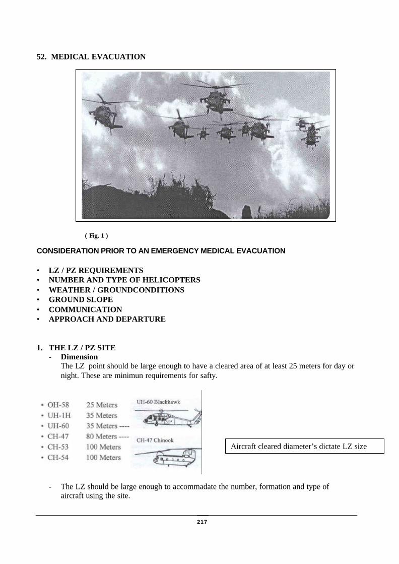
217
52. MEDICAL EVACUATION
( Fig. 1 )
CONSIDERATION PRIOR TO AN EMERGENCY MEDICAL EVACUATION
• LZ / PZ REQUIREMENTS• NUMBER AND TYPE OF HELICOPTERS• WEATHER / GROUNDCONDITIONS• GROUND SLOPE• COMMUNICATION• APPROACH AND DEPARTURE
1. THE LZ / PZ SITE- Dimension
The LZ point should be large enough to have a cleared area of at least 25 meters for day ornight. These are minimun requirements for safty.
- The LZ should be large enough to accommadate the number, formation and type ofaircraft using the site.
Aircraft cleared diameter’s dictate LZ size
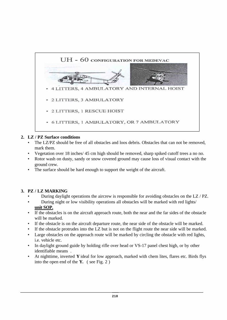
218
2. LZ / PZ Surface conditions• The LZ/PZ should be free of all obstacles and loos debris. Obstacles that can not be removed,
mark them.• Vegetation over 18 inches/ 45 cm high should be removed, sharp spiked cutoff trees a no no.• Rotor wash on dusty, sandy or snow covered ground may cause loss of visual contact with the
ground crew.• The surface should be hard enough to support the weight of the aircraft.
3. PZ / LZ MARKING• During daylight operations the aircrew is responsible for avoiding obstacles on the LZ / PZ.• During night or low visibility operations all obstacles will be marked with red lights/
unit SOP.• If the obstacles is on the aircraft approach route, both the near and the far sides of the obstacle
will be marked.• If the obstacle is on the aircraft departure route, the near side of the obstacle will be marked.• If the obstacle protrudes into the LZ but is not on the flight route the near side will be marked.• Large obstacles on the approach route will be marked by circling the obstacle with red lights,
i.e. vehicle etc.• In daylight ground guide by holding rifle over head or VS-17 panel chest high, or by other
identifiable means .• At nighttime, inverted Y ideal for low approach, marked with chem lites, flares etc. Birds flys
into the open end of the Y. ( see Fig. 2 )

219
4. GROUND SLOPE REQUIREMENTS
As mentioned before the LZ /PZ surface has to support the weight of the aircraft. After rain or inwinter the area has to be checked and obstacles are removed.
A helicopter cannot safely land an a slope more than 15 degrees.
• When the slope is under 7 degrees, the helicopter should land upslope. ( Figure 3 )
• When the slope is 7 to 15 degrees, the helicopter should land sideslope with the exit downhill.( Figure. 4 )
• Over 15 degrees the aircraft may HOVER to pick up or drop cargo.
( Figure 3. Upslope landing ) ( Figure 4. Sideslope landing )

220
The ground slope can be calculated by the following mathematical computation. ( Figure 5)
Find: Percent of slope = ?Given: Eye level height = 1,75 m
Pace length = 0.75 m
Answer: Vertical distance: = 2 x 1,75 = 35 mHorizontal distance = 75 paces + 125 paces x 0.75 m = 150 mPercent of slope = Vertical distance (Vd) x 100 = slope
Horizontal distance (Hd)= 2,34 %
5. Weather
Wind• A helicopter can land with a tail wind of 0 to 5 knots and a cross wind of 0 to 9 knots.• With head wind of 0 to 10 knots and above a aircraft can land but it prevers to land
into the wind.
Density altidute
q Density altidute determines the actual lift capability of the aircraft for that particular day.
There are three factors which affect the performance of helicopters:
q Altiduteq Temperatureq Humidity
The optimum aircraft performance is obtained during a cool day with the elevation as close to sea levelas possible and low humidity.

221
6. GROUND CREW HAND AND ARM SIGNALS
The pictures below shows the most common hand and arm signals to guide the aircraft to the ground.
7. APPROACH AND DEPARTURE DIRECTIONS
♦ 10 : 1 ratio is required♦ load of the aircraft will have a factor♦ generally should fly into the wind if possible♦ crosswind landing if less than 6 to 9 knots

222
8. COMMUNICATION
To communicate with the aircrew you have to know how to use the appropriate radio as well as callsigns, to be familiar with radio procedures either tactical or non tactical.
The STANDARD NATO NINE LINE CONTAINS ALL INFORMATION THE AIRCREW NEEDSTO APPROACH THE LZ /PZ.
Line 1 - 8 digit grid coordinateLine 2 - Call sign & radio frequency at pick up siteLine 3 - Number of patients by precedence
Urgent; immediate evacuation needed to save life, limb and eye sight
Priority; must be evacuated within 4 hours or status changed to Urgent
Routine; should be moved within 24 hoursLine 4 - Request any special equipment; needed, ie. Jungle penetratorLine 5 - Number of patients by type, litter or ambulatoryLine 6 - Security of LZ / PZ *
Patient information; injuries, illness (GSW Malaria * )Line 7 - Method of marking LZ/PZ; VS-17 panel, smoke, cem lites
etc.; direction to enterLine 8 - Patient nationality ; military , civillian, sexLine 9 - Terrain discription; slope%, approach & heading, wind,
obstacles * NBC contamination= For time of war only,
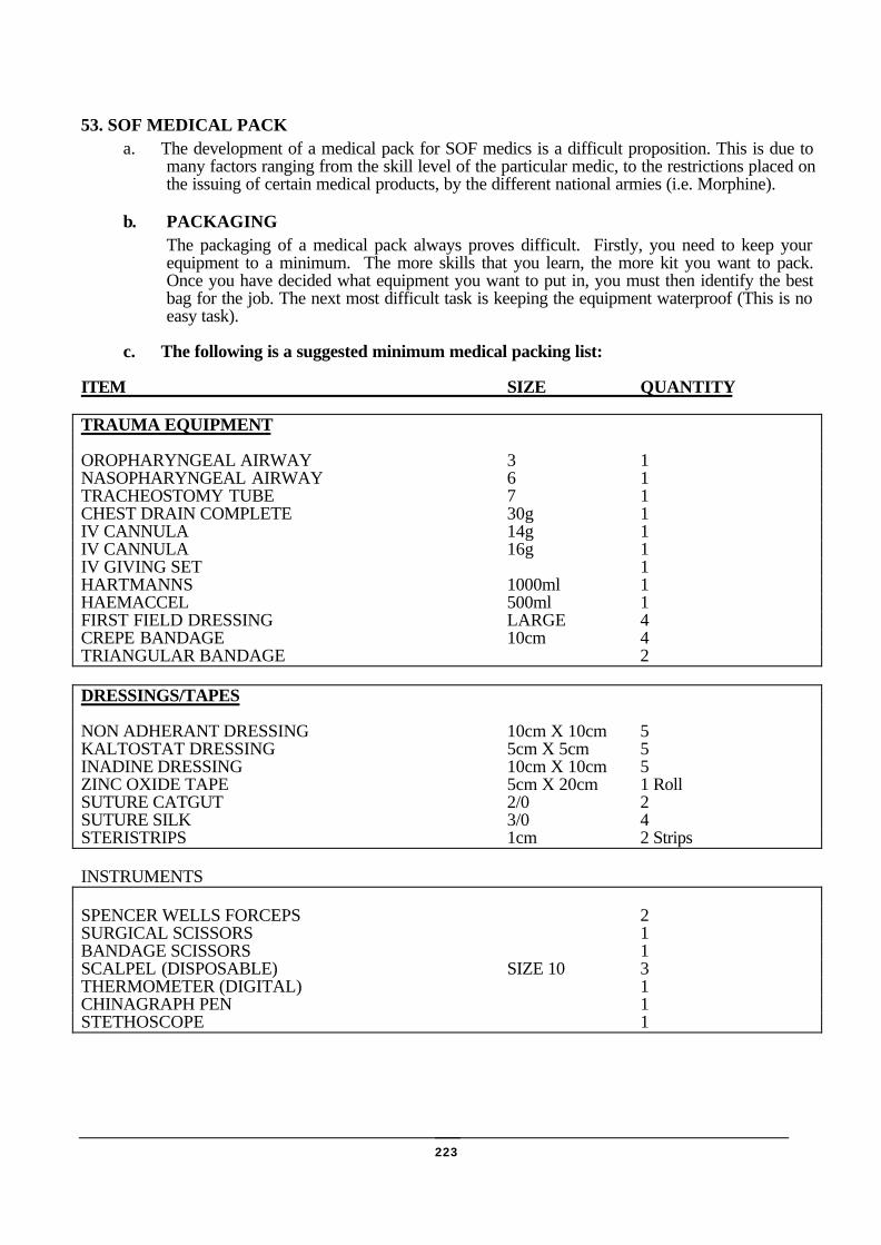
223
53. SOF MEDICAL PACKa. The development of a medical pack for SOF medics is a difficult proposition. This is due to
many factors ranging from the skill level of the particular medic, to the restrictions placed onthe issuing of certain medical products, by the different national armies (i.e. Morphine).
b. PACKAGINGThe packaging of a medical pack always proves difficult. Firstly, you need to keep yourequipment to a minimum. The more skills that you learn, the more kit you want to pack.Once you have decided what equipment you want to put in, you must then identify the bestbag for the job. The next most difficult task is keeping the equipment waterproof (This is noeasy task).
c. The following is a suggested minimum medical packing list:
ITEM SIZE QUANTITY
TRAUMA EQUIPMENT
OROPHARYNGEAL AIRWAY 3 1NASOPHARYNGEAL AIRWAY 6 1TRACHEOSTOMY TUBE 7 1CHEST DRAIN COMPLETE 30g 1IV CANNULA 14g 1IV CANNULA 16g 1IV GIVING SET 1HARTMANNS 1000ml 1HAEMACCEL 500ml 1FIRST FIELD DRESSING LARGE 4CREPE BANDAGE 10cm 4TRIANGULAR BANDAGE 2
DRESSINGS/TAPES
NON ADHERANT DRESSING 10cm X 10cm 5KALTOSTAT DRESSING 5cm X 5cm 5INADINE DRESSING 10cm X 10cm 5ZINC OXIDE TAPE 5cm X 20cm 1 RollSUTURE CATGUT 2/0 2SUTURE SILK 3/0 4STERISTRIPS 1cm 2 Strips
INSTRUMENTS
SPENCER WELLS FORCEPS 2SURGICAL SCISSORS 1BANDAGE SCISSORS 1SCALPEL (DISPOSABLE) SIZE 10 3THERMOMETER (DIGITAL) 1CHINAGRAPH PEN 1STETHOSCOPE 1
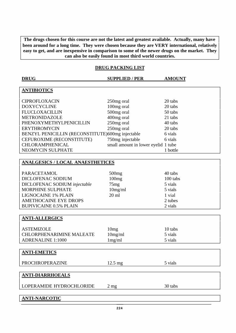
224
The drugs chosen for this course are not the latest and greatest available. Actually, many havebeen around for a long time. They were chosen because they are VERY international, relativelyeasy to get, and are inexpensive in comparison to some of the newer drugs on the market. They
can also be easily found in most third world countries.
DRUG PACKING LIST
DRUG SUPPLIED / PER AMOUNT
ANTIBIOTICS
CIPROFLOXACIN 250mg oral 20 tabsDOXYCYCLINE 100mg oral 20 tabsFLUCLOXACILLIN 500mg oral 50 tabsMETRONIDAZOLE 400mg oral 21 tabsPHENOXYMETHYLPENICILLIN 250mg oral 40 tabsERYTHROMYCIN 250mg oral 20 tabsBENZYL PENICILLIN (RECONSTITUTE)600mg injectable 6 vialsCEFUROXIME (RECONSTITUTE) 750mg injectable 6 vialsCHLORAMPHENICAL small amount in lower eyelid 1 tubeNEOMYCIN SULPHATE 1 bottle
ANALGESICS / LOCAL ANAESTHETICES
PARACETAMOL 500mg 40 tabsDICLOFENAC SODIUM 100mg 100 tabsDICLOFENAC SODIUM injectable 75mg 5 vialsMORPHINE SULPHATE 10mg/ml 5 vialsLIGNOCAINE 1% PLAIN 20 ml 1 vialAMETHOCAINE EYE DROPS 2 tubesBUPIVICAINE 0.5% PLAIN 2 vials
ANTI-ALLERGICS
ASTEMIZOLE 10mg 10 tabsCHLORPHENARIMINE MALEATE 10mg/ml 5 vialsADRENALINE 1:1000 1mg/ml 5 vials
ANTI-EMETICS
PROCHROPERAZINE 12.5 mg 5 vials
ANTI-DIARRHOEALS
LOPERAMIDE HYDROCHLORIDE 2 mg 30 tabs
ANTI-NARCOTIC

225
NALOXONE 0.4mg/ml 5 vials
ANTI-FUNGAL
CLOTRIMAZOLE CREAM 1 tube
OTHERS
SODIUM CHLORIDE 10ml 10 vialsCAVIT 1tubeFLUOROCIEN EYE DROPS 2 vialsIODINE PAINT 1 bottle
MISCELLANEOUS
SYRINGE 10ml 2HYPODERMIC NEEDLE 20 g 5HYPODERMIC NEEDLE 25 g 5RYLES TUBE 14 g 1DISPOSABLE GLOVES 2 PAIRSAM SPLINT 2CASUALTY CARD (WATERPROOFED) 2
NOTES:Belt Order. Three remaining members of patrol to carry 500ml fluid (2 x Hartmanns, 1 x Haemaccel), 2x IV cannula (14G/16G), 1 x IV giving set.
Bergan or Rucksack:. All four members of patrol to carry 500ml fluid in Bergans.

226
54. BASIC PHARMACOLOGYa. One of the principal areas, in which the activities of the SOF Medic differ from those of
other armed forces medics, is in the administration of pharmacological agents, or 'drugs'.Such agents have both lifesaving and life endangering potential, depending on how they areused. The wrong drug or the wrong dose or technique of administration of the right drug,can kill a patient as effectively as a lethal weapon. Therefore, the medic must be intimatelyfamiliar with the drugs available to him in the field; He must know their indications andcontraindications, their side effects and their dangers. Almost nowhere else in emergencycare can ignorance or carelessness on the part of the medic do so much harm as in the use ofdrugs.
b. DRUG INFORMATION(1) A drug is a chemical compound administered as an aid in the diagnosis, treatment or
prevention of a disease or other abnormal condition; Drugs may be given in an attemptto alter the disease process itself or to relieve the symptoms of the process(symptomatic relief).
(2) The drugs we use are derived from four principal sources: ANIMAL, VEGETABLE,MINERAL and SYNTHETIC. For example: Insulin - A medication taken bydiabetics, it is prepared from the pancreas of animals (usually cattle or pigs). Avariety of roots, leaves, flowers and seeds provide vegetable sources of drugs;Digitalis, used in the treatment of heart failure, is a drug of vegetable origin, preparedfrom the dried leaves of a wild flower called purple foxglove. Minerals such ascalcium, iron and magnesium are also used in the treatment of various medicalproblems. Finally, many of the drugs on the market today are manufacturedsynthetically; Synthetic forms of vitamins, steroids, narcotics and an enormous varietyof other drugs are widely available.
c. DRUG NAMESMost drugs have several names and these names fall into three broad categories:
(1) Chemical NameThis is an exact description of the drug's chemical composition, of interest only tochemists and others involved in the manufacture of drugs.
(2) Generic NameThe generic name is usually one given to the drug by the company that firstmanufactures it, before the drug has become official. This is the name by whichdrugs are identified in military medicine and also the safest way to distinguish adrug in other countries. It is by this name that the SOF Medic identifies thedifferent kinds of drugs.
(3) Trade Name (Brand Name, Trademark, Proprietary Name)This is the name given to a drug by the company marketing it. Since the drug may bemarketed by a number of manufacturers in different countries, it may have severaldifferent trade names.

227
(4) Let's take, for example, tetracycline, a widely used antibiotic:
q Chemical Name: 4-dimethylamino-1, 4, 4a, 5, 5a, 11, 12a-octahydro-3, 6, - 10, 12,12a - pentahydroxy-6-methyl-1, 11-dioxo-2 napthacenecarboxamide
q Generic Name: Tetracyclineq Trade Names: Achromycin, Panmycin, Sustamycin, Tetrabid-Organon, Tetrachel,
and Tetrex
(5) In this manual, we will always refer to drugs by their generic names. When a drug iswidely known by its proprietary name this name will be noted in brackets, forexample, naloxone (narcan), Doxycycline (Vibramycin).
d. DRUG STANDARDS AND LEGISLATIONStandards are required to ensure that drugs issued by various manufacturers are of a uniformstrength and purity. Such standards are published in the British Pharmacopia and theAmerican Physicians Desk Reference (PDR). In addition, a variety of legislation has beenenacted to protect the consumer (the patient), i.e. the medicines Act 1968 which isconcerned with the manufacture of drugs.
e. LEGAL CLASSIFICATION(1) Besides specifying which drugs can be sold over the counter and which require a
doctor's prescription, government regulations decide the degree of availability of manysubstances which have an abuse potential. The Misuse of Drugs Regulationsgenerally establish five categories, or schedules of drugs.
(2) The table below provides descriptions of the types of drugs included in each scheduleof the Misuse of Drugs Regulations. A summary of the abuse potential of the drugs ineach schedule is also included.
(a) Schedule IVirtually all the drugs in this group are prohibited except in accordance withlocal law. All of them have a high potential for abuse and are rarely usedmedicinally. Examples: Marijuana (Cannabis), LSD.
(b) Schedule IILike Schedule I drugs, these have a high potential for abuse and can lead tophysical and psychological dependence. They have an accepted medical use,but are subject to full controlled drug requirements.
Most of them are stimulants, narcotics or depressants. Example: Amphetamine,cocaine, diamorphine (heroin), and morphine, buprenorphine (Temgesic).
(c) Schedule IIIDrugs in this group have a lower potential for abuse than those in Schedules Iand II, but they are nevertheless subject to special prescription requirements.Examples: Barbiturates, pentazocine (fortral).
(d) Schedule IVThe drugs in this group have a potential for abuse lower than Schedule I-IIIdrugs and are subject to minimal control. Examples: Benzodiazepines(tranquillisers like Diazepam - Valium).
(e) Schedule VThese drugs have a low potential for abuse because of their strength. For themost part, they are preparations that contain small amounts of narcotics, but areexempt from controlled drug requirements.
228
f. DRUG FORMS(1) Drugs are available in many forms, both solid and liquid, each of which has special
properties. Some of the most common forms of medicines are:(2) Tablets
(a) These are small masses of compressed powder and are usually presented in oneof the following ways:
(b) A single drug or compound of different drugs.(c) Sugar coated to hide an unpleasant taste.(d) Enteric coated. These pills or tables are coated in such a way that they will not
dissolve until they have passed through the stomach into the intestine.(3) Capsules
(a) These are containers made of gelatine, which dissolve in the stomach andrelease their contents.
(4) Lozenges(a) These may be round or of irregular shape, usually larger than most tablets, and
are used in mouth and throat conditions.(b) The patient puts the lozenge between the cheek and back teeth, and sucks it
slowly.(5) Ointments
(a) These are semi solid preparations of medicinal substances, normally in an oilyor paraffin type base, intended for external application to the skin or mucousmembranes.
(6) Creams(a) These are similar to ointments, but usually in a non-oily base.(b) They are intended for external application to the skin and seem to disappear
when applied.(7) Suppositories
(a) These are semi solid, medicated compounds used for the introduction of drugsinto the rectum. They melt readily at body temperature.
g. ACTION OF DRUGS(1) Drugs may produce their effects locally, systematically, or both. Local effect result
form the direct application of a drug to a given tissue, as for example when lotions areapplied to the skin to relieve itching. Systemic effect occur after drugs are absorbedby any route and are distributed by the blood stream; Such effects may involve manydifferent organs.
(2) The effect of any given drug may vary depending on the age and condition of thepatient, the dosage and the route by which the drug is given.
h. ROUTES OF ADMINISTRATION(1) Several routes may administer drugs, and the route of administration appropriated for
one drug may be entirely inappropriate for another. Thus for any given drug, one mustknow the route(s) by which it is given.

229
(2) Intravenous(a) The intravenous (IV) route is the most rapidly effective and also the most
dangerous route of administration. Drugs given by this route go directly into thebloodstream and thence to the target organs, without any delay in absorption. Ingeneral, drugs given intravenously should be given slowly.
(b) In the field, it is preferable to give intravenous medication through anestablished IV line rather than by direct venipuncture, as the latter techniquemay result in infiltration of the drug.
(3) Intra-muscular(a) Drugs given by the intra-muscular (IM) route take longer to act, since they must
first be absorbed from the muscle into the blood stream. By the same token,medications given intramuscularly have a longer duration of action, since theyare absorbed gradually over a period of minutes to hours. Obviously, absorptionof medications given by this route depends on an adequate blood flow to themuscles.
(b) The intra-muscular injection involves volumes of not more than 2 ml beinggiven into the upper outer quadrant of the buttock or anterior thigh.
(4) Subcutaneous(a) Subcutaneous (SC or SQ) injections are given beneath the skin into the fat or
connective tissue immediately underlying it. Medications administered by thisroute also are absorbed more slowly and over a more prolonged period thanwhen given intravenously. Subcutaneous injections are usually given under theskin of the upper outer arm, the anterior thigh or the abdomen.
(5) Sublingual(a) Sublingual administration refers to giving a medication under the tongue and
this is the fashion in which buprenorphine (Temgesic) is administered. Drugstaken sublingually are usually rapidly absorbed, with effects apparent within afew minutes.
(6) Oral(a) Most patients take their daily medications by the oral route. Drugs taken orally
are absorbed at a somewhat unpredictable rate. The oral route is rarely indicatedin emergency situations. The major exceptions are syrup of ipecac, used toinduce vomiting, and activated charcoal, used to bind ingested poisons, both ofwhich are administered orally in certain poisoning and overdose emergencies.
(7) Rectal(a) Rectal administration is not frequently used in the field. However, certain
medications, such as the antibiotic metronidazole (Flagyl, Zadstat) are availablein suppository form. Absorption across the rectal mucosa is rapid, although notentirely predictable.
i. RATES OF ABSORPTION(1) The speed at which a drug is absorbed is related to the route by which the route by
which it is given gives it. Obviously, drugs injected directly into the circulation, as inintravenous injections, gain access to the circulation fastest. Nearly as rapid is theabsorption across the respiratory mucosa when drugs are sprayed down an

230
Endotracheal tube. Other mucosal surfaces, such as that in the rectum, also provide forrapid absorption, although somewhat variable in nature. Intra-muscular injection isslower, for the drug must be picked up from the muscles by the circulation blood.
(2) The same is true of subcutaneous injections, which are absorbed more slowly thanintra-muscular. Finally, drugs administered orally in general have the slowestabsorption rate of all.
(3) Once a drug is absorbed, it is delivered to various target organs by the blood stream.Over a period of time, the drug is inactivated (detoxified), usually by the liver, andeliminated from the body by the kidneys, digestive tract, lungs or skin.
j. PRESCRIPTION ABBREVIATIONS
• QID, QDH - 4 x daily• QQH - Every 4 hours• TID, TPS - 3 x daily• BD, BID - 2 x daily• STAT - Immediately, once only• PRN - As required, may be repeated• AC - Before food• PC - After food

231
55. INJECTIONSAs a SOF Medic, it is important to have an understanding of and be able to perform variousinjection techniques.
a. REASONS FOR INJECTIONSThere are times when it is better to administer drugs via injection. The reasons for this are:
(1) To make sure the patient receives the medication.
(2) Can be used if the patient has diarrhoea or vomiting.
(3) Patient may be unconscious.
(4) Drug is more rapidly absorbed.
b. CHECKS PRIOR TO GIVING INJECTIONThere are certain checks that must be carried out prior to giving an injection.
(1) Correct patient.
(2) Correct drug.
(3) Correct dose.
(4) Correct time.
(5) Correct route.
(6) Drug is in date.
c. TYPES OF INJECTIONSThere are three main types of injections these are:
(1) Intra-venous
(2) Intra-muscular
(3) Subcutaneous
d. SITES OF INJECTION and SELECTION CRITERIAThere are various sites for giving an injection, depending on the type of injection given.These include:
(1) Subcutaneous injections are performed when the drug to be given requires a slow, butsteady absorption into the blood. The drug is placed under the skin but above themuscle. Maximum volume for subcutaneous injection is 2ml.
Upper, outer aspect of arm, with the needle at a 45° angle.
232
(2) Intra-muscular injections. Under normal conditions, the IM route is more rapidlyabsorbed than the subcutaneous route and larger volumes can be given, but in shock,there is the danger of decreased or erratic absorption.
Deltoid muscle, upper outer aspect of buttocks, with the needle at a 90° angle.
(3) Intravenous injections are usually given through a pre-existing IV line. To avoid theinadvertent injection into surrounding tissue, the IV route has the fastest absorptionbut many drugs are not compatible with direct infusion.
(4) Once a drug is given with injection, consider it gone. Was it the right patients, rightdose, right drug, right time, right route?
e. EQUIPMENT REQUIREDThe equipment required is as follows:
(1) 1 x sterile swab
(2) 1 x syringe
(3) 2 x needles
(4) Correct drug
f. METHOD OF INJECTIONOnce the checks have been carried out, the method is as follows:
(1) Select site.
(2) Clean site with sterile swab.
(3) Insert needle firmly.
(4) Draw back on syringe (to ensure you are not in a blood vessel).
(5) Slowly inject the drug.
(6) Withdraw needle and re-clean injection site with sterile swab.

233
56. DRUGS• Anti-microbial drugs
• Anti-inflammatory drugs + Analgesics
• Anti-allergic
• Anti-emetics
• Anti-diarrhoeas
• Rest group
a. ANTIMICROBIAL DRUGS
b. ORAL
(1) CIPROFLOXACIN(a) Drug Family: Antibiotics
(b) Generic Name: Ciprofloxacin
(c) Trade Name: Ciproxin
(d) Route of Administration: Oral
(e) Indications : Broad-spectrum antibiotic for respiratoryand urinary tract infections, also forgastro-intestinal infections
(f) Adult Dosage:• How Much 250mg to 750mg• How Often 2 x day• How long minimum of 5 days
(g) Side Effects: Nausea, vomiting, diarrhoea, dyspepsia,abdominal pain, dizziness, and headache
(h) Contra-Indications : Known hypersensitivity
(i) Remarks: May impair performance of skilled tasks,enhances effects of alcohol (avoid alcohol)

234
(2) DOXYCYCLINE(a) Drug Family: Antibiotics
(b) Generic Name: Doxycycline
(c) Trade Name: Vibramycin
(d) Route of Administration: Oral
(e) Indications : Broad-spectrum antibiotic for chlamydia(urethritis), mycoplasma (respiratory andurinary tract infections), Lyme disease,and rickettsia typhus, Q fever)
(f) Adult Dosage:• How much 200mg on first day, them 100mg• How often 100mg xday (except on first day)• How long 14 days
(g) Side Effects: Nausea, vomiting, diarrhoea,photosensitivity, hypersensitivity (skinredness), intracranial hypertension(headache, visual disturbances)
(h) Contra-Indications : Do not give during pregnancy, Knownhypersensitivity
(i) Remarks: Take with plenty of fluid during meals.Avoid sunlight.
(3) FLUCLOXACILLIN(a) Drug Family: Antibiotics
(b) Generic Name: Flucloxacillin
(c) Trade Name: Floxapen
(d) Route of Administration: Oral
(e) Indications : Dirty wounds, skin infections
(f) Adult Dosage:• How much 250mg
to 300mg• How often 4 x
day• How long 5 days
(g) Side Effects: Diarrhoea, rashes, hypersensitivity(anaphylaxis), fever
(h) Contra-Indications : Penicillin hypersensitivity, Knownhypersensitivity
(i) Remarks: Take 1/2 to 1 hour before food. Makesure, patient is not sensitive to penicillin
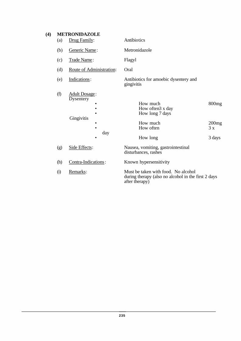
235
(4) METRONIDAZOLE(a) Drug Family: Antibiotics
(b) Generic Name: Metronidazole
(c) Trade Name: Flagyl
(d) Route of Administration: Oral
(e) Indications : Antibiotics for amoebic dysentery andgingivitis
(f) Adult Dosage:Dysentery
• How much 800mg• How often3 x day• How long 7 days
Gingivitis• How much 200mg• How often 3 x
day• How long 3 days
(g) Side Effects: Nausea, vomiting, gastrointestinaldisturbances, rashes
(h) Contra-Indications : Known hypersensitivity
(i) Remarks: Must be taken with food. No alcoholduring therapy (also no alcohol in the first 2 daysafter therapy)

236
(5) PHENOXYMETHYLPENICILLIN(a) Drug Family: Antibiotics
(b) Generic Name: Phenoxymethylpenicillin
(c) Trade Name: Distaquine V-K, Pen V
(d) Route of Administration: Oral
(e) Indications : Tonsillitis, Otitis media
(f) Adult Dosage:• How much250mg to 500mg• How often 4 x day• How long 10 days
(g) Side Effects: Diarrhoea, sensitivity reactions such asfever, Urticaria, joint pains, anaphylaxis (shock)
(h) Contra-Indications : Penicillin hypersensitivity, Knownhypersensitivity
(i) Remarks: Must be taken with food. Make sure,patient is not sensitive to penicillin, ifsensitive, give erythromycin
(6) ERYTHOMYCIN(a) Drug Family: Antibiotics
(b) Generic Name: Erythromycin
(c) Trade Name: Erythromycin, Erymax
(d) Route of Administration: Oral
(e) Indications : Alternative to Penicillin - hypersensitivepatients
(f) Adult Dosage:• How much 250mg - 500mg• How often 4 x days• How long 10 days
(g) Side Effects: Nausea, vomiting, diarrhoea after large doses
(h) Contra-Indications : Known hypersensitivity
(i) Remarks: Mild infections: 4 x 250mg a day. Severeinfections: 4 x 500mg a day

237
c. IM OR IV INJECTIONS
(1) BENZYLPENICILLIN (Penicillin G)(a) Drug Family: Antibiotics
(b) Generic Name: Benzylpenicillin (Penicillin G)
(c) Trade Name: Crystapen
(d) Route of Administration: IM or IV injection
(e) Indications : Meningitis, prophylaxis in limbamputations (gas - gangrene),tonsillitis, Otitis media, erysipelas
(f) Adult Dosage:
• How much 600mg (Meningitis 2400mg)• How often 4 x day• How long 10 days or more
(g) Side Effects: Diarrhoea, sensitivity reactions as fever,Urticaria, joint pains, anaphylaxis (shock)
(h) Contra-Indications : Penicillin hypersensitivity, Knownhypersensitivity
(i) Remarks: When giving IV, do it slowly(meningitis). Make sure patient is not sensitive topenicillin, if sensitive, give erythromycin

238
(2) CEFUROXIME(a) Drug Family: Antibiotics
(b) Generic Name: Cefuroxime
(c) Trade Name: Zinacef
(d) Route of Administration: IM or IV injection
(e) Indications : Broad spectrum antibiotic for abdominalinfections, pneumonia, meningitis
(f) Adult Dosage:• How much 750mg to 1500mg (meningitis 3000mg)• How often 3 x day• How long 10 days or more
(g) Side Effects: Rashes, Urticaria, fever, joint pain,anaphylaxis (shock)
(h) Contra-Indications : Known hypersensitivity, be cautious ofpenicillin sensitivity
(i) Remarks: Single doses larger than 750mg should notbe given by IM injection. 10% of people who aresensitive to penicillin, will also be sensitive tocefuroxime

239
d. TOPICAL
(1) CHLORAMPHEMICOL(a) Drug Family: Topical Antibiotic
(b) Generic Name: Chloramphenicol eye ointment
(c) Trade Name: Chloromycetin
(d) Route of Administration: Topical application (eye)
(e) Indications : Eye infections
(f) Adult Dosage:• How much Small amount within lower lid• How often 4 x day• How long continue 2 days after healing
(g) Side Effects: Transient stinging
(h) Contra-Indications : Known hypersensitivity
(2) CLOTRIMAZOLE(a) Drug Family: Topical antifungal
(b) Generic Name: Clotrimazol
(c) Trade Name: Canesten
(d) Route of Administration: Topical application (skin)
(e) Indications : Fungal skin infections
(f) Adult Dosage:• How much apply sparingly• How often 2-3 x daily• How long for 2 more weeks after cure
(g) Side Effects: Occasionally skin irritation or sensitivity
(h) Contra-Indications : Known hypersensitivity
(i) Remarks: Apply 5 cm around lesion

240
(3) NEOMYCIN SULPHATE(a) Drug Family: Topical Antibiotic
(b) Generic Name: Neomycin Sulphate
(c) Trade Name: Otosporin
(d) Route of Administration: Topical application (ear)
(e) Indications : Otitis Externa
(f) Adult Dosage:• How much 3 drops in affected ear• How often 5-6 x day• How long 7 days
(g) Side Effects: Local sensitivity
(h) Contra-Indications : Perforated eardrum (ear blast), knownhypersensitivity
(i) Remarks:
e. ANTI-INFLAMMATORY DRUGS + ANALGESICS
f. ORAL
(1) PARACETAMOL(a) Drug Family: Analgesic
(b) Generic Name: Paracetamol
(c) Trade Name: Panadol
(d) Route of Administration: oral
(e) Indications : Fever, mild pain
(f) Adult Dosage:• How much 500mg• How often 500mg every 4-6 hours• How long until pain or fever decreases
(g) Side Effects: Hard on stomach, take with foodor milk
(h) Contra-Indications : Known hypersensitivity
(i) Remarks: Max 8 tables (= 4000mg) a day

241
(2) DICLOFENAC SODIUM(a) Drug Family: Anti-inflammatory analgesics
(b) Generic Name: Diclofenac Sodium
(c) Trade Name: Voltarol
(d) Route of Administration: Oral
(e) Indications : Musculoskeletal pain
(f) Adult Dosage:• How much 50mg• How often2-3 x day• How long until pain decreases
(g) Side Effects: Nausea, diarrhoea,hypersensitivity reactions (rashes,asthma), occasionally ulceration ofstomach/duodenum
(h) Contra-Indications : Ulcers of stomach/duodenum,Known hypersensitivity
(i) Remarks: Take with food, max. 150mg aDay
g. IM, IV OR SC INJECTION
(1) DICLOFENAC SODIUM(a) Drug Family: Anti-inflammatory analgesics
(b) Generic Name: Diclofenac Sodium
(c) Trade Name: Voltarol
(d) Route of Administration: Deep IM
(e) Indications : Musculoskeletal pain
(f) Adult Dosage:• How much 75mg• How often2 x day• How long 2 days
(g) Side Effects: Pain at injection site,hypersensitivity reactions (rashes,asthma), occasionally ulceration ofstomach/duodenum
(h) Contra-Indications : Ulcers of stomach/duodenum,Known hypersensitivity
(i) Remarks: IM not longer than 2 days

242
(2) MORPHINE(a) Drug Family: Analgesics
(b) Generic Name: Morphine (10mg in 10ml)
(c) Trade Name: Morphine Sulphate
(d) Route of Administration: IV or IM (slower route)
(e) Indications : Treatment of severe pain
(f) Adult Dosage:• How much 1 to 3 ml of a 1mg/ml solution• How often Inject solution in 1ml amount, every 4 hrs• How long Watch for change, i.e. panic status
(g) Side Effects: Nausea, vomiting, drowsiness, respiratorydepression, hypotension, and constrictedpupils
(h) Contra-Indications : Injuries causing respiratory problems,head injuries, unconsciousness,undiagnosed closed abdominal trauma,snake bites, pregnancy, children(Autoinjector), anaesthetic overdose,Known hypersensitivity
(i) Remarks: Morphine should be given withanti-emetic. Always have Naloxoneavailable (in case of overdose)
(3) LIGNOCAINE HYDROCHLORIDE(a) Drug Family: Local Anaesthetics
(b) Generic Name: Lignocaine Hydrochloride 1%
(c) Trade Name: Xylocaine
(d) Route of Administration: Subcutaneous (SC)
(e) Indications : Local anaesthesia, intercostal block
(f) Adult Dosage:• How muchas required to produce adequate loss of
sensation to a maximum of 10ml• How often every 2-4 hours• How long as long as needed
(g) Side Effects: Hypotension and Bradycardia
(h) Contra-Indications : Hypovolemia (shock),Known hypersensitivity
(i) Remarks: Ensure that you have not gained venousaccess.

243
(4) BUPIVACAINE HYDROCHLORIDE(a) Drug Family: Local anaesthesia
(b) Generic Name: Bupivacaine Hydrochloride 0.5%
(c) Trade Name: Marcain
(d) Route of Administration: SC
(e) Indications : Intercostal block, local anaesthesia
(f) Adult Dosage:• How much as required to produce adequate loss of
sensation, to a maximum of 30ml• How often Every 4-8 hours• How long As long as needed
(g) Side Effects: Hypotension, Bradycardia
(h) Contra-Indications : Hypovolemia, Known hypersensitivity
(i) Remarks: Drug of choice for intercostal block.Ensure that you have not gained venousaccess
h. TOPICAL
(1) AMETHOCAINE(a) Drug Family: Topical anaesthetic
(b) Generic Name: Amethocaine eye drops 0.5.%
(c) Trade Name: Amethocaine
(d) Route of Administration: Topical application (eye)
(e) Indications : Topical anaesthetic for the eye
(f) Adult Dosage:• How much 1-2 drops in lateral corner of eye• How often as required• How long as long as needed
(g) Side Effects: Very painful for up to 2 minutes, warnpatient first
(h) Contra-Indications : Known hypersensitivity
(i) Remarks: This drug decreases the patient's ability toavoid dirt or dust entering the eye, so theeye should be protected and covered untilsensation returns.

244
i. ANTI-ALLERGICS
j. ORAL(1) ASTEMIZOLE
(a) Drug Family: Anti-histamines
(b) Generic Name: Astemizole
(c) Trade Name: Hismanal
(d) Route of Administration: Oral
(e) Indications : Hay fever, pruritis (itching), and Urticaria
(f) Adult Dosage:• How much 10mg• How often 10mg x day• How long As long as needed
(g) Side Effects: Seldom affects performance of skilledtasks
(h) Contra-Indications : Known hypersensitivity
(i) Remarks: Avoid alcohol
k. IM OR IV INJECTIONS(1) CHLORPHENIRAMINE MALEATE
(a) Drug Family: Anti-histamines
(b) Generic Name: Chlorpheniramine Maleate 10mg/ml
(c) Trade Name: Piriton
(d) Route of Administration: IV
(e) Indications : Anaphylaxis (and hay fever, Urticaria)
(f) Adult Dosage:
• How much 10mg diluted in syringe with5-10ml syringe blood, slowly IV
• How often 4 x day, Maximum 40mg/day• How long until patient recovers
(g) Side Effects: Drowsiness, hypotension and CNSstimulation with too rapid IV injection
(h) Contra-Indications : Known hypersensitivity
(i) Remarks: IV injection slowly during 1 minute.Avoid alcohol.

245
(2) ADRENALINE(a) Drug Family: Anti-allergics
(b) Generic Name: Adrenaline 1mg/ml (1:1000)
(c) Trade Name: Adrenaline
(d) Route of Administration: IM
(e) Indications : Drug of choice for treatment ofanaphylaxis
(f) Adult Dosage:• How much 0.5-1ml• How often every 10 minutes until
improvementoccurs (pulse down, bloodpressure up)
• How long until improvement
(g) Side Effects: Anxiety, tremor, and Tachycardia, drymouth, cold extremities, arrhythmia’s
(h) Contra-Indications : Known hypersensitivity
(i) Remarks: Massage site of injection for 30 secondsafter injection
l. ANTI-EMETICS
(1) PROCHLORPERAZINE(a) Drug Family: Anti-emetics
(b) Generic Name: Prochlorperazine 12.5mg/ml
(c) Trade Name: Stemetil
(d) Route of Administration: IM
(e) Indications : Severe nausea, vomiting, vertigo(dizziness)
(f) Adult Dosage:• How much 12.5mg deep IM• How often Every 8 hours• How long As long as needed
(g) Side Effects: Hypothermia, constipation, hypotension,and Tachycardia
(h) Contra-Indications : CNS depression (coma), alcohol usage,and hypersensitivity
(i) Remarks: Patient should remain supine for 30 minafter IM injection. Not effective againstmotion sickness.
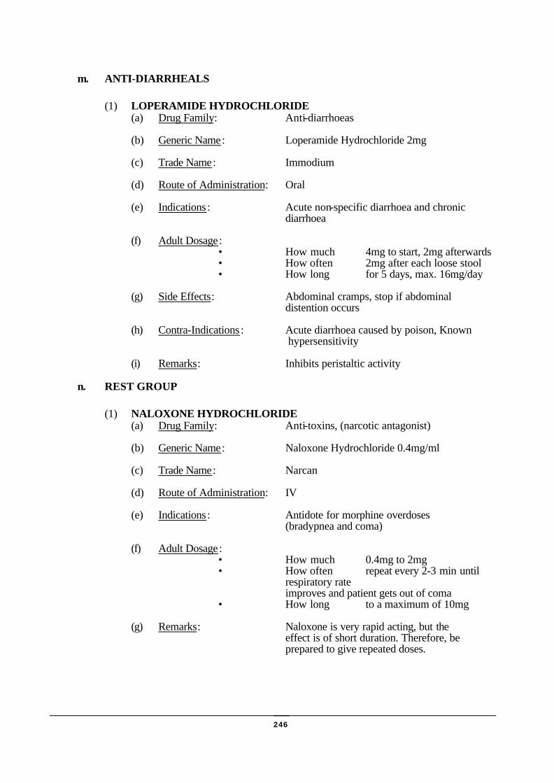
246
m. ANTI-DIARRHEALS
(1) LOPERAMIDE HYDROCHLORIDE(a) Drug Family: Anti-diarrhoeas
(b) Generic Name: Loperamide Hydrochloride 2mg
(c) Trade Name: Immodium
(d) Route of Administration: Oral
(e) Indications : Acute non-specific diarrhoea and chronicdiarrhoea
(f) Adult Dosage:• How much 4mg to start, 2mg afterwards• How often 2mg after each loose stool• How long for 5 days, max. 16mg/day
(g) Side Effects: Abdominal cramps, stop if abdominaldistention occurs
(h) Contra-Indications : Acute diarrhoea caused by poison, Known hypersensitivity
(i) Remarks: Inhibits peristaltic activity
n. REST GROUP
(1) NALOXONE HYDROCHLORIDE(a) Drug Family: Anti-toxins, (narcotic antagonist)
(b) Generic Name: Naloxone Hydrochloride 0.4mg/ml
(c) Trade Name: Narcan
(d) Route of Administration: IV
(e) Indications : Antidote for morphine overdoses(bradypnea and coma)
(f) Adult Dosage:• How much 0.4mg to 2mg• How often repeat every 2-3 min until
respiratory rateimproves and patient gets out of coma
• How long to a maximum of 10mg
(g) Remarks: Naloxone is very rapid acting, but theeffect is of short duration. Therefore, beprepared to give repeated doses.

247
(2) WATER (Sodium Chloride)(a) Drug Family: Physiological saline solution
(b) Generic Name: Sodium Chloride 0.9%
(c) Trade Name: Sodium Chloride
(d) Route of Administration: IV or IM
(e) Indications : Solvent for drugs, also for dilution ofdrugs
(f) Adult Dosage:• How much Sodium Chloride 0.9% in 10ml
ampoule, give IM or IV aftersolution or dilution with drug
(g) Side Effects: Nil of note
(h) Contra-Indications : Nil of note
(i) Remarks: Nil of note
(3) CROTAMITON(a) Drug Family: Topical Antipruritics
(b) Generic Name: Crotamiton 10%
(c) Trade Name: Eurax
(d) Route of Administration: Topical application (skin)
(e) Indications : Pruritis (including pruritis after scabies)
(f) Adult Dosage:• How much 3 cm of cream• How often 2-3 x daily• How long as required
(g) Side Effects Avoid use near eyes and broken skin
(h) Contra-Indications : Acute exudative skin diseases, Knownhypersensitivity
(i) Remarks: None

248
(4) CAVIT(a) Drug Family: Topical Preparations
(b) Generic Name: Cavit
(c) Trade Name: Cavit
(d) Route of Administration: Topical application (teeth)
(e) Indications : Temporary tooth cavity filling
(f) Adult Dosage:• How much as required• How often as required• How long Lasts up to 6 months
(g) Side Effects: Nil of note
(h) Contra-Indications : Nil of note
(i) Remarks: Ensure the tooth cavity is dry and cleanprior to application. Ensure that properdental care is sought as soon as possible.
(5) FLUORESCEIN SODIUM(a) Drug Family: Topical preparations
(b) Generic Name: Fluorescein Sodium
(c) Trade Name: Fluorescein Sodium
(d) Route of Administration: Topical application (eye)
(e) Indications : Detection of corneal abrasions and foreignbodies.
(f) Adult Dosage:• How much 1-2 drops in lateral corner of eye• How often as required• How long
(g) Side Effects: Nil of note
(h) Contra-Indications : Known hypersensitivity
(i) Remarks: Must be used in conjunction with blacklight, Amethocaine

249
(6) IODINE(a) Drug Family: Topical preparations
(b) Generic Name: Iodine
(c) Trade Name: Betadine
(d) Route of Administration: Topical application (skin)
(e) Indications : Topical antiseptic for skin disinfection (toprevent, treat infections from minor cuts,abrasions, and blisters).
(f) Adult Dosage:• How much Apply sparingly• How often as required• How long as required
(g) Side Effects: Nil of note
(h) Contra-Indications : Known hypersensitivity
(i) Remarks: Patients allergic to shellfish are probablyallergic to iodine

250
57. CONTROL OF PAIN: MORPHINE AND ANESTHESIAMorphine is an opiate analgesic derived from poppy seeds. It is a highly addictive drug, and thereforeis strictly controlled by law.
a. PRESENTATIONMorphine is supplied in a variety of presentations, i.e. Autojets, Syrettes and Vials.
b. ACTIONSMorphine alleviates pain by suppressing the pain centre located in the brain.
c. SIDE EFFECTSAs Morphine is such a potent analgesic, it does produce a number of side effects.
(1) Depressed breathing
(2) Depressed Cough Reflex
(3) Decreased Pulse
(4) Drowsiness/unconsciousness
(5) Dilated pupils
(6) Hypotension
(7) Nausea/vomiting
d. CONTRA-INDICATIONSDue to the side effects of Morphine, there are in turn a number of contra-indications to theuse of Morphine.
(1) Head injury
(2) Unconscious casualty
(3) Snake bites
(4) Pregnancy/childbirth
(5) Children (when using Autojet)
e. PRECAUTIONSOnce again, due to the side effects of Morphine, care must be taken when being used in thefollowing situations:
(1) Chest injuries
(2) Acute abdomen
(3) Abdominal injuries

251
f. METHODS OF ADMINISTRATIONMorphine can be administered via the intra-muscular (IM) or the intra-venous (IV) route.When using the Autojet, it must be given via the IM route. The sites of injection used forthis are:
(1) Upper outer aspect of the thigh.
(2) Upper outer aspect of the buttock.
(3) To inject morphine via the IV route it is injected through the IV cannula.
g. DOSAGE AND FREQUENCY (AUTOJET)The initial dose is one 20mg Autojet followed by further 20mg Autojets 4-6 hourly.
DO NOT give more than one dose of Morphine at a time, as this can lead to a build up ofMorphine at the injection site and with the return of adequate peripheral circulation thiscan cause a Morphine overdose.
h. DOSAGE (INTRA-VENOUS)The IV Morphine is presented in a glass vial containing 10mg Morphine Sulphate in 1ml ofwater. This is titrated to the casualty's pain. The way in which this is done is:
(1) Draw up one vial of 10mg Morphine Sulphate into a 5ml syringe.
(2) Add to this 4ml sterile water.
(3) This gives a solution of 10mg in 5ml (2mg per 1ml solution).
(4) The solution is then injected slowly in 1ml increments via the IV cannula.
(5) To decide on the amount given, watch the casualty's face for a change in pain status.
i. MARKING THE CASUALTYUsing a pen or crayon, mark the casualty's forehead with an M and the DTG, i.e. M 03/1745hrs.
j. MORPHINE OVERDOSE/POISONINGAs mentioned before, it is possible for a Morphine overdose to occur. To avoid this, thefollowing signs and symptoms should be watched for:
(1) Very slow and shallow breathing.
(2) Very weak pulse (radial pulse may be lost altogether).
(3) Pupils become pinpointed (note: the opposite to effects).
(4) Lost reflexes.
(5) Unconsciousness.
k. TREATMENT(1) Stimulate the casualty as much as possible.(2) Maintain airway - monitor vital signs

252
(3) Establish CPR if required(4) Administer Naloxone (Narcan).
l. NALOXONE (NARCAN)Naloxone is a specific narcotic antagonist. This means it attacks and neutralises the narcotic. Itdoes not have the opposite effect of Morphine. It is, however, very short acting so it mayrequire more than one dose to work effectively.
(1) PRESENTATIONNaloxone is presented in a glass vial containing 400mcg Naloxone in 1ml water.
(2) SIDE EFFECTSNONE
(3) CONTRA INDICATIONSNONE
(4) METHODS OF ADMINISTRATIONVia the IV route, through the IV cannula.
(5) DOSAGE0.8-2mg initially and repeated every 2-3 mins if required, to a maximum of 10mg. Ifrespiratory function does not improve, then question diagnosis.
m. ANESTHESIAAny substance which, when applied to nervous tissue, causes a reversible loss of the ability toconduct nerve impulses.
n. SITE OF ACTION(1) Peripheral Nerves Action required(2) The brain Toxic effects(3) The heart Toxic effects
o. SITES OF APPLICATION(1) Topically, e.g. eye, urethra, nose(2) Subcutaneous infiltration(3) Around nerves or nerve trunks, e.g. femoral, intercostal nerves(4) Extradural (epidural) or caudal(5) Intrathecal (spinal)The first three routes are the ones most immediately applicable in the out of hospitalenvironment.

253
p. THE THREE RULES OF LOCAL ANAESTHESIA
(1) Rule 1:ALWAYS USE THE LOWEST EFFECTIVE CONCENTRATION OF LOCALANAESTHETIC. This is important for two reasons:(a) Low concentrations are less toxic than high concentrations, even when the same
dose (in mg) is administered.
(b) Low concentrations will give you a larger volume to use and hence anaesthetisea larger area or more casualties.
(c) Solutions which are more concentrated than required, can be diluted within asyringe, preferably with equal volumes of sterile saline (or sterile water ifdesperate), to halve their concentration.
(2) Rule 2:ALWAYS CALCULATE THE SAFE DOSE THAT CAN BE ADMINISTEREDTO THE PATIENT.(a) It is important when using local anaesthetic agents that the calculated maximum
safe dose is never exceeded for a given patient. This requires knowledge of thepatient's weight and the dose in mg/kg for each local anaesthetic used.
(b) Bupivacaine 2 mg/kg(c) Lignocaine 3 mg/kg(d) Prilocaine 6 mg/kg
(e) In a patient weighing 75 kg, the maximum safe dose of Bupivacaine: 75 x 2 =150 mg
(3) Rule 3:ALWAYS CALCULATE THE VOLUME TO BE USED.(a) All local anaesthetics are available in different concentrations; consequently the
volume containing the dose to be used will depend on the concentration used.
(b) The relationship between concentration, volume and dose is given by theformula:
(c) CONC (%) x VOLUME (ml) x 10 = DOSE (mg)
(d) Providing any two values are known, the third can be calculated.
(e) Continuing with the above example, we have calculated the maximum safe doseto be .. mg, and now need to know what volume of 0.5% Bupivacaine willcontain 150 mg
0.5 x V (unknown) x 10 = ______ mg
V = ____________ = ______ =______ ml0.5 x 10 5

254
q. LOCAL ANAESTHETIC TECHNIQUES IN THE PRE-HOSPITALENVIRONMENTThere are innumerable local anaesthetic techniques, which can be used. The following havebeen chosen for their ease, effectiveness and low incidence of complications.
(1) TOPICALLYFor this method to be effective, it is necessary to use the most concentrated solutionavailable, e.g. 0.5% bupivacaine, 2% Lignocaine.
SITES
(a) The Eye
• Useful to decrease the irritation caused by foreign bodies.
• Local anaesthetic can be instilled into the eye from a syringe. Severalapplications are usually required over 10 minutes for effect.
• After the procedure, a sterile dressing must then cover the eye until thelocal anaesthetic wears off.
• Local anaesthetic agents in the eye are initially painful, warn the patient!
(b) Naso-Pharynx
• Useful to increase the ease of passing and subsequent tolerance of an N-Pairway.
• 2 ml of 1% Lignocaine can be instilled into the appropriate nostril andallowed to flow into the back of the throat.
• If the patient is conscious, they should be warned that the local tastes awful.
(c) Teeth
• Useful for toothache, particularly when associated with broken teeth.
• A cotton wool ball or swab is soaked with 5 ml of 0.5% Bupivacaine.
• Swab placed either over the fractured tooth or along the inside and outsidegum adjacent to the affected tooth.
(2) INFILTRATIONFor this technique, low concentrations of local anaesthetic agents can be used, e.g.Lignocaine 0.5%. It must be remembered that the effect is not instantaneous and lack ofpatience is the commonest reason for failure with this technique.
SITES
(a) SkinThe most common reason for performing this type of anaesthesia is to allowsuturing of skin wounds and occasionally removal of foreign bodies.

255
(b) METHOD• Clean the skin surrounding the wound.
• Insert the needle into an area of intact skin at one end of the wound. Directinjection of the local anaesthetic into the exposed wound edge is lesspainful, but runs the risk of inoculating foreign material deep into the skin.Consequently, it should not be used if the wound is dirty.
• Continue to insert the needle sub-cutaneously in a direction parallel to thewound edge.
• Aspirate on the syringe to make sure the needle is not in a blood vessel.This will prevent the accidental administration of an intra-vascular bolus oflocal anaesthetic.
• The anaesthetic agent can then be injected in a continuous flow as theneedle is withdrawn. Never inject as the needle is being inserted, becauseagain, an intra-vascular bolus may be administered.
• The infiltrated amount should be enough to slightly raise the dermal tissuesand at a slow rate so that pain is minimised.
• The procedure is continued until all the edges of the wound have beenanaesthetised. If a second puncture is needed, reintroduce the needle in anarea of skin already anaesthetised.
• Anaesthesia is usually achieved within 5 minutes if the tissue is healthy.
(c) COMPLICATIONSLarge amounts of local anaesthetic should not be injected into inflamed or infectedtissue, because it may be rapidly absorbed by the increased blood supply to thearea. This can result in systemic side effects (see later).

256
58. NERVE BLOCKSa. The advantages of using a nerve block are threefold:
(1) A large area can be anaesthetised with a smaller volume of local.
(2) There is no distortion of the wound edges.
(3) It avoids contamination or impregnation of an infected wound.
b. SUPRA ORBITAL(1) Useful for anaesthetising the forehead and anterior scalp instead of using infiltration.
(2) METHOD(a) Using 1 ml of 1% Lignocaine, a weal is raised between the eyebrows.
(b) The needle is then removed and redirected laterally under the skin of theeyebrow on he side of the injury.
(c) As the needle is being advanced, 5 ml of 1% Lignocaine is injected.
(d) If the injury is midline or crosses the midline, then bilateral blocks will need tobe performed.
c. DIGITAL(1) This includes both the fingers and toes. Although both are amenable to direct
infiltration of wounds, this can be unpleasant and may make suturing difficult. Theseblocks are also very useful to allow the removal of partially avulsed nails and providesubsequent pain relief.
(2) METHOD(a) A total of 5 ml of either 1% Lignocaine or 0.5% Bupivacaine is injected
superficially and deep on either side of the base of the affected digit afteraspirating to ensure the needle is not in a blood vessel.
(b) It may take 10-15 minutes for this block to become effective.
d. INTERCOSTAL(1) Used to provide pain relief for fractured ribs and injuries to the abdominal wall. The
lower 7 nerves are the most accessible and commonly blocked. This will result inanalgesia from approximately the level of the nipple to the pubis and in the case of ribfractures, analgesia of the lower 7 ribs. The intercostal nerve runs in the subcostalgroove on the inferior edge of the ribs, along with the intercostal vessels. They aremost easily blocked at the angle of the ribs.
(2) METHOD(a) The patient is placed in the lateral position with the affected side uppermost.
Their arm is then placed above the head to remove the scapula from over theribs.
(b) The site of injection is a hand's breath from the midline.
(c) The site should be cleansed with antiseptic solution.

257
(d) In turn, the lower border of each rib is identified with the free hand.
(e) The skin immediately overlying it is pushed cranially (upwards).
(f) The syringe (with needle attached) is held like a dart and the needle isintroduced perpendicularly through the skin to make contact with the rib.
(g) The tension in the skin is then released and the needle "marched" down the ribuntil it slips off its inferior (lower) border.
(h) The needle is then angled at 45° and advanced a further 0.5-cm, to lace the tipin the groove beneath the rib which contains the neurovascular bundle.
(i) The syringe is then aspirated looking for blood and air to ensure that the needleis not within a blood vessel or has penetrated the pleura.
(j) 3-4 ml of 0.5% Bupivacaine is then injected and the needle withdrawn.
(k) The process is repeated at the next rib.
(l) This will provide analgesia for 4-8 hours, when it can be repeated.
In the case of fractured ribs, the fracture site itself can be infiltrated with localanaesthetic (hematoma block), but this should be reserved for situations when theabove approach cannot be carried out, e.g. extrication of trapped personnel.
(3) COMPLICATIONS(a) Puncture of the intercostal vessels and bleeding. This is usually detected on
aspiration before injection of the local anaesthetic and is self-limiting. Theneedle should be withdrawn and then reintroduced again a few cms closer to thespine along the rib.
(b) Puncture of the pleura and creation of a pneumothorax. This risk can beminimised if a syringe is always attached to the needle. This will prevent airentering the chest in only the parietal pleura (lining of the chest) is punctured.However, if the visceral pleura (lining of the lung) is also punctured, air willenter the pleural cavity from the lung. A pneumothorax will develop which maytension if the patient is being ribs and a chest drain already in place, then thisrisk is eliminated.
e. FEMORAL(1) This can be used to provide analgesia over the anterior aspect of the thigh and reduce
the pain resulting from a fractured neck or shaft of the femur and dislocated patella.The femoral nerve runs beneath the inguinal ligament, lateral to the femoral artery at adepth of 3-4 cms.
(2) METHOD(a) Stand next to the patient on the side opposite the injury.
(b) The inguinal ligament is identified running from the anterior superior iliac spineto the pubic tubercle.
(c) A finger is placed over the pulsation of the femoral artery.
(d) A needle is inserted, perpendicular to the skin, 1-cm lateral to the artery, todepth of 3-4 cm

258
(e) If the needle is disconnected from the syringe, it should pulsate.
(f) Following aspiration to ensure that the needle is not within a vessel, 5 ml oflocal anaesthetic is injected.
(g) The needle is then withdrawn to the skin surface and reintroduced more laterallyto the same depth, 3 or 4 times, depositing local anaesthetic.
(h) Eliciting paresthesia (pins and needles) is rare, but if it occurs, 10 ml of localshould be injected.
(3) COMPLICATIONS(a) Very few(b) Intra-vascular injection(c) Failure of the block to work(d) Administration of a toxic dose
(4) This is usually because a mistake has been made in calculating the safe dose for apatient. This most commonly arises as a result of guesswork with concentratedsolution. A further possibility is failure to take into account any pre-existing cardiacor hepatic disease or the presence of other medications.
(5) As already stated, local anaesthetic agents have actions on all excitable tissues,including the heart and brain and it is on these organs that the effects of toxicity areseen.
f. SIGNS OF OVERDOSE AND TOXICITY
(1) MILD OR EARLY(a) Paraestesia around the lips, numbness of the tongue, visual disturbances,
light-headedness, slurred speech, twitching and restlessness.
(b) Mild hypotension and Bradycardia.
(2) SEVERE OR LATE(a) Grand mal convulsions followed by coma.
(b) Respiratory depression and eventually apnea.
(c) Cardiovascular collapse, with profound hypotension and Bradycardia.

259
g. MANAGEMENT(1) Airway
(a) Initially using basic techniques.
(b) Ideally the airway should be protected by tracheal intubation if protectivereflexes are absent.
(2) Breathing
(a) Ensure adequate ventilation if breathing spontaneously.
(b) High flow oxygen in all patients when available.
(c) Artificial ventilation (with 100% oxygen), if spontaneous ventilation isinadequate.
(3) Circulation
(a) Restore cardiac output to maintain perfusion of vital organs.
(b) Lie patient flat and raise legs to encourage venous return.
(c) Intravenous infusion, crystalloid or colloid.
(d) External cardiac compression if no major pulse palpable.
(4) Convulsions
(a) If prolonged, artificial ventilation will be required.
260
59. THE SECONDARY SURVEYa. In many situations, the SOF medic will not get as far as the secondary survey as hopefully
Casevac has intervened and the casualty will have his secondary survey performed at thebase hospital.
b. The secondary survey does not commence until the primary survey has been completed,resuscitation phase is instituted and the ABCs have been reassessed. The aim of thesecondary survey is to examine the body minutely from head to toe in a methodical manner.In scenario three it is always the time when special tests or procedures are performed.
c. HISTORY(1) The SOF medic will be fully aware of what has happened to his casualty, particularly
what weapons were responsible and from what range. He will be particularlysuspicious if a high-energy transfer round hit his casualty and there is no exit woundas these rounds are notorious for changing course once they enter the body.
(2) The cavitation effect around these missiles can be very traumatic to surroundingstructures. If the casualty is casevaced, it is crucial that a full history of what actuallyhappened to the casualty and what treatment has been performed (including dosagesand timings of narcotic analgesics and any antibiotics given) to date goes in writingattached to the casualty. Allergies are shown on the casualty's dog tags.
d. PHYSICAL EXAMINATION(1) The scalp is examined for lacerations, contusions and underlying fractures of the skull.
The combination of a scalp laceration and the suspicion of an underlying fractureshould be considered as a compound fracture and antibiotic therapy should becommenced. Scalp injuries can bleed copiously and should be treated by suture andpressure dressing to prevent further blood loss.
(2) The eyes should be examined for injury particularly foreign bodies, which should beremoved. Patching and analgesic drops should treat serious injury to an eye but onlyone eye should ever be patched at any one time. In hospital, a more detailedexamination of the eye is performed to exclude foreign bodies and trauma to thesubstance of the eye, particularly if the casualty was involved in an explosion.
(3) A special consideration should be for the possibility of a fracture to the base of skull.The classic signs should specifically be looked for and, if found, antibiotic therapycommenced.
e. MAXILLOFACIAL INJURIES(1) Maxillofacial injuries generally look horrific but their main importance is when they
threaten the airway and this area will have been dealt with during the primary surveyalthough it would obviously be continually monitored. Otherwise, these injuries dovery well.
(2) Fractures of the mandible of maxilla can be made more comfortable by wiring in thefield. In hospital, management of these injuries would be undertaken by a specialist ismaxillofacial surgery. It should be remembered that fracture of the face or base ofskull precludes the use of nasogastric intubation because of the risk of insertion intothe frontal lobe of the brain via a fracture in the cribiform plate.
f. CERVICAL SPINE(1) The factors involved in making decisions about the management of the c-spine have
already been considered.
261
(2) In scenario 1, a fracture can be excluded at this stage given a fully conscious casualtywith no tenderness over his cervical spine and with no abnormal neurological signs.
g. CHEST(1) The chest is a re-examined and any airtight dressings tidy up. Individual ribs are
examined for fractures, which should be treated by intercostal block. If there is anydoubt about the presence or absence of a pneumothorax, then a chest drain should beinserted.
(2) If, at this stage, a chest drain inserted in the primary survey is still draining more than200mls of blood or the lung has failed to re-expand despite continuing good drainageof air, then Casevac becomes a more urgent consideration.
h. ABDOMEN(1) Abdominal injuries are a major problem particularly in scenario 1. Casualties with
major intra abdominal injuries may not show any sign until well after the time ofinjury and this is particularly the case with intra abdominal haemorrhage. In dramaticcontrast to thoracic trauma 85% of penetrating injuries to the abdomen will requiresurgical intervention to survive.
(2) The abdomen must be inspected for signs of penetrating trauma, not forgetting that theabdominal cavity may extend up as high as the nipple line and down to the pelvis. Aninspection of the back and flanks must not be ignored. Palpation for signs oftenderness and peritonitis should also be performed, followed by nasogastricintubation; IV fluids and antibiotics should there be any suspicion of intra abdominalpathology. PR examination is not required in the field, but a full inspection of theperineum must be undertaken.
i. THE MUSCULOSKELETAL SYSTEM(1) The Musculoskeletal system is important not only because of the pain, blood loss and
possibility of infection, but also because fractures, particularly of the pelvis and legsseriously reduce the casualty's mobility, slow down the patrol and considerablyincrease their vulnerability. Pelvic fractures can be diagnosed by springing the
(2) Pelvis and fractures elsewhere by the classic triad of pain, deformity and loss offunction. All fractures should be reduced to length in the neutral position. This willreduce associated pain considerably, as well as reducing haemorrhage and shock.Compound fractures should be treated with antibiotics. Improvised crutches canimprove mobility considerably.

262
j. THE NERVOUS SYSTEM(1) A casualty who has been unconscious secondary to head injury must continue to be
monitored for the onset of signs suggesting an extra dural hematoma. Suspectedspinal fractures should be immobilised where possible but the military realities mayintervene and not allow this. In the case of the apparent transection of the spine, it isessential a rectal examination is performed.
(2) The presence of rectal tone or sensation indicates that this is not a completetransaction and that all possible care needs to be taken to immobilise the spine.
k. THE SKIN(1) The skin should be examined for wounds and lacerations. Lacerations should be
cleaned, closed and dressed, whereas wounds associated with high-energy transfermissiles should be cleaned, left open and antibiotic therapy commenced.
(2) Burns should be cared for but the requirements for fluids in major burns are such thatthey are well outside the resources of the patrol and the emphasis has to be on oralfluid replacement.
l. SUMMARYThis section has covered the factors that need consideration when faced with a seriouscasualty in the field and has highlighted the difference between the way it is managed in thevarious scenarios. It has emphasised the often-overriding military considerations and thenfollowed strictly the principles of primary survey with resuscitation, secondary survey anddefinitive care.

263
60. TRAUMA - THE NEXT 7 DAYS -Aim: To protect life and limb, to do no further harm and to maintain the ABCDEs, keep itsimple.
a. AIRWAYMaintain, check and re-check. Change the geudel airway every 12 hours or every 4 hours.If secretions are very heavy, suction by postural drainage or large syringe if available.Crycothyroidotomy should be checked and re-checked regularly aiming to prevent crustsand applying suction with a catheter, if available, as withdrawing. Maintain C Spinecontrol if appropriate. Neglect of mouth care will result in ulceration, infection andcrusting. This area is at risk if the casualty is mouth breathing, dehydrated or has crackedlips. Keep clean using saline and a toothbrush. Citrus drinks will promote a salivarystimulant.
b. BREATHINGMaintain, check and re-check. Aim to prevent chest infection by encouraging deepbreathing and coughing. Check and re-check any chest drain to ensure that it is patent,look for any signs of infection. Don't open the system. Remove any wet dressings.
c. CIRCULATIONCheck and re-check. Maintain hydration and normal circulation. Monitor the pulse as asign of normal circulation. Monitor urine output aiming for one eggcup full per hour. IVfluid requirements may be as high as 10 litres per hour, if available, and should be set upas clean as possible. Watch for tension on the line, infusion into subcutaneous tissue. Atthe first sign of infection, remove and change. Don't forget the use of oral fluids. Skin careis vital for comfort and infection control. Be aware of sweat and urine causing excoriation.
d. DISABILITYCheck and re-check neuro status, especially LOC. Immobilise any fractures and jointinjuries. Move every 2 hours. Move limbs passively regularly. Use postural drainage.Time free of compression of body weight to allow expansion. Care for pressure areas, ifpossible using alternate lateral positions, ensuring that the ears are flat. Watch for any CSFleaks from head injuries. Wipe away any drainage from the nose and ears and leave alone.Eyes, if not protected, are at risk of infection and damage and can be closed with tape ifthe casualty is in coma. Do not pad as the eyes may open underneath.
e. EXPOSURE AND ENVIRONMENT(1) Temperature control is important as hypothermia affects wound healing, reduced
blood supply means reduced oxygen. The body temperature is normally maintainedwithin limits. If the temperature is up, there will be vasodilation and sweatingresulting in heat loss. When the temperature is down, there will be vasoconstriction.If the casualty is hot, there will be an increase in the requirement for oxygen andnutrients. This should be managed with tepid sponging, fanning and aspirin. If thecasualty is cold, they can develop funny heart rhythms and confusion, so keep thecasualty dry and covered. Skin integrity is at high risk from skin break down andthe critical period is 1-2 hours before the damage occurs.
(2) The risk is increased by the following factors:

264
(a) Dehydration
(b) Being immobile
(c) Incontinence
(d) Hypotension
(e) Hard and uneven surface
(f) Lying on wet clothes
(g) Sweat and urine
(h) There will be an additional risk from spinal injury due to loss of sensation andcirculatory control.
(3) Almost any movement can generate friction and the vulnerable areas are theshoulders, spine, buttock and heels. Aim to reduce pressure. Lift, don't pull. Carryout 2 hourly turning, if there are skin marks, turn hourly. Do not rub. Use waddingfor support. If the casualty is incontinent, they need to be kept clean. Try to keep thecasualty on a smooth surface.
f. EXCRETIONBowels can be a problem due to diarrhoea or constipation. In the case of diarrhoea, stick towater or a dilute feed. Give fluids ++ and prevent excoriation. In the case of constipationdue to immobility and dehydration, try to push fluids and aim to establish habit. Thebladder can be a problem through incontinence so clean, convene and use canestan.
g. EATINGSevere injury increases need for food 3-4 times for protein/ calories. Inadequate nutritionwill result in weight loss. Weight loss of more than 1/3 results in a high mortality.Conventional feeding is ideal, but often difficult. It should be small and often, anythingthey want and cold or hot as appropriate. Nasogastric feeding is appropriate in facialinjuries. Gastric decompression is often required in the case of paralytic ileus though oftennot for 42-48 hours, listen for bowel sounds.
h. MONITORING(1) Temperature of 35.2 to 38 is not significant.
(2) Pulse. Quick and thready means bleeding or dehydration. Bounding means pain orinfection.
(3) B/P radial = 80 mmHg, femoral = 70 mmHg, carotid = 60 mmHg. If the patientcolour is good, this is an indication of being well.
(4) Respiration’s are often forgotten.
(5) Level of consciousness, keep checking. Disorientation in the order of time, placeand person. Check for response to painful stimulus. Restlessness is a sign ofregaining consciousness, brain injury, partially obstructed airway, distended bladder,and pain.
265
(6) Check pupils, which should be 2-5 mm. If it is high and or one is dilated, thenevacuate where possible.
i. PAIN CONTROLCasualties in pain make noise. The tendency is to overestimate pain relief andunderestimate pain so use pain scale then splint fractures and reassure. Preventing andcontrolling anxiety increases the ability to tolerate pain. Consider using nerve blocks.Analgesia should be given before moving. Don't under treat and aim to give before painbegins as you will use less. Morphine should be used little and often. A loading dose of 10mg followed by 3 mg per hour is a suggested benchmark. Do not give IM if at allpossible. Titrate at all times to ensure maximum effect with minimal risk. Expect the sideeffects, and prepare for them.
j. INFECTION CONTROL(1) Infection is caused by invasion of micro-organisms into any tissue either from inside
the body, some of which is normal, or outside the body from people, material andequipment. Trauma patients are more susceptible as they have lower resistance,suffer invasive procedures, have the use of multiple antibiotics which allow resistantorganisms to grow, and infection is epidemic in some areas. The following cancontrol this:
(a) Washing hands and wearing gloves
(b) Isolate from D+V, infected lesions, cough
(c) Carry out clean procedures before dirty ones
(d) Remove obvious debris
(e) Watch scissors and tape
(f) Remove dried blood as this provides essential nutrients to bugs
(g) Prevent fluid reflux from drainage bags
266
(2) Wound healing can be promoted by being gentle and using an aseptic technique. Usemechanics not solution and dislodge debris. Antibiotics dramatically reducemorbidity and mortality but should be used with care. They are powerful drugs withdual consequences.
(a) Prevent and cure infection.
(b) Alter the body's resident flora
(c) They promote super infection and resistance and should always be used withcare.
(3) Antibiotics should be used within 3-4 hours for prophylaxis unless with devitalisedand foreign bodies. No single antibiotic is effective against all bugs. Comparativestudies show that use in the first 24 hours versus 3-5 days gives the same results.Recommend use is 48-72 hours. Watch for pain, swelling, tenderness, redness, feverand purulent discharge. Be extra careful with CSF leaks.
k. COMMUNICATIONThe casualty may be unconscious but still able to hear. If they can't speak they may beable to write. Warn the patient before you touch him. Documentation should be completeand in a form that will survive.
l. SLEEPPlan ahead. Both the medic and 2 others can all keep watch. Try to aim for the medic tosleep at the same time as the casualty. Sleep and wakefulness form a rhythm, which, ifbroken, causes problems. However, how often is this a realistic aim?
m. SUMMARY(1) Back to basics
(2) Stick to the ABCs
(3) Check and re-check
(4) Invade as little as possible
(5) Remain tactical

267
61. THE INTERNATIONAL PATROL (SOF) MEDIC
a. The International Patrol (SOF) Medic is a graduate of the Patrol Medical Course,conducted at the International Special Training Center in Pfullendorf, Germany. Prior toattending this Course, he will most likely be a professional soldier within his own Nation’smilitary. Selection for attending a course at the School indicates that he is an aboveaverage soldier, and demonstrates both the physical and mental capability to undergo thearduous and challenging training conducted by the School.
b. While English is the language of instruction at the School, students must possessfunctional English language ability. The Patrol Medic must demonstrate a yet higherstandard of English language ability due to the didactic and technical nature of medicine.While the international community is turning more frequently to joint combat and peacesupport operations, He must be able to communicate not only to his national chain ofcommand, but to Medical personnel of other countries as well.
c. The SOF soldier is a very special soldier that calls on very specialised abilities. Inaddition to being superb infantrymen, they must also be trained in special skills that enablethem to execute their mission; whatever and wherever it may be. SOF soldiers doctrinallyoperate well behind enemy lines or in isolated areas for lengthy periods of time with littleor no support. It is this operational environment that makes SF (SOF) Medicine and it’smedics so unique.
d. A graduate of the Patrol Medical Course has received training and demonstratedproficiency in the sustained management of either a traumatised patient or a patientsuffering from a variety of communicable diseases.
e. Commanders and team-members of Patrol medics can expect the following from theirPatrol Medic:
(1) Understanding of basic anatomy and physiology.
(2) Understanding of pre-mission planning, basic health and hygiene.
(3) The role and limitations of the Patrol Medic to include medical ethics.
(4) Intermediate usage of medical terminology, anatomical language, and patient
documentation.
(5) The management of a patient sustaining either single or multi-system trauma using
basic life support, primary survey, resuscitation and secondary survey as described
in composite protocols of the ATLS, international red cross and Internationally
recognised patient handling principles.
(6) The management of a patient suffering from either localised or systemic diseases.
(7) The care of minor surgical wounds to include local anaesthesia and suturing.

268
(8) The techniques and principles governing a variety of invasive procedures to include
intravenous therapy, cricothyroidotomy, needle thoracocentisis, local infiltration,
and minor surgical wound debriedment. Along with the theoretical lectures come
intense practical training in clinical and field environments.
(9) The application of medical skills in the tactical setting as demonstrated in a field
exercise that challenges the students in a variety of scenarios to include patient
management, transportation, CASEVAC, whereby the students must demonstrate
tactical and technical proficiency.
(10) The use, care and maintenance of medical supplies. Since the Graduate could be
called on to use medical materials of foreign or indigenous origin, usage of a variety
of medical items is taught. In addition, while medical items do differ internationally,
principles of care are stressed, as well as improvisation.
(11) Familiarity with basic pharmacology as well as the use of antibiotics, narcotics and
other drugs to sustain the sick or wounded team member until a more competent
medical authority can be reached.
(12) Graduates have been extensively examined through written, practical and
performance oriented testing.