Wind-up of stretch reflexes as a measure of spasticity in chronic spinalized rats: The effects of...
15
Wind-up of stretch reflexes as a measure of spasticity in chronic spinalized rats: the effects of passive exercise and modafinil M. Kevin Garrison 1,2 , Charlotte C. Yates 1,2 , Nancy B. Reese 1,2 , Robert D. Skinner 1 , and Edgar Garcia-Rill 1 1 Center for Translational Neuroscience, Department of Neurobiology & Developmental Sciences, University of Arkansas for Medical Sciences, Little Rock, AR 2 University of Central Arkansas, Department of Physical Therapy, Conway, AR Abstract Spasticity is a common disorder following spinal cord injury that can impair function and quality of life. While a number of mechanisms are thought to play a role in spasticity, the role of motoneuron persistent inward currents (PICs) is emerging as pivotal. The presence of PICs can be evidenced by temporal summation or wind-up of reflex responses to brief afferent inputs. In this study, a combined neurophysiological and novel biomechanical approach was used to assess the effects of passive exercise and modafinil administration on hyper-reflexia and spasticity following complete T-10 transection in the rat. Animals were divided into 3 groups (n=8) and provided daily passive cycling exercise, oral modafinil, or no intervention. After 6 weeks, animals were tested for wind-up of the stretch reflex (SR) during repeated dorsiflexion stretches of the ankle. H-reflexes were tested in a subset of animals. Both torque and gastrocnemius electromyography showed evidence of SR wind-up in the transection only group that was significantly different from both treatment groups (p<0.05). H-reflex frequency dependent depression was also restored to normal levels in both treatment groups. The results provide support for the use of passive cycling exercise and modafinil in the treatment of spasticity and provide insight into the possible contribution of PICs. Keywords Exercise; H-reflex; modafinil; spasticity; spinal cord injury; stretch reflex INTRODUCTION Spasticity is a common disorder following spinal cord injury (SCI) that can impair function and quality of life. While there has been progress in understanding the spinal pathophysiology involved in the plastic changes that occur post injury, the exact mechanisms remain unknown (Hultborn, 2006, Nielsen, et al., 2007) In addition, quantifying spasticity continues to be a challenge. Voerman et al. reviewed a number of neurophysiological methods for spasticity assessment citing their advantages and Contact Information: M. Kevin Garrison, PhD, PT, Assistant Professor, Department of Physical Therapy, University of Central Arkansas, 201 Donaghey, PTC Suite 300, Conway, Arkansas 72035, Tel. 501-450-5559, Fax 501-450-5822, [email protected]. Publisher's Disclaimer: This is a PDF file of an unedited manuscript that has been accepted for publication. As a service to our customers we are providing this early version of the manuscript. The manuscript will undergo copyediting, typesetting, and review of the resulting proof before it is published in its final citable form. Please note that during the production process errors may be discovered which could affect the content, and all legal disclaimers that apply to the journal pertain. NIH Public Access Author Manuscript Exp Neurol. Author manuscript; available in PMC 2012 January 1. Published in final edited form as: Exp Neurol. 2011 January ; 227(1): 104–109. doi:10.1016/j.expneurol.2010.09.019. NIH-PA Author Manuscript NIH-PA Author Manuscript NIH-PA Author Manuscript
Transcript of Wind-up of stretch reflexes as a measure of spasticity in chronic spinalized rats: The effects of...

Wind-up of stretch reflexes as a measure of spasticity in chronicspinalized rats: the effects of passive exercise and modafinil
M. Kevin Garrison1,2, Charlotte C. Yates1,2, Nancy B. Reese1,2, Robert D. Skinner1, andEdgar Garcia-Rill11 Center for Translational Neuroscience, Department of Neurobiology & Developmental Sciences,University of Arkansas for Medical Sciences, Little Rock, AR2 University of Central Arkansas, Department of Physical Therapy, Conway, AR
AbstractSpasticity is a common disorder following spinal cord injury that can impair function and qualityof life. While a number of mechanisms are thought to play a role in spasticity, the role ofmotoneuron persistent inward currents (PICs) is emerging as pivotal. The presence of PICs can beevidenced by temporal summation or wind-up of reflex responses to brief afferent inputs. In thisstudy, a combined neurophysiological and novel biomechanical approach was used to assess theeffects of passive exercise and modafinil administration on hyper-reflexia and spasticity followingcomplete T-10 transection in the rat. Animals were divided into 3 groups (n=8) and provided dailypassive cycling exercise, oral modafinil, or no intervention. After 6 weeks, animals were tested forwind-up of the stretch reflex (SR) during repeated dorsiflexion stretches of the ankle. H-reflexeswere tested in a subset of animals. Both torque and gastrocnemius electromyography showedevidence of SR wind-up in the transection only group that was significantly different from bothtreatment groups (p<0.05). H-reflex frequency dependent depression was also restored to normallevels in both treatment groups. The results provide support for the use of passive cycling exerciseand modafinil in the treatment of spasticity and provide insight into the possible contribution ofPICs.
KeywordsExercise; H-reflex; modafinil; spasticity; spinal cord injury; stretch reflex
INTRODUCTIONSpasticity is a common disorder following spinal cord injury (SCI) that can impair functionand quality of life. While there has been progress in understanding the spinalpathophysiology involved in the plastic changes that occur post injury, the exactmechanisms remain unknown (Hultborn, 2006, Nielsen, et al., 2007) In addition, quantifyingspasticity continues to be a challenge. Voerman et al. reviewed a number ofneurophysiological methods for spasticity assessment citing their advantages and
Contact Information: M. Kevin Garrison, PhD, PT, Assistant Professor, Department of Physical Therapy, University of CentralArkansas, 201 Donaghey, PTC Suite 300, Conway, Arkansas 72035, Tel. 501-450-5559, Fax 501-450-5822, [email protected]'s Disclaimer: This is a PDF file of an unedited manuscript that has been accepted for publication. As a service to ourcustomers we are providing this early version of the manuscript. The manuscript will undergo copyediting, typesetting, and review ofthe resulting proof before it is published in its final citable form. Please note that during the production process errors may bediscovered which could affect the content, and all legal disclaimers that apply to the journal pertain.
NIH Public AccessAuthor ManuscriptExp Neurol. Author manuscript; available in PMC 2012 January 1.
Published in final edited form as:Exp Neurol. 2011 January ; 227(1): 104–109. doi:10.1016/j.expneurol.2010.09.019.
NIH
-PA Author Manuscript
NIH
-PA Author Manuscript
NIH
-PA Author Manuscript
limitations. Because there is no gold standard in spasticity measurement, these authorsconcluded that a combined neurophysiological and biomechanical approach might be usedto provide a more complete assessment (Voerman, et al., 2005).
One measurement that has been used extensively for quantifying hyper-reflexia is theelectrical analogue of the classic stretch reflex, referred to as the Hoffman or H-reflex. Thistechnique has been used to document reflex changes following SCI as well as the influenceof passive exercise on hyper-reflexia in both humans and animal models of SCI (Grey, et al.,2008, Kiser, et al., 2005, Schindler-Ivens and Shields, 2000, Skinner, et al., 1996, Yates, etal., 2008a, 2008b). Specifically, frequency dependent depression (FDD) or post activationdepression of the H-reflex has been used to assess the monosynaptic reflex arc involved inthe stretch reflex (SR) (Thompson, et al., 1998, 1992). However, a number of differences inthe H-reflex and mechanical SR have been documented (Morita, et al., 1998), includingfundamental differences in spinal motoneuron EPSPs that are evoked by stretch versus asingle synchronous discharge that produces the H-reflex (Enriquez-Denton, et al., 2002).Additionally, the timecourse of the delayed onset differs with hyper-reflexia firmlyestablished by 14–21 days post transection while SR temporal summation is delayed until 49days post SCI (Yates, et al., 2010). Finally, polysynaptic reflexes composed of group IIafferents have been suggested to play a major role in spasticity following SCI (Jankowskaand Hammar, 2002), and these contributions would not be fully assessed by the H-reflex.
Biomechanical measures of the SR, including measurement of EMG and torque response toa movement perturbation, have been reported in humans (Hornby, et al., 2006, Schmit andBenz, 2002, Schmit, et al., 2002, 2000, 1999). Similar techniques have been used in animalmodels to quantify the SR in rats, including documentation of the velocity dependentresponse in normal rats (Thompson, et al., 1996), and in rats with a contusion injury of thespinal cord (Bose, et al., 2002). Furthermore, Thompson documented the time course of theSR response post injury. However, the SR response in a transection injury has not beenreported, and evidence from human studies indicates that responses differ based oncompleteness of injury (Calancie, et al., 2002, Nakazawa, et al., 2006). From amethodological standpoint, single session quantification of the EMG response to imposedstretch is possible, but longitudinal comparison of EMG responses in rodents is problematicbecause of the inability to normalize the EMG signal. Validity of non-normalizedcomparisons can be compromised because measured differences could be due to alteredelectrode location or spacing rather than a true treatment effect (Soderberg and Knutson,2000). Altered passive tissue properties post SCI (Olsson, et al., 2006) also potentiallyconfound observed torque responses to imposed stretches. One paradigm that overcomesthis limitation is the study of the SR wind-up of repeated stretches. In a SR wind-upprotocol, the EMG and torque response to the first stretch is used to normalize subsequentresponses, thus enabling longitudinal comparisons.
SR wind-up behavior is characterized by temporal facilitation that results in increasedamplitude and duration of the reflex responses. It has been suggested that the mechanism forthis prolonged reflex response is due to alterations in intrinsic motoneuron properties,namely persistent inward currents (PICs) (Bennett, et al., 2004). These voltage gated Na andCa currents are linked to the presence of endogenous monoamines (Harvey, et al., 2006, Li,et al., 2007, Li and Bennett, 2003). In reduced preparations, PICs demonstrate the ability toamplify and prolong the response to brief inputs (Crone, et al., 1988, Hounsgaard, et al.,1988). These PICs disappear in acute spinal preparations but their reemergence is linked tothe onset of hyperreflexia in chronic spinal rats (Bennett, et al., 1999, Bennett, et al., 2001).Indirect evidence of PICs has also been presented in humans post SCI (Norton, et al., 2008).Furthermore, temporal summation has been demonstrated for flexor reflexes (Hornby, et al.,2003) and stretch reflexes post SCI (Hornby, et al., 2006).
Garrison et al. Page 2
Exp Neurol. Author manuscript; available in PMC 2012 January 1.
NIH
-PA Author Manuscript
NIH
-PA Author Manuscript
NIH
-PA Author Manuscript

While a number of pharmacological interventions such as Baclofen and Tizanadine arecommonly prescribed to decrease symptoms of spasticity, the side effects can limit their usein some patients (Zafonte, et al., 2004). Two interventions, passive exercise and oralmodafinil, have demonstrated the ability to decrease hyper-reflexia and show promise forthe human patient with a SCI (Kiser, et al., 2005, Yates, et al., 2009, 2008b). While adecrease in FDD of the H-reflex has been shown in rats with complete transection, theseinterventions have not been assessed by a direct SR measure.
The purpose of this investigation was to utilize a SR wind-up protocol to provide a measureof spasticity in animals undergoing 1) spinal cord transection, 2) spinal cord transection pluspassive exercise, or 3) spinal cord transection plus pharmacological treatment withmodafinil. We expected to find evidence of SR wind-up in the chronic spinal rat and thatthis wind-up would be diminished through interventions of passive exercise and modafinil.Preliminary results have been previously reported in abstract form (Garrison, et al., 2008).
MATERIALS AND METHODSSurgery
Adult female Sprague-Dawley rats (Harlan, 250–290 g, n = 24) underwent a lower thoraciclaminectomy under ketamine (60 mg · kg−1, i.m.) and xylazine (10 mg · kg−1, i.m.)anesthesia. A complete transection (Tx) of the spinal cord was made by aspiration and thetransected ends of the cord retracted, producing a 2–3 mm cavity. Surgery and postsurgicalcare was performed as previously described (Reese, et al., 2006). All procedures wereapproved by the Institutional Animal Care and Use Committee. We certify that allapplicable institutional and governmental regulations concerning the ethical use of animalswere followed during the course of this research.
Group assignmentAnimals were randomly assigned to one of 3 groups. All animals were housed in individualcages and were free to roam. The Tx only group (n=8) received no intervention other thandaily bladder expression and preventive care. The exercise (Ex) group (n=8) was initiated ona passive cycling exercise regimen one week after Tx. Exercise consisted of daily 1 hrsessions performed 5 days per week (see (Reese, et al., 2006) for details). The modafinilgroup (MOD) (n=8) was given orally administered doses of modafinil at 4 mg · kg−1. Bothexercise and modafinil were given following assessment procedures to avoid possible shortterm effects on the SR measures.
Stretch Reflex TestingAfter surgery, testing of the SR was performed every two weeks beginning on post-operative day 8. Bladders of the animals were manually expressed before testing. Athermoplastic splint with a soft cover was manufactured to help stabilize the animals andminimize movement. The left hind paw was taped to the footplate such that the ankle was ata 100° angle (slight plantarflexion) and the axis of the ankle rotation aligned with theactuator shaft. Electromyography (EMG) of the gastrocnemius was recorded usingpercutaneous fine wire electrodes (Nylon coated stainless steel, 0.05mm dia) inserted via a27 g needle. A clamp electrode applied to the skin of the tail was used as the reference.EMG recordings were made using amplifier (Grass P511) filter setting of 3 Hz to 1 kHzwith the 60 Hz notch filter in use. All data were sampled at 2000 Hz using a NationalInstruments (Austin, TX) USB6216.
The SR wind-up protocol consisted of 10 sequential stretches (17 degrees at 500 deg · s−1)at intervals of 1 sec, 0.4 sec or 0.25 sec. Trials were randomly presented with 1 min of rest
Garrison et al. Page 3
Exp Neurol. Author manuscript; available in PMC 2012 January 1.
NIH
-PA Author Manuscript
NIH
-PA Author Manuscript
NIH
-PA Author Manuscript

between each trial. Three trials were collected for each stretch interval and the average wasused for analysis. Trials with spontaneous EMG activity occurring before the first stretchwere discarded. The actuator control and data collection were synchronized by customwritten LabVIEW® software.
Terminal H-Reflex testingAt the conclusion of the experiment, a subset of the animals were tested for FDD of the H-reflex. Animals were anesthetized with ketamine (60 mg · kg−1, i.m.) and xylazine (10 mg/kg, i.m.) and maintained with 10% doses as needed such that vibrissal and pinna pinchreflexes were absent. A bipolar cuff electrode was placed on the tibial nerve for stimulation(0.1 msec pulses). Exposed tissue was covered with mineral oil to prevent drying. Theplantar surface of the foot was thoroughly cleaned with alcohol and a surface electrode wasplaced over the digital interosseous muscles between the fourth and fifth metatarsals forEMG recording and referenced to a second surface electrode applied to the skin on the heel.Recordings were made using amplifier (Grass P511) filter settings of 3 Hz to 3 kHz with the60 Hz notch filter in use. Responses to the stimulus were digitized and averaged using a GWInstruments (Somerville, MA) digitizer module and Super-Scope® software.
Measurement and statisticsVariables of interest included peak plantarflexion (PF) torque and integrated EMG of thegastrocnemius. Reaction torque was obtained from a Lebow 1701 rotary torque transducer(Troy, MI, USA). After low pass filtering the torque signal at 200 Hz using Matlab®, thepeak torque was identified during each PF stretch. Inertial artifact and passive tension waseffectively removed by normalizing all repetitions to the first stretch. Gastrocnemius EMGburst activity occurred reflexively beginning at approximately 12 msec following the stretchand often continued throughout the plantarflexion and dorsiflexion movements. Matlab®was used to integrate the filtered and rectified EMG signal over the 250 msec period afterthe initiation of stretch. Similar to torque, the integrated EMG values for each stretchrepetition were normalized to the first stretch. For H-reflex analysis, peak to peak measureswere taken for each frequency of stimulation and data were normalized to the 0.2 Hz trial.For comparison of data across groups, univariate analyses were conducted with a one wayANOVA. When significant differences were found, post hoc testing (least significantdifference- LSD) was performed to determine difference between groups. Differences wereconsidered significant at p<0.05 for all tests.
RESULTSAll data presented here were collected at 49 days post Tx. Typical data from the SR wind-upprotocol is shown in figure 1 for a Tx only animal and an animal in the Ex group. Ankleposition, torque, and rectified gastrocnemius EMG are plotted for single trials with stretchintervals of 0.25 sec. The time course of the SR wind-up at the various stretch intervals canbe seen in figure 2. Group averages of all ten repetitions were plotted over time. WhileMOD and Ex groups failed to show clear SR wind-up at any stretch interval, the response ofthe Tx only animals showed robust SR wind-up of the torque (figure 2), and EMG response(data not shown). Responses peaked by 1 sec following the first stretch and a rapidhabituation followed by 2 sec. While EMG and PF torque responses continued to be presentin the later repetitions (see figure 1), they were typically at or below levels of the firststretch.
EMGThe ANOVA showed significant differences for EMG at intervals of 1 sec (df=2, F=5.84,p=0.01), 0.4 sec (df=2, F=6.38, p<0.01), and 0.25 sec (df=2, F=14.40, p<0.001). Post hoc
Garrison et al. Page 4
Exp Neurol. Author manuscript; available in PMC 2012 January 1.
NIH
-PA Author Manuscript
NIH
-PA Author Manuscript
NIH
-PA Author Manuscript

analysis using least significant differences (LSD) testing revealed that the Ex group wassignificantly different than the Tx group at 1 sec and 0.4 sec (p<0.01), and 0.25 sec(p<0.001). The MOD group showed mixed results for EMG with significance differencefrom Tx at 0.4 sec and 0.25 sec (p<0.05), but not at 1 sec (p=0.1). Figure 3A shows acomparison of means across groups for each stretch interval.
TorqueSimilar results were found for peak plantarflexion torque across groups and stretch intervals.Analysis of variance showed significant differences at 1 sec (df=2, F=8.61, p<0.01), 0.4 sec(df=2, F=9.06, p<0.01), and 0.25 sec (df=2, F=16.21, p<0.001). Post hoc analysis revealedthe Ex group was significantly different from the Tx group at 1 sec, 0.4 sec, and 0.25 sec(p<0.001). The MOD group showed consistent results with significant differences from Txat 1 sec, 0.4 sec (p<0.05), and 0.25 sec (p<0.001). Figure 3B shows a comparison of meansacross groups for each stretch interval.
H-reflexThe results of terminal H-reflex testing of a randomly chosen subset of animals from eachgroup are presented in figure 4. These results are consistent with previous studies on theeffects of Ex and MOD on the FDD of the H-reflex (Yates, et al., 2009). ANOVA showedsignificant differences at 5 Hz (df=2, F=7.49, p<0.05), and 10 Hz (df=2, F=28.20, p<0.001).Post hoc analysis revealed significant differences from Tx only animals for both Ex andMOD animals at 5 Hz (p<0.05) and 10 Hz (p<0.001).
DISCUSSIONThe current study demonstrates a minimally invasive biomechanical measure of spasticitybased upon the temporal summation or wind-up of the SR in the rat post complete SCI.Furthermore, the prevention of SR wind-up following the interventions of passive exercise(Ex) and modafinil (MOD) are shown along with normalization of the electrophysiologicalH-reflex FDD. While there are multiple sources/circuits contributing to spasticity (Nielsen,et al., 2007), these combined measures, and the responses to the interventions, provideinsight and direction for further study of the mechanisms involved in spasticity.
Effect of ExerciseThe effect of passive exercise on preserving the H-reflex FDD has been shown to bedependent on afferent feedback from group I and II fibers (Ollivier-Lanvin, et al., 2009).While this requisite afferent input could arise from the stretch of the gastrocnemius, thetibialis anterior muscle is also stretched during the cycling exercise which mimics thelocomotor cycle. Reciprocal disynaptic inhibition is present in the ankle dorsiflexors andplantarflexors in normal subjects (Nielsen, et al., 1995), and has been shown to be reducedin patients with SCI (Crone, et al., 2003). It is possible that passive exercise providesafferent input to these dorsiflexor pathways resulting in restoration of this normal inhibitionon gastrocnemius motoneurons with an end result of diminishing the SR wind-up.
Exercise has been shown to increase levels of neurotrophins, including BDNF (Vaynmanand Gomez-Pinilla, 2005), and its role in recovery following contusion injury has beendocumented (Macias, et al., 2007, Ying, et al., 2008). Recently, BDNF has been shown toupregulate KCC2 and restore FDD in SCI rats (Boulenguez, et al., 2010), consistent with thedemonstrated effect of passive exercise on the H-reflex in the current study. Boulenguez etal. additionally suggested the downregulation of the potassium chloride cotransporter,KCC2, and the upregulation of PICs in chronic SCI, may work synergistically in themanifestation of spasticity. While the exact mechanism of the beneficial effects of exercise
Garrison et al. Page 5
Exp Neurol. Author manuscript; available in PMC 2012 January 1.
NIH
-PA Author Manuscript
NIH
-PA Author Manuscript
NIH
-PA Author Manuscript

is unknown, both the SR and H-reflex are robustly affected by this intervention, whichshows promise for the treatment of spasticity following SCI.
Effect of modafinilModafinil is a wakefulness promoting agent that has been used for a number of off labelindications (Ballon and Feifel, 2006). Several studies have examined the benefits ofmodafinil for the treatment of spasticity in children with cerebral palsy with mixed results(Hurst, et al., 2004, Murphy, et al., 2008). More recently, restoration of H-reflex FDD hasbeen shown in SCI rats treated with modafinil (Yates, et al., 2009). The proposedmechanism of action of modafinil in spasticity is based upon the promotion of connexin-36(Cx-36) gap junction proteins (see Yates, et al., 2010 for a review). Further molecularstudies have demonstrated that an exercise regimen fails to increase Cx-36 mRNA levels inthe ventral horn, while animals treated with modafinil show a return to control levels (Yates,et al., 2010).
A second possible mechanism of action of modafinil on the SR and FDD is throughmodulation of monoamine transporters and receptors (see Minzenberg and Carter, 2008 for areview). Cyproheptadine, a 5-HT2 receptor antagonist, has been used clinically to alleviatespasticity (Nance, 1994), and a positive correlation between the H/M ratio and serotoninlevels has been shown in a contusion model of spinal cord injury (Lee, et al., 2005).However, the effects of modafinil in a transection model is less clear since little to noserotonin is present when descending tracts are lost. Moreover, the effects of modafinil onmonoamine transporters takes place at concentrations higher than those used herein, so thatwe are uncertain if there was a significant effect in our case.
In the current study, normalized peak torque and EMG levels in the MOD group were 50–100% higher than in the Ex group for all stretch intervals. This was consistent with H-reflexrecordings that showed less pronounced FDD in the MOD group. These data suggestdifferent mechanisms for the two interventions, and future studies could explore thepossibility of additive effects as has been shown for passive exercise and L-dopa (A. Arfaj,2009).
In animals, motoneuron PICs have been shown to be responsible for the prolongedexcitatory output following a brief afferent input (Heckmann, et al., 2005). The occurrenceof PICs is strongly dependent on the presence of monoamines in the spinal cord (Harvey, etal., 2006, Li, et al., 2007). While the majority of monoamines are lost following a completeinjury (Newton and Hamill, 1988), there is a spontaneous return of PICs that coincides withthe onset of spasms (Li and Bennett, 2003). Supersensitivity of receptors to remaining levelsof serotonin has been proposed (Rank, et al., 2007), but recent findings (Murray, et al.,2010), show constitutive activity of serotonin receptors that enables PIC activity in theabsence of serotonin. Quantification of spasms in these studies has been through in vitrodorsal root or in vivo cutaneous tail stimulation with changes seen in the long latency flexionwithdrawal response. However, differences in flexor reflex and SR wind-up have beenshown in humans (Hornby, et al., 2006, 2003). The plateau behavior characterized by SRwind-up in rats adds to the body of knowledge regarding the possible role of PICs inspasticity, in addition to the more established association with flexor spasms.
The time course of the wind-up to repeated stretches was found to be very short with ahabituation of the response by 2 sec (figure 2) regardless of the stretch interval. The ratedependency is similar to the findings in humans with SR wind-up occurring at intervals ofless than 0.5 sec. However, a rapid habituation of the SR wind-up was not seen in the humanresponses as EMG and torque values continued to increase over the six imposed stretches(Hornby, et al., 2006). A possible reason for this decrease in reflex output may be from the
Garrison et al. Page 6
Exp Neurol. Author manuscript; available in PMC 2012 January 1.
NIH
-PA Author Manuscript
NIH
-PA Author Manuscript
NIH
-PA Author Manuscript

habituation of Ia afferents resulting in a diminished input to the motoneurons (Haftel, et al.,2004), presumably to a level that is sub-threshold for activation of PICs.
One of the limitations of the current study is the lack of SR wind-up testing in controlanimals. In pilot testing, an attempt was made to test spinal intact animals but the repeatedperturbations produced a dominating voluntary EMG and force activity that preventedassessment of the reflex component. However, spinal intact individuals fail to demonstratetemporal summation in response to repeated stretches (Hornby, et al., 2006), and a cleardepression is demonstrated at slower frequencies (Grey, et al., 2008). Based on theseconsistent human studies, the authors would expect similar findings in spinally intact rats.
The inherent variability of spasticity that occurs day-to-day and even hour-to-hour makesquantifying this impairment a challenge. The irregularity is evident in the current variableswith much greater variance noted in the EMG than torque measures. More modest variancein torque may be due to the inherent low pass filtering of the mechanical response. Thetorque measure also perhaps demonstrates a more valid measure as it is the combined outputof all the ankle plantarflexor muscles rather than a single head or even muscle bundle, as isthe case with fine wire EMG recordings.
The authors acknowledge that changes in musculotendinous properties likely contributed todifferences in the absolute PF torque for the groups. The relative immobilization of the Txgroup would be expected to result in increased stiffness (Dietz, et al., 1981, Gracies, 2005,Sinkjaer, et al., 1993). This could account for some of the difference when compared to theEx group but would not account for observed differences between the Tx and MOD groupswhich had similar cage activity. No attempt was made to subtract the torque due to passivetissue properties from the active/reflex component. The normalization process was used tominimize this effect along with the large inertial components of the torque response.
The EMG data does provide some insight that the H-reflex and torque responses fail toreveal. While EMG of the gastrocnemius does not wind-up in the Ex group, there remains aprolonged long latency EMG response to the imposed stretches (figure 1). This suggeststhat, while the interventions reduced spasticity and hyper-reflexia, spasticity was notcompletely eliminated. This is consistent with observations during manual expression of thebladder, when the rats often demonstrate multi-joint spastic reflexes (Schmit and Benz,2002) characterized by extension at the knees bilaterally with ankle PF.
As the search for optimal therapies for spasticity continues, it should be kept in mind thatcomplete suppression of all motor activity should not be the goal. Although spasticity is acommonly reported negative sequela of SCI, patients do report benefits of spasticity (Cook,et al., 2007, Little, et al., 1989). The current research provides additional evidence to agrowing body of work that support the use of passive exercise (Kiser, et al., 2005, Phadke, etal., 2009), and modafinil (Yates, et al., 2010, 2009) for the treatment of spasticity and hyper-reflexia. The SR wind-up also provides an objective, non-invasive measure of spasticity thatcan be used to monitor the effects of experimental interventions in both animals and humanswith SCI.
AcknowledgmentsSupported by USPHS Grants RR020146, RR016460, and NS062363
Garrison et al. Page 7
Exp Neurol. Author manuscript; available in PMC 2012 January 1.
NIH
-PA Author Manuscript
NIH
-PA Author Manuscript
NIH
-PA Author Manuscript

References1. Arfaj, A.; RDS; Yates, C.; Garrison, K.; Reese, NB.; Garcia-Rill, E. Neurosci Abstr. Las Vegas,
NV: 2009. Reversal of H-reflex Hyperactivity by L-dopa and Exercise in Alert, ChronicallySpinalized Rats.
2. Ballon JS, Feifel D. A systematic review of modafinil: Potential clinical uses and mechanisms ofaction. J Clin Psychiatry 2006;67:554–566. [PubMed: 16669720]
3. Bennett DJ, Gorassini M, Fouad K, Sanelli L, Han Y, Cheng J. Spasticity in rats with sacral spinalcord injury. J Neurotrauma 1999;16:69–84. [PubMed: 9989467]
4. Bennett DJ, Li Y, Harvey PJ, Gorassini M. Evidence for plateau potentials in tail motoneurons ofawake chronic spinal rats with spasticity. J Neurophysiol 2001;86:1972–1982. [PubMed: 11600654]
5. Bennett DJ, Sanelli L, Cooke CL, Harvey PJ, Gorassini MA. Spastic Long-Lasting Reflexes in theAwake Rat After Sacral Spinal Cord Injury. J Neurophysiol 2004;91:2247–2258. [PubMed:15069102]
6. Bose P, Parmer R, Thompson FJ. Velocity-dependent ankle torque in rats after contusion injury ofthe midthoracic spinal cord: time course. J Neurotrauma 2002;19:1231–1249. [PubMed: 12427331]
7. Boulenguez P, Liabeuf S, Bos R, Bras H, Jean-Xavier C, Brocard C, Stil A, Darbon P, Cattaert D,Delpire E, Marsala M, Vinay L. Down-regulation of the potassium–chloride cotransporter KCC2contributes to spasticity after spinal cord injury. Nat Med 2010;16:302–307. [PubMed: 20190766]
8. Calancie B, Molano MR, Broton JG. Interlimb reflexes and synaptic plasticity become evidentmonths after human spinal cord injury. Brain 2002;125:1150–1161. [PubMed: 11960903]
9. Cook KF, Teal CR, Engebretson JC, Hart KA, Mahoney JS, Robinson-Whelen S, Sherwood AM.Development and validation of Patient Reported Impact of Spasticity Measure (PRISM). J RehabilRes Dev 2007;44:363–371. [PubMed: 18247233]
10. Crone C, Hultborn H, Kiehn O, Mazieres L, Wigstrom H. Maintained changes in motoneuronalexcitability by short-lasting synaptic inputs in the decerebrate cat. J Physiol 1988;405:321–343.[PubMed: 3267152]
11. Crone C, Johnsen LL, Biering-Sorensen F, Nielsen JB. Appearance of reciprocal facilitation ofankle extensors from ankle flexors in patients with stroke or spinal cord injury. Brain2003;126:495–507. [PubMed: 12538415]
12. Dietz V, Quintern J, Berger W. Electrophysiological studies of gait in spasticity and rigidity.Evidence that altered mechanical properties of muscle contribute to hypertonia. Brain1981;104:431–449. [PubMed: 7272709]
13. Enriquez-Denton M, Morita H, Christensen LO, Petersen N, Sinkjaer T, Nielsen JB. Interactionbetween peripheral afferent activity and presynaptic inhibition of ia afferents in the cat. JNeurophysiol 2002;88:1664–1674. [PubMed: 12364496]
14. Garrison, MK.; Yates, C.; Ishida, K.; Yuen, B.; Skinner, RD.; Garcia-Rill, E. Neurosci Abstr.Washington, DC: 2008. Passive exercise alters stretch reflex properties in rats with spinal cordtransection.
15. Gracies JM. Pathophysiology of spastic paresis. I: Paresis and soft tissue changes. Muscle Nerve2005;31:535–551. [PubMed: 15714510]
16. Grey MJ, Klinge K, Crone C, Lorentzen J, Biering-Sorensen F, Ravnborg M, Nielsen JB. Post-activation depression of soleus stretch reflexes in healthy and spastic humans. Exp Brain Res2008;185:189–197. [PubMed: 17932663]
17. Haftel VK, Bichler EK, Nichols TR, Pinter MJ, Cope TC. Movement reduces the dynamicresponse of muscle spindle afferents and motoneuron synaptic potentials in rat. J Neurophysiol2004;91:2164–2171. [PubMed: 14695354]
18. Harvey PJ, Li X, Li Y, Bennett DJ. Endogenous monoamine receptor activation is essential forenabling persistent sodium currents and repetitive firing in rat spinal motoneurons. J Neurophysiol2006;96:1171–1186. [PubMed: 16760346]
19. Heckmann CJ, Gorassini MA, Bennett DJ. Persistent inward currents in motoneuron dendrites:implications for motor output. Muscle Nerve 2005;31:135–156. [PubMed: 15736297]
20. Hornby TG, Kahn JH, Wu M, Schmit BD. Temporal facilitation of spastic stretch reflexesfollowing human spinal cord injury. J Physiol 2006;571:593–604. [PubMed: 16540600]
Garrison et al. Page 8
Exp Neurol. Author manuscript; available in PMC 2012 January 1.
NIH
-PA Author Manuscript
NIH
-PA Author Manuscript
NIH
-PA Author Manuscript

21. Hornby TG, Rymer WZ, Benz EN, Schmit BD. Windup of Flexion Reflexes in Chronic HumanSpinal Cord Injury: A Marker for Neuronal Plateau Potentials? J Neurophysiol 2003;89:416–426.[PubMed: 12522190]
22. Hounsgaard J, Hultborn H, Jespersen B, Kiehn O. Bistability of alpha-motoneurones in thedecerebrate cat and in the acute spinal cat after intravenous 5-hydroxytryptophan. J Physiol1988;405:345–367. [PubMed: 3267153]
23. Hultborn H. Spinal reflexes, mechanisms and concepts: from Eccles to Lundberg and beyond. ProgNeurobiol 2006;78:215–232. [PubMed: 16716488]
24. Hurst DL, Lajara-Nanson WA, Dinakar P, Schiffer RB. Retrospective review of modafinil use forcerebral palsy. J Child Neurol 2004;19:948–951. [PubMed: 15704868]
25. Jankowska E, Hammar I. Spinal interneurones; how can studies in animals contribute to theunderstanding of spinal interneuronal systems in man? Brain Research Brain Research Reviews2002;40:19–28. [PubMed: 12589903]
26. Kiser TS, Reese NB, Maresh T, Hearn S, Yates C, Skinner RD, Pait TG, Garcia-Rill E. Use of amotorized bicycle exercise trainer to normalize frequency-dependent habituation of the H-reflex inspinal cord injury. J Spinal Cord Med 2005;28:241–245. [PubMed: 16048142]
27. Lee JK, Emch GS, Johnson CS, Wrathall JR. Effect of spinal cord injury severity on alterations ofthe H-reflex. Exp Neurol 2005;196:430–440. [PubMed: 16185689]
28. Li X, Murray K, Harvey PJ, Ballou EW, Bennett DJ. Serotonin facilitates a persistent calciumcurrent in motoneurons of rats with and without chronic spinal cord injury. J Neurophysiol2007;97:1236–1246. [PubMed: 17079337]
29. Li Y, Bennett DJ. Persistent sodium and calcium currents cause plateau potentials in motoneuronsof chronic spinal rats. J Neurophysiol 2003;90:857–869. [PubMed: 12724367]
30. Little JW, Micklesen P, Umlauf R, Britell C. Lower extremity manifestations of spasticity inchronic spinal cord injury. American Journal of Physical Medicine & Rehabilitation 1989;68:32–36. [PubMed: 2917056]
31. Macias M, Dwornik A, Ziemlinska E, Fehr S, Schachner M, Czarkowska-Bauch J, Skup M.Locomotor exercise alters expression of pro-brain-derived neurotrophic factor, brain-derivedneurotrophic factor and its receptor TrkB in the spinal cord of adult rats. Eur J Neurosci2007;25:2425–2444. [PubMed: 17445239]
32. Minzenberg MJ, Carter CS. Modafinil: a review of neurochemical actions and effects on cognition.Neuropsychopharmacology 2008;33:1477–1502. [PubMed: 17712350]
33. Morita H, Petersen N, Christensen LO, Sinkjaer T, Nielsen J. Sensitivity of H-reflexes and stretchreflexes to presynaptic inhibition in humans. J Neurophysiol 1998;80:610–620. [PubMed:9705454]
34. Murphy AM, Milo-Manson G, Best A, Campbell KA, Fehlings D. Impact of modafinil onspasticity reduction and quality of life in children with CP. Dev Med Child Neurol 2008;50:510–514. [PubMed: 18611200]
35. Murray KC, Nakae A, Stephens MJ, Rank M, D'Amico J, Harvey PJ, Li X, Harris RL, Ballou EW,Anelli R, Heckman CJ, Mashimo T, Vavrek R, Sanelli L, Gorassini MA, Bennett DJ, Fouad K.Recovery of motoneuron and locomotor function after spinal cord injury depends on constitutiveactivity in 5-HT2C receptors. Nat Med 2010;16:694–700. [PubMed: 20512126]
36. Nakazawa K, Kawashima N, Akai M. Enhanced stretch reflex excitability of the soleus muscle inpersons with incomplete rather than complete chronic spinal cord injury. Arch Phys Med Rehabil2006;87:71–75. [PubMed: 16401441]
37. Newton BW, Hamill RW. The morphology and distribution of rat serotoninergic intraspinalneurons: an immunohistochemical study. Brain Research Bulletin 1988;20:349–360. [PubMed:3365563]
38. Nielsen J, Crone C, Sinkjaer T, Toft E, Hultborn H. Central control of reciprocal inhibition duringfictive dorsiflexion in man. Exp Brain Res 1995;104:99–106. [PubMed: 7621944]
39. Nielsen JB, Crone C, Hultborn H. The spinal pathophysiology of spasticity--from a basic sciencepoint of view. Acta Physiol (Oxf) 2007;189:171–180. [PubMed: 17250567]
Garrison et al. Page 9
Exp Neurol. Author manuscript; available in PMC 2012 January 1.
NIH
-PA Author Manuscript
NIH
-PA Author Manuscript
NIH
-PA Author Manuscript

40. Norton JA, Bennett DJ, Knash ME, Murray KC, Gorassini MA. Changes in sensory-evokedsynaptic activation of motoneurons after spinal cord injury in man. Brain 2008;131:1478–1491.[PubMed: 18344559]
41. Ollivier-Lanvin K, Keeler BE, Siegfried R, Houle JD, Lemay MA. Proprioceptive neuropathyaffects normalization of the H-reflex by exercise after spinal cord injury. Exp Neurol2009;221:198–205. [PubMed: 19913536]
42. Olsson MC, Kruger M, Meyer LH, Ahnlund L, Gransberg L, Linke WA, Larsson L. Fibre type-specific increase in passive muscle tension in spinal cord-injured subjects with spasticity. J Physiol2006;577:339–352. [PubMed: 16931550]
43. Phadke CP, Flynn SM, Thompson FJ, Behrman AL, Trimble MH, Kukulka CG. Comparison ofsingle bout effects of bicycle training versus locomotor training on paired reflex depression of thesoleus H-reflex after motor incomplete spinal cord injury. Arch Phys Med Rehabil 2009;90:1218–1228. [PubMed: 19577036]
44. Rank MM, Li X, Bennett DJ, Gorassini MA. Role of endogenous release of norepinephrine inmuscle spasms after chronic spinal cord injury. J Neurophysiol 2007;97:3166–3180. [PubMed:17360828]
45. Reese NB, Skinner RD, Mitchell D, Yates C, Barnes CN, Kiser TS, Garcia-Rill E. Restoration offrequency-dependent depression of the H-reflex by passive exercise in spinal rats. Spinal Cord2006;44:28–34. [PubMed: 16044168]
46. Schindler-Ivens S, Shields RK. Low frequency depression of H-reflexes in humans with acute andchronic spinal-cord injury. Exp Brain Res 2000;133:233–241. [PubMed: 10968224]
47. Schmit BD, Benz EN. Extensor reflexes in human spinal cord injury: activation by hipproprioceptors. Exp Brain Res 2002;145:520–527. [PubMed: 12172664]
48. Schmit BD, Benz EN, Rymer WZ. Afferent mechanisms for the reflex response to imposed anklemovement in chronic spinal cord injury. Exp Brain Res 2002;145:40–49. [PubMed: 12070743]
49. Schmit BD, Dewald JP, Rymer WZ. Stretch reflex adaptation in elbow flexors during repeatedpassive movements in unilateral brain-injured patients. Arch Phys Med Rehabil 2000;81:269–278.[PubMed: 10724069]
50. Schmit BD, Dhaher Y, Dewald JP, Rymer WZ. Reflex torque response to movement of the spasticelbow: theoretical analyses and implications for quantification of spasticity. Ann Biomed Eng1999;27:815–829. [PubMed: 10625153]
51. Sinkjaer T, Toft E, Larsen K, Andreassen S, Hansen HJ. Non-reflex and reflex mediated anklejoint stiffness in multiple sclerosis patients with spasticity. Muscle Nerve 1993;16:69–76.[PubMed: 8423835]
52. Skinner RD, Houle JD, Reese NB, Berry CL, Garcia-Rill E. Effects of exercise and fetal spinalcord implants on the H-reflex in chronically spinalized adult rats. Brain Res 1996;729:127–131.[PubMed: 8874885]
53. Soderberg GL, Knutson LM. A guide for use and interpretation of kinesiologic electromyographicdata. Phys Ther 2000;80:485–498. [PubMed: 10792859]
54. Thompson FJ, Browd CR, Carvalho PM, Hsiao J. Velocity-dependent ankle torque in the normalrat. Neuroreport 1996;7:2273–2276. [PubMed: 8951839]
55. Thompson FJ, Parmer R, Reier PJ. Alteration in rate modulation of reflexes to lumbarmotoneurons after midthoracic spinal cord injury in the rat. I. Contusion injury. J Neurotrauma1998;15:495–508. [PubMed: 9674553]
56. Thompson FJ, Reier PJ, Lucas CC, Parmer R. Altered patterns of reflex excitability subsequent tocontusion injury of the rat spinal cord. J Neurophysiol 1992;68:1473–1486. [PubMed: 1479425]
57. Vaynman S, Gomez-Pinilla F. License to run: exercise impacts functional plasticity in the intactand injured central nervous system by using neurotrophins. Neurorehabil Neural Repair2005;19:283–295. [PubMed: 16263961]
58. Voerman GE, Gregoric M, Hermens HJ. Neurophysiological methods for the assessment ofspasticity: the Hoffmann reflex, the tendon reflex, and the stretch reflex. Disabil Rehabil2005;27:33–68. [PubMed: 15799143]
59. Yates C, Charlesworth A, Allen SR, Reese NB, Skinner RD, Garcia-Rill E. The onset ofhyperreflexia in the rat following complete spinal cord transection. Spinal Cord. 2008a
Garrison et al. Page 10
Exp Neurol. Author manuscript; available in PMC 2012 January 1.
NIH
-PA Author Manuscript
NIH
-PA Author Manuscript
NIH
-PA Author Manuscript

60. Yates C, Garrison M, Reese N, Charlesworth A, Garcia-Rill E. Novel mechanism for hyper-reflexia and spasticity. Prog Brain Res 2010;187 In Press.
61. Yates C, Garrison MK, Reese NB, Garcia-Rill E. Therapeutic approaches for spinal cord Injuryinduced spasticity. Translational Neurosci 2010;1:160–169.
62. Yates CC, Charlesworth A, Reese NB, Ishida K, Skinner RD, Garcia-Rill E. Modafinil normalizedhyperreflexia after spinal transection in adult rats. Spinal Cord 2009;47:481–485. [PubMed:19079357]
63. Yates CC, Charlesworth A, Reese NB, Skinner RD, Garcia-Rill E. The effects of passive exercisetherapy initiated prior to or after the development of hyperreflexia following spinal transection.Exp Neurol 2008b;213:405–409. [PubMed: 18671970]
64. Ying Z, Roy RR, Zhong H, Zdunowski S, Edgerton VR, Gomez-Pinilla F. BDNF-exerciseinteractions in the recovery of symmetrical stepping after a cervical hemisection in rats.Neuroscience 2008;155:1070–1078. [PubMed: 18672032]
65. Zafonte R, Lombard L, Elovic E. Antispasticity medications: uses and limitations of enteraltherapy. Am J Phys Med Rehabil 2004;83:S50–58. [PubMed: 15448578]
Garrison et al. Page 11
Exp Neurol. Author manuscript; available in PMC 2012 January 1.
NIH
-PA Author Manuscript
NIH
-PA Author Manuscript
NIH
-PA Author Manuscript

Figure 1. Stretch reflex recordings showing the SR wind-up protocol at a 0.25 sec intervalRepresentative data is shown at 49 days post spinal cord transection (Tx) for a Tx onlyanimal and an animal in the Ex group. Ankle position, ankle torque and rectifiedgastrocnemius EMG are plotted across time. A) After 6 weeks of daily exercise, animalsdemonstrated SR activity (see EMG) but responses did not wind up with repeated stretches.B) Tx animals showed a strong SR wind-up of both the EMG and torque responses torepeated stretches with a peak during the 4th repetition. DF – dorsiflexion, PF – plantarflexion.
Garrison et al. Page 12
Exp Neurol. Author manuscript; available in PMC 2012 January 1.
NIH
-PA Author Manuscript
NIH
-PA Author Manuscript
NIH
-PA Author Manuscript
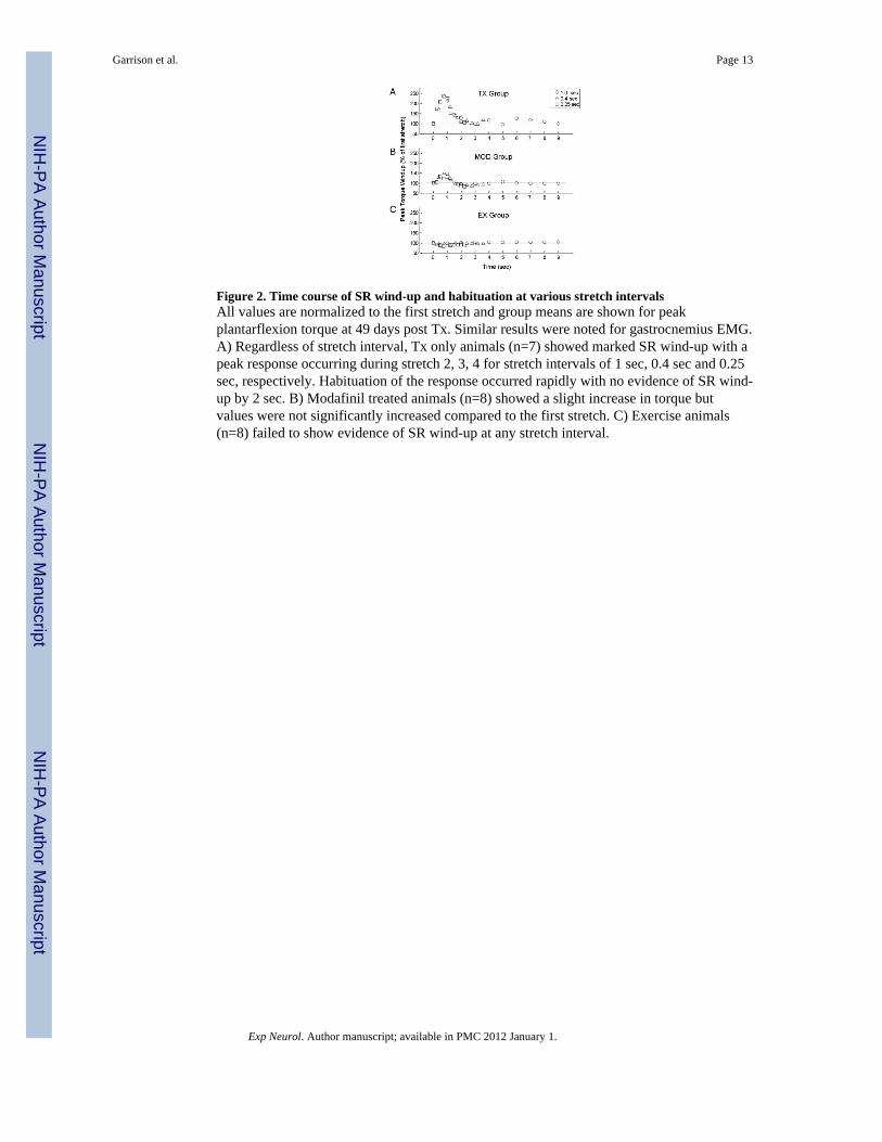
Figure 2. Time course of SR wind-up and habituation at various stretch intervalsAll values are normalized to the first stretch and group means are shown for peakplantarflexion torque at 49 days post Tx. Similar results were noted for gastrocnemius EMG.A) Regardless of stretch interval, Tx only animals (n=7) showed marked SR wind-up with apeak response occurring during stretch 2, 3, 4 for stretch intervals of 1 sec, 0.4 sec and 0.25sec, respectively. Habituation of the response occurred rapidly with no evidence of SR wind-up by 2 sec. B) Modafinil treated animals (n=8) showed a slight increase in torque butvalues were not significantly increased compared to the first stretch. C) Exercise animals(n=8) failed to show evidence of SR wind-up at any stretch interval.
Garrison et al. Page 13
Exp Neurol. Author manuscript; available in PMC 2012 January 1.
NIH
-PA Author Manuscript
NIH
-PA Author Manuscript
NIH
-PA Author Manuscript

Figure 3. Comparison of SR wind-up of integrated EMG and peak torque at 49 days post TxPeak wind-up is shown as a percentage of the first stretch with group means (±SE) plottedfor various stretch intervals. A) Peak plantarflexion torque, and B) integrated gastrocnemiusEMG showed similar patterns across groups. Tx (n=7) animals showed marked SR wind-upat all frequencies of stretch. Ex (n=8) and Mod (n=8) animals were prevented fromdeveloping SR wind-up. Significant differences from the Tx group are noted (*= p<0.05, **= p<0.01, + = p<0.001).
Garrison et al. Page 14
Exp Neurol. Author manuscript; available in PMC 2012 January 1.
NIH
-PA Author Manuscript
NIH
-PA Author Manuscript
NIH
-PA Author Manuscript

Figure 4. Frequency dependent depression of the H-ReflexMean H-reflex amplitude at 0.2, 1, 5 and 10 Hz for transection (Tx, n=3), Exercise (Ex, n=4)and Modafinil treated animals (MOD, n=4) at 49 days post SCI. Data are normalized to 0.2Hz values. Ex and MOD animals underwent 6 weeks of treatment. The Tx group failed todemonstrate normal habituation at higher frequencies. Ex and MOD groups both showedhabituation that was significantly different than the Tx group at 5Hz (*p<0.05) and 10Hz(+p<0.001).
Garrison et al. Page 15
Exp Neurol. Author manuscript; available in PMC 2012 January 1.
NIH
-PA Author Manuscript
NIH
-PA Author Manuscript
NIH
-PA Author Manuscript