Vaccinomics for the major blood feeding helminths of humans
12
Vaccinomics for the Major Blood Feeding Helminths of Humans Alex Loukas, 1 Soraya Gaze, 1 Jason P. Mulvenna, 1 Robin B. Gasser, 2 Paul J. Brindley, 3 Denise L. Doolan, 4 Jeffrey M. Bethony, 3 Malcolm K. Jones, 5 Geoffrey N. Gobert, 4 Patrick Driguez, 4 Donald P. McManus, 4 and Peter J. Hotez 3 Abstract Approximately one billion people are infected with hookworms and/or blood flukes (schistosomes) in devel- oping countries. These two parasites are responsible for more disability adjusted life years lost than most other neglected tropical diseases (NTDs), and together, are second only to malaria. Although anthelmintic drugs are effective and widely available, they do not protect against reinfection, resistant parasites are likely to emerge, and mass drug administration programs are unsustainable. Therefore, there is a pressing need for the devel- opment of vaccines against these parasites. In recent years, there have been major advances in our under- standing of hookworms and schistosomes at the molecular level through the use of ‘‘omics’’ technologies. The secretomes of these parasites have been characterized using transcriptomics, genomics, proteomics, and newly developed gene manipulation and silencing techniques, and the proteins of interest are now the target of novel antigen discovery approaches, notably immunomics. This research has resulted in the discovery, development, and early stage clinical trials of subunit vaccines against hookworms and schistosomes. Introduction T he genomics revolution has enormous implications for public health (Burke et al., 2010). In this review we focus on recent advances in the ‘‘omics’’ for neglected tropical dis- eases (NTDs), and discuss how these advances will facilitate the development of new and sustainable control methods for NTDs. Hookworms and blood flukes (schistosomes) are some the most important parasites of humans in terms of their global health impact on children, pregnant women, and people engaged in subsistence farming (Hotez et al., 2008a, 2008b). Together, their disease burdens exceed those of all other NTDs (Hotez et al., 2010). Because hookworms and schistosomes typically impact human health without result- ing in mortality, they have not gotten the full attention of the global health policymakers and have not been prioritized ei- ther for large-scale control efforts or for research funding. However, when the chronic morbidities due to hookworm disease and schistosomiasis are fully considered based on disability-adjusted life years (DALYs) lost, these diseases to- gether rank among the most important in developing coun- tries, resulting in an annual loss of between 4.5 and 92 million DALYs (Hotez et al., 2010; King and Dangerfield-Cha, 2008). Both infections/diseases can be treated with anthelmintic drugs, but this approach does not protect against the high frequency of reinfection with the parasites (Hotez et al., 2010). Moreover, single-dose mebendazole has been shown to ex- hibit poor efficacy for hookworm and after repeated ad- ministration in the same endemic population (Keiser and Utzinger, 2008), efficacy has been reported to diminish over time (Albonico et al., 2003), raising concerns about possible drug resistance. Indeed, drug failures have arisen with benzimidazoles in livestock (Geerts and Gryseels, 2000). Importantly, the mainstay of control for schistosomiasis is the quinoline compound, praziquantel (PZQ). There are concerns that mass administration of PZQ is unsustainable because of lower than expected efficacies of single-dose PZQ, and high rates of reinfection after treatment, pointing toward the potential emergence of anthelminthic drug resistance (Clements et al., 2009). Indeed, PZQ resistance has already been documented in Schistosoma mansoni in the laboratory (Fallon and Doenhoff, 1994). Mass drug administration programs are therefore inadequate in isolation, cannot hope to advance global elimination efforts, and new tools and 1 Queensland Tropical Health Alliance, James Cook University, Cairns, Queensland, Australia. 2 Parasite Genomics Program, Department of Veterinary Science, The University of Melbourne, Melbourne, Victoria, Australia. 3 Department of Microbiology, Immunology and Tropical Medicine, George Washington University, Washington, DC. 4 Division of Infectious Diseases and Immunology, Queensland Institute of Medical Research, Brisbane, Queensland, Australia. 5 School of Veterinary Sciences, The University of Queensland, Brisbane, Queensland, Australia. OMICS A Journal of Integrative Biology Volume 15, Number 9, 2011 ª Mary Ann Liebert, Inc. DOI: 10.1089/omi.2010.0150 567
Transcript of Vaccinomics for the major blood feeding helminths of humans
Vaccinomics for the Major Blood FeedingHelminths of Humans
Alex Loukas,1 Soraya Gaze,1 Jason P. Mulvenna,1 Robin B. Gasser,2 Paul J. Brindley,3 Denise L. Doolan,4
Jeffrey M. Bethony,3 Malcolm K. Jones,5 Geoffrey N. Gobert,4 Patrick Driguez,4
Donald P. McManus,4 and Peter J. Hotez3
Abstract
Approximately one billion people are infected with hookworms and/or blood flukes (schistosomes) in devel-oping countries. These two parasites are responsible for more disability adjusted life years lost than most otherneglected tropical diseases (NTDs), and together, are second only to malaria. Although anthelmintic drugs areeffective and widely available, they do not protect against reinfection, resistant parasites are likely to emerge,and mass drug administration programs are unsustainable. Therefore, there is a pressing need for the devel-opment of vaccines against these parasites. In recent years, there have been major advances in our under-standing of hookworms and schistosomes at the molecular level through the use of ‘‘omics’’ technologies. Thesecretomes of these parasites have been characterized using transcriptomics, genomics, proteomics, and newlydeveloped gene manipulation and silencing techniques, and the proteins of interest are now the target of novelantigen discovery approaches, notably immunomics. This research has resulted in the discovery, development,and early stage clinical trials of subunit vaccines against hookworms and schistosomes.
Introduction
The genomics revolution has enormous implications forpublic health (Burke et al., 2010). In this review we focus
on recent advances in the ‘‘omics’’ for neglected tropical dis-eases (NTDs), and discuss how these advances will facilitatethe development of new and sustainable control methods forNTDs. Hookworms and blood flukes (schistosomes) are somethe most important parasites of humans in terms of theirglobal health impact on children, pregnant women, andpeople engaged in subsistence farming (Hotez et al., 2008a,2008b). Together, their disease burdens exceed those of allother NTDs (Hotez et al., 2010). Because hookworms andschistosomes typically impact human health without result-ing in mortality, they have not gotten the full attention of theglobal health policymakers and have not been prioritized ei-ther for large-scale control efforts or for research funding.However, when the chronic morbidities due to hookwormdisease and schistosomiasis are fully considered based ondisability-adjusted life years (DALYs) lost, these diseases to-gether rank among the most important in developing coun-tries, resulting in an annual loss of between 4.5 and 92 million
DALYs (Hotez et al., 2010; King and Dangerfield-Cha, 2008).Both infections/diseases can be treated with anthelminticdrugs, but this approach does not protect against the highfrequency of reinfection with the parasites (Hotez et al., 2010).Moreover, single-dose mebendazole has been shown to ex-hibit poor efficacy for hookworm and after repeated ad-ministration in the same endemic population (Keiser andUtzinger, 2008), efficacy has been reported to diminish overtime (Albonico et al., 2003), raising concerns about possibledrug resistance. Indeed, drug failures have arisen withbenzimidazoles in livestock (Geerts and Gryseels, 2000).Importantly, the mainstay of control for schistosomiasis isthe quinoline compound, praziquantel (PZQ). There areconcerns that mass administration of PZQ is unsustainablebecause of lower than expected efficacies of single-dose PZQ,and high rates of reinfection after treatment, pointing towardthe potential emergence of anthelminthic drug resistance(Clements et al., 2009). Indeed, PZQ resistance has alreadybeen documented in Schistosoma mansoni in the laboratory(Fallon and Doenhoff, 1994). Mass drug administrationprograms are therefore inadequate in isolation, cannot hopeto advance global elimination efforts, and new tools and
1Queensland Tropical Health Alliance, James Cook University, Cairns, Queensland, Australia.2Parasite Genomics Program, Department of Veterinary Science, The University of Melbourne, Melbourne, Victoria, Australia.3Department of Microbiology, Immunology and Tropical Medicine, George Washington University, Washington, DC.4Division of Infectious Diseases and Immunology, Queensland Institute of Medical Research, Brisbane, Queensland, Australia.5School of Veterinary Sciences, The University of Queensland, Brisbane, Queensland, Australia.
OMICS A Journal of Integrative BiologyVolume 15, Number 9, 2011ª Mary Ann Liebert, Inc.DOI: 10.1089/omi.2010.0150
567
integrated approaches are needed to ensure sustainablecontrol of schistosomiasis (Gray et al., 2010). Vaccines are anessential component for the long-term control, elimination,or eradication of infectious diseases. Here, we describe theuse of various ‘‘omics’’ approaches to characterize the tran-scriptomes, genomes, proteomes, and glycomes of schisto-somes and hookworms, as well as the postgenomicmolecular applications of these data to identify the mostsuitable target antigens for vaccine development to combatthe diseases they cause.
The Parasites
Hookworms
Human infections with hookworms occur predominantlyin areas of rural poverty in developing countries (Hotez et al.,2004). An estimated 600–700 million people are infected withhookworms worldwide, mostly in sub-Saharan Africa, SouthAmerica, and parts of Asia, notably Indonesia, Bangladesh,and India. Approximately 85% of hookworm infections arecaused by Necator americanus, with the remainder caused byAncylostoma duodenale (Hotez et al., 2004). The infective stageis the microscopic, infective larva (third-stage larva or L3),which lives in the soil until it comes in contact with humanskin. Larvae actively penetrate the skin and migrate withinthe afferent vasculature to the lungs where they ascend thepulmonary tree to the pharynx, are swallowed, and molt tobecome adult male and female worms (approximately 1 cm inlength). Adult hookworms attach to the mucosa and sub-mucosa of the small intestine where they rupture capillariesand arterioles and feed on the extravasated blood (Brookeret al., 2004). The subsequent steps in the acquisition of thehookworm’s blood meal are a major target of current vacci-nomic strategies and will be discussed in this review. Almostall of the pathological effects and morbidity due to hookworminfections result from intestinal blood loss (Hotez et al., 2004).Female and male hookworms mate in the small intestine, thefemale releases microscopic eggs that exit the body in hostfeces. The eggs hatch in the environment, releasing first-stagelarvae, which feed on bacteria and other organic debris in thesoil before they molt twice to develop through to the en-sheathed L3 stage.
Schistosomes
In contrast, schistosome flatworms (platyhelminths), alsoknown as trematodes or flukes, cause approximately 207million cases of human schistosomiasis worldwide, mostly insub-Saharan Africa (Steinmann et al., 2006). However, someestimates indicate that as many as 400 million people are af-fected (King, 2010). In Africa, Schistosoma haematobium is themost prevalent human schistosome; it causes urinary tractschistosomiasis, comprising approximately two-thirds of theworld’s cases of schistosomiasis, whereas S. mansoni is theprincipal cause of intestinal schistosomiasis and is responsiblefor approximately one-third of all cases. S. mansoni also causesschistosomiasis in Latin America, with most of the cases oc-curring in Brazil, while S. japonicum and S. mekongi causeapproximately one million cases of intestinal schistosomiasisin East Asia (Steinmann et al., 2006). Schistosomiasis istransmitted through contact with freshwater containing theinfective free-swimming microscopic cercariae, which like
hookworm L3, actively penetrate the skin of their human host.Cercariae that have entered human skin shed their tails tobecome schistosomula, which enter the vasculature and lungsbefore relocating to the venous system where they becomesexually mature adults, pair, then mate, and egg productionbegins (Gryseels et al., 2006). Adults of S. haematobium migrateto the venous plexus that drains the bladder and reproductiveorgans, whereas, for example, S. mansoni and S. japonicummigrate to the mesenteric veins draining the intestine (Gry-seels et al., 2006). Female schistosomes produce eggs, eachequipped with a spine that helps to facilitate penetrationthrough blood vessels and into the urinary tract and geni-tals (S. haematobium) or intestine and liver (S. mansoni andS. japonicum). Much of the pathological effects from schisto-somiasis are a result of the immune response to parasite eggstrapped in host tissues during chronic infection. The resultinggranulomata become fibrotic, which in turn, results in severecirculatory impairment in the affected organs (Pearce andMacDonald, 2002). In both Brazil and in sub-Saharan Africacoinfections with hookworms and schistosomes are com-monly encountered (Hotez et al., 2010).
Genomics and Transcriptomics
Schistosomes
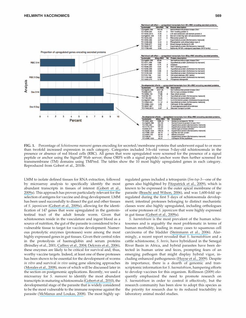
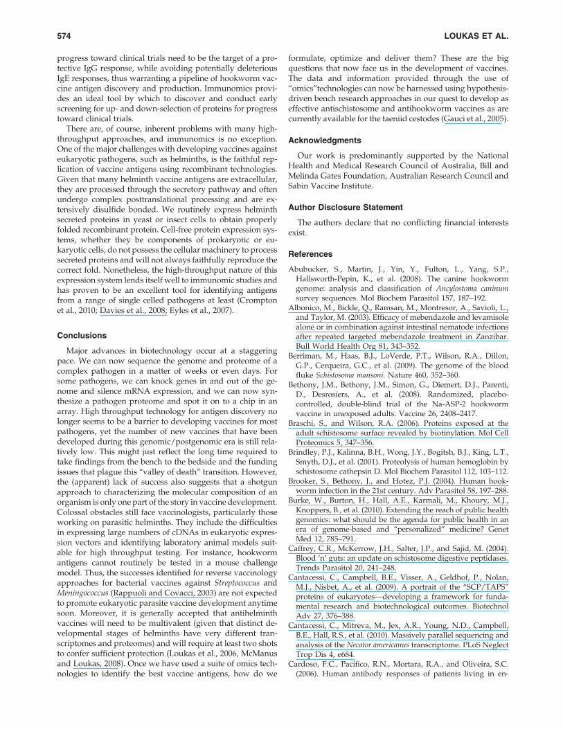
Draft genome sequences were recently published forS. mansoni and S. japonicum (Berriman et al., 2009; The Schis-tosoma japonicum Genome Sequencing and FunctionalAnalysis Consortium, 2009). There is very little annotatedsequence data available for S. haematobium, despite it beingwidely considered as the most important of the schistosomespecies in terms of prevalence and pathogenicity (Rollinson,2009). For the purposes of this review, we will focus primarilyon S. mansoni. The haploid S. mansoni genome is approxima-tely 363 Mb in size and contains almost 12,000 genes, dis-tributed across seven autosomal plus sex chromosome pairs(ZW in the female and ZZ in the male) (Berriman et al., 2009).Prior to the sequencing of the genomes, comprehensivetranscriptomic datasets (Verjovski-Almeida et al., 2003) andgenetic maps (Criscione et al., 2009) were published, provid-ing scaffolds with which to assemble the genome sequences.DNA microarrays have been generated by multiple groupsand utilized to explore transcriptional profiles of differentdevelopmental stages of the parasite (Fitzpatrick et al., 2009;Gobert et al., 2009b, 2010; Jolly et al., 2007) (Fig. 1) and thetranscriptional effects of different treatments (Gobert, 2010;Gobert et al., 2010). Fitzpatrick and colleagues (Fitzpatricket al., 2009) conducted a thorough characterization of schis-tosome development using statistical and network-basedexploratory analyses, and highlighted key transcriptionalchanges associated with life cycle progression and identifiednumerous candidate molecules for drug and vaccine devel-opment, including membrane spanning proteins, such as theG-protein coupled receptors and tetraspanins.
Schistosomes are acoelomate organisms (i.e., which lack adefined body cavity), and have parenchymal tissues sur-rounding their organs, making the manual dissection of or-gans or organ systems difficult. Recent advances in lasermicrodissection microscopy (LMM), however, has allowedresearchers to start to assemble a gene atlas for schistosomes,whereby the transcriptional profiles of defined schistosometissues or organs have been delineated using a combination of
568 LOUKAS ET AL.
LMM to isolate defined tissues for RNA extraction, followedby microarray analysis to specifically identify the mostabundant transcripts in tissues of interest (Gobert et al.,2009a). This approach has proven particularly relevant for theselection of antigens for vaccine and drug development. LMMhas been used successfully to dissect the gut and other tissuesof S. japonicum (Gobert et al., 2009a), allowing for the identi-fication of 147 genes that were upregulated in the gastroin-testinal tract of the adult female worm. Given thatschistosomes reside in the vasculature and ingest blood as asource of nutrition, the gut of the parasite is considered to be avulnerable tissue to target for vaccine development. Numer-ous proteolytic enzymes (proteases) were among the mosthighly expressed genes in gut tissues. Given their central rolesin the proteolysis of haemoglobin and serum proteins(Brindley et al., 2001; Caffrey et al., 2004; Delcroix et al., 2006),these enzymes are likely to be critical for survival and, thus,worthy vaccine targets. Indeed, at least one of these proteaseshas been shown to be essential for the development of wormsin vitro and survival in vivo using gene silencing approaches(Morales et al., 2008), more of which will be discussed later inthe section on postgenomic applications. Recently, we used amicroarray for S. mansoni to identify the most abundanttranscripts in maturing schistosomula (Gobert et al., 2010), thedevelopmental stage of the parasite that is widely consideredto be the most vulnerable to the immune response against theparasite (McManus and Loukas, 2008). The most highly up-
regulated genes included a tetraspanin (Sm-tsp-3—one of thegenes also highlighted by Fitzpatrick et al., 2009), which isknown to be expressed in the outer apical membrane of theparasite (Braschi and Wilson, 2006), and was 1,600-fold up-regulated during the first 5 days of schistosomula develop-ment; intestinal proteases belonging to distinct mechanisticclasses were also highly upregulated, including orthologuesof some proteases of S. japonicum that were highly expressedin gut tissue (Gobert et al., 2009a).
S. haemtobium is the most prevalent of the human schis-tosomes and is arguably the most problematic in terms ofhuman morbidity, leading in many cases to squamous cellcarcinoma of the bladder (Steinmann et al., 2006). Alar-mingly, a recent report revealed that S. haematobium and acattle schistosome, S. bovis, have hybridized in the SenegalRiver Basin in Africa, and hybrid parasites have been de-tected in human urine and feces, prompting fears of anemerging pathogen that might display hybrid vigor, in-cluding enhanced pathogenesis (Huyse et al., 2009). Despiteits importance, there is a dearth of genomic and tran-scriptomic information for S. haematobium, hampering effortsto develop vaccines for this organism. Rollinson (2009) elo-quently emphasized the need to promote research onS. haematobium in order to control it effectively, but theresearch community has been slow to adopt this species asthe priority for research due to its reduced tractability inlaboratory animal model studies.
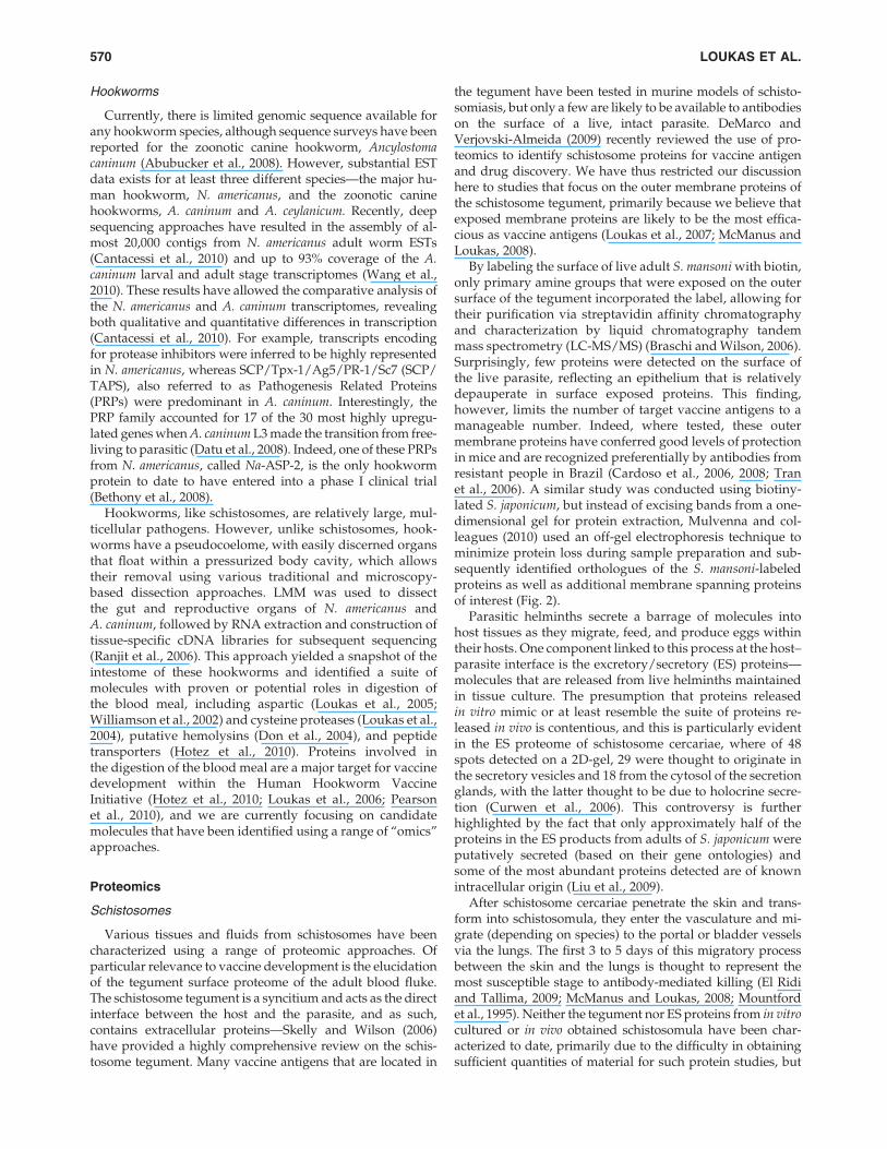
FIG. 1. Percentage of Schistosoma mansoni genes encoding for secreted/membrane proteins that underwent equal to or morethan twofold increased expression in each category. Categories included 3-h-old versus 5-day-old schistosomula in thepresence or absence of red blood cells (RBC). All genes that were upregulated were screened for the presence of a signalpeptide or anchor using the SignalP Web server; those ORFS with a signal peptide/anchor were then further screened fortransmembrane (TM) domains using TMPred. The tables show the 10 most highly upregulated genes in each category.Reproduced from Gobert et al., 2010b.
HELMINTH VACCINOMICS 569
Hookworms
Currently, there is limited genomic sequence available forany hookworm species, although sequence surveys have beenreported for the zoonotic canine hookworm, Ancylostomacaninum (Abubucker et al., 2008). However, substantial ESTdata exists for at least three different species—the major hu-man hookworm, N. americanus, and the zoonotic caninehookworms, A. caninum and A. ceylanicum. Recently, deepsequencing approaches have resulted in the assembly of al-most 20,000 contigs from N. americanus adult worm ESTs(Cantacessi et al., 2010) and up to 93% coverage of the A.caninum larval and adult stage transcriptomes (Wang et al.,2010). These results have allowed the comparative analysis ofthe N. americanus and A. caninum transcriptomes, revealingboth qualitative and quantitative differences in transcription(Cantacessi et al., 2010). For example, transcripts encodingfor protease inhibitors were inferred to be highly representedin N. americanus, whereas SCP/Tpx-1/Ag5/PR-1/Sc7 (SCP/TAPS), also referred to as Pathogenesis Related Proteins(PRPs) were predominant in A. caninum. Interestingly, thePRP family accounted for 17 of the 30 most highly upregu-lated genes when A. caninum L3 made the transition from free-living to parasitic (Datu et al., 2008). Indeed, one of these PRPsfrom N. americanus, called Na-ASP-2, is the only hookwormprotein to date to have entered into a phase I clinical trial(Bethony et al., 2008).
Hookworms, like schistosomes, are relatively large, mul-ticellular pathogens. However, unlike schistosomes, hook-worms have a pseudocoelome, with easily discerned organsthat float within a pressurized body cavity, which allowstheir removal using various traditional and microscopy-based dissection approaches. LMM was used to dissectthe gut and reproductive organs of N. americanus andA. caninum, followed by RNA extraction and construction oftissue-specific cDNA libraries for subsequent sequencing(Ranjit et al., 2006). This approach yielded a snapshot of theintestome of these hookworms and identified a suite ofmolecules with proven or potential roles in digestion ofthe blood meal, including aspartic (Loukas et al., 2005;Williamson et al., 2002) and cysteine proteases (Loukas et al.,2004), putative hemolysins (Don et al., 2004), and peptidetransporters (Hotez et al., 2010). Proteins involved inthe digestion of the blood meal are a major target for vaccinedevelopment within the Human Hookworm VaccineInitiative (Hotez et al., 2010; Loukas et al., 2006; Pearsonet al., 2010), and we are currently focusing on candidatemolecules that have been identified using a range of ‘‘omics’’approaches.
Proteomics
Schistosomes
Various tissues and fluids from schistosomes have beencharacterized using a range of proteomic approaches. Ofparticular relevance to vaccine development is the elucidationof the tegument surface proteome of the adult blood fluke.The schistosome tegument is a syncitium and acts as the directinterface between the host and the parasite, and as such,contains extracellular proteins—Skelly and Wilson (2006)have provided a highly comprehensive review on the schis-tosome tegument. Many vaccine antigens that are located in
the tegument have been tested in murine models of schisto-somiasis, but only a few are likely to be available to antibodieson the surface of a live, intact parasite. DeMarco andVerjovski-Almeida (2009) recently reviewed the use of pro-teomics to identify schistosome proteins for vaccine antigenand drug discovery. We have thus restricted our discussionhere to studies that focus on the outer membrane proteins ofthe schistosome tegument, primarily because we believe thatexposed membrane proteins are likely to be the most effica-cious as vaccine antigens (Loukas et al., 2007; McManus andLoukas, 2008).
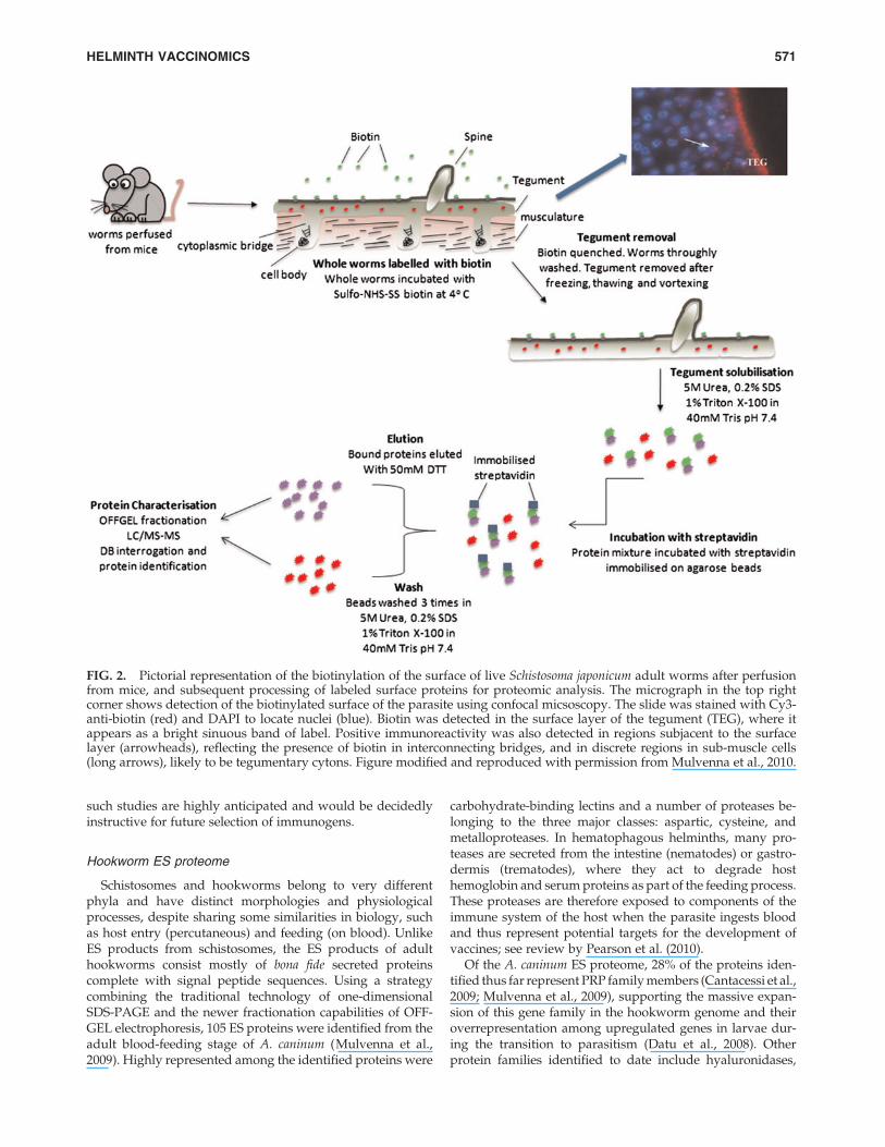
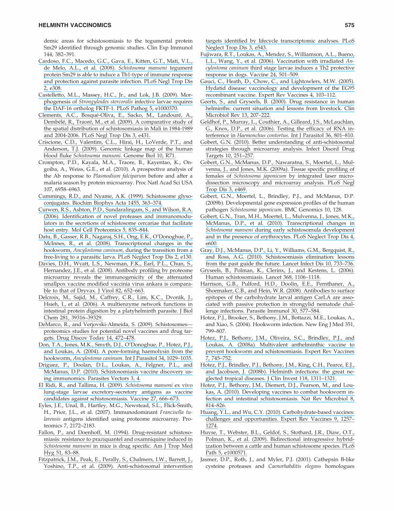
By labeling the surface of live adult S. mansoni with biotin,only primary amine groups that were exposed on the outersurface of the tegument incorporated the label, allowing fortheir purification via streptavidin affinity chromatographyand characterization by liquid chromatography tandemmass spectrometry (LC-MS/MS) (Braschi and Wilson, 2006).Surprisingly, few proteins were detected on the surface ofthe live parasite, reflecting an epithelium that is relativelydepauperate in surface exposed proteins. This finding,however, limits the number of target vaccine antigens to amanageable number. Indeed, where tested, these outermembrane proteins have conferred good levels of protectionin mice and are recognized preferentially by antibodies fromresistant people in Brazil (Cardoso et al., 2006, 2008; Tranet al., 2006). A similar study was conducted using biotiny-lated S. japonicum, but instead of excising bands from a one-dimensional gel for protein extraction, Mulvenna and col-leagues (2010) used an off-gel electrophoresis technique tominimize protein loss during sample preparation and sub-sequently identified orthologues of the S. mansoni-labeledproteins as well as additional membrane spanning proteinsof interest (Fig. 2).
Parasitic helminths secrete a barrage of molecules intohost tissues as they migrate, feed, and produce eggs withintheir hosts. One component linked to this process at the host–parasite interface is the excretory/secretory (ES) proteins—molecules that are released from live helminths maintainedin tissue culture. The presumption that proteins releasedin vitro mimic or at least resemble the suite of proteins re-leased in vivo is contentious, and this is particularly evidentin the ES proteome of schistosome cercariae, where of 48spots detected on a 2D-gel, 29 were thought to originate inthe secretory vesicles and 18 from the cytosol of the secretionglands, with the latter thought to be due to holocrine secre-tion (Curwen et al., 2006). This controversy is furtherhighlighted by the fact that only approximately half of theproteins in the ES products from adults of S. japonicum wereputatively secreted (based on their gene ontologies) andsome of the most abundant proteins detected are of knownintracellular origin (Liu et al., 2009).
After schistosome cercariae penetrate the skin and trans-form into schistosomula, they enter the vasculature and mi-grate (depending on species) to the portal or bladder vesselsvia the lungs. The first 3 to 5 days of this migratory processbetween the skin and the lungs is thought to represent themost susceptible stage to antibody-mediated killing (El Ridiand Tallima, 2009; McManus and Loukas, 2008; Mountfordet al., 1995). Neither the tegument nor ES proteins from in vitrocultured or in vivo obtained schistosomula have been char-acterized to date, primarily due to the difficulty in obtainingsufficient quantities of material for such protein studies, but
570 LOUKAS ET AL.
such studies are highly anticipated and would be decidedlyinstructive for future selection of immunogens.
Hookworm ES proteome
Schistosomes and hookworms belong to very differentphyla and have distinct morphologies and physiologicalprocesses, despite sharing some similarities in biology, suchas host entry (percutaneous) and feeding (on blood). UnlikeES products from schistosomes, the ES products of adulthookworms consist mostly of bona fide secreted proteinscomplete with signal peptide sequences. Using a strategycombining the traditional technology of one-dimensionalSDS-PAGE and the newer fractionation capabilities of OFF-GEL electrophoresis, 105 ES proteins were identified from theadult blood-feeding stage of A. caninum (Mulvenna et al.,2009). Highly represented among the identified proteins were
carbohydrate-binding lectins and a number of proteases be-longing to the three major classes: aspartic, cysteine, andmetalloproteases. In hematophagous helminths, many pro-teases are secreted from the intestine (nematodes) or gastro-dermis (trematodes), where they act to degrade hosthemoglobin and serum proteins as part of the feeding process.These proteases are therefore exposed to components of theimmune system of the host when the parasite ingests bloodand thus represent potential targets for the development ofvaccines; see review by Pearson et al. (2010).
Of the A. caninum ES proteome, 28% of the proteins iden-tified thus far represent PRP family members (Cantacessi et al.,2009; Mulvenna et al., 2009), supporting the massive expan-sion of this gene family in the hookworm genome and theiroverrepresentation among upregulated genes in larvae dur-ing the transition to parasitism (Datu et al., 2008). Otherprotein families identified to date include hyaluronidases,
FIG. 2. Pictorial representation of the biotinylation of the surface of live Schistosoma japonicum adult worms after perfusionfrom mice, and subsequent processing of labeled surface proteins for proteomic analysis. The micrograph in the top rightcorner shows detection of the biotinylated surface of the parasite using confocal micsoscopy. The slide was stained with Cy3-anti-biotin (red) and DAPI to locate nuclei (blue). Biotin was detected in the surface layer of the tegument (TEG), where itappears as a bright sinuous band of label. Positive immunoreactivity was also detected in regions subjacent to the surfacelayer (arrowheads), reflecting the presence of biotin in interconnecting bridges, and in discrete regions in sub-muscle cells(long arrows), likely to be tegumentary cytons. Figure modified and reproduced with permission from Mulvenna et al., 2010.
HELMINTH VACCINOMICS 571
lysozyme-like proteins, and transthyretin-like proteins, all ofwhich are vaccine candidates but have yet to be assessed forefficacy in animal models.
Hookworm intestome
Targeting the hookworm gut for vaccine development hasshown to be a worthwhile pursuit (Hotez et al., 2010; Loukaset al., 2006). Other blood-feeding parasites, including thebarber’s pole worm of livestock (Haemonchus contortus) andthe cattle tick (Rhipicephalus microplus), are effectively killed byvaccine-induced antibodies against native proteins and pro-tein complexes derived from the guts of these parasites (Smithet al., 1994). Indeed, the gut of adult H. contortus possesses atightly bound multiglycoprotein complex that is rich in di-gestive proteases and protease inhibitors (Knox and Smith,2001), and effective silencing of some of these genes af-fects parasite development, survival, and fecundity in vivo(Samarasinghe et al., 2011). It is not known whether theorthologous hookworm proteases form a similar complex toH-gal-GP, but, with the development of techniques such asLMM ( Jones et al., 2004; Xu, 2010) and tissue profiling viamass spectrometry, the characterization of proteins in mi-croscopic tissues, such as a hookworm gut, is now a possi-bility. Indeed, one of the lead hookworm vaccine antigens isthe intestinal haemoglobinase, Na-APR-1 (a cathepsin D-likeaspartic protease), where the protective effect appears to es-tablish neutralizing antibodies that inhibits substrate prote-olysis (Loukas et al., 2005; Pearson et al., 2009).
Glycomics
The role of helminth glycans as protective antigens is acomplex but extremely important area. Helminths are eu-karyotes and therefore decorate proteins that enter the se-cretory pathway with an array of glycan moieties. For areview on this topic, see Nyame et al. (2004). The difficulty inpurifying or synthesizing large quantities of helminth glycanshas thus far precluded their widespread use in helminthvaccine trials. However, glycans are the basis for some highlyeffective antibacterial vaccines (Huang and Wu, 2010), andsome of the most highly protective proteins from parasitichelminths are indeed glycosylated. For example, the H-gal-GP and H11 antigen complexes from H. contortus are heavilyglycosylated, a feature that permitted their initial purificationand identification of native protein via lectin affinity chro-matographies (Knox and Smith, 2001). Where they have beencharacterized at the molecular level, helminth glycans offergreat promise as vaccine candidates (Cummings and Nyame,1999; Harrison et al., 2008; Maass et al., 2009).
Postgenomics
Schistosomes
One of the most revolutionary advances in the postgenomicera for the study of parasitic helminths is gene silencing byRNA interference (RNAi). Parasitic helminths, by virtue oftheir often complex life cycles and large genomes, have ren-dered themselves refractory to many genetic manipulationtools that allow the exploration of gene function (Mann et al.,2008, 2010). RNAi is now widely used to assess gene functionin schistosomes, and appears to be particularly effective forgenes expressed in tissues readily accessible to dsRNA, such
as the tegument and gastrodermis (Krautz-Peterson et al.,2010; Stefanic et al., 2010). RNAi has been used to confirmgene function for a number of potential schistosome vaccineantigens and drug targets, and helps explain how some vac-cines, which are based on these proteins, might exert theirefficacy. RNAi was used to show that the tetraspanin,Sm-TSP-2, plays important structural roles impacting tegu-ment development, maturation or stability, and is essentialfor survival in vivo (Tran et al., 2010). Silencing of the genesencoding gastrodermal proteases of S. mansoni, includingcathepsin D (Morales et al., 2008), papain-like cysteine pro-teases, and asparaginyl endopeptidase (Delcroix et al., 2006)has confirmed the roles of these proteases and the order inwhich they cleave their substrates during the multienzymedigestive cascade of haemoglobin. The ability to silence aschistosome gene as well as assess the effect in vitro and in vivoprompted us to suggest that the lethality of silencing aschistosome gene be one of the critical steps in ranking anti-gens for progress toward clinical trials for a human schisto-somiasis vaccine (Hotez et al., 2010).
Its obvious utility notwithstanding, RNAi leads to onlytransient gene silencing and, in addition, may be inaccessibleto some developmental stages and/or tissues of schistosomes.RNAi mediated by short hairpin-RNA (shRNA) expressingtransgenes can induce specific and long-term knock down ineukaryotic cells (Paddison et al., 2004). In vivo, for example,vector-based, RNAi approaches that lead to integration oftransgenes encoding cassettes that express short interferingRNAs can circumvent deficiencies with exogenous RNAi byproviding continuous and/or conditional gene silencing(Sliva and Schnierle, 2010). In addition to vector-based RNAi,transgenesis offers the potential to endow other desirablephenotypes including the expression or coexpression of hel-minth or host antigens. Both vector-based RNAi and expres-sion of foreign transgenes have begun to be deployed instudies with schistosomes, with potential outcomes includingvalidation of putative vaccine candidates and even the over-expression of candidate antigens. Pseudotyped murine leu-kemia virus and the piggyBac transposon have both beenshown to transduce developmental stages of schistosomes,leading to chromosomal integration of retroviral transgenesand transgene reporter activity (Kines et al., 2008; Moraleset al., 2007; Yang et al., 2010). Moreover, a vector-based RNAiapproach has been reported in which the MLV transgeneencoded a long hairpin RNA specific for a schistosome pro-tease involved in hemoglobinolysis (Tchoubrieva et al. 2010).
Hookworms
There are limited reports of the application of RNAi tohookworms. The technique has been used to silence (bysoaking) the expression of some genes in the L3 stage of therelated blood-feeding nematode, H. contortus (Geldhof et al.,2006; Kotze and Bagnall, 2006; Samarasinghe et al., 2011;Zawadzki et al., 2006). Successful silencing of genes in para-sitic nematodes is plagued by a lack of reproducibility be-tween and even within laboratories (Viney and Thompson,2008). However, recently, Samarasinghe et al. (2011) showedthat the proximity of the tissue expressing the mRNA of in-terest to the dsRNA being applied played a role in the successof RNAi in H. contortus. They showed that genes for intestinalproteases (and lead vaccine antigens) could be silenced in
572 LOUKAS ET AL.
feeding third-stage larvae by soaking parasites in dsRNA.Presumably the oral uptake of this RNA afforded direct ex-posure to the intestinal cells expressing the mRNAs. On theother hand, genes that were expressed in tissues that were noteasily accessible to ingested dsRNA were not susceptible toRNAi by soaking. The dsRNA-treated larvae were then usedto infect sheep, and silencing of intestinal proteases, such asthe aminopeptidase H11, resulted in reduced survival of adultworms and reduced fecundity of female worms that didsurvive. The ability to silence the expression of intestinalprotease genes in H. contortus is a major advance in the area ofvaccine antigen discovery for blood-feeding nematodes oflivestock and humans—it allows, for the first time, an as-sessment of the function(s) of these proteases in vivo, and,therefore, their roles in parasite survival can potentially beaddressed. For example, questions regarding functional re-dundancy in pathways that involve multigene families, suchas the intestinal cysteine proteases of Haemonchus ( Jasmeret al., 2001) and hookworms (Ranjit et al., 2008), can be ad-dressed.
Like RNAi, transgenesis has yet to be reported in hook-worms. However, progress has been reported with genemanipulation of the related parasitic nematode, Strongyloidesstercoralis, in which transgene approaches developed for usein Caenorhabditis elegans have been employed to demonstratethat morphogenesis of infective L3 from earlier larval formsrequires the DAF-16 orthologue FKTF-1, a forkhead tran-scription factor that regulates dauer larval development in C.elegans (Castelletto et al., 2009).
Immunomics
The availability of the S. mansoni and S. japonicum genomescoupled with the proteomic characterization of the tegumentand ES products has provided researchers with the tools re-quired to apply postgenomic approaches to vaccine antigendiscovery. The availability of high throughput protein ex-pression techniques, such as in vitro translation using pro-karyotic or eukaryotic ribosomes, and sera from resistant
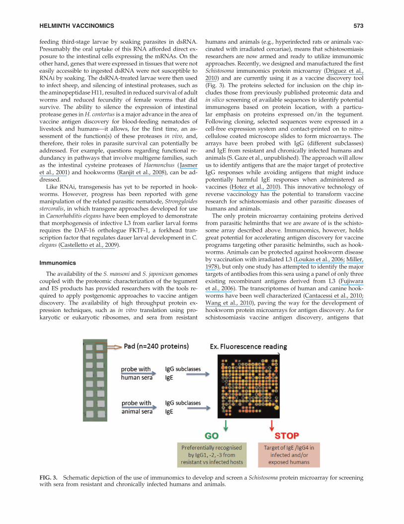
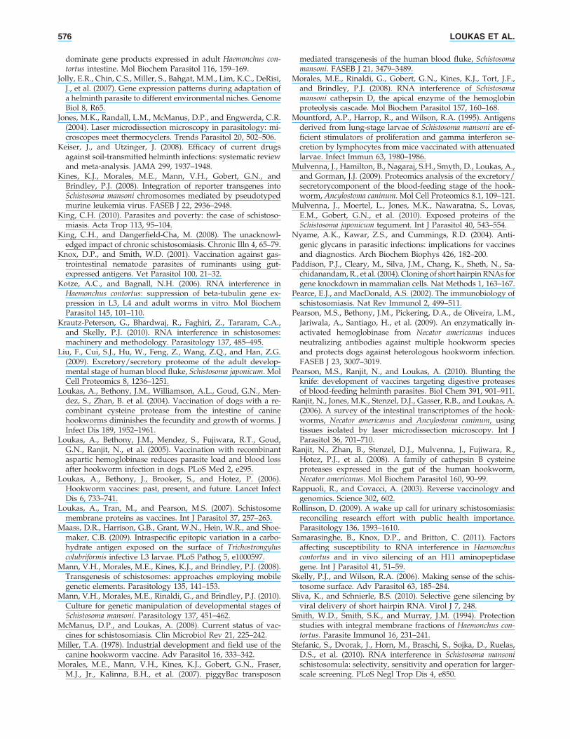
humans and animals (e.g., hyperinfected rats or animals vac-cinated with irradiated cercariae), means that schistosomiasisresearchers are now armed and ready to utilize immunomicapproaches. Recently, we designed and manufactured the firstSchistosoma immunomics protein microarray (Driguez et al.,2010) and are currently using it as a vaccine discovery tool(Fig. 3). The proteins selected for inclusion on the chip in-cludes those from previously published proteomic data andin silico screening of available sequences to identify potentialimmunogens based on protein location, with a particu-lar emphasis on proteins expressed on/in the tegument.Following cloning, selected sequences were expressed in acell-free expression system and contact-printed on to nitro-cellulose coated microscope slides to form microarrays. Thearrays have been probed with IgG (different subclasses)and IgE from resistant and chronically infected humans andanimals (S. Gaze et al., unpublished). The approach will allowus to identify antigens that are the major target of protectiveIgG responses while avoiding antigens that might inducepotentially harmful IgE responses when administered asvaccines (Hotez et al., 2010). This innovative technology ofreverse vaccinology has the potential to transform vaccineresearch for schistosomiasis and other parasitic diseases ofhumans and animals.
The only protein microarray containing proteins derivedfrom parasitic helminths that we are aware of is the schisto-some array described above. Immunomics, however, holdsgreat potential for accelerating antigen discovery for vaccineprograms targeting other parasitic helminths, such as hook-worms. Animals can be protected against hookworm diseaseby vaccination with irradiated L3 (Loukas et al., 2006; Miller,1978), but only one study has attempted to identify the majortargets of antibodies from this sera using a panel of only threeexisting recombinant antigens derived from L3 (Fujiwaraet al., 2006). The transcriptomes of human and canine hook-worms have been well characterized (Cantacessi et al., 2010;Wang et al., 2010), paving the way for the development ofhookworm protein microarrays for antigen discovery. As forschistosomiasis vaccine antigen discovery, antigens that
FIG. 3. Schematic depiction of the use of immunomics to develop and screen a Schistosoma protein microarray for screeningwith sera from resistant and chronically infected humans and animals.
HELMINTH VACCINOMICS 573
progress toward clinical trials need to be the target of a pro-tective IgG response, while avoiding potentially deleteriousIgE responses, thus warranting a pipeline of hookworm vac-cine antigen discovery and production. Immunomics provi-des an ideal tool by which to discover and conduct earlyscreening for up- and down-selection of proteins for progresstoward clinical trials.
There are, of course, inherent problems with many high-throughput approaches, and immunomics is no exception.One of the major challenges with developing vaccines againsteukaryotic pathogens, such as helminths, is the faithful rep-lication of vaccine antigens using recombinant technologies.Given that many helminth vaccine antigens are extracellular,they are processed through the secretory pathway and oftenundergo complex posttranslational processing and are ex-tensively disulfide bonded. We routinely express helminthsecreted proteins in yeast or insect cells to obtain properlyfolded recombinant protein. Cell-free protein expression sys-tems, whether they be components of prokaryotic or eu-karyotic cells, do not possess the cellular machinery to processsecreted proteins and will not always faithfully reproduce thecorrect fold. Nonetheless, the high-throughput nature of thisexpression system lends itself well to immunomic studies andhas proven to be an excellent tool for identifying antigensfrom a range of single celled pathogens at least (Cromptonet al., 2010; Davies et al., 2008; Eyles et al., 2007).
Conclusions
Major advances in biotechnology occur at a staggeringpace. We can now sequence the genome and proteome of acomplex pathogen in a matter of weeks or even days. Forsome pathogens, we can knock genes in and out of the ge-nome and silence mRNA expression, and we can now syn-thesize a pathogen proteome and spot it on to a chip in anarray. High throughput technology for antigen discovery nolonger seems to be a barrier to developing vaccines for mostpathogens, yet the number of new vaccines that have beendeveloped during this genomic/postgenomic era is still rela-tively low. This might just reflect the long time required totake findings from the bench to the bedside and the fundingissues that plague this ‘‘valley of death’’ transition. However,the (apparent) lack of success also suggests that a shotgunapproach to characterizing the molecular composition of anorganism is only one part of the story in vaccine development.Colossal obstacles still face vaccinologists, particularly thoseworking on parasitic helminths. They include the difficultiesin expressing large numbers of cDNAs in eukaryotic expres-sion vectors and identifying laboratory animal models suit-able for high throughput testing. For instance, hookwormantigens cannot routinely be tested in a mouse challengemodel. Thus, the successes identified for reverse vaccinologyapproaches for bacterial vaccines against Streptococcus andMeningococcus (Rappuoli and Covacci, 2003) are not expectedto promote eukaryotic parasite vaccine development anytimesoon. Moreover, it is generally accepted that antihelminthvaccines will need to be multivalent (given that distinct de-velopmental stages of helminths have very different tran-scriptomes and proteomes) and will require at least two shotsto confer sufficient protection (Loukas et al., 2006, McManusand Loukas, 2008). Once we have used a suite of omics tech-nologies to identify the best vaccine antigens, how do we
formulate, optimize and deliver them? These are the bigquestions that now face us in the development of vaccines.The data and information provided through the use of‘‘omics’’technologies can now be harnessed using hypothesis-driven bench research approaches in our quest to develop aseffective antischistosome and antihookworm vaccines as arecurrently available for the taeniid cestodes (Gauci et al., 2005).
Acknowledgments
Our work is predominantly supported by the NationalHealth and Medical Research Council of Australia, Bill andMelinda Gates Foundation, Australian Research Council andSabin Vaccine Institute.
Author Disclosure Statement
The authors declare that no conflicting financial interestsexist.
References
Abubucker, S., Martin, J., Yin, Y., Fulton, L., Yang, S.P.,Hallsworth-Pepin, K., et al. (2008). The canine hookwormgenome: analysis and classification of Ancylostoma caninumsurvey sequences. Mol Biochem Parasitol 157, 187–192.
Albonico, M., Bickle, Q., Ramsan, M., Montresor, A., Savioli, L.,and Taylor, M. (2003). Efficacy of mebendazole and levamisolealone or in combination against intestinal nematode infectionsafter repeated targeted mebendazole treatment in Zanzibar.Bull World Health Org 81, 343–352.
Berriman, M., Haas, B.J., LoVerde, P.T., Wilson, R.A., Dillon,G.P., Cerqueira, G.C., et al. (2009). The genome of the bloodfluke Schistosoma mansoni. Nature 460, 352–360.
Bethony, J.M., Bethony, J.M., Simon, G., Diemert, D.J., Parenti,D., Desrosiers, A., et al. (2008). Randomized, placebo-controlled, double-blind trial of the Na-ASP-2 hookwormvaccine in unexposed adults. Vaccine 26, 2408–2417.
Braschi, S., and Wilson, R.A. (2006). Proteins exposed at theadult schistosome surface revealed by biotinylation. Mol CellProteomics 5, 347–356.
Brindley, P.J., Kalinna, B.H., Wong, J.Y., Bogitsh, B.J., King, L.T.,Smyth, D.J., et al. (2001). Proteolysis of human hemoglobin byschistosome cathepsin D. Mol Biochem Parasitol 112, 103–112.
Brooker, S., Bethony, J., and Hotez, P.J. (2004). Human hook-worm infection in the 21st century. Adv Parasitol 58, 197–288.
Burke, W., Burton, H., Hall, A.E., Karmali, M., Khoury, M.J.,Knoppers, B., et al. (2010). Extending the reach of public healthgenomics: what should be the agenda for public health in anera of genome-based and ‘‘personalized’’ medicine? GenetMed 12, 785–791.
Caffrey, C.R., McKerrow, J.H., Salter, J.P., and Sajid, M. (2004).Blood ’n’ guts: an update on schistosome digestive peptidases.Trends Parasitol 20, 241–248.
Cantacessi, C., Campbell, B.E., Visser, A., Geldhof, P., Nolan,M.J., Nisbet, A., et al. (2009). A portrait of the ‘‘SCP/TAPS’’proteins of eukaryotes—developing a framework for funda-mental research and biotechnological outcomes. BiotechnolAdv 27, 376–388.
Cantacessi, C., Mitreva, M., Jex, A.R., Young, N.D., Campbell,B.E., Hall, R.S., et al. (2010). Massively parallel sequencing andanalysis of the Necator americanus transcriptome. PLoS NeglectTrop Dis 4, e684.
Cardoso, F.C., Pacifico, R.N., Mortara, R.A., and Oliveira, S.C.(2006). Human antibody responses of patients living in en-
574 LOUKAS ET AL.
demic areas for schistosomiasis to the tegumental proteinSm29 identified through genomic studies. Clin Exp Immunol144, 382–391.
Cardoso, F.C., Macedo, G.C., Gava, E., Kitten, G.T., Mati, V.L.,de Melo, A.L., et al. (2008). Schistosoma mansoni tegumentprotein Sm29 is able to induce a Th1-type of immune responseand protection against parasite infection. PLoS Negl Trop Dis2, e308.
Castelletto, M.L., Massey, H.C., Jr., and Lok, J.B. (2009). Mor-phogenesis of Strongyloides stercoralis infective larvae requiresthe DAF-16 ortholog FKTF-1. PLoS Pathog 5, e1000370.
Clements, A.C., Bosque-Oliva, E., Sacko, M., Landoure, A.,Dembele, R., Traore, M., et al. (2009). A comparative study ofthe spatial distribution of schistosomiasis in Mali in 1984-1989and 2004-2006. PLoS Negl Trop Dis 3, e431.
Criscione, C.D., Valentim, C.L., Hirai, H., LoVerde, P.T., andAnderson, T.J. (2009). Genomic linkage map of the humanblood fluke Schistosoma mansoni. Genome Biol 10, R71.
Crompton, P.D., Kayala, M.A., Traore, B., Kayentao, K., On-goiba, A., Weiss, G.E., et al. (2010). A prospective analysis ofthe Ab response to Plasmodium falciparum before and after amalaria season by protein microarray. Proc Natl Acad Sci USA107, 6958–6963.
Cummings, R.D., and Nyame, A.K. (1999). Schistosome glyso-conjugates. Biochim Biophys Acta 1455, 363–374.
Curwen, R.S., Ashton, P.D., Sundaralingam, S., and Wilson, R.A.(2006). Identification of novel proteases and immunomodu-lators in the secretions of schistosome cercariae that facilitatehost entry. Mol Cell Proteomics 5, 835–844.
Datu, B., Gasser, R.B., Nagaraj, S.H., Ong, E.K., O’Donoghue, P.,McInnes, R., et al. (2008). Transcriptional changes in thehookworm, Ancylostoma caninum, during the transition from afree-living to a parasitic larva. PLoS Neglect Trop Dis 2, e130.
Davies, D.H., Wyatt, L.S., Newman, F.K., Earl, P.L., Chun, S.,Hernandez, J.E., et al. (2008). Antibody profiling by proteomemicroarray reveals the immunogenicity of the attenuatedsmallpox vaccine modified vaccinia virus ankara is compara-ble to that of Dryvax. J Virol 82, 652–663.
Delcroix, M., Sajid, M., Caffrey, C.R., Lim, K.C., Dvorak, J.,Hsieh, I., et al. (2006). A multienzyme network functions inintestinal protein digestion by a platyhelminth parasite. J BiolChem 281, 39316–39329.
DeMarco, R., and Verjovski-Almeida, S. (2009). Schistosomes—proteomics studies for potential novel vaccines and drug tar-gets. Drug Discov Today 14, 472–478.
Don, T.A., Jones, M.K., Smyth, D.J., O’Donoghue, P., Hotez, P.J.,and Loukas, A. (2004). A pore-forming haemolysin from thehookworm, Ancylostoma caninum. Int J Parasitol 34, 1029–1035.
Driguez, P., Doolan, D.L., Loukas, A., Felgner, P.L., andMcManus, D.P. (2010). Schistosomiasis vaccine discovery us-ing immunomics. Parasites Vectors 3, 4.
El Ridi, R., and Tallima, H. (2009). Schistosoma mansoni ex vivolung-stage larvae excretory-secretory antigens as vaccinecandidates against schistosomiasis. Vaccine 27, 666–673.
Eyles, J.E., Unal, B., Hartley, M.G., Newstead, S.L., Flick-Smith,H., Prior, J.L., et al. (2007). Immunodominant Francisella tu-larensis antigens identified using proteome microarray. Pro-teomics 7, 2172–2183.
Fallon, P., and Doenhoff, M. (1994). Drug-resistant schistoso-miasis: resistance to praziquantel and oxamniquine induced inSchistosoma mansoni in mice is drug specific. Am J Trop MedHyg 51, 83–88.
Fitzpatrick, J.M., Peak, E., Perally, S., Chalmers, I.W., Barrett, J.,Yoshino, T.P., et al. (2009). Anti-schistosomal intervention
targets identified by lifecycle transcriptomic analyses. PLoSNeglect Trop Dis 3, e543.
Fujiwara, R.T., Loukas, A., Mendez, S., Williamson, A.L., Bueno,L.L., Wang, Y., et al. (2006). Vaccination with irradiated An-cylostoma caninum third stage larvae induces a Th2 protectiveresponse in dogs. Vaccine 24, 501–509.
Gauci, C., Heath, D., Chow, C., and Lightowlers, M.W. (2005).Hydatid disease: vaccinology and development of the EG95recombinant vaccine. Expert Rev Vaccines 4, 103–112.
Geerts, S., and Gryseels, B. (2000). Drug resistance in humanhelminths: current situation and lessons from livestock. ClinMicrobiol Rev 13, 207–222.
Geldhof, P., Murray, L., Couthier, A., Gilleard, J.S., McLauchlan,G., Knox, D.P., et al. (2006). Testing the efficacy of RNA in-terference in Haemonchus contortus. Int J Parasitol 36, 801–810.
Gobert, G.N. (2010). Better understanding of anti-schistosomalstrategies through microarray analysis. Infect Disord DrugTargets 10, 251–257.
Gobert, G.N., McManus, D.P., Nawaratna, S., Moertel, L., Mul-venna, J., and Jones, M.K. (2009a). Tissue specific profiling offemales of Schistosoma japonicum by integrated laser micro-dissection microscopy and microarray analysis. PLoS NeglTrop Dis 3, e469.
Gobert, G.N., Moertel, L., Brindley, P.J., and McManus, D.P.(2009b). Developmental gene expression profiles of the humanpathogen Schistosoma japonicum. BMC Genomics 10, 128.
Gobert, G.N., Tran, M.H., Moertel, L., Mulvenna, J., Jones, M.K.,McManus, D.P., et al. (2010). Transcriptional changes inSchistosoma mansoni during early schistosomula developmentand in the presence of erythrocytes. PLoS Neglect Trop Dis 4,e600.
Gray, D.J., McManus, D.P., Li, Y., Williams, G.M., Bergquist, R.,and Ross, A.G. (2010). Schistosomiasis elimination: lessonsfrom the past guide the future. Lancet Infect Dis 10, 733–736.
Gryseels, B., Polman, K., Clerinx, J., and Kestens, L. (2006).Human schistosomiasis. Lancet 368, 1106–1118.
Harrison, G.B., Pulford, H.D., Doolin, E.E., Pernthaner, A.,Shoemaker, C.B., and Hein, W.R. (2008). Antibodies to surfaceepitopes of the carbohydrate larval antigen CarLA are asso-ciated with passive protection in strongylid nematode chal-lenge infections. Parasite Immunol 30, 577–584.
Hotez, P.J., Brooker, S., Bethony, J.M., Bottazzi, M.E., Loukas, A.,and Xiao, S. (2004). Hookworm infection. New Eng J Med 351,799–807.
Hotez, P.J., Bethony, J.M., Oliveira, S.C., Brindley, P.J., andLoukas, A. (2008a). Multivalent anthelminthic vaccine toprevent hookworm and schistosomiasis. Expert Rev Vaccines7, 745–752.
Hotez, P.J., Brindley, P.J., Bethony, J.M., King, C.H., Pearce, E.J.,and Jacobson, J. (2008b). Helminth infections: the great ne-glected tropical diseases. J Clin Invest 118, 1311–1321.
Hotez, P.J., Bethony, J.M., Diemert, D.J., Pearson, M., and Lou-kas, A. (2010). Developing vaccines to combat hookworm in-fection and intestinal schistosomiasis. Nat Rev Microbiol 8,814–826.
Huang, Y.L., and Wu, C.Y. (2010). Carbohydrate-based vaccines:challenges and opportunities. Expert Rev Vaccines 9, 1257–1274.
Huyse, T., Webster, B.L., Geldof, S., Stothard, J.R., Diaw, O.T.,Polman, K., et al. (2009). Bidirectional introgressive hybrid-ization between a cattle and human schistosome species. PLoSPath 5, e1000571.
Jasmer, D.P., Roth, J., and Myler, P.J. (2001). Cathepsin B-likecysteine proteases and Caenorhabditis elegans homologues
HELMINTH VACCINOMICS 575
dominate gene products expressed in adult Haemonchus con-tortus intestine. Mol Biochem Parasitol 116, 159–169.
Jolly, E.R., Chin, C.S., Miller, S., Bahgat, M.M., Lim, K.C., DeRisi,J., et al. (2007). Gene expression patterns during adaptation ofa helminth parasite to different environmental niches. GenomeBiol 8, R65.
Jones, M.K., Randall, L.M., McManus, D.P., and Engwerda, C.R.(2004). Laser microdissection microscopy in parasitology: mi-croscopes meet thermocyclers. Trends Parasitol 20, 502–506.
Keiser, J., and Utzinger, J. (2008). Efficacy of current drugsagainst soil-transmitted helminth infections: systematic reviewand meta-analysis. JAMA 299, 1937–1948.
Kines, K.J., Morales, M.E., Mann, V.H., Gobert, G.N., andBrindley, P.J. (2008). Integration of reporter transgenes intoSchistosoma mansoni chromosomes mediated by pseudotypedmurine leukemia virus. FASEB J 22, 2936–2948.
King, C.H. (2010). Parasites and poverty: the case of schistoso-miasis. Acta Trop 113, 95–104.
King, C.H., and Dangerfield-Cha, M. (2008). The unacknowl-edged impact of chronic schistosomiasis. Chronic Illn 4, 65–79.
Knox, D.P., and Smith, W.D. (2001). Vaccination against gas-trointestinal nematode parasites of ruminants using gut-expressed antigens. Vet Parasitol 100, 21–32.
Kotze, A.C., and Bagnall, N.H. (2006). RNA interference inHaemonchus contortus: suppression of beta-tubulin gene ex-pression in L3, L4 and adult worms in vitro. Mol BiochemParasitol 145, 101–110.
Krautz-Peterson, G., Bhardwaj, R., Faghiri, Z., Tararam, C.A.,and Skelly, P.J. (2010). RNA interference in schistosomes:machinery and methodology. Parasitology 137, 485–495.
Liu, F., Cui, S.J., Hu, W., Feng, Z., Wang, Z.Q., and Han, Z.G.(2009). Excretory/secretory proteome of the adult develop-mental stage of human blood fluke, Schistosoma japonicum. MolCell Proteomics 8, 1236–1251.
Loukas, A., Bethony, J.M., Williamson, A.L., Goud, G.N., Men-dez, S., Zhan, B. et al. (2004). Vaccination of dogs with a re-combinant cysteine protease from the intestine of caninehookworms diminishes the fecundity and growth of worms. JInfect Dis 189, 1952–1961.
Loukas, A., Bethony, J.M., Mendez, S., Fujiwara, R.T., Goud,G.N., Ranjit, N., et al. (2005). Vaccination with recombinantaspartic hemoglobinase reduces parasite load and blood lossafter hookworm infection in dogs. PLoS Med 2, e295.
Loukas, A., Bethony, J., Brooker, S., and Hotez, P. (2006).Hookworm vaccines: past, present, and future. Lancet InfectDis 6, 733–741.
Loukas, A., Tran, M., and Pearson, M.S. (2007). Schistosomemembrane proteins as vaccines. Int J Parasitol 37, 257–263.
Maass, D.R., Harrison, G.B., Grant, W.N., Hein, W.R., and Shoe-maker, C.B. (2009). Intraspecific epitopic variation in a carbo-hydrate antigen exposed on the surface of Trichostrongyluscolubriformis infective L3 larvae. PLoS Pathog 5, e1000597.
Mann, V.H., Morales, M.E., Kines, K.J., and Brindley, P.J. (2008).Transgenesis of schistosomes: approaches employing mobilegenetic elements. Parasitology 135, 141–153.
Mann, V.H., Morales, M.E., Rinaldi, G., and Brindley, P.J. (2010).Culture for genetic manipulation of developmental stages ofSchistosoma mansoni. Parasitology 137, 451–462.
McManus, D.P., and Loukas, A. (2008). Current status of vac-cines for schistosomiasis. Clin Microbiol Rev 21, 225–242.
Miller, T.A. (1978). Industrial development and field use of thecanine hookworm vaccine. Adv Parasitol 16, 333–342.
Morales, M.E., Mann, V.H., Kines, K.J., Gobert, G.N., Fraser,M.J., Jr., Kalinna, B.H., et al. (2007). piggyBac transposon
mediated transgenesis of the human blood fluke, Schistosomamansoni. FASEB J 21, 3479–3489.
Morales, M.E., Rinaldi, G., Gobert, G.N., Kines, K.J., Tort, J.F.,and Brindley, P.J. (2008). RNA interference of Schistosomamansoni cathepsin D, the apical enzyme of the hemoglobinproteolysis cascade. Mol Biochem Parasitol 157, 160–168.
Mountford, A.P., Harrop, R., and Wilson, R.A. (1995). Antigensderived from lung-stage larvae of Schistosoma mansoni are ef-ficient stimulators of proliferation and gamma interferon se-cretion by lymphocytes from mice vaccinated with attenuatedlarvae. Infect Immun 63, 1980–1986.
Mulvenna, J., Hamilton, B., Nagaraj, S.H., Smyth, D., Loukas, A.,and Gorman, J.J. (2009). Proteomics analysis of the excretory/secretorycomponent of the blood-feeding stage of the hook-worm, Ancylostoma caninum. Mol Cell Proteomics 8.1, 109–121.
Mulvenna, J., Moertel, L., Jones, M.K., Nawaratna, S., Lovas,E.M., Gobert, G.N., et al. (2010). Exposed proteins of theSchistosoma japonicum tegument. Int J Parasitol 40, 543–554.
Nyame, A.K., Kawar, Z.S., and Cummings, R.D. (2004). Anti-genic glycans in parasitic infections: implications for vaccinesand diagnostics. Arch Biochem Biophys 426, 182–200.
Paddison, P.J., Cleary, M., Silva, J.M., Chang, K., Sheth, N., Sa-chidanandam, R., et al. (2004). Cloning of short hairpin RNAs forgene knockdown in mammalian cells. Nat Methods 1, 163–167.
Pearce, E.J., and MacDonald, A.S. (2002). The immunobiology ofschistosomiasis. Nat Rev Immunol 2, 499–511.
Pearson, M.S., Bethony, J.M., Pickering, D.A., de Oliveira, L.M.,Jariwala, A., Santiago, H., et al. (2009). An enzymatically in-activated hemoglobinase from Necator americanus inducesneutralizing antibodies against multiple hookworm speciesand protects dogs against heterologous hookworm infection.FASEB J 23, 3007–3019.
Pearson, M.S., Ranjit, N., and Loukas, A. (2010). Blunting theknife: development of vaccines targeting digestive proteasesof blood-feeding helminth parasites. Biol Chem 391, 901–911.
Ranjit, N., Jones, M.K., Stenzel, D.J., Gasser, R.B., and Loukas, A.(2006). A survey of the intestinal transcriptomes of the hook-worms, Necator americanus and Ancylostoma caninum, usingtissues isolated by laser microdissection microscopy. Int JParasitol 36, 701–710.
Ranjit, N., Zhan, B., Stenzel, D.J., Mulvenna, J., Fujiwara, R.,Hotez, P.J., et al. (2008). A family of cathepsin B cysteineproteases expressed in the gut of the human hookworm,Necator americanus. Mol Biochem Parasitol 160, 90–99.
Rappuoli, R., and Covacci, A. (2003). Reverse vaccinology andgenomics. Science 302, 602.
Rollinson, D. (2009). A wake up call for urinary schistosomiasis:reconciling research effort with public health importance.Parasitology 136, 1593–1610.
Samarasinghe, B., Knox, D.P., and Britton, C. (2011). Factorsaffecting susceptibility to RNA interference in Haemonchuscontortus and in vivo silencing of an H11 aminopeptidasegene. Int J Parasitol 41, 51–59.
Skelly, P.J., and Wilson, R.A. (2006). Making sense of the schis-tosome surface. Adv Parasitol 63, 185–284.
Sliva, K., and Schnierle, B.S. (2010). Selective gene silencing byviral delivery of short hairpin RNA. Virol J 7, 248.
Smith, W.D., Smith, S.K., and Murray, J.M. (1994). Protectionstudies with integral membrane fractions of Haemonchus con-tortus. Parasite Immunol 16, 231–241.
Stefanic, S., Dvorak, J., Horn, M., Braschi, S., Sojka, D., Ruelas,D.S., et al. (2010). RNA interference in Schistosoma mansonischistosomula: selectivity, sensitivity and operation for larger-scale screening. PLoS Negl Trop Dis 4, e850.
576 LOUKAS ET AL.

Steinmann, P., Keiser, J., Bos, R., Tanner, M., and Utzinger, J.(2006). Schistosomiasis and water resources development:systematic review, meta-analysis, and estimates of people atrisk. Lancet Infect Dis. 6, 411–425.
Tchoubrieva, E.B., Ong, P.C., Pike, R.N., Brindley, P.J., andKalinna, B.H. (2010). Vector-based RNA interference ofcathepsin B1 in Schistosoma mansoni. Cell Mol Life Sci 67,3739–3748.
The Schistosoma japonicum Genome Sequencing and FunctionalAnalysis Consortium. (2009). The Schistosoma japonicum ge-nome reveals features of host–parasite interplay. Nature 460,345–352.
Tran, M.H., Pearson, M.S., Bethony, J.M., Smyth, D.J., Jones,M.K., Duke, M., et al. (2006). Tetraspanins on the surface ofSchistosoma mansoni are protective antigens against schistoso-miasis. Nat Med 12, 835–840.
Tran, M.H., Freitas, T.C., Cooper, L., Gaze, S., Gatton, M.L.,Jones, M.K., et al. (2010). Suppression of mRNAs encodingtegument tetraspanins from Schistosoma mansoni results inimpaired tegument turnover. PloS Path 6, e1000840.
Verjovski-Almeida, S., DeMarco, R., Martins, E.A., Guimaraes,P.E., Ojopi, E.P., Paquola, A.C., et al. (2003). Transcriptomeanalysis of the acoelomate human parasite Schistosoma man-soni. Nat Genet 35, 148–157.
Viney, M.E., and Thompson, F.J. (2008). Two hypotheses to ex-plain why RNA interference does not work in animal parasiticnematodes. Int J Parasitol 38, 43–47.
Wang, Z., Abubucker, S., Martin, J., Wilson, R.K., Hawdon, J.,and Mitreva, M. (2010). Characterizing Ancylostoma caninum
transcriptome and exploring nematode parasitic adaptation.BMC Genomics 11, 307.
Williamson, A.L., Brindley, P.J., Abbenante, G., Prociv, P., Berry,C., Girdwood, K., et al. (2002). Cleavage of hemoglobin byhookworm cathepsin D aspartic proteases and its potentialcontribution to host specificity. FASEB J 16, 1458–1460.
Xu, B.J. (2010). Combining laser capture microdissection andproteomics: methodologies and clinical applications. Pro-teomics Clin Appl 4, 116–123.
Yang, S., Brindley, P.J., Zeng, Q., Li, Y., Zhou, J., Liu, Y., et al.(2010). Transduction of Schistosoma japonicum schistosomuleswith vesicular stomatitis virus glycoprotein pseudotypedmurine leukemia retrovirus and expression of reporter humantelomerase reverse transcriptase in the transgenic schisto-somes. Mol Biochem Parasitol 174, 109–116.
Zawadzki, J.L., Presidente, P.J., Meeusen, E.N., and De Veer,M.J. (2006). RNAi in Haemonchus contortus: a potential methodfor target validation. Trends Parasitol 22, 495–499.
Address correspondence to:Alex Loukas, Ph.D.
Queensland Tropical Health AllianceJames Cook University
Building E1McGregor Rd, Smithfield
Cairns QLD 4878, Australia
E-mail: [email protected]
HELMINTH VACCINOMICS 577