Use of Novel Silver Nanoparticles with Hyaluronan as Potential Biological Labels for Determining the...
9
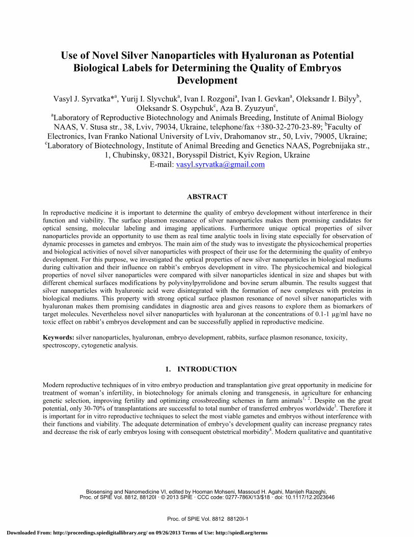
Use of Novel Silver Nanoparticles with Hyaluronan as Potential Biological Labels for Determining the Quality of Embryos Development Vasyl J. Syrvatka* a , Yurij I. Slyvchuk a , Ivan I. Rozgoni a , Ivan I. Gevkan a , Oleksandr I. Bilyy b , Oleksandr S. Osypchuk c , Aza B. Zyuzyun c , a Laboratory of Reproductive Biotechnology and Animals Breeding, Institute of Animal Biology NAAS, V. Stusa str., 38, Lviv, 79034, Ukraine, telephone/fax +380-32-270-23-89; b Faculty of Electronics, Ivan Franko National University of Lviv, Drahomanov str., 50, Lviv, 79005, Ukraine; c Laboratory of Biotechnology, Institute of Animal Breeding and Genetics NAAS, Pogrebnijaka str., 1, Chubinsky, 08321, Borysspil District, Kyiv Region, Ukraine E-mail: [email protected] ABSTRACT In reproductive medicine it is important to determine the quality of embryo development without interference in their function and viability. The surface plasmon resonance of silver nanoparticles makes them promising candidates for optical sensing, molecular labeling and imaging applications. Furthermore unique optical properties of silver nanoparticles provide an opportunity to use them as real time analytic tools in living state especially for observation of dynamic processes in gametes and embryos. The main aim of the study was to investigate the physicochemical properties and biological activities of novel silver nanoparticles with prospect of their use for the determining the quality of embryo development. For this purpose, we investigated the optical properties of new silver nanoparticles in biological mediums during cultivation and their influence on rabbit’s embryos development in vitro. The physicochemical and biological properties of novel silver nanoparticles were compared with silver nanoparticles identical in size and shapes but with different chemical surfaces modifications by polyvinylpyrrolidone and bovine serum albumin. The results suggest that silver nanoparticles with hyaluronic acid were disintegrated with the formation of new complexes with proteins in biological mediums. This property with strong optical surface plasmon resonance of novel silver nanoparticles with hyaluronan makes them promising candidates in diagnostic area and gives reasons to explore them as biomarkers of target molecules. Nevertheless novel silver nanoparticles with hyaluronan at the concentrations of 0.1-1 μg/ml have no toxic effect on rabbit’s embryos development and can be successfully applied in reproductive medicine. Keywords: silver nanoparticles, hyaluronan, embryo development, rabbits, surface plasmon resonance, toxicity, spectroscopy, cytogenetic analysis. 1. INTRODUCTION Modern reproductive techniques of in vitro embryo production and transplantation give great opportunity in medicine for treatment of woman’s infertility, in biotechnology for animals cloning and transgenesis, in agriculture for enhancing genetic selection, improving fertility and optimizing crossbreeding schemes in farm animals 1, 2 . Despite on the great potential, only 30-70% of transplantations are successful to total number of transferred embryos worldwide 3 . Therefore it is important for in vitro reproductive techniques to select the most viable gametes and embryos without interference with their functions and viability. The adequate determination of embryo’s development quality can increase pregnancy rates and decrease the risk of early embryos losing with consequent obstetrical morbidity 4 . Modern qualitative and quantitative Biosensing and Nanomedicine VI, edited by Hooman Mohseni, Massoud H. Agahi, Manijeh Razeghi, Proc. of SPIE Vol. 8812, 88120I · © 2013 SPIE · CCC code: 0277-786X/13/$18 · doi: 10.1117/12.2023646 Proc. of SPIE Vol. 8812 88120I-1 Downloaded From: http://proceedings.spiedigitallibrary.org/ on 09/26/2013 Terms of Use: http://spiedl.org/terms
Transcript of Use of Novel Silver Nanoparticles with Hyaluronan as Potential Biological Labels for Determining the...
Use of Novel Silver Nanoparticles with Hyaluronan as Potential Biological Labels for Determining the Quality of Embryos
Development
Vasyl J. Syrvatka*a, Yurij I. Slyvchuka, Ivan I. Rozgonia, Ivan I. Gevkana, Oleksandr I. Bilyyb, Oleksandr S. Osypchukc, Aza B. Zyuzyunc,
aLaboratory of Reproductive Biotechnology and Animals Breeding, Institute of Animal Biology NAAS, V. Stusa str., 38, Lviv, 79034, Ukraine, telephone/fax +380-32-270-23-89; bFaculty of
Electronics, Ivan Franko National University of Lviv, Drahomanov str., 50, Lviv, 79005, Ukraine; cLaboratory of Biotechnology, Institute of Animal Breeding and Genetics NAAS, Pogrebnijaka str.,
1, Chubinsky, 08321, Borysspil District, Kyiv Region, Ukraine E-mail: [email protected]
ABSTRACT
In reproductive medicine it is important to determine the quality of embryo development without interference in their function and viability. The surface plasmon resonance of silver nanoparticles makes them promising candidates for optical sensing, molecular labeling and imaging applications. Furthermore unique optical properties of silver nanoparticles provide an opportunity to use them as real time analytic tools in living state especially for observation of dynamic processes in gametes and embryos. The main aim of the study was to investigate the physicochemical properties and biological activities of novel silver nanoparticles with prospect of their use for the determining the quality of embryo development. For this purpose, we investigated the optical properties of new silver nanoparticles in biological mediums during cultivation and their influence on rabbit’s embryos development in vitro. The physicochemical and biological properties of novel silver nanoparticles were compared with silver nanoparticles identical in size and shapes but with different chemical surfaces modifications by polyvinylpyrrolidone and bovine serum albumin. The results suggest that silver nanoparticles with hyaluronic acid were disintegrated with the formation of new complexes with proteins in biological mediums. This property with strong optical surface plasmon resonance of novel silver nanoparticles with hyaluronan makes them promising candidates in diagnostic area and gives reasons to explore them as biomarkers of target molecules. Nevertheless novel silver nanoparticles with hyaluronan at the concentrations of 0.1-1 µg/ml have no toxic effect on rabbit’s embryos development and can be successfully applied in reproductive medicine. Keywords: silver nanoparticles, hyaluronan, embryo development, rabbits, surface plasmon resonance, toxicity, spectroscopy, cytogenetic analysis.
1. INTRODUCTION Modern reproductive techniques of in vitro embryo production and transplantation give great opportunity in medicine for treatment of woman’s infertility, in biotechnology for animals cloning and transgenesis, in agriculture for enhancing genetic selection, improving fertility and optimizing crossbreeding schemes in farm animals1, 2. Despite on the great potential, only 30-70% of transplantations are successful to total number of transferred embryos worldwide3. Therefore it is important for in vitro reproductive techniques to select the most viable gametes and embryos without interference with their functions and viability. The adequate determination of embryo’s development quality can increase pregnancy rates and decrease the risk of early embryos losing with consequent obstetrical morbidity4. Modern qualitative and quantitative
Biosensing and Nanomedicine VI, edited by Hooman Mohseni, Massoud H. Agahi, Manijeh Razeghi, Proc. of SPIE Vol. 8812, 88120I · © 2013 SPIE · CCC code: 0277-786X/13/$18 · doi: 10.1117/12.2023646
Proc. of SPIE Vol. 8812 88120I-1
Downloaded From: http://proceedings.spiedigitallibrary.org/ on 09/26/2013 Terms of Use: http://spiedl.org/terms
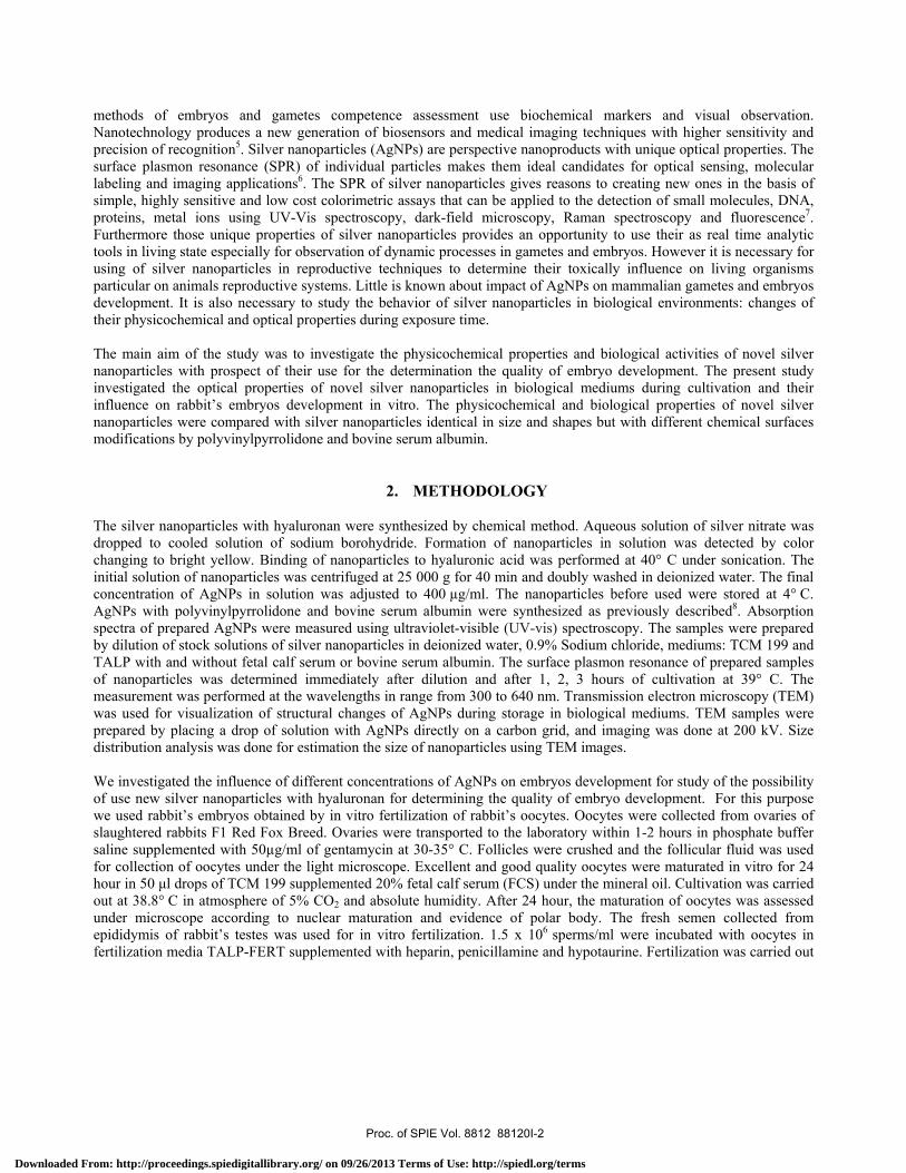
methods of embryos and gametes competence assessment use biochemical markers and visual observation. Nanotechnology produces a new generation of biosensors and medical imaging techniques with higher sensitivity and precision of recognition5. Silver nanoparticles (AgNPs) are perspective nanoproducts with unique optical properties. The surface plasmon resonance (SPR) of individual particles makes them ideal candidates for optical sensing, molecular labeling and imaging applications6. The SPR of silver nanoparticles gives reasons to creating new ones in the basis of simple, highly sensitive and low cost colorimetric assays that can be applied to the detection of small molecules, DNA, proteins, metal ions using UV-Vis spectroscopy, dark-field microscopy, Raman spectroscopy and fluorescence7. Furthermore those unique properties of silver nanoparticles provides an opportunity to use their as real time analytic tools in living state especially for observation of dynamic processes in gametes and embryos. However it is necessary for using of silver nanoparticles in reproductive techniques to determine their toxically influence on living organisms particular on animals reproductive systems. Little is known about impact of AgNPs on mammalian gametes and embryos development. It is also necessary to study the behavior of silver nanoparticles in biological environments: changes of their physicochemical and optical properties during exposure time.
The main aim of the study was to investigate the physicochemical properties and biological activities of novel silver nanoparticles with prospect of their use for the determination the quality of embryo development. The present study investigated the optical properties of novel silver nanoparticles in biological mediums during cultivation and their influence on rabbit’s embryos development in vitro. The physicochemical and biological properties of novel silver nanoparticles were compared with silver nanoparticles identical in size and shapes but with different chemical surfaces modifications by polyvinylpyrrolidone and bovine serum albumin.
2. METHODOLOGY The silver nanoparticles with hyaluronan were synthesized by chemical method. Aqueous solution of silver nitrate was dropped to cooled solution of sodium borohydride. Formation of nanoparticles in solution was detected by color changing to bright yellow. Binding of nanoparticles to hyaluronic acid was performed at 40° C under sonication. The initial solution of nanoparticles was centrifuged at 25 000 g for 40 min and doubly washed in deionized water. The final concentration of AgNPs in solution was adjusted to 400 µg/ml. The nanoparticles before used were stored at 4° C. AgNPs with polyvinylpyrrolidone and bovine serum albumin were synthesized as previously described8. Absorption spectra of prepared AgNPs were measured using ultraviolet-visible (UV-vis) spectroscopy. The samples were prepared by dilution of stock solutions of silver nanoparticles in deionized water, 0.9% Sodium chloride, mediums: TCM 199 and TALP with and without fetal calf serum or bovine serum albumin. The surface plasmon resonance of prepared samples of nanoparticles was determined immediately after dilution and after 1, 2, 3 hours of cultivation at 39° C. The measurement was performed at the wavelengths in range from 300 to 640 nm. Transmission electron microscopy (TEM) was used for visualization of structural changes of AgNPs during storage in biological mediums. TEM samples were prepared by placing a drop of solution with AgNPs directly on a carbon grid, and imaging was done at 200 kV. Size distribution analysis was done for estimation the size of nanoparticles using TEM images.
We investigated the influence of different concentrations of AgNPs on embryos development for study of the possibility of use new silver nanoparticles with hyaluronan for determining the quality of embryo development. For this purpose we used rabbit’s embryos obtained by in vitro fertilization of rabbit’s oocytes. Oocytes were collected from ovaries of slaughtered rabbits F1 Red Fox Breed. Ovaries were transported to the laboratory within 1-2 hours in phosphate buffer saline supplemented with 50µg/ml of gentamycin at 30-35° C. Follicles were crushed and the follicular fluid was used for collection of oocytes under the light microscope. Excellent and good quality oocytes were maturated in vitro for 24 hour in 50 μl drops of TCM 199 supplemented 20% fetal calf serum (FCS) under the mineral oil. Cultivation was carried out at 38.8° C in atmosphere of 5% CO2 and absolute humidity. After 24 hour, the maturation of oocytes was assessed under microscope according to nuclear maturation and evidence of polar body. The fresh semen collected from epididymis of rabbit’s testes was used for in vitro fertilization. 1.5 х 106 sperms/ml were incubated with oocytes in fertilization media TALP-FERT supplemented with heparin, penicillamine and hypotaurine. Fertilization was carried out
Proc. of SPIE Vol. 8812 88120I-2
Downloaded From: http://proceedings.spiedigitallibrary.org/ on 09/26/2013 Terms of Use: http://spiedl.org/terms

in a CO2 incubator for 22 hours at 38.8° C. Fertilized oocytes were transferred to TCM 199 with 10% fetal calf serum. Concentrations of AgNPs: 0.1, 1 and 10 μg/ml in culture medium were adjusted by adding appropriate volume of nanoparticles stock solution. Cultivation was carried out in a CO2 incubator for 6 days at 38.8° C. The monitor of embryos development was conducted every 24 hours for six days. The stages of embryos developmental were evaluated under microscope after six days of cultivation. Cytogenetic analysis was performed by evaluating the nuclear chromosomes of blastomers after embryo’s staining of 2% Gimsa dye9. After six days embryo conditioned medium was sampled by centrifugation at 900 g for 5 min at 4° C. Samples were used for determination of concentration of Calcium (Ca), Phosphorus (P), Magnesium (Mg), Potassium (K), Sodium (Na), lactate dehydrogenize (LDH) and alkaline phosphatase (ALP) activity using commercially available kits according to manufacturer’s instruction (Human GmbH, Germany). All tests were measured by biochemical analyzer.
All experiments were done in triplicate and the results were presented as mean ± standard deviation. All statistical analyses were performed by using the Minitab 15 English statistical software package. Differences between groups were determined by Student t-tests.
3. RESULTS
Kim et al 2007 were synthesized by chemical method silver nanoparticles with similar in size and shape to our nanoparticles which were characterized by high stability without composite agents10. Silver nanoparticles without stabilizing agents obtained in this study were aggregated some time after synthesis. However new silver nanoparticles with hyaluronan were stable in solution during storage. Visible signs of color changes or sedimentation were not found (Figure 1A). The results of ultraviolet-visible spectroscopy showed that new silver nanoparticles with hyaluronan had strong optical absorbance at the wavelengths in range from 385 to 415 nm with maximum at 400 nm. The light extinction of AgNPs-HA at the wavelength 375-450 nm was higher than in AgNPs-PVP and AgNPs-BSA. The peak of surface plasmon resonance absorbance of silver nanoparticles with polyvinylpyrrolidone was at the wavelength 400 nm. Whereas the maximum absorbance of AgNPs-BSA was laid on plateau at wavelengths from 395 to 430 nm (Figure 1B).
Figure 1. A – from left to right: silver nanoparticles with PVP, HA, BSA and without capping agent; B – absorption spectrums of silver nanoparticles in deionized water.
A B
Proc. of SPIE Vol. 8812 88120I-3
Downloaded From: http://proceedings.spiedigitallibrary.org/ on 09/26/2013 Terms of Use: http://spiedl.org/terms
0.7
0.6
0.5,-,
i 0.4CU
c 0.3
0.2
0.1
0
AgNPs -PVP0 hours
- - - AgNPs -PVP1 hoursAgNPs -PVP2 hoursAgNPs -PVP3 hours
IIIII1111111111111111 ',Jim I I IIIII II0 0 0 4,10 h o o o 0 o 0c4i cñ A PiRi N 4n CO ñ ñ ñ
wavelenth (nm)
0.4
0.35
0.3,-,
ti 0.25
I 0.2L
0.15..o
0.1
0.05
o
AgNPs -BSA0 hours
- - - AgNPs -BSA1 hoursAgNPs -BSA2 hoursAgNPs -BSA
..... `_.
o o1 o o hp o o o oo o poc+01M M M M 41 h h h O f+1
wavelenth (nm)
0.4
0.35
0.3
AgNPs-Ha0 hours
- - - AgNPs-HA1 hoursAgNPs -HA2 hoursAgNPs -HA
on2 0.15..oce
0.1
0.05
0
............ ....I I I I I I I I I I I I I I I I I I I I
Oo c+o1 Do 1 po hp o o o o o o poco+1M M M M 41 Q h h h O f+1
wavelenth (nm)
abso
rban
ce (
a. u
)ó
o0
0ó
0o
oO
n1
300
330
360
.?.\
375
390
DO
CD
405
420
!~
450
.48
0
510
¡
540
S"V
570
/:il
x x x x
600
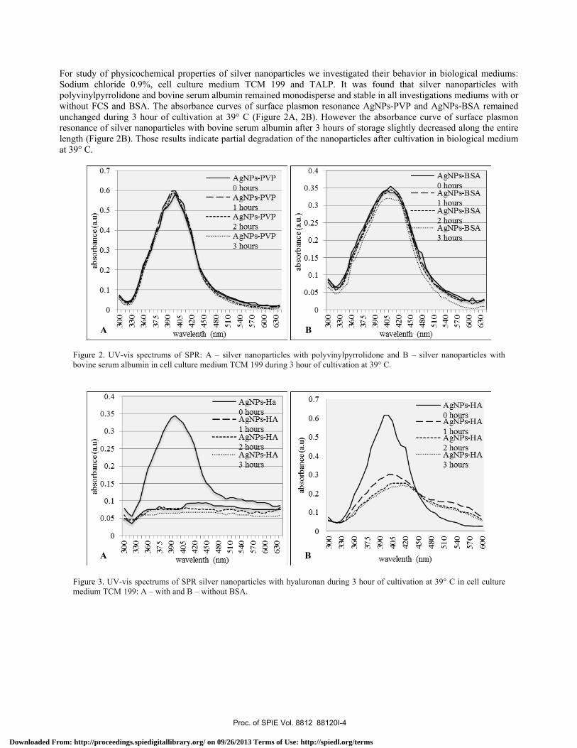
For study of physicochemical properties of silver nanoparticles we investigated their behavior in biological mediums: Sodium chloride 0.9%, cell culture medium TCM 199 and TALP. It was found that silver nanoparticles with polyvinylpyrrolidone and bovine serum albumin remained monodisperse and stable in all investigations mediums with or without FCS and BSA. The absorbance curves of surface plasmon resonance AgNPs-PVP and AgNPs-BSA remained unchanged during 3 hour of cultivation at 39° C (Figure 2A, 2B). However the absorbance curve of surface plasmon resonance of silver nanoparticles with bovine serum albumin after 3 hours of storage slightly decreased along the entire length (Figure 2B). Those results indicate partial degradation of the nanoparticles after cultivation in biological medium at 39° C.
Figure 2. UV-vis spectrums of SPR: A – silver nanoparticles with polyvinylpyrrolidone and B – silver nanoparticles with bovine serum albumin in cell culture medium TCM 199 during 3 hour of cultivation at 39° C.
Figure 3. UV-vis spectrums of SPR silver nanoparticles with hyaluronan during 3 hour of cultivation at 39° C in cell culture medium TCM 199: A – with and B – without BSA.
A B
A B
Proc. of SPIE Vol. 8812 88120I-4
Downloaded From: http://proceedings.spiedigitallibrary.org/ on 09/26/2013 Terms of Use: http://spiedl.org/terms
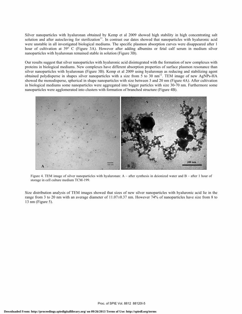
Silver nanoparticles with hyaluronan obtained by Kemp et al 2009 showed high stability in high concentrating salt solution and after autoclaving for sterilization11. In contrast our dates showed that nanoparticles with hyaluronic acid were unstable in all investigated biological mediums. The specific plasmon absorption curves were disappeared after 1 hour of cultivation at 39° C (Figure 3A). However after adding albumins or fetal calf serum in medium silver nanoparticles with hyaluronan remained stable in solution (Figure 3B). Our results suggest that silver nanoparticles with hyaluronic acid disintegrated with the formation of new complexes with proteins in biological mediums. New complexes have different absorption properties of surface plasmon resonance than silver nanoparticles with hyaluronan (Figure 3B). Kemp et al 2009 using hyaluronan as reducing and stabilizing agent obtained polydisperse in shapes silver nanoparticles with a size from 5 to 30 nm12. TEM image of new AgNPs-HA showed the monodisperse, spherical in shape nanoparticles with size between 3 and 20 nm (Figure 4A). After cultivation in biological mediums some nanoparticles were aggregated into bigger particles with size 30-70 nm. Furthermore some nanoparticles were agglomerated into clusters with formation of branched structure (Figure 4B).
Figure 4. TEM image of silver nanoparticles with hyaluronan: A – after synthesis in deionized water and B – after 1 hour of storage in cell culture medium TCM-199.
Size distribution analysis of TEM images showed that sizes of new silver nanoparticles with hyaluronic acid lie in the range from 3 to 20 nm with an average diameter of 11.07±0.37 nm. However 74% of nanoparticles have size from 8 to 13 nm (Figure 5).
BA
Proc. of SPIE Vol. 8812 88120I-5
Downloaded From: http://proceedings.spiedigitallibrary.org/ on 09/26/2013 Terms of Use: http://spiedl.org/terms

25 -.-,i 20
ti 5
01 2 3 4 5 6
O 1, ,,111 12 13 14 15 16 17 18 19 20 21
% of nanoparticles
90 - AgNPs-HA80 -+70 -60 -50 -40 - -
30 - -
20 -10 -0
0 s-PVP AgNPs-B SA
0 0.1 1 10
2NPS concentration
60 - AgNPs-HA AgNPs-PVP AgNPs-BSA
0 0.1 1
AgNPs concentration
10
Figure 5. Size distribution analysis of silver nanoparticles with hyaluronan.
Taylor et al 2009 showed that injection of 10 pl of AgNPs (average nanoparticles diameter of 15 nm) with concentration 50 μg/mL into 1 blastomere of 2-cell-stage embryos had no significant impact on blastocysts development13. Our results were shown that AgNPs-PVP, AgNPs-BSA and AgNPs-HA in concentration 0.1-1 μg/ml have no negative effect on rabbit’s embryo development. Besides that it was not found significant difference in number of embryos at stage of 2-4 cells and early morula after adding silver nanoparticles with polyvinylpyrrolidone and bovine serum albumin at concentration of 10 μg/ml (Figure 6А, 6B). However concentration of AgNPs-HA 10 µg/ml inhibited of embryos development. The number of embryos at stage of 2-4 cells and early morula significantly (p<0.025) decreased in group with 10 µg/ml silver nanoparticles with hyaluronan in culture medium (Figure 6А, 6B).
Figure 6. Persentage of embryo: A – at stage of 2-4 cells and B – at stage of early morula to total embryos number. The visual observation under light microscope showed that AgNPs-HA were aggregated into bigger particles and deposited on the embryos surface (Figure 7A, 7B).
A B
Proc. of SPIE Vol. 8812 88120I-6
Downloaded From: http://proceedings.spiedigitallibrary.org/ on 09/26/2013 Terms of Use: http://spiedl.org/terms

Figure 7. A, B – deposition of aggregated nanoparticles on embryo syrface after adding 10 µg/ml of AgNPs-HA in culture medium.
Li et al 2010 demonstrated significantly increasing apoptosis and corresponding decrease in total cell number in treated blastocysts of 50 µM AgNPs. Furthermore blastocysts pretreated with AgNPs decreased successful implantation frequency and fetal weight with increased resorption of post-implantation embryos compare to control counterparts14. Those dates indicate possible genotoxic effects of silver nanoparticles on embryos. Furthermore many study showed genotoxic effect of silver nanoparticles on somatic cells line15. However cytogenetic analysis showed that all investigated silver nanoparticles were not influenced on nuclear chromosomes of embryos (Figure 8).
Figure 8. Embryo at morula stage – A, and their cytogenetic analysis – B with 10 µg/ml AgNPs-HA (Magnification – X1000). Furthermore all silver nanoparticles were not influenced on concentration Ca, P, Mg, K and Na in embryo condition medium after six days of in vitro cultivation at 38.8° C (Table 1).
A B
A B
Proc. of SPIE Vol. 8812 88120I-7
Downloaded From: http://proceedings.spiedigitallibrary.org/ on 09/26/2013 Terms of Use: http://spiedl.org/terms

8
7
6
5
4
3
2
1
AgNPs-HA **
AgNPs-PVP
AgNPs-B SA
0 0.1 1 10
AgNPs concentration
Table 1. The influence of silver nanoparticles on concentration Ca, P, Mg, K and Na in embryo conditioned medium after six days of in vitro cultivation at 38.8° C.
Groups
Silver nanoparticles
concentration,
µg/ml
Parameters
Ca
mmol/L
Mg
mmol/L
P
mmol/L
K
mmol/L
Na
mmol/L
Control 0 1.79±0.01 0.847±0.009 1.02±0.03 6.09±0.030 131.00±0.58
AgN
Ps-P
VP
0.1 1.79±0.01 0.853±0.015 1.05±0.01 6.06±0.003 130.67±0.33
1 1.81±0.003 0.820±0.015 1.03±0.02 6.10±0.036 132.67±0.33
10 1.81±0.007 0.867±0.019 1.02±0.02 6.17±0.040 132.00±1.15
AgN
Ps-B
SA
0.1 1.77±0.01 0.860±0.015 0.97±0.05 6.10±0.040 130.00±0.58
1 1.78±0.02 0.850±0.025 0.98±0.02 6.09±0.045 130.33±0.67
10 1.78±0.02 0.840±0.021 0.99±0.03 6.05±0.015 130.00±2.08
AgN
Ps-H
A
0.1 1.79±0.02 0.840±0.013 1.00±0.02 6.08±0.024 131.00±2.08
1 1.7±0.009 0.860±0.013 1.04±0.02 6.13±0.024 129.67±2.19
10 1.81±0.009 0.867±0.020 1.07±0.01 6.13±0.028 132.00±1.15
There was no significant difference in alkaline phosphatase activity of embryo’s conditioned medium in all groups with different concentrations of silver nanoparticles, as compared to the control group after six days of in vitro cultivation (Figure 9A) However silver nanoparticles with hyaluronan at the concentration of 10 µg/mL significantly increase (p<0.01) of lactate dehydrogenase activity in embryo’s conditioned medium (Figure 9B). LDH activity is well correlated to embryos viability and this fact indicates LDH leakage into the cultural medium from blastomers with loss their viability.
Figure 9. The activity of alkaline phosphatase (a) and lactate dehydrogenase (b) in conditional medium after 6 day of in vitro
cultivation at 38.8°C.
Proc. of SPIE Vol. 8812 88120I-8
Downloaded From: http://proceedings.spiedigitallibrary.org/ on 09/26/2013 Terms of Use: http://spiedl.org/terms

3. CONCLUSIONS
These results suggest that silver nanoparticles with hyaluronic acid were disintegrated with the formation of new complexes with proteins in biological mediums. This property with strong optical surface plasmon resonance of novel silver nanoparticles with hyaluronan makes them promising candidates in diagnostic area and gives reasons to explore them as biomarkers of target molecules. Concentration of silver nanoparticles with hyaluronic acid 10 µg/ml inhibited of embryos development. This toxic effect of AgNPs-HA related with aggregation of nanoparticles and their deposition on the embryos surface. Nevertheless novel silver nanoparticles with hyaluronan at the concentration of 0.1-1 µg/mL have no toxic effect on rabbit’s embryo development and can be successfully applied in reproductive medicine.
REFERENCES [1] Hansen, P. J., "Realizing the promise of IVF in cattle – an overview," Theriogenology 65, 119–125 (2006). [2] Van Voorhis, B. J., "In vitro fertilization," New England Journal of Medicine 356(4), 379–386 (2007). [3] Center for Disease Control and Prevention., American Society for Reproductive Medicine., Society for Assisted Reproductive Technology., “2009 assisted reproductive technology success rate: national summary and fertility clinic reports,” Atlanta: Center for Disease Control and Prevention, (2011). [4] Dhawan, A., Leveille, M. C. and Vanderhyden B. C., "Inhibition by human embryos of mouse granulosa cell progesterone production: development of a sensitive bioassay," Human Reproduction 15(4), 917–924 (2000). [5] Larguinhoa, M. and Baptista, P. V., "Gold and silver nanoparticles for clinical diagnostics – From genomics to proteomics," Journal of Proteomics 75(10), 2811–2823 (2012). [6] Caro, C., Castillo, P. M., Klippstein, R., Pozo, D. and Zaderenko A. P., "Silver nanoparticles: sensing and imaging applications," in [Silver Nanoparticles] ed. by Perez D. P., 201–224 (2010). [7] Paul, S., Paul, D., Fern, G. R. and Ray, A. K., "Surface plasmon resonance imaging detection of silver nanoparticle-tagged immunoglobulin," J. R. Soc. Interface. 8(61), 1204–1211 (2011). [8] Syrvatka, V. J, Slyvchuk, Y. I., Rozgoni, I. I., Gevkan I. I. and Bilyy O. I., "Optical properties of functional composite silver nanoparticles and their potential use in reproductive medicine," Proc. SPIE 8803, 88030V-88030V (2013). [9] Tarkovsky, A. K. "An air-drying method for chromosome preparations from mouse eggs," Cytogenetics 5, 394–400 (1966). [10] Kim, J. S., Kuk, E., Yu, K. N., Kim, J. H., Park, S. J., Lee, H. J., Kim, S. H., Park, Y. K., Park, Y. H., Hwang, C. Y., Kim, Y. K., Lee, Y. S., Jeong, D. H. and Cho M. H., "Antimicrobial effects of silver nanoparticles," Nanomedicine 3, 95–101 (2007). [11] Kemp, M. M., Kumar, A., Clement, D., Ajayan, P., Mousa, S. and Linhardt R. J., "Hyaluronan- and heparin-reduced silver nanoparticles with antimicrobial properties," Nanomedicine 4(4), 421–429 (2009). [12] Kemp, M. M., Kumar, A., Mousa, S., Park, T. J., Ajayan, P., Kubotera, N., Mousa, S. A. and Linhardt R. J., "Synthesis of gold and silver nanoparticles stabilized with glycosaminoglycans naving distinctive biological activities," Biomacromolecules 10 (3), 589–595 (2009). [13] Taylor, U., Garrels, W., Petersen, S., Barcikowski, S., Klein, S., Kues, W., Lucas-Hahn, A., Niemann, H. and Rath, D., "Development of murine embryos after injection of uncoated gold and silver nanoparticles," Reprod. Fertil. Dev. 22, 240–241 (2010). [14] Li, P. W., Kuo, T. H., Chang, J. H., Yeh, J. M. and Chan, W. H., "Induction of cytotoxicity and apoptosis in mouse blastocysts by silver nanoparticles," Toxicol. Lett. 197(2), 82–72 (2010). [15] Lim, H. K, Asharani, P. V. and Hande, M. P., "Enhanced genotoxicity of silver nanoparticles in DNA repair deficient mammalian cells" Front. Gene. 3, 104 (2012).
Proc. of SPIE Vol. 8812 88120I-9
Downloaded From: http://proceedings.spiedigitallibrary.org/ on 09/26/2013 Terms of Use: http://spiedl.org/terms