Untitled - Boston College
162
-
Upload
khangminh22 -
Category
Documents
-
view
6 -
download
0
Transcript of Untitled - Boston College

Investigations inMolecular Cell Biology
Boston College | Clare O’Connor

ContentsChapter 1 ....................................................................................................1
– IntroductionChapter 2 ................................................................................................... 7
– Meet the yeastChapter 3 ................................................................................................. 15
– Mastering the micropipetteChapter 4 ...................................................................................................25 – Working with yeast Chapter 5 ..................................................................................................35
– Introduction to databases Chapter 6 ..................................................................................................51
– Analysis of mutant strainsChapter 7 ................................................................................................. 63
– Yeast colony PCR Chapter 8 ..................................................................................................77
– Agarose gel electrophoresis Chapter 9 ..................................................................................................85
– Protein conservation Chapter 10 ................................................................................................97
– Plasmids Chapter 11 ..............................................................................................105
– Restriction mapping Chapter 12 ..............................................................................................113
– Yeast transformationChapter 13 ...............................................................................................121 – Protein overexpressionChapter 14 ...............................................................................................129 – SDS-PAGEChapter 15 ...............................................................................................141 – Western blotsGlossary ...................................................................................................155

• Coursedesignandlearninggoals
• PathwaysoverTime:Ourresearchproject
• Courseoverview
• References
• Acknowledgments
Weliveinthe“post-genomic”era,inwhichtheavailabilityofcompletegenomesequencesfromahostoforganismsoffersexcitingopportunitiesforundergraduateresearch.InBI204,wewillusesomeofthestrainandcloneresourcesgeneratedbytheyeastgenomeprojecttoinvestigatetheevolutionofgenesinvolvedinthesynthesisofmethionineandcysteine,essentialsulfur-containingaminoacids.
IntroductionChapter 1
BI 204 - Investigations in Molecular Cell Biology
2
Chapter 1 WelcometoBI204-InvestigationsinMolecularCellBiology.BI204isanewkindofintroductorylabcourse,whichhasbeendesignedtoincorporateanauthenticresearchproject.Ithasbeensaidthatweliveina“post-genomic”era.Large-scalegenomeprojectshavegeneratedtremendousamountsofsequencedata,andcompletegenomesequencesareavailableforthousandsoforganisms.Manyofthe“genes”annotatedbythegenomeprojectshavebeenidentifiedbytheirsimilaritytoknowngenesinotherorganisms,buttheirfunctionshavenotbeentesteddirectly(Goffeauet al.,1996).Theselooseendsprovideexcitingopportunitiesforundergraduatestudentstoparticipateinauthenticfunctionalgenomicsresearch.
Thiscourseisdesignedasaresearchprojectinwhichstudentsstudytheevolutionofthegenesinvolvedinmethionine(Met)andcysteine(Cys)biosynthesis.MetandCysareessentialaminoacidsinalllivingcells.Thesetwoaminoacidscontainsulfurintheirsidechains,whichallowsMetandCystoplayuniquerolesinproteins.Weexpectthatstudentswillmakenovelfindingsintheirprojectseachsemesterandthatstudentswillbeabletobuildupontheresultsobtainedinprecedingsemesters.Wehopethatyouenjoytheresearchexperienceandwelookforwardtoyourexperimentalresults!
Course design and learning goals Biologyeducationattheundergraduatelevelisundergoingatransformation.Fordecades,manyhaveviewedbiologyasanencyclopedicsubject,becauseofthevastamountofcontentmatterincludedintheundergraduatecurriculum.Arecentreevaluationofundergraduatebiologyeducation,however,isguidingbiologycurriculainanewdirection,stressingtheimportanceofinvolvingstudentsintheprocessofscientificinvestigationintheircoursework(Bauerleet al.,2011).Thisreevaluationprocesshasalsochallengededucatorstosortthroughthevastamountofcontentinintroductorybiologytoidentifythecoreconceptsthatstudentsshouldlearnandthekeycompetenciesthatstudentsshouldacquireduringtheirundergraduateeducation.Thiscoursehasbeendesignedinlinewiththeserecommendations.
TheBI204researchprojectfocusesontheevolutionaryconservationofthegenesinvolvedinsynthesizingMetandCys.Theexperimentsintheprojectexplorethecoreconceptsofbiology:
• Evolution:TheproteinsinvolvedinMetandCyssynthesisshowvariedpatternsofconservationduringevolution.
• Structureandfunction:ThestructuresoftheproteinsinvolvedinMetandCyssynthesisareadaptedtotheircatalyticroles.
• Informationtransfer:MetandCyssynthesisrequiresenzymesencodedbymultiplegenes.
• Pathwaysandenergytransformation:TheenzymesinvolvedinMetandCyssynthesisarepartsofintersectingenergy-consumingpathways.
• Systemsbiology:Thereactionsinvolvedinsulfuraminoacidsynthesisintersectwithmanyothermetabolicpathwaysincells.

3
Replace Chapter number and title on A-Master Page.<- ->
Introduction Duringthecourseofthesemester,studentswillacquirethecorecompetenciesforprofessionalbiologists.
• Workinginteams,studentswillproposehypothesesanddesignexperimentstotesttheirhypotheses.
• Studentswilllearnbasicskillsofmolecularcellbiologyastheyconducttheirexperiments.
• Studentswillcollect,organizeandinterpretexperimentaldata
• Studentswillfindanduseinformationfromtheprimaryscientificliteratureandonlinedata-basesastheydeveloptheirexperimentaldesignandinterprettheirexperiments.
• Studentswillcommunicatetheirscientificresultsinaseriesofshortoralpresentationsandwrittenreports.
• Usingfeedbackfromtheirpeersandtheteachingstaff,studentswillcompiletheseshortreportsattheendofthesemesterintoaposterandafinalreportwrittenintheformatofascientificpresentation.
Pathways over time: our research project Wewillusethebuddingyeast,Saccharomyces cerevisiae,tostudytheevolutionofthegenesinvolvedinsulfuraminoacidsynthesis.S. cerevisiaeisaunicellulareukaryotethathasbeenwidelyusedasamodelorganismforover50years(BotsteinandFink,2011).S. cerevisiae hasmanyofthesamebiochemicalpathwaysashighereukaryotes,butitsgenomeissignificantlysmallerthanvertebrategenomesandpowerfulgenetictechniquesareavailableformanipulatinggeneexpression. S. cerevisiae isalsoinexpensiveandsimpletocultureinthelaboratory.Forthesereasons,theS. cerevisiaegenomewasthefirsttobesequencedinitsentirety.Completionoftheyeastgenomesequence(Goffeauet al.,1996)allowedtheyeastcommunitytopreparegenome-widecollectionsofmutantstrains(Winzeleret al.,1999)andplasmids(Gelperinet al.,2005),someofwhichyouwillusethissemester.WewilluseS. cerevisiaestrainswithdefineddefectsinmethionineandcysteinebiosynthesisasthehostsforhomologousgenesfromotherorganisms.IftheforeigngenerestorestheabilitytosynthesizemethioninetoitsS. cerevisiaehost,inaprocessknownascomplementation,wewillknowthatgenefunctionhasbeenconservedovertheevolutionarytimeframethatseparatesthetwospecies.

4
Chapter 1
Course overview Thecoursecanbeviewedasaseriesofrelatedmodules,asshowinthefigureontheoppositepage.1. Inthefirstfewlaboratories,studentswillbecomeacquaintedwithbasiclaboratoryequipment
andtechniquesforhandlingandviewingyeast.Studentswillalsobeintroducedtosomeofthemanyonlinedatabases,whichareimportantsourcesofgeneandproteininformation.
2. Inthenextsetofexperiments,studentteamswillcharacterizethreeS. cerevisiaemutants,eachofwhichisdeficientinageneinvolvedinMetorCyssynthesis(Winzeleret al.,1999).Studentswilluseselectivegrowthmediaandthepolymerasechainreaction(PCR)todistinguishthethreestrainsfromoneanother.Teamswillthenuseoneofthestrainsfortheremainingexperimentsofthesemester.
3. Teamswillreceivethreeplasmidsthatwillbeusedfortransformationandcomplementationexperiments.Theplasmidshavebeenengineeredtooverexpressproteincodingsequences(CDS)underthecontrolofthepowerfulyeastGAL1 promoter.OneplasmidcontainstheS. cerevisiae METgenethatisdeficientintheiryeaststrain(Gelperinet al.,2005).ThesecondplasmidcarriestheS. pombehomologfortheS. cerevisiae METgenethatismissingintheiryeaststrain.Thethirdplasmidisacontrolplasmidtoensurethatoverexpressionisoccurringintransformedcells.Studentswillisolatetheplasmidsfrombacterialstocksandidentifytheplasmidsusingrestrictionendonucleases.
4. Teamswilltransform(Chapter12)theiryeastdeficiencystrainwiththethreeplasmids.Selectiveplateswillbeusedtodetermineiftransformationand/orcomplementationhasoccurred.
5. Teamswillstudyexpressionoftheplasmid-encodedgenesusingwesternblots.TheproteinsexpressedintransformedcellsarefusionproteinscarryingepitopesattheirC-terminithatcanberecognizedbyantibodies.
6. Inthelastsegmentofthesemester,teamswilldesignandconducttheirownexperiments,basedonquestionsthathavearisenduringthepreviousexperiments.
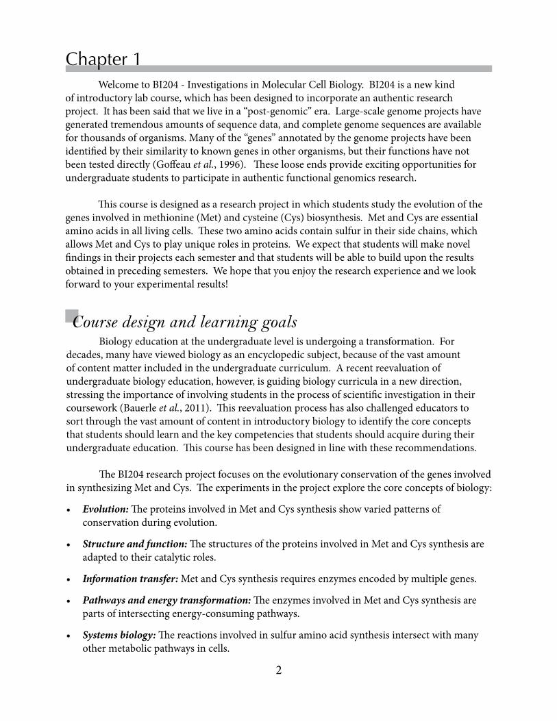
Duringthe2013-2014academicyear,wewillexploretheconservationofMetandCysbiosyntheticenzymesbetweenS. cerevisiaeandthefissionyeast,Schizosaccharomyces pombe.BothS. pombeandS. cerevisiaeareascus-formingyeastfromthephylumAscomycotainthekingdomFungi.Thetwospeciesarethoughttohavedivergedfromacommonancestorabout1billionyearsago(Hedges,2002).Sincetheirdivergence,theS. cerevisiaelineagehasundergoneawholegenomeduplication,followedbyroundsofgeneeliminationanddiversification.Today,thesizeoftheS. cerevisiaegenome(Kelliset al.,2004),~12.5Mbp,issimilartothatofS. pombe.S. pombe isconsideredtobemuchclosertoancestralmembersofthephylum.Ourresultsduringthiscomingyearwillmostlikelyidentifybothgenesthathaveandhavenotbeenfunctionallyconservedbetweenthetwoyeastspecies.Resultsfromthisyearwillalsoguidethefuturedirectionoftheprojectintootherbranchesoflife.Overtime,wehopethatstudentresultswillallowustoconstructevolutionarytreesformanyofthegenesinvolvedinMetandCyssynthesis.

5
Replace Chapter number and title on A-Master Page.<- ->
Introduction
Overview of the semester’s experiments

6
Chapter 1
Acknowledgments
IthasbeenapleasuretoworkwithmanyBostonCollegecolleaguesandstudentsonthisthirdeditionoftheBI204laboratorymanual.Inparticular,Dr.DouglasWarnerhashelpedtodesigntheexperimentsandhascontributedmanyrevisionstothetext.ManythankstoHolliRowedderforhercarefuleditingandforcompilingtheglossarythathasbeenaddedtothisedition.
Overthepasttwoyears,manyteachingassistantshaveprovidedableleadershipfortheirsectionsandofferedvaluablefeedbackabouttheeffectivenessofthemanualandsuggestionsforimprovingboththemanualandthecourse.Hundredsofundergraduatestudentshavealsocontributedcommentsandsuggestionsthathavehelpedtoimprovethemanual.SpecialthanksareduetoDavidChou,Classof2012,whodesignedthecoverandlayoutofthemanual.
Finally,IwouldliketoacknowledgethesupportfromtheNationalScienceFoundationforthePathwaysoverTimeprojectthroughgrantNSF114028.
Bauerle,C,DePass,A,Lynn,Det al.(2011)Vision and Change in Undergraduate Biology Education: A Call to Action. NationalScienceFoundation/AmericanAssociationfortheAdvancementofScience,Washington,D.C.
Botstein,D&Fink,GR(2011)Yeast:anexperimentalorganismfor21stcenturybiology.Genetics189:695-704.
Gelperin,DM,White,MA,Wilkinsonet al.(2005)BiochemicalandgeneticanalysisoftheyeastproteomewithamovableORFcollection.Genes Develop19:2816-2826.
Goffeau,A,Barrell,BG,Bussey,Het al.(1996)Lifewith6000genes.Science274:563-567.Hedges,SB(2002)Theoriginandevolutionofmodelorganisms.Nat Rev Genet3:838-849.Kellis,M,Birren,BW&Lander,ES(2004)Proofandevolutionaryanalysisofancientgenome
duplicationintheyeastSaccharomyces cerevisiae.Nature428:617-624.Thomas,D&Surdin-Kerjan,Y(1997)MetabolismofsulfuraminoacidsinSaccharomyces
cerevisiae.Microbiol Mol Biol Rev61:503-532.Winzeler,EA,Shoemaker,DD,Astromoff,Aet al. (1999)Functionalcharacterizationofthe
Saccharomyces cerevisiaegenomebygenedeletionandparallelanalysis.Science285:901-906.
References

In this laboratory, students will:• identify the components of a compound light microscope
• adjust a light microscope to observe to different yeast specimens
• stain yeast samples with iodine to improve contrast
• identify the morphological characteristics of S. cerevisiae and S. pombe
• distinguish log phase and stationary phase yeast cultures
Since the budding yeast (left), Saccharomyces cerevesiae, and the fission yeast, Schizosaccharomyces pombe diverged from a common ancestor, they have evolved distinctive morphologies and controls on cell division. In this lab, you will use the light microscope to compare cultures of S. cerevisiae and S. pombe.
Meet the yeastChapter 2
Objectives
8
Chapter 2
In this lab, you will use a compound light microscope to observe S. cerevisiae and S. pombe from both rapidly-growing and nutrient-rich and nutrient-depleted cultures. Our microscopes do not have the sophisticated optics used to obtain the images shown above, but you will be able to distinguish cells at various points in the cell cycle and to distinguish S. cerevisiae and S. pombe. In nutrient-rich media, cells grow exponentially, and this period of rapid growth is often referred to as log phase. Log phase yeast pass continuously through the cell cycle, and a log phase culture will have cells in the G1, S, G2 and M phases of the cell cycle.
As shown in the figure on the opposite page, however, the proportion of cells in each phase varies signicantly in S. cerevisiae and S. pombe cultures, because the principal cell cycle checkpoint occurs at a different place in the cycle (Turner et al., 2012). In S. cerevisiae, buds begin to form when cells enter S phase. The size of the bud, which will become the daughter cell, continues to grow until the cells divide in M phase. At the time that the cell divides, the daughter cell is still smaller than the mother cell. The daughter cell will need to grow a bit before it enters another round of cell division. By contrast, S. pombe divides by medial fission. Cells grow in length until they are 12-15 µm, at which point the cell divides and a septum begins to form. The unusually long G2 phase of S. pombe may reflect the fact that it is found primarily as a haploid in nature, unlike S. cerevisiae, which is found in both diploid and haploid forms. One would expect that haploid yeast to be more susceptible to adverse effects of spontaneous mutations than diploid yeast, in which deleterious mutations may be masked by a functional second allele.
Two very different yeast As their names imply, the budding yeast, Saccharomyces cerevisiae, and the fission yeast, Schizosaccharomyces pombe, are sugar-loving fungi that were originally isolated from beer. S. cerevisiae has been important to human civilization for millenia, because of its various roles in the preparation of wine, bread and beer. Over the past century, scientists have worked with genetically pure strains in the laboratory (Mortimer, 2000). S. cerevisiae and S. pombe are members of the ascomycota phylum, which can exist in both diploid and haploid forms. In response to various stresses, haploid strains of opposite mating type are induced to mate and undergo meiosis. The four spores generated from meiosis are contained within a resistant structure known as the ascus, which gives the phylum its name, ascomycota.
Diversification of yeast in the Phylum Ascomycota. Most phylogenetic trees predict that the budding and fission yeast diverged from a common ancestor ~1 billion years ago. S. pombe is considered more similar to the common ancestor. This partial reconstruction shows that the lineage leading to S. cerevisiae has evolved rapidly (Mortimer, 2000).

9
Replace Chapter number and title on A-Master Page.<- ->
Meet the yeast
Microscopes are essential for viewing microorganisms. The first person to observe yeast and bacteria, was Anton van Leeuwenhoek, who called them animalcules. Yeast cells typically have diameters of ~10 µm, while bacteria have diameters of ~1 µm, both of which are far too small to be seen without considerable magnification. Light microscopes have a maximum resolution of ~0.2 µm, which is sufficient to resolve individual yeast cells and provide rough infomation about intracellular organization. (More detailed information about subcellular structure requires an electron microscope.) Compound light microscopes use a system of lenses to gather and focus light passing through a specimen and to project the image on the viewer’s retina. The specimens used for light microscopy are usually stained to increase their contrast prior to observations. Today, a large number of specialized reagents and protocols for staining cells have been described, and investigators select stains to suit the purposes of their individual experiments. In this lab, we will use an iodine solution that stains glycogen particles present in yeast.
Our labs are equipped with Leica DM500 light microscopes (see the following page). Light from an LED source at the base of the microscope enters a condenser that focuses the light that will reach the specimen on the microscope stage. Users are able to control the amount of light reaching the specimen by opening or closing an iris diaphragm on the condenser. The microscope has four, interchangeable objective lenses, with magnifications of 4X, 10X, 40X and 100X. Ocular lenses in the eyepieces magnify specimens an additional 10-fold, producing final magnifications of 40X, 100X, 400X and 1000X. The lenses on the DM500 are parfocal, meaning
Observing microorganisms with light microscopy
Cell cycle control is different in S. cerevisiae and S. pombe.
The principal size checkpoint in S. cerevisiae occurs at the G1/S boundary. The corresponding checkpoint in S. pombe, which spends most of its cell cycle in G2, occurs at the G2/M boundary.
When nutrients are depleted, however, cells need to down-regulate their metabolism and enter a stress-resistant state. Yeast entering stationary phase adjust their metabolism by altering the transcription of hundreds of genes, leading to many physiological changes, including the accumulation of carbohydrate reserves and the assembly of a more resistant cell wall (reviewed in Werner-Wasburne et al., 1993). Cells can survive in stationary phase for extended periods of time, resuming growth when conditions are favorable. S. pombe enters stationary phase more rapidly than S. cerevisiae which passes through an intermediate metabolic stage in which the rate of cell division is sharply reduced before it enters true stationary phase. The “stationary” phase S. cerevisiae cells in this experiment are in this intermediate state.

10
Chapter 2
Leica DM500 Light microscope
Eyepieces have 10X magnificationInterpupillary distance is adjustable
Parfocal objective lenses 4X, 10X, 40X and 100X
Slide holder is mounted on microscope stage
XY Controls formoving stage
Light source
Coarse focus Fine focus
Iris diaphragm regulatesthe amount of light reaching the condenser
Condenser focuses the light reaching the specimen
Dimmer switch(not visible here)
11
Replace Chapter number and title on A-Master Page.<- ->
Meet the yeast
NOTE: Lenses are fragile and expensive—treat them with care!Objectives should NEVER touch the slide!
Clean lenses with lens paper only. KimwipesTM and other paper may scratch a lens.
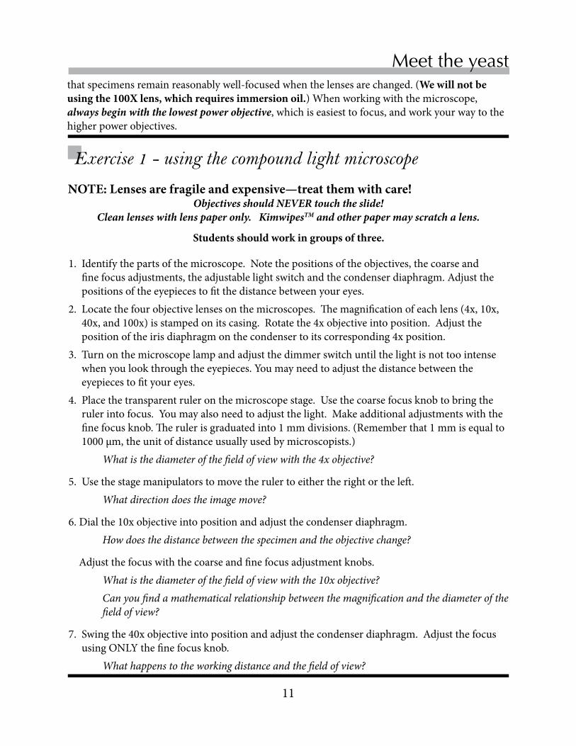
Exercise 1 - using the compound light microscope
1. Identify the parts of the microscope. Note the positions of the objectives, the coarse and fine focus adjustments, the adjustable light switch and the condenser diaphragm. Adjust the positions of the eyepieces to fit the distance between your eyes.
2. Locate the four objective lenses on the microscopes. The magnification of each lens (4x, 10x, 40x, and 100x) is stamped on its casing. Rotate the 4x objective into position. Adjust the position of the iris diaphragm on the condenser to its corresponding 4x position.
3. Turn on the microscope lamp and adjust the dimmer switch until the light is not too intense when you look through the eyepieces. You may need to adjust the distance between the eyepieces to fit your eyes.
4. Place the transparent ruler on the microscope stage. Use the coarse focus knob to bring the ruler into focus. You may also need to adjust the light. Make additional adjustments with the fine focus knob. The ruler is graduated into 1 mm divisions. (Remember that 1 mm is equal to 1000 µm, the unit of distance usually used by microscopists.)
What is the diameter of the field of view with the 4x objective?
5. Use the stage manipulators to move the ruler to either the right or the left. What direction does the image move?
6. Dial the 10x objective into position and adjust the condenser diaphragm. How does the distance between the specimen and the objective change?
Adjust the focus with the coarse and fine focus adjustment knobs. What is the diameter of the field of view with the 10x objective? Can you find a mathematical relationship between the magnification and the diameter of the
field of view?
7. Swing the 40x objective into position and adjust the condenser diaphragm. Adjust the focus using ONLY the fine focus knob.
What happens to the working distance and the field of view?
that specimens remain reasonably well-focused when the lenses are changed. (We will not be using the 100X lens, which requires immersion oil.) When working with the microscope, always begin with the lowest power objective, which is easiest to focus, and work your way to the higher power objectives.
Students should work in groups of three.

12
Chapter 2
Exercise 2 - observing yeast cultures with the microscopeEach student will prepare one slide. Students should observe the specimens on each other’s slides.
1. Prepare three slides: In this experiment, you will prepare three slides, each of which contains two different samples for easy comparison. The slides are large enough to accommodate two samples—and two coverslips. Number the slides with a Sharpie (Use the frosted area, if the slide has one.) As you work, be sure to record which of the two samples is closer to the labeled end of the slide. Use the space provided on the opposite page to record your data.
Slide 1: compare log phase cultures of S. pombe and S. cerevisiae. Slide 2: compare log and stationary phase cultures of S. cerevisiae. Slide 3: compare log and stationary phase cultures of S. pombe.
2. Prepare concentrated cell suspensions. • Concentrate the cells in your log phase yeast cultures by centrifuging the culture tube for a
count of 10 in a microcentrifuge set at top speed. Hold down the Quick button on the Labnet microcentrifuges or the button between the two dials on the Eppendorf microcentrifuges.
• Use a transfer pipet to remove most of the culture medium, until the medium just covers the cell pellet.
• Resuspend the cells with the vortex mixer. • Note: It is not necessary to centrifuge the stationary phase cultures, which are more concen-
trated than the log phase cultures.
3. Transfer and stain the cell samples• Transfer a very small drop (the size of this o) of each cell suspension to the slide, using a dis-
posable pipet. • Stain the cells by adding a drop of Gram’s Iodine to each cell suspension. The drop of iodine
should be about three times greater than the drop of cells. • Cover each sample with a coverslip.
4. Observe the cells and record your observations• Use the same sequence of microscope adjustments that you used in the ruler exercise to
visualize the cultures. Start at low magnification and gradually increase the magnification, making changes in the condenser diaphragm as needed. (Play with the position of the aperture diaphragm a bit to maximize the quality of the image.)
• In the space provided, draw some examples of the forms that you see in the cultures and the relative proportions of each form. Comment on both the sizes and shapes of the cells.
What differences did you observe between the two species? What differences did you observe between stationary and log phase cells ?
Were there any differences in iodine staining between species or growth phases? What would that infer about glycogen storage?

13
Replace Chapter number and title on A-Master Page.<- ->
Meet the yeast
Slide 1: Compare S. cerevisiae and S. pombe log phase cultures
Slide 2: Compare S. cerevisiae log and stationary phase cultures
Slide 3: Compare S. pombe log and stationary phase cultures
When you are finished with your observations, dispose of the slides in the glass waste.

14
Chapter 2
References
Mortimer, RK (2000) Evolution and variation of the yeast (Saccharomyces) genome. Genome Res 10: 403-409.
Turner, JJ, Ewald, JC & Skotheim, JM (2012) Cell size control in yeast. Curr Biol 22: R350-R359.Werner-Washburne, M, Braun, E, Johnston, GC & Singer, RA (1993) Stationary phase in the yeast
Saccharomyces cerevisiae. Microbiol Rev 57: 383-401.

• Learn to select and adjust micropipettes • Learn to accurately transfer microliter volumes
• Use the spectrophotometer to measure absorbance
• Understand experimental errors in measurements
Welcome to the microworld! In this class, you are working with microorganisms, including yeast and bacteria, millions of which would fit into a period on this page. You will also be working with costly reagents, such as plasmids and enzymes. Therefore, in every experiment, you will be required to accurately measure volumes as small as a few microliters (µL). Micropipettes will allow you to do this accurately and precisely.
Chapter 3
Objectives
Mastering the micropipette

16
Chapter 3
Using micropipettes correctly Arguably, the most important scientific equipment that you will use this semester are adjustable micropipettes. Micropipettes are precision instruments that are designed to accurately transfer volumes in the microliter range. You may use microliters or milliliters as the units of volume in your lab notebooks and lab reports, but be careful to always state the volume unit that you are using. Recall the relationships between volume units:
1 microliter (abbreviated µL) = 10-3 milliliter (mL) or 10-6 liter (L)(A useful tip for Mac users: The keyboard shortcut for the Greek letter µ is Alt-m)
Accuracy and precision Ideally, micropipettes will deliver liquids with accuracy and precision. Accuracy depends on the micropipette delivering the correct volume. Precise results are reproducible. Let’s use a target analogy to demonstrate the difference between accurate and precise results. Imagine that four students try to hit the bulls-eye five times. Students A and B are precise, while students A and C are accurate.
The best way to determine the accuracy and precision of micropipettes is to use them to weigh set volumes of distilled water on an analytical balance. The density of water is 1.0 gram per mL at 25˚C. The process is repeated several times during the calibration process, and the data is used to calculate the accuracy and precision of a micropipette.
Accuracy refers to the performance of the micropipette relative to a standard (the intended) value. Accuracy is computed from the difference between the actual volume dispensed by the micropipette and the selected (intended) volume. Note that this can be a negative or positive value. When micropipettes are calibrated, the accuracy is normally expressed as a percent of the selected value. In general, micropipettes are designed to operate with accuracies within a few percent (generally <3%) of the intended value. The accuracy of a micropipette decreases somewhat, however, when micropipettes are set to deliver volumes close to the lowest values in their range.

17
Replace Chapter number and title on A-Master Page.<- ->
Micropipettes
Precision provides information about reproducibility, without any reference to a standard. Precision reflects random errors that can never be entirely eliminated from a procedure. Precision is expressed as the standard deviation (s )of a set of measurements. Assuming random error, ~2/3 of measurements will fall within one standard deviation of the mean, and 95% of measurements will fall within two standard deviations of the mean.
Choosing the micropipette There are three different sizes of micropipettes in the laboratory, which we will refer to as the P20, P200 and P1000. Our micropipettes have been purchased from several different manufacturers, but the principles of operation are the same. The numbers after the “P” refer to the maximum number of microliters that the micropipette is designed to transfer. Use the chart below to select the correct micropipette for an operation. Note that there is some overlap in the ranges of the different micropipettes. For example, both the P200 and P20 can be used to transfer 15 µl, but the P20 is more accurate within that range. As a rule of thumb, always select the small-est volume pipette that will transfer the volume.
Micropipette Recommended range (µL) Smallest increment (µL)P20 1 - 20 0.02
P200 20 - 200 0.2P1000 100 - 1000 2.0
Micropipettes use disposable plastic tips. The P1000 tips are larger than those used with P200s and P20s. P1000 tips may be either natural/clear or blue, depending on the vendor, while P20 and P200 tips may be either yellow or natural/clear in color.
Specifying the transfer volume There are three numbers on the volume indicator. With each of the micropipettes, you will specify a volume to three digits by turning the volume adjustment knob. You will also be able to extrapolate between the lowest numbers with the vernier marks on the lower dial. Most of the measurements you will make with the micropipettes will be accurate to four significant figures!
NEVER turn the indicator dial beyond the upper or lower volume limits of the micropipette! This could damage the piston.

18
Chapter 3
Transferring volumes accurately Micropipettes work by air displacement. The operator depresses a plunger that moves an internal piston to one of two different positions. The first stop is used to fill the micropipette tip, and the second stop is used to dispense the contents of the tip. As the operator depresses the plunger to the first stop, an internal piston displaces a volume of air equal to the volume shown on the volume indicator dial. The second stop is used only to dispense the contents of the tip.
Start First stop Second stop
Filling the micropipette• Remove the lid from the box containing the correct
micropipette tips. • Attach the tip by inserting the shaft of the micropipette
into the tip and pressing down firmly (figure on right). This should produce an airtight seal between the tip and the shaft of the micropipette.
• Replace the lid of the tip box to keep the remaining tips sterile. Avoid touching the tip (especially the thinner end), because the tips are sterile.
• Depress the plunger of the micropipette to the FIRST stop. • Immerse the tip a few millimeters below the surface of the solution being drawn up into the
pipette. Pipetting is most accurate when the pipette is held vertically. Keep the angle less than 20˚ from vertical for best results.
• Release the plunger S L O W L Y, allowing the tip to fill smoothly. Pause briefly to ensure that the full volume of sample has entered the tip. Do NOT let the plunger snap up. This is particularly important when transferring larger volumes, because a splash could contaminate the shaft of the micropipette. If you inadvertently contaminate the shaft, clean it immediately with a damp Kimwipe.
NEVER rest a micropipette with fluid in its tip on the bench!

19
Replace Chapter number and title on A-Master Page.<- ->
Micropipettes
Dispensing the contents of the micropipette• Place the micropipette tip against the side of the receiving test tube.
Surface tension will help to dispense the contents of the micropipette. Do NOT attempt to eject the contents of the micropipette into “thin air.”
• Smoothly depress the plunger to the first stop. Pause, then depress the plunger to the second stop. The contents of the pipette should have been largely released at the first stop. The second stop ensures that you’ve released the “last drop.”
• Use the tip ejector to discard the tip.
WARNING: The most common - and serious - operator error is depressing the plunger to the second stop before filling the micropipette tip.
DO NOT DO THIS!!!
Using the spectrophotometer to evaluate your pipetting skills
Since you will be using micropipettes for all of your experiments, the quality of your results will depend on proper operation of the micropipette. Today’s laboratory will lead you through some exercises that will show you how to use micropipettes correctly and point out some common pitfalls associated with their use. Your results will also provide information about whether the pipettes are functioning properly.
In these exercises, you will be using the spectrophotometer to determine if your pipetting is accurate and precise. You will be using micropipettes to combine various volumes of water and solutions of a blue dye, bromophenol blue (BPB). You will measure the absorbance of the resulting solutions at 590 nm (A590 ), which is close to the absorbance maximum of bromophenol blue at neutral pH. Measuring errors will be reflected in the spectrophotometer readings.
The spectrophotometer readings provide an indirect measurement of pipette performance. The proper way to calibrate the micropipettes would be to weigh out volumes of water, which has a specific gravity of 1.0 g/mL. Unfortunately, we do not have enough balances with sufficient accuracy for the class to perform the measurements. If you suspect inaccuracies in the micropipettes that you are using, refer them to the teaching staff, who will test them properly.

20
Chapter 3
Light spectroscopy Spectrophotometers measure the amount of light absorbed by a sample at a particular wavelength. The absorbance of the sample depends on the electronic structures of the molecules present in the sample. Measurements are usually made at a wavelength that is close to the absorbance maximum for the molecule of interest in the sample.
The diagram below shows the elements present in a typical spectrophotometer. The light sources used in most spectrophotometers emit either ultraviolet or visible light. Light (Io)passes from a source to a monochromator, which can be adjusted to allows only light of a defined wavelength to pass through. The monochromatic (I) light then passes through a cuvette containing the sample to a detector.
The spectrophotometer compares the fraction of light passing through the monochromator (I0) to the light reaching the detector (I) and computes the transmittance (T) as I/I0. Absorbance (A) is a logarithmic function of the transmittance and is calculated as:
A = log10(1/T) = log10(I0/I)
Spectrophotometers can express data as either % transmittance or absorbance. Most investigators prefer to collect absorbance values, because the absorbance of a compound is directly proportional to its concentration. Recall the Lambert-Beer Law, traditionally expressed as:
A = e b C
where e is the molar extinction coefficient of a compound, b is the length of the light path through the sample, and C is the molar concentration of the compound. Cuvettes are formulated to have a 1 cm light path, and the molar extinction coefficient is expressed as L/moles-cm. Consequently, absorbance is a unitless value.
21
Replace Chapter number and title on A-Master Page.<- ->
Micropipettes
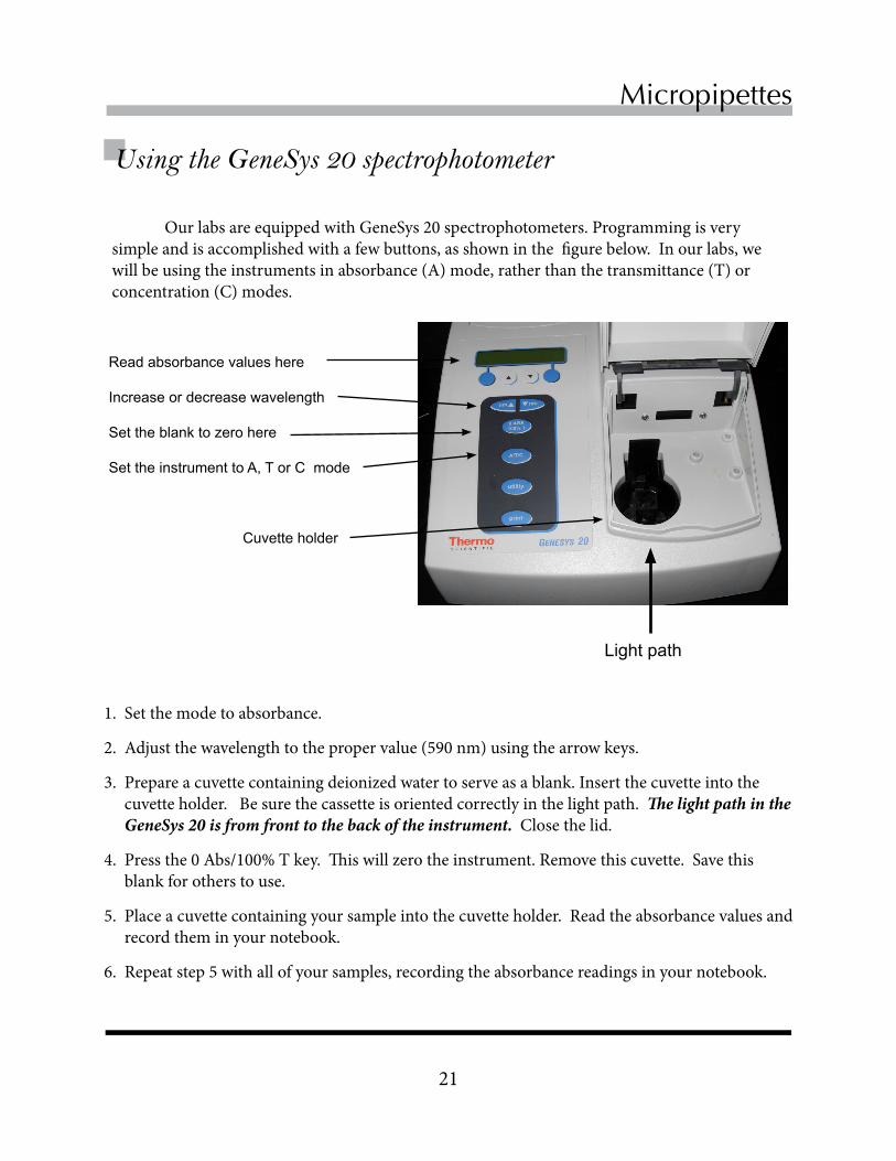
Our labs are equipped with GeneSys 20 spectrophotometers. Programming is very simple and is accomplished with a few buttons, as shown in the figure below. In our labs, we will be using the instruments in absorbance (A) mode, rather than the transmittance (T) or concentration (C) modes.
1. Set the mode to absorbance.
2. Adjust the wavelength to the proper value (590 nm) using the arrow keys.
3. Prepare a cuvette containing deionized water to serve as a blank. Insert the cuvette into the cuvette holder. Be sure the cassette is oriented correctly in the light path. The light path in the GeneSys 20 is from front to the back of the instrument. Close the lid.
4. Press the 0 Abs/100% T key. This will zero the instrument. Remove this cuvette. Save this blank for others to use.
5. Place a cuvette containing your sample into the cuvette holder. Read the absorbance values and record them in your notebook.
6. Repeat step 5 with all of your samples, recording the absorbance readings in your notebook.
Using the GeneSys 20 spectrophotometer
Read absorbance values here
Increase or decrease wavelength
Set the blank to zero here
Set the instrument to A, T or C mode
Cuvette holder
Light path

22
Chapter 3
Exercise 1 - Getting the feel of micropipettes
Concept: Micropipettes work by an air displacement mechanism
1. Set the P200 to deliver 200 µL. Be careful not to overshoot, which could damage the pipette piston.
2. Grip the pipette by wrapping your fingers around the barrel. Use your thumb to depress the plunger to its first stop.
3. Next press the plunger to the second stop. Compare the distance that the plunger moved dur-ing the first and second strokes.
4. Set the P200 to deliver 20 µL and depress the plunger to its first stop. Compare the distance that the plunger moved when the P200 was set to 200 or 20 µL.
5. Depress the plunger to the second stop. How does the distance between the first and second stops compare for 200 and 20 µL?
6. Set the P20 to deliver 20 µL. Depress the plunger to the first stop. Compare the distance to the first stop when a P20 and P200 are set to deliver 20 µL.
Concept: The filling and dispensing strokes are different.
1. Place a tip on the shaft of the P200.
2. Set the P200 to deliver 50 µL.
3. Draw up 50 µL of 0.05% BPB solution into the pipet.
4. Dispense the BPB into a microcentrifuge tube down to the first stop, holding the tip against the wall of the tube. Note whether all of the dye has been expelled. Push the plunger down to the second stop to release any remaining BPB.

23
Replace Chapter number and title on A-Master Page.<- ->
Micropipettes
Exercise 3 - How precise and accurate are your transfers? Work in groups of three. One person in the group should work with the P-20, another with the P-200 and the third with the P-1000. Each person should prepare three identical samples and then determine the A590 of the three samples. From the data, you will be able to determine if the micropipette is measuring volumes correctly.
1. Each person in your group of three will work with a different micropipette and perform the same transfers in triplicate, as detailed below. The final volume (water + BPB) in each tube will be 1.0 mL. Calculate the volume of water that will need to be combined with each of the following to give 1.0 mL, and record your calculations in your lab notebook:
Group member A: Use the P-20 to transfer 10 µL of 0.05% BPB to ____ µL of water. Group member B: Use the P-200 to transfer 100 µL of 0.005% BPB to ____ µL of water. Group member C: Use the P-1000 to transfer 300 µL of 0.005% BPB to ____ µL of water.
2. To minimize our plastic waste, strategize how to minimize the number of tips that you use without contaminating the stock solutions. A tip can be used multiple times, but a tip that has been used for BPB cannot be used to subsequently transfer deionized water. Combine the BPB solution and water to give a final volume of 1.0 mL.
1. Use the P1000 to add 990 µL of water to two microcentrifuge tubes. Label the tubes A and B. Dispose of used tips in the containers provided.
2. Use a P20 to correctly transfer 10 µL of 0.05% BPB to tube A. Make a mental note of what fraction of the pipet tip is filled with the dye. Use the vortex mixer to disperse the BPB in the water.
3. Use a P20 to INCORRECTLY transfer 10 µL of 0.05% BPB to tube B. Do this by depressing the plunger to the second stop before you take up the BPB solution. Make a mental note of how well the dye fills the tip this time.
4. Set the wavelength of the spectrophotometer to 590 (A590). Pipette 1 mL of water into a plastic cuvette and blank the spectrophotometer at this wavelength.
5. Read the A590 of the solutions in tubes A and B, in the spectrophotometer. How do the two readings compare? What kind of error results from drawing solution into the pipette incorrectly?
Lab exercise 2 - How NOT to pipette!

24
Chapter 3
3. Measure the A590 of the three solutions with the spectrophotometer and record the data in your notebook.
4. Compute the mean and standard deviations for your three measurements, using either a calculator or Excel. The standard deviation reflects the precision of your measurements.
5. Enter these values on the chart that your TA has prepared on the whiteboard. Compare the values that your group obtained each of the three pipettes with the aggregated class measurements. If the averages that your group obtained are significantly different than those that other groups obtained with the same micropipette, your micropipette may not be transferring volumes accurately.
Notify your TA if any of the micropipettes are not performing properly. Your TA will follow up on your observations and test the micropipettes with the gravimetric test described earlier in the chapter.
Test yourself As part of an interview for a research position, three applicants are asked to transfer
150 µL of distilled water with a P-200 micropipette to a weighing paper and to determine the weight of each drop with an analytical balance. The three measurements made by each of the applicants are listed below. Use the space below to calculate the mean and standard deviation of the measements made by each student.
Applicant A: 0.161 g, 0.147 g, 0.142 g Applicant B: 0.158 g, 0.156 g, 0.157 g Applicant C: 0.143 g, 0.153 g, 0.150 g
Which applicant makes the most precise measurements?
Which applicant makes the most accurate measurements?
• Understandthephasesofmicrobialgrowth
• Learnandpracticesteriletechniquesusedtocultureyeastandothermicroorganisms
• Learnhowtousespectrophotometryandspotplatingtoestimatethenumberofcellsinyeastcultures
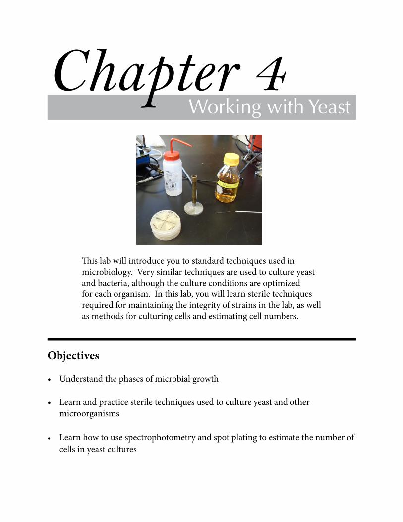
Thislabwillintroduceyoutostandardtechniquesusedinmicrobiology.Verysimilartechniquesareusedtocultureyeastandbacteria,althoughthecultureconditionsareoptimizedforeachorganism.Inthislab,youwilllearnsteriletechniquesrequiredformaintainingtheintegrityofstrainsinthelab,aswellasmethodsforculturingcellsandestimatingcellnumbers.
Working with YeastChapter 4
Objectives

26
Chapter 4 Throughoutthesemester,youwillbeworkingwithculturesofyeast.Thetechniquesusedforyeastandbacteriaaresimilar,exceptthatthemediacompositionandculturetemperatureareoptimizedforindividualorganisms.Ingeneral,culturemediacontainacarbonsource,nitrogensource,salts,vitaminsandessentialminerals.Duringthecourseofthesemester,youwilluseanumberofdifferentstrainsofyeastanddifferentkindsofmedia.Yoursuccessinthelabwilldependonyourabilitytousesteriletechnique,whichisneededtomaintaintheintegrityofthedifferentstrains.Anequallyimportantelementinlaboratorysuccessiscarefulbookkeeping!
Inthislab,youwillpreparestockculturesofS. cerevisiae met mutantstrainsonstreakplates,quantifythenumberofviablecellsinculturesofS. cerevisiaeandS.pombe usingspotplates,anduselightscatteringtoquicklyestimatecelldensitiesofthesesameyeastcultures
Sterile technique
SteriletechniqueisESSENTIALwhenworkingwithmicroorganisms!Thissemester,wewillbeworkingwithseveraldifferentstrainsofyeastandbacteria.Thestrainshavedefinedgenotypesthathavebeengeneratedbycarefulplanningandexperimentation.Itisimportanttoprotectstrainsfromcontaminationwithotherlaboratorystrainsandfromthemanyundefinedmicrobesintheenvironment.Largenumbersofdiversemicroorganismsareallaroundus-intheair,onlaboratorysurfaces,onyourskinandonyourclothing.Truetotheirname,microorgan-ismsaretoosmalltobedetectedbytheeye,buttheygrowrapidlyinlaboratoryculturemedia.Correcttransfertechniquesandtheuseofsterilereagentsareusuallyenoughtopreventcontami-nationofvaluablelaboratorystrains.
Somesimpleprecautionswillreducethepossibilityofcontamination:• Beforeworkingwithstrains,wipedownasmallworkingareaonthelabbenchwith70%
ethanol.• Usesterilereagents,micropipettetips,andtesttubes.Tipsandmicrocentrifugetubesshould
bekeptincoveredcontainerswhennotinuse.• Minimizecontaminationfromclothingandbodysurfaces.Pullbackandsecurelonghair.
Avoidtouchingorbreathingonsterilesurfacesthatwillcontactmicroorganisms.• Avoidtalkingwhenyouaretransferringstrains.• Minimizethetimethatthecapsareremovedfromvesselscontainingmicroorganismsor
sterilemedia.Capsshouldalwaysbekeptright-sideuptopreventcontaminationfromair-bornemicrobesfallingintothecaps.
Theculturemediaandreagentsthatwewillusehavebeensterilizedbyeitherautoclavingorfiltration.Anautoclave(oppositepage)isachamberthatusespressurizedsteam

27
Replace Chapter number and title on A-Master Page.<- ->
Working with yeast
Forroutineculture,scientistsusuallyuserichmediathatsupplyallthenutrientsthatcellsneedtogrow.Theindividualcomponentsofrichmediaareoftenundefined.Forexample,yeastarecommonlygrowninamediumknownasYPD,whichissimpleandinexpensivetoprepare.The“Y”inYPDreferstoayeastextract,whichcontainsthewater-solublecompoundsgeneratedwhenyeastareforcedtoself-digest.(ThoseofyouwhohavevisitedAustraliamayhaveencoun-teredyeastextractinthepopularspread,Marmite.)The“P”referstopeptone,amixtureofpeptidesandaminoacidspreparedbydigestinganimalproteinwithproteases.The“D”referstodextrose,orglucose,whichisthefavoredcarbonsourceofyeast(Sherman,2002).
BecauseYPDiscomposedlargelyofcrudeextracts,itscompositionmayshowsignificantbatch-to-batchvariation.Thisvariationisrarelyaproblem,however,becauseYPDcontainsmorethanenoughessentialnutrientstosatisfythemetabolicrequirementsofcells.Manyexperiments,however,requiremediawithadefinedcomposition.Tomeetthisneed,theyeastcommunityhasdevelopedseveralvarietiesofdefinedsyntheticmediathatsupportthegrowthofmoststrains.Individualcomponentsofthesyntheticmediamaythenbemanipulatedtosuittheneedsofanexperiment.(Laterthissemester,wewillusedefinedmediatoselectforparticulargenotypes.)
Yeastcanbegrowninliquidmediaoronthesurfaceofplatescontainingsolidmedia.Agarisusuallyaddedtoliquidmedia,causingittosolidify.Whencellsaregrowninliquidmedia,itisimpossibletodistinguishcellsthathavedifferentgenotypesfromoneanother.Bycontrast,cellsgrowincoloniesonsolidmedia.Eachcellinacolonyisderivedfromacommonancestorandthecellsarethereforegeneticallyverysimilar,ifnotidentical,toeachother.Formostofourexperimentsthissemester,wewillusesolidmedia,becauseweneedtodistinguishcellswithdifferentgenotypesfromoneanother.
tokillcellsonsurfacesandinsolutions,usingtemperaturesnear121˚Candpressuresfrom30-40psi.(Forcomparison,atmosphericpressureis~15psi.)Filtrationisusedintheplaceofautoclavingwhensolutionscontaintemperature-sensitivecompounds.Theporesinthefiltersusedtoremovemicroorganismsaretypically0.2or0.45µm,whicharesufficientlysmalltopreventthepassageofbacteria.Itisnotdifficulttokeepstocksofmediaandreagentssterileaslongasyouworkquicklyandfollowthedirectionsabove.
Yeast growth media
An autoclave.

28
Chapter 4
Theexponentialgrowthofyeastcanbedescribedbytheequation:
N=N0ekt
whereNrepresentsthenumberofcellsatanytime(t),N0representsthenumberofcellsattimezero.Scientistsoftenfinditconvenienttothinkofthegrowthconstantkintermsofthedoublingtimeoftheculture.Inthisrendering,k=ln2/T(T=thedoublingtimeoftheculture).Thegrowthrateofyeastvarieswithtemperature.Yeastgrowwellatroomtemperature,buttheygrowmorerapidlyat30˚C,whichwewilluseforourexperiments.At30˚C,wild-typeyeasthaveadoublingtimeof~90minutesinYPD.Wewillalsomakesurethattheculturesarewell-aeratedbygrowingthemoneitherrotaryshakersorarotatingwheel.
Afterafewdoublingtimes,cellsbegintodepletethenutrientsintheculture,theirgrowthrateslows,andthecellsenterstationary phase.Cellsinstationaryphasehaveadifferenttranscriptionalprogramthatallowsthemtosurviveinthelessfavorableenvironment,sometimesforconsiderablelengthsoftime.Instationaryphase,therateofcelldivisionissimilartotherateofcelldeath,sothenumberofcellsdoesnotchangeappreciably.Eventually,cellsenterdeath phase ifconditionsdonotimprove.
Yeast growth phases Whenyeastaregrowninliquidmedium,theculturefollowsawell-establishedpatternformicrobialgrowth.Culturesareusuallystartedbyinoculatingmediawithasmallnumberofcells.Alagphasefollowstheinoculation,duringwhichcellsbecomeacclimatedtothenewenvironmentandbegintoconditionthemediawiththeirownmetabolites.Lag phaseisfollowedbyanexponential, or log phase,whenthenumberofcellsincreasesexponentially.

29
Replace Chapter number and title on A-Master Page.<- ->
Working with yeast
Exercise 1 - Streak plates Microbiologistsliketobegintheirexperimentswithasinglecolony,becausethecellsinacolonyaretheprogenyofasinglecell.Aconcerninallgeneticexperimentsisunknownmutationsthatarisespontaneouslyandmayaffectthephenotypebeingstudied.Spontaneousmutationsariseconstantlyinallcells,witharateofapproximately10-8/base/generation.ForS. cerevisiae,withagenomeof12Mbp,mostcellswillhaveaccumulatedatleastonemutationbythetimethatthey’veundergone9-10divisions.Acolony,whichhashundredsofmillionsofcells,isthereforeapopulationofgeneticallyverysimilar,butnotnecessarilyidentical,organisms.
Researcherscommonlyusestreakplatestoisolatesinglecolonies.Astreakplateisactuallyaserialdilutionofanexistingcultureonsolidmedia.Researchersbeginastreakbypickingupasmallsampleofyeastoranothermicroorganismwithasterileloop,woodenapplicatorstick,toothpickorpipettetip.Theythenspreadtheculturebymakingaseriesofzig-zagstrokesacrossthesurfaceoftheplate.Thenumberofcellsonthelooportoothpickdecreasesasthestreakprogresses.Consequently,streaksappearthickestattheirstartingpoints,andthestreakthicknessdecreasesuntilitispossibletodetectwell-isolatedsinglecoloniesneartheendofthestreak.Becauseitmaybedifficulttoresolvecoloniesfromasinglestreak,manylabsuseaseriesofstreaksonthesameplatetoseparatecolonies.Eachnewstreakisdonewithafreshlysterilizedlooportoothpickthatpicksupcellsbycrossingoverthetracksofthepreviousstreak,beforebeginninganewseriesofzig-zags.Inourexperiments,wewilluseamulti-streakprotocol,whichallowsustoculturemultiplestrainsonasingleplateofculturemedium.(Seethefigurebelow.)Thestreakplatesthatyoupreparewillbeusedasstocksforfutureexperiments.Asyoustreakyourcultures,paycarefulattentiontodetailtoavoidcross-contaminationorconfusionabouttheidentitiesofindividualstrains.
Streak plate with three sectors. Plate has been divided into three clearly labeled sectors. Three streaks were used to spread the cells in each sector. The third streak in each sector contains well-separated colonies that can be used for genetics experiments.
Streak 1 zig-zag
Streak 2 vertical
Streak 3 zig-zag
30
Chapter 4
Preparing a streak plate
1.YourteamwillbeassignedthreedifferentS. cerevisiae metstrainstoculture.Gathertheparentstrainstobepropagated,steriletoothpicks,andagarplate(s)withtheappropriatefreshmedia.
2.Dividetheplateswithfreshmediaintosectorsbymarkingthebottomofeachplatewithamagicmarker.CLEARLYlabeleachsectorwithacodeforthestrainthatwillbestreakedinit.Keepthelabelsattherimoftheplateandusesmallletters.Note your initials and the date.
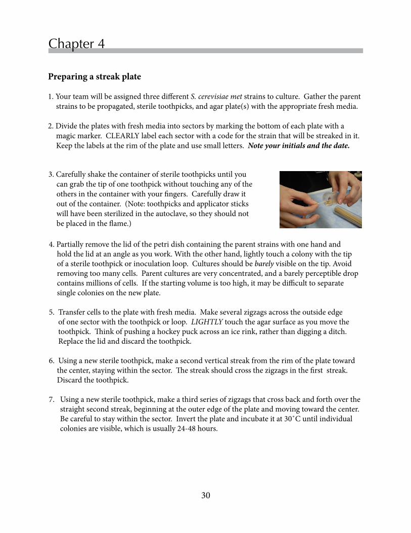
3.Carefullyshakethecontainerofsteriletoothpicksuntilyoucangrabthetipofonetoothpickwithouttouchinganyoftheothersinthecontainerwithyourfingers.Carefullydrawitoutofthecontainer.(Note:toothpicksandapplicatorstickswillhavebeensterilizedintheautoclave,sotheyshouldnotbeplacedintheflame.)
4.Partiallyremovethelidofthepetridishcontainingtheparentstrainswithonehandandholdthelidatanangleasyouwork.Withtheotherhand,lightlytouchacolonywiththetipofasteriletoothpickorinoculationloop.Culturesshouldbebarelyvisibleonthetip.Avoidremovingtoomanycells.Parentculturesareveryconcentrated,andabarelyperceptibledropcontainsmillionsofcells.Ifthestartingvolumeistoohigh,itmaybedifficulttoseparatesinglecoloniesonthenewplate.
5.Transfercellstotheplatewithfreshmedia.Makeseveralzigzagsacrosstheoutsideedgeofonesectorwiththetoothpickorloop.LIGHTLYtouchtheagarsurfaceasyoumovethetoothpick.Thinkofpushingahockeypuckacrossanicerink,ratherthandiggingaditch.Replacethelidanddiscardthetoothpick.
6.Usinganewsteriletoothpick,makeasecondverticalstreakfromtherimoftheplatetowardthecenter,stayingwithinthesector.Thestreakshouldcrossthezigzagsinthefirststreak.Discardthetoothpick.
7.Usinganewsteriletoothpick,makeathirdseriesofzigzagsthatcrossbackandforthoverthestraightsecondstreak,beginningattheouteredgeoftheplateandmovingtowardthecenter.Becarefultostaywithinthesector.Inverttheplateandincubateitat30˚Cuntilindividualcoloniesarevisible,whichisusually24-48hours.

31
Replace Chapter number and title on A-Master Page.<- ->
Working with yeast
Exercise 2 - Spot plates
YouwillusespotplatestoestimatethecelldensitiesoflogphaseandstationaryphaseculturesofS. cerevisiaeandS. pombe.Scientistsusespotplatesbothtocalculatethenumberofcellsinculturesandtoobtaininformationaboutthegrowthpropertiesofstrainsondifferentmedia.Thefigurebelowshowsanexampleofatypicalspotplate.Eachrowrepresentsadilutionseriesfromadifferentyeastculture.Thesamevolumeofdilutedcultureisusedforeachspot.Thedilutionseriesisplannedsothatthemostdilutespotscontainsasmallnumberofindividualcoloniesthatcanbedistinguishedfromoneanother,typicallylessthanten.
Spot plate.Each row on the plate represents a series of 1:10 dilutions of a liquid culture of S. cerevisiae. Five µL of each dilution was spotted on the plate. The plate was incubated for two days at 30˚C. Individual colonies are apparent at the highest dilution of each extract.
Mostcommonly,investigatorsmakeaseriesof1:10dilutionsinsterile(NOT deionized)waterandthenspotafewmicrolitersofeachdilutioninarow.Inthisexperiment,5µLaliquotswerespottedfromtheserialdilutions.Notethatit’spossibletocountindividualcoloniesinthemostdilutesamples.Thisinturnenablesyoutocalculatethenumberofviablecellsintheoriginalculture.Inthetoprow,youcandistinguish4coloniesinthesamplethathasbeen100,000-fold.Theoriginalculturewouldhavecontained400,000cellsin5µL,whichcorrespondsto80millioncellspermL(8x10⁷cells/mL).
Preparing a spot plate
1.Alignmentgridsareusefulforpreparinggood-lookingspotplates!Obtainanalignmentgrid(right)andmarkthetargetpositionsforculturedilutions.Placeanorientationmarkatonepointalongthecircumference.
2.Labeleachpetridishwithyourinitialsanddatewith
smalllettersaroundtheBOTTOMrimofthedish.Putahashmarkonthebottomedgeoftheplatetoserveasanalignmentmarker.

32
Chapter 4
3.Prepareaseriesoffive1:10dilutionsfromeachcultureusingsteriledistilledwater.(Diagramsinyourlabnotebookareoftenhelpfulindesigningdilutionseries.)Toprepareaserialdilution,firstpipette90µLsterilewaterintofivemicrocentrifugetubes.Next,useaP20totransfer10µLfromtheoriginalcultureintothefirsttube.Vortexthecellsuspension,andthentransfer10µLfromthistubetothesecondtubeintheseries,andsoon.UsethesameP20pipettetipfortheentiredilutionseries.Ejectthetipintotheappropriatewastecontainer.
4.Beginningwiththelastdilutionintheseries,spot5µLspotsinarow.Again,youwillbeabletouseasinglepipettetipforadilutionseries,sinceyoustartedwiththemostdilutesample.
5.Repeatstep3foreachculturethatyouareanalyzing.Becarefultonoteinyourlabnotebookwhichculturehasbeenspottedintoeachrowontheplate!
6.Leavetheplaterightsideupfor~30minutes,toallowtimefortheyeastdilutionstosettleandadsorbtothemedium.

33
Replace Chapter number and title on A-Master Page.<- ->
Working with yeast
7.Inverttheplatesandincubatethemat30°C.Platesareinvertedtopreventwaterdropletsthatformontheinnersurfaceofthelidfromfallingonthecolonies.Platescanalsobekeptatroom
temperature,butcellswillgrowmoreslowly.DoNOTincubatethecellsabove30°C,whichstressestheyeast.
8.Whenthecoloniesarelargeenoughtocount,theplateswillberemovedfromtheincubatorandplacedintherefrigeratororcoldroomforyouranalysislater.
9.Recordyourdatawiththescanner.To do this, remove the top from the plate and invert both the plate and the lid. Place the bottom half of the dish on the scanner and leave the inverted lid on the bench. (The lid is inverted to avoid contamination from spores and microorganisms that may be present in the air.) Place a black piece of cardboard or a folder over the plates before lowering the lid of the scanner.
10.Usespotswhereyoucancountindividualcoloniestocalculatethedensityofcellsintheoriginalcellculture,correctingforthedilutionsthatyouusedandthevolumeofthespot.
Exercise 3 - Estimating cell densities with a spectrophotometer
Thespectrophotometerprovidesa“quickanddirty”waytoestimatethedensityofcellsinaculture.Incontrasttospotplates,whichmustbeincubatedforseveraldaysbeforecoloniesappear,spectrophotometerreadingscanbeinstantlyconvertedintocelldensities.Ontheotherhand,themethoddoesnotdiscriminatebetweenlivinganddeadcells.Thespectrophotometricmethodtodeterminecellnumberisbasedonlightscattering.Mostvisiblelightcannotpenetrateacell.Whenthelightbeaminaspectrophotometerhitsacell,thelightisdeflectedfromthelightpath,sosomeofthelightneverreachesthedetector.Thegreaterthenumberofcellsinasample,themorelightscatteringthatoccurs.Thelightscatteringabilityofacelldependsonitssizeandgeometry,soacalibrationcurveisnecessarytoextrapolateopticaldensitymeasurementstocellnumber.Forexample,thesamenumberofyeastcellswouldscatterlightmorethanthesamenumberofbacterialcells,becausethebacterialcellsaremuchsmaller.
Lightscatteringismeasuredwiththespectrophotometersettoreportabsorbance.Becausetheprinciplesusedtomeasurelightscatteringandabsorbancearedifferent,theamountoflightscatteredbyasolutionisreferredtoasits“opticaldensity”ratherthanits“absorbance.”Theopticaldensityofasampleanalyzedat600nmisabbreviatedOD600,withthesubscriptindicatingthewavelengthusedforthemeasurement.
34
Chapter 4
Estimating cell densities with the spectrophotometerFollow the directions in Chapter 3 (p. 21) for operating the GeneSys 20. 1.TurnontheGeneSys20spectrophotometer.Adjustthewavelengthofthemonochromatorto
600nm.
2.Fillacuvettewith1.0mLdeionizedwaterandorientthecuvetteintheholdersothattheflatsideofthecuvettefacesthefrontoftheinstrument.(Note:itisnotnecessarytousecultureme-
diumastheblank.)3.Closethelidandpressthe“0Abs/100%T”buttontoestablishabaselinevalueforfurther
measurements.
4.Removethecuvettefromtheinstrumentandreplacethewaterwith1mLofundilutedculture.ClosethelidandreadtheOD600.Recordthisvalueinyourlabnotebook.Iftheopticalden-sityofthesampleisgreaterthan1.0,dilutethesample1:10withdeionizedwaterandreadtheopticaldensityagain.(ThelinearrelationshipbetweentheOD600andcelldensityislostwhenOD600valuesexceed1.0)Recordthenewvalueinyourlabnotebook,notinghowyoudilutedyoursample.Disposeofallcellmaterialinthewhiteliquidwastecontainer.
5.Repeatstep4witheachofyourcultures.
6.Calculateanapproximatecelldensityforeachsample,assumingthatanOD600of1.0corre-spondstoapproximately1.3x107cells/mL.UseonlydatawheretheOD600islessthan1.0forthesecalculations.
References
Sherman,F(2002)Gettingstartedwithyeast.Meth Enzymol 350:3-41.

• Learnhowinformationisprocessedindatabases• UnderstandhowtheSaccharomycesGenomeProjectprovidedthereference
sequencesforS. cerevisiaegenes
• UsetheNCBIdatabasestofindDNAandproteinsequenceinformationaboutaMET/CYSgene
• UsetheSaccharomycesGenomeDatabasetofindinformationabouttheproteinencodedbyaMETgeneandtheprotein’sfunctioninmetabolism
Thecomputerbelongsonthebenchtopinthemodernbiologylab,alongwithotheressentialequipment.Anetworkofonlinedatabasesprovidesresearcherswithquickaccesstoinformationongenes,proteins,phenotypes,andpublications.Inthislab,youwillcollectinformationonaMETgenefromavarietyofdatabases.
Chapter 5
Objectives
Introduction to Databases

36
Chapter 5
Biomedicalresearchhasbeentransformedinthepast20yearsbyrapidadvancesinDNAsequencingtechnologies,roboticsandcomputingcapacity.Theseadvanceshaveusheredinaneraofhighthroughputscience,whichisgeneratingahugeamountofinformationaboutbiologicalsystems.Thisinformationexplosionhasspurredthedevelopmentofbioinformatics,aninterdisciplinaryfieldthatrequiresskillsinmathematics,computerscienceandbiology.Bioinformaticiansdevelopcomputationaltoolsforcollecting,organizingandanalyzingawidevarietyofbiologicaldata.Theresultsarestoredinavarietyofsearchabledatabases.Today’sbiologistneedstobefamiliarwithonlinedatabasesandtobeproficientatsearchingdatabasesforinformation.
YourteamhasbeenassignedaMETgenethatyouwillbestudyingforthesemester.Inthenextfewlabs,youwillidentifywhichofyourthreeunknownyeaststrainscarriesadeletioninthatMETgene.Todistinguishbetweenthethreeyeaststrains,youwillneedtoobtaininformationabouttheDNAsequencesthathavebeendisruptedinthemutantstrains.Youwillalsoneedtofindinformationabouttherolesthattheencodedproteinsnormallyplayinmetabolism.Inthislab,youwillsearchforgene-specificinformationinseveralonlinedatabases.Asyouprogressthroughthislab,youmayfeellikeyou’regoingincirclesatpoints,becauseyouare!Therecordsindatabasesareextensivelyhyperlinkedtooneanother,soyouwilloftenfindthesamerecordviamultiplepaths.Asyouworkthroughthischapter,werecommendthatyourecordyoursearchresultsdirectlyintothislabmanual.
Databases organize information Databasesareorganizedcollectionsofinformation.Theinformationiscontainedinindividualrecords,eachofwhichisassignedauniqueaccessionnumber.Recordsinadatabasecontainanumberoffieldsthatcanbeusedtosearchthedatabase.Forasimpleexample,consideraclassroster.StudentsinaclassrosterareidentifiedbyauniqueIDnumberassignedbythecollege,whichservesastheequivalentofanaccessionnumber.Classrosterscontainavarietyoffields,suchasthestudentnames,majors,graduationyearandemailaddresses.Thus,classinstructorsareabletoquicklysearchtherostersforstudentswithaparticulargraduationyearormajor.(Theclassrosterisactuallyaderivativedatabase,becauseitdrawsoninformationfromthemuchlargerstudentinformationdatabasemaintainedbythecollege.)
Informationongenesandproteinsisorganizedintomultipledatabasesthatvarywidelyintheirsizeandfocus.Eachdatabaseassignsitsownuniqueaccessionnumbersandorganizesinformationintofieldsthatwillbeusefultoresearcherswhosearchthedatabase.Oncearecordisacceptedintoadatabase,professionalcuratorstakeover.Curatorsareprofessionalscientistswhoaddvaluetoarecordbyprovidinglinksbetweenrecordsindifferentdatabases.Curatorsalsoorganizetheinformationinnovelwaystogeneratederivativedatabasesthataredesignedtofittheneedsofparticularresearchcommunities.Forexample,scientistsstudyinghistonesorproteinkinasescanaccessinformationinhighlyspecializeddatabases.
37
Replace Chapter number and title on A-Master Page.<- ->
Introduction to Databases
Manyofthelargestdatabasecollectionsreceivesupportfromgovernments,becauseoftheirimportancetobiomedicalresearch.Byfar,thelargestcollectionofdatabasesishousedattheNationalCenterforBiotechnologyInformation(NCBI)intheUnitedStates.NCBIincludesliterature,nucleotide,proteinandstructuredatabases,aswellaspowerfultoolsforanalyzingdata.TheNCBIexchangesdatadailywithsmallercounterpartsinEuropeandJapan,providingmultipleentrypointsintotheinternationalnetworkofdatabases.
It’simportanttokeepinmindthatinformationindatabasesisnotstatic.Scientistsmakemistakesandtechnologycontinuestoimprove.Itisnotuncommontofindchangesinadatabaserecord.Scientistswithaninterestinaparticulargenearewell-advisedtocheckfrequentlyforupdates!
From the research bench to the database Theultimatesourceofinformationindatabasesistheresearchcommunity,whichsubmitstheirexperimentaldatatoprimarydatabases.Primarydatabasesaskinvestigatorsforbasicinformationabouttheirsubmission.Arecordthatmeetsthestandardsofthedatabaseisacceptedandassignedauniqueaccessionnumberthatwillremainpermanentlyassociatedwiththerecord.Eachdatabasehasitsownsystemofaccessionnumbers,makingitpossibletoidentifythesourceofarecordfromitsaccessionnumber.Oncearecordisacceptedintoadatabase,professionalcuratorstakeover.Curatorsareprofessionalscientistswhoaddvaluetoarecordbyprovidinglinksbetweenrecordsindifferentdatabases.Curatorsalsoorganizetheinformationinnovelwaystogeneratederivativedatabases.Derivativedatabases,suchasorganismdatabases,areoftendesignedtofittheneedsofparticularresearchcommunities.Inthiscourse,wewillbeusingbothprimaryandderivativedatabases.Let’slookatafewdatabases.
Theinformationindatabasesoriginatesinexperiments.Thefigureonthefollowingpagesummarizesinformationflowfromthebenchtodatabases.Whenresearcherscompleteanexperiment,theyanalyzetheirdataandcompiletheresultsforcommunicationtotheresearchcommunity.Thesecommunicationsmaytakeseveralforms.
PubMed indexes publications in the biomedical sciences Researcherswillusuallywriteapaperforpublicationinascientificjournal.Reviewersatthejournaljudgewhethertheresultsareaccurateandrepresentanovelfindingthatwilladvancethefield.Thesepeer-reviewedpapersareacceptedbythejournal,whichthenpublishestheresultsinprintand/oronlineform.Aspartofthepublicationprocess,biomedicaljournalsautomaticallysubmitthearticlecitationandabstracttoPubMed,aliteraturedatabasemaintainedbyNCBI.CitationssubmittedtoPubMedareassignedaPMIDaccessionnumber.PMIDnumbersareassignedsequentiallyandthenumbershavegrownquitelarge.Today,PubMedcurrentlycontainsover21millionrecords!PubMeduserscanrestricttheirsearchestofieldssuchastitlework,author,journal,publicationyear,reviews,andmore.TheusabilityofPubMedcontinuestogrow.Usersareabletouseaclipboard,savetheirsearches,andarrangeforRSSfeeds

38
Chapter 5
Information flow from experiments to databases. Researchers analyze their data and prepare manuscripts for publication. Journal citations are submitted automatically to PubMed. Researchers also submit data to more specialized, interconnected databases.

39
Replace Chapter number and title on A-Master Page.<- ->
Introduction to Databases
whennewsearchresultsenterPubMed.StudentsinthebiomedicalsciencesneedtobecomeproficientinusingPubMed.YoucanaccessPubMedatpubmed.govorthroughtheBCLibrary’sdatabaseportal.Anadvantageofusingthelibrary’sportalisthatyouwillbeabletousethepowerful“FindIt”buttontoaccesstheactualarticles.Later in the semester, you will use PubMed to search for articles that will provide background information for your final semester report.
Investigators submit experimental data to specialized research databases
Dependingontheexperiment,researchersmayelecttosubmittheirdatatoadditionaldatabases.Databasesthatacceptdirectsubmissionsfromresearchersareconsideredprimarydatabases.ConsiderthehypotheticalexampleofaresearcherwhohasisolatedanovelvariantofaMETgenefromawildstrainofS. cerevisiaewithasophisticatedgeneticscreen.Theresearcherhassequencedthegene,clonedthegeneintoabacterialoverexpressionplasmid,andcrystallizedtheoverexpressedprotein,whichpossessesuniqueregulatoryproperties.Theresearcherispreparingamanuscriptontheexperiments,andinpreparationforthemanuscriptsubmission(reviewersofthemanuscriptwillwanttoseetheaccessionnumbers),theresearcherplanstosubmitdatatothreedatabases:anucleotidedatabase,astructuredatabaseandanorganismdatabase.
IfourresearcherisworkingataninstitutionintheU.S.,heorshewillprobablysubmitthenucleotidesequencetoNCBI’sGenBank.GenBankwasfoundedin1982,whenDNAsequencingmethodshadjustbeendevelopedandindividualinvestigatorsweremanuallysequencingonegeneatatime.TherateofGenBanksubmissionshasincreasedinpacewithadvancedinDNAsequencingtechnologies.Today,GenBankacceptscomputationallygeneratedsubmissionsfromlargesequencingprojectsaswellassubmissionsfromindividualinvestigators.ThenumberofGenBanksubmissionshasrisentoover135records,includingsequenceinformationforwholegenomes,individualgenes,transcripts,plasmids,andmore.Notsurprisingly,thereisconsiderableredundancyinGenBankrecords.GenBankisnowpartofNCBI’sNucleotidedatabase,togetherwithsmaller,morespecializednucleotidedatabasespreparedbycurators.AmongthesedatabasesisRefSeq,asourceofnonredundantrecordspreparedbyNCBIcurators.In this lab, you will search the Nucleotide database for the reference sequences for S. cerevisiae MET and CYS genes.
Theresearcherinourhypotheticalexamplewillalsowanttosubmittheatomiccoordi-natesandstructuralmodelsforthecrystallizedproteintotheProteinDataBank.ThePDBispartofaninternationalconsortiumthatacceptsdataforproteinandnucleicacids.ThevastmajorityofPDBrecordshavebeenobtainedbyX-raydiffraction,althoughthedatabasealsoacceptsmod-elsobtainedwithnuclearmagneticresonance(NMR),electronmicroscopy,andothertechniques.ThenumberofentriesinthePDBdatabasesisordersofmagnitudesmallerthanthenumberofpredictedproteinsinGenBank,reflectingthedifficultiesinherentindeterminingstructuresofmacromolecules.PDBofferstoolsforvisualizingmacromoleculesinthree-dimensions,allowinginvestigatorstoprobeaminoacidinteractionsthatareimportanttoproteinfunction.

40
Chapter 5
Finally,ourresearcherwillwanttosubmitdataaboutthenewmutant’sphenotypeandthenovelpropertiesoftheproteinencodedbytheMETgenevarianttotheSaccharomycesGenomeDatabase.TheSaccharomycesGenomeDatabase(SGD),whichservesasacentralresourcefortheS.cerevisiaeresearchcommunity(whichnowincludesyou).TheSGDusestheyeastgenomesequenceasitsorganizingstructure.
Saccharomyces genome project provided the reference sequence
ThecompletionoftheS. cerevisiaegenomeproject(Goffeauet al.,1996)representedamilestoneinyeastgenetics.S. cerevisiaehadbeenanimportantgeneticmodelforover50years,butassociatinggeneswithphenotypeswasaslowprocess.Inclassicalgenetics,researchersgener-atecollectionsofmutantsandthenmapthegenesresponsibleformutantphenotypesbymonitor-ingtheirbehaviorduringmeiosis.Traitsthatareinheritedtogethermorethan50%ofthetimeareassignedtothesamelinkagegroup,becausetheyarelocatedonthesamechromosome.(RecallMendel’slawofindependentassortment.)Themorefrequentlytwotraitsareinheritedtogether,theclosertheyareonachromosomeandtheleastlikelytobeseparatedbyrecombinationduringmeiosis.
Priortothegenomeproject,yeastgeneticistshadidentifiedhundredsoflinkagegroups,whichweregraduallyassembledintogeneticmapsof16chromosomeswithapproximately1000knowngenes.Bythetimethatthegenomeprojectbegan,researcherswerealsousingrecombi-nantDNAtechnologytoidentifygenesthatweredeficientinmutantstrains,sosequenceinfor-mationwasavailableformanychromosomalregions,includingmanyMETgenes.Thedetailedinformationcollectedbytheyeastresearchcommunitygreatlyfacilitatedinterpretationoftheyeastgenomesequence,whichwasthefirsteukaryoticsequencetobedecoded.
Theyeastgenomeprojectwasanimpressiveexampleofcollaborationwithintheyeastresearchcommunity.Over600researchersin92laboratoriesdeterminedthecompleteDNAsequenceofstrain288Cwithahighdegreeofaccuracy(Goffeauet al.,1996).Asinglestrainwaschosenforsequencing,becauseS. cerevisiaelaboratorystrainsnaturallyaccumulatemutationsovertimeandcanrapidlydivergefromeachother(Mortimer,2000).Thedeletionstrainsthatweareusinginthisclassarederivedfromstrain288C.
The~12millionbasepair(Mbp)yeastgenomeprovidesthedefinitivephysicalmapofthe16yeastchromosomes.Thesequencegenerallyconfirmedthegeneorderpredictedbytheearliergeneticmaps,butprovidedmoreaccuratespacingforthedistancesseparatingindividualyeastgenes.Thefigureontheoppositepagedepictsthe16yeastchromosomes,withthepositionofthecentromereindicatedoneachchromosome.ThefigurealsocontainsthepositionsofMETandCYSgenesencodingenzymesinvolvedinMetandCyssynthesis.

41
Replace Chapter number and title on A-Master Page.<- ->
Introduction to Databases
ThegenomeprojectdataprovidedtheorganizingstructurefortheSaccharomyces GenomeDatabase,whichsystematicallyassignedaccessionnumberstoORFsbasedontheirlocationandorientationonyeastchromosomes,aswellasthedirectionofgenetranscription.Asshownatthetopofthefollowingpage,7-charactersystematicnamesbeginwitha“Y”foryeast,followedbylettersdepictingthechromosomenumberandchromosomearm,followedbya3-digitORFnumbercountingawayfromthecentromere.ThelastletterinthelocusnameindicatesiftranscriptionoccursontheWatsonorCrickstrandoftheDNA.
Test yourself: TheS. cerevisiaegenomecontainsgenes,SAM1andSAM2,thatcatalyzetheconversionofMettothehighenergymethyldonor,S-adenosylmethionine.Thetwogenesarosefromageneduplicationandremainalmostidenticaltooneanother.ThesystematicnameforSAM1isYLR180W,andthesystematicnameforSAM2isYDR502C.Usetheinformationbelowtodeterminetheirchromosomallocations,andplacethetwogenesonthemapabove.

42
Chapter 5
Thecompletesequencesofthe16yeastchromosomeslaidend-to-endareconsideredthereferencegenomeforS. cerevisiae.ThegenomesequencewassubmittedtoNCBI’sGenBank.NCBIcuratorsassignedanNC____toeachofthe16chromosomesequences,indicatingthatthesequencesarenon-redundantchromosomesequences.ThereferencesequenceswerecollectedintothesmallerRefSeqdatabase,whichispartofNCBINucleotide.
ThefigureontheoppositepageoutlinestheprocessusedtodecodeandannotatetheS. cerevisiaesequence.Duringtheannotationofthegenomesequence,researchersusedcomputationalmethods(seebelow)toidentifyapproximately6000openreadingframes(ORFs),orpotentialproteincodingsequences,inthegenome.ORFswereidentifiedassequencesthatbeginwithanATGinitiationcodonandterminatewithastopcodoninthesamereadingframe.ORFfindingprogramsrelyonthefactthatstopcodonsareunderrepresentedinproteincodingsequences.Because3ofthetotal64codonsarestopcodons,onewouldpredictastopcodontorandomlyoccuraboutonceinevery21aminoacidsinaproteinsequence.Mostproteins,however,contain100aminoacidsormore.EachpotentialORFidentifiedintheprojectwasassignedanNM______accessionnumber,consistentwithatranscriptsequence,orpotentialmRNA.NotethattheNM_____transcriptsareunlikerealmRNAs,becausetheylackuntranslatedsequencesatboththeir5’-and3’-ends.
ComputationalmethodswerealsousedtopredicttheaminoacidsequencesoftheproteinsencodingbytranscriptsandtheresultingNP_______recordsweredepositedinNCBI’sProteindatabase.ItisimportanttokeepinmindthatveryfewproteinsequencesintheProteinDatabasehavebeendeterminedbychemicalsequencingofaprotein,whichisamuchmorelaborioustaskthanDNAsequencing.Althoughtheproteinsequencesarenotexperimentallyvalidated,thetranscript-derivedNP_____sequencesarethoughttobecorrect,sincethesequencesarefrequentlyusedinoverexpressionplasmidsthatproducefunctionalproteins.Laterinthesemester,youwilluseplasmidsthatoverexpressMETcodingsequences.

43
Replace Chapter number and title on A-Master Page.<- ->
Introduction to Databases

44
Chapter 5
Exercise 1: Finding gene records in NCBI databasesHomepage:PointyourbrowsertotheNCBIhomepage:ncbi.nlm.nih.gov
NCBIisalargecollectionofdatabases.Clickingonthedropdownboxbringsupalistofindividualdatabasesformoretargetedsearching.Foracomprehensivesearch,usethe“Alldatabases”setting.Write the name of your MET gene in the search box and click “Search.”
Entrez summary page:TheEntrezpagesummarizesthenumberofhitsineachofthemanyNCBIdatabases.Thenumberisprobablyquitelarge!Takealookattheresults.Inyournotebook,recordthenumberofrecordsinthePubMed,Nucleotide,Protein,andStructurecategories.
• Modify the search term by adding “AND Saccharomyces cerevisiae” to the search box.Recordthenumberofrecordsineachofthecategoriesusedabove.Thenumbershaveprobablydroppedsignificantly!Whydoyouthinkthatthishappened?ThissimplecomparisonmaygiveyousomeideaofthesheervolumeofrecordsintheNCBIdatabases.YoumaynotreceiveanyhitsintheStructurecategory,sincethevastmajorityofproteinshavenotbeencrystallizedorstudiedwithNMR.
NCBI Nucleotide:
• ClickonthefirstnumberundertheNucleotideSequencesgrouping,whichbringsyoutoyoursearchresultsinNCBI’sNucleotidedatabase.TheNucleotidedatabaseaggregatesrecordsfrommultipledatabases,includingGenBankandthereferencesequencedatabase,RefSeq.DoalltherecordsinyoursearchresultsrefertoS. cerevisiae sequences?Probably not! Anymentionof“Saccharomycescerevisiae”inarecordisenoughtobringitupinanunrestrictedsearch,evenifthesequencecomesfromadifferentorganism.
• NarrowdownthesearchtorecordsthatactuallycontainS. cerevisiaesequencesbyclickingtheSaccharomyces cerevisiaelinkintheTopOrganismlistontheright.Notethatclickingonthetreeaddsanadditionalsearchterm.YouwillnowseeRefSeqrecordsinthenewlist.
• UsethehyperlinksattherighttofilteryourresultstotheRefSeqdatabase.YoushouldseeasingleNC_andasingleNM_recordinthelist.(Remembertheresultsarenon-redundant!
• Let’slookattheNC___recordfirst.
Recordtheaccessionnumber__________________________________
Whichchromosomeisrepresentedintherecord?_________________
Howmanynucleotidesareinthechromosome(bp)?__________________________

45
Replace Chapter number and title on A-Master Page.<- ->
Introduction to Databases
• ClicktoopentheNC_record.Nearthetop,youwillseealink(s)toarticlesintheprimaryliterature.Scrolldownabitinthisverylongrecordandlookatafewgenes.Asyouscrolldown,youaremovingfromoneendofthechromosometotheother,andyouwillseeannotationinformationfortheORFsidentifiedbytheSGP.EachORFhasadescriptionofitsgene,mRNA,andCDS.Youmayseesomegeneswhereintronsarepredictedtooccur.Intron-containinggenescanbeidentifiedbytheword“join”inthefirstlineofthemRNAandCDSdescriptions.
• Nowlet’stakealookattheNM_recordforyourgene.Usethebackbuttononyourbrowsertoreturntoyoursearchresults.OpentheNM_record.
Recordtheaccessionnumber__________________________________
Howmanynucleotidesareinthecodingsequence(bp)?__________________
Isthereanintroninyourgene?___________________________ (Hint: Check the CDS to see if there are interruptions in nucleotide numbers)
• Findthepredictedaminoacidsequencenearthebottomoftherecord.TheNP____recordshouldbeafewlinesabovethetranslation.
Whatistheaccession(NP)numberfortheproteinsequence?_____________________
Think: is the NM_ record the actual sequence of the mRNA for your gene? (Do mRNAs all begin with AUG and end with a stop codon?)
Usethebackbuttononyourbrowserandclick“All”tofindalltheS. cerevisiaerecords.DoanyoftherecordslookliketheymaybeanmRNAsequence?PayspecialattentiontoearlyGen-Banksubmissions,whicharenearthebottomofthelist.Theearliestrecordswereoftensubmit-tedbyinvestigatorsstudyingindividualMETtranscripts.IfyoudofindanmRNArecord,writeitsaccessionnumberinyournotebookforfuturereference.Howdoesitslength(bp)comparetothatoftheNM_sequence?

46
Chapter 5
Exercise 2: Using the Saccharomyces Genome DatabaseDirect your browser to yeastgenome.org
Inadditiontosummarizinginformationaboutyeastgenes,theSGDservesasameetingplacefortheyeastcommunity.Notethemeetingannouncementsandsummariesofnewsworthyarticles.TheCommunitytabatthetopofthepagebringsyoutoavarietyofcommunityresources,includingawiki.
• TypethenameofyourMETgeneinthesearchbox.Thisbringsupthesummarypageforyourgene.
Recordthestandardnameforyourgene_________________ Are there multiple genes for your name? What are they?
Recordthesystematicnameforyourgene__________________
What role does the product of your MET gene play in metabolism?
Nowthatwe’velookedatthestructureofyourgene,let’sconsideritsfunction.TheMETgenesthatwearestudyingallencodeenzymes.ReturntotheSGDsummarypageforyourgeneandtraveldownto“Pathways”inthesidebar.Youwillseeseverallinkstotheright,whichbringyoutotheMetaCycdatabase’sinformationforS. cerevisiaeenzymes.MetaCyc,theonlineEncy-clopediaofMetabolicPathways,isanonredundantdatabasethatcatalogspathwayssupportedbyexperimentalevidence.MetaCychasextensivecollectionsaboutreactionsinhumans,othermodelorganismsandsomemoreobscureorganisms.
• ListtheMetaCycpathway(s)thatyourenzymeisinvolvedin.Iftheenzymeisinvolvedinmorethanonepathway,howarethepathwaysrelated?

47
Replace Chapter number and title on A-Master Page.<- ->
Introduction to Databases
• Clickononeofthepathwaylinks.WhatisnameoftheenzymeencodedbyyourMETgene?
Underthenameofyourenzyme,youwillseeanumberwith3decimalpoints.Thisistheoffi-cialclassificationgiventotheenzymebytheEnzymeCommission,whichcategorizesenzymesinveryfinedetail.Thefirstnumberindicatesthebroadclassofenzyme,e.g.hydrolase,trans-ferase,oxidoreductase.Thesubsequentnumbersdrilldowntothekindsofbondsalteredinthereactionandfinallytospecificsubstrates.EnzymesfromdifferentorganismswiththesameECnumberareexpectedtocatalyzethesamereaction.RecordtheE.C.number.This E.C. number will be important later in the semester, when you evaluate whether homologs from other species are likely to catalyze the same reaction as the S. cerevisiae enzyme.
• ClickontheE.C.numbertoseethereactioncatalyzedbytheenzyme.
Whatarethesubstratesandproductsforyourenzyme?Drawthestructureofthesubstrateandproductthatareintermediatesinmethioninesynthesis.

48
Chapter 5Gene expression
Theexpressionofageneinresponsetoenvironmentalstimulioftenprovidescluestoitsphysiologicalimportance.Theabsolutelevelofgeneexpressionmaychangeinanexperiment,andit’salsoimportanttoseewhichgenesshowsimilarchangesinexpression.Microarraytech-nologyhasallowsinvestigatorstostudythesimultaneousexpressionofhundredsofgenes.SincethecompletionoftheSGP,expressionofS. cerevisiae geneshasbeenstudiedinhundredsofhigh-throughputexperiments.
Ifyouareunfamiliarwithmicroarrays,youmayfindthefollowingtutorialtobehelpful:http://learn.genetics.utah.edu/content/labs/microarray/
• ClickontheExpressionsummary.Thisbringsyoutoagraphshowingthenumberofexperi-mentsinlog2units(0indicatesnochange,1a2-foldchange,2a4-foldchange,etc.)wherethelevelofgeneexpressionchanged.Werebothdecreasesandincreasesobserved?Howlargeweremostofthechangesobservedinexperiments?TheMETgenesencodeenzymesthatcatalyzediscretestepsinthesynthesisofmethionine.Consider:
Would large changes in the concentration of a Met protein be required to alter the concentration of methionine in a cell? Would a change in the concentration of a single catalyst in the methionine biosynthetic pathway be expected to change methionine concentrations? Would changes in the concentrations of multiple enzymes be required to change methionine concentrations?
• ClickontheSPELLlink.Thiswillbringyoutoasummaryofmanyhighthroughputgeneexpressionstudiesunderavarietyofenvironmentalconditions.SPELLgivesalistaswellasgraphicaloutputofthegenesthataremostsimilartoyourgeneintheirexpressionpatterns.Greenlabelsindicateincreasedexpression,andredlabelsindicatereducedexpression.Scrollrighttoseetheresultsofexperimentsthatdon’tfitonthescreen.NotethatSPELLgivesyoutheliteraturecitationsfortheexperimentsthatproducedtheexperimentaldata.
• Recordthelistofthesixgenesthatweremostfrequentlyco-expressedwithyourgene.Lookupthereactionscatalyzedbytheirgeneproducts.WhatdoyounoticeaboutgenesthatarecoordinatelyexpressedwithyourMETgene?

49
Replace Chapter number and title on A-Master Page.<- ->
Introduction to Databases
Protein information
• Clickontheproteintabatthetopofthegenesummarypage.Thispagegivesyoualotofusefulinformationaboutyourprotein,whichwe’lluselaterinthecourse.
• Investigatorsinonestudyusedamicroscopicmethodtoestimatehowmanymoleculesofeachproteinwerepresentinalogphaseyeastcell(Ghaemmaghamiet al.,2003).Numbersvariedfromlessthan50toa~1millionmolecules/cell.
How many molecules of your enzyme are present in a log phase cell?
Is your gene highly expressed? moderately expressed? Are you surprised?
• Notethephysicalcharacteristicsofyourenzyme.How many amino acids are encoded in its sequence? Is your enzyme acidic or basic? (Check the pI).
• ClickonthePDBHomologsunderHomologstoaccessstructuralinformationonyourenzyme.
HastheS. cerevisiae enzymebeencrystallized?WhatisitsPDBaccessionnumber?
Hasahomologfromanotherspeciesbeencrystallized?Ifso,notethespeciesandthe4-characterPDBaccessionnumber(s).
Structuralhomologsdonotalwaysperformthesameenzymaticreaction.CheckthePDBnumbertomakesurethattheE.C.numberisthesameasthatofyourMetprotein.

50
Chapter 5
ReferencesCherry,JM,Ball,C,Weng,Set al. (1997)GeneticandphysicalmapsofSaccharomyces cerevisiae.
Nature387:67-73S.Ghaemmaghami,S,Huh,WK,Bower,Ket al.(2003)Globalanalysisofproteinexpressionin
yeast.Nature425: 737-741.Goffeau,A,Barrell,BG,Bussey,Het al.(1996)Lifewith6000genes.Science274:563-567.Mortimer,RK(2000)Evolutionandvariationoftheyeast(Saccharomyces)genome.Genome Res
10:403-409.Winzeler,EA,Shoemaker,DD,Astromoff,Aet al. (1999)Functionalcharacterizationofthe
Saccharomyces cerevisiaegenomebygenedeletionandparallelanalysis.Science285:901-906.

Chapter 6
Objectives
The growth properties of mutant strains can often provide information about the gene products involved in biochemical pathways within cells. In this experiment, you will use selective and differential media to identify which MET or CYS genes have been inactivated in yeast deletion strains.
• Become familiar with the biochemical pathways involved in methionine and cysteine synthesis
• Understand how selective and differential media are used in genetics
• Design a strategy to distinguish met and cys mutants by their use of sulfur sources
• Spot plate yeast met and cys strains on selective media containing different sources of organic sulfur
Analysis of mutant strains

52
Chapter 6
Mutant organisms provide powerful tools to study biochemical pathways in living cells. This semester, we are working with yeast strains that are unable to synthesize methionine (Met) or cysteine (Cys) because one of the genes involved in the biosynthetic pathway has been inactivated. Met and Cys are essential amino acids for all organisms. The sulfur atoms in their side chains imparts distinctive chemistries to Met and Cys, which has important implications for protein function. Unlike us, wild type yeast are able to synthesize both Met and Cys, using only inorganic sulfate as a sulfur source. Yeast met and cys mutants, however, are unable to synthesize Met and Cys from sulfate. Depending on the exact mutation, they require alternative sulfur sources that they are able to transport into the cell and convert to Met or Cys. In this lab, you will use selective media containing various sulfur sources and differential media to distinguish between three met or cys mutants. The mutant strains that we are using were developed as part of the yeast genome project. In each of our mutant strains, a MET or CYS gene has been replaced with a bacterial kanamycin resistance (KANR ) gene by homologous recombination (Winzeler et al., 1999). In the next lab, you will use the polymerase chain reaction (PCR) to more conclusively identify the mutant strains.
Cells require sulfur-containing amino acids
Genetic nomenclature
When referring to strains, it is important to use the correct genetic nomenclature. We will be looking for correct usage in your reports! Pay close attention to italics and capital letters. Gene names are placed in italics, while proteins and phenotypes are referred to with normal font. Gene names that begin with capital letters refer to dominant alleles, while gene names beginning with lower case letters refer to recessive alleles. (One oddity about budding yeast: S. cerevisiae gene names are unique in that dominant alleles are described with three capital letters. In almost all other species, dominant alleles would be referred to as Met6 with only the first letter capitalized.) S. cerevisiae gene names consist of three letters, followed by a number. There may be many different gene names that begin with the same three letters, e.g. there are over 20 different MET genes, but the number at the end of the gene name is specific for a particular gene. If some molecular detail is available for a particular mutant allele, the number may be followed by a hyphen and additional information about the allele.
As an example, let’s look at the nomenclature that would be used for the MET6 gene from S. cerevisiae. MET6 is one of over twenty different MET genes. The MET prefix is used because most of the MET genes were isolated in genetic screens based on the inability of mutant strains to live in the absence of methionine. The MET6 gene acquired its name before its gene product had been identified as homocysteine methyltransferase, the last step in methionine synthesis. The list below describes the naming conventions for genes, proteins, and strains related to MET6. These same rules apply for other genes in S. cerevisiae as well.

53
Replace Chapter number and title on A-Master Page.<- ->
Mutant AnalysisMET6 Dominant allele of the MET6 gene or the chromosomal locusmet6 Recessive allele of the MET6 gene (allele found in a met6 mutant)met6-12 Recessive allele - number after the parentheses refers to specific mutationmet6-∆1 Recessive allele - met6 allele has a specific deletion (∆ indicates a deletion) met6::LEU2 Recessive allele -insertion of a dominant LEU2 gene into the MET6 locus on the
chromosome has inactivated the host MET6 gene Met6p Protein encoded by the MET6 gene, i.e. homocysteine methyltransferase
We will be working with haploid strains of yeast in this course. To write the genotype of a particular strain, begin with the mating type and follow it with the mutant alleles in the strain. For example, we are using met strains constructed by inserting a bacterial kanamycin resistance (KANR) gene into yeast strain BY4742, which has the a mating type and carries mutation in genes involved in the synthesis of histidine, leucine, lysine and uracil. BY4742 is derived from strain S288C, which was used for the genome project (Brachmann et al., 1998). Thus, the genotype of our met6 mutant would include the BY4742 mutations and be written:
MATa his3-∆1 leu2∆0 lys2∆0 ura3∆0 met6::KANR
Auxotrophs and selective media The met mutants are Met auxotrophs, meaning that they are unable to grow in media that does not contain Met. Auxotrophs are microorganisms that are unable to synthesize an essential nutrient because of a gene mutation. Many laboratory strains carry multiple mutations that interfere with the synthesis of essential nutrients. For example, because the BY4742 strain carries mutations in the HIS3, LEU2, LYS2 and URA3 genes, the strain will only grow in media containing histidine, leucine, lysine and uracil. Auxotrophic strains have many uses in genetics. Researchers often use auxotrophic strains as hosts for plasmid transformation (Chapter 12). The plasmids used for transformation carry functional alleles of a gene that is defective in the host strain, making it possible to select transformants by their ability to grow on media lacking the essential nutrient.
Synthetic media are an essential tool for culturing and studying auxotrophs, because all of the components are defined. Yeast researchers have developed a variety of different formulations for synthetic media. All synthetic media contain a carbon source (usually D-glucose), a nitrogen source, and essential vitamins and minerals. The vitamins and minerals are usually purchased in a formulation known as yeast nitrogen base (YNB). The supplements added to synthetic media, can be tailored to support or select against the growth of particular genotypes. In this course, we will use Yeast Complete (YC) medium that supports the growth of most S. cerevisiae strains. The growth rate of wild type strains in YC is somewhat slower than that in rich media like YPD, but the strains are viable for long periods of time. The table on the following page shows the composition of YC, which includes a rich supply of amino acids and nucleotide bases. In addition to the complete YC medium, we will also use selective media in which some of components have been left out. For example, in this lab, we will use YC-Met “drop-out” media, which contains all of the YC components on p. 54, except methionine.

54
Chapter 6Composition of Yeast Complete (YC) Medium
Component grams/liter Component mg/liter Component mg/literYNB* 1.7 arginine 100 tyrosine 50(NH4)2SO4 5 aspartic acid 50 lysine 100D-glucose 20 isoleucine 50 methionine 50
phenylalanine 50 tryptophan 100proline 50 leucine 100serine 50 histidine 50threonine 100 uracil 10valine 50 adenine 10
*YNB is a complex mixture of vitamins, minerals and salts. Final concentrations in YC:Vitamins (µg/liter): biotin (2), calcium pantothenate (400), folic acid (2), inositol (2000), niacin (400), p-aminobenzoic acid (200), pyridoxine hydrochloride (400), riboflavin (200), thiamine hydrochloride (400).Minerals (µg/liter): boric acid (500), copper sulfate (40), potassium iodide (100), ferric chloride (200), manganese sulfate (400), sodium molybdate (200), zinc sulfate (400).Salts (mg/liter): potassium phosphate monobasic (1000), magnesium sulfate (500), so-dium chloride (100), calcium chloride (100).(Source: http://labs.fhcrc.org/gottschling/Yeast%20Protocols/yc.html)
Genetic analyses of methionine biosynthesis Looking at the pathway for Met biosynthesis in yeast on page 57, you may wonder how the gene numbers became associated with the various genes, since the numbers do not correspond to the positions of the reactions encoded by the MET gene products in the pathway. The numbering system reflects the discovery process for the MET genes. The first studies of Met biosynthesis in yeast were done by geneticists, who used genetic screens to isolate met mutants. Genetic screens are important tools for identifying new genes because they are unbiased by prior knowledge of the pathway. In addition, mutation is a random process that should affect all genes involved in producing the phenotype under study. The geneticist begins by treating a parent strain with a chemical or radiation to induce mutations in DNA. The spontaneous mutation rate in yeast, ~10-8/base/generation, is much too low for a practical genetic screen, so investigators use mutagen doses that kill up to ~50% of the cells. Mutagenesis is random and many mutations have no effect on the phenotype that is being screened. Consequently, large numbers of cells are required to uncover all the genes involved in the phenotype. For example, the yeast genome contains ~6000 genes, so a useful genetic screen might involve 20,000 or more cells.
Selective media provide important tools for identifying mutant phenotypes in genetic screens. Depending on the phenotype being studied, investigators may select for mutants using either a positive or negative selection scheme, as shown on the opposite page. The easiest kinds of screens employ positive selection, because only mutant cells grow on selective media. If investigators are analyzing pathways that are important for cell growth, such as Met synthesis,

55
Replace Chapter number and title on A-Master Page.<- ->
Mutant Analysis
Selection strategies used to isolate yeast mutants.After the initial mutagenesis, yeast are grown on a plate containing rich (or complete synthetic) media. In this figure, the mutagenesis has generated three different mutants in the gene of interest. The mutant colonies are surrounded by an empty circle. Replicas of the master plate are copied to selective media. In a negative selection scheme, the selective plate lacks a component that is normally present in rich media. In a positive selection scheme, the media contains a selective agent, which is toxic to normal cells, but tolerated by mutant cells. The selective agent is sometimes a toxic analog of a normal cellular metabolite.
they would probably use a negative selection scheme. In a negative scheme, cells are first plated on media, such as YPD or YC, that allow all cells to grow. Replicas of these master plates are then made on defined media lacking Met. (Replica plating is outlined in Chapter 12.) Since only wild-type cells grow on the selective media lacking Met, researchers look for colonies on the rich media whose counterparts are missing on the selective media.
The number and spectrum of mutants obtained in a genetic screen are unpredictable, because of the random nature of mutation. As you might expect, a screen might produce multiple mutants in one gene and no mutations in other genes involved in the phenotype. After completing a screen, investigators must next determine if the mutations are in the same or different genes. For this, geneticists rely on genetic mapping (Chapter 5, p. 40) and complementation. Complementation is a functional test of gene activity. In a complementation experiment, introduction of a functional gene from another source rescues a mutant phenotype caused by the defective gene. Classic genetic complementation in yeast takes advantage of the two yeast mating types and the ability of yeast to survive as both haploid and diploid strains. In a complementation experiment with met mutants, researchers mate a haploid met mutant in either the a or a mating type (MATa or MATa) with a haploid met mutant of the opposite mating type. If the diploid is able to grow in the absence of Met, complementation has occurred, and the met

56
Chapter 6
mutations in the two haploid strains must be in different genes. If the diploid is not able to survive on the selective plate, the two haploid strains carry mutations in the same gene (although they are almost certain to be different mutant alleles). A genetic screen can yield multiple mutant alleles of the same gene, which together form a complementation group.
By 1975, yeast labs had isolated collections of met mutants and mapped nine of the met mutations to chromosomes. In a landmark study, Masselot and DeRobichon-Szulmajster (1975) collected 100 met strains from labs around the world and did systematic complementation experiments with all the mutants. Twenty-one complementation groups, representing potential genes, were identified, and the genes were assigned names MET1 through MET25. Many of the MET genes encode enzymes in the Met biosynthetic pathway, which is outlined on the opposite page. Some gene products are involved in the synthesis of cofactors and methyl donors used in the pathway, while other MET gene products (not shown) are involved in regulation of the pathway (reviewed in Thomas & Surdin-Kerjan, 1992). For the most part, the names assigned in the 1975 study are still used today. A few genes identified in the 1975 study were subsequently shown not to be involved in Met biosynthesis, and others (e.g. MET15, MET17 and MET25) were later shown to represent different alleles of the same gene (D’Andrea et al., 1987).
At the time of the 1975 study, the biochemical reactions in the pathway were largely known, and scientists faced the challenge of associating genes with enzymatic activities. You can see from the pathway that mutations in 11 different MET genes would produce a phenotype in which strains would grow in the presence of methionine, but not in its absence. The scientists narrowed down possible gene-enzyme relationships by analyzing the ability of various met strains to use alternative sulfur sources in the place of methionine (Masselot & DeRobichon-Szulmajster, 1975). Yeast are very versatile in their use of both inorganic and organic sulfur sources. Sulfate is efficiently transported into cells by the Sul1p and Sul2p transporters in the membrane. Sulfite and sulfide are also transported into the cells with a reduced efficiency. Yeast are also able to transport and use Met, Cys, homocysteine and S-adenosylmethionine (AdoMet or SAM) as sulfur sources (reviewed in Thomas and Surdin-Kerjan, 1992). In this lab, you will use selective media in which sulfite or cysteine replaces methionine to distinguish between 3 met and cys mutants. You will also use a differential medium, BiGGY agar, that distinguishes yeast strains by their production of hydrogen sulfide.
NOTE: The met and cys mutants used in this course were NOT generated by traditional mutagenesis. Instead, the mutants were constructed by a newer molecular approach that requires detailed knowledge of the yeast genome sequence. After the yeast genome project was complete, researchers were interested in obtaining a genome-wide collection of deletion strains, each of which differed from the parental BY4742 strain at a single site. Their approach, which is discussed in more detail in Chapter 7, takes advantage of the high frequency with which S. cerevisiae undergoes homologous recombination (Winzeler et al., 1999). Each ORF in the S. cerevisiae genome was systematically replaced with a bacterial KANR gene (Winzeler et al., 1999). A major advantage of this strategy, sometimes referred to as “reverse genetics,” over the traditional genetic approach is that positive selection can be used to isolate mutants. Only strains

57
Replace Chapter number and title on A-Master Page.<- ->
Mutant Analysis
Methionine biosynthesis in yeast.The proteins catalyzing individual steps in Met and Cys biosynthesis are listed next to each step in the pathway. The names of the genes encoding the activities are shown in italicized capital letters, following S. cerevisiae conventions. The MET1 and MET8 genes encode proteins that are involved in synthesizing siroheme, an essential cofactor for sulfite reductase. The MET7 and MET13 gene products catalyze the last two steps in the synthesis of the methyl donor used by Met6p, homocysteine methyltransferase, to synthesize methionine. (Adapted from Thomas et al.,1992)

58
Chapter 6with disrupted MET genes are able to grow on media containing analogs of kanamycin. Strains with KANR-disrupted genes have other advantages over mutant strains generated with chemical mutagens or radiation treatment. They are less likely to harbor secondary mutations from mutagen treatment and spontaneous reversion to a wild type phenotype is not possible.
The consensus view of yeast Met and Cys synthesis on the previous page is now well-supported by biochemical and genetic evidence from many laboratories (reviewed in Thomas & Surdin-Kerjan, 1992). The gene-enzyme relationships could not be definitively established until the development of molecular cloning and DNA sequencing techniques, which enabled investigators to use plasmid complementation to test gene function directly. In these experiments, investigators constructed plasmids with wild type MET and CYS genes, which were transformed into yeast met and cys mutants. Plasmids could only restore Met and Cys synthesis when the plasmid contained the wild type allele of the inactivated gene in the mutant. (You will use plasmid complementation in this class as functional proof that you have correctly identified your strains and plasmids.)
The previous page shows the superpathway for the biosynthesis of sulfur amino acids. In the database lab, you found that each of the MET genes is classified as belonging to smaller biochemical pathways, as well as this superpathway. (The only genes that are not included in the superpathway are MET1 and MET8, which belong to the siroheme biosynthesis pathway. Nonetheless, these genes are essential for Met and Cys synthesis.) Some familiarity with the pathways on the previous page will be useful in this lab, where you will distinguish three strains by their utilization of sulfur compounds. In the brief description below, we will follow the progress of the sulfur atom from its transport into the yeast cell to its conversion to Met, Cys and the high-energy methyl donor, S-adenosylmethionine (AdoMet).
Sulfate assimilation involves sulfur activation and reduction to sulfide
The initial portion of the super-pathway, which encompasses the reactions involved in the conversion of sulfate to sulfide, comprises the sulfate assimilation pathway. Sulfate ions are the source of most sulfur in biological molecules, but considerable metabolic energy is required to activate sulfate from its +6 oxidation state and to convert it into sulfide, which has a -2 oxidation state. The enzymes responsible for sulfate assimilation are distributed in microorganisms and plants. In S. cerevisiae, sulfate is first activated by ATP sulfurylase, the MET3 gene product, to form 5’-adenylylsulfate (APS). APS is then phosphorylated by Met14p, or APS kinase, forming 3’-phospho-5’-adenylylsulfate (PAPS). PAPS is an interesting molecule, since it contains an activated sulfur atom that can be used for a variety of sulfur transfer reactions. In mammals, PAPS in used for a variety of sulfation reactions in the Golgi, where the acceptors include lipids,
Biochemistry of methionine and cysteine synthesis

59
Replace Chapter number and title on A-Master Page.<- ->
Mutant Analysisproteins and a variety of small molecules. (Interestingly, APS kinase is the only yeast enzyme involved in sulfate assimilation with homologs in mammals.)
The final two steps in sulfate assimilation are NADPH-dependent reduction reactions. PAPS reductase, or Met16p, catalyzes the first reaction, which adds two electrons to the sulfur atom. The final 6-electron reduction is catalyzed by sulfite reductase. Sulfite reductase is a complex metalloenzyme containing two Met5p and two Met10p subunits as well as multiple prosthetic groups, including siroheme, that participate in electron transfer. (A prosthetic group is a metal ion or organic molecule that is covalently bound to an enzyme and essential for its activity.) In yeast, siroheme is synthesized in a series of reactions catalyzed by Met1p and Met8p. Siroheme synthesis is not formally considered to be part of the sulfate assimilation pathway, but its function is critical for the assembly of functional sulfite reductase.
Homocysteine synthesis and transsulfuration
In the next step of Met and Cys biosynthesis, sulfide becomes incorporated into the amino acid homocysteine (Hcy). Hcy sits at the branch point between several pathways in yeast. The amino acid backbone of Hcy ultimately derives from aspartic acid, which has been converted in a series of steps to homoserine. (Note: “homo” amino acids have an extra carbon atom in their side chains compared to the namesakes without the prefix.) Met2p activates the homoserine in an acetylation reaction that uses acetyl-CoA. Homocysteine synthase, or O-acetyl homoserine sulfhydryase, then catalyzes the reaction of O-acetylhomoserine with sulfide to form Hcy.
In yeast, Hcy serves as the precursor for either Cys or Met. The pathway connecting Hcy and Cys is referred to as the transsulfuration pathway. Transsulfuration provides S. cerevisiae with unusual flexibility with respect to sulfur sources. Four different gene products are involved in the conversion of Hcy to Cys and vice versa, using cystathionine (below) as a common intermediate. The four genes show different patterns of evolutionary conservation. For example, E. coli is unable to synthesize Cys from Met, while mammals are unable to synthesize Met from Cys.
Cystathionine is the intermediate for transsulfuration reactions. Enzymes in the S. cerevisiae transsulfuration pathway are encoded by the STR1-STR4 genes. Str2p and Str1p (Cys3p) catalyze the synthesis and hydrolysis, respectively, of the cystathionine S-Cg bond. Str3p and Str4p (Cys4p) catalyze the synthesis and hydrolysis, respectively, of the cystathionine S-Cb bond.

60
Chapter 6Methionine and AdoMet are formed during the methyl cycle
Hcy is also the starting point of a cycle that produces Met and S-adenosylmethionine (AdoMet). The cycle begins as Met6p catalyzes the conversion of Hcy to Met, using a unusual methyl donor, polyglutamyl 5-methyl-tetrahydrofolate (THF). The MET13 and MET7 genes encode the enzymes that catalyze the last two steps in the synthesis of polyglutamyl 5-methyl-THF, which accounts for their inability of met7 and met13 cells to synthesize methionine.
As you might expect, most methionine is used for protein synthesis in cells, but an appreciable amount is converted to the high energy methyl donor, AdoMet, by two nearly identical AdoMet synthases, Sam1p and Sam2p. S. cerevisiae is able to synthesize large quantities of AdoMet, which is either used for transmethylation reactions or stored in its vacuole. (In fact, yeast is the source for most commercially-produced AdoMet.) The substrates for AdoMet-dependent transmethylation reactions are quite diverse and number in the hundreds: nucleotide bases and sugars in DNA and protein, various amino acid side chains in proteins, lipids, small molecules, and more. Each transmethylation reaction generates a molecule of S-adenosylhomocysteine (AdoHcy), which is hydrolyzed to adenosine and Hcy by Sah1p, completing the methyl cycle.
We will not be studying the enzymes involved in the methyl cycle in this class, but it is important to appreciate their importance to cell survival. The amino acid sequences of Sam1p and Sam2p are 93% identical, which is far higher than other proteins that have arisen by gene duplication in S. cerevisiae. This redundancy provides a buffer against loss of either function. Cells with a mutation in either the SAM1 or SAM2 gene are able to survive, but cells with mutations in both genes are unable to survive. Similarly, the SAH1 gene is one of the few essential genes in S. cerevisiae, probably because the build-up of AdoHcy would inhibit many methyltransferase reactions.
Mutations disrupt biochemical pathways
The met and cys mutants that you are analyzing are unable to catalyze one of the reactions shown on p. 57. In this lab, you will use selective and differential media to determine which genes have been inactivated in your strains. Think of each mutation as erasing one of the arrows shown in the superpathway. The selective media contain a variety of sulfur sources. Find the position of the sulfur source in the pathway and determine if it is possible to synthesize Met and Cys from that sulfur source if a particular gene-of-interest is mutated.
You will also be using the differential medium, BiGGY agar to distinguish yeast strains by the quantity of hydrogen sulfide that they produce. All strains are expected to grow on BiGGY, since it contains glucose and yeast extract. BiGGY also contains bismuth, which reacts with sulfide to form a brownish to black precipitate. Locate the positions of your mutated genes in the superpathway relative to sulfide. Mutations in genes that lead to sulfide should produce lighter colonies, since less sulfide will be produced. Mutations in genes that lead away from sulfide should produce darker colonies, because the strains will be unable to metabolize sulfide.

61
Replace Chapter number and title on A-Master Page.<- ->
Mutant Analysis
Exercise 1 - Predicting growth properties of mutant strains
The met and cys mutants that we are using for our experiments were derived from the parent strain BY4742, which has the genotype MATa his3-∆1 leu2∆0 lys2∆0 ura3∆0. Each strain contains a additional mutation in a MET or CYS gene. The defined media that we are using for this experiment is based on YC (p. 54), which contains histidine, leucine, lysine and uracil as supplements. In addition to complete YC medium, which contains methionine, we will use YC-Met dropout media, which lacks methionine. Alternative sulfur sources have been used in the place of Met in some plates. Predict the ability of met and cys mutants to grow on various sulfur sources and complete the table below. Place a plus (+) when you predict that the strain will grow on the plate and a minus (-) when you do not expect the strain to grow.
You will also be using BiGGY agar plates to distinguish mutant strains. Use upward- and downward-facing arrows to predict colonies that are darker or lighter, respectively, than the control BY4742 strain.
YPDYC
Complete YC - MetYC-Met
+CysYC-Met
+SO3 BiGGYmet1 +met2 +met3met5met6met7met8met10met13met14met16met25cys3cys4str2str3sam1
62
Chapter 6
ReferencesBrachmann, CB, Davies, A, Cost GJ, Caputo, E, Li, J, Hieter, P & Boeke, JD (1998) Designer
deletion strains derived from Saccharomyces cerevisiae S288C: a useful set of strains and plasmids for PCR-mediated gene disruptions and other applications. Yeast 14: 115-132.
D’Andrea, R, Surdin-Kerjan, Y, Pure, G, & Cherest, H (1987) Molecular genetics of met17 and met 25 mutants of Saccharomyces cerevisiae: intragenic complementation between mutations of a single structural gene. Mol Gen Genet 207: 165-170.
Masselot, M & DeRobichon-Szulmajster H (1975) Methionine biosynthesis in Saccharomyces cerevisiae. I. Genetical analysis of auxotrophic mutants. Mol Gen Genet 139: 121-132.
Thomas, D & Surdin-Kerjan, Y (1997) Metabolism of sulfur amino acids in Saccharomyces cerevisiae. Microbiol Mol Biol Rev 61: 503-532.
Winzeler, EA, Shoemaker, DD, Astromoff, A et al. (1999) Functional characterization of the Saccharomyces cerevisiae genome by gene deletion and parallel analysis. Science 285: 901-906.
Exercise 2 - Identifying strains by nutritional requirementsDoug - How do you want to do this? We could have students prepare inoculate liquid cultures from their own streak plates in lab 4 (backups available, of course). The text below is from last year. Send me revisions!
Your team will be given three strains, each of which carries a different met or cys mutation. Prepare spot plates to distinguish the three strains that your team has been given, following the procedure in Chapter 4.
Each member of the team should prepare serial dilutions of a single strain.
1. Spot your dilution series on each of the plates that your team received. Spot the complete dilution series on one plate before proceding to the second plate.
2. Use the same pattern of strains/rows on each of the different selective plates. Make sure that the plates are properly labeled so that you can identify which strain corresponds to which row!
3. Incubate the plates at 30 oC for at least 3 days, when colonies should become apparent. Note the size of the colonies. Rapidly growing strains produce larger colonies than slowly growing strains. Record any observations in your notebook. Note that it may take longer than 3 days for some of the strains to produce single colonies.

• UnderstandthereactionsoccuringatthedifferenttemperaturesusedinPCR
• Understandtheprinciplesofprimerdesignandhybrization
• UnderstandhowtooptimizePCRconditionsforspecificproducts
• UsePCRtodistinguishbetweenyeastcoloniesofdifferentgenotypes
TheS. cerevisiae strainsthatweareusingthissemesterwereconstructedaspartoftheSaccharomycesGeneDeletionProject.Intheproject,yeastinvestigatorssystematicallyreplacedeachORFintheyeastgenomewithanORF-specificcassettecontainingabacterialkanamycinresistance(KANR)gene.Inthislab,you’lldesignandcarryoutPCRtoidentifywhichMETorCYS geneshavebeenreplacedbyKANRgenesinyouryeaststrains.ThermostableDNApolymerases(above)playakeyroleinPCR.
Yeast Colony PCRChapter 7
Objectives

64
Chapter 7
Thepolymerasechainreaction(PCR)revolutionizedmolecularbiology.WithPCR,researchershadatoolforamplifyingDNAsequencesofinterestfromextremelysmallamountsofaDNAtemplate.Indeed,billionsofcopiescanbesynthesizedfromasingleDNAmoleculeinatypicalPCRreaction.ThedevelopmentofPCRgrewoutofresearchonDNApolymerasesandthediscoveryofthermostableDNApolymerasesabletowithstandextendedheattreatmentsthatdenaturemostproteins(Sakaiet al.,1988).Today,PCRisastandardtechniquethatiswidelyusedtoanalyzeDNAmoleculesandtoconstructnovelrecombinantmolecules.
ThermostableDNApolymerasesarecentraltoPCR.ThefirstdescriptionofPCRusedaDNApolymerasefromE. coli,whichdenaturedandhadtobereplacedaftereachroundofDNAsynthesis(Sakaiet al.,1985).Theprocedurewasmuch-improvedbyreplacingtheE. colipolymerasewithaDNApolymerasefromThermus aquaticus,abacteriumthatthrivesinthermalspringsatYellowstoneNationalPark.TheT. aquaticusDNApolymerase,orTaqpolymerase,functionsbestattemperaturesof70-75˚Candcanwithstandprolonged(butnotindefinite)incubationattemperaturesabove90˚Cwithoutdenaturation.Withinafewyears,theTaqpolymerasehadbeenclonedandoverexpressedinE. coli,greatlyexpandingitsavailability.Today,theselectionofpolymerasesavailableforPCRhasincreaseddramatically,asnewDNApolymeraseshavebeenidentifiedinotherthermophilicorganismsandgeneticmodificationshavebeenintroducedintoTaqpolymerasetoimproveitsproperties.
PCRinvolvesmultipleroundsofDNAsynthesisfrombothendsoftheDNAsegmentthatisbeingamplified.RecallwhathappensduringDNAsynthesis:asingle-strandedprimerbindstoacomplementarysequenceinDNA.Thisdouble-strandedregionprovidesananchorforDNApolymerase,whichextendstheprimer,ALWAYStravelinginthe5’to3’direction.InvestigatorscontrolthestartsitesforDNAreplicationbysupplyingoligonucleotidestoserveasprimersforthereaction(shownbelowforYour favorite gene Yfg).To design PCR primers, investigators
Polymerase chain reaction overview
Inapreviouslab,youuseddifferentculturemediatodistinguishbetweendifferentS. cerevisiae metandcysmutants.Yourresultsmayhaveallowedyoutotentativelyidentifyyourstrains.Inthislab,youwillusethepolymerasechainreaction(PCR)tomoreconclusivelyidentifythemutantstrains.ThischapterbeginsanoverviewofthePCRandtheSaccharomycesGeneDeletionProject.YouwillusethisknowledgetodesignandcarryoutaPCRstrategyforidentifyingadisruptedMETgeneinayeastcolony.

65
Replace Chapter number and title on A-Master Page.<- ->
Yeast Colony PCRneed accurate sequence information for the primer binding sites in the target DNA.(Note:Sequenceinformationisnotneededfortheentiresequencethatwillbeamplified.PCRisoftenusedtoidentifysequencesthatoccurbetweentwoknownprimerbindingsites.)TwoprimersarerequiredforPCR.OneprimerbindstoeachstrandoftheDNAhelix.
PCRtypicallybeginswithadenaturationperiodofseveralminutes,duringwhichthereactionmixtureisincubatedatatemperaturehighenoughtobreakthehydrogenbondsthatholdthetwostrandsoftheDNAhelixtogether.EffectivedenaturationofDNAiscritical,becauseDNApolymeraserequiressingle-strandedDNAforasatemplate.Thefirstcycleofdenaturationislongerthansubsequentdenaturationsteps,becausePCRtemplatesareoftenlong,complexmoleculesheldtogetherbymanyhydrogenbonds.Insubsequentcycles,shortertemplateswillpredominate(seebelow).
Followingtheinitialdenaturation,PCRinvolvesaseriesof30-35cycleswiththreesegments,asoutlinedanddiscussedingreaterdetailbelow.
• Adenaturationstep-commonly94˚C• Aprimerannealingstep-commonly55˚C• Anextensionstep-commonly72˚C
PCR reactions include multiple cycles at three different temperaturesEach cycle of a PCR reaction includes three different temperatures. During the first denaturation step, the hydrogen bonds holding DNA helix together are broken. In the following annealing step, oligonucleotide primers bind to single-stranded template molecules, providing starting points for processive DNA polymerases that will extend the primer sequence. DNA polymerases become more active at the extension temperature, which is closer to their temperature optima. Investigators adapt the temperatures and timing of the steps above to accommodate different primers, templates and DNA polymerases.
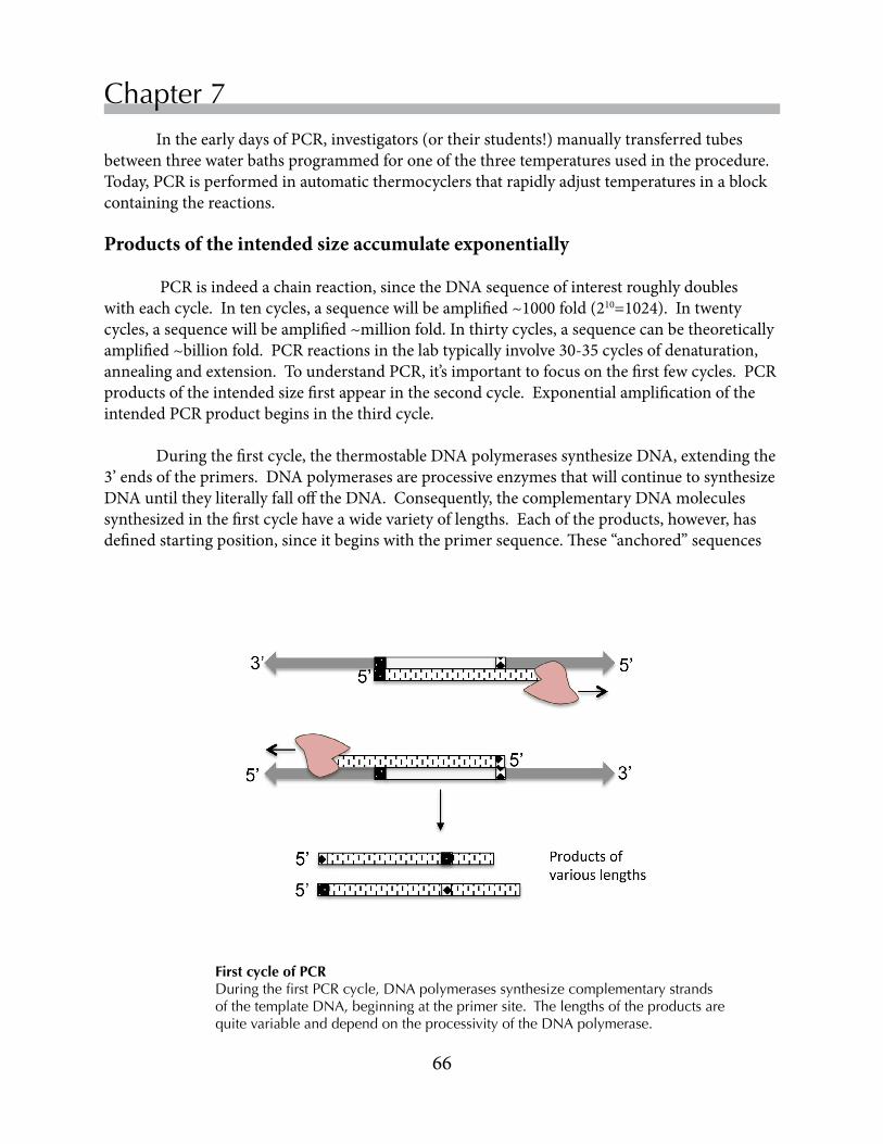
66
Chapter 7 IntheearlydaysofPCR,investigators(ortheirstudents!)manuallytransferredtubesbetweenthreewaterbathsprogrammedforoneofthethreetemperaturesusedintheprocedure.Today,PCRisperformedinautomaticthermocyclersthatrapidlyadjusttemperaturesinablockcontainingthereactions.
Products of the intended size accumulate exponentially
PCRisindeedachainreaction,sincetheDNAsequenceofinterestroughlydoubleswitheachcycle.Intencycles,asequencewillbeamplified~1000fold(210=1024).Intwentycycles,asequencewillbeamplified~millionfold.Inthirtycycles,asequencecanbetheoreticallyamplified~billionfold.PCRreactionsinthelabtypicallyinvolve30-35cyclesofdenaturation,annealingandextension.TounderstandPCR,it’simportanttofocusonthefirstfewcycles.PCRproductsoftheintendedsizefirstappearinthesecondcycle.ExponentialamplificationoftheintendedPCRproductbeginsinthethirdcycle.
Duringthefirstcycle,thethermostableDNApolymerasessynthesizeDNA,extendingthe3’endsoftheprimers.DNApolymerasesareprocessiveenzymesthatwillcontinuetosynthesizeDNAuntiltheyliterallyfallofftheDNA.Consequently,thecomplementaryDNAmoleculessynthesizedinthefirstcyclehaveawidevarietyoflengths.Eachoftheproducts,however,hasdefinedstartingposition,sinceitbeginswiththeprimersequence.These“anchored”sequences
First cycle of PCRDuring the first PCR cycle, DNA polymerases synthesize complementary strands of the template DNA, beginning at the primer site. The lengths of the products are quite variable and depend on the processivity of the DNA polymerase.

67
Replace Chapter number and title on A-Master Page.<- ->
Yeast Colony PCR
willbecometemplatesforDNAsynthesisinthenextcycle,whenPCRproductsoftheintendedlengthfirstappear.ThestartingtemplateforPCRwillcontinuetobecopiedineachsubsequentcycleofPCR,yieldingtwonew“anchored”productswitheachcycle.Becausethelengthsofthe“anchored”productsarequitevariable,however,theywillnotbedetectableinthefinalproductsofthePCRreaction. DNAstrandsoftheintendedlengthfirstappearduringthesecondcycle.Replicationfromthe“anchored”fragmentsgeneratesPCRproductsoftheintendedlength.Thenumberofthesedefinedlengthfragmentswilldoubleineachnewcycleandquicklybecomethepredominantproductinthereaction.
PCR fragments of the intended length first appear in the second cycle.The “anchored” fragments generated during the first cycle of replication begin with either the primer 1 or primer 2 sequence. During the second cycle, replication begins at the other primer site, generating a PCR fragment capped at both ends with primer sequences.
MostPCRprotocolsinvolve30-35cyclesofamplification.Inthelastfewcycles,thedesiredPCRproductsarenolongeraccumulatingexponentiallyforseveralreasons.Asinanyenzymaticreaction,PCRsubstrateshavebecomedepletedandtherepeatedroundsofincubationat94˚ChavebeguntodenatureTaqpolymerase.
Primer annealing is critical to specificity in PCR
GoodprimerdesigniscriticaltothesuccessofaPCRreaction.PCRworksbestwhentheprimersarehighlyspecificforthetargetsequenceinthetemplateDNA.Misprimingoccurswhenprimersbindtosequencesthatarepartiallycomplementary,causingDNApolymerasetocopythewrongDNAsequences.Fortunately,investigatorsareabletoadjustexperimentalparameterstomaximizetheprobabilitythatprimerswillhybridizewiththecorrecttargetsonthetemplateDNA.

68
Chapter 7
PCRprimersaretypicallysyntheticoligonucleotidesbetween18and25baseslong.Whendesigningaprimer,researchersconsideritsTm,thetemperatureatwhichhalfofthehybridsformedbetweentheprimerandthetemplatewillmelt.Ingeneral,thethermalstabilityofahybridincreaseswiththelengthoftheprimeranditsGCcontent.ThefollowingformulaprovidesaroughestimateoftheTmofoligonucleotidehybrids.Inthisformula,nreferstothenumberofnucleotides,andtheconcentrationofmonovalentcationsisexpressedinmolar(M)units.
Tm=81.5˚C+16.6(log10[K++Na+])+0.41(%[G+C])-(675/n)
Whenpossible,researchersdesignprimersthataresimilarinlengthandhavea40-60%GCcomposition.ThesequencesaredesignedsothattheTmsoftheprimer-DNAhybridsarewithinafewdegreesoftheannealingtemperature.AdjustingtheTmsoftheprimerstobeclosetotheannealingtemperaturefavorsspecifichybridsoverlessspecifichybridsthatmaycontainafewmismatchedbases.Ahybridformedbetweenaprimerandanon-targetsequencewithevenonemismatchedbasehasaTmthatislowerthanthatofthefullyhydrogen-bondedhybrid.IfDNApolymeraseextendsthemismatchedprimer,incorrectPCRproductswillbegenerated.WhenmisprimingappearstobeaprobleminaPCRreaction,investigatorshaveseveraloptions.Theycanincreasethelengthand/orGCcontentoftheprimers,alterthesaltconcentrations(resultsmaybehardtopredict)orincreasetheannealingtemperature.
WhendesigningaPCRreaction,investigatorsalsoconsiderthenatureofthetemplateDNA.AvarietyofDNAtemplatescanbeusedforPCR.Dependingonthepurposeoftheexperiment,investigatorscouldchoosetousegenomicDNA,aplasmidoracDNA(complementaryDNAgeneratedbyareversetranscriptasefrommRNA).PCRcanalsobedonewithmuchcruderpreparationsofDNA,suchasabacterialoryeastcolony.Themorecomplexthetemplate(itslengthinbp),thegreatertheprobabilitythatitwillcontainanothersequencethatisverysimilartoaprimersequence.Forexample,thehaploidyeastgenomeis12MbplongandcontainsonlyonecopyofeachMETgene.Theprobabilitythatanon-targetsequenceintheyeastgenomeissimilarenoughtoa20-nucleotideMETprimertocausemisprimingisreasonablygood.Furthermore,thesesequenceswithsmallmismatchesmayoutnumberthetargetsequence.WithcomplextemplatessuchasgenomicDNA,therefore,investigatorscansometimesreducetheimpactofmismatchedhybridsbydecreasingtheamountoftemplateDNAinthereaction.(UsingtoomuchtemplateisthemostcommonerrorinyeastcolonyPCR.)
ThecomponentsofaPCRreactionaresimple,consistingoftheDNAtemplate,primers,dNTPs,abuffercontainingMgCl2(polymerasesusedNTPscomplexedwithMg2+),andthethermostablepolymerase.For our experiments, we will be using a master mix that contains all of the components except the template DNA and the primers. Theuseofamastermixensuresthatallreactionshaveidenticalreagentsanditalsoreducesthenumberoftransfersrequiringmicropipettes.Thesmallernumberoftransfersisparticularlyadvantageous,becauseitreducestheopportunitiesforcross-contaminationofreagents.PCRisanexquisitivelysensitiveprocedure.Someresearchersusespecialbarriertipsfortheirmicropipettes,whichcontainfiltersthatpreventsamplesfromreachingthebarrelofthemicropipettes.

69
Replace Chapter number and title on A-Master Page.<- ->
Yeast Colony PCR
Saccharomyces Genome Deletion Project Thepublicationoftheyeastgenomesequenceopenednewopportunitiesforyeastgeneticists.KnowingtheDNAsequenceoftheyeastgenome,geneticistscouldtakeadvantageofthehighfrequencywithwhichyeastexchangegenesusinghomologousrecombinationtogeneratemutantsoftheirowndesign.HomologousrecombinationnormallyoccursduringmeiosisandduringcertainkindsofDNArepair.Duringhomologousrecombination,twocloselyrelatedDNAsequencesalignwithoneanother,theDNAstrandsbreakandstrandexchangeoccurswhenthebreaksarehealed.Investigatorswhowanttousehomologousrecombinationforgenereplacementusemolecularcloningtoconstructareplacementcassetteinwhichamarkergeneisflankedoneithersidebysequencesthatflankthegenetobereplacedinthetargetchromosome.
ThestrainsthatweareusingweregeneratedbythisapproachduringtheSaccharomycesGenomeDeletionProject(Winzeleret al.,1999).AfterthepublicationoftheDNAsequence,yeastresearchersbegantosystematicallyreplaceallofthepredictedORFsintheS. cerevisiae genomewithakanamycinresistance(KANR)gene.Thefigurebelowoutlinestheprocessusedtogeneratethedeletionstrains.ForeachORF,researchersusedaseriesofPCRreactionstoconstructcassettesinwhichtheKANRgenewasflankedateithersitebyshortDNAsequencesupstreamanddownstreamofthetargetedORFontheS. cerevisiaechromosome.ThePCRfragmentswerethenusedtotransformtheBY4742strain(Brachmannet al.,1998)ofS. cerevisiae.StrainsthathadincorporatedtheKANRgenewereselectedonplatescontaininganalogsofkanamycin.
AllofthedeletionstrainswereanalyzedbyPCRtoconfirmthatthetargetedORFwasreplacedbytheKANRcassette.Thus,wecanbereasonablycertainaboutthegenotypesofourmetmutants.TheDeletionProjectalsoverifiedthatstrainswereabletogrowandmate,showedtheappropriateauxotrophiesandhadfunctionalmitochondria.(SeetheDeletionProjectwebsiteforadditionaldetails:http://www-sequence.stanford.edu/group/yeast_deletion_project/deletions3.html.)Withthelargenumberofstrainsgeneratedintheproject,itwasnotpossibletodoathoroughphenotypicanalysisofindividualstrains.Thoseanalysesareleftforyoutodo!

70
Chapter 7 Inthislaboratory,wewillusetheprimersdesignedbytheSaccharomycesDeletionProject(SGDP)toanalyzetheMETgenedisruptionsinyouryeaststrains.Asshowninthefigurebelow,wehaveobtainedtwogene-specificprimers(GSP)foreachoftheMETgenesthatweareworkingwith.OneoftheGSPs,GSPPrimerA,islocated200-400bpupstreamoftheinitiationcodon.ThesecondGSP,GSPPrimerB,isanantisenseprimerthatbindswithintheORF.Wealsohaveanantisenseprimerthatbinds250bpwithintheKANRgene(KANPrimerB).Thesequencesoftheprimersareshowninthetableatthebottomofthepage.
Gene Primer A Primer BMET1 TTCTATTTTCGTTATTGGTTTCTCG AAATGAACCTGATCAGTAGCAAAAC
MET2 AAGTCATGTTAATCGTTTGGATTTG GTCCAAGTAGTTGGGATCTGAGTAA
MET3 GTAATTTTGTAACTCACCGCATTCT CATTCTTCTTTAACGCATCTCTAGC
MET5 TTCATCACGTGCGTATTATCTCTTA GGTATTCAATGGATCTTGATTGTTC
MET6 ATGCGATAGATGCACTAATTTAAGG AAAACTTGGTCGTAAAAGGAGAAGT
MET7 GTTGGTTAACAGAAAAAGGCAACTA TCATGCATTTCCAATAATGTCATAG
MET8 ATGCCATTTCAGTTACAACCTAGTC GAATAATGGATTTGTGTAGGTCAGG
MET10 AAAGAAAACACTATCAACATTCCCA AGTTTAAAGCACCAACATTCAAAAG
MET14 AAAGAATACAGTTGCTTTCATTTCG GATTGTACTTTTACCTGACGCACTT
MET16 GCTGACAAAAGAATTGGATAAAAGA ATATACTGTTTAACCTGCTCGAACG
MET25 CATCCTCATGAAAACTGTGTAACAT GCAGAATGTGTTACAATATCAGCAC
CYS3 ACCCCATACCACTTCTTTTTGTTAT ATAGGGTTAGCTGGAGAAGATTGTT
CYS4 ACAACTTCAACTTCACCCAAGTAAG TCAAGTCTTCTAGCTGTCTTTGGAT
KANR CTGCAGCGAGGAGCCGTAAT
Note: The primer sequences are written in Courier font. Courier is a nonproportional font that is often used for nucleic acids, because it assigns all letters the same width. At a glance, it is possible to see that most of the primers are the same length.

71
Replace Chapter number and title on A-Master Page.<- ->
Yeast Colony PCR
Exercise 1 - Physical properties of PCR primers ThetableontheoppositepageliststheprimerpairsusedbytheSGDPtoanalyzethedeletionmutants.Indesigningtheprimers,researchersaimedforprimerpairsthathadsimilarphysicalpropertiesandwouldgeneratePCRproductsthatwereseveralhundredbasepairslong.Likemostgenomes,theS. cerevisiaegenomehasbothAT-richandGC-richregions(Goffeauet al.,1996),soinvestigatorswerenotalwaysabletodesignprimerswiththeideal40-60%GC-content.Yourgroupwillbeworkingwiththreedeletionstrains.Completethetablebelowbyaddingthelength,GC-contentandTmfortheprimersdesignedfortoanalyzeyourdeletionstrains.Tofindthisinformation,useoneofseveralonlinetoolsforprimeranalysis:
http://www.idtdna.com/analyzer/Applications/OligoAnalyzer/http://www.basic.northwestern.edu/biotools/oligocalc.html
Primer A Primer BGene Length % GC Tm Length % GC Tm
KANR
Exercise 2 - Map the PCR product to the genome
Inthenextlab,youwillanalyzeyourPCRreactionproductsonanagarosegelthatseparatesDNAmoleculesaccordingtotheirsizes.TocalculatethesizesofthePCRproductsgeneratedinthereaction,youwillnextaligntheprimersequencesforeachgeneagainstthegenomesequenceandprepareasimplemapofthegenomicregion.
ThePCRpiecesthatwillbegeneratedinthereactionwillcontainpartoftheMETorCYScodingsequence,aswellassequencefromthe5’-flankingregion.YouwillrecallfromChapter5thatNC_____recordscontainthecompletesequenceofthechromosomewhereyourgeneislocated.TheNC____recordcouldbeusedinthealignment,butwecantakeadvantageofothersequenceresourcesthatSGDcuratorshaveprepared.Forthisexercise,wewilluserecordsthatcontaintheCDSforyourgeneaswellas1kbofupstreamsequenceand1kbofdownstreamsequence.Curatorsgeneratedtheserecordsbecauseresearchersareofteninterestedinstudyingregulatoryelementsthatcontroltranscriptionofageneandtheprocessingofgenetranscripts.BecausetheS. cerevisiae genomeiscompact(Goffeauet al.1996),theseelementsareusuallylocatedwithin1kbfromeitherendoftheCDS.

72
Chapter 7
You will need two browser windows for this exercise. Each member of the group may want to work with a single gene.
Find the genomic sequence for your gene.• Navigatetoyourgene’ssummarypageintheSGD(yeastgenome.org)• Cursordownthesidebarto“Retrievesequences”(UnderSequenceInformation)• Retrievethe“DNA+/-1kb”sequenceforyourgene.• Notethestartingandendingcoordinatesforthesequenceandcalculatethelengthofthe
sequence.
Startingandendingcoordinates:
Lengthofsequence(bp)
Lengthofthecodingsequence
Align the primer sequences with the genomic sequence. Tofindthepositionontheprimersinthegenomicsequence,wewilluseNCBI’sBLASTtool.BLASTstandsforBasicLocalAlignmentSearchToolandcanbeusedtoaligneitherproteinornucleicacidsequences.YouwilllearnmoreabouttheBLASTalgorithmsinChapter9.• DirectyourbrowsertotheNCBIsiteandaccesstheBLASTtool.SelectNucleotideBLAST.• Clickthebox“Aligntwoormoresequences.”• CopythegenomicsequencefromSGDandpastethesequenceintotheSubjectSequencebox.• TypethePrimerAforyourgeneinquerybox.ClickBLAST.TheBLASTresultsbringupa
tablewitheachmatchbetweenyourprimerandthegenomesequence.Thetopresultshouldbeaperfectmatchbetweenyourprimerandthegenomesequence.(Checkyourtypingifitisn’taperfectmatch!)Doyouseeanyothermatches?Recordthestartingandendingpointswheretheprimermatchesthegenomicsequence.
• RepeattheBLASTalignmentforprimerB.Click“EditandResubmit”atthetopoftheBLASTresultspage.Clearthequeryboxandtypeinthesequenceofyourotherprimer.ClickBLASTandrecordthealignmentresults.PrimerBisthereversecomplementthegenomicsequence.

73
Replace Chapter number and title on A-Master Page.<- ->
Yeast Colony PCR UsetheinformationthatyoucollectedfromtheBLASTsearchestoprepareamapofthegenomicregionroughlytoscale.IncludetheprimerbindingsitesandthecodingregiononthegenomicDNA.AnsamplemapisshownbelowfortheMET5gene,whichhasaCDSsequenceof4398bp.PrimerAbindstonucleotides692to716.PrimerBbindstonucleotides1180to1156.YoucanseehowstraightforwarditistocalculatethesizeofyourPCRproductfromthiskindofmap.ThePCRproductproducedwiththeMET5primersAandBwillbe488bp,ofwhich180bprepresentCDS,and308bprepresentthe5’-flankingregion.
DisruptedgenescontainingtheKANRcassettewillgivePCRproductswhentheKANRPrimerBisusedwithPrimerA.TheKANRPrimerBbindingsiteislocated250bpintotheKANRgene.ThePCRproductgeneratedwithMET5primerAandtheKANRprimerBwouldbe558bp,including308bpfromthe5’-flankingregionandtheKANRsequence.
Drawamapforyourgeneinthespacebelow.
CalculatethesizeofthePCRproductsthatwouldbegeneratedwith: PrimerAandPrimerB
PrimerAandKANRprimerB
74
Chapter 7
Exercise 3 - Design yeast colony PCR Inthislab,eachteamwillbeabletoperformsix PCRreactionstoidentifyyourthreeyeaststrains.YouwillhavetheoptionofusinganyoftheprimersintheTableonp.58.ItwillnotbepossibletotesteverystrainwithboththeGSPA-GSPBandGSPA-KANRBcombinations.Useyourresultsfromtheselectiveplatingexperimenttodeviseastrategythatwillallowyoutopositivelyidentifyyourmetyeaststrains.
Listtheprimerpairsthatyouwilluseforthereactions,togetherwiththepredictedsizesofthePCRproducts.
Exercise 4 - Yeast colony PCR
Topreparethereactions,youwillfirstmixtheprimerpairswithaVERY SMALL numberofyeastcellsthatyoutransferfromacolonytothetubewiththetipofaP-20orP-200micropipette.Thecolonyandprimerswillthenbeheatedat98˚Cfor15minutestodisrupttheyeastcells.Atthatpoint,youwilladdanequalvolumeofaPCRmastermix,containingnucleotidesandtheTaqpolymerase,toeachtube.ThetubeswillthenbereturnedtothethermocyclerforatypicalPCRreaction.
• Label the PCR tubes.Thetubesareverysmall,sodevelopacodethatyoucanusetoidentifythetubes.(Don’tforgettoincludethecodeinyournotebook.Thecodeshouldindicatewhichprimersandstrainsaremixedineachtube.)

75
Replace Chapter number and title on A-Master Page.<- ->
Yeast Colony PCR
• Prepare the primer mixture.ThefinalvolumeofthePCRreactionswillbe20µL.Theprimermixtureaccountsforhalfthefinalvolume,or10µL.Theprimersstockconcentrationsare2.0µMeach.Pipette5.0µLofthetwoprimersthatyouwouldliketouseintoeachPCRtube.What will the final concentration of each primer be in the actual PCR reaction?
NOTE: BecauseoftheextraordinarysensitivityofPCRreactions,itisveryimportantnottocross-contaminatetubeswithprimersthatdon’tbelonginareaction.Changetipsbetweeneveryprimertransferthatyoudo.
• Transfer a small quantity of yeast cells to each PCR tube. LightlytouchthetipofaP20orP200micropipettetoayeastcolony.Twirlthemicropipettetipinthetubecontainingyourprimermixtoreleasethecells.Themostcommonerroristransferringtoomanyyeastcells,whichwillinterferewiththePCRreaction.Theappropriateamountofyeastwouldfitonthetipofapin.
• Lyse the yeast cells.Placethetubesinthethermocyclerfor15minat98˚C.
• Set up the PCR reactions.Removethetubesfromthethermocyclerandadd10µLofPCRmastermixtoeachtube.
• Amplify the target gene sequences.ReturnthetubestothethermocyclerandstartthePCRprogram.
Onecycleofdenaturation: 95°Cfor2minutes
35cyclesofdenaturation,annealingandextension95°Cfor30sec.55°Cfor30sec. 72°Cfor1minute
Onecycleofextension72°Cfor10minutes
• Store the reactions at 4°C for analysis in the next lab.

76
Chapter 7
References
Brachmann,CB,Davies,A,CostGJ,Caputo,E,Li,J,Hieter,P&Boeke,JD(1998)DesignerdeletionstrainsderivedfromSaccharomyces cerevisiaeS288C:ausefulsetofstrainsandplasmidsforPCR-mediatedgenedisruptionsandotherapplications.Yeast14: 115-132.
Goffeau,A,Barrell,BG,Bussey,Het al.(1996)Lifewith6000genes.Science274:563-567.
Sakai,RK,Scharf,S,Faloona,F,Mullis,KB,Horn,GT,Erlich,HA&Amheim,N(1985).Enzymaticamplificationofbeta-globingenomicsequencesandrestrictionsiteanalysisfordiagnosisofsicklecellanemia.Science230:1350-1354.
Sakai,RK,Gelfand,DH,Stofel,S,Scharf,SJ,Higucki,R,Horn,GT,Mullis,KB&Erlich,HA(1988).Primer-directedenzymaticamplificationofDNAwithathermostableDNApolymerase.Science 239:487-491.
Winzeler,EA,Shoemaker,DD,Astromoff,Aet al. (1999)FunctionalcharacterizationoftheSaccharomyces cerevisiaegenomebygenedeletionandparallelanalysis.Science285:901-906.

• PrepareagarosegelsforseparatingDNAmolecules
• SeparateDNAmoleculesbyelectrophoresis
• VisualizeDNAmoleculesongelsusingintercalatingdyes
• CalculatethesizeofDNAmoleculesusingmolecularweightstandards
AgarosegelsareusedtoanalyzeDNAmolecules.Thesegelsaresimpletoconstruct,becausetheyrelyonlyonthegellingpropertiesofagarose.Moleculesareseparatedbysizeandvisualizedwithfluorescentintercalatingdyes.Inthislab,you’llanalyzetheproductsofthePCRreactionsfromthepreviouslab.
Chapter 8
Objectives
Agarose gel electrophoresis

78
Agarose gel electrophoresis
AgarosegelsprovideasimplemethodforanalyzingpreparationsofDNA.AlthoughthebasecompositionsofindividualDNAmoleculesvary,thebasicchemicalstructureofDNAisthesameforallDNAmolecules.DNAmoleculessharethesamecharge/massratio,whichimpartssimilarelectrophoreticpropertiestoDNAsofwidelyvaryinglengths.
Agarose gels are porous matrices Agaroseisapolysaccharidepurifiedfromredalgae,orseaweed.Agaroseismorehighlypurified(andsignificantlymoreexpensive!)thanagar,whichisobtainedfromthesameseaweed.Agarosemoleculesarelong,linearpolymersoftherepeatingdisaccharide(13)-β-D-galactopyranose-(14)-3,6-anhydro-α-L-galactopyranose(right).Atypicalagarosemoleculecontainsoveronehundredmonomers.Theagaroseusedforelectrophoresishasbeenhighly
Background
purified.Thepurificationprocessremovescontaminantsthatwouldinterferewiththeenzymesusedinmolecularcloning,suchasrestrictionendonucleases.Theprocessalsogeneratesanagarosepreparationwithdesirableelectrophoreticpropertiesandminimalbackgroundfluorescence,whichisimportantforvisualizingDNAmolecules.
Agarosemoleculesareabletoformgelswithrelativelydefinedporesizesbecauseofthechemicalpropertiesofagarosemolecules.Agarosedemonstrateshysteresis-itsmeltingtemperatureishigherthanitsgellingtemperature.Agarosemoleculesdissolveatabout90°C,formingrandomcoilsinsolution.Gelsformwhenthetemperaturefallstoapproximately40°C.Asthegelforms,theagarosemoleculesfirstassembleintohelicalfibers,whichthenfurtheraggregatetoformnetworksofsupercoiledhelicesstabilizedbyhydrogenbonds.Thesizesofthepores,whichtypicallyrangefrom50to200nm,dependontheconcentrationofagarose.Astheagaroseconcentrationincreases,theaveragediameteroftheporedecreases.
Several factors affect the migration of DNA through agarose gels BecauseofthenegativechargeofthephosphateresiduesintheDNAbackbone,DNAmoleculesmovetowardthepositivepole(anode)oftheelectrophoresisapparatus.TheuniformityofDNAstructuregivesmoleculesthesamecharge/massratio,whichmakesitstraightforwardtocalculatethesizesofDNAfragments.(MolecularweightsarenotalwaysverymeaningfulwithDNAfragments,sinceasinglemoleculecancontainthesequencesofmultiplegenes.)Ingeneral,themigrationofDNAmoleculesisinverselyproportionaltothelogarithmoftheirlengths,ornumberofbasepairs.
AlthoughtherelationshipbetweenmigrationrateandDNAlengthholdstrueinmostcases,theactualmigrationrateofDNAmoleculesinaparticularexperimentisaffectedbymultiplefactors.SomeofthesefactorsareintrinsictotheDNAmolecules,whileotherfactorsrelatetotheelectrophoreticconditions.Becauseofthisvariability,DNAstandardswithknown

79
Replace Chapter number and title on A-Master Page.<- ->
Chapter 8
sizesshouldalwaysberunonthesamegelswithsamplesbeinganalyzed.Importantly,thestandardsneedtohaveasimilarstructure(e.g.linearorsupercoiled)andtobesubjectedtothesamechemicalmodificationsastheDNAsamplesbeinganalyzed.
Size and conformation of DNA ThemigrationrateofaDNAmoleculedependsonbothitssizeanditsconformation.Withinacertainsizerangedictatedbythegelconditions,themigrationrateoflinearDNAmoleculesisinverselyproportionaltothelog10(numberofbasepairs).ThemigrationofmorestructuredDNAmolecules,suchascircularplasmids,ismuchlesspredictable.Themigrationratesofthesemorestructuredformsareinfluencedbythedensityofcoils,thepresenceofnicks,andotherstructuralfeatures.
Buffer systems ThemigrationratesofDNAmoleculesarealsoaffectedbythecompositionofthegelandrunningbuffer.ThemigrationrateofaDNAmoleculedecreasesastheconcentrationofagaroseinthegelincreases.ThemigrationrateofaDNAmoleculeissomewhathigherinagelmadewithTAE(Tris:acetate:EDTA)bufferthanwithTBE(Tris:borate:EDTA)buffer.
Fluorescent intercalating agents are used to visualize DNA fragments in gels
NucleicacidsarevisualizedbyfluorescentdyesthatbindstronglytoDNA.ThedyesareintercalatingagentsthatinsertintotheDNAhelixandintostructuredregionsofsingle-strandednucleicacids.Thefluorescenceofthesedyesincreasesbyanorderofmagnitudewhentheybindnucleicacids,sothebackgroundfluoresenceremainslow.ThemostcommonlyuseddyesareethidiumbromideandvariantsofSYBRGreen.
SAFETY NOTE: All DNA intercalating agents are potential mutagens. Be sure to wear gloves when handling solutions with intercalating dyes.
DNAintercalatingagentshaveanabsorbancemaximuminthelongultravioletrange,sotheyareviewedwithtransilluminatorsthatemitlightwithwavelengthsclosetotheabsorbancemaximumofthedyes.
SAFETY NOTE: Be careful to wear protective eyewear if you look at an unshielded transilluminator. UV light is damaging to the eye!

80
Agarose gel electrophoresis
AgarosegelsprovideinformationaboutthesizesofDNAfragments,iftheyarecalibratedwithappropriatesizestandards.AgarosegelsalsoprovideinformationaboutthequantityofDNAinaparticularfragment,becausetheamountofintercalatingagentthataDNAmoleculebindsisproportionaltoitsmass.Consequently,theintensityofabandreflectstheamountofDNAintheband.
Thegrainypictureontherightshowsthe1Kbladder,whichyouwilluseasastandardinthiscourse.TheladderisaproprietarypreparationofDNAfragmentsproducedbytheNewEnglandBiolabscompany.The3Kbfragmenthas~2.5moreDNAthantheotherfragments,sothatitappearsbrighterinastainedgel.Usingthengvaluestotheright,itispossibletousethisstandardtovisuallyestimatethequantityofDNAinanunknownsample.
ManydifferentstandardsareavailableforDNAmolecules.Investigatorschoosestandardswithasimilarsizerangetothemoleculesthattheyarestudying.
The sizes of DNA fragments can be calculated using standards
Tocalculatethesizesofbandsinotherlanesofthegel,youwouldfirstconstructastandardcurveliketheoneattheleftfromthemigrationoftheDNAfragmentsinthe1kbladder.
Plotthelog10(numberofbasepairs)foreachfragmentonthey-axisagainstthedistancethateachfragmentmigratedonthex-axis.Youwillbeabletoestimatethesizesoffragmentsinyoursamplebyinterpolatingonthestandardcurve.
Note: Always visually confirm that your estimated sizes are correct!!

81
Replace Chapter number and title on A-Master Page.<- ->
Chapter 8
Prepare the agarose gel Inthiscourse,wewilluseagarosegelstoseparateDNAmoleculesproducedinPCRandrestrictiondigests.Thesemoleculesarewell-resolvedon1.25%agarosegelspreparedinTAEbuffer,whichprovidesgoodseparationofmoleculesranginginsizefrom500bp-10kbp.Placethecastingtrayintothegelapparatus.IfyouareusingtheBioRadapparatuses,positiontheblackwedgesateachendofthecastingtray.
1.Determinetheamountofagarosethatyouwillneedfora1.25%(1.25g/100mL)agarosegelthatfitsyourcastingplatform.MostofthegelapparatusesinthelabaretheBioRadMini-SubGTsystems,whichhavea7cmx7cmcastingtray.Theseapparatusesaccommodate30-40mLgels.Checkyourcalculationswithyourteammatesbeforeyouproceed.
2.FillagraduatedcylinderwiththeappropriatevolumeofTAEbuffer.Pourthesolutionintoasmallflask.
3.Weighouttheappropriateamountofagarose.SprinkletheagaroseontothesurfaceoftheTAEintheflask.Note:theagarosewillnotdissolveuntilitisheated.
82
Agarose gel electrophoresis
4.Dissolvetheagarosebyheatingthesolutionforintervalsof15-20secondsinamicrowaveoven.Aftereachinterval,removetheflaskandgentlyswirlitaroundabittodispersethecontents.Noteiftheagaroseparticlesarestillapparentoriftheagarosehasdissolved.ThebestgelsaremadefromagarosethathasNOTbeenovercooked.
SAFETY NOTE: The agarose solution will be very HOT when you remove it from the micro-wave! Please use caution when handling the flask. Be particularly careful not to contact the steam that will be coming through the opening of the flask. Fold several paper towels and wrap them around the neck of the flask when you handle it. If you do happen to spill some hot agarose on your skin, wash it immediately with cold water and alert your TA.
5.Allowtheagarosesolutiontocooluntilyoucancomfortablytouchtheflaskwithyourhands.Agarosesolutionsover60˚Cwillwarpthecastingtray!Pourthegel.Placethesamplecombinplace.Donotmovethecastingplatformuntilthegelsets.Youwillknowthatthegelissetwhenitbecomesopaque.Allowthegeltocureforabout20minutesafteritsets.
Prepareyoursamplesforelectrophoresiswhilethegeliscuring.Add1volume(vol)of6Xloadingdyeto5volofeachsample.(Youwouldadd1µLofloadingdyeto5µLofsample.)Theloadingdyecontainstwodyes,bromophenolblueandxylenecyanol,whichmigratewith“apparent”molecularweightsof~5kband~0.5kb,respectively.Loadingdyealsocontainsglycerol,whichmakesthesampledenseenoughtosinktothebottomofthesamplewell.)Briefly,centrifugeeachsampletomixthedyeandsamples,ifnecessary.
Add the loading dye DIRECTLY into the tube containing your PCR products.
Sample preparation

83
Replace Chapter number and title on A-Master Page.<- ->
Chapter 8
1. Whenthegelhasset,carefullyremovethecombandtheblackwedges.
2. Orientthegelintheelectrophoresistanksuchthatthewells(holesmadebythecomb)areorientedtowardtheblack(negative)electrode.TheDNAfragmentswillmovefromthewelltowardthered(positive)electrode.FillthetankwithenoughTAEbuffertosubmergethegel(approx.275-300mL).
3. Loadonesampletoeachwell.Thewellsshouldholdupto20µLofsample.ForPCRreac-tions,load5-10µLoftheoriginalPCRreaction(6-12µLaftersamplebufferhasbeenadded).Trytoavoidairbubblesasyouloadthesamples.
4. Load5µLmolecularweightstandardtoonelaneofthegel.Makesurethatyouhaveaccuratelyrecordedthelocationofeachsampleinthegel.
5. Placethelidontheelectrophoresistankandconnecttheelectrodestothepowersupply(black-to-blackandred-to-red).Make sure that the polarity is correct before continuing!
6. Turnonthepowerandapplyaconstantvoltageof75V.
7. Paycarefulattentiontothegelasitruns.Turnoffthepowerwhenthebromophenolblueis~1cmfromtheendofthegel.Do not allow the dye to run off the gel, since small DNA molecules will be lost. (Think about the size of your PCR products.)
Load and run the agarose gel
Proper set-up of an agarose gelThe gel is submerged in running buffer. Samples will elecctrophorese toward the postivie (red) pole.
Red electrical leads are attached to the positive pole of the power pack.
Samples are loaded into wells.

84
Agarose gel electrophoresis
SAFETY NOTE: Wear disposable gloves when staining gels. Gloves are important when working with intercalating dyes, which are potential mutagens.
1. Removethegelfromtheapparatusandtransferthegeltoasmalltray.Coverthegelwithdeionizedwater.Add5µLofethidiumbromide(EtBr)solution(10mg/mL),followingyourTAsinstructions.WhatistheapproximateconcentrationofEtBrinthestainingsolution?Note:EtBrstocksarelightsensitive,sotheyarestoredinthedark.
2. Placethetrayonarockingplatformandrockgentlyfor30minutes.3. DraintheEtBrsolutionintotheappropriatewastecontainerinthefumehood.4. Coverthegelwithdeionizedwaterandrockgentlyfor2minutes.5. Withaspatula,carefullyplacethegelonthetransilluminatorandclosethecovertotheGel-
Logicapparatus.(Drainthewashsolutionintothewastecontainer.)6. Turnonthetransilluminatorlightandphotographthegel.Turnoffthetransilluminatorim-
mediatelyafteryouphotographthegel.Savethepictureandemailacopytoyourself.7. OpenthedooroftheGelLogicapparatus.Usethespatulatotransferthegeltoawastecon-
tainersetupforEtBr-stainedgels.8. DeterminetheapproximatelengthoftheDNAfragmentsinyoursamplesbycomparingtheir
migrationtothatofthestandards.Arethesizesconsistentwithyourexpectations?(Inthisexperiment,thePCRproductswillhavesimilarsizes,soastandardcurvewillnotbeuseful.)
9. Toconstructastandardcurve,measurethedistancethateachfragmentmigratedfromthesamplewell.MakeatableinyournotebookwiththesizeofeachDNAstandardandthedistancethateachfragmentmigratedonthegel.Plotthelog10(fragmentlength)ofeachstandardonthey-axisandthedistancethateachstandardmigratedonthex-axis.
Stain and analyze the agarose gel

• Learntheonelettercodeforaminoacids• Understandhowaminoacidsidechainchemistryisreflectedinthe
BLOSUM62matrix
• UsetheBLASTPalgorithmtocompareproteinsequences
• Prepareamultiplesequencealignmenttoidentifyconservedregionsinaprotein
The budding yeast, Saccharomyces cerevisiae, and the fission yeast, Schizosaccharomyces pombe, are separated by close to a billion years of evolution. Our goal for the semester is to determine if the proteins involved in methionine and cysteine biosynthesis have been conserved between these evolutionary divergent species. In this lab, you will use bionformatics tools to study the evolution of these protein sequences.
Protein conservationChapter 9
Objectives

86
Chapter9
Each of the 20 amino acids commonly found in proteins has an R group with its own distinctive chemistry. R groups differ in their size, polarity, charge and bonding potentials. When thinking about evolutionary changes in proteins, it is helpful to group the amino acids by their chemistry in a Venn diagram, shown on the opposite page. In general, replacing one amino acid with a second amino acid from the same sector can be considered a conservative change. Size is also important. R groups vary considerably in the bulkiness of their chains. Substitution of a large R group for a small one can significantly alter the function of a protein.
Amino acid R groups have distinct chemistries
As species evolve, their proteins change. The rate at which an individual protein sequence changes varies widely, reflecting the evolutionary pressures that organisms experience and the physiological role of the protein. Our goal this semester is to determine if the proteins involved in Met and Cys biosynthesis have been functionally conserved between S. pombe and S. cerevisiae, species that are separated by close to a billion years of evolution. In this lab, you will search databases for homologs of S. cerevisiae sequences in several species, including S. pombe. Homologs are similar DNA sequences that are descended from a common gene. When homologs are found in different species, they are referred to as orthologs.
Homologs within the same genome are referred to as paralogs. Paralogs arise by gene duplication, but diversify over time and assume distinct functions. Although a whole genome duplication occurred during the evolution of S. cerevisiae (Kellis et al., 2004), only a few genes in the methionine superpathway have paralogs. Interestingly, MET25 is paralogous to three genes involved in sulfur transfer: STR1 (CYS3), STR2 and STR4, reflecting multiple gene duplications. The presence of these four distinct enzymes confers unusual flexibility to S. cerevisiae in its use of sulfur sources. The SAM1 and SAM2 genes are also paralogs, but their sequences have remained almost identical, providing functional redundancy.
Protein function is intimately related to its structure. You’ll recall that the final folded form of a protein is determined by its primary sequence, the sequence of amino acids. Protein functionality changes less rapidly during evolution when the amino acid substitutions are conservative. Conservative substitutions occur when the size and chemistry of a new amino acid side chain is similar to the one it is replacing. In this lab, we will begin with a discussion of amino acid side chains. You will then use the BLASTP algorithm to identify orthologs in several model organisms. You will perform a multiple sequence alignment that will distinguish regions which are more highly conserved than others.
As you work through the exercises, you will note that protein sequences in databases are written in the 1-letter code. Familiarity with the 1-letter code is an essential skill for molecular biologists.

87
Replace Chapter number and title on A-Master Page.<- ->
Proteinconservation
You may find NCBI’s Amino Acid Explorer helpful for this exercise. Access it at: http://www.ncbi.nlm.nih.gov/Class/Structure/aa/aa_explorer.cgi
1. Under the amino sequence below, write the same sequence using the 1-letter code.
Met-Glu-Asn-Asp-Glu-Leu-Pro-Ile-Cys-Lys-Glu-Asp-Pro-Glu-Cys-Lys-Glu-Asp
2. What is the net charge of this peptide? (Assign -1 for each acidic amino acid and +1 for each basic amino acid. Add up the total charges.)
3. How many hydrophobic amino acids are found in this peptide?
4. Write the name of a music group that you enjoy. Then transpose the name into an amino acid sequence written with the 3-letter code. Pass the amino acid sequence to a friend and have him/her decode it. (Note: the 1-letter code uses all of the alphabet, except B, J, O, U, X and Z).
Exercise 1 - The 1-letter code for amino acids

88
Chapter9
There are many different algorithms for searching databases, but BLAST algorithms are some of the most popular. BLAST is an acronym for Basic Local Alignment Search Tool, which aptly describes its function. The original BLAST algorithms were developed and published by a group of investigators in 1990 (Altschul et al., 1990). Since that time, investigators have refined and diversified the original BLAST programs so that a whole family of BLAST programs is currently available. BLAST programs are widely used because they are able to quickly search through large amounts of sequence data. The key to BLAST’s speed is its use of local alignments that serve as seeds for more extensive alignments. Other algorithms perform global sequence alignments that match the entire sequence of a query sequence against database information. Global sequence alignments are more sensitive than BLAST in finding related sequences, but they require considerably more processing time. BLAST searches are quite adequate for our purposes, and we will use the online BLAST tools available at the NCBI site throughout this course.
The BLASTN and BLASTP algorithms are designed for searching nucleotide and protein databases, respectively, and they use different scoring matrices and search parameters. BLAST algorithms begin by breaking down query sequence into short “words” and assigning numerical values to the words. A scoring matrix is used to assign numerical values to the words. Words and synonyms above a threshold value are then used to search databases. The default word size for BLASTN is 28 nucleotides, while the default word size for BLASTP is 3 amino acids. This differ-ence in word size is because nucleic acid sequences are written in 4 letters (ACGT), while protein
Overview of the strategy used in BLAST algorithmsBLASTNandBLASTPusearollingwindowtobreakdownaquerysequenceintowordsandwordsynonymsthatformasearchset.Atleasttwowordsorsynonymsinthesearchsetmustmatchatargetsequenceinthedatabase,forthatsequencetobereportedintheresults.
BLAST algorithms are used to search databases

89
Replace Chapter number and title on A-Master Page.<- ->
Proteinconservationsequence is written with 20 different letters. The probability of obtaining random, but irrelevant, matches are much greater with nucleotide sequenes. A stretch of 3 amino acids should arise randomly once in every 8000 tripeptides, compared to once in every 64 trinucleotides. In this lab, we will use the BLASTP algorithm, which is more useful than BLASTN for studying protein evolution, because it overlooks synonymous gene mutations that do not change an amino acid.
In BLASTP, a sequence is broken into all possible 3-letter words using a moving window. A numerical score for each word is assigned by adding up values for amino acids from a scoring matrix. Using the BLOSUM62 matrix (below), BLASTP first finds word with a score of 12 of more in a query sequence. BLASTP then identifies potential synonyms that differ from the word at one position, and synonyms above a defined threshold value are included in the search set. NCBI BLASTP uses a default threshold of 10 for synonyms, which can adjusted by the user. Using this search set, BLAST rapidly scans a database and identifies protein sequences that contain at two words/synonyms from the search set. These sequences are set aside for the next part of the BLASTP process, when these short matches serve as seeds for more extended alignments in both directions from the original match. BLAST keeps a running raw score as it extends the matches. Each new amino acid either increases or decreases the raw score. Penalties are assigned for mismatches and for gaps between the two alignments. Again, users can adjust the gap penalties, which are usually quite severe. In the NCBI default settings, the presence of a
Overview of the BLASTP process.ThequerysequenceEAGLESintobrokenintothree-letterwordsorsynonymsthatareusedasasearchsetagainstrecordsinaproteinortranslatednucleotidedatabase.Seethetextforadditionaldetails.

90
Chapter9
BLOSUM62 scoring matrix The results obtained in a BLASTP search depend on the scoring matrix used to assign numerical values to different words. A variety of matrices are available, whose utility depends on whether the user is comparing more highly divergent or less divergent sequences. The BLOSUM62 matrix is used as the default scoring matrix for BLASTP. The BLOSUM62 matrix was developed by analyzing a large number of protein sequences that were more than 62% identical to each other. Investigators computationally determined the frequency of all 210 possible amino acid substitutions that had occurred in these conserved blocks of proteins, and they used this data to construct the BLOSUM62 scoring matrix for amino acid substitutions. The BLOSUM62 score for a particular substitution is a log-odds score that provides a measure of the biological probability of a substitution relative to the chance probability of the substitution. For a substitution of amino acid i for amino acid j, the score is expressed:
where pij is the frequency of the substitution in homologous proteins, and qi and qj are the frequencies of amino acids i and j in the database. The term (1/λ) is a scaling factor used to generate integral values in the matrix.
The BLOSUM62 matrix (below) is consistent with strong evolutionary pressure to conserve protein function. As expected, the most common substitution for any amino acid is itself. Overall, positive scores (shaded) are less common than negative scores, suggesting that most substitutions negatively affect protein function. The most highly conserved amino acids are cysteine, tryptophan and histidine, which have the highest scores. Interestingly, these latter amino acids have unique chemistries and often play important structural or catalytic roles in proteins.
gap brings a penalty of 11, which increases by 1 for each missing amino acid. Once the score falls below a set level, the alignment ceases. Raw scores are then converted into bit scores by correcting for the scoring matrix used in the search and the size of the database search space. The output data from BLASTP includes a table with the bit scores for each alignment as well as its E-value, or “expect score”. The E-value indicates the number of alignments with that particular bit score that would be expected to occur solely by chance in the search space. Alignments with the highest bit scores (and lowest E-values) are listed at the top of the table. For perfect or nearly perfect matches, the E-value is reported as zero - there is essentially no possibility that the match occurs randomly. The E-value takes into account both the length of the match and the size of the database that was surveyed. The longer the alignment, and/or the larger the database search space, the less likely that a particular alignment occurs strictly by chance. An E-value of 1 does not necessarily imply, however, that the match has no biological relevance. More nuanced analysis may simply be needed to evaluate the match.

91
Replace Chapter number and title on A-Master Page.<- ->
Proteinconservation
Exercise 2 - The BLOSUM62 matrix
1. Use the Venn diagram on p. 87 to hypothesize why relatively high values (2 and above) occur for the substitutions:
Asp to Asn
Asp to Glu
His to Tyr
Ile to Val
Lys to Glu
2. Calculate the word score for the following tripeptides. Which would you expect to occur most frequently in a protein database?
Leu - Cys - Pro
Gln - Ala - Met
Asp - Glu - His

92
Chapter9
In this exercise, you will use BLASTP to find a homolog for your protein in S. pombe. Direct your browser to the BLAST tools at NCBI: http://blast.ncbi.nlm.nih.gov
• Enter the NP_ number for the S. cerevisiae Met protein (p. 45) in the query box• For the database, select reference proteins• Enter S. pombe for the organism• Click BLAST
• On the results page, note the graphic summary at the top which gives you an instant idea about the extent and strength of the match with S. pombe sequences.
Does the match extend for the entire length of the protein?
How many homologs are present in the S. pombe genome?
Cursor down to the alignment statistics. Record in the table on p. 94: NP__ record number of the S. pombe match total score how much of the query sequence was aligned (coverage) E-value for the alignment
Cursor down to see the actual alignment of the S. cerevisiae and S. pombe sequences. • Take note of the center row in the alignment which summarizes the homology between the
protein sequences. If an amino acid is conserved between the two species, its 1-letter code name is shown in that row.
What do the pluses in the center row indicate?
Are some regions of the sequences more conserved than others?
Did BLASTP add gaps to one or both of the sequences in the alignment? If so, are they more common in less conserved or highly conserved regions?
Click on the link to the NP_ record for the S. pombe ortholog. Record the EC number for the protein. Is it identical to that for the S. cerevisiae enzyme?
Exercise 3 - BLASTP

93
Replace Chapter number and title on A-Master Page.<- ->
Proteinconservation
Exercise 4 - Multiple sequence alignments
BLASTP gives a pairwise alignment of sequences that is very useful for identifying homologs. Multiple sequence alignments compare a larger number of sequences simultaneously. By comparing a larger number of sequences over a wider evolutionary range, multiple sequence alignments allow researchers to identify regions of a protein that are most highly conserved, and therefore, more likely to be important for the function of a protein. In this exercise, we will study conservation of protein sequences in a number of model organisms that are widely used in genetic studies. Model organisms are easily maintained in large numbers in the laboratory. The genomes for model organisms have been sequenced, and techniques for genetic analysis are well-developed. In addition, database and clone resources are available to support research with model organisms. The organisms below have been selected because they represent important branches of evolution and because they are potential candidates for future research in this course.
Bacteria - these represent two major divisions of the bacteria Escherichia coli strain K-12 (gram negative; K-12 is the standard laboratory strain) Bacillus subtilis strain 168 (gram positive reference strain)Eukaryotes - model organisms Saccharomyces cerevisiae - needs to be included in trees and alignments! Schizosaccharomyces pombe Arabidopsis thaliana - thale crress; model organism for flowering plants Caenorhabditis elegans - nematode model organism used in developmental studies Mus musculus - laboratory mouse
1. Collect the sequence and BLAST data
The first step in a multiple sequence alignment is to collect the sequence data and analyze the BLASTP data that compare the sequences with the S. cerevisiae sequence. We will be using the reference sequences for the organisms, which begin with a NP___ number. Since you already know how to find NP____ records and set up BLASTP, we will take some shortcuts to finding the remaining numbers and BLASTP statistics. The accession numbers for the bacterial species will be available on the class website. We will use the Homologene database at NCBI (Sayers et al., 2012) to find the accession numbers for the eukaryotes. Homologene is a database that automatically detects homologs in 20 sequenced eukaryotic genomes.
Access Homologene at: http://www.ncbi.nlm.nih.gov/homologene Note the species that have been searched for homologs. Enter the name of your gene into the search box. This brings up the various Homologene groups that have a gene with that name. If search brings you to a page with more than one Homologene group list, click on the Homologene group that contains the S. cerevisiae gene. Record the accession number for the Homologene group:

94
Chapter9
Species NP Accession # Total score Coverage E-valueS. cerevisiaeS. pombe
At the top of the record, Homologene describes the distribution of homologs in eukaryotes. A narrowly conserved protein might only be found in the Ascomycota, while a widely-districuted protein would be found in the Eukaryota. • What phylogenetic divisions have homologs of your gene?
The records page has links on the left to gene summaries, which you may want to investigate. Links on the right side of the page bring you to the NP___ record. The Homologene output always provides a graphical depiction of protein domains in each of the homologs.
• How many domains are found in your protein?
• Are the domains equally well-conserved between species?
Depending on the phylogenetic distribution of your gene and the possibility of paralogs in a species, you will have different numbers of entries in the table. Record the NP___ numbers for the species listed on the Homologene page. Note: Not all the species listed on the previous page may have homologs! Add the NP_ numbers for E. coli and B. subtilis from the posted data sheet. If you have less than five entries, e.g. the protein is narrowly restricted to Ascomycota, add two additional species of your choice from the Homologene group that contains your gene.
Next, perform a pairwise BLASTP alignment for each sequence against the S. cerevisiae sequence. Collecting BLASTP data is easy with Homologene: Use the grey box on the lower hand side of the page to set up each BLASTP comparison. Record the total score, % coverage and E-value for each match.
In the next step, you will prepare a multiple sequence alignment using the sequence information in the NP___ records. Using the BLASTP data, it may be possible to exclude some sequences from further study. The best matches will have high total scores and % coverage (fraction of the two proteins that are aligned) and low E-values. For the rest of this assignment, exclude sequences where the total score is less than 100 and E-values are higher than 1E-10.
95
Replace Chapter number and title on A-Master Page.<- ->
Proteinconservation2. Prepare the multiple sequence alignment.
Examine the BLASTP results that you recorded for your gene. You will next construct a multiple sequence alignment and phylogenetic tree, using the Phylogeny suite of programs. Phy-logeny describes itself as providing “Robust Phylogenetic Analysis for the Non-Specialist.” You will be working with material at two different sites, so you need two operational browser pages. One browser tab should remain at NCBI, where you will retrieve records.
• Direct a browser window or tab to www.phylogeny.fr . Click the “Your workspace” tab and set up an account to store your work.
• Under the heading Phylogeny analysis tab, select One Click. Your sequences will be automatically brought through multiple alignment and phylogenetic tree building algorithms. The advanced option on this page would allow you to adjust the parameters associated with each program, but we will ignore them. We’ll let Phylogeny make the decisions for us!
• Construct a FASTA file containing all the sequences that you would like to compare. You will be pasting files from NCBI records directly into the Phylogeny data entry box. The title line of a FASTA file begins with a “>” character and end with a hard return. The title lines of NCBI FASTA files contain a GenBank reference number (gi_____), the RefSeq accession number (NP_xxxxxx), protein name and the species name in brackets. Edit out all the characters except the NP_xxxxxx name. (You will see why later!) IMPORTANT: Do NOT use a text editor or Word to work with sequences. These programs introduce hidden punctuation that will interfere with Phylogeny programs.
• The first file in our comparisons will be the S. cerevisiae protein sequence. Find the record in the NCBI Protein Database, using the NP_ number as a search term. When the record comes up, click the FASTA link at the upper left side of the record. (Alternatively, you can click to the NP_ record from the Homologene page.)
• Copy the title line (begins with >) and the entire amino acid sequence• Paste the FASTA record into the Phylogeny data entry box• Repeat step 3 with each of the sequences that you would like to compare. • When you are finished, give your project a title, enter your email address (the analysis can
take a little time) and click the Submit button. Your results will be posted on a web page.
3. Export the multiple sequence alignment • Click on the alignment tab to view the multiple sequence alignment. • Ask for the output in ClustalW format. The output appears on a new web page. • Right-click on the page and download the Clustal alignment with a new filename that makes
sense to you. The page will download as an ASCI text file that you will be able to open with Word or a text editor.
• Open the file in a word processor. Adjust the font size so sequences are properly aligned and fit on the page. Use a non-proportional font such as Courier so all characters line up.

96
Chapter9
ReferencesAltschul, S.F., Gish, W., Miller, W., Myers, E.W., and Lipman, D.J. (1990) Basic local alignment
search tool J. Mol. Biol. 214: 403-410. Henikoff, S., and Henikoff, J.G. (1992) Amino acid substitution matrices from protein blocks.
Proc. Natl. Acad. Sci. USA 89: 10915-10919. Kellis, M, Birren, BW & Lander, ES (2004) Proof and evolutionary analysis of ancient genome
duplication in the yeast Saccharomyces cerevisiae. Nature 428: 617-624.Sayers, E.W., Barrett, T., Benson, DA et al. (2012) Database resources of the National Center for
Biotechnology Information. Nucl Acids Res 40: D1-D25. (Note: this is an online publication that is updated annually.)
3. Construct a phylogenetic tree.
• Click the tree rendering tab to access your phylogenetic tree.• Use the editing tools to alter the appearance of your tree as you see fit. Pay particular
attention to the legends in the “leaves” of the tree, which may have been altered during the rendering processing. For example, if there are no paralogs for the protein, you may want to replace the NP_ numbers with legends that make the figure more comprehensible to a reader.
• Download the file in a format of your choice.
Discussion questionsWhich homolog would you test after the S. pombe homolog? Why?
Which gene has the strongest homolog in E. coli? B. subtilis? A. thaliana, etc.?
Look at the methionine superpathway on p. 57. Which parts of the pathway are the most widely distributed? Which reactions are more highly conserved in bacteria? in plants? in animals?

• Understandthestructureandreplicationofplasmids
• Understandfeaturesthathavebeenengineeredintoplasmidsforavarietyofexperimentalpurposes
• Learnhowthephysicalpropertiesofplasmidsareusedintheirpurification
• IsolateplasmidsfromE. coli
• UseultravioletspectroscopytoestimatetheconcentrationofplasmidDNA
Plasmidsarecloningvectorsthatarewidelyusedinmolecularbiologyandtheyplayimportantrolesinthelaboratory.Plasmidsaresmall,circularpiecesofDNAthatreplicateindependentlyofthehostchromosome.Thefirstplasmidsusedinthelabwerederivativesofnaturally-occurringplasmidsfoundinbacteria.Sincetheirdiscovery,molecularbiologistshaveaddedmanyfeaturestoplasmidstosuitavarietyofapplications.Inthislab,eachteamwillisolatethreeplasmidsfrombacterialstrains.
PlasmidsChapter 10
Objectives

98
Chapter 10
Plasmid replication depends on host cell polymerases Plasmidsarefoundnaturallyinmanymicroorganisms.Plasmidscanbetransferredbetweenspeciesbytransformationorconjugation,buttheygenerallyhavearestrictedhostrange.Whenyouthinkofplasmids,youprobablyalsothinkofbacteria,butplasmidsarenotrestrictedtobacteria.Infact,mostS. cerevisiaestrainscarryalargeplasmidknownasthe2micronor2µmplasmid.Multiplecopiesofthe2µmplasmidareusuallypresentinthenucleusofayeastcell,andtheplasmidnumberisstablethroughmanyroundsofcelldivision.
AlthoughplasmidsreplicateindependentlyofthechromosomalDNA,theyrelyonhostenzymestocatalyzetheirreplication.HostDNApolymerasesbindtoanoriginofreplication(ori)sequenceintheplasmid.PlasmidsthatreplicateinbacteriahaveorisequencesthatbindbacterialDNApolymerase,whileplasmidsthatreplicateinyeasthavedistinctorisequencesthatbindyeastDNApolymerase.Theplasmidsthatweareusingaresometimesreferredtoas“shuttlevectors,”becausetheyareabletoreplicateinmorethanonekindofcell.Ourplasmidscontaintheoriofplasmid,pBR322,whichisreplicatedinE. colitoacopynumberof30-40.Theplasmidsalsocontaintheori oftheS. cerevisiae2µmplasmiddescribedabove.Inthisclass,wewillpropagatetheshuttlevectorsinbacteria,becausebacteriagrowmorerapidlythanyeastandbecausetheyieldofplasmidfrombacteriaisusuallyhigherthanfromyeast.Wewillharvesttheplasmidsfrombacteriaandthenusethemtotransformyeastcells.
Laboratory plasmids carry selectable markers Plasmidsplaceatollonthehostcell’smetabolism,andtheywouldnormallybelostfromtheirhostcellsiftheydidnotconfersomeselectiveadvantagetothehost.Theplasmidsusedinmolecularbiologythereforecarrygenesforselectablemarkers,whichallowtransformedcellstogrowunderconditionswhereuntransformedcellsareunabletogrow.Ourplasmidscontaintheb-lactamase(ampR)gene,whichallowsE. colitogrowinthepresenceofampicillin,anantibiotic
Plasmids are composed of functional elements
Plasmidsaretheworkhorsesofmolecularbiology.Plasmidsaresmall,circularDNAmoleculesthatreplicateindependentlyofthechromosomesinthemicroorganismsthatharborthem.Plasmidsareoftenreferredtoasvectors,becausetheycanbeusedtotransferforeignDNAintoacell.Theplasmidsusedinmolecularbiologyhavebeenconstructedbymolecularbiologists,whousedrecombinantDNAtechnologytoincorporatemanydifferentfunctionalelementsintonaturally-occurringplasmids.Plasmidshavebeenengineeredtocarryupto10kbofforeignDNAandtheyareeasilyisolatedfrommicroorganismsformanipulationinthelab.Forthenextfewlabs,yourteamwillbeworkingwithyeastoverexpressionplasmids.Yourteamwillworkwiththreeplasmids:aplasmidcarryinganS. cerevisiaeMETorCYSgene,aplasmidcarryingitsS. pombehomolog,andaplasmidcarryingthebacteriallacZgene,whichwillactasanegativecontrol.Inthislab,youwillisolateplasmidsfromabacterialculture.Inthenextfewweeks,youwillcharacterizetheplasmidsandthenusethemtotransformmutantyeaststrains,testingwhetherMET/CYSgenefunctionhasbeenconservedbetweenS. cerevisiaeandS. pombe.

99
Replace Chapter number and title on A-Master Page.<- ->
Plasmidsthatinterfereswithcellwallsynthesisinbacteria.TheplasmidsalsocontaintheS. cerevisiae URA3gene,whichallowsura3mutantslikeBY4742,theparentstrainofourmutants,togrowintheabsenceofuracil.
Promoters control transcription of plasmid sequences TheplasmidsthatwewillusethissemestercontainMETandCYSgenesthathavebeenclonedintoplasmidscontainingthepromotersequencefortheyeastGAL1gene(Johnston,1987).TranscriptionfromtheGAL1promoterisnormallyregulatedbyregulatoryproteinsthatsenseglucoseandgalactoselevelsinyeast(Chapter13).Intheplasmids,theGAL1promoterhasbeenplacedatthe5’-endsofproteincodingsequencesforS. cerevisiaeproteinsortheirS. pombehomologs.ThepresenceoftheGAL1promoterwillallowyoutomanipulateexpressionoftheMetorCysproteinsorLacZintransformedyeastcells.
Comparison of yeast overexpression vectors ThefigurebelowcomparestheplasmidsthatyouwillbeusingtooverexpressS. cerevisiaeandS. pombeproteins.Theplasmidshavemanysimilarities,butsomesignificantdifferences.ORFsfortheS. cerevisiaeproteinswereclonedintotheplasmidpBG1805cloningvectorinagenome-wideexperiment(Gelperinet al.,2005).ORFsfortheS. pombeproteinswereindividuallyclonedintothepYES2.1plasmidbytheBI204staff.Inallcases,theORFswerecloneddownstreamoftheGAL1promoter(element5inthediagram).BothplasmidscontaintheyeastURA3gene(element1)andthebacterialb-lactamasegene(element2).BothplasmidsalsocontainthepBR322ori sequenceforbacterialreplicationandthe2µmori sequenceforyeastreplication.Fromtheplacementoftheseelements,however,itisclearthattheplasmidsweredevelopedindependentlybydifferentlaboratories.

100
Chapter 10 Asyoucanseefromthefigureonthepreviouspage,theplasmidsalsodifferintheC-terminaltagsthatareaddedtooverexpressedproteins.Theproteinsexpressedfrombothplasmidsarefusionproteinsthatarelongerthanthenaturalcodingsequences.Duringthecloningprocessesusedtoconstructtheoverexpressionplasmids,researchersdeletedthenaturalstopcodonsoftheORFssothattranscriptionwouldcontinueintoplasmid-encodedsequences.Theplasmidsequencesencodetagsthatcanbeusedforproceduressuchaswesternblots(Chapter15)orproteinpurification.ThepBG1805plasmidaddsa19kDaextension(element6)toexpressedproteins,whilethepYES2.1plasmidaddsasmaller5kDaextension(element7).ThetagswillbediscussedingreaterdetailinChapter13.
Plasmid nomenclature Propernomenclatureisimportantfordistinguishingplasmids.The“p”inpBG1805denotesthatitisaplasmid,whiletheremainderoftheplasmidnameisacodeusedbytheresearcherswhoconstructedtheplasmid.Often,thelettersinaplasmid’snamecontaintheinitialsoftheresearcherwhoperformedthefinalstepinitsconstruction.Inthiscase,“BG”referstoBethGreyhack,whowasoneoftheseniorauthorsonthepaper(Gelperinet al.,2005).ThepYES2.1plasmidwasdevelopedbyresearchersworkingforacommercialsource.Inthiscourse,wewillfollowtheconventionofdenotingtheplasmidbackboneinnormalfont,followedbyahyphenandthenthenameoftheclonedORFinitalics.Inourexperiments:
pBG1805-MET1woulddesignatetheS. cerevisiae MET1geneclonedintopBG1805.
pYES2.1-Met1woulddesignatetheS. pombe Met1geneclonedintopYES2.1.(RecallfromChapter6thatS. cerevisiaegenesareunusualinusing3capitallettersfortheirnames.)
pYES2.1-LacZwoulddesignatethebacterialLacZgeneclonedintopYES2.1.
NOTE: The S. pombe homolog of an S. cerevisiae gene may not share the same gene number. ManyoftheS. pombegenesreceivedtheirnamesduetotheirhomologytopreviouslyidentifiedS. cerevisiaegenes,butsomeS. pombegeneshadbeennamedbeforetheirDNAsequenceswereknown.Forexample,theorthologofS. cerevisiae MET2inS. pombeisMet6.
Plasmids are easily isolated from bacterial cells Plasmidisolationtakesadvantageoftheuniquestructuralpropertiesofplasmids.Plasmidsaresmall,supercoiledcircularpiecesofDNA.Unlikethemuchlargerbacterialchromosome(whichisalsocircular),plasmidsarequiteresistanttopermanentdenaturation.Today,mostlaboratoriesusecommercialkitsforplasmidisolations,becausethekitsareconvenientandrelativelyinexpensive.Thekitsgivegoodyieldsofhigh-qualityDNA,whileavoidingtheneedfororganicdenaturatants.Avarietyoflessexpensive,butsomewhatmoretime-consuming,procedureshavebeendescribedforinvestigatorswhowanttomaketheirownreagents.TheseproceduresgenerallygivegoodyieldsofDNAthatisslightlylesspurethan
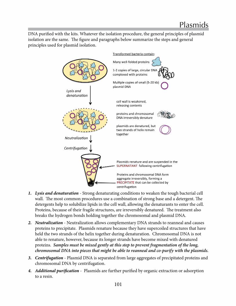
101
Replace Chapter number and title on A-Master Page.<- ->
PlasmidsDNApurifiedwiththekits.Whatevertheisolationprocedure,thegeneralprinciplesofplasmidisolationarethesame.Thefigureandparagraphsbelowsummarizethestepsandgeneralprinciplesusedforplasmidisolation.
1. Lysis and denaturation-Strongdenaturatingconditionstoweakenthetoughbacterialcellwall.Themostcommonproceduresuseacombinationofstrongbaseandadetergent.Thedetergentshelptosolubilizelipidsinthecellwall,allowingthedenaturantstoenterthecell.Proteins,becauseoftheirfragilestructures,areirreversiblydenatured.ThetreatmentalsobreaksthehydrogenbondsholdingtogetherthechromosomalandplasmidDNA.
2. Neutralization-NeutralizationallowscomplementaryDNAstrandstoreannealandcausesproteinstoprecipitate.Plasmidsrenaturebecausetheyhavesupercoiledstructuresthathaveheldthetwostrandsofthehelixtogetherduringdenaturation.ChromosomalDNAisnotabletorenature,however,becauseitslongerstrandshavebecomemixedwithdenaturedproteins. Samples must be mixed gently at this step to prevent fragmentation of the long, chromosomal DNA into pieces that might be able to reanneal and co-purify with the plasmids.
3. Centrifugation-PlasmidDNAisseparatedfromlargeaggregatesofprecipitatedproteinsandchromosomalDNAbycentrifugation.
4. Additional purification -Plasmidsarefurtherpurifiedbyorganicextractionoradsorptiontoaresin.

102
Chapter 10
Exercise 1 - Plasmid isolation with the ZyppyTM kitConcentrate the plasmid-bearing bacterial cells 1. Yourgroupwillreceive3bacterialculturesthatweregrownovernightinLuriaBertani(LB)
mediacontaining100µg/mLampicillin.Thedensityofcellsinthecultureisexpectedtobe3-4X109cells/mL. What is the purpose of the ampicillin? How does it work?
2. Transfer1.5mLofeach3mLculturetoamicrocentrifugetube.Concentratethecellsbycentrifugingthematmaximumspeed(~14,000rpm)for1min.Thecellswillformawhitepelletatthebottomofthetube.
3. UseaP200toaspirateoffasmuchoftheculturemediumaspossible.
Alkaline lysis of bacterial cells harboring the plasmids4. Re-suspendthepelletin600µlofTEbuffer(Tris-HCl,EDTApH=8.0)usingthevortexmixer.5. Add100µLof7XBlueZyppyLysisbuffertothetube.Mixthebufferandcellsbygently
invertingthetube4-6times.Begentle!ToomuchmechanicalstresswillfragmentthebacterialchromosomalDNAandcontaminateyourplasmidpreparation.Thesolutionshouldturnfromacloudybluetoaclearblue.NOTE: This step is time-sensitive!! Proceeed to step 3 within 2 minutes.
Separate plasmid DNA from denatured proteins and chromosomal DNA6. Add350µLofcoldYellowZyppyNeutralizationbuffer(w/RNAaseA)tothetube,and
mixthecontentsthoroughlybyinvertingseveraltimes.Thesolutionwillturnyellowwhenneutralizationiscomplete,andayellowishprecipitatewillform.Invertthesampleanadditional3-4timestoensurecompleteneutralization.Besurethereisnomorebluecolor.
7. Centrifugethemixtureatmaximumspeedfor3minutestoremovedenaturedproteinsandchromosomalDNA.Noticethatthetubecontainsayellowprecipitatethathascollectedononesideofthetube.ThepaleyellowsupernatantcontainstheplasmidDNA.
Purify plasmid DNA by adsorption to a silica resin. 8. Usingapipette,carefullytransferthepaleyellowsupernatant(~900µL)ontoaZyppyspin
column.Becarefulnottotransferanyoftheyellowprecipitate!9. Placethecolumnwiththecollectiontubeattachedintoacentrifugeandspinatmaximum
speedforabout15seconds.Itisbesttousethe“pulse”buttononthecentrifugeandcountto15or20secondsforthiscentrifugationstep.
10.Removethecolumnanddiscardtheflowthroughinthecollectiontube.11.Placethecolumnbackintothecollectiontubeandadd200µLofZyppyEndo-Wash
solution.(Endo-Washcontainsguanidinehydrochlorideandisopropanol,whichwillremovedenaturedproteinsfromtheresin.)
12.Centrifugefor15-20seconds,anddiscardtheflowthrough.13.Placethecolumnbackintothecollectiontubethenadd400µLofZyppyColumnWash
buffer.(Thisstepsremovescontaminatingsalts.)Centrifugefor30-40seconds.

103
Replace Chapter number and title on A-Master Page.<- ->
Plasmids
Exercise 2 - Measure the plasmid DNA concentration
Elute the plasmid DNA14.TransfertheZyppycolumntoaclean(andappropriatelylabeled)1.5mLcentrifugetube,leavingthelidofthetubeopen.15.Carefully,add100µLofsterilewaterdirectlyontopofthewhitecolumnbed.Placethe
pipettetipascloseasyoucantothewhitecolumnbedwithoutpokingit.Slowlydispensethewaterontopoftheresinbed.
16.Allowthebuffertopercolateintothecolumnbylettingthecolumnstanduprightinthemicrocentrifugefugetubefor1minute.
17.Centrifugethecolumnatmaximumspeedfor30seconds.Again,it’sfinetoleavethecapopenduringthisspin.
18.Removethecolumn,capthetubeandplaceitonice.ThistubeshouldnowcontainplasmidDNA.Label the tube. Save the DNA for future experiments.
TheconcentrationandpurityofDNAsamplescanbeestimatedfromtheirabsorbanceofultraviolet(UV)light.NucleicacidsabsorblightstronglyintheUVrange,withanabsorbancemaximumof~260nm.AnA260of1correspondsto~50µg/mLDNA.ProteinsalsoabsorbUVlightduetotheiraromaticsidechains,butmuchlessstronglythannucleicacids.Proteinshaveadifferentabsorbancemaximumof~280nm.AnA280of1correspondsto1mg/mLprotein.Becauseofthedifferencesinproteinandnucleicacidabsorbances,theA260/A280ratioofasampleisusefulindeterminingthepurityofaDNAsample.PureDNAhasanA260/A280of~1.8.Proteinorchemicalcontaminationwilllowerthisratio.
Inthislaboratory,youwilluseaninstrumentcalledtheNanodropTMtoobtainanultravioletabsorbancespectrumforyourplasmidDNAsamples.TheNanodropsoftwarewillalsocalculatetheconcentrationofDNAandtheA260/A280ratio.TheNanoDropusesfiberoptictechnologyandthesurfacetensionofasampletoprovideaccurateabsorbancemeasurementsoveralargedynamicrangeofDNAconcentrations.Fromapracticalpointofview,thismeansthatyouneedtosacrificeonlysmallamountsofyourpreciousDNAsample.TheNanoDropisamazinglysimpletouse,andyourTAwilldemonstratetheappropriateuseoftheinstrument.
1. Pipette3µLofyourplasmidsampleonthepedestalontopofthepin(shownattheendofthearrowatleft).
2. Closethearmandrecordtheabsorbance.3. Whenthemeasurementiscomplete,openthearm
andwipethepedestalsurfacewithalint-freewipeinpreparationforthenextsample.
4. Recordtheabsorbancereadingsinyournotebook.

104
Chapter 10
Dong,X,Stothard,P,Forsythe,IJ,&Wishart,DS(2004).PlasMapper:awebserverfordrawingandauto-annotatingplasmidmaps.Nucleic Acids Res.32,W660-W664.
Gelperin,DM,White,MA,Wilkinsonet al.(2005)BiochemicalandgeneticanalysisoftheyeastproteomewithamovableORFcollection.Genes Develop19:2816-2826.
Johnston,M(1987)Amodelfungalregulatorymechanism:theGAL1genesofSaccharomyces cerevisiae.Microbiol Rev51:458-476.
References
The Nanodrop results screen shows the UV spectrum of the sample and the estimated DNA concentration

• UnderstandthebiologicaloriginsandfunctionsofREs
• UnderstandthestructuresofrestrictionsitesinDNA
• Deviseastrategytodistinguishbetweenthreeyeastoverexpressionplasmidsus-ingREdigests
• Analyzerestrictiondigestsonagarosegels
Restrictionendonucleases(REs)arepartofbacterialdefensesystems.REsrecognizeandcleavespecificsitesinDNAmolecules.REsareanindispensabletoolinmolecularbiologyforbothanalyzingandconstructingDNAmolecules.Inthislab,youwillpreparerestrictiondigeststodistinguishwhichofyourplasmidscarryingareS. cerevisiaeandS. pombe ORFsorbacterialLacZ.
Restriction mappingChapter 11
Objectives

106
Chapter 11
Bacterial restriction/modification systems protect against invaders Thediscoveryofrestrictionenzymes,orrestrictionendonucleases(REs),waspivotaltothedevelopmentofmolecularcloning.REsoccurnaturallyinbacteria,wheretheyspecificallyrecognizeshortstretchesofnucleotidesinDNAandcatalyzedouble-strandbreaksatorneartherecognitionsite(alsoknownasarestrictionsite).Todate,thousandsofREswithdistinctspecificitieshavebeendescribed.Youmightwonderwhybacteriaharborthesepotentiallydestructiveenzymes.REsarepartofabacterialdefensesystemagainstforeignDNA,suchasaninfectiousbacteriophage.ThebacteriaprotecttheirownDNAfromcleavagewithamethyltransferasethatmodifiesthesamerestrictionsitesinthebacterialDNA.Thecombinedactivitiesoftheendonucleaseandmethyltransferasearereferredtoasarestriction/modificationsystem.InTypeIenzymes,theendonucleaseandmethyltransferaseactivitiesarepartofthesamecomplex.TheREsusedinthislaboratoryareTypeIIenzymes.TheTypeIIendonucleasesandmethyltransferasesdonotformacomplexandfunctionindependently.
TounderstandhowREswork,itmaybehelpfultolookatasingleenzymeingreaterdetail.Oneofthebest-studiedrestrictionenzymesisEcoR1.AlthoughthenamesofindividualREsmaysoundabitlikebabytalk,thenomenclatureisactuallyverysystematicandisbasedonitsbiologicalsource.EcoRIisfoundnaturallyintheRY13strainofEscherichia coli,orE. coli.Itsnamebeginswiththegenusandspecies(EcoforE. coli),followednextbyastrainidentifier(RforRY13),andendswithaRomannumeralthatdistinguishesthedifferentenzymesinthestrain.StrainRY13ofE. coli containsmultipleendonucleases,butonlyEcoRIandEcoRV,arewidelyusedinmolecularbiology.PurificationofREsfromtheirnaturalsourceisalaboriousprocedurethatrequireslargequantitiesofbacteria,sincetheREsarenotabundantproteinsinbacteria.Tofacilitatetheproductionoftheseusefulenzymesforroutinelaboratoryuse,molecularbiologistsclonedthecodingsequencesoftheendonucleasesintobacterialexpressionplasmids.Today,mostcommerciallyavailableenzymesarerecombinantproteinsoverexpressedinbacteria.(Alongtheway,scientistsalsointroducedsomemutationsthatgeneratedenzymeswithenhancedstabilityorhigheractivity.)
Restriction endonucleases
Inthelastexperiment,youisolatedplasmidscarryingS. cerevisiae orS. pombe ORFsfromtransformedbacteria.Eachgroupwasgiventhreedifferentplasmids.Oneofthethreeplasmids,carriestheS. cerevisiae METorCYSgenethathasbeeninactivatedinyouryeaststrain.ThisMETgenewasclonedintothepBG1805plasmid(Gelperinet al.,2005).AsecondplasmidcarriestheS. pombehomologfortheMETorCYSgene,clonedintothethepYES2.1plasmid.ThethirdplasmidisanegativecontrolthatcontainsthebacterialLacZgeneclonedintopYES2.1.Inthislab,yourteamwilldesignandcarryoutastrategytodistinguishbetweentheplasmidsusingre-strictionendonucleases.Inthenextlab,youwillseparatetheproductsoftherestrictiondigests,orrestrictionfragments,byagarosegelelectrophoresis,generatingarestrictionmap.

107
Replace Chapter number and title on A-Master Page.<- ->
Restriction mapping
Restriction enzymes cleave specific sites in DNA RestrictionenzymeslikeEcoRIarefrequentlycalled6-cutters,becausetheybindtoa6-nucleotidesequence.AssumingarandomdistributionofA,C,GandTsinDNA,probabilitypredictsthatarecognitionsitefora6-cuttershouldoccuraboutonceforevery4000bp(46)inDNA.Ofcourse,thedistributionofnucleotidesinDNAisnotrandom,sotheactualsizesofDNAfragmentsproducedbyEcoRIrangefromhundredstomanythousandsofbasepairs,butthemeansizeiscloseto4000bp.ADNAfragmentofthatlengthisveryusefulinthelab,sinceitislongenoughtocontainthecodingsequenceforsomeproteinsanditiseasilyresolvedoncommonagarosegels.ThesequencethatEcoRIrecognizesindoublestrandedDNAisGAATTC.Thesequenceisapalindromewithatwo-foldaxisofsymmetry,becausereadingfrom5’to3’oneachstrandgivesthesamesequence.Thepalindromicnatureoftherestrictionsiteismoreobviousinthefigurebelow.Thecircleinthecenteroftherestrictionsitedenotestheaxisofsymmetry.EcoRIbindsDNAattherestrictionsiteandcatalyzesthehydrolysisofthephosphodiesterbondbetweenGandAoneitherstrand.
EcoRI recognition site in DNA
...G A A T T C...
...C T T A A G...5’
5’
axis of symmetry
...G A A T T C...
...C T T A A G...
5’
5’EcoRI cleavage pattern
5’
A A T T C... G......G...C T T A A
5’5’
5’
Cleavage produces two “sticky” ends with 5’ overhangs
EcoRI binds
EcoRI cuts
EcoRI catalyzes the cleavage of a palindromic recognition site. The recognition site for EcoRI has a two-fold axis of symmetry. Cleavage generates two fragments with 5’-sticky ends.
ThestructureofEcoRIisconsistentwithanenzymethatcatalyzesidenticalreactionsattwodif-ferentsites.CrystallizationdatashowsthatEcoRIisahomodimer,anenzymewithtwoidenti-calsubunits.Thefigureonthenextpage(PDBdatafile1QPS,unpublished)showstwodifferentviewsofEcoRIboundtoDNA.Intheleftview,we’relookingdowntheaxisoftheDNAhelix.Therightviewisasideview.Takeacloselookatbothstructures,usingtheDNAhelixasthecenter.Inthesemodels,alphahelicesandbetasheetsaregivendifferentcolorsorshades.Thesymmetryintheenzymeisquiteapparent.

108
Chapter 11
Crystallization structures (1QPS) show that EcoRI is a symmetric homodimer that binds DNA.
Selecting restriction enzymes for experiments RestrictionenzymesarewidelyusedinmolecularbiologyforboththeanalysisandconstructionofDNAmolecules.ThefirststepinanexperimentinvolvingrestrictionenzymesistomapthepositionofrestrictionsitesinatargetDNAmolecules.ManysoftwareprogramsalsoidentifyopenreadingframesastheyscanDNAsequenceforRErecognitionsites.TheseprogramsusuallygenerateagraphicaloutputaswellastabulardatawithdetailsaboutthelengthsandpositionsoftherestrictionsiteintheDNAsequence.Thelistofenzymesthatcutaparticularsequenceisalwaysimpressive,butonlyafewenzymesusuallyturnouttobepracticalforthepurposeoftheexperiment.Whenchoosingenzymes,investigatorsconsiderthenumberandlengthsofthefragmentsthatwillbegeneratedinadigest,thepositionoftherestrictionsitesrelativetoageneofinterest,aswellasthekindofendsintherestrictionfragment.
Restriction maps are used to identify DNA molecules ThesequenceofaDNAmoleculedeterminesthedistributionofrestrictionsites.ThemapofrestrictionsitesinapieceofDNAisknownasarestrictionmap.Withcarefulplanning(below),investigatorscanuserestrictionmapstodesignexperimentsthatwilldistinguishbetweenDNAmolecules.Intheseexperiments,DNAsamplesaredigestedwithvariousREstoproducearestrictiondigest,acollectionofsmallerrestrictionfragmentsthathavebeencleavedateitherendbytheRE.Themoleculesinthedigestarethenseparatedbyagarosegelelectrophoresis(Chapter8).Fromthesizesoftherestrictionfragmentsthatareresolvedonthegel,investigatorsareabletoidentifytheoriginalDNAmoleculeusedintherestrictiondigest.Inthisexperiment,youwilluserestrictionmapsofyourthreeunknownplasmidstodeviseanexperimentalstrategywhichwillallowyoutoidentifythethreeplasmids.
Restriction endonucleases are used in the construction of DNA molecules TheavailabilityofalargenumberofREswithdistinctspecificitieshasbeencriticalforrecombinantDNAtechnology,whichinvolvestheconstructionofnovelDNAmoleculeswithpropertiessuitedforaparticularpurpose.Intheseexperiments,REsserveasthemolecule

109
Replace Chapter number and title on A-Master Page.<- ->
Restriction mappingscissorsthatcutsDNAintosegmentsthatcanbejoinedtoadditionalrestrictionfragmentstoproduceadesiredmolecule.REsareusedinconjunctionwithDNAligases,whichservesasthe“paste”bycatalyzingtheformationofphosphodiesterbondsbetweenrestrictionfragments.ThekindofendgeneratedbyanREisparticularlyimportantwheninvestigatorsareconstructingarecombinantDNAmolecule.Inthefigureonthepreviouspage,yousawhowEcoRIproducedDNAfragmentswith5’overhangs.Otherenzymesproducefragmentswith3’overhangs,whilestillothersproducebluntendswithnooverhangs.Endswithoverhangsareoftenreferredtoas“sticky”ends,sincetheywillformhydrogenbondswithacomplementarysequenceonasecondDNAfragment.Bycontrast,fragmentswithbluntends(i.e.nooverhang)canannealwithanyotherbluntend,regardlessoftheadjoiningsequence.
Wewillnotbegeneratingnewrecombinantmoleculesinthisclass,butitisimportanttounderstandtheirimportancetomolecularbiology.ConsiderthepBG1805andpYES2.1plasmids.Fromthemaponp.99,youcanseethatthesecomplexplasmidswereconstructedbystitchingtogetherfunctionalelementsfrompiecesofDNAobtainedfromavarietyofsources.
Handling restriction endonucleases in the laboratory TheREsthatweareusinginthelabarehighlypurified(andexpensive!)proteinsthathavebeenpurifiedfromrecombinantbacteria.Likeallenzymes,eachrestrictionendonucleasefunctionsoptimallyunderadefinedsetofreactionconditions,includingvariablessuchastem-perature,pH,andtheconcentrationsofmetalionsandsalts.Themanufacturer’ssupplyrestric-tionenzymeswithbuffersthatgeneratereactionconditionsclosetothosethatareoptimalfortheenzyme.Someofthebufferscontainbovineserumalbumin(BSA),anabundantproteinfromcowserum.BSAisincludedlargelyforitsstabilizingproperties.Ingeneral,enzymesaremorestableathigherproteinconcentrations.BSAdoesnotaffecttheenzymaticactivityoftheREs,butprotectsparticularlysensitiveREsfromdenaturation.BSAmayalsosaturatenon-specificbind-ingsitesforREsonthesidesofthetesttubesusedinthedigests.
Likeallenzymes,REsaresubjecttospontaneousdenaturationandlossoffunction,soREsneedtobehandledwithcare.(Bycomparison,DNAisanexceptionallystablemolecule.)Therateofproteindenaturationincreasesasthetemperatureincreases,soenzymesarekeptonicewhilereactionmixturesarebeingprepared.Denaturationisalsoacceleratedatair/waterin-terfaces,soit’simportanttominimizeairbubblesandfreeze/thawcyclesforsolutionscontainingrestrictionenzymes.Manufacturersattempttominimizedenaturationbysupplyingthesolutionsinasolutionof50%glycerol,whichdoesnotfreezewhentheenzymesarestoredat-20˚C.
FollowsomesimpleruleswhenyouuseREs.• BesuretousetherecommendedbufferforaparticularRE.• Keepthereactionsoniceuntiltheincubationbegins• Becarefulnottointroducebubbles.Mixthereactionsbyflickingthetubeswithyourfingers,
ratherthanusingthevortexmixer.• Gatherthereactioncomponentsatthebottomofthetube,NOTbycentrifuging,butwitha
firmflickofthewrist.

110
Chapter 11
Exercise 1 - Plan the restriction digest Assign each person in your group a different plasmid to analyze.
Youwillfirstneedtoconstructthesequencesofyouroverexpressionplasmids.RecallthattheS. cerevisiaegeneshavebeenclonedintothepBG1805vectorandthattheS. pombegenesandLacZhavebeenclonedintothepYES2.1vector.Afterthesequenceisconstructed,youwillgeneratearestrictionmapthatcanbeusedtopredictrestrictionfragmentsgeneratedinanREdigestion.Togeneratethemap,wewilluseoneofseveralonlinetoolsthatisavailableatthewebsiteofNewEnglandBiolabs,acommercialsupplierofREs.
1.Locate the coding sequence of your gene.UsetheNM___accessionnumbersofyourgene(p.45forS. cerevisiaegenesorp.94forS. pombegenes)toaccessthenucleotiderecordinNCBI’sNucleotidedatabase.ThepYES2.1-LacZsequenceispostedonBlackboard.Notethatthis8964bpsequencehasbeenmodifedfromnaturally-occurringLacZgenes.
2.Assemble the complete nucleotide sequence of your plasmid.• OpenthesequencefilefortheBG1805orpYES2.1vectorsequencepostedonBlackboard.
TheplasmidsequencesarenumberedsothattheGAL1promoterisatthe3’-endoftheDNAsequence.
• PastethecodingsequencefromtheNM_record(ortheLacZsequence)totheendoftheplasmidsequence.
• Afteryoupastethecodingsequenceattheendofthevectorsequence,deletethelastthreenucleotides,whichcomprisethegene’sstopcodon.Stopcodonsarenotincludedintheplasmidsequences,whichencodefusionproteinswithC-terminalextensions.(Note:thestopcodonintheLacZsequencehasbeenremovedbythemanufacturer.)
• RememberthatthefirstnucleotideofyourMETorCYSgeneisonenucleotidehigherthanthelastnucleotideinthevector.Forexample,theBG1805sequencecontains6573nucleo-tides.ThefirstnucleotideofaMETorCYSgeneinsertedintopBG1805willbenucleotide6574.ThepYES2.1plasmidcontains5886nucleotides.
3.Prepare a restriction map of the complete plasmid sequence.• Pastethesequencefromstep2intothesearchboxintheNEBCuttertool:
tools.neb.com/NEBcutter2/• Checktheboxtoindicatethattheplasmidiscircular.Youmightalsowanttogiveyourplas-
midaname.TheNEBsitewillstoreyourqueriesfor24hours,whichcanbeveryconvenient.Clicksubmit.
• ThesearchtoolwillreturnresultsforabewilderingnumberofREs.ThevastmajorityoftheREsitesarenotuseful,becausethefragmentsaretoolargeortoosmall,theenzymeisnotavailableinthelab,ortheendonucleaseissensitivetoDNAmethylation(whichcanbeunpredictable).

111
Replace Chapter number and title on A-Master Page.<- ->
Restriction mapping
4.Perform custom digests with enzymes that look promising. Clickthecustomdigestlink.ThisbringsupachartofREthatcuttheplasmid,theirrecognitionsites,thenumberofrecognitionsites,andtheamountofenzymeactivityineachofthefourbuffersusedfordigests.Your TA will tell you which REs are available in the lab. Wewillbeanalyzingtherestrictionfragmentsonagarosegels,whichdoagoodjobofresolvingfragmentsranginginsizefrom~200bpto~5000bp.DeterminewhichoftheREsavailableinthelabwillproducerestrictiondigestswithanicerangeoffragmentsizes.
5.Prepare a table summarizing the restriction maps for your three plasmids.Completethetablebelow,indicatingthesizesoftherestrictionfragmentsgeneratedwitheachRE.Includethetotallengthoftheplasmidinthetable.Thesumoftherestrictionfragmentlengthsshouldsumuptothisnumber.
Plasmid nameLength(bp)
Restriction Enzyme (Fragment lengths) (Fragment lengths) (Fragment lengths)
6.Choose a RE that distinguishes your three plasmids. TheteamshouldusethedatatableabovetoselectONEREthatbestallowsyoutodistinguishthethreeplasmids.Therestrictiondigestswillbeanallyzedonagaraosegelswith8 wells.• Onewellisreservedforthemolecularweightstandard.• Twowellswillbeusedforeachplasmid.
OnewellwillcontainplasmidthathasNOTbeendigestedwithRE.AsecondwellwillcontainplasmidthatcontainsplasmiddigestedwithRE.

112
Chapter 11
Exercise 2 - Set up the restriction digests TheconcentrationsofREandplasmidDNAneedtobematchedinarestrictiondigest.ManufacturersassaytheactivityofeachbatchofREandexpresstheactivityinunitsofactivity/µL.Aunitofactivity(U)isassessedinastandardizedassay.Restrictiondigestsareusuallysetuptocontain2-5UperµgplasmidDNA.Wewillbeusing5µLofplasmidminiprepDNAineachreaction.TheREsthatyouwillbeusinghavebeendilutedtoaconcentrationof0.5U/µL.
In your lab notebook,notewhichRE(s)youhavedecidedtouse.Checkwhich10Xsaltsolutionthatyoushouldusefromthemanufacturer’schartof:https://www.neb.com/tools-and-resources/usage-guidelines/nebuffer-performance-chart-with-restriction-enzymes
• Computethevolumesofeachcomponentthatyouwillneedforrestrictiondigestswithafinalvolumeof10µL.
5.0µLplasmid-togiveafinalconcentrationof0.2-0.4µgplasmidDNA 1.0µL10Xbuffer 1.0µLBSA(froma1mg/mL10xstock)-onlyIFrecommended 0-1µLdeionizedwater(tobringtotalvolumeto10µL)* 2.0µL(1.0U)restrictionenzyme
The total reaction volume should be 10 µL. • Drawaschematicinyourlabnotebookshowinghowthedigestswillbesetup.• Combinethecomponentsforeachdigestionintheirownseparatetubes,intheorderlisted
above.• Addtherestrictionendonuclease(s)lastandmixbygentlytappingthetube. • Incubatethesamplesat37°Cfor2hr.Storethesamplesinthefreezer.
Exercise 3 - Analyze the restriction digests on agarose gelsThis exercise will be performed in the next laboratory session.
1.Planyourgel.Eachgroupofstudentswillprepareoneagarosegel.Eachstudentwillrunonelanewithundigestedplasmidandasecondlanewithplasmidthathasbeendigestedwithrestrictionendonuclease(s).Anadditionallaneshouldbereservedformolecularweightstandards.Recordinyournotebookhowyouplantoloadyourgel.
2.Prepare,loadandrunyourgelsasdescribedinChapter8.Besuretorecordthedetailsofeverystepinyourlabnotebook.

• Understandtheprinciplesofyeasttransformation
• Understandhowplasmid-encodedgenescancomplementgenedeficiencies
• Transformmet or cys strainswithplasmidscarryingS. cerevisiaeandS. pombegenesinvolvedinMetorCyssynthesis
• Usereplicaplatingtotesttransformantsfortheirabilitytogrowonvariousselectivemedia
TechniquesfortransformingmicrobialorganismswithforeignDNAareessentialinmodernmolecularbiology.Inthislab,youwilltransformaS. cerevisiae metorcys strainwiththreedifferentplasmidsanduseura3 complementationtodetecttransformedcells.YouwillthenusereplicaplatingtodetermineifS. pombe MetorCys genesarefunctionallyequivalenttoS. cerevisiae MET and CYS genes.
Yeast Transformation Chapter 12
Objectives

114
Chapter 12
TransformationreferstotheuptakeofDNAbyacell,causingachangeinitsphenotype.Naturally-occurringtransformationwasfirstdescribedin1928byFrederickGriffith,whodescribedaheat-stable“transformingprinciple”fromvirulentStreptococcus pneumoniaethatcouldtransformnon-virulentS. pneumoniaetoanencapsulated,virulentform.The“transformingprinciple”wassubsequentlyidentifiedasDNAbyAveryandcolleaguesin1944.Sincethen,transformationhasbecomeanindispensabletoolinthemolecularbiologylaboratory.Thephysicalbasisfortransformationisstillpoorlyunderstood,butresearchershaveempiricallydevelopedconditionsthatgivefairlyconsistenttransformationinthelab.Reliabletransformationtechniqueshavebeendevelopedforbacteriaandmanyeukaryotes,rangingfromyeasttomammaliancells.
Transformation conditions have been developed empirically ThechallengeinlaboratorytransformationistodeviseconditionsunderwhichDNAwillpassacrossthecellwallandplasmamembraneoflivingcells,whicharenormallyimpermeabletoDNA.Veryfewcellsarenaturallycompetent,orabletotakeupDNAontheirown.Consequently,researchersuseavarietyofchemicaltreatmentstorendercellscompetent.Ingeneral,thesechemicaltreatmentshavesomekindofdestabilizingeffectontheplasmamembrane.TheintroductionofDNAintothesecompetentcellscanbefurtherencouragedbyaphysicalstress,suchasapulseofelectriccurrentortemperatureelevation.Transformationisnotaveryefficientprocess,butbecauselargenumbersofmicroorganismscanbeculturedinthelaboratory,usefulnumbersoftransformantscanbeobtainedwithmostmicroorganisms.
Techniquesforyeasttransformationarenowstandardinthelaboratory.Dependingonthedetailsoftheexperimentalprocedure,reactionscanyieldasmanyas106transformantsperµgDNA.ThestructureoftheDNAusedfortransformationgreatlyaffectsthetransformationefficiency.TransformationefficienciesareconsiderablyhigherwithsupercoiledplasmidDNAthanwithlinearpiecesofDNA,possiblybecauseplasmidsenterthecellmorereadilyand/orplasmidsarelesssusceptibletoendonucleasedigestion.
Themostcommonlyusedyeasttransformationmethodsuseacombinationoflithiumacetate,single-strandedcarrierDNAandpolyethyleneglycol(PEG).Althoughnooneknows
Transformation alters the phenotype of a cell
Inthislab,youmayreceiveapreliminaryanswertothesemester’sresearchquestionaboutthefunctionalconservationofMetandCysproteinsintheAscomycota.Duringthefirstpartofthesemester,yourteamusedselectiveplatingandcolonyPCRtoidentifyyeastdeletionmutants.YouthenisolatedandcharacterizedplasmidsthatcanbeusedtooverexpressMetandCysproteins.Thesetwosetsofexperimentscometogetherinthislab,whenyoutransformtheS. cerevisiae deletionstrainwiththeexpressionplasmids.Throughaseriesofcomplementationexperiments,youwilldetermineifthegenescarriedontheplasmidsareabletocompensateforthemissingMETorCYSgenesinthemutants.Incomplementation,theintroductionofaforeigngenerestoresthenormalphenotypetoamutantwithadefectivegene.

115
Replace Chapter number and title on A-Master Page.<- ->
Yeast transformationexactlyhowthesecomponentspromotetransformation,anumberofhypotheseshavebeenadvanced.LithiumionsneutralizethenegativechargesonDNAmoleculestobetransformedandthephospholipidbilayeroftheyeastcell,andtheymayalsogeneratesmallholesintheplasmamembranethatallowthepassageofnucleicacids.Single-strandedDNAactsasacarrierfortheplasmidDNAtobetransferredintothecellanditmayhelptoprotectthelatterfromendonucleases.ThesourceofthecarrierDNAisunimportant.SincethecarrierDNAconcentrationisconsiderablyhigherthanthatoftheDNAtobeintroducedintothecell,thecarrierDNAisusuallyisolatedfromaninexpensivesource,suchassalmonsperm.It is imperative that the carrier DNA for transformations be single-stranded. Inourexperiments,wewillboilthecarrierDNAfor5minutesandthenrapidlychillittopreventreannelingoftheDNAhelix.PEGmayhelpbringtheDNAintocloserappositionwiththemembrane.PEGisoftenusedtopromotemembranefusionandisthoughttoalterwaterstructurearoundplasmamembranes.
Transformation and plasmid complementationCompetent ura3 yeast cells are transformed incubating cells with a plasmid containing the yeast URA3 gene at an elevated promoter (top). Transformed cells are selected on media that does not contail uracil (bottom).
Complementation is often used to isolate transformants TheDNAusedfortransformationmustcarryaselectablemarkerwhosepresencecanbedetectedbyscreening.Followingatransformation,cellsareplatedonselectivemediathatwillallowtransformed,butnotuntransformed,cellstogrow.AllthepBG1805-(Gelperinet al.,2005)andpYES2.1-derivedplasmidsthatweareusingcarryanormalcopyoftheyeastURA3gene,aswellastheURA3promoter,sothegeneisregulatedmuchlikeanormalchromosomalgene.OuryeastdeletionstrainswerederivedfromstrainBY4742,whichhastheura3∆0allele(Winzeleret al.,1999)Complementationwilloccurbecausetheplasmidcarriesafunctionalcopyofthegenethatisdefectiveinthemutanthoststrain.TheUra3pproteinproducedfromtheplasmid-encodedURA3genecompensatesfortheura3deletionintheyeastchromosome,allowingtransformedcellstogrowintheabsenceofuracil,asshownbelow.Becauseofitsreliability,manyyeasttransformationschemesrelyonURA3complementationtoisolatetransformants.

116
Chapter 12
YoumaybewonderingwhywearenotusingMETorCYSgenecomplementationtoisolatetransformants,sincethisisthegoalofoursemester’sproject.Thereareseveralreasonsforthis.First,weneedtoinsurethattheoverexpressionplasmidshavesuccessfullytransformedthedeletionstrains.URA3genecomplementationoffersawell-testedandreliablemeanstoassesssuccessfultransformation.SuccessfulURA3complementationservesasanimportantpositivecontrolfortransformationiftheplasmid-encodedMETandCYSgenesfailtocomplementdeletionstrains.
AsecondissuerelatestoregulationoftheplasmidORFsbytheGAL1 promoter(Johnston,1987)intheplasmids.TheGAL1promoterisaninduciblepromoterthatisnormallyrepressedwhencellsaregrowninglucose,thepreferredcarbonsourceforyeast.Whengalactosereplacesglucoseasthecarbonsource,yeastrespondbysignificantlyincreasingthetranscriptionofseveralgenes,includingGAL1,thatareinvolvedinthemetabolismofgalactose.Initsnormalchromosomallocation,theGAL1promoterrespondstoavarietyofpositiveandnegativetranscriptionregulators(Chapter13).TheGAL1promotersequencehasbeenwidelyusedinmolecularbiologybecauseitgenerallyfunctionswellinectopiclocations,suchasplasmids.
Onceyouobtaintransformants,youwillanalyzeMET/CYSgenecomplementationusingselectivemediacontainingeitherD-glucoseandD-galacatoseasthecarbonsource.ThetransformedcellswillcontainmanymorecopiesoftheGAL1promoterthannon-transformedcells,becausebothpBG1805andpYES2.1aremulti-copyplasmids.Becausetheregulatorybalanceisalteredintransformedcells,galactoseandglucosemaynotfunctionassimple“ON”and“OFF”switches.Youshouldconsiderthepossibilitythat“leaky”genetranscriptioncouldoccurinthepresenceofthenormalrepressor,D-glucose.Ifthishappens,MET/CYSgeneswouldcomplementmet/cysmutantsgrowninD-glucose.ItisalsopossiblethattransformedcellscouldproduceexcessivequantitiesofMetandCysproteinsthataredetrimental,orevenfatal,totransformedcells.
GAL1 promoter - experimental considerations
Replica plates accelerate the screening process Asnotedabove,transformationisaninefficientprocess,soresearcherswanttomakethemostofeverycellthathasbeentransformed.Inourexperiments,wewillbeisolatingtransformedcellsfortheirabilitytogrowintheabsenceofuracil,butwearereallyinterestedintheirabilitytogrowintheabsenceofMetorCys.Replicaplatingoffersameanstoquicklyscreenaplateofcellsfortheirabilitytogrowinawiderangeofmedia,whileretaininginformationaboutindividualcolonies.Asshownontheoppositepage,theoriginalplateoftransformantsbecomesthe“masterplate.”AnimprintofthemasterplateismadebyGENTLYtappingtheinvertedplateonapieceofsterilevelveteenimmobilizedonablock.Thisimprintcanthenbetransferredtoplateswithdifferentkindsofselectivemedia,establishingthegenotypeofthetransformants.Inourexperiments,wewillmaketransferreplicasofthetransformationreactions(isolatedonYC-URAplates)toYC-UraplatesthatarealsolackingMet/Cys,witheitherglucoseorgalactoseasacarbonsource.
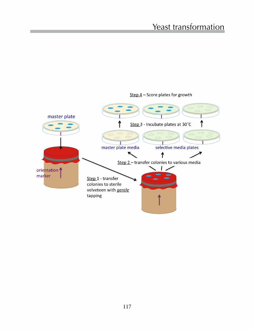
117
Replace Chapter number and title on A-Master Page.<- ->
Yeast transformation

118
Chapter 12
Thefollowingprotocolisaslightmodificationofthe“QuickandDirty”transformationprotocoldescribedbyAmberget al.(2005).Withcarefulattentiontodetailandcooperativestrains,thisprocedurecanyieldthousandsoftransformantsperµgplasmidDNA.Modificationstothismethodcanincreaseitsefficiencybyseveralordersofmagnitude,whichwouldberequiredforlinearpiecesofDNA(GietzandSchiestl,2007).
Prepare a transformation master mix1.Prepareatransformationmastermix.Thefollowingingredientsprovideenoughreagentsfor
fivetransformationreactions.Combineinamicrocentrifugetube:
100µLsterile2Mlithiumacetate(freshlyprepared) 400µLsterile50%PEG-3350 4µL2-mercaptoethanol(STINKY!!addthisinthefumehood!)
Set up individual transformation reactions - for each transformation:2.Add15µLofthedenaturedsalmonspermDNA(2mg/mL)tolabeledmicrocentrifugetubes.
Note: It is important for the salmon sperm DNA to be single-stranded for this procedure to work well. Boil the DNA for 5 minutes to denature the DNA. Quick chill the DNA by placing it immediately on ice. Keep the DNA on ice until you are ready to use it.
3.Add5µLofminiprepplasmidDNAtotheappropriatelylabeledmicrocentrifugetube.4.Add100µLoftransformationmixfromstep1tothemicrocentrifugetube.Vortexfor10-15
secondstomixthecontents.5.Usingasteriletoothpick,scrapealargeyeastcolony(ortheequivalentofa“matchhead”
ofyeast)fromaYPDplate.Transfertheyeasttothemicrocentrifugetubecontainingthetransformation/DNAsolution(step4)bytwirlingthetoothpickseveraltimes.Besurethatthecellsareuniformlysuspendedbeforeproceeding.
Repeatsteps2-5foreachofyourtransformationreactions.Be sure to include a control that contains no plasmid DNA.
6.Incubatethetransformationmixturesat37˚Cwithshakingfor30-45min.
Exercise 1 - Yeast transformation

119
Replace Chapter number and title on A-Master Page.<- ->
Yeast transformation
Plate the transformed cells on selective media lacking uracil
7.Remove10µLoftheresuspendedcellsto90µLofsterilewaterinamicrocentrifugetube.Thissamplewillbeseriallydilutedforaspotplate(step10)thatyouwillusetocalculatethetrans-formationefficiency.
8.Platetheremainderofthemixtureonaselectivemedialackinguracil.Transferthetransformationreactiontotheplate,andthenshakeout~4sterileglassbeadsthatwillspreadthecells.Covertheplatesandspend0.5-1minutesagitatingtheplatessothatthebeadsspreadthetransformationmixtureevenlyoverthesurfaceoftheplate.Discardtheglassbeadsintotheappropriatewastecontainers,sotheycanbeusedagain.Incubatetheplatesat30˚Cuntilcoloniescanbedetected.Theearliestthatcolonieswillbevisibleisusually2days.Ifthecoloniesaresmall,allowthemtogrowanadditionalday(s)at30˚C.Countthenumberofcellsontheplate.
Determine the number of viable cells in the transformation mixture.9.Prepareaseriesof4additionaldilutionsofthecellssetasideinstep7.Usethesedilutions
foraspotplateonYPDmedia.Eachrowontheplateshouldcontaincellsfromadifferenttransformationreaction.Incubatethecellsat30˚Corroomtemperatureuntilindividualcoloniescanbedetected.Do not allow the plate to overgrow, because you need to distinguish individual colonies.
Calculate the transformation efficiency10.Calculatethefractionofcellsthatweretransformed.Foreachtransformationreaction,
dividethenumberoftransformedcells(step8)bythetotalnumberofcellsinthetransformationmixture(step9).Usethespotplatedatafromstep9,correctingfordilution,tocalculatethetotalnumberofcellsinthetransformationmixture.
11.TransformationefficienciesareusuallyexpressedbythenumberofcellstransformedperµgDNA.UseyourNanodropdatatocalculatetheconcentrationofDNAusedforthetransformationandyourdatafromstep8toobtainthenumberoftransformedcells.

120
Chapter 12
Amberg,DC,Burke,DJ&Strathern,JN(2005).Methods in Yeast Genetics.ColdSpringHarborLaboratoryPress,ColdSpringHarbor.
Gelperin,DM,White,MA,Wilkinsonet al.(2005)BiochemicalandgeneticanalysisoftheyeastproteomewithamovableORFcollection.Genes Develop19:2816-2826.
Johnston,M(1987)Amodelfungalregulatorymechanism:theGAL1genesofSaccharomyces cerevisiae.Microbiol Rev51:458-476.
Winzeler,EA,Shoemaker,DD,Astromoff,Aet al. (1999)FunctionalcharacterizationoftheSaccharomyces cerevisiaegenomebygenedeletionandparallelanalysis.Science285:901-906.
Exercise 2 - Complementation
References
This exercise will be performed at the next lab session after transformants will have had a chance to grow.
Yourinitialselectionoftransformantswasdoneonplatesthatlackeduracil,butcontainedmethionine.Younextwilltesttheabilityofyourtransformedstrainstogrowonmedialackingmethionineusingreplicaplating.Wewillusemethionine-freemediacontainingeitherglucoseorgalactoseforreplicas,andyouwillalsoprepareafreshmasterplate.Predictwhichtransformantswillgrowoneachoftheplates.
It is important to have a light touch during replica plating!! Thegoalistotransferasmallportionofcellsfromeachcolonyonthemasterplate(theplatescarryingyourtransfor-mants)toanumberofplatescontainingdifferentmedia.
1. Placeanorientationmarkwithamagicmarkerontheperimeterofyourmasterplateaswellastheplatesthatwillbeusedforreplicas.
2. Placeapieceofsterilevelveteenwiththenapfaceuponthereplicaplatingblock.3. Removethelidfromyourmasterplateandinverttheplateontheblock,aligningtheorienta-
tionmarkerontheplatewiththemarkerontheblock.GENTLYandEVENLYtaponthebot-tomoftheplatetotransfercellstothevelveteen.Removethemasterplateandreplacethelid.
4. Repeatstep3withplatescontainingthefollowingmedia:• Mediumwithouturacilormethionine,containingglucose• Mediumwithouturacilormethionine,containinggalactose• Mediumwithouturacil,containingglucoseandmethionine

• UnderstandregulationoftheyeastGAL1 promoterbycarbonsources.
• Understandtheexperimentalchallengestopreparingcellextractsfromyeast.
• CultureyeastwithdifferentcarbonsourcestoinduceorrepressexpressionfromtheGAL1 promoter
• Prepareextractsfromtransformedyeastgrownunderrepressedandinducedconditions
Inthislab,youwillusevariouscarbonsourcestomanipulatetheexpressionofMetandCysfusionproteinsincellsthathavebeentransformedbyoverexpressionplasmids.TheGal4ptranscriptionfactor(above)bindstotheGAL1promoterintheplasmidsandcontrolsproteinexpression.Inthislab,youwillprepareproteinextractsfromcellsgrowingunderbothrepressedandinducedconditionsforlateranalysis.
Protein overexpressionChapter 13
Objectives
122
Chapter 13
Inyeast,glycolysisplaysamajorroleinenergyproduction,andglucoseisfarandawayitspreferredcarbonsource.Genesinvolvedinthemetabolismofothercarbonsourcesareusuallyrepressedwhenglucoseisavailable.Whenglucoseisnotavailable,however,yeastactivategenesthatmetabolizeotheravailableenergysources,suchasgalactose.Galactoseincreasesthetranscriptionofseveralgenesforenzymesthatultimatelyconvertgalactoseintoglucose-6-phosphate(G6P),anintermediateinglycolysis.Thefirstgeneinthepathwayinducedbygalactose,GAL1,encodesgalactokinase,whichphosphorylatesgalactosetogalactose-1-phosphate.(CheckouttheGAL1pathwayslinkinSGD.)TheGAL1promoterhasbeenincorporatedupstreamoftheORFsiteinboththepBG1805andpYES2.1plasmidsandthereforecontrolstranscriptionofplasmid-encodedMET/CYSgenesinourtransformedcells.
ThefigureontheoppositepageprovidesasimpleoverviewofgeneexpressionfromtheGAL1promoterinthepresenceofglucose,raffinoseandgalactose.ThepromotercontainsbothnegativeandpositiveregulatorysitesencodedwithinitsDNAsequence.Inthepresenceofglucose,repressorproteinsbindtothenegativeregulatorysitesandrepresstranscription.TheGal4ptranscriptionalactivatorbindstopositiveregulatorysites.Gal4pisazinc-fingertranscriptionfactorthatbindstoDNAasadimer.(ThefigureatthebeginningofthischaptershowsthecrystalstructureoftheDNAbindinganddimerizationdomainsofGal4pcomplexedwithDNA.)Inthepresenceofglucose,Gal4pisinactive,becauseitisboundtotherepressorprotein,Gal80p.
Glucoserepressioncanberelievedbygrowingcellsinapoorcarbonsource,suchasraffinose.Raffinoseisatrisaccharidecomposedofgalactose,fructoseandglucose.Raffinoseis
Regulation of the GAL1 promoter
Overthenextfewweeks,you’llbeanalyzingtheexpressionofS. pombeandS. cerevisiae MetorCysfusionproteinsinyourtransformedstrainsaswellasuntransformedparentalmetorcysstrains.Youhavealreadytestedtheabilityoftheoverexpressionplasmidstocomplementthemetorcys mutationsinyouryeaststrains.ComplementationdependsonthepresenceoffunctionalMetproteins.Ifyouobservedafailuretocomplementthemetorcys deficiencies,thiscouldindicatethatproteinswerenotexpressedfromtheplasmids.Alternatively,theoverexpressedproteinsmaynotfunctionnormally.RememberthattheproteinsexpressedfromtheBG1805andpYES2.1plasmidsarefusionproteinswithadditionalsequencesattheirC-termini(Gelperinetal.,2005).Thebiochemicalactivitiesofthesefusionproteinshavenotbeenevaluated.
Inthisexperiment,youwillprepareextractsfromyeastforlaterexperiments(Chapters14and15)inwhichyouwilldetermineifthefusionproteinsarebeingsuccessfullyexpressedinthetransformedyeaststrainsandhowtheexpressionofMetandCysproteinsvarieswithcarbonsources.

123
Replace Chapter number and title on A-Master Page.<- ->
Protein overexpression
notabletoinducehighlevelsofGAL1expression,whichrequiresgalactose.TheGAL1promoterisexquisitelysensitivetogalactose.Inthepresenceofgalactose,expressionoftheGAL1geneincreases~1000-foldabovethelevelobservedinthepresenceofglucose.ThisstimulationisprimarilyduetotheactivityofGal4p,whichisnolongerboundbytheinhibitoryGal80pprotein.Gal4pactsasamasterregulatorofgalactosemetabolism.InadditiontoactivatingGAL1transscription,Gal4palsobindstothepromotersoftheGAL7andGAL10genes,whicharesituatedadjacenttotheGAL1geneonyeastchromosome2.LikeGAL1,theGAL7andGAL10genesencodeproteinsinvolvedingalactosemetabolism.
Inthislab,youwillprepareextractsfromtransformedmetorcysstrainsthataregrowingineitherglucoseorgalactose.Insubsequentlabs,youwillanalyzetheproteinsintheseextractsonSDS-PAGEgels(Chapter14)orwesternblots(Chapter15).
Regulation of the GAL1 promoter. In the presence of glucose, transcription is repressed because repressor proteins bind to regulatory sites in the DNA and to the Gal4p transcriptional activator. Glucose repression is relieved in the presence of raffinose, but Gal4p remains inactive. Gal4p activates transcription in the pres-ence of galactose due to the removal of the Gal80p protein.
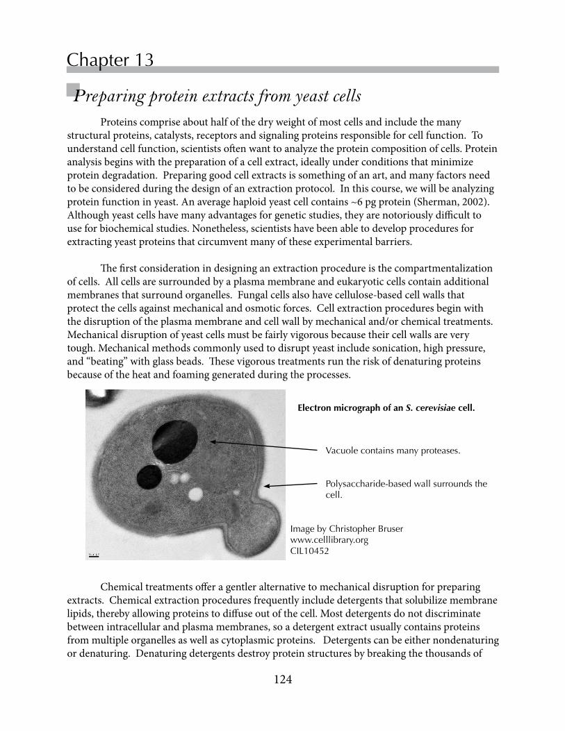
124
Chapter 13
Proteinscompriseabouthalfofthedryweightofmostcellsandincludethemanystructuralproteins,catalysts,receptorsandsignalingproteinsresponsibleforcellfunction.Tounderstandcellfunction,scientistsoftenwanttoanalyzetheproteincompositionofcells.Proteinanalysisbeginswiththepreparationofacellextract,ideallyunderconditionsthatminimizeproteindegradation.Preparinggoodcellextractsissomethingofanart,andmanyfactorsneedtobeconsideredduringthedesignofanextractionprotocol.Inthiscourse,wewillbeanalyzingproteinfunctioninyeast.Anaveragehaploidyeastcellcontains~6pgprotein(Sherman,2002).Althoughyeastcellshavemanyadvantagesforgeneticstudies,theyarenotoriouslydifficulttouseforbiochemicalstudies.Nonetheless,scientistshavebeenabletodevelopproceduresforextractingyeastproteinsthatcircumventmanyoftheseexperimentalbarriers.
Thefirstconsiderationindesigninganextractionprocedureisthecompartmentalizationofcells.Allcellsaresurroundedbyaplasmamembraneandeukaryoticcellscontainadditionalmembranesthatsurroundorganelles.Fungalcellsalsohavecellulose-basedcellwallsthatprotectthecellsagainstmechanicalandosmoticforces.Cellextractionproceduresbeginwiththedisruptionoftheplasmamembraneandcellwallbymechanicaland/orchemicaltreatments.Mechanicaldisruptionofyeastcellsmustbefairlyvigorousbecausetheircellwallsareverytough.Mechanicalmethodscommonlyusedtodisruptyeastincludesonication,highpressure,and“beating”withglassbeads.Thesevigoroustreatmentsruntheriskofdenaturingproteinsbecauseoftheheatandfoaminggeneratedduringtheprocesses.
Preparing protein extracts from yeast cells
Electron micrograph of an S. cerevisiae cell.
Vacuole contains many proteases.
Polysaccharide-based wall surrounds the cell.
Image by Christopher Bruserwww.celllibrary.org CIL10452
Chemicaltreatmentsofferagentleralternativetomechanicaldisruptionforpreparingextracts.Chemicalextractionproceduresfrequentlyincludedetergentsthatsolubilizemembranelipids,therebyallowingproteinstodiffuseoutofthecell.Mostdetergentsdonotdiscriminatebetweenintracellularandplasmamembranes,soadetergentextractusuallycontainsproteinsfrommultipleorganellesaswellascytoplasmicproteins.Detergentscanbeeithernondenaturingordenaturing.Denaturingdetergentsdestroyproteinstructuresbybreakingthethousandsof

125
Replace Chapter number and title on A-Master Page.<- ->
Protein overexpressionweakbondsthatnormallyholdproteinstogether.AgoodexampleofadenaturingdetergentisSDS,orsodiumdodecylsulfate,whichwewillbeusingtoprepareourcellextracts.Bycontrast,non-denaturingdetergentsbindtothesurfacesofproteinsandconsequentlypreserveenzymeactivity.
Whenpreparingextracts,caremustbetakentoprotectproteinsfromdegradationbycellularproteases.Cellscontainproteaseswithmanydifferentspecificitiesthatareresponsiblefornormalturnoverofproteinsincells.Celldisruptionoftenreleasesproteasesfromcompartmentssuchaslysosomes,providingthemaccesstocytoplasmicproteins.Yeastarenotoriouslyrichinproteases.Inanintactyeastcell,manyoftheseproteasesarelocatedintheyeastvacuole,whichisanalogoustothemammalianlysosome.
Theprotocolthatwewilluseinthiscourse(Amberget al.,2005)reliesonchemicaldisruptionandrapidproteindenaturationtodenaturetheyeastproteases.ExtractspreparedbythismethodaresuitableforSDS-PAGEandwesternblotanalysis.

126
Chapter 13
USE THE WHEEL CORRECTLY!
Make sure that wheel is balanced by placing tubes symmetrically with respect to the central axis.
Remember to turn the wheel ON after removing your samples!
On switch is on the right side of the base behind the wheel.
Exercise 1 - Prepare cell cultures for extraction
DAY 1 - First lab session of the week1. Inoculate2mLofYC-URAcontaining2%raffinosewithasingleyeastcolony.
2. Repeatstep1foreachofyourtransformedstrains(foratotalof3cultures).
3. Placetheculturesonthewheelat30˚C,taking care that tubes are properly balanced.Turnthewheelon.
DAY 2 - The day before your second lab session of the week (your off day)1. Removeyourculturesfromthewheel.
2. Add1mLofyourRaffinoseculturetoatubecontaining1mLYC+Glucose(repressionmedium).Addtheother1mLofyourRaffinoseculturetoatubecontaining1mLofYC+Galactose(inductionmedium)Be sure that the tubes are labeled with your initials and the sugar source. You should have a total of 6 tubes.
3. Placetheculturesbackonthewheeltogrowovernightbeforeyourclasstime.

127
Replace Chapter number and title on A-Master Page.<- ->
Protein overexpression
Harvest the cells for extraction
1.Growthecellsundervariousconditionsasinstructed.
2.Determinethecellconcentration.Vortexthetubbesandtransfer100µLofeachcellcultureto900µLdeionizedwaterandmeasuretheOD600ofculturesinthespectrophotometer.Notethevaluesinyourlabnotebook.YouwillrefertothemlaterwheninterpretingyourSDS-PAGEgels(Chapter14).
3.Transfer1.5mLofyourculturesintolabelledmicrocentrifugetubes.Collectthecellsbycentrifugationfor1minuteattopspeed-inamicrocentrifuge.Decantanddiscardthesupernatant.
4.Rinsethecells.Add1mLdeionizedwatertoeachtube.Resuspendthecellpelletsbygentlydrawingthecellsinandoutofamicropipettetip,takingcaretopreventprematurelysisofthecells.Thisrinsestepremovesproteinsfromtheculturemediumthatmaybepresentinthecellpellet.
5.Resuspendthecellsin100µLdeionizedwater.
Prepare the protein extract6.Add100µLof0.2NNaOHtoeachtube,andincubatethecellsfor5minutesatroom
temperature.(TheadditionofNaOHdoesnotlysethecells,butitmakesthemmorepermeableandmorefragile.)
7.Pelletthecellsagaininthemicrocentrifugeandremovethesupernatant.
8.Resuspendthecellsin50µl2XSDS-PAGEsamplebuffer.*Lockthelidsinplacewiththebuilt-inlockingmechanism.IMMEDIATELYplacethetubesinaboilingwaterbath.Leavethecellsinthewaterbathfor3minutes.Thistreatmenteffectivelydenaturestheproteins.Yeastcellscontainmanyproteasesthatcoulddegradeothercellularproteins,soit’simportantthattheproteasesaredenaturedbeforetheyhavethechancetoattackothercellularproteins.
NOTE: The 2 X SDS-PAGE sample buffer contains 2-mercaptoethanol (also known as b-mercaptoethanol, or BME). Use appropriate caution and work quickly when handling this reagent. BME is a volatile reagent with a strong odor reminiscent of rotten fish.
9.Afterboiling,storethesamplesinthefreezerforfutureuse.
Exercise 2 - Preparing cell extracts

128
Chapter 13
Amberg,DC,Burke,DJ&Strathern,JN(2005).Methods in Yeast Genetics.ColdSpringHarborLaboratoryPress,ColdSpringHarbor.
Gelperin,DM,White,MA,Wilkinsonet al.(2005)BiochemicalandgeneticanalysisoftheyeastproteomewithamovableORFcollection.Genes Develop19:2816-2826.
Johnston,M(1987)Amodelfungalregulatorymechanism:theGAL1genesofSaccharomyces cerevisiae.Microbiol Rev51:458-476.
Sherman,F(2002)Gettingstartedwithyeast.Method Enzymol 350:3-41.
References
*2XSDS-PAGEsamplebufferconsistsof: 120mMTris/HCl,pH6.8 10%glycerol(glycerolissignificantlymoredensethanwater) 4%SDS 8%2-mercaptoethanol 0.004%bromophenolblue(atrackingdyeforelectrophoresis)

• UnderstandthetheorygoverningproteinseparationbySDS-PAGE
• LearnhowtocastandrunSDS-PAGEgels
• Understandhowstainsareusedtovisualizeproteinsingels
• LearnhowtoestimatethemolecularweightofaproteinfromitsmigrationonSDS-PAGEgels
ThislabwillintroduceyoutoSDS-PAGE,asimpleandinexpensivemethodforresolvingproteinsincomplexmixtures.SDS-PAGEgelsprovidethestartingmaterialsforwesternblotsandforsomeproteomictechniques.Inthislab,you’lluseSDS-PAGEtoanalyzetheproteinextractsthatyoupreparedfromyeaststrainsoverexpressingMetandCysfusionproteinsofLacZ.
SDS-PAGEChapter 14
Objectives

130
Chapter 14
SDS-PAGEiswidelyusedtoanalyzetheproteinsincomplexextracts.ThemostcommonlyusedmethodsarederivedfromthediscontinuousSDS-PAGEsystemfirstdescribedbyLaemmli(1970).Thesystemactuallyconsistsoftwogels-aresolving(akarunning)gelinwhichproteinsareresolvedonthebasisoftheirmolecularweights(MWs)andastackinggelinwhichproteinsareconcentratedpriortoenteringtheresolvinggel.Differencesinthecompositionsofthestackinggel,resolvinggelandelectrophoresisbufferproduceasystemthatiscapableoffinelyresolvingproteinsaccordingtotheirMWs.
Gel electrophoresis of macromolecules Ingelelectrophoresis,anelectricfieldisusedtomovechargedmoleculesthroughamatrixofapolymerizedsubstancesuchasagaroseorpolyacrylamide.Theratesatwhichindividualmoleculesmovethroughthegeldependonthepropertiesofboththeseparationsystemandthemoleculesthemselves.Gelmatricesarepermeatedwithnetworksofporesthroughwhichthemoleculesmove.Theamountofresistancethatthematrixpresentstothemovementofamoleculedependsonthediameteroftheporeaswellasthesizeandgeometryofthemolecule.Researcherscancontrolthesizeoftheporebyadjustingtheconcentrationofgelmonomerwithinacertainrange.Ingeneral,smaller,morehighlychargedmoleculesmigratemorerapidlythroughgelsthanlargerorlesschargedmolecules.Themobilityofamoleculeisalsoaffectedbythebuffersystemandthestrengthoftheelectrophoreticfield,i.e.voltage,usedfortheseparation.
YouhavealreadyusedagarosegelelectrophoresistoseparateDNAmolecules.DNAmoleculesaremuchlargerthanproteinmoleculesandtheyhaveauniformchargetomassratio.Consequently,electrophoreticanalysisofDNAmoleculesismorestraightforwardthantheseparationofproteinmolecules.TheagarosegelsusedtoseparatedDNAmoleculeshavemuchlargerporesthanthepolyacrylamidegelstypicallyusedtoseparateproteinmolecules.(However,smallDNAmolecules,e.g.lessthan1000bp,maybeseparatedinpolyacrylamidegels.)
Chemistry of acrylamide polymerization Proteinsareusuallyseparatedonpolyacrylamidegelsformedbythechemicalpolymerizationofacrylamideandacross-linkingreagent,N,N’methylenebisacrylamide(oppositepage).Investigatorsareabletocontrolthesizeoftheporesinthegelbyadjustingtheconcentrationofacrylamide,aswellastheratioofacrylamidetobisacrylamide.Raisingeithertheconcentrationofacrylamideorbisacrylamide,whileholdingtheotherconcentration
Background
ThislabwillintroduceyoutoSDS-PAGE(sodiumdodecylsulfate-polyacrylamidegelelectrophoresis),asimpleandinexpensivemethodforresolvingproteinsincomplexmixtures.YouwilluseSDS-PAGEgelstoanalyzetheyeastproteinextractsthatyoupreparedinthelastlab.Youwillmaketwogelsinthislab.YouwillrunonegeltodayandstainitwithBrilliantBlueG-250tovisualizetheproteinsintheextracts.Youwillstoretheothergeluntilthenextlabpe-riod,whereyouwilluseitforawesternblot.
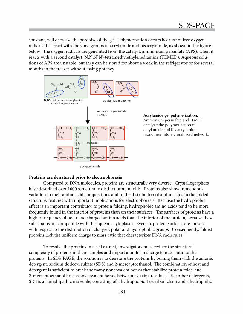
131
Replace Chapter number and title on A-Master Page.<- ->
SDS-PAGE
constant,willdecreasetheporesizeofthegel.Polymerizationoccursbecauseoffreeoxygenradicalsthatreactwiththevinylgroupsinacrylamideandbisacrylamide,asshowninthefigurebelow.Theoxygenradicalsaregeneratedfromthecatalyst,ammoniumpersulfate(APS),whenitreactswithasecondcatalyst,N,N,N’,N’-tetramethylethylenediamine(TEMED).Aqueoussolu-tionsofAPSareunstable,buttheycanbestoredforaboutaweekintherefrigeratororforseveralmonthsinthefreezerwithoutlosingpotency.
Proteins are denatured prior to electrophoresis ComparedtoDNAmolecules,proteinsarestructurallyverydiverse.Crystallographershavedescribedover1000structurallydistinctproteinfolds.Proteinsalsoshowtremendousvariationintheiraminoacidcompositionsandinthedistributionofaminoacidsinthefoldedstructure,featureswithimportantimplicationsforelectrophoresis.Becausethehydrophobiceffectisanimportantcontributortoproteinfolding,hydrophobicaminoacidstendtobemorefrequentlyfoundintheinteriorofproteinsthanontheirsurfaces.Thesurfacesofproteinshaveahigherfrequencyofpolarandchargedaminoacidsthantheinterioroftheprotein,becausethesesidechainsarecompatiblewiththeaqueouscytoplasm.Evenso,proteinsurfacesaremosaicswithrespecttothedistributionofcharged,polarandhydrophobicgroups.Consequently,foldedproteinslacktheuniformchargetomassratiothatcharacterizesDNAmolecules.
Toresolvetheproteinsinacellextract,investigatorsmustreducethestructuralcomplexityofproteinsintheirsamplesandimpartauniformchargetomassratiototheproteins.InSDS-PAGE,thesolutionistodenaturetheproteinsbyboilingthemwiththeanionicdetergent,sodiumdodecylsulfate(SDS)and2-mercaptoethanol.Thecombinationofheatanddetergentissufficienttobreakthemanynoncovalentbondsthatstabilizeproteinfolds,and2-mercaptoethanolbreaksanycovalentbondsbetweencysteineresidues.Likeotherdetergents,SDSisanamphipathicmolecule,consistingofahydrophobic12-carbonchainandahydrophilic
Acrylamide gel polymerization.Ammonium persulfate and TEMED catalyze the polymerization of acrylamide and bis-acrylamide monomers into a crosslinked network.

132
Chapter 14sulfategroup.TheSDShydrocarbonchainbindstothemanyhydrophobicgroupsinproteins,reducingtheproteintoarandomcoil,coatedwithnegativelychargesalongitslength.DenaturedproteinsbindquitealotofSDS,amountingto~1.4gSDS/gprotein,or~oneSDSmoleculeforeverytwoaminoacids.
Discontinuities between the stacking and running gels underlie the resolving power of the SDS-PAGE gels
TheLaemmliSDS-PAGEsystemcanbeconsidereda3-componentsystem.Thestackingandrunning(resolv-ing)gelshavedifferentporesizes,ionicstrengthandpH.Thethirdcomponentistheelectrophoresisbuffer(25mMTris,192mMglycine,,0.1%SDS,pH~8.3),whichcontainslargeamountsofglycine.Theionizationstateoftheglycineiscriticaltotheseparation.AtneutralpH,glycineisazwit-terion,withanegativelychargedcarboxylgroupandaposi-tivelychargedaminogroup.ThepKaoftheaminogroupis9.6,considerablyhigherthanthepHofthechamberbuffer.Consequently,verylittleglycinehasanegativechargeinthechamberbufferorstackinggel,andsignificantionizationdoes
ThesamplebufferusedforSDS-PAGEcontainsatrackingdye,bromophenolblue(BPB),whichwillmigratewiththeleadingedgeoftheproteinsbeingseparatedonthegel.Thesamplebufferalsocontainsglycerol,whichallowstheproteinsamplestosettleintothebottomofthegelwells.Thegelisverticallypositionedintheelectrophoresisapparatusandcoveredwithchamberbuffercontainingglycine(right,shaded).
Onceavoltageisapplied,thechlorideionsinthesamplebufferandstackinggelmoverapidlytowardthepositivepole,formingthelead-ingedgeofamovingionfront.Glycinemoleculeshaveverylittlechargeinthestackinggel,sotheymigrateattherearofthemovingionfront.Thisdifferenceinchlorideandglycinemobilitysetsupasteepvoltagegradientinthestackinggelthatsweepsalongthenegativelychargedprotein-SDScomplexes.Thelargeporesofthestackinggelpresentverylittleresistancetothemovementofprotein-SDScomplexes,whichthen“stackup”intoaveryconcentratedregionattheinterfacebetweentherunningandstackinggels(right).Protein-SDScomplexesremaincon-centratedattheinterfaceuntiltheslowlymigratingglycinemoleculesreachtheboundarybetweenthetwogels.
notoccuruntiltheglycineentersthemorealkalinepH8.8environmentoftherunninggel.Let’sfollowtheprogressofproteinsamplesduringSDS-PAGEtoseehowdifferencesinthecomposi-tionofthesethreecomponentsgeneratethehighresolvingpowerofSDS-PAGEgels.

133
Replace Chapter number and title on A-Master Page.<- ->
SDS-PAGE
Dramaticchangesoccurastheglycineionsentertherunninggel.ThepHoftherunninggelisclosertothepKaoftheglycineaminogroups,soasignificantfractionoftheglycinemoleculesassumeanegativecharge.Negativelychargedglycinemoleculesbegintomoveatthesamerateasthechlorideions,therebyeliminatingthevoltagedifferencethatcontrolledproteinmobilitythroughthestackinggel.Theporesintherunninggelaremuchsmallerthanthoseofthestackinggel,sotheporespresentfrictionalresistancetothemigrationofproteins.Proteinsbegintomigrateatdifferentrates,becauseofthesievingpropertiesofthegel.Smallerprotein-SDScomplexesmigratemorequicklythanlargerprotein-SDScomplexes(right).Withinacertainrangedeterminedbytheporosityofthegel,themigrationrateofaproteinintherunninggelisinverselyproportionaltothelogarithmofitsMW.
Proteins are visualized with stains. Withfewexceptions,naturally-occurringproteinsareinvisibleonSDS-PAGEgels.Consequently,researchersoftenusepre-stainedproteinstandardstomonitortheapproximatepositionsofproteinsduringelectrophoresis.Thepre-stainedstandardsareproducedbycovalentlyattachingalargenumberofchromophorestoaprotein.TheadditionofthechromophoresincreasestheMWoftheproteinandalsoproducesmorediffusebandsonthegel.Thediffusenessofthebandsreflectsvariationinthenumberofdyemoleculesattachedtoindividualproteinmolecules.Wewilluseprestainedstandardproteinsinourgels,soyouwillbeabletovisualizetheseparationthatisoccurring.Yeastproteinswillnotbevisible,however,becausetheyhavenotbeenmodifiedwithchromophores.
Tovisualizethepositionsofproteinsafterelectrophoresisiscomplete,investigatorsstainthegelswithvariousdyesthatbindnoncovalentlyandwithverylittlespecificitytoproteins.Duringthestainingprocess,proteinsarealso“fixed”inthegel,meaningthatproteinsbecomeinsolubleandunabletodiffuseoutofthegel.ThemostcommonlyuseddyesarethecloselyrelatedBrilliantBlueR-250andG-250dyes,whichbindproteinsnonspecificallythroughalargenumberofionicandVanderWaalsinteractions.AlthoughBrilliantBlueR-250stainingisslightlymoresensitivethanG-250fordetectingproteins,theG-250stainingprocedureismorerapidanddoesnotrequiredestainingwithorganicsolvents.WewillusethecommercialSimplyBlue™reagent,whichcontainsacolloidalsuspensionofBrilliantBlueG-250,tostainthegels.Inthisprocedure,gelsarerinsedwithwatertoremovethebuffersaltsusedforelectrophoresisandthentreatedwiththecolloidalG-250suspension.Proteinbandsappearrapidly,andwhennecessary,thegelscanbedestainedwithdeionizedwatertolowerthegelbackground.BrilliantBluestainingintensityisconsideredaquantitativeprocedure,becausetheintensityofastainedbandisdirectlyproportionaltotheamountofproteininaband.
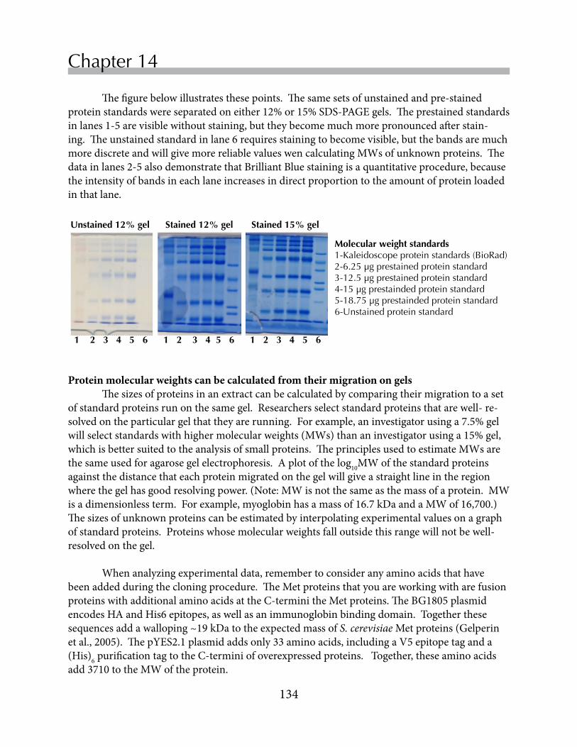
134
Chapter 14
Thefigurebelowillustratesthesepoints.Thesamesetsofunstainedandpre-stainedproteinstandardswereseparatedoneither12%or15%SDS-PAGEgels.Theprestainedstandardsinlanes1-5arevisiblewithoutstaining,buttheybecomemuchmorepronouncedafterstain-ing.Theunstainedstandardinlane6requiresstainingtobecomevisible,butthebandsaremuchmorediscreteandwillgivemorereliablevalueswencalculatingMWsofunknownproteins.Thedatainlanes2-5alsodemonstratethatBrilliantBluestainingisaquantitativeprocedure,becausetheintensityofbandsineachlaneincreasesindirectproportiontotheamountofproteinloadedinthatlane.
Molecular weight standards1-Kaleidoscope protein standards (BioRad)2-6.25 µg prestained protein standard3-12.5 µg prestained protein standard4-15 µg prestainded protein standard5-18.75 µg prestainded protein standard6-Unstained protein standard
Unstained 12% gel Stained 12% gel Stained 15% gel
1 2 3 4 5 6 1 2 3 4 5 6 1 2 3 4 5 6
Protein molecular weights can be calculated from their migration on gels Thesizesofproteinsinanextractcanbecalculatedbycomparingtheirmigrationtoasetofstandardproteinsrunonthesamegel.Researchersselectstandardproteinsthatarewell-re-solvedontheparticulargelthattheyarerunning.Forexample,aninvestigatorusinga7.5%gelwillselectstandardswithhighermolecularweights(MWs)thananinvestigatorusinga15%gel,whichisbettersuitedtotheanalysisofsmallproteins.TheprinciplesusedtoestimateMWsarethesameusedforagarosegelelectrophoresis.Aplotofthelog10MWofthestandardproteinsagainstthedistancethateachproteinmigratedonthegelwillgiveastraightlineintheregionwherethegelhasgoodresolvingpower.(Note:MWisnotthesameasthemassofaprotein.MWisadimensionlessterm.Forexample,myoglobinhasamassof16.7kDaandaMWof16,700.)Thesizesofunknownproteinscanbeestimatedbyinterpolatingexperimentalvaluesonagraphofstandardproteins.Proteinswhosemolecularweightsfalloutsidethisrangewillnotbewell-resolvedonthegel.
Whenanalyzingexperimentaldata,remembertoconsideranyaminoacidsthathavebeenaddedduringthecloningprocedure.TheMetproteinsthatyouareworkingwitharefusionproteinswithadditionalaminoacidsattheC-terminitheMetproteins.TheBG1805plasmidencodesHAandHis6epitopes,aswellasanimmunoglobinbindingdomain.Togetherthesesequencesaddawalloping~19kDatotheexpectedmassofS. cerevisiaeMetproteins(Gelperinetal.,2005).ThepYES2.1plasmidaddsonly33aminoacids,includingaV5epitopetaganda(His)6purificationtagtotheC-terminiofoverexpressedproteins.Together,theseaminoacidsadd3710totheMWoftheprotein.

135
Replace Chapter number and title on A-Master Page.<- ->
SDS-PAGE
Casting SDS-PAGE gels Theseinstructionsaredesignedforconstructingtwo12%SDS-PAGEgelswiththeBioRadMiniProteansystem.Wewillneedonlyonegelinthislab,buttheextragelprovidesinsuranceagainstleaks!Ifneithergelleaks,youwillusethesecondgelforwesternblotsinthenextlab.
Assemble the gel casting apparatus
1. Assemblethecomponentsthatyouwillneedforcastingthegel:atallglassplatewithattached1mmspacers,asmallglassplate,agreencastingframeandacastingstand.
2. Placethegreencastingframeonthebenchwiththegreen“feet”restingfirmlyagainstthebenchandtheclampsopen(perpendiculartotheframe)andfacingyou.
3. Placethetwogelplatesintheframe.Insertthetallerspacerplatewiththe“UP”arrowsupandthespacersfacingtowardyouintothecastingframe(theBioRadlogoshouldbefacingyou).Inserttheshortglassplateinthefrontofthecastingframe.There should be a space between the plates.
4. Securetheplatesinthecastingframebypushingthetwogatesoftheframeouttothesides.IMPORTANT:thebottomedgesofthetwoplatesshouldbeflushwiththelabbenchbeforeyouclamptheframeclosedtoensureawatertightseal.To do this, rest the frame vertically on the bench BEFORE closing the gates.
5. Clampthecastingframewithglassplatesintothecastingstand,withthegatesofthecastingframefacingyou.Repeat steps 1-5 to prepare a second gel in the casting frame.
6. Checktoseeiftheassembledplatesinthecastingstandaresealedproperlybypipettingasmallamountofdeionizedwaterintothegapbetweentheplates.Iftheglassplatesholdwateranddon’tleak,youarereadytomakethegels.Pourthewateroutbyholdingtheentirecast-ingplatformoveraliquidwastecontainerorsink.Usepapertowelsortissuestoabsorbanyresidualwater.Ifthegelleaks,disassembletheframe,drytheplatesandgobacktostep3.
Assembling plates in the casting frame.
Large spacer plate with spacer goes in rear, oriented so the arrows point up and the logo is readable. Small plate is placed in front. (Corners of the plates are marked with bracket marks.)
Green gates of casting frame are open.
Casting frame “feet” and bottom edges of plates are flush against the benchtop.

136
Chapter 14Prepare two resolving gels. SAFETY NOTE: Acrylamide and bisacrylamide monomers are weak neurotoxins. Gloves and goggles should be used when working with acrylamide.
Assemblethechemicalsthatyouwillneedtopourthegels.ThetablebelowshowsthequantitiesofeachchemicalthatyouwillneedtopourtwogelswiththeMini-Proteansystem.Polymerizationoccursrapidly,sobesuretofollowthestep-by-stepinstructionsbelow.NOTE: catalysts should NOT be included into the mixture until you are ready to pour the gels!!
1. Labeltwo15mLconicaltubes“Resolvinggel”and“Stackinggel”.2. Preparetheresolvinggel.Mixtheacrylamidesolution,pH 8.8Trisbufferandwater,as
showninthechartabove.Mixtheingredientsgently,tryingnottointroduceair.Oxygeninhibitspolymerizationofacrylamidegels.
3. Totheresolvinggelmixture,add100µLofa10%ammoniumpersulfate(APS)solution.Gen-tlymixthesolution,tryingnottointroduceair.Oxygeninhibitsacrylamidepolymerization.
4. Add10µLofTEMEDcatalyst.Onceagain,gentlymixinthecatalysttryingnottointroduceairbubbles.CAUTION:TEMEDhasanunpleasantodor.Coverthetubeimmediatelyafteryoualiquotthisreagent.
5. Workingquickly,useaplastictransferpipettetofillthespacebetweenthetwoplatesuntiltheresolvinggelsolutionreachesaheightjustabovethegreenclampsonthegelcastingframe.Drawupanyremainingacrylamideintothetransferpipet.(Youwillknowthattheacryl-amidehaspolymerizedwhenyoucannolongerpushtheacrylamideoutofthepipet.)
6. Usingatransferpipet,adddeionizedwatersothatitgentlyflowsacrossthesurfaceofthepolyacrylamidemixture.Thewaterlayerensuresthatthepolyacrylamidegelwillhavealevelsurfaceonceitpolymerizes.
7. Allowthegeltopolymerize,whichtakes~15-20minutes.Youwillnotethattheinterfacebetweenthepolyacrylamideandwateroverlaydisappearstemporarilywhilethegelpolymer-izes.Asharpnewinterfacethenformsbetweenthetwolayers,indicatingthatpolymerizationiscomplete.(Youcanalsochecktheremainingpolyacrylamideinthetransferpipettetoseeifithaspolymerized.)
8. Whenpolymerizationiscomplete,removethewaterfromthetopoftheresolvinggelbytilt-ingthegeltothesideandusingapapertowelorKimwipetowickoutthewater.
Reagent Resolving gel Stacking gelDeionizedwater 3.5mL 2.1mL30%acrylamide:bis-acrylamide(29:1) 4.0mL 0.63mL1.5MTris-HCl,0.4%SDS,pH8.8 2.5mL ------0.5MTris-HCl,0.4%SDS,pH6.8 ------ 1.0mL10%ammoniumpersulfate(catalyst) 100µL 30µLTEMED(catalyst) 10µL 7.5µL

137
Replace Chapter number and title on A-Master Page.<- ->
SDS-PAGE
Pour the stacking gels
1. Preparetheresolvinggel.Mixtheacrylamidesolution,pH 6.8Trisbufferandwater,asshowninthechartabove.
2. Add30µL10%APSand7.5µLTEMEDtothestackinggelacrylamidemixture.Mixthecontentsbygentlyinvertingthetubetwice.
3. Useatransferpipettetopipettethestackinggelontopoftheresolvinggelbetweenthetwoglassplates.Addenoughstackingsolutionuntilitjustreachesthetopofthesmallplate.
4. Carefully,butquickly,lowerthecombintoposition,beingcarefulnottointroduceairbubbles.(TheBioRadlogoonthecombshouldbefacingyou.)Addingthecombwillforcesomesolutionoutofthegel,butthisisfine.Ifairbubblesbecometrappedbelowthecomb,removethecombandrepositionit.
5. Removethecombwhenthegelhaspolymerized(10-15minutes).Youwillbeabletoseefaintschlierenlines(indicatorsofachangeinrefractiveindex)atthetopofthespacergelwhenpolymerizationiscomplete.Youcanalsochecktheleftoversolutioninthe15mLtubeforpolymerization.
Saving SDS-PAGE gels 1.Carefullyremovethegelsfromthecastingstandandthenfromtheirgreenframes.2.Keepingthecombsinthegel,wrapthegelinawetpapertowel.Thenwrapthegelinplastic
wraptobeusedinalaterlab.
Casting the SDS-PAGE gel.
Casting stand and plates are clamped into the casting stand
Fill plates with running gel solution to the top of the closed green gates of the casting frame.
Bottom edges of plates are flush against the grey pad of the casting stand.

138
Chapter 14
Running SDS-PAGE gelsSet up the electrophoresis apparatus1. Afterthestackinggelpolymerizes,removethegelcassettesandwichfromthecastingstand.2. Carefullyremovethecombfromthespacergel.3. Removethecastingframefromthegelcassettesandwichandplacethesandwichagainst
thegasketononesideoftheelectrodeassembly,withtheshortplatefacinginward.Placeasecondgelcassetteorabufferdamagainstthegasketintheothersideoftheelectrodeassembly.
4. Clampthegreenclampsonthesidesoftheelectrodeassembly(below).5. Lowerthechamberintotheelectrophoresistank.6. FillthespacebetweenthetwogelswithTris-glycinerunningbuffer.Thisformstheupper
chamberforelectrophoresis.7. AddTris-glycinerunningbuffertotheouter(lower)chamberuntilthelevelishighenoughto
covertheplatinumwireintheelectrodeassembly.
Electrode assembly
One gel is positioned on each side of the electrode assembly
Top of short plate fits snugly into notch in the green gasket of the electrode assembly
Electrode assembly with two gels is lowered into the clamping frame
Load and run samples on the SDS-PAGE gel1. Ifyouwish,placethesampleloadingguideinbetweenthetwogelsintheelectrodeassembly
beforeloadingyoursamples.2. Usinggelloadingmicropipettetips(tipshaveverylong,thinpointsandfitP20sorP200s),
loadupto15μLofsampleintoeachwell.Load5µLofamolecularweightstandardintoonelaneofthegel.Loadsamplesslowlyandallowthesamplestosettleevenlyonthebottomofthewell.
3. NOTE:Besuretorecordtheorderofsamplesloadedontothegel.4. Connectthetanktothepowersupply.Fitthetankcoverontotheelectrodesprotrudingup
fromtheelectrodeassembly.Inserttheelectricalleadsintothepowersupplyoutlets(connectblacktoblackandredtored).
5. Turnonthepowersupply.Runthegelataconstantvoltageof120‐150V.Runthegeluntilthebluedyefrontnearlyreachesthebottomofthegel.Thismaytakebetween45-60min.
139
Replace Chapter number and title on A-Master Page.<- ->
SDS-PAGE
Staining SDS-PAGE gels
1. Turnoffthepowersupply.
2. Removethegelapparatusfromthetank.Opentheclampingframeandremovethegelcas-settesandwich.Carefully,prythetwoplatesapartwithaspatula.Withthespatula,removethelowerrightorleftcornerofthegeltoserveasanorientationmarker.Be sure to indicate in your lab notebook whether the notched corner corresponds to lane 1 or lane 10 of the gel. Youmayalsoremovethestackinggelwiththespatula,ifyoudesire.
3. Placethegelinasmallplastictrayandlabelthetraywithyourinitialsonapieceoftape.Todothis,fillthetrayabouthalfwaywithdeionizedwater.Gentlyfreethegelfromtheglassplate,allowingittoslideintothewater.Thegelshouldmovefreelyinthewater.Placethegelandtrayonarockingplatform.Rockthegelfor5minutes.
4. Drainthewaterfromthegelandadd~20mLofSimplyBlue.Coverthegelcontainerwithsaran‐wrapandrockovernight.
5. Inthemorning,draintheSimplyBluestainintoanappropriatelylabeledwastecontainerinthehoodofthelabroom.
6. Destainthegelbyfillingthecontainerabouthalffullwithdeionizedwater.Shakethegelinthewaterfor~2minutes.Pouroffthewaterandaddnewdeionizedwater.Repeat,ifnecessary,untilproteinbandsbecomevisible.
7. Whenindividualbandsaredetectable,recordyourdata.Youmayphotographthegelwithyourcellphonecamera.Alternatively,placethegelinaclearplasticpageprotectorandscanthegel.
8. Afterrecordingthedata,disposeofthegelintheBiohazardwastecontainer.

140
Chapter 14
ReferencesGelperin,DM,White,MA,Wilkinsonet al.(2005)Biochemicalandgeneticanalysisoftheyeast
proteomewithamovableORFcollection.Genes Develop19:2816-2826.
Laemmli,UK(1970)Cleavageofstructuralproteinsduringtheassemblyoftheheadofbacterio-phageT4.Nature 227:680–685.
Shapiro,AL,Viñuela,E,&Maizel,JV,Jr.(1967)MolecularweightestimationofpolypeptidechainsbyelectrophoresisinSDS-polyacrylamidegels.Biochem Biophys Res Commun28:815–820.
Steinberg,TH(2009).Proteingelstainingmethods:Anintroductionandoverview.Method Enzymol463:542-563.

• Understandthestructuralfeaturesofantibodymolecules.• Understandhowmonoclonalandpolyclonalantibodiesareproduced.• Understandhowepitopetagsareusedinmolecularbiology• Understandhowprimaryandsecondaryantibodiesareusedtodetectproteins
inwesternblots.• Usewesternblotstoanalyzeproteinexpressioninyeasttransformedwith
overexpressionplasmids.
Westernblotsareoneofthemostwidelyusedtechniquesincellbiology.Inawesternblot,investigatorstakeadvantageoftheexquisitesensitivityofantibodiestoidentifyproteinsofinterestincomplexsamples.Inthislab,you’lllearnaboutthedifferentkindsofantibodiesusedinwesternblots.YouwillusewesternblotstoanalyzeMetandCysproteinexpressioninyourtransformedyeaststrains.
Western blotsChapter 15
Objectives

142
Chapter 15
Westernblotsprovideamethodtofindtheproverbial“needleinahaystack.”Atypicalcellexpressesthousandsofdifferentproteins,anditisoftendifficulttodetectchangesinexpressionofyourfavoriteprotein(Yfp)withoutaprobethatiscapableofdiscriminatingtheYfpagainstalargebackgroundofunrelatedcellularproteins.Fortunately,antibodiesprovidehighlyspecificmolecularprobesthatcanbeusedtodetecttheexpressionofproteinsonwesternblots.Toappreciatethesensitivityofwesternblots,it’shelpfultohavesomeunderstandingofantibodystructureandantibodyproductionduringimmunerespones.(Disclaimer:Thefollowingparagraphsprovideahighlyabbreviatedoverviewofantibodiesandonesegmentofthecomplexvertebrateimmunesystem.TheDepartmentoffersanimmunologycoursethatwillintroduceyoutothefinerdetailsofthisfascinatingsystem.)
Antibodies are produced in response to antigens Antibodiesareproteinsproducedbyvertebrateswithadaptiveimmunesystemscapableofrespondingtoforeignantigens.Antigensaredefinedassubstancesthatstimulatetheproductionofantibodies.Antigensarecommonlyabletostimulatetheproductionofmultiplekindsofantibodies,eachofwhichrecognizesasmall,distinctregiononthesurfaceoftheantigenknownasanepitope.AntibodiesareY-shapedproteinsproducedbylymphocytesthatbindepitopeswithhighaffinity.
Antibodies binding to an antigen.An antigen with three different epitopes on its surface is bound by three different antibody molecules, each of which binds a single epitope with high affinity.
Theavailabilityofhybridomacellsthatsecretelargequantitiesofantibodieswithasinglespecificityhasgreatlyfacilitatedstructuralstudiesonantibodies.ResearchersareabletoharvestantibodymoleculessecretedbyculturedhybridomacellsandtopreparecrystalsforX-raydiffraction.Basedonalargenumberofcrystallographicstudies,wenowunderstandthebasicarchitectureofantibodies,moreproperlyknownasimmunoglobins.Thecrystalstructuresshow
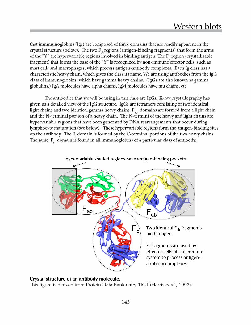
143
Replace Chapter number and title on A-Master Page.<- ->
Western blots
thatimmmunoglobins(Igs)arecomposedofthreedomainsthatarereadilyapparentinthecrystalstructure(below).ThetwoFabregions(antigen-bindingfragments)thatformthearmsofthe“Y”arehypervariableregionsinvolvedinbindingantigen.TheFcregion(crystallizablefragment)thatformsthebaseofthe“Y”isrecognizedbynon-immuneeffectorcells,suchasmastcellsandmacrophages,whichprocessantigen-antibodycomplexes.EachIgclasshasacharacteristicheavychain,whichgivestheclassitsname.WeareusingantibodiesfromtheIgGclassofimmunoglobins,whichhavegammaheavychains.(IgGsarealsoknownasgammaglobulins.)IgAmoleculeshavealphachains,IgMmoleculeshavemuchains,etc.
TheantibodiesthatwewillbeusinginthisclassareIgGs.X-raycrystallographyhasgivenusadetailedviewoftheIgGstructure.IgGsaretetramersconsistingoftwoidenticallightchainsandtwoidenticalgammaheavychains.FabdomainsareformedfromalightchainandtheN-terminalportionofaheavychain.TheN-terminioftheheavyandlightchainsarehypervariableregionsthathavebeengeneratedbyDNArearrangementsthatoccurduringlymphocytematuration(seebelow).Thesehypervariableregionsformtheantigen-bindingsitesontheantibody.TheFcdomainisformedbytheC-terminalportionsofthetwoheavychains.ThesameFcdomainisfoundinallimmunoglobinsofaparticularclassofantibody.
Crystal structure of an antibody molecule.This figure is derived from Protein Data Bank entry 1IGT (Harris et al., 1997).

144
Chapter 15
Intheinitialstagesoftheimmuneresponse,smallnumbersofimmatureBlymphocytesareabletobindforeignantigenmoleculesweaklyviatheantibodiesexpressedontheirsurfaces.Antigenbindingstimulatesthelymphocytestoproliferateandtodifferentiateintomature,antibody-secretinglymphocytes.AnamazingtransformationoccursasBlymphocytesmatureinresponsetoantigen.Antigenbindingstimulatesrespondinglymphocytestorearrangesegmentsoftheirantibody-encodinggenes,producingnewpotentialantigen-bindingsites.Mostrearrangementsareunproductive,butsomerearrangementsgenerateantibodieswithgreateraffinityfortheantigen.Antigensactasselectiveagents.Thelymphocytesthatbindtheantigenwiththehighestaffinityreceivethegreatestgrowthsignalandproliferatemostrapidly,becauseahigherfractionoftheirsurfaceantibodiesareboundtoantigenatanyonetime.Inthelatterstagesofdifferentiation,ahypermutationprocessfurtherincreasestherangeofpotentialantibodysequences.MatureBlymphocytesareknownasplasmacells,oncetheyhavesurvivedtheselectionprocess. Each plasma cell secretes a single antibody with high affinity for antigen. Plasmacellsarevirtualantibodyfactoriesthatcanbeidentifiedinelectronmicrographsbytheirextensiveroughendoplasmicreticulum.(Seetheinsidecoverforagraphicaldepictionofthisprocess.)Thescopeofantibodydiversityisimmense-vertebratesarecapableofproducingbillionsofantibodymoleculeswithdistinctspecificities.
Antibodies are produced by lymphocytes
Polyclonal vs. monoclonal antibodies Ananimal’sresponsetoantigenispolyclonal,becauseantigensstimulatetheproliferationofmultiplelymphocyteclones,eachofwhichproducesadifferentantibodytotheantigen.Consequently,serumcollectedfromanimmunizedanimalcontainsamixtureofantibodiesthatbindanantigenatdistinctsites.Theseantibody-bindingsitesonantigensareknownasepitopes.
Incontrasttopolyclonalantisera,whichareharvestedfromtheserumoflivinganimals,monoclonalantibodiesareproducedfromculturedhybridomacells.Hybridomacellsaregeneratedbyfusinglymphocytesfromanimmunizedanimal,mostcommonlyamouse,withmyelomacells,cancerouscellsthatarecapableofdividingindefinitelyinculture.Thefigureontheoppositepageoutlinestheproceduresusedtogenerateantibody-secretinghybridomacelllines.Lymphocytesareisolatedfromthespleensofanimmunizedmouseandfusedwithmyelomacells.Standardculturetechniquesareusedtoisolateindividualhybridomacelllinesfromthefusions.Eachhybridomacelllinesecretesauniqueantibodythatrecognizesasingleepitopeonanantigen.
Hybridomatechnologyhasrevolutionizedbiomedicalresearchsinceitsdescription(Kohler&Milstein,1975),becausemonoclonalantibodiesrecognizewell-definedepitopesandbecausemonoclonalantibodiescanbeproducedindefinitelybyculturedhybridomacells.Investigatorsoftenusebothmonoclonalandpolyclonalantibodiesatdifferentstepsinwesternblots.

145
Replace Chapter number and title on A-Master Page.<- ->
Western blots
Construction of hybridoma cell lines that secrete monoclonal antibodies.
Inthislab,wewillbeusingantibodiestodetectMetandCysproteinsexpressedintransformedS. cerevisiae.Theproteinsthatwillbeexpressedarenotthenativeproteins,butfusionproteins.ThepBG1805(Gelperinet al.,2005)andpYES2.1plasmidsweredesignedtobeusedformultiplepurposes.MolecularbiologistsdesignedtheplasmidsbyaddingDNAsequencesthatencodefunctionalelementsinthesamereadingframeastheyeastORFs.Thus,theexpressedproteinswillhaveC-terminaltagsthatcaninteractwithavarietyofreagents. Bothplasmidsencodeshortepitopetagsthatcanbedetectedwithantibodiesonwesternblots.Theseepitopescorrespondtohighlyaminoacidsequencesonthesurfacesofvirusesthathavebeenshowntobepotentinducersofantibodyproduction.ThepBG1805sequenceencodesan9-aminoacidsequenceofthehumaninfluenzavirushemagglutinin(HA)protein(Sleighet al.,1981).ThisHA-tag,canbedetectedwithantibodiesonwesternblots.
Plasmid-encoded proteins have C-terminal tags
ThepYES2.1plasmidencodesa15-aminoacidsequencefromthePproteinofsimianvirusV5(Southernet al.,1991).Inourblots,wewilluseamonoclonalantibodydirectedagainsttheV5protein,hereafterreferredtoasanti-V5,todetectproteinsexpressedfrompYES2.1-basedplasmids.

146
Chapter 15
Wewillnotbeusingananti-HAantibodytodetectproteinsexpressedfrompBG1805-basedplasmids.Instead,wewillmakeuseofthelargeC-terminaltagthatencodestheZZdomainofStaphylococcus aureusproteinA.S. aureususesproteinAtoevadedetectionbytheimmunesystem,contributingtoitspathogenicity.ProteinAisamembraneproteinwithmultipleextracellularZZdomains,eachofwhichisabletobindtheFcdomainofanIgG.Thesecondaryantibodythatweareusingfortheblotswasproducedinrabbits,anditsFcdomainbindstightlytoproteinA.
Bothplasmidsalsoencodeathirdtag,consistingof6histidines.ThisHis6-tagbindstightlytometalions,soitiscommonlyusedtopurifyoverexpressedproteinsbypassingthemthrougharesinwithimmobilizedzincorcobaltions.His6canbeusedasanepitopetag,butnotveryeffectively.AntiHis6-antibodiesgenerallyhaveamuchloweraffinityfortheirtargetthananti-HAoranti-V5antibodies.
Western blot overview
Inawesternblotprocedure,proteinsarefirstseparatedonanSDS-PAGEgelandthentransferredtoamembrane.Thismembranereplicaistreatedwithantibodiesthatspecificallyrecognizeaproteinorepitopeofinterest.Additionalprocessingstepsgenerateasignalatthepositionoftheboundantibody.Betweenthesteps,variouswashesaredonetoincreasethesignal-to-noiseratioonthefinal,developedblot.Themajorstepsinatypicalwesternblotarediagrammedonthefollowingpageanddiscussedingreaterdetailinsectionsthatfollow:
• ElectrophoretictransferofproteinsfromanSDS-PAGEgeltoamembrane
• Blockingofnonspecificproteinbindingsitesontransfermembranes
• Incubationofthemembranewithaprimaryantibodyspecificfortheepitopeofinterest
• Incubationwithasecondaryantibodyspecificfortheprimaryantibody
• Detectionoftheprimaryantibody-secondaryantibodycomplex

147
Replace Chapter number and title on A-Master Page.<- ->
Western blots

148
Chapter 15
Western blots involve many stepsElectrophoretic transfer of proteins from an SDS-PAGE gel to a membrane ThefirststepinawesternblotistogenerateareplicaoftheSDS-PAGEgelbytransferringproteinselectrophoreticallytoasyntheticmembranewithahighproteinbindingcapacity.Inourexperiments,wewillusemembranesmadeofpolyvinylidinefluoride(PVDF),akindofplastic.PVDFmembranesarehydrophobicandthedrymembranesdonotwetproperlywithwater.Therefore,PVDFmembranesarefirstwetwithmethanol,thenrinsedwithdeionizedwater,andfinallyrinsedwithtransferbuffer.Theymustnotbeallowedtodryoutduringthetransferandimmunoblotprocedures.Iftheydodryout,theymustbere-wetwithmethanolandrinsedwithwaterbeforeproceeding.
Duringthetransferprocess,thegelandmembraneareplaceddirectlyagainsteachotherwithina“sandwich”ofpre-wetfilterpapersandfoampads,asdescribedinExercise1.Duringtheelectrophoretictransfer,currentshouldflowevenlyacrosstheentiresurfaceareaofthegel.It’simportant,therefore,thatairbubblesarenottrappedbetweenthegelandmembrane.Aftertheelectrophoretictransfer,whichcanbedoneinafewhoursorovernightwithreducedvoltage,themembranereplicawiththetransferredproteinscanbeallowedtodryoutandstoredforlatervisualizationwithantibodies.
Blocking of non-specific protein binding sites on membranes Thetransfermembranesusedinwesternblotsbindproteinsnonspecifically.Beforethemembranesareincubatedwithspecific(andexpensive)antibodies,theymustbepretreatedwithblockingsolutionsthatcontainhighconcentrationsofabundant(andcheap)proteinstosaturatenon-specificbindingsites.Thinkofthisstepasanalogoustoanartistprimingacanvaswithalowerqualitypaintbeforethemoreexpensivemediaisapplied.Ifthetransfermembranesarenotadequatelyblockedbeforetheantibodyisapplied,thenonspecificsitesonthemembraneswillabsorbsomeoftheantibodies,reducingtheamountofantibodyavailabletobindthetargetproteins.Inourexperiments,wewillusecaseinproteinsfrommilkasblockingreagents.Becauseourexperimentsdonotrequirehighsensitivity,rehydratednon-fatdrymilk(directfromthegrocerystore!)isanadequatesourceofcaseins.
Primary antibody binding Eitherpolyclonalormonoclonalantibodiescanbeusedastheprimaryantibodyonwesternblots.Antibodiescanbedirectedtowardanaturally-occurringproteinortowardanepitopeattachedtoanoverexpressedprotein(aswearedoing).Increasingly,researchersareusingepitope-taggedproteinsintheirexperiments,becauseantibodiesagainstnaturally-occurringproteinsareexpensiveandtime-consumingtoprepare.Inaddition,anantibodydirectedagainstanepitopecanbeusedtodetectmanydifferentproteinscarryingthatsameepitope.Inourwesternblots,wewilluseamousemonoclonalantibodythatbindstheV5epitope.TheprimaryantibodywillnotreactwiththeHA-taggedproteinsencodedbythepBG1805-basedplasmids.Instead,theselatterproteinswillbedetectedwiththesecondary
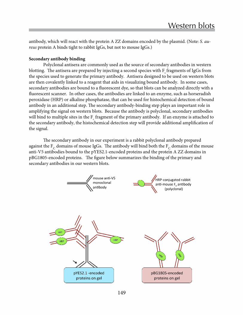
149
Replace Chapter number and title on A-Master Page.<- ->
Western blots
antibody,whichwillreactwiththeproteinAZZdomainsencodedbytheplasmid.(Note:S. au-reusproteinAbindstighttorabbitIgGs,butnottomouseIgGs.)
Secondary antibody binding Polyclonalantiseraarecommonlyusedasthesourceofsecondaryantibodiesinwesternblotting.TheantiseraarepreparedbyinjectingasecondspecieswithFcfragmentsofIgGsfromthespeciesusedtogeneratetheprimaryantibody.Antiseradesignedtobeusedonwesternblotsarethencovalentlylinkedtoareagentthataidsinvisualizingboundantibody.Insomecases,secondaryantibodiesareboundtoafluorescentdye,sothatblotscanbeanalyzeddirectlywithafluorescentscanner.Inothercases,theantibodiesarelinkedtoanenzyme,suchashorseradishperoxidase(HRP)oralkalinephosphatase,thatcanbeusedforhistochemicaldetectionofboundantibodyinanadditionalstep.Thesecondaryantibody-bindingstepplaysanimportantroleinamplifyingthesignalonwesternblots.Becausetheantibodyispolyclonal,secondaryantibodieswillbindtomultiplesitesintheFcfragmentoftheprimaryantibody.Ifanenzymeisattachedtothesecondaryantibody,thehistochemicaldetectionstepwillprovideadditionalamplificationofthesignal.
ThesecondaryantibodyinourexperimentisarabbitpolyclonalantibodypreparedagainsttheFCdomainsofmouseIgGs.TheantibodywillbindboththeFCdomainsofthemouseanti-V5antibodiesboundtothepYES2.1-encodedproteinsandtheproteinAZZdomainsinpBG1805-encodedproteins.Thefigurebelowsummarizesthebindingoftheprimaryandsecondaryantibodiesinourwesternblots.

150
Chapter 15
Exercise 1 - Preparing the membrane replica
Immunochemical detection of bound antibody Inthisfinalstep,thewesternblotisincubatedwithsubstratesfortheenzymethathasbeenconjugatedtothesecondaryantibody.OursecondaryantibodyhasbeenconjugatedtoHRP,ahardyenzymewithahighturnovernumber.(Theturnovernumberisthenumberofproductmoleculesproducedatanenzyme’sactivesitepersecond.)HRPcatalyzesthereactionofhydrogenperoxideand3,3’,5,5’-tetramethylbenzidine(TMB),whichgeneratesadarkblue-greyreactionproductthatprecipitatesatthereactionsiteonthewesternblot.Coloredreactionproductaccumulateswithtimeuntilthereactionisstoppedbywashingawayunreactedsubstrate.Thereactionshouldbeterminatedbeforenonspecificantibodybindingbecomesproblematic.
Separate proteins on an SDS-PAGE gel1.SeparatetheproteinsthatwillbeanalyzedonwesternblotsbySDS-PAGE.2.Removetheelectrodeapparatusandholderfromthetank,andremovethegelfromtheholder.
Donotremovethegelfromtheplatesuntilyouarereadytoassemblethetransfercassette(seebelow).
3.Disposeoftheremainingbufferdownthesink.RinseoutthebuffertankwithdeioinizedwatertoremoveresidualSDS,whichcaninterferewiththetransferprocess.
Prepare the transfer membraneNOTE: DO NOT touch transfer membranes with your fingers. Wear gloves and use filter forceps when you handle transfer membranes.
1.Whilethegelisrunning,gatherthePVDFmembraneandtwopiecesofthickfilterpaper,suchasWhatman3MMTM.ThePVDFmembraneandfilterpapersshouldbecuttoasizethatisslightlylargerthantheSDS-PAGEgel.Youwillalsoneedatransfercassetteandtwofiberpads.
2.PreparethePVDFmembrane.Usingpencil,placeanorientationmarkinacornerofthePVDFmembraneforlateridentification.Wetthemembranebyplacingitinasmalltraycontainingmethanolfor~30-60secondswithgentleagitation.
3.Disposeofthemethanolinthewastecontainerandadddeionizedwatertothetray.Gentlyagitatefor~1minute.
4.Replacethedeioinizedwaterwithtransferbuffer.Storethemembraneintransferbufferuntilyouarereadytostartthetransfer.
Assemble the transfer cassette1.Fillthebuffertankabouthalfwaytothetopwithtransferbuffer.Wetthefiberspongesand
filterpapersforthetransfercassettebyplacingthemintothetransferbufferinthetank.(TransferbuffercontainsTrisandglycine,aswellas10%methanol.UnlikeSDS-PAGErunningbuffer,thetransferbufferdoesnotcontainSDS.)

151
Replace Chapter number and title on A-Master Page.<- ->
Western blots
• Placeawetfiberpadontopoftheblackcassetteface.• Addtwopiecesoffilterpaper.• Positionthegelontopofthefilterpaperwhileitisstillattachedtotheglassplate.Use
aspatulatocarefullyreleasethegelfromtheplate.Youmayfinditeasiertoremovethegelbybeginningatthebottomedgenearthedyefront.
• PlacethePVDFmembraneblotontopofthegel.Orientthegelsothatthepencilmarkonthemembranecorrespondstotheclippedcornerofthegel.Be sure that there are NO air bubbles between the gel and the membrane.
• Addtheremainingfilterpaper(s)andthefiberpad.• Foldtheclearcassettefaceoverthegelassemblyandcarefullyslidetheclampinto
place.
1
23, 4
56
7, 8
9
10 Clear cassette face
Fiber padTwo filter papersTransfer membraneSDS-PAGE gel
Two filter papersFiber pad
Black cassette face
Assembly of the transfer cassette.
2.Usingaspatulaoragreenplasticwedge,removethesmallglassplatefromthegel.Thegelwillremainattachedtothelargeglassplate.Withaspatula,removethelowerrightcornerofthegeltoserveasanorientationmark.(Thiscorrepondstothefirstlaneofyourgel.)
3.Assemblethetransfercassetteasshownbe;pw.Besurethatallpartsofthetransfer“sandwich”remainmoistatalltimes.
Electrophoretic protein transfer1.Placethetransfersandwichintothecassette
holderwiththeblackfaceofthetransfercassettealignedwithblacksideofthecassetteholderandtheclearfacealignedwithredsideofthecassetteholder(right).NOTE:Eachcassetteholdercanholdtwotransfercassettes.
2.Placethecassetteholderandassembledcassettesintotheelectrophoresistank.Ifthetransferwillbedoneat100V(seestep5below),theelectrophoresistankshouldcontainanicepack.
3.Filltheelectrophoresistanktothetopwithtransferbuffer.

152
Chapter 15
4.Placelidontankbyaligningblackwithblackandredwithred.5.Runthetransferat100Vfor1houratroomtemperatureorat20Vovernightinthecoldroom.6.Whenthetransferiscomplete,removethetransfercassettefromthetank.Pourthetransfer
bufferbackintoitsoriginalbottlesothatitcanbereused.7.Disassemblethetransfercassette.Dependingonyourschedule:• Ifyouwillbecontinuingwiththewesternprocedure,skiptherehydrationstep(step1)below
andcontinuewiththeblockingstep(step2).Becarefulthatthemembraneremainsmoist!• Ifyouwillbeprocessingthemembraneatalatertime,allowthemembranetodryout.Wrap
themembraneinplasticwrapandsaveitforalaterlabperiod.
This is a multi-day procedure. Timing may vary for different classes.
Membranes are rehydrated and treated with blocking reagents1.WearingGLOVES,unwrapthedryblotfromtheplasticwrap.Usetheprestainedstandardsto
identifythesideofthemembranetowhichtheproteinsarebound.Submergethemembraneinmethanolwiththissidefacingup.Gentlyagitatethemembranebyhandrockingfor30-60secondsuntilthemembranehasbeenuniformlywetwithmethanol.Decantthemethanolintotheappropriatecontainerandfillthetrayhalfwayfullwithdeionizedwater.Gentlyagitatethemembraneforanadditionalminute.
2.DecantthewaterandreplaceitwithsufficientTBS-T(Trisbuffered-salinecontaining0.05%Tween20)tocovertheblot.Placetheblotonarockingplatform.EquilibratetheblotinTBS-Tfor5minuteswithslowrocking.Attheendof5minutes,draintheTBS-Tintothewastecontainer.
3.Pourenoughblockingsolution(5%nonfatmilkinTBS-T)ontotheblottocoverit.4.Coverthetraywithasmallpieceofplasticwrap.Placethetrayonarockingplatforminthe
coldroom(Higgins377).Theblotshouldfloatfreelyinthetraysothatbothsidesarewashed.Incubatetheblotforatleastanhourorupto24hoursat4˚C.
Exercise 2 - Immunodetection
153
Replace Chapter number and title on A-Master Page.<- ->
Western blots
Membranes are washed and incubated with primary antibody (~24 hours)1.Locateyourblotinthecoldroomandbringitbacktothelabroom.2.Removetheplasticwrapfromthecontainerholdingtheblotandpourofftheblocking
solution.AddenoughTBS-Ttocovertheblot.SAVEtheplasticwrap!Youwillneedittocoverthecontaineragain!
3.PourenoughTBS-Tsolutiontocovertheblotandplacethecontainerontherockingplatform.Rockfor5minutes.
4.PourofftheTBS-T.Add15mLofprimaryantibodydilutedinblockingbuffer.5.Coverthecontainerwiththesamepieceofplasticwrapandplacethetrayontherocking
platforminthe4˚Ccoldroom.Makesurethattheblotfloatsfreelyinthetrayandthatthestandardsareonthetopfaceoftheblot.Incubateovernightat4˚Cwithslowrocking.NOTE:Thetimingofthisstepisthemostcriticalintheprocedure.Shorteningtheincubationtimewithprimaryantibodymayreducethesensitivityofthewesternblot.
Secondary antibody binding and detection (1.5-2 hours)1.Locateyourblotinthecoldroomandbringittoyourlabclassroom.2.Carefullydraintheantibodyfromtheblotintothetesttubemarked“Usedprimaryantibody”.
(Antibodiesareexpensive.Fortunately,thesolutionscanbere-used.)3.Fillthetraywiththeblotabouthalf-fullwithTBS-T.Placethetrayonarockingplatformand
washthemembranefor5minutestoremoveunboundprimaryantibody.DraintheTBS-Twhenthewashiscomplete.
4.Repeatstep3oncemore,foratotaloftwowashes.5.Incubatethemembranewith15mlofsecondaryantibodysolutionfor1hourwithgentle
rockingatroomtemperature.Thesecondaryantibody,whichisconjuatedtohorseradishperoxidase(HRP),hasbeendilutedinblockingsolution.
6.Carefullydraintheantibodyfromtheblotintothetesttubemarked“Usedsecondaryantibody.”
7.Washthemembrane3timesfor5minuteseachwithTBS-T,asinstep3.8.DraintheTBS-Tfromtheblot.UsingaP1000micropipette,covertheblotwith1mLof
3,3’5,5’-tetramethylbenzidine(TMB),acolorigenicsubstrateforHRP.Letthecolorcontinuetodevelopuntildistinctbandsareapparent.Bandswillprobablybecomeapparentwithinminutes.Donotallowtheblottoover-develop,whennonspecificbandsbecomeapparent.
9.Stopcolordevelopmentbydilutingthesubstratewithanexcessofdeionizedwater.Drainthedilutedsubstrateintothewastecontainer.
10.Allowtheblottodryonapieceoffilterpaper.Recordyourdatawiththescanner.
154
Chapter 15
ReferencesHarris,LJ,Larson,SB,Hasel,KW&McPherson,A(1997)RefinedstructureofanintactIgG2a
monoclonalantibody.Biochemistry36:1581-1597.
Kohler,G&Milstein,C(1975)Continuousculturesoffusedcellssecretingantibodyofpredefiniedspecificity.Nature256:495-497.
Sleigh,MJ,Both,GW,Underwood,PA&Bender,VJ(1981)AntigenicdriftinthehemagglutininoftheHongKonginfluenzasubtype:Correlationofaminoacidchangeswithalterationsinviralantigenicity.J. Virol.37:845-853.
Southern,JA,Young,DF,Heaney,F,Baumgartner,WKan&dRandall,RE(1971)IdentificationofanepitopeonthePandVproteinsofsimianvirus5thatdistinguishesbetweentwoisolateswithdifferentbiologicalcharacteristics.J Gen Virol72: 1551-1557.
InordertoverifyyourproteinshavetransferredproperlytothePVDFmembrane,youwillstaintheproteinsonthemembranewithapermanentstain,AmidoBlack(NaphtholBlueBlack).AmidoBlackwillnon-specificallystainallproteinspresentonthemembrane.NotethatonceamembraneisstainedwithAmidoBlack,itcannolongerbeprobedwithanantibody.
As with all stains, wear disposable gloves when working with Amido Black.
1.Wetyourmembranewithmethanolandthenwithdeionizedwaterfor2-3minutes.2.PouroffthedH2OandaddenoughAmidoBlackstain(0.1%AmidoBlackin50%
methanol-10%aceticacid)tocoveryourmembrane.3.Coveryourtraywithplasticwrapandstainyourmembranebyshakingfor5minutesatroom
temperature.4.Pouryourstainingsolutionbackintothestockcontainer(thestaincanbeusedmanytimes),
andaddenoughdestainingsolution(20%methanol-7.5%aceticacid)tocoveryourmembrane.5.Allowthemembranetodestainfor5minutes,shakingatroomtemperature,beforepouringthe
destainingsolutionintoawastecontainerandaddinganotheraliquotofdestainingsolution.6.Destainforanadditional5minutesoruntilbandsbecomeapparent.7.Discardthedestainsolutioninthewastecontainerandrinsethemembranewithdeionized
waterfor1minute.8.Takeapictureofthemembranewhileitisstillwet.
Stain all proteins on the transfer membrane

GlossaryAabsorbance unitless spectrophotometer data; values are directly proportional to the concentration of a light-absorbing substance
accession number unique identifier for a record in a database
agar algal polysaccharides with gelling properties that are useful in preparing solid growth media for microorganisms
agarose gel electrophoresis method for separating nucleic acids; because nucleic acids have a uniform negative charge, separation is primarily determined by size
allelesalternative forms of a gene
amino acidmonomeric subunit of proteins; a central carbon binds a proton, an amino group, a carboxyl group and a variable side chain known as the R (reactive) group
annealbinding of two single-stranded nucleic acids with complementary sequences; the double-stranded hybrid is stabilized by hydrogen bonds
annotation (genome)process of analyzing genome sequences to identifying ORFs and functional elements
antibodyan immunoglobulin; protein produced by B lymphocytes that binds a specific antigen with high affinity
antigensubstances that stimulates the production of antibodies by lymphocytes
autoclavechamber for sterilizing media and equipment using pressurized steam
auxotrophmicroorganism that has lost its ability to synthesize an essential nutrient due to a gene mutation
Bbacteriophagevirus that infects bacteria cells
bioinformaticsinterdisciplinary research area concerned with the development of computational methods to analyze, store and access DNA and protein sequences
biosynthesisprocess by which enzymes in living cells catalyze the synthesis of a biological molecule.

156
Glossary
cuvetteglass or plastic sample holder used with a spectrophotometer
Ddatabaseorganized collection of records, each of which has searchable fields
denaturanttreatment or agent, such as excessive heat, extreme pH, or chemical modification, that causes denaturation of proteins or nucleic acids
denaturationdestruction of native protein or nucleic acid structures, usually resulting in the loss of biological function
diploidorganism with two copies of each chromo-some
domainregion/part of a protein that folds indepen-dently of other parts of the protein; domains are often functional units of a protein
Eenzymeprotein catalyst that accelerates the rate of a chemical reaction by lowering the activation energy
epitopeportion of an antigen that binds an antibody
bpbase pair; lengths of DNA molecules are ex-pressed in base pairs
Cclonepopulation of genetically identical cells that are descended from a single cell
cofactornonprotein component required by enzymes in order to function
colonyvisible population containing hundreds of millions of gentically similar organisms, most likely descended from a single cell
competentable to take up DNA from the environment
complementationrestoration of a normal phenotype in a mutant cell by introduction of a gene that encodes a functional version of mutant gene product
complementation groupfunctional definition for a gene derived by studying collections of mutant organisms. Cells in the same complementation group carry mutations in the same gene.
complex (molecular)association of molecules held together by noncovalent bonds
curatorprofessional who maintains and organizes records in databases

157
Replace Chapter number and title on A-Master Page.<- ->
Glossary
Iinoculationdeliberate introduction of a microorganism into a medium that supports growth and reproduction
intercalating agentchemical, such as ethidium bromide, that inserts into the DNA helix
Llag phasegrowth period following inoculation of a liquid media, during which cells acclimate to the new environment and condition the media with their own metabolites
locusposition on a chromosome where a gene is located
log phasegrowth period where the number of cells increases exponentially
lymphocyteimmune cell that produces antibodies
Mmediamixture of chemicals used to support the growth of microorganisms, generally in-cluding a carbon source, nitrogen source, salts, vitamins and essential minerals
microorganismsmall prokaryote or eukaryote; visualiza-tion of individual microorganisms requires a microscope
Ffunctional genomicsresearch area that studies the conservation of gene function during evolution
fusion proteinprotein expressed from a genetically engineered plasmid that contains non-native functional elements at its N-terminus or C-terminus
Ggenomethe entire DNA sequence of an organism
genomicsthe study of genomes, as opposed to individual genes
genotypethe complete genetic makeup of an individual organism
Hhaploidorganism or cell containing only one set of chromosomes
homologgene with high sequence similarity to another gene due to shared ancestry
homologous recombinationgenetic recombination that normally occurs during meiosis; identical sequences on different DNA molecules align and initiate crossovers

158
Glossary
Pparalogsimilar sequences within the same genome; paralogs arise by gene duplicatiion events
PCR (polymerase chain reaction)technique used to produce millions of copies of a DNA sequence of interst from a small number of template molecules
phenotypeobservable physical properties of an organism
plasmidsmall, circular DNA that replicates independently of thehost cell chromosomal DNA
Rrecombinant DNADNA molecule formed by combining segments of DNA from different sources; constructed by molecular biologists using enzymes such as polymerases, ligases and restriction endonucleases
restriction digestincubation of DNA with a restriction endonuclease, which cleaves the DNA into a collection of fragments
restriction endonucleaseenzyme that cleaves DNA at a defined sequence of bases; also known as a restriction enzyme
restriction fragment(s)products of a restriction digest generated by endonucleolytic cleavage of a large piece of DNA
model organismorganism that has been extensively studied in the laboratory for the insights it provides to higher eukaryotes; methods for genetic maniopulation are usually available; common model organisms include yeast, nematodes, fruit flies and zebrafish
monoclonal antibodyantibody binds to a defined epitope on an antigen. Monoclonal antibodies are produced by hybrid cell lines derived by fusing a lymphocyte with a meyloma cell.
mutantorganism with a mutation in a gene of interest
mutationpermanent change in a DNA sequence
Ooligonucleotideshort, single-stranded polymer of nucleotides
optical densityspectrophotometer measurement of light scattering caused by suspensions of particles or cells
ORF (open reading frame)potential protein coding sequence; stop codons are underrepresented in ORFs
orthologsimilar DNA sequences that have arisen from a common ancestral gene and are now found in different species
159
Replace Chapter number and title on A-Master Page.<- ->
Glossary
restriction mapmap of the restriction endonuclease recogni-tion sites in a length of DNA
restriction sitenucleotide sequence in a DNA molecule that is specifically recognized and cleaved by a restriction endonuclease
R groupside chain that distinguishes an amino acid; also known as the reactive group
SSDS-PAGESodium Dodecyl Sulfate-Polyacryamide Gel Electrophoresis; the separation of proteins through a polyacrylamide gel matrix using an electic field
selective mediumgrowth medium that enhances the growth of some organisms, while preventing the growth of others, due to an added or absent media component
speciesgroup of organisms capable of breeding with each other and producing fertile descendants
spectrophotometerinstrument that measures the amount of light absorbed by a sample at a particular wavelength
stationary phasegrowth period that follows log phase and can be seen as a plateau in the growth curve, as cells begin to deplete available nutrients and growth rate slows
strain (genetic)microorganisms of the same species that are descended from a single cell; members of the same strain have identical genotypes
supernatantthe liquid above the pellet after sedimenta-tion or centrifugation
Ttransformationuptake of DNA from the environment, causing a change in a cell’s phenotype
transformantcell that has taken up DNA and expresses genes from the DNA, affecting its phenotype
Vvectorin molecular biology, a plasmid or virus used to transfer genes into a cell; usually a recombinant DNA constructed by molecular cloning
YYPDrich, but undefined, media which supports yeast growth, consisting of Yeast extract, Peptone and Dextrose

160
Glossary