Unitary vs multiple semantics: PET studies of word and picture processing
16
Unitary vs multiple semantics: PET studies of word and picture processing P. Bright, a, * H. Moss, a and L.K. Tyler a,b a Department of Experimental Psychology, University of Cambridge, Cambridge, UK b Wolfson Brain Imaging Unit, University of Cambridge, Cambridge, UK Accepted 23 January 2004 Abstract In this paper we examine a central issue in cognitive neuroscience: are there separate conceptual representations associated with different input modalities (e.g., Paivio, 1971, 1986; Warrington & Shallice, 1984) or do inputs from different modalities converge on to the same set of representations (e.g., Caramazza, Hillis, Rapp, & Romani, 1990; Lambon Ralph, Graham, Patterson, & Hodges, 1999; Rapp, Hillis, & Caramazza, 1993)? We present an analysis of four PET studies (three semantic categorisation tasks and one lexical decision task), two of which employ words as stimuli and two of which employ pictures. Using conjunction analyses, we found robust semantic activation, common to both input modalities in anterior and medial aspects of the left fusiform gyrus, left parahippocampal and perirhinal cortices, and left inferior frontal gyrus (BA 47). There were modality-specific activations in both temporal poles (words) and occipitotemporal cortices (pictures). We propose that the temporal poles are involved in processing both words and pictures, but their engagement might be primarily determined by the level of specificity at which an object is processed. Activation in posterior temporal regions associated with picture processing most likely reflects intermediate, pre-semantic stages of visual processing. Our data are most consistent with a hierarchically structured, unitary system of semantic representations for both verbal and visual modalities, subserved by anterior regions of the inferior temporal cortex. Ó 2004 Elsevier Inc. All rights reserved. 1. Introduction Conceptual knowledge lies at the heart of the cogni- tive system, supporting a wealth of mental processes, including language comprehension and production, reasoning, and object recognition. This paper addresses a central issue concerning the functional and neural architecture of the conceptual system: are all these processes subserved by a unitary system of conceptual representations, or are there separate representations for the same concept for different modalities of input or output? This issue has a long history in cognitive psy- chology and neuropsychology, but no consensus has yet emerged. Some researchers have argued for a unitary semantics account, proposing that there are distinct conceptual representations for the verbal (word) and visual (object) input modalities (e.g., Paivio, 1971, 1991; Shallice, 1988; Warrington & Shallice, 1984). Others have rejected this position, claiming that all processing routes converge on a single set of conceptual represen- tations common to both modalities (e.g., Caramazza, Hillis, Rapp, & Romani, 1990). It is important to note at the outset that many of the most influential neuropsychological studies that have been presented as evidence for modality-specific se- mantics have employed visually presented words and visually presented pictures (e.g., McCarthy & Warring- ton, 1986, 1988; Shallice, 1988, 1993; Warrington & McCarthy, 1994). Thus, in one sense, such investigations do not test whether or not the semantic system is ‘‘amodal’’ because both inputs are visual. Furthermore, ‘‘verbal’’ does not correspond in a straightforward manner to a sensory modality at all, but reflects some combination of content, context, and format (Plaut, 2002). Despite such criticisms, a large number of func- tional neuroimaging studies which have addressed the unitary vs multiple semantics controversy have also * Corresponding author. Fax: +44-1223-766452. E-mail address: [email protected] (P. Bright). 0093-934X/$ - see front matter Ó 2004 Elsevier Inc. All rights reserved. doi:10.1016/j.bandl.2004.01.010 Brain and Language 89 (2004) 417–432 www.elsevier.com/locate/b&l
-
Upload
independent -
Category
Documents
-
view
2 -
download
0
Transcript of Unitary vs multiple semantics: PET studies of word and picture processing
Brain and Language 89 (2004) 417–432
www.elsevier.com/locate/b&l
Unitary vs multiple semantics: PET studies of wordand picture processing
P. Bright,a,* H. Moss,a and L.K. Tylera,b
a Department of Experimental Psychology, University of Cambridge, Cambridge, UKb Wolfson Brain Imaging Unit, University of Cambridge, Cambridge, UK
Accepted 23 January 2004
Abstract
In this paper we examine a central issue in cognitive neuroscience: are there separate conceptual representations associated with
different input modalities (e.g., Paivio, 1971, 1986; Warrington & Shallice, 1984) or do inputs from different modalities converge on
to the same set of representations (e.g., Caramazza, Hillis, Rapp, & Romani, 1990; Lambon Ralph, Graham, Patterson, & Hodges,
1999; Rapp, Hillis, & Caramazza, 1993)? We present an analysis of four PET studies (three semantic categorisation tasks and one
lexical decision task), two of which employ words as stimuli and two of which employ pictures. Using conjunction analyses, we
found robust semantic activation, common to both input modalities in anterior and medial aspects of the left fusiform gyrus, left
parahippocampal and perirhinal cortices, and left inferior frontal gyrus (BA 47). There were modality-specific activations in both
temporal poles (words) and occipitotemporal cortices (pictures). We propose that the temporal poles are involved in processing both
words and pictures, but their engagement might be primarily determined by the level of specificity at which an object is processed.
Activation in posterior temporal regions associated with picture processing most likely reflects intermediate, pre-semantic stages of
visual processing. Our data are most consistent with a hierarchically structured, unitary system of semantic representations for both
verbal and visual modalities, subserved by anterior regions of the inferior temporal cortex.
� 2004 Elsevier Inc. All rights reserved.
1. Introduction
Conceptual knowledge lies at the heart of the cogni-tive system, supporting a wealth of mental processes,
including language comprehension and production,
reasoning, and object recognition. This paper addresses
a central issue concerning the functional and neural
architecture of the conceptual system: are all these
processes subserved by a unitary system of conceptual
representations, or are there separate representations for
the same concept for different modalities of input oroutput? This issue has a long history in cognitive psy-
chology and neuropsychology, but no consensus has yet
emerged. Some researchers have argued for a unitary
semantics account, proposing that there are distinct
conceptual representations for the verbal (word) and
visual (object) input modalities (e.g., Paivio, 1971, 1991;
* Corresponding author. Fax: +44-1223-766452.
E-mail address: [email protected] (P. Bright).
0093-934X/$ - see front matter � 2004 Elsevier Inc. All rights reserved.
doi:10.1016/j.bandl.2004.01.010
Shallice, 1988; Warrington & Shallice, 1984). Others
have rejected this position, claiming that all processing
routes converge on a single set of conceptual represen-tations common to both modalities (e.g., Caramazza,
Hillis, Rapp, & Romani, 1990).
It is important to note at the outset that many of the
most influential neuropsychological studies that have
been presented as evidence for modality-specific se-
mantics have employed visually presented words and
visually presented pictures (e.g., McCarthy & Warring-
ton, 1986, 1988; Shallice, 1988, 1993; Warrington &McCarthy, 1994). Thus, in one sense, such investigations
do not test whether or not the semantic system is
‘‘amodal’’ because both inputs are visual. Furthermore,
‘‘verbal’’ does not correspond in a straightforward
manner to a sensory modality at all, but reflects some
combination of content, context, and format (Plaut,
2002). Despite such criticisms, a large number of func-
tional neuroimaging studies which have addressed theunitary vs multiple semantics controversy have also
418 P. Bright et al. / Brain and Language 89 (2004) 417–432
employed visually presented pictures and words (seebelow). The result is that, unlike visual vs auditory
presentation, the visual presentation of pictures and
words does not constitute an orthogonal comparison. It
follows that the term ‘‘modality’’ might more accurately
be replaced with ‘‘material,’’ because the comparison in
these cases is not of sensory systems of input (auditory,
visual, and tactile) but of objects (or pictures of them) vs
printed words. However, for the value of consistencywith previous literature, we continue to adopt the term
‘‘input modality’’ in our considerations of whether the
conceptual knowledge accessed by pictures and words
form two neurally distinct components of the semantic
system (modality-specific) or whether both stimulus
types converge on to the same set of representations
(unitary semantics).
The notion of modality-specific conceptual represen-tation stems predominantly from Paivio’s Dual Coding
Theory. On this account, human cognition has devel-
oped to deal simultaneously with verbal and non-verbal
objects and events, giving rise to two independent but
interconnected symbolic systems. ‘‘One (the image sys-
tem) is specialised for dealing with perceptual informa-
tion concerning non-verbal objects and events. The
other (the verbal system) is specialised for dealing withlinguistic events’’ (Paivio, 1971, p. 379). The verbal
system deals directly with linguistic input while also
serving a symbolic function with respect to non-verbal
input, which has direct access only to a non-verbal (or
imagery) system. The two systems are assumed to be
functionally and structurally distinct although inter-
connected by referential relations between representa-
tions in the two.Neuropsychological evidence for separate visual and
verbal semantic systems was first provided by War-
rington (1975), in a study of two visual agnosic patients,
one of whom (EM) appeared to be able to recognise a
visually presented object, but not its name, while the
other (AB) was able to recognise the name of an object
but not its visual representation. Further cases of related
dissociations in picture vs word based semantic judge-ments have supported the notion of neurally distinct,
modality-specific semantic representations (e.g., Ferre-
ira, Giusiano, Ceccaldi, & Poncet, 1997; Warrington &
McCarthy, 1994; Warrington & Shallice, 1984). A
strong version of the multiple, modality-specific se-
mantics position implies the existence of distinct,
self-contained semantic systems duplicated across mo-
dalities. However, most proponents propose that thesystems are in constant communication. For example,
Shallice (1988) favours the notion of distributed but
interconnected modality-specific subsystems, each of
which can only be accessed directly from the associated
input modality.
A recent study by Saffran, Coslett, Martin, and
Boronat (2003) presents data from a patient with pro-
gressive fluent aphasia (BA) who showed a clear per-formance deficit for words relative to pictures on a
variety of semantic tasks. They interpret these findings
as supporting a distributed semantic system, with
knowledge distributed across distinct and neurally sep-
arable subsystems (e.g., colour is represented in the vi-
sual system, sounds in the auditory system, knowledge
of object use in the kinaesthetic system, etc.). Thus, the
impairment for verbal stimuli shown by BA reflecteddamage to a single component of the semantic system
(the ‘propositional/encyclopaedic’ store). The type of
knowledge tapped by a task is assumed to reflect the
input route primarily involved in its acquisition. Pictures
of objects initially access structural descriptions, and
only subsequently engage motor, sensory, and proposi-
tional/encyclopaedic information. The authors argue
that the semantic system is multimodal, with differenttypes of information stored in different brain regions
and in different representational formats. However,
Lambon Ralph and Howard (2000), describing a se-
mantic dementia patient (IW) with a similar perfor-
mance profile, interpreted the disproportionate semantic
impairment for words with respect to the theory of
‘privileged accessibility’ to a unitary semantic system
(Caramazza et al., 1990) (see below).Other researchers have presented evidence that se-
mantic processes are lateralised, with only the left
hemisphere supporting naming (e.g., Coslett & Saffran,
1992; Luzzatti, Rumiati, & Ghirardi, 1998). Patients
with semantic impairments resulting from left-sided
damage have relatively preserved comprehension of vi-
sually presented objects and their functions, but they are
unable to name them. Thus, in the context of the mul-tiple semantics account, the left hemisphere supports
verbal semantics and the right hemisphere supports vi-
sual semantics. However, problematic for this account is
the observation that semantic access and retrieval from
visual input in optic aphasia (typically caused by lesions
to left medial occipital cortex) is not as preserved as
originally claimed (e.g., Hillis & Caramazza, 1995;
Riddoch & Humphreys, 1987). This suggests that adistinction between right and left hemisphere processing
on the basis verbal vs visual semantics may not be as
straightforward as claimed by these authors.
A persistent difficulty for studies which propose mo-
dality- or material-specific semantics is to demonstrate
unequivocally that the observed dissociation is, in fact,
located at the level of conceptual representation, rather
than within the pre-semantic representations or pro-cesses necessary for access to the conceptual system.
Verbal and non-verbal tasks are not always comparable
in terms of complexity or other relevant factors, which
may have led to dissociations more apparent than real
(e.g., Caramazza et al., 1990; Riddoch, Humphreys,
Coltheart, & Funnell, 1988). Unless each task unequiv-
ocally taps into conceptual representations, modality-
P. Bright et al. / Brain and Language 89 (2004) 417–432 419
specific effects may emerge because of impaired pre-se-mantic representations (e.g., visual-structural rather
than visual-semantic).
Caramazza et al. (1990) claims that all modality-
specific semantic deficits reported in the literature can be
implemented within the architecture of unitary seman-
tics theory, and argues that a common conceptual sys-
tem will be recruited during the processing of an item,
irrespective of modality, while allowing for an ‘‘asym-metry’’ in recruitment according to stimulus modality.
Their organised unitary conceptual hypothesis (OUCH)
assumes that perceptually salient features of a visually
presented object will have privileged access to its se-
mantic representation even though there is a single,
undifferentiated representation for the visually presented
object and the word for that object. Basic to this theory
is that for any item, particular predicates are more im-portant than others in defining its meaning. Thus, while
the semantic representation of a visually presented ob-
ject and its verbal description is the same, the procedure
by which that representation is accessed may differ. A
visually or aurally presented word will activate the lex-
icon, which will, in turn, activate the semantic properties
which define its meaning, but a visually presented object
will directly access those same semantic properties.These assumptions, of privileged accessibility and bias
towards particular subcomponents of a semantic repre-
sentation, are used to explain how modality-specific se-
mantic effects can arise from damage to a unitary
conceptual system.
Lambon Ralph, Graham, Patterson, and Hodges
(1999) examined definitions provided by nine patients
with semantic dementia to concepts presented in spokenword or picture form. They found that the extent of
conceptual knowledge successfully tapped by verbal in-
put (the concept label) closely predicted the quality of
conceptual information produced following pictorial
input, consistent with both inputs tapping a common
semantic system. Although two patients were particu-
larly poor in defining concepts relating to picture pre-
sentations, the authors claimed that this apparentmodality effect was caused by additional pre-semantic
visuo-perceptual deficits in these patients.
Thus, although neuropsychological dissociations
have often been taken as evidence for modality-specific
conceptual systems, the data are open to alternative
explanations consistent with a unitary modality-inde-
pendent system. Nevertheless, the unitary semantics
model is not able to fully explain the size of the disso-ciations reported in task performance, such as the rela-
tive preservation of gesturing meaning relative to
naming visually presented objects across all levels of
severity among optic aphasia patients (Plaut, 2002).
Recently, some authors have suggested a ‘middle
ground’ on the unitary vs multiple semantics debate.
For example, Plaut (2002) presents a computational
account of optic aphasia which is consistent with thesame semantic representation activated regardless of the
mode of presentation, but some parts of the system
becoming relatively specialised for particular types of
input over others (leading to more selective deficits than
could otherwise be observed).
Neuroimaging techniques offer the capacity to ex-
plore the functional neuroanatomy of the conceptual
system, and, unlike the lesion-deficit approach, can di-rectly address the extent to which neural activity in the
healthy brain is differentiated as a function of the form
of input. Furthermore, functional neuroimaging pro-
vides a systems-level approach whereby the distributed
pattern of recruitment across the brain can be explored.
The first direct comparison of word and picture
processing was undertaken by Vandenberghe, Price,
Wise, Josephs, and Frackowiak (1996) using PET, theresults of which have been interpreted as favouring a
unitary conceptual system, undifferentiated by modality.
However, a number of significant material-specific ef-
fects were in fact observed in this study. Healthy sub-
jects carried out three match-to-sample tasks on triplets
of either words or pictures. The tasks were based on (i)
meaning (termed associative semantics), (ii) real-life size
(visual semantics), or (iii) physical size (baseline). In bothsemantic tasks, a large area of left hemisphere activa-
tion, common to pictures and written words, was found
which extended from superior occipital gyrus through
middle, inferior temporal, and fusiform gyrus to inferior
frontal gyrus. No differences between word and picture
processing were found as a function of semantic task,
and there were no modality-specific semantic effects in
the right hemisphere. Modality-specific activationswhich occurred across all tasks (including baseline) were
taken to represent pre-semantic processing effects. For
pictures relative to words (irrespective of task), there
was activation of right middle occipital gyrus (BA 19/
37). The reverse contrast (words relative to pictures)
produced activation of left inferior parietal lobe (BA
40). At face value, the results favoured an extensive
distributed conceptual system underlying both wordsand pictures. Nevertheless, in addition to pre-semantic
modality effects, there was also significant word-specific
semantic recruitment for verbal materials in the left
hemisphere (discussed below).
Further evidence for modality-independent concep-
tual activation comes from a PET study by Moore and
Price (1999) in which the left fusiform gyrus (BA 20/37)
was recruited during the processing of meaningful overnon-meaningful stimuli, irrespective of input modality
(using silent naming and viewing of words and objects).
Other regions of common activation in this study in-
cluded right inferior frontal gyrus (BA 47), and anterior
cingulate (BA 24/6), most probably reflecting the greater
overall demands of the semantic over non-semantic task
conditions. A meta-analysis by Devlin et al. (2002a) on a
420 P. Bright et al. / Brain and Language 89 (2004) 417–432
set of seven PET studies (including Moore & Price,1999) also found that the same network was activated by
words and pictures, although the specific regions of
common recruitment were not described.
In addition to the neuroimaging studies that have
directly contrasted conceptual activation for words vs
pictures, a number have compared conceptual activa-
tions for a range of other modality contrasts. Buchel,
Price, and Friston (1998) compared cortical activationsin congenitally blind, late blind, and sighted subjects in a
PET study during visual or tactile reading. All subject
groups showed activation in overlapping regions of the
left fusiform (BA 37) for visual and tactile reading for
meaningful words relative to non-word letter strings.
The results were consistent with this region being un-
affected by variations in sensory input characteristics
and linking converging inputs across different modali-ties, giving rise to conceptual representations. Other
studies have also highlighted at least partially overlap-
ping regions of the left fusiform gyrus across different
modalities (e.g., generating mental images of object
words relative to listening to abstract words (D’Esposito
et al., 1997); making semantic relative to phonological
decisions on auditorially presented words (Demonet
et al., 1992); naming words and objects relative to lettersand colours (Price, Moore, Humphreys, Frackowiak, &
Friston, 1996); semantic classification of words charac-
terised by auditory, visual or non-sensory features
(Noppeney & Price, 2002)).
One of the most influential bodies of recent research
on the structure and organisation of semantic memory
has been developed by Martin and colleagues (Martin,
2001; Martin, Haxby, Lalonde, Wiggs, & Ungerleider,1995; Martin, Ungerleider, & Haxby, 2000). On the
basis of a series of neuroimaging investigations, these
authors propose that qualitatively different forms of
information are represented in lateral and ventral tem-
poral cortices and that the location of these sites appears
to parallel the organisation of the sensory and motor
systems, a view also reflected in the recent neuropsy-
chological work, for example by Saffran et al. (2003),described above. In an fMRI study which examined
activations during viewing, match-to-sample, and nam-
ing of pictures of animals and tools they found distinct
activations for these two categories of object (Chao,
Haxby, & Martin, 1999). However, the same patterns of
activity were observed when subjects read silently and
answered questions about the written names of animals
and tools, suggesting the semantic processing of wordsand pictures engage a common set of representations.
The inferior frontal gyrus (IFG) has also been con-
sistently activated, irrespective of the modality of input
in several neuroimaging studies (e.g., Thompson-Schill,
Aguirre, D’Esposito, & Farah, 1999; Wagner, Des-
mond, Demb, Glover, & Gabrieli, 1997). However, it is
unclear whether IFG activation is associated with con-
ceptual representation or processing per se or whether itrelates to more general differences in the overall cogni-
tive demands between test and baseline conditions.
There is no evidence that patients with damage re-
stricted to IFG have semantic deficits. However, some
studies which have explicitly attempted to match se-
mantic and non-semantic conditions on overall ‘‘diffi-
culty’’ have reproduced robust IFG activations during
semantic relative to non-semantic conditions (e.g.,Demb et al., 1995).
While several studies have been interpreted as consis-
tent with a unitary conceptual system, as outlined above
(e.g., Vandenberghe et al., 1996), all of these studies either
(i) show some modality-specific effects in addition to the
areas of common activation or (ii) present insufficient
information to exclude such effects. Consideration of se-
mantic modality effects requires an important distinctionto be made between modality-specific activation of con-
ceptual knowledge and modality-specific activations as-
sociated with earlier stages of input processing.
Two posterior regions have been identified as candi-
dates for early, pre-semantic stages of input processing.
First, the lateral occipital complex is preferentially acti-
vated by pictures of objects with clear shape interpreta-
tions relative to non-identifiable visual textures or noisepatterns (e.g., Kanwisher, Chun,McDermott, & Ledden,
1996; Malach et al., 1995), a finding which is consistent
with the deficits associated with damage to this region
(e.g., Farah, Hammond, Mehta, & Ratcliff, 1989; Fein-
berg, Dyckes-Berke, Miner, & Roane, 1995). Malach
et al. (1995) found no evidence for differential activation
in the lateral occipital complex as a function of familiarity
(pictures of real-life objects vs degraded, non-identifiableobjects), suggesting that the region is not involved in se-
mantic stages of representation. In a recent literature re-
view, Grill-Spector, Kourtzi, and Kanwisher (2001)
conclude that lateral occipital complex functions as a
general-purpose system for the analysis of object shape
and is not associated with a conceptual level of repre-
sentation. They propose that it is hierarchically organ-
ised, with sensitivity to the local features of an object inmore posterior, retinoptic areas, and with more global
representations (whole or half objects) associated with
activations in anterior-lateral areas.
A second region, located in the middle portion of the
left fusiform gyrus (BA 37) and called the Visual Word
Form Area (VWFA) responds maximally to visually
presented words independently of their location on the
retina (Cohen et al., 2000). This lies posteriorly to thoseregions of the fusiform gyrus which are associated with
high over low semantic processing demands (Cohen
et al., 2000). In contrast, within the VWFA, recruitment
seems to be associated with a distinction between al-
phabetic material (e.g., real words or consonant strings)
and non-alphabetic stimuli (e.g., false fonts or fixation)
but not related to semantics. Furthermore, VWFA
P. Bright et al. / Brain and Language 89 (2004) 417–432 421
recruitment does not seem to be differentiated as afunction of whether the stimuli are real words or
pseudowords (e.g., Dehaene, Le Clec, Poline, Le Bihan,
& Cohen, 2002; Fiez & Petersen, 1998), although a rel-
ative reduction in activation is observed for consonant
strings (Cohen et al., 2002) consistent with a sensitivity
of this region to orthographic regularities. Thus, the
data are consistent with functional differentiation along
the extent of the fusiform gyrus, with higher level, se-mantic representation in anterior regions, and pre-se-
mantic form and orthography-based representation in
more posterior regions.
Once activation of these pre-semantic and/or inter-
mediate level input regions is excluded from consider-
ation, there is limited support for modality-specific
semantic recruitment. In the Vandenberghe et al. (1996)
study, word-specific semantic activations were found inleft superior temporal sulcus, left anterior middle tem-
poral gyrus, and left inferior frontal sulcus. The only
picture-specific semantic activation was observed in left
posterior inferior temporal sulcus. Consistent with the
Vandenberghe study, Moore and Price (1999) found se-
mantic activation for words over pictures in the left su-
perior temporal gyrus (BA 22/41), extending to include
the supramarginal gyrus, although this finding may haverelated more to differing phonological requirements
among the tasks than to modality-specific semantic re-
cruitment per se (e.g., Demonet, Price, Wise, & Frac-
kowiak, 1994; Moore & Price, 1999). Picture-specific
activation was found in ventral occipitotemporal cortices
bilaterally (BA 19) including lateral occipital cortex,
probably relating to pre-semantic object processing.
The overall picture that has emerged from the neu-roimaging literature remains unclear with respect to
input modality effects. Although it is relatively well es-
tablished that the cortical recruitment for words and
pictures differs in more posterior, pre-semantic pro-
cessing areas (perhaps reflecting the distinction between
visual form and orthography-based representations),
whether there is modality-specificity at the level of se-
mantic representations remains poorly understood. Inthis study, we directly address the question of whether
there are distinct (separable) neural regions that underlie
the semantic representation of objects and words by
comparing brain activations associated with each type
of material. If activation of anterior temporal regions
associated with semantic representation is essentially
undifferentiated by the modality of input (objects or
words), it will be most consistent with unitary semanticstheory (Caramazza, 2000). Conversely, a finding that
recruitment in semantic regions differs according to the
type of material presented would be more consistent
with Dual Coding Theory (Paivio, 1971, 1986) and
multiple semantics theory described by Warrington,
Shallice and others (e.g., Shallice, 1988; Warrington &
Shallice, 1984).
We present a meta-analysis of four PET studies (threesemantic categorisation tasks and one lexical decision
task), two using pictures as stimuli and two using words.
Although in comparison to semantic categorisation, lex-
ical decision is likely to place weaker demands on the se-
mantic system, the task has been shown to robustly recruit
regions of the brain involved in semantic processing (see
Bookheimer, 2002 for a recent review). In a comparison of
a lexical decision and a semantic categorisation task (us-ing words), we found additional frontal and cerebellar
recruitment during the latter (presumably relating to
workingmemory, attentional, and semantic demands not
present during lexical decision). However, a direct com-
parison of the two tasks revealed no significant differences
in the pattern or extent of recruitment (Devlin et al.,
2002b). On this basis, we chose to include the lexical de-
cision task.Methodologies and procedures, stimulus sets,and scanner settings were largely held constant across all
tasks. In taking this approach we believe we can explore
and compare regional recruitment for word and picture
processing with a high level of conviction that differential
activations relate to differences in stimulus-specific pro-
cessing rather than to methodological or task differences
per se.Although itmight be argued that, by restricting our
study to categorisation or lexical decision conditions, wecompromise our capacity for exploring the neural corre-
lates of conceptual processing at different levels or types of
demand, we know that even subtle methodological dif-
ferences in a task paradigm can have wide ranging effects
on regional activations. Consequently, we have placed
our emphasis on examining modality differences in words
and pictures within a relatively unified task structure.
However, we should note that, although words, by defi-nition, evoke naming responses we do not know whether
names are automatically evoked for pictures. This is an
important issue which we will return to in the discussion.
There are two competing predictions regarding the
pattern of recruitment during conceptual processing of
words and pictures: (a) if conceptual representations for
words and pictures are separable and non-overlapping,
we would expect them to involve distinct semanticprocessing regions; (b) if the unitary conceptual system
position is correct, there should be extensive coactiva-
tion for word and picture categorisation in the more
anterior, semantic regions, although there may be
differential activations in posterior areas relating to
modality-specific pre-semantic effects.
2. Materials and methods
2.1. Subjects
A total of 38 subjects (mean age¼ 30 years; range
¼ 21–48 years; 37M, 1F) were tested across the four
tasks described in this study. All were right-handed,

422 P. Bright et al. / Brain and Language 89 (2004) 417–432
native English speakers without any known history ofneurological or psychiatric illness. No subject partici-
pated in more than one task. Each gave informed con-
sent and was medically screened for PET prior to
entering the scanning room.
2.2. Words 1: Lexical decision (Devlin et al., 2002b)
Twelve participants (mean age 32, range 21–44 years)performed a lexical decision task on visually presented
words. Ten scans2 were acquired for each subject; two
for each of four semantic categories (animals, fruit/
vegetables, vehicles, and tools) and 2 baseline scans. The
baseline task required subjects simply to ‘‘find the x’’ inorthographically illegal letter strings (e.g., RFSTEN).
Words were matched across scans, categories, and do-
mains on six variables known to affect lexical or se-mantic processing: (i) familiarity [MRC psycholinguistic
database (Coltheart, 1981) and CSL norms]; (ii) con-
creteness (MRC database and CSL norms); (iii) neigh-
bourhood size (the number of alternative words which
differ from the target word by only one letter in any
position; Macquarie neighbourhood database program);
(iv) written word frequency in British English usage
(Celex database; Baayen & Pipenbrook, 1995); (v)number of letters; and (vi) number of syllables. Baseline
stimuli matched the non-lexical and non-semantic
properties of the test stimuli (orthographic visual stim-
ulation, motor response) and the same presentation and
response time windows were used to maximise the like-
lihood that both tasks placed similar demands on
working memory and attentional processes. In the first
45 s of each scan, the time most relevant to signal ac-quisition (Silbersweig, Stern, Frith, & Cahill, 1993), 10
words from a single category appeared in a pseudoran-
dom order, intermixed with five non-words. Each item
was presented for 500ms, with 2500ms between suc-
cessive items. The same words were repeated in a dif-
ferent order for the remaining 45 s of the scan,
intermixed with five different pseudowords. No words
were replicated across scans. Subjects responded witheither a right button press (word) or left button press
(non-word). In the baseline task, subjects were asked to
press a right mouse button if the string contained an ‘‘x’’and the left hand button if it did not.
2.3. Words 2: Semantic categorisation (Devlin et al.,
2002b)
Eight participants (mean age 28, range 21–46 years)
performed a semantic categorisation task on visually
presented words (animals, fruits/vegetables, vehicles,
and tools). There were two semantic conditions [living
and non-living things]. Twelve scans were acquired for
each subject, four for each domain (living and non-liv-
ing) and four baseline scans. For the semantic condi-
tions, a trial comprised three lower case cue-wordspresented for 200ms each, followed by a target in capital
letters, also for 200ms. The interstimulus duration was
400ms, with 1500ms between successive trials. Partici-
pants were required to indicate whether the target was a
member of the set defined by the three cue items or not
by pressing the right (SAME) or left (DIFFERENT)
mouse button (e.g., ‘‘crow,’’ ‘‘owl,’’ ‘‘stork,’’ and
‘‘WHEAT’’). Twelve trials (cue triplets plus target) werepresented for the initial 45 s of the scan during the rising
phase of the head curve followed by a blank screen for
the remaining 45 s, where the participants were in-
structed to rest. The words were matched for word fre-
quency, familiarity, and letter length (MRC
Psycholinguistic Database; (Coltheart, 1981), and Celex
databases (Baayen & Pipenbrook, 1995), and CSL
norms). Trials in the baseline condition consisted ofthree variable length strings of the same letter (e.g.,
‘‘kkkk,’’ ‘‘kkk,’’ and ‘‘kkkkk’’) and a target string of the
same capitalised letter (‘‘KKKKK’’) or a different cap-
italised letter (e.g., ‘‘BBBBB’’). With this procedure, we
were able to maintain the same stimulus and response
characteristics as the semantic categorisation task, but
with no lexical or semantic component. There were a
total of 96 semantic categorisation trials and 48 baselinetrials.
2.4. Pictures 1: Semantic categorisation (previously
unpublished data)
We used the same semantic categorisation with pic-
tures as stimuli instead of written words. Ten scans were
acquired for each of nine participants (mean age 29,range 21–43 years), two for each of four semantic con-
ditions (animals, fruit/vegetables, vehicles, and tools)
and 2 baseline scans. For all semantic conditions, a trial
consisted of three pictures presented sequentially for
400ms each, followed by a framed target picture. The
interstimulus duration was 200ms, with 2217ms be-
tween the offset of the target and the first cue of the
following trial. Participants were required to indicatewhether the target was a member of the set defined by
the three cue items or not by pressing the left (SAME) or
right (DIFFERENT) mouse button (e.g., butterfly,
caterpillar, beetle, ant). Subjects made a ‘same’ response
by pressing the left mouse button and a ‘different’ re-
sponse by pressing the right mouse button. In each
condition, the stimuli were always drawn from the same
main category, but, for ‘different’ trials, the target wasfrom a different subcategory (e.g., pig, horse, sheep, and
wasp). In all conditions there were equal numbers of
‘same’ and ‘different’ trials. The cue triples and targets in
each category were matched on the following variables:
(i) familiarity, using the MRC Psycholinguistic data-
bases (Coltheart, 1981) and CSL norms; (ii) visual
complexity (matched across living and non-living do-
P. Bright et al. / Brain and Language 89 (2004) 417–432 423
mains); and (iii) semantic relatedness (a measure of therelatedness of each cue triplet to its target), based on
pre-test data collected at the CSL.
The baseline task employed simple two-dimensional
shapes (each of which varied in shape and colour) and
required participants to indicate whether the target was
of the same ‘family’ as the preceding three cues. We
chose this baseline task as a means of extracting out
non-semantic visual processing activations from our testconditions. However, as some of the shapes could be
named (e.g., square, circle), it remained possible that
semantic processing areas were also recruited during the
task. The baseline stimuli were tested for relatedness in
the same way as the test items (see above). In each
condition, 12 trials were presented during the initial 53 s
of the scan followed by a blank screen for the remaining
37 s, where the participants were instructed to rest. Allitems (including baseline stimuli) were scaled to the
same size. There were a total of 96 test trials and 48
baseline trials.
2.5. Pictures 2: Semantic categorisation (Tyler et al.,
2003)
Nine participants (mean age 28, range 21–48 years)performed a semantic categorisation task on pictures.
The experimental design, test stimuli, and the timings
employed were identical to those described in the picture
semantic categorisation task above, except that a dif-
ferent baseline was employed. The new baseline con-
sisted of meaningless, simple shapes made up of
combinations of small squares, which varied in terms of
the colour and number of squares in each object. Sets ofthree simple shapes were presented sequentially, fol-
lowed by a target shape which was either from the same
‘‘family’’ of shapes or from a different family. All
baseline items were pre-tested for relatedness in the same
way as the test items.
2.6. Data acquisition and analysis
All studies were performed at the Wolfson Brain
Imaging Centre in Cambridge, England on a GE Ad-
vance PET Scanner (General Electric Medical Systems,
Milwaukee, Wisconsin). It comprises 18 rings of crys-
tals, which results in 35 image planes, each 4.25mm
thick. The axial field-of-view is 15.3 cm thus allowing for
whole brain acquisition. In each experiment, partici-
pants received a bolus of 300MBq of H2O15 before each
scan for a total radiation exposure of 4.2mSv. The
emission data was acquired with the septa retracted (3D
mode) and reconstructed using the PROMIS algorithm
(Kinahan & Rogers, 1989) with an unapodised Colsher
filter. Corrections were applied for randoms, scatter,
attenuation, and dead time. The voxel sizes were 2.34,
2.34, and 4.25mm.
Stimuli were presented to subjects, and behaviouraldata collected, using DMDX software (Forster & For-
ster, 1991). The functional images were realigned using
SPM (Friston, Holmes, Worsley, & Poline, 1995);
SPM99, Wellcome Institute of Cognitive Neurology,
(www.fil.ion.ucl.ac.uk). Translation and rotation cor-
rections did not exceed 4mm and 3�, respectively for anyof the participants. The mean image created by the re-
alignment procedure was used to determine the param-eters for transforming the images onto the Montreal
Neurological Institute (MNI) mean brain. These pa-
rameters were then applied to the functional images
(Ashburner & Friston, 1997) and the image was
resampled into isotropic 2mm3 voxels. Finally, each
image was smoothed with a 16mm at full-width half-
maximum (FWHM) Gaussian filter.
The SPM software was used to compute a within-subjects analysis (fixed-effects) using the general linear
model (Friston et al., 1995). All main effects were cal-
culated separately for each independent study and then
combined using a conjunction analysis, to ensure that
activations were common to all contrasts entered into
the analysis (Price & Friston, 1997). A conjunction
analysis, now routinely implemented for PET and fMRI
studies, calculates main effects by summing simple maineffects and excluding regions where there are significant
differences between the simple main effects (e.g., Friston,
Holmes, Price, Buchel, & Worsley, 1999; Lee, Robbins,
Graham, & Owen, 2002; Noppeney & Price, 2002;
Tyler, Russell, Fadili, & Moss, 2001). Scan-to-scan
variability within a PET session and the session-by-
contrast interactions are approximately the same in
PET, resulting in the difference between inferences basedon first-level (fixed effects) and second-level (random
effects) greatly reduced in comparison to fMRI (Friston
et al., 1999). The advantage of conjunctions is that they
allow robust inferences to be drawn about group effects,
which in turn reflect aspects of functional anatomy that
may be typical of the population from which the group
was sampled (Friston et al., 1999). A statistical thresh-
old appropriate for each conjunction was chosen bycomputing .001 to the power of 1=n (where n equals the
number of contrasts entered into the analysis). Thus, for
a conjunction of four contrasts (Results, Section 3.1), a
p value of .178 (familywise error corrected) was used.
For a conjunction of two contrasts (Results, Section 3.2)
a p value of .032 was used. In each case, these values
provide a conjunction p value (uncorrected) of .001. This
choice of statistical thresholding was informed bycomments from K. Friston, C. Price, and J. Andersson
in the SPM mailing list archives (http://www.jisc-
mail.ac.uk/lists/spm.html).
Masking procedures (Results, Section 3.2) imple-
mented in SPM were conducted to distinguish between
common and specific activated clusters when comparing
conditions of interest (words and pictures). All masks
424 P. Bright et al. / Brain and Language 89 (2004) 417–432
were thresholded at p < :05 uncorrected, except wherespecified. With this masking procedure, results are re-
stricted either to those voxels that fall within the area
determined by the mask (inclusive masking) or to re-
gions outside the mask area (exclusive masking), en-
abling regions of activation overlap and differentiation
among contrasts to be identified (e.g., Chaminade &
Decety, 2002; Dolan, Morris, & de Gelder, 2001; Mor-
com, Good, Frackowiak, & Rugg, 2003; Pochon et al.,2002). Analysis of the clusters of interest was performed
using condition-specific parameter estimates which
reflects adjusted regional cerebral blood flow in the
different conditions relative to the fitted mean ex-
pressed as a percentage of whole brain mean blood flow.
Since SPM coordinates are given in MNI space the
results were converted to Talairach space with a non-
linear transform and mapped onto Brodmann areaswith reference to the Talairach and Tournoux (1988)
brain atlas.
3. Results
3.1. Semantic activations
To determine the semantic network activated by
words and pictures, we entered all semantic conditions
against baseline for each study in a conjunction analysis.
Table 1 presents cluster extents and voxel-level statistics.
In the left hemisphere, the largest cluster encompassed
inferior frontal gyrus (BA 47, extending superiorly to
BA 45) and anterior portions of the insula and temporal
pole (BA 38). A second cluster encompassed the lengthof the fusiform gyrus (BA 20/36), extending to include
Table 1
A conjunction of the four tasks showing brain areas of activity for the cont
Regions Cluster extent Voxel le
Pcorrected
Left Hemisphere
Inferior frontal gyrus (BA 47) 3827 0.000
Inferior frontal gyrus (BA 47) 0.000
Superior temporal gyrus (BA 38) 0.000
Fusiform gyrus (BA 36/37) 2240 0.000
Fusiform gyrus (BA 36) 0.000
Parahippocampal gyrus (BA 36) 0.000
Superior frontal gyrus (BA 8) 547 0.001
Superior frontal gyrus (BA 9) 0.007
Right Hemisphere
Cerebellum 500 0.008
Cerebellum 0.021
Cerebellum 0.023
Inferior frontal gyrus (BA 47) 442 0.014
All voxels significant at p < :178 (Z > 5:0) after statistical correction are
.001. Multiple peaks within an extent are shown on subsequent lines.
middle temporal gyrus (BA 21) and inferior temporalgyrus (BA 20). A smaller cluster was found in superior
frontal gyrus (BA 8/9). In the right hemisphere, there
were significant activations in inferior frontal gyrus (BA
47) and cerebellum.
3.2. Regional specificity of word and picture processing
To examine regional specificity and/or overlap ofword vs picture processing, we conducted three analyses.
First, we entered, as a conjunction, the semantic cate-
gories against baseline contrast for both word-based
tasks (1 & 2) and then inclusively masked it (at p ¼ :05,uncorrected for multiple comparisons) with the semantic
categories against baseline contrast for the pictures tasks
(3 & 4). This provides a more conservative estimate of
activation overlap across modalities and a more ap-propriate procedure for statistical comparison with
modality-specific activations than the conjunction of all
four studies described in (1). In reporting this masked
analysis, the z values and peak coordinates refer to the
results for the masked contrast only (words vs baseline)
(see Fig. 1).
Second, to identify word-specific semantic activa-
tions, we entered the word-based contrasts as before,but exclusively masked them with the picture-based se-
mantic activations (at p ¼ :05 uncorrected). Third, we
reversed this procedure to produce picture-specific se-
mantic activations. The results are presented in Table 2
and Fig. 2. The top panel of Fig. 2 presents semantic
activations common to both input modalities (green),
word-specific semantic activations (blue) and picture-
specific semantic activations (yellow) superimposed oninferior axial brain slices. The bottom panel presents the
rast of semantics minus baseline
vel Coordinates
Z x y z
>8.0 )38 30 )126.24 )48 26 8
5.90 )30 20 )32
7.29 )32 )38 )207.16 )34 )14 )306.99 )30 )24 )26
5.67 )14 40 50
5.20 )8 48 44
5.18 28 )88 )324.97 22 )82 )344.96 32 )82 )36
5.06 36 24 )12
reported. Cluster extents are presented at an uncorrected threshold of
Fig. 1. Examples of the experimental stimuli.
P. Bright et al. / Brain and Language 89 (2004) 417–432 425
location of peak activations for the significant clusters in
each contrast, together with rCBF plots of semantics
minus baseline activations for the four tasks.
3.2.1. Common word and picture activations
There was a large, common cluster of activation en-
compassing the left inferior frontal gyrus (BA 47), which
extended to include the most anterior portion of thesuperior temporal gyrus (BA 38). A second large cluster
involved anterior and medial portions of the fusiform
gyrus (BA 36/20/37) and the parahippocampal gyrus.
Finally, a small region of activation was found in left
middle temporal gyrus (BA 21), although this was not
significant after correcting for multiple comparisons. See
Fig. 2.
3.2.2. Word-specific activations
These were confined to two large clusters. The first
included the left anterior and superior portions of the
superior temporal gyrus (BA 38) and the insula, ex-
tending posteriorly to middle temporal gyrus (BA 21)
and medially to the parahippocampal gyrus (BA 35/34)
and the hippocampus. A second cluster extended from
the right temporal pole to the right amygdala and an-terior fusiform gyrus (BA 36).
3.2.3. Picture-specific activations
Two large picture-specific activation clusters were
located in more posterior regions. One extended from
the right inferior occipital gyrus (BA 19) to encompass a
large portion of the right fusiform gyrus (BA 18/19/37).
Table 2
Brain areas of activity for the contrast of semantics minus baseline: (i) Words-related activations inclusively masked with pictures-related activations
(at p ¼ :05); (ii) Words-related activations exclusively masked with pictures-related activations (at p ¼ :05); (iii) Pictures-related activations exclu-
sively masked with words-related activations (at p ¼ :05)
Regions Cluster extent Voxel level Coordinates
Pcorrected Z x y z
(i) Common activations (all left hemisphere)
Inferior frontal gyrus (BA 47) 1970 0.001 6.85 )34 30 )10Superior temporal gyrus (BA 38) 0.001 5.97 )30 18 )26
Fusiform gyrus (BA 36) 806 0.001 6.35 )32 )10 )30Parahippocampal gyrus (BA 36) 0.001 5.63 )30 )32 )22
(ii) Word-specific activations
Left hemisphere
Superior temporal gyrus (BA 38) 1127 0.002 5.33 )30 10 )22Parahippocampal gyrus (BA 36) 0.005 5.13 )22 )34 )20
Right hemisphere
Superior temporal gyrus (BA 38) 852 0.010 5.01 38 16 )28
(iii) Picture-specific activations
Left hemisphere
Fusiform gyrus (BA 37) 4332 0.001 7.24 )34 )50 )20Lingual gyrus (BA 18) 0.001 6.51 )16 )78 )10Fusiform gyrus (BA 19) 0.001 6.35 )38 )78 )14
Right hemisphere
Inferior occipital gyrus (BA 19) 2863 0.001 7.58 42 )78 )14Fusiform gyrus (BA 37) 0.001 7.58 40 )52 )20Cerebellum 0.001 5.40 44 )78 )30
All voxels significant at p < :032 (Z ¼ 5:0) after statistical correction are reported. Cluster extents are presented at an uncorrected threshold of
.001. Multiple peaks within an extent are shown on subsequent lines.
426 P. Bright et al. / Brain and Language 89 (2004) 417–432
A second, larger cluster was found in analogous regions
of the left hemisphere, additionally involving the lingual
gyrus (BA 18/19) and precuneus (BA 31).
We conducted a second analysis of picture-specific
semantic activations, increasing the mask threshold for
words from .05 to .01. This removed those voxels that
did not reach the more stringent level of significance in
the masking contrast, thus reducing its effect on thepictures-related activations. We found more extensive
activation in occipitotemporal regions, but further re-
cruitment of anterior temporal regions was not ob-
served. Thus, the more extensive anterior temporal
activations observed for words relative to pictures, did
not appear to relate to differences in the statistical power
of the tasks.
4. Discussion
In this study we found that both words and pictures
robustly activated a common region of the left fusiform
gyrus (BA 36, anterior BA 37), left inferior frontal gyrus
(BA 47) and the most anterior aspect of the left temporal
pole (BA 38) during semantic task conditions relative toa low-level baseline. A conjunction of the four tasks also
produced right hemisphere activation of inferior frontal
gyrus (BA 47) and cerebellum, although when we in-
clusively masked words with pictures, no right-sided
activation was observed. There was regionally extensive
recruitment of anterior temporal lobes during semantic
judgements of words but not pictures. Picture-specific
activations were primarily restricted to occipital and
posterior temporal areas, bilaterally, including inferior
occipital gyrus (BA 19), fusiform gyrus (BA 19/37) andlingual gyrus (BA 11).
4.1. Common effects for words and pictures
There is increasing evidence for successive object
processing stages that occur from early, retinoptic areas
into anterior aspects of the inferior temporal cortex
(Gauthier, Tarr, Anderson, Skudlarski, & Gore, 1999;Grill-Spector et al., 1998; Tyler et al., 2004). Our find-
ings, while consistent with this position, also indicate
that anterior fusiform involvement in semantic pro-
cessing is not differentiated by form of input (words or
pictures). Several earlier reports have distinguished an-
terior from posterior regions of the fusiform gyrus
during the processing of meaningful over non-mean-
ingful stimuli, irrespective of input modality (Demonetet al., 1992; Moore & Price, 1999). However, the peak
fusiform activations in the present study were mostly
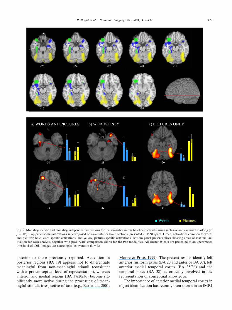
Fig. 2. Modality-specific and modality-independent activations for the semantics minus baseline contrasts, using inclusive and exclusive masking (at
p ¼ :05). Top panel shows activations superimposed on axial inferior brain sections, presented in MNI space. Green, activations common to words
and pictures; blue, word-specific activations; and yellow, pictures-specific activations. Bottom panel presents slices showing areas of maximal ac-
tivation for each analysis, together with peak rCBF comparison charts for the two modalities. All cluster extents are presented at an uncorrected
threshold of .001. Images use neurological convention (L¼L).
P. Bright et al. / Brain and Language 89 (2004) 417–432 427
anterior to those previously reported. Activation in
posterior regions (BA 19) appears not to differentiate
meaningful from non-meaningful stimuli (consistent
with a pre-conceptual level of representation), whereas
anterior and medial regions (BA 37/20/36) become sig-
nificantly more active during the processing of mean-
ingful stimuli, irrespective of task (e.g., Bar et al., 2001;
Moore & Price, 1999). The present results identify left
anterior fusiform gyrus (BA 20 and anterior BA 37), left
anterior medial temporal cortex (BA 35/36) and the
temporal poles (BA 38) as critically involved in the
representation of conceptual knowledge.
The importance of anterior medial temporal cortex in
object identification has recently been shown in an fMRI
428 P. Bright et al. / Brain and Language 89 (2004) 417–432
study where participants named pictures of commonobjects at a domain (i.e., living or non-living) or basic level
(i.e., identify the object itself as, for example, a monkey).
Domain-level naming produced activation of posterior
regions of the inferior temporal cortex while basic-level
naming produced additional anterior recruitment of left
perirhinal and entorhinal cortices (Tyler et al., 2004). The
involvement of the temporal poles could not be evaluated
because of susceptibility artefacts in fMRI (Devlin et al.,2002b).We argue that when an object is to be identified at
a high level of specificity, it requires extraction of more
detailed information in order to differentiate between
similar objects and these additional processes engage
anterior medial regions of temporal cortex. The meta-
analysis results are consistent with the existence of a hi-
erarchical object processing structure, with more anterior
temporal regions necessary for representation of objectsat high levels of specificity and posterior regions more
associated with intermediate and pre-semantic stages of
object representation. Rather than reflecting modality-
specific semantic representations, both verbal and visual
input routes appear to converge on these anterior tem-
poral sites, consistent with their being part of the verbal
basis for a unitary semantic system (although see below
for considerationof additionalword-specific activationofthe temporal poles).
Recently, Simmons and Barsalou (2003) have pro-
posed a model, based on Damasio’s (1989) convergence
zone (CZ) theory, which describes feature-binding neu-
rons arranged hierarchically from posterior to anterior
brain regions. Neurons in more posterior CZs proximal
to a particular modality-specific area (e.g., visual pro-
cessing or motor processing areas) capture the contentheld there. In more anterior regions, patterns of acti-
vation across modalities are indexed. The authors stress
Damasio’s assertion that a CZ points back to the sen-
sori-motor content held in feature maps but does not
store or represent that information (i.e., it indexes in-
formation held elsewhere). However, although this
proposal implies a common mechanism for capturing
modality-specific content, in a subsequent paper, theauthors suggest that some conjunctive neurons may
have modality-specific ‘tunings’ (Barsalou, Simmons,
Barbey, & Wilson, 2003). Our data offer limited support
for this proposal. If correct, there is likely to be quite
extensive overlap in regions of activation for words and
pictures in semantic processing areas, with differential
activations mostly restricted to the feature-map sites in
the posterior regions. We found that the areas of overlapwere primarily restricted to anterior fusiform gyrus (37/
20) and parahippocampal gyrus (BA 36), with more
widespread anterior temporal activity found only for
words and extensive bilateral occipitotemporal recruit-
ment specific to pictures.
There is an increasing body of evidence suggesting
that the types of features that define and distinguish
different objects and object categories may reflect thecortical organisation of conceptual representations in
the brain (e.g., Martin, 2001; Martin et al., 2000; Tyler
& Moss, 2001), and that the regions activated by an
object processing task relate to the type or level of se-
mantic processing required, rather than the intrinsic
properties of the presented objects (Tyler et al., 2004).
Nevertheless, although based on only one primary type
of task (semantic categorisation), our results suggestthat the anterior fusiform gyrus involvement in repre-
sentation of information at a semantic or conceptual
level is not differentially affected by the mode of input
(words or pictures).
In addition to temporal regions, we found a large
cluster of common activation encompassing the entire
extent of the inferior frontal gyrus (BA 45/47), consis-
tent with previous reports suggesting its general role insemantic processing (e.g., Vandenberghe et al., 1996;
Wagner et al., 1997). Various theories have been sug-
gested to account for activation of LIFG during se-
mantic relative to non-semantic tasks: for example,
increased competition among candidate responses
(Thompson-Schill, 2003), semantic ‘working memory’
or central executive system (e.g., Gabrieli et al., 1996;
Wagner et al., 1997) or complex syntactic operations(Carpentier et al., 2001). The present data preclude a
straightforward interpretation of the role of LIFG in
semantic tasks, but suggest that it may be involved in
tasks that require effortful, explicit processing, or the
active holding in memory of information required for
task success since these are processes common to all
word and picture semantic judgment tasks.
However, our interpretation of the data can bechallenged on at least two counts. First is the possibility
that both semantic categorisation tasks using words and
pictures present demands that are essentially word
based. Whether the stimuli employed are words or pic-
tures, the chosen strategy may be to generate a category
name for the cues (e.g., animals) and then to rate whe-
ther the category to which the target belongs matches it.
In this case, a word-based strategy would be chosen forboth stimulus types and the common fusiform activation
may reflect word-specific representations. We cannot
discount this interpretation strictly on the basis of our
own results, but in the context of evidence associating
the temporal poles (BA 38) with word retrieval, we ar-
gue that the area of common activation (BA 20/36/37) is
unlikely simply to reflect naming or word retrieval. It is
unlikely that any task of visual object processing at asemantic level can be conceived which rules out the
possibility of implicit naming or word retrieval. Never-
theless, we accept that our findings are based upon a
particular type of task and that further confirmatory
evidence, based on a wider range of tasks and contexts
may allow us to apply our results to the semantic system
in general with a greater level of confidence.
P. Bright et al. / Brain and Language 89 (2004) 417–432 429
Second, with respect to Paivio’s dual code theory(1971), it is possible that both words and pictures acti-
vate both visual and verbal codes. For example, the
frontal activation may relate to the processing of verbal
codes and the fusiform activation might be associated
with the processing of non-verbal codes. Thus, a com-
mon set of dual codes are activated in both types of task
(for words, the visual codes are automatically triggered
by the verbal codes, and the opposite pattern would takeplace for picture presentations). It follows that the re-
sults might better reflect a ‘dual-code’ process, with
verbal and non-verbal representation reflected by LIFG
and fusiform gyrus, respectively. However, while the
nature of frontal lobe involvement in semantic tasks
remains controversial, we argue that there is very little
(if any) neuropsychological evidence which indicates
that semantic-level representations are stored within theLIFG. Instead, we suggest that it is the anterior fusiform
gyrus that is the most plausible region for holding rep-
resentations, which are accessed both by pictures and
words.
Finally, we should point out that, even if either of
these two interpretations were correct, the observation
that processing converges on this antero-medial left
temporal region indicates that the neural correlates ofsemantic representations engaged in our tasks are not
dependent upon the format in which a stimulus is pre-
sented.
4.2. Word-specific effects
We found more extensive anterior temporal activa-
tion for words relative to pictures which involved abroad region of bilateral temporal poles (BA 38).
Damasio (1992) has reported that patients with pri-
marily left temporal pole damage have a difficulty with
proper noun retrieval. PET studies have associated the
temporal poles, either unilaterally or bilaterally, with
face recognition (e.g., Damasio, Grabowski, Tranel,
Hichwa, & Damasio, 1996) and general aspects of
comprehension (Maguire, Frith, & Morris, 1999). Se-mantic dementia patients (in which damage to the
temporal poles is a hallmark feature) show preserved
ability to retrieve general information about persons or
objects, but are unable to uniquely identify those objects
(e.g., Hodges, Graham, & Patterson, 1995). Thus, neu-
roimaging and lesion studies suggest that anterior tem-
poral regions may be involved in processing detailed
aspects of object attributes. Consistent with this posi-tion, our data suggest that the temporal poles are re-
cruited when fine-grained discrimination among similar
objects is required, but not when discriminating among
semantically meaningless stimuli.
Why is the extensive recruitment of the temporal
poles word-specific? Semantic categorisation of pictures
requires category-level knowledge. Participants can
perform this task on the basis of overall similarity be-tween objects and do not need to access the unique
properties that differentiate one object from another. In
contrast, in the tasks using words subjects were given
basic-level object labels, which access unique semantic
representations. If, as Grabowski et al. (2001) have ar-
gued, the left temporal pole is more engaged in naming
tasks as the level of specificity of word retrieval in-
creases, this may explain the more widespread lefthemisphere word-specific activations found in the pres-
ent study. We also found word-specific activation in the
right temporal pole, consistent with the view that se-
mantic system is bilaterally represented (Beeman, Bow-
den, & Gernsbacher, 2000; Perani et al., 1999).
In summary, although the temporal poles activation
was word-specific, we propose that these regions may be
involved in semantic representation in both modalitiesbut that their engagement might be primarily deter-
mined by the level of processing required for the task.
However, this is a provisional interpretation and further
work is needed to explore the extent to which the ad-
ditional temporal pole activation for words reflects ei-
ther material-specific semantic representations or
differences in the task processes which operate in these
regions.
4.3. Picture-specific effects
Picture-specific recruitment was restricted to more
posterior regions, including inferior occipital gyrus (BA
19), lingual gyrus (BA 18) and fusiform gyrus (BA 37).
Bilateral occipitotemporal cortex activation is routinely
found when objects are compared to non-objects (Bly &Kosslyn, 1997) and when objects are compared to words
(Moore & Price, 1999). Although the nature of the
representations along the extent of the fusiform gyrus
remains controversial, neuroimaging, and event-related
potential studies of visual object recognition have sug-
gested that the more posterior regions (BA 19) are as-
sociated with prerecognition analysis such as feature
extraction and intermediate shape processing (e.g., Baret al., 2001). Anterior fusiform gyrus (BA 37), in con-
trast, appears to mediate explicit identification (Bar
et al., 2001; Buchel et al., 1998). The extensive picture-
specific activation found in the present study involved
primarily posterior occipito-temporal regions, but did
extend to involve more posterior aspects of BA 37 (lat-
eral occipital complex). In the left hemisphere, this
modality-specific recruitment was situated adjacent toan area of anterior fusiform gyrus shared by both input
modalities. Buchel et al. (1998) have argued that BA 37
is multimodal such that its response properties should
not be altered by the modality of input. Our data suggest
that, while this proposal may be correct for more
anterior aspects of the fusiform gyrus, there may
be important functional differences throughout the
430 P. Bright et al. / Brain and Language 89 (2004) 417–432
posterior–anterior extent of this region, with modality-specificity (pictures) recruitment of posterior BA 37.
Anterior regions of the lateral occipital cortex (posterior
and mid fusiform gyrus) are activated more strongly by
whole, intact objects than by scrambled object stimuli.
However, the region appears not to distinguish familiar
from novel shapes (Malach et al., 1995), suggesting that
the picture-specific activations in fusiform gyrus found
in the present study reflect an intermediate or pre-se-mantic stage of visual processing.
4.4. Conclusions
In this study, we found evidence for a critical role of
the anterior extent of the fusiform gyrus (BA 37/20) in
the representation of conceptual knowledge. This region
does not appear to be modulated by the modality ofvisual input (pictures or words), suggesting that it holds
unitary semantic representations formed via converging
inputs from more posterior areas. We also found that
the left parahippocampal and perirhinal cortex are re-
cruited when a semantic level of representation is re-
quired (irrespective of modality), consistent with a role
for this region in the integration of sensory information
into semantically meaningful polymodal feature com-binations. Finally, we found common recruitment of the
left inferior frontal gyrus, which we suggest reflects an
executive or working memory role rather than concep-
tual representation per se.
Our findings are concerned with the semantic pro-
cessing of two types of stimulus (words and pictures).
Further work is needed in order to extend our knowl-
edge of whether semantic processing across differentprimary sensory modalities (visual, auditory, and tactile)
or different types of task also converge on the same re-
gions associated with conceptual representations. A
number of recent studies (PET and fMRI) have indi-
cated significant gender effects during lexical semantic
and naming tasks (e.g., Grabowski, Damasio, Tranel, &
Eichhorn, 2003; Rossell, Bullmore, Williams, & David,
2002), although these have usually reported such dif-ferences to be smaller than the task effects. However, the
relevance of gender in language-related activations
should be considered in future studies.
In summary, our data are most consistent with those
accounts which propose a single, common system of
semantic representations (Caramazza et al., 1990; Tyler
& Moss, 2001), rather than with the view that there are
distinct conceptual representations for the verbal andvisual modalities (Beauvois, 1982; Paivio, 1971, 1991;
Warrington & Shallice, 1984). However, we also found
word-specific activations in anterior temporal cortex
and picture-specific activations in occipitotemporal
cortex. The posterior recruitment (including posterior
BA 37/19), associated with pictures most likely relates to
intermediate, non-semantic levels of representation. Our
data suggests that the temporal poles are an importantpart of a distributed system subserving semantic repre-
sentations, but that their involvement may depend upon
the level of specificity at which an object is represented.
Acknowledgment
This research was supported by an MRC programme
grant to L.K.T.
References
Ashburner, J., & Friston, K. (1997). Multimodal image coregistration
and partitioning—a unified framework. NeuroImage, 6, 209–217.
Baayen, R., & Pipenbrook, R. (1995). The Celex Lexical Database.
Philadelphia Linguistic Data Consortium. Philadelphia: University
of Pennsylvania.
Bar, M., Tootell, R. B. H., Schacter, D. L., Greve, D. N., Fischl, B.,
Mendola, J. D., Rosen, B. R., & Dale, A. M. (2001). Cortical
Mechanisms Specific to Explicit Visual Object Recognition. Neu-
ron, 29, 529–535.
Barsalou, L. W., Simmons, W. K., Barbey, A., & Wilson, C. D. (2003).
Grounding conceptual knowledge in modality-specific systems.
Trends in Cognitive Sciences, 7, 84–91.
Beauvois, M. F. (1982). Optic aphasia: A process of interaction
between vision and language. Philosophical Transactions of the
Royal Society of London. Series B. Biological Sciences, 298(1089),
35–47.
Beeman, M. J., Bowden, E. M., & Gernsbacher, M. A. (2000). Right
and left hemisphere cooperation for drawing predictive and
coherence inferences during normal story comprehension. Brain
and Language, 71, 310–336.
Bly, B. M., & Kosslyn, S. M. (1997). Functional anatomy of object
recognition in humans: Evidence from positron emission tomog-
raphy and functional magnetic resonance imaging. Current Opinion
in Neurology, 10(1), 5–9.
Bookheimer, S. (2002). Functional MRI of language: New approaches
to understanding the cortical organization of semantic processing.
Annual Review of Neuroscience, 25, 151–188.
Buchel, C., Price, C., & Friston, K. (1998). A multimodal language
region in the ventral visual pathway. Nature, 394(6690), 274–277.
Caramazza, A. (2000). Minding the facts: A comment on Thompson-
Schill et al.’s ‘‘A neural basis for category and modality specificity
of semantic knowledge’’. Neuropsychologia, 38(7), 944–949.
Caramazza, A., Hillis, A. E., Rapp, B. C., & Romani, C. (1990). The
multiple semantics hypothesis: Multiple confusions? Cognitive
Neuropsychology, 7, 161–189.
Carpentier, A., Pugh, K. R., Westerveld, M., Studholme, C., Sknnja,
O., Thompson, J. L., Spencer, D. D., & Constable, R. I. (2001).
Functional MRI of language processing: Dependence on input
modality and temporal lobe epilepsy. Epilepsia, 42(10), 1241–1254.
Chaminade, T., &Decety, J. (2002). Leader or follower? Involvement of
the inferior parietal lobule in agency. Neuroreport, 13, 1975–1978.
Chao, L. L, Haxby, J. V., & Martin, A. (1999). Attribute-based neural
substrates in temporal cortex for perceiving and knowing about
objects. Nature Neuroscience, 2, 913–919.
Cohen, L., Dehaene, S., Naccache, L., Lehericy, S., Dehaene-
Lambertz, G., Henaff, M. A., & Michel, F. (2000). The visual
word form area. Spatial and temporal characterization of an initial
stage of reading in normal subjects and posterior split-brain
patients. Brain, 123, 291–307.
P. Bright et al. / Brain and Language 89 (2004) 417–432 431
Cohen, L., Lehericy, S., Chochon, F., Lemer, C., Rivaud, S., &
Dehaene, S. (2002). Language-specific tuning of visual cortex?
Functional properties of the visual word form area. Brain, 125(5),
1054–1069.
Coltheart, M. (1981). The MRC psycholinguistic database. Quarterly
Journal of Experimental Psychology, 33A, 497–505.
Coslett, H. B., & Saffran, E. M. (1992). Optic aphasia and the right
hemisphere—a replication and extension. Brain and Language, 43,
148–161.
Damasio, A. R. (1989). The brain binds entities and events by
multiregional activation from convergence zones. Neural Compu-
tation, 1, 123–132.
Damasio, A. R. (1992). Aphasia. New England Journal of Medicine,
326, 531–539.
Damasio, H., Grabowski, T. J., Tranel, D., Hichwa, R. D., &
Damasio, A. R. (1996). A neural basis for lexical retrieval. Nature,
380(6574), 499–505.
Dehaene, S., Le Clec, H. G., Poline, J. B., Le Bihan, D., & Cohen, L.
(2002). The visual word form area: A prelexical representation of
visual words in the fusiform gyrus. Neuroreport, 13(3), 321–332.
D’Esposito, M., Detre, J. A., Aguirre, G. K., Stallcup, M., Alsop, D.
C., Tippet, L. J., & Farah, M. J. (1997). A functional MRI study of
mental image generation. Neuropsychologia, 35, 725–730.
Demb, J. B., Desmond, J. E., Wagner, A. D., Vaidya, C. J., Glover, G.
H., & Gabrieli, J. D. (1995). Semantic encoding and retrieval in the
left inferior prefrontal cortex: A functional MRI study of task
difficulty and process specificity. Journal of Neuroscience, 15(9),
5870–5878.
Demonet, J. F., Chollet, F., Ramsay, S., Cardebat, D., Nespoulous, J.
L., Wise, R., Rascol, A., & Frackowiak, R. (1992). The anatomy of
phonological and semantic processing in normal subjects. Brain,
115(Pt 6), 1753–1768.
Demonet, J. F., Price, C., Wise, R., & Frackowiak, R. S. J. (1994). A
PET study of cognitive strategies in normal subjects during
language tasks. Influence of phonetic ambiguity and sequence
processing on phoneme monitoring. Brain, 117(4), 671–682.
Devlin, J. T., Moore, C. J., Mummery, C. J., Gorno-Tempini, M. L.,
Phillips, J. A., Noppeney, U., Frackowiak, R. S., Friston, K. J., &
Price, C. J. (2002a). Anatomic constraints on cognitive theories of
category specificity. Neuroimage, 15(3), 675–685.
Devlin, J. T., Russell, R. P., Davis, M. H., Price, C. J., Moss, H. E.,
Fadili, M. J., & Tyler, L. K. (2002b). Is there an anatomical basis
for category-specificity? Semantic memory studies in PET and
fMRI. Neuropsychologia, 40(1), 54–75.
Dolan, R. J., Morris, J. S., & de Gelder, B. (2001). Crossmodal binding
of fear in voice and face. Proceedings of the National Academy of
Sciences of the United States of America, 98, 10006–10010.
Farah, M. J., Hammond, K. M., Mehta, Z., & Ratcliff, G. (1989).
Category-specificity and modality-specificity in semantic memory.
Neuropsychologia, 27(2), 193–200.
Feinberg, T. E., Dyckes-Berke, D., Miner, C. R., & Roane, D. M.
(1995). Knowledge, implicit knowledge and metaknowledge in
visual agnosia and pure alexia. Brain, 118(Pt 3), 789–800.
Ferreira, C. T., Giusiano, B., Ceccaldi, M., & Poncet, M. (1997). Optic
aphasia: Evidence of the contribution of different neural systems to
object and action naming. Cortex, 33(3), 499–513.
Fiez, J. A., & Petersen, S. E. (1998). Neuroimaging studies of word
reading. Proceedings of the National Academy of Sciences of the
United States of America, 95, 853–860.
Forster, K. I., & Forster, J. C. (1991). The DMASTR display system
for mental chronometry. Tucson: University of Arizona.
Friston, K. J., Holmes, A. P., Worsley, K. J., & Poline, J. P. (1995).
Statistical parametric maps in functional imaging: A general linear
approach. Human Brain Mapping, 2(4), 189–210.
Friston, K. J., Holmes, A. P., Price, C. J., Buchel, C., & Worsley, K. J.
(1999). Multisubject fMRI studies and conjunction analyses.
NeuroImage, 10, 385–396.
Gabrieli, J. D. E., Desmond, J. E., Demb, J. B., Wagner, A. D., Stone,
M. V., Vaidya, C. J., & Glover, G. H. (1996). Functional Magnetic
Resonance Imaging of Semantic Memory Processes in the Frontal
Lobes. Psychological Science, 7(5), 278–283.
Gauthier, I., Tarr, M. J., Anderson, A. W., Skudlarski, P., & Gore, J.
C. (1999). Activation of the middle fusiform ‘face area’ increases
with expertise in recognising novel objects. Nature Neuroscience, 2,
568–573.
Grabowski, T. J., Damasio, H., Tranel, D., & Eichhorn, G. E. (2003).
Effects of gender on blood flow correlates of naming concrete
entities. NeuroImage, 20, 940–954.
Grabowski, T. J., Damasio, H., Tranel, D., Ponto, L. L., Hichwa, R.
D., & Damasio, A. R. (2001). A role for left temporal pole in the
retrieval of words for unique entities. Human Brain Mapping, 13,
199–212.
Grill-Spector, K., Kourtzi, Z., & Kanwisher, N. (2001). The lateral
occipital complex and its role in object recognition. Vision
Research, 41, 1409–1422.
Grill-Spector, K., Kushnir, T., Hendler, T., Edelman, S., Itzchak, Y.,
& Malach, R. (1998). A sequence of object processing stages
revealed by fMRI in the human occipital lobe. Human Brain
Mapping, 6, 316–328.
Hillis, A. E., & Caramazza, A. (1995). Cognitive and neural mecha-
nisms underlying visual and semantic processing: Implications
from ‘‘optic aphasia’’. Journal of Cognitive Neuropsychology, 7,
457–478.
Hodges, J. R., Graham, N., & Patterson, K. (1995). Charting the
progression in semantic dementia: Implications for the organisa-
tion of semantic memory. Memory, 3(3–4), 463–495.
Kanwisher, N., Chun, M. M., McDermott, J., & Ledden, P. J. (1996).
Functional imagining of human visual recognition. Cognitive Brain
Research, 5(1–2), 55–67.
Kinahan, P. E., & Rogers, J. G. (1989). Analytic 3D image
reconstruction using all detected events. IEEE Transactions on
Nuclear Science, 35, 680–684.
Lambon Ralph, M. A., Graham, K. S., Patterson, K., & Hodges, J. R.
(1999). Is a picture worth a thousand words? Evidence from
concept definitions by patients with semantic dementia. Brain and
Language, 70, 309–335.
Lambon Ralph, M. A., & Howard, D. (2000). Gogi aphasia or
semantic dementia? Simulating and assessing poor verbal compre-
hension in a case of progressive fluent aphasia. Cognitive Neuro-
psychology, 17, 437–465.
Lee, A. C. H., Robbins, T. W., Graham, K. S., & Owen, A. M. (2002).
‘‘Pray or Prey?’’ Dissociation of semantic memory retrieval from
episodic memory processes using photon emission tomography and
a novel homophone task. NeuroImage, 16, 724–735.
Luzzatti, C., Rumiati, R. I., & Ghirardi, G. (1998). A functional model
of visuo-verbal disconnection and the neuroanatomical constraints
of optic aphasia. Neurocase, 4, 71–87.
McCarthy, R. A., & Warrington, E. K. (1986). Visual associative
agnosia: A clinico-anatomical study of a single case. Journal of
Neurology, Neurosurgery, and Psychiatry, 49, 1233–1240.
McCarthy, R. A., & Warrington, E. K. (1988). Evidence for modality-
specific meaning systems in the brain. Nature, 334, 428–430.
Maguire, E. A., Frith, C. D., & Morris, R. G. M. (1999). The
functional neuroanatomy of comprehension and memory: The
importance of prior knowledge. Brain, 122(10), 1839–1850.
Malach, R., Reppas, J. B., Benson, R. R., Kwong, K. K., Jiang, H.,
Kennedy, W. A., Ledden, P. J., Brady, T. J., Rosen, B. R., &
Tootell, R. B. (1995). Object-related activity revealed by functional
magnetic resonance imaging in human occipital cortex. Proceedings
of the National Academy of Sciences of the United States of
America, 92, 8135–8139.
Martin, A. (2001). Functional neuroimaging of semantic memory. In
R. Cabeza & A. Kingstone (Eds.), Handbook of functional
neuroimaging of cognition (pp. 153–186). Cambridge: MIT Press.
432 P. Bright et al. / Brain and Language 89 (2004) 417–432
Martin, A., Haxby, J. V., Lalonde, F. M., Wiggs, C. L., & Ungerleider,
L. G. (1995). Discrete cortical regions associated with knowledge of
color and knowledge of action. Science, 270, 102–105.
Martin, A., Ungerleider, L. G., & Haxby, J. V. (2000). Category-
specificity and the brain: The sensory-motor model of semantic
representations of objects. In M. S. Gazzaniga (Ed.), The Cognitive
Neurosciences (2nd ed., pp. 1023–1036). Cambridge: MIT Press.
Moore, C. J., & Price, C. J. (1999). Three distinct ventral occipito-
temporal regions for reading and object naming. Neuroimage, 10,
181–192.
Morcom, A. M., Good, C. D., Frackowiak, R. S. J., & Rugg, M. D.
(2003). Age effects on the neural correlates of successful memory
encoding. Brain, 126, 213–229.
Noppeney, U., & Price, C. J. (2002). Retrieval of visual, auditory, and
abstract semantics. Neuroimage, 15(4), 917–926.
Paivio, A. (1971). Imagery and verbal processes. London: Holt,
Rinehart & Winston.
Paivio, A. (1986). Mental representations: A dual coding approach.
Oxford University Press: Oxford.
Paivio, A. (1991). Dual Coding Theory: Retrospect and current status.
Canadian Journal of Psychology, 45, 255–287.
Perani, D., Cappa, S. F., Schnur, T., Tettamanti, M., Collina, S.,
Rosa, M. M., & Fazio, F. (1999). The neural correlates of verb and
noun processing—a PET study. Brain, 122, 2337–2344.
Plaut, D. C. (2002). Graded modality-specific specialisation in seman-
tics: A computational account of optic aphasia. Cognitive Neuro-
psychology, 19, 603–639.
Pochon, J. B., Levy, R., Fossati, P., Lehericy, S., Poline, J. B., Pillon,
B., Le Bihan, D., & Dubois, B. (2002). The neural system that
bridges reward and cognition in humans: An fMRI study.
Proceedings of the National Academy of Sciences of the United
States of America, 99, 5669–5674.
Price, C. J., & Friston, K. J. (1997). Cognitive conjunction: A new
approach to brain activation experiments. Neuroimage, 5(4/Pt1),
261–270.
Price, C. J., Moore, C. J., Humphreys, G. W., Frackowiak, R. S. J., &
Friston, K. J. (1996). The neural regions sustaining object
recognition and naming. Proceedings of the Royal Society of
London. Series B., 263, 1501–1507.
Rapp, B. C., Hillis, A. E., & Caramazza, A. (1993). The role of
representations in cognitive theory: More on multiple semantics
and the agnosias. Cognitive Neuropsychology, 10, 235–249.
Riddoch, M. J., & Humphreys, G. W. (1987). Visual object processing
in optic aphasia: A case of semantic access agnosia. Cognitive
Neuropsychology, 4, 131–185.
Riddoch, M. J., Humphreys, G. W., Coltheart, M., & Funnell, E.
(1988). Semantic systems or system. Neuropsychological evidence
re-examined. Cognitive Neuropsychology, 5, 3–25.
Rossell, S. L., Bullmore, E. T., Williams, S. C. R., & David, A. S.
(2002). Sex differences in functional brain activation during a
lexical visual field task. Brain and Language, 80, 97–105.
Saffran, E. M., Coslett, H. B., Martin, N., & Boronat, C. B. (2003).
Access to knowledge from pictures but not words in a patient with
progressive fluent aphasia. Language and Cognitive Processes, 18,
725–757.
Shallice, T. (1988). Specialisation within the semantic system. Cognitive
Neuropsychology, 5, 133–142.
Shallice, T. (1993). Multiple semantics: Whose confusions? Cognitive
Neuropsychology, 10, 251–261.
Silbersweig, D. A., Stern, E., Frith, C. D., & Cahill, C. (1993).
Detection of thirty-second cognitive activations in single subjects
with positron emission tomography: A new low-dose H2(15)O
regional cerebral blood flow three-dimensional imaging technique.
Journal of Cerebral Blood Flow and Metabolism, 13(4), 617.
Simmons, K., & Barsalou, L. W. (2003). The similarity-in-topography
principle: Reconciling theories of conceptual deficts. Cognitive
Neuropsychology, 20, 451–486.
Talairach, J., & Tournoux, P. (1988). Co-planar stereotaxic atlas of the
human brain. Stuttgart: Georg Thieme Verlag.
Thompson-Schill, S. L. (2003). Neuroimaging studies of semantic
memory: Inferring ‘‘how’’ from ‘‘where’’.Neuropsychologia, 41, 280–
292.
Thompson-Schill, S. L., Aguirre, G. K., D’Esposito, M., & Farah, M.
J. (1999). A neural basis for category and modality specificity of
semantic knowledge. Neuropsychologia, 37(6), 671–676.
Tyler, L. K., Bright, P., Dick, E., Tavares, P., Pilgrim, L., Fletcher, P.
C., Greer, M., & Moss, H. E. (2003). Do semantic categories
activate distinct cortical regions. PET studies of picture processing.
Cognitive Neuropsychology, 20, 541–559.
Tyler, L. K., & Moss, H. (2001). Towards a distributed account of
conceptual knowledge. Trends in Cognitive Science, 5, 244–252.
Tyler, L. K., Russell, R., Fadili, J., & Moss, H. E. (2001). The neural
representation of nouns and verbs: PET studies. Brain, 124, 1619–
1634.
Tyler, L. K., Stamatakis, E. A., Bright, P., Acres, K., Abdallah, S.,
Rodd, J. M., & Moss, H. E. (2004). Processing objects at different
levels of specificity. Journal of Cognitive Neuroscience, 16, 1–12.
Vandenberghe, R., Price, C., Wise, R., Josephs, O., & Frackowiak, R.
S. J. (1996). Functional anatomy of a common semantic system for
words and pictures. Nature, 383, 254–256.
Wagner, A. D., Desmond, J. E., Demb, J. B., Glover, G. H., &
Gabrieli, J. D. E. (1997). Semantic repetition priming for verbal
and pictorial knowledge: A functional MRI study of left inferior
prefrontal cortex. Journal of Cognitive Neuroscience, 9, 714–726.
Warrington, E. K. (1975). The selective impairment of semantic
memory. Quarterly Journal of Experimental Psychology, 27, 635–
657.
Warrington, E. K., & McCarthy, R. A. (1994). Multiple meaning
systems in the brain: A case for visual semantics. Neuropsychologia,
32(12), 1465–1473.
Warrington, E. K., & Shallice, T. (1984). Category specific semantic
impairments. Brain, 107(Pt 3), 829–854.