Trends in Eye Care - American Academy of Ophthalmology
56
Pain Without Stain When It’s Not Dry Eye “North of 600 Microns” Managing Challenging Macular Holes Rhegmatogenous Retinal Detachments Diagnostic Pearls DECEMBER 2018 EyeNet ® Trends in Eye Care What’s New, What’s Next
-
Upload
khangminh22 -
Category
Documents
-
view
0 -
download
0
Transcript of Trends in Eye Care - American Academy of Ophthalmology

Pain Without StainWhen It’s Not Dry Eye
“North of 600 Microns”Managing Challenging Macular Holes
Rhegmatogenous Retinal DetachmentsDiagnostic Pearls
D E C E M B E R 2 0 1 8
EyeNet®
Trends in Eye CareWhat’s New, What’s Next
01_Cover_F.indd 1 11/15/18 10:59 AM

NEW Accurate Coding
IMPROVED Knowledge Base
Experience IntelleChartPRO
NEW Assisted Compliance
IntelleChartPRO
IntelleChartPRO
Nextech.com/Ophthalmology(800) 868-3694
00_Ads_F.indd 2 11/8/18 8:20 PM
We are happy to announce another significant policyholder dividend equal to 15% of premium to be paid throughout 2019.
OMIC consistently outperforms multispecialty malpractice insurance companies in almost all financial results, benchmarks, and ratios used to evaluate performance in the industry.
OMIC’s dividend returns averaged 20.7% of earned premium the past five years vs. 6.7% for other carriers that have declared dividends.
New physicians who join OMIC before year-end 2018 qualify for the dividend at 2019 renewal.
Robert Wiggins, MDChair, Finance Committee and Treasurer
$80 MillionCumulative declared dividends 1987-2017
OMIC.com/request-a-quote800.562.6642
OMICPageMyOMIC
00_Ads_F.indd 3 11/13/18 7:40 AM

©Bausch & Lomb Incorporated. All rights reserved. Printed in USA. JEV.XXXX.USA.18
© 2018 Bausch & Lomb Incorporated. All rights reserved. LGX.0058.USA.18
COMING SOON BAUSCH + LOMB’S NEWEST ARRIVAL
FROM THE EXPERTS IN LOTEPREDNOL ETABONATE FOR OVER 20 YEARS
A NEW LOTEPREDNOL ETABONATE FORMULATION
BAUS4885 Loteprednol .38 Coming Soon Journal Ad Eyenet 4/C Page Live: .5" from trim File Format: PDF/X1-A Carling Communications 11/6/18 Trim: 8.125"x10.875" Bleed: 8.375"x11.125"
00_Ads_F.indd 4 11/13/18 5:14 PM

E Y E N E T M A G A Z I N E • 5
EyeNet® Magazine (ISSN 1097-2986) is published monthly by the American Academy of Ophthal mology, 655 Beach St., San Fran-cisco, CA 94109-1336, as a membership service. Subscription is included in U.S. members’ annual dues. International Mem ber, IMIT, $135 per year. Nonmember in U.S., $150 per year. Nonmember outside U.S., $210 per year. Periodicals Postage Paid at San Francisco, CA, and at additional mailing offices. POSTMASTER: Send address changes to EyeNet, P.O. Box 7424, San Francisco, CA 94120-7424. American Academy of Ophthalmic Executives®, EyeSmart®, EyeWiki®, IRIS® Registry, MIPS QCDR measures, and ONE® Network are trademarks of the American Academy of Ophthalmology®. All other trademarks are the property of their respective owners.
CONTENTSDECEMBER 2018VOLUME 22 • NUMBER 12
3127
2313
36
FEATURE
36-43 A Look AheadEyeNet editorial board members assess their areas of subspecialty—with a focus on recent developments—and discuss how the future of their fields may be shaped by what’s happen-ing today. See what they say about glaucoma, low vision rehabilitation, ocular oncology, and refractive surgery and corneal ectatic disease.
CLINICAL INSIGHTS
13-15 News in ReviewCornea First-in-class drug approved for neuro-trophic keratitis.
Glaucoma Meta-analysis of SD-OCT benefits.
Neuro-Ophthalmology Assessing eye move-ment metrics following concussion.
Infection Control Infections in a NICU highlight the need to follow protocols.
17-21 Journal HighlightsKey findings from Ophthalmology, Ophthalmol-ogy Glaucoma, Ophthalmology Retina, AJO, JAMA Ophthalmology, and more.
23-29 Clinical Update Cornea When it’s “pain without stain,” suspect neuropathic pain. Treatment options to consider.
Retina Large and chronic macular holes, and those in which surgery has failed, may pose special challenges for the retina surgeon. Advice from 3 experts.
31-32 Ophthalmic Pearls Rhegmatogenous Retinal Detachment Part 1 of a 2-part series on RRD covers risk factors, features, and examination.
05-06_TOC_F.indd 5 11/13/18 7:27 AM

6 • D E C E M B E R 2 0 1 8
COPYRIGHT © 2018, American Academy of Ophthalmology, Inc.® All rights reserved. No part of this publication may be reproduced with-out written permission from the publisher. Letters to the editor and other unsolicited material are assumed intended for publication and are subject to editorial review, acceptance, and editing. Disclaimer. The ideas and opinions expressed in EyeNet are those of the authors,
and do not necessarily reflect any position of the editors, the publisher, or the American Academy of Ophthalmology. Because this publication provides information on the latest developments in ophthalmology, articles may include information on drug or device applications that are not considered community standard, or that reflect indications not included in ap proved FDA labeling. Such ideas are provided as information and education only so that practitioners may be aware of alternative methods of the practice of medicine. Information in this publication should not be considered endorsement, promotion, or in any other way encouragement for the use of any particular procedure, technique, device, or product. EyeNet, its editors, the publisher, or the Academy in no event will be liable for any injury and/or damages arising out of any decision made or action taken or not taken in reliance on information contained herein.
EyeNet®
xx
CLINICAL INSIGHTS
33-35 Morning Rounds The Black Spot At first, the overworked 34-year-old ignored the black spot in his vision. But over 3 weeks, it grew in size, and the letters on his computer screen began to blur.
IN PRACTICE
45-46 Practice PerfectFour Ways to Boost Patient Satisfaction This strategic approach to improving the pa-tient experience will increase patient volume and improve employee job satisfaction.
FROM THE AAO
49-51 Academy Notebook State societies recognized. • Two crucial MIPS deadlines. • Vitrectomy codes restored.
VIEWPOINTS
10 Opinion Why advocate for increased research funding?
MYSTERY IMAGE
54 BlinkWhat do you see?
COVER PHOTOGRAHY
Jason S. Calhoun
33
49
54
05-06_TOC_F.indd 6 11/8/18 7:50 PM

For desktop and mobile devices
©2018 Modernizing Medicine, Inc.
I AM
It’s so advanced, it actually learns from you.
Modernizing Medicine®’s all-in-one platform
was designed by practicing ophthalmologists to
streamline treatment and help improve workflow.
From the moment you first log in, it begins
learning how you practice, diagnose and treat
patients, customizing itself to help give your
practice greater efficiency.
So you can see more patients, while
seeing more of your patients. It’s time
to demand more from your EHR.
MODERNIZINGOPHTHALMOLOGYWITH A SMARTER EHR
VIEW OUR 2-MINUTE DEMO
M O D M E D O P H T H . C O M TOGETHER, WE ARE MODERNIZING MEDICINE.
00_Ads_F.indd 7 11/8/18 8:20 PM

8 • D E C E M B E R 2 0 1 8
David W. Parke II, MDEditor-in-Chief
Ruth D. Williams, MDChief Medical Editor
Dale E. Fajardo, EdD, MBAPublisher
Patty AmesExecutive Editor
Carey S. BallardArt Director /
Production Manager
Chris McDonagh, Jean ShawSenior Editors
Krista ThomasAssistant Editor /
Advertising Manager
Lori Baker-Schena, MBA, EdD; Leslie Burling; Peggy Denny;
Miriam Karmel; Mike Mott; Linda Roach; Lynda Seminara; Annie Stuart; Gabrielle Weiner
Contributing Writers
Mark Mrvica, Kelly MillerM.J. Mrvica Associates, Inc.
2 West Taunton Ave., Berlin, NJ 08009
856-768-9360 [email protected]
Advertising Sales
655 Beach St.San Francisco, CA 94109
866-561-8558, 415-561-8500 aao.org
Governmental Affairs Division20 F Street NW, Suite 400
Washington, DC 20001 202-737-6662
ARTICLE REVIEW PROCESS. Articles involving single-source medical and tech-nical news are sent to quoted sources for verification of accuracy prior to publication. Quotes and other information in multisource articles are subject to confirmation by their respective sources. The chief medical editor and the executive editor review all news and feature articles and have sole discretion as to the acceptance and rejection of material and final authority as to revisions deemed necessary for publication.
DISCLOSURE KEY. Financial interests are indicated by the following abbrevia-tions:C = Consultant/AdvisorE = EmployeeL = Speakers bureauO = Equity ownerP = Patents/Royalty S = Grant supportFor definitions of each category, see aao.org/eyenet/disclosures.
MAGAZINE
®
ACADEMY BOARDPRESIDENTKeith D. Carter, MD, FACS
PRESIDENT-ELECTGeorge A. Williams, MD
PAST PRESIDENTCynthia A. Bradford, MD
CEODavid W. Parke II, MD
SR. SECRETARY FOR ADVOCACYDaniel J. Briceland, MD
SECRETARY FOR ANNUAL MEETINGMaria M. Aaron, MD
SR. SECRETARY FOR CLINICAL EDUCATIONLouis B. Cantor, MD
SR. SECRETARY FOR OPHTHALMIC PRACTICERobert E. Wiggins Jr., MD, MHA
CHAIR, THE COUNCILLynn K. Gordon, MD, PhD
VICE CHAIR, THE COUNCILSarwat Salim, MD, FACS
OPHTHALMOLOGY EDITORStephen D. McLeod, MD
CHAIR OF THE FOUNDATION ADVISORY BOARDChristie L. Morse, MD
PUBLIC TRUSTEESPaul B. Ginsburg, PhD
TRUSTEES-AT-LARGEMichael F. Chiang, MD William S. Clifford, MDSanjay D. Goel, MDCynthia Mattox, MD, FACSWilliam F. Mieler, MDAndrew M. Prince, MD
INTERNATIONAL TRUSTEESKgaogelo Edward Legodi, MDLihteh Wu, MD
Learn more about the Board at aao.org/bot.
EDITORIAL BOARD
CATARACTKevin M. Miller, MD,Section Editor
William R. Barlow, MDKenneth L. Cohen, MDKendall E. Donaldson, MDJason J. Jones, MDBoris Malyugin, MD, PhDCathleen M. McCabe, MDRandall J. Olson, MDMarie Jose Tassignon, MD
COMPREHENSIVE OPHTHALMOLOGYPreston H. Blomquist, MD,Section Editor
Sherleen Huang Chen, MDApril Y. Maa, MDLinda M. Tsai, MD
CORNEA AND EXTERNAL DISEASEChristopher J. Rapuano, MD,Section Editor
Kathryn A. Colby, MD, PhDHelena Prior Filipe, MDBennie H. Jeng, MDStephen D. McLeod, MDSonal S. Tuli, MD
GLAUCOMASanjay G. Asrani, MD, Section Editor
Iqbal K. Ahmed, MDLama Al-Aswad, MD, MPHAhmad A. Aref, MD, MBAAnne Louise Coleman, MD, PhDSteven J. Gedde, MDCatherine Green, MBChB
Steven L. Mansberger, MD, MPHRonit Nesher, MDRichard K. Parrish II, MDSarwat Salim, MD, FACS
LOW VISIONLylas G. Mogk, MDJohn D. Shepherd, MD
NEURO-OPHTHALMOLOGYLeah Levi, MD, Section Editor
Kimberly Cockerham, MD, FACSHelen V. Danesh-Meyer, MD, PhD, FRANZCOPrem S. Subramanian, MD, PhD
OCULOPLASTICSEvan H. Black, MD, Section Editor
Elizabeth A. Bradley, MDFemida Kherani, MDDon O. Kikkawa, MD
OPHTHALMIC ONCOLOGYZélia M. Corrêa, MD, PhD, Section Editor
Dan S. Gombos, MDTatyana Milman, MD
OPHTHALMIC PATHOLOGYDeepak Paul Edward, MDDavid J. Wilson, MD
OPHTHALMIC PHOTOGRAPHYJason S. CalhounMichael P. Kelly, FOPS
PEDIATRIC OPHTHALMOLOGYDavid A. Plager, MD, Section Editor
Michael F. Chiang, MDJane C. Edmond, MDFrank Joseph Martin, MDFederico G. Velez, MD
REFRACTIVE SURGERYGeorge O. Waring IV, MD, Section Editor
Damien Gatinel, MDSoosan Jacob, FRCS A. John Kanellopoulos, MDJ. Bradley Randleman, MDKarolinne M. Rocha, MDMarcony R. Santhiago, MD
RETINA/VITREOUSJulia A. Haller, MD, Section Editor
Neil M. Bressler, MDKimberly A. Drenser, MD, PhDSharon Fekrat, MDMitchell Goff, MDLawrence S. Halperin, MDGregg T. Kokame, MDAndreas K. Lauer, MDPrithvi Mruthyunjaya, MD, MHSKyoko Ohno-Matsui, MDAndrew P. Schachat, MD Ingrid U. Scott, MD, MPHGaurav K. Shah, MD
UVEITISGary N. Holland, MD, Section Editor
Muge R. Kesen, MD H. Nida Sen, MDSteven Yeh, MD
08_Advisors_F.indd 8 11/15/18 2:09 PM

840 Walnut St., Philadelphia, PA 19107 | willseye.org | 877.289.4557
Allen C. Ho, MD, FACSDirector, Retina Research
*Based on a Doximity clinical reputation survey for 2018-2019.
When conquering eye disease demands resolve, we
embrace the long and difficult road. That’s part of the
reason so many residents want to come to Wills Eye
Hospital to train. We teach perseverance.
Voted #1 ophthalmology residency program in the United States.*
Where no challenge is too great.
00_Ads_F.indd 9 11/8/18 8:20 PM

10 • D E C E M B E R 2 0 1 8
Ruth D. Williams, MDChief Medical
Editor, EyeNet
Opinion
RUTH D. WILLIAMS, MD
Why Advocate for Increased Research Funding?
At this year’s Women in Ophthalmology annual meet-ing, Arlene Drack showed riveting video footage of a child with Leber congenital amaurosis (LCA) before
and after gene therapy treatment. An 11-year-old girl—who previously required an aide to help her maneuver between classes—walks confidently into the schoolroom alone, an improvement in quality of life that isn’t captured through mere visual acuity measurement.
The landmark gene therapy that made this girl’s indepen-dence possible grew out of decades of basic science research, much of it supported by NIH funding. Arlene said, “For the first time in human history, we can improve vision in children with a form of congenital blindness. This only hap-pened because of medical research—and NIH funding is the cornerstone of medical research in the United States.”
Advances in treatments for eye diseases like LCA grow out of academic research programs (see “Collaboration in Aca-demic Funding,” November), which rely on external funding. After a decade of cuts, flat funding, or no inflationary in-creases, Congress finally boosted NIH funding by $7 billion in 2016-2018, a 23% increase. And the president recently signed the 2019 spending package, which includes another $2 billion of funding to NIH (a 5.4% increase). Of this amount, the NEI will be allocated an additional $24.2 million to bring its funding to nearly $800 million.
Vision researchers have even more reason to be optimis-tic. Traditionally, funding for academic research has centered around the NEI R01 investigator-initiated award. However, additional federal funding opportunities for vision research are newly available. For example, Congress allocated $500 million for research related to combating the opioid epi-demic, and researchers—including those studying ocular pain and dry eye—are encouraged to submit proposals. In addition, the Department of Defense (DOD) offers research support through its Vision Research Program (VRP). James Jorkasky, executive director of the National Alliance for Eye and Vision Research (NAEVR), pointed out that “as research-ers become familiar with DOD funding opportunities, many then submit grants beyond the Vision Research Programs,” using key words such as “sensory” and “rehabilitation” and targeting diseases with visual implications. Thanks to
advocacy by NAEVR and the Academy during the Mid-Year Forum, VRP was funded at $20 million for 2019.
Another funding opportunity, the 21st Century Cures Act (H.R. 34), was passed by Congress in 2016. Included in this bill are the BRAIN Initiative (initially established in 2013 by President Obama), the Precision Medicine Initiative, and the Regenerative Medicine Initiative, all funding sources available to vision re-searchers. Ophthalmology has been awarded more than one-third of the BRAIN Initiative funding in the last 4 cycles.
These increasingly diverse funding options reflect the pivotal role of ophthalmic research in neuroscience. In BRAIN 2025: A Scientific Vision, the inititive’s working group pointed out that “The retina is the region in which the most progress has been made in the characterization of different cell types . . . it could serve as a flagship project for the BRAIN Initiative. It is relevant to the fields of vision, general sensory and signal processing, and to clinical issues including neurodegenerative diseases and vision disorders.”1
My patients often ask when we will have a cure for glau-coma. I tell them that eye research is leading the way, and when we cure glaucoma, we’ll have also made gigantic strides in addressing spinal cord injuries, Alzheimer and Parkinson diseases, and the effects of stroke.
When Arlene showed the video of the girl with LCA before and after gene therapy treatment, the room full of ophthal-mologists was vibrating with joy. Together we celebrated the impact of research on the life of a single child. This is why we are ophthalmologists. This is why we support and advocate for vision research. This is what gives our work meaning.
1 https://acd.od.nih.gov/documents/reports/06052014report-BRAIN.pdf.
Accessed Nov. 12, 2018.
10_Opin_F.indd 10 11/13/18 7:28 AM

00_Ads_F.indd 11 11/14/18 11:57 AM

PRACTICE MANAGEMENT
Position your practice for success
Ophthalmology Business SummitMarch 23-24, 2019 | Chicago
Join us in addressing the urgent business challenges facing ophthalmic practices right now.
You’ll acquire strategic insights and tactical tools that can be applied immediately and have a meaningful impact on your revenue and growth.
Seats are limited. Register today.aao.org/business-summit
Early registration deadline is Dec. 6
NEW Open to physicians and administrators
00_Ads_F.indd 12 11/13/18 7:48 AM

E Y E N E T M A G A Z I N E • 13
News in ReviewCOMMENTARY AND PERSPECT IVE
Bo
nin
i S e
t al
. Op
hth
alm
olo
gy.
20
18;1
25
(9):
133
2-1
34
3.
CORNEA
First-in-Class Rx for Neurotrophic KeratitisA RECOMBINANT FORM OF HUMAN nerve growth factor (rhNGF) has be-come the first FDA-approved drug for the treatment of neurotrophic keratitis (NK), a degenerative disease of the corneal epithelium that results from loss of corneal sensation.1 The drug—cenegermin (Oxervate, Dompé)—rep-resents 2 additional firsts: It is the first application of an rhNGF as a drug and the first topical biologic medication approved in ophthalmology. It may become available in the United States early next year.
Urgent need. With NK, trigeminal nerve damage originates in the eye or the brain because of factors ranging from herpes eye infection to brain sur g ery, said Harminder S. Dua, FRCS, FRCOphth, PhD, at the University of Nottingham in the United Kingdom. Independently or simultaneously, NK then impairs sensory and trophic functions, including blinking, secretion of tears, and the nutritional health of corneal cells.2 Although NK affects few-er than 5 in 10,000 people, its impact can be severe, ranging from corneal thinning, ulceration, and perforation to visual impairment or blindness.3
Alleviating the damage. Cenegermin was evaluated in 2 studies involving a total of 151 patients. Treatment in-volved 8 weeks of cenegermin eyedrops, delivered 8 times a day. Across both
studies, 70% of patients treated with the drug experienced complete corneal healing, versus 28% in the control groups. Adverse reactions included eye pain and inflammation, ocular hyper-emia, and tearing. After 1 year, about 80% of those who were successfully treated remained free of disease.3
“The studies were quite promising, but it was difficult to believe this was really possible until I saw it myself,” said Prof. Dua, who has now successful-ly treated 3 patients with severe cases of NK. Previously, he said, “We had sup-portive treatments such as lubricating drops, eye patching, autologous serum drops, tarsorrhaphy, and amniotic mem-brane patches. But we had nothing to treat the underlying disease.”
A neural revival. Because rhNGF is involved in the development, main-tenance, and survival of nerve cells, cenegermin has the ability to address the root cause of NK and restore cor-neal integrity. “It revitalizes the nerves’ ability to secrete neuropeptides that support the health and regeneration of the corneal epithelium and the kerato-cytes of the stroma,” said Prof. Dua.
Prior to treatment, corneal sensation was as low as 5 mm in his patients with NK, with 60 mm representing normal sensation, he said. “After treatment with
Oxervate eyedrops, we’ve seen corneal sensation return to 50 mm. In addition, we can observe nerve regeneration using in vivo confocal microscopy. Al-though the new nerves are more coiled and tortuous, their sprouting in the cornea is very exciting.”
Challenges ahead. Cenegermin requires frequent instillation. But NK patients may take that in stride, as many of them have already been using multiple drops several times a day, said Prof. Dua. A bigger challenge may be the weekly visits to the phar-macy, where drops must be frozen and released a batch at a time and then kept in the patient’s refrigerator for no longer than 7 days.
“Another challenge is the cost of 2 months of treatment,” said Prof. Dua. In the United Kingdom, where clini-cians have had access to the drug since 2017, that cost is about £11,000 to £12,000, he said. U.S. pricing has yet to be determined. —Annie Stuart
1 Voelker R. JAMA. 2018;320(13):1309.
2 Dua HS et al. Prog Retin Eye Res. 2018;66:107-
131.
3 Bonini S et al. Ophthalmology. 2018;125(9):
1332-1343.
Relevant financial disclosures—Prof. Dua:
Dompé: C.
EFFICACY. This patient received cenegermin for NK. Photographs taken under diffuse white light (top) and cobalt-blue light (bottom).
Baseline Week 4 Week 6 Week 8
13-15_News_F.indd 13 11/8/18 7:44 PM

14 • D E C E M B E R 2 0 1 8
Mal
ik R
. et
al. O
ph
thal
mo
log
y. 2
016
;12
3(6
):11
81-
118
9.
GLAUCOMA
OTA Outlines SD-OCT Benefits CAN SPECTRAL-DOMAIN OPTICAL coherence tomography (SD-OCT) help clinicians detect structural glaucoma-tous damage and the changes associ-ated with the diagnosis of glaucoma? Yes and yes, according to an Academy Ophthalmic Technology Assessment (OTA).1
“Classic structural changes associ-ated with glaucoma can be detected in the retinal nerve fiber layer, the macula, and the optic nerve with SD-OCT technology,” said Teresa C. Chen, MD, at Harvard Medical School in Boston. She called SD-OCT “a useful tool in the management of glaucoma patients.”
Expansion in knowledge. The lit erature review began where the previous imaging OTA left off—Feb-ruary 2006—and concluded in April
2018. During that time, 708 articles on the use of SD-OCT to help clinicians detect changes in eyes diagnosed with glaucoma appeared in the literature. Of those, 74 met inclusion criteria, with 2 identified as level I, and 57 as level II. The remaining 15 articles were not used in the analysis.
Expansion in technology. “Most clinical practices have transitioned from the older 2-D time-domain OCT
machines to the newer 3-D SD-OCT machines,” Dr. Chen said. In the stud-ies evaluated in the OTA, the Cirrus High-Definition OCT (Carl Zeiss Med-itec) was the most commonly studied machine, followed by the RTVue-100 (Optovue), the Spectralis SD-OCT (Heidelberg Engineering), and the 3D OCT-1000 and 3D OCT-2000 (Topcon).
Results. “Though different ma-chines have different scan protocols
NEURO-OPHTHALMOLOGY
Video-Oculography for Assessing Concussive InjuryDESPITE THE CURRENT USE OF BRAIN IMAGING AND neurocognitive assessment, the optimal diagnosis and treatment of concussive injuries have long been ham-pered by a lack of objective testing. That might soon change, as researchers have identified a group of ocu-lomotor, vestibular, and reaction time (OVRT) metrics associated with both acute and chronic concussion.1
Testing protocol. Drawing on 50 high school–aged athletes clinically diagnosed with a sports-related con-cussion and 170 control students, the study team used a video nystagmography device that combines eye tracking, stimulation, and analysis to assess OVRT function.
Tests were conducted in dim light with the students seated in front of a white reflective screen over a broad range of postconcussion times, from 1 day to 1 year after injury. Full-field stimuli were created by a rotating projector. Other visual stimuli were projected by a 650-nm laser onto the display surface.
A potential biomarker. “We found multiple deficits in the concussion population compared with the con-trols,” said Kevin M. Kelly, MD, PhD, a neurologist at Allegheny General Hospital in Pittsburgh. “These included alterations in smooth pursuit tracking, delays
in smooth pursuit initiation, delayed reaction times, and dramatically impaired response during optokinetic nystagmus tests.”
Optokinetic nystagmus (OKN) gain was the only metric that remained significantly impaired more than 3 weeks following injury. This suggests not only that concussions can induce oculomotor deficits beyond the initial phases of recovery, but also that OKN might serve as a potential biomarker of protracted healing from traumatic brain injury.
Therapeutic importance. Because linear regression models were able to distinguish between concussed students and controls with high accuracy, the research team believes that OVRT metrics could serve as a diag-nostic aid for general clinical use.
“These results indicate that OVRT tests can be used as a reliable adjunctive tool in the diagnosis of concus-sion,” said Dr. Kelly. “And given the potential for OVRT measurements to shift over the course of recovery, they might also provide the practitioner with objective assessments regarding the utility and efficacy of ther-apeutic approaches—such as medications and physical therapy—for treating traumatic brain injury.”
—Mike Mott
1 Kelly KM et al. J Head Trauma Rehabil. Published online Sept.
18, 2018.
Relevant financial disclosures—Dr. Kelly: U.S. Department of
Defense: S; Neuro Kinetics: S.
CONFIRMATION. Radial scan (1A) and corresponding SD-OCT image (1B). In this imaging example, SD-OCT was used to rule out glaucoma in myopic eyes. MRW = minimum rim width; ILM = internal limiting membrane.
1A 1B
13-15_News_F.indd 14 11/13/18 7:30 AM

E Y E N E T M A G A Z I N E • 15
See the financial disclosure key, page 8. For full disclosures, including category descriptions, view this News in Review at aao.org/eyenet.Th
e A
mer
ican
So
ciet
y o
f R
etin
a S
pec
ialis
ts. F
or
full
cred
it, s
ee t
his
art
icle
at
aao
.org
/eye
net
.
and different software packages, all can detect the same classic pattern of struc-tural changes noted in glaucoma—su-perior and inferior thinning,” said Dr. Chen. Findings from the OTA include the following:• All instruments were capable of detecting damage to the retinal nerve fiber layer (RNFL), macula, and optic nerve in patients with preperimetric and perimetric glaucoma. • RNFL was the most commonly stud-ied single parameter, followed by the macula and optic nerve. • All instruments can detect the same typical pattern of glaucomatous RNFL loss that affects primarily the inferior, inferior temporal, superior, and superi-or temporal regions of the optic nerve. • The best disc parameters for detect-ing glaucomatous nerve damage are global rim area, inferior rim area, and vertical cup-to-disc ratio. • Newer reference-plane independent optic nerve parameters may have the same or better detection capability when compared with older reference- plane dependent disc parameters.
Bottom line. The OTA does caution clinicians to be aware of factors that may influence test results, including “testing artifacts, false positives, false negatives, refractive error . . . and nor-mal aging changes.” But overall, “SD-OCT machines allow for better axial resolutions, faster acquisition speeds, better scan quality, and better repro-ducibility, all of which affords us better information to care for our patients,” Dr. Chen said. —Miriam Karmel
1 Chen TC et al. Ophthalmology. 2018;125(11):
1817-1827.
Relevant financial disclosures—Dr. Chen: None.
INFECTION CONTROL
Eye Exams Linked to NICU InfectionsOPHTHALMOLOGISTS ARE WELL aware of the need for infection control measures when performing direct-con-tact exams. But a recent epidemiolog-ical investigation of an adenovirus
outbreak in a Pennsylvania neonatal intensive care unit (NICU) highlights the crit-ical need for rigorous infec-tion-control protocols even with indirect eye exams.
Adenovirus outbreak. During rou tine microbiolog-ic surveillance, the Depart-ment of Infection Prevention and Control at the Children’s Hospital of Philadelphia (CHOP) discovered adeno-virus-positive respiratory specimens in their NICU pa-tients in August 2016.1 Their epidemiologic investigation included detailed review of neonates’ medical records, interviews with staff, and direct obser-vation of clinical practices.
Connection with eye exams. Next- generation sequencing of the virus strain definitively linked the outbreak cases with ophthalmic equipment used by the providers. Real-time polymerase chain reaction (PCR) and genome se-quencing found adenovirus serotype-3 DNA on 2 indirect ophthalmoscopes and 2 hand held lenses used during rou-tine, weekly screening for retinopathy of prematurity (ROP).
Neonatal outcomes. Out of 43 neo-nates tested for ROP in August 2016, 23 tested positive for the adenovirus. Of these 23 cases, all had respiratory symp-toms, 12 needed additional respiratory support, 5 developed pneumonia, and 11 had ocular symptoms. Four neo-nates died; of these, 3 had underlying serious conditions prior to infection. All 23 had received an ophthalmologi-cal exam during “ROP rounds” within 14 days of onset.
Adult outcomes. Nine adults (6 em-ployees, 3 parents) were affected by the outbreak. All had conjunctivitis symp-toms and either had provided care to or had direct contact with the infants.
Infection control. The investigation found that 2 providers, each using a handheld lens and indirect ophthal-moscope, moved bedside to bedside around the NICU, carrying their equip-ment by hand or in a pocket. Observa-
tion revealed a lack of standard hygiene practices, inconsistent handwashing, and limited glove use with this shared equipment.
The NICU then instituted stricter infection control protocols, including isolation, heightened vigilance of hand hygiene and use of gloves, daily staff screening for symptoms, and environ-mental disinfection. The NICU was able to contain the outbreak; no sec-ondary transmission occurred with this vulnerable, high-risk population.
Looking ahead. As the outbreak has triggered legal action, CHOP officials declined to comment. But Kimberly A. Drenser, MD, PhD, at Beaumont Eye Institute in Royal Oak, Michigan, pointed out, “For premature infants, the risk of exposure is high, since they receive eye exams weekly and the oph-thalmologists aren’t regular NICU staff. It’s much harder to control infection with outside staff coming in to do bedside exams.”
As a result, Dr. Drenser added, “more NICUs are moving toward dig-ital teleophthalmology exams for ROP. NICU staff take infants’ photos and an outside reader evaluates them.”
—Rebecca Taylor
1 Sammons JS et al. Ophthalmology. Published
online Sept. 1, 2018.
Relevant financial disclosures—Dr. Drenser:
None.
INFECTION RISK. Adenovirus was transmitted via handheld lenses and indirect ophthalmoscopes during routine ROP screening.
13-15_News_F.indd 15 11/13/18 7:30 AM

Save the Date
Mid-Year Forum 2019 Politics. Policy. Practice Management. April 10 – 13 Washington, DC
ADVOCACY
Where the Cornerstones of Ophthalmology and Business Come TogetherMid-Year Forum is one of the Academy’s most significant yearly meetings, bringing the ophthalmology community together to drive change and shape our profession’s future.
• Meet with federal lawmakers during Congressional Advocacy Day.
• Directly advocate for your profession and patients.
• Learn about changes that impact how you practice.
• Develop key strategies for successfully implementing new programs into your patient-care approach.
• Hear from expert panels on the future of our profession.
• Play a key role in driving the highest quality of care for your patients.
Registration Opens in January 2019.aao.org/myf #myf2019
00_Ads_F.indd 16 11/8/18 8:20 PM

E Y E N E T M A G A Z I N E • 17
Journal HighlightsN E W F I N D I N G S F R O M T H E P E E R - R E V I E W E D L I T E R AT U R E
OphthalmologySelected by Stephen D. McLeod, MD
Metastasis Risk After Biopsy for Posterior Uveal Melanoma December 2018
Do the risks of biopsy in patients with uveal melanoma outweigh the benefits? Although some investigators have found a low frequency of ocular complications after such biopsies, the long-term risks have not been studied extensively. In a large longitudinal study spanning 32 years, Bagger et al. looked at the risk of metastasis after biopsy for posterior uveal melanoma. They found that rates of all-cause and melanoma-specific mortality were similar between biop-sied and nonbiopsied patients.
This study included all patients with posterior uveal melanoma treated in Denmark between January 1985 and December 2016 (N = 1,637). Clinical and histopathologic findings for the study population were linked to pathol-ogy, cancer, and mortality registries. Patients had follow-up from diagnosis of choroidal or ciliary body melanoma until migration, death, or study conclu-sion (November 2017). Data included age, sex, tumor characteristics, and diagnostic and therapeutic measures.
The absolute risk of melanoma- specific death was denoted by cumu-lative incidence curves that accounted for competing risks. Cox regression models were applied to estimate crude and adjusted hazard ratios and 95% confidence intervals for all-cause mor-tality and melanoma-specific mortality
among patients and to com-pare data between biopsied and nonbiopsied cohorts. Fine and Gray risk regres-sion served as a sensitivity analysis of the effects of competing risks.
Of the 1,637 patients, 567 (35%) had a biopsy during primary treatment. At the time of diagnosis, those who received a biopsy had better prognostic factors, includ-ing smaller tumor size and younger age. Adjusted analyses showed no meaningful differences between the study groups in all-cause mortality or melanoma-specific mortality.
Combination of Imaging Modalities for Highly Asymmetric Keratoconus December 2018
Hwang et al. assessed whether variables from Scheimpflug imaging and/or spectral-domain optical coherence tomography (SD-OCT) could help distinguish clinically unaffected eyes of patients with asymmetric keratoconus (AKC) from normal control eyes. They found that a combination of metrics from the 2 modalities was useful for this purpose and was superior to the metrics of either modality alone.
The authors reviewed medical re-cords of 30 patients with AKC. In these patients, 1 eye had clinical evidence of keratoconus (based on slit-lamp, retinoscopic, and topographic find-ings) and corrected distance acuity
worse than 20/20. The fellow eye was clinically unaffected, with corrected distance acuity of 20/20 or better. The control group consisted of 60 normal eyes of 60 patients who had uneventful LASIK and at least 2 years of
follow-up. Scheimpflug imaging and SD-OCT were obtained for all study eyes, and receiver operating characteris-tic curves were generated to determine area under the curve (AUC), sensitivity, and specificity for each machine-de-rived variable and each combination of variables. The main outcome was the ability to distinguish clinically unaffect-ed AKC eyes from controls.
According to the analyses, no indi-vidual machine-derived metric from Scheimpflug or SD-OCT technology was able to produce an AUC >0.75. With Scheimpflug imaging, the best results were achieved by combining 5 metrics: index height decentration, index vertical asymmetry, pachyme-try apex, inferior-superior value, and Ambrosio’s relational thickness maxi-mum. Together, they produced AUC of 0.86 and sensitivity and specificity of 83%. For SD-OCT, an aggregate of 11 thickness-related parameters achieved the greatest accuracy, yielding AUC of 0.96 and sensitivity and specificity of 89%. However, the best results were
Vo
lume 125 | N
umb
er 12 | pp
. XX
X–X
XX
Decem
ber 20
18O
PH
THA
LMO
LOG
Y
INSERT A
DVERT
Volume 125 | Number 12 | December 2018Elsevier | ISSN 0161-6420
OPHTHA_v125_i12_COVER.indd 1 24-08-2018 17:46:42
17-21_JHI_F.indd 17 11/8/18 7:51 PM

18 • D E C E M B E R 2 0 1 8
obtained with a mix of 13 metrics from the 2 modalities, which produced AUC of 1.0 and sensitivity and specificity of 100%. The most influential variables in combination models were epithe-lial thickness and total focal corneal thickness (from SD-OCT) and anterior curvature and topometric indices (from Scheimpflug). No posterior corneal metrics were helpful.
Identifying corneal ectasia at its earliest stages is a challenge and will likely remain so until it’s possible to directly measure corneal biomechanics rather than corneal morphology alone. At present, a combination of metrics from the Scheimpflug and SD-OCT modalities appears to have excellent discriminative utility. (Also see related commentary by Stephen D. Klyce, PhD, in the same issue.)
Low-Dose Bevacizumab for ROP: Update on OutcomesDecember 2018
Although intravitreal bevacizumab continues to gain popularity for treat-ment of severe retinopathy of prema-turity (ROP), concerns remain regard-ing long-term sequelae. In an earlier publication, Wallace et al. reported short-term outcomes for 61 infants in a dose de-escalation study. The authors have updated their study. Although they observed good structural outcomes after low-dose bevacizumab treatment, many eyes needed further treatment.
This masked multicenter study included 61 infants with type 1 ROP in at least 1 eye. If the ROP was bilateral at enrollment, the study eye was chosen randomly. Study eyes received intra-vitreal injections of bevacizumab at de-escalating doses (0.25 mg, 0.125 mg, 0.063 mg, or 0.031 mg). If necessary, fellow eyes received 1 dose level higher (0.625 mg, 0.25 mg, 0.125 mg, or 0.063 mg, respectively). After 4 weeks, the de-cision to use additional treatment was made at the investigator’s discretion. Main outcomes were early ROP recur-rence, late ROP recurrence, additional treatment, and structural findings.
Of the 61 eyes, 25 (41%) had ad-ditional treatment: 3 for early failure (within 4 weeks), 11 for late recurrence
of ROP (after 4 weeks), and 11 for per-sistent avascular retina. Retreatment for late recurrence or early failure occurred in 2 of the 11 eyes receiving 0.25 mg (18%), 4 of the 16 eyes receiving 0.125 mg (25%), 8 of the 24 eyes given 0.063 mg (33%), and none of the 10 eyes given 0.031 mg. By the 6-month corrected age, 56 of the 61 study eyes (92%) exhibited ROP regression and normal posterior poles. One eye developed stage 5 retinal detachment, and 4 pa-tients died of preexisting conditions.
In this study, bevacizumab doses as low as 0.031 mg resulted in favorable outcomes. It has been estimated that the standard 0.625-mg dose for ROP may be 10,000 times that needed to neutralize intraocular vascular endo-thelial growth factor. Hence, it may be prudent to reduce the dosage as much as possible. (Also see related commen-tary by Andreas Stahl, MD, in the same issue.)
—Summaries by Lynda Seminara
Ophthalmology GlaucomaSelected by Henry D. Jampel, MD, MHS
Correlation of IOP and Anterior Segment ImagingNovember/December 2018
Xu et al. set out to characterize the relationship between intraocular pres-sure (IOP) and angle configuration measured by anterior segment optical coherence tomography (AS-OCT). They found that there is an anatomic threshold for angle configuration below which IOP is strongly related to the degree of angle closure. Specifically, IOP tends to increase as angle width decreases in patients with untreated primary angle-closure disease (PACD).
The authors evaluated participants in the Chinese American Eye Study, a population-based epidemiologic study based in Los Angeles. The researchers examined 10 anterior segment param-eters that directly measure the configu-ration of the angle, and the relationship between these AS-OCT measurements and IOP was assessed using locally weighted scatterplot smoothing regres-sion and change-point analysis.
Mean IOP was 16.3 ± 3.9 mm Hg for angle-closure eyes (n = 382) and 15.3 ± 2.7 mm Hg for open-angle eyes (n = 320). In closed-angle eyes, the mean IOP increased as AS-OCT decreased for all parameters except the trabecular-iris angle measured at 750 μm from the scleral spur. The parame-ters that had the strongest correlation with IOP below their threshold values were angle recess area and trabecular- iris space area, both at 500 μm and 750 μm from the scleral spur. There was no correlation between AS-OCT measure-ments and IOP in open-angle eyes.
The authors suggested that their findings support the theory that PACD occurs along a disease continuum, and they recommended development of a classification system that would reflect that understanding. They also noted that this study supports an expanded role for AS-OCT in the management of angle-closure patients as a comple-ment—or possibly even an alternative —to gonioscopy.
—Summary by Jean Shaw
Ophthalmology RetinaSelected by Andrew P. Schachat, MD
Real-World Outcomes of Anti- VEGF for DME December 2018
Ciulla et al. set out to assess visual acuity (VA) outcomes in patients treated with anti–vascular endothelial growth factor (VEGF) for diabetic macular edema (DME). They found that in the “real world,” eyes with DME experienced worse visual outcomes and received slightly fewer anti-VEGF injections than did eyes enrolled in randomized controlled trials.
For this retrospective population- based analysis, the researchers evalu-ated electronic health records from a demographically diverse sample of U.S. retina specialists. The treatment period spanned from January 2011 to March 2017. Eyes included in the study were those that had received at least 3 intra-vitreal injections within 4 months of the first injection and that had follow- up data available up to March 2018.
Eyes (N = 15,608) were initially
17-21_JHI_F.indd 18 11/8/18 7:51 PM

E Y E N E T M A G A Z I N E • 19
classified into 3 groups based on choice of anti-VEGF agent and then subdi-vided into 3 cohorts depending on length of follow-up. Primary out come measures were VA and the number of treatments. Results were compared to those achieved in several randomized controlled trials, including the Diabetic Retinopathy Clinical Research Network (DRCR.net) Protocol T study.
For the entire study group at 12 months, eyes initiated on aflibercept, bevacizumab, and ranibizumab gained 5.5, 5.5, and 4.0 letters, respectively, compared with gains of 13.3, 9.7, and 11.2 letters for the same 3 agents in the Protocol T trial. With regard to the number of injections, the mean num-ber of injections at 12 months was 7.5, 7.9, and 7.7 for aflibercept, bevacizu-mab, and ranibizumab versus 9.2, 9.7, and 9.4, respectively, in Protocol T.
When stratified by baseline VA, DME eyes with well-preserved VA (i.e., 20/40 or better at baseline) experienced some visual loss by month 12 despite treat-ment. Those initiated on aflibercept, bevacizumab, and ranibizumab lost 2.5, 2.0, and 2.7 letters, respectively. In con-trast, eyes in the Protocol T trial with a baseline VA of 20/40 or better gained 7.4, 6.0, and 6.1 letters with the same 3 agents at the 12-month mark.
At 12 months, the real-world out-comes were inferior to those achieved in randomized controlled trials by approximately 1 line of VA for all eyes and 2 lines for eyes with a baseline VA of 20/40 or better. The results cannot be pinned entirely on undertreatment, as patient characteristics found outside of controlled trials—such as uncontrolled systemic comorbidities—will obviously play a role in real-world outcomes.
—Summary by Jean Shaw
American Journal of OphthalmologySelected by Richard K. Parrish II, MD
Neovascular AMD: Less-Frequent Dosing With Conbercept December 2018
Monthly intravitreal injections of anti- vascular endothelial growth factor (VEGF) drugs are the standard treat-
ment for choroidal neovascularization in age-related macular degeneration (AMD). However, the frequent visits can be burdensome. Liu et al. tested less-frequent treatment intervals for conbercept, a new anti-VEGF drug, and found the regimen to be effective and well tolerated.
This prospective, double-masked, sham-controlled, phase 3 PHOENIX trial was conducted at 9 sites in the People’s Republic of China from 2011 to 2013. Participants (N = 124) were ≥50 years old with a best-corrected vi-sual acuity (BCVA) ranging from 19 to 73 letters. They were assigned randomly (2:1) to receive either 3 monthly injec-tions of 0.5-mg conbercept followed by quarterly injections until month 12 (n = 81) or 3 monthly sham injections plus 3 monthly doses of 0.5-mg con-bercept followed by quarterly admin-istration of the agent until month 12 (n = 43). The main outcome was mean change in BCVA score from baseline to month 3. Tolerability also was assessed.
Baseline demographics and ocular characteristics were similar for the study groups. Overall, 123 patients completed the initial 3 months of treatment, and 113 patients completed the full 12 months. The mean number of injections within 12 months was 5.8 in the conbercept group and 4.8 in the sham group. From baseline to 3 months, the mean change in BCVA score was +9.20 in the conbercept group and +2.02 in the sham groups. From 3 to 12 months, the mean addi-tional changes were +0.78 and +6.76, respectively. In general, both treatments were well-tolerated. The most common adverse ocular events in both groups were injection-site hemorrhage, con-junctivitis, reduced VA, and elevated intraocular pressure.
Because no significant between- group differences in VA or central reti-nal thickness were noted at 12 months (once all patients had received 3 mon-thly injections of conbercept), the authors focused on the first 3 months, when improvement in VA occurred quickly for the conbercept group. They suggested that the long half-life and strong bioavailability of conbercept support a quarterly dosing schedule.
Glistening and Straylight in Hydrophobic-Acrylic IOLsDecember 2018
Glistenings, or fluid-filled microvac-uoles (MVs), have been reported for implanted intraocular lenses (IOLs). However, relationships between glistenings and glare symptoms (i.e., straylight) and their effects on visual acuity are subjects of debate. In a study of 6 IOL models, Łabuz et al. found that although glistening formation var-ied, higher quantities correlated with elevated levels of straylight, regardless of the type of IOL. In 20% of IOLs, the amount of light scatter was high enough to hinder vision.
The authors looked at 5 samples of all 6 hydrophobic-acrylic IOL models. (Each model has a unique composition of polymers.) All lenses were manufac-tured recently and had an expiration date of ≥3 years. To mimic accelerated aging, IOLs were incubated for 24 hours at 45 degrees C (113 degrees F) before placement into a water bath (37 C; 98.6 F) for 2.5 hours. Light micros-copy and digital processing of images revealed the number of MVs per square millimeter and their size. A modified clinical meter depicted in vitro stray-light originating from the IOL before and after the aging process. Results were compared with data from 20-, 70-, and 80-year-old crystalline lenses.
Glistenings were observed in all but a single IOL model. The number of glistenings ranged from 0-3,532 MV/mm2, and their mean size varied from 5.2 μm to 10.2 μm. In 4 models, peak density occurred in the center of the lens; in another model, glisten-ing appeared only in the periphery. Aging increased the mean straylight in IOLs from 0.6-5.0 degrees squared per steradian, and a strong correlation was observed between straylight parameters and the number of glistenings.
Although the importance of stray-light remains debatable, such light has been associated with impaired visual function, especially during driving. In this study, light scattering was sufficient to compromise visual function in one- fifth of the IOLs. The relationship be-tween MVs and straylight was main-
17-21_JHI_F.indd 19 11/8/18 7:51 PM

20 • D E C E M B E R 2 0 1 8
tained despite differences in glistening size and IOL material. Glistening varia-tions were observed between, as well as within, the IOL models.
—Summaries by Lynda Seminara
JAMA OphthalmologySelected and reviewed by Neil M. Bressler, MD, and Deputy Editors
Can OCT Angiography Detect Preclinical Alzheimer Disease?November 2018
Current methods to diagnose asymp-tomatic preclinical Alzheimer disease (AD) are costly and invasive. Optical coherence tomographic angiography (OCT-A) is a noninvasive technique for analyzing retinal and microvascular anatomy, which is altered in early-stage AD. O’Bryhim et al. used OCT-A in a case-control study and found that the foveal avascular zone (FAZ) was larger in participants with preclinical AD than in those without AD. Hence, OCT-A may have utility as a rapid, noninvasive method to identify preclinical AD.
For their study, the authors recruited 32 participants from an AD research center in St. Louis, Missouri. Results of extensive neuropsychometric testing determined that the enrollees were cognitively healthy. The participants received positron emission tomography and/or cerebral spinal fluid testing to determine biomarker status. Individu-als with previous ophthalmic disease, media opacity, diabetes, or uncontrolled hypertension were excluded. Primary outcome measures were retinal nerve fiber layer thickness, ganglion cell layer thickness, inner and outer foveal thickness, vascular density, macular volume, and FAZ size. Measurements were obtained by OCT-A. Mixed-effects analysis of covariance was applied to evaluate individual outcomes.
Thirty participants (58 eyes) were included in the analysis (mean age, 74.5 years). Twenty-nine participants were white; 1 was African American. Four-teen had biomarkers positive for AD, denoting preclinical AD (mean age, 73.5 years). The 16 participants without biomarkers served as the control group (mean age, 75.4 years). The group with
positive biomarkers had larger FAZs (mean, 0.364 vs. 0.275 mm2; p = .002) and narrower inner foveae (mean, 66.0 vs. 75.4 μm; p = .03).
These findings suggest that people with biomarker-positive preclinical AD may experience retinal vascular and architectural changes before their cog-nitive symptoms manifest clinically. Ac-cording to the authors, this may imply that the retina undergoes neuronal loss and vascular modifications much earli-er in the disease process than previous-ly thought. However, they cautioned, confounding factors (unrelated to FAZ enlargement) may have contributed to the results. (Also see related commentary by Christine A. Curcio, PhD, in the same issue.)
Differences in Tertiary Glaucoma Care Among VA Health System ModelsNovember 2018
In a retrospective review, Lee et al. compared rates of tertiary glaucoma management among the 4 care deliv-ery models of the Veterans Affair (VA) health system. They noted substantial disparity in the use of glaucoma sur-gery: Rates of laser and filtering surgery were much lower in optometry-only clinics than in those with an ophthal-mology component or specialty.
The eye care models in the health system are 1) ophthalmology-only clinics, 2) optometry-only clinics, 3) centers with optometry and ophthal-mology functioning as an integrated unit with ophthalmology at the helm, and 4) centers with optometry and ophthalmology functioning separately. Data were extracted from a large VA database, which included the medical records of 490,926 veterans with a glau-coma-related diagnosis who received care at a VA medical center in 2016.
Documented data included demo-graphics, baseline clinical factors, ICD-10 and CPT codes, and rates of glaucoma surgery procedures. Also noted was the organizational structure of each facility. Univariate and multi-variate regression analyses were applied to discern log percent associations with laser peripheral iridotomy (LPI), laser
trabeculoplasty (LTP), and filtering sur-gery. The main outcomes were rates of LPI, LTP, and filtering surgery. (Treat-ment outcomes were not addressed.)
Most patients were male (95%); more than half were white (63%); and 41% were 65 to 74 years of age. The rates of LPI were 0.30%, 0.28%, 0.67%, and 0.69% for optometry-only clinics, ophthalmology-only clinics, integrated centers, and nonintegrated centers, re-spectively (p < .001). The correspond-ing rates of LTP were 0.31%, 1.06%, 0.93%, and 0.92% (p < .001). The rates of filtering surgery were 0.32%, 0.51%, 0.69%, and 0.60%, respectively (p < .001). In multivariate regression analy-ses, these differences remained signifi-cant even with adjustment for potential confounders.
Overall, rates of laser and filtering surgery were 3.39-fold to 19.11-fold higher in care delivery models that included ophthalmologists. Further research is needed to identify factors responsible for this disparity and to determine whether the discrepancy in rates is associated with differences in clinical outcomes. (Also see related commentary by Alan L. Robin, MD, in the same issue.)
Optimal Time to Intervene for Nasolacrimal Duct ObstructionNovember 2018
Congenital nasolacrimal duct obstruc-tion occurs in 1 of 9 newborns and will spontaneously resolve in most. How-ever, 25% of affected children require mechanical probing of the duct. Some investigators have proposed delaying such treatment until the child is about 1 year old. In a retrospective review, Sathiamoorthi et al. aimed to define the optimal time to probe nasolacrimal duct obstructions. They noted that spontaneous resolution appeared to plateau after 9 months of age, where-as the success rate for initial probing declined after 15 months of age. Hence, the ideal window for successful surgical intervention may be earlier and smaller than that used in clinical practice.
The study cohort comprised 1,998 infants in Olmstead County, Minnesota, who received follow-up for 10 years
17-21_JHI_F.indd 20 11/13/18 7:31 AM

E Y E N E T M A G A Z I N E • 21
after diagnosis. The median age at di-agnosis was 1.2 months. All told, 1,669 of the infants experienced spontaneous resolution, 289 required surgical inter-vention, and 40 were lost to follow-up. The rate of resolution was 35% faster at <1 month than at 3 months of age, 43% faster at 3 months than at 6 months, 39% faster at 6 months than at 9 months, and 1% slower at 9 months than at 12 months. Probing after 15 months of age was linked to lower likelihood of success. Success rates for initial probing, by ascending age group, were 90.2% (6-12 months), 83.1% (12-18 months), 71.4% (18-24 months), and 64.7% (24+ months).
Most earlier studies showing spon-taneous resolution in >90% of con-servatively treated infants involved fewer than 200 patients, with associated biases that may have skewed results. In this study, the authors corroborated the tendency for congenital nasolacrimal duct obstructions to resolve without surgical treatment, and they affirmed that the rate of spontaneous resolution declines with age and eventually pla-teaus. A narrower-than-typical time frame for intervention (between 9 and 15 months of age) may capitalize on variations in resolution and the de-clining success rate for initial probing. (Also see related commentary by Michael X. Repka, MD, MBA, in the same issue.)
—Summaries by Lynda Seminara
Other JournalsSelected by Deepak P. Edward, MD
Use of OCT-A to Evaluate Acute Coronary Syndrome Investigative Ophthalmology & Visual Science2018;59(10):4299-4306
Microcirculation abnormalities con - tribute to processes that induce isch-emic coronary heart disease. Although various devices can quantify microvas-cular perfusion, most entail invasive techniques. Arnould et al. conducted a pilot study of retinal examination with optical coherence tomography angi-ography (OCT-A) to see whether this noninvasive technology could provide information about the cardiovascular
profile of patients with acute coronary syndrome (ACS). Their findings showed that inner vascular density measured by OCT-A coincides with cardiovascular risk profile and left ventricular ejection fraction (LVEF).
This prospective study was per-formed at Dijon University Hospital in France. Within 2 days of hospital admission, each patient under went OCT-A, during which the vascular den-sity of the superficial retinal capillary plexus was measured. Patients were grouped into tertiles, from lowest to highest retinal vascular density (RVD).
Overall, 237 cases were analyzed. Patients in the first (lowest) RVD tertile were older and were more likely to have diabetes and systemic hypertension than were patients in the third tertile. The first tertile also had greater Amer-ican Heart Association (AHA) risk, higher Global Registry of Acute Coro-nary Events (GRACE) scores, and lower LVEF. Multivariate analysis showed that, among the first tertile, associations between AHA risk score and LVEF were significant. A link between RVD and a high-risk cardiovascular profile was confirmed by the moderate correlation with GRACE scores.
To the authors’ knowledge, this is the first study of the potential utility of retinal examination with OCT-A to gauge cardiovascular risk in patients with ACS. Results suggest that retinal vascular density may be a biomarker of overall microvascular status and cardiovascular risk. Larger studies are needed for validation.
Spironolactone or Observation for Acute CSC?British Journal of Ophthalmology2018;102(8):1060-1065
Central serous chorioretinopathy (CSC) usually is benign and self-limiting, and most cases resolve spontaneously within 3 months of onset. Therefore, observation is indicated initially. How-ever, cases that don’t resolve on their own may become chronic.
Corticosteroids have been implicated in the development of CSC, but their pathogenic mechanism is unclear. Re-search in rats showed expression of the
mineralocorticoid receptor in ganglion cells, retinal Müller glial cells (RMGs), and cells of the inner nuclear layer. Aldosterone maintains homeostasis of retinal fluid by upregulating the ion and water channel, which is expressed in the apical region of RMGs. Sub-sequently, aldosterone was shown to increase expression of the KCa2.3 chan-nel. Since then, a novel pathogenesis was proposed: Excessive activation of the mineralocorticoid receptor sig naling pathway induces dilation and leakage of choroid vessels, resulting in choroidal thickening and leading to CSC. Clinical evidence indicates that mineralocorti-coid receptor antagonism is effective in patients with chronic or recurrent CSC, leading Sun et al. to test spironolac-tone in acute CSC. They found that the treatment led to faster absorption of subretinal fluid.
For this randomized study, the researchers included 30 patients (30 eyes) with acute CSC. Eighteen patients received oral spironolactone (40 mg twice daily) for 2 months, and 12 had observation (control group) for the same period. Outcomes of interest were complete resolution of subretinal fluid and changes in central macular thick-ness (CMT), subretinal fluid height, best-corrected visual acuity (BCVA), and subfoveal choroidal thickness.
By 2 months, complete resolution of subretinal fluid had occurred in 10 patients (55.6%) of the spironolactone group and 1 patient (8.3%) of the con-trol group. Mean CMT and subretinal fluid height declined significantly in both groups, and the between-group differences at 2 months were significant. By 2 months, BCVA had improved in both groups. The reduction in mean subfoveal choroidal thickness from baseline to month 2 was significant in the spironolactone group but not in the control group. Between-group differ-ences in actual BCVA and subfoveal choroidal thickness were not significant.
The authors concluded that oral spi-ronolactone is a promising treatment for acute CSC. They emphasized that, because the condition is multifactorial, the mineralocorticoid receptor may not play a major role in all cases.
—Summaries by Lynda Seminara
17-21_JHI_F.indd 21 11/8/18 7:51 PM

Sometimes Google Isn’t Good Enough and PubMed Isn’t PracticalTurn to the reference that is vetted by 100 ophthalmologists every year.
The BCSC® is your definitive source in an ever-expanding universe of clinical information.
Update your complete Basic and Clinical Science Course™ set.Visit aao.org/bcsc
00_Ads_F.indd 22 11/13/18 7:48 AM

E Y E N E T M A G A Z I N E • 23
Die
ckm
ann
G e
t al
. Op
hth
alm
olo
gy.
20
17;1
23
(11)
:S3
4-S
47.
Pain Without Stain: Managing Neuropathic Ocular Pain
CORNEA
CLINICAL UPDATE
A patient walks into your office complaining of chronic dry eye symptoms but shows no
staining or other signs of tear deficiency or hyperevaporation. Unsure of a diagnosis, you initiate treatment with topical therapies to optimize the ocular surface. The patient finds no relief. Now what?
Looking Beyond the SurfaceOver the past decade, a new understanding has emerged: Some patients classified as having dry eye disease (DED) may not, in fact, have a problem with moisture on the ocular surface. Rather, they have a disruption of homeostasis in the nervous system.1 Process of sensitization. The nerve endings in the cornea become “sensitized” to normal environmental stimuli, perhaps from injury during refractive surgery or from a systemic disease. This sensitization resets the sensory thresholds too low, causing the biological alarm to trigger prematurely and produce symptoms of DED, despite a normal tear film. Inflammation from increased nerve activation further ramps up pain signaling.
Theoretically, plumping up the tear film should keep this dry eye alarm in check, reducing nociceptive pain—that is, nociceptor activation caused by stimuli at the ocular surface. But dysregulation of the ocular sensory apparatus, including central nervous system exten
sions, becomes entrenched over time, rendering surface treatments ineffective at man aging the patient’s pain.2
At this point, the problem lies in the nervous system itself and is called neuropathic ocular pain. It also is referred to as corneal neuralgia or keratoneuralgia.3
A Challenging Dx“Pain without stain” is primarily a clinical diagnosis made for patients with corneal pain that has fewtono clinical signs and is minimally, if at all, relieved by conventional treatments for DED.4
Signs and symptoms. Currently, there are no standard clinical criteria and no ocular sensory tests that are diagnostic. Anat Galor, MD, MSPH, at Bascom Palmer Eye Institute in Miami, looks for the specific symptom profile of burning and pain associated with wind and light. She also looks for signs that tend to be associated with neuropathic pain, such as persistent pain after anesthesia (topical proparacaine), which has emerged as a marker of “central sensitization” or centralized neuropathic pain.5
Roy C. Levitt, MD, a neuroanesthe
siologist at the University of Miami Miller School of Medicine, added that patients with neuropathic pain tend to have more severe and chronic symptoms compared to patients with other subtypes of DED.
Comorbidities associated with neuropathic pain—such as anxiety, depression, fibromyalgia, and migraine—are also common in patients with chronic ocular pain.
Need for awareness. “In the absence of staining and responsiveness to treatments, busy doctors tend to say the eyes
BY GABRIELLE WEINER, INTERVIEWING ANAT GALOR, MD, MSPH, DEBORAH S. JACOBS, MD, AND ROY C. LEVITT, MD.
IMPROVEMENT. Before treatment (1A, 1C), micro-neuromas (black arrows), decreased nerve density, and increased tortuosity are evident in 2 patients. Following 4 and 6 months of treatment with autol-ogous serum tears and low-dose anti-inflammatory therapy (1B, 1D), subbasal corneal nerve density is increased, and microneuromas are no longer present.
1A
1C
1B
1D
23-25_CUcorn_F.indd 23 11/13/18 7:33 AM

24 • D E C E M B E R 2 0 1 8
are fine and may dismiss complaints as psychosomatic or hysterical,” cautioned Deborah S. Jacobs, MD, at the Massa-chusetts Eye and Ear in Boston.
“Our practices are set up to deal with signs, not symptoms, and it is difficult to diagnose neuropathic pain because it is not concrete,” she acknowledged. That said, Dr. Jacobs pointed out that “you’re doing the patient a favor by spotting a potential
nerve problem early—the earlier, the better, before the central pain pathways become sensitized.”
As the cornea is the most exposed mucosal tissue in the body and has the highest density of nociceptors of any tissue in the body, it is particularly vul-nerable to dysregulation.3 “Our highest priority is educating ophthalmologists that dry eye can represent a ‘chronic overlapping pain condition’ that is best managed with a multidisciplinary approach,” Dr. Galor said.
Overarching Treatment ApproachPain management is a field with no sim-ple solutions, according to Dr. Jacobs. She recommends several macro-level strategies:6
Label the condition as a nerve problem. When communicating with patients about neuropathic pain, it is helpful to describe the condition as a nerve problem rather than an eye prob-lem. “Patients typically appreciate the distinction,” said Dr. Jacobs. Reassure the patient that there is no blinding process occurring with the pain—that the nerves are sending false alarms.
Set realistic expectations. Both cli-nicians and patients need to recognize that there is no silver bullet to elimi-nate the pain. A combination of local, systemic, and psychological therapies provides the most benefit.
Schedule frequent visits. “Pain
makes patients anxious; anxious patients are more susceptible to pain,” said Dr. Jacobs. Setting up office visits at 4- to 8-week intervals helps eliminate pan-icked visits and gives you a chance to assess and reassure the patient. You can increase the interval as the patient improves.
Collaborate with other clinicians. Dr. Levitt—who is not only a neuroan-esthesiologist but also a pain physician
and geneticist—emphasized the importance of collaborative care, especially when multimodal sys-temic approach-es are needed. Depending on the
patient’s comorbidities, for instance, you might work with pain specialists, psychiatrists, neurologists (especially when migraines are present), or oral surgeons (if tempo romandibular joint disorder is present). Typically, these cli-nicians do not have experience assess-ing ocular endpoints, making ongoing ophthalmic care imperative.
Dr. Galor further emphasized, “Pain specialists have years of research behind them, and ocular pain with a neuro-pathic component resembles neu-ropathic pain elsewhere in the body. The key is not to reinvent the wheel! Let’s work with pain specialists and try what’s already available.”
Specific Treatment OptionsThe goal of treatment is to reduce pain signaling, not only to provide relief for the patient but also to prevent peripheral signaling from converting to centralized pain.4 Ophthalmic data for treating neuropathic pain are not strong, according to Dr. Levitt, so the following recommendations are based on available anecdotal, scientific, and preliminary clinical data in dry eye patients as well as evidence-based ap-proaches from other neuropathic pain conditions. This is particularly true for systemic agents.
Local support and protection. The first step is to provide support and protection of the ocular surface and nerves. This may include lubricants
(whichever one the patient likes best), punctal occlusion, goggles/glasses, ban-dage soft lenses, and the use of scleral lenses or PROSE (Prosthetic Replace-ment of the Ocular Surface Ecosystem).
Topical suppression of inflamma-tion. A low-dose topical steroid can downregulate local inflammation. Other options include topical cyclosporine (Restasis), lifitegrast (Xiidra), and topical nonsteroidal anti-inflammato-ries (NSAIDs), although their benefit is uncertain.
Topical suppression of peripheral sensitization. Topical analgesics have not proven safe or effective for long-term treatment of ocular pain but may be helpful in the short term.
Other support of local nerve recov-ery. Autologous serum tears (Fig. 1) and amniotic membrane can be used to promote recovery of nerve structure and function.
Systemic suppression of central sensitization. To block the “learning” of pain, the following systemic agents can be used:
Gabapentin and pregabalin. Gab-apentin is FDA-approved for posther-petic neuralgia (PHN) and epilepsy. Pregabalin is approved for PHN, fi bro- myalgia, diabetic neuropathy, and cer-tain seizure disorders.
Tricyclic antidepressants. Although tricyclic antidepressants are not labeled for neuropathic pain, a substantial body of literature supports their usefulness.
Serotonin-norepinephrine reuptake inhibitors (SNRIs). SNRIs are a class of antidepressants known to produce analgesia, while serotonin reuptake inhibitors (SRIs) do not. Duloxetine, an SNRI, is approved for depression/anxiety, peripheral neuropathy, and fibromyalgia.
Other antiepileptics. These include carbamazepine, lamotrigine, and topi-ramate.
Opioids. The opioids tramadol and low-dose naltrexone are additional options.
Suppression of peripheral sensiti-zation. Oral NSAIDs (e.g., diclofenac, ibuprofen, and naproxen); antiepileptic agents (e.g., gabapentin and pregaba-lin), and analgesic antidepressants (e.g., duloxetine and nortriptyline) are good
“Ocular pain with a neuropathic component resembles neuropathic pain elsewhere in the body. The key is not to reinvent the wheel! Let’s work with pain specialists and try what’s already available.” —Dr. Galor
23-25_CUcorn_F.indd 24 11/8/18 7:53 PM

E Y E N E T M A G A Z I N E • 25
choices, according to Dr. Levitt. Bottom line on systemic drugs.
Although some ophthalmologists are uncomfortable prescribing systemic drugs, these agents can be helpful when a patient has been suffering for many months (after this much time, the pain has likely centralized) or when a patient has history of a pain syndrome in other areas of the body (also suggestive of a central process). “Gabapentin, pregab-alin, and antidepressants have excellent safety profiles. In some patients, sys-temic agents will transform their lives,” said Dr. Levitt.
Other Treatment OptionsOmega-3 fatty acids. Dr. Jacobs is a proponent of omega-3 fatty acid sup-plementation to help reverse inflamma-tion surrounding and sensitizing sen-sory nerves, despite the recent nega tive result from the DREAM study, which evaluated a much broader group of pa-tients than only those with neuropathic “dry eye.”7 Often patients don’t take enough to see a benefit. Two to 3 grams per day are necessary, she said.
Stimulation treatments. Acupunc-ture, transcutaneous electrical nerve stimulation, transcranial magnetic stimulation, and transcranial direct current stimulation are used frequently in pain management. The rationale is that these treatments interrupt learned pain pathways, enhancing central “gat-ing,” Dr. Galor said.
Psychological support. Data show that cognitive-behavioral therapy can be helpful for people with chronic pain syndromes.8 And other physicians, such as psychiatrists and internists, can play an integral role in pain management by prescribing the appropriate anxio-lytic drugs and analgesic antidepressive drugs. The latter category does not in-clude selective SRIs, such as fluoxetine and sertraline, which are not effective for neuropathic pain.
Nerve blocks. In severe cases, Drs. Galor and Levitt use nerve blocks to manage pain. They have 2 protocols: 1) botulinim toxin and 2) a combina-tion of a long-acting anesthetic and ste-roids. Botulinim toxin A (BoNT-A) is already used to treat migraine. “When I talk to neuro-ophthalmologists, they
talk about migraine pain and photo-phobia, and I talk about sensations of ocular dryness and photophobia with headaches. Turns out, we’re really talking about the same patients,” said Dr. Galor.
BoNT-A appears to have an effect on calcitonin gene-related peptide, which is part of the pathophysiology of migraine pain, so the team at Universi-ty of Miami started following the exact same protocol used for migraine pain and had good results (see also “Botox Effective for Dry Eye and Photopho-bia,” News in Review, October).
When they do sensory blocks in pa-tients who don’t suffer migraines, they use the anesthetic-steroid combina-tion injections to block the periocular nerves (supraorbital, supratrochlear, in-fraorbital, infratrochlea). The effect of these blocks may last hours to months; thus, repeat injections are often needed.
1 Belmonte C et al. Ocul Surf. 2017;15(3):404-
437.
2 Galor A et al. Eye. 2015;29(3):301-312.
3 Goyal S, Hamrah P. Semin Ophthalmol. 2016;
31(1-2):59-70.
4 Rosenthal P et al. Ocul Surf. 2009;7(1):28-40.
5 Crane AM et al. Br J Ophthalmol. 2017;101(9):
1238-1243.
6 Jacobs DS. Curr Ophthalmol Rep. 2017;5(4):
271-275.
7 Asbell PA et al., for the Dry Eye Assessment and
Management Study Research Group. N Engl J
Med. 2018;378(18):1681-1690.
8 Castelnuovo G et al. Front Psychol. 2016;7:115.
Dr. Galor is staff physician at the Miami VA Med-
ical Center and associate professor of ophthal-
mology at Bascom Palmer Eye Institute in Miami.
Relevant financial disclosures: None.
Dr. Jacobs is associate professor of ophthalmolo-
gy at Harvard Medical School. She is also faculty
on the Cornea Service and director of the Ocular
Surface Imaging Center at Massachusetts Eye
and Ear in Boston. Relevant financial disclosures:
None.
Dr. Levitt is professor and vice chair of trans-
lational research and academic affairs in the
Department of Anesthesiology, Perioperative
Medicine, and Pain Management at the Universi-
ty of Miami Miller School of Medicine. Relevant
financial disclosures: None.
For full disclosures, see this article at aao.org/
eyenet.
Coming in the next
FeatureVR in Med Ed Medical education may never be the same, thanks to virtual reality. A look at 3 ophthalmology-specific platforms.
Clinical UpdateCornea Fungal keratitis update: From advances in earlier diagnosis to improv-ing treatment outcomes.
Glaucoma A close look at the debate over setting a target IOP, with opinions and recommendations from 3 experts.
PearlsRhegmatogenous Retinal Detachment Part 2 focuses on management.
Practice PerfectMIPS 2019 Stay up to date with the Merit-Based Improvement Payment System. Learn how the key changes will impact you.
BlinkTake a guess at the next issue’s mystery image.
For Your ConvenienceThese stories also will be available online at aao.org/eyenet.
FOR ADVERTISING INFORMATION
Mark Mrvica or Kelly MillerM. J. Mrvica Associates [email protected]
®
23-25_CUcorn_F.indd 25 11/13/18 7:33 AM

Discoveries and Analysis from Experts You Can Rely OnThe Academy’s growing family of journals keeps you in tune with the most impactful research and breakthroughs in ophthalmology.
Ophthalmology® is the most read original-research journal in our field and has an impressive 7.5 Impact Factor.*
First published January 2017, Ophthalmology® Retina is already among the most-read ophthalmic journals and is read cover to cover with high frequency.*
Ophthalmology® Glaucoma, published in partnership with the American Glaucoma Society, is the newest and most promising journal for this dynamic subspecialty.
*Source: Kantar Media
Delve deeper at aao.org/journals
00_Ads_F.indd 26 11/13/18 7:49 AM

E Y E N E T M A G A Z I N E • 27
Him
ansh
u K
. Ban
da,
MD
Managing Challenging Macular Holes
RETINA
CLINICAL UPDATE
Treatment of most macular holes has a high success rate with the standard approach—pars plana
vitrectomy, internal limiting membrane (ILM) peel, and gas tamponade. In fact, the recent Manchester Large Macular Hole Study found that standard surgical repair had very high success rates in holes up to 650 μm.1
“However, some macular holes don’t always have the same outcomes as regular gardenvariety macular holes,” said Gaurav K. Shah, MD, at The Retina Institute in St. Louis. Outliers include large and chronic macular holes and macular holes in highly myopic patients. Holes that have been previously operated on but have failed can also pose a big challenge.
“With these macular holes, you might need to change your techniques to maximize the success rate, but which techniques are best is not yet clear,” said Carl D. Regillo, MD, at Wills Eye Hospital in Philadelphia. “We don’t have Level 1 evidence to guide us with any of these scenarios.” That means doctors must still rely on anecdotal evidence.
Large and Chronic HolesAlthough the traditional definition of a large hole is more than 400 μm, this appears to be evolving, said Dr. Regillo. “I think most would agree, however, that a hole north of 600 microns may not respond as well to standard techniques.”
In addition to size, the morphology
of the hole may also matter, said Dr. Shah. “If a hole is larger vertically than horizontally, you probably have a better chance of closing it with the standard techniques. This is one reason it’s im portant to measure hole size at the apex, not the base, said Paul E. Tornambe, MD, at Retina Consultants San Diego in La Jolla, California.
The standard techniques. For large
holes not previously operated on, Dr. Regillo does a vitrectomy and hyaloid peel. “I often use triamcinolone to ensure that I’ve got all the vitreous gel up off the posterior pole,” he said. “I use indocyanine green (ICG) stain for the ILM peel. The larger the hole, the larger ILM peel I do. I will also use C
3F
8 gas.
This approach works for me well over 90% of the time.”
For bigger holes, Dr. Tornambe also uses the longeracting perfluoropropane (C
3F
8) tamponade, rather than sulphur
hexafluoride (SF6). “The bubble acts
BY ANNIE STUART, CONTRIBUTING WRITER, INTERVIEWING CARL D. REGILLO, MD, GAURAV K. SHAH, MD, AND PAUL E. TORNAMBE, MD.
ILM FLAP/FLOWER TECHNIQUE. (1) Passive rather than active extrusion used during air-fluid-exchange to prevent flap decentration. Viscoelastic devices may not be required to fixate the ILM flap/flower if fluid is extruded with minimal turbu-lence. (2) Engaging ILM peel from both nasal (green) and temporal (red) aspects of the flap. This technique requires initiating a rhexis from both sides, allowing the free edges to “scroll,” covering the hole. (3) After membrane peel, prior to air fluid exchange. (4) First postoperative day exam following repair of full thickness macu-lar hole using ILM flap technique. The hole (asterisk) is closed under gas with trace subretinal fluid.
1
4
2 3
*
27-29_CUretina_F.indd 27 11/15/18 11:01 AM

28 • D E C E M B E R 2 0 1 8
like a Band-Aid or bridge for healing tissue to cross,” he said.
Adding an ILM flap. Dr. Shah’s stan-dard approach is similar to that of Dr. Regillo and Dr. Tornambe. However, for holes larger than 600 μm, he adds an ILM flap technique. “To do this, you need to preserve the ILM in a way that allows you to cover the hole.”
For holes up to 650 μm, Dr. Tor-nambe is satisfied with a conventional procedure, and he never uses an ILM flap. There are no prospective, random-ized studies that show ILM flaps have a higher anatomic or visual success rate than a complete broad ILM peel, he said. “There is no proof you can close these holes any better with an ILM flap than with a complete broad ILM peel and make the central scotoma smaller,” he said. “If you don’t get photoreceptors in the center of the hole, you won’t achieve decent central vision.”
Dr. Shah agreed that the presence and function of photoreceptors is key. However, he said, the patient’s foveal function might improve due to eccen-tric fixation. “Although visual acuity outcomes may be variable,” he said, “repairing the hole may still improve vision by increasing depth perception, allowing both eyes to function better together.”
Holes in Highly Myopic EyesHinged flap. For cases of high myopia, particularly with posterior staphyloma, Dr. Regillo adds an ILM hinged-flap technique. After a hyaloid peel and ICG stain, he does a generous ILM peel but leaves the temporal side hinged so he can flap it over the hole. For the gas tamponade, he uses the long-acting C
3F
8.
Flower flap. Dr. Shah recommends an approach similar to that used for large, chronic holes. “In patients with myopic staphyloma, you need to stain and restain to make sure you’ve re-moved the cortical vitreous and ILM,” said Dr. Shah. “I then use an ILM flap technique, draping the flap over the hole from both sides—either the nasal or temporal side. I typically use a flower technique where I peel toward the hole and then leave the ILM there. When I do the air-fluid exchange, I can see the ILM go into the hole.”
Other approaches. These cases of myopic staphyloma are challenging for multiple reasons, said Dr. Tornambe. “The staphyloma increases the radius of the retina’s curvature, creating a big-ger gap. And if these myopic eyes have lost a lot of choriocapillaris and retinal pigment epithelium cells, the holes will be harder to close because the RPE cells won’t pump out fluid very well.” Al-though there may be a place for scleral indentation in these patients, said Dr. Tornambe, he does conventional surgery for holes with posterior staphylomas. “If this does not work well, I would try a type of posterior plombage macular push for hole closure.”
Failed Macular HolesWhat if surgery fails to close the hole? A conservative approach may be the best first response, and then you may want to consider other newer techniques.
Remove any remaining tissue. Tan-gential traction on the ILM may keep a hole from closing, said Dr. Tornambe. “When I find a failed macular hole, I make sure all the ILM is peeled off the hole. Even if there is no ILM around the edges of the hole, I do a much broader peel of the ILM—about a disc diameter radially from the foveal center for 360
degrees, arcade to arcade. If you do less than that, the ILM can still contract and keep the edges of the hole open.”
Add a flap. The most challenging and troublesome failed holes, said Dr. Regillo, are cases where the hyaloid is out, a generous ILM peel has been done, and the hole is still open. “That’s when you’re left with limited options,” he said. “If there is ILM outside the macula, you can peel it away and do a free flap. To do this, you have to use viscoelastic material to help hold the flap in place.”
Other loosening maneuvers. Using Alcon’s Flex Loop, Dr. Regillo has achieved success by loosening up the area of the central macula by making radially directed maneuvers on the surface of the retina toward the hole. “The theory here is that there may be something unidentifiable on the surface that may be holding the hole open,” he said. “Disrupting that tissue may loosen up the edges and make them come together.”
Dr. Tornambe has also made cuts at the edge of the hole to release the traction and tried peeling membrane underneath the retina. However, he cautioned, doing so can peel inner photoreceptors that don’t come back.
Facedown Positioning?
A recent survey by the American Society of Retina Specialists (ASRS) found that 70% of retina surgeons who do macular hole surgery had never tried facedown positioning, said Dr. Tornambe. He never puts patients face down (nor face up) no matter the size of the hole. “As long as the bubble is large enough to be on the trouble, it doesn’t really matter what position the patient is in,” he said.
Dr. Tornambe does strongly advocate using 1% pilocarpine 2-3 times a day for about a week after surgery for eyes that have recently been rendered pseudophakic. This keeps the pupil small and helps prevent iris capture of the lens if the patient happens to lie flat on his or her back while sleeping.
Both Drs. Shah and Regillo do use variations of short-term facedown positioning. “I typically position patients face down for 8 hours a day for 3 days,” said Dr. Shah. “But having a full gas fill—at least a 90% bubble—on Day 1 is most important. At night, patients can sleep on either side, but not flat on their backs.” Dr. Shah typically gets an OCT on Day 1, at which time the hole is often closed. “This gives patients a lot of reassurance.”
Dr. Regillo also recommends 2-3 days of facedown positioning for larger holes or reoperations, but only for as many hours as the patient can tolerate. “If this might boost success rates for larger holes, why not do it?” he asked.
27-29_CUretina_F.indd 28 11/8/18 7:54 PM

E Y E N E T M A G A Z I N E • 29
Transplantations. “If there is no tis-sue left for me to peel,” said Dr. Shah, “I do an autologous ILM transplantation.” This involves taking ILM from wher-ever it exists and placing it inside the hole as a bridge to help glial cells close the hole. “If there isn’t adequate ILM for this procedure, you may try 2 other transplant techniques.”
One is autologous retinal tissue transplantation—a full-thickness reti-nal graft from the peripheral retina, in-troduced by Tamer H. Mahmoud, MD, PhD, at Duke Eye Center.2 The other technique, said Dr. Shah, uses amniotic membrane to bridge the gap between the edges of the hole, pioneered by Stanislao Rizzo, MD, with Universitaria Careggi in Florence, Italy.3
Tips for Enhancing the OutcomeAlthough newer techniques have not been fully evaluated, surgeons can take certain steps to help enhance the chance of success with challenging holes.
Minimize manipulation. “Regardless of the hole size, you want to minimize manipulation of macular tissue and the amount of time in the eye, as well as exposure to the ICG for staining the ILM,” said Dr. Regillo. “All these things can be potentially detrimental.”
Make sure the patient is pseu-dophakic. Dr. Tornambe recommends that every patient is either pseudopha-kic or rendered pseudophakic during the vitrectomy surgery. “I do this for a few reasons,” he said. “First, if the pa-tient is older than 50 and has a macular hole, he or she will get a cataract within a year or 2.
“Second, complications are low-er. I have never seen the Irvine-Gass syndrome in patients who are pseu-dophakic or rendered pseudophakic at the time of vitrectomy.” Also, posterior dislocated lenses are less common, he said, because it is easier to do a cataract operation before, rather than after, the vitrectomy operation.
Both Drs. Tornambe and Shah rec-ommend pseudophakia for yet another reason: It helps maximize the amount of vitreous that can be removed, which maximizes the size of the gas bubble. “The bigger the gas fill in chronic or
large holes, the greater the chance of success,” said Dr. Shah.
Get an adequate gas fill. “The big-gest mistake people make is not having at least an 85% to 90% bubble on Day 1,” said Dr. Shah. “If you make eyes with really big holes pseudophakic, use a 16% C
3F
8 bubble, and have a 95% or
greater fill,” added Dr. Tornambe, “that bubble will be on the trouble in the upright position for about 5 weeks.”
Use sutures. To prevent loss of gas, Dr. Tornambe also sutures the wounds, even if they don’t look like they are leaking at the end of surgery. “If you lose a little bit of gas in a macular hole operation and don’t have a 95% fill on the first day, you will have a problem,” he said.
Practice new techniques on smaller holes. If you want to learn something new, such as an ILM flap, try these tech-niques first on small or medium holes, rather than on a very large hole, where success may depend upon the extra procedure, said Dr. Shah. If you’re not comfortable trying new techniques, refer to someone who is. “There’s no shame in doing this,” he said.
Manage the fellow eye. Patients with challenging macular holes should be watched carefully for signs of prob-lems in the fellow eye, said Dr. Shah. He advises that OCT be done on the fellow eye each time the patient comes into the office.
1 Ch’ng SW et al. Am J Ophthalmol. 2018;195:36-
42.
2 Grewal DS, Mahmoud TH. JAMA Ophthalmol.
2016;134(2):229-230.
3 https://retinaroundup.com/2018/07/23/asrs-
2018-surgical-symposium/. Accessed Oct 3, 2018.
Dr. Regillo is director of the Retina Service at
Wills Eye Hospital and professor of ophthalmol-
ogy at Thomas Jefferson University in Philadel-
phia. Relevant financial disclosures: Alcon: C.
Dr. Shah is a retina specialist, partner, and
codirector of the retina fellowship at The Retina
Institute in St. Louis, Mo. Relevant financial
disclosures: None.
Dr. Tornambe is president of Retina Consultants
San Diego in La Jolla, Calif. Relevant financial
disclosures: None.
See disclosure key, page 8. For full disclosures,
view this article at aao.org/eyenet.
OPHTHALMOLOGY JOB CENTER
Find the Right Fit Fast Choose the only job site that matters.
Start your search today: aao.org/jobcenter
27-29_CUretina_F.indd 29 11/13/18 7:35 AM

Watch your favorite presentations again or see what you missed with AAO 2018 Meetings on Demand.
AAO 2018 Complete: Includes content from all eight Subspecialty Day meetings, the AAOE Program, and highlights from AAO 2018 — nearly 200 hours and 1,000 presentations.
Subspecialty Day: Choose from Cornea, Glaucoma, Ocular Oncology/Pathology, Oculofacial Plastic Surgery, Pediatric Ophthalmology, Refractive Surgery, Retina or Uveitis.
AAOE Practice Management Program: Includes 25 hours of presentations from the American Academy of Ophthalmic Executives Program.
aao.org/store
Enjoy AAO 2018 All Year-RoundAAO 2018 Meetings on Demand
00_Ads_F.indd 30 11/8/18 8:20 PM

E Y E N E T M A G A Z I N E • 31
Th
e O
cula
r Im
agin
g D
epar
tmen
t, S
ing
apo
re N
atio
nal
Eye
Cen
tre
Rhegmatogenous Retinal Detachment: Features, Part 1
RETINA
OPHTHALMIC PEARLS
Retinal detachment is a condition in which the neurosensory retina is separated from the retinal pig-
ment epithelium. If untreated, perma-nent loss of vision may occur. Types of retinal detachment include rhegmatog-enous, exudative, tractional, combined tractional-rhegmatogenous, and mac u-lar hole–associated detachment. Rheg-matogenous retinal detachment (RRD) is the most common of these. Part 1 of this 2-part article covers RRD risk factors, features, and examination. Next month, part 2 covers management.
Defining RRDThe word rhegmatogenous is derived from the Greek word rhegma, which means broken. The pathogenesis of RRD involves vitreoretinal tractional forces that result in a full-thickness ret-inal break. Liquefied vitreous gel then enters the subretinal space through the break, causing separation of the neu-rosensory retina from the underlying retinal pigment epithelium.1
Total RRD denotes separation of the entire retina; subtotal RRD refers to detachment of most of it. Subclinical retinal detachments are those with sub-retinal fluid extending more than 1 disc diameter from the break but less than 2 disc diameters posterior to the equator. If subretinal fluid extends less than 1 disc diameter, the condition is defined as a retinal break without detachment.2
Risk FactorsRisk factors for RRD include high my-opia, trauma to the eye or head, RRD in the fellow eye, underlying hereditary vitreoretinopathy, previous intraocular surgeries, and previous viral retinitis. Other risk factors are intraocular pro-cedures (especially vitreous manip-ulation), laser capsulotomy, pseudo-phakia/aphakia,3 and retinal lesions such as lattice degeneration, snail track degeneration, snowflake degeneration, vitreoretinal tufts, meridional folds, retinoschisis, and white lesions (with or without pressure).2
Clinical FeaturesPatients with RRD may present with floaters, photopsia, and/or a “curtain” defect that obscures part of the visual field. Visual acuity (VA) ranges from excellent to poor, depending on whether the macula is still attached. In patients with macula-off RRD, vision usually is decreased. If the area of detachment is large, an afferent pupillary defect may be present.
Intraocular pressure (IOP) can be low or high. Low IOP results from increased outflow of intraocular fluid through the subretinal space and peri-papillary connective tissue, particularly if the optic disc border is involved. High IOP may occur with chronic RRD, in which photoreceptor outer segments transgress into the anterior chamber
and trabecular meshwork, impeding aqueous outflow. This is also known as Schwartz-Matsuo syndrome. Other features of chronic RRD may include a pigmented demarcation line at the detachment border, intraretinal macro-cysts, atrophic thinned retina, subret-inal white precipitates, and signs of proliferative vitreoretinopathy (PVR), such as fixed retinal folds.
Assessment of RRD requires a thor-ough 360-degree fundus examination. When visualization of the fundus is poor, as in patients with dense cataract or vitreous hemorrhage, an ultrasound B-scan may be useful.
ExaminationBinocular indirect ophthalmoscopy (BIO) of the fundus. BIO with a lens of 20 or 28 D allows visualization of the peripheral retina. For some eyes,
BY NATHALIE PEI YU CHIAM, MD, DANIEL SHU WEI TING, MD, PHD, LEE SHU YEN, FRCS(ED), AND CHONG LYE ANG, FRCOPHTH. EDITED BY SHARON FEKRAT, MD, AND INGRID U. SCOTT, MD, MPH.
RRD. Macula-off primary rhegmatoge-nous retinal detachment with multiple breaks located within 1.5 clock hours of the highest border of the detachment (consistent with Lincoff rules).
1
31-32_Pearls_F.indd 31 11/8/18 7:56 PM

32 • D E C E M B E R 2 0 1 8
Dig
ital
dra
win
g b
y N
ath
alie
Ch
iam
scleral depression during indirect ophthalmoscopy or contact fundus lens examination using the slit lamp (e.g., Goldmann 3mirror lens) may help view smaller peripheral retinal breaks.
Examination. This should include the following steps:
1. Identify the extent of detachment. The detached area will appear opaque and corrugated, with undulating retinal folds during eye movement. The borders of the detached tissue usually are convex, and the subretinal fluid is clear and nonshifting. (See “Differentiating the Main Types of Retinal Detachment,” with this article at aao.org/eyenet, for pearls to differentiate tractional and exudative retinal detachment from RRD.) Other features that may accompany RRD include a positive Shafer’s sign (pigment in the anterior vitreous), vitreous hemorrhage, and lower IOP than in the fellow eye.
2. Find all retinal breaks, which will help guide the surgical approach. It is important to note the size, number, and location of each break. Lincoff rules are useful for identifying the precise location of the retinal break in cases of primary RRD.3 If there are multiple breaks, the highest retinal hole is considered the primary hole. (See “Lincoff Rules.”)
The location of retinal detachment plays a major role in management and prognosis.
3. Determine whether the RRD
is macula-on or macula-off (Fig. 1). Although visual prognosis is much better for maculaon RRD that spares the fovea, urgent intervention is still needed.
4. Check for associated features. Retinal lesions that predispose to retinal breaks, such as lattice degeneration, should be identified. Also look for features that might affect management and prognosis, such as coexisting vitreous hemorrhage and PVR.
5. Document the findings on an Amsler-Dubois chart or in the elec-tronic medical record, using color codes and symbols to represent retinal lesions (Fig. 2).
Ultrasonography. If the fundus view is obscured, dynamic Bscan ultrasonog raphy is helpful to confirm RRD and determine the status of macular involvement, presence of posterior vitreous detachment, location of retinal break (occasionally), and chronicity of RRD (mobile or fixed).
Typical ultrasound findings for RRD
include high reflectivity, a high spike on the Ascan, a membrane within the vitreous cavity, and mobility during eye movements. Posterior vitreous detachment is characterized by a posterior hyaloid face, low reflectivity, a low spike on the Ascan, and a high degree of mobility during eye movements (Fig. 3, online with this article, demonstrates the ultrasound appearance of a funnel retinal detachment).
1 Kuhn F, Aylward B. Ophthalmic Res. 2014;51(1):
1531.
2 Schubert HD et al. Retinal detachment and
predisposing lesions. Retina and Vitreous. San
Francisco: American Academy of Ophthalmolo
gy; 20162017.
3 Lincoff H, Gieser R. Arch Ophthalmol. 1971;
85(5):565569.
Dr. Chiam is an ophthalmology resident at Singa
pore National Eye Centre. Dr. Ting is the surgical
retinal fellow at the Singapore National Eye
Centre and an assistant professor at DukeNa
tional University Singapore (DukeNUS) Medical
School. Dr. Lee is the senior consultant, deputy
head of the Surgical Retinal Department of Sin
gapore National Eye Centre, and an adjunct asso
ciate professor at DukeNUS. Dr. Ang is a senior
consultant at the Surgical Retinal Department of
Singapore National Eye Centre and a clinical pro
fessor at DukeNUS. Financial disclosures: None.
MORE ONLINE. See this article at aao.org/eyenet for an
ultra sound image of a total retinal detachment and a table differentiating 3 types of retinal detachment.
AMSLER-DUBOIS RETINAL CHART. The innermost circle represents the equator, the middle circle represents the ora serrata (scalloped edges), and the outermost circle represents the junction of the pars plana and pars plicata. Lesions commonly associated with RRD are marked: a horseshoe tear (2 o’clock position) with a torn vessel, a resultant retinal detachment (extending through 3 clock hours), lattice degeneration (8 o’clock), and vitre-ous hemorrhage inferiorly (green area).
Lincoff Rules: Finding the Break in Primary RRD
Rule 1. In superior-temporal or superior-nasal detachments: The primary break is within 1.5 clock hours of the highest border (in 98% of cases).
Rule 2. In total detachments or superior detachments that cross the 12 o’clock meridian (vertically above the disc): The primary break is at 12 o’clock or the break is a triangle with the apex at the ora serrata and the base at the equa-tor, extending from 11 to 1 o’clock (in 93% of cases).
Rule 3. In inferior detachments: The higher side of the detachment indicates the side of the disc where the primary break lies, and the break is found below the horizontal meridian (in 95% of cases). However, in inferior detachments where right/left borders are equally high, the break is in the inferior retina at 6 o’clock.
Rule 4. In inferior bullous detachments: The primary break is located above the horizontal meridian.
SOURCE: Lincoff H, Gieser R. Arch Ophthalmol. 1971;85(5):565-569.
2
WRITE A PEARLS ARTICLE
Writers guidelines are available at aao.org/eyenet/write-for-us.
Submit your Ophthalmic Pearls article to us at: [email protected].
31-32_Pearls_F.indd 32 11/8/18 7:56 PM

E Y E N E T M A G A Z I N E • 33
WHAT’S YOUR DIAGNOSIS?
MORNING ROUNDS
Bas
com
Pal
mer
Eye
In
stit
ute
The Case of the Black Spot and Blurred Lines
A few weeks before the holidays, while working long hours, Sean Rodriguez* began to
notice a new black spot in his vision. The spot lingered in the center of the vision in his left eye and would not disappear. At first, he convinced himself that stress and weariness were causing him to imagine things. But the spot grew in size, and the letters on his computer screen began to blur. After 3 weeks, he grew frantic and rushed to see us.
The PresentationMr. Rodriguez sat in the exam room chair, tapping his foot anxiously.
Medical history. The 34-year-old Hispanic male was generally healthy. He sometimes suffered from bouts of asthma, for which he used an albuterol or fluticasone-salmeterol inhaler.
Ocular history. He had previously been diagnosed with anatomic narrow angles and had undergone bilateral laser peripheral iridotomies in 2012.
Social history. He worked as a hos-pital case manager during the day and was attending night school to fulfill his dreams of becoming a nurse practi-tioner. He was feeling overwhelmed with his workload.
The ExamOn initial examination, vision with correction was 20/20 in the right eye; it was 20/200 in the left eye, improving with pinhole to 20/60. Mr. Rodriguez’s
pupils were round and reactive with no relative afferent pupillary defect. Intraocular pressure was 16 mm Hg in both eyes. Extraocular movements and confrontation visual fields were full.
The anterior segment exam was notable for patent superior peripheral iridotomies in both eyes. On posterior segment examination, a small pigment epithelial detachment (PED) was noted superotemporally at the edge of the macula of his right eye (Fig. 1A), and there was a large serous detachment in the macula of his left eye (Fig. 1B).
Notably, there were no retinal tears or breaks, retinal hemorrhages, drusen, or optic nerve pits in either eye.
Differential DiagnosisThe leading diagnosis at this time was central serous retinopathy (CSR), given the presence of a macular serous detach-ment in a young male with active stressors.
Other potential diagnoses included an optic pit with associated serous retinal detachment (although no optic pits were noted on exam), age-related macular degeneration (although the patient was younger than the usual de-mographic affected by this condition), or an inflammatory choroidal disorder.
Tests and Final DiagnosisOptical coherence tomography (OCT) imaging confirmed a small PED in the superotemporal macula of the right eye (Figs. 2A, 2B). In the left eye, there was a large amount of subretinal fluid, a small superonasal PED, and a thickened choroid (Figs. 2C, 2D).
Autofluorescence images revealed a circular area of hypoautofluorescence in the area of the small PED in the right eye (Fig. 3A) and a large area of central hypoautofluorescence corresponding to the area of subretinal fluid in the left eye (Fig. 3B).
Lastly, a fluorescein angiogram (FA) showed a circular area of leakage in the area of the right eye’s PED (Fig. 3C) and a progressive pattern of leakage
BY NANDINI VENKATESWARAN, MD, AND HARRY W. FLYNN JR., MD. EDITED BY STEVEN J. GEDDE, MD.
WE GET A LOOK. His right eye (1A) has a small PED superotemporally at the edge of the macula (arrows); his left eye (1B) has a large serous detachment (arrow).
1A 1B
33-35_AMR_F.indd 33 11/8/18 7:57 PM

34 • D E C E M B E R 2 0 1 8
Bas
com
Pal
mer
Eye
In
stit
ute
resembling a “smokestack pattern” in the left eye’s macula (Fig. 3D).
All of these findings confirmed our leading diagnosis of bilateral CSR, which was symptomatic in the left eye and asymptomatic in the right eye.
DiscussionCSR—first described by J. Donald M. Gass, MD, in 1967—is a condition characterized by serous retinal detach-ments and/or retinal PEDs, often found in the macular region.1,2
Symptoms. Typical symptoms in-clude blurred vision, micropsia, meta-morphopsia, scotomas, and decreased contrast sensitivity; however, patients can often be asymptomatic if the macu-la is not involved.2
Demographics. CSR class ically affects men in their third to fifth decades of life. Asians and African Americans are thought to be at greater risk.2
Risk factors. Major risk factors for the condition include high levels of stress and/or a type A personality,3 exogenous steroid or testosterone use, Cushing syndrome, and pregnancy,4 all of which can cause elevations in systemic corticosteroids or cortisol.
Pathogenesis. Rises in serum gluco-corticoid or catecholamine levels affect vascular autoregulation and retinal pigment epithelium (RPE) function. Many hypothesize that in CSR the cho-roidal vasculature becomes hyperper-meable.5 Vessel hyperpermeability leads to an increase in hydrostatic pressure that overwhelms the barrier function of the RPE. Consequently, there may be progressive choroidal thickening, PED formation, and subretinal fluid accumulation.2,5
Imaging. Multimodal imaging is powerful in establishing this diagnosis. OCT can identify neurosensory ret inal detachments and PEDs. Enhanced- depth OCT images, in particular, can measure choroidal thickness; a thick-ened choroid is characteristically seen in patients with CSR. FA can identify active leaks causing accumulation of subretinal fluid. Patterns of leakage observed include an inkblot pattern, where a small, circular, hyperfluores-cent leak increases gradually in size, or a smokestack pattern, where a point of
leakage expands and extends vertically to resemble a plume of smoke, as was seen in our patient. Indocyanine green imaging often shows dilated and en-gorged choroidal vessels with concomi-tant leakage, corresponding to the areas of leakage identified on FA.2
In a subset of patients, CSR can be a chronic condition. For those patients, subretinal fluid often lasts for more than 3 months, and patients are at a 30%-50% risk for having recurrences. On exam and imaging, patients with chronic CSR can have unremitting intraretinal cystoid edema and RPE atrophy. In rare cases, choroidal neo-vascular membranes (CNVM) can be identified.2 (Also, see Journal High-lights, page 21.)
Management The treatment goals for CSR are to im-prove or preserve visual acuity, induce reattachment of the retina, and prevent further recurrences of the condition.
The condition is often self-limiting, usually requiring a 3-month period to allow for the subretinal fluid to resolve spontaneously. Lifestyle modification is paramount; patients must reduce stressors and discontinue steroid use (oral, intranasal, or topical).6
Several additional management options have been investigated. Trials in the 1980s looked at laser coagulation to the RPE to hasten resolution of sub-retinal fluid, but this led to formation
of permanent scotomas, enlargement of laser scars, or CNVM formation.2
Photodynamic therapy (PDT) has been used, as it induces choroidal hypoperfusion and vascular narrow-ing and remodeling, and is thought to tighten the blood retinal barrier. Stud-ies have shown resolution of macular detachments with improved vision with half-dosage or half-fluence therapy, but risks include choroidal ischemia, RPE atrophy, and CNVM formation.6
Micropulse laser (MPL) has also been used. A recent study found PDT to be more effective than MPL for chronic CSR, but additional studies are needed to further elucidate MPL’s role.7
Anti-vascular endothelial growth factor (VEGF) drugs have been utilized for CSR and for CNVM associated with CSR, targeting hypoxic conditions in the choroid or RPE that can lead to VEGF production; however, evidence supporting their use is mixed.8
Mineralocorticoid receptor antag-onists, such as spironolactone and epleronone, are oral medications that have shown potential in the treatment paradigm of CSR. They have shown re-duction of subretinal fluid and central macular thickness in studies. However, standardized dosage and duration of therapy continue to be determined.9
Our PatientWe urged our patient to try to reduce his stress levels; and we also told him
IMAGING. OCT of the right (2A, 2B) and left (2C, 2D) eyes confirmed small PEDs in each (green arrows) and a large amount of subretinal fluid in the latter (red arrow). Autofluorescence images showed areas of hypoautofluorescence: In the right eye (3A), this was circular and in the area of the small PED (arrow); in the left eye (3B), this was larger and corresponded to the area of subretinal fluid (arrow). Fluores-cein angiograms showed leakage in both eyes: In the right eye (3C), it was circular and in the area of the PED (green arrow); in the left eye (3D), there is a progressive pattern of leakage resembling a smokestack in the macula (green arrow).
2A 3A
2C 3C
2B 3B
2D 3D
33-35_AMR_F.indd 34 11/13/18 4:16 PM

E Y E N E T M A G A Z I N E • 35
Bas
com
Pal
mer
Eye
In
stit
ute
to dis continue his fluticasone-salme-terol inhaler as tolerated. One month after his initial presentation, his vision improved to 20/30 in the left eye with inferior displacement of the subretinal fluid. Six months later, he remains 20/20 in the left eye with no recurrence of symptoms (Fig. 4). His right eye still has a small but stable PED that is not visually significant. He is followed every 6 months or sooner if needed.
* Patient name is fictitious.
1 Gass JD. Am J Ophthalmol. 1967;63(3):Suppl:
1-139.
2 Nicholson B et al. Surv Ophthalmol. 2013;58(2):
103-126.
3 Yannuzzi LA. Trans Am Ophthalmol Soc. 1986;
84:799-845.
4 Bedrossian RH. Am J Ophthalmol. 1974;78(1):
152.
5 Daruich A et al. Prog Retin Eye Res. 2015;48:82-
118.
6 Goldhagen BE, Goldhardt R. Curr Ophthalmol
Rep. 2017;5(2):141-148.
7 van Dijk EHC et al. Ophthalmology. 2018;125
(10):1547-1555.
8 Kim M et al. Ophthalmologica. 2013;229(3):152-
157.
9 Pichi F et al. Int Ophthalmol. 2017;37(5):1115-
1125.
Dr. Venkateswaran is a current third-year resi-
dent at the Bascom Palmer Eye Institute. Dr. Fly-
nn is the J. Donald M. Gass Distinguished Chair
in Ophthalmology and Professor of Ophthalmol-
ogy at the Bascom Palmer Eye Institute and is a
retina specialist. Financial disclosures: None.
SIX MONTHS LATER. He remains 20/20 in his left eye, with OCT revealing no recurrence of subretinal fluid.
4 Morning Rounds: 2018 in Review
TACKLE THIS YEAR’S MEDICAL MYSTERIES. See this article online where you’ll find links to the 10 cases below (aao.org/eyenet).
• A Case of Aches and Pains and Blurry Vision. Becky Brown,* a 28-year-old Caucasian woman, had experienced flu-like symptoms—followed by red, pain-ful, photophobic eyes and decreased vision. (January.)
• The Mystery Choroidal Lesion. Tabitha Tisch* was anxious. For several hours, the 53-year-old experienced episodic diplopia, had difficulty breathing, and struggled to find words. (February.)
• Is This Déjà Vu? Iris Brown* is an 85-year-old woman who came to our clinic complaining of a slowly progressing, painless decrease in vision in her left eye over the last year. We noted a white-yellow opacity that resembled a nuclear sclerotic cataract—but she had undergone cataract surgery 7 years earlier! (April.)
• An Unusual Case of Left-Sided Vision Loss. Janet Jenkins,* an active 73-year-old woman, first presented to her optometrist with the chief complaint of a 1-week history of a new green tint to her vision. Within a few weeks, her men-tal status had altered and she was blind. (May.)
• Doctor, There’s a Screaming Sound in My Ears! Laura Mitchell,* an 11-year-old girl, was plagued by worsening headaches and a “screaming” that she some-times heard for hours. (June.)
• “The Most Thorough Examination I’ve Ever Had.” It was turning out to be a long day for Gerard Gooman.* He initially saw his optometrist for a floater and was now sitting in the sub-waiting room of the busy ophthalmology office waiting for a diagnostic test. (July.)
• Rethinking a Case of Chronic Scleritis. Meiling Chen* is a 59-year-old Taiwan-ese woman. She complained of ocular irritation and redness in her left eye, starting about 4 months earlier. Despite topical nepafenac and oral ibuprofen, her left eye was still red and inflamed. (August.)
• Foggy With a Chance of Hemorrhage. Tatiana Ivanov,* a 79-year-old Alba-nian woman now living in New York City, grimaced and held her hand over her right eye. She reported 4 days of blurry vision and 2 days of severe pulsating eye pain and worsening vision. Now, she said that she could see only “fog” with her right eye. (September.)
• The Case of Enigmatic Lid Swelling. Charles Prince,* a 53-year-old, had a presumptive diagnosis of orbital cellulitis—but he didn’t respond to intra-venous antibiotics or corticosteroids. (October.)
• A Bothersome Bump. One morning, 8-year-old Mark Mario* woke up with a tender, swollen left eyelid. After several days of worsening swelling and pain, Mark’s mother sought help. (November.)
* Patient names are fictitious.
PUBLISH IN EYENET
Morning Rounds. If you encounter a mysterious case, share it with your colleagues.
Ophthalmic Pearls. Review a medical condition or surgical procedure, with the emphasis on clinically applicable information.
Letters. Respond to EyeNet articles.
How to Get Started. Go to aao.org/eyenet and click on “Write for us.”
33-35_AMR_F.indd 35 11/15/18 11:04 AM
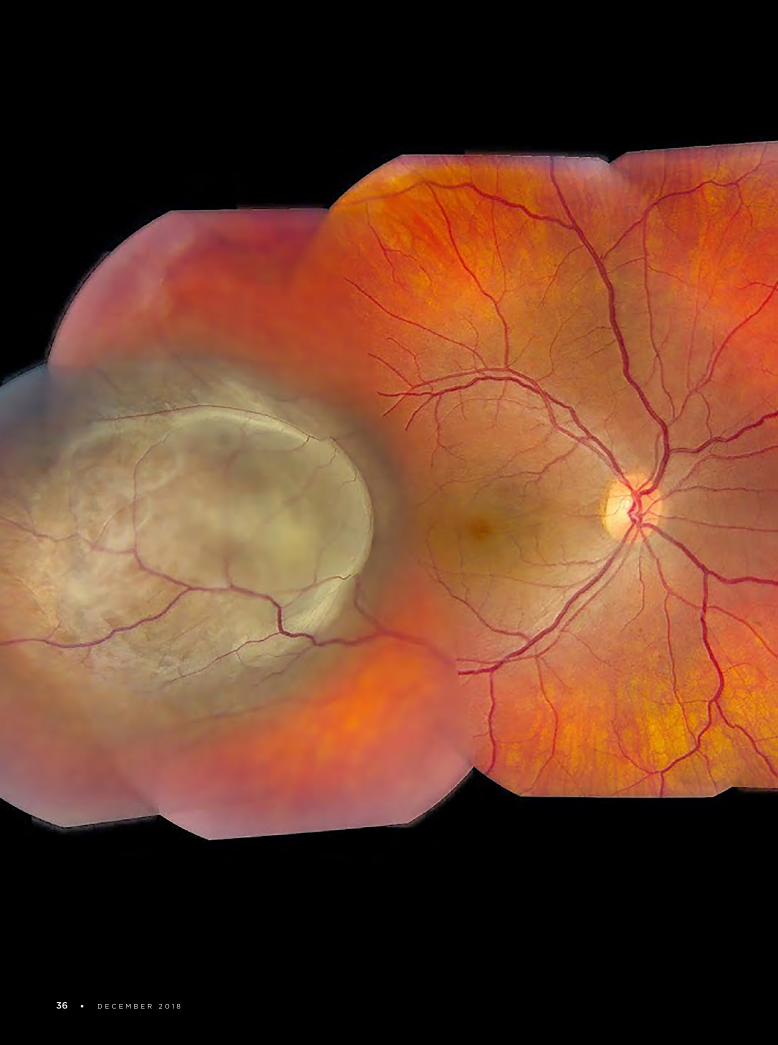
36 • D E C E M B E R 2 0 1 8
36-43_Feature_F.indd 36 11/15/18 11:07 AM

E Y E N E T M A G A Z I N E • 37
Perspectives on the Profession
By Sanjay Asrani, MD; Lylas G. Mogk, MD, and Mary Lou Jackson, MD; Zélia M. Corrêa, MD, PhD; and Mehdi Roozbahani, MD, and J. Bradley Randleman, MD
BECAUSE THE END OF THE YEAR IS A FITTING TIME TO TAKE
stock of recent events, EyeNet has asked a few of its board mem-
bers to review developments or trends in their areas of expertise
and to consider which of these has the greatest potential to shape
their subspecialty over the next several years. Interestingly, many
of their observations touched on—or focused squarely on—tech-
nologies or testing that may offer personalized patient care.
Below are their perspectives in the fields of glaucoma, low vision
rehabilitation, ocular oncology, and refractive surgery and corneal
ectatic disease.
Glaucoma diagnosis and management are both complex issues. This is because glaucoma is a group of diseases, not all of which are associated with raised intra-ocular pressure (IOP). Today, diagnosis is made by a pattern recognition of a typical optic nerve appearance and/or a
typical visual field defect. Visual field testing is problematic, however, because it is universally disliked by patients, and its fluctuating and subjective nature makes interpretation difficult for the ph y sician. Our one objective technique of diagnosis involves optical coherence tomography (OCT), which also has significant pitfalls, such as artifacts, relying on the OCT report, using it in isolation, etc. To state the obvious, there is room for improvement. Fortunately, new areas of innovation and investigation are opening up.
Objective assessments. The next frontier in diagnostics is in detecting glau-coma progression objectively. Many studies in progress involve using OCT early enough to initiate or advance treatment before visual field loss occurs or worsens. New technology for objectively measuring visual fields is being refined using head-mounted sets and virtual reality right here at Duke University. (See “The New World of Virtual Reality,” in the October EyeNet.) And the promise of DARC technology (Detection of Apoptosing Retinal Cells) using annexins to label dying/stressed ganglion cells lies in being able to detect the IOP level that is detrimen-
DR. ASRANI ON GLAUCOMA
Where Glaucoma Is Now, and Where It Is Going: A Survey
Jaso
n S
. Cal
ho
un
36-43_Feature_F.indd 37 11/15/18 12:32 PM
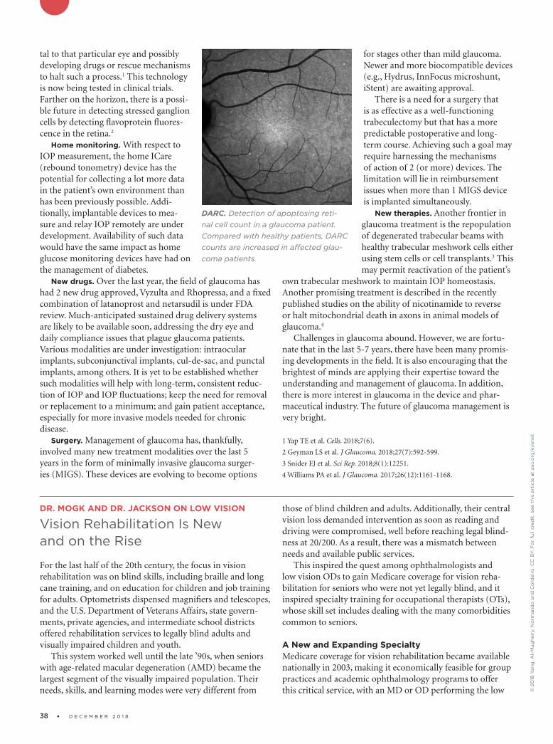
38 • D E C E M B E R 2 0 1 8
tal to that particular eye and possibly developing drugs or rescue mechanisms to halt such a process.1 This technology is now being tested in clinical trials. Farther on the horizon, there is a possi-ble future in detecting stressed ganglion cells by detecting flavoprotein fluores-cence in the retina.2
Home monitoring. With respect to IOP measurement, the home ICare (rebound tonometry) device has the potential for collecting a lot more data in the patient’s own environ ment than has been previously possible. Addi-tionally, implantable devices to mea-sure and relay IOP remotely are under development. Availability of such data would have the same impact as home glucose monitoring devices have had on the management of diabetes.
New drugs. Over the last year, the field of glaucoma has had 2 new drug approved, Vyzulta and Rhopressa, and a fixed combination of latanoprost and netarsudil is under FDA review. Much-anticipated sustained drug delivery systems are likely to be available soon, addressing the dry eye and daily compliance issues that plague glaucoma patients. Various modalities are under investigation: intraocular implants, subconjunctival implants, cul-de-sac, and punctal implants, among others. It is yet to be established whether such modalities will help with long-term, consistent reduc-tion of IOP and IOP fluctuations; keep the need for removal or replacement to a minimum; and gain patient acceptance, especially for more invasive models needed for chronic disease.
Surgery. Management of glaucoma has, thankfully, involved many new treatment modalities over the last 5 years in the form of minimally invasive glaucoma surger-ies (MIGS). These devices are evolving to become options
for stages other than mild glaucoma. Newer and more biocompatible devices (e.g., Hydrus, InnFocus microshunt, iStent) are awaiting approval.
There is a need for a surgery that is as effective as a well-functioning trabeculectomy but that has a more predictable postoperative and long-term course. Achieving such a goal may require harnessing the mechanisms of action of 2 (or more) devices. The limitation will lie in reimbursement issues when more than 1 MIGS device is implanted simultaneously.
New therapies. Another frontier in glaucoma treatment is the repopulation of degenerated trabecular beams with healthy trabecular meshwork cells either using stem cells or cell transplants.3 This may permit reactivation of the patient’s
own trabecular meshwork to maintain IOP homeostasis. Another promising treatment is described in the recently published studies on the ability of nicotinamide to reverse or halt mitochondrial death in axons in animal models of glaucoma.4
Challenges in glaucoma abound. However, we are fortu-nate that in the last 5-7 years, there have been many promis-ing developments in the field. It is also encouraging that the brightest of minds are applying their expertise toward the understanding and management of glaucoma. In addition, there is more interest in glaucoma in the device and phar-maceutical industry. The future of glaucoma management is very bright.
1 Yap TE et al. Cells. 2018;7(6).
2 Geyman LS et al. J Glaucoma. 2018;27(7):592-599.
3 Snider EJ et al. Sci Rep. 2018;8(1):12251.
4 Williams PA et al. J Glaucoma. 2017;26(12):1161-1168.
DR. MOGK AND DR. JACKSON ON LOW VISION
Vision Rehabilitation Is New and on the Rise
For the last half of the 20th century, the focus in vision rehabilitation was on blind skills, including braille and long cane training, and on education for children and job training for adults. Optometrists dispensed magnifiers and telescopes, and the U.S. Department of Veterans Affairs, state govern-ments, private agencies, and intermediate school districts offered rehabilitation services to legally blind adults and visually impaired children and youth.
This system worked well until the late ’90s, when seniors with age-related macular degeneration (AMD) became the largest segment of the visually impaired population. Their needs, skills, and learning modes were very different from
those of blind children and adults. Additionally, their central vision loss demanded intervention as soon as reading and driving were compromised, well before reaching legal blind-ness at 20/200. As a result, there was a mismatch between needs and available public services.
This inspired the quest among ophthalmologists and low vision ODs to gain Medicare coverage for vision reha-bilitation for seniors who were not yet legally blind, and it inspired specialty training for occupational therapists (OTs), whose skill set includes dealing with the many comorbidities common to seniors.
A New and Expanding Specialty Medicare coverage for vision rehabilitation became available nationally in 2003, making it economically feasible for group practices and academic ophthalmology programs to offer this critical service, with an MD or OD performing the low
DARC. Detection of apoptosing retinal cell count in a glaucoma patient. Compared with healthy patients, DARC counts are increased in affected glaucoma patients.
© 2
018
Yan
g, A
l-M
ug
hei
ry, N
orm
and
o a
nd
Co
rdei
ro. C
C B
Y. F
or
full
cred
it, s
ee t
his
art
icle
at
aao
.org
/eye
net
.
36-43_Feature_F.indd 38 11/13/18 7:58 AM

E Y E N E T M A G A Z I N E • 39
vision exam and OTs conduc ting rehabilitation training. Programs in academic departments continue to grow, both in number and size. Specialty training in vision rehabilita-tion for OTs is thriving, with The University of Alabama in Birmingham, Salus University in Pennsylvania, and Western Mich igan University offering advanced certificates for OTs. Many private agencies are now adding OTs to their staffs.
For awhile now, knowledge has been spreading beyond the low vision community. For example, the Academy has published a Focal Points, a Preferred Practice Pattern, and a coding module in vision rehabilitation. The Basic Clinical and Science Course series includes a newly revised chapter, and the ONE Network offers tutorials in vision rehabili-tation. The Academy annual meeting includes symposia, breakfast with the experts, and instruction courses in vision rehabilitation, sponsored by the Vision Rehabilitation Com-mittee, chaired by John D. Shepherd, MD.
Beyond that, an increasing number of academic programs are requesting Grand Rounds speakers in vision rehabilitation. Internationally, among its Priority Assistive Products List, the World Health Organization has included 9 low vision devic-es; it has published the International Consensus Document on Vision Rehabilitation Standards; and it is about to publish a vision rehabilitation curriculum for training personnel. Vision rehabilitation has come a long way in a short time.
TechnologyNot surprisingly, technology has been a cornerstone of con temporary vision rehabilitation. The original break-through technologies included video cameras with screens
(closed-circuit TV or video magnifiers). These technologies continue to be up-dated and are a mainstay for those with low vision. Talking devices, including watches, scales, timers, and liquid level indicators, have been available for years.
Technology for all vision. The big news in technology is that many of the most important devices for patients with vision loss are the same devices used by the sighted population: smart-phones, tablets, and computers. This makes them readily available and psy-chologically comfortable. • Computers, of course, are endlessly adaptable, with built-in accessibility features and software that can enlarge text or offer audio input and output. • E-readers, like Kindle, offer large print sizes and reverse polarity (white text on a black background). The latter is particularly helpful to the many low vision patients with decreased contrast sensitivity. Tablets offer magnification, text to speech, voice recognition, and photography without requiring precise focus.
• Smartphones include built-in powerful flashlights, mag-nification features, cameras, and voice recognition such as Siri, in addition to a bevy of apps that read text aloud, interpret bar codes, identify colors, read handwriting, and orally describe objects and people. These include, among others, TapTapSee for all smartphones, Seeing AI for iOS at no charge, and the KNFB Reader for a fee.• Smart speakers (e.g., Alexa, Google Home, or Echo) offer easy audio access to weather, news, music, and information for everyone. • Ride services, including Uber and Lyft, offer welcome travel options in the many places throughout this country with limited public transportation. Uber has instituted a “Go Go Granny” service that allows riders with low vision to phone in their description along with their pickup location, so the driver can identify them. • Fully autonomous vehicles will someday be a game changer for individuals with low vision. Currently, partially autonomous vehicles requiring a capable driver are in devel-opment.
Tech for impaired vision. Technologies designed specif-ically for those with visual impairments are bountiful and include but are not limited to the following: • Head-mounted devices for stationary use that magnify at varying distances offer a broad, clear view rather than the limited area viewed through a magnifier or telescope. These include, among others, the IrisVision, NuEyes, and E-Sight, each with its particular features and price points.• The OrCam is a camera mounted on an eyeglass frame that offers text to speech capability, as well as audio descrip-
LOW VISION APPS. With TapTapSee (left), the user takes a photo or video, and the app identifies the object aloud. Seeing AI (right) offers multiple “channels” that can read documents and identify objects, people, and scenes.
36-43_Feature_F.indd 39 11/15/18 12:33 PM

40 • D E C E M B E R 2 0 1 8
tions of objects and people. It is for stationary or mobile use.• A subscription service, called AIRA, connects the person with vision loss to a live agent via wearable smart glasses. The agent can see the user’s environment in real time, knows their location by GPS, and can assist with navigation, finding objects, or doing tasks.• For individuals with significant visual field loss and mobility issues, GPS in smartphones (e.g., Blindsquare) or as wearable systems that vibrate (iWatch) are available to assist with way finding.• Even white canes have become high tech. The SmartCane has ultrasound to detect objects. Other detection devices can be clipped on clothing (BuzzClip) or worn as glasses (iGlasses). • Smaller-scale technologies include audio medication labels (ScriptTalk) and Digital Accessible Information System (DAISY) with over 600,000 free/low cost formatted audio-books. Optical character recognition technology has made
freestanding text-to-speech scanning devices possible (e.g., the Sara), which reads printed material aloud.
Further down the road—but currently available to certain individuals who have total or near total blindness—is artifi-cial vision, being developed in centers around the world. The most advanced example of this is the Argus II Second Sight retinal prosthesis.
ConclusionVision rehabilitation is a vibrant, growing specialty. Point in fact, the Academy has declared referral to vision rehabilita-tion as the standard of care in ophthalmology. The more low vision programs that there are, the greater the benefit to patients and the greater the likelihood of those services being of high quality. Advances in technology, coupled with training to address the spectrum of daily activities, promise to change the experience and lighten the burden of visual impairment.
DR. CORRÊA ON OCULAR ONCOLOGY
Trending Toward Personalized Medicine
There is a lot of talk about personalized, or precision, medi-cine today. This is thanks to the exciting possibility of using available knowledge in molecular and cell biology, genetics, and genomics to evaluate each patient. Such information may help physicians with prevention, screening, and treat-ment strategies that may be more effective than those that are currently available. Another goal with personalized medi-cine is to find treatments with fewer side effects compared to standard options. Ultimately, by performing genetic tests on cancer cells and on normal cells, physicians may be able to customize treatment to each patient’s needs.
Since completion of the human genome sequencing a little over 15 years ago, light has been shed on hundreds of genes involved in the pathogenesis of diseases. In ophthal-mology, faulty genes are implicated in diseases ranging from rare hereditary syndromes to common conditions such as myopia, primary open-angle glaucoma, AMD, and ocular cancers.
Survival Prognosis in Uveal MelanomaUveal melanoma has probably been the biggest challenge for ocular oncologists due to its unpredictable nature and its ability to metastasize. Ample literature has been published on the clinical, histopathologic, and chromosomal features of uveal melanoma and their association with patient out-comes.1-3 Despite the fact that ocular oncologists have refined those features and studied them to exhaustion, old parame-ters have failed to provide a systematic understanding of this tumor’s biology.
Gene expression profiling. The identification of mono-somy 3 as a risk factor for development of metastatic disease in patients with uveal melanoma occurred more than 20
years ago,4,5 but the first significant step toward more precise identification of patient risk was made with high-density microarrays to study gene expression profiles (GEP) of these tumors.6-7 Researchers showed that uveal melanomas exist in 1 of 2 basic molecular forms that are intensely associated with metastatic proclivity: class 1 tumors have a low risk and class 2 tumors have a high risk of metastasis. Using sophis-ticated bioinformatic analyses, studies have shown that the genes expressed in class 1 tumors are similar to those in normal uveal melanocytes (derived from the neural crest), and genes expressed in class 2 tumors resemble primitive stem-like cells.
With this new information in mind, it seems likely that previously identified histopathologic risk factors for metas-tasis, such as epithelioid cell type and vasculogenic mimicry patterns, correlate with de-differentiated stem-like features of these tumors.7 This is just one example of how GEP discover-ies have provided new insights into the pathobiology of uveal melanoma. Subsequently, independent peer-reviewed pub-lications have shown that the prognostic accuracy of GEP outperforms clinicopathologic features and chromosomal gains and losses.6,8
Prognostic testing. While relatively new, prognostic testing is becoming an important part of risk stratification of patients with posterior uveal melanomas. Understanding the information obtained by each test and its application is fast becoming an important part of ocular oncology clinical practice. Currently, the 2 commercially available prognostic tests for uveal melanoma employ different platforms that consequently yield distinct results that should be viewed differently.9
Impact Genetics test. One of the prognostic tests is Impact Genetics Uveal Melanoma Prognostic Genetic Test (Impact Genetics).• How it works. The test involves multiplex ligand probe amplification to evaluate copy number on chromosomes 1p,
36-43_Feature_F.indd 40 11/13/18 7:58 AM

E Y E N E T M A G A Z I N E • 41
3, 6, and 8 to detect monosomy, disomy, and trisomy; microsatellite analysis on chromosome 3 to detect loss of a chromosome copy and isodisomy; and sequencing GNAQ, GNA11, SF3B1, and EIF1AX to detect frequently occurring mutations in uveal melanoma tumor for confirmation of tumor sampling where indicated.9 • Concerns. Before using this test, consider the following. This assay employs preferably FFPE specimens and requires larger quantities of tissue than the test below. Multiple articles describe this assay and its complex risk stratification as both challenging to apply in clinical practice and to explain to patients. There has been no prospective validation of this test. Another limitation to adopting this test: insurance coverage for patients incurring outofpocket costs.
Castle Biosciences test. The other test, UMDecisionDx (Castle Biosciences), is used to detect upregulation or downregulation of particular genes of interest in minute tissue samples.79 This test’s GEP technique mainly uses fineneedle biopsy samples that are too small to be reliably assessed using chromosomebased assays. It evaluates the expression of 12 discriminating genes and 3 control genes using quantitative polymerase chain reaction (PCR) on a microfluidics platform after targeted amplification. • Patient stratification. Through GEP assay of untreated uveal melanoma tissue samples, 2 distinct prognostic classes and 1 subdivision of these classes have been identified and shown to predict metastatic risk and strongly correlate with survival in patients with posterior uveal melanoma. Patients with class 1 tumor gene expression profiles have lowgrade tumors with a decreased risk of metastatic spread. Class 1 patients have been further subdivided into A and B according to their midterm prognosis. On the other hand, patients with class 2 tumor gene expression profiles have highgrade tumors with an increased tendency to metastasize.10
• A comparison with current testing. When compared to the presence of monosomy 3, and to clinical and pathologic tumor features, GEP demonstrated superior accuracy at predicting the risk of metastatic disease in patients with primary uveal melanoma. Importantly, because GEP is an RNAbased assay, it predicts how the tumor cells are likely to behave as far as metastatic spread, whereas DNAbased assays provide a snapshot in time of the genetic makeup of tumor cells.10 UMDecisionDx is the only prognostic test for uveal melanoma to undergo prospective multicenter validation, which is required for a cancer biomarker to achieve the highest level I evidence according to the National Comprehensive Cancer Network Task Force on cancer biomarkers and the Tumor Marker Utility Grading System. • PRAME is recent finding. Recently, researchers have shown that preferentially expressed antigen in melanoma
(PRAME) mRNA expression was ass o ciated with an increased risk of metastasis in both class 111 and class 212 melanomas. This test has been incorporated into the GEP test and has been reported to increase the accuracy of the assay.
Although survival of uveal melanoma patients remains poor, researchers are gaining important understandings of this disease. First, they now believe that prognostic mutations occur prior to primary tumor treatment. Second, driver mutations have been investigated and 3 appear to be mutually exclusive: Class 2 profile tumors are strongly associated with inactivating mutations in the BRCA1Associated
Protein 1 (BAP1) tumor suppressor gene; EIF1AX correlates with lowrisk class 1A signature tumors; and SF3B1 correlates with moderate risk class 1B tumors.12 These newly detected mutations are providing insight into the pathways that these tumors may take.
Treatment for Uveal Melanoma MetastasisDespite these recent discoveries in prognostication, personalized medical care to treat metastatic disease is underwhelming, but promising therapies are in the pipeline.
Available therapies. Current options to treat metastatic uveal melanoma include liverdirected chemotherapy, systemic and targeted immune therapies, and targeted therapy with T cells directed against tumorassociated antigens.13 Unfortunately, these treatments have yielded longlasting responses in very few patients. As for immunotherapy specifically, its potential efficacy has been limited, presumably owing to the small number of mutations leading to neoantigen expression in uveal melanoma; immunotherapy with checkpoint inhibitors has shown very low response rates in metastatic uveal melanoma.
Investigational therapies. A number of novel therapies based on supposed biological mechanisms are being investigated in the adjuvant setting. The growth factor receptors cMet and cKit are highly expressed in uveal melanoma and may play a role in metastatic progression.
Target: c-Met. Crizotinib is a tyrosine kinase inhibitor (TKI) shown to inhibit phosphorylation of cMet and in vitro migration of uveal melanoma cells. Interestingly, at doses that selectively inhibit cMet, crizotinib only marginally reduced the growth of established tumors, suggesting that other tyrosine kinase receptors such as epidermal growth factor receptor (EGFR) and insulin growth factor receptor 1 (IGFR1) are critical for uveal melanoma cell proliferation and survival.
Target: c-Kit. Sunitinib—another TKI that inhibits cKit, vascular endothelial growth factor receptor (VEGFR), and other receptors—yielded a 5year survival benefit (75% versus 55%) compared to matched controls in a retrospective study.
Both crizotinib and sunitinib are being evaluated in
UVEAL MELANOMA. Prognostic testing for uveal melanoma may help bring personalized medicine to patients.
Jaso
n S
. Cal
ho
un
36-43_Feature_F.indd 41 11/13/18 7:58 AM

42 • D E C E M B E R 2 0 1 8
ongoing adjuvant trials [ClinicalTrials.gov identifiers: NCT02223819, NCT02068586].
Histone inhibitor. Because BAP1 has been shown to regulate melanocytic differentiation by modifying histones, researchers are looking into personalized trials using histone deacetylase (HDAC) inhibitors for class 2 tumors (BAP1 mutation).14 Based on the potential role for HDAC inhibition in the adjuvant setting, valproic acid and vorinostat are being evaluated in ongoing trials [ClinicalTrials.gov identifiers: NCT02068586, NCT01587352].
Others. Another promising targeted therapy involves the aberrant hypomethylation of PRAME that can be activated in uveal melanomas and is associated with increased meta-static risk in both GEP classes. Finally, various other strate-gies are being investigated including immune-based thera-pies such as immune checkpoint inhibition and autologous dendritic cell vaccines.
ConclusionIn summary, exciting new research is showing that the treatment of metastatic uveal melanoma will likely involve
personalized management strategies based on genetic testing. As we look even further into the future, new progress in looking at the peripheral blood of melanoma patients may represent another frontier on the prognostic evaluation of these patients.
1 Augsburger JJ, Gamel JW. Cancer. 1990;66(7):1596-1600.
2 McLean IW et al. Ophthalmology. 1995;102(7):1060-1064.
3 Gamel JW et al. Invest Ophthalmol Vis Sci. 1992;33(6):1919-1922.
4 Prescher G et al. J Natl Cancer Inst. 1990;82(22):1765-1769.
5 Prescher G et al. Lancet. 1996;347(9010):1222-1225.
6 Worley LA et al. Clin Cancer Res. 2007;13(5):1466-1471.
7 Onken MD et al. Cancer Res. 2004;64(20):7205-7209.
8 Petrausch U et al. Eye (Lond). 2008;22(8):997-1007.
9 Onken MD et al. Ophthalmology. 2012;119(8):1596-1603.
10 Schopper V, Correa ZM. Int J Retin Vitr. (2016) 2:4. doi: 10.1186/s40942-
016-0030-2.
11 Field MG et al. Clin Cancer Res. 2016;22(5):1234-1242.
12 Field MG et al. Oncotarget. 2016;7(37):59209-59219.
13 Gezgin G et al. JAMA Ophthalmol. 2017;135(6):541-549.
14 Moschos MM et al. Anticancer Res. 2018;38(7):3817-3824.
DR. ROOZBAHANI AND DR. RANDLEMAN ONREFRACTIVE SURGERY AND CORNEA
Corneal Biomechanics for Ectasia
Advances in direct corneal biomechanical measurements have the potential to change both the paradigms for diag-nosis and treatment of ectatic disorders, such as keratoco-nus, and for ectasia screening for refractive surgery. These conditions are tightly intertwined, and the demand for these services is growing.
The prevalence of myopia is expected to double and affect up to 50% of the U.S. and other populations worldwide by 2050,1 and keratoconus—the leading cause of full-thickness corneal transplantation in the United States—is up to 10 times more prevalent than the previously reported figure of 1 in 2000.2 Thus, in the coming years, more patients will be presenting for refractive surgical correction and will require optimal screening for surgical candidacy. Meanwhile more keratoconus patients can benefit from corneal cross-linking (CXL), especially if they are identified at the earliest stages of disease, when they can get the greatest benefit from the procedure. Corneal imaging, and ultimately corneal biome-chanics, lie at the heart of optimizing care for both patient groups (Fig. 1A-1C).
Biomechanical failure is widely recognized to be the root cause of keratoconus,3 and it is the leading concern for refractive surgery screening for ectasia risk.4 Unfortunately, the refractive surgery screening and keratoconus testing available today measure later-onset morphologic surrogates, such as corneal curvature and thickness, rather than making primary, direct biomechanical measurements of the cornea. Because of this, many otherwise good candidates are refused
surgery because they have an atypical corneal curvature, when in fact they would have done well with surgery. But this is changing.
Current in vivo Measurement of Corneal Biomechanical PropertiesThe ocular response analyzer (ORA, Reichert Technolo-gies), which has been available for many years, is a dynamic bidirectional applanation device that quantifies corneal deformation after air puff–stressing. It measures 2 primary biomechanical variables, corneal hysteresis and corneal resis-tance factor. These variables have been thoroughly studied, and while they perform well at distinguishing between keratoconic and normal populations, they perform poorly at detection of early ectatic disease. Custom variables derived from comprehensive signal analysis have performed better,5 but further work is necessary to determine the ultimate utili-ty of ORA for screening.
More recently, air puffs have also been combined with Scheimpflug imaging (CorvisST, Oculus Optikgeräte) to evaluate corneal response to perturbation.6 The CorvisST couples with the Scheimpflug camera to provide cross-sec-tional corneal images during the indentation cycle. This provides a host of parameters that can be evaluated. To date, results are mixed in terms of screening utility for subclini-cal keratoconus, with some authors finding best results by combining data from the CorvisST with various Scheimpflug parameters.7
What Does the Future Hold?A variety of different technologies are being explored to as-sess their utility in directly measuring corneal biomechanics. These include optical coherence tomography–based elastog-raphy,8 ultrasound methods,9 and Brillouin microscopy.10-12
36-43_Feature_F.indd 42 11/13/18 7:58 AM

E Y E N E T M A G A Z I N E • 43
Scarcelli and Yun developed Brillouin microscopy as an all-optical, nonperturbative mechanical process that pro-vides high-resolution 3-D corneal mapping. In preliminary analyses, Brillouin microscopy has been able to differentiate normal and keratoconic corneas in vivo and has demonstrat-ed the focal nature of keratoconus, with the conical region demonstrating significant reduction in corneal stiffness, while the nonconical regions appeared similar to normal control corneas.11 Brillouin microscopy has also proved to be sensitive to identifying the stiffening effects of CXL ex vivo.12
ConclusionDirect and accurate corneal biomechanical measurements could create a paradigm shift in the way corneas are evaluat-ed in a large variety of clinical settings. Such measurements
may aid in early identification of corneal ectasia, thereby improving the management of keratoconus and making screening for refractive surgery even more precise. Moreover, the process of correlating biomechanical profiles and mor-phological behavior of the cornea, combined with the devel-opment of a predictive modeling based on this connection, could ultimately lead to completely individualized refractive procedures and patient-specific CXL treatment protocols.13
1 Holden BA et al. Ophthalmology. 2016;123(5):1036-1042.
2 Godefrooij D et al. Am J Ophthalmol. 2017;175:169-172.
3 Scarcelli G et al. Invest Ophthalmol Vis Sci. 2014;55(7):4490-4495.
4 Randleman JB et al. Ophthalmology. 2008;115(1):37-50.
5 Dupps WJ Jr., Seven I. Trans Am Ophthalmol Soc. 2016;114:T1-1-T1-16.
6 Valbon BF et al. J Refract Surg. 2014;30(7):468-473.
7 Vinciguerra R et al. J Refract Surg. 2017;33(6):399-407.
8 Ford MR et al. J Biomedical Optics. 2011;16(1):016005.
9 He X, Liu J. Invest Ophthalmol Vis Sci. 2009;50(11):5148-5154.
10 Scarcelli G, Yun SH. Nat Photonics. 2007;2:39-43.
11 Scarcelli G et al. JAMA Ophthalmol. 2015;133(4):480-482.
12 Scarcelli G et al. Invest Ophthalmol Vis Sci. 2013;54(2):1418-1425.
13 Seven I et al. Invest Ophthalmol Vis Sci. 2016;57(14):6287-6297.
MEET THE EXPERTSSANJAY G. ASRANI, MD Professor of ophthal-mology and head of the Glaucoma OCT Read-ing Center, Duke University at the Duke Eye Center in Durham, N.C. He is also medical direc-
tor of the Duke Eye Center of Cary, N.C. Relevant financial disclosures: Aerie: C; Bausch + Lomb: C; Camras Vision: C; Noveome Biotherapeutics: C; Regenexbio: C.
ZÉLIA M. CORRÊA, MD, PHD The Tom Clancy Professor of Ophthalmology at the Wilmer Eye Institute in Baltimore. Relevant financial disclo-sures: Castle Biosciences: C.
MARY LOU JACKSON, MD Clinical associate pro-fessor of ophthalmology and visual sciences at the University of British Columbia, Vancouver. Relevant financial disclosures: None.
LYLAS G. MOGK, MD At the Center for Vision Rehabilitation and Research at Henry FordHealth System in the Greater Detroit area. Relevant financial disclosures: None.
J. BRADLEY RANDLEMAN, MD Professor of ophthalmology; director, cornea, external dis-ease, and refractive surgery; and medical direc-tor of the University of Southern California (USC) Roski Eye Beverly Hills Clinic. Relevant financial disclosures: None.
MEHDI ROOZBAHANI, MD Cornea fellow at USC Roski Eye Institute in Los Angeles. Relevant financial disclosures: None. See the disclosure key, page 8. For full disclosures, see this article at aao.org/eyenet.
KERATOCONUS. Representative images from patients with highly asymmetric keratoconus (1A: affected eye, 1B: clinically unaffected eye) and a normal control eye (1C). While kerato-conus can be identified using a variety of devices, identifying eyes at the earliest stages of disease remains challenging, even when using multiple technologies.
1A 1B 1C
Hw
ang
ES
et
al. O
ph
thal
mo
log
y. P
ub
lish
ed a
hea
d o
f p
rin
t Ju
ly 2
5, 2
018
.
36-43_Feature_F.indd 43 11/13/18 7:58 AM

CODING & REIMBURSEMENT
2019 editions now available
American Academy of Ophthalmic Executives®
Code With Confidence Every DayMaximize reimbursements and protect your practice from audits. The most-trusted suite of precision coding tools is just a click away.
Order Today aao.org/codingproducts 866.561.8558
00_Ads_F.indd 44 11/13/18 5:27 PM

E Y E N E T M A G A Z I N E • 45
BUSINESS OPERATIONS & FINANCE
PRACTICE PERFECT
4 Ways to Boost Patient Satisfaction
Patient care is the major reason many people go into health care. But dramatic changes in the field
over the past decade have added com-plexity to ophthalmic practices. Today, so much time is required to maintain electronic health records and comply with regulations that it’s difficult to prioritize the patient experience.
In my 20-plus years as an ophthal-mic administrator, I have developed the following 4 principles for focusing on the patient care experience. Although these tips may seem basic, we often lose sight of the obvious in an attempt to get through busy days. In a health care en-vironment where patients are increas-ingly concerned about out-of-pocket expenses, the winners in the future will be those practices that maintain a laser focus on the patient.
1. Commit to a Practice Culture That Values Time Spent With PatientsMany practices are adopting the lean management approach (aao.org/lean) as an excellent way to increase pro-ductivity by reducing inefficiency and eliminating long wait times. The foundational principle of lean man-agement is designing practice processes with patients in mind and putting their experience at the core of what we do.
First and foremost, management must value staff who take the extra time to help patients. If we say we value
empathic care and then write up an employee for not working fast enough, we are sending mixed messages. We must all share the vision of exception-al patient service, and our employee feedback and rewards system must be consistent with that vision.
I once had a tech supervisor who focused on benchmarking the average technician workup time. The highest- performing technician by this measure, however, was not fully reconciling med-ications or performing diagnostic tests as ordered. The supervisor’s focus on this single metric (workup time) meant that our physician often had to perform what should have been technician work. Ultimately, this was not good for patient satisfaction because patients took longer to complete the physician portion of the exam and could see that the team was not working as cohesively or efficiently as planned. We learned to use this metric of workup time in combination with patient and physi-cian feedback.
Both administrative and physician leaders must agree on a vision and lead in a consistent manner. Here are a few guidelines:• When considering a process im-provement, evaluate the possible im-pact on the patient care experience. • When the practice has efficiency standards or benchmarks, always allow ranges of productivity to enable staff to add the extra level of service that you
would want for yourself or your family member. • Always evaluate the impact of any process change on the entire patient care team.
2. Recruit the Right People and Hire Enough StaffGreat support staff may not show up on the balance sheet, but they make a dramatic impact on the bottom line. They are the front line in creating a pa-tient-centric culture. One of the biggest barriers to achieving a consistent level of patient care is an understaffed office. Although practices want to reduce costs, there is a downside to chronic understaffing, which can be defined in terms of skill set and workload.
Skill set. Finding and hiring the right staff is the hardest part of a manager’s job. The right staff are those who have the emotional intelligence to handle interpersonal relationships judiciously and empathetically. You can teach hard skills, but soft skills—such as good listening and communication and a team-oriented work ethic—are aptitudes some individuals naturally excel at and enjoy. These are the core traits to target in new hires.
Although many human resources screening tests assess emotional intel-ligence, I developed a simple observa-tional screening strategy for these soft skills and used it when interviewing applicants. I personally walked the candidate around the practice, explain-ing what happened in each area of the office, and I made sure to lead them through busy patient areas to see how
BY SARA BURNS RAPUANO, MBA, OCS, PRACTICE MANAGEMENT CONSULTANT.
45-46_PP_F.indd 45 11/13/18 7:38 AM
46 • D E C E M B E R 2 0 1 8
they interacted with patients in the hallways and elevators. In particular, I took note of the following:• Did they hold the door for patients?• Did they make eye contact with patients?• Did they step aside when someone with a walker or a baby carriage passed?
Many applicants naturally noticed the patients and interacted well, while others bumped into elderly patients and were unaware of their visual chal-lenges. Smart, empathetic staff, who are given the time and resources to provide excellent service, will bring new busi-ness and increase patient retention.
Workload. Be aware of the amount of work each employee has. In addition to hiring the right employees, we still need to monitor the workload in each area of the practice to ensure the team is the right size. Patients can readily discern when an employee is over-worked and the practice environment is stressed. Overworked employees have a compromised ability to listen to patients and take the necessary time and initiative to respond to their needs. Stress may also trigger mistakes in registration or patient triage.
If your staff is too busy to help a visually challenged patient complete his or her paperwork, assist a confused patient with obtaining a necessary referral, or listen carefully to a patient with a potentially serious medical concern, then the office is understaffed. The staffing should be adequate to allow the team the opportunity to help patients and always provide excellent service. Supervisors and managers can model this behavior every day.
Once we find good employees, we need to create an environment where
they stay and thrive. Practices often add physicians and increase patient volume without increasing the support staff. It is critical that we do not burn out employees.
3. Measure Patient Satisfaction Through Regular SurveysTo provide an excellent patient expe-rience, we first need to measure how patients perceive our office. One of the best ways to gather this informa-tion is through a patient satisfaction survey. Although there are standardized surveys, such as Clinician and Group Consumer Assessment of Healthcare Providers (CG CAHPS), they tend to be more appropriate for a primary care setting. Ophthalmology practices may want to devise a customized pa-tient satisfaction survey. These can be developed either in-house or through a vendor. They can provide valuable insight about how patients see your environment—and sometimes survey results are different than expected.
When I instituted a regular patient satisfaction survey in the Wills Eye resident clinics, I expected to hear about our lengthy wait times and chal-lenging parking situations. Although a few patients noted those problems, these comments did not impact overall patient satisfaction ratings. Many saw the lengthy appointments as thorough visits. When patients did complain about wait times, they focused on a lack of communication, such as not having been advised of the expected wait time.
What I found most useful were the notes and patient stories in the com-ments area at the end of the survey. These often highlighted staff interac-tions. One patient took the time to say how helpful and compassionate the patient assistance coordinator was. This staff member handles both charity patients and VIP patients at Wills Eye, and she ensures that both groups get the same level of care. Knowing how valuable this employee was to 1 patient gave me insight I never would have had without the survey.
This qualitative feedback was my favorite management tool. Sharing the positive feedback is as important as sharing the constructive criticism.
I also used these stories when training new staff to help them understand the importance of communication skills.
4. Maintain Your Patient Service GoalsThe last element of my strategy to cre-ate an exceptional patient care experi-ence is to maintain the practice’s focus on the patient. Management does not just devise a plan, implement it, and move on. Strong leaders realize that this is an iterative process.
We must use the feedback we receive from patients, referring doctors, and staff to identify areas for improvement. We must also follow through by taking corrective action with employees who do not focus on the patient, as well as reward the staff who consistently pro-vide the highest level of care. Rewards can take many forms. Some are mone-tary, and some are simply making sure that staff know they are valued every day. We demonstrate that we value our employees when we:• share positive comments about their performance from patients and providers;• provide ongoing education to ad-vance their knowledge base;• acknowledge their contribution to practice improvements;• ensure they get an adequate lunch break; and • ensure they are not asked to work late every day.
Managers and doctors must always hire, train, and retrain with patient service goals in mind. And managers and doctors must be reviewed on their ability to support their staff in these goals.
ConclusionWhen practices take these 4 steps, they will find that patient online reviews improve and physician referrals and volume of return patients increase. In addition, employees will be more satis-fied with their jobs, potentially resulting in lower staff turnover.
Ms. Burns Rapuano is a practice management
consultant in Philadelphia who works with the
AAOE. Financial disclosures: Avedro: C,L; Wills
Eye Hospital: C,L.
AAOEAdapted from “How Do You Achieve Patient Service Excellence in Today’s Ophthalmic Practice?” which was published Sept. 24, 2018, by the American Academy of Ophthalmic Executives (AAOE), the practice management arm of the Academy. Learn more about AAOE at aao.org/aaoe.
45-46_PP_F.indd 46 11/13/18 7:38 AM

NEW Accurate Coding
IMPROVED Knowledge Base
Experience IntelleChartPRO
NEW Assisted Compliance
IntelleChartPRO
IntelleChartPRO
Nextech.com/Ophthalmology(800) 868-3694
00_Ads_F.indd 47 11/8/18 8:20 PM

AAO 2019
aao.org/2019
Inspire!
Submit an AbstractInstruction Courses and New Skills Labs December 13, 2018 – January 8, 2019
Papers/Posters and Videos March 7, 2019 – April 9, 2019
Join Us in San Francisco
AAO 2019 October 12 – 15
Subspecialty Day October 11 – 12
AAOE Program October 11 – 15
Blaze the trail to a future built on technology and innovation – or master the teachings from ophthalmic visionaries.
Watch this short video at aao.org/2018 to celebrate AAO 2018.
Where All of Ophthalmology Meets®
00_Ads_F.indd 48 11/13/18 5:15 PM

E Y E N E T M A G A Z I N E • 49
Academy NotebookN E W S • T I P S • R E S O U R C E S
WHAT’S HAPPENING
State Societies HonoredOn Oct. 28 during AAO 2018, the Academy Secretariat for State Affairs recognized 3 state ophthalmology soci-eties with its Star Award for outstand-ing efforts on programs or projects implemented in the previous year. The winning societies are as follows:
Kentucky Academy of Eye Physi-cians and Surgeons for its Advocacy Campaign to Protect Telemedicine, which opposed legislation that would have limited Kentucky physicians’ right to evaluate and adopt new technologies that might improve patient care.
Puerto Rico Society of Ophthal-mology for its Hurricane Maria Relief Effort, which coordinated ophthalmol-ogist volunteers to deliver much-need-ed eye care.
Washington Academy of Eye Phy-sicians and Surgeons for its Military Outreach Project to increase member-ship as well as participation of active- duty military ophthalmologists in the society’s programs and services.
Since the Star Award program’s inception in 2001, the Secretariat for State Affairs has recognized 67 state ophthalmology society programs. State ophthalmology societies may apply for this award by responding to the Secretariat for State Affairs’ survey of
state societies, emailed every summer to state society executive directors/ad-ministrators and presidents.
State Societies’ Outstanding Executive DirectorsEach year, the Academy Secretariat for State Affairs publicly honors select state ophthalmology society executive directors for their contributions to their societies and for their partnership and collaboration with the Academy on its national efforts. During AAO 2018 in Chicago, the Secretariat recognized Nanette R. Gilbertson of the Montana Academy of Ophthalmology and Eliza-beth G. Roach of the Kentucky Acad-emy of Eye Physicians and Surgeons. They received the 2018 Outstanding Executive Director awards in Organi-zational Development and in Political Action, respectively.
The Academy Secretary for State Affairs, Kurt F. Heitman, MD, praised the efforts of all executive directors on behalf of state societies and ophthal-
mologists across the country. “Through their professionalism, energy, and commitment to ophthalmologists and their patients, state society executive directors help to elevate the profession. We in State Affairs appreciate their expertise and respect their dedication to preserving quality eye care.”
TAKE NOTICE
Nominate a Colleague for the Laureate Award Every year, ophthalmologists distinguish themselves and the profession by mak-ing exceptional scientific contributions toward preventing blindness and re-storing sight worldwide. The Academy Board of Trustees will recognize these extraordinary contributions with its Laureate Award, the Academy’s single highest honor.
The award recipient is announced each fall, and the Laureate is recog-nized during the Opening Session of the annual meeting.
STARS OF PUERTO RICO. The Puerto Rico Society of Ophthalmology (PROS) was awarded a 2018 State Affairs Star Award during AAO 2018 for its Hurricane Maria Relief Effort (HMRE). From left to right: Carmen E. Amaral, MD (HMRE Project Co-ordinator), Jackie Del Valle (PROS Executive Director), Lorna A. Vargas, MD (HMRE Project Coordinator), Wandsy M. Vélez, MD (PROS President), John D. Peters, MD (Academy Associate Secretary for State Affairs), and Kurt Frederick Heitman, MD (Academy Secretary for State Affairs).
49-51_Note_F.indd 49 11/13/18 7:44 AM

50 • D E C E M B E R 2 0 1 8
Nominate a colleague using the application at aao.org/about/awards/laureate by Jan. 31, 2019.
Need a Holiday Gift Idea? Donate to the Foundation This is the perfect time of year to make a gift to the Academy Foundation in honor or memory of a mentor, col-league, or family member. Donations at the Partners for Sight level ($1,000-$2,499) are especially encouraged. Your tax-deductible donation will be used to support the Academy programs that are important to you, including the Museum of Vision campaign to build a permanent space for the 38,000-piece collection at Academy headquarters. Be sure to make your gift by Dec. 31 to receive the tax deduction for 2018.
Learn more at aao.org/foundation.
Glaucoma Researcher?This summer, the Academy and the American Glaucoma Society collab-orated in launching Ophthalmology Glaucoma. This new journal provides an opportunity to disseminate your glaucoma research directly to those who find it most relevant. Joining the ranks of Ophthalmology and Ophthal-mology Retina, Ophthalmology Glauco-ma provides readers with innovative, peer-reviewed works.
Submit your research at https://www.evise.com/profile/#/OGLA/login. Subscribe at www.ophthalmologyglau coma.org.
Meet These MIPS DeadlinesDec. 31: Deadline for EHR hardship exception. In the Merit-Based Incentive Payment System (MIPS), the electronic health record (EHR)–based perfor-mance category is called promoting interoperability (PI). It is 1 of 4 MIPS performance categories and contrib-utes up to 25 points to your MIPS final score (0-100 points). Typically, if you were to report no PI measures, your PI score would be zero and your maximum MIPS final score, 75.
The significant hardship exception. You can apply to be exempted from PI if you are facing a significant hard-ship, such as insufficient internet con-nectivity or extreme and uncontrollable
circumstances. If the Centers for Medicare & Medi-
caid Services (CMS) accepts your appli cation for a hardship exception, PI’s contribution to your final score will be reweighted to zero, and the quality performance category’s con-tribution will be reweighted upward; thus you could still earn the maximum MIPS final score of 100 points despite not reporting any PI measures.
New for 2018: If small practices can demonstrate that obtaining and maintaining certified EHR technology would cause undue hardship, CMS may grant them a PI hardship exception.
For guidance on submitting this application, see aao.org/medicare/ad vancing-care-information-exceptions.
Jan 15: Deadline for IRIS Registry submissions. If you are using the IRIS Registry (Intelligent Research in Sight) to report MIPS, Jan. 15 is a key date on 2 counts.
1. Finish manual entry. This dead-line applies if you are using the IRIS Registry web portal to manually report quality measures, PI measures, or improvement activ ities. If you successfully integrated your EHR with the IRIS Registry, your MIPS quality data are automatically extracted, but you can only report PI measures and improvement activities manually.
New for 2018: If you are manually reporting patients for a quality measure, you must submit to the IRIS Registry the total number of patients eligible, excluded, and except-ed from that measure.
2. Submit a signed data-release consent form. The IRIS Registry won’t submit a provider’s MIPS data to CMS unless it has received the signed consent form. Providers who are reporting as individuals should sign their own consent forms; providers who are reporting as a group can be included on a single consent form, which can be signed by the administrator. You must submit a new consent form each year and can do so via the IRIS Registry dashboard. For instructions, see aao.org/consent-form.
Learn more about the IRIS Registry
and MIPS at aao.org/iris-registry and aao.org/medicare.
MEMBERS AT LARGE
Troutman PrizesTroutman Cornea Prize for Young Clinician Investigators. This award, established by a Castroviejo Cornea Society Founder, Richard C. Troutman, MD, DSc (Hon), is awarded annually to the investigator under 41 years of age who authored the best paper published in Cornea the year before.
This year’s recipient was Gregory Moloney, BScMed (Hon), MBBS, MMed, FRANZCO, FRCS, an ophthal-mologist at Sydney Eye Hospital in Sydney, Australia, specializing in cata-ract, corneal, and oculoplastic surgery. His paper, “Descemetorhexis Without Grafting for Fuchs Dystrophy-Supple-mentation With Topical Ripasudil,” investigates the effect of topical ROCK (Rho-kinase) inhibitor as an adjuvant to the descemetorhexis procedure. Dr. Moloney was awarded a $5,000
honorarium from the Trout-man Endowment and had
the opportunity to present his work at the annual scientific meeting of the Cornea Society prior to AAO 2018.
Dr. Moloney said, “I am extremely honored to receive the Troutman award, which recognizes
many people’s work at Sydney Eye Hos-pital. This work would not have been possible without the efforts of our eye bank staff, fellows, and clinicians, and the funding provided by the Sydney Eye Hospital Foundation. We hope to continue this research to find more treatment options for patients with Fuchs dystrophy.”
Richard C. Troutman, MD, DSc (Hon) Prize. This prize is awarded on behalf of the International Society of Refractive Surgery to a young author published in the Journal of Refractive Surgery.
This year’s recipient, Yumeng Wang, MBBS, MMed, PhD, is a postdoctoral fellow specializing in cornea and glau-coma at the Department of Ophthal-
Dr. Moloney
49-51_Note_F.indd 50 11/13/18 4:22 PM

E Y E N E T M A G A Z I N E • 51
mology and Visual Sciences in the Chinese University of Hong Kong. Her paper, “Histological and Micro RNA Signatures of Corneal Epithelium in Keratoconus,” discusses the histopa-thology of keratoconic corneal epithelia and its micro-ribonucleic acid (miR-
NA) regulation as compared to corneal epithelia of normal eyes. Dr. Wang received a $5,000 hon-orarium from the Troutman Endowment and presented an honorary lecture
during Refractive Surgery Subspecialty Day 2018.
Dr. Wang said, “Receiving the Trout-man Award is a true honor for both myself and our team at the Department of Ophthalmology and Visual Sciences. We look forward to more opportuni-ties to further investigate diagnostic options for keratoconus patients.”
ACADEMY RESOURCES
Position Your Practice for SuccessWith the health care industry continu-ing to shift dramatically, the Academy will host its second annual, business- focused “boot camp” designed to address the complex challenges facing ophthalmic practices. Attend the Ophthalmology Business Summit from March 23-24, 2019, in Chicago.
This event is now open to both physician leaders and practice admin-istrators.
For more information, visit aao.org/business-summit.
Get 10% Off Patient Educa-tion Brochures Until Dec. 31Save time, improve recall, and mitigate malpractice risk by giving your patients easy-to-understand, ophthalmologist- reviewed brochures from the Academy. Now through Dec. 31, get 10% off when you use code PEB2018. Take advantage of this limited-time offer. No minimum purchase is required.
Order today by visiting aao.org/
patientbrochures, or by contacting Member Services at 415-561-8581, 866-561-8558 (U.S. toll free), or mem [email protected].
Register for 2019 Ophthal-mology Coding Update WebinarStay up to date on coding changes and audit regulations with the Academy’s most popular annual webinar. This year’s 2019 Ophthalmology Coding Updates takes place on Jan. 15. Sue Vicchrilli, Academy Director of Coding and Reimbursement, and David Glasser, MD, Academy Associate Secretary of Health Policy, will present.
For practice management webinars, visit aao.org/webinars.
MEETING MATTERS
Submit a Practice Manage-ment CourseEach year, the American Academy of Ophthalmic Executives (AAOE) offers a wide range of new courses as part of the AAOE program during the Acad-emy’s annual meeting.These sessions address current practice management challenges. You can have a hand in creating this content by submitting an
instruction course abstract between Dec.13, 2018, and Jan. 8, 2019.
To submit, visit aao.org/abstracts. For more information, contact Licia Wells, AAOE Program Manager, at [email protected].
Submit an Instruction Course or New Skills Lab Want to create content for AAO 2019? Submit your ideas for an instruction course or new Skills Transfer lab. Ab-stracts will be accepted from Dec. 13, 2018, through Jan. 8, 2019.
To submit, visit aao.org/presenter central.
Claim CME for AAO 2018 AAO 2018 and Subspecialty Day reg-istrants whose attendance was verified onsite in Chicago received an email with instructions for claiming Continu-ing Medical Education (CME) credits online. Starting Thursday, Dec. 13, attendees can claim credits (if they did not already do so onsite) and obtain transcripts with overall credits earned at aao.org/cme-central. The Academy transcript will not list individual course attendance.
For more information, visit aao.org/annual-meeting/cme.
D.C. REPORT
Yearlong Academy Effort Succeeds in Restoring Vitrectomy Codes The Centers for Medicare & Medicaid Services (CMS) has officially reinstated a majority of previously deleted ICD-10 codes to the Vit-rectomy National Coverage Determination (NCD 80.11). This mitigates CMS’ decision in October 2017 to authorize the deletion of 25% of vit-rectomy’s diagnosis codes, including those for vitreous hemorrhage, macular hole, and macular pucker. The 2017 decision was part of an agency effort to clean up its volume of ICD-10 codes associated with national coverage decisions.
Nearly a year ago, the Academy successfully took immediate ac-tion to halt implementation of the code deletions. Next, the Academy worked with the CMS Coverage Team to restore the vast majority of appropriate diagnoses. As a result of these efforts, CMS is putting forward a new vitrectomy coverage policy that ensures that all appro-priate diagnoses are covered as of Jan. 1, 2019.
If you are still experiencing denials from Medicare Advantage plans or have previously denied claims that are still unpaid, the Academy urges you to forward the information to [email protected].
Dr. Wang
49-51_Note_F.indd 51 11/14/18 9:57 AM

Access Innovative Tools to Enhance Quality Eye CareRenew your Academy membership to take advantage of these unparalleled ophthalmic resources.
IRIS® Registry (Intelligent Research in Sight) — Benchmark your practice against 16,000+ other ophthalmologists using medicine’s largest clinical database. In 2020, qualifying participants who reported MIPS successfully in 2018 will save an average of $22,250 through penalties avoided.
Ophthalmology® comes to you monthly, packed with the latest research and clinical updates (a $542 value).
EyeNet® Magazine delivers 12 issues of the latest industry news, plus clinical roundups and in-depth supplements such as the MIPS 2018: A Primer and Reference (a $150+ value).
Activate your benefits and renew your valuable membership today. aao.org/benefits
Diana R. Shiba, Academy fellow since 2010, shares an uplifting moment with her patient. The Academy’s IRIS Registry aggregates patient data to facilitate new scientific discoveries.
00_Ads_F.indd 52 11/13/18 5:17 PM

The world of healthcare is evolving. With increased pressure to reduce costs and improve the patient experience, our need for quality medicine is greater than ever before. That’s why we are committed to expanding our product lines to meet the needs of more patients.
Visit leiters.com to learn more about high-quality, cost e�ective ophthalmic services, including:
Repackaged Avastin® that is compliant with the new FDA guidance1
Moxifloxacin, now available in 2 concentrations (1 mg/mL or 5 mg/mL)
COMPOUNDING HEALTH™
1 Mixing, Diluting, or Repackaging Biological Products Outside the Scope of an Approved Biologics License Application Guidance for Industryhttps://www.fda.gov/downloads/drugs/guidances/ucm434176.pdfAvastin® is a registered trademark of Genentech, Inc.
Your journey to better medicine begins here.
Your one-stop shop for the following:
• In-depth clinical information in Pearls, Clinical Updates, and Features.
• Bite-sized research summaries in News in Review and Journal Highlights.
• Intriguing mystery cases in Morning Rounds and Blink.
• Practice management tips from the experts in Practice Perfect and Savvy Coder.
• Thought-provoking editorials in Opinion and Current Perspective.
Visit Us Onlineaao.org/eyenet
Write to [email protected]
EyeNet Gives You the Full Picture Poll-topping, digestible coverage of all things ophthalmologic
Facial TransplantsMaximizing Periocular Results
DMEK Enters the Mainstream
OPINION
Why Consensus Statements Matter
F E B R U A R Y 2 0 1 8
EyeNet®
MIGS RoundupNew Options, Trends, and Caveats
2018.eyenet.HALF_H.indd 1 2/27/18 10:29 AM
00_Ads_F.indd 53 11/13/18 7:50 AM

MYSTERY IMAGE
BLINK
54 • D E C E M B E R 2 0 1 8
LAST MONTH’S BLINK
“Evanescent Cataract,” or Gas-Induced Lenticular Opacification
Ru
ben
Ku
ruvi
lla, M
D, L
aser
Eye
Su
rger
y o
f E
rie,
Eri
e, P
enn
.
WHAT IS THIS MONTH’S MYSTERY CONDITION? Be sure to visit aao.org/eyenet to make your diagnosis in the comments area.
A 45-year-old man presented with com-plaints of diminish-
ing vision in his right eye for 1 week. Fundus examination revealed a total retinal de-tachment with hand motion vision in that eye. The left eye was normal. He under-went pars plana vitrectomy with scleral buckle and a gas tamponade with sulfur hexa-chloride. On the first day postoperatively, the pos-terior segment examination revealed an attached retina with a 90% gas-filled eye. In addition, we observed a new-onset posterior subcapsular cata-ract, which had a ferning pattern (Fig. 1).
On subsequent follow-up appointments over 7 days, we saw the gradual disappearance of the cataract (Fig. 2) without any intervention, which is why we call it “evanescent cataract.” The reason
for the disappearance is unclear; we attribute it mainly to gas-induced oxidative stress—as the gas was absorbed the cataract vanished.
WRITTEN BY PRIYA BAJGAI, MBBS, AND RAMAN-
DEEP SINGH, MBBS. PHOTOS BY DR. BAJGAI. BOTH
ARE AT ADVANCED EYE CENTRE, POST GRADUATE
INSTITUTE OF MEDICAL EDUCATION AND RE-
SEARCH, CHANDIGARH, INDIA.
1 2
16_Blink_F.indd 54 11/8/18 7:39 PM

PREPARED BY FCB
Job #: 10900408Releasing as: PDFX1a Production: Helen Sera� n x3069
Colors: 4C AD: Margo Szarzynski
Client: Genentech Flat Size: 7.875”w x 10.5”h AE: Genevieve Ocampo
Product: Lucentis Bleed: 8.75"w x 11.75"h Producer: Jazmin Acevedo x2933
Client Code: LUC/120517/0094 Trim: 7.875"w x 10.5"h QC: L. Powell x8654
Date: April 4, 2018 1:21 PM Safety: 7"w x 10"h Digital Artist: tp, VA, gw, lp, gh, gw
Proof: M6FRAdd’l Info:
FR Spellcheck: Luigi Mennella
Path: PrePress:Genentech:LUCENTIS:10900408:_PACKAGED:10900408_New_DR_Cmpn_JA:10900408_New_DR_Cmpn_JA_M6FR
4C New DR Campaign JA - A SIZE
Brief summary–please see the LUCENTIS® packageinsert for full prescribing information.
1 INDICATIONS AND USAGELUCENTIS is indicated for the treatment of patients with:1.1 Neovascular (Wet) Age-Related Macular Degeneration (AMD)1.2 Macular Edema Following Retinal Vein Occlusion (RVO)1.3 Diabetic Macular Edema (DME)1.4 Diabetic Retinopathy (DR)1.5 Myopic Choroidal Neovascularization (mCNV)4 CONTRAINDICATIONS4.1 Ocular or Periocular InfectionsLUCENTIS is contraindicated in patients with ocular or periocular infections.4.2 HypersensitivityLUCENTIS is contraindicated in patients with known hypersensitivity toranibizumab or any of the excipients in LUCENTIS. Hypersensitivity reactionsmay manifest as severe intraocular inflammation.5 WARNINGS AND PRECAUTIONS5.1 Endophthalmitis and Retinal DetachmentsIntravitreal injections, including those with LUCENTIS, have been associatedwith endophthalmitis and retinal detachments. Proper aseptic injectiontechnique should always be used when administering LUCENTIS. In addition,patients should be monitored following the injection to permit early treatment should an infection occur [see Dosage and Administration (2.6, 2.7(2.6, 2.7( ) in the full 2.6, 2.7) in the full 2.6, 2.7prescribing information and Patient Counseling Information (17)].5.2 Increases in Intraocular PressureIncreases in intraocular pressure have been noted both pre-injection and post-injection (at 60 minutes) while being treated with LUCENTIS. Monitor intraocularpressure prior to and following intravitreal injection with LUCENTIS and manage appropriately [see Dosage and Administration (2.7 Administration (2.7 Administration ( in the full prescribing information)].5.3 Thromboembolic EventsAlthough there was a low rate of arterial thromboembolic events (ATEs)observed in the LUCENTIS clinical trials, there is a potential risk of ATEsfollowing intravitreal use of VEGF inhibitors.ATEs are defined as nonfatal stroke,nonfatal myocardial infarction, or vascular death (including deaths of unknowncause).Neovascular (Wet) Neovascular (Wet) Neovascular (W Age-Related Macular DegenerationThe ATE rate in the three controlled neovascular AMD studies (AMD-1, AMD-2,AMD-3) during the first year was 1.9% (17 of 874) in the combined group ofpatients treated with 0.3 mg or 0.5 mg LUCENTIS compared with 1.1% (5 of441) in patients from the control arms [see Clinical Studies (14.1 in the fullprescribing information)]. In the second year of Studies AMD-1 and AMD-2, theATE rate was 2.6% (19 of 721) in the combined group of LUCENTIS-treatedpatients compared with 2.9% (10 of 344) in patients from the control arms.In Study AMD-4, the ATE rates observed in the 0.5 mg arms during the firstand second year were similar to rates observed in Studies AMD-1, AMD-2, andAMD-3.In a pooled analysis of 2-year controlled studies (AMD-1, AMD-2, and a study of LUCENTIS used adjunctively with verteporfin photodynamic therapy), the stroke rate (including both ischemic and hemorrhagic stroke) was 2.7% (13 of 484) in patients treated with 0.5 mg LUCENTIS compared to 1.1% (5 of 435) in patients in the control arms (odds ratio 2.2 (95% confidence interval (0.8-7.1))).Macular Edema Following Retinal Vein OcclusionThe ATE rate in the two controlled RVO studies during the first 6 months was0.8% in both the LUCENTIS and control arms of the studies (4 of 525 in thecombined group of patients treated with 0.3 mg or 0.5 mg LUCENTIS and 2of 260 in the control arms) [see Clinical Studies (14.2 in the full prescribinginformation)]. The stroke rate was 0.2% (1 of 525) in the combined group ofLUCENTIS-treated patients compared to 0.4% (1 of 260) in the control arms.Diabetic Macular Edema and Diabetic RetinopathySafety data are derived from studies D-1 and D-2. All enrolled patients hadDME and DR at baseline [see Clinical Studies (14.3,3,3 14.4 in the full prescribing4 in the full prescribing4information)].In a pooled analysis of Studies D-1 and D-2 [see Clinical Studies (14.3 in the full prescribing information)], the ATE rate at 2 years was 7.2% (18 of 250) with 0.5 mg LUCENTIS, 5.6% (14 of 250) with 0.3 mg LUCENTIS, and 5.2% (13 of 250) with control. The stroke rate at 2 years was 3.2% (8 of 250) with 0.5 mgLUCENTIS, 1.2% (3 of 250) with 0.3 mg LUCENTIS, and 1.6% (4 of 250) with control. At 3 years, the ATE rate was 10.4% (26 of 249) with 0.5 mg LUCENTIS and 10.8% (27 of 250) with 0.3 mg LUCENTIS; the stroke rate was 4.8% (12 of 249) with 0.5 mg LUCENTIS and 2.0% (5 of 250) with 0.3 mg LUCENTIS. 5.4 Fatal Events in Patients with DME and DR at baselineDiabetic Macular Edema and Diabetic RetinopathySafety data are derived from studies D-1 and D-2. All enrolled patients hadDME and DR at baseline [see Clinical Studies (14.3, 14.4 in the full prescribinginformation)].A pooled analysis of Studies D-1 and D-2 [see Clinical Studies (14.3 in the full prescribing information)], showed that fatalities in the first 2 years occurred in 4.4% (11 of 250) of patients treated with 0.5 mg LUCENTIS, in 2.8% (7 of 250) of patients treated with 0.3 mg LUCENTIS, and in 1.2% (3 of 250) of control patients. Over 3 years, fatalities occurred in 6.4% (16 of 249) of patients treated with 0.5 mg LUCENTIS and in 4.4% (11 of 250) of patients treated with 0.3 mg LUCENTIS. Although the rate of fatal events was low and included causes of death typical of patients with advanced diabetic complications, a potential relationship between these events and intravitreal use of VEGF inhibitors cannot be excluded.6 ADVERSE REACTIONSThe following adverse reactions are discussed in greater detail in other sectionsof the label:• Endophthalmitis and Retinal Detachments [see Warnings and Precautions
(5.1)]• Increases in Intraocular Pressure [see Warnings and Precautions (5.2)]• Thromboembolic Events [see Warnings and Precautions (5.3)]• Fatal Events in patients with DME and DR at baseline [see Warnings and
Precautions (5.4)] 6.1 Injection ProcedureSerious adverse reactions related to the injection procedure have occurred in < 0.1% of intravitreal injections, including endophthalmitis [see Warnings and Precautions (5.1)], rhegmatogenous retinal detachment, and iatrogenic traumatic cataract.
6.2 Clinical Studies ExperienceBecause clinical trials are conducted under widely varying conditions, adverse reaction rates observed in one clinical trial of a drug cannot be directly compared with rates in the clinical trials of the same or another drug and may not reflect the rates observed in practice.The data below reflect exposure to 0.5 mg LUCENTIS in 440 patients with neovascular AMD in Studies AMD-1, AMD-2, and AMD-3; in 259 patients with macular edema following RVO. The data also reflect exposure to 0.3 mg LUCENTIS in 250 patients with DME and DR at baseline [see Clinical Studies (14 in the full prescribing information)].Safety data observed in Study AMD-4, D-3, and in 224 patients with mCNV were consistent with these results. On average, the rates and types of adverse reactions in patients were not significantly affected by dosing regimen.Ocular ReactionsTable 1 shows frequently reported ocular adverse reactions in LUCENTIS-treated patients compared with the control group.
Table 1 Ocular Reactions in the DME and DR, AMD, and RVO Studies
DME and DR AMD AMD RVO2-year 2-year 1-year 6-month
Adverse Reaction n=250 n=250 n=379 n=379 n=440 n=441 n=259 n=260Conjunctival hemorrhage 47% 32% 74% 60% 64% 50% 48% 37%Eye pain 17% 13% 35% 30% 26% 20% 17% 12%Vitreous floaters 10% 4% 27% 8% 19% 5% 7% 2%Intraocular pressure increased 18% 7% 24% 7% 17% 5% 7% 2%Vitreous detachment 11% 15% 21% 19% 15% 15% 4% 2%Intraocular inflammation 4% 3% 18% 8% 13% 7% 1% 3%Cataract 28% 32% 17% 14% 11% 9% 2% 2%Foreign body sensation in eyes 10% 5% 16% 14% 13% 10% 7% 5%Eye irritation 8% 5% 15% 15% 13% 12% 7% 6%Lacrimation increased 5% 4% 14% 12% 8% 8% 2% 3%Blepharitis 3% 2% 12% 8% 8% 5% 0% 1%Dry eye 5% 3% 12% 7% 7% 7% 3% 3%Visual disturbance or vision blurred 8% 4% 18% 15% 13% 10% 5% 3%Eye pruritus 4% 4% 12% 11% 9% 7% 1% 2%Ocular hyperemia 9% 9% 11% 8% 7% 4% 5% 3%Retinal disorder 2% 2% 10% 7% 8% 4% 2% 1%Maculopathy 5% 7% 9% 9% 6% 6% 11% 7%Retinal degeneration 1% 0% 8% 6% 5% 3% 1% 0%Ocular discomfort 2% 1% 7% 4% 5% 2% 2% 2%Conjunctival hyperemia 1% 2% 7% 6% 5% 4% 0% 0%Posterior capsule opacification 4% 3% 7% 4% 2% 2% 0% 1%Injection site hemorrhage 1% 0% 5% 2% 3% 1% 0% 0%
Non-Ocular ReactionsNon-ocular adverse reactions with an incidence of ≥ 5% in patients receiving LUCENTIS for DR, DME, AMD, and/or RVO and which occurred at a ≥ 1% higher frequency in patients treated with LUCENTIS compared to control are shown in Table 2. Though less common, wound healing complications were also observed in some studies.
Table 2 Non-Ocular Reactions in the DME and DR, AMD, and RVO Studies
DME and DR AMD AMD RVO2-year 2-year 1-year 6-month
Adverse Reaction n=250 n=250 n=379 n=379 n=440 n=441 n=259 n=260Nasopharyngitis 12% 6% 16% 13% 8% 9% 5% 4%Anemia 11% 10% 8% 7% 4% 3% 1% 1%Nausea 10% 9% 9% 6% 5% 5% 1% 2%Cough 9% 4% 9% 8% 5% 4% 1% 2%Constipation 8% 4% 5% 7% 3% 4% 0% 1%Seasonal allergy 8% 4% 4% 4% 2% 2% 0% 2%Hypercholesterolemia 7% 5% 5% 5% 3% 2% 1% 1%Influenza 7% 3% 7% 5% 3% 2% 3% 2%Renal failure 7% 6% 1% 1% 0% 0% 0% 0%Upper respiratory tract infection 7% 7% 9% 8% 5% 5% 2% 2%Gastroesophageal reflux disease 6% 4% 4% 6% 3% 4% 1% 0%Headache 6% 8% 12% 9% 6% 5% 3% 3%Edema peripheral 6% 4% 3% 5% 2% 3% 0% 1%Renal failure chronic 6% 2% 0% 1% 0% 0% 0% 0%Neuropathy peripheral 5% 3% 1% 1% 1% 0% 0% 0%Sinusitis 5% 8% 8% 7% 5% 5% 3% 2%Bronchitis 4% 4% 11% 9% 6% 5% 0% 2%Atrial fibrillation 3% 3% 5% 4% 2% 2% 1% 0%Arthralgia 3% 3% 11% 9% 5% 5% 2% 1%Chronic obstructive pulmonary disease 1% 1% 6% 3% 3% 1% 0% 0%Wound healing complications 1% 0% 1% 1% 1% 0% 0% 0%
6.3 ImmunogenicityAs with all therapeutic proteins, there is the potential for an immune response in patients treated with LUCENTIS. The immunogenicity data reflect the percentage of patients whose test results were considered positive for antibodies to LUCENTIS in immunoassays and are highly dependent on the sensitivity and specificity of the assays.The pre-treatment incidence of immunoreactivity to LUCENTIS was 0%-5% across treatment groups. After monthly dosing with LUCENTIS for 6 to 24 months, antibodies to LUCENTIS were detected in approximately 1%-9% of patients.The clinical significance of immunoreactivity to LUCENTIS is unclear at this time. Among neovascular AMD patients with the highest levels of immunoreactivity, some were noted to have iritis or vitritis. Intraocular inflammation was not observed in patients with DME and DR at baseline, or RVO patients with the highest levels of immunoreactivity.6.4 Postmarketing ExperienceThe following adverse reaction has been identified during post-approval use of LUCENTIS. Because this reaction was reported voluntarily from a population of uncertain size, it is not always possible to reliably estimate the frequency or establish a causal relationship to drug exposure.• Ocular: Tear of retinal pigment epithelium among patients with
neovascular AMD7 DRUG INTERACTIONSDrug interaction studies have not been conducted with LUCENTIS.LUCENTIS intravitreal injection has been used adjunctively with verteporfin photodynamic therapy (PDT). Twelve (12) of 105 (11%) patients with neovascular AMD developed serious intraocular inflammation; in 10 of the 12 patients, this occurred when LUCENTIS was administered 7 days (± 2 days) after verteporfin PDT.8 USE IN SPECIFIC POPULATIONS8.1 PregnancyRisk SummaryRisk SummaryThere are no adequate and well-controlled studies of LUCENTIS administration in pregnant women. Administration of ranibizumab to pregnant monkeys throughout the period of organogenesis resulted in a low incidence of skeletal abnormalities at intravitreal doses 13-times the predicted human exposure (based on maximal serum trough levels [Cmax]) after a single eye treatment at the recommended max]) after a single eye treatment at the recommended max
clinical dose. No skeletal abnormalities were observed at serum trough levels equivalent to the predicted human exposure after a single eye treatment at the recommended clinical dose [see Animal Data].Animal reproduction studies are not always predictive of human response, and it is not known whether ranibizumab can cause fetal harm when administered to a pregnant woman. Based on the anti-VEGF mechanism of action for ranibizumab [see Clinical Pharmacology (12.1 in the full prescribing information)], treatment with LUCENTIS may pose a risk to human embryofetal development.LUCENTIS should be given to a pregnant woman only if clearly needed.DataAnimal DataAn embryo-fetal developmental toxicity study was performed on pregnant cynomolgus monkeys. Pregnant animals received intravitreal injections of ranibizumab every 14 days starting on Day 20 of gestation, until Day 62 at doses of 0, 0.125, and 1 mg/eye. Skeletal abnormalities including incomplete and/or irregular ossification of bones in the skull, vertebral column, and hindlimbs and shortened supernumerary ribs were seen at a low incidence in fetuses from animals treated with 1 mg/eye of ranibizumab. The 1 mg/eye dose resulted in trough serum ranibizumab levels up to 13 times higher than predicted Cmax levels with single eye treatment in humans. No skeletal max levels with single eye treatment in humans. No skeletal max
abnormalities were seen at the lower dose of 0.125 mg/eye, a dose which resulted in trough exposures equivalent to single eye treatment in humans. No effect on the weight or structure of the placenta, maternal toxicity, or embryotoxicity was observed.8.2 Lactation Risk SummaryRisk SummaryThere are no data available on the presence of ranibizumab in human milk, the effects of ranibizumab on the breastfed infant or the effects of ranibizumab on milk production/excretion. Because many drugs are excreted in human milk, and because the potential for absorption and harm to infant growth and development exists, caution should be exercised when LUCENTIS is administered to a nursing woman. The developmental and health benefits of breastfeeding should be considered along with the mother’s clinical need for LUCENTIS and any potential adverse effects on the breastfed child from ranibizumab.8.3 Females and Males of Reproductive PotentialInfertilityInfertilityNo studies on the effects of ranibizumab on fertility have been conducted. and it is not known whether ranibizumab can affect reproduction capacity. Based on the anti-VEGF mechanism of action for ranibizumab, treatment with LUCENTIS may pose a risk to reproductive capacity.8.4 Pediatric UseThe safety and effectiveness of LUCENTIS in pediatric patients have not been established.8.5 Geriatric UseIn the clinical studies, approximately 76% (2449 of 3227) of patients randomized to treatment with LUCENTIS were ≥ 65 years of age and approximately 51% (1644 of 3227) were ≥ 75 years of age [see Clinical Studies (14 in the full prescribing information)]. No notable differences in efficacy or safety were seen with increasing age in these studies. Age did not have a significant effect on systemic exposure.10 OVERDOSAGEMore concentrated doses as high as 2 mg ranibizumab in 0.05 mL have been administered to patients. No additional unexpected adverse reactions were seen.17 PATIENT COUNSELING INFORMATIONAdvise patients that in the days following LUCENTIS administration, patients are at risk of developing endophthalmitis. If the eye becomes red, sensitive to light, painful, or develops a change in vision, advise the patient to seek immediate care from an ophthalmologist [see Warnings and Precautions (5.1)].
LUCE
NTIS
0.3
mg
Cont
rol
LUCE
NTIS
0.5
mg
Cont
rol
LUCE
NTIS
0.5
mg
Cont
rol
LUCE
NTIS
0.5
mg
Cont
rol
LUCE
NTIS
0.3
mg
Cont
rol
LUCE
NTIS
0.5
mg
Cont
rol
LUCE
NTIS
0.5
mg
Cont
rol
LUCE
NTIS
0.5
mg
Cont
rol
LUCENTIS®
[ranibizumab injection]Manufactured by:Genentech, Inc.A Member of the Roche Group1 DNA WaySouth San Francisco, CA94080-4990
Initial US Approval: June 2006Revision Date: LUC/021815/0050(4) 2017LUCENTIS® is a registeredtrademark of Genentech, Inc.©2017 Genentech, Inc.
S:7”S:10”
T:7.875”T:10.5”
B:8.75”B:11.75”
00_Ads_F.indd 55 11/8/18 8:20 PM

PREPARED BY FCB
Job #: 10900408Releasing as: PDFX1a Production: Helen Sera� n x3069
Colors: 4C AD: Margo Szarzynski
Client: Genentech Flat Size: 7.875”w x 10.5”h AE: Genevieve Ocampo
Product: Lucentis Bleed: 8.75"w x 11.75"h Producer: Jazmin Acevedo x2933
Client Code: LUC/120517/0094 Trim: 7.875"w x 10.5"h QC: L. Powell x8654
Date: April 4, 2018 1:17 PM Safety: 7"w x 10"h Digital Artist: tp, VA, gw, lp, gh, gw
Proof: M6FRAdd’l Info:
FR Spellcheck: Luigi Mennella
Path: PrePress:Genentech:LUCENTIS:10900408:_PACKAGED:10900408_New_DR_Cmpn_JA:10900408_New_DR_Cmpn_JA_M6FR
4C New DR Campaign JA - A SIZE
The e� cacy and safety of LUCENTIS in DR, studied in 3 clinical trials,available in a prefi lled syringe.1
© 2018 Genentech USA, Inc. 1 DNA Way, South San Francisco, CA 94080-4990All rights reserved. LUC/120517/0094 04/18
INDICATIONSLUCENTIS® (ranibizumab injection) is indicated for the treatment of patients with:• Diabetic retinopathy (DR) • Diabetic macular edema (DME)
IMPORTANT SAFETY INFORMATION
CONTRAINDICATIONS• LUCENTIS is contraindicated in patients with ocular or
periocular infections or known hypersensitivity to ranibizumab or any of the excipients in LUCENTIS. Hypersensitivity reactions may manifest as severe intraocular inflammation
WARNINGS AND PRECAUTIONS• Intravitreal injections, including those with LUCENTIS, have
been associated with endophthalmitis, retinal detachment, and iatrogenic traumatic cataract. Proper aseptic injection technique should always be utilized when administering LUCENTIS. Patients should be monitored following the injection to permit early treatment, should an infection occur
• Increases in intraocular pressure (IOP) have been noted bothpre-injection and post-injection (at 60 minutes) with LUCENTIS. Monitor intraocular pressure prior to and following intravitreal injection with LUCENTIS and manage appropriately
• Although there was a low rate of arterial thromboembolic events (ATEs) observed in the LUCENTIS clinical trials, there is a potential risk of ATEs following intravitreal use of VEGF inhibitors. ATEs are defi ned as nonfatal stroke, nonfatal myocardial infarction, or vascular death (including deaths of unknown cause)
• In a pooled analysis of Studies DME-1 and DME-2, the ATE rate at 2 years was 7.2% (18 of 250) with 0.5 mg LUCENTIS, 5.6% (14 of 250) with 0.3 mg LUCENTIS, and 5.2% (13 of 250) with control. The stroke rate at 2 years was 3.2% (8 of 250) with 0.5 mg LUCENTIS, 1.2% (3 of 250) with 0.3 mg LUCENTIS, and 1.6% (4 of 250) with control. At 3 years, the ATE rate was 10.4% (26 of 249) with 0.5 mg LUCENTIS and 10.8% (27 of 250) with 0.3 mg LUCENTIS; the stroke rate was 4.8% (12 of 249) with 0.5 mg LUCENTIS and 2.0% (5 of 250) with 0.3 mg LUCENTIS
• Fatal events occurred more frequently in patients with DME and DR at baseline treated monthly with LUCENTIS compared with control. A pooled analysis of Studies D-1 and D-2, showed that fatalities in the first 2 years occurred in 4.4% (11 of 250) of patients treated with 0.5 mg LUCENTIS, in 2.8% (7 of 250) of patients treated with 0.3 mg LUCENTIS, and in 1.2% (3 of 250) of control patients. Over 3 years, fatalities occurred in 6.4% (16 of 249) of patients treated with 0.5 mg LUCENTIS and in 4.4% (11 of 250) of patients treated with 0.3 mg LUCENTIS. Although the rate of fatal events was low and included causes of death typical of patients with advanced diabetic complications, a potential relationship between these events and intravitreal use of VEGF inhibitors cannot be excluded
HELP PATIENTS TURN BACK TO AN EARLIER STAGEOF DIABETIC RETINOPATHY (DR)1
0.3 MG LUCENTIS PREFILLED SYRINGE
REGRESSION DELIVERED1
≥2-STEP IMPROVEMENTS AT 2 YEARS1*
39393939
58.558.558.558.5
37.837.837.837.8
PA
TIEN
TS (%
)P
ATI
ENTS
(%)
PA
TIEN
TS (%
)P
ATI
ENTS
(%)
60606060
40404040
20202020
0000
37373737
7777
WITHOUT DMEWITHOUT DMEWITHOUT DMEWITHOUT DMEWITH DMEWITH DMEWITH DMEWITH DMESHAMSHAMSHAMSHAMLUCENTISLUCENTISLUCENTISLUCENTISLUCENTISLUCENTISLUCENTISLUCENTIS
RISERISERISERISE RIDERIDERIDERIDE PROTOCOL SPROTOCOL SPROTOCOL SPROTOCOL S
(n=117)(n=117)(n=117)(n=117)
(n=115)(n=115)(n=115)(n=115) 4444(n=124)(n=124)(n=124)(n=124)
(n=117)(n=117)(n=117)(n=117)
(n=41)(n=41)(n=41)(n=41)
(n=148)(n=148)(n=148)(n=148)
Confidence intervals (95%): ≥2-step—RISE: 31% (21%, 40%); RIDE: 35% (26%, 44%). Protocol S(DR with DME): 58.5% (43.5%, 73.6%); (DR without DME): 37.8% (30%, 45.7%). ≥3-step—RISE: 9% (4%, 14%); RIDE: 15% (7%, 22%). Protocol S (DR with DME): 31.7% (17.5%, 46%); (DR without DME): 28.4% (21.1%, 35.6%).1
≥3-STEP IMPROVEMENTS AT 2 YEARS1:RISE AND RIDE• LUCENTIS 0.3 mg: 9% (n=117)
and 17% (n=117), respectively• Sham arms: 0% (n=115) and 2%
(n=124), respectively
PROTOCOL S• Patients without DME:
28.4% (n=148)• Patients with DME: 31.7% (n=41)
* The following clinical trials were conducted for the DR & DME indications:RISE & RIDE—Two methodologically identical, randomized, double-masked, sham injection–controlled, Phase III pivotal trials (N=759) that studied the efficacy and safety of LUCENTIS 0.3 mg and 0.5 mg administered monthly to patients with DR and DME at baseline. The primary outcome was the proportion of patients gaining ≥15 letters at 2 years. Protocol S—A randomized, active-controlled study that evaluated LUCENTIS 0.5 mg vs panretinal photocoagulation in DR patients with and without DME. All eyes in the LUCENTIS group (n=191) received a baseline 0.5 mg intravitreal injection followed by 3 monthly injections. Further treatments were guided by prespecified retreatment criteria. FDA approval was based on an analysis of the LUCENTIS arm of Protocol S. The primary outcome was mean change in visual acuity from baseline to 2 years.2-3
LUCENTIS 0.3 mg is recommended to be administered by intravitreal injection once a month (approximately 28 days).1
DME, diabetic macular edema.
REFERENCES: 1. LUCENTIS [package insert]. South San Francisco, CA: Genentech, Inc; 2018. 2. Brown DM, et al; RISE and RIDE Research Group. Ophthalmology. 2013;120:2013-2022. 3. Gross JG, et al; Writing Committee for the Diabetic Retinopathy Clinical Research Network. JAMA. 2015;314:2137-2146.
ADVERSE EVENTS• Serious adverse events related to the injection procedure that occurred in <0.1%
of intravitreal injections included endophthalmitis, rhegmatogenous retinal detachment, and iatrogenic traumatic cataract
• In the LUCENTIS Phase III clinical trials, the most common ocular side e ̄ects included conjunctival hemorrhage, eye pain, vitreous fl oaters, and increased intraocular pressure. The most common non-ocular side e ̄ects included nasopharyngitis, anemia, nausea, and cough
• As with all therapeutic proteins, there is the potential for an immune response in patients treated with LUCENTIS. The clinical signifi canceof immunoreactivity to LUCENTIS is unclear at this time
Please see Brief Summary of LUCENTIS full Prescribing Information on following page.
S:7”S:10”
T:7.875”T:10.5”
B:8.75”B:11.75”
00_Ads_F.indd 56 11/8/18 8:20 PM