Training Requirements for the Specialty of Nuclear Medicine
30
UNION EUROPÉENNE DES MÉDECINS SPÉCIALISTES EUROPEAN UNION OF MEDICAL SPECIALISTS Association internationale sans but lucratif International non-profit organisation RUE DE L’INDUSTRIE, 24 T +32 2 649 51 64 BE- 1040 BRUSSELS F +32 2 640 37 30 www.uems.eu [email protected] RUE DE L’INDUSTRIE, 24 T +32 2 649 51 64 BE- 1040 BRUSSELS F +32 2 640 37 30 www.uems.eu [email protected] Training Requirements for the Specialty of Nuclear Medicine European Standards of Postgraduate Medical Specialist Training (old chapter 6) Preamble The UEMS is a non-governmental organisation representing national associations of medical specialists at the European Level. With a current membership of 37 national associations and operating through 43 Specialist Sections and European Boards, the UEMS is committed to promote the free movement of medical specialists across Europe while ensuring the highest level of training which will pave the way to the improvement of quality of care for the benefit of all European citizens. The UEMS areas of expertise notably encompass Continuing Medical Education, Post Graduate Training and Quality Assurance. It is the UEMS' conviction that the quality of medical care and expertise is directly linked to the quality of training provided to the medical professionals. Therefore the UEMS committed itself to contribute to the improvement of medical training at the European level through the development of European Standards in the different medical disciplines. No matter where doctors are trained, they should have at least the same core competencies. In 1994, the UEMS adopted its Charter on Post Graduate Training aiming at providing the recommendations at the European level for good medical training. Made up of six chapters, this Charter set the basis for the European approach in the field of Post Graduate Training. With five chapters being common to all specialties, this Charter provided a sixth chapter, known as “Chapter 6”, that each Specialist Section was to complete according to the specific needs of their discipline. More than a decade after the introduction of this Charter, the UEMS Specialist Sections and European Boards have continued working on developing these European Standards in Medical training that reflects modern medical practice and current scientific findings. In doing so, the UEMS Specialist Sections and European Boards did not aim to supersede the National Authorities' competence in defining the content of postgraduate training in their own State but rather to complement these and ensure that high quality training is provided across Europe. At the European level, the legal mechanism ensuring the free movement of doctors through the recognition of their qualifications was established back in the 1970s by the European Union. Sectorial Directives were adopted and one Directive addressed specifically the issue of medical Training at the European level. However, in 2005, the European Commission proposed to the European Parliament and Council to have a unique legal framework for the recognition of the Professional Qualifications to facilitate and improve the mobility of all
-
Upload
khangminh22 -
Category
Documents
-
view
3 -
download
0
Transcript of Training Requirements for the Specialty of Nuclear Medicine

UNION EUROPÉENNE DES MÉDECINS SPÉCIALISTES EUROPEAN UNION OF MEDICAL SPECIALISTS
Association internationale sans but lucratif International non-profit organisation
RUE DE L’INDUSTRIE, 24 T +32 2 649 51 64 BE- 1040 BRUSSELS F +32 2 640 37 30 www.uems.eu [email protected]
RUE DE L’INDUSTRIE, 24 T +32 2 649 51 64 BE- 1040 BRUSSELS F +32 2 640 37 30 www.uems.eu [email protected]
Training Requirements for the Specialty of Nuclear Medicine
European Standards of Postgraduate Medical Specialist Training
(old chapter 6)
Preamble
The UEMS is a non-governmental organisation representing national associations of medical specialists at the European Level. With a current membership of 37 national associations and operating through 43 Specialist Sections and European Boards, the UEMS is committed to promote the free movement of medical specialists across Europe while ensuring the highest level of training which will pave the way to the improvement of quality of care for the benefit of all European citizens. The UEMS areas of expertise notably encompass Continuing Medical Education, Post Graduate Training and Quality Assurance. It is the UEMS' conviction that the quality of medical care and expertise is directly linked to the quality of training provided to the medical professionals. Therefore the UEMS committed itself to contribute to the improvement of medical training at the European level through the development of European Standards in the different medical disciplines. No matter where doctors are trained, they should have at least the same core competencies. In 1994, the UEMS adopted its Charter on Post Graduate Training aiming at providing the recommendations at the European level for good medical training. Made up of six chapters, this Charter set the basis for the European approach in the field of Post Graduate Training. With five chapters being common to all specialties, this Charter provided a sixth chapter, known as “Chapter 6”, that each Specialist Section was to complete according to the specific needs of their discipline. More than a decade after the introduction of this Charter, the UEMS Specialist Sections and European Boards have continued working on developing these European Standards in Medical training that reflects modern medical practice and current scientific findings. In doing so, the UEMS Specialist Sections and European Boards did not aim to supersede the National Authorities' competence in defining the content of postgraduate training in their own State but rather to complement these and ensure that high quality training is provided across Europe. At the European level, the legal mechanism ensuring the free movement of doctors through the recognition of their qualifications was established back in the 1970s by the European Union. Sectorial Directives were adopted and one Directive addressed specifically the issue of medical Training at the European level. However, in 2005, the European Commission proposed to the European Parliament and Council to have a unique legal framework for the recognition of the Professional Qualifications to facilitate and improve the mobility of all
UNION EUROPÉENNE DES MÉDECINS SPÉCIALISTES EUROPEAN UNION OF MEDICAL SPECIALISTS
Association internationale sans but lucratif – International non-profit organisation
RUE DE L’INDUSTRIE, 24 T +32 2 649 51 64 BE- 1040 BRUSSELS F +32 2 640 37 30 www.uems.eu [email protected]
workers throughout Europe. This Directive 2005/36/EC established the mechanism of automatic mutual recognition of qualifications for medical doctors according to training requirements within all Member States; this is based on the length of training in the Specialty and the title of qualification. Given the long-standing experience of UEMS Specialist Sections and European Boards on the one hand and the European legal framework enabling Medical Specialists and Trainees to move from one country to another on the other hand, the UEMS is uniquely in position to provide specialty-based recommendations. The UEMS values professional competence as “the habitual and judicious use of communication, knowledge, technical skills, clinical reasoning, emotions, values, and reflection in daily practice for the benefit of the individual and community being served”. While professional activity is regulated by national law in EU Member States, it is the UEMS understanding that it has to comply with International treaties and UN declarations on Human Rights as well as the WMA International Code of Medical Ethics. This document derives from the previous Chapter 6 of the Training Charter and provides definitions of specialist competencies and procedures as well as how to document and assess them. For the sake of transparency and coherence, it has been renamed as “Training Requirements for the Specialty of X”. This document aims to provide the basic Training Requirements for each specialty and should be regularly updated by UEMS Specialist Sections and European Boards to reflect scientific and medical progress. The three-part structure of this documents reflects the UEMS approach to have a coherent pragmatic document not only for medical specialists but also for decision-makers at the National and European level interested in knowing more about medical specialist training.
Nuclear Medicine is a branch of medicine that uses unsealed radioactive substances for diagnosis and therapy. These substances are used to investigate disorders of metabolism and function most often at molecular level, under physiological and physiopathological conditions. The procedures within the scope of this definition include in vivo imaging with radiopharmaceuticals, correlative/multimodality imaging, radionuclide-guided surgery, dosimetry, therapy with radioactive substances, techniques related to nuclear physics in medicine, as well as the medical applications of radiobiology, in vitro procedures and radiation protection. The aim and scope of this document are to set the standards for training nuclear medicine specialists which are competent to serve as directors of nuclear departments, with responsibility for establishing and reviewing imaging protocols, supervising other non-specialist nuclear image interpreting practitioners, and establishing and reviewing laboratory and physician quality metrics. They should be competent to perform all radiopharmaceutical therapies. This document will be revised every five years.
UNION EUROPÉENNE DES MÉDECINS SPÉCIALISTES EUROPEAN UNION OF MEDICAL SPECIALISTS
Association internationale sans but lucratif – International non-profit organisation
RUE DE L’INDUSTRIE, 24 T +32 2 649 51 64 BE- 1040 BRUSSELS F +32 2 640 37 30 www.uems.eu [email protected]
I. TRAINING REQUIREMENTS FOR TRAINEES
1. Content of training and learning outcome a. Theoretical knowledge
A good general background in medicine (e.g. internal medicine, oncology, cardiology, endocrinology, surgery, etc.) is assumed. More detailed knowledge of those conditions which may need to be investigated or treated by NM techniques is required. NM specialists also use complementary methods related to NM procedures. These include: ultrasound, ECG (including dynamic + pharmacological stress testing) and management of emergencies and adverse reactions, correlative/multimodality imaging methods, such as CT, MRI and MRS, laboratory assays, bone densitometry, other available techniques complementary to NM procedures, such as optical imaging. NM specialists may cooperate in the assessment, prevention and treatment of physical or medical accidental contamination or incorporation of radionuclides. Required theoretical knowledge comprises scientific principles, clinical nuclear medicine (NM) and integrative objectives:
(i) Scientific principles:
Basic knowledge in physics, statistics, mathematics and computer science
Basic knowledge in biology (including molecular biology), physiology and physiopathology
Radiation physics
Radiobiology
Radiochemistry
Radiopharmacy
Clinical radiopharmacology
Tracer kinetic modeling
Applications of radiopharmaceuticals and administrable or implantable medical devices: indications, justification, procedures/protocols and results, methodology and dosimetry
Radiation protection: justification and optimization [ALARA (as low as reasonably achievable), ALARP (as low as reasonably practicable) and limitation of doses (only for medical workers)] and radiation hazards
Instrumentation
Quantitative techniques in NM and their standardization.
Principles of radiology modalities including dual energy X ray absorption (DEXA), ultrasound, CT, MRI and MRS
Data acquisition and image processing techniques, including SPECT, SPECT/CT, PET, PET/CT and PET/MRI
Statistics of radioactive counting
Quality control.
UNION EUROPÉENNE DES MÉDECINS SPÉCIALISTES EUROPEAN UNION OF MEDICAL SPECIALISTS
Association internationale sans but lucratif – International non-profit organisation
RUE DE L’INDUSTRIE, 24 T +32 2 649 51 64 BE- 1040 BRUSSELS F +32 2 640 37 30 www.uems.eu [email protected]
(ii) Clinical NM:
Diagnostic imaging: - Patterns of radiopharmaceutical uptake; normal and abnormal
appearances of images, normal variants and common artifacts in images. - Cross-sectional anatomy - basic clinical CT and MRI including those
findings requiring further action. - Comprehensive knowledge of imaging diagnostic thinking (e.g.,
advantages and limitations of various CT protocols that can be used in PET/CT).
- Correlative imaging of NM images and those from other imaging techniques.
- Special diagnostic investigations in cardiology, lung disease, gastroenterology, hepatobiliary dysfunction, nephro-urology, neurology and psychiatry, endocrinology, hematology, oncology and infection.
- Radionuclide-guided surgery techniques - Radiotherapy treatment planning using NM techniques - Types and applications of X-ray contrast materials and gadolinium
chelates, contraindications of contrast agents and management of their adverse reaction.
.
Therapeutic applications: - Benign and malignant thyroid diseases. - Hematological disorders (e.g. lymphoma, polycythemia). - Metastatic bone disease. - Radiosynoviorthesis. - Neuroendocrine tumors. - Primary and secondary liver tumors.
(iii) Integrative objectives
Obtain a pertinent history and perform an appropriate physical examination.
Select the most appropriate nuclear medicine examination to address the clinical problem.
Integration and evaluation of the diagnostic findings with the clinical data and the results of other imaging procedures and laboratory results.
Comprehensive knowledge of the diagnostic algorithms in clinical fields with a high added value of NM examinations.
Recommend further study or treatment as appropriate.
Communicate effectively and promptly with patients and referring physicians in both written and verbal reports.
Methodology for targeted imaging and treatment.
Prescription and administration of diagnostic and therapeutic radiopharmaceuticals, as well as, administrable or implantable medical devices.
Principles of other diagnostic imaging techniques (including ultrasound, CT, MRI, MRS).
Basic principles of scientific research methodology including clinical trial design.
Radionuclide labelling of cells, sub-cellular structures and biological molecules
Participate in lifelong education and development of new skills.
UNION EUROPÉENNE DES MÉDECINS SPÉCIALISTES EUROPEAN UNION OF MEDICAL SPECIALISTS
Association internationale sans but lucratif – International non-profit organisation
RUE DE L’INDUSTRIE, 24 T +32 2 649 51 64 BE- 1040 BRUSSELS F +32 2 640 37 30 www.uems.eu [email protected]
Assume responsibility for patient management or be an active participant in the management team when nuclear medicine therapy is indicated.
Develop and supervise programs for quality assurance and quality control
Regulations related to the transportation, storage, disposal and use of radioactive material
Principles and applications of radioimmunological and immunoradiometric techniques in vitro (country specific)
Organization and management of a NM department.
(iv) Development, new trends and research
Approaches to identification of targets for molecular imaging.
Testing and validation of new imaging tracers for molecular targets.
Reporter gene strategies.
Optical bioluminescence and fluorescence imaging
Regulatory requirements for clinical translation of new molecular imaging agents.
b. Practical and clinical skills
Training in other specialties is required during NM training, for example oncology (medical and radiation), cardiology, endocrinology, neurology. The proportion of the total training period devoted to clinical training in other specialties may vary according to several factors, amongst them the total length of the training. Dedicated training in cross-sectional imaging using CT and MRI is highly recommended. Postgraduate trainees are obliged to play an active in-service role in the practice of NM in order to familiarize themselves with all the techniques required from a NM practitioner, such as:
Protocols of in vivo and therapeutic procedures;
Data acquisition and processing with various types of equipment, quality control of instruments and labeled agents;
Interventional procedures, including physiological, pharmacological and mental stress related to diagnostic applications, and also all therapeutic interventions;
In vitro protocols and procedures (country specific).
At the end of the training program, postgraduate trainees must be able to plan, perform, process, analyse report and archive any type of diagnostic procedure in vivo related to the following clinical areas:
Central nervous system
Bone and joints
Cardiovascular system
Respiratory system
Gastrointestinal system
Nephro-urinary and genital system
Endocrine system
Haematopoietic and lymphatic system
UNION EUROPÉENNE DES MÉDECINS SPÉCIALISTES EUROPEAN UNION OF MEDICAL SPECIALISTS
Association internationale sans but lucratif – International non-profit organisation
RUE DE L’INDUSTRIE, 24 T +32 2 649 51 64 BE- 1040 BRUSSELS F +32 2 640 37 30 www.uems.eu [email protected]
Oncology
Inflammatory and infectious diseases
Training should include initial evaluation for indication, justification, administration, and therapeutic applications of radiopharmaceuticals and administrable or implantable medical devices, dosimetry, radiation protection and follow-up after therapy.
The trainee must complete a minimum of 3,000 documented diagnostic procedures. The minimum recommended number for each procedure is as follows: a) Oncology 800 (80% at least PET or PET/CT) b) Bone and joint 600 (50% at least SPECT or SPECT/CT) c) Cardiovascular 400 d) Endocrinology 300 e) Neurology 200 f) Respiratory system (50% combined V/Q) 100 g) Urinary and GI track 100 h) Others or additional from the above 500
It is recommended that at least 150 procedures have been performed in pediatric patients. Some flexibility may be accepted, but a broad-spectrum of most currently used procedures has to be covered. This list will be subject to periodic revision. It is strongly recommended that a period of training is spent away from the main department in at least one other recognized training centre (this must always be emphasized). Therapeutic applications should cover the following: a) Patient selection, including the diagnostic procedures necessary to establish the need for
and safety of radionuclide therapy, the indications and contraindications for the use of radionuclide therapeutic procedures, and the effectiveness of these procedures in relation to other therapeutic approaches.
b) Absorbed radiation dose, including calculation of dose to the target area, to the surrounding tissue, to other organ systems, and to the total body.
c) Patient care during radionuclide therapy, including understanding potential early and late adverse reactions, additive toxicity when combined with other therapy, the timing and parameters of anticipated response, and follow-up care and evaluation.
d) Potential adverse effects of radiation, including carcinogenic, teratogenic, and mutagenic effects and doses to family members and to the general public.
e) Specific therapeutic applications, including radioiodine treatment in benign and malignant thyroid diseases, radionuclides for metastatic bone disease, radiosynoviorthesis, radiolabeled antibody therapy, intraarterial radiolabeled microspheres for therapy of primary and secondary liver tumors, and radiolabeled peptide therapy.
The trainee must take part in at least 100 therapeutic procedures.
UNION EUROPÉENNE DES MÉDECINS SPÉCIALISTES EUROPEAN UNION OF MEDICAL SPECIALISTS
Association internationale sans but lucratif – International non-profit organisation
RUE DE L’INDUSTRIE, 24 T +32 2 649 51 64 BE- 1040 BRUSSELS F +32 2 640 37 30 www.uems.eu [email protected]
c. Competences
The trainee should be prepared to the basic responsibilities of a nuclear medicine specialist.
The trainee should have received education in NM Clinical Audit (including Quality Control and Quality Assurance), in the management of NM services and cost-effectiveness of the NM procedures. The trainee must acquire regulatory expertise in health care problems related to unsealed radionuclide sources. Further practice and experience of techniques should also be learned in this training period:
• Ethics.
• Legal and regulatory requirements including telemedicine when relevant.
• Clinical Audit including Quality Control and Quality Assurance.
• Departmental and hospital management.
• Research techniques and evaluation.
• Teaching and training.
2. Organisation of training
a. Schedule of training
The period of training should be a minimum of four and preferably five calendar years. Any candidate who fulfilled the requirements of the NM training program is granted access to the specialty.
b. Curriculum of training See appendix c. Assessment and evaluation
The quality of entire training has to be objectively assessed after satisfactory completion of a minimum number of courses and/or workshops and a formally organized and controlled practical training. Each training program should contain a standard against which the progress of the trainee can be assessed for each element of the syllabus. Trainees must pass qualification tests that cover both theoretical knowledge and practical abilities in the day-to-day practice of NM. A board or similar form of academic or national authority will award a certificate. It will be based upon:
Final examination (covering basic science and clinical knowledge) on national basis and/or
Satisfactory completion of accredited, regional or national (international) courses or workshops in different fields (physics etc.) and/or
Regular evaluation of skill and progress by an accredited trainer/supervisor
Radiation protection and regulatory issues have to obey local/national requirements.
UNION EUROPÉENNE DES MÉDECINS SPÉCIALISTES EUROPEAN UNION OF MEDICAL SPECIALISTS
Association internationale sans but lucratif – International non-profit organisation
RUE DE L’INDUSTRIE, 24 T +32 2 649 51 64 BE- 1040 BRUSSELS F +32 2 640 37 30 www.uems.eu [email protected]
The final examination may take the form of an interview, a written paper, an essay, a set of multiple-choice questions, or an oral examination of displayed images of various NM techniques in clinical practice. Continuous assessment is an alternative. Each end of year or training program assessment should carry a score that indicates how the candidate has progressed against the set target. A degree of flexibility regarding the syllabus may be accepted because of the diverse emphasis and attitudes in training in different states. In no way however this flexibility could impact standards and patient safety negatively. In particular, the number of procedures indicated in 1.b. should be respected. Successful trainees are awarded with a final certificate, degree or diploma that is recognized by the government.
II. TRAINING REQUIREMENTS FOR TRAINERS
1. Process for recognition as trainer Trainers should have a minimum experience of 3 years in the practice of nuclear medicine and must be recognized as nuclear medicine specialist by its national accreditation body. At the minimum the trainers are competent and accredited to: Define the patient’s and clinician’s rationale for the request or referral, i.e. justify all
referrals.
Inform the patient about the entire procedure and administration of radiopharmaceutical or therapeutic applications.
Determine and organize the appropriate tests and protocols according to accepted guidelines.
Adapt the protocols to the needs and condition of the patient.
Prescribe radiopharmaceutical and appropriate activity.
Prescribe appropriate medication needed for patient preparation (before or after) the examination or therapy.
Organize or accomplish interventions (physiological, pharmacological or thyroid fine needle aspiration biopsy).
Regulate the study analysis and interpretation according to the clinical information.
Interpret the results and their clinical, biological and pathological implications.
Consider follow-up consultations.
Guarantee the safety of both the patient and staff.
Participate in training and education of medical students, residents and technical staff
2. Quality management for trainers
It is hoped that trainers will have their job description agreed with their employer which will allow them sufficient time for support of trainees The number of trainees would determine the amount of time each week that would be allocated to their support. Trainers will collaborate with trainees and their Institution to ensure that the delivery of training is optimal. Feedback from trainees will assist in this regard. The educational work of trainers will be appraised typically on no less that an annual basis within their
UNION EUROPÉENNE DES MÉDECINS SPÉCIALISTES EUROPEAN UNION OF MEDICAL SPECIALISTS
Association internationale sans but lucratif – International non-profit organisation
RUE DE L’INDUSTRIE, 24 T +32 2 649 51 64 BE- 1040 BRUSSELS F +32 2 640 37 30 www.uems.eu [email protected]
Department/Institution as local circumstances determines. Educational support of trainers will be provided by their Department and Institution and through the Section and Board of Nuclear Medicine of UEMS.
III. TRAINING REQUIREMENTS FOR TRAINING INSTITUTIONS Training centers should be both nationally accredited and capable of fulfilling the quantitative criteria presented in the section Training Requirements for Trainees of the present document, in particular the number of therapeutic and diagnostic procedures. Developing and validating innovative training methods is encouraged. Clinical Skills Centres in particular do not depend on ”real patients”, offer a structured trainee-centred training with immediate feedback and debriefing, repeated practice and objective metrics of performance in a safe, risk-free environment. It is expected that training institutions are engaged in structured quality assurance programmes. Training centres would be recognised within their own country as being suitable for being such and for being suitable for performing a wide range of diagnostic and therapeutic nuclear medicine procedures. It would be expected that training centres would be subject to regular review within their country and this would include data relating to the progress of trainees and their acquisition of specialist accreditation
UNION EUROPÉENNE DES MÉDECINS SPÉCIALISTES EUROPEAN UNION OF MEDICAL SPECIALISTS
Association internationale sans but lucratif – International non-profit organisation
RUE DE L’INDUSTRIE, 24 T +32 2 649 51 64 BE- 1040 BRUSSELS F +32 2 640 37 30 www.uems.eu [email protected]
APPENDIX 1: DIAGNOSTIC PROCEDURES IN NUCLEAR MEDICINE
Central nervous system
BRAIN PERFUSION
Radiopharmaceuticals: 99mTc-hexamethylpropyleneamineoxime (HMPAO) or 99mTc-ethyl cysteine dimer (ECD), [15O] water
Indication: Evaluation of cerebrovascular disease Presurgical localization of epileptogenic foci Dementia Traumatic brain injury Inflammation Assessment of brain death
Possible intervention: Carbonic anhydrase inhibitor (acetazolamide, Diamox®) for testing cerebrovascular flow reserve).
Methodology: SPECT or SPECT/CT
DOPAMINE TRANSPORTER
Radiopharmaceuticals: [123I]2β-carboxymethoxy-3β-(4-iodophenyl) tropane ([123I]β-CIT, DOPASCAN®) or [123I]N-ωfluoropropyl-2β-carbome-thoxy-3β-(4-iodophenyl) nortropane ([123I]FP-CIT, DaTSCAN®,)
Indications: Differentiate neurodegenerative parkinsonian syndromes from patients with
parkinsonian symptoms unrelated to neurodegeneration (essential tremor, vascular parkinsonism, side effects of neuroleptica) but unable to differentiate among Parkinson´s disease, multiple system atrophy and progressive supranuclear palsy.
Early diagnosis of Parkinsonian syndroms Differential diagnosis between dementia of Lewy bodies and other dementias.
Methodology: SPECT or SPECT/CT
D2 RECEPTOR IMAGING
Radiopharmaceuticals:
UNION EUROPÉENNE DES MÉDECINS SPÉCIALISTES EUROPEAN UNION OF MEDICAL SPECIALISTS
Association internationale sans but lucratif – International non-profit organisation
RUE DE L’INDUSTRIE, 24 T +32 2 649 51 64 BE- 1040 BRUSSELS F +32 2 640 37 30 www.uems.eu [email protected]
[123I](S)-2-hydroxy-3-iodo-6-methoxy (1-ethyl-2-pyrrolidinylmethyl) benzamide) (123I IBZM), [123I] (S)-N-((1-ethyl-2-pyrrolidinyl) methyl)-5-iodo-2,3-dimethoxybenzamide (123I epidepride)
Indications: Differentiation of Parkinson´s disease from essential tremor or other neurodegenerative
parkinsonian syndromes (e.g. multiple system atrophy, progressive supranuclear palsy).
Methodology: SPECT or SPECT/CT
BRAIN METABOLISM
Radiopharmaceutical: 2-deoxy-2-[18F] fluoro-D-glucose (FDG), [11C] methionine (MET) or O-[18F]-fluoro-L-tyrosine (FET)
Indications: FDG: Presurgical assessment (functional deficit zone) of medical refractory complex partial
seizures (should be combined with ictal SPECT/CT with 99mTc HMPAO or 99mTc ECD) Differentiate cerebral tumor from infection in immuno-compromised patients with
indeterminate lesions on CT and/or MR. CNS-lymphoma MET or FET Identify the grade of malignancy of brain tumors Suspected relapse of brain tumor Assess transformation of low-grade glioma to high-grade.
Methodology: PET/CT or PET/MR
CISTERNOGRAPHY AND CSF LEAK
Radiopharmaceutical: 111In or 99mTc labeled diethylenetriamine-pentaacetic acid (DTPA).
Indications: Hydrocephalus patients with normal-pressure hydrocephalus to determine whether the
patient might benefit from CFS shunting or not. Shunt patency Cerebrospinal fluid leak
Methodology: Planar imaging or SPECT/CT
UNION EUROPÉENNE DES MÉDECINS SPÉCIALISTES EUROPEAN UNION OF MEDICAL SPECIALISTS
Association internationale sans but lucratif – International non-profit organisation
RUE DE L’INDUSTRIE, 24 T +32 2 649 51 64 BE- 1040 BRUSSELS F +32 2 640 37 30 www.uems.eu [email protected]
Bone and joints
BONE SCINTIGRAPHY
Radiopharmaceutical: 99mTc-bisphosphonates
Indications: Neoplastic disease Post-traumatic assessment Arthritides Reflex sympathetic dystrophy Osteomyelitis Unexplained bone pain
Methodology: Depending on the indication, flow images and blood pool images may be useful. Delayed images (skeletal phase) may include spot images, possibly with a pinhole collimator, whole-body images and SPECT or SPECT/CT, in varying combinations.
UNION EUROPÉENNE DES MÉDECINS SPÉCIALISTES EUROPEAN UNION OF MEDICAL SPECIALISTS
Association internationale sans but lucratif – International non-profit organisation
RUE DE L’INDUSTRIE, 24 T +32 2 649 51 64 BE- 1040 BRUSSELS F +32 2 640 37 30 www.uems.eu [email protected]
Cardiovascular system
MYOCARDIAL PERFUSION
Radiopharmaceuticals: 201Tl or 99mTc-sestamibi or 99mTc-tetrofosmin for SPECT 82Rb or 13N-ammonia for PET
Indications: Depict the distribution of blood flow in the myocardium at rest and or during stress to
assess myocardial ischemia and scar.
Intervention: Stress testing performed either by exercise or pharmacologically using vasodilators
(Adenosine or dipyridamole) or inotropic (Dobutamine +/- Atropine) drugs.
Methodology: SPECT synchronized to ECG (Gated SPECT or gSPECT). PET/CT with ECG gating whenever possible.
MYOCARDIAL VIABILITY
Radiopharmaceuticals: 201Tl or 99mTc-sestamibi or 99mTc-tetrofosmin 18F-FDG
Indications: Identify myocardium with potentially reversible contractile dysfunction in patients with
chronic CAD
Intervention - Nitrate administration for SPECT (Optional) - Fasting followed by glucose loading and insulin injection for PET
Methodology: SPECT at rest, with either thallium after reinjection (optional) and/or redistribution imaging between 4h and 24h after injection
PET
CARDIAC FUNCTION
Equilibrium radionuclide angiography
Radiopharmaceuticals: In vivo or in vitro 99mTc-labeled red blood cells (RBC) or 99mTc-human serum albumin (HSA)
Indications:
UNION EUROPÉENNE DES MÉDECINS SPÉCIALISTES EUROPEAN UNION OF MEDICAL SPECIALISTS
Association internationale sans but lucratif – International non-profit organisation
RUE DE L’INDUSTRIE, 24 T +32 2 649 51 64 BE- 1040 BRUSSELS F +32 2 640 37 30 www.uems.eu [email protected]
Measurement of left and right, diastolic and systolic, global and regional ventricular function. The most commonly measured parameter being the Left ventricular ejection fraction (LVEF)
Interventions: Usually performed at rest. Stress study is possible either exercise on a bicycle ergometer or
pharmacologically by injecting an inotropic agent.
Methodology: ECG-gated planar imaging in LAO or gated SPECT imaging
First pass radionuclide angiography
Radiopharmaceuticals: 99mTc-diethylaminetriaminepentaacetic acid (DTPA), 99mTc-pertechnetate, 99mTc- labelled
myocardial perfusion agents.
Indications: Measurement of left and right, diastolic and systolic, global and regional ventricular
function. Measurement of left to right shunt.
Interventions: Usually performed at rest.
Methodology: Planar dynamic acquisition with ECG gating is necessary for ventricular function
Myocardial perfusion SPECT, see above
Myocardial perfusion PET, see above
UNION EUROPÉENNE DES MÉDECINS SPÉCIALISTES EUROPEAN UNION OF MEDICAL SPECIALISTS
Association internationale sans but lucratif – International non-profit organisation
RUE DE L’INDUSTRIE, 24 T +32 2 649 51 64 BE- 1040 BRUSSELS F +32 2 640 37 30 www.uems.eu [email protected]
Respiratory system
LUNG SCINTIGRAPHY
Radiopharmaceutical: 99mTc-macro-aggregates of albumin (MAA) for perfusion imaging 81mKr, 99mTc-labeled ultrafine carbon suspension (Technegas®) or 99mTc-DTPA aerosol for
ventilation imaging
Indications: Pulmonary embolism diagnosis Quantify regional lung function prior to surgery or radiation therapy, Evaluate cystic fibrosis
Methodology: Static images of ventilation and perfusion when both ventilation and perfusion agents are
labeled with 99mTc. When the ventilation agent is 81mKr, alternating imaging is possible. SPECT or SPECT/CT is increasingly used.

UNION EUROPÉENNE DES MÉDECINS SPÉCIALISTES EUROPEAN UNION OF MEDICAL SPECIALISTS
Association internationale sans but lucratif – International non-profit organisation
RUE DE L’INDUSTRIE, 24 T +32 2 649 51 64 BE- 1040 BRUSSELS F +32 2 640 37 30 www.uems.eu [email protected]
GASTROINTESTINAL AND HEPATOBILIARY SYSTEM
SALIVARY GLAND SCINTIGRAPHY
Radiopharmaceuticals: 99mTc-pertechnetate
Indications: Sjögren’s disease, Pleomorphic adenoma, Wharton tumour, Adenoid cyctic carcinoma, Mucoepidermoid carcinoma.
Methodology: Planar dynamic imaging followed by delayed static imaging.
OESOPHAGEAL TRANSIT SCINTIGRAPHY
Radiopharmaceuticals: 99mTc-sulphur colloid
Indications: Achalasia, Diffuse esophageal spasm, Scleroderma, Diabetes mellitus.
Methodology: Planar dynamic imaging during liquid swallowing phases.
GASTRO-ESOPHAGEAL REFLUX SCINTIGRAPHY
Radiopharmaceuticals: 99mTc-sulphur colloid
Indications: Gastro-esophageal reflux evaluation.
Methodology: Planar dynamic imaging after liquid nutrition.

UNION EUROPÉENNE DES MÉDECINS SPÉCIALISTES EUROPEAN UNION OF MEDICAL SPECIALISTS
Association internationale sans but lucratif – International non-profit organisation
RUE DE L’INDUSTRIE, 24 T +32 2 649 51 64 BE- 1040 BRUSSELS F +32 2 640 37 30 www.uems.eu [email protected]
ENTERO-GASTRIC REFLUX SCINTIGRAPHY
Radiopharmaceuticals: 99mTc-HIDA
Indication: Assessment of entero-gastric reflux.
Methodology: Planar dynamic imaging.
GASTRIC EMPTYING SCINTIGRAPHY Radiopharmaceuticals: 99mTc-sulphur colloid Indications:
Dumping syndromes, Gastroparesis (diabetic, post-surgical or idiopathic), Functional dyspepsia, Pyloric stenosis.
Methodology: Planar dynamic imaging after liquid/solid phase nutrition.
GASTROINTESTINAL BLEEDING SCINTIGRAPHY Radiopharmaceuticals: 99mTc-sulphur colloid or 99mTc-labelled red blood cells Indications:
Lower or upper gastrointestinal tract bleeding assessment. Methodology: Planar dynamic imaging and favorably SPECT/CT
MECKEL’S DIVERTICULUM SCINTIGRAPHY Radiopharmaceuticals 99mTc-pertechnetate Indications:
Assesment of ectopic gastric mucosa (Meckel’s diverticulum) Interventions: Premedication with oral cimetidine (20 mg/kg) 48 hr before or ranitidine (1 mg/kg)1 hr
before test. Methodology:
UNION EUROPÉENNE DES MÉDECINS SPÉCIALISTES EUROPEAN UNION OF MEDICAL SPECIALISTS
Association internationale sans but lucratif – International non-profit organisation
RUE DE L’INDUSTRIE, 24 T +32 2 649 51 64 BE- 1040 BRUSSELS F +32 2 640 37 30 www.uems.eu [email protected]
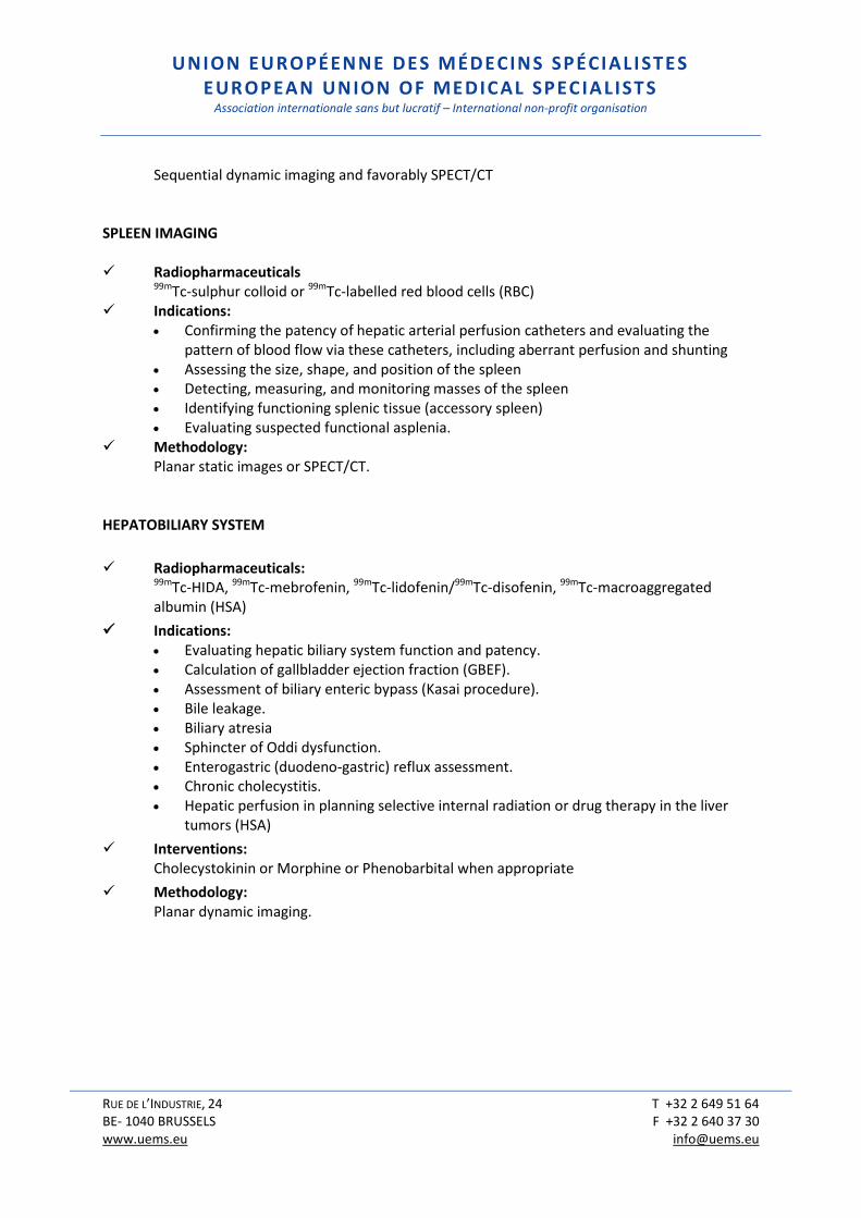
Sequential dynamic imaging and favorably SPECT/CT
SPLEEN IMAGING Radiopharmaceuticals 99mTc-sulphur colloid or 99mTc-labelled red blood cells (RBC) Indications:
Confirming the patency of hepatic arterial perfusion catheters and evaluating the pattern of blood flow via these catheters, including aberrant perfusion and shunting
Assessing the size, shape, and position of the spleen Detecting, measuring, and monitoring masses of the spleen Identifying functioning splenic tissue (accessory spleen) Evaluating suspected functional asplenia.
Methodology: Planar static images or SPECT/CT.
HEPATOBILIARY SYSTEM
Radiopharmaceuticals: 99mTc-HIDA, 99mTc-mebrofenin, 99mTc-lidofenin/99mTc-disofenin, 99mTc-macroaggregated albumin (HSA)
Indications: Evaluating hepatic biliary system function and patency. Calculation of gallbladder ejection fraction (GBEF). Assessment of biliary enteric bypass (Kasai procedure). Bile leakage. Biliary atresia Sphincter of Oddi dysfunction. Enterogastric (duodeno-gastric) reflux assessment. Chronic cholecystitis. Hepatic perfusion in planning selective internal radiation or drug therapy in the liver
tumors (HSA)
Interventions: Cholecystokinin or Morphine or Phenobarbital when appropriate
Methodology: Planar dynamic imaging.
UNION EUROPÉENNE DES MÉDECINS SPÉCIALISTES EUROPEAN UNION OF MEDICAL SPECIALISTS
Association internationale sans but lucratif – International non-profit organisation
RUE DE L’INDUSTRIE, 24 T +32 2 649 51 64 BE- 1040 BRUSSELS F +32 2 640 37 30 www.uems.eu [email protected]
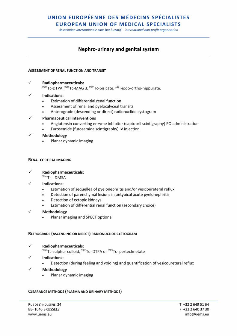
Nephro-urinary and genital system
ASSESSMENT OF RENAL FUNCTION AND TRANSIT
Radiopharmaceuticals: 99mTc-DTPA, 99mTc-MAG 3, 99mTc-bisicate, 123I-iodo-ortho-hippurate.
Indications: Estimation of differential renal function Assessment of renal and pyelocalyceal transits Anterograde (descending or direct) radionuclide cystogram
Pharmaceutical interventions Angiotensin converting enzyme inhibitor (captopril scintigraphy) PO administration Furosemide (furosemide scintigraphy) IV injection
Methodology Planar dynamic imaging
RENAL CORTICAL IMAGING
Radiopharmaceuticals: 99mTc - DMSA
Indications: Estimation of sequellea of pyelonephritis and/or vesicoureteral reflux Detection of parenchymal lesions in untypical acute pyelonephritis Detection of ectopic kidneys Estimation of differential renal function (secondary choice)
Methodology Planar imaging and SPECT optional
RETROGRADE (ASCENDING OR DIRECT) RADIONUCLIDE CYSTOGRAM
Radiopharmaceuticals: 99mTc-sulphur colloid, 99mTc -DTPA or 99mTc- pertechnetate
Indications: Detection (during feeling and voiding) and quantification of vesicoureteral reflux
Methodology Planar dynamic imaging
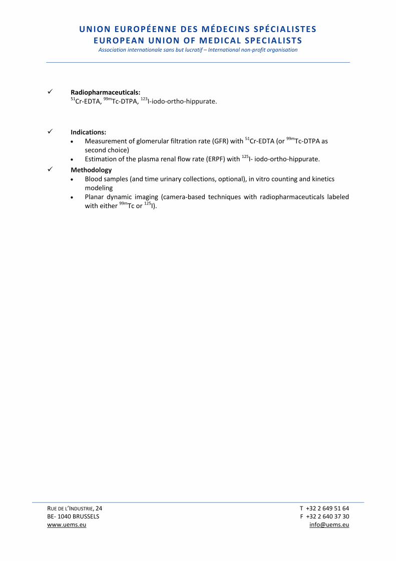
CLEARANCE METHODS (PLASMA AND URINARY METHODS)
UNION EUROPÉENNE DES MÉDECINS SPÉCIALISTES EUROPEAN UNION OF MEDICAL SPECIALISTS
Association internationale sans but lucratif – International non-profit organisation
RUE DE L’INDUSTRIE, 24 T +32 2 649 51 64 BE- 1040 BRUSSELS F +32 2 640 37 30 www.uems.eu [email protected]
Radiopharmaceuticals: 51Cr-EDTA, 99mTc-DTPA, 123I-iodo-ortho-hippurate.
Indications: Measurement of glomerular filtration rate (GFR) with 51Cr-EDTA (or 99mTc-DTPA as
second choice) Estimation of the plasma renal flow rate (ERPF) with 125I- iodo-ortho-hippurate.
Methodology Blood samples (and time urinary collections, optional), in vitro counting and kinetics
modeling Planar dynamic imaging (camera-based techniques with radiopharmaceuticals labeled
with either 99mTc or 125I).
UNION EUROPÉENNE DES MÉDECINS SPÉCIALISTES EUROPEAN UNION OF MEDICAL SPECIALISTS
Association internationale sans but lucratif – International non-profit organisation
RUE DE L’INDUSTRIE, 24 T +32 2 649 51 64 BE- 1040 BRUSSELS F +32 2 640 37 30 www.uems.eu [email protected]
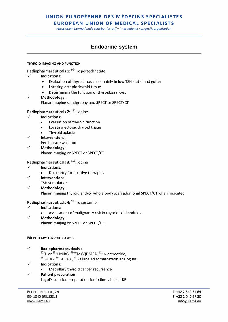
Endocrine system
THYROID IMAGING AND FUNCTION
Radiopharmaceuticals 1: 99mTc pertechnetate Indications:
Evaluation of thyroid nodules (mainly in low TSH state) and goiter
Locating ectopic thyroid tissue
Determining the function of thyroglossal cyst Methodology: Planar imaging scintigraphy and SPECT or SPECT/CT
Radiopharmaceuticals 2: 123I iodine Indications:
Evaluation of thyroid function Locating ectopic thyroid tissue Thyroid aplasia
Interventions: Perchlorate washout Methodology: Planar imaging or SPECT or SPECT/CT
Radiopharmaceuticals 3: 131I iodine Indications:
Dosimetry for ablative therapies Interventions: TSH stimulation Methodology: Planar imaging thyroid and/or whole body scan additional SPECT/CT when indicated
Radiopharmaceuticals 4: 99mTc-sestamibi Indications:
Assessment of malignancy risk in thyroid cold nodules Methodology: Planar imaging or SPECT or SPECT/CT.
MEDULLARY THYROID CANCER Radiopharmaceuticals :
123I- or 131I-MIBG, 99m Tc (V)DMSA, 111In-octreotide, 18F-FDG, 18F-DOPA, 68Ga labeled somatostatin analogues
Indications: Medullary thyroid cancer recurrence
Patient preparation: Lugol’s solution preparation for iodine labelled RP
UNION EUROPÉENNE DES MÉDECINS SPÉCIALISTES EUROPEAN UNION OF MEDICAL SPECIALISTS
Association internationale sans but lucratif – International non-profit organisation
RUE DE L’INDUSTRIE, 24 T +32 2 649 51 64 BE- 1040 BRUSSELS F +32 2 640 37 30 www.uems.eu [email protected]
Methodology: Planar thyroid and whole body imaging, and SPECT or SPECT/CT PET or PET-CT
PARATHYROID LOCATION IN HYPERPARATHYROIDISM
Radiopharmaceuticals
201Tl, 99mTc-sestamibi, 11C-Methionine
Indications: Parathyroid adenoma or hyperplasia Ectopic parathyroid gland.
Methodology: Planar imaging of neck and mediastinum (with either co-registration or subtraction when
using either 99mTc pertechnetate or 123I for double radionuclide technique), SPECT or SPECT/CT recommended. PET/CT for 11C-Methionine
ADRENAL MEDULLA TUMOR LOCATION
Radiopharmaceuticals
123I- or 131I-MIBG
Indications: Pheochromocytoma, Paraganglioma, Neuroblastoma.
Patient preparation: Protection of thyroid iodine uptake with Lugol’s solution
Methodology: Planar imaging, whole body imaging, SPECT or SPECT/CT
ADRENAL CORTEX IMAGING
Radiopharmaceuticals
131I-Noriodocholesterol
Indications: Cushing’s syndrome, Primary aldosteronism, Hyperandrogenism.
Patient preparation: Dexamethasone suppression
Methodology:

UNION EUROPÉENNE DES MÉDECINS SPÉCIALISTES EUROPEAN UNION OF MEDICAL SPECIALISTS
Association internationale sans but lucratif – International non-profit organisation
RUE DE L’INDUSTRIE, 24 T +32 2 649 51 64 BE- 1040 BRUSSELS F +32 2 640 37 30 www.uems.eu [email protected]
Planar imaging, SPECT or SPECT/CT

UNION EUROPÉENNE DES MÉDECINS SPÉCIALISTES EUROPEAN UNION OF MEDICAL SPECIALISTS
Association internationale sans but lucratif – International non-profit organisation
RUE DE L’INDUSTRIE, 24 T +32 2 649 51 64 BE- 1040 BRUSSELS F +32 2 640 37 30 www.uems.eu [email protected]
Haematopoietic and lymphatic system
BODY FLUID VOLUME DETERMINATION
Radiopharmaceutical 1 : 51Cr-labelled red blood cells
Indications: Red blood cell volume measurement and red blood cell life span
Radiopharmaceutical 2: 99mTc- or 125I labelled human serum albumin
Indications: Plasma volume measurement
Radiopharmaceutical 3: 51Cr-EDTA Indications:
Extracellular volume measurement
Methodology (common to the 3 RP): Blood sampling, in vitro counting and kinetics modelling
PLATELET SURVIVAL STUDY
Radiopharmaceutical: 111In-labelled platelets
Methodology: Planar imaging
SPLENIC FUNCTION
Radiopharmaceuticals: 99mTc-labelled heat-treated red blood cells
Methodology: Planar imaging
LYMPHOSCINTIGRAPHY
Radiopharmaceuticals: 99mTc-nanocolloids
UNION EUROPÉENNE DES MÉDECINS SPÉCIALISTES EUROPEAN UNION OF MEDICAL SPECIALISTS
Association internationale sans but lucratif – International non-profit organisation
RUE DE L’INDUSTRIE, 24 T +32 2 649 51 64 BE- 1040 BRUSSELS F +32 2 640 37 30 www.uems.eu [email protected]
Indications: Lymphoedema
Methodology: Planar imaging

UNION EUROPÉENNE DES MÉDECINS SPÉCIALISTES EUROPEAN UNION OF MEDICAL SPECIALISTS
Association internationale sans but lucratif – International non-profit organisation
RUE DE L’INDUSTRIE, 24 T +32 2 649 51 64 BE- 1040 BRUSSELS F +32 2 640 37 30 www.uems.eu [email protected]
Oncology
TUMOUR IMAGING AND CHARACTERISATION
Radiopharmaceutical 1: 18F-FDG Indications:
Differentiating benign and malignant tumours, Tumour staging, Monitoring the effect of therapy, Detecting residual tumour or recurrence
Methodology: PET/CT
Radiopharmaceutical 2: 18F-choline, 11C-choline, 18F-FACBC & 68Ga-PSMA-ligands Indication:
Restaging of patients with biochemical failure after local treatment of a prostate carcinoma.
Methodology: PET/CT
Radiopharmaceutical 3: Analogues of somatostatin such as 111In-Octreotide or 68Ga-somatostatin analogues Indications:
To identify primary neuroendocrine tumors and metastases that express somatostatin subtype 2 receptors.
Intervention: To withdraw (or temporally lower as much as possible) somatostatin analogue treatment
before the scintigraphy. Methodology: Planar imaging or SPECT/CT when using of 111In labelled somatostatin analogues Whole body PET or PET/CT when using 68Ga labelled somatostatin analogues.
Radiopharmaceutical 4: 18F-F-DOPA Indications:
To identify neuroendocrine tumour and their metastases expressing the L-type amino acid transporter 1 (LAT1)
Interventions: Carbidopa has been reported to increase tumour uptake. Methodology: PET/CT

UNION EUROPÉENNE DES MÉDECINS SPÉCIALISTES EUROPEAN UNION OF MEDICAL SPECIALISTS
Association internationale sans but lucratif – International non-profit organisation
RUE DE L’INDUSTRIE, 24 T +32 2 649 51 64 BE- 1040 BRUSSELS F +32 2 640 37 30 www.uems.eu [email protected]
Radiopharmaceutical 5: 18F-NaF Indications:
Detection and localisation of bone metastases in case of cancer in adults. Methodology: PET/CT
LYMPHOSCINTIGRAPHY AND INTRAOPERATIVE PROBE FOR SENTINEL LYMPH NODE LOCALIZATION
Radiopharmaceuticals:
99mTc labelled colloid particles or nanocolloid. Indication:
Surgical localization of the sentinel node. Methodology: Planar imaging or SPECT (Optional), and radioguided surgery.
UNION EUROPÉENNE DES MÉDECINS SPÉCIALISTES EUROPEAN UNION OF MEDICAL SPECIALISTS
Association internationale sans but lucratif – International non-profit organisation
RUE DE L’INDUSTRIE, 24 T +32 2 649 51 64 BE- 1040 BRUSSELS F +32 2 640 37 30 www.uems.eu [email protected]
Inflammatory and infectious diseases
Infection/Inflammatory site imaging
Radiopharmaceutical 1:
18F-FDG
Indications: Localization of abnormal foci guiding the etiologic diagnosis in case of fever of unknown origin Fever in an AIDS patient Detection of the extension of inflammation in case of:
o Inflammatory bowel diseases o Vasculitis involving the great vessels o Vascular prosthesis o Sarcoidosis
Suspected chronic infection of bone and/or adjacent structures: o Osteomyelitis and/or soft tissue infection o Spondilitis, o Diskitis o Osteitis including when metallic implants are present o Diabetic foot with suspicion of Charcot’s neuroarthropathy o Painful hip prosthesis
Therapy follow-up of unresectable alveolar echinococcosis
Methodology: PET or PET/CT
Radiopharmaceuticals 2:
99mTc (or 111In) labelled-white blood cells or 99mTc-labelled antigranulocyte antibodies
Indications: Localization of abnormal foci guiding the etiologic diagnosis in case of fever of unknown origin Detection of musculoskeletal infection such as septic arthritis and osteomyelitis and/or soft tissue infection Diabetic foot with suspicion of Charcot’s neuroarthropathy
Methodology: Planar whole body imaging or SPECT or SPECT/CT
UNION EUROPÉENNE DES MÉDECINS SPÉCIALISTES EUROPEAN UNION OF MEDICAL SPECIALISTS
Association internationale sans but lucratif – International non-profit organisation
RUE DE L’INDUSTRIE, 24 T +32 2 649 51 64 BE- 1040 BRUSSELS F +32 2 640 37 30 www.uems.eu [email protected]
APPENDIX 2: THERAPEUTIC APPLICATIONS IN NUCLEAR
MEDICINE (marketed or in development)
1. Palliation of painful sclerotic and mixed bone metastases
a. Strontium-89 (89Sr-chloride) b. Samarium-153 (153Sm3arium-153 (painfutetramethylene phosphonic acid or
153Sm-EDTMP or 153Sm-lexidronam) c. Rhenium-186 (186Re6hydroxyethylidene diphosphonate or 186Re-HEDP or
186Re-etidronate) d. Tin-117m (177mSn-DTPA)
2. Treatment of sclerotic metastases a. Radium-223 (223Ra3ium-223 ( s
3. Radiosynoviorthesis a. Small joints
i. Erbium-169 (169Er-citrate and 169Er-colloids) ii. Holmium-166 (166Ho-ferric hydroxide macroaggregate or 166Ho-FHMA)
b. Medium-sized joints i. Rhenium-186 (186Re-colloids)
c. Knee joint i. Yttrium-90 (90Y-citrate and 90Y-silicate and 90Y-colloids) ii. Samarium-153(153Sm-particulate hydroxyapatite or 153Sm-PHYP)
4. Thyroid diseases a. Well-differentiated thyroid cancer:
i. Iodine-131 b. Radioiodine-refractory differentiated thyroid cancer
i. Lutetium-177 (177Lu-[DOTA0, Tyr3]] octreotate or 177Lu-DOTATATE and 177Lu- [DOTA0-1-Nal3] octreotide or 177Lu-DOTANOC)
c. Benign thyroid diseases (Hyperthyroidism and volume reduction in goiter) i. Iodine-131
5. Malignant neural crest tumors (neuroblastoma, pheochromocytoma, paraganglioma, medullary thyroid carcinoma, ,...)
a. Iodine-131 (131I-meta-iodo-benzylguanidine or 131I-mIBG)
6. Neuroendocrine tumors (peptide receptor radionuclide therapy or PRRT using radiolabeled somatostatine analogs)
UNION EUROPÉENNE DES MÉDECINS SPÉCIALISTES EUROPEAN UNION OF MEDICAL SPECIALISTS
Association internationale sans but lucratif – International non-profit organisation
RUE DE L’INDUSTRIE, 24 T +32 2 649 51 64 BE- 1040 BRUSSELS F +32 2 640 37 30 www.uems.eu [email protected]
i. Indium-111 (111In-DTPA-octreotide) ii. Lutetium-177 (177Lu-[DOTA0, Tyr3]] octreotate or 177Lu-DOTATATE and
177Lu- [DOTA0-1-Nal3] octreotide or 177Lu-DOTANOC) iii. Yttrium-90(90Y-[DOTA0, Tyr3] octreotide or 90Y-DOTATOC).
7. Hepatocarcinoma and liver metastases (selective internal radiation therapy or SIRT) a. Iodine-131 (131I-lipiodol) b. Yttrium-90(90Y microspheres) c. Rhenium-188 (188Re-hyaluronic acid)
8. Polycythaemia vera a. Phosphorus-32
9. Melanoma a. Copper-64 (64CuCl2)
10. Internal vectorized radiotherapy using labeled tumor-specific monoclonal antibodies a. Non-Hogdkin Lymphoma
i. Yttrium-90 (90Y-ibritumomab tiuxetan or 90Y-epratuzumab tetraxetan ii. Lutetium-177 (177Lu-DOTA-rituximab) iii. Astatine-211 (211At-atine-211 (mab)axetanr d tum
b. Prostate carcinoma i. Yttrium-90 or lutetium-177 radiolabeled monoclonal antibody targeting
the epitope PSMA (90Y or 177Lu-J591 mAb) ii. Zirconium-89 (89Zr-desferrioxamine B-J591 mAB)
c. Glioma i. Astatine-211 (211At-labeled Me1-14 F(ab')2)
d. Head and neck carcinoma i. Astatine-211 (211At-labeled U36 chimeric monoclonal antibody)
11. Disseminated cancer a. Astatine-211 (211At-EGFRvIII monoclonal antibody)
12. Prevention of reactive hyperplasia after stent placement (intravascular, intraurethral) a. Rhenium-188 (188Re-filled balloon dilation)