Toxic and Trace Metal Concentrations In Liver and Kidney of Dogs
18
Toxic and Trace Metal Concentrations in Liver and Kidney of Dogs Influence of Diet, Sex, Age, and Pathological Lesions MARTA LÓPEZ-ALONSO,* ,1 MARTA MIRANDA, 2 PAULINO GARCÍA-PARTIDA, 3 ADRIANA MENDEZ, 1 CRISTINA CASTILLO, 1 AND JOSÉ LUIS BENEDITO 1 1 Departamentos de Patoloxía Animal, 2 Ciencias Clínicas Veterinarias, and 3 Patología Animal II, Facultad de Veterinaria, Universidad Complutense de Madrid, 28040 Madrid, Spain Received July 27, 2006; Accepted August 15, 2006 ABSTRACT The aim of this study was to provide data on the main toxic and trace metals in the liver and kidney of domestic dogs in Galicia, NW Spain and to evaluate the influence of diet, sex, age, and pathological lesions on metal accumulation. Samples of the liver and kidney from 77 male and female dogs, aged between 6 mo and 18 yr, were collected during ordinary necropsy. Samples were acid-digested and metal concentrations deter- mined by inductively coupled plasma (ICP)–mass spectrometry and ICP–atomic emission spectrometry. Mean toxic metal concentrations (geo- metric means for liver and kidney respectively) were 11.5 and 15.8 µg/kg wet weight for As, 56.3 and 166 µg/kg for Cd, 32.7 and 51.9 µg/kg for Hg, and 60.1 and 23.6 µg/kg for Pb. For the trace metals, these concentrations were respectively 16.3 and 21.0 µg/kg for Co, 57.6 and 43.9 µg/kg for Cr, 42.1 and 5.95 mg/kg for Cu, 394 mg/kg and 95.7 mg/kg for Fe, 2.39 and 0.956 mg/kg for Mn, 0.522 and 0.357 mg/kg for Mo, 23.8 and 26.8 µg/kg for Ni, 0.686 and 1.39 mg/kg for Se, and 46.7 and 26.0 mg/kg for Zn. Cd concentrations in the kidney significantly increased with age, and Co con- centrations in the liver and kidney significantly decreased with age. Hepatic Pb concentrations were significantly higher in growing (<1 yr) and old (>10 yr) dogs. Animals with pathological lesions showed significantly higher Co and lower Mn and Zn concentrations in liver than animals with- out macroscopic abnormalities. Dogs that received commercial diets in general showed low variability in hepatic mineral status compared to ani- Biological Trace Element Research 185 Vol. 116, 2007 © Copyright 2007 by Humana Press Inc. All rights of any nature, whatsoever, reserved. 0163-4984/(Online) 1559-0720/06/11602–185 $30.00 *Author to whom all correspondence and reprint requests should be addressed.
Transcript of Toxic and Trace Metal Concentrations In Liver and Kidney of Dogs
Toxic and Trace Metal Concentrationsin Liver and Kidney of Dogs
Influence of Diet, Sex, Age, and Pathological Lesions
MARTA LÓPEZ-ALONSO,*,1 MARTA MIRANDA,2
PAULINO GARCÍA-PARTIDA,3 ADRIANA MENDEZ,1
CRISTINA CASTILLO,1 AND JOSÉ LUIS BENEDITO1
1Departamentos de Patoloxía Animal, 2Ciencias ClínicasVeterinarias, and 3Patología Animal II, Facultad de Veterinaria,
Universidad Complutense de Madrid, 28040 Madrid, Spain
Received July 27, 2006; Accepted August 15, 2006
ABSTRACT
The aim of this study was to provide data on the main toxic and tracemetals in the liver and kidney of domestic dogs in Galicia, NW Spain andto evaluate the influence of diet, sex, age, and pathological lesions on metalaccumulation. Samples of the liver and kidney from 77 male and femaledogs, aged between 6 mo and 18 yr, were collected during ordinarynecropsy. Samples were acid-digested and metal concentrations deter-mined by inductively coupled plasma (ICP)–mass spectrometry andICP–atomic emission spectrometry. Mean toxic metal concentrations (geo-metric means for liver and kidney respectively) were 11.5 and 15.8 µg/kgwet weight for As, 56.3 and 166 µg/kg for Cd, 32.7 and 51.9 µg/kg for Hg,and 60.1 and 23.6 µg/kg for Pb. For the trace metals, these concentrationswere respectively 16.3 and 21.0 µg/kg for Co, 57.6 and 43.9 µg/kg for Cr,42.1 and 5.95 mg/kg for Cu, 394 mg/kg and 95.7 mg/kg for Fe, 2.39 and0.956 mg/kg for Mn, 0.522 and 0.357 mg/kg for Mo, 23.8 and 26.8 µg/kgfor Ni, 0.686 and 1.39 mg/kg for Se, and 46.7 and 26.0 mg/kg for Zn. Cdconcentrations in the kidney significantly increased with age, and Co con-centrations in the liver and kidney significantly decreased with age.Hepatic Pb concentrations were significantly higher in growing (<1 yr) andold (>10 yr) dogs. Animals with pathological lesions showed significantlyhigher Co and lower Mn and Zn concentrations in liver than animals with-out macroscopic abnormalities. Dogs that received commercial diets ingeneral showed low variability in hepatic mineral status compared to ani-
Biological Trace Element Research 185 Vol. 116, 2007
© Copyright 2007 by Humana Press Inc.All rights of any nature, whatsoever, reserved.0163-4984/(Online) 1559-0720/06/11602–185 $30.00
*Author to whom all correspondence and reprint requests should be addressed.
mals that receive homemade feeds or a mixture of commercial and home-made feeds.
Index Entries: Toxic metals; trace metals; dog; age; sex; liver or kid-ney pathology.
INTRODUCTION
The common toxic metals, notably As, Cd, Hg, and Pb, are not knownto serve any essential biological function, and their presence in tissues isgenerally associated with pollution (1). At high concentrations, these ele-ments can be acutely lethal, but moderate tissue concentrations can resultin nonlethal adverse effects on a variety of physiological and biochemicalprocesses (2). Essential trace metals, in contrast to toxic metals, have awell-recognized function in the body, and health problems related to theseelements are usually the result of deficiency (3), although toxicity resultingfrom exposure or accumulation might also occur.
The physiological processes governing the availability and toxicity ofthese metals are highly complex. For example, some metals such as Cd orCu accumulate in tissues bound to metallothioneins, and it is only whenthe capacity of these proteins to bind metals is overloaded that the metalis released in free form, causing oxidative damage (4,5). Similarly, free Femight be present if the amount of Fe in the liver exceeds the capacity tobind it to protein to form ferritin (6), resulting in free-radical formation.Increased levels of metals in the tissues might also magnify the effects ofother causes of disease, such as viruses or alcohol in humans (7–9). In othercases, abnormal metal concentrations in tissues are secondary to otherpathologies; for example, increased hepatic Cu concentrations might besecondary to biliary lesions that interfere with normal excretion of excessCu into the bile (10), whereas increases in hepatic Fe concentration mightbe the result of increased intestinal absorption or to abnormalities inhemoglobin metabolism and delivery of Fe to the liver (11). Finally,increased tissue metal concentrations might sometimes be the result ofdefective metal metabolism pathways; inherited hepatic disease inBedlington terriers associated with chronic Cu accumulation in the liver isperhaps one of the best studied examples (12).
In spite of the great efforts made to understand the exact biochemicalmechanisms of metal imbalances, both in the tissues where the metalsaccumulate and in other organ locations, they remain incompletely under-stood (13). For example, it has been demonstrated that Cr concentrationsare decreased in dogs and cats with diabetes mellitus and that Cr supple-mentation is beneficial for such animals (14), but the reasons for theseobservations are unclear. Similarly, in the pathogenesis of some neurode-generative disorders, such as Alzheimer’s disease (15), metals like Cu, Fe,or Mn might be involved, but the nature of this involvement remainsunknown. Nevertheless, the quantitative measurement of these metals in
186 López-Alonso et al.
Biological Trace Element Research Vol. 116, 2007
tissues might be a useful tool for the diagnosis and prognosis of diversepathologies.
In dogs, metal accumulation and toxicity has been poorly studied, andinformation on tissue metal concentrations, whether in healthy or diseasedanimals, is sparse and limited to a very small number of metals. The objec-tives of the present study were to provide data on concentrations of themain toxic and trace metals in the liver and kidney (the principal organsfor metal accumulation) of dogs in Galicia, NW Spain and to evaluate theinfluence of diet, sex, age, and pathological liver/kidney lesions on metalaccumulation.
MATERIAL AND METHODS
Tissue Samples
Samples of the liver and kidney from 77 dogs euthanized in 2001 or2002 in the Veterinary Hospital and other veterinary clinics in Lugo (NWSpain) were collected during the routine necropsy.
Information on age, sex, breed, diet, and other data related to thehealth of the animals was obtained from the clinical record. Half of thedogs (40 of 77) were of unknown or mixed breed; among the pure breeds,the most common were German shepherd (10), boxer (4), cocker spaniel(3), and doberman pinscher (3). Of the 77 dogs, 26 were male, 50 female,and 1 unrecorded. Age ranged from 6 mo to 18 yr. To analyze the effect ofage on tissue metal concentrations, we grouped the dogs into the follow-ing age groups: growing dogs (<1 yr, n = 12), young adults (1–5 yr, n = 14),senior adults (6–9 yr, n = 15), old adults (10–12 yr, n = 15), and geriatricdogs (>12 yr, n = 14). In relation to the type of diet, animals were groupedinto three classes: commercial feed (n = 26), homemade feed (n = 21), anda mixture of commercial and homemade feed (n = 12).
Information on the presence of pathological lesions in the liver or kid-ney was obtained from macroscopic examination of these organs duringthe routine necropsy procedure. Pathological lesions were observed in theliver of 14 dogs and indicated diseases, including steatosis, cirrhosis, andmultifocal necrosis. In the kidney, the main pathological lesions corre-sponded to chronic fibrosis (chronic glomerulonephritis and chronic inter-stitial nephritis), observed in 26 dogs. Ten dogs showed pathologicallesions in both the liver and kidney.
Liver and kidney samples for metal analysis (approx 20 g) were col-lected in triplicate after the necropsy, packed in plastic bags, immediatelytransported to the laboratory, and stored at –18°C until analysis.
Metal Analyses
Approximately 2-g subsamples were digested in 5 mL of concentratednitric acid (Suprapur grade) and 2 mL of 30% (w/v) hydrogen peroxide in
Toxic and Trace Metals in Dogs 187
Biological Trace Element Research Vol. 116, 2007
a microwave digestion system (Milestone, Ethos Plus). Because of the like-lihood of different metal contents in the cortex and medulla, the kidneysamples were homogenized prior to digestion to ensure that the subsam-ple analyzed was representative of the whole organ. Digested sampleswere transferred to polypropylene sample tubes and diluted to 25 mL withultrapure water. Metals at high concentrations in the digest (Cu, Fe, Mn,Mo, Se, and Zn) were determined by inductively coupled plasma–atomicemission spectrometry (ICP-AES; Perkin-Elmer Optima 4300 DV),whereas metals at low concentrations (As, Cd, Hg, Pb, Co, Cr, and Ni)were determined by inductively coupled plasma–mass spectrometry (ICP-MS; VGElemental PlasmaQuad II-SOption).
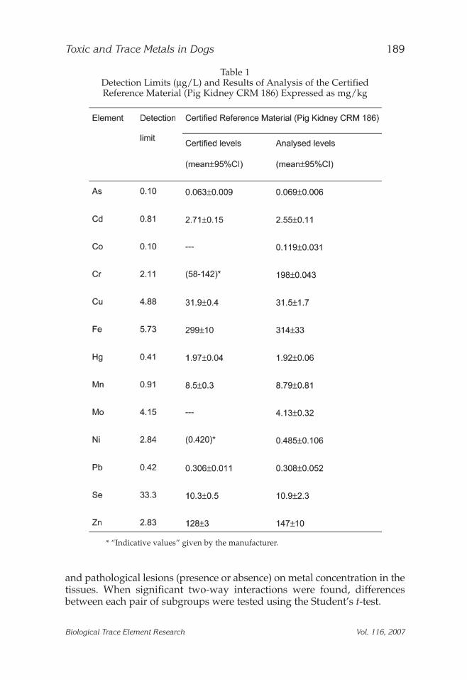
An analytical quality control program was employed. Blankabsorbance values were monitored throughout the study and were sub-tracted from the readings before the results were calculated. The limit ofdetection in the acid digest was set at three times the standard deviation ofthe reagent blanks (Table 1). Limits of detection, expressed as concentra-tions in tissue, were calculated on the basis of the mean sample weight andvolume analyzed.
Analytical recoveries were determined from a certified referencematerial (Pig Kidney CRM 186, BCR Reference Materials) analyzedtogether with the samples. The results are given in Table 1 and showacceptable agreement between the measured and certified values for As,Cd, Cu, Fe, Hg, Mn, Pb, Se, and Zn. Indicative values only are given for Crand Ni in the CRM; there was, however, good agreement between thesevalues and our measured values. No information is given regarding Coand Mo in the CRM, and analytical recoveries were determined usingspiked samples at a level giving absorbance values that were generally2–10 times greater than the normal levels in the various tissues (n = 10),recoveries being between 88% and 96% respectively. To evaluate precision,12 absorbance readings from the same digest (precision of analyticalmethod) and single absorbance readings from 12 digests of the same sam-ple (precision of overall method) were recorded. The relative standarddeviation (RSD) of these values was 0.63–7.91% for the analytical methodand 3.02–11.3% for the overall procedure.
Data Analysis
All statistical analyses were done using SPSS for Windows (v. 12.0). Tocalculate mean metal concentrations in the different tissues, nondetectableconcentrations were assigned a value of half of the quantification limit.Normal distribution of datasets was checked using the Kolmogorov–Smirnov test. In general, data were not Normally distributed and were log-transformed before analysis. Average values for the data are therefore givenas geometric means. Analysis of variance (general linear model) was used todetermine the influence of diet (commercial, homemade, or mixed commer-cial and homemade), sex (male or female), age (<1, 1–5, 6–9, 10–12 and >12 yr),
188 López-Alonso et al.
Biological Trace Element Research Vol. 116, 2007
and pathological lesions (presence or absence) on metal concentration in thetissues. When significant two-way interactions were found, differencesbetween each pair of subgroups were tested using the Student’s t-test.
Toxic and Trace Metals in Dogs 189
Biological Trace Element Research Vol. 116, 2007
Table 1Detection Limits (µg/L) and Results of Analysis of the Certified Reference Material (Pig Kidney CRM 186) Expressed as mg/kg
* “Indicative values” given by the manufacturer.
RESULTS
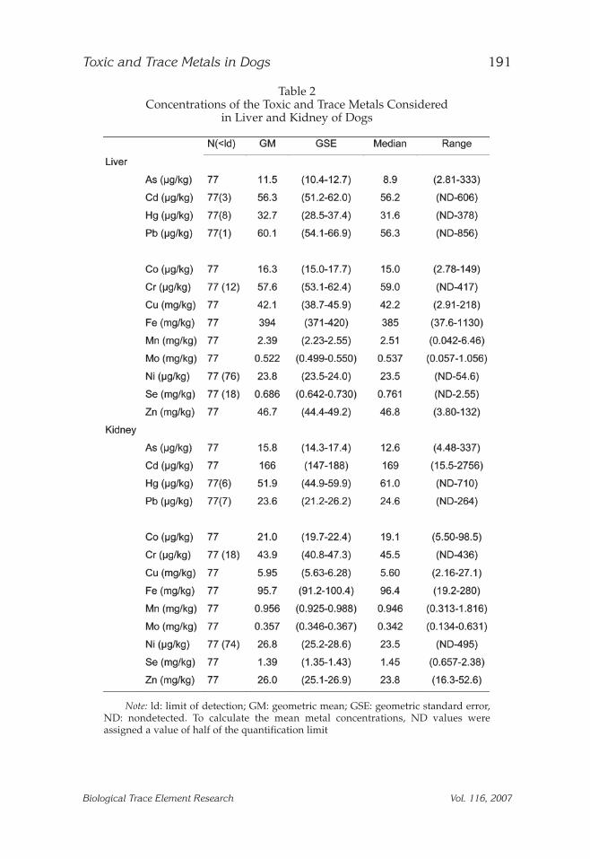
Toxic and trace metal concentrations in the liver and kidney of the 77dogs in this study are listed in Table 2. Considering the toxic metals, meanconcentrations were higher in kidney than in liver for As (1.37-fold higherin the kidney), Cd (2.95-fold), and Hg (1.59-fold), although the oppositewas true for Pb (2.55-fold higher in the liver) and in good agreement withthose described as normal for dogs (16). Considering the trace metals,mean concentrations were, in most cases, higher in the liver than in thekidney (Table 2). In addition, the liver was the organ that showed thegreatest variability in metal content among animals, with most trace met-als showing an approx 200-fold difference between minimum and maxi-mum values, versus much lower variability in the kidney (10-fold or less).Cu and Zn concentrations in both organs were, in general, within the ade-quate ranges for dog tissues (16), with less than 10% of animals showingmineral concentrations in the deficient or high range. In contrast, Fe con-centrations were, in general, high (29% and 26% of the sampled animalshad Fe levels within the high range for liver and kidney, respectively),whereas Mn concentrations were low (75% of animals showed Mn con-centrations within the deficient range) (16). Most samples (98.7% and96.1% in the liver and kidney, respectively) also had Ni concentrationsbelow the quantification limit (47 µg/kg) and, thus, below the adequaterange (16). Finally, for Se, although mean values both in the liver and kid-ney were within the adequate ranges, 18 and 9 of the 77 sampled animalsshowed Se concentrations considered within the deficient ranges for theliver and kidney, respectively (16). For the other elements determined inthis study, Cr, Co, and Mo, we are not aware of any published data on nor-mal ranges in dogs.
The results of general linear modeling (GLM) to assess the influenceof diet, sex, age, and pathological lesions on metal concentrations in liverare summarized in Table 3.
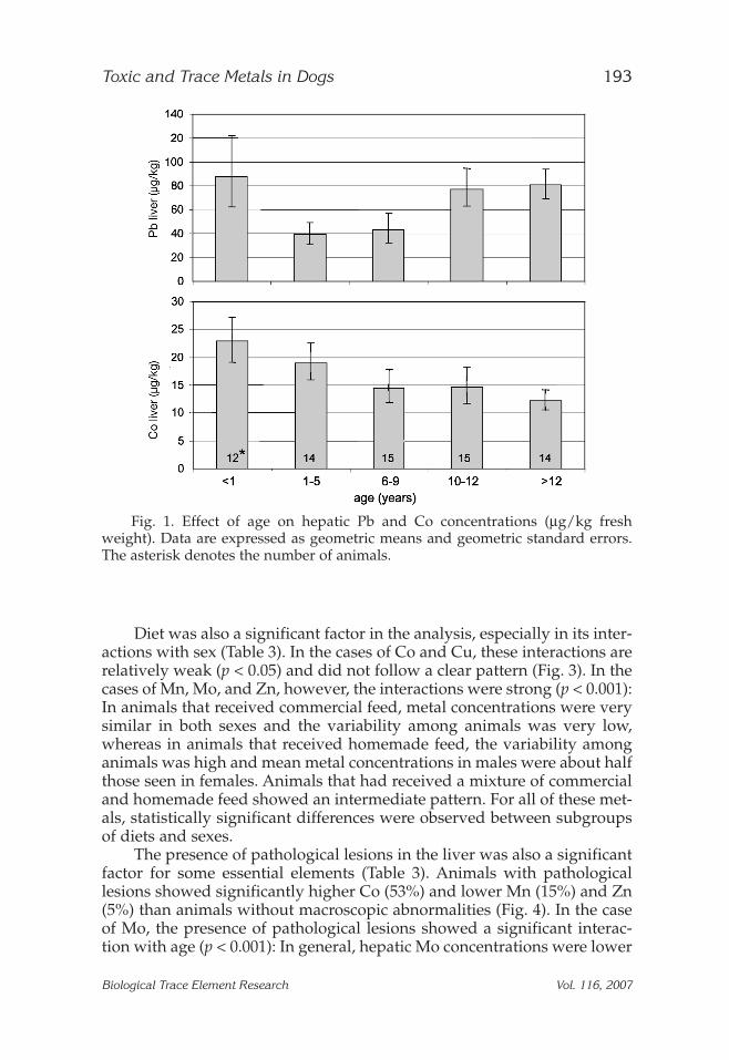
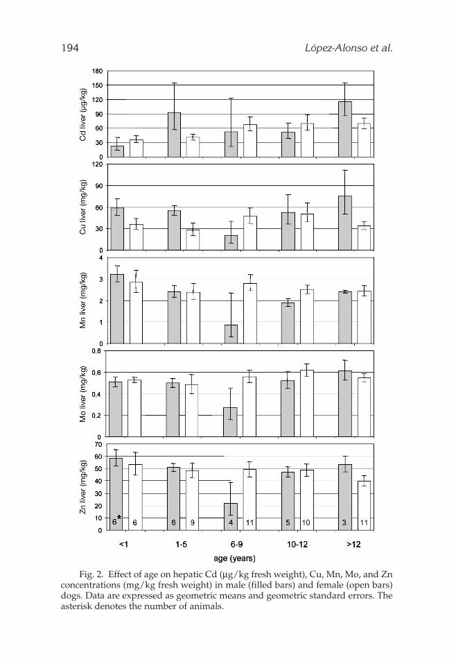
Age was the most important single factor in the analysis for most toxicand essential elements; however, significant interactions between age andsex were found for all elements, with the exception of Pb and Co. Pb con-centrations were higher in animals <1 yr, decreased from 1 to 9 yr, andincreased in animals >10 yr (Fig. 1). For Co, the mean concentrations in theliver gradually decreased from the <1 to >12 yr groups (Fig. 1). For themetals that showed significant interactions between sex and age, the twofactors have to be evaluated together. Detailed analysis of these interac-tions (Fig. 2) showed that Cd tends to accumulate with age, although theeffects of sex vary over time. In general, Cd accumulation in females washigher than in males, except in the 1–5 and >12 yr groups, in whichfemales showed lower variability than males. In any case, the differencesbetween these subgroups (between males and females in each age groupand between age groups for each sex) were statistically significant. Theessential metals showed similar patterns in all cases. In general, metal con-
190 López-Alonso et al.
Biological Trace Element Research Vol. 116, 2007
Toxic and Trace Metals in Dogs 191
Biological Trace Element Research Vol. 116, 2007
Table 2Concentrations of the Toxic and Trace Metals Considered
in Liver and Kidney of Dogs
Note: ld: limit of detection; GM: geometric mean; GSE: geometric standard error,ND: nondetected. To calculate the mean metal concentrations, ND values wereassigned a value of half of the quantification limit
centrations decreased from <1 to 6–9 yr, and then increased to >12 yr, withmales showing higher metal concentrations than females. The significantinteraction between age and sex in the GLM analysis seems to be the resultof the low and very variable metal concentrations in males in the 6- to 9-year group, with mean concentrations between about 50% and 30% ofthose in females; none of these elements showed significant differencesbetween subgroups of age and sex.
192 López-Alonso et al.
Biological Trace Element Research Vol. 116, 2007
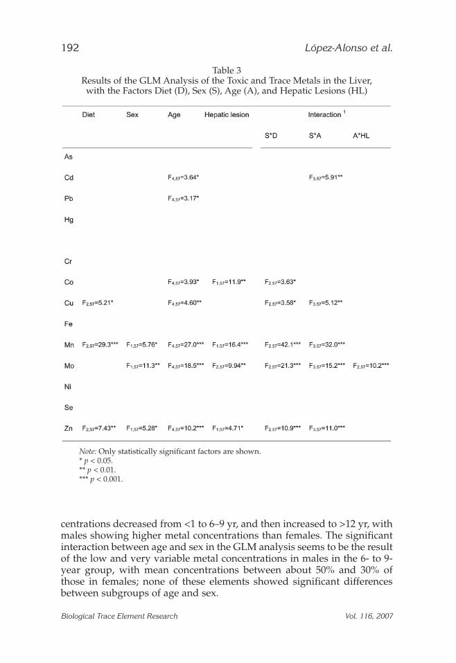
Table 3Results of the GLM Analysis of the Toxic and Trace Metals in the Liver,
with the Factors Diet (D), Sex (S), Age (A), and Hepatic Lesions (HL)
Note: Only statistically significant factors are shown.* p < 0.05.** p < 0.01.*** p < 0.001.
Diet was also a significant factor in the analysis, especially in its inter-actions with sex (Table 3). In the cases of Co and Cu, these interactions arerelatively weak (p < 0.05) and did not follow a clear pattern (Fig. 3). In thecases of Mn, Mo, and Zn, however, the interactions were strong (p < 0.001):In animals that received commercial feed, metal concentrations were verysimilar in both sexes and the variability among animals was very low,whereas in animals that received homemade feed, the variability amonganimals was high and mean metal concentrations in males were about halfthose seen in females. Animals that had received a mixture of commercialand homemade feed showed an intermediate pattern. For all of these met-als, statistically significant differences were observed between subgroupsof diets and sexes.
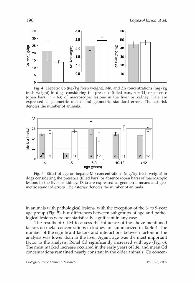
The presence of pathological lesions in the liver was also a significantfactor for some essential elements (Table 3). Animals with pathologicallesions showed significantly higher Co (53%) and lower Mn (15%) and Zn(5%) than animals without macroscopic abnormalities (Fig. 4). In the caseof Mo, the presence of pathological lesions showed a significant interac-tion with age (p < 0.001): In general, hepatic Mo concentrations were lower
Toxic and Trace Metals in Dogs 193
Biological Trace Element Research Vol. 116, 2007
Fig. 1. Effect of age on hepatic Pb and Co concentrations (µg/kg freshweight). Data are expressed as geometric means and geometric standard errors.The asterisk denotes the number of animals.
194 López-Alonso et al.
Fig. 2. Effect of age on hepatic Cd (µg/kg fresh weight), Cu, Mn, Mo, and Znconcentrations (mg/kg fresh weight) in male (filled bars) and female (open bars)dogs. Data are expressed as geometric means and geometric standard errors. Theasterisk denotes the number of animals.
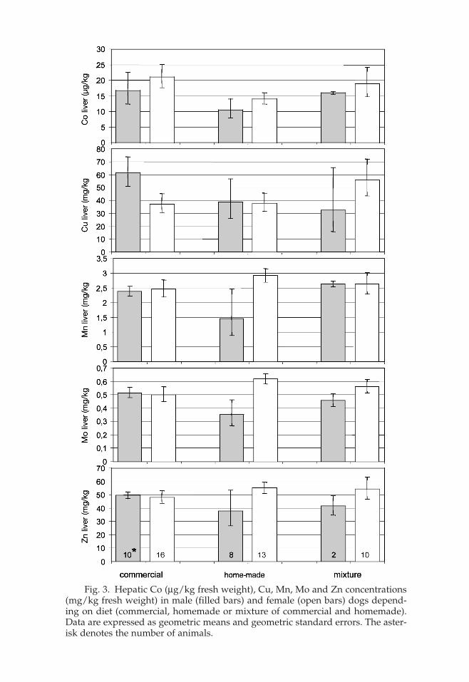
Fig. 3. Hepatic Co (µg/kg fresh weight), Cu, Mn, Mo and Zn concentrations(mg/kg fresh weight) in male (filled bars) and female (open bars) dogs depend-ing on diet (commercial, homemade or mixture of commercial and homemade).Data are expressed as geometric means and geometric standard errors. The aster-isk denotes the number of animals.
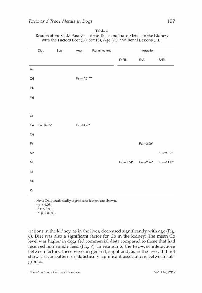
in animals with pathological lesions, with the exception of the 6- to 9-yearage group (Fig. 5), but differences between subgroups of age and patho-logical lesions were not statistically significant in any case.
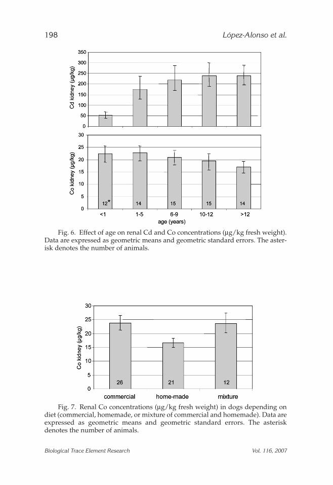
The results of GLM to assess the influence of the above-mentionedfactors on metal concentrations in kidney are summarized in Table 4. Thenumber of the significant factors and interactions between factors in theanalysis was lower than in the liver. Again, age was the most importantfactor in the analysis. Renal Cd significantly increased with age (Fig. 6):The most marked increase occurred in the early years of life, and mean Cdconcentrations remained nearly constant in the older animals. Co concen-
196 López-Alonso et al.
Biological Trace Element Research Vol. 116, 2007
Fig. 4. Hepatic Co (µg/kg fresh weight), Mn, and Zn concentrations (mg/kgfresh weight) in dogs considering the presence (filled bars, n = 14) or absence(open bars, n = 63) of macroscopic lesions in the liver or kidney. Data areexpressed as geometric means and geometric standard errors. The asteriskdenotes the number of animals.
Fig. 5. Effect of age on hepatic Mo concentrations (mg/kg fresh weight) indogs considering the presence (filled bars) or absence (open bars) of macroscopiclesions in the liver or kidney. Data are expressed as geometric means and geo-metric standard errors. The asterisk denotes the number of animals.
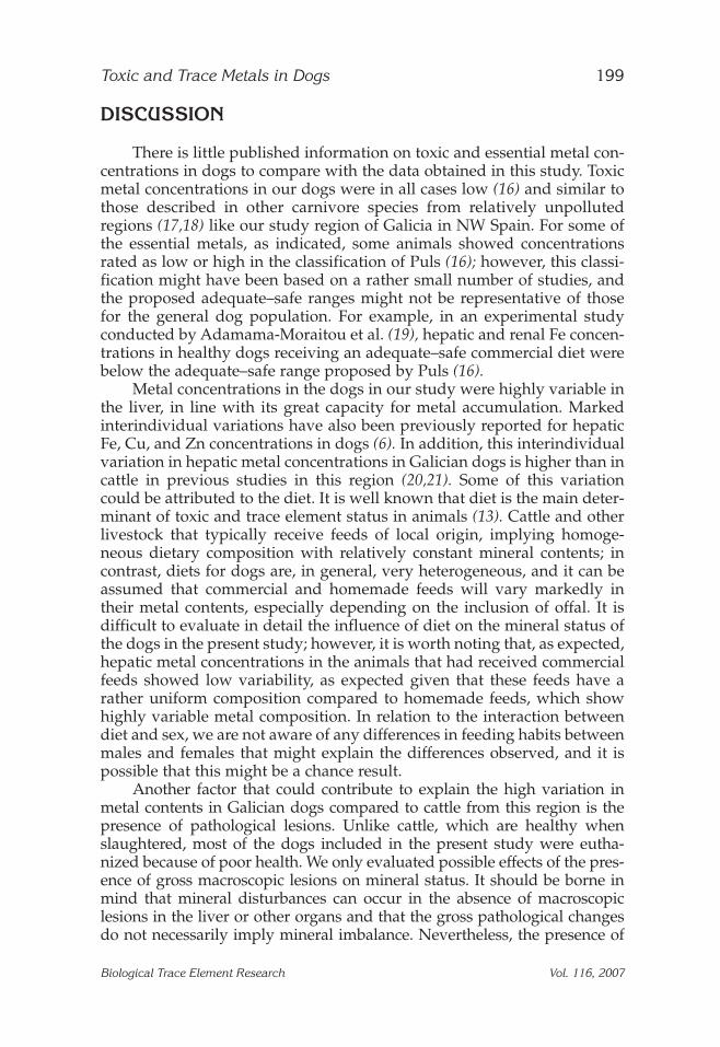
trations in the kidney, as in the liver, decreased significantly with age (Fig.6). Diet was also a significant factor for Co in the kidney: The mean Colevel was higher in dogs fed commercial diets compared to those that hadreceived homemade feed (Fig. 7). In relation to the two-way interactionsbetween factors, these were, in general, slight and, as in the liver, did notshow a clear pattern or statistically significant associations between sub-groups.
Toxic and Trace Metals in Dogs 197
Biological Trace Element Research Vol. 116, 2007
Table 4Results of the GLM Analysis of the Toxic and Trace Metals in the Kidney,
with the Factors Diet (D), Sex (S), Age (A), and Renal Lesions (RL)
Note: Only statistically significant factors are shown.* p < 0.05.** p < 0.01.*** p < 0.001.
198 López-Alonso et al.
Biological Trace Element Research Vol. 116, 2007
Fig. 6. Effect of age on renal Cd and Co concentrations (µg/kg fresh weight).Data are expressed as geometric means and geometric standard errors. The aster-isk denotes the number of animals.
Fig. 7. Renal Co concentrations (µg/kg fresh weight) in dogs depending ondiet (commercial, homemade, or mixture of commercial and homemade). Data areexpressed as geometric means and geometric standard errors. The asteriskdenotes the number of animals.
DISCUSSION
There is little published information on toxic and essential metal con-centrations in dogs to compare with the data obtained in this study. Toxicmetal concentrations in our dogs were in all cases low (16) and similar tothose described in other carnivore species from relatively unpollutedregions (17,18) like our study region of Galicia in NW Spain. For some ofthe essential metals, as indicated, some animals showed concentrationsrated as low or high in the classification of Puls (16); however, this classi-fication might have been based on a rather small number of studies, andthe proposed adequate–safe ranges might not be representative of thosefor the general dog population. For example, in an experimental studyconducted by Adamama-Moraitou et al. (19), hepatic and renal Fe concen-trations in healthy dogs receiving an adequate–safe commercial diet werebelow the adequate–safe range proposed by Puls (16).
Metal concentrations in the dogs in our study were highly variable inthe liver, in line with its great capacity for metal accumulation. Markedinterindividual variations have also been previously reported for hepaticFe, Cu, and Zn concentrations in dogs (6). In addition, this interindividualvariation in hepatic metal concentrations in Galician dogs is higher than incattle in previous studies in this region (20,21). Some of this variationcould be attributed to the diet. It is well known that diet is the main deter-minant of toxic and trace element status in animals (13). Cattle and otherlivestock that typically receive feeds of local origin, implying homoge-neous dietary composition with relatively constant mineral contents; incontrast, diets for dogs are, in general, very heterogeneous, and it can beassumed that commercial and homemade feeds will vary markedly intheir metal contents, especially depending on the inclusion of offal. It isdifficult to evaluate in detail the influence of diet on the mineral status ofthe dogs in the present study; however, it is worth noting that, as expected,hepatic metal concentrations in the animals that had received commercialfeeds showed low variability, as expected given that these feeds have arather uniform composition compared to homemade feeds, which showhighly variable metal composition. In relation to the interaction betweendiet and sex, we are not aware of any differences in feeding habits betweenmales and females that might explain the differences observed, and it ispossible that this might be a chance result.
Another factor that could contribute to explain the high variation inmetal contents in Galician dogs compared to cattle from this region is thepresence of pathological lesions. Unlike cattle, which are healthy whenslaughtered, most of the dogs included in the present study were eutha-nized because of poor health. We only evaluated possible effects of the pres-ence of gross macroscopic lesions on mineral status. It should be borne inmind that mineral disturbances can occur in the absence of macroscopiclesions in the liver or other organs and that the gross pathological changesdo not necessarily imply mineral imbalance. Nevertheless, the presence of
Toxic and Trace Metals in Dogs 199
Biological Trace Element Research Vol. 116, 2007
macroscopic lesions proved to be a significant factor, indicating that mineralmetabolism can be significantly affected by pathological conditions. There islittle information available on metal concentrations in tissues with patho-logical lesions in dogs. Increased hepatic Cu concentrations have beenreported in animals with hepatic pathologies, the most extensively studiedbeing breed-hereditary Cu storage disease in Bedlington terriers and otherbreeds as a result of defective pathways for biliary Cu excretion (12); how-ever, it is also possible that the relatively high Cu concentrations found indogs with liver disease might be secondary to biliary lesions that interferewith normal excretion of excess Cu into bile (6). Increased Fe concentrationshave been found in some studies in animals suffering from chronic hepati-tis (22) or with portosystemic shunts (23); however, no such consistent ele-vations were found in other studies in animals with hepatic histologicallesions (6), or in this study. Increased Fe and decreased Cu and Zn concen-trations in blood and tissues have been observed in experimentally inducedexocrine pancreatic insufficiency in dogs and might be associated withchanges in mineral absorption from the gastrointestinal tract (19).
Age is another factor that has proved to influence toxic and essentialmetals concentrations in animals (13). In the present study, the most sig-nificant age effects were found for the toxic metals Cd and Pb. Cd concen-trations significantly increased with age in the kidney, the organ thataccumulates the highest concentrations of metallothionein-bound Cd fol-lowing low-level chronic exposure (24). The most marked increase in dogsin our study occurred in the early years of life, and mean Cd concentra-tions remained nearly invariable in the older animals. Similar results havebeen observed in humans, who show a tendency to accumulate Cd untilapprox 50 yr of age, whereas beyond this age, renal Cd levels remainessentially constant or decrease (25). The highest mean hepatic Pb concen-tration was found in the growing group (<1 yr old). It has been demon-strated in many species, including humans and experimental animals, thatgrowing individuals are the most vulnerable age group for Pb toxicity (26).For example, puppies are more likely to ingest materials containing Pbbecause of their normal chewing and play activities, which results ingreater ingestion of Pb than adults. Also, because of their greater absorp-tion and retention of Pb, the body burdens in young animals resultingfrom a given external exposure level tend to be higher than adults. In thepresent study, mean hepatic Pb concentrations were also high in old dogs(>10 yr). Because Pb concentration in the liver is an indicator of recentexposure (27), our results could suggest that Pb exposure is higher in oldanimals. However, it is well known that bone Pb is readily mobilized toblood and soft tissues (28), with bone Pb appearing to be a major source ofblood Pb in older individuals with previous environmental Pb exposure.Of particular importance is mobilization of Pb from bone in pregnant andnursing mothers (29) because Pb crosses the placenta to the fetus and isalso excreted in milk, which presumably contributes to the high Pbresidues often found in very young animals.
200 López-Alonso et al.
Biological Trace Element Research Vol. 116, 2007
With regard to the essential elements, mean hepatic and renal Co con-centrations significantly decreased with age in the present study. We are notaware of any previously published data on the effects of age on tissue Co con-centrations in carnivore species. In ruminants, for which more information isavailable, various patterns have been reported: No differences between agegroups were observed for hepatic Co in moose (30) and buffalo (31), whereasin dairy cattle, significantly higher Co concentrations were reported foryounger cows (32). The other essential metals showed significant interactionsbetween age and sex, with males in the 6- to 9- yr age group showing lowhepatic and renal concentrations of these metals. We cannot offer any expla-nation for these results, which might be attributable to chance.
In conclusion, this study has found a wide range of toxic and tracemetal concentrations in domestic dogs, which are at least, in part, relatedto diet, sex, age, and the presence of pathological lesions in the liver or kid-ney. Further studies to establish the normal ranges of these metals inhealthy dogs and to characterize their variations in dogs suffering fromdifferent pathologies would contribute to elucidating their possible role inthese pathologies and, consequently, to their treatment.
ACKNOWLEDGMENTS
The authors would like to thank Maite Férnandez and María NievesMuñoz for technical assistance and for advice on statistics, respectively.
REFERENCES
1. L. Friberg, G. F. Nordberg, and V. B. Vouk, Handbook on the Toxicology of Metals, Elsevier,Amsterdam (1979).
2. J. Bires, J. Dianovsky, P. Bartko, and Z. Juhasova, Effects on enzymes and the geneticapparatus of sheep after administration of samples from industrial emissions, BioMet-als 8, 53–58 (1995).
3. E. J. Underwood and N. F. Suttle, The Mineral Nutrition of Livestock, 3rd ed., CAB Inter-national, Wallingford, UK (1999).
4. I. Bremner, Nutritional and physiological significance of metallothionein, Methods Enz-imol. 205, 25–35 (1991).
5. I. Bremner, Manifestations of copper excess, Am. J. Clin. Nutr. 67, 1069S–1073S (1998).6. P. C. Schultheiss, C. L. Bedwell, D. W. Hamar, and M. J. Fettman, Canine liver iron, cop-
per, and zinc concentrations and association with histologic lesions, J. Vet. Diagn. Invest.14, 396–402 (2002).
7. A. Piperno, A. Vergani, I. Malosio, et al., Hepatic iron overload in patients with chronicviral hepatite: role of HFE gene mutations, Hepatology 28, 1105–1109 (1998).
8. S. E. Bassett, A. M. Bisceglie, B. R. Bacon, et al., Effects of iron loading on pathogenicityin hepatite C virus-infected chimpanzees, Hepatology 29, 1884–1892 (1999).
9. N. Ganne-Carrie, C. Christidis, C. Chastang, et al., Liver iron is predictive of death onalcoholic cirrhosis: a multivariate study of 229 consecutive patients with alcoholicand/or hepatite C virus cirrhosis: a prospective follow up study, Gut 46, 277–282 (2000).
Toxic and Trace Metals in Dogs 201
Biological Trace Element Research Vol. 116, 2007
10. D. S. Rolfe and D. C. Twedt, Copper-associated hepatophaties in dogs, Vet. Clin. N. Am.:Small. Anim. Pract. 25, 399–417 (1995).
11. J. W. Halliday and J. Searle, Hepatic iron deposition in human disease and animal mod-els, BioMetals 2, 205–209 (1996).
12. S. Haywood, I. C. Fuentealba, and S. J. Kemp, Copper toxicosis in Bedlington terriers,Vet. Rec. 25, 383–384 (2000).
13. R. A. Goyer, Factors influencing metal toxicity, in Metal Toxicology, R. A. Goyer, C. D.Klaassen, and M. P. Waalkes, eds., Academic, San Diego, pp. 31–45 (1995).
14. G. J. Ryan, N. S. Wanko, A. R. Redman, and C. B. Cook, Chromium as adjunctive treat-ment for type 2 diabetes, Ann. Pharmacother. 37, 876–885 (2003).
15. G. Perry, M. A. Taddeo, R. B. Petersen, et al., Adventiously-bound redox active iron andcopper are at the center of oxidative damage in Alzheimer disease, BioMetals 16, 77–81(2003).
16. R. Puls, Mineral Levels in Animal Health, Sherpa International, Clearbrook, Canada (1994).17. M. Gamberg and B. M. Braune, Contaminant residue levels in arctic wolves (Canis
lupus) from the Yukon Territory, Canada, Sci. Total Environ. 244, 329–338 (1999).18. K. G. Charlton, D. W. Hird, and L. K. Spiegel, Trace metal concentrations in San Joaquin
kit foxes from the southern San Joaquin Valley of California, Calif. Fish Game 87, 45–50(2001).
19. K. Adamama-Moraitou, T. Rallis, A. Papasteriadis, N. Roubies, and H. Kaldrimidou, Iron,zinc and copper concentration in serum, various organs, and hair of dogs with experi-mentally induced exocrine pancreatic insufficiency, Dig. Dis. Sci. 46, 1444–1457 (2001).
20. M. López-Alonso, J. L. Benedito, M. Miranda, C. Castillo, J. Hernández, and R. F. Shore,Arsenic, cadmium, lead, copper and zinc in cattle from Galicia, NW Spain, Sci.TotalEnviron. 246, 237–248 (2000).
21. M. López-Alonso, F. Prieto Montaña, M. Miranda, C. Castillo, J. Hernández, and J. L.Benedito, Interactions between toxic (As, Cd, Hg and Pb) and nutritional essential (Ca,Co, Cr, Cu, Fe, Mn, Mo, Ni, Se, Zn) elements in the tissues of cattle from NW Spain, Bio-Metals 17, 389–397 (2004).
22. C. Fuentealba, S. Guest, S. Haywood, and B. Horney, Chronic hepatitis: a retrospectivestudy in 34 dogs, Can. Vet. J. 38, 365–373 (1997).
23. D. J. Meyer and J. W. Harvey, Hematologic changes associated with serum and hepaticiron alterations in dogs with congenital portosystemic vascular anomalies, J. Vet. Intern.Med. 8, 55–56 (1994).
24. R. Dudley, L. Gammal, and C. Klassen, Cadmium-induced hepatic and renal injury inchronically exposed rats: Likely role of hepatic cadmium–metallothionein in nephro-toxicity, Toxicol. Appl. Pharm. 77, 414–426 (1985).
25. K. Kostial, Cadmium, in Trace Elements in Human and Animal Nutrition, W. Mertz, ed.,Academic, San Diego, pp. 319–345 (1987).
26. WHO, Inorganic Lead, Environmental Health Criteria 165, WHO Geneva (1995).27. J. Quarterman, Lead, in Trace Elements in Human and Animal Nutrition, W. Mertz, ed.,
Academic, San Diego, pp. 298–317 (1986).28. M. B. Rabinowitz, J. D. Wang, and W. T. Soong, Dentine lead and children intelligence
in Taiwan, Arch. Environ. Health 46, 351–360 (1991).29. E. K. Silbergeld, Lead in bone: implications for toxicology during pregnancy and lacta-
tion, Environ. Health Perspect. 91, 63–70 (1991).30. M. Gamberg, M. Palmer, and P. Roach, Temporal and geographic trends in trace element
concentrations in moose from Yukon, Canada, Sci. Total Environ. 351, 530–538 (2005).31. M. Webb, The metallothioneins, in The Chemistry, Biochemistry and Biology of Cadmium.
Topics in Environmental Health, M. Webb, ed., Elsevier, Amsterdam, pp. 195–266 (1979).32. R. L. Kincaid, L. E. Lefebvre, J. D. Cronrath, M. T. Socha, and A. B. Johnson, Effect of
dietary cobalt supplementation on cobalt metabolism and performance of dairy cattle,J. Dairy Sci. 86, 1405–1414 (2003).
202 López-Alonso et al.
Biological Trace Element Research Vol. 116, 2007